

Photocoagulation versus intravitreal injection in diabetic retinopathy ...
Photocoagulation versus intravitreal injection in diabetic retinopathy ...
Photocoagulation versus intravitreal injection in diabetic retinopathy ...

Create successful ePaper yourself
Turn your PDF publications into a flip-book with our unique Google optimized e-Paper software.
Composition of <strong>in</strong>traocular foreign bodies: experimental study of ultrasonographic presentation<br />
In the literature, there are few studies show<strong>in</strong>g differences of ge -<br />
nerated artifacts consider<strong>in</strong>g different materials, <strong>in</strong>clud<strong>in</strong>g analysis of<br />
its dimensions determ<strong>in</strong>ed by ultrasonography.<br />
In a study performed <strong>in</strong> 1994, <strong>in</strong> Tro<strong>in</strong>a (Italy), it was found that<br />
echographic measurements of metallic <strong>in</strong>traocular foreign bodies<br />
were overestimated when compared to actual values, especially <strong>in</strong><br />
foreign bodies of smaller diameter, with measurements of 2.0 mm<br />
and 2.2 mm (1) .<br />
The current study aims to compare the actual dimensions to<br />
those obta<strong>in</strong>ed by ultrasonography and to characterize the generated<br />
acoustic artifacts of IOFBs from various materials and of similar<br />
sizes. It aims to <strong>in</strong>vestigate the reliability of ultrasonography <strong>in</strong> determ<strong>in</strong><strong>in</strong>g<br />
the dimension and identify the artifacts generated by IOFBs<br />
from different materials.<br />
METHODS<br />
Project number and responsible <strong>in</strong>stitution for notion of the<br />
Ethics Research Committee/Investigational Review Board: Ethics Research<br />
Committee CEP - UNIFESP, Protocol approved under number:<br />
1695/11.<br />
Thirty-six enucleated porc<strong>in</strong>e eyes were used for this experimental<br />
study. The eyes were enucleated immediately after sacrifice at<br />
a local slaughterhouse and were kept <strong>in</strong> a refrigerator (1 hour) at a<br />
cons tant temperature of +5ºC until the implantation procedure of<br />
the foreign body and the ultrasonography evaluation. Fragments<br />
of 9 different materials such as wood, glass, plastic, cardboard, iron,<br />
alum<strong>in</strong>um, lead, powder and concrete, present<strong>in</strong>g similar measurements,<br />
approximately 4 mm of radial dimension, were used. In 4<br />
eyes for each material type, fragments were <strong>in</strong>serted <strong>in</strong>to the vitreous<br />
cavity through scleral <strong>in</strong>cision positioned 4 mm posteriorly to limbus,<br />
with subsequent scleral <strong>in</strong>cision sutures us<strong>in</strong>g 10-0 nylon monofilament<br />
su tures. Ultrasonography evaluation was then performed on<br />
each eye. The transducer was protected with a disposable glove du -<br />
r<strong>in</strong>g the exam, which was subsequently discarded.<br />
The ophthalmic ultrasonography device used was the A- and<br />
B-mode EZScan (Sonomed) equipped with a 10-MHz transducer.<br />
Exa m<strong>in</strong>ation was performed by one echographer us<strong>in</strong>g contact te -<br />
chnique and conductive gel.<br />
The ophthalmic ultrasonography was acquired <strong>in</strong> axial, radial and<br />
transversal scans, <strong>in</strong> order to f<strong>in</strong>d the best representation of the re -<br />
ferred foreign body. After all measurements, the images were recorded<br />
accord<strong>in</strong>g to each material used.<br />
For comparison of foreign bodies measures, an analysis of variance<br />
(ANOVA) was performed us<strong>in</strong>g the material type of FB as a<br />
factor under the measurement values made by ultrasonography<br />
(de pendent variable). For comparisons between groups, Tukey post<br />
hoc tests were used. For all tests, the alpha value considered was 0.05.<br />
The IOFBs were evaluated with the 10-MHz transducer and 60 dB<br />
ga<strong>in</strong>. Each FB was classified accord<strong>in</strong>g to its reflectivity (high, medium<br />
and low) and the presence of artifacts. The artifacts could be classified<br />
as: reverberation (or echo duplication) and acoustic shadow (shadow<strong>in</strong>g).<br />
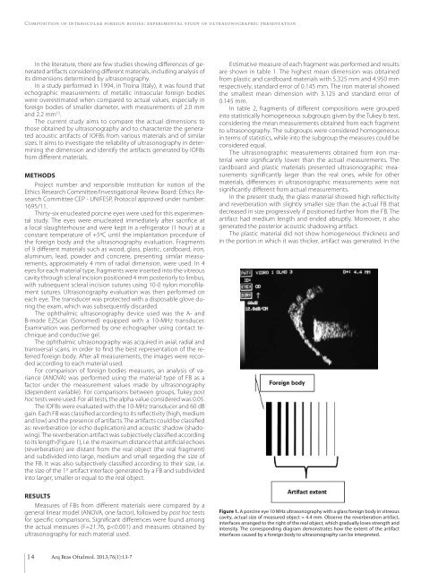
The reverberation artifact was subjectively classified accord<strong>in</strong>g<br />
to its length (Figure 1), i.e. the maximum distance that artificial echoes<br />
(reverberation) are distant from the real object (the real fragment)<br />
and subdivided <strong>in</strong>to large, medium and small regard<strong>in</strong>g the size of<br />
the FB. It was also subjectively classified accord<strong>in</strong>g to their size, i.e.<br />
the size of the 1 st artifact <strong>in</strong>terface generated by a FB and subdivided<br />
<strong>in</strong>to larger, smaller or equal to the real object.<br />
RESULTS<br />
Measures of FBs from different materials were compared by a<br />
general l<strong>in</strong>ear model (ANOVA, one factor), followed by post hoc tests<br />
for specific comparisons. Significant differences were found among<br />
the actual measures (F=21.76, p


