

Photocoagulation versus intravitreal injection in diabetic retinopathy ...
Photocoagulation versus intravitreal injection in diabetic retinopathy ...
Photocoagulation versus intravitreal injection in diabetic retinopathy ...

You also want an ePaper? Increase the reach of your titles
YUMPU automatically turns print PDFs into web optimized ePapers that Google loves.
Maestr<strong>in</strong>i HA, et al.<br />
of pigment on the corneal endothelium. IOP was low (6/9 mmHg<br />
OD/OS). The <strong>in</strong>itial diagnosis was bilateral anterior uveitis, and the<br />
patient was treated us<strong>in</strong>g a topical steroid (1% prednisolone every<br />
3 hours) with marked relief of symptoms. However, on the fourth<br />
day, while play<strong>in</strong>g “foot volley”, his vision suddenly blurred. The next<br />
day, slit-lamp evaluation revealed a dense Krukenberg sp<strong>in</strong>dle bilaterally.<br />
The aqueous humor was turbid with heavy pigment dispersion<br />
<strong>in</strong> the anterior chamber. Pigment granules were deposited on<br />
the anterior surface of the iris and posterior lens surface (Figure 1).<br />
Transillum<strong>in</strong>ation showed extensive bilateral iris defects (Figure 2).<br />
IOP was 36/40 mmHg OD/OS. Gonioscopy revealed a wide open angle,<br />
a dark trabecular meshwork 360 degrees, and a heavy pigment<br />
deposition, particularly <strong>in</strong> the <strong>in</strong>ferior sector (Figure 3). The vitreous<br />
was clear, the fundus was normal, and the optic nerve head cupdisc<br />
ratio was 0.1 bilaterally. Over the follow<strong>in</strong>g days, IOP <strong>in</strong>creased<br />
to 48 mmHg <strong>in</strong> both eyes. The pupil be came progressively irregular<br />
with small posterior synechiae (Figure 4) and iris transillum<strong>in</strong>ation<br />
gradually <strong>in</strong>creased. Corneal endothelial cell density was normal<br />
(2,877 cells/mm 2 OD and 2,984 cells/mm 2 OS).<br />
Previous cl<strong>in</strong>ical and ocular histories were unremarkable, with<br />
the exception of a previous laser photocoagulation <strong>in</strong> the right eye<br />
to treat a peripheral ret<strong>in</strong>al tear 1 year prior to BADI onset. IOP was<br />
normal at that time (10 mmHg bilaterally).<br />
The patient underwent a complete laboratory evaluation, <strong>in</strong>clud<strong>in</strong>g<br />
complete blood count, erythrocyte sedimentation rate, liver en -<br />
zymes, chest X-ray, anti-nuclear factor, rheumatoid factor, C-reactive<br />
prote<strong>in</strong>, tubercul<strong>in</strong> sk<strong>in</strong> test, blood and ur<strong>in</strong>ary calcium, angiotens<strong>in</strong><br />
convert<strong>in</strong>g enzyme, and lysozyme. The results were unremarkable.<br />
Serological tests for toxoplasmosis, syphilis, herpes simplex virus (HSV)<br />
I and II, varicella-zoster virus, and cytomegalovirus (CMV) were performed.<br />
All IgM antibodies were negative. IgG antibodies were positive<br />
aga<strong>in</strong>st toxoplasmosis, HSV I and II, varicella-zoster virus and CMV.<br />
Prednisolone eye drops were adm<strong>in</strong>istered every 3 hours and the<br />
maximum tolerated hypotensive treatment was <strong>in</strong>stituted (timolol,<br />
brimonid<strong>in</strong>e, and oral acetazolamide 750 mg/day), which lowered<br />
IOP to 25-30 mmHg bilaterally. Mydriasis always caused an acute rise<br />
<strong>in</strong> IOP up to 42 mmHg because of an <strong>in</strong>crease <strong>in</strong> pigment discharge.<br />
Abrupt discont<strong>in</strong>uation of prednisolone resulted <strong>in</strong> immediate relapse.<br />
On day 28, empirical treatment with oral valacyclovir 3 g/day was<br />
<strong>in</strong>troduced result<strong>in</strong>g <strong>in</strong> a significant reduction <strong>in</strong> pigment dispersion<br />
<strong>in</strong> a few days. After 3 weeks, the dose was reduced to 1,500 mg/day<br />
for 3 more weeks, and then to 500 mg/day for 8 weeks. Prednisolone<br />
was gradually tapered and completely discont<strong>in</strong>ued after 18 weeks.<br />
IOP gradually decreased throughout the second and third months,<br />
and hypotensive medications were gradually tapered until discont<strong>in</strong>uation<br />
after 24 weeks. The time to complete resolution of pigment<br />
dispersion was 18 weeks. No cupp<strong>in</strong>g of the optic discs was observed;<br />
however, <strong>in</strong> the seventh week, a small disc hemorrhage appeared <strong>in</strong><br />
the left eye. Visual acuity and visual fields rema<strong>in</strong>ed normal; however,<br />
the photophobia was permanent. Six months after complete<br />
resolution, we performed a water dr<strong>in</strong>k<strong>in</strong>g test and IOP rema<strong>in</strong>ed at<br />
normal levels (IOP rose from 13 to 19 mmHg bilaterally). After 1 year,<br />
the patient developed bilateral cataracts, which were removed uneventfully.<br />
IOP was high on the first postoperative day (34/26 mmHg<br />
OD/OS), but returned to12 mmHg after 1 week.<br />
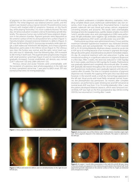
Figure 1. Heavy pigment deposit on the posterior lens surface <strong>in</strong> the left eye.<br />
Figure 3. Gonioscopic view of the <strong>in</strong>ferior sector of the anterior chamber angle (right<br />
eye). Heavy pigment deposit <strong>in</strong> the trabecular meshwork. The aspect is identical <strong>in</strong><br />
the left eye.<br />
A<br />
Figure 2. Diffuse and severe iris defects seen on retroillum<strong>in</strong>ation <strong>in</strong> the right (A) and<br />
left (B) eyes.<br />
B<br />
A<br />
Figure 4. Iris aspect 1 month after presentation <strong>in</strong> the right (A) and left (B) eyes. Areas<br />
of discrete atrophy are apparent <strong>in</strong> the midstroma, particularly <strong>in</strong> the <strong>in</strong>ferior half. Small<br />
pigmented granules deposited on the anterior surface of the iris. Pupils are distorted<br />
and unresponsive to light.<br />
B<br />
Arq Bras Oftalmol. 2013;76(1):42-4<br />
43


