Breast Cancer whole genome sequences-Nature May 2016

Create successful ePaper yourself
Turn your PDF publications into a flip-book with our unique Google optimized e-Paper software.
RESEARCH<br />
ARTICLE<br />
a C>A C>G C>T T>A T>C T>G C>A C>G C>T T>A T>C T>G<br />
0.2<br />
Signature 1<br />
0.2<br />
Signature 18<br />
0.1<br />
0.1<br />
b<br />
c<br />
0.2<br />
0.1<br />
0.2<br />
0.1<br />
0.2<br />
0.1<br />
0.2<br />
0.1<br />
0.2<br />
0.1<br />
Signature 2<br />
Signature<br />
13<br />
Signature 3<br />
Signature 8<br />
Signature 5<br />
0.2<br />
Signature 6<br />
0.1<br />
(CCCCAGATGGTGGG)), shifting it away from the consensus 36 . The<br />
association with particular mutational signatures suggests that these<br />
may also be in a region of hypermutability rather than drivers.<br />
0.2<br />
0.1<br />
0.2<br />
0.1<br />
0.2<br />
0.1<br />
0.1<br />
Signature 17<br />
Signature 20<br />
Signature 26<br />
0.2 Signature 30<br />
Signatures 1 5 2 13 6 20 26 3 8 18 17 30<br />
Percentage<br />
of samples<br />
100<br />
80<br />
60<br />
40<br />
20<br />
100,000<br />
Mutation<br />
10,000<br />
count 1,000<br />
per sample<br />
100<br />
10<br />
1<br />
1 2 13 3 8 5 18 17 6 20 26 30<br />
Previously identified<br />
in breast cancers<br />
Previously identified in<br />
other cancer types<br />
Novel<br />
signatures<br />
100,000<br />
0<br />
100%<br />
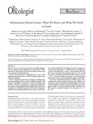
Figure 3 | Extraction and contributions of base substitution signatures<br />
in 560 breast cancers. a, Twelve mutation signatures extracted using nonnegative<br />
matrix factorization. Each signature is ordered by mutation class<br />
(C>A/G>T, C>G/G>C, C>T/G>A, T>A/A>T, T>C/A>G, T>G/A<br />
>C), taking immediate flanking sequence into account. For each class,<br />
mutations are ordered by 5′ base (A, C, G, T) first before 3′ base (A, C,<br />
G, T). b, The spectrum of base substitution signatures within 560 breast<br />
cancers. Mutation signatures are ordered (and coloured) according to<br />
broad biological groups: signatures 1 and 5 are correlated with age of<br />
diagnosis; signatures 2 and 13 are putatively APOBEC-related; signatures 6,<br />
20 and 26 are associated with mismatch-repair deficiency; signatures 3<br />
and 8 are associated with homologous-recombination deficiency;<br />
signatures 18, 17 and 30 have unknown aetiology. For ease of reading, this<br />
arrangement is adopted for the rest of the manuscript. Samples are ordered<br />
according to hierarchical clustering performed on mutation signatures.<br />
Top, absolute numbers of mutations of each signature in each sample.<br />
Bottom, proportion of each signature in each sample. c, Distribution of<br />
mutation counts for each signature in relevant breast cancer samples.<br />
Percentage of samples carrying each signature provided above each<br />
signature.<br />
0%<br />
The WDR74 promoter showed base substitutions and indels<br />
(q value 4.6 × 10 −3 ) forming a cluster of overlapping mutations 20<br />
(Fig. 2a). Coding sequence driver mutations in WDR74 have not been<br />
reported. No differences were observed in WDR74 transcript levels<br />
between cancers with WDR74 promoter mutations compared to those<br />
without. Nevertheless, the pattern of this non-coding mutation cluster,<br />
with overlapping and different mutation types, is more compatible with<br />
the possibility of the mutations being drivers.<br />
Two long non-coding RNAs, MALAT1 (q value 8.7 × 10 −11 , as previously<br />
reported 12 ) and NEAT1 (q value 2.1 × 10 −2 ) were enriched with<br />
mutations. Transcript levels were not significantly different between<br />
mutated and non-mutated samples. Whether these mutations are drivers<br />
or result from local hypermutability is unclear.<br />
Mutational signatures<br />
Mutational processes generating somatic mutations imprint particular<br />
patterns of mutations on cancer <strong>genome</strong>s, termed signatures 2,24,37 .<br />
Applying a mathematical approach 25 to extract mutational signatures<br />
previously revealed five base-substitution signatures in breast<br />
cancer: signatures 1, 2, 3, 8 and 13 (refs 2, 24). Using this method for<br />
the 560 cases revealed twelve signatures, including those previously<br />
observed and a further seven, of which five have formerly been detected<br />
in other cancer types (signatures 5, 6, 17, 18 and 20) and two are new<br />
(signatures 26 and 30) (Fig. 3a, b, 4a, Supplementary Table 21a–c,<br />
Supplementary Methods section 15). Two indel signatures were also<br />
found 2,24 .<br />
Signatures of rearrangement mutational processes have not previously<br />
been formally investigated. To enable this we adopted a rearrangement<br />
classification incorporating 32 subclasses. In many cancer<br />
<strong>genome</strong>s, large numbers of rearrangements are regionally clustered, for<br />
example in zones of gene amplification. Therefore, we first classified<br />
rearrangements into those inside and outside clusters, further subclassified<br />
them into deletions, inversions and tandem duplications, and then<br />
according to the size of the rearranged segment. The final category in<br />
both groups was interchromosomal translocations.<br />
Application of the mathematical framework used for base substitution<br />
signatures 2,24,25 extracted six rearrangement signatures (Fig. 4b,<br />
Supplementary Table 21). Unsupervised hierarchical clustering on the<br />
basis of the proportion of rearrangements attributed to each signature<br />
in each breast cancer yielded seven major subgroups exhibiting distinct<br />
associations with other genomic, histological or gene expression features<br />
(Fig. 5, Extended Data Figs 4–6).<br />
Rearrangement signature 1 (9% of all rearrangements) and rearrangement<br />
signature 3 (18% rearrangements) were characterized<br />
predominantly by tandem duplications (Fig. 4b). Tandem duplications<br />
associated with rearrangement signature 1 were mostly >100 kb<br />
(Fig. 4b), and those with rearrangement signature 3 were