Breast Cancer whole genome sequences-Nature May 2016

You also want an ePaper? Increase the reach of your titles
YUMPU automatically turns print PDFs into web optimized ePapers that Google loves.
ARTICLE<br />
doi:10.1038/nature17676<br />
Landscape of somatic mutations in 560<br />
breast cancer <strong>whole</strong>-<strong>genome</strong> <strong>sequences</strong><br />
Serena Nik-Zainal 1,2 , Helen Davies 1 , Johan Staaf 3 , Manasa Ramakrishna 1 , Dominik Glodzik 1 , Xueqing Zou 1 , Inigo Martincorena 1 ,<br />
Ludmil B. Alexandrov 1,4,5 , Sancha Martin 1 , David C. Wedge 1 , Peter Van Loo 1,6 , Young Seok Ju 1 , Marcel Smid 7 , Arie B. Brinkman 8 ,<br />
Sandro Morganella 9 , Miriam R. Aure 10,11 , Ole Christian Lingjærde 11,12 , Anita Langerød 10,11 , Markus Ringnér 3 , Sung-Min Ahn 13 ,<br />
Sandrine Boyault 14 , Jane E. Brock 15 , Annegien Broeks 16 , Adam Butler 1 , Christine Desmedt 17 , Luc Dirix 18 , Serge Dronov 1 ,<br />
Aquila Fatima 19 , John A. Foekens 7 , Moritz Gerstung 1 , Gerrit K. J. Hooijer 20 , Se Jin Jang 21 , David R. Jones 1 , Hyung-Yong Kim 22 ,<br />
Tari A. King 23 , Savitri Krishnamurthy 24 , Hee Jin Lee 21 , Jeong-Yeon Lee 25 , Yilong Li 1 , Stuart McLaren 1 , Andrew Menzies 1 ,<br />
Ville Mustonen 1 , Sarah O’Meara 1 , Iris Pauporté 26 , Xavier Pivot 27 , Colin A. Purdie 28 , Keiran Raine 1 , Kamna Ramakrishnan 1 ,<br />
F. Germán Rodríguez-González 7 , Gilles Romieu 29 , Anieta M. Sieuwerts 7 , Peter T. Simpson 30 , Rebecca Shepherd 1 ,<br />
Lucy Stebbings 1 , Olafur A. Stefansson 31 , Jon Teague 1 , Stefania Tommasi 32 , Isabelle Treilleux 33 , Gert G. Van den Eynden 18,34 ,<br />
Peter Vermeulen 18,34 , Anne Vincent-Salomon 35 , Lucy Yates 1 , Carlos Caldas 36 , Laura van’t Veer 16 , Andrew Tutt 37,38 ,<br />
Stian Knappskog 39,40 , Benita Kiat Tee Tan 41,42 , Jos Jonkers 16 , Åke Borg 3 , Naoto T. Ueno 24 , Christos Sotiriou 17 , Alain Viari 43,44 ,<br />
P. Andrew Futreal 1,45 , Peter J. Campbell 1 , Paul N. Span 46 , Steven Van Laere 18 , Sunil R. Lakhani 30,47 , Jorunn E. Eyfjord 31 ,<br />
Alastair M. Thompson 28,48 , Ewan Birney 9 , Hendrik G. Stunnenberg 8 , Marc J. van de Vijver 20 , John W. M. Martens 7 ,<br />
Anne-Lise Børresen-Dale 10,11 , Andrea L. Richardson 15,19 , Gu Kong 22 , Gilles Thomas 44 & Michael R. Stratton 1<br />
We analysed <strong>whole</strong>-<strong>genome</strong> <strong>sequences</strong> of 560 breast cancers to advance understanding of the driver mutations conferring<br />
clonal advantage and the mutational processes generating somatic mutations. We found that 93 protein-coding cancer<br />
genes carried probable driver mutations. Some non-coding regions exhibited high mutation frequencies, but most have<br />
distinctive structural features probably causing elevated mutation rates and do not contain driver mutations. Mutational<br />
signature analysis was extended to <strong>genome</strong> rearrangements and revealed twelve base substitution and six rearrangement<br />
signatures. Three rearrangement signatures, characterized by tandem duplications or deletions, appear associated with<br />
defective homologous-recombination-based DNA repair: one with deficient BRCA1 function, another with deficient<br />
BRCA1 or BRCA2 function, the cause of the third is unknown. This analysis of all classes of somatic mutation across<br />
exons, introns and intergenic regions highlights the repertoire of cancer genes and mutational processes operating, and<br />
progresses towards a comprehensive account of the somatic genetic basis of breast cancer.<br />
The mutational theory of cancer proposes that changes in DNA<br />
sequence, termed ‘driver’ mutations, confer proliferative advantage<br />
on a cell, leading to outgrowth of a neoplastic clone 1 . Some<br />
driver mutations are inherited in the germline, but most arise in<br />
somatic cells during the lifetime of the cancer patient, together with<br />
many ‘passenger’ mutations not implicated in cancer development 1 .<br />
Multiple mutational processes, including endogenous and exogenous<br />
mutagen exposures, aberrant DNA editing, replication errors<br />
1 Wellcome Trust Sanger Institute, Hinxton, Cambridge CB10 1SA, UK. 2 East Anglian Medical Genetics Service, Cambridge University Hospitals NHS Foundation Trust, Cambridge CB2 9NB, UK.<br />
3 Division of Oncology and Pathology, Department of Clinical Sciences Lund, Lund University, Lund SE-223 81, Sweden. 4 Theoretical Biology and Biophysics (T-6), Los Alamos National Laboratory,<br />
Los Alamos, NM 87545, New Mexico, USA. 5 Center for Nonlinear Studies, Los Alamos National Laboratory, Los Alamos, New Mexico 87545, USA. 6 Department of Human Genetics, University of<br />
Leuven, B-3000 Leuven, Belgium. 7 Department of Medical Oncology, Erasmus MC <strong>Cancer</strong> Institute and <strong>Cancer</strong> Genomics Netherlands, Erasmus University Medical Center, Rotterdam 3015CN, The<br />
Netherlands. 8 Radboud University, Department of Molecular Biology, Faculty of Science, 6525GA Nijmegen, The Netherlands. 9 European Molecular Biology Laboratory, European Bioinformatics<br />
Institute, Wellcome Trust Genome Campus, Hinxton, Cambridge CB10 1SD, UK. 10 Department of <strong>Cancer</strong> Genetics, Institute for <strong>Cancer</strong> Research, Oslo University Hospital, The Norwegian Radium<br />
Hospital, Oslo 0310, Norway. 11 K. G. Jebsen Centre for <strong>Breast</strong> <strong>Cancer</strong> Research, Institute for Clinical Medicine, University of Oslo, Oslo 0310, Norway. 12 Department of Computer Science,<br />
University of Oslo, Oslo, Norway. 13 Gachon Institute of Genome Medicine and Science, Gachon University Gil Medical Center, Incheon, South Korea. 14 Translational Research Lab, Centre Léon<br />
Bérard, 28, rue Laënnec, 69373 Lyon Cedex 08, France. 15 Department of Pathology, Brigham and Women’s Hospital, Boston, Massachusetts 02115, USA. 16 The Netherlands <strong>Cancer</strong> Institute,<br />
1066 CX Amsterdam, The Netherlands. 17 <strong>Breast</strong> <strong>Cancer</strong> Translational Research Laboratory, Université Libre de Bruxelles, Institut Jules Bordet, Bd de Waterloo 121, B-1000 Brussels, Belgium.<br />
18 Translational <strong>Cancer</strong> Research Unit, Center for Oncological Research, Faculty of Medicine and Health Sciences, University of Antwerp, Antwerp, Belgium. 19 Dana-Farber <strong>Cancer</strong> Institute, Boston,<br />
Massachusetts 02215, USA. 20 Department of Pathology, Academic Medical Center, Meibergdreef 9, 1105 AZ Amsterdam, The Netherlands. 21 Department of Pathology, Asan Medical Center,<br />
College of Medicine, Ulsan University, Ulsan, South Korea. 22 Department of Pathology, College of Medicine, Hanyang University, Seoul 133-791, South Korea. 23 Memorial Sloan Kettering <strong>Cancer</strong><br />
Center, 1275 York Avenue, New York, New York 10065, USA. 24 Morgan Welch Inflammatory <strong>Breast</strong> <strong>Cancer</strong> Research Program and Clinic, The University of Texas MD Anderson <strong>Cancer</strong> Center,<br />
1515 Holcombe Boulevard., Houston, Texas 77030, USA. 25 Institute for Bioengineering and Biopharmaceutical Research (IBBR), Hanyang University, Seoul, South Korea. 26 Institut National du<br />
<strong>Cancer</strong>, Research Division, Clinical Research Department, 52 avenue Morizet, 92513 Boulogne-Billancourt, France. 27 University Hospital of Minjoz, INSERM UMR 1098, Bd Fleming, Besançon<br />
25000, France. 28 Pathology Department, Ninewells Hospital and Medical School, Dundee DD1 9SY, UK. 29 Oncologie Sénologie, ICM Institut Régional du <strong>Cancer</strong>, Montpellier, France. 30 The<br />
University of Queensland, UQ Centre for Clinical Research and School of Medicine, Brisbane, Queensland 4029, Australia. 31 <strong>Cancer</strong> Research Laboratory, Faculty of Medicine, University of Iceland,<br />
101 Reykjavik, Iceland. 32 IRCCS Istituto Tumori “Giovanni Paolo II”, Bari, Italy. 33 Department of Pathology, Centre Léon Bérard, 28 rue Laënnec, 69373 Lyon Cédex 08, France. 34 Department of<br />
Pathology, GZA Hospitals Sint-Augustinus, Antwerp, Belgium. 35 Institut Curie, Paris Sciences Lettres University, Department of Pathology and INSERM U934, 26 rue d’Ulm, 75248 Paris Cedex<br />
05, France. 36 <strong>Cancer</strong> Research UK Cambridge Institute, University of Cambridge, Li Ka Shing Centre, Robinson Way, Cambridge CB2 0RE, UK. 37 <strong>Breast</strong> <strong>Cancer</strong> Now Research Unit, King’s College<br />
London, London SE1 9RT, UK. 38 <strong>Breast</strong> <strong>Cancer</strong> Now Toby Robins Research Centre, Institute of <strong>Cancer</strong> Research, London SW3 6JB, UK. 39 Department of Clinical Science, University of Bergen, 5020<br />
Bergen, Norway. 40 Department of Oncology, Haukeland University Hospital, 5021 Bergen, Norway. 41 National <strong>Cancer</strong> Centre Singapore, 11 Hospital Drive, 169610, Singapore. 42 Singapore General<br />
Hospital, Outram Road, 169608, Singapore. 43 Equipe Erable, INRIA Grenoble-Rhône-Alpes, 655, Avenue de l’Europe, 38330 Montbonnot-Saint Martin, France. 44 Synergie Lyon <strong>Cancer</strong>, Centre<br />
Léon Bérard, 28 rue Laënnec, Lyon Cedex 08, France. 45 Department of Genomic Medicine, UT MD Anderson <strong>Cancer</strong> Center, Houston, Texas 77230, USA. 46 Department of Radiation Oncology,<br />
Department of Laboratory Medicine, Radboud University Medical Center, Nijmegen 6525GA, The Netherlands. 47 Pathology Queensland, The Royal Brisbane and Women’s Hospital, Brisbane,<br />
Queensland 4029, Australia. 48 Department of <strong>Breast</strong> Surgical Oncology, University of Texas MD Anderson <strong>Cancer</strong> Center, 1400 Pressler Street, Houston, Texas 77030, USA.<br />
00 MONTH <strong>2016</strong> | VOL 000 | NATURE | 1<br />
© <strong>2016</strong> Macmillan Publishers Limited. All rights reserved
RESEARCH<br />
ARTICLE<br />
a<br />
80,000<br />
5,000<br />
1,200<br />
Substitutions<br />
Indels<br />
Rearrangements<br />
* *<br />
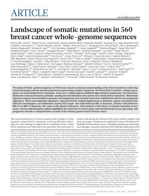
Figure 1 | Cohort and catalogue of somatic mutations in 560 breast<br />
cancers. a, Catalogue of base substitutions, insertions/deletions,<br />
rearrangements and driver mutations in 560 breast cancers (sorted by<br />
total substitution burden). Indel axis limited to 5,000(*). b, Complete list<br />
of curated driver genes sorted by frequency (descending). Fraction of ERpositive<br />
(left, total 366) and ER-negative (right, total 194) samples carrying<br />
a mutation in the relevant driver gene presented in grey. log 10 P value of<br />
enrichment of each driver gene towards the ER-positive or ER-negative<br />
cohort is provided in black. Highlighted in green are genes for which there<br />
is new or further evidence supporting these as novel breast cancer genes.<br />
b<br />
20<br />
Fraction of samples<br />
with driver<br />
100 80<br />
60<br />
ER positive<br />
Driver mutations<br />
40<br />
20<br />
0<br />
ER positive<br />
Enrichment<br />
log P value<br />
–10<br />
TP53<br />
PIK3CA<br />
MYC<br />
CCND1<br />
PTEN<br />
ERBB2<br />
ZNF703/FGFR1<br />
GATA3<br />
RB1<br />
MAP3K1<br />
MAP2K4<br />
ZNF217<br />
CDH1<br />
MLL3<br />
ARID1B<br />
CDKN2A<br />
MLLT4<br />
AKT1<br />
FBXW7<br />
ARID1A<br />
CCND3<br />
CBFB<br />
MDM2<br />
CCNE1<br />
CDKN2B<br />
NCOR1<br />
SF3B1<br />
SPEN<br />
TBX3<br />
IGF1R<br />
BRCA2<br />
NF1<br />
PIK3R1<br />
EGFR<br />
KRAS<br />
ESR1<br />
FOXA1<br />
MED23<br />
CDK6<br />
NOTCH2<br />
AKT2<br />
BRCA1<br />
CTCF<br />
KDM6A<br />
SETD2<br />
CREBBP<br />
DNMT3A<br />
FOXP1<br />
MLL2<br />
RUNX1<br />
USP9X<br />
XBP1<br />
PDGFRA<br />
ATR<br />
ERBB3<br />
FGFR2<br />
PALB2<br />
RHOA<br />
SMAD4<br />
ATM<br />
ATRX<br />
AXIN1<br />
BCOR<br />
CDKN1B<br />
CUX1<br />
GNAS<br />
MLH1<br />
NOTCH1<br />
PHF6<br />
SMARCA4<br />
STAG2<br />
ZFP36L1<br />
APC<br />
CASP8<br />
CBLB<br />
CNOT3<br />
ECT2L<br />
MEN1<br />
MSH2<br />
NRAS<br />
PBRM1<br />
PMS2<br />
STK11<br />
TET2<br />
ASXL1<br />
BRAF<br />
BUB1B<br />
CIC<br />
ERCC4<br />
HRAS<br />
NF2<br />
PRDM1<br />
PREX2<br />
010<br />
ER negative<br />
Fraction of samples<br />
with driver<br />
0 20 40 60 80 100<br />
ER negative<br />
and defective DNA maintenance, are responsible for generating<br />
these mutations 1–3 .<br />
Over the past five decades, several waves of technology have advanced<br />
the characterization of mutations in cancer <strong>genome</strong>s. Karyotype<br />
analysis revealed rearranged chromosomes and copy number<br />
alterations. Subsequently, loss of heterozygosity analysis, hybridization<br />
of cancer-derived DNA to microarrays and other approaches provided<br />
higher resolution insights into copy number changes 4–8 . Recently, DNA<br />
sequencing has enabled systematic characterization of the full repertoire<br />
of mutation types including base substitutions, small insertions/<br />
deletions, rearrangements and copy number changes 9–13 , yielding<br />
substantial insights into the mutated cancer genes and mutational<br />
processes operative in human cancer.<br />
As for many cancer classes, most currently available breast cancer<br />
<strong>genome</strong> <strong>sequences</strong> target protein-coding exons 8,11–15 . Consequently,<br />
there has been limited consideration of mutations in untranslated,<br />
intronic and intergenic regions, leaving central questions pertaining<br />
to the molecular pathogenesis of the disease unresolved. First, the role<br />
of activating driver rearrangements 16–18 forming chimaeric (fusion)<br />
genes/proteins or relocating genes adjacent to new regulatory regions<br />
as observed in haematological and other malignancies 19 . Second, the<br />
role of driver substitutions and indels in non-coding regions of the<br />
<strong>genome</strong> 20,21 . Common inherited variants conferring susceptibility to<br />
human disease are generally in non-coding regulatory regions and<br />
the possibility that similar mechanisms operate somatically in cancer<br />
was highlighted by the discovery of somatic driver substitutions in the<br />
TERT gene promoter 22,23 . Third, which mutational processes generate<br />
the somatic mutations found in breast cancer 2,24 . Addressing this<br />
question has been constrained because exome <strong>sequences</strong> do not inform<br />
on <strong>genome</strong> rearrangements and capture relatively few base substitution<br />
mutations, thus limiting statistical power to extract the mutational<br />
signatures imprinted on the <strong>genome</strong> by these processes 24,25 .<br />
Here we analyse <strong>whole</strong>-<strong>genome</strong> <strong>sequences</strong> of 560 cases in order to<br />
address these and other questions and to pave the way to a comprehensive<br />
understanding of the origins and con<strong>sequences</strong> of somatic<br />
mutations in breast cancer.<br />
<strong>Cancer</strong> genes and driver mutations<br />
The <strong>whole</strong> <strong>genome</strong>s of 560 breast cancers and non-neoplastic<br />
tissue from each individual (556 female and 4 male) were<br />
sequenced (Supplementary Fig. 1, Supplementary Table 1).<br />
We detected 3,479,652 somatic base substitutions, 371,993 small indels<br />
and 77,695 rearrangements, with substantial variation in the number<br />
of each between individual samples (Fig. 1a, Supplementary Table 3).<br />
Transcriptome sequence, microRNA expression, array-based copy number<br />
and DNA methylation data were obtained from subsets of cases.<br />
To identify new cancer genes, we combined somatic substitutions<br />
and indels in protein-coding exons with data from other series 12–15,26 ,<br />
constituting a total of 1,332 breast cancers, and searched for mutation<br />
clustering in each gene beyond that expected by chance. Five cancer<br />
genes were found for which evidence was previously absent or equivocal<br />
(MED23, FOXP1, MLLT4, XBP1, ZFP36L1), or for which the mutations<br />
indicate the gene acts in breast cancer in a recessive rather than in<br />
a dominant fashion, as previously reported in other cancer types (see<br />
Supplementary Methods section 7.4 for detailed descriptions). From<br />
published reports on all cancer types (http://cancer.sanger.ac.uk/census),<br />
2 | NATURE | VOL 000 | 00 MONTH <strong>2016</strong><br />
© <strong>2016</strong> Macmillan Publishers Limited. All rights reserved
ARTICLE<br />
RESEARCH<br />
we then compiled a list of 727 human cancer genes (Supplementary<br />
Table 12). On the basis of driver mutations found previously, we<br />
defined conservative rules for somatic driver base substitutions and<br />
indel mutations in each gene and sought mutations conforming to<br />
these rules in the 560 breast cancers. We identified 916 probable driver<br />
mutations of these classes (Fig. 1b, Supplementary Table 14, Extended<br />
Data Fig. 1).<br />
To explore the role of genomic rearrangements as driver mutations<br />
16,18,19,27 , we sought predicted in-frame fusion genes that might<br />
create activated, dominant cancer genes. We identified 1,278 unique<br />
and 39 infrequently recurrent in-frame gene fusions (Supplementary<br />
Table 15). Many of the latter, however, were in regions of high<br />
rearrangement density, including amplicons 28 and fragile sites, and<br />
their recurrence is probably attributable to chance 27 . Furthermore,<br />
transcriptome <strong>sequences</strong> from 260 cancers did not show expression of<br />
these fusions and generally confirmed the rarity of recurrent in-frame<br />
fusion genes. By contrast, recurrent rearrangements interrupting the<br />
gene footprints of CDKN2A, RB1, MAP3K1, PTEN, MAP2K4, ARID1B,<br />
FBXW7, MLLT4 and TP53 were found beyond the numbers expected<br />
from local background rearrangement rates, indicating that they contribute<br />
to the driver mutation burden of recessive cancer genes. Several<br />
other recurrently rearranged genomic regions were observed, including<br />
dominantly acting cancer genes ETV6 and ESR1 (without consistent<br />
elevation in expression levels), L1 retrotransposition sites 29 and fragile<br />
sites. The significance of these recurrently rearranged regions remains<br />
unclear (Extended Data Fig. 2).<br />
Incorporation of recurrent copy number changes, including homozygous<br />
deletions and amplifications, generated a total of 1,628 likely<br />
driver mutations in 93 cancer genes (Fig. 1b). At least one driver was<br />
identifiable in 95% of cancers. The 10 most frequently mutated genes<br />
were TP53, PIK3CA, MYC, CCND1, PTEN, ERBB2, ZNF703/FGFR1<br />
locus, GATA3, RB1 and MAP3K1 (Fig. 1b, Extended Data Fig. 1), and<br />
these accounted for 62% of drivers.<br />
Recurrent somatic mutations in non-coding regions<br />
To investigate non-coding somatic driver substitutions and indels, we<br />
searched for non-coding genomic regions with more mutations than<br />
expected by chance (Fig. 2a, Supplementary Table 16, Extended Data Fig. 3).<br />
The promoter of PLEKHS1 exhibited recurrent mutations at two<br />
genomic positions 30 (Fig. 2a) TTTTGCAAT TGAACA ATTGCAAAA<br />
(as previously reported 30 ). The two mutated bases, within a 6 base pair<br />
(bp) core motif, are flanked, on either side by 9 base pairs of palindromic<br />
sequence forming inverted repeats 31 . Most cancers with these<br />
mutations showed many base substitutions of mutational signatures 2<br />
and 13 that have been attributed to activity of APOBEC DNA-editing<br />
proteins that target the TCN sequence motif. One of the mutated bases<br />
is a cytosine in a TCA sequence context (shown above as the reverse<br />
complement, TGA) at which predominantly C>T substitutions were<br />
found. The other is a cytosine in ACA context, which showed both<br />
C>T and C>G mutations.<br />
The TGAACA core sequence was mutated at the same two positions<br />
at multiple locations elsewhere in the <strong>genome</strong> (Supplementary<br />
Table 16c) where the TGAACA core was also flanked by palindromes<br />
albeit of different <strong>sequences</strong> and lengths (Supplementary Table 16c).<br />
These mutations were also usually found in cancers with many signature<br />
2 and 13 mutations (Fig. 2a). TGAACA core <strong>sequences</strong> with<br />
longer flanking palindromes generally exhibited a higher mutation rate,<br />
and TGAACA <strong>sequences</strong> flanked by 9 bp palindromes exhibited an<br />
~265-fold higher mutation rate than <strong>sequences</strong> without them (Fig. 2b,<br />
Supplementary Table 16d). However, additional factors must influence<br />
the mutation rate because it varied markedly between TGAACA core<br />
<strong>sequences</strong> with different palindromes of the same length (Fig. 2c).<br />
Some TGAACA-inverted repeat sites were in regulatory regions but<br />
others were intronic or intergenic without functional annotation<br />
(examples in Supplementary Table 16c) or exonic. The propensity for<br />
mutation recurrence at specific positions in a distinctive sequence motif<br />
in cancers with numerous mutations of particular signatures renders it<br />
plausible that these are hypermutable hotspots 32–34 , perhaps through<br />
formation of DNA hairpin structures 35 , which are single-stranded at<br />
their tips enabling attack by APOBEC enzymes, rather than driver<br />
mutations.<br />
Two recurrently mutated sites were also observed in the promoter<br />
of TBC1D12 (TBC1 domain family, member 12) (q value 4.5 × 10 −2 )<br />
(Fig. 2a). The mutations were characteristic of signatures 2 and 13 and<br />
enriched in cancers with many signature 2 and 13 mutations (Fig. 2a).<br />
The mutations were within the TBC1D12 Kozak consensus sequence<br />
a<br />
Substitutions Indels<br />
0% 20% 40% 60% 80% 100%<br />
lncRNA<br />
MALAT1<br />
q value 8.07 × 10 –11 chr11: 65266000 chr11: 65274000<br />
lncRNA<br />
NEAT1<br />
q value 2.10 × 10 –2<br />
chr11: 65190000 chr11: 65210000<br />
Promoter<br />
WDR74<br />
q value 4.60 × 10 –3 chr11: 62600000 chr11: 62610000<br />
Promoter<br />
TBC1D12<br />
q value 4.45 × 10 –2 chr10: 96162000 chr10: 96162350<br />
Promoter<br />
PLEKHS1<br />
q value 1.34 × 10 –5 chr10: 115511200 chr10: 115511700<br />
Proportion of mutational signatures<br />
in total dataset of 560 breast cancers<br />
Signatures 1 5 2 13 6 20 26 3 8 18 17 30<br />
Figure 2 | Non-coding analyses of breast cancer <strong>genome</strong>s.<br />
a, Distributions of substitution (purple dots) and indel (blue dots)<br />
mutations within the footprint of five regulatory regions identified as<br />
being more significantly mutated than expected is provided on the left.<br />
The proportion of base substitution mutation signatures associated with<br />
corresponding samples carrying mutations in each of these non-coding<br />
b<br />
c<br />
Fold difference of mutability<br />
within inverted repeat<br />
Mutation counts<br />
250<br />
200<br />
150<br />
100<br />
50<br />
0<br />
12<br />
10<br />
8<br />
6<br />
4<br />
2<br />
0<br />
P = 7.62 × 10 –69<br />
P = 1.87 × 10 –19<br />
1 2 3 4 5 6 7 8 9 10 11 12 13 14 15<br />
Length of palindrome<br />
chr3: 40588271<br />
chr3: 175146022<br />
chr9: 132583256<br />
chrX: 151686628<br />
chr4: 98589562<br />
chr6: 82403486<br />
chr6: 142706206<br />
chr10: 61432133<br />
Location of inverted repeat<br />
with 9 bp palindrome<br />
chr10: 115511590<br />
chr11: 74510805<br />
regions, is displayed on the right. b, Mutability of TGAACA/TGTTCA<br />
motifs within inverted repeats of varying flanking palindromic sequence<br />
length compared to motifs not within an inverted repeat. c, Variation in<br />
mutability between loci of TGAACA/TGTTCA inverted repeats with 9 bp<br />
palindromes.<br />
00 MONTH <strong>2016</strong> | VOL 000 | NATURE | 3<br />
© <strong>2016</strong> Macmillan Publishers Limited. All rights reserved
RESEARCH<br />
ARTICLE<br />
a C>A C>G C>T T>A T>C T>G C>A C>G C>T T>A T>C T>G<br />
0.2<br />
Signature 1<br />
0.2<br />
Signature 18<br />
0.1<br />
0.1<br />
b<br />
c<br />
0.2<br />
0.1<br />
0.2<br />
0.1<br />
0.2<br />
0.1<br />
0.2<br />
0.1<br />
0.2<br />
0.1<br />
Signature 2<br />
Signature<br />
13<br />
Signature 3<br />
Signature 8<br />
Signature 5<br />
0.2<br />
Signature 6<br />
0.1<br />
(CCCCAGATGGTGGG)), shifting it away from the consensus 36 . The<br />
association with particular mutational signatures suggests that these<br />
may also be in a region of hypermutability rather than drivers.<br />
0.2<br />
0.1<br />
0.2<br />
0.1<br />
0.2<br />
0.1<br />
0.1<br />
Signature 17<br />
Signature 20<br />
Signature 26<br />
0.2 Signature 30<br />
Signatures 1 5 2 13 6 20 26 3 8 18 17 30<br />
Percentage<br />
of samples<br />
100<br />
80<br />
60<br />
40<br />
20<br />
100,000<br />
Mutation<br />
10,000<br />
count 1,000<br />
per sample<br />
100<br />
10<br />
1<br />
1 2 13 3 8 5 18 17 6 20 26 30<br />
Previously identified<br />
in breast cancers<br />
Previously identified in<br />
other cancer types<br />
Novel<br />
signatures<br />
100,000<br />
0<br />
100%<br />
Figure 3 | Extraction and contributions of base substitution signatures<br />
in 560 breast cancers. a, Twelve mutation signatures extracted using nonnegative<br />
matrix factorization. Each signature is ordered by mutation class<br />
(C>A/G>T, C>G/G>C, C>T/G>A, T>A/A>T, T>C/A>G, T>G/A<br />
>C), taking immediate flanking sequence into account. For each class,<br />
mutations are ordered by 5′ base (A, C, G, T) first before 3′ base (A, C,<br />
G, T). b, The spectrum of base substitution signatures within 560 breast<br />
cancers. Mutation signatures are ordered (and coloured) according to<br />
broad biological groups: signatures 1 and 5 are correlated with age of<br />
diagnosis; signatures 2 and 13 are putatively APOBEC-related; signatures 6,<br />
20 and 26 are associated with mismatch-repair deficiency; signatures 3<br />
and 8 are associated with homologous-recombination deficiency;<br />
signatures 18, 17 and 30 have unknown aetiology. For ease of reading, this<br />
arrangement is adopted for the rest of the manuscript. Samples are ordered<br />
according to hierarchical clustering performed on mutation signatures.<br />
Top, absolute numbers of mutations of each signature in each sample.<br />
Bottom, proportion of each signature in each sample. c, Distribution of<br />
mutation counts for each signature in relevant breast cancer samples.<br />
Percentage of samples carrying each signature provided above each<br />
signature.<br />
0%<br />
The WDR74 promoter showed base substitutions and indels<br />
(q value 4.6 × 10 −3 ) forming a cluster of overlapping mutations 20<br />
(Fig. 2a). Coding sequence driver mutations in WDR74 have not been<br />
reported. No differences were observed in WDR74 transcript levels<br />
between cancers with WDR74 promoter mutations compared to those<br />
without. Nevertheless, the pattern of this non-coding mutation cluster,<br />
with overlapping and different mutation types, is more compatible with<br />
the possibility of the mutations being drivers.<br />
Two long non-coding RNAs, MALAT1 (q value 8.7 × 10 −11 , as previously<br />
reported 12 ) and NEAT1 (q value 2.1 × 10 −2 ) were enriched with<br />
mutations. Transcript levels were not significantly different between<br />
mutated and non-mutated samples. Whether these mutations are drivers<br />
or result from local hypermutability is unclear.<br />
Mutational signatures<br />
Mutational processes generating somatic mutations imprint particular<br />
patterns of mutations on cancer <strong>genome</strong>s, termed signatures 2,24,37 .<br />
Applying a mathematical approach 25 to extract mutational signatures<br />
previously revealed five base-substitution signatures in breast<br />
cancer: signatures 1, 2, 3, 8 and 13 (refs 2, 24). Using this method for<br />
the 560 cases revealed twelve signatures, including those previously<br />
observed and a further seven, of which five have formerly been detected<br />
in other cancer types (signatures 5, 6, 17, 18 and 20) and two are new<br />
(signatures 26 and 30) (Fig. 3a, b, 4a, Supplementary Table 21a–c,<br />
Supplementary Methods section 15). Two indel signatures were also<br />
found 2,24 .<br />
Signatures of rearrangement mutational processes have not previously<br />
been formally investigated. To enable this we adopted a rearrangement<br />
classification incorporating 32 subclasses. In many cancer<br />
<strong>genome</strong>s, large numbers of rearrangements are regionally clustered, for<br />
example in zones of gene amplification. Therefore, we first classified<br />
rearrangements into those inside and outside clusters, further subclassified<br />
them into deletions, inversions and tandem duplications, and then<br />
according to the size of the rearranged segment. The final category in<br />
both groups was interchromosomal translocations.<br />
Application of the mathematical framework used for base substitution<br />
signatures 2,24,25 extracted six rearrangement signatures (Fig. 4b,<br />
Supplementary Table 21). Unsupervised hierarchical clustering on the<br />
basis of the proportion of rearrangements attributed to each signature<br />
in each breast cancer yielded seven major subgroups exhibiting distinct<br />
associations with other genomic, histological or gene expression features<br />
(Fig. 5, Extended Data Figs 4–6).<br />
Rearrangement signature 1 (9% of all rearrangements) and rearrangement<br />
signature 3 (18% rearrangements) were characterized<br />
predominantly by tandem duplications (Fig. 4b). Tandem duplications<br />
associated with rearrangement signature 1 were mostly >100 kb<br />
(Fig. 4b), and those with rearrangement signature 3 were
ARTICLE<br />
RESEARCH<br />
a<br />
0.20<br />
b<br />
Clustered rearrangements<br />
Non-clustered rearrangements<br />
del tds inv trans del tds<br />
inv trans<br />
log 10 transcription strand asymmetry<br />
0.15<br />
0.10<br />
0.05<br />
Associated with<br />
mismatch repair<br />
deficiency<br />
Associated with<br />
APOBEC deaminase<br />
activity<br />
Rearrangement<br />
signature 1<br />
Rearrangement<br />
signature 2<br />
Rearrangement<br />
signature 3<br />
Rearrangement<br />
signature 4<br />
60%<br />
0%<br />
60%<br />
0%<br />
60%<br />
0%<br />
60%<br />
0%<br />
0.00<br />
Rearrangement<br />
signature 5<br />
60%<br />
0%<br />
0.00<br />
0.05 0.10 0.15<br />
log 10 replication strand asymmetry<br />
0.20<br />
Rearrangement<br />
signature 6<br />
60%<br />
0%<br />
Signatures 1 5 2 13 6 20 26 3 8 18 17 30<br />
1–10 kb<br />
10–100 kb<br />
100 kb–1 Mb<br />
1–10 Mb<br />
>10 Mb<br />
1–10 kb<br />
10–100 kb<br />
100 kb–1 Mb<br />
1–10 Mb<br />
>10 Mb<br />
1–10 kb<br />
10–100 kb<br />
100 kb–1 Mb<br />
1–10 Mb<br />
>10 Mb<br />
1–10 kb<br />
10–100 kb<br />
100 kb–1 Mb<br />
1–10 Mb<br />
>10 Mb<br />
1–10 kb<br />
10–100 kb<br />
100 kb–1 Mb<br />
1–10 Mb<br />
>10 Mb<br />
1–10 kb<br />
10–100 kb<br />
100 kb–1 Mb<br />
1–10 Mb<br />
>10 Mb<br />
Figure 4 | Additional characteristics of base substitution signatures and<br />
novel rearrangement signatures in 560 breast cancers. a, Contrasting<br />
transcriptional strand asymmetry and replication strand asymmetry<br />
between twelve base substitution signatures. b, Six rearrangement<br />
Rearrangement size<br />
signatures extracted using non-negative matrix factorization. Probability<br />
of rearrangement element on y axis. Rearrangement size on x axis.<br />
del, deletion; tds, tandem duplication; inv, inversion; trans, translocation.<br />
there were nine locations at which recurrence of tandem duplications<br />
was found across the breast cancers and which often showed multiple,<br />
nested tandem duplications in individual cases (Extended Data Fig. 8).<br />
These may be mutational hotspots specific for these tandem duplication<br />
mutational processes, although we cannot exclude the possibility that<br />
they represent driver events.<br />
Rearrangement signature 5 (accounting for 14% rearrangements)<br />
was characterized by deletions 100 kb), inversions<br />
and interchromosomal translocations, was present in most cancers but<br />
was particularly enriched in oestrogen receptor (ER)-positive cancers<br />
with quiet copy number profiles (cluster E, GISTIC (genomic identification<br />
of significant targets in cancer) cluster 3; Fig. 5). Rearrangement<br />
signature 4 (accounting for 18% of rearrangements) was characterized<br />
by clustered interchromosomal translocations, whereas rearrangement<br />
signature 6 (19% of rearrangements) had clustered inversions and<br />
deletions (clusters A, B, C; Fig. 5).<br />
Short segments (1–5 bp) of overlapping microhomology characteristic<br />
of alternative methods of end-joining repair were found at most<br />
rearrangements 2,14 . Rearrangement signatures 2, 4 and 6 were characterized<br />
by a peak at 1 bp of microhomology, whereas rearrangement<br />
signatures 1, 3 and 5, associated with homologous recombination<br />
DNA repair deficiency, exhibited a peak at 2 bp (Extended Data Fig. 9).<br />
Thus, different end-joining mechanisms may operate with different<br />
rearrangement processes. A proportion of breast cancers showed rearrangement<br />
signature 5 deletions with longer (>10 bp) microhomologies<br />
involving <strong>sequences</strong> from short-interspersed nuclear elements, most<br />
commonly AluS (63%) and AluY (15%) family repeats (Extended Data<br />
Fig. 9). Long segments (more than 10 bp) of non-templated sequence<br />
were particularly enriched amongst clustered rearrangements.<br />
Localized hypermutation: kataegis<br />
Focal base-substitution hypermutation, termed kataegis, is generally<br />
characterized by substitutions with characteristic features of signatures 2<br />
and 13 (refs 2, 24). Kataegis was observed in 49% breast cancers, with<br />
4% exhibiting 10 or more foci (Supplementary Table 21c). Kataegis colocalizes<br />
with clustered rearrangements characteristic of rearrangement<br />
signatures 4 and 6 (Fig. 4b). <strong>Cancer</strong>s with tandem duplications or deletions<br />
of rearrangement signatures 1, 3 and 5 did not usually demonstrate<br />
kataegis. However, there must be additional determinants of kataegis as<br />
only 2% of rearrangements are associated with it. A rare (14 out of 1,557<br />
foci, 0.9%) alternative form of kataegis, colocalizing with rearrangements<br />
but with a base-substitution pattern characterized by T>G and<br />
T>C mutations, predominantly at NTT and NTA <strong>sequences</strong> (where<br />
N can be any base A, T, C or G), was also observed (Extended Data<br />
Fig. 10). This pattern of base substitutions most closely matches signature<br />
9 (Extended Data Fig. 10; http://cancer.sanger.ac.uk/cosmic/signatures),<br />
previously observed in B lymphocyte neoplasms and attributed to<br />
polymerase eta activity 41 .<br />
Mutational signatures exhibit distinct DNA replication<br />
strand biases<br />
The distributions of mutations attributable to each of the 20 mutational<br />
signatures (12 base substitution, 2 indel and 6 rearrangement)<br />
were explored 42 with respect to DNA replication strand. We found an<br />
asymmetric distribution of mutations between leading and lagging<br />
replication strands for many, but not all signatures 42 (Fig. 4a). Notably,<br />
signatures 2 and 13, owing to APOBEC deamination, showed marked<br />
lagging-strand replication bias (Fig. 4a) suggesting that lagging-strand<br />
replication provides single-stranded DNA for APOBEC deamination.<br />
Of the three signatures associated with mismatch-repair deficiency<br />
(signatures 6, 20 and 26), only signature 26 exhibited replicative-strand<br />
bias, highlighting how different signatures arising from defects of the<br />
same pathway can exhibit distinct relationships with replication.<br />
Mutational signatures associated with BRCA1 and<br />
BRCA2 mutations<br />
Of the 560 breast cancers, 90 had germline (60) or somatic (14) inactivating<br />
mutations in BRCA1 (35) or BRCA2 (39) or showed methylation<br />
of the BRCA1 promoter (16). Loss of the wild-type chromosome<br />
17 or 13 was observed in 80 out of 90 cases. The latter exhibited<br />
many base substitution mutations of signature 3, accompanied by<br />
deletions of >3 bp with microhomology at rearrangement breakpoints,<br />
and signature 8 together with CC>AA double nucleotide<br />
substitutions. Cases in which the wild-type chromosome 17 or 13<br />
was retained did not show these signatures. Thus signature 3 and,<br />
to a lesser extent, signature 8 are associated with absence of BRCA1<br />
and BRCA2 functions.<br />
00 MONTH <strong>2016</strong> | VOL 000 | NATURE | 5<br />
© <strong>2016</strong> Macmillan Publishers Limited. All rights reserved
RESEARCH<br />
ARTICLE<br />
GISTIC 5<br />
GISTIC 4<br />
GISTIC 3<br />
GISTIC 2<br />
GISTIC 1<br />
HRD top 25%<br />
HRD low 25%<br />
GATA3 mut<br />
PIK3CA mut<br />
PTEN mut<br />
RB1 mut<br />
TP53 mut<br />
HER2<br />
PR<br />
ER<br />
A<br />
B<br />
C<br />
D<br />
E<br />
F<br />
G<br />
miRNAcluster<br />
MYC amp<br />
ZNF217 amp<br />
ZNF703 amp<br />
CCND1 amp<br />
ERBB2 amp<br />
Kataegis<br />
BRCA status<br />
AIMS subtype<br />
Figure 5 | Integrative analysis of rearrangement<br />
signatures. Heatmap of rearrangement<br />
signatures following unsupervised hierarchical<br />
clustering based on proportions of<br />
rearrangement signatures in each cancer. Seven<br />
cluster groups (A–G) noted and relationships<br />
with expression (AIMS) subtype (basal, red;<br />
luminal B, light blue; luminal A, dark blue),<br />
immunohistopathology status (ER, progesterone<br />
receptor (PR), HER2 status; black, positive),<br />
abrogation of BRCA1 (purple) and BRCA2<br />
(orange) (whether germline, somatic or through<br />
promoter hypermethylation), presence of 3 or<br />
more foci of kataegis (black, positive), HRD<br />
index (top 25% or lowest 25%; black, positive),<br />
GISTIC cluster group (black, positive) and<br />
driver mutations in cancer genes. miRNA<br />
cluster groups: 0, red; 1, purple; 2, blue; 3, light<br />
blue; 4, green; 5, orange. Contribution of basesubstitution<br />
signatures in these seven cluster<br />
groups is provided in the bottom panel.<br />
A<br />
B<br />
C<br />
D<br />
E<br />
F<br />
G<br />
Rearrangement signature 1<br />
Rearrangement signature 2<br />
Fraction<br />
of<br />
signature<br />
Rearrangement signature 3<br />
Rearrangement signature 4<br />
0 1<br />
Rearrangement signature 5<br />
Rearrangement signature 6<br />
100%<br />
A<br />
B<br />
C<br />
D<br />
E<br />
F<br />
G<br />
80%<br />
Base substitution<br />
signatures<br />
60%<br />
40%<br />
20%<br />
0%<br />
Signatures 1 5 2 13 6 20 26 3 8 18 17 30<br />
<strong>Cancer</strong>s with inactivating BRCA1 or BRCA2 mutations usually carry<br />
many genomic rearrangements. <strong>Cancer</strong>s with BRCA1, but not BRCA2,<br />
mutations exhibit large numbers of rearrangement signature 3 small<br />
tandem duplications. <strong>Cancer</strong>s with BRCA1 or BRCA2 mutations show<br />
substantial numbers of rearrangement signature 5 deletions. No other<br />
rearrangement signatures were associated with BRCA1- or BRCA2-null<br />
cases (clusters D and G, Fig. 5). Some breast cancers without identifiable<br />
BRCA1/2 mutations or BRCA1 promoter methylation showed these<br />
features and segregated with BRCA1/2-null cancers in hierarchical<br />
clustering analysis (Fig. 5). In such cases, the BRCA1/2 mutations may<br />
have been missed or other mutated or promoter methylated genes may be<br />
exerting similar effects (see http://cancer.sanger.ac.uk/cosmic/sample/<br />
<strong>genome</strong>s for examples of <strong>whole</strong>-<strong>genome</strong> profiles of typical BRCA1-<br />
null, (for example, PD6413a, PD7215a) and BRCA2-null tumours (for<br />
example, PD4952a, PD4955a)).<br />
A further subset of cancers (cluster F, Fig. 5) show similarities in<br />
mutational pattern to BRCA1/2-null cancers, with many rearrangement<br />
signature 5 deletions and enrichment for base substitution signatures 3<br />
and 8. However, these do not segregate together with BRCA1/2-null<br />
cases in hierarchical clustering analysis, have rearrangement signature 1<br />
large tandem duplications and do not show BRCA1/2 mutations.<br />
Somatic and germline mutations in genes associated with the DNA<br />
double-strand break repair pathway including ATM, ATR, PALB2,<br />
RAD51C, RAD50, TP53, CHEK2 and BRIP1, were sought in these cancers.<br />
We did not observe any clear-cut relationships between mutations<br />
in these genes and these mutational patterns.<br />
<strong>Cancer</strong>s with BRCA1/2 mutations are particularly responsive to cisplatin<br />
and PARP inhibitors 43–45 . Combinations of base substitution, indel<br />
and rearrangement mutational signatures may be better biomarkers<br />
of defective homologous-recombination-based DNA double-strand<br />
break repair and responsiveness to these drugs 46 than BRCA1/2 mutations<br />
or promoter methylation alone and thus may constitute the basis<br />
of future diagnostics.<br />
Conclusions<br />
A comprehensive perspective on the somatic genetics of breast cancer<br />
is drawing closer (see http://cancer.sanger.ac.uk/cosmic/sample/<br />
<strong>genome</strong>s for individual patient <strong>genome</strong> profile, and Methods for<br />
orientation). At least 12 base substitution mutational signatures and 6<br />
rearrangement signatures contribute to the somatic mutations found,<br />
and 93 mutated cancer genes (31 dominant, 60 recessive, 2 uncertain)<br />
are implicated in genesis of the disease. However, dominantly<br />
acting activated fusion genes and non-coding driver mutations appear<br />
rare. Additional infrequently mutated cancer genes probably exist.<br />
6 | NATURE | VOL 000 | 00 MONTH <strong>2016</strong><br />
© <strong>2016</strong> Macmillan Publishers Limited. All rights reserved
ARTICLE<br />
RESEARCH<br />
However, the genes harbouring the substantial majority of driver<br />
mutations are now known.<br />
Nevertheless, important questions remain to be addressed. Recurrent<br />
mutational events including <strong>whole</strong>-chromosome copy number changes<br />
and unexplained regions with recurrent rearrangements could harbour<br />
additional cancer genes. Identifying non-coding drivers is challenging<br />
and requires further investigation. Although almost all breast cancers<br />
have at least one identifiable driver mutation, the number with only<br />
a single identified driver is perhaps surprising. The roles of viruses<br />
or other microbes have not been exhaustively examined. Thus, further<br />
exploration and analysis of <strong>whole</strong>-<strong>genome</strong> <strong>sequences</strong> from breast<br />
cancer patients will be required to complete our understanding of the<br />
somatic mutational basis of the disease.<br />
Online Content Methods, along with any additional Extended Data display items and<br />
Source Data, are available in the online version of the paper; references unique to<br />
these sections appear only in the online paper.<br />
Received 29 June 2015; accepted 17 March <strong>2016</strong>.<br />
Published online 2 <strong>May</strong> <strong>2016</strong>.<br />
1. Stratton, M. R., Campbell, P. J. & Futreal, P. A. The cancer <strong>genome</strong>. <strong>Nature</strong> 458,<br />
719–724 (2009).<br />
2. Nik-Zainal, S. et al. Mutational processes molding the <strong>genome</strong>s of 21 breast<br />
cancers. Cell 149, 979–993 (2012).<br />
3. Nik-Zainal, S. et al. The life history of 21 breast cancers. Cell 149, 994–1007<br />
(2012).<br />
4. Hicks, J. et al. Novel patterns of <strong>genome</strong> rearrangement and their association<br />
with survival in breast cancer. Genome Res. 16, 1465–1479 (2006).<br />
5. Bergamaschi, A. et al. Extracellular matrix signature identifies breast<br />
cancer subgroups with different clinical outcome. J. Pathol. 214, 357–367<br />
(2008).<br />
6. Ching, H. C., Naidu, R., Seong, M. K., Har, Y. C. & Taib, N. A. Integrated analysis<br />
of copy number and loss of heterozygosity in primary breast carcinomas using<br />
high-density SNP array. Int. J. Oncol. 39, 621–633 (2011).<br />
7. Fang, M. et al. Genomic differences between estrogen receptor (ER)-positive<br />
and ER-negative human breast carcinoma identified by single nucleotide<br />
polymorphism array comparative <strong>genome</strong> hybridization analysis. <strong>Cancer</strong> 117,<br />
2024–2034 (2011).<br />
8. Curtis, C. et al. The genomic and transcriptomic architecture of 2,000 breast<br />
tumours reveals novel subgroups. <strong>Nature</strong> 486, 346–352 (2012).<br />
9. Pleasance, E. D. et al. A comprehensive catalogue of somatic mutations from a<br />
human cancer <strong>genome</strong>. <strong>Nature</strong> 463, 191–196 (2010).<br />
10. Pleasance, E. D. et al. A small-cell lung cancer <strong>genome</strong> with complex signatures<br />
of tobacco exposure. <strong>Nature</strong> 463, 184–190 (2010).<br />
11. Banerji, S. et al. Sequence analysis of mutations and translocations across<br />
breast cancer subtypes. <strong>Nature</strong> 486, 405–409 (2012).<br />
12. Ellis, M. J. et al. Whole-<strong>genome</strong> analysis informs breast cancer response to<br />
aromatase inhibition. <strong>Nature</strong> 486, 353–360 (2012).<br />
13. Shah, S. P. et al. The clonal and mutational evolution spectrum of primary<br />
triple-negative breast cancers. <strong>Nature</strong> 486, 395–399 (2012).<br />
14. Stephens, P. J. et al. The landscape of cancer genes and mutational processes<br />
in breast cancer. <strong>Nature</strong> 486, 400–404 (2012).<br />
15. The <strong>Cancer</strong> Genome Atlas Network Comprehensive molecular portraits of<br />
human breast tumours. <strong>Nature</strong> 490, 61–70 (2012).<br />
16. Wu, Y. M. et al. Identification of targetable FGFR gene fusions in diverse<br />
cancers. <strong>Cancer</strong> Discovery 3, 636–647 (2013).<br />
17. Giacomini, C. P. et al. Breakpoint analysis of transcriptional and genomic<br />
profiles uncovers novel gene fusions spanning multiple human cancer types.<br />
PLoS Genet. 9, e1003464 (2013).<br />
18. Robinson, D. R. et al. Functionally recurrent rearrangements of the MAST<br />
kinase and Notch gene families in breast cancer. <strong>Nature</strong> Med. 17, 1646–1651<br />
(2011).<br />
19. Karlsson, J. et al. Activation of human telomerase reverse transcriptase through<br />
gene fusion in clear cell sarcoma of the kidney. <strong>Cancer</strong> Lett. 357, 498–501<br />
(2015).<br />
20. Khurana, E. et al. Integrative annotation of variants from 1092 humans:<br />
application to cancer genomics. Science 342, 1235587 (2013).<br />
21. West, J. A. et al. The long noncoding RNAs NEAT1 and MALAT1 bind active<br />
chromatin sites. Mol. Cell 55, 791–802 (2014).<br />
22. Huang, F. W. et al. Highly recurrent TERT promoter mutations in human<br />
melanoma. Science 339, 957–959 (2013).<br />
23. Vinagre, J. et al. Frequency of TERT promoter mutations in human cancers.<br />
<strong>Nature</strong> Commun. 4, 2185 (2013).<br />
24. Alexandrov, L. B. et al. Signatures of mutational processes in human cancer.<br />
<strong>Nature</strong> 500, 415–421 (2013).<br />
25. Alexandrov, L. B., Nik-Zainal, S., Wedge, D. C., Campbell, P. J. & Stratton, M. R.<br />
Deciphering signatures of mutational processes operative in human cancer.<br />
Cell Rep. 3, 246–259 (2013).<br />
26. Lawrence, M. S. et al. Discovery and saturation analysis of cancer genes across<br />
21 tumour types. <strong>Nature</strong> 505, 495–501 (2014).<br />
27. Natrajan, R. et al. Characterization of the genomic features and expressed<br />
fusion genes in micropapillary carcinomas of the breast. J. Pathol. 232,<br />
553–565 (2014).<br />
28. Kalyana-Sundaram, S. et al. Gene fusions associated with recurrent amplicons<br />
represent a class of passenger aberrations in breast cancer. Neoplasia 14,<br />
702–708 (2012).<br />
29. Tubio, J. M. Somatic structural variation and cancer. Brief. Func. Genomics 14,<br />
339–351 (2015).<br />
30. Weinhold, N., Jacobsen, A., Schultz, N., Sander, C. & Lee, W. Genome-wide<br />
analysis of noncoding regulatory mutations in cancer. <strong>Nature</strong> Genet. 46,<br />
1160–1165 (2014).<br />
31. Ussery, D. W., Binnewies, T. T., Gouveia-Oliveira, R., Jarmer, H. & Hallin, P. F.<br />
Genome update: DNA repeats in bacterial <strong>genome</strong>s. Microbiology 150,<br />
3519–3521 (2004).<br />
32. Lu, S. et al. Short inverted repeats are hotspots for genetic instability: relevance<br />
to cancer <strong>genome</strong>s. Cell Rep. 10, 1674–1680 (2015).<br />
33. Voineagu, I., Narayanan, V., Lobachev, K. S. & Mirkin, S. M. Replication stalling<br />
at unstable inverted repeats: interplay between DNA hairpins and fork<br />
stabilizing proteins. Proc. Natl Acad. Sci. USA 105, 9936–9941 (2008).<br />
34. Wojcik, E. A. et al. Direct and inverted repeats elicit genetic instability by both<br />
exploiting and eluding DNA double-strand break repair systems in<br />
mycobacteria. PLoS ONE 7, e51064 (2012).<br />
35. Pearson, C. E., Zorbas, H., Price, G. B. & Zannis-Hadjopoulos, M. Inverted<br />
repeats, stem-loops, and cruciforms: significance for initiation of DNA<br />
replication. J. Cell. Biochem. 63, 1–22 (1996).<br />
36. Kozak, M. Interpreting cDNA <strong>sequences</strong>: some insights from studies on<br />
translation. Mamm. Genome 7, 563–574 (1996).<br />
37. Helleday, T., Eshtad, S. & Nik-Zainal, S. Mechanisms underlying mutational<br />
signatures in human cancers. <strong>Nature</strong> Rev. Genet. 15, 585–598 (2014).<br />
38. Birkbak, N. J. et al. Telomeric allelic imbalance indicates defective DNA repair<br />
and sensitivity to DNA-damaging agents. <strong>Cancer</strong> Disc. 2, 366–375 (2012).<br />
39. Abkevich, V. et al. Patterns of genomic loss of heterozygosity predict<br />
homologous recombination repair defects in epithelial ovarian cancer.<br />
Br. J. <strong>Cancer</strong> 107, 1776–1782 (2012).<br />
40. Popova, T. et al. Ploidy and large-scale genomic instability consistently identify<br />
basal-like breast carcinomas with BRCA1/2 inactivation. <strong>Cancer</strong> Res. 72,<br />
5454–5462 (2012).<br />
41. Puente, X. S. et al. Whole-<strong>genome</strong> sequencing identifies recurrent mutations in<br />
chronic lymphocytic leukaemia. <strong>Nature</strong> 475, 101–105 (2011).<br />
42. Morganella, S. A. et al. The topography of mutational processes in breast<br />
cancer <strong>genome</strong>s. <strong>Nature</strong> Commun. http://dx.doi.org/10.1038/ncomms11383<br />
(<strong>2016</strong>).<br />
43. Fong, P. C. et al. Inhibition of poly(ADP-ribose) polymerase in tumors from<br />
BRCA mutation carriers. N. Engl. J. Med. 361, 123–134 (2009).<br />
44. Forster, M. D. et al. Treatment with olaparib in a patient with PTEN-deficient<br />
endometrioid endometrial cancer. <strong>Nature</strong> Rev. Clin. Oncol. 8, 302–306<br />
(2011).<br />
45. Turner, N., Tutt, A. & Ashworth, A. Targeting the DNA repair defect of BRCA<br />
tumours. Curr. Opin. Pharmacol. 5, 388–393 (2005).<br />
46. Waddell, N. et al. Whole <strong>genome</strong>s redefine the mutational landscape of<br />
pancreatic cancer. <strong>Nature</strong> 518, 495–501 (2015).<br />
Supplementary Information is available in the online version of the paper.<br />
Acknowledgements This work has been funded through the ICGC <strong>Breast</strong><br />
<strong>Cancer</strong> Working group by the <strong>Breast</strong> <strong>Cancer</strong> Somatic Genetics Study (BASIS),<br />
a European research project funded by the European Community’s Seventh<br />
Framework Programme (FP7/2010-2014) under the grant agreement number<br />
242006; the Triple Negative project funded by the Wellcome Trust (grant<br />
reference 077012/Z/05/Z) and the HER2+ project funded by Institut National<br />
du <strong>Cancer</strong> (INCa) in France (grant numbers 226-2009, 02-2011, 41-2012,<br />
144-2008, 06-2012). The ICGC Asian <strong>Breast</strong> <strong>Cancer</strong> Project was funded<br />
through a grant of the Korean Health Technology R&D Project, Ministry of<br />
Health and Welfare, Republic of Korea (A111218-SC01). Personally funded<br />
by grants above: F.G.R.-G., S.M., K.R., S.M. were funded by BASIS. Recruitment<br />
was performed under the auspices of the ICGC breast cancer projects run by<br />
the UK, France and Korea. For contributions towards instruments, specimens<br />
and collections: Tayside Tissue Bank (funded by CRUK, University of Dundee,<br />
Chief Scientist Office & <strong>Breast</strong> <strong>Cancer</strong> Campaign), Asan Bio-Resource Center<br />
of the Korea Biobank Network, Seoul, South Korea, OSBREAC consortium, The<br />
Icelandic Centre for Research (RANNIS), The Swedish <strong>Cancer</strong> Society and the<br />
Swedish Research Council, and Fondation Jean Dausset-Centre d’Etudes du<br />
polymorphisme humain. Icelandic <strong>Cancer</strong> Registry, The Brisbane <strong>Breast</strong> Bank<br />
(The University of Queensland, The Royal Brisbane and Women’s Hospital<br />
and QIMR Berghofer), <strong>Breast</strong> <strong>Cancer</strong> Tissue and Data Bank at KCL and NIHR<br />
Biomedical Research Centre at Guy’s and St Thomas’s Hospitals. Breakthrough<br />
<strong>Breast</strong> <strong>Cancer</strong> and <strong>Cancer</strong> Research UK Experimental <strong>Cancer</strong> Medicine Centre<br />
at KCL. For pathology review: The Mouse Genome Project and Department<br />
of Pathology, Cambridge University Hospitals NHS Foundation Trust for<br />
microscopes. A. Richardson, A. Ehinger, A. Vincent-Salomon, C. Van Deurzen,<br />
C. Purdie, D. Larsimont, D. Giri, D. Grabau, E. Provenzano, G. MacGrogan,<br />
G. Van den Eynden, I. Treilleux, J. E. Brock, J. Jacquemier, J. Reis-Filho,<br />
L. Arnould, L. Jones, M. van de Vijver, Ø. Garred, R. Salgado, S. Pinder,<br />
S. R. Lakhani, T. Sauer, V. Barbashina. Illumina UK Ltd for input on optimization<br />
of sequencing throughout this project. Wellcome Trust Sanger Institute<br />
Sequencing Core Facility, Core IT Facility and <strong>Cancer</strong> Genome Project Core IT<br />
00 MONTH <strong>2016</strong> | VOL 000 | NATURE | 7<br />
© <strong>2016</strong> Macmillan Publishers Limited. All rights reserved
RESEARCH<br />
ARTICLE<br />
team and <strong>Cancer</strong> Genome Project Core Laboratory team for general support.<br />
Personal funding: S.N.-Z. is a Wellcome Beit Fellow and personally funded by<br />
a Wellcome Trust Intermediate Fellowship (WT100183MA). L.B.A. is supported<br />
through a J. Robert Oppenheimer Fellowship at Los Alamos National Laboratory.<br />
A.L.R. is partially supported by the Dana-Farber/Harvard <strong>Cancer</strong> Center SPORE<br />
in <strong>Breast</strong> <strong>Cancer</strong> (NIH/NCI 5 P50 CA168504-02). D.G. was supported by the<br />
EU-FP7-SUPPRESSTEM project. A.S. was supported by <strong>Cancer</strong> Genomics<br />
Netherlands through a grant from the Netherlands Organisation of Scientific<br />
research (NWO). M.S. was supported by the EU-FP7-DDR response project.<br />
C.S. and C.D. are supported by a grant from the <strong>Breast</strong> <strong>Cancer</strong> Research<br />
Foundation. E.B. was funded by EMBL. C.S. is funded by FNRS (Fonds National<br />
de la Recherche Scientifique). S.J.J. is supported by Leading Foreign Research<br />
Institute Recruitment Program through the National Research Foundation of<br />
Republic Korea (NRF 2011-0030105). G.K. is supported by National Research<br />
Foundation of Korea (NRF) grants funded by the Korean government (NRF<br />
2015R1A2A1A10052578). J.F. received funding from an ERC Advanced grant<br />
(no. 322737). For general contribution and administrative support: Fondation<br />
Synergie Lyon <strong>Cancer</strong> in France. J. G. Jonasson, Department of Pathology,<br />
University Hospital & Faculty of Medicine, University of Iceland. K. Ferguson,<br />
Tissue Bank Manager, Brisbane <strong>Breast</strong> Bank and The <strong>Breast</strong> Unit, The Royal<br />
Brisbane and Women's Hospital, Brisbane, Australia. The Oslo <strong>Breast</strong> <strong>Cancer</strong><br />
Consortium of Norway (OSBREAC). Angelo Paradiso, IRCCS Istituto Tumori<br />
“Giovanni Paolo II”, Bari Italy. A. Vines for administratively supporting to<br />
identifying the samples, organizing the bank, and sending out the samples.<br />
M. Schlooz-Vries, J. Tol, H. van Laarhoven, F. Sweep, P. Bult in Nijmegen for<br />
contributions in Nijmegen. This research used resources provided by the<br />
Los Alamos National Laboratory Institutional Computing Program, which<br />
is supported by the US Department of Energy National Nuclear Security<br />
Administration under contract no. DE-AC52-06NA25396. Research performed<br />
at Los Alamos National Laboratory was carried out under the auspices of the<br />
National Nuclear Security Administration of the United States Department of<br />
Energy. N. Miller (in memoriam) for her contribution in setting up the clinical<br />
database. Finally, we would like to acknowledge all members of the ICGC <strong>Breast</strong><br />
<strong>Cancer</strong> Working Group and ICGC Asian <strong>Breast</strong> <strong>Cancer</strong> Project.<br />
Author Contributions S.N.-Z., M.R.S. designed the study, analysed data and<br />
wrote the manuscript. H.D., J.S., M. Ramakrishna, D.G., X.Z. performed curation<br />
of data and contributed towards genomic and copy number analyses. M.S.,<br />
A.B.B., M.R.A., O.C.L., A.L., M. Ringner, contributed towards curation and analysis<br />
of non-genomic data (transcriptomic, miRNA, methylation). I.M., L.B.A., D.C.W.,<br />
P.V.L., S. Morganella, Y.S.J., contributed towards specialist analyses. G.T., G.K.,<br />
A.L.R., A-L.B.-D., J.W.M.M., M.J.v.d.V., H.G.S., E.B., A. Borg., A.V., P.A.F., P.J.C.,<br />
designed the study, drove the consortium and provided samples. S.Martin was<br />
the project coordinator. S.McL., S.O.M., K.R., contributed operationally. S.-M.A.,<br />
S.B., J.E.B., A.Brooks., C.D., L.D., A.F., J.A.F., G.K.J.H., S.J.J., H.-Y.K., T.A.K., S.K.,<br />
H.J.L., J.-Y.L., I.P., X.P., C.A.P., F.G.R.-G., G.R., A.M.S., P.T.S., O.A.S., S.T., I.T., G.G.V.d.E.,<br />
P.V., A.V.-S., L.Y., C.C., L.v.V., A.T., S.K., B.K.T.T., J.J., N.t.U., C.S., P.N.S., S.V.L., S.R.L.,<br />
J.E.E., A.M.T contributed pathology assessment and/or samples. A. Butler., S.D.,<br />
M.G., D.R.J., Y.L., A.M., V.M., K.R., R.S., L.S., J.T. contributed IT processing and<br />
management expertise. All authors discussed the results and commented on<br />
the manuscript.<br />
Author Information Raw data have been submitted to the European-Genome<br />
Phenome Archive under the overarching accession number EGAS00001001178<br />
(please see Supplementary Notes for breakdown by data type). Somatic<br />
variants have been deposited at the International <strong>Cancer</strong> Genome Consortium<br />
Data Portal (https://dcc.icgc.org/). Reprints and permissions information<br />
is available at www.nature.com/reprints. The authors declare no competing<br />
financial interests. Readers are welcome to comment on the online version of<br />
the paper. Correspondence and requests for materials should be addressed to<br />
G.K. (gkong@hanyang.ac.kr), S.N.-Z. (snz@sanger.ac.uk),<br />
M.S. (mrs@sanger.ac.uk) or A.V. (Alain.Viari@inria.fr).<br />
8 | NATURE | VOL 000 | 00 MONTH <strong>2016</strong><br />
© <strong>2016</strong> Macmillan Publishers Limited. All rights reserved
ARTICLE<br />
RESEARCH<br />
METHODS<br />
Data reporting. No statistical methods were used to predetermine sample size.<br />
The experiments were not randomized and the investigators were not blinded to<br />
allocation during experiments and outcome assessment.<br />
Sample selection. DNA was extracted from 560 breast cancers and normal tissue<br />
(peripheral blood lymphocytes, adjacent normal breast tissue or skin) and total<br />
RNA extracted from 268 of the same individuals. Samples were subjected to pathology<br />
review and only samples assessed as being composed of >70% tumour cells,<br />
were accepted for inclusion in the study (Supplementary Table 1).<br />
Massively parallel sequencing and alignment. Short insert 500 bp genomic libraries<br />
and 350 bp poly-A-selected transcriptomic libraries were constructed, flowcells<br />
prepared and sequencing clusters generated according to Illumina library protocols<br />
47 . We performed 108 base/100 base (genomic), or 75 base (transcriptomic)<br />
paired-end sequencing on Illumina GAIIx, Hiseq 2000 or Hiseq 2500 <strong>genome</strong><br />
analysers, in accordance with the Illumina Genome Analyzer operating manual.<br />
The average sequence coverage was 40.4-fold for tumour samples and 30.2-fold<br />
for normal samples (Supplementary Table 2).<br />
Short insert paired-end reads were aligned to the reference human <strong>genome</strong><br />
(GRCh37) using Burrows-Wheeler Aligner, BWA (v0.5.9) 48 . RNA sequencing data<br />
was aligned to the human reference <strong>genome</strong> (GRCh37) using TopHat (v1.3.3)<br />
(http://ccb.jhu.edu/software/tophat/index.shtml).<br />
Processing of genomic data. CaVEMan (<strong>Cancer</strong> Variants Through Expectation<br />
Maximization: http://cancerit.github.io/CaVEMan/) was used for calling somatic<br />
substitutions.<br />
Indels in the tumour and normal <strong>genome</strong>s were called using a modified Pindel<br />
version 2.0. (http://cancerit.github.io/cgpPindel/) on the NCBI37 <strong>genome</strong> build 49 .<br />
Structural variants were discovered using a bespoke algorithm, BRASS<br />
(BReakpoint AnalySiS) (https://github.com/cancerit/BRASS) through discordantly<br />
mapping paired-end reads. Next, discordantly mapping read pairs that<br />
were likely to span breakpoints, as well as a selection of nearby properly paired<br />
reads, were grouped for each region of interest. Using the Velvet de novo assembler<br />
50 , reads were locally assembled within each of these regions to produce a<br />
contiguous consensus sequence of each region. Rearrangements, represented by<br />
reads from the rearranged derivative as well as the corresponding non-rearranged<br />
allele were instantly recognizable from a particular pattern of five vertices in the<br />
de Bruijn graph (a mathematical method used in de novo assembly of (short)<br />
read <strong>sequences</strong>) component of Velvet. Exact coordinates and features of junction<br />
sequence (for example, microhomology or non-templated sequence) were<br />
derived from this, following aligning to the reference <strong>genome</strong>, as though they<br />
were split reads.<br />
See Supplementary Table 3 for summary of somatic variants. Annotation was<br />
according to ENSEMBL version 58.<br />
Single nucleotide polymorphism (SNP) array hybridization using the<br />
Affymetrix SNP6.0 platform was performed according to Affymetrix protocols.<br />
Allele-specific copy number analysis of tumours was performed using ASCAT<br />
(v2.1.1), to generate integral allele-specific copy number profiles for the tumour<br />
cells 51 (Supplementary Tables 4 and 5). ASCAT was also applied to next-generation<br />
sequencing data directly with highly comparable results.<br />
We sampled 12.5% of the breast cancers for validation of substitutions, indels<br />
and/or rearrangements in order to make an assessment of the positive predictive<br />
value of mutation calling (Supplementary Table 6).<br />
Further details of these processing steps as well as processing of transcriptomic<br />
and miRNA data (Supplementary Tables 7 and 8) can be found in Supplementary<br />
Methods.<br />
Identification of novel breast cancer genes. To identify recurrently mutated<br />
driver genes, a dN/dS method that considers the mutation spectrum, the sequence<br />
of each gene, the impact of coding substitutions (synonymous, missense, nonsense,<br />
splice site) and the variation of the mutation rate across genes 52,53 was<br />
used for substitutions (Supplementary Table 9). Owing to the lack of a neutral<br />
reference for the indel rate in coding <strong>sequences</strong>, a different approach was required<br />
(Supplementary Table 10, Supplementary Methods for details). To detect genes<br />
under significant selective pressure by either point mutations or indels, for each<br />
gene, the P values from the dN/dS analysis of substitutions and from the recurrence<br />
analysis of indels were combined using Fisher’s method. Multiple testing<br />
correction (Benjamini–Hochberg FDR) was performed separately for the more<br />
than 600 putative driver genes and for all other genes, stratifying the FDR correction<br />
to increase sensitivity (as described in ref. 54). To achieve a low false<br />
discovery rate, a conservative q-value cutoff of A, C>G, C>T, T>A, T>C, and T>G; all substitutions are referred to<br />
by the pyrimidine of the mutated Watson–Crick base pair) using each possible<br />
5′ (C, A, G, and T) and 3′ (C, A, G, and T) context for all samples. After conversion,<br />
the previously developed algorithm was applied in a hierarchical manner<br />
to the matrix M that contains K mutation types and G samples. The algorithm<br />
deciphers the minimal set of mutational signatures that optimally explains the<br />
proportion of each mutation type and then estimates the contribution of each<br />
signature across the samples. More specifically, the algorithm makes use of a<br />
© <strong>2016</strong> Macmillan Publishers Limited. All rights reserved
RESEARCH<br />
ARTICLE<br />
well-known blind source separation technique, termed non-negative matrix factorization<br />
(NNMF). NNMF identifies the matrix of mutational signature, P, and the<br />
matrix of the exposures of these signatures, E, by minimizing a Frobenius norm,<br />
while maintaining non-negativity:<br />
min<br />
( K<br />
, N)<br />
( N,<br />
G)<br />
P∈<br />
<br />
E∈<br />
<br />
+<br />
+<br />
2<br />
F<br />
|| M− PE||<br />
K is the number of mutation types (that is, 96), and ̇K is the number of mutation<br />
<br />
types after dimensionality reduction. ∈ ( K,<br />
N<br />
P<br />
)<br />
<br />
is a matrix of real non-negative<br />
+<br />
numbers of dimension ̇K × N. ∈ ( N,<br />
G<br />
E<br />
)<br />
<br />
is a matrix of real non-negative numbers<br />
of dimension N × G. The method for deciphering mutational signatures,<br />
+<br />
including evaluation with simulated data and list of limitations, can be found in<br />
ref. 25. The framework was applied in a hierarchical manner to increase its ability<br />
to find mutational signatures present in few samples as well as mutational signatures<br />
exhibiting a low mutational burden. More specifically, after application to the original<br />
matrix M containing 560 samples, we evaluated the accuracy of explaining the<br />
mutational patterns of each of the 560 breast cancers with the extracted mutational<br />
signatures. All samples that were well-explained by the extracted mutational signatures<br />
were removed and the framework was applied to the remaining sub-matrix<br />
of M. This procedure was repeated until the extraction process did not reveal any<br />
new mutational signatures. Overall, the approach extracted 12 unique mutational<br />
signatures operative across the 560 breast cancers (Fig. 3, Supplementary Table 21).<br />
Updating the set of consensus mutational signatures. The 12 hierarchically extracted<br />
breast cancer signatures were compared to the census of consensus mutational<br />
signatures 25 . Of the 12 signatures, 11 closely resembled previously identified mutational<br />
patterns. The patterns of these 11 signatures, weighted by the numbers of<br />
mutations contributed by each signature in the breast cancer data, were used to<br />
update the set of consensus mutational signatures as previously performed in<br />
ref. 25. One of the 12 extracted signatures is novel and at present, unique for breast<br />
cancer. This novel signature is consensus signature 30 (http://cancer.sanger.ac.uk/<br />
cosmic/signatures).<br />
Evaluating the contributions of consensus mutational signatures in 560 breast cancers.<br />
The complete compendium of consensus mutational signatures that was found<br />
in breast cancer includes: signatures 1, 2, 3, 5, 6, 8, 13, 17, 18, 20, 26, and 30. We<br />
evaluated the presence of all of these signatures in the 560 breast cancer <strong>genome</strong>s<br />
by re-introducing them into each sample. More specifically, the updated set of<br />
consensus mutational signatures was used to minimize the constrained linear<br />
function for each sample:<br />
∑<br />
min|| m− ( pe )||<br />
ei≥0<br />
N<br />
i=<br />
1<br />
Here, m is a vector with 96 components corresponding to the counts of each of the<br />
mutation types in a sample, p i represents a vector with 96 components (corresponding<br />
to a consensus mutational signature i), e i is a non-negative scalar reflecting the<br />
number of mutations contributed by signature i in that sample. N is equal to 12<br />
and it reflects the number of all possible signatures that can be found in a single<br />
breast cancer sample. Mutational signatures that did not contribute large numbers<br />
(or proportions) of mutations or that did not significantly improve the correlation<br />
between the original mutational pattern of the sample and the one generated by<br />
the mutational signatures were excluded from the sample. This procedure reduced<br />
over-fitting the data and allowed only the essential mutational signatures to be<br />
present in each sample (Supplementary Table 21b).<br />
Kataegis. Kataegis, or foci of localized hypermutation, has been previously<br />
defined 25 as 6 or more consecutive mutations with an average intermutation<br />
distance of less than or equal to 1,000 bp. Kataegis were sought in 560 <strong>whole</strong><strong>genome</strong><br />
sequenced breast cancers from high-quality base substitution data using<br />
the method described previously 25 . This method likely misses some foci of kataegis<br />
sacrificing sensitivity of detection for a higher positive predictive value of kataegic<br />
foci (Supplementary Table 21c).<br />
Rearrangement signatures. Clustered vs non-clustered rearrangements. We sought<br />
to separate rearrangements that occurred as focal catastrophic events or focal driver<br />
amplicons from <strong>genome</strong>-wide rearrangement mutagenesis using a piecewise constant<br />
fitting method. For each sample, both breakpoints of each rearrangement were<br />
considered individually and all breakpoints were ordered by chromosomal position.<br />
The inter-rearrangement distance, defined as the number of base pairs from one rearrangement<br />
breakpoint to the one immediately preceding it in the reference <strong>genome</strong>,<br />
was calculated. Putative regions of clustered rearrangements were identified as having<br />
an average inter-rearrangement distance that was at least 10 times greater than the<br />
<strong>whole</strong>-<strong>genome</strong> average for the individual sample. Piecewise constant fitting parameters<br />
used were γ = 25 and k min = 10, with γ as the parameter that controls smoothness<br />
of segmentation, and k min the minimum number of breakpoints in a segment.<br />
i i<br />
2<br />
F<br />
The respective partner breakpoint of all breakpoints involved in a clustered<br />
region are likely to have arisen at the same mechanistic instant and so were considered<br />
as being involved in the cluster even if located at a distant chromosomal<br />
site. The rearrangements within clusters (‘clustered’) and not within clusters (‘nonclustered’)<br />
are summarized in Extended Data Table 4.<br />
Classification: types and size. In both classes of rearrangements, clustered and<br />
non-clustered, rearrangements were subclassified into deletions, inversions and<br />
tandem duplications, and then further subclassified according to size of the rearranged<br />
segment (1–10 kb, 10–100 kb, 100 kb–1 Mb, 1–10 Mb, more than 10 Mb).<br />
The final category in both groups was interchromosomal translocations.<br />
Rearrangement signatures by NNMF. The classification produces a matrix of 32 distinct<br />
categories of structural variants across 544 breast cancer <strong>genome</strong>s. This matrix<br />
was decomposed using the previously developed approach for deciphering mutational<br />
signatures by searching for the optimal number of mutational signatures that<br />
best explains the data without over-fitting the data 25 (Supplementary Table 21d, e).<br />
Consensus clustering of rearrangement signatures. To identify subgroups of samples<br />
sharing similar combinations of six identified rearrangement signatures derived<br />
from <strong>whole</strong> <strong>genome</strong> sequencing analysis we performed consensus clustering using<br />
the ConsensusClusterPlus R package 56 . Input data for each sample (n = 544, a<br />
subset of the full sample cohort) was the proportion of rearrangements assigned<br />
to each of the six signatures. Thus, each sample has 6 data values, with a total sum<br />
of 1. Proportions for each signature were mean-centred across samples before<br />
clustering. The following settings were used in the consensus clustering: number<br />
of repetitions = 1000; pItem = 0.9 (resampling frequency samples); pFeature = 0.9<br />
(resampling frequency); Pearson distance metric; Ward linkage method.<br />
Distribution of mutational signatures relative to genomic architecture.<br />
Following extraction of mutational signatures and quantification of the exposures<br />
(or contributions) of each signature to each sample, a probability for each mutation<br />
belonging to each mutation signature (for a given class of mutation for example,<br />
substitutions) was assigned 42 .<br />
The distribution of mutations as signatures were assessed across multiple<br />
genomic features including replication time, strands, transcriptional strands and<br />
nucleosome occupancy. See ref. 42 for technical details, per signature results.<br />
Individual patient <strong>whole</strong>-<strong>genome</strong> profiles. <strong>Breast</strong> cancer <strong>whole</strong>-<strong>genome</strong> profiles<br />
were adapted from the R Circos package 57 . See http://cancer.sanger.ac.uk/cosmic/<br />
sample/<strong>genome</strong>s for individual patient <strong>genome</strong> profiles. Features depicted in circos<br />
plots from outermost rings heading inwards: Karyotypic ideogram outermost. Base<br />
substitutions next, plotted as rainfall plots (log 10 intermutation distance on radial<br />
axis, dot colours: blue, C>A; black, C>G; red, C>T; grey, T>A; green, T>C;<br />
pink, T>G). Ring with short green lines, insertions; ring with short red lines, deletions.<br />
Major copy number allele (green, gain) ring, minor copy number allele ring<br />
(pink, loss), Central lines represent rearrangements (green, tandem duplications;<br />
pink, deletions; blue, inversions; grey, interchromosomal events). In each profile,<br />
the top right-hand panel displays the number of mutations contributing to each<br />
mutation signature extracted using NNMF in individual cancers. Middle righthand<br />
panel represents indels. Bottom right corner shows histogram of rearrangements<br />
present in this cancer. Bottom left corner shows all curated driver mutations,<br />
top- and middle-left panels show clinical and pathology data respectively.<br />
47. Kozarewa, I. et al. Amplification-free Illumina sequencing-library preparation<br />
facilitates improved mapping and assembly of (G+C)-biased <strong>genome</strong>s. <strong>Nature</strong><br />
Methods 6, 291–295 (2009).<br />
48. Li, H. & Durbin, R. Fast and accurate short read alignment with Burrows-<br />
Wheeler transform. Bioinformatics 25, 1754–1760 (2009).<br />
49. Ye, K., Schulz, M. H., Long, Q., Apweiler, R. & Ning, Z. Pindel: a pattern growth<br />
approach to detect break points of large deletions and medium sized<br />
insertions from paired-end short reads. Bioinformatics 25, 2865–2871<br />
(2009).<br />
50. Zerbino, D. R. & Birney, E. Velvet: algorithms for de novo short read assembly<br />
using de Bruijn graphs. Genome Res. 18, 821–829 (2008).<br />
51. Van Loo, P. et al. Allele-specific copy number analysis of tumors. Proc. Natl<br />
Acad. Sci. USA 107, 16910–16915 (2010).<br />
52. Greenman, C., Wooster, R., Futreal, P. A., Stratton, M. R. & Easton, D. F. Statistical<br />
analysis of pathogenicity of somatic mutations in cancer. Genetics 173,<br />
2187–2198 (2006).<br />
53. Lawrence, M. S. et al. Mutational heterogeneity in cancer and the search for<br />
new cancer-associated genes. <strong>Nature</strong> 499, 214–218 (2013).<br />
54. Sun, L., Craiu, R. V., Paterson, A. D. & Bull, S. B. Stratified false discovery control<br />
for large-scale hypothesis testing with application to <strong>genome</strong>-wide association<br />
studies. Genet. Epidemiol. 30, 519–530 (2006).<br />
55. The ENCODE Project Consortium An integrated encyclopedia of DNA elements<br />
in the human <strong>genome</strong>. <strong>Nature</strong> 489, 57–74 (2012).<br />
56. Wilkerson, M. D. & Hayes, D. N. ConsensusClusterPlus: a class discovery tool<br />
with confidence assessments and item tracking. Bioinformatics 26, 1572–1573<br />
(2010).<br />
57. Zhang, H., Meltzer, P. & Davis, S. RCircos: an R package for Circos 2D track<br />
plots. BMC Bioinformatics 14, 244 (2013).<br />
© <strong>2016</strong> Macmillan Publishers Limited. All rights reserved
ARTICLE<br />
RESEARCH<br />
A<br />
560 <strong>whole</strong>-<strong>genome</strong><br />
sequenced breast cancers<br />
B<br />
34%<br />
36%<br />
C<br />
0 500<br />
0 500<br />
0 500<br />
0 500<br />
0 500<br />
0 500<br />
0 500<br />
0 500<br />
0 500<br />
0 500<br />
0 500<br />
Chr1<br />
ER pos<br />
320<br />
0.0e+00 5.0e+07 1.0e+08 1.5e+08 2.0e+08 2.5e+08<br />
Chr2<br />
0.0e+00 5.0e+07 1.0e+08 1.5e+08 2.0e+08 2.5e+08<br />
Chr3<br />
0.0e+00 5.0e+07 1.0e+08 1.5e+08 2.0e+08<br />
Chr4<br />
0.0e+00 5.0e+07 1.0e+08 1.5e+08<br />
Chr5<br />
0.0e+00 5.0e+07 1.0e+08 1.5e+08<br />
Chr6<br />
0.0e+00 5.0e+07 1.0e+08 1.5e+08<br />
Chr7<br />
0.0e+00 5.0e+07 1.0e+08 1.5e+08<br />
Chr8<br />
0.0e+00 5.0e+07 1.0e+08 1.5e+08<br />
Chr9<br />
0.0e+00 2.0e+07 4.0e+07 6.0e+07 8.0e+07 1.0e+08 1.2e+08 1.4e+08<br />
Chr10<br />
0.0e+00 2.0e+07 4.0e+07 6.0e+07 8.0e+07 1.0e+08 1.2e+08 1.4e+08<br />
Chr11<br />
0.0e+00 2.0e+07 4.0e+07 6.0e+07 8.0e+07 1.0e+08 1.2e+08<br />
46<br />
27<br />
HER2 pos<br />
Extended Data Figure 1 | Landscape of driver mutations. a, Summary of<br />
subtypes of cohort of 560 breast cancers. b, Driver mutations by mutation<br />
type. c, Distribution of rearrangements throughout the <strong>genome</strong>. Black<br />
ER neg<br />
167<br />
0 500<br />
0 500<br />
0 500<br />
0 500<br />
0 500<br />
0 500<br />
0 500<br />
0 500<br />
0 500<br />
0 500<br />
0 500<br />
0 500<br />
10% 21%<br />
substitutions<br />
rearrangements<br />
indels<br />
copy number aberrations<br />
Chr12<br />
0.0e+00 2.0e+07 4.0e+07 6.0e+07 8.0e+07 1.0e+08 1.2e+08<br />
Chr13<br />
0e+00 2e+07 4e+07 6e+07 8e+07 1e+08<br />
Chr14<br />
0e+00 2e+07 4e+07 6e+07 8e+07 1e+08<br />
Chr15<br />
0e+00 2e+07 4e+07 6e+07 8e+07 1e+08<br />
Chr16<br />
0e+00 2e+07 4e+07 6e+07 8e+07<br />
Chr17<br />
0e+00 2e+07 4e+07 6e+07 8e+07<br />
Chr18<br />
0e+00 2e+07 4e+07 6e+07<br />
Chr19<br />
0e+00 1e+07 2e+07 3e+07 4e+07 5e+07 6e+07<br />
Chr20<br />
0e+00 1e+07 2e+07 3e+07 4e+07 5e+07 6e+07<br />
Chr21<br />
0e+00 1e+07 2e+07 3e+07 4e+07<br />
Chr22<br />
0e+00 1e+07 2e+07 3e+07 4e+07 5e+07<br />
Chr23<br />
0.0e+00 5.0e+07 1.0e+08 1.5e+08<br />
line represents background rearrangement density (calculation based on<br />
rearrangement breakpoints in intergenic regions only). Red lines represent<br />
frequency of rearrangement within breast cancer genes.<br />
© <strong>2016</strong> Macmillan Publishers Limited. All rights reserved
● ● ●<br />
● ●●<br />
● ●● ● ● ●●● ● ●●●<br />
●<br />
● ●●●●●●●● ● ●<br />
● ● ●●● ● ● ● ●●●●● ●● ●● ● ●●● ●<br />
●<br />
●●● ● ●● ●<br />
● ●●●● ● ●●●●●●●●● ● ●●●●●●● ●<br />
●<br />
●●●<br />
● ●●● ●<br />
● ● ● ●<br />
● ●● ● ● ●●●●●●● ●<br />
●<br />
●●●●● ●●●●●●● ● ●● ● ●● ● ●●● ●<br />
●●●●<br />
● ●●●●●● ● ●●●● ● ●<br />
●● ●●●<br />
●<br />
● ●● ● ● ● ● ● ● ●<br />
●●●●● ● ●<br />
● ●●<br />
● ● ●●●● ●●●<br />
●●●●●●● ● ●●● ● ● ●●● ●●● ● ● ● ●●● ●<br />
●<br />
● ●●●●●●●●●●●● ● ● ●● ● ●● ● ● ●●●●●<br />
● ●<br />
● ●●● ●●●●●● ●●●<br />
●<br />
●<br />
●<br />
●●●●●<br />
●●<br />
● ●●●● ●<br />
●<br />
●<br />
●<br />
●<br />
●<br />
●<br />
● ●<br />
●<br />
●● ● ● ● ●<br />
●<br />
● ● ● ●● ●<br />
● ●<br />
● ● ● ●●●●●● ● ●● ● ● ● ● ● ● ●●<br />
● ● ● ● ●<br />
●<br />
●<br />
●<br />
●<br />
●●<br />
●<br />
●<br />
● ●● ●<br />
●●●●<br />
● ● ● ● ● ●● ● ● ● ● ● ●● ●<br />
● ● ● ● ● ● ●<br />
●<br />
● ● ●<br />
●<br />
●● ●●●●● ●<br />
● ● ● ●<br />
●<br />
●<br />
● ●<br />
●<br />
● ●● ●<br />
●<br />
●<br />
● ●<br />
●<br />
● ●<br />
●<br />
●<br />
●● ●● ● ● ● ● ●●●<br />
● ●<br />
●● ● ●<br />
● ● ●<br />
●●●<br />
●<br />
●<br />
● ● ● ● ●<br />
● ●<br />
●● ●<br />
●<br />
●<br />
●<br />
● ● ● ●● ● ● ●●● ● ● ●<br />
● ●●<br />
●● ● ●<br />
● ●<br />
●<br />
●<br />
●<br />
● ●●<br />
●<br />
● ●<br />
● ● ●<br />
●<br />
●<br />
●●<br />
●<br />
●● ● ● ● ● ●● ●<br />
● ●●●<br />
● ●●●●●●●● ● ●<br />
● ● ● ●<br />
●<br />
●● ●<br />
●<br />
● ●●●● ● ● ●<br />
●<br />
● ● ●<br />
●<br />
●<br />
●<br />
●<br />
● ●● ● ●<br />
● ●<br />
● ●<br />
● ● ● ● ●<br />
● ● ● ●<br />
●<br />
●<br />
●● ● ●● ●●<br />
●<br />
● ●<br />
● ● ●<br />
●<br />
●<br />
●● ● ● ●<br />
● ● ● ●<br />
● ● ●<br />
● ●<br />
●<br />
●<br />
● ●<br />
● ●<br />
●<br />
●<br />
●<br />
●<br />
●<br />
●<br />
●<br />
●<br />
● ●<br />
● ●<br />
●● ●●●●●● ●●<br />
●<br />
●<br />
●<br />
●<br />
● ●● ●<br />
●<br />
● ●●● ●● ●<br />
●●●<br />
●<br />
● ● ● ● ● ●●● ● ●<br />
● ● ●<br />
●<br />
● ● ●<br />
● ●<br />
● ● ●<br />
●<br />
●<br />
●<br />
●<br />
● ●●● ● ● ● ●●●●● ●● ●●<br />
● ● ●<br />
●<br />
● ●<br />
● ● ● ●<br />
●●<br />
●●<br />
●<br />
●<br />
● ●<br />
●<br />
●<br />
●<br />
● ● ●<br />
●<br />
●●<br />
●<br />
●<br />
●<br />
● ● ● ● ● ●<br />
●<br />
●<br />
● ● ●● ● ●<br />
● ●●●●<br />
● ● ● ●●●● ● ● ●<br />
●<br />
●<br />
●●●<br />
● ●<br />
● ● ●<br />
●<br />
●<br />
●<br />
● ●<br />
●<br />
● ● ● ●●<br />
● ●<br />
● ●<br />
● ●<br />
●<br />
●● ● ●●● ● ●<br />
●<br />
● ● ●● ●<br />
●<br />
●<br />
●<br />
●<br />
● ●● ●● ● ● ● ● ● ●● ●●●● ● ●<br />
●<br />
●<br />
● ●<br />
●● ● ● ● ● ●● ●<br />
● ● ● ●<br />
●<br />
● ●<br />
● ●<br />
●<br />
●<br />
● ● ●<br />
●<br />
●●<br />
● ●<br />
●<br />
●● ● ● ●●●●●● ●● ● ●<br />
●<br />
●<br />
●<br />
●● ●● ● ●<br />
●●<br />
●<br />
●<br />
●<br />
● ●●●● ● ● ●<br />
● ●<br />
●<br />
●<br />
●<br />
● ●● ●<br />
● ●● ● ●<br />
●<br />
●<br />
●<br />
●<br />
●<br />
● ● ● ●<br />
● ● ● ●<br />
●<br />
●<br />
●<br />
●<br />
●<br />
●<br />
●<br />
●<br />
●<br />
● ●●<br />
●<br />
●<br />
● ●<br />
●<br />
●<br />
● ●● ●● ● ●● ● ●●<br />
● ●<br />
●<br />
●●<br />
● ●● ● ●●<br />
● ● ● ●●●●●●●●● ● ●●●<br />
●<br />
●<br />
●<br />
● ●● ●<br />
●<br />
● ● ●<br />
●<br />
●<br />
●<br />
● ●<br />
●<br />
● ●●<br />
●<br />
●●<br />
●<br />
● ● ●●● ● ●●<br />
● ●<br />
●<br />
● ●●● ●<br />
● ● ● ●●●● ●<br />
● ● ●● ●●● ●<br />
●●<br />
● ● ●<br />
● ●<br />
●<br />
●<br />
● ●●●● ●<br />
●<br />
●<br />
●<br />
● ●<br />
●●<br />
●<br />
●<br />
●<br />
●<br />
●<br />
●<br />
●<br />
●<br />
●<br />
●<br />
●<br />
● ●<br />
●<br />
●<br />
● ●<br />
●<br />
● ●<br />
●<br />
● ● ●<br />
● ●<br />
● ● ●●● ● ●<br />
● ●<br />
●●<br />
● ●<br />
●<br />
● ● ●●●<br />
● ●<br />
●<br />
●<br />
● ● ●<br />
● ● ●<br />
● ● ●●<br />
● ● ● ● ●<br />
●●●<br />
●<br />
● ● ● ● ●<br />
●<br />
●<br />
●<br />
●<br />
●<br />
● ●<br />
● ●<br />
●<br />
●<br />
●<br />
●<br />
●<br />
●<br />
●● ● ●<br />
●<br />
● ● ● ● ●<br />
●<br />
●<br />
●<br />
●<br />
●<br />
●<br />
● ● ●<br />
●<br />
● ● ●<br />
●<br />
●<br />
●<br />
●<br />
● ●●<br />
●<br />
●<br />
● ●<br />
●<br />
●<br />
●<br />
●<br />
●<br />
● ●<br />
● ●●<br />
●<br />
●<br />
●<br />
●<br />
●<br />
●<br />
● ● ●<br />
●<br />
●●<br />
●<br />
●<br />
●<br />
●<br />
●<br />
● ●<br />
●<br />
● ● ●● ● ●● ●<br />
●<br />
● ●<br />
● ● ●<br />
● ●<br />
●<br />
● ●<br />
● ● ●<br />
●<br />
●<br />
●<br />
●<br />
●<br />
●<br />
●<br />
●<br />
●<br />
●<br />
●<br />
●<br />
● ●<br />
●<br />
●<br />
●<br />
●<br />
● ● ● ●<br />
● ●● ● ● ● ● ●<br />
● ● ●<br />
● ●<br />
●<br />
●<br />
●<br />
●●<br />
● ● ● ● ●<br />
●<br />
●<br />
●<br />
●<br />
●<br />
●<br />
●<br />
●<br />
●<br />
●<br />
●<br />
●<br />
● ●<br />
●<br />
●<br />
●<br />
●●<br />
● ●<br />
●<br />
● ●<br />
●<br />
●<br />
● ●<br />
● ●<br />
●<br />
●<br />
● ●●●● ● ● ●<br />
● ●●●●●●●● ●●● ● ● ●<br />
● ●●●●●●●●<br />
● ●● ●<br />
●<br />
●<br />
●<br />
● ●●●● ● ● ● ● ●● ● ●● ●●●● ● ●● ● ● ●● ● ●●● ● ● ●● ● ● ●●●●● ●●<br />
●<br />
● ●● ● ●● ● ●● ●● ●●● ●<br />
● ●●●●● ● ● ● ● ●● ● ● ●●●●● ● ●●●● ● ●●●● ●●● ● ●● ● ●●● ● ●<br />
● ●<br />
●<br />
●●●<br />
●●●●●●● ●<br />
●<br />
●<br />
●<br />
●<br />
●<br />
●<br />
●<br />
●<br />
● ●<br />
●● ● ● ● ●<br />
●● ● ● ●<br />
● ● ● ●<br />
●<br />
● ●<br />
●●●●<br />
●●<br />
●<br />
● ●● ●● ●<br />
●<br />
●<br />
● ● ● ● ● ●<br />
●●●● ●● ● ●<br />
●<br />
● ● ●<br />
●<br />
●<br />
●<br />
● ●<br />
●<br />
●<br />
●<br />
●<br />
● ●<br />
●● ●<br />
● ● ● ● ●<br />
●<br />
●<br />
● ●● ●<br />
● ●<br />
●<br />
●<br />
● ● ●<br />
●<br />
●<br />
●<br />
● ● ●●<br />
●●<br />
● ● ● ●<br />
● ● ●<br />
● ● ●<br />
●<br />
● ●<br />
●<br />
● ●<br />
● ● ●●<br />
●<br />
●<br />
● ● ●<br />
● ●<br />
● ●<br />
●<br />
●<br />
● ●<br />
●<br />
●<br />
●<br />
●<br />
●<br />
●<br />
● ●<br />
●<br />
●●<br />
● ● ●<br />
●<br />
●●<br />
●<br />
● ● ●●● ●<br />
●●<br />
●<br />
●<br />
●<br />
●<br />
●● ● ●●● ● ●● ● ● ● ●●●●●●● ● ● ●<br />
●●●●●●● ● ●● ● ●●<br />
●● ● ●●●● ● ●●● ●● ●<br />
●●● ●● ●<br />
●●●●●●●● ● ● ●<br />
● ● ●●●<br />
●<br />
●<br />
●<br />
●<br />
●<br />
●<br />
● ●<br />
●<br />
● ● ●<br />
● ● ●●<br />
●<br />
●<br />
●<br />
●<br />
●<br />
●<br />
● ●<br />
●<br />
● ●<br />
● ● ●<br />
●<br />
●<br />
●<br />
●<br />
●<br />
● ● ●<br />
●<br />
● ●<br />
● ●<br />
● ● ● ●● ●<br />
● ● ● ● ● ● ● ●<br />
● ●<br />
● ●<br />
●<br />
● ●<br />
● ●<br />
●● ● ●<br />
● ● ●● ● ●<br />
●●<br />
●<br />
●<br />
● ●●<br />
●<br />
●<br />
●<br />
● ●<br />
● ●<br />
●<br />
●<br />
●<br />
● ● ●● ●<br />
● ●<br />
●<br />
●●<br />
● ●● ● ● ● ● ●<br />
● ●<br />
●●● ● ●<br />
● ●● ● ● ●<br />
●<br />
● ●<br />
●<br />
● ● ● ●<br />
●<br />
● ● ● ● ●●<br />
●<br />
● ●●<br />
●<br />
●<br />
● ●<br />
●<br />
●<br />
●<br />
●●<br />
● ●●<br />
●<br />
● ● ●<br />
●<br />
● ● ●<br />
● ●<br />
●<br />
● ●<br />
●<br />
● ●<br />
●●●<br />
●●●● ● ●● ● ●● ●<br />
● ● ●● ●●<br />
●<br />
●<br />
●●● ●<br />
●●● ●●●● ● ●<br />
● ●●●<br />
● ●●●● ●● ● ●●●●● ● ●●●● ● ●●●●●●● ● ● ●● ● ● ●● ●●●● ● ● ● ●●● ●●●●●●●●●●●●● ● ● ● ●●●● ● ● ● ●● ●●●●● ●● ● ● ● ● ●<br />
● ●<br />
●●●●● ●<br />
● ● ● ●● ● ● ●●<br />
● ●● ● ●<br />
●<br />
●<br />
● ●<br />
●<br />
●<br />
●<br />
●<br />
● ●<br />
●<br />
● ●<br />
●<br />
●●<br />
●<br />
●<br />
●<br />
● ●<br />
● ● ●● ● ●<br />
●<br />
● ● ●<br />
● ●<br />
●<br />
●<br />
● ● ● ●<br />
●<br />
●<br />
●<br />
● ●<br />
●<br />
●<br />
●<br />
●<br />
● ●● ●●<br />
●<br />
●<br />
●<br />
● ● ●<br />
●<br />
● ●<br />
●<br />
●<br />
●<br />
● ●<br />
●<br />
●<br />
●<br />
●<br />
●<br />
●●<br />
● ●<br />
● ●●<br />
●<br />
●<br />
●<br />
●<br />
●<br />
●<br />
● ●<br />
●<br />
●<br />
●<br />
● ●<br />
●<br />
●<br />
● ●● ● ●<br />
●<br />
● ●<br />
●<br />
●<br />
●<br />
●● ●<br />
●<br />
●<br />
●<br />
● ●<br />
●<br />
●<br />
●<br />
●<br />
●<br />
●<br />
●<br />
●<br />
●<br />
● ● ● ●<br />
●<br />
● ● ●<br />
● ●<br />
● ●<br />
●<br />
●<br />
●<br />
●<br />
● ●<br />
●<br />
●<br />
● ● ● ● ● ● ●<br />
● ●<br />
● ●<br />
●<br />
● ●<br />
●<br />
●<br />
● ●<br />
●<br />
● ●<br />
● ● ●●<br />
●<br />
● ●<br />
● ● ●<br />
●<br />
●<br />
●<br />
●<br />
● ● ● ●<br />
●● ● ● ●<br />
●<br />
● ● ●<br />
●<br />
●<br />
● ● ●●● ●<br />
● ● ●<br />
● ● ● ● ● ●<br />
●<br />
●<br />
●●<br />
● ●<br />
● ●● ● ●<br />
●<br />
●<br />
● ●<br />
●<br />
● ● ●<br />
●<br />
● ●<br />
● ● ●<br />
●<br />
●<br />
● ● ●●<br />
● ●●● ●●<br />
●<br />
● ●<br />
●<br />
●<br />
●<br />
●●<br />
●<br />
● ●<br />
● ● ● ●●<br />
●<br />
● ●<br />
●<br />
●<br />
● ●<br />
●<br />
●<br />
●<br />
●<br />
●<br />
●<br />
● ●<br />
●<br />
●<br />
● ●<br />
●<br />
●<br />
●<br />
●<br />
● ●<br />
●<br />
●<br />
●●<br />
●<br />
● ●<br />
●<br />
●<br />
●<br />
●<br />
●<br />
●<br />
●<br />
●<br />
●<br />
●<br />
●<br />
●<br />
● ●<br />
●<br />
● ● ●● ●●<br />
● ● ●<br />
●<br />
●●● ● ●● ●<br />
● ● ●●●●●● ●<br />
● ● ●● ●●<br />
●● ●<br />
●●<br />
●<br />
● ●<br />
● ● ●<br />
●<br />
●<br />
● ●<br />
●●<br />
● ● ● ● ●<br />
●<br />
●<br />
●<br />
●<br />
● ● ●<br />
●<br />
●<br />
● ●<br />
●<br />
●<br />
● ●<br />
● ●<br />
● ● ●<br />
●<br />
●<br />
● ● ● ● ● ●<br />
●<br />
●<br />
● ● ●<br />
●<br />
● ● ●<br />
●<br />
●<br />
● ● ●<br />
●<br />
●<br />
●● ●<br />
● ● ●<br />
●● ●<br />
●<br />
●<br />
●<br />
●<br />
●<br />
●<br />
● ● ●<br />
●<br />
●<br />
●●● ● ● ●<br />
● ● ●<br />
●<br />
●<br />
●●<br />
● ●● ● ●<br />
●<br />
●<br />
● ● ●<br />
●<br />
● ●<br />
●<br />
● ● ●●<br />
●<br />
●<br />
● ●●<br />
● ● ● ● ●<br />
●● ● ●<br />
●●●<br />
● ● ●<br />
●<br />
● ●●● ● ●<br />
● ● ● ●<br />
●<br />
●<br />
●<br />
● ●<br />
●<br />
●<br />
●<br />
●<br />
●<br />
●●●● ●<br />
● ●●<br />
●●<br />
●● ●<br />
●<br />
●<br />
● ●<br />
●<br />
●<br />
●<br />
●<br />
● ● ● ●<br />
●<br />
●<br />
●<br />
● ●● ●<br />
● ●<br />
●<br />
● ● ● ● ● ●<br />
●<br />
●●<br />
● ●<br />
●<br />
●<br />
● ●<br />
●<br />
● ●<br />
●<br />
● ●<br />
● ●<br />
●<br />
●●<br />
●<br />
●●<br />
●<br />
● ● ●●●<br />
●<br />
● ● ●<br />
● ●<br />
● ●<br />
● ●● ● ●<br />
●<br />
●<br />
● ●●<br />
●<br />
● ●●<br />
● ●● ●<br />
●<br />
●<br />
●<br />
● ●<br />
●<br />
● ●<br />
●<br />
●●<br />
● ●<br />
● ● ●<br />
●●●●<br />
●<br />
● ● ● ●<br />
● ● ● ●<br />
●<br />
●<br />
●<br />
●<br />
● ●●<br />
●<br />
●<br />
● ●<br />
● ● ● ● ●●<br />
●<br />
●<br />
● ● ● ●<br />
●●<br />
● ● ●<br />
●<br />
● ●<br />
●●<br />
●<br />
● ●<br />
●<br />
●●<br />
●<br />
●<br />
●<br />
● ●<br />
● ●<br />
● ● ● ●<br />
● ● ● ● ● ●<br />
● ● ●<br />
● ● ●<br />
●●<br />
● ●<br />
●<br />
● ● ●● ● ●<br />
●<br />
●<br />
● ●<br />
●<br />
●<br />
●<br />
●<br />
● ●<br />
●<br />
● ●<br />
● ●<br />
● ●<br />
●<br />
● ● ● ● ●● ● ●<br />
●<br />
●<br />
●● ●●<br />
●<br />
● ●●<br />
●<br />
● ●●<br />
●<br />
●<br />
●● ● ●●● ● ● ● ● ●<br />
●<br />
●<br />
● ●<br />
● ● ●<br />
● ● ●●<br />
● ● ● ● ● ●●<br />
●<br />
●<br />
●<br />
●<br />
●<br />
●<br />
●<br />
●<br />
●<br />
● ●●<br />
● ●●●<br />
●<br />
● ●<br />
● ●<br />
●<br />
● ●<br />
● ●<br />
●●●● ● ● ●<br />
● ●<br />
●<br />
● ● ●<br />
●● ● ● ●●●● ●<br />
● ●<br />
●●<br />
●<br />
● ● ●● ● ●●●● ●<br />
●●<br />
● ●● ●<br />
● ●<br />
● ● ●●<br />
● ●<br />
●● ● ● ●●●● ● ●● ●<br />
●<br />
● ●<br />
●<br />
●<br />
● ●<br />
●●<br />
● ●<br />
● ● ●<br />
●<br />
●<br />
● ●●<br />
● ●<br />
●<br />
●●<br />
●●<br />
● ● ●<br />
●<br />
● ● ● ●<br />
●●<br />
●<br />
●<br />
● ● ●● ● ● ● ● ●<br />
● ●<br />
●<br />
● ●<br />
● ●<br />
●<br />
● ●<br />
●<br />
● ● ●●<br />
● ● ● ● ● ●● ● ● ●<br />
●<br />
●●<br />
● ●<br />
●<br />
●<br />
● ●●<br />
●<br />
●<br />
●<br />
●<br />
● ● ●<br />
●<br />
● ● ●●<br />
●<br />
● ●<br />
●<br />
●<br />
● ● ●<br />
● ●<br />
●<br />
●<br />
●<br />
● ●<br />
● ● ●● ● ●<br />
● ● ●<br />
●<br />
●<br />
●<br />
●<br />
●<br />
●<br />
● ●● ●<br />
● ●<br />
●<br />
●<br />
●<br />
●<br />
●<br />
●<br />
●<br />
●<br />
● ● ● ●● ●<br />
●<br />
●<br />
●<br />
●<br />
●<br />
●<br />
●<br />
●●● ●●●●●● ●●●●●●●● ●●●●●● ●<br />
● ● ●●● ●● ● ●●●●●●●● ● ●●●●● ●●●● ●●●●●●<br />
● ●● ●●●● ● ●●●●●●●● ● ● ● ●●●● ● ●●●●● ●●●●●● ● ●● ● ●● ● ●●●● ● ●●●● ● ●●●●●●●●●● ● ●●● ● ●●●●● ●●●●●● ● ●●●●●●●●●●●●●●● ● ●●●●●● ●●●●●●●●●●●●●● ● ● ●●●● ● ● ●●●●●●●● ● ●●● ● ●<br />
●●● ● ●●●●● ●●●●●●● ●● ●●●<br />
● ●●●●●●●● ●●●●●● ●●●●● ●● ● ●●●●● ●●●●●● ●●●● ●●●● ●● ● ●●<br />
● ●●●●● ●● ● ●●●●●●●●●●●●● ● ●●● ●●● ●●● ● ●●●●●●● ● ●● ●●●●<br />
● ●●●●●●● ●●●●●●●● ● ●●● ●●●●●●●● ● ●●●●●● ●● ●●●●● ● ● ●●●<br />
●●●●●●● ● ● ● ●●●●● ●●●● ●●●●●●●●●●●●●●● ●● ● ●● ● ● ● ●● ●●●● ● ●● ●●●●● ● ●●●●●●●●● ● ● ●● ● ●● ● ● ●● ● ●●●●●● ● ●●●●●●● ● ● ●●●●● ● ●●● ● ●● ●●●● ● ● ●● ●●●●●●●●● ●●● ●●●●● ● ●●●● ●●●●●●●●●●● ●●●●●●● ● ● ●●● ●●●● ● ● ●●●●●● ●●●●● ●● ● ●●●●●●●●●●●● ● ●●●●●●●● ● ●●● ● ●●●● ●● ●●●●●● ●● ● ●●●●● ● ●●<br />
●<br />
●<br />
● ●<br />
● ● ● ●<br />
●<br />
● ●<br />
●<br />
●<br />
●<br />
●<br />
●<br />
●<br />
●<br />
● ● ●<br />
● ● ●●<br />
●<br />
●<br />
●<br />
●<br />
● ●●<br />
●<br />
●<br />
● ●● ●<br />
● ●<br />
●<br />
●<br />
● ●<br />
●<br />
●<br />
●<br />
● ●●●● ●<br />
● ●<br />
● ●●● ●<br />
●<br />
●<br />
●● ● ● ● ●●● ● ●<br />
● ● ●<br />
● ● ● ● ●<br />
● ● ● ●<br />
● ●<br />
●<br />
●<br />
● ●<br />
●<br />
●<br />
●<br />
●●● ●<br />
● ●<br />
●<br />
●<br />
●<br />
●●<br />
●<br />
●<br />
●<br />
● ●●●● ●<br />
● ● ● ●<br />
● ● ●<br />
●● ● ● ●<br />
●<br />
●●●●● ● ● ●<br />
●●<br />
● ●<br />
●<br />
●<br />
● ●●<br />
●<br />
●<br />
●<br />
●<br />
●<br />
● ●●● ●<br />
●<br />
●<br />
● ●<br />
●<br />
●<br />
●<br />
●<br />
●<br />
● ● ●<br />
●<br />
●<br />
●<br />
●●<br />
● ● ● ● ● ●<br />
● ●<br />
●● ● ●<br />
● ●<br />
●<br />
● ●<br />
●<br />
●<br />
●<br />
●<br />
●●<br />
● ●<br />
●<br />
●<br />
●<br />
● ● ●<br />
●<br />
●<br />
●<br />
●<br />
●<br />
●<br />
● ●<br />
● ●●<br />
●<br />
● ● ●<br />
● ●<br />
●<br />
●<br />
● ●<br />
●<br />
●<br />
●<br />
●<br />
● ●<br />
●<br />
●<br />
●<br />
●<br />
●●<br />
● ●<br />
●<br />
●<br />
● ●●<br />
● ● ●<br />
●<br />
●<br />
●<br />
●<br />
●<br />
●<br />
●<br />
●<br />
●<br />
●<br />
● ●●<br />
● ●<br />
● ● ●<br />
●●<br />
●<br />
● ● ●●● ● ● ●<br />
● ●<br />
● ●●<br />
● ●<br />
● ● ● ● ●<br />
●<br />
●●<br />
●<br />
●<br />
●<br />
● ●<br />
●<br />
●<br />
●<br />
● ● ●<br />
●<br />
●<br />
●<br />
●<br />
●<br />
●<br />
●<br />
●<br />
●<br />
● ●● ●<br />
● ●●●● ●<br />
●<br />
●<br />
●<br />
●<br />
●<br />
●<br />
●<br />
● ●<br />
●<br />
●<br />
● ●<br />
●● ●<br />
●<br />
● ● ● ●<br />
●●●<br />
● ● ●<br />
●<br />
●<br />
●<br />
●<br />
●<br />
●<br />
● ●●●●●<br />
●<br />
●<br />
●<br />
●●<br />
●<br />
●<br />
● ● ● ●<br />
● ● ●<br />
● ●●<br />
● ● ●● ●<br />
●<br />
●<br />
● ●<br />
● ● ● ●<br />
●<br />
●<br />
●<br />
● ● ●●<br />
● ●<br />
●<br />
●<br />
●●<br />
●<br />
●<br />
●<br />
●<br />
●<br />
●<br />
●<br />
●<br />
●●<br />
● ●<br />
● ● ● ●<br />
●<br />
● ● ●<br />
●<br />
●<br />
● ● ●<br />
●<br />
● ●<br />
●<br />
●<br />
● ● ●<br />
● ●<br />
●<br />
●<br />
●<br />
●<br />
●●<br />
● ●<br />
●<br />
●<br />
●<br />
●<br />
● ●<br />
●<br />
●●<br />
● ●<br />
●<br />
●<br />
●<br />
●<br />
●<br />
● ●<br />
● ●●●<br />
●<br />
●<br />
●<br />
●<br />
●<br />
●<br />
●<br />
●<br />
●<br />
●<br />
●<br />
● ● ●<br />
●<br />
●<br />
●<br />
●<br />
●<br />
●<br />
●<br />
● ●<br />
●<br />
● ●<br />
●<br />
●<br />
● ● ●<br />
●<br />
●<br />
●● ●<br />
● ●<br />
● ●<br />
● ●●<br />
●<br />
● ●<br />
●<br />
●<br />
● ●<br />
●●<br />
●<br />
●<br />
● ● ●● ●<br />
●<br />
● ●<br />
● ●<br />
●<br />
●<br />
● ● ● ●<br />
●<br />
●<br />
●<br />
●<br />
●<br />
● ●●●● ●<br />
● ●<br />
●<br />
●●<br />
●<br />
● ●<br />
●<br />
●<br />
●<br />
●<br />
●<br />
● ● ● ● ●<br />
●<br />
● ● ● ●<br />
● ●● ● ●●● ●<br />
●<br />
● ●● ●<br />
●<br />
●●<br />
● ●<br />
● ●● ● ● ●● ● ●<br />
●<br />
● ● ●● ● ●● ●●●●● ● ●<br />
● ●<br />
●<br />
●<br />
● ●<br />
● ●●● ●<br />
● ●<br />
●●<br />
●<br />
●<br />
●<br />
●<br />
●<br />
●<br />
●<br />
●<br />
●<br />
●<br />
●<br />
●<br />
●<br />
●<br />
●<br />
●<br />
● ●<br />
●<br />
● ●<br />
● ● ●●<br />
●<br />
●<br />
● ● ●<br />
● ● ●<br />
● ●<br />
●<br />
●●<br />
● ● ● ● ● ● ●●<br />
●<br />
● ●<br />
●<br />
● ●<br />
● ● ● ● ●<br />
● ●<br />
● ●●<br />
●<br />
RESEARCH<br />
ARTICLE<br />
A<br />
PD13425a: ER positive<br />
ESR1<br />
4<br />
16<br />
B<br />
70<br />
60<br />
50<br />
40<br />
30<br />
20<br />
10<br />
0<br />
●● ●<br />
● ●●●● ●● ● ●● ● ●●● ● ● ●●● ● ● ●● ●<br />
●●●● ●● ●● ● ● ● ●● ●●● ●●●● ● ●<br />
●● ● ● ●<br />
●<br />
●<br />
● ●●●●●●●● ● ●● ●<br />
● ● ●● ● ●●●<br />
●<br />
● ●●●●●●● ●●● ● ● ● ●●●● ●●●●●●● ●●●●●<br />
● ●<br />
● ●●●●● ● ●●●● ●● ●<br />
ETV6<br />
1 2 3 4 5 6 7 8<br />
NT-Pointed<br />
domain<br />
Central domain ETS<br />
145 150 155 160<br />
chr6 position (Mb)<br />
1 2 3 4 5 6 7 8<br />
3<br />
PD7243a: ER positive<br />
ESR1<br />
PD24202a<br />
1 2 3 4 5 6 7 8<br />
PD4980a 1 2 3 4 5 6 7 8<br />
6<br />
PD13297a<br />
PD23574a<br />
1 2 3 4 5 6 7 8<br />
3<br />
5<br />
4<br />
3<br />
2<br />
●<br />
●●<br />
●<br />
● ●● ●<br />
● ●●●●<br />
● ●● ● ● ●●<br />
● ● ●<br />
● ●<br />
●●<br />
●<br />
● ●● ●<br />
● ●●<br />
● ●●●●● ●<br />
●<br />
●<br />
●<br />
●<br />
●<br />
● ●<br />
● ●●<br />
●<br />
●<br />
●●<br />
● ●<br />
● ●● ●<br />
● ●● ●<br />
●<br />
● ● ● ●● ● ●●<br />
●<br />
● ●●●● ● ●●● ●●<br />
● ●● ●<br />
● ●●●<br />
●<br />
● ●●●●●<br />
●<br />
● ●● ●<br />
● ●● ●<br />
● ●●<br />
●<br />
● ●● ●<br />
● ● ● ● ●<br />
●<br />
●<br />
●● ●<br />
●●<br />
● ● ● ●● ●<br />
● ●●●<br />
● ● ●<br />
●●●●●●<br />
●<br />
● ●●<br />
● ● ● ● ●<br />
●<br />
●<br />
● ●● ●<br />
● ● ● ●●<br />
●<br />
●<br />
●<br />
●●●● ● ●<br />
●<br />
● ●<br />
●<br />
●<br />
●<br />
●<br />
●<br />
● ●● ● ●●● ● ●●●<br />
● ● ●● ●<br />
●<br />
●<br />
●● ●<br />
●<br />
● ● ●<br />
● ●●●● ● ●<br />
●<br />
●<br />
●<br />
●●<br />
●<br />
●<br />
●<br />
●● ●<br />
●<br />
●● ●<br />
●<br />
●<br />
●<br />
●<br />
● ●<br />
●<br />
● ●● ● ● ●●●● ● ●●<br />
●<br />
●<br />
●<br />
● ● ●● ●<br />
●● ●<br />
●<br />
●<br />
● ● ●●● ●<br />
● ●<br />
●<br />
● ● ●<br />
●<br />
● ●●●●●<br />
PD7069a<br />
ERC C<br />
Deletion<br />
NT-Pointed<br />
domain<br />
Central domain<br />
ETS<br />
1<br />
0<br />
PD4953a: ER positive<br />
145 150 155 160<br />
chr6 position (Mb)<br />
ESR1<br />
PD7069a<br />
PD6729a2<br />
PD8982a<br />
WNK1<br />
Tandem<br />
duplication n<br />
NT-Pointed<br />
domain<br />
NT-Pointed<br />
domain<br />
NT-Pointed<br />
domain<br />
Central domain<br />
Central domain<br />
Inversion<br />
Central domain<br />
ETS<br />
ETS<br />
Inversion<br />
ETS<br />
ATP2B1<br />
LRP6<br />
4<br />
3<br />
2<br />
1<br />
● ●● ●<br />
● ●●● ●<br />
● ●<br />
● ● ●<br />
● ●● ●<br />
●<br />
●<br />
●<br />
●<br />
● ●● ●<br />
● ●●●● ●<br />
● ●●● ●<br />
● ●●<br />
●<br />
● ●●●●<br />
● ● ●<br />
● ●●●<br />
● ●<br />
●●<br />
● ●●●<br />
●<br />
● ● ● ● ●<br />
● ●● ●<br />
● ●●● ●<br />
● ●● ●<br />
● ●● ●<br />
●<br />
●<br />
● ●<br />
●<br />
●<br />
● ●●<br />
● ●●● ●<br />
●<br />
● ● ●<br />
● ●<br />
●<br />
● ●●●<br />
●<br />
●<br />
● ●●<br />
●<br />
● ●<br />
●● ●●<br />
●<br />
● ● ●<br />
●<br />
● ●● ● ●<br />
●<br />
●<br />
●● ●<br />
●<br />
● ● ●●<br />
●<br />
●●<br />
●<br />
●<br />
● ● ●<br />
●<br />
●<br />
●●●<br />
●<br />
●● ● ●<br />
●<br />
●<br />
● ●<br />
● ●●●● ●<br />
● ●●● ●<br />
●<br />
●<br />
● ●<br />
●<br />
● ●● ●<br />
●<br />
● ● ●<br />
●<br />
●<br />
● ●● ●<br />
● ●● ●<br />
●<br />
● ●● ●<br />
● ●● ● ●<br />
●<br />
●●● ●<br />
● ●●<br />
●<br />
● ●● ●<br />
●<br />
● ●<br />
●<br />
●<br />
●<br />
●● ●<br />
●<br />
●<br />
● ●<br />
●<br />
● ● ● ●● ● ● ●<br />
●<br />
●<br />
●<br />
●<br />
● ●● ●<br />
●<br />
●<br />
● ●<br />
● ● ●<br />
●● ● ●<br />
● ●●●●● ●<br />
●● ●<br />
●<br />
● ●●<br />
●<br />
● ●● ●<br />
●<br />
●<br />
● ● ● ●<br />
●<br />
●<br />
●<br />
●<br />
●<br />
●<br />
● ● ● ● ●<br />
●<br />
●<br />
●<br />
●<br />
0<br />
145 150 155 160<br />
chr6 position (Mb)<br />
Extended Data Figure 2 | Rearrangements in oncogenes. a, Variation<br />
in rearrangement and copy number events affecting ESR1. Clear<br />
amplification in top panel, transection of ESR1 in middle panel and<br />
focused tandem duplication events in bottom panel. b, Predicted outcomes<br />
of some rearrangements affecting ETV6. Red crosses indicate exons<br />
deleted as a result of rearrangements within the ETV6 genes, black dotted<br />
lines indicate rearrangement break points resulting in fusions between<br />
ETV6 and ERC, WNK1, ATP2B1 or LRP6. ETV6 domains indicated are:<br />
N-terminal (NT) pointed domain and E26 transformation-specific DNA<br />
binding domain (ETS).<br />
© <strong>2016</strong> Macmillan Publishers Limited. All rights reserved
ARTICLE<br />
RESEARCH<br />
A<br />
−log10(pval)<br />
B<br />
30<br />
25<br />
20<br />
15<br />
10<br />
5<br />
0<br />
intergenic<br />
SF3B1<br />
PIK3CA<br />
PIK3CA<br />
intron<br />
GPR126<br />
GATA3<br />
GATA3<br />
1 2 3 4 5 6 7 8 9 10 11 12 13 14 15 16 1718 1920 22 X Y<br />
chromosome<br />
promoter<br />
PLEKHS1<br />
intergenic<br />
AKT1<br />
TP53<br />
TP53<br />
XBP1<br />
lncRNA NEAT1<br />
lncRNA MALAT1<br />
sub<br />
● ● ● ● ● ●●●●● ● ● ● ● ●●● ● ● ●● ●● ●● ● ●<br />
sub<br />
● ● ●● ● ● ● ● ● ● ● ● ● ● ● ● ●● ●● ● ● ● ●●●●<br />
indel<br />
●● ● ● ● ●● ● ● ●● ●<br />
indel<br />
● ●● ●● ● ● ● ●● ● ● ●<br />
65,190,000 65,200,000 65,210,000<br />
chr11 coordinate (Mbp)<br />
65,266,000 65,270,000 65,274,000<br />
chr11 coordinate (Mbp)<br />
<strong>genome</strong> view<br />
Extended Data Figure 3 | Recurrent non-coding events in breast<br />
cancers. a, Manhattan plot demonstrating sites with most significant<br />
P values as identified by binning analysis. Purple highlighted sites were<br />
also detected by the method seeking recurrence when partitioned by<br />
genomic features. b, Locus at chr11 65 Mb, which was identified by<br />
independent analyses as being more mutated than expected by chance.<br />
Bottom, a rearrangement hotspot analysis identified this region as a<br />
tandem duplication hotspot, with nested tandem duplications noted at<br />
this site. Partitioning the <strong>genome</strong> into different regulatory elements, an<br />
analysis of substitutions and indels identified lncRNAs MALAT1 and<br />
NEAT1 (topmost panels) with significant P values.<br />
© <strong>2016</strong> Macmillan Publishers Limited. All rights reserved
RESEARCH<br />
ARTICLE<br />
A<br />
100<br />
ASCAT CNA frequency<br />
Frequency<br />
0<br />
−100<br />
1 2 3 4 5 6 7 8 9 10 11 12 13 1415161718 202223<br />
B<br />
G-score<br />
22<br />
0.53 0.1 0.8<br />
0.25 10 − 4 10 − 20 10 − 50 10 −200 21q22.3<br />
1<br />
1q21.3<br />
1q43<br />
3q26.32<br />
2<br />
5p15.33<br />
6p22.3<br />
3<br />
6q21<br />
4<br />
7p22.3<br />
8p11.23<br />
5<br />
8q23.3<br />
6<br />
8q24.21<br />
10p15.1<br />
7<br />
10q22.3<br />
8<br />
11p13<br />
9<br />
11q13.3<br />
10<br />
12p13.33<br />
12q15<br />
11<br />
14q13.1<br />
12<br />
15q26.3<br />
13<br />
16p13.3<br />
14<br />
15<br />
17q12<br />
16<br />
17q23.1<br />
17<br />
18<br />
19q12<br />
19 20<br />
21<br />
20q13.2<br />
C<br />
G-score<br />
1<br />
2<br />
3<br />
4<br />
5<br />
6<br />
7<br />
8<br />
9<br />
10<br />
11<br />
12<br />
13<br />
14<br />
15<br />
16<br />
17<br />
18<br />
19 20<br />
21 22<br />
0.1 0.2<br />
0.4<br />
1p36.23<br />
1p36.11<br />
1p21.1<br />
2q37.1<br />
3p14.3<br />
4p15.2<br />
4q32.3<br />
5q14.1<br />
6q21<br />
6q27<br />
7p22.1<br />
7q11.21<br />
8p23.1<br />
8p21.2<br />
9p24.1<br />
9p21.3<br />
9q32<br />
10q23.31<br />
11p15.4<br />
11q11<br />
11q22.3<br />
12p13.1<br />
12q24.31<br />
13q14.2<br />
14q11.2<br />
14q23.3<br />
15q15.3<br />
16q22.1<br />
16q23.1<br />
17p13.3<br />
17p12<br />
17q21.32<br />
18q21.32<br />
0.25 10 − 3 10 − 7 10 −30 10 −70 19p13.3<br />
D<br />
HRD top 25%<br />
HRD low 25%<br />
Lobular<br />
CDH1 mut<br />
MAP3K1 mut<br />
GAT A3 mut<br />
PIK3CA mut<br />
RB1 mut<br />
TP53 mut<br />
BRCA<br />
HER2<br />
PR<br />
ER<br />
AIMS subtype<br />
1 2 3 4 5<br />
chr1<br />
chr2<br />
chr3<br />
chr4<br />
chr5<br />
chr6<br />
1q<br />
4p & 4q<br />
5q<br />
chr7<br />
chr8<br />
chr9<br />
chr10<br />
chr11<br />
chr12<br />
chr13<br />
chr14<br />
chr15<br />
chr16<br />
chr17<br />
chr18<br />
chr19<br />
chr20<br />
chr21<br />
Extended Data Figure 4 | Copy number analyses. a, Frequency of copy<br />
number aberrations across the cohort. Chromosome position along<br />
x axis, frequency of copy number gains (red) and losses (green) y axis.<br />
b, Identification of focal recurrent copy number gains by the GISTIC<br />
method (Supplementary Methods). c, Identification of focal recurrent<br />
copy number losses by the GISTIC method. d, Heatmap of GISTIC<br />
regions following unsupervised hierarchical clustering. Five cluster<br />
8p11.23: ZNF703<br />
8q<br />
11q13.3: CCND1<br />
16q<br />
17p<br />
17q12: ERBB2<br />
17q23.1<br />
20q13.2: ZNF217<br />
GISTIC regions<br />
−2 0 2<br />
Copy number value<br />
groups are noted and relationships with expression subtype (basal, red;<br />
luminal B, light blue; luminal A, dark blue), immunohistopathology<br />
status (ER, PR, HER2 status; black, positive), abrogation of BRCA1 (red)<br />
and BRCA2 (blue) (whether germline, somatic or through promoter<br />
hypermethylation), driver mutations (black, positive), HRD index<br />
(top 25% or lowest 25%; black, positive).<br />
© <strong>2016</strong> Macmillan Publishers Limited. All rights reserved
ARTICLE<br />
RESEARCH<br />
I<br />
Color Key<br />
10 0 5<br />
Value<br />
PART<br />
PAM50<br />
GISTIC<br />
Rearrangement cluster<br />
Lymphocyte infiltration<br />
Grade<br />
PART<br />
1<br />
2<br />
3<br />
4<br />
5<br />
NA<br />
PAM50<br />
Luminal A<br />
Luminal B<br />
HER2<br />
Normal-like<br />
Basal<br />
NA<br />
GISTIC<br />
1<br />
2<br />
3<br />
4<br />
5<br />
NA<br />
Rearrangement<br />
cluster<br />
A<br />
B<br />
C<br />
D<br />
E<br />
F<br />
G<br />
NA<br />
Lymphocyte<br />
mild<br />
moderate<br />
nil<br />
severe<br />
NA<br />
Grade<br />
1<br />
2<br />
3<br />
Extended Data Figure 5 | miRNA analyses. Hierarchical clustering of<br />
the most variant miRNAs using complete linkage and Euclidean distance.<br />
miRNA clusters were assigned using the partitioning algorithm using<br />
recursive thresholding (PART) method. Five main patient clusters were<br />
revealed. The horizontal annotation bars show (from top to bottom):<br />
PART cluster group, PAM50 mRNA expression subtype, GISTIC cluster,<br />
rearrangement cluster, lymphocyte infiltration score and histological<br />
grade. The heatmap shows clustered and centred miRNA expression data<br />
(log 2 transformed). Details on colour coding of the annotation bars are<br />
presented below the heatmap.<br />
© <strong>2016</strong> Macmillan Publishers Limited. All rights reserved
RESEARCH<br />
ARTICLE<br />
A. Survival (years) by Rearrangement Signature<br />
100<br />
90<br />
80<br />
70<br />
OS (%)<br />
60<br />
50<br />
p= 0.235<br />
40<br />
30<br />
20<br />
10<br />
0<br />
A :n= 60<br />
B :n= 54<br />
C :n= 60<br />
D :n= 61<br />
E :n= 89<br />
F :n= 36<br />
G :n= 66<br />
0 2 4 6 8 10<br />
Time ( Years)<br />
B. Age<br />
C. Grade<br />
D. Menopausal status<br />
80<br />
70<br />
100<br />
80<br />
1<br />
2<br />
3<br />
100<br />
80<br />
post<br />
peri<br />
pre<br />
60<br />
60<br />
60<br />
Age<br />
50<br />
%<br />
40<br />
%<br />
40<br />
40<br />
20<br />
20<br />
30<br />
A B C D E F G<br />
Rearrangement Signature cluster<br />
0<br />
A B C D E F G<br />
Rearrangement Signature cluster<br />
0<br />
A B C D E F G<br />
Rearrangement Signature cluster<br />
E. ER status<br />
F. Immune Response metagene panel<br />
G. Lymphocytic infiltration score<br />
%<br />
100<br />
80<br />
60<br />
40<br />
negative<br />
positive<br />
score<br />
5<br />
4<br />
3<br />
2<br />
1<br />
%<br />
100<br />
80<br />
60<br />
40<br />
nil<br />
mild<br />
moderate<br />
severe<br />
20<br />
0<br />
20<br />
0<br />
A B C D E F G<br />
Rearrangement Signature cluster<br />
−1<br />
A B C D E F G<br />
Rearrangement Signature cluster<br />
0<br />
A B C D E F G<br />
Rearrangement Signature cluster<br />
Extended Data Figure 6 | Rearrangement cluster groups and associated features. a, Overall survival (OS) by rearrangement cluster group. b, Age of<br />
diagnosis. c, Tumour grade. d, Menopausal status. e, ER status. f, Immune response metagene panel. g, Lymphocytic infiltration score.<br />
© <strong>2016</strong> Macmillan Publishers Limited. All rights reserved
ARTICLE<br />
RESEARCH<br />
PD4841a chromosome X<br />
15<br />
11<br />
5 9<br />
11<br />
Copy number<br />
7<br />
6<br />
5<br />
4<br />
3<br />
2<br />
1<br />
0<br />
●<br />
●●<br />
● ●<br />
●<br />
● ●<br />
● ●<br />
● ●<br />
● ●<br />
● ● ●<br />
●<br />
● ● ● ●<br />
● ● ● ● ●<br />
● ●●●●<br />
●<br />
●<br />
● ●<br />
●<br />
●<br />
●<br />
●<br />
●<br />
●● ●● ●<br />
●<br />
● ●<br />
● ●●● ● ● ●<br />
●<br />
●<br />
● ●●● ●●<br />
● ● ●●● ● ●●<br />
● ● ● ● ●● ●●<br />
● ● ●<br />
●<br />
● ● ●●<br />
● ● ● ●● ●<br />
●● ● ● ● ●●● ●●<br />
● ●<br />
●<br />
● ● ● ●● ● ● ●●<br />
● ● ● ● ● ● ●<br />
● ● ●<br />
●<br />
● ● ●<br />
● ●<br />
● ●<br />
● ● ● ●● ● ● ● ●●●<br />
● ● ●<br />
●<br />
0 50 100 150<br />
chrX position (Mb)<br />
PD4833a chromosome X<br />
15<br />
1<br />
1<br />
Copy number<br />
5<br />
4<br />
3<br />
2<br />
1<br />
0<br />
●<br />
● ● ● ● ●●<br />
● ●● ● ●● ●●<br />
●● ● ● ● ● ●●● ●● ●● ● ● ● ●● ● ● ●● ● ● ●● ● ●●●●● ●● ● ●● ● ● ● ● ● ●● ● ●● ● ●● ● ● ● ● ● ●● ●● ●● ● ● ● ● ● ● ●● ● ● ● ● ● ● ● ●● ● ●● ● ●● ● ● ●● ●● ● ● ● ●●●● ● ●● ● ● ●<br />
● ● ● ● ● ●● ●● ●<br />
●● ●<br />
● ● ● ● ●● ● ●● ● ●<br />
●●● ● ● ● ●●● ● ●● ● ● ● ● ●● ● ● ●●● ● ● ●<br />
● ●● ● ● ●●<br />
●● ●● ● ● ● ●● ● ● ● ● ● ●●● ● ● ● ● ● ●● ● ● ●● ● ●● ● ●● ● ●<br />
● ● ● ●●● ●●● ● ● ●● ● ● ●●<br />
●<br />
●<br />
●<br />
0 50 100 150<br />
chrX position (Mb)<br />
Extended Data Figure 7 | Contrasting tandem duplication phenotypes.<br />
Contrasting tandem duplication phenotypes of two breast cancers using<br />
chromosome X. Copy number (y axis) depicted as black dots. Lines<br />
represent rearrangements breakpoints (green, tandem duplications;<br />
pink, deletions; blue, inversions; black, translocations with partner<br />
breakpoint provided). Top, PD4841a has numerous large tandem<br />
duplications (>100 kb, rearrangement signature 1), whereas PD4833a<br />
has many short tandem duplications (
● ● ● ● ● ●<br />
●<br />
● ● ●<br />
● ● ● ● ● ● ●<br />
●<br />
● ● ● ●<br />
● ●<br />
● ●<br />
●<br />
●<br />
●<br />
●<br />
● ●<br />
● ● ● ● ● ● ● ● ● ● ● ● ● ●<br />
● ● ● ● ● ●<br />
●<br />
●<br />
●<br />
●<br />
●<br />
●<br />
●<br />
● ● ● ● ● ● ● ●<br />
● ●<br />
● ● ●<br />
●<br />
●<br />
● ● ●<br />
●<br />
●<br />
● ● ●<br />
●<br />
● ● ●<br />
●<br />
● ● ● ● ● ● ● ● ● ● ● ● ● ● ● ● ● ●<br />
●<br />
●<br />
●<br />
● ● ●<br />
●<br />
●<br />
● ● ● ●<br />
●<br />
● ● ●<br />
●<br />
● ● ● ●<br />
●<br />
●<br />
●<br />
●<br />
●<br />
●<br />
● ● ● ● ●<br />
● ●<br />
● ● ● ●<br />
● ●<br />
●<br />
●<br />
●<br />
● ●<br />
●<br />
● ● ● ●<br />
● ● ● ● ● ● ● ● ● ● ● ● ● ●<br />
● ●<br />
● ●<br />
●<br />
● ● ● ● ●<br />
● ● ●<br />
● ● ●<br />
● ● ● ● ● ● ● ● ● ● ●<br />
● ● ●<br />
●<br />
●<br />
●<br />
● ●<br />
● ●<br />
● ●<br />
●<br />
●<br />
●<br />
●<br />
●<br />
●<br />
●<br />
●<br />
● ● ●<br />
● ● ● ●<br />
● ● ● ●<br />
● ● ● ● ●<br />
● ● ● ● ● ● ● ● ● ● ● ● ●<br />
● ● ● ●<br />
●<br />
●<br />
●<br />
● ●<br />
● ● ● ● ● ● ● ● ● ● ● ● ●<br />
● ● ● ● ● ● ● ● ● ● ● ● ● ● ● ● ● ● ● ● ● ● ● ●<br />
● ● ● ● ● ● ● ● ● ●<br />
● ● ● ● ● ● ● ● ● ● ● ● ● ● ● ● ● ● ● ● ● ● ●<br />
● ● ● ● ● ● ● ● ● ● ● ● ● ● ● ● ● ● ● ● ●<br />
● ● ● ● ● ● ● ● ● ● ● ● ● ● ● ● ● ● ● ● ● ● ● ● ● ● ● ●<br />
● ● ●<br />
● ● ● ● ●<br />
● ● ● ● ●<br />
● ● ● ● ● ● ● ● ● ● ● ● ● ● ● ● ● ● ● ● ● ●<br />
● ● ● ● ● ● ● ● ● ●<br />
● ●<br />
● ●<br />
● ● ● ● ● ● ● ●<br />
●<br />
●<br />
●<br />
● ●<br />
● ●<br />
● ● ● ● ● ● ● ● ● ●<br />
● ●<br />
●<br />
●<br />
● ●<br />
● ● ●<br />
●<br />
● ● ● ● ● ● ● ● ●<br />
●<br />
●<br />
●<br />
●<br />
●<br />
● ● ●<br />
●<br />
●<br />
●<br />
●<br />
●<br />
● ● ●<br />
● ● ● ●<br />
● ● ● ● ● ●<br />
●<br />
●<br />
● ● ● ● ● ● ●<br />
● ● ●<br />
●<br />
●<br />
●<br />
●<br />
●<br />
●<br />
● ● ● ●<br />
● ●<br />
● ●<br />
●<br />
● ● ●<br />
● ●<br />
●<br />
●<br />
●<br />
● ●<br />
●<br />
●<br />
●<br />
●<br />
● ● ●<br />
●<br />
●<br />
●<br />
● ● ● ● ● ● ● ●<br />
● ● ● ●<br />
● ●<br />
●<br />
●<br />
● ● ● ●<br />
●<br />
●<br />
●<br />
●<br />
●<br />
●<br />
● ●<br />
● ● ● ● ● ● ● ● ● ● ● ● ●<br />
●<br />
● ●<br />
● ● ● ● ● ● ●<br />
●<br />
● ● ●<br />
● ● ● ●<br />
●<br />
● ● ● ●<br />
●<br />
● ● ●<br />
● ● ● ● ●<br />
●<br />
● ● ● ● ●<br />
● ● ● ● ●<br />
●<br />
●<br />
●<br />
●<br />
● ● ●<br />
● ● ●<br />
● ● ● ●<br />
● ● ● ● ● ● ● ●<br />
●<br />
●<br />
● ● ● ● ●<br />
● ● ● ● ● ● ● ● ● ● ● ●<br />
●<br />
● ● ● ● ● ●<br />
● ●<br />
● ● ● ●<br />
● ● ● ● ● ● ●<br />
● ●<br />
● ● ● ●<br />
●<br />
● ●<br />
● ● ● ●<br />
●<br />
●<br />
● ●<br />
●<br />
●<br />
●<br />
● ● ●<br />
● ● ●<br />
●<br />
●<br />
●<br />
●<br />
●<br />
●<br />
● ● ● ● ● ●<br />
● ● ●<br />
● ● ● ● ● ●<br />
●<br />
● ●<br />
●<br />
●<br />
●<br />
●<br />
●<br />
● ● ●<br />
● ●<br />
●<br />
●<br />
●<br />
●<br />
● ● ● ● ● ● ●<br />
●<br />
●<br />
●<br />
●<br />
●<br />
●<br />
●<br />
●<br />
●<br />
●<br />
● ● ● ●<br />
●<br />
● ● ●<br />
● ● ● ●<br />
●<br />
●<br />
● ●<br />
●<br />
●<br />
●<br />
●<br />
●<br />
●<br />
● ● ●<br />
●<br />
● ● ● ● ● ● ●<br />
● ● ● ● ● ● ● ● ● ● ●<br />
● ●<br />
● ● ●<br />
●<br />
● ● ● ●<br />
●<br />
●<br />
●<br />
● ●<br />
●<br />
● ● ●<br />
● ●<br />
● ● ● ●<br />
●<br />
● ● ● ●<br />
● ● ● ●<br />
●<br />
●<br />
●<br />
●<br />
●<br />
●<br />
●<br />
● ●<br />
●<br />
● ● ●<br />
● ● ● ●<br />
● ●<br />
● ●<br />
●<br />
● ● ●<br />
● ● ●<br />
●<br />
● ● ● ●<br />
●<br />
●<br />
● ● ● ● ● ● ● ● ● ●<br />
● ●<br />
●<br />
● ● ● ●<br />
●<br />
●<br />
● ●<br />
●<br />
CDKN1B<br />
● ●<br />
● ● ● ● ● ● ● ● ● ● ● ● ● ●<br />
● ● ● ● ●<br />
● ● ●<br />
●<br />
● ● ●<br />
●<br />
●<br />
●<br />
●<br />
●<br />
● ● ● ● ● ●<br />
●<br />
●<br />
CDKN1B<br />
● ● ●<br />
●<br />
●<br />
●<br />
● ●<br />
●<br />
CDKN1B<br />
CDKN1B<br />
●<br />
CDKN1B<br />
●<br />
●<br />
● ●<br />
● ● ●<br />
●<br />
●<br />
●<br />
●<br />
●<br />
●<br />
CDKN1B<br />
●<br />
●<br />
● ●<br />
● ● ● ●<br />
● ● ● ● ● ● ● ●<br />
● ● ●<br />
●<br />
●<br />
● ● ● ● ● ● ● ● ● ● ●<br />
●<br />
● ● ● ● ● ● ● ● ● ● ● ● ● ● ● ●<br />
● ● ● ● ● ● ● ● ●<br />
● ●<br />
●<br />
●<br />
● ●<br />
● ● ●<br />
● ● ● ● ● ● ●<br />
●<br />
●<br />
● ● ●<br />
● ●<br />
● ●<br />
●<br />
● ● ●<br />
● ● ● ●<br />
● ● ●<br />
● ● ●<br />
●<br />
●<br />
●<br />
● ● ● ● ● ●<br />
●<br />
● ● ● ● ● ● ● ● ● ● ●<br />
● ● ● ● ● ● ● ●<br />
● ● ● ● ● ● ● ● ● ●<br />
● ● ●<br />
● ● ● ● ●<br />
● ● ● ● ● ● ● ● ● ●<br />
●<br />
●<br />
● ● ● ● ● ● ● ● ● ● ● ● ● ● ● ● ● ●<br />
●<br />
● ● ● ●<br />
●<br />
● ● ● ● ●<br />
● ● ●<br />
●<br />
● ● ● ● ● ● ●<br />
●<br />
● ● ●<br />
● ●<br />
● ● ● ●<br />
● ● ● ●<br />
● ● ● ● ● ● ●<br />
● ● ● ●<br />
● ● ● ● ●<br />
●<br />
● ● ● ● ● ● ● ●<br />
● ●<br />
●<br />
● ● ● ●<br />
●<br />
● ● ● ● ● ● ● ● ● ● ● ● ● ● ● ● ● ●<br />
● ●<br />
● ● ● ● ● ● ● ●<br />
● ● ● ●<br />
● ● ● ●<br />
●<br />
●<br />
●<br />
●<br />
●<br />
● ●<br />
●<br />
●<br />
●<br />
●<br />
●<br />
● ●<br />
●<br />
● ● ●<br />
●<br />
●<br />
●<br />
●<br />
● ●<br />
●<br />
●<br />
●<br />
●<br />
●<br />
●<br />
●<br />
●<br />
● ● ● ●<br />
● ● ●<br />
● ● ● ● ● ● ●<br />
●<br />
● ●<br />
●<br />
● ● ● ● ●<br />
● ●<br />
● ● ● ● ● ● ●<br />
● ● ● ● ● ● ● ● ● ● ● ● ●<br />
● ● ● ● ● ● ● ● ● ● ● ● ● ●<br />
●<br />
●<br />
●<br />
● ● ● ● ● ● ●<br />
● ●<br />
● ●<br />
●<br />
● ● ● ● ● ● ● ●<br />
●<br />
●<br />
● ● ● ● ● ●<br />
●<br />
● ●<br />
● ● ● ●<br />
● ● ● ●<br />
●<br />
●<br />
● ● ● ● ● ●<br />
● ●<br />
● ● ● ● ● ●<br />
●<br />
● ● ● ● ●<br />
●<br />
● ●<br />
●<br />
●<br />
● ● ● ● ● ● ●<br />
●<br />
●<br />
●<br />
●<br />
●<br />
●<br />
●<br />
●<br />
●<br />
●<br />
●<br />
●<br />
●<br />
● ● ● ● ● ●<br />
● ●<br />
●<br />
● ● ● ● ● ● ● ● ● ● ● ●<br />
●<br />
● ●<br />
● ● ● ●<br />
●<br />
●<br />
●<br />
●<br />
●<br />
●<br />
● ●<br />
●<br />
● ● ● ● ● ● ●<br />
● ●<br />
● ● ● ● ● ● ● ● ● ● ● ● ● ● ● ● ● ● ●<br />
● ● ●<br />
● ●<br />
● ●<br />
● ● ● ● ●<br />
● ● ● ● ●<br />
●<br />
● ● ● ● ● ● ● ●<br />
●<br />
● ● ● ●<br />
● ●<br />
● ● ● ● ●<br />
● ●<br />
● ●<br />
●<br />
●<br />
● ●<br />
●<br />
●<br />
● ● ● ● ●<br />
● ● ● ● ● ● ●<br />
●<br />
● ●<br />
● ● ● ●<br />
● ● ● ● ● ● ● ● ● ● ●<br />
● ● ● ●<br />
●<br />
● ● ●<br />
● ●<br />
● ● ● ● ● ● ● ●<br />
●<br />
● ● ● ● ● ● ● ● ●<br />
● ●<br />
● ●<br />
● ● ●<br />
● ● ● ● ●<br />
●<br />
● ● ● ● ● ●<br />
● ●<br />
●<br />
●<br />
●<br />
●<br />
● ● ● ●<br />
●<br />
●<br />
●<br />
● ● ● ●<br />
●<br />
●<br />
●<br />
●<br />
● ● ● ●<br />
● ● ●<br />
● ● ● ● ● ● ● ● ●<br />
● ● ● ● ● ● ●<br />
● ●<br />
● ● ●<br />
● ● ● ●<br />
●<br />
● ● ●<br />
●<br />
●<br />
● ● ●<br />
●<br />
●<br />
● ● ● ● ● ● ● ● ●<br />
● ● ● ● ● ● ● ● ●<br />
● ●<br />
● ● ● ● ● ●<br />
● ● ● ● ● ●<br />
●<br />
● ●<br />
● ● ●<br />
●<br />
● ●<br />
●<br />
●<br />
●<br />
● ●<br />
● ●<br />
● ● ● ● ● ● ● ● ● ● ● ●<br />
● ● ● ● ● ● ● ● ● ●<br />
●<br />
●<br />
●<br />
●<br />
●<br />
●<br />
● ●<br />
●<br />
● ● ● ●<br />
●<br />
● ●<br />
● ●<br />
●<br />
●<br />
●<br />
● ●<br />
● ● ● ●<br />
●<br />
● ●<br />
●<br />
● ●<br />
● ● ●<br />
●<br />
●<br />
●<br />
● ●<br />
● ● ● ● ● ● ● ●<br />
●<br />
● ● ● ● ● ● ● ●<br />
●<br />
● ● ● ●<br />
● ● ● ● ● ● ●<br />
●<br />
●<br />
●<br />
●<br />
●<br />
●<br />
●<br />
●<br />
●<br />
●<br />
RESEARCH<br />
ARTICLE<br />
PD4833a<br />
Copy number<br />
12<br />
10<br />
8<br />
6<br />
4<br />
2<br />
0<br />
● ● ● ●<br />
● ●<br />
●<br />
● ● ●<br />
●<br />
●<br />
●<br />
●<br />
● ●<br />
●<br />
● ● ● ● ● ● ● ●<br />
● ●<br />
● ● ● ● ● ● ● ●<br />
● ● ● ● ● ● ● ● ● ●<br />
● ● ● ● ● ● ● ●<br />
●<br />
●<br />
●<br />
● ●<br />
●<br />
● ● ● ● ● ● ●<br />
● ● ●<br />
●<br />
●<br />
●<br />
●<br />
●<br />
● ● ●<br />
●<br />
● ●<br />
● ● ● ● ●<br />
●<br />
● ●<br />
● ● ● ●<br />
● ●<br />
● ●<br />
●<br />
●<br />
●<br />
●<br />
●<br />
●<br />
● ●<br />
●<br />
● ● ●<br />
●<br />
●<br />
● ●<br />
● ●<br />
●<br />
●<br />
●<br />
● ●<br />
● ● ● ● ● ● ● ● ●<br />
● ● ●<br />
●<br />
10 11 12 13 14<br />
10<br />
PD7066a<br />
Copy number<br />
6<br />
5<br />
4<br />
3<br />
2<br />
1<br />
0<br />
● ●<br />
● ●●<br />
●<br />
● ● ●<br />
● ●<br />
● ● ● ● ● ● ● ● ● ● ● ● ●<br />
● ● ● ● ● ● ● ● ● ● ●<br />
●<br />
●<br />
●<br />
●<br />
● ●<br />
● ● ●<br />
●<br />
● ●<br />
● ● ●<br />
10 11 12 13 14<br />
PD9571a<br />
9<br />
8<br />
11<br />
Copy number<br />
35<br />
30<br />
25<br />
20<br />
15<br />
10<br />
5<br />
0<br />
● ● ●<br />
● ● ● ● ● ●<br />
● ●<br />
● ● ●<br />
●<br />
● ● ● ● ● ● ● ● ● ● ● ● ● ● ● ● ●<br />
● ● ● ●<br />
● ● ● ● ● ● ● ● ● ● ● ● ● ● ● ● ● ● ● ● ● ● ● ● ● ● ● ● ● ● ● ● ● ● ● ●<br />
● ● ● ● ● ● ● ● ● ● ● ● ● ● ● ● ● ● ●<br />
● ● ● ● ● ● ● ● ● ● ● ● ● ● ●● ● ● ● ● ● ● ● ● ● ● ● ● ● ● ● ● ● ● ● ● ● ● ● ● ● ● ● ● ● ● ● ● ● ● ● ● ● ● ● ● ● ● ● ● ● ● ● ● ● ● ● ● ● ● ● ● ● ● ● ● ● ● ● ● ● ● ● ● ● ● ● ● ● ● ● ● ● ● ● ● ● ● ● ● ● ● ● ● ● ● ● ● ● ● ● ● ● ● ● ● ● ● ● ● ● ● ● ● ● ● ● ● ● ● ● ● ●<br />
● ● ● ●● ● ● ● ● ● ● ● ● ● ● ● ● ● ● ● ● ● ● ● ● ● ● ● ● ●<br />
10 11 12 13 14<br />
PD6406a<br />
Copy number<br />
8<br />
6<br />
4<br />
●<br />
● ● ● ● ●<br />
●<br />
● ● ● ●<br />
●<br />
● ●<br />
● ●<br />
●<br />
● ● ● ●<br />
●<br />
● ● ●<br />
● ●<br />
● ●<br />
● ● ● ● ●<br />
● ● ● ● ●<br />
● ● ● ● ● ● ● ● ● ●<br />
●<br />
●<br />
● ●<br />
● ● ● ● ●<br />
● ● ● ● ● ●<br />
●<br />
● ● ● ●<br />
●<br />
●<br />
● ● ● ●<br />
●<br />
●<br />
●<br />
●<br />
● ● ●<br />
●<br />
● ● ● ●<br />
● ● ●<br />
● ● ● ●<br />
● ● ● ●<br />
● ●<br />
● ● ● ●<br />
●<br />
● ●<br />
●<br />
● ●<br />
● ●<br />
●<br />
●<br />
● ● ● ●<br />
● ● ● ●<br />
● ●<br />
●<br />
●<br />
● ● ● ● ●<br />
● ● ●<br />
●<br />
●<br />
●● ●<br />
● ● ● ● ●<br />
●<br />
● ● ● ● ●<br />
●<br />
● ● ● ● ● ● ●<br />
●<br />
● ● ● ●<br />
●<br />
●<br />
●<br />
● ● ● ●<br />
● ●<br />
●<br />
●<br />
●<br />
● ●<br />
●<br />
● ●<br />
●<br />
●<br />
●<br />
● ● ● ● ●<br />
●<br />
● ● ● ● ●<br />
● ● ●<br />
●<br />
2<br />
0<br />
10 11 12 13 14<br />
chr12 position (Mb)<br />
PD5948a<br />
Copy number<br />
5<br />
4<br />
3<br />
2<br />
1<br />
● ● ● ● ●<br />
● ● ● ● ●<br />
●<br />
● ●<br />
● ●<br />
●<br />
● ● ● ● ● ● ● ● ● ● ●<br />
● ● ●<br />
● ● ● ●<br />
● ● ●<br />
●<br />
● ●<br />
● ● ● ● ● ● ● ● ● ●<br />
●<br />
● ● ● ●<br />
● ● ● ● ●<br />
● ●<br />
● ● ● ● ●<br />
● ● ● ● ● ● ● ●<br />
●<br />
● ● ●<br />
● ● ● ● ●<br />
● ● ●<br />
●<br />
● ●<br />
● ● ●<br />
● ● ●<br />
●<br />
●<br />
● ● ● ● ● ● ●<br />
● ● ● ●<br />
● ● ●<br />
● ● ● ● ● ● ● ●<br />
●<br />
● ● ● ● ● ● ● ● ● ●<br />
● ● ●<br />
● ●<br />
●<br />
● ● ● ● ● ● ● ● ● ● ● ● ● ●<br />
●<br />
●<br />
●<br />
● ● ● ● ●<br />
0<br />
10 11 12 13 14<br />
chr12 position (Mb)<br />
PD9604a<br />
Copy number<br />
8<br />
6<br />
4<br />
2<br />
●<br />
● ●<br />
●<br />
● ●<br />
● ● ●<br />
● ●<br />
●<br />
●<br />
● ●<br />
●<br />
● ●<br />
● ● ● ●<br />
● ● ● ● ● ● ● ● ● ● ● ● ● ● ●<br />
● ● ●<br />
● ● ●<br />
● ● ● ● ● ● ● ● ● ● ●<br />
●<br />
●<br />
● ●<br />
● ●<br />
●<br />
●<br />
● ● ● ● ● ● ● ● ● ● ● ● ● ●<br />
●<br />
● ● ● ●<br />
●<br />
● ● ●<br />
●<br />
●<br />
●<br />
● ● ● ● ● ●<br />
● ●<br />
●<br />
●<br />
●<br />
● ● ●<br />
● ● ● ● ● ● ●<br />
● ● ● ● ● ● ● ● ●<br />
●<br />
● ● ● ● ● ● ● ● ●<br />
● ●<br />
●<br />
●<br />
●<br />
● ● ●<br />
● ● ● ● ● ● ● ● ● ● ● ●<br />
● ● ●<br />
●<br />
●<br />
● ● ● ●<br />
● ● ● ●<br />
● ● ●<br />
●<br />
●<br />
●<br />
●<br />
● ●<br />
● ● ● ● ●<br />
●<br />
● ● ● ● ● ● ● ●<br />
● ●<br />
● ●<br />
● ● ● ●<br />
● ● ● ● ●<br />
● ● ● ●<br />
● ● ●<br />
●<br />
● ●<br />
●<br />
●<br />
●<br />
●<br />
● ●<br />
●<br />
●<br />
●<br />
● ●<br />
●<br />
●<br />
● ● ●<br />
● ●<br />
● ● ● ●<br />
● ● ● ● ●<br />
● ●<br />
● ● ●<br />
● ●<br />
●<br />
●<br />
● ● ●<br />
●<br />
●<br />
● ●<br />
● ● ● ● ●<br />
● ● ● ● ● ●<br />
● ●<br />
●<br />
● ● ●<br />
● ●<br />
● ● ●<br />
●<br />
●<br />
●<br />
● ● ● ● ● ● ● ● ● ●<br />
●<br />
●<br />
●<br />
0<br />
10 11 12 13 14<br />
chr12 position (Mb)<br />
Extended Data Figure 8 | Hotspots of tandem duplications. A tandem duplication hotspot occurring in six different patients.<br />
© <strong>2016</strong> Macmillan Publishers Limited. All rights reserved
ARTICLE<br />
RESEARCH<br />
A<br />
Rearrangement<br />
Signature 1<br />
3,000<br />
>10 10 9 8 7 6 5 4 3 2 1 blunt 1 2 3 4 5 6 7 8 9 10 >10<br />
Rearrangement<br />
Signature 2<br />
3,000<br />
>10 10 9 8 7 6 5 4 3 2 1 blunt 1 2 3 4 5 6 7 8 9 10 >10<br />
Rearrangement<br />
Signature 3<br />
3,000<br />
>10 10 9 8 7 6 5 4 3 2 1 blunt 1 2 3 4 5 6 7 8 9 10 >10<br />
Rearrangement<br />
Signature 4<br />
1,500<br />
>10 10 9 8 7 6 5 4 3 2 1 blunt 1 2 3 4 5 6 7 8 9 10 >10<br />
non−template sequence<br />
blunt end-joining<br />
microhomology<br />
Rearrangement<br />
Signature 5<br />
1,500<br />
>10 10 9 8 7 6 5 4 3 2 1 blunt 1 2 3 4 5 6 7 8 9 10 >10<br />
Rearrangement<br />
Signature 6<br />
3,000<br />
>10 10 9 8 7 6 5 4 3 2 1 blunt 1 2 3 4 5 6 7 8 9 10 >10<br />
B<br />
non-templated sequence none microhomology<br />
ratio of<br />
BRCA/nonBRCA<br />
1<br />
0.9<br />
0.8<br />
0.7<br />
0.6<br />
0.5<br />
0.4<br />
0.3<br />
0.2<br />
0.1<br />
0<br />
more than<br />
10<br />
4 to 10 0 to 3 0 0 to 3 4 to 10<br />
more than<br />
10<br />
1<br />
0.9<br />
0.8<br />
0.7<br />
0.6<br />
0.5<br />
0.4<br />
0.3<br />
0.2<br />
0.1<br />
0<br />
proportion explained<br />
by Alu repeats<br />
Extended Data Figure 9 | Rearrangement breakpoint junctions. a, Breakpoint features of rearrangements in 560 breast cancers by rearrangement<br />
signature. b, Breakpoint features in BRCA and non-BRCA cancers.<br />
© <strong>2016</strong> Macmillan Publishers Limited. All rights reserved
●● ● ●●●●●●●●● ●●●●●●●●●●●●●●●●●●●● ●●●<br />
●● ●<br />
●<br />
●<br />
●<br />
●<br />
●<br />
● ● ●<br />
● ● ●<br />
●<br />
● ●<br />
●<br />
●<br />
●<br />
●<br />
●<br />
●<br />
●<br />
●<br />
●<br />
●<br />
●<br />
● ● ● ● ● ●<br />
●<br />
●<br />
●<br />
●<br />
●<br />
●<br />
●<br />
●<br />
●<br />
●<br />
●● ● ●● ● ● ● ●<br />
●<br />
● ● ●<br />
● ●<br />
● ●<br />
● ●●●<br />
●<br />
● ●<br />
● ●<br />
●<br />
●<br />
●● ●●●● ●<br />
● ●●<br />
●<br />
● ●<br />
●<br />
● ● ●●<br />
● ●●●●● ●●●●●●●●●●●●●●●●●●●●●●● ● ●●●●●●●●●●●●● ●●●<br />
● ●<br />
●●<br />
●<br />
●<br />
●<br />
●<br />
●<br />
●<br />
●<br />
●<br />
●<br />
●<br />
●<br />
●<br />
●<br />
●<br />
●<br />
●<br />
●<br />
●●<br />
● ●●<br />
●●● ●●● ●●●● ● ● ● ●●● ● ●● ● ●●<br />
●● ●<br />
●<br />
●<br />
●●● ●<br />
●● ● ● ● ●● ●<br />
●<br />
●<br />
● ●<br />
● ●●●●● ● ●<br />
●<br />
●●<br />
●●●●●●●●●●●●●●●●●●●●●●●●●●●●●●●●●●●●●●●●●●<br />
● ● ● ● ● ● ● ● ● ● ● ● ● ● ● ● ● ● ● ● ● ● ● ● ● ● ● ● ● ● ● ● ● ● ● ● ● ● ● ● ● ●<br />
●<br />
●<br />
●<br />
●<br />
●<br />
●<br />
● ● ●<br />
●<br />
●<br />
●<br />
●●<br />
● ●<br />
●<br />
●<br />
●<br />
●<br />
●<br />
●<br />
●<br />
● ● ● ● ●●●● ●<br />
●●<br />
●● ●<br />
●<br />
●<br />
●●<br />
●<br />
● ● ●<br />
●<br />
●<br />
●<br />
●<br />
●<br />
●● ●● ●<br />
● ●<br />
●<br />
●<br />
●<br />
●<br />
●<br />
●<br />
●<br />
●<br />
●<br />
●●<br />
●<br />
●<br />
●<br />
●<br />
●<br />
●<br />
●<br />
●●●●●●●●●●●●●●●●●●●●●●●●●●●●●●●●●●●●●●●●●●● ● ●●●● ●●●●●●●●●●●●● ● ●●●●●●●●●● ●<br />
●●●●●●●●●●● ●●●● ●●●●<br />
● ●●●●● ●●●●●● ●●● ●●●●●●●●● ●●●●●●●●●●●●●●●●●● ● ●●● ●●●●●●●●●●●●● ●●●<br />
●●●●● ● ●●●●●●●●●●●●●●●●● ●●●●●●●●●●●●●●●●●●●●●●●●●●●●●●●●●●●●●●●●●●●●●●●●●●●●●●●●●●●●●●●●●●●●●●●●●●●●●●●●●●● ●●●●●●●● ●●●● ●●●●● ●●●● ●●●●●●●●●●●●●●●●●●●●●●●●●●●●●●●●●●●●●●●●●●●●●●●●●●●●●●●●●●●●●●●●●●●●●●●●●●●●●●●●●●●●●●●●●● ●●● ●● ●●●●●●●●● ●● ●●●●●●● ●●●●●●●●●●●●●●●●● ● ● ● ●●●<br />
●● ●●●●●●●●●●●●●●●●●●●●●●●●●●●●●●●●● ●<br />
●<br />
● ●<br />
● ●<br />
●<br />
●<br />
● ●<br />
● ●<br />
●<br />
● ●<br />
● ●<br />
● ● ●<br />
●<br />
●<br />
● ● ●<br />
●<br />
●<br />
●<br />
●<br />
●<br />
●<br />
●<br />
● ●<br />
●<br />
●<br />
●<br />
●<br />
●<br />
●<br />
●<br />
●<br />
● ● ●<br />
● ●<br />
●<br />
●<br />
●<br />
●<br />
●<br />
●<br />
●<br />
●<br />
● ●<br />
●<br />
●<br />
●<br />
●<br />
●<br />
●<br />
●<br />
●<br />
●<br />
●<br />
RESEARCH<br />
ARTICLE<br />
A<br />
chr17 position (Mb)<br />
34 36 38 40 42<br />
●● ● ●<br />
● ●<br />
●<br />
●<br />
● ●<br />
● ●<br />
● ●<br />
●<br />
Copy number<br />
120<br />
60<br />
0<br />
●●●●●●●●●●●●●●●●●●●●●●●●●●●●●●●●●●●●● ●●●●●●●●●●●●●●●●●●●●●●●●●●●●●●●●●●●●●●●●●●●●●●●●●●●●●●●●●●●●●●●●●●●●●●●●●●●●●●●●●●●●●●●●●●●● ● ●●●● ●<br />
●<br />
●●<br />
● ●●● ● ●●●●●<br />
● ●<br />
● ●●●●● ●● ●●●● ● ●<br />
●● ● ● ●● ●● ●●●● ● ●●● ●<br />
●<br />
● ●● ●<br />
●●●●●●●●●● ●●●●●●●●●●●●●● ●●●●●●●●●●●●●●●●●●●●●●●●●●●●●●●●●●●●●●●●●●●●●●●●●●●●●●●●●●●●●●●●●● ●●●<br />
● ●● ● ● ●● ●●●●<br />
●●●●●● ● ●●<br />
●● ●●●●● ●<br />
● ● ● ●●<br />
●<br />
● ●<br />
●<br />
● ●<br />
●<br />
Copy number<br />
log 10<br />
intermutation distance<br />
120<br />
60<br />
0<br />
6<br />
5<br />
4<br />
3<br />
2<br />
1<br />
●<br />
●<br />
●<br />
●●<br />
●<br />
● ●<br />
●<br />
●<br />
●<br />
●<br />
● ●<br />
●<br />
●<br />
●<br />
36.6 36.8 37.0 37.2 37.4 37.6 37.8<br />
chr17 position (Mb)<br />
●<br />
●<br />
●<br />
●<br />
●<br />
●<br />
●<br />
●<br />
C>A<br />
C>G<br />
C>T<br />
T>A<br />
T>C<br />
T>G<br />
B<br />
2500<br />
kataegis signature<br />
1,543 foci<br />
16,793 base substitutions<br />
0<br />
ACA<br />
ACC<br />
ACG<br />
ACT<br />
CCA<br />
CCC<br />
CCG<br />
CCT<br />
GCA<br />
GCC<br />
GCG<br />
GCT<br />
TCA<br />
TCC<br />
TCG<br />
TCT<br />
ACA<br />
ACC<br />
ACG<br />
ACT<br />
CCA<br />
CCC<br />
CCG<br />
CCT<br />
GCA<br />
GCC<br />
GCG<br />
GCT<br />
TCA<br />
TCC<br />
TCG<br />
TCT<br />
ACA<br />
ACC<br />
ACG<br />
ACT<br />
CCA<br />
CCC<br />
CCG<br />
CCT<br />
GCA<br />
GCC<br />
GCG<br />
GCT<br />
TCA<br />
TCC<br />
TCG<br />
TCT<br />
ATA<br />
ATC<br />
ATG<br />
ATT<br />
CTA<br />
CTC<br />
CTG<br />
CTT<br />
GTA<br />
GTC<br />
GTG<br />
GTT<br />
TTA<br />
TTC<br />
TTG<br />
TTT<br />
ATA<br />
ATC<br />
ATG<br />
ATT<br />
CTA<br />
CTC<br />
CTG<br />
CTT<br />
GTA<br />
GTC<br />
GTG<br />
GTT<br />
TTA<br />
TTC<br />
TTG<br />
TTT<br />
ATA<br />
ATC<br />
ATG<br />
ATT<br />
CTA<br />
CTC<br />
CTG<br />
CTT<br />
GTA<br />
GTC<br />
GTG<br />
GTT<br />
TTA<br />
TTC<br />
TTG<br />
TTT<br />
10<br />
alternative kataegis signature<br />
14 foci<br />
109 base substitutions<br />
0<br />
ACA<br />
ACC<br />
ACG<br />
ACT<br />
CCA<br />
CCC<br />
CCG<br />
CCT<br />
GCA<br />
GCC<br />
GCG<br />
GCT<br />
TCA<br />
TCC<br />
TCG<br />
TCT<br />
ACA<br />
ACC<br />
ACG<br />
ACT<br />
CCA<br />
CCC<br />
CCG<br />
CCT<br />
GCA<br />
GCC<br />
GCG<br />
GCT<br />
TCA<br />
TCC<br />
TCG<br />
TCT<br />
ACA<br />
ACC<br />
ACG<br />
ACT<br />
CCA<br />
CCC<br />
CCG<br />
CCT<br />
GCA<br />
GCC<br />
GCG<br />
GCT<br />
TCA<br />
TCC<br />
TCG<br />
TCT<br />
ATA<br />
ATC<br />
ATG<br />
ATT<br />
CTA<br />
CTC<br />
CTG<br />
CTT<br />
GTA<br />
GTC<br />
GTG<br />
GTT<br />
TTA<br />
TTC<br />
TTG<br />
TTT<br />
ATA<br />
ATC<br />
ATG<br />
ATT<br />
CTA<br />
CTC<br />
CTG<br />
CTT<br />
GTA<br />
GTC<br />
GTG<br />
GTT<br />
TTA<br />
TTC<br />
TTG<br />
TTT<br />
ATA<br />
ATC<br />
ATG<br />
ATT<br />
CTA<br />
CTC<br />
CTG<br />
CTT<br />
GTA<br />
GTC<br />
GTG<br />
GTT<br />
TTA<br />
TTC<br />
TTG<br />
TTT<br />
Extended Data Figure 10 | Signatures of focal hypermutation.<br />
a, Kataegis and alternative kataegis occurring at the same locus (ERBB2<br />
amplicon in PD13164a). Copy number (y axis) depicted as black dots.<br />
Lines represent rearrangements breakpoints (green, tandem duplications;<br />
pink, deletions; blue, inversions). Top, an ~10 Mb region including<br />
the ERBB2 locus. Middle, zoomed-in tenfold to an ~1 Mb window<br />
highlighting co-occurrence of rearrangement breakpoints, with copy<br />
number changes and three different kataegis loci. Bottom, demonstrates<br />
kataegis loci in more detail. log 10 intermutation distance on y axis. Black<br />
arrow, kataegis; blue arrows, alternative kataegis. b, Sequence context of<br />
kataegis and alternative kataegis identified in this data set.<br />
© <strong>2016</strong> Macmillan Publishers Limited. All rights reserved