A manual of diseases of eucalyptus in South-East Asia - Center for ...
A manual of diseases of eucalyptus in South-East Asia - Center for ...
A manual of diseases of eucalyptus in South-East Asia - Center for ...
You also want an ePaper? Increase the reach of your titles
YUMPU automatically turns print PDFs into web optimized ePapers that Google loves.
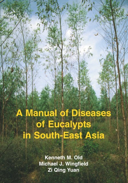
A Manual <strong>of</strong> Diseases<br />
<strong>of</strong> Eucalypts<br />
<strong>in</strong> <strong>South</strong>-<strong>East</strong> <strong>Asia</strong><br />
Kenneth M. Old<br />
Michael J. W<strong>in</strong>gfield<br />
Zi Q<strong>in</strong>g Yuan
A Manual <strong>of</strong> Diseases <strong>of</strong> Eucalypts<br />
<strong>in</strong> <strong>South</strong>-<strong>East</strong> <strong>Asia</strong><br />
Kenneth M. Old<br />
CSIRO Forestry and Forest Products, Canberra, Australia<br />
Michael J. W<strong>in</strong>gfield<br />
Forestry and Agricultural Biotechnology Institute, University <strong>of</strong> Pretoria,<br />
Pretoria, <strong>South</strong> Africa<br />
Zi Q<strong>in</strong>g Yuan<br />
School <strong>of</strong> Agricultural Science, University <strong>of</strong> Tasmania, Hobart, Australia<br />
Australian Centre <strong>for</strong> International Agricultural Research (ACIAR), Canberra, Australia<br />
<strong>Center</strong> <strong>for</strong> International Forestry Research (CIFOR), Bogor, Indonesia
A <strong>manual</strong> <strong>of</strong> <strong>diseases</strong> <strong>of</strong> eucalypts <strong>in</strong> <strong>South</strong>-<strong>East</strong> <strong>Asia</strong><br />
K.M. Old, M.J. W<strong>in</strong>gfield and Z.Q. Yuan<br />
© 2003 <strong>Center</strong> <strong>for</strong> International Forestry Research<br />
Published by<br />
<strong>Center</strong> <strong>for</strong> International Forestry Research<br />
Mail<strong>in</strong>g address: P.O. Box 6596 JKPWB, Jakarta 10065, Indonesia<br />
Office address: Jl. CIFOR, Situ Gede, S<strong>in</strong>dang Barang, Bogor Barat 16680, Indonesia<br />
Tel.: +62 (251) 622622; Fax: +62 (251) 622100<br />
E-mail: ci<strong>for</strong>@cgiar.org<br />
Web site: http://www.ci<strong>for</strong>.cgiar.org<br />
ISBN 064306530<br />
With the support <strong>of</strong><br />
CSIRO Forestry and Forest Products<br />
PO Box E4008<br />
K<strong>in</strong>gston ACT 2604<br />
Canberra, Australia<br />
Cover, designed by Ruth Gibbs, <strong>in</strong>cludes a plantation <strong>of</strong> Eucalyptus camaldulensis,<br />
with trees <strong>in</strong> <strong>for</strong>eground severely damaged by leaf and shoot blight (front cover)<br />
and a clonal nursery <strong>of</strong> the same eucalypt species (back cover).
Contents<br />
Acknowledgements and dedication iv<br />
Preface<br />
Introduction<br />
v<br />
Eucalypt plantations <strong>in</strong> <strong>South</strong>-<strong>East</strong> <strong>Asia</strong> 1<br />
Eucalyptus <strong>diseases</strong> and the scope <strong>of</strong> this <strong>manual</strong> 2<br />
Key to <strong>diseases</strong> and pathogens described <strong>in</strong> this <strong>manual</strong><br />
Plantation <strong>diseases</strong><br />
4<br />
Black mildew 6<br />
Cryptosporiopsis leaf and shoot blight 10<br />
Cyl<strong>in</strong>drocladium foliar spot and foliar blight 14<br />
Mycosphaerella leaf <strong>diseases</strong> 19<br />
Phaeophleospora leaf <strong>diseases</strong> 25<br />
Other eucalypt leaf spot <strong>diseases</strong> 32<br />
Cryphonectria cankers 41<br />
Coniothyrium canker 50<br />
P<strong>in</strong>k disease 55<br />
Stem canker <strong>diseases</strong> 60<br />
Bacterial wilt 67<br />
Woody root and stem rots <strong>of</strong> liv<strong>in</strong>g trees<br />
Nursery <strong>diseases</strong><br />
71<br />
Damp<strong>in</strong>g-<strong>of</strong>f 79<br />
Web blight 84<br />
Powdery mildew 88<br />
Eucalyptus rust 93<br />
Glossary <strong>of</strong> terms 97
iv<br />
Acknowledgements<br />
The preparation <strong>of</strong> this <strong>manual</strong> was made possible by f<strong>in</strong>ancial support received from the<br />
<strong>Center</strong> <strong>for</strong> International Forestry Research (CIFOR) and the Australian Centre <strong>for</strong> International<br />
Agricultural Research (ACIAR). The authors have been supported throughout by their respective<br />
organisations, CSIRO Forestry and Forest Products be<strong>in</strong>g the manag<strong>in</strong>g agency. We thank the<br />
above organisations <strong>for</strong> their susta<strong>in</strong>ed support <strong>in</strong> br<strong>in</strong>g<strong>in</strong>g this <strong>manual</strong> to completion. We<br />
gratefully acknowledge the skilled work <strong>of</strong> Ms Ruth Gibbs who took many micro and macrophotographs<br />
<strong>of</strong> specimens and prepared the photographic illustrations <strong>for</strong> publication. Thanks<br />
are also due to Dr Carol<strong>in</strong>e Mohammed and Mr Alan Brown <strong>for</strong> critical read<strong>in</strong>g <strong>of</strong> the manuscript<br />
and to Mr Mark Dudz<strong>in</strong>ski <strong>for</strong> editorial assistance and support dur<strong>in</strong>g the field work <strong>in</strong> <strong>South</strong>-<br />
<strong>East</strong> <strong>Asia</strong>.<br />
Mycological draw<strong>in</strong>gs were prepared by Dr Zi Q<strong>in</strong>g Yuan. The photographs <strong>of</strong> diseased trees<br />
and fungal pathogens were taken by the authors or by their close colleagues, with the<br />
exceptions <strong>of</strong> Fig. 76 which was k<strong>in</strong>dly provided by Ms K. Pongpanich, Figs 92 and 93 by Dr<br />
S.S. Lee and Fig. 107 by Pr<strong>of</strong>essor A.C. Alfenas.<br />
Dedication<br />
With the k<strong>in</strong>d agreement <strong>of</strong> our coauthor, Zi Q<strong>in</strong>g Yuan, Ken Old and Mike W<strong>in</strong>gfield would like<br />
to dedicate this book to the late Pr<strong>of</strong>essor Dave French, Department <strong>of</strong> Plant Pathology,<br />
University <strong>of</strong> M<strong>in</strong>nesota. For both <strong>of</strong> us he was an <strong>in</strong>spirational teacher, mentor and valued<br />
friend.
Preface<br />
Eucalypts are second only to p<strong>in</strong>es as the major <strong>for</strong>est plantation species grown <strong>in</strong>ternationally<br />
and are <strong>of</strong> prime importance <strong>in</strong> the southern hemisphere, much <strong>of</strong> <strong>South</strong>-<strong>East</strong> <strong>Asia</strong>, southern<br />
Ch<strong>in</strong>a and the Indian subcont<strong>in</strong>ent. With a few exceptions, eucalypts are unique to Australia.<br />
They have evolved under selection pressure from their major pests and pathogens and from<br />
the harsh constra<strong>in</strong>ts <strong>of</strong> the Australian environment. Their capacity <strong>for</strong> rapid growth, even on<br />
difficult sites, their growth habit, ease <strong>of</strong> vegetative propagation and their desirable product<br />
qualities have led to widespread establishment <strong>of</strong> large eucalypt plantations <strong>in</strong> many countries<br />
<strong>of</strong> <strong>South</strong>-<strong>East</strong> <strong>Asia</strong>.<br />
Eucalypts, <strong>in</strong> their native environments are hosts to a very wide range <strong>of</strong> fungal pathogens,<br />
especially those attack<strong>in</strong>g leaves, shoots and stems. The generally broad genetic base <strong>of</strong><br />
<strong>in</strong>dividual species and their presence <strong>in</strong> heterogeneous <strong>for</strong>est communities, however, provides<br />
significant protection aga<strong>in</strong>st disease epidemics. In contrast, <strong>in</strong>dustrial eucalypt plantations <strong>in</strong><br />
<strong>South</strong>-<strong>East</strong> <strong>Asia</strong> are typically s<strong>in</strong>gle species or hybrid plant<strong>in</strong>gs, <strong>of</strong>ten from a few clones which<br />
may share common parentage. Modern propagation techniques, such as shoot multiplication<br />
or tissue culture, make it possible to plant large areas with identical clones, with the expectation<br />
<strong>of</strong> uni<strong>for</strong>mly rapid growth and high product quality. Such practices are very dangerous from<br />
the standpo<strong>in</strong>t <strong>of</strong> disease, as pathogens, <strong>in</strong>clud<strong>in</strong>g endemic fungi and those newly <strong>in</strong>troduced<br />
<strong>in</strong>to a plantation region, may cause widespread epidemics. This risk is heightened by movement<br />
<strong>of</strong> ‘improved’ germ-plasm between eucalypt grow<strong>in</strong>g regions and even <strong>in</strong>ternationally, as<br />
pathogens may be transmitted by <strong>in</strong>fested seed or <strong>in</strong>fected plant<strong>in</strong>g stock.<br />
Avoidance <strong>of</strong> major epidemics <strong>of</strong> eucalypt <strong>diseases</strong> <strong>in</strong> <strong>South</strong>-<strong>East</strong> <strong>Asia</strong> requires an <strong>in</strong>creased<br />
awareness <strong>of</strong> the risks from pathogens, <strong>in</strong>herent <strong>in</strong> plantation <strong>for</strong>estry and a systematic approach<br />
to disease management. A good knowledge is needed <strong>of</strong> those <strong>diseases</strong>, present <strong>in</strong> plantations,<br />
which <strong>of</strong>fer significant threats and <strong>of</strong> those, which although currently absent, could cause<br />
future problems. Based on such knowledge, tree species, provenances and clones can be<br />
assessed <strong>for</strong> their susceptibility to major pathogens and strategies can be devised <strong>for</strong> the<br />
systematic deployment <strong>of</strong> different clones <strong>of</strong> widely vary<strong>in</strong>g parentage throughout plantation<br />
regions. Clonal <strong>for</strong>estry then has the potential to become a powerful means <strong>for</strong> the control <strong>of</strong><br />
plantation <strong>diseases</strong>, as practised <strong>in</strong> Brazil and <strong>South</strong> Africa <strong>for</strong> eucalypt cankers, rather than<br />
<strong>in</strong>creas<strong>in</strong>g the risk <strong>of</strong> epidemics.<br />
This <strong>manual</strong> <strong>of</strong> <strong>diseases</strong> <strong>of</strong> eucalypts <strong>in</strong> <strong>South</strong>-<strong>East</strong> <strong>Asia</strong> will be <strong>of</strong> assistance to those charged<br />
with ma<strong>in</strong>ta<strong>in</strong><strong>in</strong>g the health <strong>of</strong> eucalypt plantations, <strong>in</strong> identify<strong>in</strong>g the most common <strong>diseases</strong><br />
present <strong>in</strong> their region. It provides recommendations <strong>for</strong> disease management and <strong>of</strong>fers an<br />
<strong>in</strong>troduction to relevant world literature. The <strong>manual</strong> is a companion to the earlier CIFORpublished<br />
“A Manual <strong>of</strong> Diseases <strong>of</strong> Tropical Acacias <strong>in</strong> Australia, <strong>South</strong>-<strong>East</strong> <strong>Asia</strong> and India”.<br />
David Kaimowitz<br />
Director General, CIFOR<br />
v
Introduction<br />
Eucalypt plantations <strong>in</strong> <strong>South</strong>-<strong>East</strong> <strong>Asia</strong><br />
Eucalyptus species are second to p<strong>in</strong>es <strong>in</strong> global importance as plantation trees. In the<br />
tropics and subtropics they are the most widely planted genus. Data published by FAO (1995)<br />
<strong>in</strong>dicated that there were at least 1.4 million ha <strong>of</strong> <strong>for</strong>mal Eucalyptus plantations <strong>in</strong> the<br />
<strong>South</strong>-<strong>East</strong> <strong>Asia</strong>n region (Table 1). Midgley and P<strong>in</strong>yopusarerk (1996), <strong>in</strong> report<strong>in</strong>g these<br />
statistics, <strong>in</strong>dicated that the data did not <strong>in</strong>clude the equivalent <strong>of</strong> about 2.0 million ha<br />
grow<strong>in</strong>g as boundaries around fields and scattered trees. S<strong>in</strong>ce 1995 a number <strong>of</strong> countries,<br />
notably Ch<strong>in</strong>a, Laos, Thailand and Vietnam, have accelerated plant<strong>in</strong>g programs. It is likely<br />
that eucalypt plantations <strong>in</strong> the <strong>South</strong>-<strong>East</strong> <strong>Asia</strong> region now exceed 2.0 million ha.<br />
Table 1. Estimated areas <strong>of</strong> Eucalyptus plantations <strong>in</strong> the <strong>South</strong>-<strong>East</strong> <strong>Asia</strong>n region <strong>in</strong> 1995<br />
(FAO)<br />
Country Area (ha)<br />
Ch<strong>in</strong>a 670 000<br />
Indonesia 80 000<br />
Laos 62 000<br />
Malaysia 8 000<br />
Myanmar 40 000<br />
Philipp<strong>in</strong>es 10 000<br />
Thailand 195 000<br />
Vietnam 350 000<br />
Total 1 415 000<br />
In addition to large-scale plantations and farm plant<strong>in</strong>gs grown to supply fibre <strong>for</strong> <strong>in</strong>dustrial<br />
plants, eucalypts are highly valued <strong>in</strong> rural communities <strong>for</strong> a wide range <strong>of</strong> uses. These<br />
<strong>in</strong>clude fuel, poles, small lumber and furniture, essential oils and tann<strong>in</strong>s (Midgley and<br />
P<strong>in</strong>yopusarerk 1996). The ma<strong>in</strong> species grown <strong>in</strong>clude Eucalyptus camaldulensis, E.<br />
tereticornis, E. urophylla and E. grandis <strong>in</strong> subtropical and tropical regions, and E. globulus<br />
<strong>in</strong> more temperate climes. There is a strong trend <strong>for</strong> plantations to be established from<br />
clones <strong>of</strong> trees, <strong>of</strong>ten <strong>in</strong>ter-specific hybrids, selected <strong>for</strong> good growth and product quality.<br />
Great benefits can be ga<strong>in</strong>ed from such clonal <strong>for</strong>estry but this practice requires particular<br />
vigilance with regard to the susceptibility <strong>of</strong> <strong>in</strong>dividual clones to pests and <strong>diseases</strong>. A<br />
thorough knowledge <strong>of</strong> the nature and biology <strong>of</strong> these agents is thus required. In the<br />
equatorial humid tropics, acacias have become more widely planted than eucalypts, due<br />
partly to the high levels <strong>of</strong> leaf and shoot <strong>diseases</strong> susta<strong>in</strong>ed by Eucalyptus plantations,<br />
which depress growth rates and affect product quality. These <strong>diseases</strong> are the focus <strong>of</strong> this<br />
<strong>manual</strong>, which is complementary to a <strong>manual</strong> <strong>of</strong> <strong>diseases</strong> <strong>of</strong> tropical acacias published<br />
earlier (Old et al. 2000).<br />
1
2 A MANUAL OF DISEASES OF EUCALYPTS IN SOUTH-EAST ASIA<br />
Eucalyptus <strong>diseases</strong> and the scope <strong>of</strong> this <strong>manual</strong><br />
Tree <strong>diseases</strong> can be grouped accord<strong>in</strong>g to the stage <strong>of</strong> growth <strong>of</strong> the plant, that is, seedl<strong>in</strong>gs<br />
<strong>in</strong> nurseries or trees after out-plant<strong>in</strong>g, and by the part <strong>of</strong> the tree affected. Although tree<br />
disease problems <strong>of</strong>ten orig<strong>in</strong>ate <strong>in</strong> nurseries, management solutions are commonly available.<br />
Disease outbreaks <strong>in</strong> plantations are less readily conta<strong>in</strong>ed, diagnosis is more difficult and<br />
management options are few. For these reasons the primary purpose <strong>of</strong> the <strong>manual</strong> is to<br />
provide a field guide <strong>for</strong> identification <strong>of</strong> eucalypt <strong>diseases</strong> by plantation managers and<br />
their staff. Diseases are dealt with <strong>in</strong> turn, based on the part <strong>of</strong> the tree affected, namely<br />
foliage, branches, stems and roots, <strong>in</strong>ternal defect and decay development <strong>in</strong> non-liv<strong>in</strong>g<br />
tissue, and a section is devoted to nursery <strong>diseases</strong>. A description is also given <strong>of</strong> a rust<br />
pathogen, not found as yet <strong>in</strong> <strong>South</strong>-<strong>East</strong> <strong>Asia</strong>, which could cause serious disease if accidentally<br />
<strong>in</strong>troduced <strong>in</strong>to the region.<br />
Until recently there was no published work that provided a comprehensive account <strong>of</strong><br />
eucalypt <strong>diseases</strong> and pathogens. The need <strong>for</strong> such a reference document has been met by<br />
Keane et al. (2000), who have provided an account <strong>of</strong> eucalypt <strong>diseases</strong> worldwide. In<br />
contrast, this <strong>manual</strong> is a guide to the more damag<strong>in</strong>g <strong>diseases</strong> <strong>of</strong> Eucalyptus plantations<br />
that have been encountered by the authors and their colleagues dur<strong>in</strong>g disease surveys,<br />
consultancies and research and development projects <strong>in</strong> <strong>South</strong>-<strong>East</strong> <strong>Asia</strong> (primarily <strong>East</strong><br />
Timor, Indonesia, Laos, Thailand and Vietnam) over the past decade. In view <strong>of</strong> the very<br />
large numbers <strong>of</strong> foliar and stem <strong>diseases</strong> recorded on Eucalyptus, both <strong>in</strong> this region and<br />
worldwide (Sankaran et al. 1995), and budgetary constra<strong>in</strong>ts, the guide could not be<br />
comprehensive. The <strong>for</strong>est health specialist who critically exam<strong>in</strong>es diseased Eucalyptus<br />
specimens will f<strong>in</strong>d many genera and species <strong>of</strong> fungal pathogens not covered here. An<br />
attempt has been made, however, to <strong>in</strong>clude the most serious eucalypt pathogens found <strong>in</strong><br />
the region and also some <strong>diseases</strong> that may not cause significant damage, but have conspicuous<br />
symptoms.<br />
Each disease is attributed to one or more pathogens and is described us<strong>in</strong>g standard head<strong>in</strong>gs.<br />
Accounts are based on first-hand experience <strong>of</strong> these <strong>diseases</strong> <strong>in</strong> <strong>South</strong>-<strong>East</strong> <strong>Asia</strong>, supplemented<br />
by <strong>in</strong><strong>for</strong>mation from other regions and selected literature references. Orig<strong>in</strong>al colour<br />
photographs <strong>of</strong> symptoms, and <strong>in</strong> many cases, spor<strong>in</strong>g structures <strong>of</strong> pathogens are presented.<br />
L<strong>in</strong>e draw<strong>in</strong>gs <strong>of</strong> fungal characteristics used <strong>in</strong> identification to genus or species level have<br />
been prepared from fresh or herbarium specimens <strong>for</strong> users <strong>of</strong> the <strong>manual</strong> who have access<br />
to a microscope and basic plant pathology skills (Johnston and Booth 1983). A key is provided<br />
to assist <strong>in</strong> diagnosis, and a glossary <strong>of</strong> terms that may be unfamiliar to non-specialists.<br />
References<br />
FAO 1995. Proceed<strong>in</strong>gs <strong>of</strong> the FAO Regional Expert Consultation on Eucalyptus, October<br />
1993. FAO Regional Office <strong>for</strong> <strong>Asia</strong> and the Pacific, Bangkok. 196p.<br />
Johnston, A. and Booth, C. (eds).1983. Plant pathologists pocketbook. Commonwealth<br />
Mycological Institute, Commonwealth Agricultural Bureaux, Farnham. 439p.<br />
Keane, P.J., Kile, G.A., Podger, F.D. and Brown, B.N. (eds). 2000. Diseases and pathogens <strong>of</strong><br />
eucalypts. CSIRO, Coll<strong>in</strong>gwood, Victoria. 565p.<br />
Midgley, S.J. and P<strong>in</strong>yopusarerk, K. 1996. The role <strong>of</strong> Eucalyptus <strong>in</strong> local development <strong>in</strong> the<br />
emerg<strong>in</strong>g countries <strong>of</strong> Ch<strong>in</strong>a, Vietnam and Thailand. In: Eldridge K.G., Crowe, M.P.<br />
and Old, K.M. (eds). Environmental management: the role <strong>of</strong> Eucalyptus and other<br />
fast grow<strong>in</strong>g species. Proceed<strong>in</strong>gs <strong>of</strong> the Jo<strong>in</strong>t Australian/Japanese Workshop held <strong>in</strong><br />
Australia 23–27 October 1995, 4-10. CSIRO, Canberra.
Kenneth M. Old, Michael J. W<strong>in</strong>gfield and Zi Q<strong>in</strong>g Yuan<br />
Old, K.M., Lee, S.S., Sharma, J.K. and Yuan, Z.Q. 2000. A <strong>manual</strong> <strong>of</strong> <strong>diseases</strong> <strong>of</strong> tropical<br />
acacias <strong>in</strong> Australia, <strong>South</strong> <strong>East</strong> <strong>Asia</strong> and India. <strong>Center</strong> <strong>for</strong> International Forestry Research,<br />
Bogor, Indonesia. 104p.<br />
Sankaran, K.V., Sutton, B.C. and M<strong>in</strong>ter, D.W. 1995. A checklist <strong>of</strong> fungi recorded on<br />
Eucalyptus. Mycological Papers 170. CABI Bioscience, Egham, Surrey. 376p.<br />
3
4 A MANUAL OF DISEASES OF EUCALYPTS IN SOUTH-EAST ASIA<br />
Key to <strong>diseases</strong> and pathogens described <strong>in</strong> this <strong>manual</strong><br />
Page number<br />
Foliar <strong>diseases</strong><br />
A Mycelial growth superficial<br />
A1 Hyphae white, powdery appearance Oidium 88<br />
A2 Hyphae black, perithecia may be present Meliola 6<br />
B Caus<strong>in</strong>g necrotic spots, spores borne freely on lesion surfaces<br />
B1 Cyl<strong>in</strong>drical, septate spores Cyl<strong>in</strong>drocladium 14<br />
B2 Slender, tapered, septate spores Pseudocercospora 32<br />
B3 Spheroidal, aseptate, yellow spores<br />
or brown, septate stalked spores Pucc<strong>in</strong>ia 93<br />
C Caus<strong>in</strong>g necrotic spots, spores borne with<strong>in</strong><br />
cup-, flask-shaped or spheroidal fruit<strong>in</strong>g bodies<br />
embedded <strong>in</strong> leaf lesions and extruded when moist<br />
C1 Cup-shaped fruit<strong>in</strong>g bodies conta<strong>in</strong><strong>in</strong>g<br />
spheroidal spores Cryptosporiopsis 10<br />
C2 Spheroidal, sub-stomatal fruit<strong>in</strong>g bodies<br />
conta<strong>in</strong><strong>in</strong>g two-celled ascospores Mycosphaerella 19<br />
C3 Flask-shaped fruit<strong>in</strong>g bodies extrud<strong>in</strong>g hair-like<br />
columns <strong>of</strong> slender, septate spores Phaeophleospora 25<br />
C4 Flask-shaped fruit<strong>in</strong>g bodies conta<strong>in</strong><strong>in</strong>g darkly<br />
pigmented spores<br />
C4.1 Spores spheroidal, thick walled with<br />
truncated collar Microsphaeropsis 32<br />
C4.2 Spores lemon shaped Coniella 32<br />
D Caus<strong>in</strong>g corky spots, spores borne with<strong>in</strong><br />
elongate to branched fruit<strong>in</strong>g bodies scattered<br />
on the surface <strong>of</strong> lesions Aulograph<strong>in</strong>a 32<br />
Stem cankers<br />
A White to p<strong>in</strong>k mycelium superficial on bark Erythricium 55<br />
B Mycelium not superficial, fruit<strong>in</strong>g bodies<br />
embedded <strong>in</strong> bark or on the bark surface<br />
B1 Flask-shaped fruit<strong>in</strong>g bodies conta<strong>in</strong><strong>in</strong>g<br />
ascospores and/or conidia<br />
B1.1 Sexual and asexual fruit<strong>in</strong>g bodies<br />
embedded <strong>in</strong> yellow or orange Cryphonectria<br />
stromata eucalypti 41<br />
B1.2 Sexual fruit<strong>in</strong>g bodies with long necks,<br />
bases embedded <strong>in</strong> stroma. Asexual Cryphonectria<br />
fruit<strong>in</strong>g bodies on bark surface cubensis 41<br />
B1.3 Fruit<strong>in</strong>g bodies embedded <strong>in</strong><br />
black stromata Valsa 60
Kenneth M. Old, Michael J. W<strong>in</strong>gfield and Zi Q<strong>in</strong>g Yuan<br />
B2 Fruit<strong>in</strong>g bodies not flask-shaped, ascospores<br />
and conidia <strong>for</strong>med <strong>in</strong> chambers with<strong>in</strong><br />
black stromata Botryosphaeria 60<br />
B3 M<strong>in</strong>ute, sub-epidermal fruit<strong>in</strong>g bodies<br />
conta<strong>in</strong><strong>in</strong>g ellipsoidal, brown conidia Coniothyrium 50<br />
Bacterial wilt Ralstonia 67<br />
Rots <strong>of</strong> woody stems and roots<br />
A Range <strong>of</strong> basidiomycete species <strong>in</strong>clud<strong>in</strong>g; Armillaria<br />
Ganoderma<br />
Phell<strong>in</strong>us 71<br />
Nursery <strong>diseases</strong><br />
A Pre-emergence and post-emergence<br />
damp<strong>in</strong>g-<strong>of</strong>f Phytophthora 79<br />
Pythium<br />
B Web blight Rhizoctonia 84<br />
C Powdery mildew Oidium 88<br />
5
Black mildew<br />
Disease<br />
Black mildew<br />
Causal organism<br />
This disease is caused by fungi belong<strong>in</strong>g to the genus Meliola, family Meliolaceae, order<br />
Meliolales (Ascomycota). The Meliolales are obligate parasites produc<strong>in</strong>g a variety <strong>of</strong> structures<br />
which penetrate <strong>in</strong>to the host cells. Meliola amphitricha Fr., M. densa Cooke and M. eucalypti<br />
F. Stevens & Roldan ex Hans<strong>for</strong>d have been recorded on leaves <strong>of</strong> Eucalyptus spp. The black<br />
mildews are <strong>of</strong>ten confused with the common sooty moulds which are superficial, epiphytic<br />
saprophytes and also with members <strong>of</strong> the genus Meliol<strong>in</strong>a, family Meliol<strong>in</strong>aceae, order<br />
Dothideales.<br />
Host range and distribution<br />
Species <strong>of</strong> Meliola are primarily tropical. Meliola amphitricha was recorded on Eucalyptus<br />
sp. from Queensland and Victoria, Australia (Cooke 1892) and M. densa on Eucalyptus spp.,<br />
Callistemon vim<strong>in</strong>alis and Tristania conferta from Queensland (Simmonds 1966), and on E.<br />
tereticornis from Papua New Gu<strong>in</strong>ea (Shaw 1984). Meliola densa was also recorded on<br />
Eugenia sp. from India (Hosagoudar et al. 1994) and on a hybrid <strong>of</strong> E. pellita x E. brassiana<br />
<strong>in</strong> a provenance and family trial <strong>in</strong> Melville Island, Northern Territory <strong>of</strong> Australia (the authors,<br />
unpublished data). Meliola eucalypti was identified on Eucalyptus sp. from the Philipp<strong>in</strong>es<br />
(Hans<strong>for</strong>d 1962).<br />
Symptoms<br />
Species <strong>of</strong> Meliola grow on the surfaces <strong>of</strong> leaves and stems and <strong>for</strong>m thick, black, radiate,<br />
velvety colonies <strong>of</strong> up to 1 cm diameter (Fig.1). In cases <strong>of</strong> heavy <strong>in</strong>festation, the entire<br />
leaf surface may be covered by the fungus. The <strong>in</strong>fection is usually more frequent on upper<br />
than lower leaf surfaces. Sometimes young stems and twigs can also be <strong>in</strong>fected. Numerous<br />
m<strong>in</strong>ute spherical fruit<strong>in</strong>g bodies develop on the fungal thallus on the leaf surface (Figs 1, 3).<br />
In Meliola densa on E. pellita x E. brassiana these ascocarps have pigmented walls bear<strong>in</strong>g<br />
setae and conta<strong>in</strong> sac-shaped asci with two pigmented ascospores (Figs 2, 3).<br />
Pathology<br />
No <strong>in</strong>-depth studies have been carried out with the black mildews <strong>of</strong> Eucalyptus. The<br />
ascospores <strong>of</strong> Meliola are generally believed to germ<strong>in</strong>ate on the surface <strong>of</strong> the host leaves,<br />
immediately produc<strong>in</strong>g capitate hyphopodia. Mycelium then grows on the surface <strong>of</strong> leaves,<br />
obta<strong>in</strong><strong>in</strong>g nourishment through haustoria that extend <strong>in</strong>to the epidermal cells from the<br />
hyphopodia and spread to <strong>for</strong>m a colony. The close association <strong>of</strong> Meliola with scale <strong>in</strong>sects<br />
and mealy bugs on mango suggests that the honeydew excreted by these <strong>in</strong>sects provides a<br />
rich food source <strong>for</strong> growth and establishment <strong>of</strong> black mildews (Lim and Khoo 1985). Water<br />
splash and crawl<strong>in</strong>g <strong>in</strong>sects may be their ma<strong>in</strong> dispersal agents.<br />
6
Kenneth M. Old, Michael J. W<strong>in</strong>gfield and Zi Q<strong>in</strong>g Yuan 7<br />
Impact<br />
Little <strong>in</strong><strong>for</strong>mation is available on the impact <strong>of</strong> black mildews on growth <strong>of</strong> eucalypts. De<br />
Guzman (1977) <strong>in</strong>dicated that heavily <strong>in</strong>fected phyllodes <strong>of</strong> acacias turned yellow and abscissed<br />
prematurely, with repeated <strong>in</strong>fection lead<strong>in</strong>g to stunt<strong>in</strong>g <strong>of</strong> seedl<strong>in</strong>gs. On older trees, however,<br />
black mildew does not cause any serious damage although it may be common on the foliage.<br />
A study on biochemical changes <strong>in</strong> the leaves <strong>of</strong> ebony trees affected with a black mildew<br />
(M. diospyri Yates) <strong>in</strong>dicated a reduction, with<strong>in</strong> <strong>in</strong>fected leaves, <strong>of</strong> primary metabolites<br />
necessary <strong>for</strong> the production <strong>of</strong> soluble sugars and starch, total prote<strong>in</strong>, am<strong>in</strong>o acid and<br />
chlorophyll (Hosagoudar et al. 1997).<br />
Control and management<br />
Control <strong>of</strong> the disease is seldom necessary as it has little impact on the host. If <strong>in</strong>festation is<br />
heavy, however, black mildew can be controlled by spray<strong>in</strong>g fungicides and <strong>in</strong>secticides to<br />
elim<strong>in</strong>ate scale <strong>in</strong>sects and mealybugs.<br />
References<br />
Cooke, M.C. 1892. Handbook <strong>of</strong> Australian fungi. Williams and Norgate, London.<br />
De Guzman, E.D. 1977. Potentially dangerous <strong>diseases</strong> <strong>of</strong> <strong>for</strong>est trees <strong>in</strong> the Philipp<strong>in</strong>es.<br />
Biotrop Special Publication No. 2:189–194.<br />
Hans<strong>for</strong>d, C.G. 1962. The Meliol<strong>in</strong>eae supplement. Sydowia 16:302–323.<br />
Hosagoudar, V.B., Kaveriappa, K.M., Raghu, P.A. and Goos, R.D. 1994. Meliolaceae <strong>of</strong> southern<br />
India - XVI. Mycotaxon 51:107–118.<br />
Hosagoudar, V.B., Abraham, T.K. Krishnan, P.N. and Vijayakumar, K. 1997. Biochemical changes<br />
<strong>in</strong> the leaves <strong>of</strong> ebony tree affected with black mildew. Indian Phytopathology<br />
50:439–440.<br />
Lim, T.K. and Khoo, K.C. 1985. Diseases and disorders <strong>of</strong> mango <strong>in</strong> Malaysia. Tropical Press<br />
Sdn. Bhd. Kuala Lumpur. 101p.<br />
Shaw, D.E. 1984. Microorganisms <strong>in</strong> Papua New Gu<strong>in</strong>ea. Department <strong>of</strong> Primary Industry<br />
Research Bullet<strong>in</strong> 33:344.<br />
Simmonds, J.H. 1966. Host <strong>in</strong>dex <strong>of</strong> plant <strong>diseases</strong> <strong>in</strong> Queensland. Queensland Department <strong>of</strong><br />
Primary Industries, Brisbane. 111p.
8 A MANUAL OF DISEASES OF EUCALYPTS IN SOUTH-EAST ASIA<br />
Fig. 2<br />
Fig. 1<br />
Figure 1. Black mildew on a leaf surface, denser bodies are fruit<strong>in</strong>g bodies (perithecia)<br />
Figure 2. Meliola densa: A. Seta, bar = 40 µm; B. pigmented hyphae; C. hyphopodia, bar = 12.5<br />
µm; D. perithecium wall; E-F. septate pigmented ascospores, bar = 12.5 µm
Kenneth M. Old, Michael J. W<strong>in</strong>gfield and Zi Q<strong>in</strong>g Yuan<br />
A<br />
B<br />
C<br />
E<br />
D<br />
Figure 3. Meliola densa on Eucalyptus pellita: A. perithecium surrounded by sterile hyphae<br />
(setae); B. longitud<strong>in</strong>al section <strong>of</strong> a perithecium (half); C. asci conta<strong>in</strong><strong>in</strong>g two mature<br />
ascospores; D. ascospores; E. hyphae with lateral cells: capitate hyphopodia (swollen)<br />
and a mucronate hyphopodium (horn-like); F. a seta. Bar = 80 µm <strong>for</strong> A; 30 µm<br />
<strong>for</strong> B; 45 µm <strong>for</strong> C; 25 µm <strong>for</strong> D and E; and 40 µm <strong>for</strong> F.<br />
F<br />
9
Cryptosporiopsis leaf and shoot blight<br />
Disease<br />
Cryptosporiopsis leaf and shoot blight<br />
Causal organism<br />
Cryptosporiopsis eucalypti Sankaran & B. Sutton<br />
Host range and known distribution<br />
Not known to occur on hosts other than Eucalyptus spp., C. eucalypti has been collected by<br />
the co-authors from Eucalyptus with leaf spot or shoot blight symptoms <strong>in</strong> Australia, Japan,<br />
Laos, Indonesia, Sri Lanka, Thailand and Vietnam, (Old and Yuan 1994, Old et al. 2002) <strong>South</strong><br />
Africa and Uruguay (W<strong>in</strong>gfield unpublished). Other reports are from Brazil (Ferreira et al.<br />
1998), Australia, India and Hawaii (Sankaran et al. 1995) and New Zealand (Gadgil and Dick<br />
1999).<br />
Symptoms<br />
Symptoms <strong>of</strong> C. eucalypti <strong>in</strong>fection develop on both leaves and shoots <strong>of</strong> eucalypts. Leaf<br />
spots occur on both sides <strong>of</strong> the leaves and vary <strong>in</strong> size, shape and colour, with<strong>in</strong> and<br />
between Eucalyptus species. For example, there are at least four lesion types on E.<br />
camaldulensis. These <strong>in</strong>clude large, brown, spread<strong>in</strong>g necrotic lesions lead<strong>in</strong>g to a leaf<br />
blight symptom; circular or sub-circular spots 1–2 cm <strong>in</strong> diameter; irregular chocolate-brown<br />
or greyish spots cover<strong>in</strong>g much <strong>of</strong> the leaf area (Fig. 5); irregular roughened or corky lesions<br />
with eruption and necrosis <strong>of</strong> epidermal tissue, sometimes localised along ve<strong>in</strong>s, on which<br />
the fungus fruits. Term<strong>in</strong>al shoots <strong>of</strong> young trees can be totally defoliated and are commonly<br />
blighted (Fig. 6).<br />
Conidiomata develop on foliar lesions on blighted shoots and have also been found associated<br />
with cankers on small-diameter woody branches. Fruit<strong>in</strong>g bodies are cup-shaped when<br />
moist with pigmented marg<strong>in</strong>s, bear<strong>in</strong>g creamy masses <strong>of</strong> macroconidia (Fig. 7). The<br />
conidiomata are scattered irregularly on lesions and erupt through the epidermis or stem<br />
periderm but can be quite <strong>in</strong>conspicuous when leaves are dry. Macroconidia are thick walled<br />
and ellipsoid to elongate-ellipsoid <strong>in</strong> shape with dist<strong>in</strong>ctive protuberant attachment scars<br />
(Figs 8, 9).<br />
Pathology<br />
Cryptosporiopsis eucalypti can exist as a canker pathogen <strong>in</strong> woody stem tissue, so that<br />
<strong>in</strong>oculum persists dur<strong>in</strong>g dry months when conditions are not favourable <strong>for</strong> leaf and shoot<br />
blight. Dur<strong>in</strong>g the onset <strong>of</strong> epidemic disease, leaf spots develop and affected leaves are<br />
eventually shed. The most damag<strong>in</strong>g phase <strong>of</strong> the disease, however, is blight and dieback<br />
<strong>of</strong> term<strong>in</strong>al shoots. The typically conical shape <strong>of</strong> fast-grow<strong>in</strong>g plantation trees becomes<br />
flattened, ma<strong>in</strong> stems suffer dieback and multiple branch<strong>in</strong>g, apical dom<strong>in</strong>ance is reduced<br />
10
Kenneth M. Old, Michael J. W<strong>in</strong>gfield and Zi Q<strong>in</strong>g Yuan 11<br />
and growth can be stunted. Canker pathogens, such as Cytospora sp. and Lasiodiplodia<br />
theobromae, <strong>in</strong>vade affected stems and trees may be killed.<br />
Impacts<br />
Impacts <strong>in</strong> plantations vary from scattered lesions, especially on lower crowns and coppice<br />
shoots, to severe defoliation and death <strong>of</strong> shoots <strong>in</strong> the crowns <strong>of</strong> susceptible trees. The<br />
limited <strong>in</strong><strong>for</strong>mation available from observations <strong>in</strong> Vietnam suggests that E. camaldulensis<br />
ssp. obtusa is relatively susceptible, E. camaldulensis ssp. simulata and E. tereticornis are<br />
moderately resistant whereas E. pellita appears to be highly resistant. Differences <strong>in</strong> the<br />
susceptibility to C. eucalypti leaf and shoot blight <strong>of</strong> E. camaldulensis occurred at the<br />
species, provenance and family levels (Old et al. 2002).<br />
Control and management<br />
As <strong>for</strong> other leaf and shoot blight <strong>diseases</strong> <strong>in</strong> eucalypt plantations, the only feasible<br />
management option is selection <strong>of</strong> disease-resistant trees (Fig. 4). Depend<strong>in</strong>g on management<br />
objectives, this can be a choice <strong>of</strong> species or provenances that, on the basis <strong>of</strong> field trials,<br />
appear relatively resistant to C. eucalypti leaf blight. Where species can be readily grown<br />
from cutt<strong>in</strong>gs, selection and propagation <strong>of</strong> selected, highly resistant <strong>in</strong>dividuals can be<br />
practised. Old et al. (2002) found that all six provenances studied and many families <strong>of</strong> E.<br />
camaldulensis conta<strong>in</strong>ed highly resistant trees, many with good silvicultural characteristics.<br />
These selections could then either be established as clonal seed orchards or propagated as<br />
cutt<strong>in</strong>gs and used as clonal plantations <strong>in</strong> disease-prone areas. Selection <strong>of</strong> resistant trees<br />
requires some knowledge <strong>of</strong> leaf and shoot pathology as C. eucalypti leaf blight can be<br />
confused with other <strong>diseases</strong>, <strong>for</strong> example cyl<strong>in</strong>drocladium leaf blight and foliar <strong>diseases</strong><br />
caused by Mycosphaerella spp. and their anamorphs.<br />
References<br />
Ferreira, F.A., Silveira, S.F., Alfenas, A.C. and Demuner, A.M. 1998. Mancha-de-criptoriopsis em<br />
eucalipto no Brasil. [Eucalyptus leaf spot <strong>in</strong> Brazil caused by Cryptosporiopsis eucalypti].<br />
Fitopatologia Brasileira 23:414.<br />
Gadgil, P.D. and Dick, M. 1999. Fungi Silvicolae Novazelandiae: 2. New Zealand Journal <strong>of</strong><br />
Forest Science 29:440–458.<br />
Old, K.M. and Yuan, Z.Q. 1994. Foliar and stem <strong>diseases</strong> <strong>of</strong> Eucalyptus <strong>in</strong> Vietnam and<br />
Thailand. Internal Report, CSIRO Division <strong>of</strong> Forestry and Australian Centre <strong>for</strong> Agricultural<br />
Research, Canberra. 15p.<br />
Old, K.M., Dudz<strong>in</strong>ski, M.J., Pongpanich, K., Yuan, Z.Q., Thu, P.Q. and Nguyen, N.T. 2002.<br />
Cryptosporiopsis leaf spot and shoot blight <strong>of</strong> eucalypts. Australasian Plant Pathology<br />
31:337–344.<br />
Sankaran, K.V., Sutton, B.C. and Balasundaran, M. 1995. Cryptosporiopsis eucalypti sp. nov.<br />
caus<strong>in</strong>g leaf spots <strong>of</strong> eucalypts <strong>in</strong> Australia, India and USA. Mycological Research<br />
99:827–830.
12 A MANUAL OF DISEASES OF EUCALYPTS IN SOUTH-EAST ASIA<br />
Fig. 4<br />
Fig. 6<br />
Fig. 7<br />
Fig. 5 Fig. 8<br />
Figure 4. Eucalyptus camaldulensis clonal trial <strong>in</strong>fected by Cryptosporiopsis eucalypti with resistant<br />
and susceptible trees<br />
Figure 5. Leaf spot caused by C. eucalypti<br />
Figure 6. E. camaldulensis seedl<strong>in</strong>g defoliated by C. eucalypti; healthy family <strong>in</strong> background<br />
Figure 7. Masses <strong>of</strong> conidia ooz<strong>in</strong>g from pycnidia onto leaf surface<br />
Figure 8. Macroconidia <strong>of</strong> C. eucalypti, bar = 32 µm
Kenneth M. Old, Michael J. W<strong>in</strong>gfield and Zi Q<strong>in</strong>g Yuan<br />
A<br />
E<br />
B<br />
C<br />
Figure 9. Cryptosporiopsis eucalypti on Eucalyptus camaldulensis: A. conidia; B. conidia attached<br />
to conidiogenous cells; C. conidiogenous cells; D. conidia; E. longitud<strong>in</strong>al section <strong>of</strong><br />
conidioma. Bar = 20 µm <strong>for</strong> A-D; and 40 µm <strong>for</strong> E.<br />
D<br />
13
Cyl<strong>in</strong>drocladium foliar spot and foliar blight<br />
Disease<br />
Cyl<strong>in</strong>drocladium foliar blight<br />
Causal organisms<br />
Cyl<strong>in</strong>drocladium spp. are widespread and damag<strong>in</strong>g pathogens <strong>of</strong> a very wide range <strong>of</strong><br />
plant hosts <strong>in</strong>clud<strong>in</strong>g eucalypts. Cyl<strong>in</strong>drocladium spp. have sexual states (teleomorphs) <strong>in</strong><br />
the genus Calonectria de Not. There have been two major reviews <strong>of</strong> Cyl<strong>in</strong>drocladium <strong>in</strong><br />
the last decade, (Crous and W<strong>in</strong>gfield 1994, Crous 2002). In the latter publication, Crous has<br />
dist<strong>in</strong>guished 39 Cyl<strong>in</strong>drocladium spp. Of these 24 are listed as pathogens <strong>of</strong> Eucalyptus<br />
spp. and 15 <strong>of</strong> these have been found <strong>in</strong> <strong>South</strong>-<strong>East</strong> <strong>Asia</strong>.<br />
Host range<br />
These fungi are most commonly found as the Cyl<strong>in</strong>drocladium anamorph (asexual state) and<br />
those most commonly recorded <strong>of</strong>ten have very wide host ranges. For example C. reteaudii<br />
(as C. qu<strong>in</strong>queseptatum) has been recorded from many hosts <strong>in</strong> northern Australia, <strong>South</strong>-<br />
<strong>East</strong> <strong>Asia</strong> and India.<br />
Known distribution<br />
Distribution maps <strong>for</strong> all known species are provided <strong>in</strong> Crous (2002). Some species, e.g. C.<br />
reteaudii, occur primarily <strong>in</strong> tropical regions <strong>of</strong> <strong>South</strong>-<strong>East</strong> <strong>Asia</strong>, India and northern Australia.<br />
C. pauciramosum C.L. Schoch & Crous occurs <strong>in</strong> many countries around the world and may<br />
have been <strong>for</strong>merly confused with C. scoparium, which appears to be limited <strong>in</strong> its confirmed<br />
distribution to North and <strong>South</strong> America (Crous 2002). Other widely distributed species,<br />
known to attack eucalypts, and which occur <strong>in</strong> <strong>South</strong>-<strong>East</strong> <strong>Asia</strong> <strong>in</strong>clude C. <strong>in</strong>sulare C.L.<br />
Schoch & Crous, C. parasiticum Crous, M.J. W<strong>in</strong>gf. & Alfenas, C. floridanum Sobers & E.P.<br />
Seym., C. theae (Petch) Subram. and C. pteridis F.A. Wolf.<br />
Symptoms<br />
The most common and severe causal agent <strong>of</strong> cyl<strong>in</strong>drocladium leaf blight <strong>in</strong> <strong>South</strong>-<strong>East</strong> <strong>Asia</strong><br />
is C. reteaudii (Bugn.) Boesew. (syn. C. qu<strong>in</strong>queseptatum Boedijn & Reitsma), which is<br />
responsible <strong>for</strong> epidemic disease <strong>in</strong> several countries <strong>in</strong>clud<strong>in</strong>g Australia (Fig. 10), India,<br />
Vietnam, Laos and parts <strong>of</strong> Thailand. The disease first shows as greyish water-soaked spots<br />
on young leaves (Figs 11, 13, 16). These spots coalesce and develop <strong>in</strong>to extensive necrotic<br />
areas. Large numbers <strong>of</strong> sh<strong>in</strong><strong>in</strong>g white spores can be seen at the marg<strong>in</strong> <strong>of</strong> lesions, on older<br />
necrotic portions <strong>of</strong> leaves, especially along midribs on the abaxial surfaces, and on f<strong>in</strong>e<br />
shoots (Fig. 12). Under favourable conditions <strong>of</strong> high humidity and frequent ra<strong>in</strong>fall, necrotic<br />
lesions cover the entire area <strong>of</strong> the leaf and fruit pr<strong>of</strong>usely on young shoot tips which are<br />
killed, result<strong>in</strong>g <strong>in</strong> leaf and shoot blight symptoms (Bolland et al. 1985).<br />
14
Kenneth M. Old, Michael J. W<strong>in</strong>gfield and Zi Q<strong>in</strong>g Yuan 15<br />
Conidia <strong>of</strong> Cyl<strong>in</strong>drocladium are typically cyl<strong>in</strong>drical <strong>in</strong> shape with one or more cross walls<br />
(septa). Figures 14, 15 and 17 illustrate fruit<strong>in</strong>g structures produced by C. reteaudii, <strong>in</strong>clud<strong>in</strong>g<br />
six-celled macroconidia, two-celled microconidia, vesicles at the tips <strong>of</strong> sterile hyphae and<br />
barrel-shaped phialides which give rise to the conidia. The fungus also <strong>for</strong>ms pigmented<br />
chlamydospores, swollen hyphal cells that develop pigmentation and are resistant to<br />
biodegradation, thereby aid<strong>in</strong>g survival <strong>in</strong> soil.<br />
Pathology<br />
Cyl<strong>in</strong>drocladium spp. cause a variety <strong>of</strong> <strong>diseases</strong> <strong>in</strong> the nursery and <strong>in</strong> plantations <strong>in</strong>clud<strong>in</strong>g<br />
root and collar rot, shoot blight, leaf blight and foliar spots (Crous et al. 1991, Sharma and<br />
Mohanan 1982, 1991). Epidemic disease is favoured by high ra<strong>in</strong>fall and humidity (Sharma<br />
and Mohanan 1991). Booth et al. (2000) used bioclimatic mapp<strong>in</strong>g to predict high hazard<br />
areas <strong>for</strong> C. reteaudii leaf blight <strong>for</strong> <strong>South</strong>-<strong>East</strong> <strong>Asia</strong> and other parts <strong>of</strong> the world. Good<br />
agreement was found between predicted high hazard areas and locations where epidemics<br />
occurred <strong>in</strong> <strong>South</strong>-<strong>East</strong> <strong>Asia</strong>. Annual ra<strong>in</strong>fall <strong>of</strong> >1400 mm and m<strong>in</strong>imum temperatures <strong>of</strong> the<br />
coldest month >16°C were useful predictors <strong>of</strong> high hazard. The spread <strong>of</strong> the disease is by<br />
means <strong>of</strong> conidia, (Figs 12, 14, 15, 17) which are borne <strong>in</strong> vast numbers on leaf surfaces.<br />
Dur<strong>in</strong>g heavy ra<strong>in</strong>, these spores are splashed <strong>in</strong>to the air and <strong>in</strong>fect nearby trees.<br />
Cyl<strong>in</strong>drocladium species are commonly able to survive <strong>in</strong> soil by means <strong>of</strong> thick-walled<br />
chlamydospores, and these propagules are probably responsible <strong>for</strong> <strong>in</strong>itial <strong>in</strong>fections with<strong>in</strong><br />
eucalypt stands. Infections usually appear on the foliage <strong>of</strong> lower branches and spread<br />
upwards <strong>in</strong>to the crown. Although diseased foliage can be found <strong>in</strong> crowns <strong>of</strong> large trees,<br />
disease is most obvious <strong>in</strong> sapl<strong>in</strong>gs and pole-sized trees, <strong>in</strong> which defoliation can be very<br />
extensive (Fig. 10).<br />
Impacts<br />
Cyl<strong>in</strong>drocladium foliar blight is a major problem on eucalypts grown <strong>in</strong> the humid tropics. In<br />
high-ra<strong>in</strong>fall regions <strong>of</strong> south-eastern and central Vietnam, repeated defoliation <strong>of</strong> susceptible<br />
provenances <strong>of</strong> E. camaldulensis has led to crown dieback with secondary <strong>in</strong>fection by<br />
canker fungi, loss <strong>of</strong> <strong>for</strong>m and even death. Eucalyptus urophylla has also suffered significant<br />
damage <strong>in</strong> central Vietnam. Overall, losses to cyl<strong>in</strong>drocladium leaf blight have contributed<br />
to the generally poor growth rates <strong>of</strong> E. camaldulensis <strong>in</strong> disease-prone regions <strong>of</strong> Vietnam<br />
(<strong>of</strong>ten less than 10 m 3 ha -1 y -1 on a 5–6 year rotation).<br />
Control<br />
In nurseries, Cyl<strong>in</strong>drocladium <strong>in</strong>fection can be effectively controlled by carbendazim applied<br />
as a foliar spray or soil drench (Sharma et al. 1984, Ferreira 1994). In plantations, however,<br />
it is not economically feasible to control the disease by fungicidal treatment. The disease<br />
on eucalypts has been serious enough to warrant selection <strong>for</strong> resistant species, provenances,<br />
families and clones (Blum et al. 1992, Nghia and Old 1997, Sharma et al. 1999). Eucalyptus<br />
pellita has been found to be resistant to C. reteaudii leaf blight <strong>in</strong> northern Queensland and<br />
<strong>in</strong> Vietnam, where E. brassiana has also per<strong>for</strong>med well.<br />
References<br />
Blum, L.E.B., Dianese, J.C. and Costa, C.L. 1992. Comparative pathology <strong>of</strong> Cyl<strong>in</strong>drocladium<br />
clavatum and C. scoparium on Eucalyptus spp. and screen<strong>in</strong>g <strong>of</strong> Eucalyptus<br />
provenances <strong>for</strong> resistance to Cyl<strong>in</strong>drocladium damp<strong>in</strong>g-<strong>of</strong>f. Tropical Pest Management<br />
38:2155–2159.
16 A MANUAL OF DISEASES OF EUCALYPTS IN SOUTH-EAST ASIA<br />
Bolland, L., Tierney, J.W. and Tierney, B.J. 1985. Studies on leaf spot and shoot blight <strong>of</strong><br />
Eucalyptus caused by Cyl<strong>in</strong>drocladium qu<strong>in</strong>queseptatum. European Journal <strong>of</strong> Forest<br />
Pathology 15:385–397.<br />
Booth, T.H., Jovanovic, T., Old, K.M. and Dudz<strong>in</strong>ski, M.J. 2000. Climatic mapp<strong>in</strong>g to identify<br />
high risk areas <strong>for</strong> Cyl<strong>in</strong>drocladium qu<strong>in</strong>queseptatum leaf blight on eucalypts <strong>in</strong><br />
ma<strong>in</strong>land <strong>South</strong>-<strong>East</strong> <strong>Asia</strong> and around the world. Environmental Pollution<br />
108:365–372.<br />
Crous, P.W. 2002. Taxonomy and pathology <strong>of</strong> Cyl<strong>in</strong>drocladium (Calonectria) and allied<br />
genera. American Phytopathology Society Press, St Paul, M<strong>in</strong>nesota, 278p.<br />
Crous, P.W. and W<strong>in</strong>gfield, M.J. 1994. A monograph <strong>of</strong> Cyl<strong>in</strong>drocladium, <strong>in</strong>clud<strong>in</strong>g anamorphs<br />
<strong>of</strong> Calonectria. Mycotaxon 51:341–453.<br />
Crous, P.W., Phillips, A.J. and W<strong>in</strong>gfield, M.J. 1991. The genera Cyl<strong>in</strong>drocladium and<br />
Cyl<strong>in</strong>drocladiella <strong>in</strong> <strong>South</strong> Africa with special reference to <strong>for</strong>estry nurseries. <strong>South</strong><br />
African Forestry Journal 157:69–85.<br />
Ferreira, F.A. 1994. Control <strong>of</strong> Eucalyptus nursery <strong>diseases</strong> <strong>in</strong> Brazil, 1990-1993. In: Perr<strong>in</strong>, R.<br />
and Sutherland, J.R. (eds). Diseases and <strong>in</strong>sects <strong>in</strong> <strong>for</strong>est nurseries. Dijon, France,<br />
October 3-10 1993, 315–320. Les Colloques No. 68. INRA, Paris.<br />
Nghia, N.H. and Old, K.M. 1997. Variation <strong>in</strong> growth and disease resistance <strong>of</strong> Eucalyptus<br />
species and provenances tested <strong>in</strong> Vietnam. In: Higa, A.R., Schaitza, E. and Gaiad, S.<br />
(eds). Silviculture and genetic improvement <strong>of</strong> eucalypts, proceed<strong>in</strong>gs <strong>of</strong> IUFRO<br />
Conference, Brazil, 1997, 416–422.<br />
Sharma, J.K. and Mohanan, C. 1982. Cyl<strong>in</strong>drocladium spp. associated with various <strong>diseases</strong><br />
<strong>of</strong> Eucalyptus <strong>in</strong> Kerala. European Journal <strong>of</strong> Forest Pathology 12:129–136.<br />
Sharma, J.K. and Mohanan, C. 1991. Epidemiology and control <strong>of</strong> <strong>diseases</strong> <strong>of</strong> Eucalyptus<br />
caused by Cyl<strong>in</strong>drocladium spp. <strong>in</strong> Kerala. Research Report 70. Kerala Forest Research<br />
Institute, Peechi, Kerala. 155p.<br />
Sharma, J.K., Mohanan, C. and Florence, E.J.M. 1984. Nursery <strong>diseases</strong> <strong>of</strong> Eucalyptus <strong>in</strong><br />
Kerala. European Journal <strong>of</strong> Forest Pathology 14:77–89.<br />
Sharma, J.K., Balasundaran, M. and Florence, E.J.M. 1999. Increas<strong>in</strong>g the productivity <strong>of</strong><br />
eucalypts <strong>in</strong> Kerala through selection <strong>for</strong> <strong>diseases</strong> resistance, higher growth and clonal<br />
technology. In: Sivapragasam, A. et al. (eds). Plant protection <strong>in</strong> the tropics: tropical plant<br />
protection <strong>in</strong> the <strong>in</strong><strong>for</strong>mation age. Proceed<strong>in</strong>gs <strong>of</strong> the Fifth International Conference<br />
<strong>of</strong> the Malaysian Plant Protection Society, MPPS. Kuala Lumpur, 164–167.
Kenneth M. Old, Michael J. W<strong>in</strong>gfield and Zi Q<strong>in</strong>g Yuan<br />
Fig. 10<br />
Fig. 12<br />
Fig. 14<br />
Fig. 15<br />
Fig. 11<br />
Fig. 13<br />
Fig. 16<br />
Figure 10. Eucalyptus grandis x E. urophylla clone defoliated by Cyl<strong>in</strong>drocladium reteaudii<br />
(syn. C. qu<strong>in</strong>queseptatum)<br />
Figure 11. E. camaldulensis show<strong>in</strong>g leaf spot caused by C. reteaudii (upper leaf surface)<br />
Figure 12. Conidiophores <strong>of</strong> C. reteaudii on shoot surface, bar = 250 µm<br />
Figure 13. E. camaldulensis show<strong>in</strong>g leaf spot caused by C. reteaudii (lower leaf surface)<br />
Figure 14. Conidiophores with vesicle (swollen tip <strong>of</strong> project<strong>in</strong>g hypha), bar = 48 µm<br />
Figure 15. Macroconidia with, typically, 3–5 septa, bar = 32 µm<br />
Figure 16. E. pellita with leaf spot caused by C. reteaudii<br />
17
18 A MANUAL OF DISEASES OF EUCALYPTS IN SOUTH-EAST ASIA<br />
A<br />
F<br />
G<br />
B<br />
Figure 17. Cyl<strong>in</strong>drocladium reteaudii (syn. C. qu<strong>in</strong>queseptatum) on Eucalyptus camaldulensis<br />
from Vietnam: A. conidiophore term<strong>in</strong>at<strong>in</strong>g <strong>in</strong> a vesicle; B. typical branched<br />
conidiophores, and conidia; C. vesicles; D. phialides, cells which produce conidia;<br />
E. more densely branched conidiophores on agar culture; F. two-celled microconidia<br />
produced on agar culture; G. chlamydospores produced on agar culture. Bar = 30 µm<br />
<strong>for</strong> A-C and E-G; and 15 µm <strong>for</strong> D.<br />
C<br />
D<br />
E
Mycosphaerella leaf <strong>diseases</strong><br />
Diseases<br />
Leaf spot, leaf blotch or leaf cr<strong>in</strong>kle, depend<strong>in</strong>g on symptomology<br />
Causal organisms<br />
Worldwide more than thirty dist<strong>in</strong>ct species <strong>of</strong> Mycosphaerella are recognised on Eucalyptus<br />
(Park et al. 2000), caus<strong>in</strong>g a great variety <strong>of</strong> symptoms. The genus Mycosphaerella as found<br />
on Eucalyptus has not been clearly def<strong>in</strong>ed and may actually represent several dist<strong>in</strong>ct<br />
genera. This possibility is supported by the association, with different Mycosphaerella<br />
species, <strong>of</strong> a wide range <strong>of</strong> anamorphic states. Anamorphs <strong>in</strong>clude Colletogloeum,<br />
Colletogloeopsis, Coniothyrium, Phaeophleospora, Pseudocercospora, Sonderhenia,<br />
Stagonospora, Stenella and Uwebraunia. Several <strong>of</strong> these anamorphs are discussed <strong>in</strong> the<br />
sections on Phaeophleospora and Pseudocercospora spp.<br />
Significant changes <strong>in</strong> name comb<strong>in</strong>ations and association <strong>of</strong> named anamorphs with<br />
Mycosphaerella have <strong>of</strong>ten been <strong>of</strong>ten made. This provides considerable difficulty <strong>for</strong> the<br />
non-taxonomist <strong>in</strong> assess<strong>in</strong>g which species are present <strong>in</strong> plantations and how their<br />
observations relate to published <strong>in</strong><strong>for</strong>mation. Names used <strong>in</strong> older literature might not<br />
accurately represent the fungus <strong>in</strong> question and these should be used with some<br />
circumspection. Descriptions <strong>of</strong> 10 Mycosphaerella spp. found on Eucalyptus, their synonyms,<br />
anamorphs and hosts are provided <strong>in</strong> Set 124, IMI Descriptions <strong>of</strong> Fungi and Bacteria (Crous<br />
et al. 1995), the most recent taxonomic treatment be<strong>in</strong>g provided by Crous (1998). Useful<br />
characteristics <strong>for</strong> identification are lesion morphology and their position on the leaf (Figs<br />
18, 19, 21), fruit<strong>in</strong>g body morphology (Figs 20, 23), ascospore and conidial size and shape<br />
and the mode <strong>of</strong> germ<strong>in</strong>ation <strong>of</strong> ascospores (Figs 22, 23). Molecular methods are <strong>in</strong>creas<strong>in</strong>gly<br />
be<strong>in</strong>g used, to support more traditional approaches, <strong>in</strong> the identification <strong>of</strong> species <strong>of</strong><br />
Mycosphaerella (Carnegie et al. 2001, Crous et al. 2001)<br />
Collections <strong>of</strong> several Mycosphaerella species, associated with Eucalyptus, have been made<br />
<strong>in</strong> <strong>East</strong> Timor (Old unpublished), Indonesia (Crous and Alfenas 1995, Crous and W<strong>in</strong>gfield<br />
1997), Thailand (Pongpanich unpublished) and Vietnam (Yuan unpublished). No systematic<br />
attempt has been made, however, to determ<strong>in</strong>e which are the most common and damag<strong>in</strong>g<br />
Mycosphaerella species <strong>in</strong> <strong>South</strong>-<strong>East</strong> <strong>Asia</strong>. For this reason the ma<strong>in</strong> biological, morphological<br />
and pathological characteristics <strong>of</strong> the fungi will be summarised at the generic level, draw<strong>in</strong>g<br />
largely on <strong>in</strong><strong>for</strong>mation derived from temperate species.<br />
Host range<br />
It is likely that all Eucalyptus spp. can be <strong>in</strong>fected by one or more species <strong>of</strong> Mycosphaerella.<br />
For example, Crous et al. (1995) listed more than 50 hosts <strong>for</strong> M. cryptica (Cooke) Hansf.<br />
across all Eucalyptus subgenera. Juvenile, <strong>in</strong>termediate and adult foliage are susceptible to<br />
<strong>in</strong>fection by this pathogen. In their check-list <strong>of</strong> fungi recorded on Eucalyptus, Sankaran et<br />
al. (1995) provided host lists <strong>for</strong> many Mycosphaerella spp. and their anamorphs. Host<br />
19
20 A MANUAL OF DISEASES OF EUCALYPTS IN SOUTH-EAST ASIA<br />
records <strong>for</strong> fungi collected <strong>in</strong> Australia <strong>of</strong>ten <strong>in</strong>clude several to many Eucalyptus spp. whereas<br />
overseas records <strong>of</strong> Mycosphaerella on plantation eucalypts are sometimes restricted to a<br />
s<strong>in</strong>gle species. This may reflect the limited range <strong>of</strong> Eucalyptus spp. present <strong>in</strong> exotic<br />
plantations, rather than host specificity and is probably also related to difficulties experienced<br />
<strong>in</strong> dist<strong>in</strong>guish<strong>in</strong>g between species.<br />
Distribution<br />
Mycosphaerella spp. have been recorded on Eucalyptus <strong>in</strong> all States and most <strong>for</strong>ested<br />
regions <strong>of</strong> Australia although the knowledge <strong>of</strong> the species present and their impacts on tree<br />
health is <strong>in</strong>complete (Park et al. 2000). These pathogens are also present <strong>in</strong> virtually all<br />
countries where significant exotic eucalypt plantations have been established, with most<br />
<strong>in</strong><strong>for</strong>mation be<strong>in</strong>g available from <strong>South</strong> Africa (Crous and W<strong>in</strong>gfield 1996), Brazil (Crous et<br />
al. 1993a,b) and New Zealand (Dick and Gadgil 1983, Dick and Dobbie 2001). Mycosphaerella<br />
heimioides Crous & M.J. W<strong>in</strong>gf. and M. gracilis Crous & Alfenas (Crous and Alfenas 1995,<br />
Crous and W<strong>in</strong>gfield 1996) have been collected <strong>in</strong> Indonesia along with M. parkii Crous, M.J.<br />
W<strong>in</strong>gf., F.A. Ferreira & Alfenas and M. suberosa Crous, F.A. Ferreira, Alfenas & M.J.W<strong>in</strong>gf.<br />
Fungi <strong>in</strong> this group have shown a remarkable capacity <strong>for</strong> <strong>in</strong>ternational movement. For<br />
example, M. marksii Carnegie & Keane, a species first described <strong>in</strong> Australia (Carnegie and<br />
Keane 1994), was found <strong>in</strong> 1995 caus<strong>in</strong>g a leaf spot on E. camaldulensis <strong>in</strong> Vietnam (Yuan<br />
unpublished). Phaeophleospora epicoccoides, the anamorph <strong>of</strong> M. suttoniae, occurs <strong>in</strong> most<br />
countries where Eucalyptus spp. are grown (Crous et al. 1998).<br />
Pathology<br />
Detailed <strong>in</strong><strong>for</strong>mation on the pathology and epidemiology <strong>of</strong> Mycosphaerella <strong>diseases</strong> has<br />
been largely derived from research carried out <strong>in</strong> Australia (Carnegie and Keane 1994, Carnegie<br />
et al. 1997, Park and Keane 1982, Park and Keane 1987, Park 1988) on M. cryptica and M.<br />
nubilosa. These fungi are major pathogens <strong>of</strong> temperate eucalypts, although M. cryptica can<br />
be found on species with distribution well <strong>in</strong>to the tropics, e.g. E. grandis.<br />
Epidemics <strong>in</strong> plantations are favoured by abundant moisture, especially prolonged periods<br />
<strong>of</strong> ra<strong>in</strong>, dur<strong>in</strong>g which spores are released from pseudothecia and disperse to <strong>in</strong>fect susceptible<br />
foliage. As many species also produce conidia on or with<strong>in</strong> asexual fruit<strong>in</strong>g structures,<br />
plantations are subjected to very high levels <strong>of</strong> <strong>in</strong>oculum and virtually all foliage can be<br />
exposed to <strong>in</strong>fection. There is no s<strong>in</strong>gle pattern <strong>of</strong> disease development, etiology vary<strong>in</strong>g<br />
with host-pathogen comb<strong>in</strong>ation. For example, Park (1988) found that M. cryptica completed<br />
several generations dur<strong>in</strong>g each epidemic, through <strong>for</strong>mation <strong>of</strong> conidia and secondary<br />
ascospores. Mycosphaerella nubilosa, on the other hand, showed a delay <strong>in</strong> symptom<br />
development <strong>of</strong> 4–5 months between <strong>in</strong>fection <strong>of</strong> newly-<strong>for</strong>med leaves to the onset <strong>of</strong><br />
epidemic disease levels <strong>in</strong> expanded foliage.<br />
Many variations <strong>in</strong> symptom development are associated with Mycosphaerella <strong>in</strong>fections<br />
(Figs 18, 21), result<strong>in</strong>g <strong>in</strong> different comb<strong>in</strong>ations <strong>of</strong> lesion size, colour and morphology.<br />
Fruit<strong>in</strong>g bodies can <strong>for</strong>m on one, or both surfaces <strong>of</strong> leaves. Infected leaves develop spots<br />
and blotches, their severity depend<strong>in</strong>g on the pathogen species and the susceptibility <strong>of</strong> the<br />
host. In highly susceptible <strong>in</strong>teractions, large lesions develop, <strong>of</strong>ten with cr<strong>in</strong>kl<strong>in</strong>g <strong>of</strong> the<br />
leaves. Affected trees suffer premature defoliation and severe disease can cause stunt<strong>in</strong>g <strong>of</strong><br />
trees.
Kenneth M. Old, Michael J. W<strong>in</strong>gfield and Zi Q<strong>in</strong>g Yuan<br />
Impacts<br />
Impacts <strong>of</strong> Mycosphaerella spp. on tree health can be severe or negligible, depend<strong>in</strong>g on<br />
the fungal species, host susceptibility, host physiology (<strong>in</strong>clud<strong>in</strong>g juvenility) and climate.<br />
Significant damage to plantations, associated with Mycosphaerella only, appears to be unusual<br />
<strong>in</strong> <strong>South</strong>-<strong>East</strong> <strong>Asia</strong>. This contrasts with the very severe epidemics caused by M. nubilosa and<br />
M. cryptica <strong>in</strong> temperate eucalypt plantations <strong>in</strong> parts <strong>of</strong> Australia and New Zealand and by a<br />
fungus, identified as M. juvenis Crous & W<strong>in</strong>gf., <strong>in</strong> <strong>South</strong> Africa (Crous and W<strong>in</strong>gfield 1996).<br />
Where significant epidemics have occurred <strong>in</strong> <strong>South</strong>-<strong>East</strong> <strong>Asia</strong> associated with these fungi,<br />
the anamorphs have been most prom<strong>in</strong>ent and several <strong>of</strong> these are covered elsewhere <strong>in</strong><br />
this <strong>manual</strong>, e.g. Phaeophleospora spp. Mycosphaerella stages are <strong>of</strong>ten <strong>in</strong>conspicuous or<br />
have not been found. An exception is M. marksii, which has been found <strong>in</strong> several locations<br />
<strong>in</strong> Vietnam associated with necrosis <strong>of</strong> leaf marg<strong>in</strong>s and defoliation <strong>of</strong> susceptible clones <strong>of</strong><br />
E. camaldulensis (Fig. 18).<br />
Control and management<br />
Despite the common occurrence <strong>of</strong> these fungi and their anamorphs, serious epidemics <strong>of</strong><br />
mycosphaerella leaf disease <strong>in</strong> <strong>South</strong>-<strong>East</strong> <strong>Asia</strong> have not been reported. In contrast, temperate<br />
Eucalyptus spp. suffer significant damage <strong>in</strong> many parts <strong>of</strong> the southern hemisphere, <strong>in</strong>clud<strong>in</strong>g<br />
Australasia, <strong>South</strong> America and <strong>South</strong> Africa. In these countries, variation <strong>in</strong> susceptibility to<br />
Mycosphaerella has been identified between provenances <strong>of</strong> E. globulus (Carnegie et al.<br />
1994) and between <strong>in</strong>ter-provenance and <strong>in</strong>ter-specific hybrids <strong>of</strong> E. globulus and E. nitens<br />
(Dungey et al. 1997). In the event <strong>of</strong> future epidemics <strong>of</strong> mycosphaerella leaf <strong>diseases</strong><br />
occurr<strong>in</strong>g <strong>in</strong> <strong>South</strong>-<strong>East</strong> <strong>Asia</strong>, it can be expected that selection <strong>for</strong> resistant plant<strong>in</strong>g material<br />
at the species, family and clonal levels will be effective counter measures.<br />
References<br />
Carnegie, A.J. and Keane, P.J. 1994. Further Mycosphaerella species associated with leaf<br />
<strong>diseases</strong> <strong>of</strong> Eucalyptus. Mycological Research 98:413–418.<br />
Carnegie, A.J., Keane, P.J., Ades, P.K. and Smith, I.W. 1994. Variation <strong>in</strong> susceptibility <strong>of</strong><br />
Eucalyptus globulus provenances to Mycosphaerella leaf disease. Canadian Journal <strong>of</strong><br />
Forest Research 24:1751–1757.<br />
Carnegie, A.J., Keane, P.J. and Podger, F.D. 1997. The impact <strong>of</strong> three species <strong>of</strong><br />
Mycosphaerella newly recorded on Eucalyptus <strong>in</strong> Western Australia. Australasian Plant<br />
Pathology 26:71–77.<br />
Carnegie, A.J., Ades, P.K. and Ford, R. 2001. The use <strong>of</strong> RAPD-PCR analysis <strong>for</strong> the<br />
differentiation <strong>of</strong> Mycosphaerella species from Eucalyptus <strong>in</strong> Australia. Mycological<br />
Research 105:1313–1320.<br />
Crous, P.W. 1998. Mycosphaerella spp. and their anamorphs associated with leaf spot <strong>diseases</strong><br />
<strong>of</strong> Eucalyptus. Mycologia Memoir 21. APS Press, St Paul, M<strong>in</strong>nesota. 170p.<br />
Crous, P.W. and Alfenas, A.C. 1995. Mycosphaerella gracilis and other species <strong>of</strong> Mycosphaerella<br />
associated with leaf spots <strong>of</strong> Eucalyptus <strong>in</strong> Indonesia. Mycologia 87:121–126.<br />
Crous, P.W. and W<strong>in</strong>gfield, M.J. 1996. Species <strong>of</strong> Mycosphaerella and their anamorphs<br />
associated with leaf blotch disease <strong>of</strong> Eucalyptus <strong>in</strong> <strong>South</strong> Africa. Mycologia 88:441–458.<br />
Crous, P.W. and W<strong>in</strong>gfield, M.J. 1997. New species <strong>of</strong> Mycosphaerella occurr<strong>in</strong>g on Eucalyptus<br />
leaves <strong>in</strong> Indonesia and Africa. Canadian Journal <strong>of</strong> Botany 75:781–790.<br />
Crous, P.W., Ferreira, F.A., Alfenas, A.C. and W<strong>in</strong>gfield, M.J. 1993a. Mycosphaerella suberosa<br />
associated with corky leaf spots on Eucalyptus <strong>in</strong> Brazil. Mycologia 85:705–710.<br />
21
22 A MANUAL OF DISEASES OF EUCALYPTS IN SOUTH-EAST ASIA<br />
Crous, P. W., W<strong>in</strong>gfield, M. J., Ferreira, F. A. and Alfenas, A. 1993b. Mycosphaerella parkii<br />
and Phyllosticta eucalyptorum, two new species from Eucalyptus leaves <strong>in</strong> Brazil.<br />
Mycological Research 97:582–584.<br />
Crous, P. W. Carnegie, A.J. and Keane, P.J. 1995. IMI Descriptions <strong>of</strong> Fungi and Bacteria Set<br />
124. CABI Bioscience, Egham, Surrey.<br />
Crous, P.W., W<strong>in</strong>gfield, M.J., Mohammed, C. and Yuan, Z.Q. 1998. New foliar pathogens <strong>of</strong><br />
Eucalyptus from Australia and Indonesia. Mycological Research 102:527–532.<br />
Crous, P.W., Hong, L., W<strong>in</strong>gfield, B.D. and W<strong>in</strong>gfield, M.J. 2001. ITS rDNA phylogeny <strong>of</strong><br />
selected Mycosphaerella spp. and their anamorphs occurr<strong>in</strong>g on Myrtaceae. Mycologia<br />
105:425–431.<br />
Dick, M.A. and Dobbie, K. 2001. Mycosphaerella suberosa and M. <strong>in</strong>termedia sp. nov. on<br />
Eucalyptus <strong>in</strong> New Zealand. New Zealand Journal <strong>of</strong> Botany 39: 269–276.<br />
Dick, M.A. and Gadgil, P.D. 1983. Eucalyptus leaf spots. Forest Pathology <strong>in</strong> New Zealand 1: 7.<br />
Dungey, H.S., Potts, B.M., Carnegie, A.J. and Ades, P.K. 1997. Mycosphaerella leaf disease:<br />
genetic variation <strong>in</strong> damage to Eucalyptus nitens, Eucalyptus globulus, and their F 1<br />
hybrid. Canadian Journal <strong>of</strong> Forest Research 27:750–759.<br />
Park, R.F. 1988. Effect <strong>of</strong> certa<strong>in</strong> host, <strong>in</strong>oculum and environmental factors on <strong>in</strong>fection <strong>of</strong><br />
Eucalyptus species by two Mycosphaerella species. Transactions <strong>of</strong> the British<br />
Mycological Society 90:221–228.<br />
Park, R.F. and Keane, P.J. 1982. Leaf <strong>diseases</strong> <strong>of</strong> Eucalyptus associated with Mycosphaerella<br />
species. Transactions <strong>of</strong> the British Mycological Society 79:101–115.<br />
Park, R.F. and Keane, P.J. 1987. Spore production by Mycosphaerella species caus<strong>in</strong>g leaf<br />
<strong>diseases</strong> <strong>of</strong> Eucalyptus. Transactions <strong>of</strong> the British Mycological Society 89: 461–470.<br />
Park, R.F., Keane, P.J., W<strong>in</strong>gfield, M.J. and Crous, P.W. 2000. Fungal <strong>diseases</strong> <strong>of</strong> eucalypt<br />
foliage. In: Keane, P.J., Kile, G.A., Podger, F.D. and Brown B.N. (eds). Diseases and<br />
pathogens <strong>of</strong> eucalypts, 153–239. CSIRO, Coll<strong>in</strong>gwood, Victoria.<br />
Sankaran, K.V., Sutton, B.C. and M<strong>in</strong>ter, D.W. 1995. A checklist <strong>of</strong> fungi recorded on<br />
Eucalyptus. Mycological Papers 170. CABI Bioscience, Egham, Surrey. 376p.
Kenneth M. Old, Michael J. W<strong>in</strong>gfield and Zi Q<strong>in</strong>g Yuan<br />
Fig. 18<br />
Fig. 19<br />
Fig. 20<br />
Fig. 21 Fig. 22<br />
Figure 18. Eucalyptus camaldulensis, leaf spots caused by Mycosphaerella marksii <strong>in</strong>fection<br />
Figure 19. Detail <strong>of</strong> lesion with pseudothecia (small black structures embedded <strong>in</strong> the leaf)<br />
Figure 20. Detail <strong>of</strong> pseudothecium <strong>of</strong> Mycosphaerella sp. shown <strong>in</strong> Figure 21, note asci located<br />
with<strong>in</strong> central region, bar = 15 µm<br />
Figure 21. Juvenile leaf <strong>of</strong> E. alba with Mycosphaerella lesions<br />
Figure 22. Germ<strong>in</strong>at<strong>in</strong>g ascospores <strong>of</strong> Mycosphaerella cryptica, bar = 26 µm<br />
23
24 A MANUAL OF DISEASES OF EUCALYPTS IN SOUTH-EAST ASIA<br />
B C<br />
A<br />
G<br />
D<br />
E<br />
Figure 23. Mycosphaerella spp.: A. diseased leaf <strong>of</strong> Eucalyptus camaldulensis <strong>in</strong>fected by M.<br />
marksii, Vietnam; B, C. ascospores and ascus <strong>of</strong> M. marksii; D. ascospores <strong>of</strong> M.<br />
cryptica; E. germ<strong>in</strong>ated ascospores <strong>of</strong> M. cryptica (24 hr <strong>in</strong>cubation); F. conidia <strong>of</strong> the<br />
M. cryptica anamorph Colletogloeopsis nubilosum; G. longitud<strong>in</strong>al section <strong>of</strong> a<br />
perithecium <strong>of</strong> M. cryptica; H. ascospores <strong>of</strong> M. nubilosa; I. germ<strong>in</strong>ated ascospores<br />
<strong>of</strong> M. nubilosa (24 hr <strong>in</strong>cubation). Bar = 25 mm <strong>for</strong> A; 15 µm <strong>for</strong> B, C, D, E, H and I;<br />
20 µm <strong>for</strong> F; and 30 µm <strong>for</strong> G.<br />
F<br />
H<br />
I
Phaeophleospora leaf <strong>diseases</strong><br />
Diseases<br />
Phaeophleospora leaf <strong>diseases</strong><br />
Causal organisms<br />
Phaeophleospora spp.<br />
Four species have been recognised so far on Eucalyptus:<br />
P. epicoccoides (Cooke & Massee) Crous, F.A. Ferreira & B. Sutton;<br />
P. eucalypti (Cooke & Massee) J. Walker, B. Sutton & I. Pascoe; P. lilianiae (Cooke &<br />
Massee) J. Walker, B. Sutton & I. Pascoe (Walker et al. 1992); P. destructans M.J. W<strong>in</strong>gf. &<br />
Crous (W<strong>in</strong>gfield et al. 1996).<br />
There have been many changes <strong>in</strong> the nomenclature <strong>of</strong> P. epicoccoides and P. eucalypti,<br />
which can lead to confusion <strong>in</strong> identification and report<strong>in</strong>g <strong>of</strong> outbreaks. Taxonomic changes<br />
and new comb<strong>in</strong>ations are cited <strong>in</strong> detail <strong>in</strong> Walker et al. (1992), <strong>in</strong> Crous et al.(1997), who<br />
resurrected the name Phaeophleospora <strong>for</strong> Kirramyces spp., and by Crous and W<strong>in</strong>gfield<br />
(1997), <strong>in</strong> which the teleomorph <strong>of</strong> K. epicoccoides (Phaeophleospora epicoccoides) is<br />
described as Mycosphaerella suttoniae Crous & M.J. W<strong>in</strong>gf. Synonyms are listed below:<br />
Phaeophleospora epicoccoides:<br />
Kirramyces epicoccoides (Cooke & Massee) J. Walker, B. Sutton & I. Pascoe<br />
Cercospora epicoccoides Cooke & Massee apud Cooke<br />
Hendersonia grandispora McAlp.<br />
Phaeoseptoria eucalypti (Hansf.) J. Walker<br />
Phaeoseptoria luzonensis T. Kobayashi<br />
Phaeophleospora eucalypti:<br />
Kirramyces eucalypti (Cooke & Massee) J. Walker, B. Sutton & I. Pascoe<br />
Cercospora eucalypti Cooke & Massee apud Cooke<br />
Pseudocercospora eucalypti (Cooke & Massee) Guo & Liu<br />
Septoria pulcherrima Gadgil & Dick<br />
Stagonospora pulcherrima (Gadgil & Dick) Swart<br />
Phaeophleospora destructans:<br />
Phaeophleospora destructans (M.J. W<strong>in</strong>gf. & Crous) Crous, F.A. Ferreira & B. Sutton<br />
Kirramyces destructans M.J. W<strong>in</strong>gf. & Crous<br />
Host ranges<br />
P. epicoccoides: 26 Eucalyptus and Corymbia species were listed by Walker et al. (1992),<br />
and 35 by Sankaran et al. (1995). It is likely that many more Eucalyptus species will be hosts<br />
<strong>of</strong> this pathogen.<br />
25
26 A MANUAL OF DISEASES OF EUCALYPTS IN SOUTH-EAST ASIA<br />
P. eucalypti: 13 species were listed by Walker et al. (1992), <strong>in</strong> the Eucalyptus subgenus<br />
Symphyomyrtus only. Sankaran et al. (1995) listed 60 hosts encompass<strong>in</strong>g Symphyomyrtus,<br />
Monocalyptus and Corymbia.<br />
P. lilianiae: found so far only on Corymbia eximia (Walker et al. 1992).<br />
P. destructans: found so far on E. grandis (W<strong>in</strong>gfield et al. 1996), E. camaldulensis and E.<br />
urophylla (Old et al. 2003).<br />
Distribution<br />
P. epicoccoides: is widely distributed <strong>in</strong> virtually all parts <strong>of</strong> the world where eucalypts are<br />
grown, <strong>in</strong>clud<strong>in</strong>g Africa, <strong>South</strong> America, Australia, India, <strong>South</strong>-<strong>East</strong> <strong>Asia</strong>, Japan, Indonesia,<br />
Philipp<strong>in</strong>es and New Zealand.<br />
P. eucalypti: records exist ma<strong>in</strong>ly from Australia and New Zealand. There are records from<br />
<strong>South</strong> America, <strong>South</strong> Africa, India, Taiwan and Italy but these may be mistaken identifications,<br />
possibly with P. epicoccoides or Pseudocercospora spp. This fungus does not appear to have<br />
been recorded <strong>in</strong> <strong>South</strong>-<strong>East</strong> <strong>Asia</strong>.<br />
P. destructans: was orig<strong>in</strong>ally described by W<strong>in</strong>gfield et al. (1996) from Sumatra. This<br />
pathogen caused severe defoliation <strong>in</strong> clonal plantations <strong>of</strong> E. camaldulensis <strong>in</strong> eastern<br />
Thailand <strong>in</strong> 1999 (Pongpanich unpublished) and was found on native E. urophylla <strong>in</strong> <strong>East</strong><br />
Timor 2002 (Old unpublished). In 2002 the pathogen was recorded <strong>in</strong> several locations <strong>in</strong><br />
northern, central and southern Vietnam (Old et al. 2003).<br />
Pathology<br />
These fungi are anamorphs <strong>of</strong> Mycosphaerella, although the teleomorph may be unusual or<br />
not yet recognised. They are dist<strong>in</strong>guished by their <strong>for</strong>mation <strong>of</strong> pigmented columns (cirrhi)<br />
or irregular aggregations <strong>of</strong> conidia, <strong>of</strong>ten on the abaxial surfaces <strong>of</strong> leaves, exud<strong>in</strong>g from<br />
substomatal pycnidia (Figs 27, 32, 34). These may be associated with discrete chlorotic or<br />
necrotic lesions, e.g. P. eucalypti (Figs 24, 25), P. destructans (Figs 30, 31), or widespread<br />
on leaves with little evidence <strong>of</strong> discrete lesion development or cell damage apart from<br />
general discoloration. This <strong>of</strong>ten gives the leaf a reddish or burgundy hue, e.g. P. epicoccoides<br />
(Fig. 26). In the less pathogenic species damage is <strong>of</strong>ten restricted to lower crowns but P.<br />
destructans can cause complete defoliation <strong>of</strong> susceptible trees and may <strong>in</strong>vade shoots. In<br />
Sumatra, P. destructans <strong>in</strong>fected young grow<strong>in</strong>g shoots <strong>of</strong> susceptible E. grandis trees,<br />
result<strong>in</strong>g <strong>in</strong> severe dieback and loss <strong>of</strong> apical growth. Such trees may die when they are outcompeted<br />
by less susceptible <strong>in</strong>dividuals. Stands <strong>of</strong> susceptible clones would probably not<br />
survive epidemics <strong>of</strong> P. destructans leaf blight.<br />
The ability <strong>of</strong> this group <strong>of</strong> pathogens to spread is great, as very large amounts <strong>of</strong> <strong>in</strong>oculum<br />
are produced on leaves. Survival on fallen leaves and subsequent splash dispersal <strong>of</strong> spores<br />
is also likely <strong>in</strong> humid environments. Phaeophleospora spp. are common nursery pathogens<br />
and can be spread with <strong>in</strong>fected plant<strong>in</strong>g stock. The widespread occurrence <strong>of</strong> P. epicoccoides<br />
suggests that this species may also be seed-borne, possibly through surface contam<strong>in</strong>ation.<br />
The most common species <strong>of</strong> Phaeophleospora are dist<strong>in</strong>guished by their conidial morphology.<br />
Spores <strong>of</strong> P. epicoccoides are cyl<strong>in</strong>drical or slightly club-shaped with rough walls and variable<br />
numbers <strong>of</strong> septa—<strong>of</strong>ten more than four (Figs 29, 34). Spores <strong>of</strong> P. eucalypti are variable <strong>in</strong>
Kenneth M. Old, Michael J. W<strong>in</strong>gfield and Zi Q<strong>in</strong>g Yuan<br />
size usually hav<strong>in</strong>g one septum per spore (Figs 28, 34), and are produced on both upper and<br />
lower surfaces <strong>of</strong> leaves. Conidia <strong>of</strong> P. destructans have three septa, are slender, curved or<br />
s<strong>in</strong>uate (Figs 33, 34).<br />
Impacts<br />
Phaeophleospora epicoccoides is common <strong>in</strong> the lower crowns <strong>of</strong> trees and can cause<br />
significant defoliation <strong>of</strong> seedl<strong>in</strong>gs <strong>in</strong> nurseries (Sharma and Mohanan 1981). It caused severe<br />
<strong>in</strong>fection <strong>of</strong> younger leaves <strong>of</strong> some E. grandis clones <strong>in</strong> <strong>South</strong> Africa, and stressed trees<br />
seemed to be somewhat more susceptible to disease (Crous et al. 1989; Knipscheer et al.<br />
1990). Similarly an E. grandis x E. camaldulensis clone grown <strong>in</strong> New <strong>South</strong> Wales, Australia<br />
was severely defoliated by P. epicoccoides (Carnegie, personal communication).<br />
Phaeophleospora destructans, as implied by its name (W<strong>in</strong>gfield et al. 1996), has caused<br />
severe damage to young E. grandis and hybrids with E. urophylla <strong>in</strong> Sumatra, to E.<br />
camaldulensis <strong>in</strong> Thailand and to E. camaldulensis and E. urophylla <strong>in</strong> Vietnam.<br />
Phaeophleospora destructans appears to be far more damag<strong>in</strong>g than P. epicoccoides, which<br />
can <strong>of</strong>ten be found on the same trees. In <strong>East</strong> Timor, P. destructans appeared to cause little<br />
damage to native E. urophylla suggest<strong>in</strong>g that this fungus may be <strong>in</strong>digenous to that region.<br />
Phaeophleospora lilianiae and P. eucalypti have not been recorded <strong>in</strong> the <strong>South</strong>-<strong>East</strong> <strong>Asia</strong>n<br />
region and will not be further considered here.<br />
Control and management<br />
Until P. destructans emerged as a significant threat to eucalypt plantations <strong>in</strong> Indonesia,<br />
Thailand and Vietnam, these pathogens would not have been considered worthy <strong>of</strong> a<br />
management response. The very severe damage <strong>in</strong>flicted by P. destructans <strong>in</strong> Indonesia<br />
(recorded s<strong>in</strong>ce 1996) and Thailand (s<strong>in</strong>ce 2000) and <strong>in</strong> Vietnam dur<strong>in</strong>g 2002, requires urgent<br />
action <strong>in</strong> terms <strong>of</strong> selection <strong>of</strong> resistant species, families or clones <strong>of</strong> preferred Eucalyptus<br />
species. A recent report (Old et al. 2003) <strong>in</strong>dicates that the pathogen has spread throughout<br />
most <strong>of</strong> Vietnam. The pathogen has ma<strong>in</strong>ly been a plantation problem and the only viable<br />
management strategy is to select resistant germplasm. Substantial success has already been<br />
achieved <strong>in</strong> this regard <strong>in</strong> Sumatra where disease tolerant clones have been selected and<br />
deployed (W<strong>in</strong>gfield unpublished). Chemical control may be required <strong>in</strong> nurseries and clonal<br />
propagation facilities, but it is too early to recommend suitable control schedules.<br />
References<br />
Crous, P.W. and W<strong>in</strong>gfield, M.J. 1997. New species <strong>of</strong> Mycosphaerella occurr<strong>in</strong>g on Eucalyptus<br />
leaves <strong>in</strong> Indonesia and Africa. Canadian Journal <strong>of</strong> Botany 75:781–790.<br />
Crous, P.W., Knox-Davies, P.S. and W<strong>in</strong>gfield, M.J. 1989. A list <strong>of</strong> Eucalyptus leaf fungi and<br />
their potential importance to <strong>South</strong> African <strong>for</strong>estry. <strong>South</strong> African Forestry Journal<br />
149:17–29.<br />
Crous, P.W., Ferreira, F.A. and Sutton, B. 1997. A comparison <strong>of</strong> the fungal genera<br />
Phaeophleospora and Kirramyces (Coelomycetes). <strong>South</strong> African Journal <strong>of</strong> Botany<br />
63:111–115.<br />
Knipscheer, N.S., W<strong>in</strong>gfield, M.J. and Swart, W.J. 1990. Phaeoseptoria leaf spot <strong>of</strong> Eucalyptus<br />
<strong>in</strong> <strong>South</strong> Africa. <strong>South</strong> African Forestry Journal 154:56–59.<br />
Old, K.M., Pongpanich, K., Thu, P.Q., W<strong>in</strong>gfield, M.J. and Yuan, Z.Q. 2003. Phaeophleospora<br />
destructans caus<strong>in</strong>g leafblight epidemics <strong>in</strong> <strong>South</strong> <strong>East</strong> <strong>Asia</strong>. Proceed<strong>in</strong>gs 8 th International<br />
Congress <strong>of</strong> Plant Pathology, Christchurch, New Zealand. Vol 2:165.<br />
27
28 A MANUAL OF DISEASES OF EUCALYPTS IN SOUTH-EAST ASIA<br />
Sankaran, K.V., Sutton, B.C. and M<strong>in</strong>ter, D.W. 1995. A checklist <strong>of</strong> fungi recorded on<br />
Eucalyptus. Mycological Papers 170. CABI Bioscience, Egham, Surrey. 376p.<br />
Sharma, J.K. and Mohanan, C. 1981. An unrecorded leaf spot disease <strong>of</strong> Eucalyptus <strong>in</strong> Kerala<br />
caused by Phaeoseptoria eucalypti (Hansf.) Walker. Current Science 50:865–866.<br />
Walker, J., Sutton, B.C. and Pascoe, I.G. 1992. Phaeoseptoria eucalypti and similar fungi on<br />
Eucalyptus, with description <strong>of</strong> Kirramyces gen. nov. (Coelomycetes). Mycological<br />
Research 96:911–924.<br />
W<strong>in</strong>gfield, M.J., Crous, P.W. and Boden, D. 1996. Kirramyces destructans sp. nov., a serious leaf<br />
pathogen <strong>of</strong> Eucalyptus <strong>in</strong> Indonesia. <strong>South</strong> African Journal <strong>of</strong> Botany 62:325–327.
Kenneth M. Old, Michael J. W<strong>in</strong>gfield and Zi Q<strong>in</strong>g Yuan<br />
Fig. 24<br />
Fig. 25<br />
Fig. 27<br />
Fig. 28 Fig. 29<br />
Fig. 26<br />
Figure 24. Leaf <strong>of</strong> Eucalyptus sp., <strong>in</strong>fected by Phaeophleospora eucalypti, with chlorotic lesions<br />
Figure 25. Older <strong>in</strong>fection; lesions have become necrotic<br />
Figure 26. Leaf <strong>of</strong> E. camaldulensis <strong>in</strong>fected by P. epicoccoides; note burgundy hue <strong>of</strong> lesions<br />
Figure 27. Pycnidium <strong>of</strong> P. epicoccoides, bar = 75 µm<br />
Figure 28. Generally uniseptate conidia <strong>of</strong> P. eucalypti, bar = 25 µm<br />
Figure 29. Multiseptate conidia <strong>of</strong> P. epicoccoides, bar = 25 µm<br />
29
30 A MANUAL OF DISEASES OF EUCALYPTS IN SOUTH-EAST ASIA<br />
Fig. 30<br />
Fig. 31<br />
Fig. 32 Fig. 33<br />
Figure 30. Juvenile leaf <strong>of</strong> Eucalyptus camaldulensis; note chlorosis and heavy sporulation <strong>of</strong><br />
Phaeophleospora destructans<br />
Figure 31. Older P. destructans <strong>in</strong>fection <strong>of</strong> E. urophylla; note redden<strong>in</strong>g <strong>of</strong> lesion marg<strong>in</strong>s with<br />
this Eucalyptus species, sporulation is ma<strong>in</strong>ly on underside <strong>of</strong> the leaf<br />
Figure 32. Conidial masses <strong>of</strong> P. destructans on the underside <strong>of</strong> the leaf shown <strong>in</strong> Figure 31<br />
Figure 33. Typically three-septate, s<strong>in</strong>uous conidia <strong>of</strong> P. destructans, bar = 25 µm
Kenneth M. Old, Michael J. W<strong>in</strong>gfield and Zi Q<strong>in</strong>g Yuan<br />
B<br />
A<br />
C D<br />
Figure 34. Phaeophleospora spp.: A. longitud<strong>in</strong>al section <strong>of</strong> P. eucalypti on Eucalyptus nitens;<br />
B. conidia <strong>of</strong> P. epicoccoides on E. urophylla; C. conidia <strong>of</strong> P. eucalypti on E.<br />
nitens; D. conidia <strong>of</strong> P. destructans on E. camaldulensis. Bar = 20 µm <strong>for</strong> B; 12 µm<br />
<strong>for</strong> C; and 18 µm <strong>for</strong> D.<br />
31
Other eucalypt leaf spot <strong>diseases</strong><br />
Causal organisms<br />
In addition to Cyl<strong>in</strong>drocladium spp., Cryptosporiopsis eucalypti, and Mycosphaerella spp.<br />
and their anamorphs such as Phaeophleospora spp. (described elsewhere <strong>in</strong> this <strong>manual</strong>),<br />
many other foliar pathogens <strong>of</strong> Eucalyptus are present <strong>in</strong> plantations <strong>in</strong> <strong>South</strong>-<strong>East</strong> <strong>Asia</strong>. Most<br />
<strong>of</strong> these are described by Park et al. (2000) but there is generally little known regard<strong>in</strong>g<br />
their pathogenicity or capacity to cause serious disease. In this section, several <strong>of</strong> these<br />
pathogens and their symptomology are briefly described, as they may be confused with<br />
more damag<strong>in</strong>g organisms. Fungi <strong>in</strong>cluded <strong>in</strong> this category are Aulograph<strong>in</strong>a eucalypti (Cooke<br />
& Massee) Arx & E. Mull., Coniella spp., Microsphaeropsis spp. and Pseudocercospora<br />
eucalyptorum. Crous, M.J. W<strong>in</strong>gf., Marasas & B. Sutton.<br />
Host ranges and distribution<br />
Aulograph<strong>in</strong>a eucalypti causes corky leaf spot and occurs only on Eucalyptus spp. (Figs 35,<br />
36, 37). The fungus has been found <strong>in</strong> many temperate countries <strong>in</strong> which eucalypts are<br />
grown extensively (Sankaran et al. 1995) but there appear to be few records from the<br />
tropics. The authors have collected this fungus <strong>in</strong> Vietnam (Yuan unpublished) and Sri Lanka.<br />
Coniella spp. appear to have wide host ranges <strong>in</strong>clud<strong>in</strong>g both tropical and temperate species<br />
(Figs 38–41, Fig. 48). C. australiensis Petr. and C. fragariae (Oudemans) B. Sutton have been<br />
found associated with leaf spots <strong>of</strong> Eucalyptus spp. <strong>in</strong> one or more <strong>of</strong> the follow<strong>in</strong>g tropical<br />
countries: Australia, India, Indonesia, Sri Lanka and Vietnam.<br />
Microsphaeropsis spp. (Figs 42–48) and Pseudocercospora eucalyptorum (Figs 49–52 are less<br />
well known fungi. Microsphaeropsis leaf spot has been recorded <strong>in</strong> Tasmania on E. obliqua as<br />
M. callista (H. Syd.) B. Sutton, (Yuan 1999). The same fungus has been recorded <strong>in</strong> Argent<strong>in</strong>a<br />
and <strong>South</strong> Africa, suggest<strong>in</strong>g a temperate range. Old (unpublished) found a Microsphaeropsis,<br />
closely resembl<strong>in</strong>g M. globulosa (Sousa da Camara) B. Sutton caus<strong>in</strong>g a severe leaf spot on E.<br />
grandis <strong>in</strong> Sri Lanka (Figs 42–45).<br />
Pseudocercospora eucalyptorum has been found <strong>in</strong> many parts <strong>of</strong> the world (Crous et al.<br />
1989, Sankaran et al. 1995). Records <strong>for</strong> the region <strong>in</strong>clude India, Sabah, Taiwan, Thailand<br />
and Vietnam. Pseudocercospora is one <strong>of</strong> the most common agents caus<strong>in</strong>g leaf spots <strong>of</strong><br />
lower crowns <strong>of</strong> E. camaldulensis <strong>in</strong> the latter two countries (Old et al. 2002). Although the<br />
last authors reta<strong>in</strong>ed P. eucalyptorum it seems likely that this pathogen may be an undescribed<br />
species (Yuan unpublished).<br />
Symptoms<br />
These fungi, which are most commonly found on the lower crowns <strong>of</strong> young trees, or on<br />
coppice shoots, cause leaf spots <strong>of</strong> various shapes and sizes as illustrated <strong>in</strong> Figs 35, 36, 38,<br />
41, 42–45, 49 and 51. The size and shape <strong>of</strong> lesions and the numbers <strong>of</strong> fruit<strong>in</strong>g bodies vary<br />
32
Kenneth M. Old, Michael J. W<strong>in</strong>gfield and Zi Q<strong>in</strong>g Yuan 33<br />
with the host and environmental conditions. Correct diagnosis, there<strong>for</strong>e, may require the<br />
recognition <strong>of</strong> the spores through microscopical exam<strong>in</strong>ation <strong>of</strong> <strong>in</strong>fected leaves (Figs 37, 48, 52).<br />
Aulograph<strong>in</strong>a eucalypti <strong>for</strong>ms dark brown ‘corky’ leaf spots with elongate, or branched<br />
fruit<strong>in</strong>g bodies called hysterothecia, open<strong>in</strong>g by a longitud<strong>in</strong>al slit (Figs 35, 36), scattered on<br />
the surface <strong>of</strong> the lesions.<br />
Coniella fragariae and C. australiensis cause medium to large reddish-brown lesions which<br />
<strong>of</strong>ten cause the leaf to partially curl as the tissue dessicates. Prom<strong>in</strong>ent black pycnidia are<br />
embedded <strong>in</strong> the lesions, sometimes concentrically arranged, and extrude vast numbers <strong>of</strong><br />
dark brown to black spheroidal conidia onto the lesion surface (Figs 38, 41).<br />
Microsphaeropsis globulosa on E. grandis <strong>for</strong>ms large brown lesions with raised purple<br />
marg<strong>in</strong>s (Figs 44, 45); sporulation is sparse but the few pycnidia produced are easily seen<br />
with the naked eye (Fig. 43). These are embedded <strong>in</strong> the leaf surface and extrude small<br />
dark brown to black, thick- walled, ellipsoidal conidia (Figs 46, 47).<br />
Pseudocercospora eucalyptorum <strong>for</strong>ms pr<strong>of</strong>use angular spots on <strong>in</strong>fected leaves (Figs 49,<br />
51). The central portions <strong>of</strong> these lesions bear dense tufts <strong>of</strong> conidiophores bear<strong>in</strong>g needleshaped,<br />
septate conidia (Fig. 50).<br />
Pathology and impacts<br />
These fungi vary greatly <strong>in</strong> their taxonomy, morphology and physiology. For example,<br />
A. eucalypti is an obligate pathogen on Eucalyptus, whereas Coniella spp. have wide host<br />
ranges and may require leaf damage or prior <strong>in</strong>fection by other pathogens to <strong>in</strong>vade leaves.<br />
However, they occur mostly on the lower crowns <strong>of</strong> Eucalyptus, are favoured by moist<br />
climates and are generally regarded as be<strong>in</strong>g <strong>of</strong> m<strong>in</strong>or importance. Nevertheless as clonal<br />
<strong>for</strong>estry becomes more common <strong>in</strong> the <strong>South</strong>-<strong>East</strong> <strong>Asia</strong>n region, some clones may be<br />
particularly susceptible to one or more <strong>of</strong> these pathogens, which could cause significant<br />
damage. For example, the E. grandis provenance shown <strong>in</strong> Figs 42–45 was very susceptible<br />
to Microsphaeropsis sp. and was elim<strong>in</strong>ated from a provenance trial <strong>in</strong> Sri Lanka on this<br />
basis.<br />
Control<br />
No control measures are warranted with these fungi, except <strong>for</strong> elim<strong>in</strong>ation from provenance<br />
or clonal trials <strong>of</strong> any selection that shows unusual susceptibility. Their importance also lies<br />
<strong>in</strong> possible mis-diagnosis <strong>of</strong> these fungi <strong>for</strong> more damag<strong>in</strong>g pathogens.<br />
References<br />
Crous, P.W., W<strong>in</strong>gfield, M.J., Marasas, W.F.O. and Sutton, B.C. 1989. Pseudocercospora<br />
eucalyptorum sp. nov. on Eucalyptus leaves. Mycological Research 93:394–398.<br />
Old K.M., Dudz<strong>in</strong>ski, M.J., Pongpanich, K., Yuan, Z.Q., Thu, P.Q. and Nguyen, N.T. 2002.<br />
Cryptosporiopsis leaf spot and shoot blight <strong>of</strong> eucalypts. Australasian Plant Pathology<br />
31:337–344.<br />
Park, R.F., Keane, P.J., W<strong>in</strong>gfield, M.J. and Crous, P.W. 2000. Fungal <strong>diseases</strong> <strong>of</strong> eucalypt<br />
foliage. In: Keane, P.J., Kile, G.A., Podger, F.D. and Brown B.N. (eds). Diseases and<br />
pathogens <strong>of</strong> eucalypts, 153–239. CSIRO, Coll<strong>in</strong>gwood, Victoria.
34 A MANUAL OF DISEASES OF EUCALYPTS IN SOUTH-EAST ASIA<br />
Sankaran, K.V., Sutton, B.C. and M<strong>in</strong>ter, D.W. 1995. A checklist <strong>of</strong> fungi recorded on<br />
Eucalyptus. Mycological Papers 170. CABI Bioscience, Egham, Surrey. 376p.<br />
Yuan, Z.Q. 1999. Fungi associated with <strong>diseases</strong> detected dur<strong>in</strong>g health surveys <strong>of</strong> eucalypt<br />
plantations <strong>in</strong> Tasmania. Report to Forest and Wood Products Research and Development<br />
Corporation. School <strong>of</strong> Agricultural Science, University <strong>of</strong> Tasmania, Hobart. 110p.
Kenneth M. Old, Michael J. W<strong>in</strong>gfield and Zi Q<strong>in</strong>g Yuan<br />
Fig. 35 Fig. 36<br />
A<br />
Fig. 37<br />
B<br />
Figure 35. Eucalyptus globulus <strong>in</strong>fected by Aulograph<strong>in</strong>a eucalypti<br />
Figure 36. Detail <strong>of</strong> lesions, show<strong>in</strong>g corky radiate appearance<br />
Figure 37. Aulograph<strong>in</strong>a eucalypti: A. an ascus conta<strong>in</strong><strong>in</strong>g eight ascospores; B. ascospores.<br />
Bar = 5 µm <strong>for</strong> A and B.<br />
35
36 A MANUAL OF DISEASES OF EUCALYPTS IN SOUTH-EAST ASIA<br />
Fig. 38<br />
Fig. 41<br />
Fig. 39<br />
Fig. 40<br />
Figure 38. Coniella leaf spot on Eucalyptus camaldulensis<br />
Figure 39. Pycnidium <strong>of</strong> Coniella fragariae, bar = 40 µm<br />
Figure 40. Conidia <strong>of</strong> C. Fragariae, bar = 12 µm<br />
Figure 41. Leaf spots associated with <strong>in</strong>fection by C. fragariae; note concentric arrangement <strong>of</strong><br />
pycnidia on the right-hand leaf
Kenneth M. Old, Michael J. W<strong>in</strong>gfield and Zi Q<strong>in</strong>g Yuan<br />
Fig. 42 Fig. 43 Fig. 44<br />
Fig. 45<br />
Fig. 46<br />
Fig. 47<br />
Figure 42. Leaf and stem lesions <strong>of</strong> Eucalyptus grandis caused by Microsphaeropsis globulosa<br />
Figure 43. Detail <strong>of</strong> lesion show<strong>in</strong>g scattered black pycnidia exud<strong>in</strong>g spore masses<br />
Figure 44. Detail <strong>of</strong> lesion show<strong>in</strong>g raised reddened marg<strong>in</strong>s common to <strong>in</strong>fection <strong>of</strong> E. grandis<br />
by this pathogen<br />
Figure 45. Infected foliage <strong>of</strong> E. grandis with straw-coloured lesions and reddened marg<strong>in</strong>s<br />
Figure 46. Pycnidium <strong>of</strong> M. globulosa, bar = 27 µm<br />
Figure 47. Conidia <strong>of</strong> M. globulosa, bar = 7 µm<br />
37
38 A MANUAL OF DISEASES OF EUCALYPTS IN SOUTH-EAST ASIA<br />
B<br />
A<br />
C<br />
F<br />
Figure 48. Microsphaeropsis spp. and Coniella spp.: A, B. conidia and conidiogenous cells <strong>of</strong><br />
M. callista on Eucalyptus obliqua; C. conidia <strong>of</strong> M. globulosa on E. grandis; D, E. conidia<br />
and conidiogenous cells <strong>of</strong> C. fragariae on E. urophylla; F. conidia <strong>of</strong> C. australiensis<br />
on E. camaldulensis. Bar = 5 µm <strong>for</strong> A and C; 8 µm <strong>for</strong> D and F; 10 µm <strong>for</strong> B; and 12.5<br />
µm <strong>for</strong> E.<br />
D<br />
E
Kenneth M. Old, Michael J. W<strong>in</strong>gfield and Zi Q<strong>in</strong>g Yuan<br />
Fig. 49 Fig. 50<br />
Fig. 51<br />
Figure 49. Eucalyptus camaldulensis <strong>in</strong>fected by Pseudocercospora sp.<br />
Figure 50. Conidium <strong>of</strong> a Pseudocercospora sp., bar = 15 µm<br />
Figure 51. Juvenile leaves <strong>of</strong> E. camaldulensis heavily <strong>in</strong>fected by Pseudocercospora sp.<br />
39
40 A MANUAL OF DISEASES OF EUCALYPTS IN SOUTH-EAST ASIA<br />
A<br />
C<br />
Figure 52. Pseudocercospora spp.: A, B. conidiophores and conidia <strong>of</strong> Pseudocercospora sp. on<br />
Eucalyptus sp.; C, D. conidiophores and conidia <strong>of</strong> Pseudocercospora sp. on<br />
E. camaldulensis from Vietnam. Bar = 25 µm <strong>for</strong> A, B, and C; and 15 µm <strong>for</strong> D.<br />
B<br />
D
Cryphonectria cankers<br />
Diseases<br />
Cryphonectria cankers<br />
Causal organisms<br />
Cryphonectria cubensis<br />
Diaporthe cubensis Bruner<br />
Cryphonectria cubensis (Bruner) Hodges<br />
C. eucalypti<br />
Endothia gyrosa (Schwe<strong>in</strong>.:Fr.) Fr.<br />
C. eucalypti M. Venter & M.J. W<strong>in</strong>gf.<br />
C. gyrosa<br />
E. tropicalis Shear & N.E. Stevens<br />
E. havanensis Bruner<br />
C. gyrosa (Berk.& Br.) Sacc.<br />
Cryphonectria cubensis was first described as Diaporthe cubensis by Bruner (1917), who<br />
noted the pathogen on Eucalyptus cankers <strong>in</strong> Cuba. Hodges (1980) questioned the generic<br />
aff<strong>in</strong>ity <strong>of</strong> D. cubensis, plac<strong>in</strong>g it <strong>in</strong> the genus Cryphonectria on the basis <strong>of</strong> the stromatic<br />
tissue, the arrangement <strong>of</strong> the perithecia <strong>in</strong> the stromata and especially the presence <strong>of</strong><br />
septate ascospores (Figs 57, 58). Walker et al. (1985) provided a summary <strong>of</strong> the taxonomy<br />
<strong>of</strong> Endothia and Cryphonectria spp. found on Eucalyptus. Currently, the taxonomy <strong>of</strong> these<br />
fungi is confused. In the near future, however, DNA-based studies are likely to resolve<br />
outstand<strong>in</strong>g issues. For example, recent analyses <strong>of</strong> DNA-sequence data have shown that<br />
C. cubensis collections from <strong>Asia</strong> and <strong>South</strong> America represent discrete groups that may be<br />
dist<strong>in</strong>ct species (Myburg et al. 1999). Also, the fungus that has been known as C. cubensis <strong>in</strong><br />
<strong>South</strong> Africa is dist<strong>in</strong>ctly different to that occurr<strong>in</strong>g elsewhere <strong>in</strong> the world (Myburg et al.<br />
2002) and has recently been found on native Myrtaceae. Although very similar to C. cubensis,<br />
this fungus is a discrete species and is currently be<strong>in</strong>g described (Heath et al. 2002). There<br />
is also good evidence to suggest that the genus Cryphonectria is probably not appropriate<br />
<strong>for</strong> C. cubensis and related fungi.<br />
Endothia gyrosa (syn. C. eucalypti) was identified as a widespread pathogen <strong>of</strong> Eucalyptus<br />
<strong>in</strong> Australia by Walker et al. (1985). It has subsequently been found <strong>in</strong> plantations <strong>in</strong> several<br />
parts <strong>of</strong> the world, <strong>in</strong>clud<strong>in</strong>g <strong>South</strong> Africa (van der Westhuizen et al. 1993). This fungus has<br />
recently been reclassified as a Cryphonectria species on the basis <strong>of</strong> stromatic features and<br />
DNA analysis (Venter et al. 2002). It is, however, quite dist<strong>in</strong>ct from other Cryphonectria<br />
spp. by virtue <strong>of</strong> its allantoid, aseptate ascospores (Fig. 59E).<br />
Cryphonectria gyrosa is <strong>in</strong>termediate <strong>in</strong> morphology between C. cubensis and C. eucalypti,<br />
with septate ascospores, well developed orange stromata, and perithecia develop<strong>in</strong>g at<br />
41
42 A MANUAL OF DISEASES OF EUCALYPTS IN SOUTH-EAST ASIA<br />
the base <strong>of</strong> the stromata. Perithecia bear long necks which grow upwards and <strong>for</strong>m ostioles<br />
on the stromatic surface. Pycnidia, on the other hand, are <strong>of</strong> the Dendrophoma type with<br />
spheroidal bases and elongated necks (Fig. 60A). Conidia <strong>of</strong> C. cubensis and C. gyrosa are<br />
very small and spheroidal (Fig. 60B), whereas those <strong>of</strong> C. eucalypti are slightly curved <strong>in</strong><br />
shape (Fig. 59B).<br />
Host range<br />
Most records <strong>of</strong> C. cubensis have been from Eucalyptus spp. on which the fungus is a major<br />
pathogen. Hodges et al. (1986), however, suggested that C. cubensis may also cause cankers<br />
on other Myrtaceae, and showed the fungus to be conspecific with Endothia eugeniae<br />
(Nutman & Roberts) Reid & Booth, a pathogen <strong>of</strong> cloves, Syzygium aromaticum. Similarly<br />
W<strong>in</strong>gfield et al. (2001) have shown that Tibouch<strong>in</strong>a spp. (Myrtales: Melastomataceae),<br />
which are native to <strong>South</strong> America and are grown as ornamentals <strong>in</strong> many parts <strong>of</strong> the<br />
world, are commonly <strong>in</strong>fected by C. cubensis. This fungus was also recently found on water<br />
berry (Syzygium cordatum), a plant native to <strong>South</strong> Africa (Heath et al. 2002).<br />
Cryphonectria gyrosa has been recorded from Eucalyptus <strong>in</strong> Japan (Kobayashi 1970), Florida<br />
(Barnard et al. 1987), India (Sharma et al. 1989), Vietnam (Old and Yuan unpublished) and as<br />
Endothia havanensis Bruner from several other woody hosts <strong>in</strong> Japan (Kobayashi 1970).<br />
As C. eucalypti has not been recorded <strong>in</strong> <strong>South</strong>-<strong>East</strong> <strong>Asia</strong>, this fungus will not be further<br />
considered.<br />
Distribution<br />
Cryphonectria cubensis is a widespread and important pathogen <strong>of</strong> plantation eucalypts <strong>in</strong><br />
the tropics and subtropics. It has been recorded from Sur<strong>in</strong>am (Boerboom and Maas 1970),<br />
Brazil (Hodges and Reis 1974), Cuba (Bruner 1917), Puerto Rico, Florida and Hawaii (Hodges<br />
et al. 1979), Mexico (W<strong>in</strong>gfield unpublished), Colombia (van der Merwe et al. 2001), Tr<strong>in</strong>idad<br />
and Western Samoa (Hodges 1980), India and Cameroon (Sharma et al. 1985), Congo (Roux<br />
et al. 2000), and Tanzania (Hodges 1980). Records from <strong>South</strong>-<strong>East</strong> <strong>Asia</strong> <strong>in</strong>clude Hong Kong<br />
(Sharma et al. 1985), Indonesia (W<strong>in</strong>gfield unpublished), Thailand (W<strong>in</strong>gfield et al. unpublished)<br />
and Vietnam (Thu unpublished). The only records from more temperate regions are from<br />
<strong>South</strong> Africa (Gibson 1981, W<strong>in</strong>gfield et al. 1989, Conradie et al. 1990), where the fungus<br />
probably represents a different species, and the south-west <strong>of</strong> Western Australia (Davison<br />
and Coates 1991).<br />
The likely reclassification <strong>of</strong> C. cubensis found on Eucalyptus and other Myrtaceae <strong>in</strong>to at<br />
least two dist<strong>in</strong>ct species will require the host ranges and distribution <strong>of</strong> these pathogens to<br />
be re-exam<strong>in</strong>ed <strong>in</strong> the future.<br />
Cryphonectria gyrosa is a less aggressive pathogen than C. cubensis and is <strong>in</strong>frequently<br />
recorded. Collections have been made <strong>in</strong> Japan (Kobayashi 1970), Florida (Barnard et al.<br />
1987) and India (Sharma et al. 1989). Old found this fungus <strong>in</strong> Vietnam (Old and Yuan<br />
unpublished) on E. camaldulensis and <strong>in</strong> Indonesia on Corymbia citriodora (Old and Yuan<br />
unpublished).
Kenneth M. Old, Michael J. W<strong>in</strong>gfield and Zi Q<strong>in</strong>g Yuan<br />
Symptoms<br />
Whole-tree symptoms associated with <strong>in</strong>fection by C. cubensis <strong>in</strong>clude basal cankers, which<br />
can extend several metres up the stem (Fig. 53), and less severe cankers <strong>of</strong>ten associated<br />
with branch stubs. Where stems have been girdled, trees may wilt and die suddenly dur<strong>in</strong>g<br />
hot dry weather. Older trees, which have survived <strong>in</strong>itial <strong>in</strong>fection, <strong>of</strong>ten develop basal<br />
swell<strong>in</strong>gs and severe bark crack<strong>in</strong>g over brown necrotic sapwood (Fig. 55). Infected stems<br />
become discoloured, deep red or brown, through copious secretion <strong>of</strong> k<strong>in</strong>o from cankers,<br />
which dries on the bark surface. Large numbers <strong>of</strong> sexual or asexual fruit<strong>in</strong>g structures are<br />
produced either on the bark surface or <strong>in</strong> fissures and can be seen with the naked eye or<br />
us<strong>in</strong>g a hand lens (Fig. 54). Both perithecia (sexual) and pycnidia (asexual) fruit<strong>in</strong>g bodies<br />
may be produced concurrently on necrotic tissue. Perithecia <strong>for</strong>m globose bases under the<br />
bark surface from which the necks protrude, especially <strong>in</strong> humid weather, to discharge<br />
septate ascospores (Figs 56, 57). Conidia are produced with<strong>in</strong> pycnidia, which are <strong>of</strong>ten<br />
borne superficially on the bark and bear long necks which ooze masses <strong>of</strong> yellow, elliptical<br />
conidia under humid conditions.<br />
Cryphonectria gyrosa is a secondary pathogen <strong>of</strong> stressed trees, <strong>for</strong> example, trees which<br />
have been severely defoliated by Cyl<strong>in</strong>drocladium shoot blight, <strong>for</strong>m<strong>in</strong>g elongate stripe<br />
cankers which <strong>of</strong>ten persist without kill<strong>in</strong>g the trees. Both perithecia and the Dendrophoma<br />
pycnidia are <strong>for</strong>med at the marg<strong>in</strong>s <strong>of</strong> healthy and diseased bark or on recently-dead branches.<br />
Pathology<br />
Infection by both <strong>of</strong> these fungi is through wounds. Natural growth cracks at the base <strong>of</strong><br />
rapidly grow<strong>in</strong>g trees are a common means <strong>of</strong> entry. Other avenues <strong>in</strong>clude branch stubs<br />
where the pathogen ga<strong>in</strong>s entry through poorly occluded suppressed branches. If coppice<br />
rotations are practiced (Barnard et al. 1987) the pathogens can grow from basal cankers on<br />
stumps to attack newly developed coppice stems. For C. cubensis, the most common <strong>in</strong>fection<br />
propagules appear to differ <strong>in</strong> different parts <strong>of</strong> the world. Ascospores and conidia are<br />
found <strong>in</strong> <strong>South</strong> America (Hodges 1980), India (Sharma et al. 1989), Indonesia, Thailand and<br />
Vietnam (Old and W<strong>in</strong>gfield, personal observation). In Africa, only conidia have been found<br />
(W<strong>in</strong>gfield et al. 1989). Spread <strong>of</strong> C. cubensis appears to be favoured by high ra<strong>in</strong>fall (>2000<br />
mm), high humidity throughout the year, temperatures which average 23ºC or higher, and<br />
the presence <strong>of</strong> susceptible host species. Unlike the more aggressive C. cubensis, <strong>in</strong><strong>for</strong>mation<br />
on the pathology <strong>of</strong> C. gyrosa is limited.<br />
Impacts<br />
The ma<strong>in</strong> damage to trees caused by C. cubensis is the development <strong>of</strong> large basal cankers<br />
which can kill trees dur<strong>in</strong>g the first 2–3 years <strong>of</strong> growth. On older trees, extensive perennial<br />
cankers develop several metres <strong>in</strong> length up the bole <strong>of</strong> the tree. Under favourable climatic<br />
conditions with susceptible species or clones, up to 50% <strong>of</strong> stems <strong>in</strong> plantations have been<br />
killed (Alfenas et al. 1983). Cankers are characterised by death <strong>of</strong> phloem, cambium and<br />
sapwood with partial girdl<strong>in</strong>g <strong>of</strong> trees and copious flow <strong>of</strong> k<strong>in</strong>o.<br />
Economic effects <strong>of</strong> C. cubensis canker are reduced growth rate (Camargo et al. 1991),<br />
reduced coppic<strong>in</strong>g (Hodges and Reis 1976, Sharma et al. 1985, Barnard et al. 1987), and<br />
<strong>in</strong>creased mortality (Boerboom and Maas 1970, Hodges et al. 1979). Wood yield was<br />
significantly reduced when cankers extended to more than 25% <strong>of</strong> the commercially useful<br />
stem length (Ferrari et al. 1984). Compared with normal wood, cankered wood conta<strong>in</strong>s<br />
43
44 A MANUAL OF DISEASES OF EUCALYPTS IN SOUTH-EAST ASIA<br />
more extractives and lign<strong>in</strong>, and is denser with shorter fibres and th<strong>in</strong>ner cell walls (Foekkel<br />
et al. 1976). Although the ma<strong>in</strong> problem <strong>in</strong> process<strong>in</strong>g wood from affected stands is the loss<br />
<strong>in</strong> pulp yield, an <strong>in</strong>crease <strong>in</strong> extractives adversely affects bleach<strong>in</strong>g. Pulp <strong>of</strong> a quality similar<br />
to that from healthy stands can be produced, provided <strong>in</strong>festation levels are below 34%.<br />
Stands with more than 50% <strong>of</strong> trees <strong>in</strong>fected cannot be recommended <strong>for</strong> pulp<strong>in</strong>g (Foekkel<br />
et al. 1981).<br />
Control<br />
Some Eucalyptus spp. are very susceptible to C. cubensis, e.g. E. saligna, and the disease<br />
has been a major impediment to cultivation <strong>of</strong> this species. Eucalyptus grandis can also be<br />
highly susceptible. In Brazil, control has been highly successful through cultivation <strong>of</strong><br />
E. grandis x E. urophylla hybrids and selection with<strong>in</strong> these hybrids <strong>for</strong> resistant genotypes.<br />
Clonal propagation <strong>of</strong> resistant genotypes has provided an effective control <strong>of</strong> the disease<br />
<strong>in</strong> Brazil, and <strong>in</strong> <strong>South</strong> Africa selection <strong>for</strong> resistant hybrid clones based on E. grandis and<br />
many other pure species has been <strong>in</strong> progress <strong>for</strong> the last decade (Van Zyl and W<strong>in</strong>gfield<br />
1999, Van Heerden and W<strong>in</strong>gfield 2002).<br />
References<br />
Alfenas, A.C., Jeng, R. and Hubbes, M. 1983. Virulence <strong>of</strong> Cryphonectria cubensis on Eucalyptus<br />
species differ<strong>in</strong>g <strong>in</strong> resistance. European Journal <strong>of</strong> Forest Pathology 13:197–205.<br />
Barnard, E.L., Geary, T., English, J.T. and Gilly, S.P. 1987. Basal cankers and coppice failure<br />
<strong>of</strong> Eucalyptus grandis <strong>in</strong> Florida. Plant Disease 71:358–361.<br />
Boerboom, J.H.A. and Maas, P.W. 1970. Canker <strong>in</strong> Eucalyptus grandis and E. saligna <strong>in</strong><br />
Sur<strong>in</strong>am caused by Endothia havanensis. Turrialba 20:94–99.<br />
Bruner, S.C. 1917. Una enfermedad gangrenosa de los eucsaliptos. Estacion Experimental<br />
Agronomica. Santiago De Las Vegas, Cuba 37:1–38.<br />
Camargo, L.E., Filho, A.B., Krugner, T.L., Chaves, R.A.B. and do Couto, H.T.Z. 1991. Growth<br />
losses <strong>in</strong> Eucalyptus due to <strong>eucalyptus</strong> canker caused by Cryphonectria cubensis.<br />
Phytopathology 81:1235.<br />
Conradie, E., Swart, W.J. and W<strong>in</strong>gfield, M.J. 1990. Cryphonectria canker <strong>of</strong> Eucalyptus,<br />
an important disease <strong>in</strong> plantation <strong>for</strong>estry <strong>in</strong> <strong>South</strong> Africa. <strong>South</strong> African Forestry<br />
Journal 152:43–49.<br />
Davison, E.M. and Coates, D.J. 1991. Identification <strong>of</strong> Cryphonectria cubensis and Endothia<br />
gyrosa from Eucalyptus <strong>in</strong> Western Australia us<strong>in</strong>g isozyme analysis. Australasian Plant<br />
Pathology 20:157–160.<br />
Ferrari, M.P., Krugner, T.L. and Couto, H.T.Z. 1984. Avaliacao de perdas em rendimento de<br />
madeiras devido ao cancro do Eucalyptus causado por Cryphonectria cubensis (Bruner)<br />
Hodges. Instituto de Pesquisas e Estudos Florestais Report 27:9–15.<br />
Foekkel, C.E.B., Zr<strong>in</strong>akevicius, C. and Andrada de Papel, J.O.M. 1976. Evaluation <strong>of</strong> quality<br />
<strong>of</strong> wood <strong>of</strong> Eucalyptus saligna and Eucalyptus grandis affected with canker. Abstract<br />
<strong>in</strong> Bullet<strong>in</strong> <strong>of</strong> the Institute <strong>of</strong> Paper Chemistry 48:1553.<br />
Foekkel, C.E.B., Zr<strong>in</strong>akevicius, C. and Andrada de Papel, J.O.M. 1981. Eucalypt canker and<br />
its <strong>in</strong>fluence on quality <strong>of</strong> kraft pulp. Abstract <strong>in</strong> Bullet<strong>in</strong> <strong>of</strong> the Institute <strong>of</strong> Paper<br />
Quality 52:1112.<br />
Gibson, I.A.S. 1981. A canker disease <strong>of</strong> Eucalyptus new to Africa. Forest Genetic Resources<br />
In<strong>for</strong>mation 10:23–24.<br />
Heath, R.N., Venter, M., Roux, J. and W<strong>in</strong>gfield, M.J. 2002. Discovery <strong>of</strong> Cryphonectria<br />
cubensis on native Syzygium cordatum <strong>in</strong> <strong>South</strong> Africa. <strong>South</strong> African Journal <strong>of</strong> Science<br />
98:v.
Kenneth M. Old, Michael J. W<strong>in</strong>gfield and Zi Q<strong>in</strong>g Yuan<br />
Hodges, C.S. 1980. The taxonomy <strong>of</strong> Diaporthe cubensis. Mycologia 67:542–548.<br />
Hodges, C.S. and Reis, M. 1974. Identificaçao do fungo causador do cancro de Eucalyptus<br />
spp. no Brasil. Brasil Florestal 19:14.<br />
Hodges, C.S. and Reis, M.S. 1976. A canker disease <strong>of</strong> Eucalyptus <strong>in</strong> Brazil caused by Diaporthe<br />
cubensis. Brazilian Institute <strong>for</strong> Forestry Development United Nations Development<br />
Programme and Agriculture Organisation <strong>of</strong> the United Nations, FO:DP/BRA/71/545<br />
Field Document No. 14.<br />
Hodges, C.S., Geary, T.F. and Cordell, C.E. 1979. The occurrence <strong>of</strong> Diaporthe cubensis on<br />
Eucalyptus <strong>in</strong> Florida, Hawaii and Puerto Rico. Plant Disease Reporter 63:216–220.<br />
Hodges, C.S., Alfenas, A.C. and Ferreira, F.A. 1986. The conspecificity <strong>of</strong><br />
Cryphonectria cubensis and Endothia eugeniae. Mycologia 78:343–350.<br />
Kobayashi, T. 1970. Taxonomic studies <strong>of</strong> Japanese Diaporthaceae with reference to their<br />
life histories. Bullet<strong>in</strong> <strong>of</strong> the Government Forest Research Station, Tokyo. 242p.<br />
Myburg, H., W<strong>in</strong>gfield, B.D. and W<strong>in</strong>gfield M.J. 1999. Phylogeny <strong>of</strong> Cryphonectria cubensis<br />
and allied species <strong>in</strong>ferred from DNA analysis. Mycologia 91:286–298.<br />
Myburg, H., W<strong>in</strong>gfield, B.D. and W<strong>in</strong>gfield, M.J. 2002. Beta tubul<strong>in</strong> and histone H3 gene<br />
sequences dist<strong>in</strong>guish Cryphonectria cubensis from <strong>South</strong> Africa, <strong>Asia</strong> and <strong>South</strong> America.<br />
Canadian Journal <strong>of</strong> Botany 80:590–596.<br />
Roux, J., Cout<strong>in</strong>ho, T.A., W<strong>in</strong>gfield, M.J. and Bouillet, J.P. 2000. Diseases <strong>of</strong> plantation<br />
Eucalyptus <strong>in</strong> the Republic <strong>of</strong> the Congo. <strong>South</strong> African Journal <strong>of</strong> Science 96:454–456.<br />
Sharma, J.K., Mohanan, C. and Florence, E.J.M. 1985. Occurrence <strong>of</strong> Cryphonectria canker<br />
disease <strong>of</strong> Eucalyptus <strong>in</strong> Kerala, India. Annals <strong>of</strong> Applied Biology 106:265–276.<br />
Sharma, J.K., Mohanan, C. and Florence, E.J.M. 1989. Diseases <strong>of</strong> <strong>for</strong>est trees <strong>in</strong> Kerala; 5.<br />
Diseases <strong>of</strong> eucalypts <strong>in</strong> plantations. Evergreen (Trichur) 23:4–6.<br />
Van der Merwe, N.A., Myburg, H., W<strong>in</strong>gfield, B.D., Rodas, C. and W<strong>in</strong>gfield, M.J. 2001.<br />
Identification <strong>of</strong> Cryphonectria cubensis from Colombia based on rDNA sequences.<br />
<strong>South</strong> African Journal <strong>of</strong> Science 97:295–296.<br />
Van der Westhuizen, I.P., W<strong>in</strong>gfield, M.J., Kemp, G.H.J. and Swart, W.J. 1993. First report<br />
<strong>of</strong> the canker pathogen Endothia gyrosa on Eucalyptus <strong>in</strong> <strong>South</strong> Africa. Plant Pathology<br />
42:661–663.<br />
Van Heerden, S.W. and W<strong>in</strong>gfield, M.J. 2002. Effect <strong>of</strong> environment on the response<br />
<strong>of</strong> Eucalyptus clones to <strong>in</strong>oculation with Cryphonectria cubensis. Forest<br />
Pathology 32:395–402.<br />
Van Zyl, L.M. and W<strong>in</strong>gfield, M.J. 1999. Wound response <strong>of</strong> Eucalyptus clones after <strong>in</strong>oculation<br />
with Cryphonectria cubensis. European Journal <strong>of</strong> Forest Pathology 29:161–167.<br />
Venter, M., Myburg, H., W<strong>in</strong>gfield, M.J., Cout<strong>in</strong>ho, T.A. and W<strong>in</strong>gfield, B.D. 2002. A new<br />
species <strong>of</strong> Cryphonectria from <strong>South</strong> Africa and Australia pathogenic to Eucalyptus.<br />
Sydowia 54:98–117.<br />
Walker, J., Old, K.M. and Murray, D.I.L. 1985. Endothia gyrosa on Eucalyptus <strong>in</strong> Australia<br />
with notes on some other species <strong>of</strong> Endothia and Cryphonectria. Mycotaxon<br />
23:353–370.<br />
W<strong>in</strong>gfield, M.J., Swart, W.J. and Abear, B. 1989. First record <strong>of</strong> Cryphonectria canker <strong>of</strong><br />
Eucalyptus <strong>in</strong> <strong>South</strong> Africa. Phytophylactica 21:311–313.<br />
W<strong>in</strong>gfield, M.J., Rodas, C., Myburg, H., Venter, M., Wright, J. and W<strong>in</strong>gfield, B.D. 2001.<br />
Cryphonectria canker on Tibouch<strong>in</strong>a <strong>in</strong> Colombia. Forest Pathology 31:297–306.<br />
45
46 A MANUAL OF DISEASES OF EUCALYPTS IN SOUTH-EAST ASIA<br />
Fig. 53<br />
Fig. 55<br />
Fig. 54<br />
Fig. 56<br />
Fig. 57<br />
Figure 53. Eucalyptus urophylla clone severely damaged by Cryphonectria cubensis, northern<br />
Vietnam<br />
Figure 54. Perithecial necks <strong>of</strong> C. cubensis protrud<strong>in</strong>g from <strong>in</strong>fected bark<br />
Figure 55. E. grandis hybrid with recent <strong>in</strong>fection by C. cubensis; note necrotic sapwood under<br />
the cracked bark<br />
Figure 56. Section through a perithecium with a dense mass <strong>of</strong> asci and ascospores, bar = 82 µm<br />
Figure 57. Asci <strong>of</strong> C. cubensis show<strong>in</strong>g eight septate ascospores with<strong>in</strong> the asci, bar = 7 µm
Kenneth M. Old, Michael J. W<strong>in</strong>gfield and Zi Q<strong>in</strong>g Yuan<br />
B<br />
A<br />
Figure 58. Cryphonectria cubensis: A. longitud<strong>in</strong>al section <strong>of</strong> a perithecium; B. asci; C. ascospores.<br />
Bar = 70 µm <strong>for</strong> A; and 8 µm <strong>for</strong> B and C.<br />
C<br />
47
48 A MANUAL OF DISEASES OF EUCALYPTS IN SOUTH-EAST ASIA<br />
A<br />
C<br />
B<br />
Figure 59. Cryphonectria eucalypti on Eucalyptus nitens: A, B. conidia and conidiogenous cells<br />
<strong>of</strong> Endothiella anamorph; C. longitud<strong>in</strong>al section <strong>of</strong> conidiomata; D, E. asci and<br />
ascospores <strong>of</strong> C. eucalypti; F. longitud<strong>in</strong>al section <strong>of</strong> ascomata. Bar = 12.5 µm <strong>for</strong> A<br />
and D; 5 µm <strong>for</strong> B; 250 µm <strong>for</strong> C; 15 µm <strong>for</strong> E; and 300 µm <strong>for</strong> F.<br />
F<br />
D<br />
E
Kenneth M. Old, Michael J. W<strong>in</strong>gfield and Zi Q<strong>in</strong>g Yuan<br />
A<br />
D<br />
E<br />
Figure 60. Cryphonectria gyrosa on Eucalyptus camaldulensis: A. Portion <strong>of</strong> stem show<strong>in</strong>g<br />
superficial pycnidia <strong>of</strong> Dendrophoma anamorph and immersed perithecia <strong>of</strong> C. gyrosa<br />
<strong>in</strong> brightly coloured stromata; B, C. conidia and conidiogenous cells <strong>of</strong> Dendrophoma<br />
sp.; D. asci <strong>of</strong> C. gyrosa; E. ascospores <strong>of</strong> C. gyrosa. Bar = 10 µm <strong>for</strong> B-E; A not<br />
scaled.<br />
B<br />
C<br />
49
Coniothyrium canker<br />
Disease<br />
Coniothyrium canker<br />
Causal organism<br />
Coniothyrium zuluense M.J. W<strong>in</strong>gf., Crous & T.A. Cout<strong>in</strong>ho<br />
Host range and distribution<br />
This disease, known only on Eucalyptus spp., was first detected on clones <strong>of</strong> E. grandis <strong>in</strong><br />
the KwaZulu Natal prov<strong>in</strong>ce <strong>of</strong> <strong>South</strong> Africa <strong>in</strong> 1991 (W<strong>in</strong>gfield et al. 1997). Infections were<br />
<strong>in</strong>itially limited to a small locality but the disease spread rapidly throughout all subtropical<br />
areas <strong>of</strong> <strong>South</strong> Africa (Van Zyl et al. 2002a). In 2002, coniothyrium canker was reported on E.<br />
camaldulensis <strong>in</strong> Thailand (Van Zyl et al. 2002b) and was recently found <strong>in</strong> Mexico (Roux et<br />
al. 2002), Vietnam (Old and W<strong>in</strong>gfield unpublished), Argent<strong>in</strong>a and Uruguay (W<strong>in</strong>gfield<br />
unpublished).There is circumstantial evidence to suggest that the disease <strong>in</strong> <strong>South</strong> and<br />
Central America was <strong>in</strong>troduced from <strong>South</strong> Africa, possibly with seed. Thus far, all areas<br />
where the disease is found are <strong>in</strong> the tropics and subtropics.<br />
Symptoms<br />
Initial symptoms <strong>of</strong> <strong>in</strong>fection appear as discrete black, or dark brown, sunken lesions on the<br />
young green tissue at the tops <strong>of</strong> trees (Fig. 61). Trees appear not to be <strong>in</strong>fected prior to the<br />
second year <strong>of</strong> growth. On highly susceptible trees, lesions are numerous and merge to <strong>for</strong>m<br />
large necrotic patches on the stems. Infections penetrate the cambium and result <strong>in</strong> obvious<br />
k<strong>in</strong>o pockets <strong>in</strong> the wood (Figs 62, 63). Copious exudation <strong>of</strong> k<strong>in</strong>o is common from these<br />
lesions. Because k<strong>in</strong>o is water soluble, severely <strong>in</strong>fected trees tend to have stems sta<strong>in</strong>ed<br />
from red to dark black. This is particularly obvious <strong>in</strong> clonal stands where resistant trees are<br />
planted alongside those that are highly susceptible.<br />
New <strong>in</strong>fections generally occur each year on new growth. Thus, on highly susceptible trees<br />
discrete cankered areas can be seen sequentially up the stems <strong>of</strong> trees and on young shoots<br />
(Fig. 64). Severely cankered stems tend to produce epicormic shoots around the cankers. As<br />
these shoots grow, tops <strong>of</strong> trees commonly die. In severely <strong>in</strong>fected clonal stands, height<br />
growth ceases and trees assume brush-like flattened crowns. In seedl<strong>in</strong>g stands or stands<br />
comprised <strong>of</strong> mixed clones <strong>of</strong> vary<strong>in</strong>g susceptibility, severely <strong>in</strong>fected trees are suppressed<br />
by trees that grow more vigorously, and large numbers <strong>of</strong> trees can die.<br />
Pathology<br />
Little is known regard<strong>in</strong>g the pathology <strong>of</strong> C. zuluense. The pathogen appears to be favoured<br />
by hot humid conditions and to <strong>in</strong>fect young green stem tissue <strong>in</strong> the spr<strong>in</strong>g. The pathogen<br />
produces large numbers <strong>of</strong> small slightly darkened asexual spores (conidia) <strong>in</strong> small flask<br />
shaped pycnidia on the surface <strong>of</strong> lesions (Figs 65–68). These spores exude from the pycnidia<br />
50
Kenneth M. Old, Michael J. W<strong>in</strong>gfield and Zi Q<strong>in</strong>g Yuan 51<br />
<strong>in</strong> wet weather and are apparently distributed by ra<strong>in</strong> splash. Infections are thought to<br />
occur directly through the green bark <strong>in</strong> the absence <strong>of</strong> wounds.<br />
Isolations from pycnidia commonly result <strong>in</strong> mixed cultures <strong>of</strong> C. zuluense and two bacteria<br />
belong<strong>in</strong>g to the genus Pantoea. There may be a synergistic relationship between the<br />
fungus and the bacteria as co-<strong>in</strong>oculation with the two organisms <strong>in</strong>duced lesions significantly<br />
larger than those associated with C. zuluense alone (Van Zyl 1999).<br />
Impact<br />
Coniothyrium canker is one <strong>of</strong> the most serious <strong>diseases</strong> affect<strong>in</strong>g plantation development<br />
<strong>in</strong> <strong>South</strong> Africa. When the disease first appeared <strong>in</strong> that country, many clones were highly<br />
susceptible to <strong>in</strong>fection and had to be replaced. In severe cases, the disease leads to death<br />
<strong>of</strong> lead<strong>in</strong>g stems and <strong>in</strong> some cases trees die. Infected trees have many lesions on the stems,<br />
severely h<strong>in</strong>der<strong>in</strong>g bark removal and reduc<strong>in</strong>g log value at pulp mills that require timber<br />
free <strong>of</strong> bark. Timber <strong>in</strong>tended <strong>for</strong> solid wood products conta<strong>in</strong>s k<strong>in</strong>o pockets that greatly<br />
down-grade its value.<br />
Control and management<br />
Coniothyrium canker is a relatively recently recognised disease <strong>of</strong> Eucalyptus and it appears<br />
to be rapidly spread<strong>in</strong>g to new areas. In <strong>South</strong> Africa, where the disease has been known<br />
longest, substantial success has been achieved <strong>in</strong> reduc<strong>in</strong>g its impact. This has generally<br />
been through the selection <strong>of</strong> disease-tolerant clones. Eucalyptus grandis appears to be<br />
highly susceptible although some clones <strong>of</strong> this species relatively resistant to the disease<br />
have been identified. In <strong>South</strong> Africa, hybrids <strong>of</strong> E. grandis with E. camaldulensis and E.<br />
urophylla have been selected <strong>for</strong> tolerance to <strong>in</strong>fection by C. zuluense. Thus, selection <strong>of</strong><br />
resistant plant<strong>in</strong>g stock <strong>of</strong>fers many opportunities to manage coniothyrium canker. There is<br />
good evidence, however, that clones lose resistance with time, presumably due to changes<br />
<strong>in</strong> the pathogen. Thus avoidance through breed<strong>in</strong>g and selection is likely to be an ongo<strong>in</strong>g<br />
and costly process.<br />
References<br />
Roux, J., W<strong>in</strong>gfield, M.J. and Cibrian, D. 2002. First report <strong>of</strong> coniothyrium canker on<br />
Eucalyptus <strong>in</strong> Mexico. Plant Pathology 51:382.<br />
Van Zyl, L.M. 1999. Coniothyrium canker <strong>of</strong> Eucalyptus <strong>in</strong> <strong>South</strong> Africa. Ph.D. thesis. University<br />
<strong>of</strong> Pretoria, <strong>South</strong> Africa.<br />
Van Zyl, L.M., Cout<strong>in</strong>ho, T.A. and W<strong>in</strong>gfield, M.J. 2002a. Morphological, cultural and pathogenic<br />
characteristics <strong>of</strong> Coniothyrium zuluense isolates from different plantation regions <strong>in</strong><br />
<strong>South</strong> Africa. Mycopathologia 155:149–153.<br />
Van Zyl, L.M., Cout<strong>in</strong>ho, T.A, W<strong>in</strong>gfield, M.J., Pongpanich, K. and W<strong>in</strong>gfield, B.D. 2002b.<br />
Morphological and molecular relatedness <strong>of</strong> geographically diverse isolates <strong>of</strong><br />
Coniothyrium zuluense from <strong>South</strong> Africa and Thailand. Mycological Research<br />
106:51–59.<br />
W<strong>in</strong>gfield, M.J., Crous, P.W. and Cout<strong>in</strong>ho, T.A. 1997. A serious canker disease <strong>of</strong> Eucalyptus<br />
<strong>in</strong> <strong>South</strong> Africa caused by a new species <strong>of</strong> Coniothyrium. Mycopathologia 136:139–145.
52 A MANUAL OF DISEASES OF EUCALYPTS IN SOUTH-EAST ASIA<br />
Fig. 61 Fig. 62<br />
Fig. 63 Fig. 64<br />
Figure 61. Stem <strong>of</strong> Eucalyptus grandis show<strong>in</strong>g small cankers associated with Coniothyrium<br />
zuluense <strong>in</strong>fection<br />
Figure 62. Stem shown <strong>in</strong> Fig. 61 with bark removed show<strong>in</strong>g k<strong>in</strong>o pockets <strong>in</strong> the sapwood<br />
Figure 63. Cross-section <strong>of</strong> stem with <strong>in</strong>cluded k<strong>in</strong>o pockets; the cankers make bark removal<br />
difficult and reduce stem quality <strong>for</strong> pulp<strong>in</strong>g<br />
Figure 64. Girdl<strong>in</strong>g canker on young shoot <strong>of</strong> E. grandis
Kenneth M. Old, Michael J. W<strong>in</strong>gfield and Zi Q<strong>in</strong>g Yuan<br />
Fig. 65<br />
Fig. 66<br />
Fig. 67<br />
Figure 65. Stem canker caused by Coniothyrium zuluense show<strong>in</strong>g small pycnidia embedded <strong>in</strong><br />
the lesion<br />
Figure 66. Section through a pycnidium show<strong>in</strong>g conidia <strong>of</strong> C. zuluense, bar = 24 µm<br />
Figure 67. Detail <strong>of</strong> conidia, bar = 10 µm<br />
53
54 A MANUAL OF DISEASES OF EUCALYPTS IN SOUTH-EAST ASIA<br />
B<br />
A<br />
Figure 68. Coniothyrium zuluense: A. longitud<strong>in</strong>al section <strong>of</strong> a conidioma; B. conidia and<br />
conidiogenous cells; C. conidia. Bar = 15 µm <strong>for</strong> A; 5 µm <strong>for</strong> B; and 7.5 µm <strong>for</strong> C.<br />
C
P<strong>in</strong>k disease<br />
Disease<br />
P<strong>in</strong>k disease<br />
Causal organism<br />
Erythricium salmonicolor (Berk. & Br.) Burds.<br />
As Corticium salmonicolor, this fungus is described <strong>in</strong> IMI Description 511 (Mordue and<br />
Gibson 1976). Other synonyms <strong>in</strong>clude Thanatephorus, Phanerochaete and Pellicularia spp.<br />
Host range<br />
The fungus has a very wide host range, <strong>in</strong>clud<strong>in</strong>g Eucalyptus, Acacia and other genera<br />
important <strong>in</strong> <strong>for</strong>estry plantations. Erythricium salmonicolor is also a pathogen <strong>of</strong> fruit trees<br />
<strong>in</strong> orchards and <strong>in</strong> home gardens. It has caused serious damage to many tropical crops such<br />
as cacao, citrus c<strong>of</strong>fee, tea, and rubber (Hilton 1958, Browne 1968).<br />
Known distribution<br />
Worldwide <strong>in</strong> the tropics and subtropics, e.g. Australia (on fruit trees, not recorded on<br />
Eucalyptus) (Penrose 1987); New Zealand; India on many hosts (Seth et al. 1978); Japan on<br />
citrus (Oniki et al. 1985); Vietnam, Laos and Sri Lanka on rubber and acacias (Old personal<br />
observation); Malaysia on rubber and acacias (Hilton 1958, Ch<strong>in</strong> 1990, Lee 1993), Papua New<br />
Gu<strong>in</strong>ea (Muthappa 1987), Indonesia on rubber and acacias <strong>in</strong>clud<strong>in</strong>g Kalimantan (Hadi and<br />
Nuhumara 1997) and Sumatra (Zulfiyah and Gales 1997). Records <strong>of</strong> damage on Eucalyptus<br />
<strong>in</strong>clude India (Seth et al. 1978, Sharma et al. 1999); Philipp<strong>in</strong>es (Soriano personal<br />
communication); Indonesia (Sumatra) (W<strong>in</strong>gfield unpublished); Zambia (Shakacite personal<br />
communication); <strong>South</strong> Africa (Roux et al. 2002); Costa Rica (de Segura 1970); Brazil (Ferreira<br />
and Alfenas 1977, Ferreira 1989) and Vietnam (Old unpublished).<br />
Symptoms<br />
Erythricium salmonicolor attacks trees <strong>of</strong> all ages. The fungus <strong>for</strong>ms four dist<strong>in</strong>ct types <strong>of</strong><br />
growth on stems and branches, namely ‘cobweb’, ‘pustule’, ‘necator’ and ‘p<strong>in</strong>k <strong>in</strong>crustation’.<br />
The cobweb stage is the first sign <strong>of</strong> <strong>in</strong>fection with white, sparse mycelium grow<strong>in</strong>g rapidly<br />
across stem surfaces (Figs 69, 70). As the fungus <strong>in</strong>vades the bark and cambium, a diffuse<br />
canker develops and can be seen by stripp<strong>in</strong>g <strong>of</strong>f the bark (Fig. 71). By this stage, p<strong>in</strong>k<br />
aggregates or pustules <strong>of</strong> sterile mycelium <strong>for</strong>m on the bark surface and these may later<br />
develop <strong>in</strong>to the necator stage which is conidial, orange-red <strong>in</strong> colour and borne ma<strong>in</strong>ly on<br />
the upper side <strong>of</strong> branches. The necator stage is less frequently seen than the pustule phase<br />
and is <strong>for</strong>med late <strong>in</strong> the disease cycle. The salmon p<strong>in</strong>k encrust<strong>in</strong>g hymenia <strong>of</strong> the Erythricium<br />
teleomorph stage is the characteristic symptom <strong>of</strong> this disease and occurs mostly on the<br />
underside <strong>of</strong> dead and dy<strong>in</strong>g branches (Fig. 72).<br />
55
56 A MANUAL OF DISEASES OF EUCALYPTS IN SOUTH-EAST ASIA<br />
Whole-tree symptoms <strong>in</strong>clude crown dieback and stem breakage, especially dur<strong>in</strong>g high<br />
w<strong>in</strong>ds. Dieback is due to elongated diffuse cankers, which eventually girdle stems. Eucalyptus<br />
and acacias respond to <strong>in</strong>fection by production <strong>of</strong> clusters <strong>of</strong> epicormic shoots <strong>in</strong> the vic<strong>in</strong>ity<br />
<strong>of</strong> cankers and these are useful <strong>in</strong>dicators <strong>of</strong> disease. Crown dieback, stem breakage and<br />
tree death lead to gaps <strong>in</strong> the plantation, further <strong>in</strong>creas<strong>in</strong>g susceptibility to w<strong>in</strong>d damage.<br />
Pathology<br />
P<strong>in</strong>k disease is prevalent <strong>in</strong> high-ra<strong>in</strong>fall areas or locations where mists persist. The disease<br />
is spread between trees by w<strong>in</strong>dborne basidiospores and conidia and with<strong>in</strong> the crowns <strong>of</strong><br />
affected trees by the rapid growth <strong>of</strong> the cobweb stage. All these aspects <strong>of</strong> disease spread<br />
are favoured by high ra<strong>in</strong>fall. The fungus is a primary pathogen, able to <strong>in</strong>vade healthy<br />
<strong>in</strong>tact bark and attack the cambium (Seth et al. 1978). The role <strong>of</strong> surround<strong>in</strong>g vegetation,<br />
especially susceptible crops such as rubber (Fig. 69) and acacias, as sources <strong>of</strong> <strong>in</strong>oculum <strong>for</strong><br />
the fungus has been documented <strong>for</strong> eucalypt plantations <strong>in</strong> India (Seth et al. 1978).<br />
Impact<br />
Most reports <strong>of</strong> E. salmonicolor caus<strong>in</strong>g significant damage to eucalypt plantations come<br />
from southern India (Seth et al. 1978, Sharma et al. 1984). The pathogen causes economic<br />
damage on many species <strong>of</strong> Eucalyptus <strong>in</strong> Brazil (Ferreira 1989) and has limited the use <strong>of</strong><br />
some clones <strong>of</strong> E. grandis and hybrids <strong>in</strong> some regions <strong>of</strong> that country. Similarly, E. grandis x<br />
E. urophylla hybrids have been severely damaged by the pathogen <strong>in</strong> northern Sumatra<br />
(W<strong>in</strong>gfield unpublished). In India, Seth et al. (1978) estimated losses to mortality <strong>in</strong> 5-11year-old<br />
plantations <strong>of</strong> E. tereticornis at 55-96%. In south-eastern Vietnam, p<strong>in</strong>k disease is<br />
more commonly a problem on acacias with significant damage be<strong>in</strong>g recorded on Acacia<br />
mangium and hybrids between A. mangium and A. auriculi<strong>for</strong>mis (Thu unpublished) but the<br />
disease has been found on E. camaldulensis <strong>in</strong> high-ra<strong>in</strong>fall locations <strong>in</strong> central Vietnam<br />
(Fig. 73). The impact <strong>of</strong> the disease may not be immediately apparent as diseased trees<br />
may be most common with<strong>in</strong> stands, where the humidity is higher than at the edge <strong>of</strong><br />
plantations.<br />
Control and management<br />
As <strong>in</strong>dicated by Old et al. (2000), <strong>in</strong> rubber plantations and fruit tree orchards, the disease<br />
can be successfully controlled through early recognition <strong>of</strong> the symptoms followed by prompt<br />
application <strong>of</strong> suitable fungicides. Bordeaux mixture, which is an aqueous suspension <strong>of</strong><br />
CuSO 4 : CaO: H 2 O (1:2:10), and a brush-on <strong>for</strong>mulation <strong>of</strong> tridemorph, applied at regular<br />
<strong>in</strong>tervals have been shown to be effective <strong>in</strong> controll<strong>in</strong>g the disease <strong>in</strong> rubber plantations<br />
and mango orchards <strong>in</strong> Malaysia (Lim and Khoo 1985). These measures are unlikely to be<br />
economical, nor practicable, <strong>in</strong> <strong>for</strong>est tree plantations. The strategy with most promise is<br />
use <strong>of</strong> species, provenances or clones which are resistant to p<strong>in</strong>k disease, especially <strong>in</strong><br />
high-risk regions with high ra<strong>in</strong>fall. Selection <strong>of</strong> such trees is most advanced <strong>in</strong> southern<br />
India, where clonal propagation <strong>of</strong> E. tereticornis and selection <strong>of</strong> clones resistant to both<br />
leaf blight and p<strong>in</strong>k disease has been successful (Sharma et al. 1999).<br />
References<br />
Browne, F.G. 1968. Pests and <strong>diseases</strong> <strong>of</strong> <strong>for</strong>est plantation trees: an annotated list <strong>of</strong> the<br />
pr<strong>in</strong>cipal species occurr<strong>in</strong>g <strong>in</strong> the British Commonwealth. Clarendon Press, Ox<strong>for</strong>d.<br />
1330p.
Kenneth M. Old, Michael J. W<strong>in</strong>gfield and Zi Q<strong>in</strong>g Yuan<br />
Ch<strong>in</strong>, F.H. 1990. P<strong>in</strong>k disease—its <strong>in</strong>cidence and economic importance <strong>in</strong> Sarawak, Malaysia.<br />
In: Proceed<strong>in</strong>gs <strong>of</strong> the 3 rd International Conference on Plant Protection <strong>in</strong> the Tropics,<br />
Gent<strong>in</strong>g Highlands, Malaysia, Vol.IV. 156–160.<br />
de Segura, C.B.1970. Corticium salmonicolor and Pellicularia koleroga on various species<br />
<strong>of</strong> Eucalyptus <strong>in</strong> Turrialba, Costa Rica. Turrialba 20:254–255.<br />
Ferreira, F.A. 1989. Patologia Florestal—Pr<strong>in</strong>cipais Doenças Florestais no Brasil. Sociedada de<br />
Investigações Florestais, Viçosa. 570p.<br />
Ferreira, F.A. and Alfenas, A.C. 1977. P<strong>in</strong>k disease <strong>of</strong> Eucalyptus caused by Corticium<br />
salmonicolor Berk. et Br. <strong>in</strong> Brazil. Fitopatologia Brasileira 2:109–115.<br />
Hadi, S. and Nuhamara, S.T. 1997. Diseases <strong>of</strong> species and provenances <strong>of</strong> acacias <strong>in</strong> West<br />
and <strong>South</strong> Kalimantan, Indonesia. In: Old, K.M., Lee, S.S. and Sharma, J.K. (eds).<br />
Diseases <strong>of</strong> tropical acacias. Proceed<strong>in</strong>gs <strong>of</strong> an International Workshop held at Subanjeriji<br />
(<strong>South</strong> Sumatra), 28 April – 3 May, 1996, 23–47. <strong>Center</strong> <strong>for</strong> International Forestry Research,<br />
Bogor, Indonesia.<br />
Hilton, R.N. 1958. P<strong>in</strong>k disease <strong>of</strong> Hevea caused by Corticium salmonicolor Berk. et Br.<br />
Journal <strong>of</strong> the Rubber Research Institute, Malaya 15:275–292.<br />
Lee, S.S. 1993. Diseases. In: Kamis Awang and Taylor, D. (eds). Acacia mangium – grow<strong>in</strong>g<br />
and utilization. MPTS Monograph Series No. 3. Bangkok, Thailand, 203–223. W<strong>in</strong>rock<br />
International and FAO, Bangkok.<br />
Lim, T.K. and Khoo, K.C. 1985. Diseases and disorders <strong>of</strong> mango <strong>in</strong> Malaysia. Tropical Press,<br />
Kuala Lumpur. 101p.<br />
Mordue, J.E.M. and Gibson, I.A.S. 1976. Corticium salmonicolor: CMI Descriptions <strong>of</strong><br />
Pathogenic Fungi and Bacteria, No. 511. Commonwealth Mycological Institute, Kew.<br />
Muthappa, B.N. 1987. Records <strong>of</strong> microorganisms <strong>in</strong> Papua New Gu<strong>in</strong>ea 1977–1986. Research<br />
Bullet<strong>in</strong> 43. Department <strong>of</strong> Agriculture and Livestock, Port Moresby.<br />
Old, K.M., Lee, S.S., Sharma, J.K. and Yuan, Z.Q. 2000. A <strong>manual</strong> <strong>of</strong> <strong>diseases</strong> <strong>of</strong> tropical<br />
acacias <strong>in</strong> Australia, <strong>South</strong>-<strong>East</strong> <strong>Asia</strong> and India. <strong>Center</strong> <strong>for</strong> International Forestry Research,<br />
Bogor, Indonesia. 104p.<br />
Oniki, M., Ogoshi, A. and Araki, T. 1985. Development <strong>of</strong> the perfect state and taxonomic<br />
assessment <strong>of</strong> the citrus p<strong>in</strong>k disease fungus, Corticium salmonicolor. Transactions <strong>of</strong><br />
the Mycological Society <strong>of</strong> Japan 26:441–448.<br />
Penrose, L.J. 1987. Wood rots <strong>of</strong> fruit trees and other plants. Agfact H1.AB.7, 2nd ed.<br />
Department <strong>of</strong> Agriculture, New <strong>South</strong> Wales.<br />
Roux, J., van der Hoef, A. and W<strong>in</strong>gfield, M.J. 2002. P<strong>in</strong>k disease on Eucalyptus and<br />
Podocarpus <strong>in</strong> <strong>South</strong> Africa. <strong>South</strong> African Journal <strong>of</strong> Science 98:vi.<br />
Seth, S.K., Bakshi, B.K., Reddy, M.A.R. and Sujan S<strong>in</strong>gh. 1978. P<strong>in</strong>k disease <strong>of</strong> Eucalyptus <strong>in</strong><br />
India. European Journal <strong>of</strong> Forest Pathology 8:200–216.<br />
Sharma, J.K., Mohanan, C. and Florence, E.J.M. 1984. Outbreak <strong>of</strong> p<strong>in</strong>k disease caused by<br />
Corticium salmonicolor <strong>in</strong> Eucalyptus grandis. Tropical Pest Management 30:253–255.<br />
Sharma, J.K., Balasundaran, M. and Florence, E.J.M. 1999. Increas<strong>in</strong>g the productivity <strong>of</strong><br />
eucalypts <strong>in</strong> Kerala through selection <strong>for</strong> disease resistance, higher growth and clonal<br />
technology. In: Sivapragasam, A. et al. (eds). Plant protection <strong>in</strong> the tropics: tropical<br />
plant protection <strong>in</strong> the <strong>in</strong><strong>for</strong>mation age. Proceed<strong>in</strong>gs <strong>of</strong> the Fifth International<br />
Conference <strong>of</strong> the Malaysian Plant Protection Society, 164–167. MPPS, Kuala<br />
Lumpur.<br />
Zulfiyah, A. and Gales, K. 1997. Diseases <strong>of</strong> tropical acacias <strong>in</strong> <strong>South</strong> Sumatra. In: Old, K.M.,<br />
Lee, S.S. and Sharma, J.K. (eds). Diseases <strong>of</strong> tropical acacias. Proceed<strong>in</strong>gs <strong>of</strong> an<br />
International Workshop held at Subanjeriji, <strong>South</strong> Sumatra, 28 April–3 May, 1996, 48–52.<br />
<strong>Center</strong> <strong>for</strong> International Forestry Research, Bogor, Indonesia.<br />
57
58 A MANUAL OF DISEASES OF EUCALYPTS IN SOUTH-EAST ASIA<br />
Fig. 69 Fig. 70 Fig. 71<br />
Fig. 72 Fig. 73<br />
Figure 69. Rubber tree show<strong>in</strong>g spread<strong>in</strong>g mycelium <strong>of</strong> Erythricium salmonicolor<br />
Figure 70. Infected stem covered with mycelium <strong>of</strong> p<strong>in</strong>k disease<br />
Figure 71. Stem shown <strong>in</strong> Fig. 70 with bark stripped <strong>of</strong>f to show a diffuse canker which has<br />
girdled the stem<br />
Figure 72. Dead branch encrusted with p<strong>in</strong>k mycelium <strong>of</strong> E. salmonicolor<br />
Figure 73. Eucalyptus camaldulensis with girdled ma<strong>in</strong> stem and poor <strong>for</strong>m due to p<strong>in</strong>k disease
Kenneth M. Old, Michael J. W<strong>in</strong>gfield and Zi Q<strong>in</strong>g Yuan<br />
D<br />
A<br />
Figure 74. Corticium salmonicolor: A. basidium and basidiospores; B. conidiogenous cells and<br />
conidia; C. hypha with a ‘clamp connection’ commonly found <strong>in</strong> basidiomycete mycelium;<br />
D. longitud<strong>in</strong>al section <strong>of</strong> fruit<strong>in</strong>g structure produc<strong>in</strong>g conidia. Bar = 15 µm <strong>for</strong> A; 20<br />
µm <strong>for</strong> B and C; and 120 µm <strong>for</strong> D.<br />
B<br />
C<br />
59
Stem canker <strong>diseases</strong><br />
Disease<br />
Stem and branch cankers<br />
Causal organisms<br />
Botryosphaeria spp. and their anamorphs, Lasiodiplodia theobromae (Pat.) Griff. & Maubl.,<br />
Dothiorella and Fusicoccum. Most collections on Eucalyptus have been placed <strong>in</strong> the<br />
B. dothidea–B. ribis complex. These species have <strong>of</strong>ten been regarded as synonyms but the<br />
taxa have recently been separated on the basis <strong>of</strong> DNA analyses (Zhou and Stanosz 2001,<br />
Slippers et al. In press). Dothiorella is regarded by Crous and Palm (1999) as a synonym <strong>of</strong><br />
the earlier described genus Diplodia.<br />
Valsa., anamorphs Cytospora spp.<br />
Host range<br />
A wide range <strong>of</strong> trees and woody shrubs, <strong>in</strong> plantations and native vegetation throughout<br />
temperate and tropical regions, <strong>in</strong>clud<strong>in</strong>g eucalypts, acacias and other <strong>for</strong>estry species and<br />
fruit, shade and amenity trees.<br />
Known distribution<br />
Botryosphaeria spp. and their anamorphs are associated with stem cankers <strong>of</strong> Eucalyptus<br />
and other plantation species <strong>in</strong> tropical and temperate regions worldwide (Roux and W<strong>in</strong>gfield<br />
1997, Old and Davison 2000). Cytospora eucalypticola van der Westhuizen is common <strong>in</strong><br />
native <strong>for</strong>ests and plantations <strong>in</strong> Australia and has been found <strong>in</strong> <strong>South</strong> Africa, where it was<br />
first described (van der Westhuizen 1965). Old et al. (1991) found a teleomorph <strong>for</strong> this<br />
fungus that could not be separated from Valsa ceratosperma (Tode: Fr.) Maire, a fungus with<br />
a world-wide distribution on many woody hosts. Both anamorphs and teleomorphs <strong>of</strong> this<br />
fungus have been found by the authors associated with stem cankers <strong>of</strong> Eucalyptus <strong>in</strong> Sri<br />
Lanka, Vietnam, Indonesia and Thailand.<br />
Symptoms<br />
Stem cankers are dead areas <strong>of</strong> bark which sometimes extend <strong>in</strong>to the underly<strong>in</strong>g sapwood.<br />
They vary <strong>in</strong> size from localised lesions conf<strong>in</strong>ed by callus tissue to sunken lesions which may<br />
extend more than a metre along the branch or stem axis. Such ‘diffuse’ cankers may be<br />
darkly discoloured and cracked especially toward the centre. Eucalyptus stems usually secrete<br />
k<strong>in</strong>o from these lesions, discolour<strong>in</strong>g the bark with reddish to dark brown pigments (Figs 77,<br />
82). Branches and stems may be partially or completely girdled, caus<strong>in</strong>g crown dieback and<br />
possibly tree death. Fruit<strong>in</strong>g bodies <strong>of</strong> the causal fungi can usually be found on the cankers<br />
themselves, especially at the marg<strong>in</strong>s between diseased and healthy bark or on newly dead<br />
branches. The fruit<strong>in</strong>g bodies (Fig. 76) are typically partially submerged <strong>in</strong> the outer bark<br />
but can be readily seen through a hand lens. Cankers are <strong>of</strong>ten associated with wounds or<br />
borer damage, or with branch stubs.<br />
60
Kenneth M. Old, Michael J. W<strong>in</strong>gfield and Zi Q<strong>in</strong>g Yuan 61<br />
Identification <strong>of</strong> the causal agents <strong>of</strong> cankers is <strong>of</strong>ten quite difficult, requir<strong>in</strong>g detailed<br />
exam<strong>in</strong>ation <strong>of</strong> fruit<strong>in</strong>g structures (Figs 78–81). In this section several species commonly<br />
found <strong>in</strong> eucalypt plantations <strong>in</strong> the tropics are illustrated, with draw<strong>in</strong>gs <strong>of</strong> the structures<br />
most useful <strong>in</strong> identification (Figs 81, 83). Fungi associated with cankers span a range <strong>of</strong><br />
pathogenicities. Botryosphaeria spp. and their anamorphs cause cankers and dieback on<br />
many woody species (Figs 75–80) whereas Valsa spp. (Figs 82, 83), usually found as the<br />
anamorphic state, Cytospora, are opportunist pathogens with little capacity to <strong>in</strong>vade healthy<br />
trees. Although no attempt is made here to ascribe pathogenic roles to <strong>in</strong>dividual species <strong>of</strong><br />
Botryosphaeria, advances are be<strong>in</strong>g made <strong>in</strong> separat<strong>in</strong>g members <strong>of</strong> the B. dothidea complex,<br />
<strong>in</strong>clud<strong>in</strong>g those most frequently found on Eucalyptus (Smith et al. 2001, Slippers et al. In<br />
press). Doubtless many other fungal genera are able to <strong>in</strong>vade stressed trees or live as<br />
saprophytes or endophytes <strong>in</strong> woody tissues <strong>of</strong> eucalypts but there is little <strong>in</strong><strong>for</strong>mation<br />
available on their occurrence and pathogenic status <strong>in</strong> <strong>South</strong>-<strong>East</strong> <strong>Asia</strong>.<br />
Pathology<br />
Trees planted <strong>in</strong> unsuitable environments, <strong>for</strong> example <strong>in</strong>fertile soils and climates to which<br />
they are poorly adapted (drought-prone areas), are more susceptible to canker <strong>diseases</strong><br />
(Crist and Schoeneweiss 1975). Trees <strong>in</strong> dense stands, especially if suppressed, subjected to<br />
<strong>in</strong>sect attack (either defoliation or stem borers), are also more readily <strong>in</strong>vaded. Poor<br />
silvicultural operations result<strong>in</strong>g <strong>in</strong> stem wounds also predispose trees to <strong>in</strong>fection. These<br />
fungi are likely to be present at low frequency <strong>in</strong> all stands, and vigorous trees can be<br />
<strong>in</strong>fected but show no symptoms <strong>in</strong> the absence <strong>of</strong> environmental stress.<br />
Some canker fungi can ga<strong>in</strong> access to stem tissue through wounds, either naturally caused or<br />
result<strong>in</strong>g from prun<strong>in</strong>g <strong>of</strong> branches. Other species, <strong>in</strong>clud<strong>in</strong>g Botryosphaeria spp. and their<br />
anamorphs and probably Valsa spp., can <strong>in</strong>fect healthy shoots and rema<strong>in</strong> as endophytes <strong>in</strong><br />
healthy tissue until trees are stressed by drought, temperature extremes or defoliation<br />
(Smith et al. 1996). Both groups <strong>of</strong> canker pathogens kill cambial tissue and sapwood but do<br />
not cause decay. Open lesions exposed by death <strong>of</strong> bark and cambium, however, provide<br />
avenues <strong>for</strong> <strong>in</strong>fection by decay fungi.<br />
Impacts<br />
Impacts <strong>of</strong> these pathogens are usually small <strong>in</strong> vigorous, well-managed stands, and occurrence<br />
may be limited to suppressed trees. Where stands are stressed, <strong>for</strong> example where species<br />
or provenances are poorly adapted to climatic and edaphic factors (Shearer et al. 1987),<br />
<strong>in</strong>cidence <strong>of</strong> cankers can be significant. In Australia, where eucalypts are commonly exposed<br />
to chronic <strong>in</strong>sect attack, Old et al. (1990) demonstrated that defoliated trees were more<br />
susceptible to <strong>in</strong>vasion by B. ribis Grossenb. & Duggar and Cryphonectria eucalypti (syn.<br />
Endothia gyrosa). Defoliation <strong>of</strong> eucalypt plantations by <strong>in</strong>sects is relatively uncommon <strong>in</strong><br />
<strong>South</strong>-<strong>East</strong> <strong>Asia</strong> but trees are <strong>of</strong>ten grown <strong>in</strong> suboptimal conditions and can suffer extreme<br />
defoliation by leaf blight pathogens (Fig. 75). Cankers caused by B. ribis were the most<br />
common and serious symptoms <strong>of</strong> disease <strong>of</strong> Eucalyptus <strong>in</strong> central Japan, where the climate<br />
is generally unsuitable <strong>for</strong> their cultivation (Old and Kobayashi unpublished).<br />
Pongpanich (unpublished) showed that B. ribis is a significant pathogen <strong>in</strong> plantations <strong>of</strong> E.<br />
camaldulensis <strong>in</strong> western Thailand where clonal trees are grown on a large scale. Some<br />
eucalypt clones were found to be very susceptible to stem <strong>in</strong>vasion and have been discarded<br />
<strong>in</strong> favour <strong>of</strong> more resistant clones (Fig. 77). Also <strong>in</strong> Thailand, large clonal plantations <strong>of</strong> E.<br />
camaldulensis have been found susceptible to Cryptosporiopsis eucalypti and
62 A MANUAL OF DISEASES OF EUCALYPTS IN SOUTH-EAST ASIA<br />
Phaeophleospora destructans. Trees defoliated by these pathogens were rendered extremely<br />
susceptible to <strong>in</strong>vasion by L. theobromae and many hectares <strong>of</strong> young trees were killed by<br />
this comb<strong>in</strong>ation <strong>of</strong> defoliation and <strong>in</strong>vasion by stem pathogens (Fig. 75). Similar damage,<br />
though on a less extensive scale, has been recorded <strong>in</strong> trials <strong>of</strong> E. camaldulensis <strong>in</strong> southeastern<br />
Vietnam, where provenances defoliated by Cyl<strong>in</strong>drocladium reteaudii and C.<br />
eucalypti were killed by stem <strong>in</strong>vasion by L. theobromae, C. gyrosa and Cytospora<br />
eucalypticola.<br />
Control and management<br />
The only feasible control is through good match<strong>in</strong>g <strong>of</strong> species and provenance to climatic<br />
and edaphic factors and avoidance <strong>of</strong> stress through good silviculture (spac<strong>in</strong>g, th<strong>in</strong>n<strong>in</strong>g).<br />
Where clonal <strong>for</strong>estry is practised there is a need <strong>for</strong> extensive clonal trials, prior to<br />
widespread release <strong>of</strong> clones to farmers, to ensure that trees are relatively resistant to<br />
foliar and stem pathogens.<br />
References<br />
Crist, C.R. and Schoeneweiss, D.F. 1975. The <strong>in</strong>fluence <strong>of</strong> controlled stresses on susceptibility<br />
<strong>of</strong> European white birch stems to attack by Botryosphaeria dothidea. Phytopathology<br />
65:369–373.<br />
Crous, P.W. and Palm, M.E. 1999. Reassessment <strong>of</strong> the anamorph genera Botryodiplodia,<br />
Dothiorella and Fusicoccum. Sydowia 51:165-175.<br />
Old, K.M. and Davison, E.M. 2000. Canker <strong>diseases</strong> <strong>of</strong> eucalypts. In: Keane, P.J., Kile, G.A.,<br />
Podger, F.D., and Brown B.N. (eds). Diseases and pathogens <strong>of</strong> eucalypts, 241–257.<br />
CSIRO, Coll<strong>in</strong>gwood, Victoria.<br />
Old, K.M., Gibbs, R., Craig, I., Myers, B.J. and Yuan, Z.Q. 1990. Effect <strong>of</strong> drought and<br />
defoliation on the susceptibility <strong>of</strong> eucalypts to cankers caused by Endothia gyrosa<br />
and Botryosphaeria ribis. Australian Journal <strong>of</strong> Botany 38:571–581.<br />
Old, K.M., Kobayashi, T. and Yuan, Z.Q. 1991. A Valsa teleomorph <strong>for</strong> Cytospora eucalypticola.<br />
Mycological Research 95:1253–1256.<br />
Roux, J. and W<strong>in</strong>gfield, M.J. 1997. Survey and virulence <strong>of</strong> fungi occurr<strong>in</strong>g on diseased<br />
Acacia mearnsii <strong>in</strong> <strong>South</strong> Africa. Forest Ecology and Management 99:327–336.<br />
Shearer, B.L., Tippett, J.T. and Bartle, J.R. 1987. Botryosphaeria ribis <strong>in</strong>fection associated<br />
with death <strong>of</strong> Eucalyptus radiata <strong>in</strong> species selection trials. Plant Disease 71:140–145.<br />
Slippers, B., Crous, P.W., W<strong>in</strong>gfield, B.D., Denman, S., Cout<strong>in</strong>ho, T.A. and W<strong>in</strong>gfield, M.J.<br />
In Press. Multiple gene genealogies differentiate several species <strong>in</strong> the Botryosphaeria<br />
dothidea complex. Mycologia.<br />
Smith, H., W<strong>in</strong>gfield, M.J., Crous, P.W. and Cout<strong>in</strong>ho, T.A. 1996. Sphaeropsis sap<strong>in</strong>ea and<br />
Botryosphaeria dothidea endophytic <strong>in</strong> P<strong>in</strong>us spp. and Eucalyptus spp. <strong>in</strong> <strong>South</strong> Africa.<br />
<strong>South</strong> African Journal <strong>of</strong> Botany 62:86–88.<br />
Smith, H., Crous, P.W., Cout<strong>in</strong>ho, T.A. and W<strong>in</strong>gfield, B.D. 2001. Botryosphaeria eucalyptorum<br />
sp. nov. a new species <strong>in</strong> the B. dothidea complex on Eucalyptus <strong>in</strong> <strong>South</strong> Africa.<br />
Mycologia 93:277–285.<br />
van der Westhuizen, G.C.A. 1965. A disease <strong>of</strong> young Eucalyptus saligna <strong>in</strong> Northern Transvaal.<br />
<strong>South</strong> African Forestry Journal 54:12–16.<br />
Zhou, S. and Stanosz, G.R. 2001. Relationships among Botryosphaeria species and associated<br />
anamorphic fungi <strong>in</strong>ferred from the analyses <strong>of</strong> ITS and 5.8S rDNA sequences. Mycologia<br />
93:516–527.
Kenneth M. Old, Michael J. W<strong>in</strong>gfield and Zi Q<strong>in</strong>g Yuan<br />
Fig. 75<br />
Fig. 76<br />
Fig. 78<br />
Fig. 77 Fig. 79 Fig. 80<br />
Figure 75. Defoliation <strong>of</strong> this Eucalyptus camaldulensis clonal plantation <strong>in</strong> Thailand was followed<br />
by rapid <strong>in</strong>vasion <strong>of</strong> ma<strong>in</strong> stems by Lasiodiplodia theobromae, caus<strong>in</strong>g widespread<br />
tree death. The tree on the left was resistant to defoliation and did not suffer from<br />
cankers<br />
Figure 76. Detail <strong>of</strong> fruit<strong>in</strong>g bodies <strong>of</strong> Botryosphaeria ribis. Photo K. Pongpanich<br />
Figure 77. E. camaldulensis stem lesion associated with Botryosphaeria sp.<br />
Figure 78. Perithecium <strong>of</strong> Botryosphaeria show<strong>in</strong>g asci, bar = 15 µm<br />
Figure 79. Conidia <strong>of</strong> Fusicoccum, an anamorphic state <strong>of</strong> Botryosphaeria, bar = 13 µm<br />
Figure 80. Ascospores <strong>of</strong> B. appendiculata, bar = 12.5 µm<br />
63
64 A MANUAL OF DISEASES OF EUCALYPTS IN SOUTH-EAST ASIA<br />
D<br />
C<br />
E<br />
A B<br />
Figure 81. Botryosphaeria and anamorphs on Eucalyptus sp.: A. longitud<strong>in</strong>al section <strong>of</strong> ascoma<br />
<strong>of</strong> B. appendiculata from Vietnam; B. ascus and ascospores <strong>of</strong> B. appendiculata;<br />
C. conidia <strong>of</strong> Lasiodiplodia theobromae; D. conidiophores <strong>of</strong> Fusicoccum sp.; E. conidia<br />
<strong>of</strong> Fusicoccum sp.; F. longitud<strong>in</strong>al section <strong>of</strong> conidioma <strong>of</strong> Fusicoccum sp.. Bar = 25<br />
µm <strong>for</strong> B; and 70 µm <strong>for</strong> C.<br />
F
Kenneth M. Old, Michael J. W<strong>in</strong>gfield and Zi Q<strong>in</strong>g Yuan<br />
Figure 82. Eucalyptus grandis with stem <strong>in</strong>fections by Cytospora<br />
eucalypticola<br />
65
66 A MANUAL OF DISEASES OF EUCALYPTS IN SOUTH-EAST ASIA<br />
A<br />
D<br />
E<br />
F<br />
Figure 83. Cytospora eucalypticola and Valsa ceratosperma: A. longitud<strong>in</strong>al section <strong>of</strong> conidiomata<br />
<strong>of</strong> C. eucalypticola; B. conidiophores <strong>of</strong> C. eucalypticola; C. conidia <strong>of</strong> C. eucalypticola;<br />
D. asci <strong>of</strong> V. ceratosperma; E. ascospores <strong>of</strong> V. ceratosperma; F. longitud<strong>in</strong>al section<br />
<strong>of</strong> ascomata <strong>of</strong> V. ceratosperma. Bar = 160 µm <strong>for</strong> A; 8 µm <strong>for</strong> B and E; 6 µm <strong>for</strong> C;<br />
12 µm <strong>for</strong> D; and 250 µm <strong>for</strong> F.<br />
B<br />
C
Bacterial Wilt<br />
Causal organism<br />
Ralstonia solanacearum (Yabuuchi et al. 1995) Smith<br />
Synonyms: Bacillus solanacearum (Smith), Pseudomonas solanacearum (Smith) Smith,<br />
Burkholderia solanacearum (Smith) Yabuuchi et al.<br />
Host range<br />
Eucalypt-<strong>in</strong>fect<strong>in</strong>g stra<strong>in</strong>s are all Race 1 (the race with a broadest host range) and either<br />
biovar 1 (<strong>in</strong> <strong>South</strong> America) or biovar 3 (<strong>Asia</strong> and Australia) (Gill<strong>in</strong>gs and Fahy 1993). These<br />
biovars also attack a wide range <strong>of</strong> hosts <strong>in</strong>clud<strong>in</strong>g many woody species, e.g. casuar<strong>in</strong>a,<br />
olive, teak, neem, cassava and cashew (Hayward 1993). Eucalypt hosts recorded so far<br />
<strong>in</strong>clude E. camaldulensis, C. citriodora, E. grandis, E. ‘leizhou’, E. pellita, E. prop<strong>in</strong>qua,<br />
E. saligna, E. urophylla and E. grandis (Ciesla et al. 1996).<br />
Distribution<br />
Bacterial wilt is widespread throughout tropical, subtropical and warm temperate regions<br />
<strong>of</strong> the world (Smith et al. 1992). On Eucalyptus, recorded <strong>in</strong> Brazil (Dianese et al. 1990),<br />
Ch<strong>in</strong>a (Wu and Liang 1988), Indonesia (Machmud 1985), Taiwan (Wang 1992), Thailand<br />
(Pongpanich 2000), Vietnam (Thu et al. 2000), <strong>South</strong> Africa (Cout<strong>in</strong>ho et al. 2000), Uganda<br />
(Roux et al. 2000) and Australia (Akiew and Trevorrow 1994).<br />
Symptoms<br />
These pathogens are soil-borne, and disease symptoms develop shortly after plant<strong>in</strong>g (Figs<br />
84, 85). Affected trees are <strong>of</strong>ten scattered through the stand and show wilt<strong>in</strong>g, leaf drop,<br />
stem death and reduced growth rate (Fig. 85). Vascular discoloration commonly occurs,<br />
roots die and basal cankers may be found on affected trees (Figs 86, 87). Symptomatic trees<br />
usually wilt and die. Stem sections, cut through discoloured vascular tissue (Fig. 87) exude<br />
bacterial masses if <strong>in</strong>cubated moist <strong>in</strong> plastic bags <strong>for</strong> 24 hours or suspended <strong>in</strong> water (Fig.<br />
88). Selective media, serological and molecular methods <strong>in</strong>clud<strong>in</strong>g DNA analyses are rout<strong>in</strong>ely<br />
used to detect and type bacterial isolates (Seal and Elph<strong>in</strong>stone 1994).<br />
Pathology<br />
There is a large body <strong>of</strong> physiological, biochemical and ecological <strong>in</strong><strong>for</strong>mation <strong>in</strong>dicat<strong>in</strong>g<br />
that R. solanacearum is a complex and heterogeneous species, caus<strong>in</strong>g disease on an extremely<br />
wide range <strong>of</strong> crops. A full description, based on standard morphological and biochemical<br />
characteristics, is given <strong>in</strong> Saddler (1994) and a summary <strong>of</strong> the subspecific classification<br />
system is described <strong>in</strong> Hayward (1991) and Gill<strong>in</strong>gs and Fahy (1993). The latter authors also<br />
describe rapid DNA-based methods <strong>for</strong> detect<strong>in</strong>g the bacteria and identify<strong>in</strong>g isolates to<br />
subspecific levels. The biology and epidemiology <strong>of</strong> R. solanacearum are discussed by Hayward<br />
(1991).<br />
67
68 A MANUAL OF DISEASES OF EUCALYPTS IN SOUTH-EAST ASIA<br />
Bacteria occur <strong>in</strong> soil but the relationship between the epidemiology <strong>of</strong> disease on other<br />
hosts and the disease on Eucalyptus is not known. On other hosts, spread can be by movement<br />
<strong>of</strong> cutt<strong>in</strong>gs, storage organs and seed. There is also some evidence <strong>for</strong> <strong>in</strong>sect vectors. Local<br />
splash-dispersal with<strong>in</strong> plantations from cankers on <strong>in</strong>fected eucalypt stems seems likely.<br />
Sequences and lengths <strong>of</strong> crop rotations may also be a factor <strong>in</strong> the <strong>in</strong>cidence <strong>of</strong> bacterial<br />
wilt <strong>in</strong> Eucalyptus. For example, experience <strong>in</strong> Vietnam and eastern Thailand suggested that<br />
wilt <strong>of</strong> clonal plant<strong>in</strong>gs <strong>of</strong> E. camaldulensis occurred more commonly when eucalypts were<br />
planted <strong>in</strong> fields <strong>for</strong>merly grow<strong>in</strong>g cassava.<br />
Impacts<br />
In severely affected plantations <strong>of</strong> E. urophylla <strong>in</strong> northern Vietnam, tree mortality was as<br />
high as 30% one year after plant<strong>in</strong>g. In a plantation <strong>of</strong> 13-month-old E. pellita exam<strong>in</strong>ed <strong>in</strong><br />
northern Queensland by the authors <strong>in</strong> 1996, significant numbers <strong>of</strong> one-year-old trees suffered<br />
bacterial wilt. Eighteen months later, no newly diseased trees were found and some recovery<br />
<strong>of</strong> trees was evident through heal<strong>in</strong>g <strong>of</strong> basal cankers. In older trees, such cankers may be<br />
<strong>in</strong>vaded by secondary pathogens caus<strong>in</strong>g stem and butt rot with <strong>in</strong>creased <strong>in</strong>cidence <strong>of</strong><br />
w<strong>in</strong>d-throw.<br />
Control and management<br />
Bacterial wilt can orig<strong>in</strong>ate from <strong>in</strong>fected nursery stock, although <strong>for</strong> Eucalyptus, R.<br />
solanacearum is rarely reported as a nursery problem. Different Eucalyptus species<br />
undoubtedly vary <strong>in</strong> their susceptibility to disease, with E. pellita and E. urophylla <strong>of</strong>ten<br />
be<strong>in</strong>g susceptible. Variation <strong>in</strong> resistance to bacterial wilt has been identified <strong>for</strong> a range <strong>of</strong><br />
eucalypt species <strong>in</strong> Brazil (Dianese and Dristig 1993). Clonal variation <strong>in</strong> susceptibility also<br />
occurs, with evidence from <strong>South</strong> Africa that the disease is restricted to some <strong>in</strong>ter-specific<br />
hybrid clones. The circumstantial evidence <strong>for</strong> enhanced disease <strong>in</strong>cidence follow<strong>in</strong>g cassava<br />
is <strong>of</strong> <strong>in</strong>terest, and other crop sequences may <strong>in</strong>fluence pathogen populations. No control<br />
practices <strong>for</strong> bacterial wilt <strong>of</strong> eucalypts have been evaluated, nor can any be recommended.<br />
For example, cull<strong>in</strong>g <strong>of</strong> diseased trees would not be effective as the pathogen would rema<strong>in</strong><br />
<strong>in</strong> <strong>in</strong>fected roots and <strong>in</strong>fested soil.<br />
References<br />
Akiew, E. and Trevorrow, P.R. 1994. Management <strong>of</strong> bacterial wilt <strong>of</strong> tobacco. In: Hayward,<br />
A.C. and Hartman, G.L. (eds). Bacterial wilt: the disease and its causative agent,<br />
Pseudomonas solanacearum, 179–198. CABI, Wall<strong>in</strong>g<strong>for</strong>d.<br />
Ciesla, W.M., Diekmann, M. and Putter, C.A.J. 1996. Eucalyptus spp. technical guidel<strong>in</strong>es<br />
<strong>for</strong> the Safe Movement <strong>of</strong> Germplasm 17, FAO/IPGRI, Rome. 66p.<br />
Cout<strong>in</strong>ho, T.A., W<strong>in</strong>gfield, M.J., Roux, J., Riedel K.-H. and Terblanche, J. 2000. First report<br />
<strong>of</strong> bacterial wilt caused by Ralstonia solanacearum on Eucalyptus <strong>in</strong> <strong>South</strong> Africa.<br />
European Journal <strong>of</strong> Forest Pathology 30:205–210.<br />
Dianese, J.C. and Dristig, M.C.G. 1993. Screen<strong>in</strong>g Eucalyptus selections <strong>for</strong> resistance to<br />
bacterial wilt caused by Pseudomonas solanacearum. In: Hayward, A.C. and Hartman,<br />
G.L. (eds). Bacterial wilt. ACIAR Proceed<strong>in</strong>gs 45, 206–210. ACIAR, Canberra.<br />
Dianese, J.C., Dristig, M.C.G. and Cruz, A.P. 1990. Susceptibility to wilt associated with<br />
Pseudomonas solanacearum among six species <strong>of</strong> Eucalyptus grow<strong>in</strong>g <strong>in</strong> equatorial<br />
Brazil. Australasian Plant Pathology 19:71–76.<br />
Gill<strong>in</strong>gs, M. and Fahy, P. 1993. Genomic f<strong>in</strong>gerpr<strong>in</strong>t<strong>in</strong>g and PCR analysis: rapid sensitive and<br />
<strong>in</strong>expensive means <strong>of</strong> differentiat<strong>in</strong>g stra<strong>in</strong>s <strong>of</strong> Pseudomonas solanacearum. In:
Kenneth M. Old, Michael J. W<strong>in</strong>gfield and Zi Q<strong>in</strong>g Yuan<br />
Hayward, A.C. and Hartman, G.L. (eds). Bacterial wilt. ACIAR Proceed<strong>in</strong>gs 45, 85–92.<br />
ACIAR, Canberra.<br />
Hayward, A.C. 1991. Biology and epidemiology <strong>of</strong> bacterial wilt caused by Pseudomonas<br />
solanacearum. Annual Review <strong>of</strong> Phytopathology 29:65–87.<br />
Hayward, A.C. 1993. Phytopathogenic prokaryotes 1962–1992—an Australian perspective.<br />
Australasian Plant Pathology 22:113–121.<br />
Machmud, M. 1985. Bacterial wilt <strong>in</strong> Indonesia. In: Craswell, E.T and Pushperajah, E. (eds).<br />
Bacterial wilt disease <strong>in</strong> <strong>Asia</strong> and the <strong>South</strong> Pacific, 30–34. ACIAR Proceed<strong>in</strong>gs 13.<br />
ACIAR, Canberra.<br />
Pongpanich, K. 2000. Eucalyptus pathology <strong>in</strong> Thailand. In: Eucalypt <strong>diseases</strong> and their<br />
management. Proceed<strong>in</strong>gs <strong>of</strong> a workshop, Kasetsart University, Bangkok 6–8 November<br />
2000. ACIAR 9441 F<strong>in</strong>al Report. Attachment 4.<br />
Roux, J., Cout<strong>in</strong>ho, T.A., Majuni Byabashaija, D. and W<strong>in</strong>gfield, M.J. 2000. Diseases <strong>of</strong><br />
plantation Eucalyptus <strong>in</strong> Uganda. <strong>South</strong> African Journal <strong>of</strong> Science 97:16–18.<br />
Saddler, G.S. 1994. Burkholderia solanacearum. IMI Descriptions <strong>of</strong> Pathogenic Fungi and<br />
Bacteria No. 1220. Mycopathologia 128:61– 63.<br />
Seal, S.E. and Elph<strong>in</strong>stone, J.G. 1994. Advances <strong>in</strong> identification and detection <strong>of</strong> Pseudomonas<br />
solanacearum. In: Hayward, A.C. and Hartman, G.L. (eds). Bacterial wilt: the disease<br />
and its causative agent, Pseudomonas solanacearum, 35–57. CABI, Wall<strong>in</strong>g<strong>for</strong>d.<br />
Smith, I.M., McNamara, D.G., Scott, P.R. and Harris, K.M. (eds). 1992. Quarant<strong>in</strong>e pests <strong>for</strong><br />
Europe: data sheets on quarant<strong>in</strong>e pests <strong>for</strong> the European Communities and <strong>for</strong> the<br />
European and Mediterranean Plant Protection Organization. CABI, Wall<strong>in</strong>g<strong>for</strong>d.<br />
Thu, P.Q., Old, K.M., Dudz<strong>in</strong>ski, M.J. and Gibbs, R.J. 2000. Results <strong>of</strong> eucalypt disease<br />
surveys <strong>in</strong> Vietnam. In: Eucalypt <strong>diseases</strong> and their management. Proceed<strong>in</strong>gs <strong>of</strong> a<br />
workshop, Kasetsart University, Bangkok 6–8 November 2000. ACIAR 9441 F<strong>in</strong>al Report.<br />
Attachment 4.<br />
Wang, W. 1992. Survey <strong>of</strong> Eucalyptus <strong>diseases</strong> <strong>in</strong> Taiwan. Bullet<strong>in</strong> Taiwan Forest Research<br />
Institute 7:179–194.<br />
Wu, Q.P. and Liang, Z.C. 1988. Identification and pathogenic tests <strong>of</strong> the causal organism <strong>of</strong><br />
the bacterial wilt <strong>of</strong> Eucalyptus. Journal <strong>of</strong> <strong>South</strong> Ch<strong>in</strong>a Agricultural University 9:59–67.<br />
Yabuuchi, E., Yano, I., Hotta, H. and Hishiuchi, Y. (1995). Transfer <strong>of</strong> two Burkholderia and<br />
an Alcaligenes species to Ralstonia gen. nov.: proposal <strong>of</strong> Ralstonia pickettii (Ralston,<br />
Pleroni and Douder<strong>of</strong>f 1973) comb. nov., Ralstonia solanacearum (Smith 1896) comb.<br />
nov. and Ralstonia eutropha (Davis 1969) comb. nov. Microbiology and Immunology<br />
39:897–904.<br />
69
70 A MANUAL OF DISEASES OF EUCALYPTS IN SOUTH-EAST ASIA<br />
Fig. 84<br />
Fig. 86<br />
Fig. 85<br />
Fig. 87<br />
Fig. 88<br />
Figure 84. Eucalyptus urophylla clonal plant<strong>in</strong>g affected by bacterial wilt (Ralstonia<br />
solanacearum) <strong>in</strong> northern Vietnam<br />
Figure 85. Clonal plantation show<strong>in</strong>g scattered bacterial wilt disease<br />
Figure 86. Stem with sapwood exposed show<strong>in</strong>g dark sta<strong>in</strong><strong>in</strong>g<br />
Figure 87. Transverse section <strong>of</strong> affected stem<br />
Figure 88. Stem section <strong>in</strong>cubated 24 h with<strong>in</strong> a plastic bag to promote bacterial ooz<strong>in</strong>g
Woody root and stem rots <strong>of</strong> liv<strong>in</strong>g trees<br />
Causal organisms<br />
Several species with<strong>in</strong> the Class Basidiomycot<strong>in</strong>a (Basidiomycetes), which encompasses<br />
fungi with sexually-produced spores borne on macroscopic fruit<strong>in</strong>g structures bear<strong>in</strong>g gills,<br />
pores or smooth hymenial surfaces. Important pathogens <strong>in</strong>clude species <strong>of</strong> Armillaria,<br />
Ganoderma and Phell<strong>in</strong>us, especially P. noxius (Corner) G.H. Cunn. These fungi <strong>in</strong>vade<br />
woody roots and can ga<strong>in</strong> access to butts and stems <strong>of</strong> trees via this route, <strong>in</strong> addition to<br />
<strong>in</strong>fect<strong>in</strong>g above-ground stems and branches. Several other genera can <strong>in</strong>vade stems through<br />
wounds and cause heart rots <strong>of</strong> stand<strong>in</strong>g trees. Their importance <strong>in</strong> eucalypt plantations <strong>in</strong><br />
<strong>South</strong>-<strong>East</strong> <strong>Asia</strong>, however, is probably slight as rotations are usually less than ten years and<br />
heart rots are unlikely to be extensive <strong>in</strong> such young trees.<br />
Host range<br />
These fungi have very wide host ranges <strong>in</strong>clud<strong>in</strong>g eucalypts, acacias and other species<br />
grown <strong>in</strong> <strong>for</strong>estry plantations. Ganoderma species are also important pathogens <strong>of</strong> rubber,<br />
oil palm and other plantation species, and P. noxius causes brown root rot <strong>of</strong> many woody<br />
hosts throughout the tropics (Ivory 1996).<br />
Known distribution<br />
Armillaria luteobubal<strong>in</strong>a Watl<strong>in</strong>g and Kile is the most damag<strong>in</strong>g <strong>of</strong> the four Armillaria<br />
species recorded on Eucalyptus and Corymbia spp. <strong>in</strong> Australasia (Kile 2000), but this fungus<br />
is <strong>in</strong>digenous to southern and eastern Australia and has not been found on other cont<strong>in</strong>ents.<br />
For the region <strong>of</strong> <strong>in</strong>terest <strong>of</strong> this <strong>manual</strong>, there are records <strong>of</strong> Armillaria spp. caus<strong>in</strong>g<br />
disease <strong>of</strong> Eucalyptus <strong>in</strong> Papua New Gu<strong>in</strong>ea (Arentz and Simpson 1989) on E. grandis, E.<br />
globulus ssp. bicostata and E. robusta, and caus<strong>in</strong>g death <strong>of</strong> E. grandis on high altitude sites<br />
<strong>in</strong> northern Sumatra (W<strong>in</strong>gfield unpublished).<br />
Ganoderma species are commonly recorded as root and stem rot pathogens <strong>of</strong> plantations <strong>in</strong><br />
<strong>South</strong>-<strong>East</strong> <strong>Asia</strong> (Figs 89–90) and are particularly important <strong>in</strong> acacia plantations (Old et al.<br />
2000). Ganoderma spp. have been reported as root rot pathogens <strong>of</strong> Eucalyptus <strong>in</strong> southern<br />
Africa (Masuka and Nyoka 1995), India (Bakshi 1974, 1976), and more widely as agents <strong>of</strong><br />
heart rot. Kile (2000), however, does not list any such disease records <strong>for</strong> <strong>South</strong>-<strong>East</strong> <strong>Asia</strong>.<br />
Ganoderma philippii (Bresad. & P.Henn) Bresad is regarded as a cause <strong>of</strong> root rot <strong>of</strong> acacias<br />
<strong>in</strong> Sumatra, Indonesia, where large areas are planted with A. mangium. Ganoderma root rot<br />
is also important on a wide range <strong>of</strong> crops <strong>in</strong> Malaysia <strong>in</strong>clud<strong>in</strong>g rubber, tea cocoa and oil<br />
palms (Varghese and Chew 1984). The observation <strong>of</strong> root rot patches <strong>in</strong> a plantation <strong>of</strong> E.<br />
pellita near Pekanbaru, Sumatra, with Ganoderma fruit<strong>in</strong>g on recently-dead trees suggested<br />
that this fungus was the cause <strong>of</strong> Eucalyptus deaths (Old personal observation). Phell<strong>in</strong>us<br />
noxius is pan-tropical <strong>in</strong> distribution (Figs 92, 93) and Ivory (1996) lists Corymbia citriodora<br />
and six Eucalyptus species as hosts <strong>of</strong> this pathogen but these observations were made <strong>in</strong><br />
Oceania and not <strong>South</strong>-<strong>East</strong> <strong>Asia</strong>.<br />
71
72 A MANUAL OF DISEASES OF EUCALYPTS IN SOUTH-EAST ASIA<br />
Symptoms<br />
Root rot is regarded as a major problem <strong>for</strong> the cultivation <strong>of</strong> exotic acacia plantations,<br />
especially <strong>in</strong> Indonesia and Malaysia (Lee 1997, 2001) and <strong>in</strong><strong>for</strong>mation on symptoms, pathology,<br />
impact and control are provided <strong>in</strong> Old et al. (2000). Outbreaks <strong>of</strong> rot <strong>of</strong> woody roots and<br />
<strong>in</strong>vasion <strong>of</strong> eucalypt stems by basidiomycete fungi <strong>in</strong> <strong>South</strong>-<strong>East</strong> <strong>Asia</strong> are few. Etiology at the<br />
plantation and <strong>in</strong>dividual tree levels, however, are similar <strong>for</strong> these pathogens, which <strong>of</strong>ten<br />
have wide host ranges. The brief description provided below is ma<strong>in</strong>ly based on experience<br />
with other host/pathogen comb<strong>in</strong>ations.<br />
Root rot disease is characterised by expand<strong>in</strong>g patches <strong>of</strong> dead and dy<strong>in</strong>g trees with the<br />
most severely affected trees at the centre and trees with early symptoms at the periphery.<br />
The foliage <strong>of</strong> affected trees usually becomes paler green and dull <strong>in</strong> appearance with<br />
reduced leaf size and eventual leaf shedd<strong>in</strong>g. Crown dieback commences with flatten<strong>in</strong>g <strong>of</strong><br />
the normally conical apex characteristic <strong>of</strong> rapidly grow<strong>in</strong>g Eucalyptus, and death <strong>of</strong> f<strong>in</strong>e<br />
branches. In common with other root <strong>diseases</strong>, partial crown recovery through the <strong>for</strong>mation<br />
<strong>of</strong> epicormic shoots may be observed, but this will be short-lived and death eventually<br />
follows. Gaps are created <strong>in</strong> the stand and the comb<strong>in</strong>ation <strong>of</strong> gaps and root rot predisposes<br />
trees to w<strong>in</strong>d-throw.<br />
Root rots <strong>of</strong> <strong>for</strong>est trees and plantations can be partly dist<strong>in</strong>guished based on the appearance,<br />
especially colour, <strong>of</strong> <strong>in</strong>fected roots and the fungal structures associated with them. These<br />
are: red root disease (associated with Ganoderma <strong>in</strong>fection), brown root rot (caused by P.<br />
noxius) and the less common white root rot.<br />
Roots affected by red root rot are covered by a reddish-brown fungal crust, visible after<br />
wash<strong>in</strong>g clean <strong>of</strong> adher<strong>in</strong>g soil. A white mottl<strong>in</strong>g pattern is evident on the underside <strong>of</strong> the<br />
<strong>in</strong>fected bark. Although many Ganoderma spp., especially when associated with heart rot<br />
<strong>of</strong> large stand<strong>in</strong>g live or dead trees <strong>for</strong>m conspicuous fruit<strong>in</strong>g bodies (Figs 89–91), these may<br />
be absent from disease foci <strong>in</strong> fast-grown short-rotation plantations. In other <strong>in</strong>stances<br />
fruit<strong>in</strong>g bodies can be found on dead or dy<strong>in</strong>g trees near the centre <strong>of</strong> patches <strong>of</strong> diseased<br />
trees.<br />
Brown root rot is described <strong>in</strong> detail by Ivory (1996), and this account applies to pathogenic<br />
<strong>in</strong>fections <strong>of</strong> all hosts <strong>of</strong> P. noxius. The most characteristic sign is a thick brown mycelial<br />
mantle on the surface <strong>of</strong> woody roots that <strong>of</strong>ten extends to the above-ground stem (Fig.<br />
92). These hyphal mats exude sticky fluid which causes soil particles to adhere. The fungus<br />
<strong>in</strong>vades the ma<strong>in</strong> stem where it causes a white pocket rot with a characteristic honeycomb<br />
appearance (Fig. 93). Although fruit<strong>in</strong>g bodies <strong>of</strong> Phell<strong>in</strong>us spp. are common on large stand<strong>in</strong>g<br />
or fallen trees <strong>in</strong> <strong>for</strong>ests, they are rarely found <strong>in</strong> exotic plantations.<br />
White root rot caused by Rigidoporus lignosus (Klotzsch) Imazeki is a very important disease<br />
<strong>of</strong> plantation crops <strong>in</strong> tropical <strong>South</strong>-<strong>East</strong> <strong>Asia</strong>, <strong>in</strong>clud<strong>in</strong>g rubber, oil palm and other crops<br />
(Nandris et al. 1987). Although a Rigidoporus species has been found to be an important<br />
decay fungus <strong>of</strong> m<strong>in</strong><strong>in</strong>g timbers <strong>in</strong>clud<strong>in</strong>g E. tereticornis (Sharma et al. 1988), white root<br />
rot does not appear to have been <strong>for</strong>mally reported <strong>in</strong> eucalypts plantations.
Kenneth M. Old, Michael J. W<strong>in</strong>gfield and Zi Q<strong>in</strong>g Yuan<br />
Pathology<br />
Root rot problems <strong>in</strong> exotic plantations typically occur on sites recently cleared <strong>of</strong> natural or<br />
secondary native <strong>for</strong>est. The causal fungi rarely cause widespread damage <strong>in</strong> native <strong>for</strong>ests,<br />
disease patches rema<strong>in</strong><strong>in</strong>g small. Population studies <strong>of</strong> <strong>in</strong>fected stands have shown that<br />
each patch is colonised by a unique clonal genotype (Ivory 1996). Vegetative spread appears<br />
to be limited by natural equilibria that are not well understood. Clear<strong>in</strong>g <strong>of</strong> sites <strong>for</strong> plantation<br />
establishment leaves large stumps and woody root masses that subsequently act as <strong>in</strong>oculum<br />
sources <strong>for</strong> many years. From these food bases, the root pathogens spread <strong>in</strong>to the newly<br />
established plantations and <strong>in</strong>itiate disease foci, which can cont<strong>in</strong>ue to enlarge throughout<br />
the first rotation and possibly <strong>in</strong>to successive rotations. The degree <strong>of</strong> damage will depend<br />
on the susceptibility <strong>of</strong> the exotic plantation species and the build-up and persistence <strong>of</strong><br />
the <strong>in</strong>oculum base <strong>in</strong> the woody residues and <strong>in</strong>fected trees.<br />
Impacts<br />
The apparently low <strong>in</strong>cidence and m<strong>in</strong>or importance <strong>of</strong> these pathogens <strong>of</strong> woody roots <strong>in</strong><br />
eucalypt plantations <strong>in</strong> <strong>South</strong>-<strong>East</strong> <strong>Asia</strong> is probably related to the common practice <strong>of</strong><br />
establish<strong>in</strong>g such plantations on sites which were cleared <strong>of</strong> native <strong>for</strong>est many years ago.<br />
This situation could change, <strong>for</strong> example if eucalypts are planted on <strong>for</strong>mer rubber, tea,<br />
c<strong>of</strong>fee or cacao plantation sites, <strong>in</strong> response to market trends or to diversify <strong>in</strong>come <strong>of</strong> rural<br />
populations. Under such circumstances red, brown and white root rots could emerge as<br />
significant problems <strong>for</strong> Eucalyptus cultivation. These fungi, <strong>in</strong> common with many related<br />
basidiomycetes, have the capacity to <strong>in</strong>vade the heartwood <strong>of</strong> stand<strong>in</strong>g trees and cause<br />
decay. As eucalypts are primarily grown, <strong>in</strong> <strong>South</strong>-<strong>East</strong> <strong>Asia</strong>, on very short rotations, heart<br />
rot is not currently a significant issue. If <strong>in</strong> future Eucalyptus is grown <strong>for</strong> structural uses,<br />
then wood decay <strong>in</strong> stand<strong>in</strong>g trees could become important. Measures would then be necessary<br />
to achieve the necessary product quality by m<strong>in</strong>imis<strong>in</strong>g access <strong>of</strong> decay fungi to stems<br />
through wounds.<br />
Control<br />
No control measures are appropriate as these <strong>diseases</strong> are seldom reported <strong>in</strong> Eucalyptus<br />
plantations. If problems emerge <strong>in</strong> the future responses should be modelled on experience<br />
ga<strong>in</strong>ed with exotic acacia plantations (Old et al. 2000).<br />
References<br />
Arentz, F. and Simpson, J.A. 1989. Root rot <strong>diseases</strong> <strong>of</strong> exotic plantation tree species <strong>in</strong><br />
Papua New Gu<strong>in</strong>ea. In: Morrison, D.J. (ed). Proceed<strong>in</strong>gs <strong>of</strong> the 7 th International IUFRO<br />
Conference on Root and Butt Rots, Vernon and Victoria, British Columbia, 1988, 83–91.<br />
Forestry Canada, Victoria, British Columbia.<br />
Bakshi, B.K. 1974. Control <strong>of</strong> root <strong>diseases</strong> <strong>in</strong> plantations <strong>in</strong> re<strong>for</strong>ested stands (with special<br />
reference to Khair, Sissoo, Eucalyptus, etc.). Indian Forester 100:77–78.<br />
Bakshi, B.K. 1976. Forest pathology: pr<strong>in</strong>ciples and practice <strong>in</strong> <strong>for</strong>estry. Forest Research<br />
Institute Press, Dehra Dun.<br />
Ivory, M.H. 1996. Diseases <strong>of</strong> <strong>for</strong>est trees caused by the pathogen Phell<strong>in</strong>us noxius. In:<br />
Raychaudhuri, S.P. and Maramarosch, K. (eds). Forest trees and palms, disease and<br />
control, 111–133. Ox<strong>for</strong>d and IBH Publish<strong>in</strong>g Co Pty Ltd, New Delhi/Calcutta.<br />
Kile, G.A. 2000. Woody root rots <strong>of</strong> eucalypts. In: Keane, P.J., Kile, G.A., Podger, F.D. and<br />
Brown, B.N. (eds). Diseases and pathogens <strong>of</strong> eucalypts, 293–306. CSIRO, Coll<strong>in</strong>gwood,<br />
Victoria.<br />
73
74 A MANUAL OF DISEASES OF EUCALYPTS IN SOUTH-EAST ASIA<br />
Lee S.S. 1997. Diseases <strong>of</strong> some tropical plantation species <strong>in</strong> Malaysia. In: Old, K.M., Lee,<br />
S.S. and Sharma, J. K. (eds). Diseases <strong>of</strong> tropical acacias. Proceed<strong>in</strong>gs <strong>of</strong> an International<br />
Workshop held at Subanjeriji, (<strong>South</strong> Sumatra), 28 April–3 May 1996, 53–61. <strong>Center</strong> <strong>for</strong><br />
International Forestry Research, Bogor, Indonesia.<br />
Lee, S.S. 2001. The current status <strong>of</strong> root <strong>diseases</strong> <strong>of</strong> Acacia mangium Willd. In: Flood, J.,<br />
Bridge, P. and Holderness, M. (eds). Ganoderma <strong>diseases</strong> <strong>of</strong> perennial crops, 71–80.<br />
CABI, Wall<strong>in</strong>g<strong>for</strong>d.<br />
Masuka, A.J. and Nyoka, B.I. 1995. Susceptibility <strong>of</strong> Eucalyptus grandis provenances to a<br />
root rot associated with Ganoderma sculptrutum <strong>in</strong> Zimbabwe. European Journal <strong>of</strong><br />
Forest Pathology 25:65–72.<br />
Nandris, D., Nicole, M. and Geiger, J.P. 1987. Root rot <strong>diseases</strong> <strong>of</strong> rubber trees. Plant<br />
Disease 71: 298–306.<br />
Old, K.M., Lee, S.S., Sharma, J.K. and Yuan, Y. 2000. A <strong>manual</strong> <strong>of</strong> <strong>diseases</strong> <strong>of</strong> tropical<br />
acacias <strong>in</strong> Australia, <strong>South</strong>-<strong>East</strong> <strong>Asia</strong> and India. <strong>Center</strong> <strong>for</strong> International Forestry Research,<br />
Bogor, Indonesia. 104p.<br />
Sharma, S.N. Narayanappa, P. and Rao, P.V.K. 1988. Behaviour <strong>of</strong> preservative treated<br />
timber <strong>in</strong> m<strong>in</strong><strong>in</strong>g use. Journal <strong>of</strong> the Timber Development Association <strong>of</strong> India 34: 23–33.<br />
Varghese, G. and Chew, P.S. 1984. Pathological significance and problems <strong>of</strong> control <strong>of</strong><br />
Ganoderma spp. parasitic to plantation crops <strong>in</strong> Malaysia. In: Kile, G.A. (ed.) Proceed<strong>in</strong>gs<br />
<strong>of</strong> the Sixth International Conference on Root and Butt Rots <strong>of</strong> Forest Trees, 297–304.<br />
Melbourne, Australia. IUFRO Work<strong>in</strong>g Party S 2.06.01.
Kenneth M. Old, Michael J. W<strong>in</strong>gfield and Zi Q<strong>in</strong>g Yuan<br />
Fig. 89<br />
Fig. 90<br />
Fig. 91<br />
Fig. 92 Fig. 93<br />
Figure 89. Ganoderma sp. grow<strong>in</strong>g on <strong>in</strong>fected stem <strong>of</strong> Acacia auriculi<strong>for</strong>mis, Sri Lanka<br />
Figure 90. Ganoderma fruit<strong>in</strong>g bodies on ra<strong>in</strong><strong>for</strong>est tree species, Indonesia<br />
Figure 91. Ganoderma sp. on ra<strong>in</strong><strong>for</strong>est tree species, Australia<br />
Figure 92. Phell<strong>in</strong>us noxius with spread<strong>in</strong>g brown mycelium which gives the disease the name<br />
‘brown root rot’. Photo S.S. Lee.<br />
Figure 93. Stem shown <strong>in</strong> Fig. 92 with bark removed to show honeycomb appearance <strong>of</strong> white<br />
pocket stem rot. Photo S.S. Lee.<br />
75
Nursery <strong>diseases</strong><br />
Healthy plant<strong>in</strong>g stock is a necessary requirement <strong>for</strong> the success <strong>of</strong> <strong>for</strong>est plantations,<br />
regardless <strong>of</strong> the species be<strong>in</strong>g grown. The design, construction, operation and ma<strong>in</strong>tenance<br />
<strong>of</strong> Eucalyptus nurseries <strong>in</strong> <strong>South</strong>-<strong>East</strong> <strong>Asia</strong> is rapidly chang<strong>in</strong>g <strong>in</strong> pace with the <strong>in</strong>crease <strong>in</strong><br />
plantation area and the adoption <strong>of</strong> new technologies, especially clonal <strong>for</strong>estry. Nursery<br />
techniques <strong>in</strong>clude simple operations us<strong>in</strong>g plastic bags conta<strong>in</strong><strong>in</strong>g seedl<strong>in</strong>gs <strong>in</strong> non-sterile<br />
soil, placed on the ground (Fig. 94); facilities with selected trees provid<strong>in</strong>g large numbers<br />
<strong>of</strong> cutt<strong>in</strong>gs propagated on a large scale (Fig. 95); and enterprises where plantlets, grown<br />
under sterile conditions from tissue-cultured mother plants, are propagated. It is expected<br />
that companies <strong>in</strong> <strong>South</strong>-<strong>East</strong> <strong>Asia</strong> will <strong>in</strong>vest <strong>in</strong> large-scale, highly sophisticated propagation<br />
facilities with rigorous control <strong>of</strong> growth conditions such as are found <strong>in</strong> Brazil and <strong>South</strong><br />
Africa (Fig. 96).<br />
It is beyond the scope <strong>of</strong> this <strong>manual</strong> to provide detailed recommendations <strong>for</strong> ma<strong>in</strong>ta<strong>in</strong><strong>in</strong>g<br />
Eucalyptus seedl<strong>in</strong>gs or clonal plantlets <strong>in</strong> a healthy condition prior to plant<strong>in</strong>g out.<br />
Comprehensive <strong>in</strong><strong>for</strong>mation on nursery practice <strong>in</strong> develop<strong>in</strong>g countries can be found <strong>in</strong><br />
Quayle and Gunn (1998) and chapters on nursery <strong>diseases</strong> and their management can be<br />
found <strong>in</strong> Keane et al. (2000). Instead a few general pr<strong>in</strong>ciples will be stated and the ma<strong>in</strong><br />
characteristics <strong>of</strong> commonly-encountered problems, namely damp<strong>in</strong>g-<strong>of</strong>f, seedl<strong>in</strong>g blights,<br />
powdery mildew and their causal pathogens, will be discussed. Although some <strong>diseases</strong> are<br />
primarily associated with propagation <strong>of</strong> eucalypts, other pathogens span boundaries between<br />
nurseries and plantations. Several <strong>of</strong> these, <strong>in</strong>clud<strong>in</strong>g Cyl<strong>in</strong>drocladium spp., Phaeophleospora<br />
spp. and Pucc<strong>in</strong>ia psidii, are dealt with elsewhere <strong>in</strong> this <strong>manual</strong>.<br />
Nurseries, from the most basic to the most sophisticated, grow eucalypt seedl<strong>in</strong>gs <strong>in</strong> conta<strong>in</strong>ers<br />
<strong>in</strong>clud<strong>in</strong>g polythene bags, s<strong>in</strong>gle plastic tubes or arrays <strong>of</strong> tubes, which can be moved as<br />
s<strong>in</strong>gle units. The key feature is that the grow<strong>in</strong>g medium surround<strong>in</strong>g the plant roots is<br />
separated from that <strong>of</strong> neighbour<strong>in</strong>g plants. Provided that the growth medium is <strong>in</strong>itially<br />
free from soil-borne pathogens, such conta<strong>in</strong>ers make it possible to ensure that the impact<br />
<strong>of</strong> any chance contam<strong>in</strong>ation can be limited to one or a few <strong>in</strong>dividual plants. It is essential<br />
to have effective hygiene such as sterilisation <strong>of</strong> tubes, trays or other equipment that may<br />
have been contam<strong>in</strong>ated dur<strong>in</strong>g prior use.<br />
Incubation <strong>of</strong> plastic bag conta<strong>in</strong>ers or tubes on bare ground <strong>in</strong>vites contam<strong>in</strong>ation with soilborne<br />
organisms and may impede dra<strong>in</strong>age, which <strong>in</strong>hibits root growth and favours some<br />
pathogens. Rais<strong>in</strong>g conta<strong>in</strong>ers above the ground on benches, preferably constructed <strong>of</strong> sturdy<br />
mesh, is good practice and will greatly reduce these risks. If benches are unavailable,<br />
conta<strong>in</strong>ers should be set on beds <strong>of</strong> freely dra<strong>in</strong><strong>in</strong>g stone chips or gravel, and opportunities<br />
taken to replace or partially sterilise this matrix on a systematic basis to reduce the build-up<br />
<strong>of</strong> pathogens.<br />
Airborne contam<strong>in</strong>ation <strong>of</strong> seedl<strong>in</strong>gs by pathogenic fungi occurs <strong>in</strong> even the best-managed<br />
nursery. The regular water<strong>in</strong>g and high humidity required <strong>for</strong> rapid early growth <strong>of</strong> seedl<strong>in</strong>gs,<br />
cutt<strong>in</strong>gs or tissue-cultured plantlets provide ideal conditions <strong>for</strong> fungal proliferation. The nursery<br />
76
Kenneth M. Old, Michael J. W<strong>in</strong>gfield and Zi Q<strong>in</strong>g Yuan 77<br />
manager’s skills and experience are needed to ma<strong>in</strong>ta<strong>in</strong> the balance between these conflict<strong>in</strong>g<br />
<strong>in</strong>fluences, and to identify the time when any application <strong>of</strong> fungicide is warranted. Other<br />
hygiene measures <strong>in</strong>clude ensur<strong>in</strong>g that water supplies are free from water-borne pathogens,<br />
and reduction <strong>of</strong> sources <strong>of</strong> airborne spores, <strong>for</strong> example by efficient disposal <strong>of</strong> diseased or<br />
otherwise discarded plant material.<br />
Eucalyptus cutt<strong>in</strong>gs are subject to attack by many <strong>of</strong> the same pathogens that attack seedl<strong>in</strong>gs,<br />
with the additional problem <strong>of</strong> ma<strong>in</strong>ta<strong>in</strong><strong>in</strong>g the clonal hedges or other <strong>for</strong>ms <strong>of</strong> foundation<br />
stock <strong>in</strong> a healthy condition. As propagation <strong>in</strong>evitably <strong>in</strong>cludes wound<strong>in</strong>g <strong>of</strong> mother plants,<br />
strict hygiene is essential when collect<strong>in</strong>g shoots, <strong>in</strong>clud<strong>in</strong>g clean<strong>in</strong>g and surface sterilisation<br />
<strong>of</strong> cutt<strong>in</strong>g tools. The ma<strong>in</strong>tenance <strong>of</strong> clonal hedges <strong>in</strong> a juvenile condition can prolong their<br />
susceptibility to pathogens, e.g. Pucc<strong>in</strong>ia psidii. Infection <strong>of</strong> clone banks by pathogens can<br />
reduce their viability and also provides a means <strong>for</strong> dissem<strong>in</strong>at<strong>in</strong>g pathogens to non-<strong>in</strong>fested<br />
areas via contam<strong>in</strong>ated plant<strong>in</strong>g stock.<br />
References<br />
Keane, P.J., Kile, G.A., Podger, F.D. and Brown, B.N. (eds). 2000. Diseases and pathogens <strong>of</strong><br />
eucalypts. CSIRO, Coll<strong>in</strong>gwood, Victoria. 565p.<br />
Quayle, S. and Gunn, B. 1998. Tree nursery <strong>manual</strong> <strong>for</strong> Namibia. Directorate <strong>of</strong> Forestry,<br />
Namibia Australia Forestry Project, CSIRO. Canberra. 109p.
78 A MANUAL OF DISEASES OF EUCALYPTS IN SOUTH-EAST ASIA<br />
Fig. 94<br />
Fig. 95<br />
Fig. 96<br />
Figure 94. Nursery south <strong>of</strong> Hue City, central Vietnam. Seedl<strong>in</strong>gs be<strong>in</strong>g grown <strong>in</strong> polythene bags<br />
placed on the ground<br />
Figure 95. Clonal Eucalyptus camaldulensis be<strong>in</strong>g raised on a large scale <strong>in</strong> western Thailand<br />
Figure 96. Advanced tree propagation facility <strong>in</strong> Sao Paulo State, Brazil. Clonal cutt<strong>in</strong>gs be<strong>in</strong>g<br />
grown under conditions <strong>of</strong> controlled temperature and humidity
Damp<strong>in</strong>g-<strong>of</strong>f<br />
Disease<br />
Damp<strong>in</strong>g-<strong>of</strong>f<br />
Causal organisms<br />
The organisms most commonly associated with damp<strong>in</strong>g-<strong>of</strong>f <strong>in</strong> eucalypt nurseries are Botrytis<br />
c<strong>in</strong>erea Pers., Cyl<strong>in</strong>drocladium spp., Pythium spp., Phytophthora spp., and Rhizoctonia<br />
solani Kühn.<br />
Host range<br />
Damp<strong>in</strong>g-<strong>of</strong>f affects many host species <strong>in</strong>clud<strong>in</strong>g Eucalyptus spp. and is caused by a number<br />
<strong>of</strong> different pathogens shar<strong>in</strong>g the capacity to exist <strong>in</strong> soils or other media <strong>in</strong> which seedl<strong>in</strong>gs<br />
and cutt<strong>in</strong>gs are grown. The pathogens can attack seedl<strong>in</strong>gs pre- or post-emergence and<br />
their impact is greatly heightened by poor nursery practice, especially lack <strong>of</strong> hygiene and<br />
over-water<strong>in</strong>g. All Eucalyptus species are potential hosts to damp<strong>in</strong>g-<strong>of</strong>f fungi, although<br />
susceptibility undoubtedly varies. For example, Podger and Bat<strong>in</strong>i (1971), test<strong>in</strong>g seedl<strong>in</strong>gs<br />
<strong>for</strong> susceptibility to P. c<strong>in</strong>namomi, demonstrated that members <strong>of</strong> the subgenus Monocalyptus<br />
were generally more susceptible to disease than those classified <strong>in</strong> the subgenus<br />
Symphyomyrtus.<br />
Known distribution<br />
Damp<strong>in</strong>g-<strong>of</strong>f probably occurs wherever eucalypts are nursery-grown on a large scale. Published<br />
<strong>in</strong><strong>for</strong>mation from tropical and subtropical <strong>South</strong>-<strong>East</strong> <strong>Asia</strong> on causal organisms and their<br />
etiology is limited, with the exception <strong>of</strong> the Philipp<strong>in</strong>es (Kobayashi and De Guzman 1988,<br />
De Guzman et al. 1991). Most reports from these latitudes emanate from India (Sharma et<br />
al. 1984, Sharma and Mohanan 1991), Australia (Brown and Wylie 1991) and Brazil (Ferreira<br />
and Muchovej 1991). The few published reports, however, <strong>in</strong>dicate that the pathogens<br />
listed above are also responsible <strong>for</strong> damp<strong>in</strong>g-<strong>of</strong>f <strong>of</strong> eucalypts <strong>in</strong> Indonesia, Papua New<br />
Gu<strong>in</strong>ea (Arentz 1991), Thailand (Pongpanich 2002) and Vietnam (Mao 1996).<br />
Symptoms<br />
Damp<strong>in</strong>g-<strong>of</strong>f is <strong>of</strong>ten separated <strong>in</strong>to pre- and post-emergence phases although the same<br />
groups <strong>of</strong> fungi are usually responsible. The pre-emergence phase results <strong>in</strong> poor seedl<strong>in</strong>g<br />
emergence due to kill<strong>in</strong>g <strong>of</strong> the seed be<strong>for</strong>e germ<strong>in</strong>ation or by <strong>in</strong>vasion <strong>of</strong> seedl<strong>in</strong>g radicles<br />
and hypocotyls. The post-emergence phase occurs soon after seedl<strong>in</strong>g emergence, when<br />
succulent hypocotyls and stem tissues are not lignified and are especially vulnerable. Stems<br />
become water soaked due to <strong>in</strong>vasion <strong>of</strong> the tissues and cell maceration, and the plants fall<br />
over. This sequence <strong>of</strong> events typifies <strong>in</strong>fection by Pythium species, which are commonly<br />
but not exclusively responsible <strong>for</strong> the disease. Damp<strong>in</strong>g-<strong>of</strong>f <strong>in</strong> its broadest sense <strong>in</strong>cludes<br />
disease <strong>of</strong> very young, non-lignified seedl<strong>in</strong>gs associated with <strong>in</strong>vasion by any <strong>of</strong> the abovelisted<br />
pathogens. Rhizoctonia and Cyl<strong>in</strong>drocladium and Botrytis species are especially notable,<br />
79
80 A MANUAL OF DISEASES OF EUCALYPTS IN SOUTH-EAST ASIA<br />
due <strong>in</strong> part to their ready identification to genus by non-specialists, compared to the more<br />
difficult Pythium and Phytophthora species.<br />
Pathology<br />
Damp<strong>in</strong>g-<strong>of</strong>f is the most common disease <strong>of</strong> <strong>for</strong>est nurseries (Brown and Ferreira 2000). The<br />
causal pathogens are soil-borne and are able to grow or survive as dormant propagules <strong>in</strong><br />
soil, compost and other nursery pott<strong>in</strong>g media. These fungi are commonly endemic <strong>in</strong> nurseries<br />
and management is aimed to m<strong>in</strong>imise their effects as far as possible. Seedl<strong>in</strong>gs rapidly<br />
become resistant to damp<strong>in</strong>g-<strong>of</strong>f because <strong>of</strong> secondary thicken<strong>in</strong>g and lignification <strong>of</strong> stem<br />
tissue, but these same fungi can attack either roots or aerial parts and cause seedl<strong>in</strong>g<br />
blights. Cyl<strong>in</strong>drocladium spp. are <strong>of</strong> particular importance <strong>in</strong> the tropics and subtropics as<br />
seedl<strong>in</strong>g blight and leaf blight pathogens. Plants rema<strong>in</strong> susceptible to these latter fungi<br />
throughout their time <strong>in</strong> the nursery and even after out-plant<strong>in</strong>g is completed.<br />
Excessive moisture <strong>in</strong> grow<strong>in</strong>g media due to over-water<strong>in</strong>g, high humidity, high seedl<strong>in</strong>g<br />
density and high organic content <strong>of</strong> growth media are the ma<strong>in</strong> factors contribut<strong>in</strong>g to<br />
<strong>in</strong>itiation and spread <strong>of</strong> damp<strong>in</strong>g-<strong>of</strong>f by Pythium spp. Rhizoctonia spp. are less demand<strong>in</strong>g<br />
with regard to soil moisture conditions and can cause disease under a broader range <strong>of</strong><br />
environmental regimes (Vaartaja and Morgan 1961). Phytophthora spp., especially P. cryptogea<br />
and P. c<strong>in</strong>namomi, are major nursery pathogens <strong>in</strong> Australasia (Figs 97, 98) and are also<br />
associated with tree deaths <strong>in</strong> plantations and native <strong>for</strong>ests. Old and Dudz<strong>in</strong>ski (2000),<br />
however, found few significant reports <strong>of</strong> nursery <strong>diseases</strong> attributed to this genus <strong>in</strong> <strong>Asia</strong>.<br />
An exception is Papua New Gu<strong>in</strong>ea, where Arentz (1991) considered Phytophthora spp. to<br />
be the most serious pathogens <strong>in</strong> nurseries.<br />
Impacts<br />
Damp<strong>in</strong>g-<strong>of</strong>f becomes apparent as irregular patches <strong>of</strong> dead and dy<strong>in</strong>g seedl<strong>in</strong>gs, either <strong>in</strong><br />
seedl<strong>in</strong>g trays or arrays <strong>of</strong> plants pricked out or sown directly <strong>in</strong>to conta<strong>in</strong>ers. In wellmanaged,<br />
hygienic nurseries, the disease is almost absent. Under conducive environmental<br />
conditions, however, the disease can be very serious result<strong>in</strong>g <strong>in</strong> high mortality, economic<br />
loss and disruption to plant<strong>in</strong>g programmes. Once the <strong>in</strong>fection starts, it can spread very<br />
quickly and kill a large number <strong>of</strong> seedl<strong>in</strong>gs with<strong>in</strong> a few days.<br />
Vegetative propagation from cutt<strong>in</strong>gs <strong>of</strong> tissue-cultured plantlets is <strong>in</strong>creas<strong>in</strong>gly important <strong>in</strong><br />
plantation <strong>for</strong>estry. The term ‘damp<strong>in</strong>g-<strong>of</strong>f’ is not strictly applicable to cutt<strong>in</strong>gs, as the stem<br />
tissue is somewhat lignified. Nevertheless, similar suites <strong>of</strong> fungal pathogens thrive <strong>in</strong> clonal<br />
nurseries, and shoot blights caused by Botrytis c<strong>in</strong>erea, Cyl<strong>in</strong>drocladium spp. and Rhizoctonia<br />
solani are major problems.<br />
Control and management<br />
Damp<strong>in</strong>g-<strong>of</strong>f can be managed effectively by follow<strong>in</strong>g appropriate nursery practices (Brown<br />
2000). Ferreira and Muchovej (1991) provide a detailed account <strong>of</strong> a seedl<strong>in</strong>g management<br />
system developed <strong>in</strong> Brazil which reliably controls damp<strong>in</strong>g–<strong>of</strong>f and shoot blight. The ma<strong>in</strong><br />
features are:<br />
1. use <strong>of</strong> pathogen-free seed;<br />
2. direct sow<strong>in</strong>g <strong>in</strong>to conical plastic tubes, conta<strong>in</strong><strong>in</strong>g pathogen-free grow<strong>in</strong>g media,<br />
which are suspended <strong>in</strong> racks about 1 m above the ground;
Kenneth M. Old, Michael J. W<strong>in</strong>gfield and Zi Q<strong>in</strong>g Yuan<br />
3. th<strong>in</strong>n<strong>in</strong>g seedl<strong>in</strong>gs to one per tube as soon as possible (be<strong>for</strong>e 7 cm <strong>in</strong> height);<br />
4. use <strong>of</strong> pathogen-free water;<br />
5. cull<strong>in</strong>g seedl<strong>in</strong>gs <strong>for</strong> uni<strong>for</strong>m height with removal <strong>of</strong> diseased or dead seedl<strong>in</strong>gs;<br />
6. when such cultural controls fail to prevent disease, fungicide sprays are applied at<br />
<strong>in</strong>tervals <strong>of</strong> 3–4 days over 14 days, us<strong>in</strong>g paired comb<strong>in</strong>ations <strong>of</strong> thiabendazole, captan,<br />
benomyl and thiram.<br />
When chemical treatment becomes necessary to control outbreaks <strong>of</strong> damp<strong>in</strong>g-<strong>of</strong>f, drench<strong>in</strong>g<br />
with fungicide <strong>in</strong> place <strong>of</strong> normal water<strong>in</strong>g has been found to be very effective. An alternative<br />
is the use <strong>of</strong> foliar sprays, the choice <strong>of</strong> application method be<strong>in</strong>g determ<strong>in</strong>ed by the<br />
selected chemical and available facilities. Sharma and Mohanan (1991) <strong>in</strong>vestigated a wide<br />
range <strong>of</strong> fungicide comb<strong>in</strong>ations <strong>for</strong> nursery <strong>diseases</strong> <strong>in</strong>clud<strong>in</strong>g damp<strong>in</strong>g-<strong>of</strong>f and seedl<strong>in</strong>g<br />
blights. Recommended treatments <strong>in</strong>cluded comb<strong>in</strong>ations <strong>of</strong> captan, carbendazim, copper<br />
oxychloride and qu<strong>in</strong>tozene. As the optimum fungicide treatment depends partly on the<br />
causal agent, Brown (2000) has listed chemicals suitable <strong>for</strong> the control <strong>of</strong> a wide range <strong>of</strong><br />
fungal pathogens. After fungicidal treatment, control <strong>of</strong> water<strong>in</strong>g to prevent excessive soil<br />
moisture helps to check further spread <strong>of</strong> the disease.<br />
References<br />
Arentz, F. 1991. Forest nursery <strong>diseases</strong> <strong>in</strong> Papua New Gu<strong>in</strong>ea. In: Sutherland, J.R. and<br />
Glover, S.G. (eds). Diseases and <strong>in</strong>sects <strong>in</strong> <strong>for</strong>est nurseries. Proceed<strong>in</strong>gs <strong>of</strong> the First<br />
Meet<strong>in</strong>g <strong>of</strong> IUFRO Work<strong>in</strong>g Party S2.07-09, Victoria, British Columbia, Canada, 22–30<br />
August 1990, 97–99. In<strong>for</strong>mation Report BC-X-331. Forestry Canada, Pacific and Yukon<br />
Region, Pacific Forestry Centre, Victoria British Columbia.<br />
Brown, B.N. 2000. Management <strong>of</strong> disease dur<strong>in</strong>g eucalypt propagation. In: Keane, P.J.,<br />
Kile, G.A., Podger, F.D. and Brown, B.N. (eds). Diseases and pathogens <strong>of</strong> eucalypts,<br />
487–517. CSIRO, Coll<strong>in</strong>gwood, Victoria.<br />
Brown, B.N. and Ferreira, F.A. 2000. Disease dur<strong>in</strong>g propagation <strong>of</strong> eucalypts. In: Keane,<br />
P.J., Kile, G.A., Podger, F.D. and Brown, B.N. (eds). Diseases and pathogens <strong>of</strong> eucalypts,<br />
119–151. CSIRO, Coll<strong>in</strong>gwood, Victoria.<br />
Brown, B.N. and Wylie, F.R. 1991. Diseases and pests <strong>of</strong> Australian <strong>for</strong>est nurseries past and<br />
present In: Sutherland, J.R. and Glover, S.G. (eds). Diseases and <strong>in</strong>sects <strong>in</strong> <strong>for</strong>est<br />
nurseries. Proceed<strong>in</strong>gs <strong>of</strong> the First Meet<strong>in</strong>g <strong>of</strong> IUFRO Work<strong>in</strong>g Party S2.07-09, Victoria,<br />
British Columbia, Canada, 22–30 August 1990, 3–15. In<strong>for</strong>mation Report BC-X-331. Forestry<br />
Canada, Pacific and Yukon Region, Pacific Forestry Centre, Victoria British Columbia.<br />
De Guzman, E.D., Militante, E.P. and Lucero, R. 1991. Forest nursery <strong>diseases</strong> and <strong>in</strong>sects <strong>in</strong><br />
the Philipp<strong>in</strong>es. In: Sutherland, J.R. and Glover, S.G. (eds). Diseases and <strong>in</strong>sects <strong>in</strong><br />
<strong>for</strong>est nurseries. Proceed<strong>in</strong>gs <strong>of</strong> the First Meet<strong>in</strong>g <strong>of</strong> IUFRO Work<strong>in</strong>g Party S2.07-09,<br />
Victoria, British Columbia, Canada, 22–30 August 1990, 101–104. In<strong>for</strong>mation Report<br />
BC-X-331. Forestry Canada, Pacific and Yukon Region, Pacific Forestry Centre, Victoria<br />
British Columbia.<br />
Ferreira, F.A. and Muchojev, J.J. 1991. Diseases <strong>of</strong> <strong>for</strong>est nurseries <strong>in</strong> Brazil. In: Sutherland,<br />
J.R. and Glover, S.G. (eds). Diseases and <strong>in</strong>sects <strong>in</strong> <strong>for</strong>est nurseries. Proceed<strong>in</strong>gs <strong>of</strong><br />
the First Meet<strong>in</strong>g <strong>of</strong> IUFRO Work<strong>in</strong>g Party S2.07-09, Victoria, British Columbia, Canada,<br />
22–30 August 1990, 17–23. In<strong>for</strong>mation Report BC-X-331. Forestry Canada, Pacific and<br />
Yukon Region, Pacific Forestry Centre, Victoria British Columbia.<br />
81
82 A MANUAL OF DISEASES OF EUCALYPTS IN SOUTH-EAST ASIA<br />
Kobayashi, F.A. and De Guzman, E.D. 1988. Monograph <strong>of</strong> tree <strong>diseases</strong> <strong>in</strong> the Philipp<strong>in</strong>es<br />
with taxonomic notes on their associated organisms. Bullet<strong>in</strong> <strong>of</strong> the Forestry and<br />
Forest Products Research Institute 351:99–200.<br />
Mao, T.V. 1996. Impact <strong>of</strong> <strong>diseases</strong> <strong>in</strong> nurseries, plantations and natural stands <strong>in</strong> Vietnam. In:<br />
Nair, K.S.S., Sharma J.K. and Varma R.V. (eds). Impact <strong>of</strong> <strong>diseases</strong> and <strong>in</strong>sect pests <strong>in</strong><br />
tropical <strong>for</strong>ests. Proceed<strong>in</strong>gs <strong>of</strong> IUFRO Symposium 23–26 November 1993, 20–27. Kerala<br />
Forest Research Institute, Peechi, Kerala.<br />
Old, K.M. and Dudz<strong>in</strong>ski, M.J. 2000. Phytophthora <strong>in</strong> <strong>for</strong>ests and native vegetation <strong>in</strong><br />
Australasia and eastern <strong>Asia</strong>. In: Hansen, E.M. and Sutton, W. (eds). Phytophthora<br />
<strong>diseases</strong> <strong>of</strong> <strong>for</strong>est trees. Proceed<strong>in</strong>gs <strong>of</strong> the First International Meet<strong>in</strong>g on Phytophthoras<br />
<strong>in</strong> Forests and Wildland Ecosystems. Grants Pass, Oregon USA, 30 August–3 September<br />
1999, 14–22. Oregon University Press, Corvallis, Oregon.<br />
Podger, F.D. and Bat<strong>in</strong>i, F. 1971. Susceptibility to Phytophthora c<strong>in</strong>namomi root rot <strong>of</strong><br />
thirty-six species <strong>of</strong> Eucalyptus. Australian Forest Research 5:9–20.<br />
Pongpanich, K. 2002. Diseases <strong>of</strong> Eucalyptus <strong>in</strong> Thailand and options <strong>for</strong> reduc<strong>in</strong>g their<br />
impact. In: Hutacharern, C., Napompeth, B., Allard, G. and Wylie, F.R. (eds). Pest<br />
management <strong>in</strong> tropical <strong>for</strong>est plantations. Proceed<strong>in</strong>gs <strong>of</strong> the IUFRO/FAO Workshop<br />
25–29 May 1998, Chanthaburi, Thailand, 47–52. FORSPA Publication No 30/2002.<br />
Sharma, J.K. and Mohanan, C. 1991. Epidemiology and control <strong>of</strong> <strong>diseases</strong> <strong>of</strong> Eucalyptus<br />
caused by Cyl<strong>in</strong>drocladium spp. <strong>in</strong> Kerala. Research Report No. 70. Kerala Forest Research<br />
Institute, Peechi, Kerala. 155p.<br />
Sharma, J.K., Mohanan, C. and Florence, E.J.M. 1984. Nursery <strong>diseases</strong> <strong>of</strong> Eucalyptus <strong>in</strong><br />
Kerala. European Journal <strong>of</strong> Forest Pathology 14:77–89.<br />
Vaartaja, O. and Morgan, G.A. 1961. Damp<strong>in</strong>g-<strong>of</strong>f etiology especially <strong>in</strong> <strong>for</strong>est nurseries.<br />
Phytopathology 51:35–42.
Kenneth M. Old, Michael J. W<strong>in</strong>gfield and Zi Q<strong>in</strong>g Yuan<br />
Fig. 97<br />
Fig. 98<br />
Figure 97. Asexual fruit<strong>in</strong>g structure (sporangium) <strong>of</strong> Phytophthora c<strong>in</strong>namomi which discharges<br />
many small motile spores with the ability to spread <strong>in</strong> irrigation water or <strong>in</strong> wellwatered<br />
soil, bar = 20 µm<br />
Figure 98. Sexual spore <strong>of</strong> P. c<strong>in</strong>namomi, thick-walled <strong>for</strong> prolonged survival and with the potential<br />
<strong>for</strong> sexual recomb<strong>in</strong>ation, although this may be a rare event, bar = 13 µm<br />
83
Web blight<br />
Disease<br />
Web blight<br />
Causal organism<br />
Rhizoctonia solani Kühn, teleomorph Thanatephorus cucumeris (Frank) Donk. (Mordue 1974)<br />
is the cause <strong>of</strong> a particularly damag<strong>in</strong>g seedl<strong>in</strong>g blight occurr<strong>in</strong>g primarily <strong>in</strong> humid regions<br />
<strong>of</strong> the tropics. The genus Rhizoctonia represents a morphological group, be<strong>in</strong>g the mycelial<br />
state <strong>of</strong> several basiomycete fungal genera such as Thanatephorus and Ceratobasidium.<br />
Rhizoctonia spp. are characterised by sterile mycelia with rather wide hyphae (Fig. 99) and<br />
wide angled branch<strong>in</strong>g. The lateral branches are narrowed and septa occur near the junctions<br />
with the ma<strong>in</strong> axis <strong>of</strong> the hyphae (Fig. 100). Isolates identified as R. solani are pathogenic<br />
to plants, have brown or yellow-pigmented hyphae and <strong>of</strong>ten <strong>for</strong>m discrete rounded<br />
aggregations <strong>of</strong> hyphae known as sclerotia. The teleomorphs <strong>of</strong> <strong>in</strong>dividual isolates are <strong>of</strong>ten<br />
not known and are difficult to produce on artificial media.<br />
Host range<br />
Web blight occurs on a wide range <strong>of</strong> woody and non-woody hosts <strong>in</strong>clud<strong>in</strong>g Acacia spp.,<br />
Albizia lebbek, Azadirachta <strong>in</strong>dica, Eucalyptus spp., Paraserianthes falcataria, Melia<br />
azedarach, Ceiba pentandra, Lagerstroemia speciosa, Cupaniopsis anarcardioides and species<br />
<strong>of</strong> bamboo.<br />
Known distribution<br />
Although Rhizoctonia is distributed worldwide <strong>in</strong> temperate and tropical regions and is a<br />
damag<strong>in</strong>g nursery pathogen, the disease known as web blight is less commonly reported<br />
than damp<strong>in</strong>g-<strong>of</strong>f and seedl<strong>in</strong>g root rot. Most reports <strong>of</strong> web blight over the last few decades<br />
are from southern India (Sharma et al. 1984, Sharma and Sankaran 1991, Mohanan 1996) on<br />
Eucalyptus, P. falcataria, A. <strong>in</strong>dica and bamboo. The geographical distribution may relate<br />
to the high temperatures and humidity <strong>of</strong> the humid tropics be<strong>in</strong>g especially conducive to<br />
the extensive proliferation <strong>of</strong> aerial mycelium characteristic <strong>of</strong> web blight.<br />
There are reports <strong>of</strong> severe damage to Melia azedarach, A. <strong>in</strong>dica and A. lebbek and<br />
seedl<strong>in</strong>gs <strong>of</strong> several other tree species <strong>in</strong> Assam <strong>in</strong> northern India (Mehrotra 1989 a,b). Other<br />
reports <strong>of</strong> web blight are from Florida (McMillan et al. 1994) on C. anacardioides, from<br />
Virg<strong>in</strong>ia on woody ornamental plants (Lambe 1982) and from Japan on Cupressus macrocarpa<br />
(Hoshi et al. 1995). Kobayashi and Oniki (1993) reported R. solani caus<strong>in</strong>g web blight <strong>in</strong><br />
Indonesia on chrysanthemum, geranium and Mentha spp.<br />
Symptoms<br />
The disease is characterised by the growth <strong>of</strong> aerial mycelium <strong>of</strong> R. solani which proliferates<br />
from <strong>in</strong>fested soil or other nursery grow<strong>in</strong>g media, to attack the stems, cotyledons and<br />
84
Kenneth M. Old, Michael J. W<strong>in</strong>gfield and Zi Q<strong>in</strong>g Yuan 85<br />
young leaves <strong>of</strong> densely spaced seedl<strong>in</strong>gs. The strands <strong>of</strong> mycelium are visible to the naked<br />
eye or through a hand lens as webs <strong>of</strong> hyphae, giv<strong>in</strong>g the disease its name. Light brown<br />
irregularly-shaped sclerotia <strong>for</strong>m on the mycelial web. Infected seedl<strong>in</strong>gs develop watersoaked<br />
lesions, wilt and die. If appropriate control measures are not adopted the disease<br />
spreads rapidly to adjacent healthy seedl<strong>in</strong>gs<br />
Pathology and impacts<br />
Rhizoctonia solani is able to grow as a saprophyte <strong>in</strong> soil or compost. The sclerotia described<br />
above are resistant to biodegradation and allow the fungus to survive <strong>in</strong> the absence <strong>of</strong> host<br />
plants. The pathogen will commonly be present <strong>in</strong> non-sterile soil or nursery media without<br />
caus<strong>in</strong>g significant disease. Excessive moisture due to over-water<strong>in</strong>g and shade, however,<br />
coupled with high seedl<strong>in</strong>g density and high organic content <strong>of</strong> growth media, create an<br />
environment conducive to web blight and epidemics can occur. Re-use <strong>of</strong> plastic pots or<br />
tubes without sterilisation is not recommended as the fungus can survive on contam<strong>in</strong>ated<br />
conta<strong>in</strong>ers. Impacts on seedl<strong>in</strong>g production can be severe, as the disease spreads rapidly.<br />
Control and management<br />
Web blight is more likely to be a problem where seed is sown directly <strong>in</strong>to mother beds or<br />
germ<strong>in</strong>ation trays rather than <strong>in</strong> nursery operations us<strong>in</strong>g plastic tubes or poly-pots. In the<br />
event <strong>of</strong> an outbreak, chemical treatment can become necessary <strong>for</strong> control. Drench<strong>in</strong>g<br />
with carbendazim, or Terraclor ® (qu<strong>in</strong>tozene) applied <strong>in</strong> place <strong>of</strong> normal water<strong>in</strong>g, is reported<br />
to be effective (Mohanan 1996, Sankaran et al. 1996) After treatment, control <strong>of</strong> water<strong>in</strong>g<br />
to prevent excessive soil moisture helps to check further spread <strong>of</strong> the disease.<br />
References<br />
Hoshi, H., Horie, H., Ishizuka, R. and Sato, S. 1995. Web blight <strong>of</strong> Monterey cypress is<br />
caused by Rhizoctonia solani. Proceed<strong>in</strong>gs <strong>of</strong> the Kanto-Tosan Plant Protection Society<br />
42:133–136.<br />
Kobayashi, T. and Oniki, M. 1993. Diagnostic <strong>manual</strong> <strong>for</strong> <strong>in</strong>dustrial crop <strong>diseases</strong> <strong>in</strong> Indonesia.<br />
Japan International Cooperation Agency and Research Institute <strong>for</strong> Spice and Medic<strong>in</strong>al<br />
Crops, Indonesia. 107p.<br />
Lambe, R.C. 1982. Web blight <strong>of</strong> ornamentals. American Nurseryman 155:105.<br />
McMillan, R.T., Hei, H.V. and Graves, W.R. 1994. First report <strong>of</strong> web blight caused by<br />
Thanatephorus cucumeris on Cupaniopsis anacardioides <strong>in</strong> the United States. Plant<br />
Disease 78:317.<br />
Mehrotra, M.D. 1989a. Rhizoctonia web blight <strong>of</strong> Albizia lebbek a destructive disease <strong>in</strong><br />
<strong>for</strong>est nurseries <strong>in</strong> India. European Journal <strong>of</strong> Forest Pathology 19: 382–384.<br />
Mehrotra, M.D. 1989b. Leaf web blight <strong>of</strong> some hardwood species <strong>in</strong> Assam and Meghalaya and its<br />
control <strong>in</strong> the nursery. Indian Forester 115:378-384.<br />
Mohanan, C. 1996. Epidemiology and control <strong>of</strong> rhizoctonia web blight <strong>of</strong> bamboos. In: Nair,<br />
K.S.S., Sharma J.K. and Varma R.V. (eds). Impact <strong>of</strong> <strong>diseases</strong> and <strong>in</strong>sect pests <strong>in</strong><br />
tropical <strong>for</strong>ests. Proceed<strong>in</strong>gs <strong>of</strong> IUFRO Symposium 23–26 November 1993, 169–185.<br />
Kerala Forest Research Institute, Peechi, Kerala.<br />
Mordue, J.E.M. 1974. Thanatephorus cucumeris. CMI Descriptions <strong>of</strong> Pathogenic Fungi and<br />
Bacteria 406. Commonwealth Mycological Institute, Kew, Surrey.<br />
Sankaran, K.V. 1996. Diseases <strong>of</strong> Paraserianthes <strong>in</strong> Kerala and their possible control measures.<br />
In: Nair, K.S.S., Sharma J.K. and Varma R.V. (eds). Impact <strong>of</strong> <strong>diseases</strong> and <strong>in</strong>sect pests
86 A MANUAL OF DISEASES OF EUCALYPTS IN SOUTH-EAST ASIA<br />
<strong>in</strong> tropical <strong>for</strong>ests. Proceed<strong>in</strong>gs <strong>of</strong> IUFRO Symposium 23–26 November 1993, 134–142.<br />
Kerala Forest Research Institute, Peechi, Kerala.<br />
Sharma. J.K., Mohanan, C. and Florence, E.J.M. 1984. Nursery <strong>diseases</strong> <strong>of</strong> Eucalyptus <strong>in</strong><br />
Kerala. European Journal <strong>of</strong> Forest Pathology 14:77–89.<br />
Sharma, J.K. and Sankaran, K.V. 1991. Epidemiological studies <strong>of</strong> Rhizoctonia web blight <strong>of</strong><br />
Albizia falcataria. Indian Phytopathology 44:201–205.
Kenneth M. Old, Michael J. W<strong>in</strong>gfield and Zi Q<strong>in</strong>g Yuan<br />
Fig. 99 Fig. 100<br />
Figure 99. Stout hyphae <strong>of</strong> Rhizoctonia solani, the cause <strong>of</strong> web blight, show<strong>in</strong>g characteristic<br />
branch<strong>in</strong>g and pigmentation, bar = 45 µm<br />
Figure 100. Detail <strong>of</strong> hypha show<strong>in</strong>g constriction <strong>of</strong> the branch at the po<strong>in</strong>t <strong>of</strong> right-angle branch<strong>in</strong>g,<br />
bar = 25 µm<br />
87
Powdery mildew<br />
Disease<br />
Powdery mildew<br />
Causal organisms<br />
Several species <strong>of</strong> genera Erysiphe and Sphaerotheca belong<strong>in</strong>g to the family Erysiphaceae<br />
<strong>of</strong> the order Erysiphales are reported to cause powdery mildews <strong>of</strong> various eucalypt species.<br />
Due to the absence <strong>of</strong> the teleomorph stage, powdery mildews on Eucalyptus are commonly<br />
recorded <strong>in</strong> various parts <strong>of</strong> the world as Oidium spp., the asexual stages <strong>of</strong> Erysiphe and<br />
Sphaerotheca. This problem <strong>of</strong> identification may be resolved <strong>in</strong> future by DNA sequenc<strong>in</strong>g<br />
<strong>of</strong> anamorph collections. Records <strong>in</strong> which teleomorphs have been associated with powdery<br />
mildew on Eucalyptus spp. are listed below.<br />
Erysiphe cichoracearum DC. and Erysiphe polyphaga Hammarl [= Erysiphe orontii Castagne]<br />
were found <strong>in</strong> New Zealand (Boesew<strong>in</strong>kel 1979, 1981). The <strong>for</strong>mer species was also recorded<br />
<strong>in</strong> UK (Gibson 1975) and USA (Gardner and Yarwood 1974, Matheron and Matejka 1992).<br />
Sphaerotheca aphanis (Wallr.) U.Braun [= S. alchemillae (Grev.) L.Junell; Erysiphe alchemillae<br />
Grev.] was reported <strong>in</strong> New Zealand (Boesew<strong>in</strong>kel 1979, 1981), S. macularis (Wallr.) Jacz. <strong>in</strong><br />
Germany (Brandenburger 1961) and S. pannosa (Wallr.) Lév., the teleomorph <strong>of</strong> Oidium<br />
eucalypti Rostr., <strong>in</strong> Argent<strong>in</strong>a, Australia, Brazil, Denmark, Italy, New Zealand, Poland, Portugal,<br />
<strong>South</strong> Africa and United K<strong>in</strong>gdom (Grasso 1948, Glasscock and Rosser 1957, Spauld<strong>in</strong>g 1961,<br />
Yarwood and Gardner 1972, Gibson 1975, Boesew<strong>in</strong>kel 1981, Crous et al. 1989).<br />
Host range<br />
Powdery mildews have been commonly found on many species <strong>of</strong> Eucalyptus:<br />
Erysiphe cichoracearum on E. calycogona, E. camaldulensis, E. campaspe, E. cladocalyx, E.<br />
globulus, E. leucoxylon, E. leucoxylon var. rosea, E. microcarpa, E. pauciflora, E. pauciflora<br />
ssp. niphophila, E. platypus, E. polyanthemos, E. populnea, E. porosa, E. pulverulenta, E.<br />
tetragona, E. vim<strong>in</strong>alis and E. viridis;<br />
Erysiphe polyphaga on E. crebra and E. moluccana;<br />
Sphaerotheca aphanis on E. albens, E. c<strong>in</strong>erea, E. crebra, E. diversicolor, E. grossa, E.<br />
megacarpa, E. nutans, E. paniculata, E. tereticornis and E. torquata;<br />
Sphaerotheca macularis on E. algeriensis, C. citriodora, E. cornuta, E. diversicolor and E.<br />
gomphocephala;<br />
Sphaerotheca pannosa on E. albens, E. camaldulensis, E. gunnii, E. moluccana, E. nitens,<br />
E. perr<strong>in</strong>iana and E. tereticornis;<br />
88
Kenneth M. Old, Michael J. W<strong>in</strong>gfield and Zi Q<strong>in</strong>g Yuan 89<br />
Oidium sp. on E. botryoides, E. camaldulensis, E. crenulata, E. globulus, E. globulus ssp.<br />
maidenii, E. gunnii, E. muellerana, E. perr<strong>in</strong>iana, E. tereticornis and E. vim<strong>in</strong>alis.<br />
Known distribution<br />
The disease has been reported on Eucalyptus <strong>in</strong> Argent<strong>in</strong>a, Australia, Brazil, Denmark, Germany,<br />
India, Italy, New Zealand, Poland, Portugal, <strong>South</strong> Africa, UK and USA (Sankaran et al. 1995).<br />
Formal reports <strong>of</strong> powdery mildew on Eucalyptus spp. <strong>in</strong> nurseries <strong>in</strong> <strong>South</strong>-<strong>East</strong> <strong>Asia</strong> are<br />
few although Kobayashi (2001) reported that Oidium is <strong>of</strong>ten prevalent on Eucalyptus seedl<strong>in</strong>gs<br />
<strong>in</strong> nurseries <strong>in</strong> the region.<br />
Symptoms<br />
Sphaerotheca pannosa, known as the ‘rose mildew’ is one <strong>of</strong> the most common and destructive<br />
species caus<strong>in</strong>g powdery mildews <strong>of</strong> a variety <strong>of</strong> plant hosts. It attacks leaves and young<br />
shoots <strong>of</strong> Eucalyptus, produc<strong>in</strong>g a thick layer <strong>of</strong> densely <strong>in</strong>ter-woven white mycelium (Fig.<br />
101) on the surface <strong>of</strong> leaves and shoots (Crous et al. 1989), sometimes caus<strong>in</strong>g spott<strong>in</strong>g and<br />
mal<strong>for</strong>mation <strong>of</strong> older growth (Gibson 1975, Boesew<strong>in</strong>kel 1981).<br />
Pathology<br />
Spores <strong>of</strong> Oidium spp. germ<strong>in</strong>ate on the surfaces <strong>of</strong> the leaves produc<strong>in</strong>g germ tubes that<br />
penetrate the walls <strong>of</strong> the leaf epidermal cells. The fungus <strong>for</strong>ms absorb<strong>in</strong>g structures known<br />
as haustoria through which it obta<strong>in</strong>s nourishment from the host cells. The fungus proliferates<br />
over the leaf surfaces, produc<strong>in</strong>g abundant conidia (Fig. 102), which result <strong>in</strong> the powdery<br />
white appearance from which the name <strong>of</strong> the disease is derived. The spores, which are<br />
produced successively on specialised hyphae aris<strong>in</strong>g from the superficial mycelium (Figs<br />
102, 103), are dispersed by w<strong>in</strong>d to other susceptible hosts, <strong>in</strong>itiat<strong>in</strong>g new <strong>in</strong>fections. When<br />
the perfect stages are present, small black dots (cleistothecia) are observed immersed <strong>in</strong><br />
white mycelium layers. The cleistothecia <strong>of</strong> Erysiphe and Sphaerotheca have hairlike,<br />
unbranched flexuous appendages and conta<strong>in</strong> 1–several asci with eight ascospores.<br />
Impacts<br />
Powdery mildews <strong>of</strong> Eucalyptus rarely occur <strong>in</strong> plantations, although are commonly<br />
encountered <strong>in</strong> glasshouses and nurseries where they primarily cause leaf distortion and<br />
poor growth <strong>of</strong> seedl<strong>in</strong>gs. In Arizona, USA, Erysiphe cichoracearum was observed only on<br />
young plants <strong>of</strong> E. camaldulensis, E. cladocalyx, E. leucoxylon, E. polyanthemos and E.<br />
vim<strong>in</strong>alis <strong>in</strong> greenhouse conditions (Matheron and Matejka 1992). In Tasmania, Oidium sp.<br />
causes severe nursery problems <strong>of</strong> susceptible species, such as E. nitens and E. globulus ssp.<br />
globulus (Wardlaw and Phillips 1990). In India, powdery mildew causes distortion, necrosis<br />
and ultimately leaf fall on eucalypt hybrid seedl<strong>in</strong>gs (Sehgal et al. 1975).<br />
Control and management<br />
Early recognition and prompt removal <strong>of</strong> <strong>in</strong>fected plants are important <strong>in</strong> prevent<strong>in</strong>g disease<br />
spread and fallen leaves should be destroyed to reduce <strong>in</strong>oculum potential.<br />
Chemical treatments are seldom necessary, but sometimes the control <strong>of</strong> powdery mildew<br />
relies primarily on the use <strong>of</strong> fungicides (Wardlaw and Phillips 1990) and can be effected by<br />
spray<strong>in</strong>g with a fungicide, such as benomyl, chlorothalonil, triademefon, maneb or z<strong>in</strong>eb<br />
(Sehgal et al. 1975).
90 A MANUAL OF DISEASES OF EUCALYPTS IN SOUTH-EAST ASIA<br />
Biological control is an alternative means <strong>of</strong> management <strong>of</strong> foliar <strong>diseases</strong> <strong>in</strong>clud<strong>in</strong>g powdery<br />
mildews, especially <strong>in</strong> greenhouses. Commercial biocontrol products conta<strong>in</strong><strong>in</strong>g Trichoderma<br />
harzianum T39, Ampelomyces quisqualis, Bacillus and Ulocladium have been developed <strong>for</strong><br />
greenhouse crops (Paulitz and Belanger 2001). Foliar sprays with other substances such as<br />
JMS Stylet-oil <strong>for</strong> cucurbit powdery mildew (McGrath and Shishk<strong>of</strong>f 2000) and nonswell<strong>in</strong>g<br />
chlorite mica clay <strong>for</strong> cucumber powdery mildew (Ehret et al. 2001) have been proved to<br />
significantly reduce the severity <strong>of</strong> the disease <strong>in</strong> greenhouses.<br />
References<br />
Boesew<strong>in</strong>kel, H.J. 1979. Erysiphaceae <strong>of</strong> New Zealand. Sydowia 32:13–56.<br />
Boesew<strong>in</strong>kel, H.J. 1981. A first record<strong>in</strong>g <strong>of</strong> rose mildew, Sphaerotheca pannosa, on three<br />
species <strong>of</strong> Eucalyptus. Nova Hedwigia 34:721–730.<br />
Brandenburger, W. 1961. Beobachtungen über das Auftreten e<strong>in</strong>er Sphaerotheca-Art an<br />
Eucalyptus. Sydowia 15:194–196.<br />
Crous, P.W., Knox-Davies, P.S. and W<strong>in</strong>gfield, M.J. 1989. A summary <strong>of</strong> fungal leaf pathogens<br />
<strong>of</strong> Eucalyptus and the <strong>diseases</strong> they cause <strong>in</strong> <strong>South</strong> Africa. <strong>South</strong> African Forestry<br />
Journal 149:9–16.<br />
Ehret, D.L., Koch, C., Menzies, J. Sholberg, P. and Garland, T. 2001. Foliar sprays <strong>of</strong> clay<br />
reduce the severity <strong>of</strong> powdery mildew on long English cucumber and w<strong>in</strong>e grapes.<br />
Hortscience 36:934–936.<br />
Gardner, M.W. and Yarwood, C.E. 1974. A list <strong>of</strong> powdery mildews <strong>of</strong> Cali<strong>for</strong>nia. University<br />
<strong>of</strong> Cali<strong>for</strong>nia Agriculture Experiment Station Extension Service Leaflet. 30p.<br />
Gibson, I.A.S. 1975. Diseases <strong>of</strong> <strong>for</strong>est trees widely planted as exotics <strong>in</strong> the tropics and<br />
<strong>South</strong>ern Hemisphere. Part I. Important members <strong>of</strong> the Myrtaceae, Legum<strong>in</strong>osae,<br />
Verbenaceae and Meliaceae. Mycological Institute and Commonwealth Forestry Institute,<br />
Kew and Ox<strong>for</strong>d.<br />
Glasscock, H.H. and Rosser, W.R. 1957. Powdery mildew on Eucalyptus. Plant Pathology 7:152.<br />
Grasso, V. 1948. L’oidio dell’eucalipto [Oidium <strong>of</strong> Eucalyptus]. Nuovo Giornale Botanico<br />
Italiano 55:581–584.<br />
Kobayashi, T. 2001. Diagnostic <strong>manual</strong> <strong>for</strong> tree <strong>diseases</strong> <strong>in</strong> the Tropics—with some <strong>diseases</strong><br />
<strong>of</strong> agr<strong>of</strong>orestry crops. Japanese International Forestry Promotion and Cooperation<br />
<strong>Center</strong>, Tokyo. 178p.<br />
Matheron, M.E. and Matejka, J.C. 1992. Powdery mildew caused by Erysiphe cichoracearum<br />
on five new Eucalyptus hosts <strong>in</strong> Arizona. Plant Disease 76:1077.<br />
McGrath, M.T. and Shishk<strong>of</strong>f, N. 2000. Control <strong>of</strong> cucurbit powdery mildew with JMS Stylet-<br />
Oil. Plant Disease 84:989–993.<br />
Paulitz, T.C. and Belanger, R.R. 2001. Biological control <strong>in</strong> greenhouse systems. Annual Review<br />
<strong>of</strong> Phytopathology 39:103–133.<br />
Sankaran, K.V., Sutton, B.C. and M<strong>in</strong>ter, D.W. 1995. A checklist <strong>of</strong> fungi recorded on<br />
Eucalyptus. Mycological Papers 170. CABI Bioscience, Egham, Surrey. 376p.<br />
Sehgal, H.S., Nair, J.M., Nair, J.J. and Stanley, S. 1975. Diseases <strong>of</strong> Eucalyptus <strong>in</strong> <strong>South</strong><br />
India. The <strong>South</strong>ern Forest Rangers College Magaz<strong>in</strong>e 51:21–25.<br />
Spauld<strong>in</strong>g, P. 1961. Foreign <strong>diseases</strong> <strong>of</strong> <strong>for</strong>est trees <strong>of</strong> the world—an annotated list. USDA<br />
Agriculture Handbook 197. US Government Pr<strong>in</strong>t<strong>in</strong>g Office, Wash<strong>in</strong>gton DC. 361p.<br />
Wardlaw, T. and Phillips, T. 1990. Nursery <strong>diseases</strong> and their management at the Forestry<br />
Commission nursery, Perth. Tas<strong>for</strong>ests 2:21–26.<br />
Yarwood, C.E. and Gardner, M.W. 1972. Powdery mildews favoured by agriculture.<br />
Phytopathology 62:799.
Kenneth M. Old, Michael J. W<strong>in</strong>gfield and Zi Q<strong>in</strong>g Yuan<br />
Fig. 101<br />
Fig. 102<br />
Figure 101. Powdery mildew on a Eucalyptus seedl<strong>in</strong>g grown <strong>in</strong> the greenhouse; the mealy-white<br />
appearance is due to production <strong>of</strong> vast numbers <strong>of</strong> conidia<br />
Figure 102. Barrel-shaped conidia <strong>of</strong> powdery mildew, bar = 10 µm<br />
91
92 A MANUAL OF DISEASES OF EUCALYPTS IN SOUTH-EAST ASIA<br />
A<br />
B<br />
Figure 103. Oidium sp.: A. conidia <strong>in</strong> cha<strong>in</strong>; B. conidia. Bar = 15 µm <strong>for</strong> A and B.
Eucalyptus rust<br />
Disease<br />
Eucalyptus rust, guava rust<br />
Causal organism<br />
Pucc<strong>in</strong>ia psidii W<strong>in</strong>ter<br />
Known distribution<br />
<strong>South</strong> America, ma<strong>in</strong>ly east <strong>of</strong> the Andes, <strong>in</strong>clud<strong>in</strong>g northern-most Argent<strong>in</strong>a, Uruguay, Paraguay,<br />
Brazil, Venezuela, Ecuador and Columbia. Also present <strong>in</strong> Central America and the Caribbean,<br />
<strong>in</strong>clud<strong>in</strong>g Cuba, Dom<strong>in</strong>ican Republic, Jamaica, Puerto Rico, Tr<strong>in</strong>idad and Florida (Cout<strong>in</strong>ho<br />
et al. 1998). This fungus is not known to occur <strong>in</strong> Africa, Australasia, Oceania or <strong>Asia</strong> and<br />
must be regarded as a dangerous exotic pathogen <strong>in</strong> countries where Eucalyptus and other<br />
Myrtaceae are grown or constitute native vegetation.<br />
Host range<br />
All genera with<strong>in</strong> the family Myrtaceae are potentially susceptible to this rust, but <strong>in</strong><strong>for</strong>mation<br />
on the host range is <strong>in</strong>complete. In addition to Eucalyptus, 9 genera and more than 30<br />
species <strong>of</strong> Myrtaceae are recorded hosts <strong>of</strong> P. psidii (Laundon and Waterston 1965, Burnett<br />
and Schubert 1985, Ferreira 1989, Cout<strong>in</strong>ho et al. 1998). Recent unpublished research by<br />
Zauza et al. has revealed further hosts <strong>of</strong> P. psidii and has identified resistant species<br />
with<strong>in</strong> most genera. Susceptible genera <strong>in</strong>clude several which are well represented <strong>in</strong><br />
Australian native vegetation, e.g. Angophora, Callistemon, Corymbia, Eucalyptus, Kunzea,<br />
Melaleuca, Syzygium and Syncarpia. Of particular importance are plantation and fruit trees<br />
<strong>in</strong>clud<strong>in</strong>g guava, cloves and other Syzygium species, Melaleuca spp., Myrciaria jaboticaba<br />
and Pimenta dioica (allspice). Evidence <strong>of</strong> host specialisation exists with<strong>in</strong> the pathogen,<br />
so isolates from one host genus may or may not <strong>in</strong>fect other genera with<strong>in</strong> Myrtaceae<br />
(Ferreira 1989). Old et al. (2003) described a disease severity scale <strong>for</strong> artificially <strong>in</strong>oculated<br />
eucalypt seedl<strong>in</strong>gs, developed by Alfenas and coworkers (unpublished). Almost 100 seedlots<br />
compris<strong>in</strong>g 30 species <strong>of</strong> Eucalyptus and Corymbia have been screened <strong>for</strong> rust susceptibility.<br />
Eucalyptus grandis (Queensland), E. camaldulensis var. obtusa (Northern Territory) and E.<br />
cloeziana were among the most susceptible, whereas seedlots <strong>of</strong> E. alba, E. paniculata, E.<br />
pellita, E. tereticornis, E. res<strong>in</strong>ifera, E. brassiana and C. tessellaris had high proportions<br />
(> 70%) <strong>of</strong> resistant plants.<br />
Symptoms<br />
Egg-yellow ured<strong>in</strong>ia develop on juvenile leaves <strong>of</strong> host plants (Fig. 104). Older pustules<br />
conta<strong>in</strong> both s<strong>in</strong>gle celled ured<strong>in</strong>iospores and two-celled, stalked teliospores. The rust attacks<br />
both foliage and young green shoots on which cankers are <strong>for</strong>med, with rupture <strong>of</strong> the<br />
epidermis and phloem. The presence <strong>of</strong> these cankers can cause stem distortion and crowns<br />
<strong>of</strong> badly affected trees become stunted and multi-stemmed (Figs 105, 106). The fungus also<br />
93
94 A MANUAL OF DISEASES OF EUCALYPTS IN SOUTH-EAST ASIA<br />
attacks fruits <strong>of</strong> susceptible species such as guava (Fig. 107), Myrciaria jaboticaba (Fig.<br />
108), and Syzygium jambos, which are <strong>of</strong>ten found <strong>in</strong> Brazil <strong>in</strong> gardens <strong>in</strong> the vic<strong>in</strong>ity <strong>of</strong><br />
Eucalyptus plantations and act as reservoirs <strong>of</strong> <strong>in</strong>oculum. On these hosts the fungus fruits<br />
prolifically.<br />
Pathology<br />
Pucc<strong>in</strong>ia psidii is an autoecious rust, both sexual and vegetative sporulation occurr<strong>in</strong>g on the<br />
same host. Ured<strong>in</strong>iospores and teliospores <strong>for</strong>m on the same lesions, but teliospores are<br />
rarely found on Eucalyptus hosts. The pathology <strong>of</strong> the rust is well described <strong>in</strong> Cout<strong>in</strong>ho et<br />
al. (1998) along with other aspects <strong>of</strong> the biology <strong>of</strong> the fungus and epidemiology <strong>of</strong> disease.<br />
Disease spread results from airborne ured<strong>in</strong>iospores alight<strong>in</strong>g on leaf surfaces and germ<strong>in</strong>at<strong>in</strong>g<br />
dur<strong>in</strong>g darkness and conditions <strong>of</strong> leaf wetness at temperatures <strong>of</strong> 15–25°C. Rust lesions then<br />
become visible 2–4 days later. Unusually, <strong>for</strong> a rust pathogen, penetration <strong>of</strong> the host epidermis<br />
occurs directly rather than through stomata. Penetration <strong>of</strong> mature leaves is generally<br />
unsuccessful and this is consistent with the observation that, on Eucalyptus, trees less than<br />
two years old are susceptible to disease. Coppice shoots <strong>of</strong> older trees are, however,<br />
vulnerable to <strong>in</strong>fection due to their juvenile foliage.<br />
Disease impacts<br />
Disease impacts are difficult to predict across the range <strong>of</strong> crop species and natural vegetation<br />
that is potentially susceptible to attack by this pathogen. For example, <strong>in</strong> the 1930s an<br />
<strong>in</strong>dustry <strong>in</strong> Jamaica based on extract<strong>in</strong>g essential oils from pimento was <strong>for</strong>ced to close only<br />
two years after P. psidii was first reported (Maclachlan 1938). For Eucalyptus <strong>in</strong> Brazil,<br />
impacts are generally small where resistant clones are selected from the E. grandis x E.<br />
urophylla hybrids that are extensively grown as short-rotation crops <strong>for</strong> pulp and paper<br />
mills. A significant proportion <strong>of</strong> the plantation estate, however, is grown from seed and<br />
outbreaks <strong>of</strong> rust may occur on a scale that needs to be managed. The impacts <strong>of</strong> rust are<br />
particularly important dur<strong>in</strong>g propagation <strong>of</strong> cutt<strong>in</strong>gs <strong>in</strong> clonal nurseries, as juvenile foliage<br />
can be very susceptible to <strong>in</strong>fection.<br />
Control and management<br />
Control can be readily achieved by use <strong>of</strong> species with a high or moderate level <strong>of</strong> resistance<br />
to rust and by selection with<strong>in</strong> hybrid progeny <strong>for</strong> resistant clones. This approach has been<br />
highly successful <strong>in</strong> Brazil where rust-resistant selections with<strong>in</strong> pure E. grandis and E.<br />
grandis x E. urophylla hybrids have been readily identified. If, <strong>for</strong> commercial reasons,<br />
seedl<strong>in</strong>gs are used on a significant scale to establish plantations, clonal seed orchards conta<strong>in</strong><strong>in</strong>g<br />
resistant trees have been found to be a viable option (Alfenas personal communication).<br />
Seed <strong>for</strong> plant<strong>in</strong>g <strong>in</strong> known high rust-hazard areas can be collected from trees homozygous<br />
<strong>for</strong> dom<strong>in</strong>ant resistant genes, thus ensur<strong>in</strong>g a rust-resistant plantation. Experience from<br />
agriculture suggests that it is unwise to rely on on a s<strong>in</strong>gle, or few major genes <strong>for</strong> resistance<br />
to rust <strong>diseases</strong>. Fungicides are available that will control rust effectively (Cout<strong>in</strong>ho et al.<br />
1998). These <strong>in</strong>clude the protectant fungicide mancozeb and the systemic fungicides<br />
triademenol and tri<strong>for</strong><strong>in</strong>e. Fungicide application is most appropriate, however, <strong>in</strong> nurseries<br />
and clonal hedge plant<strong>in</strong>gs, and is not generally practised <strong>in</strong> plantations. In most <strong>in</strong>stances<br />
susceptible plant<strong>in</strong>g stock is best discarded <strong>in</strong> favour <strong>of</strong> resistant trees.
Kenneth M. Old, Michael J. W<strong>in</strong>gfield and Zi Q<strong>in</strong>g Yuan<br />
Incursion pathways<br />
Pucc<strong>in</strong>ia psidii is recognised as an extremely dangerous pathogen as it has orig<strong>in</strong>ated <strong>in</strong> a<br />
cont<strong>in</strong>ent where Eucalyptus is grown widely but is not <strong>in</strong>digenous (Ciesla et al. 1996).<br />
Eucalyptus spp. are widely planted as exotics <strong>in</strong> countries, <strong>in</strong>clud<strong>in</strong>g <strong>South</strong>-<strong>East</strong> <strong>Asia</strong>, where<br />
the climates should be suitable <strong>for</strong> rust epidemics to occur. Australia is at particular risk as<br />
Eucalyptus and other Myrtaceae dom<strong>in</strong>ate much <strong>of</strong> the native vegetation. In several regions<br />
<strong>of</strong> <strong>South</strong>-<strong>East</strong> <strong>Asia</strong>, <strong>in</strong> addition to Myrtaceae <strong>in</strong> native vegetation, there are many important<br />
fruit, oil and spice crops which would be susceptible to disease, e.g. Psidium guajava,<br />
Melaleuca cajuputi and Syzygium aromaticum. The most dangerous pathway <strong>for</strong> spread <strong>of</strong><br />
the pathogen to new regions <strong>of</strong> the world is by germplasm, <strong>in</strong>clud<strong>in</strong>g rooted cutt<strong>in</strong>gs and<br />
other vegetatively propagated trees, seed and pollen. Recent research us<strong>in</strong>g a highly sensitive<br />
PCR-based DNA diagnostic method (Langrell et al. 2003) has detected rust spores <strong>in</strong> many<br />
seed and pollen samples from commercial sources <strong>in</strong> Brazil. Spores were also detected on<br />
cloth<strong>in</strong>g, footwear, camera bags and other field equipment exposed to contam<strong>in</strong>ation from<br />
rust-affected plantations.<br />
References<br />
Burnett, H.C. and Schubert T.S. 1985. Pucc<strong>in</strong>ia psidii on allspice and related plants. Plant<br />
Pathology Circular 271. Division <strong>of</strong> Plant Industry, Florida Department <strong>of</strong> Agriculture<br />
and Consumer Services, Florida.<br />
Ciesla, W.M., Diekmann, M. and Putter, C.A.J. 1996. Eucalyptus spp. Technical Guidel<strong>in</strong>es<br />
<strong>for</strong> the Safe Movement <strong>of</strong> Germplasm 17. FAO/IPGRI, Rome. 66p.<br />
Cout<strong>in</strong>ho, T.A., W<strong>in</strong>gfield, M.J., Alfenas, A.C. and Crous, P.W. 1998. Eucalyptus rust: a disease<br />
with the potential <strong>for</strong> serious <strong>in</strong>ternational implications. Plant Disease 82:819–825.<br />
Ferreira, F.A. 1989. Patologia Florestal-Pr<strong>in</strong>cipais Doenças Florestais no Brasil. Sociedada de<br />
Investigações Florestais, Viçosa. P. 129–152.<br />
Langrell, S.R.H., Tommerup, I.C., Zauza, E.A.V. and Alfenas, A.C. 2003. PCR based detection<br />
<strong>of</strong> Pucc<strong>in</strong>ia psidii from contam<strong>in</strong>ated Eucalyptus germplasm—implications <strong>for</strong> global<br />
biosecurity and safeguard<strong>in</strong>g commercial resources. Proceed<strong>in</strong>gs <strong>of</strong> the 8 th International<br />
Congress <strong>of</strong> Plant Pathology, Christchurch New Zealand, February 2003. Vol. 2:51.<br />
Laundon, G.F. and Waterston, J.M. 1965. Pucc<strong>in</strong>ia psidii. CMI Descriptions <strong>of</strong> Plant Pathogenic<br />
Fungi and Bacteria 56. Commonwealth Mycological Institute, Kew.<br />
Maclachlan, J.D. 1938. A rust <strong>of</strong> the pimento tree <strong>in</strong> Jamaica. Phytopathology 28:157–169.<br />
Old, K.M., Alfenas, A.C. and Tommerup, I.C. 2003. Guava rust <strong>in</strong> Brazil, a threat to Eucalyptus.<br />
Proceed<strong>in</strong>gs <strong>of</strong> the 8 th International Congress <strong>of</strong> Plant Pathology, Christchurch, New<br />
Zealand, February 2003. Vol. 1:80.<br />
95
96 A MANUAL OF DISEASES OF EUCALYPTS IN SOUTH-EAST ASIA<br />
Fig. 104 Fig. 105 Fig. 106<br />
Fig. 107 Fig. 108<br />
Figure 104. Egg-yellow spores <strong>of</strong> Pucc<strong>in</strong>ia psidii produced on a juvenile leaf <strong>of</strong> Eucalyptus grandis<br />
Figure 105. E. grandis heavily <strong>in</strong>fected by rust; note stunt<strong>in</strong>g <strong>of</strong> foliage<br />
Figure 106. Detail <strong>of</strong> <strong>in</strong>fected stems show<strong>in</strong>g stem lesions and shrivelled leaves<br />
Figure 107. Guava fruit <strong>in</strong>fected by P. psidii<br />
Figure 108. Myrciaria jaboticaba <strong>in</strong>fected by P. psidii
Glossary <strong>of</strong> terms<br />
Acervulus (plural acervuli), a saucer-shaped fruit<strong>in</strong>g structure embedded <strong>in</strong> host tissue and<br />
bear<strong>in</strong>g conidia<br />
Allantoid, slightly curved with rounded ends (sausage shaped)<br />
Anamorph, the asexual (imperfect) <strong>for</strong>m <strong>of</strong> a fungus<br />
Annular, r<strong>in</strong>g-like<br />
Anthracnose, a group <strong>of</strong> plant <strong>diseases</strong> characterised by the <strong>for</strong>mation <strong>of</strong> discrete necrotic<br />
spots on young shoots and foliage<br />
Ascospore, a spore produced <strong>in</strong>side a sac-shaped ascus result<strong>in</strong>g from meiotic cell division,<br />
characteristic <strong>of</strong> fungi classified <strong>in</strong> the Ascomycetes<br />
Autoecious (applied to rusts), complet<strong>in</strong>g life cycle on one host<br />
Basidiospore, a propagative spore <strong>for</strong>med after meiosis by a basidiomycete<br />
Capitate, hav<strong>in</strong>g a well-<strong>for</strong>med head<br />
Chlamydospore, an asexual spore produced primarily as a survival structure (rather than <strong>for</strong><br />
dissem<strong>in</strong>ation), orig<strong>in</strong>at<strong>in</strong>g from a pre-exist<strong>in</strong>g cell by the <strong>for</strong>mation <strong>of</strong> a thickened <strong>in</strong>ner<br />
cell wall layer<br />
Clamp connection, a hyphal swell<strong>in</strong>g that is found between adjacent cells <strong>in</strong> many<br />
Basidiomycete fungi. The connection <strong>for</strong>ms dur<strong>in</strong>g cell division and is a way <strong>of</strong> ma<strong>in</strong>ta<strong>in</strong><strong>in</strong>g<br />
the ratio <strong>of</strong> nuclei <strong>of</strong> differ<strong>in</strong>g genetic orig<strong>in</strong> with<strong>in</strong> fungal mycelia<br />
Cleistothecium, a closed fruit<strong>in</strong>g body without a def<strong>in</strong>ed open<strong>in</strong>g, conta<strong>in</strong><strong>in</strong>g ascospores<br />
<strong>for</strong>med by powdery mildews<br />
Conidium (plural conidia), a specialised non motile asexual spore produced on a conidiophore,<br />
typically <strong>for</strong> dissem<strong>in</strong>ation purposes<br />
Conidioma, (plural conidiomata), a specialised conidium-bear<strong>in</strong>g structure now used widely<br />
<strong>for</strong> all such fruit<strong>in</strong>g structures, e.g. acervulus, pycnidium<br />
Etiology, the science <strong>of</strong> the causes <strong>of</strong> <strong>diseases</strong><br />
Hymenium, the spore-bear<strong>in</strong>g layer <strong>of</strong> a basidiomycete fruit<strong>in</strong>g body<br />
K<strong>in</strong>o, polyphenolic substances produced by eucalypts <strong>in</strong> response to cell damage<br />
97
98 A MANUAL OF DISEASES OF EUCALYPTS IN SOUTH-EAST ASIA<br />
Hyphopodium, a short hyphal branch <strong>of</strong> one or two cells <strong>in</strong> extent<br />
Microconidium, the smaller conidium produced by a fungus that also <strong>for</strong>ms macroconidia<br />
Necrosis, the death <strong>of</strong> plant cells, <strong>of</strong>ten result<strong>in</strong>g <strong>in</strong> tissue becom<strong>in</strong>g dark <strong>in</strong> colour<br />
Perithecium, subglobose or flask-shaped fruit<strong>in</strong>g structure, conta<strong>in</strong><strong>in</strong>g asci and ascospores,<br />
<strong>for</strong>med by many ascomycete fungi<br />
Pseudothecium, a stromatic fruit<strong>in</strong>g body <strong>for</strong>med by some ascomycete fungi. Asci are<br />
<strong>for</strong>med with<strong>in</strong> chambers lack<strong>in</strong>g dist<strong>in</strong>ct walls (locules)<br />
Phialide, a specialised cell, <strong>of</strong>ten borne on a conidiophore, which generates a succession<br />
<strong>of</strong> conidia <strong>in</strong> basipetal succession without any <strong>in</strong>crease <strong>in</strong> its own length<br />
Pycnidium (plural pycnidia), a flask-shaped fruit<strong>in</strong>g structure embedded <strong>in</strong> host tissue<br />
conta<strong>in</strong><strong>in</strong>g conidiophores and conidia<br />
Rhizomorph, a root-like aggregation <strong>of</strong> fungal hyphae with a well def<strong>in</strong>ed apical meristem,<br />
<strong>for</strong>med by some basidiomycete tree pathogens and decomposer fungi <strong>for</strong> spread<strong>in</strong>g through<br />
soils and along root surfaces<br />
Sclerotium (plural sclerotia), a firm mass <strong>of</strong> fungal hyphae, <strong>of</strong>ten round <strong>in</strong> shape, not<br />
conta<strong>in</strong><strong>in</strong>g spores but <strong>of</strong>ten able to survive <strong>in</strong> the absence <strong>of</strong> host tissue and germ<strong>in</strong>ate<br />
when conditions are favourable <strong>for</strong> <strong>in</strong>fection<br />
Septum (plural septa), cross walls present <strong>in</strong> fungal hyphae and spores <strong>of</strong> many species<br />
Seta (plural setae), sterile hair-like hyphae which <strong>of</strong>ten project from spore bear<strong>in</strong>g structures<br />
and are useful <strong>in</strong> classification<br />
Sporangium (plural sporangia), a structure <strong>for</strong>med by many algae and fungi with<strong>in</strong> which<br />
spores are <strong>for</strong>med asexually by nuclear division and protoplasmic cleavage <strong>of</strong> the contents.<br />
The spores may be motile (zoospores) or non-motile. Sporangia may be borne on<br />
sporangiophores<br />
Stroma (plural stromata), a mass <strong>of</strong> vegetative stromatic hyphae with<strong>in</strong> or on which<br />
spores <strong>of</strong> fruit<strong>in</strong>g bodies are <strong>for</strong>med<br />
Teleomorph, the sexual (perfect) <strong>for</strong>m <strong>of</strong> a fungus<br />
Teliospore, a rest<strong>in</strong>g or over-w<strong>in</strong>ter<strong>in</strong>g rust spore<br />
Thallus, the vegetative body <strong>of</strong> fungi<br />
Ured<strong>in</strong>iospore, repeat<strong>in</strong>g, vegetative rust spores <strong>for</strong>med rapidly dur<strong>in</strong>g epidemic disease<br />
Vesicle, a sac-shaped structure which may be the swollen apex <strong>of</strong> a conidiophore or a<br />
sterile hypha as <strong>in</strong> Cyl<strong>in</strong>drocladium
The <strong>Center</strong> <strong>for</strong> International Forestry Research (CIFOR) was established <strong>in</strong> 1993 as part <strong>of</strong><br />
the Consultative Group on International Agricultural Research (CGIAR) <strong>in</strong> response to global<br />
concerns about the social, environmental and economic consequences <strong>of</strong> <strong>for</strong>est loss and<br />
degradation. CIFOR research produces knowledge and methods needed to improve the wellbe<strong>in</strong>g<br />
<strong>of</strong> <strong>for</strong>est-dependent people and to help tropical countries manage their <strong>for</strong>ests wisely<br />
<strong>for</strong> susta<strong>in</strong>ed benefits. This research is done <strong>in</strong> more than two dozen countries, <strong>in</strong> partnership<br />
with numerous partners. S<strong>in</strong>ce it was founded, CIFOR has also played a central role <strong>in</strong><br />
<strong>in</strong>fluenc<strong>in</strong>g global and national <strong>for</strong>estry policies.<br />
Donors<br />
The <strong>Center</strong> <strong>for</strong> International Forestry Research (CIFOR) receives its major fund<strong>in</strong>g from<br />
governments, <strong>in</strong>ternational development organizations, private foundations and regional<br />
organizations. In 2002, CIFOR received f<strong>in</strong>ancial support from the African Timber Organization,<br />
Aracruz Celulose SA - Brazil, <strong>Asia</strong>n Development Bank, Australia, Belgium, Brazil, Canada,<br />
Ch<strong>in</strong>a, Conservation International Foundation, Denmark, European Commission, F<strong>in</strong>land, Food<br />
and Agriculture Organization <strong>of</strong> the United Nations (FAO), Ford Foundation, France, German<br />
Agency <strong>for</strong> Technical Cooperation (GTZ), German Federal M<strong>in</strong>istry <strong>for</strong> Economic Cooperation<br />
and Development (BMZ), Indonesia, International Development Research Centre (IDRC),<br />
International Tropical Timber Organization (ITTO), Japan, Korea, MacArthur Foundation,<br />
Netherlands, Norway, Peruvian Institute <strong>for</strong> Natural Renewable Resources (INRENA), Philipp<strong>in</strong>es,<br />
PI Environmental Consult<strong>in</strong>g, Secretariat <strong>of</strong> the Convention on Biological Diversity, Sweden,<br />
Switzerland, The Overbrook Foundation, Tropical Forest Foundation, USA, United K<strong>in</strong>gdom,<br />
United Nations Environment Programme, United States Forest Service, United States National<br />
Oceanic and Atmospheric Adm<strong>in</strong>istration, World Bank, World Conservation Union (IUCN), World<br />
Resources Institute and World Wide Fund <strong>for</strong> Nature (WWF).
This <strong>manual</strong> represents an outcome <strong>of</strong> 10 years<br />
<strong>of</strong> work by the authors <strong>in</strong> <strong>South</strong>-<strong>East</strong> <strong>Asia</strong>.<br />
It focuses on the ma<strong>in</strong> <strong>diseases</strong> and<br />
pathogens <strong>of</strong> eucalypts found <strong>in</strong><br />
the region and will assist <strong>in</strong> disease diagnosis.<br />
Summaries <strong>of</strong> published work on<br />
the pathogens and recommendations<br />
<strong>for</strong> their control are provided.


