Indian Academy of Forensic Medicine (IAFM) - Official website of IAFM
Indian Academy of Forensic Medicine (IAFM) - Official website of IAFM
Indian Academy of Forensic Medicine (IAFM) - Official website of IAFM

You also want an ePaper? Increase the reach of your titles
YUMPU automatically turns print PDFs into web optimized ePapers that Google loves.
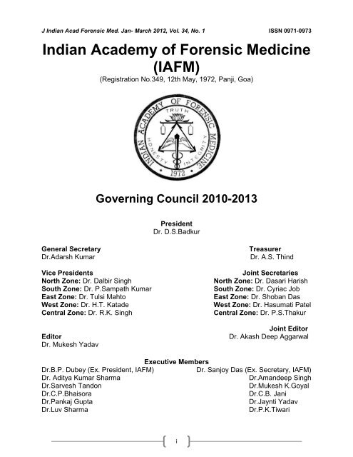
J <strong>Indian</strong> Acad <strong>Forensic</strong> Med. Jan- March 2012, Vol. 34, No. 1 ISSN 0971-0973<br />
<strong>Indian</strong> <strong>Academy</strong> <strong>of</strong> <strong>Forensic</strong> <strong>Medicine</strong><br />
(<strong>IAFM</strong>)<br />
(Registration No.349, 12th May, 1972, Panji, Goa)<br />
Governing Council 2010-2013<br />
President<br />
Dr. D.S.Badkur<br />
General Secretary Treasurer<br />
Dr.Adarsh Kumar Dr. A.S. Thind<br />
Vice Presidents<br />
North Zone: Dr. Dalbir Singh<br />
South Zone: Dr. P.Sampath Kumar<br />
East Zone: Dr. Tulsi Mahto<br />
West Zone: Dr. H.T. Katade<br />
Central Zone: Dr. R.K. Singh<br />
Editor<br />
Dr. Mukesh Yadav<br />
Dr.B.P. Dubey (Ex. President, <strong>IAFM</strong>)<br />
Dr. Aditya Kumar Sharma<br />
Dr.Sarvesh Tandon<br />
Dr.C.P.Bhaisora<br />
Dr.Pankaj Gupta<br />
Dr.Luv Sharma<br />
Executive Members<br />
i<br />
Joint Secretaries<br />
North Zone: Dr. Dasari Harish<br />
South Zone: Dr. Cyriac Job<br />
East Zone: Dr. Shoban Das<br />
West Zone: Dr. Hasumati Patel<br />
Central Zone: Dr. P.S.Thakur<br />
Joint Editor<br />
Dr. Akash Deep Aggarwal<br />
Dr. Sanjoy Das (Ex. Secretary, <strong>IAFM</strong>)<br />
Dr.Amandeep Singh<br />
Dr.Mukesh K.Goyal<br />
Dr.C.B. Jani<br />
Dr.Jaynti Yadav<br />
Dr.P.K.Tiwari
J <strong>Indian</strong> Acad <strong>Forensic</strong> Med. Jan- March 2012, Vol. 34, No. 1 ISSN 0971-0973<br />
Journal <strong>of</strong> <strong>Indian</strong> <strong>Academy</strong> <strong>of</strong> <strong>Forensic</strong> <strong>Medicine</strong><br />
(J<strong>IAFM</strong>)<br />
The <strong>of</strong>ficial publication <strong>of</strong> <strong>Indian</strong> <strong>Academy</strong> <strong>of</strong> <strong>Forensic</strong> <strong>Medicine</strong><br />
Editor,<br />
Dr. Mukesh Yadav<br />
Pr<strong>of</strong>essor & H.O.D.,<br />
<strong>Forensic</strong> <strong>Medicine</strong> & Toxicology,<br />
School <strong>of</strong> Medical Sciences and<br />
Research, Sharda University, Greater<br />
Noida-201306, Uttar Pradesh, INDIA<br />
Residence:<br />
G-216, Parsvanath Edens,<br />
Alfa-II, Greater Noida, G.B. Nagar, U.P.INDIA<br />
Ph. No. 0120-2326060, Cell: 09411480753<br />
Email: drmukesh65@yahoo.co.in<br />
Pr<strong>of</strong>. Derrick J Pounder, Dundee, UK<br />
Pr<strong>of</strong>. DN Vieira, Coimbra, Portugal<br />
Pr<strong>of</strong>. Dan Dermengiu, Romania<br />
Pr<strong>of</strong>. Peter Vanezis, London, UK<br />
Pr<strong>of</strong>. Roger Byard, Australia<br />
Dr. Michael S. Pollanen, Canada<br />
Pr<strong>of</strong>. Leandro Duerte De Carvalho, Brazil<br />
Srivastava A.K. (U.P.)<br />
Pillay V.V. (Kerala)<br />
Gorea R.K. (U.P.)<br />
Jani C.B. (Gujarat)<br />
Bose T.K (West Bengal)<br />
Pradeep Kumar G. (Karnatka)<br />
Verma S.K. (New Delhi)<br />
Kaur Balbir (Haryana)<br />
Kumar Shantha B. (Tamil Nadu)<br />
Gupta B.D. (Gujrat)<br />
Murty O.P. (New Delhi)<br />
International Advisory Board<br />
National Advisory Board<br />
Manju Nath K.H, (Karnatka)<br />
Das Sanjoy, (Uttarakhand)<br />
Mahtoo Tulsi, (Jharkhand)<br />
Ravindran K. (Puducherry)<br />
Rastogi Prateek (Karnatka)<br />
Potwary AJ (Assam)<br />
Singh R.K. (Chhatisgarh)<br />
Dongre A.P. (Maharastra)<br />
Sharma Aditya (H.P.)<br />
Gupta Pankaj (Punjab)<br />
Harish D. (Chandigarh)<br />
ii<br />
Joint Editor,<br />
Dr. Akash Deep Aggarwal<br />
Assistant Pr<strong>of</strong>essor,<br />
Department <strong>of</strong> <strong>Forensic</strong> <strong>Medicine</strong>,<br />
Govt. Medical College, Patiala<br />
Punjab, INDIA<br />
Residence:<br />
H.No. 14, Desi Mehmandari,<br />
Patiala-147001, Punjab, INDIA<br />
Cell: 9815652621<br />
Email:toakashdeep@yahoo.co<br />
Dr. BL Meel, South Africa<br />
Dr. John Clark, Glasgow, UK<br />
Dr. George Paul, Singapore<br />
Dr. Serap Annette AKGUR, Turkey<br />
Dr. Clifford Perera, Sri Lanka<br />
Dr. BN Yadav, Nepal<br />
Dr. KP Saha, Bangladesh<br />
Rastogi Pooja (U.P.)<br />
Khanagwal V. (Haryana)<br />
Khaja Shaikh (A.P.)<br />
Basu R (W.B.)<br />
Naik R.S. (Maharastra)<br />
Godhikirakar Madhu (Goa)<br />
Job Cyriac (Kerala)<br />
Vinita K. (U.P.)<br />
Mohite Shailesh (Mumbai)<br />
Yadav Jayanti (M.P.)<br />
Kochar S.R. (Rajasthan)<br />
Printed and published by Dr. Mukesh Yadav, Editor, J<strong>IAFM</strong> and Dr. A. D. Aggarwal, Joint Editor, J<strong>IAFM</strong> on<br />
behalf <strong>of</strong> <strong>Indian</strong> <strong>Academy</strong> <strong>of</strong> <strong>Forensic</strong> <strong>Medicine</strong> at name <strong>of</strong> the press [SHIVANI PRINTERS, NOIDA, [U.P.]
J <strong>Indian</strong> Acad <strong>Forensic</strong> Med. Jan- March 2012, Vol. 34, No. 1 ISSN 0971-0973<br />
Journal <strong>of</strong> <strong>Indian</strong> <strong>Academy</strong> <strong>of</strong> <strong>Forensic</strong> <strong>Medicine</strong><br />
Volume: 34 • Number: 1 • Jan- March 2012<br />
Contents<br />
Sr. Page<br />
I.<br />
II.<br />
From the Editor’s Desk 03-03<br />
Editorial: Use <strong>of</strong> Computer and IT in improving the Quality <strong>of</strong> Medicolegal<br />
Services in India<br />
Original Research Paper<br />
1. Age Determination from Clavicle: A Radiological Study in Mumbai Region<br />
S.S. Bhise, G. S. Chavan, B. G. Chikhalkar, S. D. Nanandkar<br />
2. An Entomological Study to Determine the Time since Death in Cases <strong>of</strong><br />
Decomposed Bodies Parmod Kumar Goyal<br />
3. Pr<strong>of</strong>ile <strong>of</strong> Deaths due to Electrocution: A Retrospective Study B. D. Gupta,<br />
R.A. Mehta, M. M. Trangadia<br />
4. Victim Pr<strong>of</strong>ile and Pattern <strong>of</strong> Thoraco-Abdominal Injuries Sustained in Fatal<br />
Road Traffic Accidents B.Suresh Kumar Shetty, Tanuj Kanchan, Ritesh G. Menezes,Shankar<br />
M. Bakkannavar, Vinod C Nayak, K Yoganarasimha<br />
5. A Study <strong>of</strong> Sexual Dimorphism <strong>of</strong> Femoral Head In Gujarat Region<br />
Pandya A.M., Gupta B.D., Singel T.C., Patel M.P.<br />
6. Abdominal Organ Involvement in Blunt Injuries Mousami Singh, Amit Kumar, Anoop<br />
Kumar Verma, Sanjeet Kumar, Abhas Kumar Singh<br />
7. Determination <strong>of</strong> Sex by Sciatic Notch/Acetabular Ratio (Kelley’s Index) in<br />
<strong>Indian</strong> Bengali Skeletal Remains Partha Pratim Mukhopadhyay<br />
8. Physician’s Perspectives about Consent in Medical Practice A<br />
Questionnaire Based Study Sanjay Gupta, Ravi Panchal<br />
9. Nomenclature for Knot Position in Hanging: A Study <strong>of</strong> 200 Cases<br />
D.S Badkur, Jayanthi Yadav, Arneet Arora, Ranjan Bajpayee, B.P.Dubey<br />
1<br />
04-06<br />
07-09<br />
10-12<br />
13-15<br />
16-19<br />
20-23<br />
24-26<br />
27-30<br />
31-33<br />
34-36<br />
10. Newer Trends in Hanging Death Ashok Kumar Samanta, Soumya Ranjan Nayak 37-39<br />
11. Pattern <strong>of</strong> Ligature Mark in Cases <strong>of</strong> Compressed Neck in Rajkot Region: A<br />
Prospective Study Sadikhusen G. Momin, Hari Mohan Mangal, Hetal C. Kyada, M.T.Vijapura,<br />
S.D.Bhuva<br />
12. Pattern <strong>of</strong> Injuries due to Electric Current Bharath Kumar Guntheti, Sheikh Khaja,<br />
Uday .P .Singh<br />
13. Trends <strong>of</strong> Maxill<strong>of</strong>acial Trauma at Tertiary Care Hospital in Rural Area <strong>of</strong><br />
Southern Punjab Vishal Garg, Harinder Singh, K Vij<br />
40-43<br />
44-48<br />
49-51<br />
14. Serial Bomb Blasts in North-East India: A Postmortem Study 52-54
J <strong>Indian</strong> Acad <strong>Forensic</strong> Med. Jan- March 2012, Vol. 34, No. 1 ISSN 0971-0973<br />
Yogender Malik, Ritu Raj Chaliha<br />
Review Research Papers<br />
15. Application <strong>of</strong> Genetics and Molecular Biology In <strong>Forensic</strong> Odontology<br />
Rohit Malik, Deepankar Misra, PC Srivastava, Akansha Misra<br />
16. Scenario <strong>of</strong> Hooch Tragedy in Gujarat State Pankaj Prajapati, Ganesh Govekar,<br />
Akhilesh Pathak<br />
17. Bite mark Analysis: An Overview Kalyani Bhargava, Deepak Bhargava, Pooja Rastogi,<br />
Mayura Paul, Rohit Paul, Jagadeesh H.G<br />
18. Second Autopsy- The <strong>Indian</strong> Scenario Swapnil S Agarwal, Lavlesh Kumar, Binay K<br />
Bastia, Krishnadutt H Chavali<br />
19. Voice Fingerprinting: A Very Important Tool against Crime Pragnesh Parmar,<br />
Udhayabanu R.<br />
20. Pathological Autopsy: Most Valuable Aid in the Present Medical and<br />
Medico Legal Scenario Jayashree. G. Pawar, Gurudatta. S. Pawar<br />
Case Reports<br />
21. A Suicidal Hanging with Unusual Findings at Crime Scene C Behera, Ankita Dey,<br />
Anju Rani, Kulbhushan, P.C Dikshit<br />
22. Occupational Injury at Gangsaw Machine: A Rare Occurrence Anil Yadav, R.K.<br />
Gahlot, N.S. Kothari<br />
23. Rupture Uterus: Carelessness or Negligence? Pranav Prajapati, M I Sheikh,<br />
Rajesh Patel<br />
24. MRKH Syndrome: Psychological Disturbances and Suicide Mohit Gupta, Varsha<br />
Kharb<br />
25. Assessment <strong>of</strong> Age in Foetus: A Medicolegal Aspect Jaswinder Kaur, Zora Singh,<br />
Rajiv Joshi<br />
26. Electrocution Method to Conceal Homicide: A Rare Case Report<br />
M. P. Jambure, R. M. Tandle, K U Zine<br />
From Editor’s Desk<br />
2<br />
55-57<br />
58-60<br />
61-66<br />
67-69<br />
70-73<br />
74-76<br />
77-79<br />
80-81<br />
82-85<br />
86-88<br />
89-91<br />
92-94<br />
Copy Right © All rights reserved: No part <strong>of</strong> this publication may be reprinted or publish without the<br />
prior permission <strong>of</strong> the Editor, J<strong>IAFM</strong>. Submission <strong>of</strong> all manuscripts to the journal is understood to imply<br />
that it is not being considered for publication elsewhere. Submission <strong>of</strong> multi authored papers implies that<br />
the consent <strong>of</strong> each author has been obtained. In this journal, every effort has been made NOT to publish<br />
inaccurate or misleading information. However, the Editor, Joint Editor, Peer Review Group and Advisory<br />
Board accept NO liability in consequences <strong>of</strong> such statements. The Journal <strong>of</strong> <strong>Indian</strong> <strong>Academy</strong> <strong>of</strong><br />
<strong>Forensic</strong> <strong>Medicine</strong> is indexed in Index Copernicus [Poland] and IndMED [India]<br />
Print ISSN: 0971-0973. Electronic ISSN: 0974-0848. IndMED www.medind.nic.in/jal/jalm.shtml<br />
Address request for reprint or further information relating to any article may please be made with author and<br />
in case <strong>of</strong> multi authored article, please communicate to Corresponding Author or the First Author
J <strong>Indian</strong> Acad <strong>Forensic</strong> Med. Jan- March 2012, Vol. 34, No. 1 ISSN 0971-0973<br />
3<br />
J<strong>IAFM</strong><br />
A Quarterly Publication<br />
Volume 34, Number 1, January-March, 2012<br />
I feel immense pleasure to present before you the first issue <strong>of</strong> 2012. I would like to inform all <strong>of</strong><br />
you that our esteemed Journal <strong>of</strong> <strong>Indian</strong> <strong>Academy</strong> <strong>of</strong> <strong>Forensic</strong> <strong>Medicine</strong> which is published quarterly since<br />
1991 has been started gaining wide recognition not only in India but globally among the scientific<br />
community. I am trying to maintain your faith and trust in me to bring this journal to highest level <strong>of</strong> its<br />
achievements.<br />
I have received many requests from other countries about inclusion <strong>of</strong> many papers in their<br />
indexing data base, including USA Government agencies. J<strong>IAFM</strong> is indexed not only in IndMed and<br />
MedInd <strong>Indian</strong> indexing agencies but also in the SCOPUS, IMSEAR informed by the Information<br />
Management and Dissemination (IMD), World Health Organization, Regional Office for South-East<br />
Asia, Indraprastha Estate, New Delhi, India. It is hoped that once this journal indexed in IMSEAR it<br />
would be automatically indexed in the Global Index Medicus managed by WHO Headquarters in<br />
Geneva as informed.<br />
The title mentioned above has been evaluated for inclusion in SCOPUS by the Content<br />
Selection & Advisory Board (CSAB). The review <strong>of</strong> this title is now complete and the CSAB has<br />
advised that the title will be accepted for inclusion in Scopus. For your information, the reviewer<br />
comments are copied below:<br />
This is a well produced journal in an important subject field with interesting content, which<br />
deserves a wide readership. The editors are to be commmended on their efforts.<br />
I assure you about the quality <strong>of</strong> research papers and quality <strong>of</strong> printing in future issues. Your<br />
valuable suggestions are always encouraging me and I heartily welcome for future suggestions.<br />
Pr<strong>of</strong>essor [Dr.] Mukesh Yadav<br />
Editor, J<strong>IAFM</strong><br />
Subscription Information<br />
Members <strong>of</strong> <strong>IAFM</strong> will receive the free <strong>of</strong> cost.<br />
Non Members and Institutions (Annual Subscription rates)<br />
Personal: In India, Rs. 1000/ (Rest <strong>of</strong> the world: US$ 200/ or equivalent)<br />
Institutions: In India, Rs. 3000/ (Rest <strong>of</strong> the world: US$ 400/ or equivalent)<br />
We Accept: Bank Cheque / Demand Drafts (Add Rs. 50/- for outstation Cheques)<br />
The Scope <strong>of</strong> the Journal covers all aspects <strong>of</strong> <strong>Forensic</strong> <strong>Medicine</strong> and allied fields, research and<br />
applied.<br />
Subscription orders and payments should be made in favour <strong>of</strong><br />
“Editor, J<strong>IAFM</strong>, payable at Greater Noida”<br />
Claims for missing issue:<br />
A copy will be sent free to the member / subscriber provided the claim is made within 2 months <strong>of</strong> publication<br />
<strong>of</strong> the issue & self addressed envelop <strong>of</strong> the size 9” x 12” is sent to the Editor. (Those who want the journals to<br />
be dispatched by Registered Post must affix Rs. 50/ worth postage stamps).<br />
The journal is indexed with IndMed and made available online by following <strong>website</strong>:<br />
www.iafmonline.com www.jiafm.com<br />
www.medind.nic.in www.indianjournals.com<br />
http://indmed.nic.in www.forensicindia.com
J <strong>Indian</strong> Acad <strong>Forensic</strong> Med. Jan- March 2012, Vol. 34, No. 1 ISSN 0971-0973<br />
Editorial:<br />
Use <strong>of</strong> Computer and IT in improving the Quality <strong>of</strong><br />
Medico-legal Services in India<br />
Hon’ble Court has shown its concern for preparation <strong>of</strong> Medico-legal record by junior and not properly<br />
trained Casualty Medical Officer. There is need for more and more involvement <strong>of</strong> <strong>Forensic</strong> <strong>Medicine</strong><br />
experts in Medico-legal work in the casualty. Diagrammatic illustration <strong>of</strong> injuries in the form <strong>of</strong> sketch<br />
diagram in medico-legal report and post-mortem reports is <strong>of</strong> immense value for court and all concerned<br />
stake holders in better appreciation <strong>of</strong> findings and reaching to conclusive opinion during trial.<br />
The decisive and conclusive medical evidence which should be able to connect the alleged<br />
accused with the crime and go against the defence version whenever case come for appeal before a<br />
higher court regarding the cause <strong>of</strong> death has been relied by the court for conviction. Manner <strong>of</strong><br />
preparation <strong>of</strong> medical record and its legibility is very important in saving lots <strong>of</strong> time and improving the<br />
quality <strong>of</strong> medical records in adding better administration <strong>of</strong> justice which is all about the speciality <strong>of</strong><br />
<strong>Forensic</strong> <strong>Medicine</strong>.<br />
The prosecution, with a view to connect the appellants with the crime, relied upon:<br />
(i) Motive;<br />
(ii) Direct evidence <strong>of</strong> assault on the deceased;<br />
(iii) Extra Judicial Confession;<br />
(iv) Medical evidence;<br />
(v) Conduct <strong>of</strong> the appellants<br />
In Mukhtiar Singh & Ors. vs. State <strong>of</strong> Punjab, Criminal Appeal No. 480 <strong>of</strong> 1985, Date <strong>of</strong> Judgment: dated<br />
30.11.1995, Hon’ble SC observed that:<br />
“…. (iv) Medical evidence: According to PW1, Dr. Dalal, the cause <strong>of</strong> death was coma as a result <strong>of</strong><br />
dislocation <strong>of</strong> 2nd/3rd cervical vertebrae which was ante mortem in nature and sufficient to cause death<br />
in the ordinary course <strong>of</strong> nature. The doctor then opined the possibility <strong>of</strong> the dislocation <strong>of</strong> second<br />
and third vertebrae due to Lathi blow cannot ruled out, but since the body was in a burnt condition it<br />
was not possible to specify the exact type <strong>of</strong> the weapon with which the injury may have been caused.<br />
During the cross-examination, PW1 admitted the possibility <strong>of</strong> injuries No.1, 2 and 3 on the dead<br />
body <strong>of</strong> Pritam Kaur having been suffered in a fall from a height cannot be ruled out. At the places where<br />
injuries No.1 and 2 were located no bony injury was found.<br />
SC concluded that “thus, we find that the medical evidence is neither decisive nor conclusive and<br />
it fails to connect the appellants with the crime and does not go against the defence version <strong>of</strong> the<br />
deceased having died instantaneously as a result <strong>of</strong> the fall. The absence <strong>of</strong> any bony injury is more<br />
consistent with the defence version than the prosecution case.”<br />
In the same case (Supra) at point 17, SC quoted another judgment, Shivaji Sahebrao Bobade & Anr.<br />
vs. State <strong>of</strong> Maharashtra, AIR 1973 SC 2622, SC court held:<br />
"...Thus too frequent acquittals <strong>of</strong> the guilty may lead to a ferocious penal law, eventually eroding<br />
the judicial protection <strong>of</strong> the guiltless. For all these reasons it is true to say, with Viscount Simon, that "a<br />
miscarriage <strong>of</strong> justice may arise from the acquittal <strong>of</strong> the guilty no less than from the conviction <strong>of</strong><br />
the innocent ..." In short our jurisprudential enthusiasm for presumed innocence must be moderated by<br />
the pragmatic need to make criminal justice potent and realistic.<br />
A balance has to be struck between chasing chance possibilities as good enough to set the<br />
delinquent free and chopping the logic <strong>of</strong> preponderant probability to punish marginal innocents. We have<br />
adopted these cautions in analysing the evidence and appraising the soundness <strong>of</strong> the contrary<br />
conclusions reached by the courts below. Certainly, in the last analysis reasonable doubts must operate<br />
to the advantage <strong>of</strong> the appellant..."<br />
The <strong>Indian</strong> Medical Council (Pr<strong>of</strong>essional Conduct, Ethics and Etiquettes)<br />
Regulations, 2002, in Chapter I at point 1.3, mentioned about<br />
Maintenance <strong>of</strong> medical records:<br />
1.3.1 Every physician shall maintain the medical records pertaining to his/her indoor patients for a<br />
period <strong>of</strong> 3 years from the date <strong>of</strong> commencement <strong>of</strong> the treatment in a standard pr<strong>of</strong>orma laid down by<br />
the Medical Council <strong>of</strong> India and attached as Appendix 3.<br />
4
J <strong>Indian</strong> Acad <strong>Forensic</strong> Med. Jan- March 2012, Vol. 34, No. 1 ISSN 0971-0973<br />
1.3.2 If any request is made for medical records either by the patients/authorized attendant or legal<br />
authorities involved, the same may be duly acknowledged and documents shall be issued within the<br />
period <strong>of</strong> 72 hours.<br />
1.3.3 A registered medical practitioner shall maintain a Register <strong>of</strong> Medical Certificates giving full<br />
details <strong>of</strong> certificates issued. When issuing a medical certificate he/she shall always enter the<br />
identification marks <strong>of</strong> the patient and keep a copy <strong>of</strong> the certificate. He/She shall not omit to record the<br />
signature and/or thumb mark, address and at least one identification mark <strong>of</strong> the patient on the medical<br />
certificates or report. The medical certificate shall be prepared as in Appendix 2.<br />
1.3.4 Efforts shall be made to computerize medical records for quick retrieval.<br />
Thus, computerization <strong>of</strong> medical record is important not only for easy retrieval and about the quality<br />
<strong>of</strong> medical record especially <strong>of</strong> medico-legal record <strong>of</strong> immense value in administration <strong>of</strong> justice.<br />
Role <strong>of</strong> Court in Computerization <strong>of</strong> Medical Record:<br />
Many High courts reacting to the quality <strong>of</strong> writing which is not legible and consumed lot <strong>of</strong> time in<br />
deciphering the said writing and its impact on disposal <strong>of</strong> the cases. Some examples are illustrated below:<br />
Scenario in State <strong>of</strong> Delhi:<br />
Hon’ble Delhi High Court in case <strong>of</strong> State <strong>of</strong> Govt. <strong>of</strong> NCT <strong>of</strong> Delhi vs. Jitender and State vs.<br />
Virender @ Ballu & Anr., Judgment dated 19.01.2012 observed that “Further from the perusal <strong>of</strong> the<br />
Post Mortem report and the MLC prepared in the present case and in many other cases, which comes to<br />
the notice <strong>of</strong> this court daily, during routine court proceedings, where Medico-legal reports are prepared<br />
and the doctors attend the court for their depositions relating to post-mortems and different types <strong>of</strong><br />
MLC(s). In most <strong>of</strong> the cases, the said writing in the MLC(s)/Post-mortem reports are not at all legible and<br />
lot <strong>of</strong> time is consumed in deciphering the said writing. In all those cases, the doctors/Autopsy Surgeons<br />
have to first describe the contents written in the MLC(s) and post-mortem reports, so that the same can<br />
be dictated and can be converted into legible words.”<br />
Court further added that “Sometimes the concerned doctors have either left the services <strong>of</strong> the<br />
hospital or are not traceable and other doctors are directed to appear in the court in their place, but due to<br />
illegible handwriting <strong>of</strong> the concerned doctors(s) including the signatures, put on the MLC(s) the doctor<br />
who is deputed in the place <strong>of</strong> such Doctor(s) are most <strong>of</strong> time are not able to decipher the handwriting <strong>of</strong><br />
the concerned doctor and feel great inconvenience in communicating the exact facts mentioned in the<br />
reports and MLC(s) during their examination, which adversely effects the ends <strong>of</strong> justice and<br />
unnecessarily consumes lot <strong>of</strong> time <strong>of</strong> the court.<br />
Suggestions for Improving the Quality <strong>of</strong> Medico-Legal Record:<br />
Further lots <strong>of</strong> other deficiencies have been observed by court regarding the manner <strong>of</strong> preparation <strong>of</strong><br />
MLC(s) and the post-mortem reports. Consequently, following suggestions be implemented for<br />
improvement <strong>of</strong> Medico-legal and post-mortem reports for better administration <strong>of</strong> Criminal Justice:<br />
(a) The MLCs, especially the postmortem reports be prepared by computer typing, rather than<br />
handwriting to save the time <strong>of</strong> the court, defence lawyers and the accused and to give better clarity<br />
to the accused persons, as to what is against them.<br />
(b) The MLC(s) at present in the Casualty <strong>of</strong> the hospital are being prepared by very junior or trainee<br />
doctors resulting in incomplete information being mentioned in the MLC(s), about the injury and the<br />
associated findings. Therefore, the MLC(S) should not be prepared by the doctors who are not<br />
properly trained in <strong>Forensic</strong> <strong>Medicine</strong>.<br />
(c) Any injury(s) found on the body <strong>of</strong> the injured including burn injuries(s), should be clearly illuminated<br />
on the sketch diagram(s) <strong>of</strong> the human body which are already on the back side <strong>of</strong> the MLC’s and<br />
along with that the colour changes, dimensions <strong>of</strong> injury(s), duration <strong>of</strong> injuries(s), depth <strong>of</strong> injury(s),<br />
location in respect to land marks <strong>of</strong> the body, be clearly mentioned, which medical facts are most<br />
essential for better appreciation <strong>of</strong> the injuries and for better appreciation <strong>of</strong> facts during the trial.<br />
(d) Further along with postmortem reports the injuries found on the body <strong>of</strong> deceased should also be<br />
illustrated on the sketch diagram <strong>of</strong> human body, including exit and entry wound <strong>of</strong> bullet injuries at<br />
least in burn and murder cases.<br />
(e) Histo-pathological report <strong>of</strong> organ(s) <strong>of</strong> the deceased be submitted at the earliest, rather it is<br />
observed, it is not being submitted/done at all in most <strong>of</strong> the cases.<br />
(f) The subsequent/final opinion regarding the cause <strong>of</strong> death, by the Autopsy Surgeon is opined very<br />
late in most <strong>of</strong> the cases, due to delayed, reporting/collection <strong>of</strong> FSL/Chemical Analysis report <strong>of</strong><br />
viscera, and histopathological report, which results in grave injustice to the fundamental rights <strong>of</strong> the<br />
accused and the victim during the trial for speedy justice. Therefore, FSL report be given in a time<br />
bound manner, at least in serious cases.<br />
5
J <strong>Indian</strong> Acad <strong>Forensic</strong> Med. Jan- March 2012, Vol. 34, No. 1 ISSN 0971-0973<br />
Hon’ble Court had directed that the copy <strong>of</strong> this order be sent to Principal Secretary, Health, and<br />
Principal Secretary Home, Govt. <strong>of</strong> NCT <strong>of</strong> Delhi, for compliance and circulate amongst all the Medical<br />
Superintendent(s) <strong>of</strong> the Hospitals <strong>of</strong> Delhi Govt.<br />
Scenario in State <strong>of</strong> Punjab:<br />
Similarly in the States <strong>of</strong> Punjab and Haryana, in terms <strong>of</strong> the order passed by the Punjab &<br />
Haryana High Court on 6.7.2011, learned counsel appearing for the State <strong>of</strong> Punjab has produced<br />
minutes <strong>of</strong> meeting held under the Chairmanship <strong>of</strong> Principal Secretary to Government, Punjab,<br />
Department <strong>of</strong> Health and Family Welfare, in terms <strong>of</strong> which it has been, inter alia, decided that from<br />
1.09.2011 onwards, all post-mortem reports/medico-legal reports shall be computer typed. Even if, some<br />
report is prepared in hand initially, a fresh typed copy duly signed by the concerned doctor shall be<br />
supplied later. With the challan invariably computer typed copy shall be submitted.<br />
Scenario in State <strong>of</strong> Haryana:<br />
Dr. Narvir Singh, Director General Health Services, Haryana was appeared in court and appraised that as<br />
for as State <strong>of</strong> Haryana is concerned, copy <strong>of</strong> the communication dated 20.04.2011 has been produced<br />
stating therein that doctors have been advised to prepare a legible post-mortem report or medico-legal<br />
report. Learned counsel for the State submitted that fresh instructions regarding preparation <strong>of</strong> the<br />
aforesaid documents on computer are in the process <strong>of</strong> being issued.<br />
Scenario in Union Territory, Chandigarh:<br />
Similarly, Union Territory, Chandigarh submitted that the matter is under active consideration and<br />
necessary instructions shall be issued, which shall be produced in court on the next date <strong>of</strong> hearing.<br />
Certain more hospitals in the States <strong>of</strong> Punjab and Haryana and Union Territory, Chandigarh, issue<br />
computerized post-mortem reports/medico-legal reports.<br />
Role <strong>of</strong> MCI:<br />
This is the right time for other States in India to follow the judicial trend for better administration <strong>of</strong><br />
justice and reducing the backlog <strong>of</strong> cases and enhanced conviction rate. Regulatory body Medical<br />
Council <strong>of</strong> India should realise the intent behind these direction <strong>of</strong> the Hon’ble Court and restore the<br />
reduced faculty and implement restructured new curriculum to meet the demand <strong>of</strong> judiciary in larger<br />
public interest.<br />
Mukesh Yadav<br />
Editor<br />
6
J <strong>Indian</strong> Acad <strong>Forensic</strong> Med. Jan- March 2012, Vol. 34, No. 1 ISSN 0971-0973<br />
Original Research Paper<br />
Age Determination from Clavicle: A Radiological Study in<br />
Mumbai Region<br />
*S.S. Bhise, **G. S. Chavan, **B. G. Chikhalkar, ***S. D. Nanandkar<br />
Abstract<br />
The bones <strong>of</strong> human skeletons develop from separate ossification centres. From these centers<br />
ossification progresses till the bone is completely formed. These changes can be studied by means <strong>of</strong> Xrays<br />
and these changes are age related. It is therefore possible to determine the approximate age <strong>of</strong> an<br />
individual by radiological examination <strong>of</strong> bones till ossification is complete.<br />
This radiological study was carried out with the objective to assess the general skeletal maturity<br />
around Medial end <strong>of</strong> clavicle, <strong>of</strong> subjects in Mumbai region. 131 males between age group <strong>of</strong> 9-25 years<br />
and 68 females between age group <strong>of</strong> 3-23 years attending the outpatient department <strong>of</strong> this hospital<br />
were selected. Age confirmed from history and noting the birth dates from driving license, passport,<br />
rations card or voter’s card. The cases were selected after ruling out the nutritional, developmental, and<br />
endocrinal abnormality which affects the skeletal growth. Data analysis was done in P4 computer using<br />
HPSS s<strong>of</strong>tware. At the end conclusions were drawn which are compared with available results <strong>of</strong> various<br />
previous studies.<br />
Key Words: Epiphyseal Fusion, Ossification Centres, X-Rays<br />
Introduction:<br />
Determination <strong>of</strong> the age <strong>of</strong> an individual<br />
from the appearance and the fusion <strong>of</strong> the<br />
ossification centres is a well accepted fact in the<br />
field <strong>of</strong> medical and legal pr<strong>of</strong>essions. Epiphysis<br />
<strong>of</strong> bones unites during age periods which are<br />
remarkably constant for a particular epiphysis.<br />
The determination <strong>of</strong> age presents a<br />
task <strong>of</strong> considerable importance from the viewpoint<br />
<strong>of</strong> the administration <strong>of</strong> justice. It is not<br />
possible to enunciate a hard and fast rule for<br />
age determination from this union for the whole<br />
India because the various geographical areas <strong>of</strong><br />
our country differ in climatic, dietetic and disease<br />
factors. The present study was carried out to<br />
study roentgenographically the epiphyseal<br />
appearance and fusion <strong>of</strong> medial end <strong>of</strong> clavicle<br />
in subjects between age group <strong>of</strong> 3 to 25 years<br />
attending outpatient department <strong>of</strong> this hospital.<br />
Aims and Objectives:<br />
To assess the skeletal maturity at medial<br />
end <strong>of</strong> clavicle for a known chronological<br />
age in subjects <strong>of</strong> Mumbai region.<br />
Corresponding Author:<br />
* Assistant Pr<strong>of</strong>essor, Dept. <strong>of</strong> <strong>Forensic</strong> medicine,<br />
Grant medical college Mumbai.<br />
E-mail: sadanand_bhise@rediffmail.com<br />
** Associate pr<strong>of</strong>essor<br />
***Pr<strong>of</strong>essor & HOD,<br />
7<br />
Do Comparative study <strong>of</strong> appearance &<br />
fusion <strong>of</strong> medial end <strong>of</strong> clavicle with known<br />
standards.<br />
To evaluate sex related variation & its<br />
correlation with age.<br />
To know variation if any & exception <strong>of</strong><br />
appearance & fusion <strong>of</strong> medial end <strong>of</strong><br />
clavicle.<br />
To evaluate the medico legal aspects <strong>of</strong><br />
different ages.<br />
To suggest any additional radiological<br />
investigation to aid and to reduce range in<br />
determining age.<br />
Material and Methods:<br />
The study was carried out in Grant<br />
Medical College and Sir J. J. Group <strong>of</strong> Hospitals<br />
Mumbai which is a tertiary referral centre. The<br />
objective was to assess the general skeletal<br />
maturity <strong>of</strong> medial end <strong>of</strong> clavicle in subjects in<br />
Mumbai region. 131 males between age group<br />
<strong>of</strong> 9-25 years and 68 females between age<br />
group <strong>of</strong> 3-23 years attending the outpatient are<br />
selected. Age confirmed from history and noting<br />
the birth dates from driving license, passports<br />
ration card or voter’s card. The cases were<br />
selected after ruling out the nutritional,<br />
developmental, and endocrinal abnormality<br />
which affects the skeletal growth. X-rays <strong>of</strong><br />
medial end <strong>of</strong> clavicle, AP view were taken at<br />
department <strong>of</strong> radiology. The epiphysis <strong>of</strong> medial<br />
end <strong>of</strong> clavicle was observed for appearance (A)<br />
and non appearance (NA) and different phases
J <strong>Indian</strong> Acad <strong>Forensic</strong> Med. Jan- March 2012, Vol. 34, No. 1 ISSN 0971-0973<br />
<strong>of</strong> fusion were graded according to Dr. William<br />
Sangma et al and Mckern and Stewart’s<br />
methods. The 5 stages were as follows-<br />
Stage 1 (F1): Non union – when the<br />
epiphyseal cartilage did not begin to<br />
decrease in thickness<br />
Stage 2(F2): Commence <strong>of</strong> union – when<br />
the thickness <strong>of</strong> epiphyseal cartilage was<br />
found to be reduced appreciably (1/4 th<br />
united)<br />
Stage 3(F3): Incomplete union – when the<br />
epiphysis has begun to fuse with shaft and<br />
complete union was well underway (1/2<br />
united)<br />
Stage 4(F4): Complete union – when the<br />
epiphyseal cartilage was bony in<br />
architecture and its density indistinguishable<br />
from the epiphysis and diaphysis in its<br />
neighbourhood but an epiphyseal line called<br />
epiphyseal scar could still be distinguished.<br />
(3/4 united)<br />
Stage 5(F5): Complete union – with<br />
absence <strong>of</strong> epiphyseal scar.<br />
The appearance and fusion <strong>of</strong> medial<br />
end <strong>of</strong> clavicle was evaluated radiologically and<br />
the results were compared with the previous<br />
known standard studies<br />
Results and observations:<br />
Table No. 1 shows in males in 34 cases<br />
(89.6%) at 9 – 15 years and in 4 cases (10.4%)<br />
at 15 – 16 years centre was not appeared. In 6<br />
(60%) cases at 15 – 16 years and 4 cases<br />
(40%) at 16 – 17 years centre was appeared<br />
F1 stage <strong>of</strong> fusion was seen in 2 cases<br />
(40%) at 16 – 17 years age group and in 3<br />
cases (60%) at 17 – 18 years age group.<br />
F2 stage <strong>of</strong> fusion was seen in 1 case<br />
(10%) at 16 – 17 years age group, in 5 cases<br />
(50%) at 17 – 18 years age group, in 1 case<br />
(10%) at 18 – 19 years age group and in 3<br />
cases (30%) at 19 – 20 years age group.<br />
F3 stage <strong>of</strong> fusion was seen in 5 cases<br />
(17.2%) at 17 - 18 years age group, in 18 cases<br />
(62.1%) at 18 – 19 years age group, in 3 case<br />
(10.3%) at 19 – 20 years age group and in 3<br />
cases (10.3%) at 20 – 21 years age group .<br />
F4 stage <strong>of</strong> fusion was seen in 1 case<br />
(4.8%) at 18 - 19 years age group, in 1 case<br />
(4.8%) at 19 – 20 years age group, in 10 cases<br />
(47.6%) at 20 – 21 years age group and in 5<br />
cases (23.8%) at 21 – 22 years age group and<br />
in 4 cases (19%)at 22 – 23 years age group.<br />
Complete fusion (F5) was seen in 5<br />
cases (27.8%) at 21 - 22 years age group, in 3<br />
cases (16.7%) at 22 – 23 years age group, in 6<br />
cases (33.3%) at 23 – 24 years age group and<br />
in 4 cases (22.2%) at 24 – 25 years age group.<br />
8<br />
Table No. 2 shows in females in 26 cases<br />
(83.9%) at 3 – 13 years, in 3 cases (9.6%) at 13<br />
– 14 years and in 2 cases (6.5%) centre was not<br />
appeared.<br />
In 2 (25%) cases at 14 – 15 years, 3<br />
cases (37.5%) at 15 – 16 years and in 3 cases<br />
(37.5%) 16 - 17 centre was appeared<br />
F1 stage <strong>of</strong> fusion was seen in 1 case<br />
(33.3%) at 15 – 16 years age group and in 2<br />
cases (66.7%) at 16 – 17 years age group.<br />
F2 stage <strong>of</strong> fusion was seen in 2 cases<br />
(50%) at 17 – 18 years age group and in 2<br />
cases (50%) at 18 – 19 years age group.<br />
F3 stage <strong>of</strong> fusion was seen in 2 cases<br />
(33.3%) at 17 - 18 years age group, in 4 cases<br />
(67.7%) at 18 – 19 years age group.<br />
F4 stage <strong>of</strong> fusion was seen in 3 cases<br />
(60%) at 19 - 20 years age group and in 2 cases<br />
(40%) at 20 – 21 years age group.<br />
Complete fusion (F5) was seen in 4<br />
cases (36.4%) at 20 - 21 years age group, in 4<br />
cases (36.4%) at 21 – 22 years age group and<br />
in 3 cases (27.3%) at 22 – 23 years age group.<br />
Discussion:<br />
In present study both males and<br />
females in majority <strong>of</strong> cases show epiphyseal<br />
appearance at 15 – 16 years age group.<br />
In present study males show epiphyseal<br />
union at 23 - 24 years age group and earliest<br />
union occurred at 21 years. Females show<br />
epiphyseal union at 21 - 22 years age group and<br />
earliest union occurred at 20 years.<br />
The present study findings are close to<br />
Flecker, Galstaun, B. D. Chaurassia, Parikh, and<br />
Krishan Vij. [(5, 7, 9, 13]<br />
According to Stevenson (1924) in both males<br />
and females earliest union occurred at 18 years<br />
but in present study for males, earliest union<br />
occurred at 21 years <strong>of</strong> age and for females it is<br />
20 years <strong>of</strong> age. Present study and Stevenson<br />
show different results because they are<br />
performed in different races (Table - 3).<br />
In present study majority <strong>of</strong> cases show<br />
complete union at 23 – 24 years for males and<br />
at 21 – 22 years for females. These findings are<br />
in tandem with study carried out by B. D.<br />
Chaurassia and Parikh because both studies are<br />
done in India.<br />
Conclusions:<br />
From the present study it can be<br />
concluded, that-<br />
Epiphysis <strong>of</strong> Medial end <strong>of</strong> Clavicle appears<br />
at 15 – 16 years in both males and females<br />
Epiphysis <strong>of</strong> medial end <strong>of</strong> clavicle fused in<br />
most <strong>of</strong> the cases at 23 – 24 years for males<br />
and at 21 – 22 years for females. Earliest
J <strong>Indian</strong> Acad <strong>Forensic</strong> Med. Jan- March 2012, Vol. 34, No. 1 ISSN 0971-0973<br />
union occurs at 21 years in males and at 20<br />
years in females.<br />
References:<br />
1. R.N. Karmakar, J.B. Mukharjee. Essential <strong>of</strong> forensic <strong>Medicine</strong><br />
and toxicology 3 rd ed. p 126, 146, 147, 154, 155<br />
2. H.Flecker. Roentgenographic observations <strong>of</strong> the times <strong>of</strong><br />
appearance <strong>of</strong> epiphyses and their fusion with the diaphyses, J.<br />
Anat. 67 (1933), pp. 118–164.<br />
3. Krogman WM, Iscan, MY. in The human skeleton in <strong>Forensic</strong><br />
<strong>Medicine</strong>, Charles C.Thomas Publisher, Illinois, USA. II Edition,<br />
1986.<br />
4. Hepworth SM. determination <strong>of</strong> age in <strong>Indian</strong>s from study <strong>of</strong><br />
ossification <strong>of</strong> long bones Ind. Med. Gaz., 64,128,1929<br />
5. Chaurassia. Upper limb and thorax volume one; Human Anatomy<br />
C. B. S. Publisher and Distributors (ed.) second, 1991<br />
6. Chhokar V, Aggarwal S.N., Bhardwaj D.N. estimation <strong>of</strong> age <strong>of</strong> 16<br />
years in females by Radiological and dental examination: Journal<br />
9<br />
<strong>Forensic</strong> <strong>Medicine</strong> and Toxicology. Vol. IX No. 1 and 2 Jan-June<br />
1992, 25-30<br />
7. Galstaun, G. <strong>Indian</strong> Journal <strong>of</strong> Medical Research (1937) 25,267.<br />
8. Modi. Personal identity, Modi’s Medical Jurisprudence and<br />
Toxicology; Butterworth’s (edi.)22 nd , 1988; 35 – 42.<br />
9. Parikh. Personal identity, Parikh’s Textbook <strong>of</strong> Medical<br />
Jurisprudence and Toxicology. C.B.S. (edi.) 5 th ; 1990, 39 – 50.<br />
10. Reddy K.S.N. Identification; The synopsis <strong>of</strong> <strong>Forensic</strong> <strong>Medicine</strong> and<br />
Toxicology; (ed.) 8 th , 1992; 28-45.<br />
11. Singh Pardeep, Gorea R.K., Oberoi S.S. Kapila A.K. Age<br />
estimation from medial end <strong>of</strong> clavicle by x-ray examination, journal<br />
<strong>of</strong> <strong>IAFM</strong>, Vol.32, No.1 Jan-march 2010.<br />
12. Stewart. Recent improvements in estimating stature, sex, age and<br />
race from skeletal remains; The Modern Trends in <strong>Forensic</strong><br />
<strong>Medicine</strong>-3 Butterworth and Company (Publishers) limited.<br />
13. Vij K. Identification, text book <strong>of</strong> <strong>Forensic</strong> <strong>Medicine</strong>, Principle and<br />
Practice B.I. Churchil Livingston, (ed.), 1 st 2001; 74-82.<br />
Table1<br />
Extent <strong>of</strong> Appearance and Fusion <strong>of</strong> Medial End <strong>of</strong> Clavicle in Males Different Age Groups<br />
Extent <strong>of</strong> 9-15 15-16 16-17 17-18 18-19 19-20 20-21 21-22 22-23 23-24 24-25 Total<br />
appearance<br />
fusion<br />
&<br />
Cases<br />
(%)<br />
Cases<br />
(%)<br />
Cases<br />
(%)<br />
Cases<br />
(%)<br />
Cases<br />
(%)<br />
Cases<br />
(%)<br />
Cases<br />
(%)<br />
Cases<br />
(%)<br />
Cases<br />
(%)<br />
Cases<br />
(%)<br />
Cases<br />
(%)<br />
Cases<br />
(%)<br />
NA 34(89.6) 4(10.4) 0(0) 0(0) 0(0) 0(0) 0(0) 0(0) 0(0) 0(0) 0(0) 38(100)<br />
A 0(0) 6(60) 4(40) 0(0) 0(0) 0(0) 0(0) 0(0) 0(0) 0(0) 0(0) 10(100)<br />
F1 0(0) 0(0) 2(40) 3(60) 0(0) 0(0) 0(0) 0(0) 0(0) 0(0) 0(0) 5(100)<br />
F2 0(0) 0(0) 1(10) 5(50) 1(10) 3(30) 0(0) 0(0) 0(0) 0(0) 0(0) 10(100)<br />
F3 0(0) 0(0) 0(0) 5(17.2) 18(62.1) 3(10.3) 3(10.3) 0(0) 0(0) 0(0) 0(0) 29(100)<br />
F4 0(0) 0(0) 0(0) 0(0) 1(4.8) 1(4.8) 10(47.6) 5(23.8) 4(19) 0(0) 0(0) 21(100)<br />
F5 0(0) 0(0) 0(0) 0(0) 0(0) 0(0) 0(0) 5(27.8) 3(16.7) 6(33.3) 4(22.2) 18(100)<br />
Table 2<br />
Extent <strong>of</strong> Appearance and Fusion <strong>of</strong> Medial End <strong>of</strong> Clavicle in Females Different Age Gps<br />
Extent <strong>of</strong> 3-13 13-14 14-15 15-16 16-17 17-18 18-19 19-20 20-21 21-22 22-23 Total<br />
appearance<br />
& fusion<br />
cases<br />
(%)<br />
cases<br />
(%)<br />
cases<br />
(%)<br />
cases<br />
(%)<br />
cases<br />
(%)<br />
cases<br />
(%)<br />
cases<br />
(%)<br />
cases<br />
(%)<br />
cases<br />
(%)<br />
cases<br />
(%)<br />
cases<br />
(%)<br />
Cases<br />
(%)<br />
NA 26(83.9) 3(9.6) 2(6.5) 0(0) 0(0) 0(0) 0(0) 0(0) 0(0) 0(0) 0(0) 31(100)<br />
A 0(0) 0(0) 2(25) 3(37.5) 3(37.5) 0(0) 0(0) 0(0) 0(0) 0(0) 0(0) 8(100)<br />
F1 0(0) 0(0) 0(0) 1(33.3) 2(66.7) 0(0) 0(0) 0(0) 0(0) 0(0) 0(0) 3(100)<br />
F2 0(0) 0(0) 0(0) 0(0) 0(0) 2(50) 2(50) 0(0) 0(0) 0(0) 0(0) 4(100)<br />
F3 0(0) 0(0) 0(0) 0(0) 0(0) 2(33.3) 4(67.7) 0(0) 0(0) 0(0) 0(0) 6(100)<br />
F4 0(0) 0(0) 0(0) 0(0) 0(0) 0(0) 0(0) 3(60) 2(40) 0(0) 0(0) 5(100)<br />
F5 0(0) 0(0) 0(0) 0(0) 0(0) 0(0) 0(0) 0(0) 4(36.4) 4(36.4) 3(27.3) 11(100)<br />
Author Year Race<br />
Table 3<br />
Comparison <strong>of</strong> Time <strong>of</strong> Fusion (n Years)<br />
Sex Earliest<br />
Males Females Mixed<br />
Union (Yrs)<br />
Appear Fusion Appear Fusion<br />
Male/female<br />
Stevenson’s 1924 White and Negroes 22-24 22-25 -- 18<br />
Davies& Parson 1927 English -- -- 25<br />
Flecker 1932 Australians 21 21 ---<br />
Galstaun 1937 Bengalies (<strong>Indian</strong>s) 15-19 22 14-16 20 --<br />
Krogman 1962 U.S.A. -- -- 25-28<br />
Stewart 1973 U.S.A. 26 or more -- --<br />
Chaurassia 1980 <strong>Indian</strong> -- -- 21-22<br />
Parikh 1990 <strong>Indian</strong> -- -- 22<br />
Inderbir 1993 <strong>Indian</strong> -- -- 25<br />
Krishnan Vij 2001 <strong>Indian</strong> -- -- 20-22<br />
Present study 2010 Mumbai (<strong>Indian</strong>) 23-24 21-22 -- Male-21<br />
Female-20
J <strong>Indian</strong> Acad <strong>Forensic</strong> Med. Jan- March 2012, Vol. 34, No. 1 ISSN 0971-0973<br />
Original Research Paper<br />
An Entomological Study to Determine the Time since Death<br />
in Cases <strong>of</strong> Decomposed Bodies<br />
*Parmod Kumar Goyal<br />
Abstract<br />
<strong>Forensic</strong> entomology is the application <strong>of</strong> knowledge <strong>of</strong> insects during investigation <strong>of</strong> crimes or<br />
other legal matters. For investigation <strong>of</strong> crime, it is very important to determine time since death, which is<br />
easy to determine in the early post-mortem period, but poses a problem in the late stages. In this study<br />
an effort has been made to determine post-mortem interval in the late stages (decomposed bodies) by<br />
studying insect evidence. Insect evidence in the form <strong>of</strong> blow flies in their different stage <strong>of</strong> development<br />
was found on fresh and decaying corpses. Beetles were found on skeletonised bodies. Since the<br />
arthropods are poikilothermic and their development period gets influenced by ambient temperature<br />
therefore a record <strong>of</strong> prevailing temperature was maintained for the set period. Average temperature and<br />
humidity was calculated from the meteorological department at Raja Sansi Airport, Amritsar, from the day<br />
<strong>of</strong> recovery <strong>of</strong> body to rearing up <strong>of</strong> insects to adult stage. Identification <strong>of</strong> insects was carried out in<br />
collaboration with Zoology Department <strong>of</strong> Guru Nanak Dev University, Amritsar.<br />
Key Words: <strong>Forensic</strong> entomology, Post-mortem interval, Decomposed bodies, Blow flies, Beetles<br />
Introduction:<br />
The determination <strong>of</strong> post-mortem<br />
interval (PMI) is one <strong>of</strong> the main objectives <strong>of</strong><br />
doing medicolegal autopsy. In the early postmortem<br />
period, the PMI can be calculated from<br />
algor mortis, rigor mortis, livor mortis, eye<br />
changes, and gastric contents etc. [1] In the late<br />
post-mortem period in addition to signs <strong>of</strong><br />
decomposition, insect play a considerable role in<br />
calculating PMI. Sarcophagus flies, lay their<br />
eggs on moist areas, particularly around the<br />
eyes, nose, mouth and if exposed anus and<br />
genitalia. The eggs hatch into larvae, which<br />
grow and shed their skins a number <strong>of</strong> times,<br />
each moult being called an instar, finally they<br />
pupate and a new winged insect emerges. The<br />
time from egg lying through the instar to pupae<br />
depends on the species and on the ambient<br />
temperature. [1]<br />
Materials and Methods:<br />
The study was carried out on 47<br />
decomposed bodies brought for post-mortem<br />
examination in the Department <strong>of</strong> <strong>Forensic</strong><br />
<strong>Medicine</strong> and Toxicology at Government<br />
Medical College, Amritsar.<br />
Corresponding Author:<br />
*Associate Pr<strong>of</strong>essor<br />
Department <strong>of</strong> <strong>Forensic</strong> <strong>Medicine</strong> & Toxicology<br />
Adesh Institute <strong>of</strong> Medical Sciences & Research<br />
(AIMSR), Bathinda (Punjab)<br />
E-mail-ishaancuty2002@yahoo.co.in<br />
10<br />
Bodies were divided into four stages <strong>of</strong><br />
decomposition i.e. fresh, bloated, decay and dry.<br />
[2] Signs <strong>of</strong> decomposition like colour changes,<br />
marbling <strong>of</strong> skin, foul smelling gases, bloating <strong>of</strong><br />
body features, skin slippage, degloving <strong>of</strong> skin,<br />
easy pull out <strong>of</strong> hairs, loosening <strong>of</strong> nails and<br />
teeth, colliquative putrefactive changes,<br />
adipocere formation, conditions <strong>of</strong> viscera<br />
whether recognizable or not, degree <strong>of</strong><br />
skeletonisation <strong>of</strong> body etc were noted. [3]<br />
Bodies were screened thoroughly for the<br />
adult arthropod specimens flying or crawling<br />
over it. Crawling insects were collected with a<br />
camel hair brush and flying insects by making<br />
aerial collections with sweep nets. The collected<br />
adult specimens were dry mounted for<br />
identification. The body was screened for eggs,<br />
larvae or pupae particularly around the eyes,<br />
ears, nostrils, mouth, vagina, anal region and<br />
wounds. The collected material in the form <strong>of</strong><br />
eggs, larvae or pupae were reared by<br />
transferring them to 500 ml transparent glass<br />
beaker partially filled with sterilized sand<br />
covered with circular piece <strong>of</strong> filter paper. A<br />
piece <strong>of</strong> flesh from same body was provided as<br />
food for the development <strong>of</strong> material to adult<br />
stage. After transferring immature stage insects<br />
the mouth <strong>of</strong> jar was covered with muslin kept in<br />
position with rubber bands. [4]<br />
The glass beakers were observed daily<br />
for emergence <strong>of</strong> adults and development period<br />
was noted. The emerged adults were preserved<br />
by dry mounting for identification. The standard
J <strong>Indian</strong> Acad <strong>Forensic</strong> Med. Jan- March 2012, Vol. 34, No. 1 ISSN 0971-0973<br />
data about total development period <strong>of</strong> identified<br />
species was collected from literature, as well as<br />
from their rearing under experimental conditions<br />
and time <strong>of</strong> infestation by insects was calculated<br />
by subtracting the observed development period<br />
from it and this data were used for making<br />
estimates regarding the time <strong>of</strong> death. [5]<br />
Since the arthropods are poikilothermic<br />
and their development period gets influenced by<br />
ambient temperature therefore a record <strong>of</strong><br />
prevailing temperature was maintained for the<br />
set period. [6] Average temperature and<br />
humidity was calculated from the meteorological<br />
department at raja sansi Airport, Amritsar, from<br />
the day <strong>of</strong> recovery <strong>of</strong> body to rearing up <strong>of</strong><br />
insects to adult stage. Identification <strong>of</strong> insects<br />
was carried out in collaboration with Zoology<br />
Department <strong>of</strong> Guru Nanak Dev University,<br />
Amritsar.<br />
Observations:<br />
Table 1: Frequency Distribution <strong>of</strong> Dead<br />
Bodies according to Various Characteristics<br />
Characteristic Number (%)<br />
Distribution <strong>of</strong> cases according to<br />
age (in years)<br />
Distribution <strong>of</strong> cases according to<br />
Sex<br />
Distribution <strong>of</strong> cases according to<br />
the location from where dead body<br />
was found<br />
Distribution <strong>of</strong> cases according to<br />
the stage <strong>of</strong> decomposition<br />
Table 2:<br />
Distribution <strong>of</strong> cases based<br />
upon entomological evidence<br />
found*<br />
Distribution <strong>of</strong> cases based<br />
upon insects identified*<br />
0-20 5 (10.63)<br />
21-40 25 (53.19)<br />
41-60 10 (21.27)<br />
>60 4 (8.50)<br />
Unknown 3 (6.38)<br />
Male 41 (87.23)<br />
Female 4 (8.50)<br />
Unknown 2 (4.25)<br />
Water 11 (23.40)<br />
Ground 33 (70.21)<br />
Buried 3 (6.38)<br />
Fresh 7 (14.89)<br />
Bloated 23 (48.93)<br />
Decay 15 (31.91)<br />
Dry 2 (4.25)<br />
Adult fly 29 (61.70)<br />
Egg colonies 16 (34.04)<br />
1st instar larva 11 (23.40)<br />
2nd instar larva 10 (21.27)<br />
3rd instar larva 7 (14.89)<br />
Pupae 2 (4.25)<br />
Beetles 8 (17.02)<br />
Sarcophagidae 45 (95.74)<br />
Calliphoridae 45 (95.74)<br />
Muscidae 45 (95.74)<br />
Silphidae 15 (31.91)<br />
Dermestidae 17 (36.17)<br />
*values are more than total number <strong>of</strong> dead<br />
bodies studied as in many bodies more than one<br />
entomological evidence or insect was found<br />
11<br />
Table 3: Decomposition Stage wise<br />
Distribution <strong>of</strong> Arthropods<br />
Decomposition Arthropod Developmental<br />
stage<br />
state<br />
Fresh<br />
Calliphoridae (Diptera) Adult<br />
(up to 36 hours)<br />
Sarcophagidae (Diptera) Adult<br />
Bloated<br />
(36 hours to 7<br />
days)<br />
Decay<br />
(7 days to 3<br />
months)<br />
Muscidae (Diptera) Adult<br />
Calliphoridae (Diptera) Adult and larvae<br />
Sarcophagidae (Diptera) Adult and larvae<br />
Muscidae (Diptera) Adult<br />
Calliphoridae (Diptera) Adult and larvae<br />
Sarcophagidae (Diptera) Adult and larvae<br />
Muscidae (Diptera) Adult and larvae<br />
Silphidae (Coleoptera) Adult and larvae<br />
Dermestidae<br />
Adult and larvae<br />
(Coleoptera)<br />
Dry (3 months Dermestidae (Coleoptera Adult and larvae<br />
to 1 year)<br />
Discussion:<br />
The present study was conducted on<br />
putrefied bodies recovered from ground, water<br />
and burial. Rodriguez and Bass [7] conducted<br />
their studies on buried corpses, Whereas<br />
Vanlaer Hoven and Anderson [8], Arnaldos et al<br />
[9] Devinder and Meenakshi [2] conducted their<br />
studies on putrefied carcasses on ground.<br />
Campobasso et al [10] studied various factors<br />
influencing putrefaction as well as crop pattern<br />
<strong>of</strong> insects. In this study also it was observed that<br />
in bodies underwater and in burial, process <strong>of</strong><br />
putrefaction was delayed. In the present study it<br />
is quite clear that when the body was in fresh,<br />
bloated and early decay stage, dermestidae<br />
(Beetles) were conspicuously absent. They were<br />
only seen when skeletonisation occurred,<br />
whereas blow fly i.e. green and blue bottled<br />
were the main insect attracted to the corpses in<br />
wet stage. Knight [11] Kulshrestha and Satpathy<br />
[12, 13] also observed same as in the present<br />
study.<br />
Decomposition process is slowed in<br />
winter season and buried bodies as favourable<br />
factors for decomposition and bacterial growth<br />
are not available. This was evident from the<br />
current study, it took longer time for bodies to<br />
get putrefied and even infestation <strong>of</strong> these<br />
bodies with arthropod also took longer time and<br />
even rearing <strong>of</strong> arthropod took longer time.<br />
Similar observations were made by Bass [14],<br />
Arnaldos [9] and Carvalho & Linhares. [15]<br />
Conclusions:<br />
<strong>Forensic</strong> entomology is a highly<br />
specialized branch <strong>of</strong> <strong>Forensic</strong> Science which<br />
needs special equipment and facilities. In this<br />
study an effort has been made to calculate the<br />
post-mortem interval from entomology, but to
J <strong>Indian</strong> Acad <strong>Forensic</strong> Med. Jan- March 2012, Vol. 34, No. 1 ISSN 0971-0973<br />
make the study applicable for legal purpose<br />
further studies needs to be done and standard<br />
data should be prepared for the local arthropod<br />
<strong>of</strong> the region and then standard life cycle under<br />
various weather condition should be prepared<br />
and visit to the scene <strong>of</strong> crime should form a part<br />
<strong>of</strong> the exercise.<br />
References:<br />
1. Shepherd R. Simpson’s <strong>Forensic</strong> <strong>Medicine</strong>, 12th edition. Oxford<br />
university press; 2003, p41-48.<br />
2. Devinder S and Meenakshi B. Decay pattern and insect<br />
succession on rabbit carcasses during different seasons. Journal <strong>of</strong><br />
Punjab <strong>Academy</strong> <strong>of</strong> Sciences 2000; 2 (1): 183-185.<br />
3. Vij K. Text book <strong>of</strong> <strong>Forensic</strong> <strong>Medicine</strong> & Toxicology, Principles and<br />
Practice, 4th edition.Elsevier; 2009, p.110-133.<br />
4. Byrd J H and James L C. <strong>Forensic</strong> entomology: the utility <strong>of</strong><br />
arthropods in legal investigations. CRC Press, New York.<br />
2000,p.81-120<br />
5. Byrd J H and James L C. <strong>Forensic</strong> entomology: the utility <strong>of</strong><br />
arthropods in legal investigations. CRC Press, New York.<br />
2000,p.143-175<br />
6. Byrd J H and James L C. <strong>Forensic</strong> entomology: the utility <strong>of</strong><br />
arthropods in legal investigations. CRC Press, New York. 2000,<br />
p.263-285.<br />
12<br />
7. Rodriguez WC and Bass WM. Decomposition <strong>of</strong> buried bodies and<br />
methods that may aid in their location. J <strong>Forensic</strong> Sci 1985; 30: 836-<br />
852.<br />
8. Vanlaerhoven SL and Anderson GS. Insect succession on buried<br />
carrion in two biogeoclimatic zones <strong>of</strong> Birth Columbia. Journal <strong>of</strong><br />
<strong>Forensic</strong> Science, 1999: 44 (1): 32-33.<br />
9. Arnaldos I, Romera E, Garcia MD and Luna A. An initial study on<br />
the succession <strong>of</strong> sarcosaprophagus diptera (Insecta) on carrion in<br />
the South Eastern Iberian Peninsula. International Journal <strong>of</strong> Legal<br />
<strong>Medicine</strong>, 2001; 114 (3): 156-162.<br />
10. Campobasso CP, Vella GD and Introna F. Factors affecting<br />
decomposition and diptera colonization. <strong>Forensic</strong> Science<br />
International 2001; 120: 18-27.<br />
11. Knight B. <strong>Forensic</strong> Pathology. Oxford University Press, New York<br />
1991; 11: 58-72.<br />
12. Kulshreshta P and Chandra H. Time since death an entomological<br />
study on corpses. Am J <strong>Forensic</strong> Med Pathol 1987; 8 (3): 233-38.<br />
13. Kulshrestha P and Satpathy DK. Use <strong>of</strong> beetles in forensic<br />
entomology. <strong>Forensic</strong> Science International 2001; 120: 15-17.<br />
14. Bass WM. Outdoor decomposition rates in Tennessee, in: WD<br />
Haglund, MA Sorg (eds), <strong>Forensic</strong> Taphonomy: The postmortem<br />
Fate <strong>of</strong> Human Remains, CRC Press, Boston 1997; pp. 181-186.<br />
15. Carvalho de LML and Linhares AX. Seasonality <strong>of</strong> insect<br />
succession and pig carcass decomposition in a natural forest area<br />
in South Eastern Brazil. Journal <strong>of</strong> <strong>Forensic</strong> Sciences, 2001; 46 (3):<br />
604-8.
J <strong>Indian</strong> Acad <strong>Forensic</strong> Med. Jan- March 2012, Vol. 34, No. 1 ISSN 0971-0973<br />
Original Research Paper<br />
Pr<strong>of</strong>ile <strong>of</strong> Deaths due to Electrocution: A Retrospective Study<br />
* B. D. Gupta, ** R.A. Mehta, ** M. M. Trangadia<br />
Abstract<br />
We carried out a retrospective analysis <strong>of</strong> deaths due to electrocution from the medico-legal<br />
deaths reported to our institution. Majority <strong>of</strong> the victims were males belonging to the age group <strong>of</strong> 11-50<br />
years. Almost all deaths were accidental and most <strong>of</strong> them were concentrated in the period <strong>of</strong> monsoon<br />
implicating the important role <strong>of</strong> wetness in causing these deaths. In contrast to the studies done in the<br />
West, bathtubs, heaters or hair dryers were not involved in any <strong>of</strong> the deaths. The mortality rate due to<br />
electrocution was significantly higher at 4.4 per one lakhs (100000) population in the present study as<br />
against the figures <strong>of</strong> 0.94 and 0.14 from Bulgaria and Canada respectively. Most <strong>of</strong> the deaths were<br />
either instantaneous or immediate and most <strong>of</strong> the deaths were preventable by electrocution. It signifies<br />
that people living at home did not have elementary knowledge <strong>of</strong> risks <strong>of</strong> electrocution; therefore<br />
awareness about use <strong>of</strong> good quality electric appliances and cables is the need <strong>of</strong> the hour.<br />
Key Words: Electrocution, Gujarat, India, Accidental Deaths, Burns<br />
Introduction:<br />
Electricity is such an integral part <strong>of</strong> life,<br />
that it’s hard to imagine life without it. But, with<br />
the advantages and convenience <strong>of</strong> electricity<br />
come the hazards as well. Sometimes, the use<br />
<strong>of</strong> electricity may result in cases <strong>of</strong> morbidity or<br />
mortality and most <strong>of</strong> these are accidental in<br />
nature, [1-5] Although, on rare occasions,<br />
electricity has also been put to use for suicidal<br />
[6-11] and homicidal purposes. [2]<br />
A distinct pattern is seen in deaths due<br />
to electrocution all over the world. In the<br />
Western world, accidental deaths caused by<br />
electrocution are not common owing to the good<br />
safety measures and high level <strong>of</strong> awareness.<br />
However, many cases <strong>of</strong> suicides are reported.<br />
As against this, in developing countries like<br />
India, accidental deaths caused by electrocution<br />
are for more common than suicidal deaths.<br />
Materials and Methods:<br />
The present study comprises <strong>of</strong> an<br />
analysis <strong>of</strong> the Medico-Legal autopsies that we<br />
conducted during the period <strong>of</strong> 2004-2008(five<br />
years). During this period, we received 5028<br />
cases for autopsies and <strong>of</strong> these 102 cases<br />
were <strong>of</strong> electrocution. We collected the general<br />
information about these cases from the history,<br />
the police papers and post mortem reports.<br />
Corresponding Author:<br />
*Pr<strong>of</strong>. & HOD<br />
M. P. Shah Medical College,<br />
Jamnagar, Gujarat, India-361008<br />
E-mail: bdgujn@yahoo.com<br />
** Assistant Pr<strong>of</strong>essor<br />
13<br />
This information was then entered in a<br />
pr<strong>of</strong>orma made for this purpose and there after<br />
analysed.<br />
Results:<br />
Most <strong>of</strong> the victims were male 75 and<br />
the remaining were female 27(26.47%). (Table<br />
1) On an average we received 20 cases per<br />
year. This was 2.02% <strong>of</strong> the cases we autopsied<br />
and 4.4 per 100000 populations per year. Most<br />
<strong>of</strong> these cases, 52(50.99%), were concentrated<br />
in the months <strong>of</strong> June, July and August. (Table<br />
2) The age <strong>of</strong> the victims was spread over the<br />
range <strong>of</strong> 1 year to 70 years, though most <strong>of</strong><br />
them fell in the age group <strong>of</strong> 11-50 years. (Table<br />
3) Most <strong>of</strong> the victims suffered electrocution<br />
while they were working in their homes. (Table<br />
4) The common voltage used in India is 220-<br />
240v and therefore they suffered the same.<br />
(Table 5) In 48 cases (47.06 %), only entry<br />
marks were seen, while 34 cases (33.34 %)<br />
showed both the entry and exit marks. In 9<br />
cases (8.82 %), no marks were seen. In 19<br />
cases (18.62 %), there were additional marks<br />
(non electrocution injuries) over and above the<br />
entry and or exit marks <strong>of</strong> electrocution. 13<br />
cases (12.74 %) showed burns on the body and<br />
clothes as per the sites involved. (Table 6)<br />
Upper limbs were the most common sites to<br />
show the electrocution marks, 91 cases (89.21<br />
%). This was followed by lower limbs 29 cases<br />
(28.43 %). (Table 7)<br />
In 68 cases, the surrounding area, at the<br />
site <strong>of</strong> incident, was found to be damp or wet,<br />
while it was dry in 34 cases (33.33%). (Table 8)<br />
98 (96.07%) victims died on the spot or we can
J <strong>Indian</strong> Acad <strong>Forensic</strong> Med. Jan- March 2012, Vol. 34, No. 1 ISSN 0971-0973<br />
say that declared dead when brought to our<br />
hospital. 3 cases (2.94%) cases survived the<br />
initial shock, though died within 24 hours <strong>of</strong> the<br />
incident. One victim died after 24 hours. (Table<br />
9) All cases except one were accidental and the<br />
exception was suicidal. We did not find any case<br />
<strong>of</strong> homicidal electrocution.<br />
Discussion:<br />
Males accounted for a major number <strong>of</strong><br />
fatalities (73.53%). This is in consistency with<br />
the work <strong>of</strong> other researchers. [1, 2, 12, 13] Most<br />
<strong>of</strong> our cases fell in the age group <strong>of</strong> 11-50 years<br />
(84.29%). This finding is also in consistency with<br />
the work <strong>of</strong> others, though Rautji et al [12]<br />
narrowed down the range to 21-40 years and<br />
Dokov [13] classified age group as 25-44 years.<br />
Surprisingly, the age group <strong>of</strong> 0-10 years was<br />
also not spared. In our study there were 7.84 per<br />
cent cases in this group while in another study<br />
from India [12] the figure was 11.8 per cent and<br />
from the one in Turkey it was 31.7%.[1]<br />
In the present study, electrocution<br />
deaths accounted for 2.02% <strong>of</strong> total, while Rautji<br />
[12] reported the figure <strong>of</strong> 1.98 per cent and<br />
Tirasci et al [1] reported 3.3%. In terms <strong>of</strong><br />
deaths due to electrocution per one lakhs<br />
population the figure turns out to be 4.4. This is<br />
significantly higher when compared to studies<br />
done by Dokov et al in Bulgaria [13] and<br />
Laupland et al [15] in Canada who reported the<br />
figures <strong>of</strong> 0.94 and 0.14 respectively. The<br />
average number <strong>of</strong> fatalities reported by Dokov<br />
was 35 in the span <strong>of</strong> 22 years, while in our<br />
study it was about 20 per year. Obviously many<br />
factors like illiteracy amongst the general public,<br />
lack <strong>of</strong> awareness about the hazards <strong>of</strong><br />
electricity, poor maintenance <strong>of</strong> equipments and<br />
wire linings etc. must have been responsible for<br />
this difference. As per the site <strong>of</strong> the body<br />
involved, findings in our study were in<br />
consistency with the work <strong>of</strong> Rautji et al [12] and<br />
Dokov et al [13] in most <strong>of</strong> the cases it was the<br />
upper extremity followed by lower extremities.<br />
In our study, in 48(47.06 %) cases, only<br />
entry marks were seen and both entry and exit<br />
marks were seen in 34(33.34 %) cases. in<br />
contrast to the findings <strong>of</strong> the other worker from<br />
India [12] who had figures as high as 86.27 %<br />
for cases with only entry marks and 13.73 % for<br />
both entry and exit wounds. Surprisingly, in their<br />
study there was not a single case <strong>of</strong><br />
electrocution without any mark. In our study<br />
there were 9 (8.82%) cases lacking a mark <strong>of</strong><br />
electrocution. Such cases were seen in the rainy<br />
season, first being the easy passing <strong>of</strong> current in<br />
damp material and second, and the lowered<br />
resistance <strong>of</strong> skin <strong>of</strong> the victims due to wetness<br />
14<br />
or dampness. These cases certainly became<br />
cases <strong>of</strong> negative autopsies. In such cases the<br />
cause <strong>of</strong> death was ascertained by inference<br />
after full legal and medical investigations.<br />
Tirasci et al [1] and Karger [14] reported<br />
“wet” cases <strong>of</strong> electrocution using bathtubs,<br />
heaters and hair dryers. We did not find any<br />
such case and there was no such case reported<br />
by other workers from India. [2, 12] This<br />
difference can be explained on the basis <strong>of</strong> the<br />
fact that the prevalence <strong>of</strong> bathtubs and hair<br />
dryers is almost negligible in India. However, we<br />
did get cases <strong>of</strong> electrocution due to<br />
involvement <strong>of</strong> water by way <strong>of</strong> the effects<br />
discussed earlier. In fact, the wet surrounding<br />
was responsible for 68 (66.67 %) cases in the<br />
present study. Almost half <strong>of</strong> our total cases, 52<br />
(50.99%), were concentrated in three months <strong>of</strong><br />
the year, namely June, July and August. These<br />
are the months <strong>of</strong> monsoon in this part <strong>of</strong> the<br />
world. These findings are consistent with the<br />
findings <strong>of</strong> other worker [12] from Delhi, India.<br />
Tirasci et al also report maximum<br />
number <strong>of</strong> cases in the months <strong>of</strong> June, July and<br />
August but the season during this period in<br />
Turkey is summer rather than monsoon. Most <strong>of</strong><br />
our victims (73.63 %) suffered electrocution in<br />
the surroundings <strong>of</strong> home.<br />
Even in cases <strong>of</strong> deaths due to<br />
electrocutions; some other injuries may be seen.<br />
In our study we found non electrocution injuries<br />
in 19 (18.63%) cases. In all, except two cases,<br />
these injuries were trivial and consistent with fall<br />
on ground after getting electrocuted. Injuries<br />
found in the remaining two cases are worth<br />
mentioning. In one <strong>of</strong> the cases, the victim was<br />
travelling in an auto-rickshaw when a live wire<br />
fell on the auto-rickshaw and the victim was<br />
thrown out <strong>of</strong> the rickshaw due to electrocution.<br />
In the second case, the victim got<br />
electrocuted on the first floor and fell on ground<br />
floor suffering serious injuries. In both <strong>of</strong> these<br />
cases, injuries sustained were consistent with<br />
the history available. These victims died late due<br />
to effects <strong>of</strong> injuries. Surprisingly, other authors<br />
were silent on this aspect <strong>of</strong> non electrocution<br />
injuries in their studies. [2, 12, 13, 15]<br />
In our study, in addition to the injuries<br />
we also found flame burns in 13 (12.75%)<br />
cases. Four <strong>of</strong> such cases were due to high<br />
voltage and two <strong>of</strong> them actually died due to<br />
burn. Over all, four <strong>of</strong> our victims died late (24 to<br />
48 hours) due to either effects <strong>of</strong> injuries or<br />
burns, after surviving the initial shock <strong>of</strong><br />
electrocution. In this region, cases <strong>of</strong><br />
electrocution fall in a category in which some<br />
compensation from various agencies as well as<br />
some civil laws is awarded. Therefore these
J <strong>Indian</strong> Acad <strong>Forensic</strong> Med. Jan- March 2012, Vol. 34, No. 1 ISSN 0971-0973<br />
cases <strong>of</strong> delayed deaths were attributed to<br />
electrocution and termed accidental.<br />
All deaths, except one in our study were<br />
accidental, the exception was suicidal. The<br />
suicide knew the technicalities <strong>of</strong> electricity<br />
being electrician; other victims were not trained<br />
in any way to deal with electricity.<br />
Conclusion:<br />
Most <strong>of</strong> the deaths were accidental, only one<br />
case was suicidal. It is in contrast to studies<br />
done in West where suicidal cases were as<br />
high as 2/3rd. [14]<br />
Males were the predominant victims.<br />
In the rainy season, more than 50 per cent<br />
deaths occurred.<br />
Most <strong>of</strong> the deaths were either<br />
instantaneous or immediate.<br />
Rate <strong>of</strong> fatality is significantly higher in India<br />
as compared to Bulgaria13 and Canada15<br />
other parts <strong>of</strong> world.<br />
We did not find any cases <strong>of</strong> deaths<br />
involving bathtubs, heaters and hair dryers.<br />
More than 73.54 per cent deaths occurred in<br />
domestic surroundings. It signifies that<br />
people living at home did not have<br />
elementary knowledge <strong>of</strong> risks <strong>of</strong><br />
electrocution.<br />
Most <strong>of</strong> the deaths were preventable.<br />
References:<br />
1. Tirasci Y, Goren S, Subasi M, Grukan F. Electrocution related<br />
mortality: A Review <strong>of</strong> 123 Deaths in Diyarbakir, Turkey between<br />
1996 and 2002. Tohoku J Exp Med 2006;(208):141-45<br />
2. Shrigiriwar M, Bardake R, Dixit PG. Electrocution: A six year<br />
study <strong>of</strong> Electrical Fatalities. J<strong>IAFM</strong> 2007;(29):50-3<br />
3. Mathiharan K, Patnaik AK. Modi’s Medical Jurisprudence and<br />
Toxicology. 23 rd Ed. New Delhi Lexis Nexis Butterworths 2008:653<br />
4. Reddy KSN. The Essentials <strong>of</strong> <strong>Forensic</strong> <strong>Medicine</strong> and Toxicology.<br />
27 th ed, Hyderabad, K. Suguna Devi 2008:295<br />
5. Vij K. Textbook <strong>of</strong> <strong>Forensic</strong> <strong>Medicine</strong> and Toxicology. 4th ed, New<br />
Delhi, Elesvier 2008:237<br />
6. Chan P, Duflou J. Suicidal Electrocution in Sydney—A 10-Year<br />
Case Review . J <strong>of</strong> <strong>Forensic</strong> Sciences 2008;(53):455-59<br />
7. Dokov W. <strong>Forensic</strong> Characteristics <strong>of</strong> Suicide by Electrocution in<br />
Bulgaria. J <strong>of</strong> <strong>Forensic</strong> Sciences 2009;(54):669-71<br />
8. Törő, K, Kristóf I, Kardos M. Suicidal Hanging on High-Voltage<br />
Line Pylon. J <strong>of</strong> <strong>Forensic</strong> Sciences 2008;(53):1200-03<br />
9. Eren B, Türkmen N, Fedakar R, Şenel B. Suicidal electrocution<br />
using a homemade electrocution device. Kathmandu University<br />
Medical Journal 2007;(53):102-104<br />
10. Fernando R, Liyanage S. Suicide by Electrocution. Med Sci Law<br />
1990;(30):219-20<br />
11. Khandekar I, Tirpude BH, Murkey PN. Suicide by Electrocution<br />
with High Voltage Current: an unusual way <strong>of</strong> suicide. J <strong>Indian</strong> Acad<br />
<strong>Forensic</strong> <strong>Medicine</strong> 2008;(30):145-8<br />
12. Rautji R, Rudra A, Behra C, dogra TD. Electrocution in south<br />
Delhi: a retrospective study. Med Sci Law 2003;(43):350-2<br />
13. Dokov W, Baltov M. A study <strong>of</strong> Fatal Electrical Injuires in Smolyan<br />
District. Republic <strong>of</strong> Bulgaria. Anil Aggrawal’s Internet Journal <strong>of</strong><br />
<strong>Forensic</strong> <strong>Medicine</strong> and Toxicology[serial online] 2009;(10): 6<br />
14. Karger B. Electrocution – autopsy study with emphasis on<br />
“electrical Petechie”. <strong>Forensic</strong> Sci International (126):210<br />
15. Laupland KB, Korbeek JB, Finlay C, Kirkpatrick AW, Hameed<br />
SM. Population based study <strong>of</strong> severe trauma due to electrocution<br />
15<br />
in the Calgary Health Region 1996-2002. Can J surg 2005;(48):289-<br />
92<br />
Table-1: Cases according to Sex<br />
Sex No. %<br />
Male 75 73.53<br />
Female 27 26.47<br />
Total 102 100<br />
Table- 2: Cases according to Months/ Year<br />
Month 2004 2005 2006 2007 2008 No. %<br />
Jan 1 1 1 0 0 3 2.94<br />
Feb 2 0 0 1 1 4 3.92<br />
Mar 0 2 1 0 0 3 2.94<br />
April 2 3 0 1 2 8 7.84<br />
May 1 1 1 1 2 6 5.88<br />
Jun 7 3 5 6 1 22 21.57<br />
July 2 1 2 3 5 13 12.75<br />
Aug 3 4 7 1 2 17 16.67<br />
Sep 0 2 3 0 3 8 7.84<br />
Oct 0 2 1 2 2 7 6.86<br />
Nov 0 3 1 0 1 5 4.90<br />
Dec 2 1 0 1 2 6 5.88<br />
Total 20 23 21 16 21 102 100<br />
Table 3: Cases according to Age<br />
Age (Yr.) No. %<br />
90 0 0<br />
Total 102 100<br />
Table 4: Cases according to Place <strong>of</strong> Incident<br />
Place No. %<br />
House 75 73.54<br />
Industrial 7 6.86<br />
On road 12 11.76<br />
Others 8 7.84<br />
Total 102 100<br />
Table- 5: Cases according to Types <strong>of</strong> Mark<br />
Present over the Body<br />
Type <strong>of</strong> marks No. %<br />
Only entry mark 48 39.02<br />
Entry and exit 34 27.64<br />
No marks 9 7.32<br />
Additional marks 19 15.45<br />
Burns 13 10.57<br />
Total 123 100<br />
Table- 6: Cases according to Site <strong>of</strong> Mark<br />
Site <strong>of</strong> marks Entry Exit Others No %<br />
Upper limb 70 13 8 91 55.82<br />
Lower limb 7 14 8 29 17.79<br />
Head & neck 4 1 8 13 7.98<br />
Chest 6 2 9 17 10.43<br />
Abdomen 3 2 8 13 7.98<br />
Total 90 32 41 163 100<br />
Table- 7: Cases according to Surrounding <strong>of</strong><br />
the Place <strong>of</strong> Incident<br />
Surrounding No %<br />
Wet 68 66.67<br />
Dry 34 33.33
J <strong>Indian</strong> Acad <strong>Forensic</strong> Med. Jan- March 2012, Vol. 34, No. 1 ISSN 0971-0973<br />
Original Research Paper<br />
Victim Pr<strong>of</strong>ile and Pattern <strong>of</strong> Thoraco-Abdominal Injuries<br />
Sustained in Fatal Road Traffic Accidents<br />
*B. Suresh Kumar Shetty, *Tanuj Kanchan, **Ritesh G. Menezes, ***Shankar M. Bakkannavar,<br />
****Vinod C Nayak, *****K Yoganarasimha<br />
Abstract<br />
The present research aims to highlight the pattern <strong>of</strong> thoraco-abdominal injuries sustained by the<br />
victims <strong>of</strong> fatal road traffic accidents (RTA) in Manipal, South India. The study is an autopsy based<br />
observation <strong>of</strong> thoraco-abdominal injuries in victims <strong>of</strong> fatal road traffic accidents during 1999 – 2003.<br />
Road traffic accidents accounted for most <strong>of</strong> the injury related deaths (77%) during the study period. A<br />
male preponderance (86%) was observed with a male-female ratio <strong>of</strong> 6:1. Individuals in the age group <strong>of</strong><br />
21 to 50 years formed the most vulnerable (83%) group. External thoracic injuries were more common<br />
than internal thoracic injuries in the thoracic region. In the abdominal region, internal injuries were more<br />
common than external injuries. Lungs (61%) and kidneys (23%) were the most commonly involved organs<br />
in the thoracic and abdominal regions respectively. Majority <strong>of</strong> the victims were two wheeler occupants<br />
(35%) followed by pedestrians (23%). The study indicates the pattern <strong>of</strong> thoraco-abdominal injuries<br />
sustained along with the trend <strong>of</strong> road traffic accidents in the region.<br />
Key Words: Thoraco-Abdominal Injuries; Road Traffic Accidents; Blunt Force Trauma<br />
Introduction:<br />
Accidents are a counter product <strong>of</strong><br />
modernization and hasty life and are considered<br />
as a modern day epidemic. Analysis <strong>of</strong> the trend<br />
<strong>of</strong> RTA and associated risk factors influence the<br />
planning <strong>of</strong> preventive and remedial measures<br />
pertaining to the human habitations, roadways<br />
and in the setup <strong>of</strong> health care institutions for<br />
any eventualities. A number <strong>of</strong> studies on<br />
various aspects <strong>of</strong> non-natural deaths reported<br />
road traffic accidents to be the major cause <strong>of</strong><br />
mortality arising from non-natural causes in<br />
different regions <strong>of</strong> India. [1, 2] Accidents rank<br />
fourth among the leading causes <strong>of</strong> death.<br />
Among all accidental deaths, road traffic injuries<br />
claim 1.2 million lives every year and form the<br />
main bulk <strong>of</strong> deaths from non-natural causes.<br />
More than 25% <strong>of</strong> the global accidental deaths<br />
occur in South East Asia region. [3]<br />
Corresponding Author:<br />
*Associate Pr<strong>of</strong>essor<br />
Department <strong>of</strong> <strong>Forensic</strong> <strong>Medicine</strong><br />
Kasturba Medical College, M.U., Mangalore<br />
Email:tanujkanchan@yahoo.co.in,<br />
** Assoc. Pr<strong>of</strong>essor<br />
Srinivas Institute <strong>of</strong> Medical Sciences & Research<br />
Centre, Mangalore, India<br />
*** Assist. Pr<strong>of</strong>., KMC, Manipal, India<br />
****Assoc. Pr<strong>of</strong>., KMC, Manipal, India<br />
*****Pr<strong>of</strong>.BLDEA’s Shri B M Patil Medical College,<br />
Bijapur<br />
16<br />
It is estimated that by the year 2020, 8.4<br />
million people will die every year <strong>of</strong> injuries.<br />
Injuries from road traffic accidents is estimated<br />
to be the third most common cause <strong>of</strong> disability<br />
worldwide and the second most common cause<br />
<strong>of</strong> disability in the developing world. [4]<br />
There are differences in the road use<br />
and pattern <strong>of</strong> road traffic injury across different<br />
countries. Regional differences exist in the<br />
pattern <strong>of</strong> injury sustained by different types <strong>of</strong><br />
road user that can have significant implications<br />
in the development <strong>of</strong> prevention policies. The<br />
present research is aimed at understanding the<br />
pattern <strong>of</strong> thoraco-abdominal injuries caused in<br />
road traffic crashes in and around Manipal,<br />
South India.<br />
Material and Methods:<br />
The present study was carried out in the<br />
Department <strong>of</strong> <strong>Forensic</strong> <strong>Medicine</strong>, Kasturba<br />
Medical College (KMC), Manipal. KMC is<br />
situated in Udupi district <strong>of</strong> coastal Karnataka, in<br />
South India. The present research is a<br />
retrospective analysis <strong>of</strong> the autopsies<br />
conducted during January 2000 and December<br />
2003 at the aforementioned center. All the<br />
deaths from RTA were included in the study.<br />
The autopsy case files and information furnished<br />
by the police in inquest documents were studied<br />
in detail. A detailed Victimiologic pr<strong>of</strong>ile was<br />
made. The data were compiled with a focus on<br />
the analysis <strong>of</strong> injuries in the thoraco-abdominal<br />
region with special reference to the nature <strong>of</strong> the
J <strong>Indian</strong> Acad <strong>Forensic</strong> Med. Jan- March 2012, Vol. 34, No. 1 ISSN 0971-0973<br />
wound, and organs most commonly affected in<br />
road traffic accidents.<br />
Results:<br />
A total <strong>of</strong> 633 autopsies were conducted<br />
during the study period (2000 – 2003), <strong>of</strong> which<br />
222 were injury related deaths. Age distribution<br />
<strong>of</strong> the victims is shown in Figure 1. RTA alone<br />
accounted for the majority (77%, n=171) <strong>of</strong> injury<br />
related deaths. Detailed distribution <strong>of</strong> injury<br />
related deaths during the study period is shown<br />
in Figure 2. With regards to the victim pr<strong>of</strong>ile in<br />
fatal road traffic accidents, males were more<br />
commonly involved (86%, n=147), male-female<br />
ratio being 6:1. The most vulnerable age group<br />
to sustain injuries was 21-50 years (83%,<br />
n=142). Two wheeler occupants were the most<br />
common victims <strong>of</strong> road traffic accidents (n=59,<br />
34.5%) followed by pedestrians (n=43, 23%).<br />
The exact status <strong>of</strong> 19 victims <strong>of</strong> RTA in the<br />
study remained unclear at the time <strong>of</strong> autopsy.<br />
The most common <strong>of</strong>fending agents in were<br />
heavy motor vehicles (n=76, 44%). In 43 cases<br />
(25%), the <strong>of</strong>fending vehicles could not be<br />
identified and remained unknown indicating the<br />
incidence <strong>of</strong> ‘hit and run’ cases. Details <strong>of</strong> the<br />
victims and <strong>of</strong>fending vehicles in RTA are shown<br />
in Table 1. Injuries to the thoraco-abdominal<br />
region were observed in 155 cases (91%).<br />
Distribution <strong>of</strong> thoraco-abdominal injuries is<br />
shown in Figure 3. External thoracic injuries<br />
were observed in 136 cases. Contusions were<br />
the most common external thoracic injury (n=72)<br />
followed by abrasions (n=56), lacerations (n=7)<br />
and incised wounds (n=1). Fractures <strong>of</strong> ribs<br />
were observed in 95 cases, sternum in 17, and<br />
clavicle in 15 and vertebrae in 9 cases <strong>of</strong> fatal<br />
road traffic accidents. Internal thoracic injuries<br />
were observed in 80 cases. Among internal<br />
thoracic injuries, lungs were the most commonly<br />
involved organ (n=49, 61.3%) followed by the<br />
heart (n=9, 11.3%). (Figure 4)<br />
External abdominal injuries were<br />
involved in 74 cases. Contusions were the<br />
commonest (n=38) followed by abrasion (n=29),<br />
laceration (n=5) and incised wounds (n=2).<br />
Internal abdominal injuries were observed in 153<br />
cases. Kidney was the most commonly involved<br />
abdominal organ (n=33) in road traffic accidents<br />
followed by the liver (n=29). (Figure 5) Among<br />
the abdominal injuries sustained in road traffic<br />
accidents, the kidneys sustained more<br />
contusions than lacerations where as in the liver<br />
lacerations were more common than contusions.<br />
External injuries to the thorax were more<br />
frequent than internal injuries while in the<br />
abdomen, internal injuries were more frequently<br />
observed than external injuries.<br />
17<br />
Discussion:<br />
RTA cause mechanical trauma, resulting<br />
in morbidity, disability and even mortality. The<br />
fatality rate in road traffic accident in India is one<br />
<strong>of</strong> the highest in the world and reported to be 20<br />
times more than that reported in developed<br />
countries. [3] In our study, fatal road traffic<br />
accidents accounted for most <strong>of</strong> the injury<br />
related deaths. Our findings are comparable to<br />
that reported in the earlier studies from different<br />
regions in India. [1, 5] The incidence, however,<br />
is much higher to that reported by Wong et al. in<br />
a study conducted in Singapore. [6] The lesser<br />
number <strong>of</strong> cases in their study may be attributed<br />
to very good and spacious roads, and excellent<br />
awareness and abidance <strong>of</strong> traffic rules by the<br />
people in Singapore. The patterns <strong>of</strong> injuries in<br />
RTA are quite different in developed countries<br />
as compared to developing countries like India<br />
and also in different cities within India. Road use<br />
pattern in the <strong>Indian</strong> cities is very different from<br />
those in developed countries. Two wheelers,<br />
motorized as well as non-motorized vehicles<br />
form the main components <strong>of</strong> <strong>Indian</strong> traffic.<br />
Research has shown that the two<br />
wheeler occupants are among the majority to be<br />
affected in RTA. [7] In our study most <strong>of</strong> the<br />
victims who sustained thoraco-abdominal<br />
injuries in the road traffic crashes were two<br />
wheeler occupants followed by pedestrians and<br />
pedal cyclists. The more number <strong>of</strong> two<br />
wheelers and its utility in this region possibly can<br />
be directly related to these injuries. Our findings<br />
are in agreement with the study by Jha et al [8]<br />
other studies, however, report an increased<br />
incidence <strong>of</strong> injuries in the pedestrian group. [9,<br />
10] Two wheeler occupants are reported to be<br />
the commoner victims <strong>of</strong> RTA in the<br />
neighbouring city <strong>of</strong> Mangalore. [11]<br />
With regard to <strong>of</strong>fending vehicles in fatal<br />
RTA in our study, the heavy motor vehicles<br />
constituted 44.4% followed by two wheelers<br />
(14.0%). Our findings may be attributed to the<br />
high speed, narrow roads, and hilly terrain <strong>of</strong> the<br />
region. Similar observations are made in the<br />
earlier studies. [8-10] The study observed that<br />
the age group <strong>of</strong> 21-50 years was most<br />
susceptible to mechanical injuries with a very<br />
evident male predominance <strong>of</strong> the victims. In our<br />
study, the male to female ratio was reported as<br />
6: 1. Our findings are in accordance with the<br />
studies done by Husaini et al [5] and Wong et al.<br />
[6] Jha et al [8] and Kaul et al [9] have reported a<br />
lower male-female ratio <strong>of</strong> 3: 1 while a higher<br />
male-female ratio <strong>of</strong> 9: 1 was reported by Singh<br />
and Dhattarwal. [10] Highest incidence <strong>of</strong> road<br />
traffic fatalities in the age group <strong>of</strong> 21-50 years in
J <strong>Indian</strong> Acad <strong>Forensic</strong> Med. Jan- March 2012, Vol. 34, No. 1 ISSN 0971-0973<br />
our study is in general agreement with the<br />
studies done by other researchers. [5, 6, 8, 9]<br />
Males are usually the earning members <strong>of</strong> the<br />
family in this region and are increasingly<br />
exposed to traffic making them more vulnerable<br />
to the RTA. Kanchan et al. have reported the<br />
age and gender variations in the trend <strong>of</strong> road<br />
traffic fatalities [12] and various earlier studies<br />
have discussed the probable causes for the<br />
vulnerability <strong>of</strong> younger males to accidents. [2,<br />
13, 14] The increase in population and vehicles<br />
in general leading to increased congestion on<br />
the <strong>Indian</strong> roads can be directly related to the<br />
number <strong>of</strong> RTA. Road traffic fatalities are<br />
reported to be the common cause <strong>of</strong><br />
unintentional injuries among children in the<br />
region. [15]<br />
Thoraco-abdominal involvement in the<br />
RTA can be related to the anatomical location <strong>of</strong><br />
this region that makes it easily susceptible to<br />
impact in any form <strong>of</strong> blunt force trauma. In our<br />
study significant number <strong>of</strong> victims had<br />
combined thoraco-abdominal injuries followed by<br />
thoracic injuries and abdominal injuries alone. In<br />
modern day civilian trauma centres, thoracic<br />
injury directly accounts for 20-25% <strong>of</strong> deaths due<br />
to trauma; and thoracic injury or its<br />
complications are a contributing factor in a<br />
further 25% <strong>of</strong> trauma deaths. [16]<br />
Most <strong>of</strong> the victims in our study were two<br />
wheeler riders and pedestrians and the common<br />
region <strong>of</strong> injury was the chest. This semi-yielding<br />
nature makes the area more vulnerable to<br />
injuries commonly seen as contusions and<br />
abrasions. Our findings in this regard correlate<br />
well with the earlier studies. Thoracic injuries<br />
were more <strong>of</strong>ten seen in RTA similar to other<br />
study groups. [5, 17]<br />
Blunt force trauma to the chest can<br />
damage the organ without damaging the<br />
thoracic wall. Sub grouping thoracic injuries to<br />
external and internal thoracic injuries revealed<br />
that external thoracic injuries are more common<br />
than the internal, which correlates with the<br />
observations made by the previous researchers.<br />
[18] Among the internal thoracic injuries, lungs<br />
were the most common organ involved followed<br />
by the heart. Injury to the lungs and heart may<br />
be due to impact <strong>of</strong> these organs to the solid rib<br />
cage and the shearing forces to the hilum <strong>of</strong> the<br />
lungs and the heart by the impact <strong>of</strong> a moving<br />
vehicle without any rib fractures or due to a<br />
direct impact <strong>of</strong> the fractured ribs to the heart<br />
and lungs. The lungs occupy most <strong>of</strong> the rib<br />
cage, and thus are probably more vulnerable to<br />
injury when compared to the heart. Our findings<br />
are in agreement with the earlier studies by<br />
other researchers. [5, 19] In the present study,<br />
18<br />
kidneys were the more commonly involved solid<br />
organ than the liver in the abdomen region.<br />
These findings are in contrast to studies by<br />
Husaini et al [5] and Kaul et al [9] where the liver<br />
was the most commonly affected organ followed<br />
by the spleen. Liver being the largest internal<br />
organ and owing to its anterior location is more<br />
<strong>of</strong>ten the target <strong>of</strong> blunt force trauma as reported<br />
in the earlier studies. Chandulal reported<br />
involvement <strong>of</strong> the abdominal organs like the<br />
liver, spleen, bladder, and kidneys in a<br />
descending order <strong>of</strong> frequency in road traffic<br />
crashes. [20] The analysis <strong>of</strong> risk stratification in<br />
road traffic accidents reveals that two wheeler<br />
riders were more commonly injured than the<br />
pedestrians. These findings can be correlated to<br />
the involvement <strong>of</strong> younger age group who use<br />
two wheelers as a more common mode <strong>of</strong><br />
transport. The heavy vehicles were the <strong>of</strong>fending<br />
vehicles in maximum number <strong>of</strong> cases<br />
suggestive <strong>of</strong> the fact that the accidents with the<br />
heavy vehicle usually have a fatal outcome<br />
owing to a greater impact. The involvement <strong>of</strong><br />
the heavy vehicles may be due to rash driving<br />
and lack <strong>of</strong> discipline among the drivers.<br />
Unknown vehicles were involved in a number <strong>of</strong><br />
cases in our study which are reported as ‘hit and<br />
run’ cases. Unknown vehicles have been<br />
reported to be the major <strong>of</strong>fending agents in<br />
motor vehicle accidents. [18-20]<br />
The thoraco-abdominal injuries require<br />
multidisciplinary approach <strong>of</strong> management. This<br />
post-mortem study <strong>of</strong> pattern <strong>of</strong> thoracoabdominal<br />
injuries, its type-pattern and nature <strong>of</strong><br />
external and internal injuries involved is an<br />
attempt to highlight the trends in the region. The<br />
risk stratification in the susceptible population<br />
and the study <strong>of</strong> nature <strong>of</strong> <strong>of</strong>fending agent in<br />
RTA can help the authorities in propagating<br />
safety measures and better availability <strong>of</strong> health<br />
care on roads.<br />
References:<br />
1. Sharma BR, Singh VP, Sharma R, Sumedha. Unnatural deaths in<br />
Northern India – a pr<strong>of</strong>ile. J <strong>Indian</strong> Acad <strong>Forensic</strong> Med 2004;<br />
26:140-6.<br />
2. Kumar TS, Kanchan T, Yoganarasimha K, Kumar GP. Pr<strong>of</strong>ile <strong>of</strong><br />
unnatural deaths in Manipal, Southern India 1994-2004. J Clin<br />
<strong>Forensic</strong> Med 2006; 13:117-20.<br />
3. Park JE, Park K. Textbook <strong>of</strong> preventive and social medicine. 18th<br />
ed. Jabalpur, India: M/S Banarsidas Bhanot; 2005. p. 323-6.<br />
4. Murray CJ, Lopez AD. Alternative projections <strong>of</strong> mortality and<br />
disability by cause 1990-2020: Global Burden <strong>of</strong> Disease Study.<br />
Lancet 1997; 349:1498-504.<br />
5. Husaini N, Chavan KD, Bangal RS, Singh B. Pattern <strong>of</strong> thoracoabdominal<br />
injuries in rural region. <strong>Indian</strong> J <strong>Forensic</strong> Med Pathol<br />
2009; 2:97-103.<br />
6. Wong ZH, Chong CK, Tai BC, Lau G. A review <strong>of</strong> fatal road traffic<br />
accidents in Singapore from 2000 to 2004. Ann Acad Med<br />
Singapore 2009; 38:594-6.
J <strong>Indian</strong> Acad <strong>Forensic</strong> Med. Jan- March 2012, Vol. 34, No. 1 ISSN 0971-0973<br />
7. Norton R, Matlin SA. The role <strong>of</strong> health research in the prevention<br />
and control <strong>of</strong> road traffic injuries in South Asia. J Coll Physicians<br />
Surg Pak 2004; 14:705-6.<br />
8. Jha N, Srinivasa D.K, Roy G, Jagdish S. Epidemiological study <strong>of</strong><br />
road traffic accident cases: a study from South India. <strong>Indian</strong> J<br />
Community Med 2004; 24:20-24.<br />
9. Kaul A, Sinha US, Pathak YK, Singh A, Kapoor AK, Sharma S,<br />
Singh S. Fatal road traffic accidents, study <strong>of</strong> distribution, nature<br />
and type <strong>of</strong> injury. J <strong>Indian</strong> Acad <strong>Forensic</strong> Med 2005;27:71-6.<br />
10. Singh H, Dhattarwal SK. Pattern and distribution <strong>of</strong> injuries in fatal<br />
road traffic accidents in Rohtak (Haryana). J <strong>Indian</strong> Acad <strong>Forensic</strong><br />
Med 2004; 26:20-3.<br />
11. Jain A, Menezes RG, Kanchan T, Gagan S, Jain R. Two wheeler<br />
accidents on <strong>Indian</strong> roads- a study from Mangalore, India. J<br />
<strong>Forensic</strong> Leg Med 2009; 16:130-3.<br />
12. Kanchan T, Menezes RG, Bakkannavar SM. Age and gender<br />
variations in trend <strong>of</strong> road traffic fatalities in Manipal, India. Med Sci<br />
Law 2010 ;50:192-6.<br />
13. Poole GV, Ward EF. Causes <strong>of</strong> mortality in patients with pelvic<br />
fractures. Orthopedics 1994; 17:691-6.<br />
14. Androulakis G. Multiple injured patients as an inhospital problem.<br />
In: Androulakis G, Gousis P, eds. Proceedings <strong>of</strong> the Recovery<br />
Days Symposium on ‘‘Trauma’’. Athens: Zita editions.1992:7-15.<br />
15. Kanchan T, Menezes RG, Monteiro FNP. Fatal unintentional<br />
injuries among young children- a hospital based retrospective<br />
analysis. J <strong>Forensic</strong> Leg Med 2009; 16:307-11.<br />
16. American College <strong>of</strong> Surgeons Committee on Trauma. Advanced<br />
Trauma Life Support Course. Chicago: American College <strong>of</strong><br />
Surgeons, 1997;1:1-10<br />
17. Pathak MK, Ahmad Z, Agrawal P. Fatality due to chest injury in<br />
road traffic accident victims <strong>of</strong> Varanasi and adjoining Districts, U.P.<br />
Medico-Legal Update 2006;6:65-70.<br />
18. Biswas G, Verma SK, Sharma JJ, Aggarwal NK. Pattern <strong>of</strong> road<br />
traffic accidents in North-East Delhi. J <strong>Forensic</strong> Med Toxicol 2003;<br />
20:27-32.<br />
19. Banerjee KK, Aggarwal BBL, Kohli A. Study <strong>of</strong> thoraco-abdominal<br />
injuries in fatal road traffic accidents in North East Delhi. J <strong>Forensic</strong><br />
Med Toxicol 1997; 14:40-3.<br />
20. Chandulal R. Fatal road accidents. J Police Research Development<br />
1971; 12:17-9.<br />
Fig 1: Age (years) Distribution <strong>of</strong> the Victims<br />
8<br />
19<br />
47 47 48<br />
30<br />
23<br />
< 10 11--20 21-30 31-40 41-50 51-60 > 60<br />
Fig. 2: Circumstances <strong>of</strong> the Deaths<br />
68<br />
21<br />
48<br />
13 17 34 42<br />
1 2 6 3 6 3 9<br />
RTA Fall Assault<br />
171<br />
19<br />
Fig. 3: Thoraco-Abdominal Injuries Sustained<br />
in Road Traffic Crashes<br />
Fig. 4: Distribution <strong>of</strong> Thoracic Injuries<br />
Fig. 5: Distribution <strong>of</strong> Abdominal Injuries<br />
7<br />
34 29 33<br />
9 7<br />
14<br />
3 2<br />
13<br />
Table 1: Victims and Offending Vehicles in<br />
Road Traffic Accidents*<br />
Victim Offending vehicle<br />
Pedestrian (n=40, 23.4%) Two wheeler (n=24, 14.03%)<br />
Pedal-cyclist(n=18,10.5) Auto-rickshaw (n=04, 2.3%)<br />
Vehicular passengers: Light motor vehicle (n=21,<br />
12.3%)<br />
Two wheeler (n=59, 34.5%) Heavy motor vehicle (n=76,<br />
44.4%)<br />
Auto-rickshaw (n=2, 1.2%) Unknown (n=43, 25.1%)<br />
Light motor vehicle (n=21, 12.3%) Others (n=03, 1.8%)<br />
Heavy motor vehicle (n=12, 7.0%)<br />
Unknown (n=19, 11.1%)<br />
* This being a retrospective study, exact cause and<br />
responsibility <strong>of</strong> road traffic incident was not<br />
established at the time <strong>of</strong> autopsy. The information is<br />
based on history and initial police investigations<br />
2
J <strong>Indian</strong> Acad <strong>Forensic</strong> Med. Jan- March 2012, Vol. 34, No. 1 ISSN 0971-0973<br />
Original Research Paper<br />
A Study <strong>of</strong> Sexual Dimorphism <strong>of</strong> Femoral Head<br />
In Gujarat Region<br />
*Pandya A.M., **Gupta B.D., ***Singel T.C., ****Patel M.P.<br />
Abstract<br />
Various diameters <strong>of</strong> head <strong>of</strong> femur have been in use for sex determination. These diameters<br />
vary region wise also. Therefore we undertook the study in Jamnagar region <strong>of</strong> Gujarat. Maximum<br />
diameter <strong>of</strong> the femoral head was measured in 184 dry, normal, adult, human femora (136 male & 48<br />
female) obtained from M. P. Shah Medical College Jamnagar Gujarat. Mean Values obtained were, 43.75<br />
and 40.33 for right male and female, and 43.88 and 40.64 for left male and female respectively. Higher<br />
value in male was statistically highly significant (P< 0.001) on both sides. The data was subjected to<br />
demarking point (D.P.) analysis. Maximum head diameter identified 11.90% <strong>of</strong> right male femora and<br />
7.25% <strong>of</strong> left male femora; in female it identified 4% <strong>of</strong> left female femora while it was not useful (0.00%)<br />
for right female bone. Though the sex <strong>of</strong> the bone can be determined from head <strong>of</strong> the femur bone, in<br />
itself it is far from conclusive.<br />
Key Words: Head diameter, Sexual dimorphism, Femur, Jamnagar region<br />
Introduction:<br />
The determination <strong>of</strong> sex from skeletal<br />
remains is immensely important medico-legally<br />
as well as anthropologically. Morphological<br />
(Non-metrical) methods such as the visual<br />
inspection <strong>of</strong> bone depend largely on the ability<br />
and experience <strong>of</strong> an observer. However,<br />
metrical methods for sexing from bones are<br />
simple, allow no individual variations and are<br />
entirely objective assessment.<br />
Sex determination is relatively easy if<br />
the entire skeleton is available, pelvis and skull<br />
are the most reliable bones for this purpose. [1]<br />
However, in medicolegal cases one does not<br />
always have a complete pelvis or skull.<br />
Therefore it is important to be able to assess sex<br />
from the other parts <strong>of</strong> the skeleton also.<br />
Sexual dimorphism <strong>of</strong> femoral head is<br />
studied by several workers in different<br />
populations: Javdekar, [2] Kate in sample from<br />
fifteen cities <strong>of</strong> India, [3] Singh & Singh, in<br />
Varanasi zone India, [4] Dittrick and Suchey in<br />
the Prehistoric Central California skeletal<br />
remains. [5]<br />
Corresponding Author:<br />
*Assistant Pr<strong>of</strong>essor, Dept. <strong>of</strong> Anatomy<br />
P.D.U. Medical College, Rajkot, Gujarat<br />
**Dean, Faculty <strong>of</strong> <strong>Medicine</strong>,<br />
Saurashtra Univeristy, &<br />
Pr<strong>of</strong>essor & Head, Dept. <strong>of</strong> <strong>Forensic</strong> <strong>Medicine</strong><br />
M. P. Shah Medical College,<br />
Jamnagar, Gujarat-361008)<br />
***Pr<strong>of</strong>essor & Head, Dept. <strong>of</strong> Anatomy<br />
****Assistant Pr<strong>of</strong>essor, Dept. <strong>of</strong> Anatomy<br />
20<br />
Iscan and Shihai in Chinese femora, [6]<br />
Steyn and Iscan in South African whites, [7]<br />
King, Iscan and Loth in Thai femur, [8] Trancho<br />
et al in Spanish people, [9] in Nigerian<br />
population, [10] Igbigbi and in Black Malawians,<br />
[11] Purkait and Chandra in Bhopal, India, [12]<br />
Asala in South African white and black [13] and<br />
in South African Blacks. [14]<br />
According to Krogman and Iscan [1]<br />
standards <strong>of</strong> morphological and morphometric<br />
attributes in the skeleton may differ with the<br />
population samples involved and this is true with<br />
reference to dimensions and indices (average<br />
and range) and as a general rule standards<br />
should be used with reference to group from<br />
which they are drawn and upon which they are<br />
based, they are not interchangeable. So, present<br />
study was carried out to ascertain values <strong>of</strong><br />
maximum head diameter <strong>of</strong> femora from Gujarat<br />
region; and to evaluate its possible efficacy in<br />
determining correct sexual identification.<br />
Material and Methods:<br />
Material for the present study consisted<br />
<strong>of</strong> 136 male (67 <strong>of</strong> right & 69 <strong>of</strong> left side) and 48<br />
female (23 <strong>of</strong> right & 25 <strong>of</strong> left side) dry, human,<br />
adult femora from the skeletal collection <strong>of</strong><br />
Anatomy department, M.P. Shah Medical<br />
College, Jamnagar, Gujarat. Femora showing<br />
pathological abnormality or from the persons<br />
outside Gujarat region were not included in<br />
study.<br />
Vertical (superoinferior), mediolateral &<br />
anteroposterior diameters <strong>of</strong> head were<br />
measured as mentioned below-
J <strong>Indian</strong> Acad <strong>Forensic</strong> Med. Jan- March 2012, Vol. 34, No. 1 ISSN 0971-0973<br />
Vertical diameter: The ends <strong>of</strong> the calliper<br />
touching the highest and lowest point on the<br />
margin <strong>of</strong> head [15]. (Figure: 1)<br />
Mediolateral diameter: The ends <strong>of</strong> the calliper<br />
touching the medial most and lateral most point<br />
<strong>of</strong> the head [15]. (Figure: 1)<br />
Anteroposterior diameter: The ends <strong>of</strong> the<br />
calliper touching the farthest point on the<br />
anterior and posterior surface <strong>of</strong> the head [15].<br />
(Figure: 1)<br />
Each bone was measured thrice and<br />
measurements were repeated by two<br />
independent observers, mean <strong>of</strong> these<br />
observations was taken as a final reading to<br />
nullify any intra and inter-observer error. Data<br />
collected was tabulated and analysed<br />
statistically side wise and sex wise by demarking<br />
point (D.P.) analysis.<br />
Results:<br />
Right femur: Table 1 showed that the maximum<br />
head diameter <strong>of</strong> right male femur varied from<br />
37.00mm to 51.00mm (average: 43.75 &<br />
S.D.:2.72) and <strong>of</strong> right female femur varied from<br />
37.00mm to 44.00mm (average: 40.33 &<br />
S.D.:2.18).<br />
Mean value <strong>of</strong> maximum femoral head<br />
diameter was higher in male as compared to<br />
female. Calculated t-value and P value showed<br />
that the difference in the mean head diameter in<br />
male and female was statistically highly<br />
significant with P46.87) was 11.94 %( no=8) and in<br />
female right bone (
J <strong>Indian</strong> Acad <strong>Forensic</strong> Med. Jan- March 2012, Vol. 34, No. 1 ISSN 0971-0973<br />
[5]; was higher than what was observed by<br />
Purkait et al in Bhopal [12] and in various other<br />
<strong>Indian</strong> samples studied by Kate, [3] but was<br />
identical to that found in Thai (8], Chinese [6]<br />
and Spanish femora [9]. This difference in the<br />
value <strong>of</strong> maximum femoral head diameter in<br />
between populations may possibly be a result <strong>of</strong><br />
factors affecting bone morphology like genetic<br />
constitution, diet, nutrition status, environment<br />
and physical activity.<br />
The most marked difference between<br />
the present study and other studies is the low<br />
percentage <strong>of</strong> correct sexual classification in<br />
present study (11.94% & 7.25% for right & left<br />
male respectively, 0% for female bones<br />
bilaterally).(Table 2) This could be explained on<br />
the basis <strong>of</strong> statistical method applied. While<br />
most <strong>of</strong> the studies referred above were based<br />
on multivariate analysis, present study had used<br />
the demarking point analysis. Percentage <strong>of</strong><br />
correctly sexed bone dropped down sharply with<br />
the statistically calculated demarking points.<br />
Though the bones which could be identified by<br />
Demarcation Points are mostly few in numbers<br />
but identification <strong>of</strong> bone is with100% accuracy,<br />
this makes demarking point analysis and results<br />
obtained from it very much vital in medicolegal<br />
cases. [18]<br />
Conclusion:<br />
Mean values <strong>of</strong> maximum femoral head<br />
diameter <strong>of</strong> normal human adult femora from<br />
Gujarat region, in male were 43.75 mm (Right) &<br />
43.88 mm (Left) and for female were 40.33 mm<br />
(Right) & 40.64 mm (Left). By demarking point<br />
analysis maximum head diameter identified<br />
11.94% <strong>of</strong> right male femora, 7.25% <strong>of</strong> left male<br />
femora, 0.00% <strong>of</strong> right female femora and 4% <strong>of</strong><br />
left female femora.<br />
References:<br />
1. Krogman WM, Iscan MY. Human Skeleton in <strong>Forensic</strong> <strong>Medicine</strong>.<br />
2nd Ed. Springfield, Charles C. Thomas, 1986.<br />
2. Javdekar BS. A Study <strong>of</strong> the measurements <strong>of</strong> the head <strong>of</strong> the<br />
femur with special reference to sex - A Preliminary Report. Journal<br />
<strong>of</strong> Anatomical Society <strong>of</strong> India 1961; 10:25-27.<br />
3. Kate BR. A study <strong>of</strong> the regional variation <strong>of</strong> the <strong>Indian</strong> femur- The<br />
Diameter <strong>of</strong> the Head- Its Medicolegal and Surgical Application.<br />
Journal <strong>of</strong> Anatomical Society <strong>of</strong> India 1964; 13(1): 80-84.<br />
4. Singh SP, Singh S. The sexing <strong>of</strong> adult femora: Demarking points<br />
for Varanasi zone, Journal <strong>of</strong> the <strong>Indian</strong> <strong>Academy</strong> <strong>of</strong> <strong>Forensic</strong><br />
Sciences 1972 B; 11:1- 6.<br />
5. Dittrick J, Suchey JM. Sex determination <strong>of</strong> prehistoric central<br />
California skeleton remains using discriminant analysis <strong>of</strong> the<br />
femur and humerus, American Journal <strong>of</strong> Physical Anthropology<br />
1986; 70: 3-9.<br />
6. Iscan MY, Shihai D. Sexual Dimorphism in the Chinese Femur.<br />
<strong>Forensic</strong> Science International. 1995; 74(1-2): 79-87.<br />
7. Steyn M, Iscan MY. Sex determination from the femur and tibia in<br />
South African whites, <strong>Forensic</strong> Science International 1997; 90:<br />
111-19.<br />
8. King CA, Iscan MY, Loth SR. Metric and comparative analysis <strong>of</strong><br />
sexual dimorphism in the Thai Femur. Journal <strong>of</strong> <strong>Forensic</strong> Science<br />
1998; 43(5): 954–58.<br />
22<br />
9. Trancho GJ, Robledo B, Lopez-Bueis I Sanchez SA. Sexual<br />
determination <strong>of</strong> femur using discriminate function analysis <strong>of</strong> a<br />
Spanish population <strong>of</strong> known sex and age, Journal <strong>of</strong> <strong>Forensic</strong><br />
Sciences 1997; 42:181-85.<br />
10. Asala SA, Mbajiorgu FE, Papandro BA. A comparative study <strong>of</strong><br />
femoral head diameters and sex differentiation in Nigerians. Acta<br />
Anatomica (Basel). 1998; 162(4):232-7.<br />
11. Igbigbi PS, Msamati BC. Sex determination from femoral head<br />
diameters in black Malawians. East African Medical Journal. 2000<br />
Mar; 77(3):147-51<br />
12. Purkait R, Chandra H. Sexual Dimorphism in Femora: An <strong>Indian</strong><br />
Study. <strong>Forensic</strong> Science Communications 2002 July; 4(3): 1-6.<br />
13. Asala SA. The efficiency <strong>of</strong> the demarking point <strong>of</strong> the femoral<br />
head as a sex determining parameter <strong>Forensic</strong> Science<br />
International. 2002 Jun 25; 127(1-2):114-8.<br />
14. Asala SA et al. Discriminant function sexing <strong>of</strong> fragmentary femur<br />
<strong>of</strong> South African blacks. <strong>Forensic</strong> Science International. 2004;<br />
145(1):25-9.<br />
15. Singh IP, Bhasin MK. Manual <strong>of</strong> biological anthropology in<br />
Osteometry. Delhi, Kamla-Raj Prakashan, 2004, pp: 79-83.<br />
16. William PL, Warwick R, Dyson M, Bannister LH. Gray’s<br />
Anatomy, in Osteology-Femur, 37th Edition, Edinburgh, Churchill<br />
Livingstone. 1989, pp: 434-439.<br />
17. Iscan MY, Miller-Shaivitz P. Determination <strong>of</strong> sex from the femur<br />
in blacks and whites. Coll Antropol. 1984; 8(2):169–75. as cited by<br />
King C.A. et al, 1998<br />
18. Pal GP. Reliability <strong>of</strong> criteria used for sexing <strong>of</strong> hip bone. Journal <strong>of</strong><br />
Anatomical Society <strong>of</strong> India. 2004; 53 (2): 58-60.<br />
Figure 1: Measurements <strong>of</strong> femoral head
J <strong>Indian</strong> Acad <strong>Forensic</strong> Med. Jan- March 2012, Vol. 34, No. 1 ISSN 0971-0973<br />
Table: 1<br />
Statistical Values <strong>of</strong> the Maximum Head Diameter <strong>of</strong> the Femur (All Dimensions in Mm)<br />
RIGHT LEFT<br />
Statistical values<br />
Male (N=67) Female (N=23) Male (N=69) Female (N=25)<br />
Range 37-51 37-44 36-51 35-45<br />
Mean 43.75 40.33 43.88 40.64<br />
S.D. 2.72 2.18 2.83 2.22<br />
t-value 6.07 5.81<br />
P value P< 0.001 P< 0.001<br />
Calculated Range mean±3S.D. 35.59-51.91 33.79-46.87 35.39-52.37 33.08-48.20<br />
Demarking Points(D.P) >46.87 48.20
J <strong>Indian</strong> Acad <strong>Forensic</strong> Med. Jan- March 2012, Vol. 34, No. 1 ISSN 0971-0973<br />
Original Research Paper<br />
Abdominal Organ Involvement in Blunt Injuries<br />
*Mousami Singh, ** Amit Kumar, **Anoop Kumar Verma, *Sanjeet Kumar, ** Abhas Kumar Singh<br />
Abstract<br />
Visceral injuries <strong>of</strong> the abdomen following blunt trauma present a great medico-legal problem to<br />
the forensic experts. Thus the doctor should remain alert to the development <strong>of</strong> signs and symptom after<br />
blunt trauma to the abdomen in vehicular accident otherwise he may be charged <strong>of</strong> negligence. Among<br />
55 fatal cases <strong>of</strong> blunt abdominal injury, maximum number <strong>of</strong> cases belongs to the age group 20-29 yrs.<br />
Males were the predominant victims with ratio 4:1. It was found that road traffic accidents by heavy<br />
vehicle like bus; car and truck were responsible for 70% <strong>of</strong> blunt abdominal injuries. Among 55 cases <strong>of</strong><br />
blunt injury, the incidence <strong>of</strong> the involvement <strong>of</strong> liver, spleen, small intestine, kidney, stomach and urinary<br />
bladder were 67%, 30.91%, 18%, 10.9%, 9.09%, 5% cases respectively. Genitals were found injured in 3%<br />
cases <strong>of</strong> blunt injury abdomen. For Blunt Injuries <strong>of</strong> Abdomen majority <strong>of</strong> the cases i.e. 78.18% were <strong>of</strong><br />
accidental, 18.18% were homicidal and 3.6% suicidal in nature. Immediate cause <strong>of</strong> death was shock and<br />
hemorrhage and in those cases where death was delayed, cause <strong>of</strong> death was septicemia.<br />
Key Words: Blunt Injuries, Vehicular Accident, Visceral Injuries<br />
Introduction:<br />
Visceral injuries following blunt trauma<br />
present a great medico-legal problem to the<br />
forensic experts. Medico-legal problems<br />
increases more when there are visceral injuries<br />
in absence <strong>of</strong> any evidence <strong>of</strong> external mark by<br />
blunt trauma.<br />
Abdomen is considered as a magic<br />
box both by the surgeons as well as the<br />
physicians. Sometimes exact injury is diagnosed<br />
only when abdomen is opened either during<br />
operation by the surgeon or during autopsy by<br />
the autopsy surgeon.<br />
Sometimes there may not be any<br />
external injury still patient had died <strong>of</strong> intraabdominal<br />
hemorrhage. On the other hand the<br />
treating doctor should also be remain alert to the<br />
development <strong>of</strong> clinical features regarding after<br />
blunt trauma to the abdomen after vehicular<br />
accident to avoid charged <strong>of</strong> negligence upon<br />
them.<br />
Corresponding Author:<br />
*Lecturer<br />
Department <strong>of</strong> <strong>Forensic</strong> <strong>Medicine</strong> and Toxicology<br />
CSM Medical University, Lucknow<br />
E-mail: drmousami6@gmail.com<br />
**Associate Pr<strong>of</strong>essor,<br />
Subharti Medical College, Meerut<br />
CSM Medical University, Lucknow<br />
*Lecturer, BRD Medical College, Gorakhpur<br />
**Associate Pr<strong>of</strong>essor<br />
Shri Ram Murti Smarak Institute <strong>of</strong> Medical Sciences,<br />
Bareilly<br />
24<br />
In this study the epidemiological,<br />
medico-legal and clinic-pathological aspects <strong>of</strong><br />
blunt abdominal injuries are studied in the cases<br />
that were brought to the mortuary <strong>of</strong> Chhatrapati<br />
Shahu ji Maharaj Medical University, UP,<br />
Lucknow.<br />
Material and Methods:<br />
The materials for the present study were<br />
the 55 fatal cases from all age group and both<br />
sex having history <strong>of</strong> blunt injuries <strong>of</strong> the<br />
abdomen sent by police for medico-legal<br />
autopsy to the mortuary <strong>of</strong> C.S.M Medical<br />
University, Lucknow in 2007-08 were studied.<br />
Decomposed bodies were not considered in the<br />
study. Only those cases taken into consideration<br />
in which cause <strong>of</strong> death is clear, certain &<br />
exclusively due to blunt trauma abdomen.<br />
Results:<br />
The age <strong>of</strong> the victims in present study<br />
varied from 1-70 years. The peak incidence was<br />
observed in the age group <strong>of</strong> 20-29 years<br />
comprising 38.18% <strong>of</strong> cases. It was also<br />
observed that 29.09% belonged to the age<br />
group 30-39 years. So the highest number <strong>of</strong><br />
cases due to blunt force injuries to the abdomen<br />
occurred in second to fourth decade. (Table-1)<br />
Individuals related to extreme <strong>of</strong> the age group<br />
were least affected i.e. 50-70 years & 0-9 years.<br />
Males comprised a majority and constituted<br />
78.18% compared to females who were only<br />
21.82%.<br />
Largest number <strong>of</strong> cases was due to<br />
crushing by heavy motor vehicles, next were
J <strong>Indian</strong> Acad <strong>Forensic</strong> Med. Jan- March 2012, Vol. 34, No. 1 ISSN 0971-0973<br />
due to direct impact by some blunt object and<br />
fall from height. (Table-2) Out <strong>of</strong> 55 cases <strong>of</strong><br />
Blunt injury abdomen, 92.73% cases had visible<br />
signs <strong>of</strong> external injury either on anterior and<br />
posterior aspect <strong>of</strong> abdomen or both. On the<br />
other hand in 7.27% cases did not show any<br />
sign <strong>of</strong> external injuries on the abdomen, but<br />
there was underlying fatal visceral injuries.<br />
Out <strong>of</strong> 55 cases, 3.64% cases were<br />
suicidal, 18.18% cases were homicidal and rest<br />
78.18% was accidental in nature and hence the<br />
accidental cases were most common among<br />
blunt injury abdomen.<br />
Out <strong>of</strong> 55 cases, liver was found injured<br />
in 67.27% cases, spleen in 30.91%cases, small<br />
intestine in 18.18% cases, kidney in 18.18%<br />
cases, stomach in 9.09% cases, urinary bladder<br />
in 5.45% cases, gallbladder in 7.27% cases and<br />
pancreas in 5.45% cases and genitals in 3.64%<br />
cases. Hence liver is the most common injured<br />
organ in Blunt injury abdomen. Next one is<br />
spleen then small intestine and stomach.<br />
Genitals were found injured only in 3.64%<br />
cases, and in all cases cause being motor<br />
vehicle accident.<br />
Discussion:<br />
As shown in table 1, most common age<br />
group affected in this series was between<br />
20-29 yrs. It was consistent with the findings <strong>of</strong><br />
Kuloski, [1] Asogwa, [2] Chandra et al. [3] The<br />
second most common age group was between<br />
30 – 39 years <strong>of</strong> age. It was also consistent with<br />
the observations <strong>of</strong> the previous authors.<br />
Persons below 10 years <strong>of</strong> age contribute very<br />
less (3.75%) while in the study <strong>of</strong> Chandra et al<br />
[3] (1979) the incidence was 11.39%. This<br />
reduction in children fatalities could be due to<br />
better treatment, better education and more<br />
attention by parents.<br />
As shown in table 2 maximum number<br />
<strong>of</strong> cases <strong>of</strong> blunt injuries <strong>of</strong> abdomen was due to<br />
crushing by motor vehicles. Next higher were<br />
due to direct impact <strong>of</strong> blunt object as Lathi.<br />
Very few cases were due to fall from height and<br />
one case was due to fall <strong>of</strong> ro<strong>of</strong> wall. Gorden<br />
Turner and price [4], Keith Simpson and Modi [5]<br />
were <strong>of</strong> same opinion, that accidental crush<br />
injuries due to motor vehicles were more<br />
common. In 55 cases <strong>of</strong> blunt injuries <strong>of</strong><br />
abdomen, external injuries were present on<br />
anterior aspect in 87.27% cases and on<br />
posterior aspect in 5.45% cases. According to<br />
Modi, there is no definite pattern <strong>of</strong> blunt injuries<br />
<strong>of</strong> abdomen. They do not tell whether the blunt<br />
injuries are inflicted more on anterior or posterior<br />
aspect. In 7.27% cases, no external injuries<br />
were found but there were fatal visceral injuries.<br />
25<br />
Modi have the same opinion that in some cases<br />
there is no external abdominal injury, even that<br />
there is deep seated visceral injuries <strong>of</strong> abdomen.<br />
Out <strong>of</strong> 55 cases, 3.64% cases were<br />
suicidal, 18.18% cases were homicidal and rests<br />
78.18% were accidental and hence the<br />
accidental cases were most common among<br />
blunt injury <strong>of</strong> abdomen. According to Modi and<br />
other foreign writers, accidental cause <strong>of</strong> blunt<br />
injuries <strong>of</strong> abdomen was most common, that<br />
was also found in present study.<br />
As shown in Table 3, Liver was<br />
commonest organ injured in 67% cases. Out <strong>of</strong><br />
37 cases, 26 cases were due to injury by motor<br />
vehicles, four cases due to fall from height, 6<br />
cases due to direct blow on abdomen by lathi,<br />
hockey etc., one case was due to fall <strong>of</strong> ro<strong>of</strong><br />
upon the deceased. Tonge et al [6] had reported<br />
24.9% incidence <strong>of</strong> liver injury in fatal road<br />
accidents. Kaur [7] study showed liver injuries in<br />
16.55% cases <strong>of</strong> motor cyclists.<br />
As shown in Table 4, splenic injury was<br />
found in 30.91% cases. Tonge et al [6] reported<br />
splenic injury in 21% <strong>of</strong> cases. Another<br />
observation derived in the present series shows<br />
out <strong>of</strong> 17 cases, 13 cases were due to injury by<br />
motor vehicles, one case due to fall from height<br />
and three cases due to direct blow by some<br />
blunt object.<br />
As shown in table 5, Injury to intestine<br />
was found in 18% cases. This incidence was<br />
6.2% in the observation <strong>of</strong> Tonge et al. [22] All<br />
10 cases were due to injury by motor vehicles.<br />
As shown in table 6, Kidney was found<br />
injured in 10.91%cases. Out <strong>of</strong> 6 cases, 4 cases<br />
were due to injury by motor vehicles and 2<br />
cases due to direct blow by some blunt object. It<br />
is consistent with the findings <strong>of</strong> Kaur [7] in<br />
which she reported that kidney injury was found<br />
more commonly in fatal heavy motor vehicular<br />
and two wheeler accidents.<br />
As shown in table 7, stomach was found<br />
injured in 9.09%. All cases were injured due to<br />
motor vehicles. Tonge et al [6] reported the<br />
incidence <strong>of</strong> 0.8% while Bruce et al (1965)<br />
reported stomach injury in 2% <strong>of</strong> cases.<br />
Urinary bladder was found injured in 5%<br />
cases. All cases were injured due to injury by<br />
motor vehicles. Incidence <strong>of</strong> urinary Bladder<br />
injury reported by Tonge et al [6] was 4.9%.<br />
Kaur [7] in her study <strong>of</strong> fatal auto vehicular two<br />
wheeler accidents reported incidence <strong>of</strong> 1.37%.<br />
Gall Bladder was found injured in 7.27% cases<br />
and pancreas was found injured in 3.45% cases.<br />
The incidence <strong>of</strong> gall bladder & pancreatic injury<br />
reported by Tonge et al [6] was 1.7%; the similar<br />
explanation had been given by Orr. [8]
J <strong>Indian</strong> Acad <strong>Forensic</strong> Med. Jan- March 2012, Vol. 34, No. 1 ISSN 0971-0973<br />
Conclusion:<br />
Crushing by heavy motor vehicles is the<br />
most common mode (70%) <strong>of</strong> blunt trauma<br />
abdomen.<br />
Majority <strong>of</strong> victims were young adult males<br />
between 20-39 years <strong>of</strong> age group.<br />
In majority <strong>of</strong> cases (92.7%) <strong>of</strong> blunt<br />
abdominal injuries have signs <strong>of</strong> external<br />
abdominal injuries, but in very few cases<br />
(7.2%) does not show any external signs <strong>of</strong><br />
abdominal injuries, but there is deep seated<br />
fatal visceral injuries.<br />
Liver is the most commonly organ involved<br />
in blunt injuries <strong>of</strong> abdomen followed by<br />
spleen, small intestine, kidney, stomach, gall<br />
bladder, urinary bladder and pancreas in<br />
decreasing order respectively.<br />
In most <strong>of</strong> the cases, involvement <strong>of</strong> more<br />
than one viscera is reported.<br />
Most <strong>of</strong> the cases <strong>of</strong> blunt abdominal injuries<br />
were accidental in nature, very few are<br />
homicidal and suicidal in nature.<br />
Most common cause <strong>of</strong> death was shock<br />
and haemorrhage, especially when the<br />
person dies within few hours. In those cases<br />
where death was delayed, cause <strong>of</strong> death<br />
was septicaemia.<br />
References:<br />
1. Kuloski J. Crush injuries spring filed charts C. Thomas. 1960.<br />
2. Chandra J. et al. Some medico-legal observations <strong>of</strong> fatal vehicular<br />
accidents in Delhi; International micr<strong>of</strong>orm New York; Jour. <strong>of</strong> legal<br />
medicine. 1779; 3: 31 – 45.<br />
3. Chandra J. et al. Some medico-legal observations <strong>of</strong> fatal vehicular<br />
accidents in Delhi; International micr<strong>of</strong>orm New York; Jour. <strong>of</strong> legal<br />
medicine. 1779; 3: 31 – 45.<br />
4. Gordon R. Turner and T.W. Price. Pattern <strong>of</strong> injuries in accidents.<br />
1970: 3.<br />
5. N. Modi. Text Book <strong>of</strong> Medical Jurisprudence N.M. Tripathi Pvt. Ltd.<br />
Press Bombay; Regional Injuries; 20: 262 – 303.<br />
6. Tonge et al. Traffic crash fatalities, injury pattern and other factor,<br />
medicine, science and law 1977; 17; 9 – 24.<br />
7. B. Kaur. Study on the pattern <strong>of</strong> injuries in fatal two wheeler auto<br />
vehicular accidents with and without helmet; A thesis M.D.F. Med.<br />
A.I.I.M.S., 1983.<br />
8. K.B. Orr. Severe pancreatico – duodenal injury; Med. J. <strong>of</strong><br />
Australia. 1978; 1: 48 – 49.<br />
Table 1: Age & Sex <strong>of</strong> the Victims<br />
Age<br />
Groups<br />
(in Yrs)<br />
Male Female Total<br />
No. % No. % No. %<br />
0 – 9 3 5.45 0 0 3 5.45<br />
10 – 19 6 10.91 1 1.82 7 12.73<br />
20 – 29 14 25.45 7 12.73 21 38.18<br />
30 – 39 13 23.64 3 5.45 16 29.09<br />
40 – 49 5 9.09 1 1.82 6 10.91<br />
50 – 59 1 1.82 0 0 1 1.82<br />
60 – 69 1 1.82 0 0 1 1.82<br />
70 – 79 0 0 0 0 0 0<br />
80 – 89 0 0 0 0 0 0<br />
26<br />
Table 2: Mode <strong>of</strong> Injuries Inflicted<br />
Modes <strong>of</strong> blunt injuries <strong>of</strong> abdomen Cases (%)<br />
(a) By motor car, bus, truck 39(70.91)<br />
(b)<br />
By crushing in crowd, or injured<br />
heavy object e.g. fall <strong>of</strong> ro<strong>of</strong> wall<br />
1(1.82)<br />
(c) By fall from height 5(9.09)<br />
(d)<br />
By direct impact <strong>of</strong> Blunt object eg.<br />
10(18.18)<br />
Lathi/ Hockey/Kick<br />
Total 55(100)<br />
Table 3: Blunt injuries <strong>of</strong> Liver<br />
Internal Organs Cases (%)<br />
(a) Liver 22 (59.46)<br />
(b) Liver + Stomach 1(2.70)<br />
(c) Liver + Gall Bladder 2 (5.41)<br />
(d) Liver + Kidney 2 (5.41)<br />
(e) Liver + Spleen 3(8.11)<br />
(f) Liver + Small intestine + Spleen + Stomach 1(2.70)<br />
(g) Liver + Small intestine + Kidney + Spleen 1(2.70)<br />
(h) Liver + Small intestine + Spleen + Pancreas 1(2.70)<br />
(i) Liver + Small intestine + Spleen + Gall Bladder 2(5.41)<br />
(j) Liver + Small intestine + Stomach + Genitals 1(2.70)<br />
(k)<br />
Liver + Small intestine + Large intestine +<br />
1(2.70)<br />
Spleen + Genitals + Gall Bladder<br />
Total<br />
Table 4: Blunt injuries <strong>of</strong> Spleen<br />
37(100)<br />
Internal Organ Cases (%)<br />
(a) Spleen 7 (41.18)<br />
(b) Spleen and liver 3(17.65)<br />
(c) Spleen + Kidney + Pancreas 1(5.88)<br />
(d) Spleen + Liver + Small intestine + Stomach 1(5.88)<br />
(e) Spleen + Liver + Small intestine + Kidney 1(5.88)<br />
(f) Spleen + Liver + Small intestine + Pancreas 1(5.88)<br />
(g) Spleen + Liver + Small intestine + Gall Bladder 2(11.77)<br />
(h)<br />
Spleen + Small intestine + Liver + Genitals +<br />
1(5.88)<br />
Gall Bladder<br />
Total 17(100)<br />
Table 5: Blunt injuries <strong>of</strong> Small Intestine<br />
Internal Organs Cases (%)<br />
(a) Small intestine 1(10.00)<br />
(b) Small intestine and Pancreas 2(20.00)<br />
(c)<br />
Small intestine + Large intestine + Liver +<br />
Spleen + Gall Bladder + Genitals<br />
1(10.00)<br />
(d) Small intestine + Liver + Spleen + Gall Bladder 2(20.00)<br />
(e) Small intestine. + Liver + Stomach + Genital 1(10.00)<br />
(f) Small intestine + Liver + Spleen + Pancreas 1(10.00)<br />
(g) Small intestine + Liver + Spleen + Stomach 1(10.00)<br />
(h) Small intestine + Liver + Kidney + Spleen 1(10.00)<br />
TOTAL 10(100.00)<br />
Table 6: Blunt injuries <strong>of</strong> Kidney<br />
Internal Organs Cases (%)<br />
(a) Kidney 2(33.33)<br />
(b) Kidney + Liver 2(33.33)<br />
(c) Kidney + Spleen + Pancreas 1(16.67)<br />
(d) Kidney + Liver + Spleen + Small intestine 1(16.67)<br />
Total 6 (100.00)<br />
Table 7: Blunt injuries <strong>of</strong> Stomach<br />
Internal Organs Cases (%)<br />
(a) Stomach 2(40)<br />
(b) Stomach + Liver 1(20)<br />
(c) Stomach + Spleen + Small intestine + Liver 1(20)<br />
(d) Stomach + Liver + Small intestine + Genital 1(20)<br />
Total 5(100)
J <strong>Indian</strong> Acad <strong>Forensic</strong> Med. Jan- March 2012, Vol. 34, No. 1 ISSN 0971-0973<br />
Original Research Paper<br />
Determination <strong>of</strong> Sex by Sciatic Notch/Acetabular Ratio<br />
(Kelley’s Index) in <strong>Indian</strong> Bengali Skeletal Remains<br />
*Partha Pratim Mukhopadhyay<br />
Abstract<br />
Kelley used the sciatic notch is to acetabular ratio as the osteometric method to differentiate<br />
between male and female hipbones. The present study was designed to examine the applicability <strong>of</strong> this<br />
index (sciatic notch /acetabular height ratio) and the rule <strong>of</strong> thumb (<strong>of</strong> Kelley) in <strong>Indian</strong> Bengali skeletal<br />
remains. A preliminary attempt was made to derive a sectioning point and propose a new “rule <strong>of</strong> thumb”<br />
approach for determining sex from skeletal remains.<br />
The study showed that bones with sciatic notch /acetabular height index equal or greater than .93<br />
(or 93 when expressed as a percentage) was definitely female and those with values less than .93 were<br />
male. The results <strong>of</strong> applying this new rule on the present series <strong>of</strong> <strong>Indian</strong> Bengali hipbones indicate that<br />
93.3 % <strong>of</strong> the total sample could be correctly classified in to the proper sex. Of those 92.8% <strong>of</strong> the males<br />
and 93.75% <strong>of</strong> the females were correctly classified by the new rule. The present new rule <strong>of</strong> thumb<br />
approach is a valid, effective, reliable, population specific and easy method to differentiate between male<br />
and female pelvis in <strong>Indian</strong> Bengali skeletal remains<br />
Key Words: <strong>Forensic</strong> anthropology, sex determination, hipbone, Kelley’s index, <strong>Indian</strong> Bengali<br />
Introduction:<br />
Sexual dimorphism <strong>of</strong> the human<br />
hipbone has been extensively researched,<br />
reviewed and published in forensic as well as<br />
anthropological literature. [1] Earlier works on<br />
sexing <strong>of</strong> the innominate were based on<br />
morphological features. [2] Subsequently<br />
researchers adopted osteometric methods to<br />
quantitatively differentiate between male and<br />
female hipbones. Primarily those objective<br />
methods were based on (a) osteometry (b)<br />
derivation <strong>of</strong> indices or ratios (c) linear<br />
discriminate function analysis. All <strong>of</strong> these<br />
methods in essence were population specific. [3,<br />
4]<br />
The indices /ratios were effectively used<br />
to determine sex <strong>of</strong> skeletal remains only when<br />
they belonged to one major racial group. [5] In<br />
1979, Kelley [6] used the sciatic notch is to<br />
acetabular index to differentiate between male<br />
and female hipbones. It was proposed to<br />
overcome the limitations posed by the racial<br />
difference in osteometry.<br />
Corresponding Author:<br />
*Pr<strong>of</strong>essor and Head<br />
Department <strong>of</strong> <strong>Forensic</strong> & State <strong>Medicine</strong><br />
Calcutta National Medical College<br />
Kolkata –14, West Bengal<br />
E-mail: drpartha99md@gmail.com<br />
27<br />
Kelley while working on an American<br />
sample showed that 90% <strong>of</strong> the adult skeletal<br />
remains could be correctly classified in the<br />
proper sex. Kelley suggested a useful “rule <strong>of</strong><br />
thumb” approach whereby innominate with an<br />
index <strong>of</strong> more than 88 were classified as female<br />
and those 86 or less as male.<br />
Kelley’s index (rule <strong>of</strong> thumb approach)<br />
however had low discriminatory capacity for both<br />
English and Dutch sample. [7] The investigators<br />
showed that the index was an efficient indicator<br />
<strong>of</strong> male pelvis but it proved little better than<br />
chance at correctly determining sex in the<br />
female pelvis.<br />
The present study was designed to<br />
examine the applicability <strong>of</strong> the index (sciatic<br />
notch /acetabular height ratio) and its rule <strong>of</strong><br />
thumb (<strong>of</strong> Kelley) in <strong>Indian</strong> Bengali skeletal<br />
remains. Also a preliminary attempt was made<br />
to derive a sectioning point and propose a new<br />
“rule <strong>of</strong> thumb” approach for determining sex<br />
from skeletal remains (hip bone) in <strong>Indian</strong><br />
Bengali population.<br />
Materials and Methods:<br />
A study was conducted at the<br />
department <strong>of</strong> <strong>Forensic</strong> <strong>Medicine</strong>, Calcutta<br />
National Medical College, Kolkata, West Bengal,<br />
India. The sample comprised <strong>of</strong> 30 adult fully<br />
ossified innominate bones from the museum<br />
collection <strong>of</strong> the department. The bones with<br />
documented sex belonged to the population <strong>of</strong>
J <strong>Indian</strong> Acad <strong>Forensic</strong> Med. Jan- March 2012, Vol. 34, No. 1 ISSN 0971-0973<br />
the area and were supplied from cadaver<br />
dissection for routine teaching purpose.<br />
Osteometric measurements were taken<br />
by a technical quality divider and metallic (steel)<br />
graduated scale with readings up to one mm. All<br />
measurements were taken keeping the bone in<br />
the anatomical positions. To test the intra<br />
observer differences and repeatability <strong>of</strong> those<br />
measurements ten randomly selected hipbones<br />
were measured after one week and the values<br />
<strong>of</strong> the two groups were tested by a paired t test.<br />
No significant difference was found in the two<br />
measurements <strong>of</strong> all the three variables.<br />
The following measurements (in cm.) were<br />
taken. (Figure 1)<br />
1. Sciatic notch width (AB): Measured<br />
from the ischial spine to the pyramidal<br />
process <strong>of</strong> the posterior border <strong>of</strong> the ilium.<br />
2. Acetabular height (CD): This is the<br />
vertical diameter <strong>of</strong> the acetabulum<br />
measured along the axis keeping the bone<br />
in anatomical position.<br />
The Sciatic notch /Acetabular index<br />
(SAI) was calculated for each bone from the<br />
following formula – SAI = AB/CD x 100<br />
Metric data was summarized as<br />
maximum, minimum, range, mean and standard<br />
deviations. One-Sample Kolmogorov-Smirnov<br />
Test was used to examine the normality <strong>of</strong> the<br />
distribution. Sectioning point (sum <strong>of</strong> the mean<br />
<strong>of</strong> the two sexes divided by two) was obtained<br />
for each variable. Student t-Test for independent<br />
sample was performed to examine the<br />
difference in the means <strong>of</strong> the variables in the<br />
two groups.<br />
Sex was determined by using the<br />
Kelley’s rule and compared with the documented<br />
sex <strong>of</strong> the bone. Subsequent sectioning point for<br />
the sciatic notch /acetabular ratio was calculated<br />
from the sample and it was applied for to<br />
determine the sex <strong>of</strong> the bone. This new rule <strong>of</strong><br />
thumb was then compared with the Kelley’s and<br />
examined in the sample under study. Statistical<br />
analysis was performed using SPSS s<strong>of</strong>tware<br />
version 10.0 for windows. A two-tailed P value <strong>of</strong><br />
less than 0.05 was considered significant.<br />
Results:<br />
The table 1 and 1A shows the summary<br />
statistics <strong>of</strong> the variables used in the present<br />
study. The sciatic notch width in males was 4.39<br />
and in females it was 4.91cm. The mean<br />
acetabular height in cm for the male and female<br />
were 5.29 and 4.80 respectively.<br />
The results <strong>of</strong> t-Test are seen from table<br />
number 2.The t-test indicates that there is<br />
significant difference in the mean sciatic notch<br />
width and Kelley’s index between male and<br />
28<br />
female. The acetabular height was also<br />
significantly different in the two sexes. (t = 6.95,<br />
degree <strong>of</strong> freedom = 28, p value= .000)<br />
Table 3 shows the results <strong>of</strong> applying<br />
Kelley’s rule <strong>of</strong> thumb approach to the study<br />
sample in the present series. It was observed<br />
that moderate 76.7% <strong>of</strong> the total bones could<br />
be correctly classified into its proper sex. Of<br />
these, 64.28% <strong>of</strong> the male bones and 87.5% <strong>of</strong><br />
the female hipbones were classified correctly by<br />
the Kelley’s rule. According to Kelley’s method<br />
bones with sciatic notch /acetabular height ratio<br />
equal or greater than 88 were female while<br />
those with values less or equal to 86 were male.<br />
Table 4 shows the sectioning point<br />
obtained for the variables in the present series.<br />
For the Kelley’s index the sectioning point for<br />
<strong>Indian</strong> Bengali sample was 0.93.The Kelley’s<br />
index was significantly greater in females than in<br />
males (t= 8.39, degree <strong>of</strong> freedom = 28, p=<br />
0.000). Thus we computed the cut <strong>of</strong>f value <strong>of</strong> 0<br />
.93 from the present series and formulated a<br />
new rule <strong>of</strong> thumb (Hereafter Called the new<br />
rule). The new rule was as follows. Bones with<br />
sciatic notch /acetabular height index equal or<br />
greater than .93 (or 93 when expressed as a<br />
percentage) were definitely female and those<br />
with values less than .93 were male.<br />
Table 5 shows the results <strong>of</strong> applying<br />
the new rule on the present series <strong>of</strong> <strong>Indian</strong><br />
Bengali hipbones the results indicate that 93.3<br />
% <strong>of</strong> the total sample could be correctly<br />
classified in to the proper sex. Of those 13 out <strong>of</strong><br />
14 (92.8%) <strong>of</strong> the males and 15 out <strong>of</strong> 16<br />
(93.75%) <strong>of</strong> the females were correctly classified<br />
by the new rule.<br />
Discussion:<br />
The present investigation was<br />
conducted to examine the applicability <strong>of</strong><br />
Kelley’s index in sexing hipbones <strong>of</strong> <strong>Indian</strong><br />
skeletal remains. The results clearly indicate that<br />
the Kelley’s rule <strong>of</strong> thumb approach could<br />
correctly classify only moderate <strong>of</strong> the hipbones.<br />
This is in consonance with works on English and<br />
Dutch samples where % could be correctly<br />
assigned to its proper sex. [7] Regarding sex<br />
specific application <strong>of</strong> Kelley’s rule the results<br />
are better for female than male bones. This fact<br />
is contrary to the earlier observation on English<br />
and Dutch sample where males could be<br />
classified with greater accuracy. This proves,<br />
though highly dimorphic, the pelvis shows some<br />
racial /population variation in Morphometric.<br />
The Kelley’s index was significantly<br />
different in the two sexes. The index was greater<br />
in females than in males. Taking the individual<br />
variables it was seen that the mean Acetabular
J <strong>Indian</strong> Acad <strong>Forensic</strong> Med. Jan- March 2012, Vol. 34, No. 1 ISSN 0971-0973<br />
height was more in males and this difference<br />
was statistically significant. This finding is in<br />
congruence with the observations <strong>of</strong><br />
investigators on European sample. [6, 7]<br />
The sciatic notch width in the present<br />
series were comparable with those from a North<br />
<strong>Indian</strong> study [10] that showed the mean width for<br />
males was 4.49cm (standard deviation 0.44) and<br />
for females it was 4.84 cm (standard deviation <strong>of</strong><br />
.48)The sciatic notch width is significantly<br />
different in the two sexes. The sciatic notch is<br />
rather a better indicator <strong>of</strong> sex in the hipbone as<br />
has been postulated by earlier workers. [8, 9,<br />
10, 11] In the present study a new rule <strong>of</strong> thumb<br />
was formulated on the basis <strong>of</strong> the sectioning<br />
point and the standard deviation <strong>of</strong> the values <strong>of</strong><br />
the index in the two sexes. On applying this rule<br />
93.3% <strong>of</strong> the bones could be correctly assigned<br />
to its proper sex. These results are better than<br />
the Kelley’s approach when applied to the <strong>Indian</strong><br />
Bengali sample. Thus the present investigation<br />
clearly showed that the sciatic notch /acetabular<br />
height index is a good method <strong>of</strong> sexing human<br />
hipbones in <strong>Indian</strong>s when the new rule is<br />
applied. This will be equally effective even in<br />
fragmentary remains as the index can be easily<br />
calculated by measuring the two variables.<br />
This method has one shortcoming that<br />
had also been highlighted by Kelley. The main<br />
difficulty lies with defining the exact anatomical<br />
landmark for the measurements and the effects<br />
<strong>of</strong> wear and destruction <strong>of</strong> the bony points in<br />
bones that are obtained for forensic casework.<br />
Also the chances <strong>of</strong> inter-observer variations<br />
could not be tested in this series, as it was a<br />
single author work. However in the present<br />
series a standard protocol [10, 11] was followed<br />
for the measurement that has been described in<br />
the method section.<br />
Besides the present work is a<br />
preliminary study as the sample comprised thirty<br />
fully ossified human bones belonging to the<br />
<strong>Indian</strong> Bengali population. Given the diversity <strong>of</strong><br />
the <strong>Indian</strong> population and the results <strong>of</strong> earlier<br />
works [12], such a small sample size is not at all<br />
effective in providing results that can be<br />
generalized. This approach need to be further<br />
worked out with a larger sample preferably in a<br />
multicentric study. There is also ample scope to<br />
examine the regional variation in <strong>Indian</strong> bones<br />
and its applicability in sexing skeletal remains.<br />
It is concluded that the present new rule<br />
<strong>of</strong> thumb approach is a valid, effective, reliable,<br />
population specific and easy method to<br />
differentiate between male and female pelvis in<br />
<strong>Indian</strong> Bengali skeletal remains. This will have<br />
useful application in anthropology, archaeology<br />
and <strong>Forensic</strong> casework involving unidentified<br />
29<br />
human remains belonging, the <strong>Indian</strong> Bengali<br />
population.<br />
References:<br />
1. W.M. Krogman, M.Y. İşcan. The Human Skeleton in <strong>Forensic</strong><br />
<strong>Medicine</strong>, Charles C. Thomas, Springfield, IL, 1986: 202-08.<br />
2. Bruzek J. A method for visual determination <strong>of</strong> sex, using the<br />
human hipbone, Am. J. Phys. Anthropol. 2002; 117: 157-68.<br />
3. MacLaughlin S.M, Bruce M.F. Sex determination from the pelvis in<br />
a Dutch skeletal series, J. Anat. 1985; 140: 532.<br />
4. Albanese J. A metric method for sex determination using the<br />
hipbone and the femur, J. <strong>Forensic</strong> Sci. 2003; 48 (2): 263-73.<br />
5. DiBennardo R, Taylor J.V. Multiple discriminant function analysis<br />
<strong>of</strong> sex and race in the postcranial skeleton, Am. J. Phys. Anthropol.<br />
1983; 61: 305-14.<br />
6. Kelley M.A. Sex determination with fragmented skeletal remains, J.<br />
<strong>Forensic</strong> Sci. 1979; 24(1): 154-58.<br />
7. MacLaughlin S.M, Bruce M.F. The sciatic notch/acetabular index<br />
as a discriminator <strong>of</strong> sex in European skeletal remains. J.<strong>Forensic</strong><br />
Sci 1986; 31(4): 1380-90.<br />
8. Walker P.L. Greater sciatic notch morphology: Sex, age, and<br />
population differences, Am. J. Phys. Anthropology. 2005; 127 (4):<br />
385-91.<br />
9. Milne N. Sexing <strong>of</strong> human hip bones J. Anat. 1990; 172: 221-26.<br />
10. Singh S, Potturi B.R. Greater sciatic notch in sex determination J.<br />
Anatomy 1978; 125: 619-22.<br />
11. Kalsey G, Singla R .K, Sachdeva K. Role <strong>of</strong> the greater sciatic<br />
notch <strong>of</strong> the hip bone in sexual dimorphism: a morphometric study<br />
<strong>of</strong> the North <strong>Indian</strong> population Med Sci Law 2011; 51(2): 81–86.<br />
12. Nagesha K.R., Kanchana T, Bastiab B K. Sexual dimorphism <strong>of</strong><br />
acetabulum–pubis index in South-<strong>Indian</strong> population Legal <strong>Medicine</strong><br />
2007; 9(6): 305-08.<br />
Table 1A: Showing summary statistics<br />
SEX Statistic<br />
SCIATICN Male Mean 4.3929<br />
Std. Deviation .3050<br />
Minimum 4.00<br />
Maximum 5.30<br />
Female Mean 4.9125<br />
Std. Deviation .2778<br />
Minimum 4.50<br />
Maximum 5.30<br />
Range .80<br />
ACETABUH Male Mean 5.2929<br />
Std. Deviation .1817<br />
Minimum 5.00<br />
Maximum 5.60<br />
Range .60<br />
Female Mean 4.8000<br />
Std. Deviation .2033<br />
Minimum 4.40<br />
Maximum 5.10<br />
Range .70<br />
KELLYS Male Mean .8317<br />
Std. Deviation 7.785E-02<br />
Minimum .77<br />
Maximum 1.06<br />
Range .29<br />
Female Mean 1.0237<br />
Std. Deviation 4.511E-02<br />
Minimum .94<br />
Maximum 1.10<br />
Range .17<br />
Table1: Descriptive Statistics <strong>of</strong> the Variables<br />
N Minimum Maximum Mean Std.<br />
Deviation<br />
KELLYS 30 .77 1.10 .9341 .1151<br />
SCIATICN 30 4.00 5.30 4.6700 .3888<br />
ACETABUH 30 4.40 5.60 5.0300 .3142
J <strong>Indian</strong> Acad <strong>Forensic</strong> Med. Jan- March 2012, Vol. 34, No. 1 ISSN 0971-0973<br />
Table 3: Percentage <strong>of</strong> correct sex prediction<br />
Kelley’s rule Frequency Percent<br />
Using Kelley’s rule<br />
(Greater than 88<br />
Female<br />
86 Or Less Male)<br />
Correct 23 76.7<br />
Incorrect 7 23.3<br />
Total 30 100.0<br />
Table 5: Percentage <strong>of</strong> Sex Prediction<br />
Using New rule Frequency (%)<br />
Using new rule<br />
(Greater than 93* Female<br />
Less than 93 Male)<br />
* Expressed as percent<br />
Correct 28 (93.3)<br />
Incorrect 2 (6.7)<br />
Total 30(100)<br />
Table 4: Showing the Sectioning Point<br />
SECTIONING POINT<br />
SCIATIC NOTCH WIDTH 4.65<br />
ACETABULAR HEIGHT 5.14<br />
KELLEY’S INDEX 0.93<br />
SCIATICN Equal<br />
assumed<br />
variances<br />
Equal variances<br />
not assumed<br />
ACETABUH Equal<br />
assumed<br />
variances<br />
Equal variances<br />
not assumed<br />
KELLYS Equal<br />
assumed<br />
variances<br />
Equal variances<br />
not assumed<br />
30<br />
Fig. I: The Measurements, Used in the Study<br />
Table 2: Showing the Result <strong>of</strong> T-Test for Equality <strong>of</strong> Means<br />
t-test for Equality <strong>of</strong> Means<br />
T Df Sig. (2tailed)<br />
Mean<br />
Difference<br />
Std. Error<br />
Difference<br />
95% Confidence Interval <strong>of</strong> the<br />
Difference<br />
Lower Upper<br />
-4.884 28 .000 -.5196 .1064 -.7376 -.3017<br />
-4.853 26.583 .000 -.5196 .1071 -.7395 -.2998<br />
6.957 28 .000 .4929 7.084E-02 .3477 .6380<br />
7.011 27.981 .000 .4929 7.030E-02 .3489 .6369<br />
-8.396 28 .000 -.1920 2.287E-02 -.2388 -.1451<br />
-8.112 20.244 .000 -.1920 2.367E-02 -.2413 -.1426
J <strong>Indian</strong> Acad <strong>Forensic</strong> Med. Jan- March 2012, Vol. 34, No. 1 ISSN 0971-0973<br />
Original Research Paper<br />
Physician’s Perspectives about Consent in Medical Practice<br />
A Questionnaire Based Study<br />
*Sanjay Gupta, **Ravi Panchal<br />
Abstract<br />
It is a principle recognised not only by our own but by other legal systems that ignorance <strong>of</strong> the<br />
law is no excuse for violating it. Obtaining informed consent is act like a shield to protect a medical<br />
practitioner from litigation suits and claims and also help in maintaining autonomy <strong>of</strong> the patient. With this<br />
background, the study was aimed to assess knowledge and various other perspectives <strong>of</strong> medical<br />
fraternity regarding some aspects <strong>of</strong> consent. A cross sectional survey was conducted at Pramukhswami<br />
Medial College, Karamsad during the year 2009. Total 150 physicians in the institute were given<br />
questionnaire, asking for information about their knowledge & level <strong>of</strong> satisfaction. Out <strong>of</strong> which 117<br />
physicians have responded. 68.42% physicians felt satisfied about their knowledge <strong>of</strong> consent in medical<br />
practice but 76.31% physicians responded that their knowledge about various aspects applicable to them<br />
is limited or nil. 54.39% physicians expressed that they knew validity <strong>of</strong> consent. 50% physicians were<br />
not clear what to do if relatives deny for giving consent in emergency situation. Medical practitioner<br />
should upgrade their knowledge regarding medical jurisprudence and legal medicine to avoid any<br />
litigation, by regular medicolegal training programmes.<br />
Key Words: Consent; Physician; Knowledge; Satisfaction; Training Programme<br />
Introduction:<br />
As per Section13 <strong>of</strong> <strong>Indian</strong> Contract Act,<br />
two or more persons are said to consent when<br />
they agree upon same thing in same sense. [1]<br />
The word ‘consent’ means voluntary agreement,<br />
compliance or permission. [2] According to<br />
section 90 <strong>of</strong> <strong>Indian</strong> Penal Code, a consent is<br />
not such a consent as is intended by any section<br />
<strong>of</strong> this code, if the consent is given by a person<br />
under fear <strong>of</strong> injury, or under misconception <strong>of</strong><br />
fact, and if the person doing the act knows, or<br />
had reason to believe, that the consent was<br />
given in consequences <strong>of</strong> such fear or<br />
misconception; or If the consent is given by<br />
person who, from unsoundness <strong>of</strong> mind, or<br />
intoxication, is un able to understand the nature<br />
and consequences <strong>of</strong> that to which he gives<br />
consent; or unless the contrary appears from<br />
the context, if the consent is given by a person<br />
who is under twelve years <strong>of</strong> age. [3]<br />
It is clearly observed from the literature<br />
review that informed consent is a vital<br />
component <strong>of</strong> medical practice. [4]<br />
Corresponding Author:<br />
*Associate Pr<strong>of</strong>essor,<br />
**Ex-tutor<br />
Department <strong>of</strong> <strong>Forensic</strong> <strong>Medicine</strong> & Toxicology<br />
Pramukhswami Medical College, Karamsad<br />
Dist.: Anand- 388325<br />
E- mail: drsanjaymdfm@yahoo.co.in<br />
31<br />
Legal concepts <strong>of</strong> battery, selfdetermination,<br />
and the fiduciary relationship<br />
create a legal foundation for informed consent.<br />
The patient's moral right to self-determination<br />
and the corresponding duty <strong>of</strong> health<br />
pr<strong>of</strong>essionals to "do no harm" create a strong<br />
moral basis for gaining a patient's informed<br />
consent. [5] The process <strong>of</strong> informed consent is<br />
one mechanism for protecting a patient's dignity<br />
in the health-care environment. [6] Now days,<br />
more and more patients are demanding detailed<br />
information about their disease, various<br />
diagnostic methods available and options<br />
available for treatment. Consent is not mere<br />
submission <strong>of</strong> the patient to a particular<br />
treatment, but a process <strong>of</strong> communication<br />
requiring the fulfilment <strong>of</strong> certain established<br />
elements like competence, sufficient disclosure,<br />
understanding and volunteering. [7]<br />
Considering the above facts, the study<br />
was undertaken to know their perspectives (level<br />
<strong>of</strong> satisfaction & knowledge) about some<br />
aspects <strong>of</strong> consent in medical practice.<br />
Methodology:<br />
A cross sectional survey was conducted<br />
at Shree Krishna Hospital and Pramukhswami<br />
Medial College, Karamsad during the year 2009.<br />
All physicians who possess degree MBBS and<br />
above were included for the study. Self<br />
designed, validated questionnaire Pr<strong>of</strong>orma<br />
which covered sets <strong>of</strong> questions and
J <strong>Indian</strong> Acad <strong>Forensic</strong> Med. Jan- March 2012, Vol. 34, No. 1 ISSN 0971-0973<br />
hypothetical situation, commonly seen in the<br />
medical practice is given for all participants to<br />
check their level <strong>of</strong> satisfaction and knowledge<br />
about consent in medical practice. Ethical<br />
clearance was obtained from Institutional Ethics<br />
Committee prior to the study. Total 150<br />
Pr<strong>of</strong>orma were distributed among physicians <strong>of</strong><br />
various specialities, out <strong>of</strong> which 114 responses<br />
were received within stipulated period.<br />
The perception about consent from<br />
various levels <strong>of</strong> physicians including pr<strong>of</strong>essor,<br />
associate pr<strong>of</strong>essor, assistant pr<strong>of</strong>essor, tutors,<br />
medical <strong>of</strong>ficers and residents were obtained.<br />
These physicians were belonging to various<br />
specialities <strong>of</strong> medicine including preclinical,<br />
Results:<br />
32<br />
Para-clinical and clinical departments. Their<br />
implied consent was taken for the study. They<br />
were informed that this Pr<strong>of</strong>orma is the part <strong>of</strong><br />
study and they are free to accept or deny<br />
completing it. They were asked not to disclose<br />
their name and Department. Various types <strong>of</strong><br />
questions were kept to check their level <strong>of</strong><br />
satisfaction, and their knowledge about consent<br />
in general, age for consent and role <strong>of</strong> consent<br />
in emergency situations. Received responses<br />
were checked and cross checked for subjective<br />
(level <strong>of</strong> satisfaction) and objective (level <strong>of</strong><br />
knowledge) ailments. The results were tabulated<br />
and statistically analysed.<br />
Trait<br />
Table 1: Response to the Question<br />
Response Number %<br />
How satisfied are you with your knowledge about consent in medical practice?<br />
Satisfied 78 68.42<br />
Not satisfied 36 31.58<br />
Total 114 100<br />
Trait<br />
Table 2: Level <strong>of</strong> Knowledge<br />
Number %<br />
No knowledge 42 36.84<br />
Limited knowledge 45 39.47<br />
Most <strong>of</strong> things 20 17.55<br />
All aspects 7 06.14<br />
Total 114<br />
Table 3: General Aspects <strong>of</strong> Consent<br />
100<br />
Sr. No. Trait Yes, (%) No (%) Total (%)<br />
1 Do you know the various types <strong>of</strong> consent applicable in medical practice? 32(28.07%) 82(71.93%) 114(100%)<br />
2 Do you that whose consent is valid? 62(54.39%) 52(45.61%) 114(100%)<br />
3 Do you know some sections <strong>of</strong> <strong>Indian</strong> Penal Code applicable to consent in medical<br />
practice?<br />
2(1.75%) 112(98.25%) 114(100%)<br />
Table 4: Response to Age for Consent<br />
Sr. No. Trait Disagree Somewhat agree Fully agree Total<br />
1 Only a person above 18 years <strong>of</strong> age with sound mind can give<br />
valid consent for general & physical examination?<br />
17(14.91%) 25(21.93%) 72(63.16%) 114(100%)<br />
2 A child above 12 years <strong>of</strong> age with sound mind can give valid<br />
consent for simple, general & physical examination?<br />
50(43.86%) 30(26.32%) 34(29.82%) 114(100%)<br />
Sr. No. Trait<br />
Table 5: Consent in Emergency Situation<br />
Correct response Incorrect response Total<br />
1 A male patient in unconscious state brought to emergency dept. t <strong>of</strong> a hospital<br />
with alleged history <strong>of</strong> vehicular accident. Surgeon felt need <strong>of</strong> craniotomy. In<br />
absence <strong>of</strong> relative or legal heir, what should he supposed to do?<br />
107(93.86%) 7(06.14%) 114(100%)<br />
2 An unconscious patient requires an emergency surgery but relatives/legal heir<br />
are not ready to give consent. What a physician should do?<br />
57(50.00%) 57(50.00%) 114(100%)<br />
Discussion:<br />
them in their practice. More than three forth<br />
It is depicted from the table 1 that about (76.31%) physicians have admitted that their<br />
two third (68.42%) physicians have expressed knowledge about consent is either nil or limited.<br />
that they are satisfied with their knowledge If we compare table 1 and 2, it can be said that<br />
about consent in medical practice while one third most <strong>of</strong> physicians felt satisfied despite <strong>of</strong><br />
(31.58%) felt unsatisfied. The reason for<br />
dissatisfaction may be that they are not keeping<br />
themselves updated with various aspects <strong>of</strong><br />
consent in medical practice.<br />
It is obvious from the table 2, as far as<br />
level <strong>of</strong> knowledge is concerned, less than one<br />
forth (23.69%) physicians have responded that<br />
they know most <strong>of</strong> aspects (including 6.14%<br />
expressed all aspects) <strong>of</strong> consent applicable to<br />
having limited knowledge about the consent in<br />
medical practice. The reason may be their<br />
limited access to various medicolegal training<br />
programmes or it could be just lack <strong>of</strong> interest to<br />
learn medical jurisprudence. These questions<br />
were subjective and operational definitions <strong>of</strong><br />
what these terminology mean were not clearly<br />
highlighted that was the limitation <strong>of</strong> the study.<br />
To overcome the ailment <strong>of</strong> subjectivity, authors
J <strong>Indian</strong> Acad <strong>Forensic</strong> Med. Jan- March 2012, Vol. 34, No. 1 ISSN 0971-0973<br />
have mentioned various other aspects <strong>of</strong><br />
consent applicable in medical practice in<br />
questionnaire pr<strong>of</strong>orma which were then asked<br />
to cross check their responses.<br />
It is depicted from table 3 that<br />
responses to serial no.1 were quite alarming.<br />
71.93% physicians responded that they do not<br />
know various types <strong>of</strong> consent applicable to<br />
them in medical practice. If we look at response<br />
to serial no.2, it was observed that only 54.39%<br />
physicians knew validity <strong>of</strong> consent. Form this<br />
observation, it can be opined that 26.34%<br />
physicians were knew validity <strong>of</strong> consent without<br />
having knowledge <strong>of</strong> various types <strong>of</strong> consent<br />
applicable to medical practice. In another words,<br />
it can be say that a large number <strong>of</strong> physicians<br />
are practicing medicine without having<br />
knowledge and understanding <strong>of</strong> various<br />
aspects <strong>of</strong> consent applicable in their context. It<br />
is also observed from serial no.3 <strong>of</strong> table-3,<br />
98.25% physicians were not aware about law<br />
applicable to consent.<br />
The reason may be lack <strong>of</strong> realization to<br />
its importance in their context. Author felt that<br />
this is a serious concern which needs to be<br />
addressed. It is <strong>of</strong> paramount importance that all<br />
physicians must know the actual meaning <strong>of</strong><br />
valid consent. The valid consent is one which is<br />
informed, given voluntarily without any fear or<br />
misconception in mind by a person who is<br />
eligible for it. The Supreme Court <strong>of</strong> India in a<br />
recent Judgment, Samira Kohli vs. Dr. Prabha<br />
Manchanda & ANR, Appeal (civil) 1949 <strong>of</strong> 2004<br />
(SC) has elaborated various aspects <strong>of</strong> consent<br />
taking. It has further laid down certain guidelines<br />
for taking a real or valid consent. As such it is an<br />
attempt to stream line consent process in India.<br />
[8] It is observed from table 4 that many<br />
physicians are not having concept <strong>of</strong> age for<br />
consent. When we have checked the response<br />
to some hypothetical situation applicable in day<br />
to day practice, it was opinion <strong>of</strong> 63.16%<br />
physicians that only a person above 18 years is<br />
eligible for giving consent even for simple<br />
general & physical examination. These<br />
observations again raise doubt about their<br />
knowledge <strong>of</strong> valid consent. It can be clearly<br />
inferred from section 87-89 <strong>of</strong> <strong>Indian</strong> penal code<br />
that a child above 12 years <strong>of</strong> age can give<br />
consent for simple, general or physical<br />
examination which is not associated with any<br />
harm to his/her life. In cases where a procedure<br />
is associated with harm or may lead to grievous<br />
hurt or death, consent <strong>of</strong> a person above 18<br />
years is mandatory if he or she is in position to<br />
give. [9-10]<br />
It is depict from the table 5, two different<br />
emergency situations were given to them and it<br />
33<br />
was observed that most <strong>of</strong> physicians have right<br />
concept what to do in emergency situation if<br />
relatives or legal heir are not there to give<br />
consent but they were confused and their<br />
responses were 50-50 (only 50% responded<br />
correctly) if relatives deny for consent. It can be<br />
inferred from section 92 <strong>of</strong> <strong>Indian</strong> penal code<br />
that act done in good faith for benefit <strong>of</strong> a person<br />
without consent is not an <strong>of</strong>fence if the<br />
circumstances are such that it is impossible for<br />
that person to signify consent, or if that person is<br />
incapable <strong>of</strong> giving consent, and has no<br />
guardian or other person in lawful charge <strong>of</strong> him<br />
from whom it is possible to obtain consent in<br />
time for the thing to be done with benefit. [11] So<br />
it is obvious that if relatives or legal heir are<br />
denying for consent in the situation where major<br />
patient is in unconscious state, it is not advisable<br />
to perform surgery against their refusal. As<br />
mentioned by Christian P Selinger that there are<br />
several legal exceptions to the right <strong>of</strong> consent<br />
concerning minor, patient with mental illness,<br />
patient suffering from communicable disease<br />
and incapacitated patient (as mentioned here in<br />
the present study). [12]<br />
Conclusion:<br />
1. The study reflects that physicians are not<br />
equipped with their knowledge <strong>of</strong> consent in<br />
medical practice.<br />
2. Physicians felt frequent need <strong>of</strong> arranging<br />
training programme/seminar by the subject<br />
expert to address issues related to consent<br />
and various other medicolegal aspects.<br />
3. Medical practitioner should upgrade their<br />
Medico-legal knowledge to avoid any<br />
litigation in future.<br />
4. Regular medicolegal training programmes<br />
are need <strong>of</strong> hour.<br />
References:<br />
1. Section 13 <strong>of</strong> the <strong>Indian</strong> Contract Act, 1872.<br />
2. Reddy KSN. The essentials <strong>of</strong> <strong>Forensic</strong> <strong>Medicine</strong> and Toxicology.<br />
26 th edition. Hyderabad: K Suguna Devi; 2007. P.41.<br />
3. Section 90 <strong>of</strong> the <strong>Indian</strong> Penal Code, 1860<br />
4. Daljit Singh. Informed vs. Valid Consent: Legislation and<br />
Responsibilities. <strong>Indian</strong> Journal <strong>of</strong> Neurotrauma (IJNT), Vol. 5, No.<br />
2, 2008, Vol. 5, No. 2, pp. 105- 108<br />
5. Purtilo RB. Applying the Principles <strong>of</strong> Informed Consent to Patient<br />
Care: Legal and Ethical Considerations for Physical Therapy.<br />
Physical Therapy. 1984. 64( 6); 934-937<br />
6. Purtilo RB, Cassel CK. Ethical Dimensions in the Health<br />
Pr<strong>of</strong>essions. Philadelphia, PA, WB Saunders Co, 1981; pp 67-68.<br />
7. Bastia BK. Consent to treatment: practice vis-à-vis principle. <strong>Indian</strong><br />
Journal <strong>of</strong> Medical Ethics. 3(3): 2008:113-114<br />
8. The Supreme Court <strong>of</strong> India in a recent Judgment Samira Kohli vs.<br />
Dr. Prabha Manchanda & ANR, Appeal (civil) 1949 <strong>of</strong> 2004 (SC),<br />
2008.<br />
9. Section 87 <strong>of</strong> the <strong>Indian</strong> Penal Code, 1860<br />
10. Section 89 <strong>of</strong> the <strong>Indian</strong> Penal Code, 1860<br />
11. Section 92 <strong>of</strong> the <strong>Indian</strong> Penal Code, 1860<br />
12. Christian P Selinger. The right to consent: is it absolute. BMJP.<br />
2009: 2(2);50-54
J <strong>Indian</strong> Acad <strong>Forensic</strong> Med. Jan- March 2012, Vol. 34, No. 1 ISSN 0971-0973<br />
Original Research Paper<br />
Nomenclature for Knot Position in Hanging<br />
A Study <strong>of</strong> 200 cases<br />
*D.S Badkur, **Jayanthi Yadav, ***Arneet Arora, ****Ranjan Bajpayee, *****B.P.Dubey<br />
Abstract<br />
Hanging is one <strong>of</strong> the common methods <strong>of</strong> committing suicide world wide. Position <strong>of</strong> the knot, in<br />
hanging cases is important as it determines the post-mortem findings <strong>of</strong> the head and face and can also<br />
be used to predict the expected autopsy findings. Although hanging has been described in forensic<br />
literature since ages, there has been no proper scientific nomenclature for classifying the position <strong>of</strong> knot<br />
in hanging cases. This paper describes a new nomenclature <strong>of</strong> exact knot position on the neck based on<br />
commonly used anatomical landmarks with self explanatory terms <strong>of</strong> classification, so that it can be<br />
understood and used by the autopsy surgeons and the pathologists with ease.<br />
In the present study 200 cases <strong>of</strong> hanging were studied retrospectively and position <strong>of</strong> knot is<br />
classified according to a newly described nomenclature. The new nomenclature classifies the position <strong>of</strong><br />
knot into 6 major classes each <strong>of</strong> which are further subdivided into 3 subcategories thus making 18<br />
different positions on each side <strong>of</strong> neck. The most common position <strong>of</strong> knot was found at occipitomastoid<br />
region (32%) and the least common position being at mental region (2%).<br />
Key Words: Hanging, Nomenclature, Knot Position<br />
Introduction:<br />
Hanging is caused by suspension <strong>of</strong> the<br />
body by a ligature around the neck having a knot<br />
or a loop. The position <strong>of</strong> knot in cases <strong>of</strong><br />
hanging is one criterion on which other variable<br />
findings <strong>of</strong> hanging are based. The knot position<br />
determines the tilt <strong>of</strong> the head, the presence or<br />
absence <strong>of</strong> saliva and its direction <strong>of</strong> flow, the<br />
obliquity <strong>of</strong> the ligature mark, etc. The literature<br />
describes the hanging on basis <strong>of</strong> knot position<br />
as typical or atypical. Therefore to designate the<br />
position <strong>of</strong> the knot in a scientific manner is<br />
important to indicate the constellation <strong>of</strong> findings<br />
dependent on its position.<br />
Material & Method:<br />
In this study 200 cases <strong>of</strong> hanging were<br />
studied retrospectively to locate the position <strong>of</strong><br />
knot and the subsequent findings based on it.<br />
The position <strong>of</strong> the knot was ascertained<br />
from the presence <strong>of</strong> ligature material,<br />
impression <strong>of</strong> the knot and direction <strong>of</strong> the<br />
ligature mark on the neck.<br />
Corresponding Author:<br />
**Associate Pr<strong>of</strong>essor,<br />
Department <strong>of</strong> <strong>Forensic</strong> <strong>Medicine</strong><br />
Gandhi Medical College, Bhopal, M.P. India<br />
E- mail: jayanthiyadav@yahoo.co.in<br />
*Director, Medicolegal Institute Bhopal, M.P.<br />
*** Pr<strong>of</strong>., Bundelkhand Medical College, Sagar, M.P.<br />
**** Assoc. Pr<strong>of</strong>., AJ Medical College, Mangalore,<br />
*****Pr<strong>of</strong>. & Head, Gandhi Medical College, Bhopal<br />
34<br />
It is classified according to the proposed<br />
new scientific nomenclature.<br />
Results:<br />
The age group <strong>of</strong> study was between<br />
10-65yrs with lowest being 13 years and the<br />
highest being 62years. Maximum no. <strong>of</strong> cases<br />
was seen between the age group <strong>of</strong> 16-25 years<br />
(48%). There were 136 males and 64 females in<br />
the study. Fixed knot was present in 26 cases<br />
(13%), loop in 25 cases (12.5%) and sliding knot<br />
was seen in 141 (70.5%) cases. In 8 cases type<br />
<strong>of</strong> knot could not be ascertained. In all cases <strong>of</strong><br />
hanging by fixed knot the position <strong>of</strong> knot was<br />
higher up i.e. at the classified anatomical<br />
landmark or at the supra position. The sliding<br />
knot or the loop was found to constrict the neck<br />
at level lower (sub) to the anatomical land mark.<br />
The incidences <strong>of</strong> various knot position has<br />
been depicted in table 1.<br />
Discussion:<br />
In a case <strong>of</strong> hanging, the knot is that<br />
point <strong>of</strong> ligature around the neck where<br />
maximum force <strong>of</strong> traction occurs and the part <strong>of</strong><br />
ligature diagonally opposite to the knot bears the<br />
maximum body weight thus exerting maximum<br />
pressure on underlying neck structures. The<br />
sequential pressure including tractional force<br />
from maximum pressure to minimum on<br />
particular structures like trachea and larynx,<br />
jugular veins, carotid and vertebral arteries, neck<br />
muscles, thyroid cartilage, hyoid bone, vertebral<br />
column and other adjacent structures are
J <strong>Indian</strong> Acad <strong>Forensic</strong> Med. Jan- March 2012, Vol. 34, No. 1 ISSN 0971-0973<br />
determined by the position <strong>of</strong> the knot and the<br />
type <strong>of</strong> knot. The knot position determines<br />
whether the effects <strong>of</strong> pressure/traction are<br />
unilateral or bilateral. The mechanism <strong>of</strong> death<br />
and the autopsy findings <strong>of</strong> the neck and above<br />
it are also primarily dependent on the position <strong>of</strong><br />
the knot. Inconsistency in knot position and the<br />
direction <strong>of</strong> flow <strong>of</strong> saliva or other body fluid<br />
suggests suspicious death and foul play.<br />
At present, there is no scientific<br />
nomenclature for classification <strong>of</strong> knot position in<br />
the ligature around the neck in hanging cases.<br />
The literature mentions only about typical and<br />
atypical hanging based on position <strong>of</strong> knot on<br />
the occiput or otherwise [1-4]. Few texts have<br />
mentioned about the position <strong>of</strong> knot being at<br />
the angle <strong>of</strong> mandible, mastoid, occiput or below<br />
chin or on either side <strong>of</strong> neck [5-8] but no proper<br />
classification have been mentioned.<br />
I Morild [9] classify hanging as typicalcomplete,<br />
typical-incomplete, atypical-complete,<br />
and atypical-incomplete. P Betz and W<br />
Eisenmenger [10] have made an attempt at<br />
assigning the location <strong>of</strong> knot scientifically,<br />
dividing the possible positions <strong>of</strong> the knot around<br />
the head into four quadrants and at four sites,<br />
denoting these sites by roman numbers.<br />
In the proposed new nomenclature the<br />
position <strong>of</strong> knot has been assigned in relation to<br />
the anatomical landmarks and the post mortem<br />
findings are consequent upon the position <strong>of</strong><br />
knot. The head and neck has been divided into<br />
six segments based on anatomical landmarks,<br />
covering the whole circumference <strong>of</strong> the neck<br />
and adjoining part <strong>of</strong> head and face. Each<br />
segmental position has been further divided and<br />
prefix sub- and supra- added to indicate the<br />
position <strong>of</strong> knot, and cover all possible location<br />
<strong>of</strong> the knot. (Fig1)<br />
1. Mental position <strong>of</strong> the knot:<br />
The knot is situated over the chin in midline<br />
or just right or left to the midline. When the knot<br />
is on the under surface <strong>of</strong> lower jaw or in the<br />
neck, it is sub mental and when above the chin,<br />
it is supra mental. In supra mental position, the<br />
knot impression may not be present as the knot<br />
does not touch the skin. The chin is directed<br />
upwards and anteriorly causing upward<br />
inclination <strong>of</strong> the face. The saliva may not<br />
dribble out but gets collected in the oral cavity<br />
and may come out when the body is laid on the<br />
ground. Maximum weight <strong>of</strong> the body is borne by<br />
the posterior part <strong>of</strong> the ligature on the back <strong>of</strong><br />
the neck. Larynx and trachea do not get<br />
completely and/or suddenly compressed and<br />
occluded.<br />
Death usually occurs due to cerebral anoxia,<br />
venous congestion, asphyxia due to backward<br />
35<br />
fall <strong>of</strong> tongue, fracture <strong>of</strong> cervical vertebrae<br />
when associated with sudden drop or<br />
combination <strong>of</strong> two or more <strong>of</strong> them. Hypostasis<br />
will be seen on the posterior aspect <strong>of</strong> face and<br />
hence the face may appear normal rather than<br />
congested.<br />
The Proposed Nomenclature for the Knot<br />
Positions in Cases <strong>of</strong> Hanging:<br />
Position <strong>of</strong> the knot Subtype<br />
1. Mental a) Mental<br />
b) Sub mental<br />
c) Supra mental<br />
2. Mandibular a) Mandibular(Left/Right)<br />
b) Sub Mandibular<br />
3. Mandibulo mastoid<br />
or Auricular<br />
c) Supra mandibular<br />
a) Mandibulo-mastoid<br />
or Auricular (Left/Right)<br />
b) Infra Auricular or Infra Lobular<br />
c) Supra Auricular<br />
4. Mastoid a) Mastoid or<br />
Retro-Auricular (Left/Right)<br />
b) Infra Mastoid<br />
5. Occipital<br />
or Occipito Mastoid<br />
c) Supra mastoid<br />
a) Occipital or Occipito- Mastoid<br />
(Left/Right)<br />
b) Infra Occipital<br />
c) Supra Occipital<br />
6. Mid Occipital a) Mid-Occipital<br />
b) Infra Mid Occipital<br />
c) Supra Mid Occipital<br />
The supra mental position <strong>of</strong> knot is rarely<br />
fatal because the head slips out <strong>of</strong> the noose<br />
and the constriction around the neck is loosened<br />
very soon , but when it is associated with double<br />
circle <strong>of</strong> loop around the neck, it causes<br />
constriction <strong>of</strong> neck by the loop.<br />
2. Mandibular position <strong>of</strong> knot:<br />
The knot is situated between the chin and<br />
the angle <strong>of</strong> mandible just over the lower border<br />
<strong>of</strong> mandible. When it is on the under surface <strong>of</strong><br />
the lower jaw or on the neck, it is sub<br />
mandibular. When it is on the surface <strong>of</strong> the<br />
cheek over the body or ramus <strong>of</strong> mandible, it is<br />
supra mandibular and may or may not touch the<br />
surface <strong>of</strong> the cheek.<br />
The chin in mandibular position <strong>of</strong> knot is<br />
directed towards the side <strong>of</strong> the knot and slightly<br />
upward and the head is tilted opposite to the<br />
knot. Saliva dribbles out from the angle <strong>of</strong> mouth<br />
opposite to the knot and may flow downwards<br />
and laterally on the skin surface towards the<br />
angle <strong>of</strong> mandible before dropping on the body<br />
or clothing. Tongue deviates to the side <strong>of</strong> tilt <strong>of</strong><br />
the head and may or may not protrude out.<br />
Hypostasis on the face will be on the side<br />
opposite to the knot.<br />
3. Mandibulo mastoid or Auricular:<br />
The knot is situated on lateral aspect <strong>of</strong> the<br />
neck between the angle <strong>of</strong> mandible and<br />
mastoid process. When it is below the ear lobule<br />
or in the region <strong>of</strong> neck, it is called infra auricular
J <strong>Indian</strong> Acad <strong>Forensic</strong> Med. Jan- March 2012, Vol. 34, No. 1 ISSN 0971-0973<br />
or infra lobular, when l over the pinna, called<br />
auricular and when above the pinna, called<br />
supra auricular.<br />
In supra auricular knot, the impression <strong>of</strong> the<br />
knot may not be seen. When the knot is sliding,<br />
it is usually situated below the ear lobule and<br />
when fixed, usually over or above the pinna. The<br />
head is tilted opposite to the knot; the saliva<br />
dribbles out from the angle <strong>of</strong> mouth opposite to<br />
the knot. The line <strong>of</strong> flow <strong>of</strong> saliva is almost<br />
parallel to the line <strong>of</strong> suspension.<br />
4. Mastoid<br />
The knot is situated over the mastoid<br />
process or in the mastoid region. When it is<br />
situated below the mastoid region in the neck, it<br />
is sub mastoid and when above the mastoid, it is<br />
supra mastoid. In supra mastoid position, the<br />
knot impression is usually not seen. Sometimes<br />
faintly seen or partly seen because <strong>of</strong> the<br />
presence <strong>of</strong> scalp hair or because it is not<br />
touching the scalp.<br />
5. Occipital or Occipito Mastoid:<br />
The knot is situated between the posterior<br />
midline and the mastoid process on occipital<br />
region called occipital position. When it is below<br />
the occipital region at the nape <strong>of</strong> the neck, it is<br />
called infra occipital and when above the level <strong>of</strong><br />
occiput and not touching the head, it is supra<br />
occipital. Saliva flows from lower half <strong>of</strong> the lip<br />
close to the lateral angle <strong>of</strong> the mouth, from the<br />
side opposite to the knot. It may drop on the<br />
clothes or the chest and not flow up to the lower<br />
border <strong>of</strong> mandible. This was the most common<br />
site <strong>of</strong> knot position found in our study (32%)<br />
6. Mid Occipital:<br />
The knot is situated in posterior mid line.<br />
When it is situated below the occiput, it is called<br />
infra mid occipital and when over the occiput, it<br />
is called supra mid occipital. The head is tilted<br />
anteriorly and downwards and the chin is<br />
directed downwards and posteriorly. Saliva in<br />
such cases flows from the lower lip downwards<br />
and may not flow over the chin before it drops<br />
on the clothes or the body surface. The direction<br />
<strong>of</strong> ligature material near the knot is inverted ‘V’<br />
and limbs <strong>of</strong> this ‘V’ may be visible very clearly,<br />
faint with impression with knot or may or may<br />
not be visible in case <strong>of</strong> supra position <strong>of</strong> knots<br />
because in such cases the knot is situated<br />
above landmark and hence neither the knot nor<br />
the limb are clearly evident in ligature mark.<br />
Conclusion:<br />
The present nomenclature for assigning<br />
the knot position in cases <strong>of</strong> hanging if applied<br />
<strong>of</strong>fers the exact location <strong>of</strong> the knot in reference<br />
to the anatomical bony landmarks and also<br />
predicts the associated autopsy findings on the<br />
36<br />
head and neck and the possible mechanism <strong>of</strong><br />
death. Any gross inconsistencies in this regard<br />
would serve to indicate foul play or the presence<br />
<strong>of</strong> suspicious circumstances.<br />
References:<br />
1. David Dolinak, Evan Maths, Emma Law. Editors, <strong>Forensic</strong><br />
Pathology – Principles and practice Elsevier academic press 2005<br />
p211.<br />
2. Francis E Camps and Ann E Robinson & Bernard G B Lucas.<br />
Gradwhols legal medicine, 3 rd Edn, John Wright & sons’ Ltd 1976<br />
p331.<br />
3. Nandy A. Principles <strong>of</strong> <strong>Forensic</strong> <strong>Medicine</strong>. Calcutta: New Central<br />
Book Agency (P) Ltd., 2 nd Edn 2000,p315-16<br />
4. Subrahmanyam BV. Modi’s Medical Jurisprudence and Toxicology.<br />
New Delhi: Butterworths India, 1999 22 nd ed.p254<br />
5. RN Karmakar. JB Mukherjee’s. <strong>Forensic</strong> <strong>Medicine</strong> and Toxicology.<br />
2 nd ed, Delhi: Arnold Associates, 1994,<br />
6. Dominick J DiMaio, Vincent JM Dimaio. <strong>Forensic</strong> Pathology; CRC<br />
press LLC 1993 p 224<br />
7. CJ Polson & DJ Gee & Knight. Essentails <strong>of</strong> <strong>Forensic</strong> <strong>Medicine</strong>;<br />
Pergamon press 4thedition 1985 p358-59.<br />
8. Pillay VV. book entitled Textbook <strong>of</strong> <strong>Forensic</strong> <strong>Medicine</strong> and<br />
Toxicology; (2004)<br />
9. Morild I. Fractures <strong>of</strong> neck structures in suicidal hanging (1996)<br />
Med. Sci. Law 1996; Vol 36 No. 1: 80-84.<br />
10. Betz P and Eisenmenger W. Frequency <strong>of</strong> throat skeleton fracture<br />
in hanging. American Journal <strong>of</strong> <strong>Forensic</strong> <strong>Medicine</strong> and Pathology<br />
1996; Vol 17 No.3: 191-3.<br />
Table- 1: Incidence <strong>of</strong> various knot position<br />
Position <strong>of</strong> knot Right Left Midline Total<br />
Mental<br />
Supra<br />
Sub<br />
Mandibular<br />
Supra<br />
Sub<br />
Mandibulo-mastoid<br />
Supra<br />
Sub<br />
Mastoid<br />
Supra<br />
Sub<br />
Occipitomastoid<br />
Supra<br />
Sub<br />
Occipital<br />
Supra<br />
Sub<br />
-<br />
0<br />
0<br />
2<br />
6<br />
2<br />
4<br />
5<br />
20<br />
2<br />
9<br />
15<br />
3<br />
1<br />
21<br />
0<br />
0<br />
3<br />
-<br />
0<br />
0<br />
1<br />
5<br />
4<br />
5<br />
3<br />
9<br />
4<br />
1<br />
16<br />
3<br />
2<br />
34<br />
6<br />
0<br />
2<br />
1<br />
1<br />
2<br />
-<br />
-<br />
-<br />
-<br />
-<br />
-<br />
-<br />
-<br />
-<br />
-<br />
-<br />
-<br />
8<br />
0<br />
0<br />
1<br />
1<br />
2<br />
3<br />
11<br />
6<br />
9<br />
8<br />
29<br />
6<br />
10<br />
Fig-1: Various Knot Position According to<br />
the Newly Proposed Nomenclature<br />
LEFT<br />
KNOT POSITIONS IN HANGING –<br />
A SUGGESTED NEW NOMENCLATURE<br />
2. MANDIBULAR<br />
3. AURICULAR or<br />
MANDIBULO-MASTOID<br />
4. MASTOID OR<br />
RETRO AURICULAR<br />
ANTERIOR<br />
1. MENTAL<br />
2. MANDIBULAR<br />
31<br />
6<br />
3<br />
55<br />
14<br />
0<br />
5<br />
3. AURICULAR or<br />
MANDIBULO-MASTOID<br />
4. MASTOID OR<br />
RETRO AURICULAR<br />
5. OCCIPITO-MASTOID 5. OCCIPITO-MASTOID<br />
6. MID OCCIPITAL<br />
POSTERIOR<br />
RIGHT<br />
28
J <strong>Indian</strong> Acad <strong>Forensic</strong> Med. Jan- March 2012, Vol. 34, No. 1 ISSN 0971-0973<br />
*Ashok Kumar Samanta, ** Soumya Ranjan Nayak<br />
Original Research Paper<br />
Newer Trends in Hanging Death<br />
Abstract<br />
A total <strong>of</strong> 105 cases <strong>of</strong> deaths due to hanging were studied in a span <strong>of</strong> 5 years time. This<br />
present study throws some light on the emerging trends that Hanging is increasingly being adopted by a<br />
relatively younger age group who are mostly illiterate and <strong>of</strong> poor socio-economic status. Hanging being<br />
viewed as giving swift painless death and without much expenses and without arousing much suspicion,<br />
this mode is increasingly adopted to commit suicide. Dribbling saliva mark, which was a hallmark <strong>of</strong> Antemortem<br />
hanging, is increasingly seen in less number <strong>of</strong> cases. No incidence <strong>of</strong> fracture <strong>of</strong> Hyoid bone<br />
being seen in this study, due to the fact that most cases were <strong>of</strong> lower age group and also may be due to<br />
most <strong>of</strong> the victims adopting s<strong>of</strong>t ligatures and low suspension points. Transverse tears in the Carotid<br />
arteries were not seen here and on subjecting to Histo-pathological study, we could detect disruption <strong>of</strong><br />
intimal layer <strong>of</strong> Carotid artery. Effect <strong>of</strong> pressure on layers <strong>of</strong> skin were seen on Histo-pathological study<br />
showing vital reaction changes useful for labelling Ante-mortem hanging.<br />
Key Words: Hanging deaths, Ante-mortem hanging, Fracture <strong>of</strong> throat skeleton<br />
Introduction:<br />
Since ages people in distress with<br />
mounting pressures <strong>of</strong> life, break down to end<br />
their life and invariably it is seen that the easier<br />
route which is readily available to them is death<br />
by hanging. Hanging deaths have fascinated so<br />
many minds and increased frequency <strong>of</strong> hanging<br />
cases have developed interest to take up active<br />
projects. In the past, so many <strong>Forensic</strong><br />
Pathologists have taken an active interest in<br />
hanging deaths and have done many studies in<br />
quest for an answer to their enquiring minds.<br />
Aims & Objectives:<br />
Many literature articles on hanging exist<br />
and each differs from the other in their approach<br />
to the subject. The present study on hanging<br />
makes a detailed autopsy examination and tries<br />
to compare and correlate established findings by<br />
previous authors. An attempt is made to<br />
establish newer trends and deviations from the<br />
earlier studies. A newer dimension <strong>of</strong> Histopathological<br />
examination has been added to this<br />
present study to highlight microscopic events.<br />
An attempt is done to find plausible explanations<br />
to findings got in the study keeping in view the<br />
<strong>Indian</strong> scenario.<br />
Corresponding Author:<br />
*Associate Pr<strong>of</strong>essor, Dept <strong>of</strong> <strong>Forensic</strong> <strong>Medicine</strong>,<br />
Hi-Tech Medical College, Bhubaneswar, Odisha<br />
E-mail: drashoksamantaabc@gmail.com<br />
**Assistant Pr<strong>of</strong>essor,<br />
SCB Medical College, Cuttack, Odisha<br />
37<br />
Materials & Methods:<br />
The materials taken for this study are<br />
those autopsy cases which had history <strong>of</strong> death<br />
by hanging as reported by the police inquest and<br />
the period <strong>of</strong> prospective study was for 5 years.<br />
Regular autopsy examination is done with<br />
special attention to thorough examination <strong>of</strong> the<br />
neck structures. Skin samples are taken from<br />
the ligature mark and also corresponding area <strong>of</strong><br />
the underlying carotid vessels and sent for<br />
Histo-pathological examination along with<br />
control. Demographic history is collected from<br />
the relatives and also the police, regarding<br />
various epidemiological parameters and a<br />
special protocol was made for this purpose. The<br />
result so obtained was then subjected to<br />
statistical analysis.<br />
Observations:<br />
A total <strong>of</strong> 105 cases <strong>of</strong> deaths due to<br />
hanging were studied out <strong>of</strong> total 1468 autopsy<br />
cases within a three years time. Among all<br />
autopsy cases, incidence <strong>of</strong> hanging deaths<br />
came to 7.15% with males forming 4.42% and<br />
females forming 2.72% thus showing a higher<br />
preponderance <strong>of</strong> males to females.<br />
Segregating sex-wise, females showed a higher<br />
preference than males, to hanging as a form <strong>of</strong><br />
committing suicide (13.88% & 5.51%<br />
respectively). Bulk <strong>of</strong> male cases belonged to<br />
age range <strong>of</strong> 21 – 40 years (80%) but bulk <strong>of</strong><br />
female cases were restricted to a lower age<br />
range <strong>of</strong> 11 – 30 years (90%). The minimum &<br />
maximum age range noted for males were 16<br />
yrs & 45 yrs respectively and for females, 18 yrs
J <strong>Indian</strong> Acad <strong>Forensic</strong> Med. Jan- March 2012, Vol. 34, No. 1 ISSN 0971-0973<br />
& 35 yrs respectively. Socio-economic status <strong>of</strong><br />
the victims showed that majority <strong>of</strong> them<br />
belonged to poor or average status (80% males<br />
& 85% females) and rest <strong>of</strong> them can be<br />
categorized to above average status. Analysing<br />
the Educational status, it was seen that most <strong>of</strong><br />
the cases belonged to Illiterate group (45.7%<br />
combined sex) followed by High school dropouts<br />
(38.10%) and Graduates (15.23%). On analysis<br />
<strong>of</strong> Occupational status, most <strong>of</strong> the victims were<br />
Daily Labourers (30.48% combined sex)<br />
followed by housewife (20%), business class<br />
(18%), service (16%) and students (14.3%).<br />
With respect to preference to site, majority <strong>of</strong> the<br />
victims have chosen the safe confinement <strong>of</strong> the<br />
house (66.15% Males & 80% Females) to<br />
commit suicide by Hanging. As per the history<br />
given by Police or site visit, it was found that<br />
majority <strong>of</strong> the males (70.76%) adopted<br />
Complete suspension mode and majority <strong>of</strong><br />
females (57.50%) choosing Partial suspension<br />
mode <strong>of</strong> Hanging. (Table 1 & 2)<br />
On External Examination, majority <strong>of</strong><br />
both sex combined (58.09%) utilized Slipping<br />
knot for the process <strong>of</strong> hanging. A Discontinuous<br />
Ligature mark was seen in about 92.38% <strong>of</strong><br />
cases. 40.95% <strong>of</strong> cases had utilized Cloth based<br />
and 46.67% <strong>of</strong> cases adopted Rope as Ligature<br />
material. In all the cases, the Ligature mark was<br />
running in Oblique direction with 82.85%<br />
showing atypical position <strong>of</strong> the Knot either at<br />
right or left mastoid area. Rest 17.14% showed<br />
typical knot position. As pertaining to Level <strong>of</strong><br />
Ligature mark over the neck, 88.5% <strong>of</strong> the cases<br />
were above the Thyroid Cartilage level and rest<br />
almost over the Thyroid cartilage. On closer<br />
examination with a hand lens, Ecchymotic spots<br />
were observed along the upper border <strong>of</strong><br />
Ligature mark in 18.46% <strong>of</strong> males and 35% <strong>of</strong><br />
female cases with an average duration <strong>of</strong><br />
suspension <strong>of</strong> ligature in males being 10.5 hours<br />
and 11.7 hours in females. Dribbling Saliva mark<br />
was seen in about 32.31% <strong>of</strong> males and 15% <strong>of</strong><br />
females, the total incidence being only in<br />
25.71% <strong>of</strong> cases. Conjunctiva petechiae were<br />
found in about 46.67% <strong>of</strong> cases, majority <strong>of</strong><br />
them being seen in the group adopting<br />
Complete & Atypical type <strong>of</strong> hanging. 91 cases<br />
(86.7%) showed the normally seen Glove &<br />
Stocking pattern <strong>of</strong> Post-mortem lividity. 90<br />
cases (86.5%) showed faecal incontinence.<br />
On Internal Examination, the neck<br />
structures were dissected carefully and<br />
examined in detail to detect injuries due to the<br />
process <strong>of</strong> hanging. In this present study, not a<br />
single case exhibited Hyoid bone or Thyroid<br />
cartilage fracture. There were no Transverse<br />
tears in the intima <strong>of</strong> Carotid artery in any <strong>of</strong> the<br />
38<br />
cases. 48 cases (45.7%) showed Visceral<br />
Pleura petechial hemorrhages. In 47 cases<br />
(44.76%) right side <strong>of</strong> the heart showed dark<br />
fluid blood and in the rest it was empty. Signs <strong>of</strong><br />
Asphyxia like Nail bed cyanosis and visceral<br />
congestion were observed in all the cases in this<br />
study.In about 95 cases, where the cadaver was<br />
relatively fresh, section <strong>of</strong> Skin from Ligature<br />
mark and corresponding Carotid Artery were<br />
taken for histopathological examination. In all<br />
the cases subjected for HP examination <strong>of</strong> the<br />
Skin, the report showed Vital reaction [1] with<br />
features like infiltration <strong>of</strong> Neutrophils, RBC,<br />
Hemosiderin laden Macrophages which was not<br />
seen in the control samples taken from normal<br />
areas not involved with the Ligature Mark. In the<br />
Carotid artery samples, there were features <strong>of</strong><br />
disruption <strong>of</strong> the Tunica Intima layer from the<br />
Tunica Media with infiltration <strong>of</strong> Neutrophils,<br />
RBC and Hemosiderin laden Macrophages.<br />
Discussion:<br />
It is <strong>of</strong> common view that Hanging is<br />
mostly suicidal and in this present study, all the<br />
cases were attributed to suicide as per Inquest<br />
report and proved later from Autopsy<br />
examination. Suicide by hanging was usually<br />
adopted by the subjects because <strong>of</strong><br />
convenience and minimal requirements for<br />
committing the act. The present study showed<br />
age range <strong>of</strong> the victims from 11 – 40 years<br />
where victims are under increased pressures<br />
and burdens <strong>of</strong> life and when combined with<br />
illiteracy (45.7%) and poor socio-economic<br />
status (82.5%), might have led to failure to cope<br />
with pressures <strong>of</strong> life and thereby committing<br />
suicide by hanging. The common pressure<br />
situations faced could be domestic quarrels,<br />
failure in love affairs, mental illness, poverty,<br />
failure in examinations, dowry torture,<br />
unemployment etc as quoted by the National<br />
Crime Record Bureau, 1994.<br />
The usual material utilized for<br />
committing the act in this study were those that<br />
were readily available nearby which includes<br />
Rope and Cloth based materials like saree,<br />
dhoti, lungi etc.[2] The fact that most <strong>of</strong> the male<br />
victims in this study adopted complete hanging<br />
and majority <strong>of</strong> females adopted partial mode <strong>of</strong><br />
hanging was explained due to the ease <strong>of</strong> higher<br />
reach for tying the knot to the ceiling by the<br />
males and utilizing rope based ligature material.<br />
Dribbling Saliva mark [5] which signifies Antemortem<br />
hanging was seen in only 25.71% <strong>of</strong><br />
cases because <strong>of</strong> the fact that relatives in their<br />
hurried attempt to resuscitate the victim and<br />
save them from dying, might have led to erasure<br />
<strong>of</strong> the mark from the body.
J <strong>Indian</strong> Acad <strong>Forensic</strong> Med. Jan- March 2012, Vol. 34, No. 1 ISSN 0971-0973<br />
Majority <strong>of</strong> the conjunctiva petechiae [5]<br />
were observed in those subjects adopting a<br />
combo <strong>of</strong> Complete and Atypical hanging which<br />
might signify the role <strong>of</strong> rise <strong>of</strong> venous pressure<br />
in these positions along with increased duration<br />
<strong>of</strong> suspension, average being 10.5 hours. The<br />
present study did not show any Hyoid bone<br />
fracture because <strong>of</strong> younger age <strong>of</strong> the study<br />
group and the usual age showing fracture in<br />
earlier studies quotes average age <strong>of</strong> 40 years<br />
or above. [3, 4] So also not a single case<br />
showed transverse tears [2] in the intima <strong>of</strong><br />
carotid artery which can be explained by the fact<br />
that most <strong>of</strong> the cases were from low<br />
suspension points. [4, 6, 7] Histo-pathological<br />
examination findings <strong>of</strong> skin and carotid artery<br />
indicated Vital Reactions signifying ante-mortem<br />
hanging. Compression <strong>of</strong> Skin layers and<br />
Disruption <strong>of</strong> Intima <strong>of</strong> Carotid artery signifies<br />
the effect <strong>of</strong> pressure on the layers <strong>of</strong> Carotid<br />
artery by the Ligature material during the act <strong>of</strong><br />
hanging.<br />
Conclusion:<br />
The present study throws some light on<br />
the emerging trends that Hanging is increasingly<br />
being adopted by a relatively younger age group<br />
who are mostly illiterate and <strong>of</strong> poor socioeconomic<br />
status. With increasing disparity<br />
between the poor and the rich and due to high<br />
ambitions, these victims fall short <strong>of</strong> their<br />
expectations and who then adopts to commit<br />
suicide by hanging. Hanging being viewed as<br />
giving swift painless death and without much<br />
expenses and without arousing much suspicion,<br />
this mode is increasingly adopted to commit<br />
suicide.<br />
Dribbling saliva mark, which was a<br />
hallmark <strong>of</strong> Ante-mortem hanging, is<br />
increasingly seen in less number <strong>of</strong> cases. No<br />
incidence <strong>of</strong> fracture <strong>of</strong> Hyoid bone being seen<br />
in this study, due to the fact that most cases<br />
were <strong>of</strong> lower age group and also may be due to<br />
most <strong>of</strong> the victims adopting s<strong>of</strong>t ligatures and<br />
low suspension points. Transverse tears in the<br />
Carotid arteries were not seen here and on<br />
subjecting to Histo-pathological study, we could<br />
detect disruption <strong>of</strong> intimal layer <strong>of</strong> Carotid<br />
artery. Effect <strong>of</strong> pressure on layers <strong>of</strong> skin were<br />
seen on Histo-pathological study showing vital<br />
reaction changes useful for labelling Antemortem<br />
hanging.<br />
References:<br />
1. Anderson W. A. D. (1948), Anderson’s Pathology, p-533, quoted<br />
by Gordon & Shapiro, 1975 in <strong>Forensic</strong> <strong>Medicine</strong> (p-70)<br />
2. Balabantaray J. K., Findings in neck structures in asphyxiation due<br />
to hanging, J<strong>IAFM</strong>, 1998, 20, (4), 82 – 84.<br />
3. Betz P., Eisenmenger W. Frequency <strong>of</strong> throat skeleton fractures in<br />
Hanging, Am J <strong>Forensic</strong> Med Pathol, 1996: 17(3); 191 – 3.<br />
39<br />
4. Feigin G., Frequency <strong>of</strong> Neck Organ Fractures in Hanging, Am. J.<br />
<strong>Forensic</strong> Med Pathol, 1999, 20(2); 128 – 130.<br />
5. Gordon I. & Shapiro H. A. (1975), <strong>Forensic</strong> <strong>Medicine</strong> – A Guide to<br />
Principles, Edinburg, Churchill Livingston.<br />
6. Mishin E. S. & Kokhlov V. D. (1992), Injuries <strong>of</strong> Hyo-Laryngo –<br />
Tracheal Complex, in cases <strong>of</strong> Hanging and Strangulation by a<br />
Ligature, quoted by Morild I., Med Sci Law (1996), vol 36(1), 80 –<br />
84.<br />
7. Somonsen J., Patho-anatomic Findings in Neck Structures in<br />
Asphyxiation due to Hanging: a survey <strong>of</strong> 80 cases, <strong>Forensic</strong> Sci<br />
Int., 1988; 38; 83 – 91.<br />
Photo 1: C.S. study <strong>of</strong> Carotid Artery 1<br />
Photo 2: C.S. study <strong>of</strong> Ligature mark 2<br />
Table 1: Literacy Status<br />
S.<br />
No.<br />
Literacy Status Males Females Total (%)<br />
1 Illiterate 28 20 48(45.70)<br />
2 High School Dropout 24 16 40(38.10)<br />
3 Graduates & Above 12 4 16(15.23)<br />
4 Unknown 1 0 1(0.97)<br />
Total 65 40 105(100)<br />
Table 2: Occupational Status<br />
S.<br />
No.<br />
Occupation Males Females Total (%)<br />
1 Manual Labourers 27 5 32(30.48)<br />
2 Housewife 0 21 21(20.00)<br />
3 Students 9 6 15(14.28)<br />
4 Service 15 2 17(16.19)<br />
5 Business 13 6 19(18.09)<br />
6 Unknown 1 0 1(0.96)<br />
Total 65 40 105(100)<br />
Table 3: Incidence <strong>of</strong> Conjunctival Petechiae<br />
S. No. Type <strong>of</strong> Hanging Cases Percentage<br />
1 Complete & Typical 2 04.08<br />
2 Complete & 26 53.07<br />
Atypical<br />
3 Partial & Typical 3 06.12<br />
4 Partial & Atypical 18 36.73<br />
Total 65 100.00
J <strong>Indian</strong> Acad <strong>Forensic</strong> Med. Jan- March 2012, Vol. 34, No. 1 ISSN 0971-0973<br />
Original Research Paper<br />
Pattern <strong>of</strong> Ligature Mark in Cases <strong>of</strong> Compressed Neck in<br />
Rajkot Region: A Prospective Study<br />
*Sadikhusen G. Momin, **Hari Mohan Mangal, ***Hetal C. Kyada, ****M.T.Vijapura, *****S.D.Bhuva<br />
Abstract<br />
Ligature mark may be the only evidence available in cases <strong>of</strong> asphyxial deaths due to either<br />
hanging or strangulation. A through examination <strong>of</strong> the ligature mark and analysis <strong>of</strong> the information<br />
provided by it is therefore, a must to arrive at the most probable cause <strong>of</strong> death and differentiate between<br />
hanging and the ligature strangulation. A prospective study was conducted at the Department <strong>of</strong> <strong>Forensic</strong><br />
<strong>Medicine</strong>, P.D.U. Medical College & Hospital, Rajkot from January 2008 to December 2008. During that<br />
period out <strong>of</strong> 2159 cases, 90 cases <strong>of</strong> hanging and 7 cases <strong>of</strong> ligature strangulation were selected for the<br />
present study. We found that deaths due to hanging constituted 4.17% <strong>of</strong> the total unnatural deaths<br />
subjected to medicolegal autopsy; young adults <strong>of</strong> the age group 21 to 30 years accounted for the<br />
maximum cases 40% and the male: female ratio was 2:1. Chunni (34.44%) was the most common<br />
ligature material used. The mark was obliquely placed (100% cases) above thyroid cartilage (80% cases).<br />
In all the cases <strong>of</strong> ligature strangulation the mark was transverse and below the level <strong>of</strong> thyroid cartilage.<br />
Key Words: Hanging, Ligature strangulation, Ligature mark<br />
Introduction:<br />
Compression <strong>of</strong> neck is a broad term<br />
used for non specific causes <strong>of</strong> neck pressure<br />
which may be sudden. Ligature mark is found in<br />
two types <strong>of</strong> neck compression- (I) Hanging (II)<br />
Ligature strangulation. Hanging is a form <strong>of</strong><br />
death produced by suspension <strong>of</strong> the body by a<br />
ligature round the neck, constricting force being<br />
the weight <strong>of</strong> the body (or apart <strong>of</strong> the body<br />
weight). [1] In India hanging is among the top 5<br />
methods <strong>of</strong> choice for committing suicide. [2]<br />
Strangulation is a violent form <strong>of</strong> death<br />
which results from constricting the neck by<br />
means <strong>of</strong> a ligature or by any other means<br />
without suspension <strong>of</strong> body. [1] In hanging,<br />
ligature material may be any substance that is<br />
available at the time <strong>of</strong> the impulse has been<br />
used by the suicides as a ligature. Knot is<br />
frequently in the form <strong>of</strong> a single knot to produce<br />
a running noose or fixed by a granny or reef<br />
knot, occasionally a simple loop is used. [1]<br />
There may be more than one turn around the<br />
neck and / or more than one knot imparting<br />
corresponding complexity to the mark.<br />
Corresponding Author:<br />
*Assistant Pr<strong>of</strong>essor, Department <strong>of</strong> <strong>Forensic</strong><br />
<strong>Medicine</strong>, B.J. Medical College, Ahmedabad<br />
E-mail: momin.sadik@gmail.com<br />
**Pr<strong>of</strong>essor& Head, P.D.U. Medical College, Rajkot<br />
*** Assistant Pr<strong>of</strong>essor,<br />
****3rd yr Resident,<br />
*****Tutor<br />
40<br />
A running noose can tighten at the time<br />
<strong>of</strong> suspension and may then produce a mark<br />
which takes a horizontal turn but it is likely to be<br />
above the thyroid cartilage. Ligature mark<br />
depends on the nature and position <strong>of</strong> the<br />
ligature used, and the time <strong>of</strong> suspension <strong>of</strong><br />
body after death. If the ligature is s<strong>of</strong>t, and the<br />
ligature removed immediately after death, there<br />
may be no mark. Again, the intervention <strong>of</strong> a<br />
thick and long beard or clothes on the neck<br />
leads to the formation <strong>of</strong> a slight mark.<br />
Sometimes, the pattern <strong>of</strong> the ligature<br />
material is impressed on the skin and a<br />
characteristic diagonal mark <strong>of</strong> the strands found<br />
when the rope is used. The wide band <strong>of</strong> cloth<br />
when used as a ligature on the bare skin may<br />
cause a narrow ligature mark, due to tension<br />
lines in the stretched cloth. The mark is a groove<br />
or furrow the base is pale, hard leathery and<br />
parchment like and margins are red and<br />
congested. Ecchymoses and slight abrasions in<br />
the groove are rare, but may be found in some<br />
cases for instance in judicial hanging.<br />
Usually only one mark is found. Multiple<br />
marks may be present due to multiple turns<br />
around the neck or upward displacement after<br />
application due to fall. The mark is usually<br />
situated above thyroid cartilage between larynx<br />
and the chin and is directed obliquely, upwards<br />
following the line <strong>of</strong> mandible and interrupted at<br />
the back or may show an irregular impression <strong>of</strong><br />
a knot, reaching the mastoid processes behind<br />
the ears towards the point <strong>of</strong> suspension.
J <strong>Indian</strong> Acad <strong>Forensic</strong> Med. Jan- March 2012, Vol. 34, No. 1 ISSN 0971-0973<br />
The mark may be found on or below the<br />
thyroid cartilage, especially in case <strong>of</strong> partial<br />
hanging. It may be circular if a ligature is first<br />
placed at the nape <strong>of</strong> neck and then its two ends<br />
are brought horizontally forwards and crossed,<br />
and carried upwards to the point <strong>of</strong> suspension<br />
from behind the angle <strong>of</strong> the lower jaw on each<br />
side. The mark will be circular and oblique if a<br />
ligature is passed round the neck more than<br />
once. Near the position <strong>of</strong> the knot, it is like an<br />
inverted “V”.<br />
In strangulation, ligature may be applied<br />
as one turn around the neck or even less, as<br />
homicide have been perpetrated by assailant<br />
pulling U shaped ligature against the front and<br />
sides <strong>of</strong> neck, while standing at the back.<br />
Ligature mark is a well defined and<br />
slightly depressed mark corresponding roughly<br />
to the breadth <strong>of</strong> the ligature, usually situated<br />
low down in the neck below thyroid cartilage and<br />
encircling the neck horizontally and completely.<br />
The marks are multiple if the ligature is twisted<br />
several times round the neck. The mark may be<br />
oblique as in hanging if the victim has been<br />
dragged by a cord after he has been strangled in<br />
recumbent posture or if the victim was sitting<br />
and assailant applied a ligature on the neck<br />
while standing behind him, thus using the force<br />
backwards and upwards. The base <strong>of</strong> the mark,<br />
which is known as a groove or furrow, is usually<br />
pale with reddish and ecchymosed margin.<br />
It becomes dry, hard and parchment<br />
like, several hours after death, if the skin has<br />
been excoriated. The pattern <strong>of</strong> ligature may<br />
also be seen very <strong>of</strong>ten, there are abrasions and<br />
ecchymoses in the skin and adjacent to marks.<br />
In some cases, the mark in the neck may not be<br />
present at all, or may be very slight, if the<br />
ligature used is s<strong>of</strong>t and if it is removed soon<br />
after death. However, it is not necessary that all<br />
these differentiating features are present<br />
simultaneously in all cases. In practice, the<br />
distinction between the two groups is important<br />
because strangulation is usually homicidal and<br />
hanging in vast majority is considered to be<br />
suicidal. [3] So there is always a necessity to<br />
differentiate hanging mark from strangulation<br />
mark before giving an opinion otherwise an error<br />
in judgment can convict an innocent or a<br />
murderer can go Scot free in the society.<br />
The ligature mark is a vital piece <strong>of</strong><br />
evidence especially when the killer has taken<br />
away the actual ligature. Taking the most<br />
important finding i.e. ligature mark into<br />
consideration, there are a few points like (1)<br />
level (2) continuous/non-continuous (3) oblique/<br />
transverse <strong>of</strong> the ligature mark which<br />
differentiate hanging from ligature strangulation.<br />
41<br />
Many times there are so many<br />
difficulties faced in diagnosing ligature mark <strong>of</strong><br />
hanging and strangulation on many occasions.<br />
The author has made an attempt to establish the<br />
most reliable factor for differentiating pattern <strong>of</strong><br />
ligature mark <strong>of</strong> hanging from ligature mark <strong>of</strong><br />
strangulation.<br />
Material and Method:<br />
This prospective study was conducted in<br />
all cases <strong>of</strong> death due to hanging and ligature<br />
strangulation for the purpose <strong>of</strong> studying the<br />
pattern <strong>of</strong> ligature mark at the Department <strong>of</strong><br />
<strong>Forensic</strong> <strong>Medicine</strong>, P.D.U. Medical College &<br />
Hospital, Rajkot from January 2008 to December<br />
2008. During that period out <strong>of</strong> 2159 cases, 90<br />
cases <strong>of</strong> hanging and 7 cases <strong>of</strong> ligature<br />
strangulation were selected for the present<br />
study. A detailed history from police and<br />
relatives regarding age, sex, socio economical<br />
status, marital status, habits, illness (mental /<br />
other disease / deformity), previous attempted<br />
suicides, suicide note if any etc. were taken.<br />
Detailed history from police regarding scene <strong>of</strong><br />
crime, position <strong>of</strong> body etc. were taken.<br />
Irrespective <strong>of</strong> information collected, both<br />
external and internal post mortem findings were<br />
observed meticulously especially the ligature<br />
mark. During observation <strong>of</strong> ligature mark, all the<br />
parameters like its site, size, level, number,<br />
discontinuity and obliquity were noted.<br />
Observations:<br />
As per Table 1, maximum 39 cases<br />
(40.2%) <strong>of</strong> hanging and ligature strangulation<br />
deaths were reported in age group <strong>of</strong> 21-30<br />
years. Out <strong>of</strong> 36 cases, 24 (64%) male and 12<br />
(34%) female died due to hanging while 3 case<br />
<strong>of</strong> ligature strangulation equal reported in male.<br />
As per Table 2, Chunni was used as a<br />
ligature material in maximum 31 cases (34.44%)<br />
<strong>of</strong> hanging followed by nylon rope in 28 cases<br />
(31.1%) <strong>of</strong> hanging whereas in ligature<br />
strangulation cases, ligature material was not<br />
known in maximum number 3 cases (42.85%).<br />
In all cases <strong>of</strong> hanging only one ligature<br />
mark was present. The ligature mark was<br />
situated above thyroid cartilage in 72 cases<br />
(80%) and in 49 cases (54.44%), the length <strong>of</strong><br />
ligature mark was less than neck circumference.<br />
Ligature mark was one in number in 6 cases<br />
(85.71%) <strong>of</strong> ligature strangulation, in all 7 cases<br />
(100%) it was situated below thyroid cartilage<br />
and the length <strong>of</strong> ligature mark was equal or less<br />
than neck circumference.(Table 3)<br />
In all cases <strong>of</strong> hanging underlying s<strong>of</strong>t<br />
tissues <strong>of</strong> neck were pale, white and glistening,<br />
ligature mark was incompletely encircling the<br />
neck in 72 cases (80%) and obliquely present
J <strong>Indian</strong> Acad <strong>Forensic</strong> Med. Jan- March 2012, Vol. 34, No. 1 ISSN 0971-0973<br />
around the neck all 90 cases (100%) <strong>of</strong> hanging.<br />
In all cases <strong>of</strong> ligature strangulation underlying<br />
s<strong>of</strong>t tissues showed extravasation <strong>of</strong> blood. The<br />
ligature mark was completely encircled and<br />
transversely present around the neck in all 7<br />
cases (100%) <strong>of</strong> ligature strangulation. (Table 4)<br />
Discussion:<br />
In the present study 4.49% cases were<br />
declared on autopsy <strong>of</strong> hanging and<br />
strangulation deaths which is similar to study <strong>of</strong><br />
Sheikh et al. [10] In the present study maximum<br />
number <strong>of</strong> cases 40.2% were reported in the age<br />
group 21-30 years, which is consistent with<br />
observations <strong>of</strong> Sheikh et al [10] (42.4%) and<br />
Joshi et al [11](44.18%). In present study<br />
66.67% male and 33.33% female cases were<br />
observed in hanging, which is consistent with<br />
observations <strong>of</strong> Sheikh et al [10] and Jani et al.<br />
[12] Chunni as a ligature material was used in<br />
34.44% cases <strong>of</strong> hanging in the present study. In<br />
study by Sharma B R et al [13] commonest<br />
ligature material was Chunni in 17 cases<br />
(30.90%). Number <strong>of</strong> ligature mark is one in all<br />
cases <strong>of</strong> hanging are similar with observation <strong>of</strong><br />
Momonchand et al (96.7%). [14]<br />
In present study six case <strong>of</strong> ligature<br />
strangulation where number <strong>of</strong> ligature mark is<br />
one while in one case, number <strong>of</strong> ligature mark<br />
is more than one. Ligature mark was situated<br />
above the level <strong>of</strong> thyroid cartilage in 72 cases<br />
(80%) <strong>of</strong> hanging which is similar with<br />
observation <strong>of</strong> Naik S K (82.94%). [15]In all 7<br />
cases (100%) <strong>of</strong> ligature strangulation, ligature<br />
mark was found below thyroid cartilage.<br />
Out <strong>of</strong> 90 cases <strong>of</strong> hanging, in 49 cases<br />
(50.51%) the length <strong>of</strong> ligature mark was less<br />
than neck circumference and in all cases <strong>of</strong><br />
ligature strangulation; length <strong>of</strong> ligature mark<br />
was equal to the neck circumference.<br />
In all 90 cases (100%) <strong>of</strong> hanging the<br />
direction <strong>of</strong> the ligature mark was oblique which<br />
was consistent with the observation by Naik S K.<br />
[15] In all 7 cases (100%) <strong>of</strong> ligature<br />
strangulation, the direction <strong>of</strong> the ligature mark<br />
was horizontal which was consistent with<br />
observations <strong>of</strong> Naik S K. [15] Ligature mark was<br />
completely encircled in 18 cases (20%) <strong>of</strong><br />
hanging and in 72 cases (80%) <strong>of</strong> hanging the<br />
ligature mark was incompletely encircled, which<br />
was consistent with observations <strong>of</strong> Naik S K.<br />
[15] In all cases <strong>of</strong> ligature strangulation, the<br />
ligature mark was completely encircling the<br />
neck. Out <strong>of</strong> 97 cases <strong>of</strong> neck compression, in<br />
internal appearance <strong>of</strong> neck underlying s<strong>of</strong>t<br />
tissues were pale, white and glistening in 90<br />
cases (100%) <strong>of</strong> hanging while in 7 cases<br />
42<br />
(100%) <strong>of</strong> ligature strangulation it showed<br />
extravasations <strong>of</strong> blood.<br />
It is a well-accepted fact that the ligature<br />
mark <strong>of</strong> hanging and strangulation are not found<br />
at same level. Authors have reported that<br />
hanging mark is situated higher in the neck<br />
usually above the laryngeal prominence. [4-8]<br />
Jason P J et al [9] have reported that position <strong>of</strong><br />
mark <strong>of</strong> hanging depends on how the device<br />
was fixed and the suspension point. Reddy KSN<br />
[8] has mentioned that mark <strong>of</strong> hanging is<br />
situated above the level <strong>of</strong> thyroid cartilage,<br />
between larynx and chin in 80% cases. It may<br />
be situated at the level <strong>of</strong> thyroid cartilage in<br />
about 15% cases and below the level <strong>of</strong> thyroid<br />
cartilage in about 5% cases, especially in partial<br />
suspension. [8] In the present study, it was<br />
observed that ligature mark was on or above the<br />
level <strong>of</strong> thyroid cartilage in most cases <strong>of</strong><br />
hanging. It is also well known fact that<br />
discontinuity along the course <strong>of</strong> the ligature<br />
mark is another important criterion while<br />
describing ligature mark <strong>of</strong> hanging or<br />
strangulation. Authors have mentioned that<br />
hanging mark almost never completely encircles<br />
the neck. [4-9] In strangulation, unless the killer<br />
is pulling upwards, there will be no gap in the<br />
mark. However, there can be discontinuity along<br />
the course <strong>of</strong> ligature mark due to interposing<br />
clothing, scalp or beard hairs or fingers <strong>of</strong> the<br />
victim in both hanging and strangulation. [5]<br />
In the present study, it was noticed that<br />
though discontinuity <strong>of</strong> the mark was a common<br />
feature in most cases <strong>of</strong> hanging still it was<br />
missing in 20% cases <strong>of</strong> hanging. In<br />
strangulation deaths, discontinuity <strong>of</strong> the ligature<br />
mark was not present even in a single case.<br />
Observation is incomplete when<br />
obliquity along the course <strong>of</strong> the ligature mark is<br />
not noted in cases <strong>of</strong> hanging and strangulation.<br />
Authors have reported that hanging mark is<br />
situated obliquely across the circumference <strong>of</strong><br />
neck. [5-8] where suspension point is low, the<br />
pull on the rope is almost at right angle to the<br />
axis <strong>of</strong> the body, so the resulting mark may be<br />
almost horizontal. In strangulation, unlike<br />
hanging, the mark tends to encircle victim's neck<br />
horizontally. However, the mark may be oblique<br />
as in hanging, if the victim has been compressed<br />
by a cord while in recumbent posture, or if the<br />
victim was sitting and the assailant applied the<br />
ligature on the neck while standing behind<br />
victim, thus using the force backward and<br />
upward. [6, 8] Simpson K has opined that the<br />
mark <strong>of</strong> hanging usually rises to a 'peak' pointing<br />
the junction <strong>of</strong> the noose and vertical part <strong>of</strong> the<br />
ligature, this being a distinguish feature from<br />
ligature strangulation.
J <strong>Indian</strong> Acad <strong>Forensic</strong> Med. Jan- March 2012, Vol. 34, No. 1 ISSN 0971-0973<br />
However, exceptions occur if the<br />
suspension point is low, a horizontal mark may<br />
be produced which can be confused with<br />
strangulation. In strangulation, the mark is<br />
usually horizontal and will not show any rising<br />
peak to a suspension, as do many hanging.<br />
Present study revealed that obliquity along the<br />
course <strong>of</strong> ligature mark was a constant feature in<br />
all deaths due to hanging.In ligature<br />
strangulation; horizontal ligature mark was<br />
observed in all cases.<br />
Conclusion:<br />
Obliquity in the course <strong>of</strong> ligature mark<br />
being directed upwards to the knot position is a<br />
better criterion for diagnosis <strong>of</strong> hanging than the<br />
factor <strong>of</strong> discontinuity which may not be present<br />
in all cases <strong>of</strong> hanging. It can be thus concluded<br />
that the presence <strong>of</strong> discontinuity along the<br />
course <strong>of</strong> ligature mark is highly suggestive <strong>of</strong><br />
hanging but its absence neither rules out<br />
hanging nor concludes strangulation. Therefore,<br />
based upon the low level <strong>of</strong> ligature mark and<br />
absence <strong>of</strong> discontinuity, opinion as a case <strong>of</strong><br />
ligature strangulation should not be given in<br />
haste unless obliquity along the course <strong>of</strong><br />
ligature mark is completely ruled out.<br />
References:<br />
1. Mathiharan K., Patnaik A. K. Modi’s Medical Jurisprudence and<br />
Toxicology, (2005); 23 rd edition. Lexis Nexis Butterworth, India. New<br />
Delhi. Infanticide. pp 1000, Death from asphyxia .pp 565.<br />
2. Bhatia M S, Agarwal N K, Millo T, Murthy O P. Suicide, suicide<br />
note, and psychological autopsy. International Journal <strong>of</strong> Medical<br />
Toxicology and Legal <strong>Medicine</strong>. (1999); 16:38 -39<br />
3. Taylor AS. Taylor’s principles and Practice <strong>of</strong> Medical<br />
Jurisprudence, (1984); 13 th ed. by Mant AK. B.I. Churchill<br />
Livingstone, p.303-319.<br />
4. Simpson K. Simpson's <strong>Forensic</strong> <strong>Medicine</strong> , (1997); 11 th ed.<br />
by Knight B.,p.92-94<br />
5. Knight B. <strong>Forensic</strong> Pathology, Arnold Publication, (1996) 2 nd<br />
ed. P.361-389<br />
6. Modi JP. Modi`s Medical Jurisprudence & Toxicology,<br />
Butterworth’s, India, New Delhi, (1988); 22 nd ed. by Subramanian<br />
BV., p.251-272<br />
7. Nandy A. Principle <strong>of</strong> <strong>Forensic</strong> <strong>Medicine</strong>, New Central Book<br />
Agency Pvt. Ltd, Calcutta, (2000) 2 nd ed. p.315-323<br />
8. Reddy KSN. The Essential <strong>of</strong> <strong>Forensic</strong> <strong>Medicine</strong> &<br />
Toxicology, Published by K. Suguna Devi, Hyderabad, (2000)<br />
19 th ed. p.283-295<br />
43<br />
9. Jason PJ, Anthony B, William S. <strong>Forensic</strong> <strong>Medicine</strong>-Clinical<br />
and Pathological Aspects, Greenwich Medical Media Ltd,<br />
London, 2003; 1st ed. P.266-269<br />
10. Sheikh MI and Agarwal SS. Medico-legal implications <strong>of</strong> hyoid<br />
bone fracture- a study paper. Journal <strong>of</strong> <strong>Indian</strong> <strong>Academy</strong> <strong>of</strong> <strong>Forensic</strong><br />
<strong>Medicine</strong> 2001. Volume 23. Number 4. pp 61 – 63.<br />
11. Joshi Rajeev, Chanana Ashok, Rai Hakumal. Incidence and<br />
Medico-legal importance <strong>of</strong> Autopsy study <strong>of</strong> fracture <strong>of</strong> Neck<br />
structure in hanging and strangulation. Medico legal update (2007),<br />
volume 7(4). Pp 105-109.<br />
12. Jani CB, Gupta BD. An autopsy study <strong>of</strong> parameter influencing<br />
injury to osteocartilaginous structures <strong>of</strong> neck in hanging.<br />
13.<br />
International Journal <strong>of</strong> Medical Toxicology and Legal <strong>Medicine</strong><br />
(2002). Volume No. 5(1). pp 4 – 7.<br />
Sharma B R, Harish D, Singh V P. Ligature Mark on neck: How<br />
informative? A study paper Journal <strong>of</strong> <strong>Indian</strong> <strong>Academy</strong> <strong>of</strong> <strong>Forensic</strong><br />
<strong>Medicine</strong> (2005). Volume 27(1) pp 10 -15.<br />
14. Momonchand A, Meeradevi T. H. and Fimate L. Violent asphyxia<br />
death in Imphal. Journal <strong>of</strong> <strong>Forensic</strong> <strong>Medicine</strong> and Toxicology<br />
(1998). Volume 15(1). pp 60-64.<br />
15. Naik S K. Obliquity vs. discontinuity <strong>of</strong> ligature mark in diagnosis <strong>of</strong><br />
hanging – A comparative study. Anil Agarwal internet journal <strong>of</strong><br />
forensic medicine and toxicology 2006.volume 7(1). Jan-June.<br />
Table 1: Age & Sex Wise Distribution <strong>of</strong><br />
Hanging and Ligature Strangulation Deaths<br />
Age grps<br />
(yrs)<br />
Type <strong>of</strong> neck compression<br />
Hanging Ligature<br />
strangulation<br />
M F M F<br />
Total<br />
(%)<br />
0-10 1 0 0 1 2(2.06)<br />
11-20 4 12 0 0 16(16.4)<br />
21-30 24 12 3 0 39(40.2)<br />
31-40 15 4 1 1 21(21.6)<br />
41-50 11 1 0 0 12(12.3)<br />
51-60 5 0 0 0 5(5.15)<br />
>60 0 1 1 0 2(2.06)<br />
Total (%) 60(66.6) 30(33.3) 5(71.4) 2(28.5) 97(100)<br />
Table 2: Distribution <strong>of</strong> Ligature Material<br />
Type <strong>of</strong> Hanging Ligature<br />
Total (%)<br />
material<br />
(%)<br />
strangulation<br />
Chunni 31 (34.44) 0 31 (31.96)<br />
Nylon rope 28 (31.1) 0 28 (28.87)<br />
Sari 15 (16.66) 0 15 (15.46)<br />
Cotton rope 15 (16.66) 0 15 (15.46)<br />
Shirt 1 (1.11) 0 1 (1.03)<br />
Handkerchief 0 2 (28.57) 2 (2.06)<br />
Metal wire 0 2 (28.57) 2 (2.06)<br />
Not known 0 3 (42.85) 3 (3.09)<br />
Total (%) 90 (92.78) 7 (7.22) 97 (100)<br />
Table 3: Relationship between Numbers, Level & Size <strong>of</strong> Ligature Mark<br />
Type <strong>of</strong> Neck compression Number Level Size in relation to neck circumference<br />
1 2 3 Above TC On TC Below TC SizeNC<br />
Hanging No. (%) 90(100) 0 0 72(80) 14(15.56) 4(4.44) 49(54.44) 0 41(45.56)<br />
Lig. Strangulation No. (%) 6(85.71) 1(14.28) 0 0 0 7(100) 0 7(100) 0<br />
Total No. (%) 96(98.97) 1(1.03) 0 72(74.22) 14(14.43) 11(11.34) 49(50.51) 7(7.21) 41(42.26)<br />
* TC- Thyroid cartilage, NC- Neck circumference<br />
Table 4: Relationship between Direction, Encirclement and Internal Appearance <strong>of</strong> Ligature Mark<br />
Type <strong>of</strong> neck compression Internal appearance Encirclement Direction<br />
Pale white Extravasation <strong>of</strong> blood Complete Incomplete Transverse Oblique<br />
Hanging (%) 90 (100) 0 18(20) 72 (80) 0 90(100)<br />
Lig. Strangulation (%) 0 7(100) 7(100) 0 7(100) 0<br />
Total (%) 90 (92.79) 7(7.21) 25(25.77) 72(74.22) 7 (7.21) 90 (92.79)
J <strong>Indian</strong> Acad <strong>Forensic</strong> Med. Jan- March 2012, Vol. 34, No. 1 ISSN 0971-0973<br />
Original Research Paper<br />
Pattern <strong>of</strong> Injuries due to Electric Current<br />
*Bharath Kumar Guntheti, ** Sheikh Khaja, *** Uday .P .Singh<br />
Abstract<br />
A one year study, from October 2007 to September 2008, was under taken in an attempt to study<br />
the pattern <strong>of</strong> electrical injury, the cause <strong>of</strong> death, manner <strong>of</strong> death, and diagnosis by histopathological<br />
examination. The study was compromised <strong>of</strong> 62 cases brought with history <strong>of</strong> electrical shock. Of the total<br />
62 cases, males were 57(91.93%) and females were 5(8.06%) the mean age is 26 .27 year. The peak<br />
age <strong>of</strong> victims are 21-30 years age group 24 cases (38.71%].In the age group <strong>of</strong> 1n 0-10 years age group<br />
victims 3. Most <strong>of</strong> cases were due to low tension and domestic circuit. The patterns <strong>of</strong> electrical injuries<br />
are noted, such as entry, exit and both entry, exit wounds, flash burns and no electrical signs. The<br />
maximum victims showed dermo-epidermal degree <strong>of</strong> electrical burns .The histopathological examination<br />
is an important aid in diagnosis <strong>of</strong> electrical injuries and recommendations about preventive safety<br />
measures to reduce the mortality.<br />
Key Words: Entry Wound, Exit and Pattern, Flash Burns, Electrocution<br />
Introduction:<br />
Electricity is an integral part <strong>of</strong> modern<br />
society, without electricity existence <strong>of</strong> human<br />
life seems difficult, but it has capacity to stand<br />
life and destroyed the life up to the death. The<br />
most fatalities caused due to electricity are<br />
accidental and result from passage <strong>of</strong> an electric<br />
current [both low & high voltage] through the<br />
body .Suicides and homicides from electrocution<br />
are very rare. They are always affected by<br />
alternate current and use <strong>of</strong> direct current is<br />
rare. In developing countries like India, lack <strong>of</strong><br />
awareness, not operating as per standards and<br />
cheap alternates are available at low cost, those<br />
are most common causes <strong>of</strong> electrocution, that<br />
leads to penalty <strong>of</strong> lives, the electrical fatalities<br />
are increased.<br />
Most <strong>of</strong> the times, the <strong>Forensic</strong><br />
Pathologist is able to diagnose the electrical<br />
injuries with pathognomic marks present where<br />
as in absence <strong>of</strong> typical marks, he may face<br />
problems. In such conditions circumstantial and<br />
laboratory evidence are aids in diagnosis. This<br />
study was under taken in an attempt to study,<br />
the pattern <strong>of</strong> electrical injuries, the cause and<br />
manner <strong>of</strong> death. Histopathological examination<br />
<strong>of</strong>fers an important aid in diagnosis <strong>of</strong> electrical<br />
injuries.<br />
Corresponding Author:<br />
*Assistant Pr<strong>of</strong>essor, Dept. <strong>of</strong> <strong>Forensic</strong> <strong>Medicine</strong>,<br />
Mamata Medical College, Khammam,<br />
Andra Pradesh, India, PIN: 507002<br />
E- Mail: bk64743@gmail.com<br />
** Pr<strong>of</strong>& & HOD,<br />
***Pr<strong>of</strong><br />
44<br />
Materials and Methods:<br />
In present study a total <strong>of</strong> 62 Electrical<br />
injury cases were admitted in Mamata General<br />
Hospital, Khammam for a period <strong>of</strong> one year<br />
from October 2007 to September 2008. In this<br />
study the cases were evaluate difference to sex,<br />
voltage, contact details, body region distribution.<br />
Involved by domestic, industrial, about scene <strong>of</strong><br />
incident, history given by victims and their<br />
relatives, typical and non-specific electrical<br />
injuries, flash burns, cause <strong>of</strong> death ,manner <strong>of</strong><br />
death, extent <strong>of</strong> injuries, and confirmed by<br />
histopathological examination were recorded.<br />
Detailed postmortem examination was<br />
conducted on 5 bodies who succumbed out <strong>of</strong><br />
the 62 cases. All findings are compiled in<br />
especially designed Pr<strong>of</strong>orma for study and data<br />
was collected and statistically analyzed and<br />
compared with previous literature.<br />
Observations:<br />
Out <strong>of</strong> the total 62 cases, males were<br />
57(91.93%) and females were 5(8.06%). The<br />
age wise distribution <strong>of</strong> cases shows that in 21-<br />
30 years age group 24 cases (38.71%), with<br />
male preponderance <strong>of</strong> 22 (33.87%) and<br />
females 2(3.23%) followed by 31-40 years age<br />
group, only males victims 14 (22.58%). In the<br />
age group <strong>of</strong> 11-20 years only male victims are<br />
13 (20.97%) and in 0-10 years age group female<br />
victims 2 (3.23%) and male victim only<br />
1(1.61%). In the age group 51-70 years male<br />
victims 2 (3.23%) and female victim 1(1.61%).<br />
The Low tension accidents were<br />
42(67.74%) and high tension 20(32.26%) noted.<br />
The epidermal electrical burns are common
J <strong>Indian</strong> Acad <strong>Forensic</strong> Med. Jan- March 2012, Vol. 34, No. 1 ISSN 0971-0973<br />
34(54.84%) followed by dermo-epidermal<br />
12(19.35%) and deep 3(4.84%).The commonly<br />
associated injures are lacerations followed by<br />
fractures and Abrasion next is head injury.<br />
Highest number <strong>of</strong> victims have received<br />
treatment with in 1 hour 43(69.35%) followed by<br />
1-2 hours 11(17.74%) and more than 2 hours<br />
3(4.84%).<br />
As per hospital stay most victims, were<br />
admitted within 3 days 40(64.51%), followed by<br />
3-10days 16(25.80%), and more than 10 days<br />
1(1.61%).The results reveal that discharges<br />
were after treatment 40(64.52%), followed by<br />
referred cases 10(16.13%) and left against<br />
medical advice are 7(11.29%), lastly deaths<br />
were 5 (08.06%). In our study, most <strong>of</strong> the<br />
electrical injury cases were entry wounds 47<br />
(75.80%), followed by exit wound 24(38.71%)<br />
and both entry and exit wounds 22 (35.48%),<br />
next was flash burns 16(25.80%).<br />
In 13(20.96%) cases no signs <strong>of</strong><br />
electrocution were observed. In our observation<br />
only entry wound were 25(40.32%) and both<br />
entry and exit wounds were 22(38.48%) cases.<br />
Majority <strong>of</strong> entry wound were involved in right<br />
hand 25(40.32%) followed by left hand<br />
9(11.51%) and head and neck 5 (8.06%), next is<br />
thorax 4(6.45%); lastly right feet in 1(1.61%). In<br />
3(4.84%) cases both hands are involved. The<br />
most common site <strong>of</strong> entry wound is hand.<br />
(Table No. 41). Only exit wound were 2(3.23%)<br />
and both entry and exit wounds 22(38.48%)<br />
were observed. Majority <strong>of</strong> exit wounds are<br />
involved in left feet 13 (20.96%), followed by<br />
right feet 6(9.68%) and left hand 4(6.45%), lastly<br />
left thigh 1(1.61%).<br />
The commonest site <strong>of</strong> exit wound is<br />
feet. Our study shows in 16 (25.80%) cases<br />
were flash burns. Majority <strong>of</strong> flash burns are<br />
effected on face and neck 6(9.68%) followed by<br />
thorax 3(4.85%) and upper limbs 3(4.84%) next<br />
is abdomen 2(3.23%), lastly whole body in<br />
2(3.23%) cases were 95-98% <strong>of</strong> T.B.S.<br />
Our study revealed that in all death<br />
cases, there was internal viscera congestion and<br />
in one case diffuse petechial hemorrhage in<br />
brain white matter was noted.<br />
Discussion:<br />
The widespread commercial utilization<br />
<strong>of</strong> electrical power has been associated with a<br />
rapid increase <strong>of</strong> both fatal and nonfatal injury.<br />
In India, death occurs mostly at voltage between<br />
220-240 volts alternative current however death<br />
due to lower voltage had also been reported. [2,<br />
7, 14] The knowledge <strong>of</strong> incident <strong>of</strong> electrical<br />
injuries and underlying causes is <strong>of</strong> prime<br />
importance, the ultimate goal being their<br />
45<br />
prevention. In general the injuries caused by<br />
electrical force that reach the statistics are those<br />
that are fatal or that cause disability for some<br />
duration. Accounts <strong>of</strong> industrial accidents are<br />
usually accompanied by an investigation report<br />
and this provides useful information. On the<br />
other hand accidents occurring within the home<br />
are poorly documented or not reported at all. For<br />
the year oct-2007-sept-2008, even though they<br />
constitute a miniscule percentage 0.811%<br />
compared to wide spectrum <strong>of</strong> causative factors<br />
<strong>of</strong> medico-legal deaths. It is still on higher note<br />
and requires rapt attention as it is the field where<br />
all most all the fatalities are accidental in nature<br />
and mostly due to human error. There are<br />
different types <strong>of</strong> electrical injuries such as entry<br />
wound, exit wound, both entry and exit wounds<br />
and flash burns and no signs <strong>of</strong> electrical<br />
injuries. Of the total 62 cases, male were 57 and<br />
female were 5 including children. The<br />
percentage <strong>of</strong> male victims 57(91.93%) were<br />
more compared to female victims 5(8.06%) as<br />
mention in other studies. [4, 5, 10, 14, 15] The<br />
reason for such a marked male predominance in<br />
a variety <strong>of</strong> studies from different communities<br />
probably includes the fact that only males are<br />
involved in the electrical works.<br />
The peak incidence was more in the age<br />
group <strong>of</strong> 21-30 years 24(38.71%) then other<br />
groups. The reason can be attributed to the fact<br />
the age at which one earns for lively hood,<br />
whereas at extreme ages the fatality was quite<br />
rare. These results are consistent with the work<br />
<strong>of</strong> others in age group 0-11years (2.38%) and<br />
9%. [7]Least number <strong>of</strong> cases 2(3.23%) in age<br />
group 51-60 and 1(1.61%) in 61-70 years age<br />
group. In children electrical accidents are due to<br />
playing near power lines, removal <strong>of</strong> entangled<br />
kite from live wires. [1]In extreme ages electrical<br />
accidents are quite rare.<br />
Accidental electrocution among these<br />
would have occurred because <strong>of</strong> carelessness,<br />
ignorance, haste, malfunction <strong>of</strong> appliances or<br />
equipment such as ineffective insulation, lack <strong>of</strong><br />
protective earthing, faulty grounding and short<br />
circuits. [7]<br />
Out <strong>of</strong> 62 cases, 59(95.16%) were due<br />
to domestic supply <strong>of</strong> current and 3(4.84%) were<br />
industrial supply <strong>of</strong> current. Cases <strong>of</strong><br />
electrocution are increasing year after year due<br />
to increased utility <strong>of</strong> electrical appliances<br />
without taking proper pre caution in the domestic<br />
front. The main factor is being the frequent<br />
power cut, low voltage for most <strong>of</strong> the time, year<br />
after year, consistent with others. [6]<br />
Low-voltage victims are 42(62.74%)<br />
more than high-voltage victims 20(32.26%). [7]<br />
In death cases 5, out <strong>of</strong> this 4 cases due to high-
J <strong>Indian</strong> Acad <strong>Forensic</strong> Med. Jan- March 2012, Vol. 34, No. 1 ISSN 0971-0973<br />
voltage and 1case is low voltage current. [8,<br />
9]The majority <strong>of</strong> electrical burns are epidermal<br />
34(54.84%) as compared to dermo-epidermal<br />
burns (i.e. mixed degree <strong>of</strong> burns) 12(19.35%)<br />
and deep burns 3(4.84%). In 13(20.97%), no<br />
sign <strong>of</strong> electrical injury was found, result are<br />
similar with others. [7] In addition to electrical<br />
injuries, the common injuries were laceration (5)<br />
fracture (5) then abrasion (4), and head injuries<br />
(2) are associated with a fall due to the electric<br />
shock. [7-10] Majority <strong>of</strong> victims received<br />
treatment within one hour 43(69.35%), then<br />
between 1 to 2 hours 11(17.74%); more than 2<br />
hours 3(4.84%) and brought dead 5(8.06%)<br />
cases. Similar findings made by. [6]<br />
Majority <strong>of</strong> victims stayed in hospital<br />
less than 3days 40(64.51%) as compared to 3-<br />
10days 16(25-80%) and more than 10 days<br />
1(1.61%) these findings are inconsistent with<br />
others. [15] Most <strong>of</strong> victims were discharged 40<br />
(64.52%) whereas referred cases were<br />
7(11.29%).These results are inconsistent with<br />
other studies. [15, 7] Out <strong>of</strong> total 62 accidental<br />
cases 5 cases were brought dead and all victims<br />
are males. In this study no suicidal and<br />
homicidal cases were recorded. [7-9]<br />
The analysis <strong>of</strong> medical literature<br />
confirms the rarity <strong>of</strong> suicide or homicide by<br />
electrocution. [11, 13, 16] In this study the<br />
majority <strong>of</strong> the fatalities are the result <strong>of</strong><br />
accidental contact with electricity normally<br />
domestic supply. Under estimation <strong>of</strong> the danger<br />
<strong>of</strong> live circuits carelessness play a part in work<br />
place incidents 30 (48.39%), where as<br />
ignorance, faulty domestic appliances, frayed or<br />
broken flex <strong>of</strong> electric cables, improper earthing<br />
accounts for many <strong>of</strong> the 16(25.80%) domestic<br />
accidents.<br />
The production <strong>of</strong> electrical injury<br />
depends on voltage, amount <strong>of</strong> current follow,<br />
the area <strong>of</strong> the contact and duration <strong>of</strong> contact.<br />
[13] An electrical burn occurs only if the<br />
temperature <strong>of</strong> the skin is raised enough for a<br />
sufficiently long period to produce damage. On<br />
the other hand, a glancing contact or fall against<br />
conductor results in break in the circuit; in the<br />
cases <strong>of</strong> high- tension supplies the victim is<br />
usually repelled violently. [11, 16] The fatal<br />
injuries may be then due to fall. It was noted that<br />
contact injuries resulted in 49(79.32%) <strong>of</strong> cases.<br />
In 13 (20.96%) cases no electrical burn mark<br />
found. There was can be enough current to<br />
make it difficult for a person to remove himself<br />
from source <strong>of</strong> current.<br />
From our study, it was noted that<br />
electrical injuries are only entry wound in 25<br />
cases, only exit wound in (3.22%) and both entry<br />
and exit wounds in 35.48% cases. Flash burns<br />
46<br />
were found in 25.80% cases. In 20.98% cases<br />
no electric burn was found. These results are<br />
consistent with studies by others. [1, 5, 7,15]<br />
In present study the exit wounds were<br />
present in the feet, in majority <strong>of</strong> cases left foot<br />
19.35% cases followed by right foot 9.67%<br />
cases and left leg 3.22% cases lastly left hand<br />
3.22%) cases. [7, 15] 16(25.80%) flash burns<br />
are seen, majority affected face and neck. In 2<br />
cases (90-98%) T.B.S. area is involved; where<br />
as in flash burns T.B.S varied from 20-95%. [5,<br />
7] In addition to electrical injuries, are<br />
Lacerations (5), fractures (5) are more common<br />
then abrasion (4) and head injuries (4). These<br />
are associated with electric shock due to fall<br />
from electric pole. These results are similar with<br />
others also. [7] 59(95.16%) cases were affected<br />
by domestic supply where as industrial<br />
3(4.83%). These results are differing from those<br />
observed 67% [6] and 84% [2] and 62% [7] and<br />
88%.[15] Low-voltage cases were 42(67.74%)<br />
more as compared to high-voltage<br />
20(32.25%).[2-15] An arc produces considerably<br />
more burn than a contact that readily transmits<br />
the current and the greater the resistance<br />
<strong>of</strong>fered by the individual tissue, the greater is the<br />
damage. [13] 57victims were alive, whereas 5<br />
persons dead on the spot.<br />
It is well known that the electric current<br />
is particularly more dangerous when it use one<br />
<strong>of</strong> the circuits involving the heart muscle and in<br />
this study hands were involved in 37 cases ,<br />
Electrocution deaths are uncommon and are<br />
usually due to ventricular fibrillation from a direct<br />
effect on the heart or respiratory paralysis from a<br />
direct effect on respiratory mussels, or in cardiorespiratory<br />
arrest following damage to<br />
autonomic centers within the brainstem. [1-15]<br />
Deaths may also be caused by burns and<br />
secondary trauma or subsequent multi organ<br />
failure. [9, 12] The effects <strong>of</strong> electricity depend<br />
on the voltage, type <strong>of</strong> current (direct or<br />
alternating) the area and duration <strong>of</strong> contact,<br />
skin resistance and path <strong>of</strong> current flow through<br />
tissues and organs [9] and the region <strong>of</strong> the<br />
body in contact with an electrical conductor. [15]<br />
Most electrocution is accidental and no suicides<br />
and homicides are seen in our study.<br />
These studies are similar to other<br />
studies. [3, 5, 9, 10, 15] In 13cases no electric<br />
burn was found. In some cases the victims in<br />
contact with water and iron box, three way plug,<br />
switch board. Unlike dry skin, wet skin does not<br />
<strong>of</strong>fer resistance to the passage <strong>of</strong> electric<br />
current these producing no visible electric burn<br />
mark at the site <strong>of</strong> contact. [11, 13, 15]These<br />
findings are similar with others 7 6.97% and 6%[6]<br />
and 7%.[15]Three persons was found to have
J <strong>Indian</strong> Acad <strong>Forensic</strong> Med. Jan- March 2012, Vol. 34, No. 1 ISSN 0971-0973<br />
consumed alcohol it is well know that<br />
consumption <strong>of</strong> alcohol and consequent<br />
intoxication had adverse effects, in form <strong>of</strong> motor<br />
in coordination, increased reaction time, and<br />
improper judgment. [13] Among 62 cases, 5<br />
died, and postmortem examination was<br />
conducted. In all cases internal organs showed<br />
congestion and in one case had diffuse<br />
petechial hemorrhage in the brain. Similar<br />
studies are conducted by others 74% [10] and<br />
72%.<br />
[5]The path gnomic features <strong>of</strong><br />
electrocution are the electric marks and joule<br />
burn when low or medium voltage current is<br />
involved.[11, 13, 15] Electrical marks are not<br />
always obvious especially are the hands <strong>of</strong><br />
manual workers. Among 5 death cases, entry<br />
wound in 4 cases, exit wound in 2 cases and<br />
flash burn 2 cases noted. These findings are<br />
similar in studies conducted by others. [1, 5, 7,<br />
15]<br />
Pro<strong>of</strong> <strong>of</strong> an electric mark is obtained by<br />
histopathological examination. In this context,<br />
we had found that histopathological examination<br />
could be an important aid in study, where the<br />
findings were suggestive <strong>of</strong> electrical injuries. Of<br />
these changes, the most common findings were<br />
nucleus streaming demo-epidermal junction<br />
separation and coagulation necrosis. Electrical<br />
injuries frequently represent high temperature<br />
burns and this produces characteristic finding <strong>of</strong><br />
severe thermal denaturation <strong>of</strong> collagen causing<br />
it to stain blue with hematoxylin. The epidermis<br />
is <strong>of</strong>ten separated and elevated with micro<br />
blisters within the sqamus epithelium as well as<br />
in the horny layer. Nuclei <strong>of</strong> epidermal cell at the<br />
site <strong>of</strong> an electrical burn frequently show<br />
stretching <strong>of</strong> and narrowing <strong>of</strong> the contour to<br />
produce a palisade type <strong>of</strong> appearance. This<br />
change is <strong>of</strong>ten referred as streaming <strong>of</strong> the<br />
nuclei. These findings are similar to studies<br />
carried by others. [7, 11, 13] From the present<br />
study we conclude that histopathological<br />
examination <strong>of</strong>fers an important aid in diagnosis<br />
<strong>of</strong> electrical injuries<br />
Though fatalities caused by electricity<br />
are preventable, still deaths due to electrocution<br />
are on the rise. This study revealed that the<br />
commonest site <strong>of</strong> entry wound is palmer and<br />
crease fold <strong>of</strong> hands. The inference is that<br />
commonest site <strong>of</strong> exit wound is feet. This<br />
concludes that flash burns mostly affect face<br />
and neck. In 13 cases electrical burn marks<br />
were not observed, it suggest that the electrical<br />
marks not present in all cases. This concluded<br />
that the pattern <strong>of</strong> electrical injuries such as<br />
entry wound alone; exit alone, both and flash<br />
burns and no external typical signs. All cases<br />
were in accidental nature no suicidal and<br />
47<br />
homicidal cases war observed. This concludes<br />
that the manner <strong>of</strong> electrocution is accidental in<br />
nature. From the study conducted it is evident<br />
and important that electrocution deaths be<br />
through documented and investigated for safety<br />
prevention and compensatory reasons.<br />
Summary:<br />
Accidental electrical accidents are more<br />
common in both sexes in the age group <strong>of</strong> 21-30<br />
years. Only 3 children sustained electrical<br />
injuries in our study. Electrical injuries are more<br />
common in male, rural population, Laborers, in<br />
day time and at work place is commonest<br />
place for accidental. Domestic, low-voltage<br />
accidents were more common, and unskilled<br />
personnel affected more. Most <strong>of</strong> the accidents<br />
occurred during monsoon and maximum victims<br />
are treated with in 1hour. Among 62 cases, 49<br />
cases had contact electrical injuries and in 13<br />
cases no signs <strong>of</strong> electrical burn marks found,<br />
whereas in 16 cases flash burns are observed.<br />
Out <strong>of</strong> 25 cases only entry wound, in 2cases<br />
only exit wound and both entry and exit wounds<br />
in 22 cases were observed.47entry wounds with<br />
both entry and exit wounds noted. Commonest<br />
site <strong>of</strong> entry wound is hand. Of 24 exit wounds<br />
with both entry and exit wounds were recorded.<br />
Commonest site <strong>of</strong> exit wound was feet. Of 16<br />
cases <strong>of</strong> flash burns, majority were affected on<br />
face and neck. In 2 cases flash burns extended<br />
to 90-98% <strong>of</strong> TBS. Associated injures are seen<br />
in 16 cases only, majority are lacerations and<br />
fractures. Of 5 cases succumbed death, out <strong>of</strong><br />
62 cases, postmortem examination was<br />
conducted. In All cases internal viscera showed<br />
congestion and in one case diffuse brain<br />
petechial hemorrhage was observed. High<br />
voltage current is the most common factor for<br />
fatal injuries.<br />
Recommendations:<br />
The following precautionary measures<br />
against electrocution are suggested: Proper<br />
precautions should always be taken while<br />
handling any electrical fittings or gadgets, water<br />
heaters, electric blankets. Constant vigilance in<br />
the observance <strong>of</strong> safety precautions and their<br />
extension as may be required is needed to<br />
maintain the high degree <strong>of</strong> safety and maintain<br />
standards in industry and the home. Education<br />
<strong>of</strong> the public especially in children in respect <strong>of</strong><br />
potential dangers is a worthwhile investment. To<br />
limit the risk <strong>of</strong> fire and electrocution to the<br />
minimum in the house, the house holders should<br />
not only have the wiring <strong>of</strong> the house approved<br />
by the electricity board but also may connection<br />
be made only by a pr<strong>of</strong>essional <strong>of</strong> repute.
J <strong>Indian</strong> Acad <strong>Forensic</strong> Med. Jan- March 2012, Vol. 34, No. 1 ISSN 0971-0973<br />
References:<br />
1. Byard R W, Hanson K A, Gilbert J D et al. Death due to<br />
electrocution in childhood and early adolescence. Journal <strong>of</strong><br />
pediatric child health.2003; 39:46-8.<br />
2. Davis M R. burns caused by electricity, a review <strong>of</strong> seventy cases,<br />
British journal <strong>of</strong> plastic surgery, 1958; 288[11]:288-300.<br />
3. Fatovich D M. Electrocution in Western Australia 1976-1990.Med J<br />
Aust 1992; 157:762-4.<br />
4. Hyldgaard L, Sondergaard E, Leth P. Autopsies <strong>of</strong> fatal<br />
electrocution in jutland .scand J forensic sci 2004;1:8-12.<br />
5. Manish Shrigiriwar et al. Electrocution: a six year study <strong>of</strong><br />
Electrical fatalities, J<strong>IAFM</strong> 2007; 29[2]50-37.<br />
6. Martinez J A, Nguyen T. Electrical injuries .south Med J 2000;<br />
12:1165-8<br />
7. Mellen P F, Weedn VW, Kao G. Electrocution: a review <strong>of</strong> 155<br />
cases with emphasis on human factors. Journal <strong>of</strong> forensic science,<br />
1992; 37:1016-22. 5.<br />
8. Karger B, Suggeler O, Brinkmann B. Electrocution-autopsy study<br />
with emphasis on ‘electrical petechiae’.<strong>Forensic</strong> Sci Int.126:210-3.<br />
9. Knights B. <strong>Forensic</strong> Pathology; 2004 3 rd edition, Arnold publishers,<br />
Electrical Fatalities –pp326-338.<br />
10. Koumbourlis A C. Electrical injuries .Crit Care Med .Nov 2002:30<br />
[11] pp 424-30<br />
11. Polson D J, Gee D J & Knight B. The essentials <strong>of</strong> forensic<br />
medicine, 4th edition Pergamon press, oxford, 1985.<br />
12. Rautji R, Rudra A, Behera C, Dogra TD. Electricution in south<br />
Dehli: a retrospective study .Med Sci Law 200; 43:350-2.<br />
13. Regula Wick. Fatal electrocution in adults -30 years study, Med Sci<br />
Law 2006, 46[2]160-172.<br />
14. Tedechi C G. Eckert William G and Tedechi Luke G. Mechanical<br />
Trauma and Environmental Hazards, 1977 <strong>Forensic</strong> <strong>Medicine</strong> 6 th<br />
edition, Vol 1; W B.Saunders Company. Philadelphia, U S A,<br />
15. Wright R K and Davis J H. The investigation <strong>of</strong> electrical deaths; A<br />
report <strong>of</strong> 220 fatalities J. F.Sci .25 514-21<br />
Table1: Pattern <strong>of</strong> Electrocution<br />
Pattern Cases %<br />
Entry wound 47 75.80<br />
Exit wound 24 38.71<br />
Entry Wound alone 25 40.32<br />
Exit Wound alone 2 3.22<br />
Both entry & exit 22 35.48<br />
Flash burns 16 25.80<br />
No signs <strong>of</strong> electrocution 13 20.96<br />
Total 62 100<br />
Table 2: Pattern <strong>of</strong> Entry Wound<br />
Pattern Cases %<br />
Head and neck 6 37.50<br />
Thorax 3 18.76<br />
Abdomen 2 12.50<br />
Upper limbs 3 18.75<br />
Whole body 2 12.50<br />
Total 16 100<br />
Table 3: Pattern <strong>of</strong> Exit Wound<br />
Pattern Cases %<br />
Left foot 13 54.16<br />
Right foot 6 25.00<br />
Left hand 4 16.67<br />
Left thigh 1 4.16<br />
Total 24 100<br />
Table 4: Pattern <strong>of</strong> Flash Burns<br />
Pattern Cases %<br />
Right hand 25 53.19<br />
Left hand 9 19.14<br />
Both hands 3 6.38<br />
Head and neck 5 10.63<br />
Right foot 1 2.12<br />
Total 47 100<br />
48<br />
Figure1: Entry Wound<br />
Figure 2: Exit Wound<br />
Figure3: Flash Burn<br />
Figure 4: Contact Mark
J <strong>Indian</strong> Acad <strong>Forensic</strong> Med. Jan- March 2012, Vol. 34, No. 1 ISSN 0971-0973<br />
Original Research Paper<br />
Trends <strong>of</strong> Maxill<strong>of</strong>acial Trauma at Tertiary Care Hospital in<br />
Rural Area <strong>of</strong> Southern Punjab<br />
*Vishal Garg, **Harinder Singh, ***K Vij<br />
Abstract<br />
A four years retrospective study from was conducted to analyze the pattern and magnitude <strong>of</strong><br />
maxill<strong>of</strong>acial trauma in medico-legal cases coming to the casualty <strong>of</strong> a rural hospital <strong>of</strong> Punjab. The study<br />
revealed that out <strong>of</strong> 1237 medico-legal cases 130 (10.5%) suffered maxill<strong>of</strong>acial trauma. The commonest<br />
age group prone to maxill<strong>of</strong>acial injury was between 16-30 years. Male preponderance was quiet evident<br />
(6:1). The commonest cause <strong>of</strong> such injuries was road traffic accident including 83.1% <strong>of</strong> the total cases.<br />
S<strong>of</strong>t tissue was the most common type <strong>of</strong> maxill<strong>of</strong>acial trauma (52.3%). Most common bones involved<br />
were nasal bone and mandible (18.5% each) and the commonest associated injury was involvement <strong>of</strong><br />
limbs (30.0%). Most common weapon involved was blunt (90.8%). Drawing public attention and<br />
awareness towards the traffic rules especially use <strong>of</strong> helmets by the motorcyclists and separation <strong>of</strong><br />
pedestrians from motor vehicles could possibly reduce the number <strong>of</strong> maxill<strong>of</strong>acial trauma cases.<br />
Key Words: Medico-Legal Case, Maxill<strong>of</strong>acial Trauma, Road Traffic Accidents<br />
Introduction:<br />
Maxill<strong>of</strong>acial injuries occur in a<br />
significant proportion <strong>of</strong> medico-legal cases and<br />
pose a therapeutic challenges to trauma,<br />
maxill<strong>of</strong>acial and plastic surgeons practicing in<br />
developing countries [1]. Being the most<br />
exposed part <strong>of</strong> the body face is particularly<br />
vulnerable to traumatic injuries. In the rural area<br />
road traffic accidents are still the major cause <strong>of</strong><br />
maxill<strong>of</strong>acial trauma. This may be due to the lack<br />
<strong>of</strong> enforcement <strong>of</strong> traffic laws by police and<br />
insufficient compliance <strong>of</strong> the population in<br />
obeying traffic rules [2, 3].<br />
The present study is aimed at<br />
determining trends <strong>of</strong> maxill<strong>of</strong>acial trauma to<br />
create public awareness and help guide the<br />
development <strong>of</strong> its preventive measures.<br />
Method:<br />
The study was retrospective analysis<br />
from Apr 1st, 2007 to Mar 31st, 2011 <strong>of</strong><br />
maxill<strong>of</strong>acial trauma in all medico-legal cases<br />
admitted in the emergency department <strong>of</strong> Adesh<br />
Institute <strong>of</strong> Medical Sciences and Research,<br />
Bathinda. The institute is situated in the rural<br />
area <strong>of</strong> South-West Punjab on the national<br />
highway (NH-64).<br />
Corresponding Author:<br />
*Assistant Pr<strong>of</strong>essor, Dept. <strong>of</strong> <strong>Forensic</strong> <strong>Medicine</strong>,<br />
AIMSR, Bathinda, Punjab<br />
Email: drvishalg@yahoo.co.in<br />
**Associate Pr<strong>of</strong>essor, Dept. <strong>of</strong> ENT<br />
*** Pr<strong>of</strong>essor & Head, Dept. <strong>of</strong> <strong>Forensic</strong> <strong>Medicine</strong>,<br />
49<br />
Information regarding gender, age,<br />
demography, mode <strong>of</strong> occurrence, type <strong>of</strong> injury<br />
was confirmed from the hospital records, victim’s<br />
attendants and police. The collected data were<br />
analysed, observations discussed and compared<br />
with other studies.<br />
Objectives:<br />
1. To analyse causative agents and pattern <strong>of</strong><br />
cases.<br />
2. To draw public attention and awareness<br />
towards facial trauma.<br />
3. To suggest preventive measures, this possibly<br />
could reduce incidence <strong>of</strong> these cases.<br />
Observations and Results:<br />
Demographic pr<strong>of</strong>ile: Maxill<strong>of</strong>acial trauma<br />
contributed for 10.5% (130/1237) <strong>of</strong> all medicolegal<br />
cases attending the emergency department<br />
<strong>of</strong> the institution during the study period <strong>of</strong> four<br />
years. The percentage <strong>of</strong> male victims (85.4%)<br />
was more than the females (14.6%), in the ratio<br />
<strong>of</strong> 6:1 and the commonest age group involved<br />
was between 16-30 years (54 cases; 44.6%)<br />
followed by 31-45 years (41 cases; 37.7%).<br />
(Table 1)<br />
Rural victims (57.7%) were more than<br />
the urban which comprised 42.3% <strong>of</strong> the total<br />
cases. (Table-2)<br />
Cause <strong>of</strong> injury: the most common cause <strong>of</strong><br />
facial trauma was road traffic accident (108<br />
cases; 83.1%) followed by assault (10 cases;<br />
7.7%) as shown in Table 3. Motorcyclists formed<br />
the majority amongst road traffic accidents.
J <strong>Indian</strong> Acad <strong>Forensic</strong> Med. Jan- March 2012, Vol. 34, No. 1 ISSN 0971-0973<br />
Type <strong>of</strong> Injury: Out <strong>of</strong> 130 patients, 68 cases<br />
(52.3%) had s<strong>of</strong>t tissue injuries and 58 cases<br />
(44.6%) suffered from fracture <strong>of</strong> different facial<br />
bones. There were 3 cases that had fracture or<br />
extraction <strong>of</strong> teeth and one case had dislocation<br />
<strong>of</strong> temporo-mandibular joint (Table 4). Mandible<br />
and nasal bone fractures were the most<br />
common types <strong>of</strong> bony injuries comprising 24<br />
cases each (18.5%) as shown in Table 5.<br />
Associated Injuries: The most common<br />
associated injury was involvement <strong>of</strong> limbs (39<br />
cases; 30.0%) followed by head (28 cases;<br />
21.5%) as shown in Table 6.<br />
Weapon Used: Most common weapon involved<br />
was blunt (118 cases; 90.8%). [Table 7]<br />
Discussion:<br />
During the period <strong>of</strong> four years the<br />
present study revealed that out <strong>of</strong> total 1237<br />
medico-legal cases admitted in the emergency<br />
department 130 (10.5%) cases presented with<br />
maxill<strong>of</strong>acial trauma. The higher prevalence <strong>of</strong><br />
males in maxill<strong>of</strong>acial trauma (6:1) is well<br />
documented. [1, 2, 4-7]<br />
It is due to greater male exposure on<br />
roads, active social life and drug use. Majority <strong>of</strong><br />
the victims were between the age group <strong>of</strong> 16-30<br />
year 54 (44.6%).<br />
This is possibly due to the fact that<br />
during this phase <strong>of</strong> life there is great personal<br />
independence, social excitement, intense<br />
mobility, careless driving and involvement in<br />
violence. [2, 8] Rural victims also outnumbered<br />
the urban 1.4:1, which probably is due to less<br />
awareness towards the traffic rules.<br />
The most common cause <strong>of</strong><br />
maxill<strong>of</strong>acial trauma was road traffic accident<br />
108 (83.1%), consistent with other studies [1, 2,<br />
7-8] and amongst them motorcyclists formed the<br />
majority which again signifies the importance <strong>of</strong><br />
traffic rules and use <strong>of</strong> protection measures.<br />
Most <strong>of</strong> the cases suffered from s<strong>of</strong>t<br />
tissue injury 68 (52.3%) and fracture <strong>of</strong> facial<br />
bones 58 (44.6%) and the most common bones<br />
fractured were mandible and nasal bone 24<br />
(18.5%) each.<br />
This is consistent with other studies. [1,<br />
2, 7, 8] Maximum associated injuries were<br />
involvement <strong>of</strong> limbs and head 39 (30.0%) and<br />
28 (21.5%) respectively. Most common weapon<br />
involved in maxill<strong>of</strong>acial trauma was blunt 118<br />
(90.8%) as face is most exposed part and prone<br />
to injuries especially in road traffic accidents.<br />
Conclusion:<br />
Management <strong>of</strong> injured patient should<br />
also be aimed at reducing the incidences <strong>of</strong><br />
maxill<strong>of</strong>acial injuries by using preventive and<br />
interventional programs.<br />
50<br />
There is need to ensure strict<br />
compliance <strong>of</strong> traffic rules and regulations,<br />
implement improved safety devices in<br />
automobiles, use <strong>of</strong> helmets by motorcyclists,<br />
separation <strong>of</strong> pedestrians from motor vehicles<br />
and educating people to obey traffic rules<br />
especially at school level and in rural areas<br />
could reduce the number <strong>of</strong> maxill<strong>of</strong>acial trauma.<br />
Table 1: Age and Gender Wise Distribution<br />
Age Groups Male Female Total %<br />
0-15 4 5 9 6.9<br />
16-30 54 4 58 44.6<br />
31-45 41 8 49 37.7<br />
46-60 12 2 14 10.8<br />
>60 0 0 0 0.0<br />
Total 111 19 130 100.0<br />
Table 2: Gender & Area Wise Distribution<br />
Area Male Female Total %<br />
Rural 67 8 75 57.7<br />
Urban 44 11 55 42.3<br />
Total 111 19 130 100.0<br />
Table 3: Cause <strong>of</strong> Injury Wise Distribution<br />
Cause <strong>of</strong> Injury Number %<br />
RTA 108 83.1<br />
Assault 10 7.7<br />
FFH 5 3.8<br />
Electric Burns 4 3.1<br />
Burns 2 1.5<br />
Railway Accident 1 0.8<br />
Total 130 100.0<br />
Table 4: Type <strong>of</strong> Injury<br />
Type <strong>of</strong> Injury Number %<br />
S<strong>of</strong>t Tissue 68 52.3<br />
Facial Bones 58 44.6<br />
Teeth 3 2.3<br />
Joint 1 0.8<br />
Total 130 100.0<br />
Table 5: Type <strong>of</strong> Bone Involved<br />
Type <strong>of</strong> Bone Number %<br />
Nasal 24 18.5<br />
Mandible 24 18.5<br />
Maxilla 11 8.5<br />
Zygomatic 4 3.1<br />
Orbit 3 2.3<br />
Total 66 50.8<br />
Table 6: Associated Site <strong>of</strong> Injury<br />
Associated Site Number %<br />
Limbs 39 30.0<br />
Head 28 21.5<br />
Chest 14 10.8<br />
Neck 5 3.8<br />
Abdomen 1 0.8<br />
Total 87 66.9<br />
Table 7: Type <strong>of</strong> Weapon Involved<br />
Weapon Number %<br />
Blunt 118 90.8<br />
Sharp 5 3.8<br />
Electric Burns 4 3.1<br />
Flame Burns 2 1.5<br />
Firearm 1 0.8<br />
Total 130 100.0
J <strong>Indian</strong> Acad <strong>Forensic</strong> Med. Jan- March 2012, Vol. 34, No. 1 ISSN 0971-0973<br />
References:<br />
1. Chalya PL, McHembe M, Mabula JB, Kanumba ES, Gilyoma JM.<br />
Etiological spectrum, injury characteristics and treatment outcome<br />
<strong>of</strong> maxill<strong>of</strong>acial injuries in a Tanzanian teaching hospital. J Trauma<br />
Manag Outcomes. 2011; 5(1):7.<br />
2. Lima Júnior SM, Santos SE, Kluppel LE, Asprino L, Moreira<br />
RWF, de Moraes M. A Comparison <strong>of</strong> Motorcycle and Bicycle<br />
Accidents in Oral and Maxill<strong>of</strong>acial Trauma. J Oral Maxill<strong>of</strong>ac Surg<br />
[Internet]. 2011 Jun 10 [cited 2011 Aug 31]; Available from:<br />
http://www.ncbi.nlm.nih.gov/pubmed/21665346<br />
3. Chrcanovic BR. Factors influencing the incidence <strong>of</strong> maxill<strong>of</strong>acial<br />
fractures. Oral Maxillo fac Surg [Internet] 2011 Jun 9 [cited 2011<br />
Aug31];Availablefromhttp://www.ncbi.nlm.nih.gov/pubmed/2165612<br />
5<br />
4. Gandhi S, Ranganathan LK, Solanki M, Mathew GC, Singh I,<br />
Bither S. Pattern <strong>of</strong> maxill<strong>of</strong>acial fractures at a tertiary hospital in<br />
51<br />
northern India: a 4-year retrospective study <strong>of</strong> 718 patients. Dent<br />
Traumatol. 2011 Aug; 27(4):257–62.<br />
5. Mesgarzadeh AH, Shahamfar M, Azar SF, Shahamfar J. Analysis<br />
<strong>of</strong> the pattern <strong>of</strong> maxill<strong>of</strong>acial fractures in north western <strong>of</strong> Iran: A<br />
retrospective study. J Emerg Trauma Shock. 2011 Jan; 4(1):48–52.<br />
6. Zix JA, Schaller B, Lieger O, Saulacic N, Thorén H, Iizuka T.<br />
Incidence, aetiology and pattern <strong>of</strong> mandibular fractures in central<br />
Switzerland. Swiss Med Wkly. 2011; 141:w13207.<br />
7. Agnihotri AK, Joshi HS and Tsmilshina N. Study <strong>of</strong> Crani<strong>of</strong>acial<br />
Trauma in a Tertiary Care Hospital, West Nepal. Medico-Legal<br />
Update. 2005; 5(1): 1-3.<br />
8. Hussaini HM, Rahman NA, Rahman RA, Nor GM, Ai Idrus SM,<br />
Ramli R. Maxill<strong>of</strong>acial trauma with emphasis on s<strong>of</strong>t-tissue injuries<br />
in Malaysia. Int J Oral Maxill<strong>of</strong>ac Surg. 2007 Sep;36(9):797–801.
J <strong>Indian</strong> Acad <strong>Forensic</strong> Med. Jan- March 2012, Vol. 34, No. 1 ISSN 0971-0973<br />
Original Research Paper<br />
Serial Bomb Blasts in North-East India: A Postmortem Study<br />
*Yogender Malik, **Ritu Raj Chaliha<br />
Abstract<br />
Terrorism has become a global phenomenon and most <strong>of</strong> the countries, whether developed or<br />
developing, are facing terror activities. Recently the terror activities have become a regular feature in<br />
India where more than 15 major terrorist strikes occurred from 2008 to 2010. Assam, the gateway <strong>of</strong><br />
northeast India, on 30 th October 2008 witnessed a strategically planned terrorist multiple consequential<br />
serial bomb blast attack where more than 13 bombs were exploded within a span <strong>of</strong> merely 10-20<br />
minutes. The news on the next day indicated the initial death <strong>of</strong> 66 persons; and 470 sustained minor to<br />
severe injuries during the explosions in different places. Out <strong>of</strong> these ultimately 90 persons had died,<br />
which was the highest mortality in any blast in the Assam till that date. The dead bodies <strong>of</strong> 56 victims<br />
were brought to Gauhati Medical College and Hospital mortuary for postmortem examination. During<br />
autopsy apart from the demographic data collected on interaction with police, relatives <strong>of</strong> the victims and<br />
inquest papers <strong>of</strong> every victim, the postmortem findings were recorded in details and the difficulties faced<br />
in the management <strong>of</strong> this disaster in mortuary will be discussed in this paper.<br />
Key Words: Bomb Blast, Injuries, Terrorist, Post-mortem<br />
Introduction:<br />
Terrorism has become a global<br />
phenomenon and most <strong>of</strong> the countries, are<br />
facing terror activities for one or the other<br />
reasons. The manmade disasters have the<br />
potential to rival the natural ones in enormity and<br />
the impact on human life. [1] Recently the terror<br />
activities have become a regular feature in India<br />
where more than 15 major terrorist strikes<br />
occurred from 2008 to 2010. The Assam, the<br />
gateway <strong>of</strong> northeast India, on 30 th October<br />
2008 witnessed a strategically planned terrorist<br />
multiple consequential serial bomb blast attack<br />
where more than 13 bombs were exploded<br />
within a span <strong>of</strong> merely 10-20 minutes that was<br />
marked as a “never before phenomenon”, in its<br />
four adjoining districts viz Kamrup, Bongaigaon,<br />
Barpeta & Kokrajhar. There is no <strong>of</strong>ficial data on<br />
exact composition <strong>of</strong> the improvised explosive<br />
devices used by the attackers. According to<br />
media reports these were executed by the<br />
persons involved with modern skills and<br />
materials like cyclotrimethylene trinitramine or<br />
royal demolition explosive (RDX), pentaerythritol<br />
tetranitrate (PETN) and trinitrotoluene (TNT).<br />
Corresponding Author:<br />
*Demonstrator,<br />
Dept. <strong>of</strong> <strong>Forensic</strong> <strong>Medicine</strong> and Toxicology,<br />
Pt. BD Sharma PGIMS, Rohtak, Haryana<br />
Email: dryogendermalik@gmail.com<br />
**Pr<strong>of</strong> and Head,<br />
Gauhati Medical College and Hospital, Guwahati<br />
52<br />
The news on the next very day indicated<br />
the initial death <strong>of</strong> 66 persons; and 470<br />
sustained minor to severe injuries during the<br />
explosions in different places. Out <strong>of</strong> these<br />
ultimately 90 persons had died which was the<br />
highest mortality in any blast in the Assam till<br />
that date.<br />
The dead bodies <strong>of</strong> 56 victims were<br />
brought to Gauhati Medical College and Hospital<br />
mortuary for postmortem examination. During<br />
autopsy apart from the demographic data<br />
collected on interaction with police, relatives <strong>of</strong><br />
the victims and inquest papers <strong>of</strong> every victim,<br />
the postmortem findings were recorded in details<br />
and the difficulties faced in the management <strong>of</strong><br />
this disaster in mortuary are being presented in<br />
this paper.<br />
Observations and Results:<br />
The table 1 depicts in sequence septet<br />
blasts; first occurred at 11.30 am at three sites<br />
and then at the interval <strong>of</strong> five minutes there was<br />
another explosion followed by three more blasts<br />
in different parts in a distance <strong>of</strong> 223km. Most <strong>of</strong><br />
the victims were in their prime age <strong>of</strong> 21 to 40<br />
years comprising <strong>of</strong> 75% <strong>of</strong> the victims. (Table 2)<br />
Males were six times more commonly<br />
involved than the females. (Table 3) Out <strong>of</strong> the<br />
total victims, 36 were from Guwahati and 20<br />
were from outside Guwahati. (Table4) Victims <strong>of</strong><br />
blast injuries were predominantly from business<br />
class. (Table 5) The thorax plus upper limbs<br />
were most commonly (51.79%) affected part <strong>of</strong>
J <strong>Indian</strong> Acad <strong>Forensic</strong> Med. Jan- March 2012, Vol. 34, No. 1 ISSN 0971-0973<br />
the body in this study and head was injured in<br />
35.71% <strong>of</strong> the cases. (Table 6)<br />
Maximum casualties were due to burns<br />
& haemorrhage and shock comprising <strong>of</strong>3/4 th<br />
followed by head injuries. (Table 7)<br />
Table 1: Place, location and timings <strong>of</strong> bomb<br />
blast<br />
Place and location Date and Time Distance from<br />
Dispur<br />
Ganesh Guri 30/10/2008 at 11.30 AM 0KM<br />
(Guwahati)<br />
(Bomb was planted in car)<br />
Pan<br />
(Guwahati)<br />
Bazaar 30/10/2008 at 11.30 AM 9KM<br />
Krishna Nagar<br />
(Barpeta Road)<br />
30/10/2008 at 11.30 AM 141KM<br />
Pagalstan<br />
30/10/2008 at 11.35 AM 194KM<br />
(Bongaigaon)<br />
DC Court<br />
(Guwahati)<br />
30/10/2008 at 11.40 AM 10KM<br />
(Bomb was planted in two<br />
wheeler)<br />
30/10/2008 at 11.40 AM 223 KM<br />
Pooja<br />
(Kokrajhar)<br />
Mandal<br />
Fish<br />
(Kokrajhar)<br />
Market 30/10/2008 at 11.40 AM 223KM<br />
Table 2: Age wise distribution <strong>of</strong> the victims<br />
Age in years No. Of cases %age<br />
0-10 2 3.57<br />
11-20 4 7.14<br />
21-30 20 35.71<br />
31-40 22 39.29<br />
>40 8 14.29<br />
Total 56 100<br />
Table 3: Sex wise distribution<br />
Sex <strong>of</strong> the victims No. Of cases %age<br />
Male 48 85.71<br />
Female 8 14.29<br />
Total 56 100<br />
Table 4: Residential status <strong>of</strong> subjects<br />
Locality <strong>of</strong> the victims No. <strong>of</strong> cases %age<br />
Locals 36 64.29<br />
Outsiders 20 35.71<br />
Total 56 100<br />
Table 5: Occupation wise apportionment<br />
Occupation No. <strong>of</strong> cases %age<br />
Govt. servants 7 12.5%<br />
Private business 44 78.57%<br />
Students 5 8.93%<br />
Total 56 100<br />
Table 6: Impact sites <strong>of</strong> blast over the body<br />
Impacts No. Of cases %age<br />
Head injuries 20 35.71<br />
Face and neck 11 19.64<br />
Thoracic<br />
limbs<br />
and upper 29 51.79<br />
Abdomen 1 1.79<br />
Table 7: Causes <strong>of</strong> Death in Blast Victim<br />
Cause <strong>of</strong> death No. Of cases %age<br />
Burns 25 44.64<br />
Haemorrhage and shock 16 28.57<br />
Head injury 14 25<br />
Septicaemia 1 1.<br />
Total 56 100<br />
53<br />
Map: showing relative positions <strong>of</strong> blast sites<br />
in Assam<br />
The map <strong>of</strong> sites <strong>of</strong> blast shows that all<br />
are in a straight high way<br />
Discussion:<br />
A bomb is an explosive device made up<br />
<strong>of</strong> usually some kind <strong>of</strong> a container filled with<br />
explosive material; designed to cause<br />
destruction when set <strong>of</strong>f .The explosion <strong>of</strong> the<br />
bomb has to be triggered, usually a clock, a<br />
remote control, or some kind <strong>of</strong> sensor, usually<br />
pressure (altitude), radar, or contact. The<br />
description and adaptation or preparations <strong>of</strong> an<br />
explosive is defined in section 4 (d) <strong>of</strong> <strong>Indian</strong><br />
Explosive Act 1884. (2) Explosions can be<br />
atomic, mechanical and chemical. A bomb blast<br />
is a type <strong>of</strong> chemical explosion. Explosives are<br />
classified into low and high explosives. Primary<br />
high explosives like mercury fulminate and lead<br />
azide are too sensitive to be used in bulk and<br />
are ideal for detonators. Secondary high<br />
explosives are less sensitive and do not explode<br />
on handling. To produce an explosion they must<br />
be subjected to shock wave from other<br />
detonating explosive, usually supplied by a<br />
detonator or blasting cap. [2, 3, 4]<br />
One <strong>of</strong> the places attacked in this study,<br />
Ganesh Guri, is the most bombed site (this was<br />
19 th time) in India. The bomb was planted in a<br />
car parked under the flyover. Similarly all other<br />
places which were targeted are among the most<br />
crowded placed.<br />
Maximum numbers <strong>of</strong> cases were seen<br />
in the age group <strong>of</strong> 20-40 years as persons in<br />
this age group are mostly the bread earners <strong>of</strong><br />
their family and go out for other household works<br />
too. In India, being a patriarchal society, most <strong>of</strong><br />
the outdoor activities are performed by male. So,<br />
male were common victims in this attack.<br />
Most <strong>of</strong> the people dying in the blast<br />
were from Guwahati but the city being business<br />
centre <strong>of</strong> Assam, people from other parts <strong>of</strong> the<br />
state were also commonly involved. As the<br />
attack targeted most crowded business centres<br />
the blasts victims were predominantly people<br />
engaged in their own business.
J <strong>Indian</strong> Acad <strong>Forensic</strong> Med. Jan- March 2012, Vol. 34, No. 1 ISSN 0971-0973<br />
Closed space explosions cause an<br />
increase in mean injury severity scores, primary<br />
blast injury in admitted patients and overall<br />
mortality. [5] Patients injured in open spaces are<br />
more likely to suffer from penetrating injury and<br />
less likely to suffer from the effects <strong>of</strong> the blast<br />
wave compared with patients injured in semi<br />
confined spaces and buses. Victims in proximity<br />
to the epicentre <strong>of</strong> the blast are more severely<br />
injured than the victims farther away. The most<br />
severe types <strong>of</strong> thoracic injuries after terrorist<br />
attacks are caused by penetrating missiles.<br />
Abdominal organs such as liver, spleen and<br />
kidneys, which are partially protected by the rib<br />
cage, are less frequently injured. [8] Blast<br />
induced burns are present in up to 27% <strong>of</strong> the<br />
people injured by an explosion and is associated<br />
with immediate mortality and a high rate <strong>of</strong><br />
coexisting primary blast injury. [5] In our study<br />
the most common part <strong>of</strong> the body injured was<br />
combined thorax and upper limbs. Burns was<br />
most common type <strong>of</strong> injury seen on dead<br />
bodies <strong>of</strong> the victims. Burn injuries were<br />
common in this attack as the bombs were<br />
planted in car or two wheeler in parking due to<br />
which the vehicles standing by the side caught<br />
fire. Bomb blasts with heavy casualties present<br />
an unusual circumstance in the mortuary. In<br />
present study there were hundreds <strong>of</strong> emotional<br />
relatives to identify the bodies and they wanted<br />
quick disposal <strong>of</strong> their cases.<br />
Deficiency <strong>of</strong> police made it more<br />
difficult to control the crowd and it created a law<br />
and order situation. The staff in mortuary was<br />
not sufficient to deal with such a large number <strong>of</strong><br />
cases and request for more paramedical staff &<br />
sweepers were sent to hospital administration<br />
immediately. District administration was asked to<br />
send executive magistrates and police<br />
personnel; to control the law & order situation, to<br />
conduct the inquest & other documentary<br />
formalities as early as possible. Whole staff was<br />
called on duty for faster disposal <strong>of</strong> cases. The<br />
prime objectives <strong>of</strong> the necropsies are<br />
identification, documentation <strong>of</strong> the injuries and<br />
reconstruction <strong>of</strong> the events. [7]<br />
In present study, 49 bodies were<br />
identified by the relatives on the basis <strong>of</strong> facial<br />
features, clothes and personal belongings. Out<br />
<strong>of</strong> 7 charred bodies, 4 were identified the same<br />
day based on belongings and 1 was similarly<br />
identified next day. Last 2 bodies were claimed<br />
by more than 2 families for which samples for<br />
DNA analysis were sent to SFSL Kahilipara,<br />
(Guwahati) for identification.<br />
Human intelligence is central in<br />
preventing such type <strong>of</strong> terrorist attacks. People<br />
are to be more vigilant and should inform the<br />
54<br />
police immediately if they notice any suspicious<br />
object or person. Law enforcement agencies<br />
should be strengthened for routine checkup <strong>of</strong><br />
vehicles. Unlawful use <strong>of</strong> LPG cylinders for fuel<br />
should be severely punished. Parking laws<br />
should be properly enforced. Proper<br />
technologies like CCTV cameras should be<br />
installed in markets & crowded places to nab the<br />
culprits. Understanding the modus operandi <strong>of</strong><br />
the terrorists by security agencies may help in<br />
nabbing the culprits and preventing terrorist<br />
attacks. [7]<br />
References:<br />
1. Williams NS, Bulstorode CJK, O;Connel PR, editors. Bailey and<br />
loves- Short practice <strong>of</strong> surgery. 25 th Edition; 2008. p. 410-23.<br />
2. Matiharan K and Patnaik AK, editors. Modi’s. Medical<br />
jurisprudence and toxicology. 23 rd edition. New Delhi: Lexis Nexis;<br />
2006. p. 731-42.<br />
3. Tedeschi CG, Eckert WG, Tedeschi LG, editors. <strong>Forensic</strong><br />
medicine- a study in trauma and environmental hazards.<br />
Mechanical Trauma. Vol. 1. 1977. p. 570-635.<br />
4. James FAJ. <strong>Forensic</strong> pathology <strong>of</strong> victims <strong>of</strong> an explosion- forensic<br />
investigation <strong>of</strong> death. Ed Alexander Beveridge, Taylor and Francis<br />
Ltd. London. p. 453-66.<br />
5. Wolf SJ, Bebarta VS, Bonnett CJ, Pons PT, Cantrill SV. Blast<br />
injuries. Lancet. 2009; 374: 405-15.<br />
6. Saukko P, Knight B. Knight’s forensic pathology. 3 rd Edi. 2004. p.<br />
41-46, 274-75.<br />
7. Hiss J, Kahana T. Suicide bombers in Israel. Am J For Med and<br />
Path 19(1): 63-66. 1998..<br />
8. Bala M, Shussman N, Rivkind AI, Izhar U, Almogy G. The pattern<br />
<strong>of</strong> thoracic trauma after suicide terrorist bombing attacks. J Trauma<br />
2010; 69: 1022-28.<br />
Fig.1: conglumeration <strong>of</strong> dead bodies inside<br />
the mortuary campus<br />
Fig. 2: Body with stippling on right thigh<br />
garment region
J <strong>Indian</strong> Acad <strong>Forensic</strong> Med. Jan- March 2012, Vol. 34, No. 1 ISSN 0971-0973<br />
Review Research Paper<br />
Application <strong>of</strong> Genetics and Molecular Biology<br />
In <strong>Forensic</strong> Odontology<br />
* Rohit Malik, ** Deepankar Misra, ***PC Srivastava, **** Akansha Misra<br />
Abstract<br />
Various methods <strong>of</strong> <strong>Forensic</strong> Science have been applied to solve many mysteries <strong>of</strong> world<br />
including criminal investigation. <strong>Forensic</strong> odontology researches are usually associated to the dentist’s<br />
field <strong>of</strong> action by using bite marks, dental records and radiographs plays an important role in solving many<br />
crimes. These methods help in age determination and sex identification <strong>of</strong> the people who have lost their<br />
identity after death. Nevertheless, since the development <strong>of</strong> genetics and molecular biology there were an<br />
increase in number and quality <strong>of</strong> solved cases. The present article emphasizes the importance to<br />
associate certain forensic biology areas to traditional investigation methods in human identification,<br />
especially with forensic odontology. It also shows that in some situations, teeth are an important source to<br />
genetic analysis and molecular studies. Finally it was concluded that knowledge <strong>of</strong> forensic genetics<br />
applied to traditional forensic investigations would produce an increase <strong>of</strong> information to Justice.<br />
Key Words: <strong>Forensic</strong> Odontology, Genetics, Human Identification<br />
Introduction:<br />
<strong>Forensic</strong> odontology researches are<br />
usually consists <strong>of</strong> analysis, expertise and<br />
evaluation <strong>of</strong> dentistry related episodes, but it<br />
can also be extended to other areas if it is<br />
necessary for justice and administration<br />
interests. The most common studied area where<br />
a dentist acts as a forensic expert is related to<br />
oral maxillo-facial trauma caused by different<br />
sources such as mechanical, physical and<br />
chemical and dental analysis used for human<br />
identification. [1]<br />
Development <strong>of</strong> genetics in the 80's<br />
allowed innovations to medical field as well to<br />
forensic sciences. This was due to discovery <strong>of</strong><br />
specific mini satellite regions <strong>of</strong> human genome<br />
which had the so called DNA (deoxyribonucleic<br />
acid) "digital impressions". Its analysis led to<br />
human individuality information. From that point,<br />
genetics research dealing with DNA<br />
polymorphism has had a great development<br />
Corresponding Author:<br />
**Senior Lecturer<br />
Department <strong>of</strong> Oral <strong>Medicine</strong> and Radiology<br />
Institute <strong>of</strong> Dental Studies and Technology<br />
Modinagar, UP, India<br />
E-mail: deepankarmisra@rediffmail.com<br />
*Pr<strong>of</strong>essor<br />
***Assoc. Pr<strong>of</strong>., Dept. <strong>of</strong> <strong>Forensic</strong> <strong>Medicine</strong>,<br />
Rohilkhand Medical College & Hospital, Bareilly, UP,<br />
****PG Student,<br />
Dept. <strong>of</strong> Oral & Maxill<strong>of</strong>acial Pathology<br />
55<br />
This human identification methodology<br />
relies upon three important steps: DNA isolation<br />
or extraction, amplification <strong>of</strong> specific DNA<br />
regions using PCR technique (Polymerase<br />
Chain Reaction) and molecule pr<strong>of</strong>iles analysis.<br />
[2] Human individuality identification tests using<br />
DNA are based upon polymorphism <strong>of</strong> several<br />
mini and micro satellite loci. This polymorphism<br />
in autosomal chromosomes is considered a<br />
great individuality indicator. First, in order to do<br />
the amplification phase highly specific probes<br />
were developed which allow simultaneous<br />
scanning in several loci <strong>of</strong> the sample. After the<br />
material amplification using PCR technique, they<br />
are analysed in agarose or polyacrylamide gel or<br />
in automatic sequencer. Depending on the<br />
quality and quantity <strong>of</strong> the obtained sample,<br />
analysis can be done from nuclear DNA or<br />
mitochondrial (mt DNA), both show advantages<br />
and limitations. [3]<br />
Computer aid in several laboratory steps<br />
associated to biotechnology and forensic<br />
mathematics increase the reliability <strong>of</strong><br />
examinations to determine sex, age estimation,<br />
parenthood and human identification. Therefore<br />
it became mandatory that forensic experts in<br />
several areas <strong>of</strong> criminal investigation, forensic<br />
medicine and dentistry would associate classical<br />
investigation techniques to molecular biology<br />
analysis and DNA examination in order to<br />
achieve more reliable, objective and specific<br />
results facing complex cases.<br />
Expertise examination must show a<br />
multi disciplinary approach; therefore the
J <strong>Indian</strong> Acad <strong>Forensic</strong> Med. Jan- March 2012, Vol. 34, No. 1 ISSN 0971-0973<br />
present paper presents a scientific literature<br />
review aiming to point out the importance <strong>of</strong><br />
forensic genetics used along with traditional<br />
investigation methods in human identification,<br />
mainly with forensic odontology. It also intends<br />
to show the tooth forensic relevance as a source<br />
<strong>of</strong> material to genetic and molecular studies.<br />
Molecular Biology Studies:<br />
Nowadays, <strong>Forensic</strong> Anthropology has<br />
molecular biology as a powerful allied, mainly in<br />
species, family background, sex and age<br />
investigation. Since that, determination <strong>of</strong><br />
species using bone, tooth, hair or organic fluid<br />
samples was done searching for exclusive<br />
human molecular indicators. Those indicators<br />
are polymorphic and easily detectable in the<br />
population. They can refer to a gene, a restricted<br />
site or any other DNA sequence which presents<br />
different allelic version to that locus. The<br />
mechanisms that explain polymorphism can be<br />
single changes in nucleotides (substitution) such<br />
as SNP (Single Nucleotide Polymorphism),<br />
VNTR (variable number <strong>of</strong> tandem repeats) and<br />
STR (short tandem repeats) and the insertion or<br />
exclusion <strong>of</strong> DNA sequences (Indels), such as<br />
Alu insertions. It is important to add that some<br />
indicators are specific to a determined<br />
population, this happens because the difference<br />
<strong>of</strong> frequency between two big populations is<br />
more than 50% (such as Europeans and<br />
Africans). Those indicators are named ancestor<br />
indicators (AIMs). [4]<br />
In order to increase age estimative<br />
precision <strong>of</strong> decomposed bodies such as bones<br />
several researches have been developed trying<br />
to relate mainly the racemic mixture <strong>of</strong> aspartic<br />
acid (D/L) from tooth tissues with one’s<br />
chronological age. It is well known that aspartic<br />
acid, particularly in its dextrogen form (D),<br />
linearly increases along aging. [5] Other<br />
biochemical indicators such as gelatinase A and<br />
glutamic acid from dentine are also being<br />
researched to be used in age estimation. [6]<br />
Human Identification Using DNA:<br />
Genetic material can be obtained from<br />
several biological sources such as body fluids,<br />
s<strong>of</strong>t tissues and mineralized tissues. When<br />
dealing with living suspects in parenthood test<br />
(father, mother and son), genetic material is<br />
preferably obtained from either blood (leukocyte)<br />
or buccal mucosa.<br />
On the other hand, if it is necessary to<br />
verify genetic relation involving pot-mortem<br />
material, time from death and corpse condition<br />
are relevant factors to choose which method <strong>of</strong><br />
DNA extraction is to be used. In a recent death<br />
case blood, viscera and s<strong>of</strong>t tissue are the first<br />
56<br />
choice <strong>of</strong> materials, but as time goes by those<br />
sources become inappropriate, leading towards<br />
the mineralized tissue sources like bones and<br />
teeth. Bones are an important source to obtain<br />
genetic material since they are located inside<br />
the body and are mineralized; also the cortical<br />
part protects the medullary part from external<br />
factors and micro-organism that may degrade<br />
the DNA. [7]<br />
Teeth as Genetic Material Source:<br />
Teeth also are a good source to obtain<br />
genetic material. This is true mainly because <strong>of</strong><br />
their great tissue resistance (enamel, dentin,<br />
cementum and pulp) against external injuries. [8]<br />
Pulp tissue is a loose connective tissue and it<br />
degrades easily when compared to others dental<br />
tissues. Dental pulp is protected by tooth<br />
structures and therefore can present better<br />
condition than others s<strong>of</strong>t tissue for DNA<br />
extraction. Lessing et al [9] showed that pulp<br />
can be a source <strong>of</strong> DNA in teeth that had been<br />
kept or obtained in different conditions.<br />
Amelogenin can also be studied from<br />
dental material that had been through adverse<br />
situations. There are several techniques to<br />
obtain dental material to extract DNA such as<br />
tooth grinding or crushing, tooth horizontal<br />
sectioning, pulp extirpation by tooth irrigation<br />
and sectioning and nitrogen liquid cryogenic<br />
pulverization. [10]<br />
Some legal precautions must be<br />
followed when dealing with dental material as<br />
source to obtain DNA since the process<br />
destroys the material used in the examination.<br />
Those precautions involve proper teeth<br />
identification, describing all characteristics and if<br />
it is possible, taking pictures and radiographs in<br />
the original positions when they are removed<br />
from dental arch. Those precautions are done to<br />
preserve dental characteristics <strong>of</strong> evidences in<br />
order to prove their genuine value when doubts<br />
are raised concerning their origin or the results<br />
<strong>of</strong> the person identity.<br />
After the dental recording and filing<br />
phase and before the handling <strong>of</strong> samples, it is<br />
necessary to decontaminate dental surface,<br />
Sodium Hypochlorite is substance most used,<br />
but it is necessary to have an optimization<br />
among concentration, time and applying method<br />
<strong>of</strong> this substance. [11]<br />
Multi Disciplinary Approach in<br />
Human Identification:<br />
In <strong>Forensic</strong> dentistry, DNA examination<br />
can be used together with traditional techniques<br />
showing great results to identify destroyed or<br />
advanced decomposed stage corpses. [12]<br />
Besides that, DNA analysis obtained from oral
J <strong>Indian</strong> Acad <strong>Forensic</strong> Med. Jan- March 2012, Vol. 34, No. 1 ISSN 0971-0973<br />
mucosa cells has great importance in dental<br />
impressions, also known as bite marks.<br />
Normally, the primary investigation approach <strong>of</strong><br />
this type <strong>of</strong> evidence is related to the analysis <strong>of</strong><br />
dental characteristics left in the victim or in the<br />
object. Nevertheless, when those marks do not<br />
give a conclusive result, biological material<br />
collection from the place where bite was applied<br />
is extremely important to find the identity <strong>of</strong> the<br />
person who was responsible for the mark.<br />
Among the techniques to obtain DNA from<br />
human skin there is the double swab technique<br />
which consists <strong>of</strong> applying a swab with distilled<br />
water followed by another dry swab on the bite<br />
mark. This technique showed good results to<br />
obtain biological material to be researched. [13]<br />
In the Borgula et al [14] experimental<br />
research was demonstrated that is possible to<br />
analyse the genotype <strong>of</strong> specific bacteria found<br />
in the oral cavity (Streptococcus) <strong>of</strong> individuals<br />
as an alternative when it is not possible to obtain<br />
the DNA <strong>of</strong> the one who caused the bite mark.<br />
In more complex cases such as<br />
presented by Bilge et al [15] a multi field<br />
approach investigation was necessary to identify<br />
a corpse. Anthropologic techniques, forensic<br />
dentistry, computer superposition (face/cranium)<br />
and DNA investigation were used. Sex was<br />
determined by cranium characteristics and as<br />
well by amelogenin analysis. Age was estimated<br />
using longitudinal divided crown measurements<br />
and computer superposition showed a positive<br />
identification between victim facial structures<br />
and the found head.<br />
DNA was extracted from dental pulp,<br />
bones, muscle tissues and compared to genetic<br />
pr<strong>of</strong>ile <strong>of</strong> the victim's presumed daughter and<br />
wife. Fatherhood indication was verified in 11<br />
examined loci. In another case, Sweet et al [16]<br />
presented an identification <strong>of</strong> human parts from<br />
a woman that had been disappeared for 3 years.<br />
Investigations showed that the presumed victim<br />
had 3 smear cell laminas in the laboratory files.<br />
DNA was extracted and compared to genetic<br />
pr<strong>of</strong>ile obtained from the dental sample <strong>of</strong> the<br />
found corpse. The result was positive, showing<br />
coincidence in 8 <strong>of</strong> the 8 examined loci,<br />
including amelogenin.<br />
In the identification <strong>of</strong> charred bodies,<br />
the great resistance <strong>of</strong> mineralized tissues<br />
allows the victims to be identified not only by<br />
DNA extracted from bone material [17], but also<br />
from dental material. Sweet et al [18] showed an<br />
identification <strong>of</strong> a homicide victim that had been<br />
57<br />
charred with fuel. DNA was obtained from dental<br />
pulp extracted from intra osseous third molar.<br />
Facing an overwhelming result<br />
presented in the forensic literature and from the<br />
even more reliable techniques, DNA<br />
examinations and Molecular Biology analysis<br />
became an essential tool to help or solve<br />
investigation matters that had been considered<br />
irresolvable in crime investigation and <strong>Forensic</strong><br />
medicine. Therefore it is mandatory that those<br />
who are in forensic investigations must acquire<br />
knowledge about forensic genetics to provide<br />
greater justice and benefit the society.<br />
References:<br />
1. Smith BC. Introduction to DNA analysis. Dent Clin North Am. 2001;<br />
45: 229-35.<br />
2. Edwards A, Civitello A, Hammond HA, Caskey CT. DNA Typing<br />
and Genetic Mapping with Trimeric and Tetrameric Tandem<br />
Repeats. Am J Hum Genet. 1991; 49: 746-56.<br />
3. Shriver MD, Mei R, Parra EJ, Sonpar V, Halder J, Tishk<strong>of</strong>f AS, et<br />
al. Large-scale SNP analysis reveals clustered and continuous<br />
patterns <strong>of</strong> human genetic variation. Hum Genomics. 2005; 2: 81-9.<br />
4. Slavkin HC. Sex, enamel and forensic dentistry: a search for<br />
identity. J Am Dent Assoc. 1997; 128: 1021-5.<br />
5. Othani S. Estimation <strong>of</strong> age from dentin by utilizing the<br />
racemization <strong>of</strong> aspartic acid: influence <strong>of</strong> pH. <strong>Forensic</strong> Sci Int.<br />
1995; 75: 181-7.<br />
6. Sajdok J, Pilin A, Pudil F, Zídková J, Kás J. A new method <strong>of</strong> age<br />
estimation based on the changes in human non-collagenous<br />
proteins from dentin. <strong>Forensic</strong> Sci Int. 2006; 156: 245-9.<br />
7. Iwamura ESM, Soares-Vieira JA, Muñoz DR. Human identification<br />
and analysis <strong>of</strong> DNA in bones. Rev Hosp Clin Fac Med Sao Paulo.<br />
2004; 59: 383-8.<br />
8. Pretty IA, Sweet D. A look at forensic dentistry. Part 1: the role <strong>of</strong><br />
teeth in the determination <strong>of</strong> human identity. Br Dent J. 2001; 190:<br />
359-66.<br />
9. Lessig R, Edelmann J. Individualisation <strong>of</strong> dental tissue - an aid for<br />
odontological identification? J <strong>Forensic</strong> Odontostomatol. 1995; 13:<br />
1-3.<br />
10. Sweet D, Hildebrand D. Recovery <strong>of</strong> DNA from human teeth by<br />
cryogenic grinding. J <strong>Forensic</strong> Sci. 1998; 43: 1199-1202.<br />
11. Kemp BM, Smith DG. Use <strong>of</strong> bleach to eliminate contaminating<br />
DNA from the surface <strong>of</strong> bones and teeths. <strong>Forensic</strong> Sci Int. 2005;<br />
154: 53-61.<br />
12. Sweet D, Lorente M, Lorente JA, Valenzuela A, Villanueva E. An<br />
improved method to recover saliva from human skin: the double<br />
swab technique. J <strong>Forensic</strong> Sci. 1997; 42: 320-2.<br />
13. Atsü SS, Gökdemir K, Kedici PS, Ikyaz YY. Bite marks in forensic<br />
odontology. J <strong>Forensic</strong> Odontostomatol. 1998; 16: 30-4.<br />
14. Borgula LM, Robinson FG, Rahimi M, Chew KE, Birchmeier KR,<br />
Owens SG, et al. Isolation and genotypic comparison <strong>of</strong> oral<br />
streptococci from experimental bite marks. J <strong>Forensic</strong><br />
Odontostomatol. 2003; 21: 23-30.<br />
15. Bilge Y, Kedici PS, Alakoç YD, Ûlküer KÛ, Ilkyaz YY. The<br />
identification <strong>of</strong> a dismembered human body: a multidisciplinary<br />
approach. <strong>Forensic</strong> Sci Int. 2003; 137: 141-6.<br />
16. Sweet D, Hidel brand D, Phillips D. Identification <strong>of</strong> a skeleton<br />
using DNA from teeth and PAP smear. J <strong>Forensic</strong> Sci. 1999; 44:<br />
630-3.<br />
17. Soares-Vieira JA, Billerbeck AEC, Iwamura ESM, Cardoso LA,<br />
Muñoz DR. Post-mortem forensic identity testing: application <strong>of</strong><br />
PCR to the identification <strong>of</strong> fire victim. Sao Paulo Med J. 2000; 118:<br />
75-7.<br />
18. Sweet D, Sweet CHW. DNA analysis <strong>of</strong> dental pulp to link<br />
incinerated remains <strong>of</strong> homicide victim to crime scene. J <strong>Forensic</strong><br />
Sci. 1995; 40: 310-4.
J <strong>Indian</strong> Acad <strong>Forensic</strong> Med. Jan- March 2012, Vol. 34, No. 1 ISSN 0971-0973<br />
Review Research Paper<br />
Scenario <strong>of</strong> Hooch Tragedy in Gujarat State<br />
*Pankaj Prajapati, **Ganesh Govekar *** Akhilesh Pathak<br />
Abstract<br />
Currently, the Hooch Tragedy occurred in July 2009 at Ahmedabad, Gujarat, resulting in the<br />
death <strong>of</strong> 136 people from consumption <strong>of</strong> bootlegged liquor. This was the highest death toll in Gujarat<br />
from consuming moonshine since 1989 when 132 people had died in a matter <strong>of</strong> days in Vadodara. The<br />
number <strong>of</strong> patients admitted in the four hospitals <strong>of</strong> Ahmedabad-Civil Hospital, V.S. Hospital, LG Hospital<br />
and Shardaben Hospital had risen to 276 with nearly 100 needing observation either in ICUs or surgical<br />
wards. Embarrassed by the scale <strong>of</strong> the tragedy in a state where consumption and sale <strong>of</strong> liquor is<br />
<strong>of</strong>ficially banned, police swept through Gujarat in search <strong>of</strong> those, illegally selling home-made liquor.<br />
Gujarat is the country's only state where sale and consumption <strong>of</strong> liquor is banned in deference to<br />
Mahatma Gandhi, a Gujarati who was passionately opposed to liquor. This has led to a proliferation <strong>of</strong><br />
illegal liquor dens, whose home-made brew is mostly consumed by those from low income families who<br />
can't afford high priced drinks available outside the state.<br />
Key Words: Hooch Tragedy, Moonshine Tragedy, Liquor, Brew<br />
Background:<br />
Gujarat had banned alcohol<br />
consumption since 1961 as homage to<br />
Mohandas Karamchand Gandhi. However<br />
bootlegged alcohol, known as Hooch<br />
(abbreviation <strong>of</strong> hoochinoo, name <strong>of</strong> liquor<br />
making Alaskan tribe), is widely available,<br />
allegedly under the patronage <strong>of</strong> the local police.<br />
[2] The tragedy happened at a time when the<br />
police had clamped down on bootleggers thus<br />
creating a shortage <strong>of</strong> <strong>Indian</strong> Made Foreign<br />
Liquor (IMFL) bottles. Trends <strong>of</strong> hooch tragedies<br />
in Gujarat have indicated that they mostly<br />
happen when police takes dry laws seriously<br />
and clamps down on bootlegging, because<br />
regular drinkers find it hard to let go <strong>of</strong> the habit,<br />
or when bootleggers do good business and the<br />
demand is so good that they go to any extent to<br />
meet it.<br />
Year City Deaths<br />
1976 Ahmedabad 100<br />
1987 Ahmedabad 108<br />
1988 Vadodara 200<br />
1989 Veraval 32<br />
2005 Surat 12<br />
Corresponding Author:<br />
*Assistant Pr<strong>of</strong>essor,<br />
Dept. <strong>of</strong> <strong>Forensic</strong> <strong>Medicine</strong> and Toxicology,<br />
Government Medical College, Vadodara<br />
E-mail: drpankaj_7@yahoo.co.in<br />
**Pr<strong>of</strong>essor and Head,<br />
Government Medical College, Surat<br />
***Associate Pr<strong>of</strong>essor<br />
Dept <strong>of</strong> FMT, GMC, Vadodara<br />
58<br />
Gujarat has witnessed several<br />
occasions <strong>of</strong> alcohol poisoning, claiming the<br />
lives <strong>of</strong> more than 400 people after the ban was<br />
enforced. [3]<br />
To counter the liquor mafia, the state<br />
government in 1997 formed the State Prohibition<br />
Department, which was dismantled in 2006<br />
because <strong>of</strong> a shortage <strong>of</strong> police personnel.<br />
During the tenure <strong>of</strong> the department there were<br />
no incidents <strong>of</strong> alcohol poisoning in Gujarat. [4]<br />
In 1997, the RJP government in Gujarat formed<br />
the State Prohibition Department, armed with<br />
3,000 policemen and dozen IPS <strong>of</strong>ficers. It could<br />
not stop trafficking <strong>of</strong> alcohol, but effectively<br />
controlled bootlegging. Not a single hooch<br />
tragedy was reported in its time. But in 2006,<br />
BJP Chief Minister decided to dismantle the<br />
department to strengthen the state police force,<br />
which was facing a 28 per cent shortfall.<br />
The result -- prohibition-related crimes<br />
shot up by 26 per cent. The illicit liquor tragedy<br />
in Gujarat has claimed many lives. Nearly 136<br />
people have died, and many others have lost<br />
their eyesight. The recent hooch tragedy is<br />
clearly a result <strong>of</strong> failed policing. Recognising<br />
the failure, the BJP government has decided to<br />
build a similar crack team on the 1997 model to<br />
counter the liquor mafia. The state government<br />
is also planning to revise laws to make<br />
punishments tougher. But it seems like a<br />
meaningless cycle - building, breaking and<br />
rebuilding teams to enforce prohibition. Precious<br />
human lives are lost in the process.<br />
Gujarat's government has estimated that<br />
it loses Rs 30 Billion [5] ($ 615 Million, € 441
J <strong>Indian</strong> Acad <strong>Forensic</strong> Med. Jan- March 2012, Vol. 34, No. 1 ISSN 0971-0973<br />
Million, £ 379 Million) a year in excise revenues<br />
from prohibition, hinting at the true magnitude <strong>of</strong><br />
the state's underground alcohol trade. Several<br />
other <strong>Indian</strong> states have tried prohibition in the<br />
past, but all except Gujarat have abandoned it.<br />
Current incident:<br />
On 7 th July 2009, ten people died in<br />
Behrampura, Ahmedabad [6] after drinking<br />
spurious liquor a concoction <strong>of</strong> country brew with<br />
high percentage <strong>of</strong> ethyl alcohol.<br />
The liquor was brewed in the house <strong>of</strong><br />
one bootlegger, who also died after consuming<br />
the liquor. His death triggered panic in the area.<br />
By then, several people were complaining <strong>of</strong><br />
vision loss, nausea, stomach-ache and<br />
uneasiness and were admitted to V S Municipal<br />
Hospital. All victims died <strong>of</strong> heart attack. The<br />
death toll rose to 43 next days [7] and crossed<br />
120 by July 12. [8] There were 276 people<br />
admitted in various hospitals with nearly 100 <strong>of</strong><br />
them in intensive care units. [9]<br />
The <strong>Forensic</strong> Science Laboratory (FSL)<br />
report on the Gujarat hooch tragedy reveals that<br />
the country-made liquor, consumed by many in<br />
Ahmedabad, had a large dose <strong>of</strong> methyl alcohol<br />
– four times the permissible dose - making it<br />
lethal. As high as 48% methanol was found from<br />
sample <strong>of</strong> soil stained with Lattha (country-made<br />
liquor).[10] In all, 136 people died in Ahmedabad<br />
after consuming this liquor.[1] More than 1000<br />
litres <strong>of</strong> hooch containing methanol was brought<br />
to Ahmedabad from Mehamdabad, Kheda<br />
district. [11]<br />
Deaths from drinking illegally brewed<br />
cheap alcohol are common in India, where few<br />
people can afford licensed liquor. Known locally<br />
as Desi Daru (pronounced THEY- see DAA-<br />
roo), illicit liquor is <strong>of</strong>ten spiked with pesticides or<br />
chemicals to increase its potency. The spirits are<br />
sold in plastic sachets that cost less than Rs 10,<br />
mainly to poor labourers in the state.<br />
In Gujarat, the problem is worse<br />
because the state law prohibits the sale <strong>of</strong> all<br />
liquor. Some people believe that this is<br />
happening because producers, police and<br />
politicians are all involved in making easy<br />
money. According to sources, at least seven<br />
police stations in Ahmedabad knew about the<br />
hooch sale and did nothing to check it. There<br />
are 44 brokers who regularly collect hafta<br />
(extortion money) from 32 illegal liquor<br />
manufacturers and then share the spoils with the<br />
cops. There are at least two brokers associated<br />
with each station.<br />
Reactions:<br />
After the incident, there had been<br />
demands for repealing the prohibition <strong>of</strong> alcohol<br />
59<br />
in the state. [12] In a survey, 60 % people<br />
agreed that prohibition <strong>of</strong> alcohol in Gujarat<br />
should be scrapped, while 40 % disagreed.<br />
Some people believes that ‘Dry law’ not culprit,<br />
hooch deaths in ‘wet’ places too. It is universally<br />
recognised, as also in India - where several<br />
states have abolished prohibition on the<br />
consumption <strong>of</strong> alcohol - which the policy leads<br />
directly to criminalisation and to illicit liquor. It is<br />
what drives the liquor trade underground.<br />
Prohibition is an invitation to more such<br />
tragedies to take place.<br />
Some people criticised the Gujarat<br />
government and claimed that "Blanket<br />
prohibition has never worked in this free world".<br />
[13]<br />
The Gujarat state chief minister [14] and<br />
home minister, whose ministry oversees the<br />
state police force has faced calls for his<br />
resignation after more than 100 people died<br />
from drinking bootlegged alcohol in the western<br />
state <strong>of</strong> Gujarat. Police abetted in bootlegging,<br />
hundreds <strong>of</strong> protesters attacked buses with<br />
sticks, threw stones at police and burned effigies<br />
<strong>of</strong> chief minister. Members <strong>of</strong> a women's rights<br />
group raided a bootlegging shop, destroyed the<br />
alcohol stocks and handed the owner over to<br />
police. The police are hand in glove with the<br />
bootleggers and that's how (the illegal business)<br />
has proliferated, resulting in this tragedy.<br />
The chief minister appealed to the<br />
citizens <strong>of</strong> Ahmedabad for keeping calm and<br />
promised to take deterrent action against the<br />
guilty. Under public pressure to crack down on<br />
illegal booze, police raided illegal alcohol outlets<br />
and rounded up more than 800 alleged<br />
bootleggers. They have also arrested the<br />
alleged main supplier <strong>of</strong> the deadly alcohol.<br />
Authorities have asked a retired judge to<br />
investigate the deaths and suspended six police<br />
<strong>of</strong>ficers for negligence. However, activists<br />
accuse <strong>of</strong>ficers and politicians <strong>of</strong> taking bribes<br />
and turning a blind eye to the bootlegging.<br />
Following the tragedy, the police<br />
conducted more than 8000 raids in the state,<br />
book 6713 persons for violation <strong>of</strong> prohibition.<br />
[15] Chief Minister held a high-level meeting with<br />
Home Department <strong>of</strong>ficials and top police<br />
<strong>of</strong>ficers to review the case even as many<br />
schools and colleges remained voluntarily<br />
closed in view <strong>of</strong> the “bandh” calls given by<br />
various organisations to protest against the<br />
liquor tragedy. The state government introduced<br />
a bill in the state Assembly to amend the<br />
Prohibition Law:<br />
Lattha, defined [16] in the proposed<br />
amendment to Bombay Prohibition Act 1949, as:<br />
“Spurious liquor, which contains methanol or any
J <strong>Indian</strong> Acad <strong>Forensic</strong> Med. Jan- March 2012, Vol. 34, No. 1 ISSN 0971-0973<br />
other poisonous substances, which may cause<br />
harmful or injurious effect on human body or<br />
death to a person”<br />
Punishment: [16-18]<br />
• Minimum 7 years and maximum 10 years<br />
imprisonment plus fine to anyone<br />
manufacturing, selling, buying, keeping or<br />
transporting lattha<br />
• Capital or life imprisonment to manufacturer,<br />
distributor or seller <strong>of</strong> lattha in case <strong>of</strong> death<br />
due to spurious liquor<br />
• Life imprisonment to anyone supplying raw<br />
material for manufacturing lattha in case <strong>of</strong><br />
death due to spurious liquor<br />
• One year imprisonment and Rs. 3000 fine to<br />
any prohibition or police <strong>of</strong>ficer failing to<br />
send sample <strong>of</strong> seized liquor for forensic test<br />
• Vehicle transporting liquor, including lattha,<br />
not to be released on bond till court<br />
judgment.<br />
However, the Bill still remains a Bill as<br />
the governor <strong>of</strong> India is yet to sign on the same.<br />
"Stringent laws may not be a sure way <strong>of</strong><br />
deterring criminals from wrongdoings, but they<br />
certainly help in reining them in and later in their<br />
prosecution," a government <strong>of</strong>ficial said.[19]<br />
The amended bill has proposed 7 to<br />
10 years imprisonment for those found guilty <strong>of</strong><br />
manufacturing, selling or distributing country<br />
liquor in cases where consumption <strong>of</strong> the same<br />
does not result in death. The Bombay<br />
Prohibition Act 1949 had undergone<br />
amendments in 1964, 1978, 2003 and 2005.<br />
The Bombay Prohibition (Gujarat<br />
Amendment Act) 2009 makes it mandatory for<br />
the police to send the seized consignment <strong>of</strong><br />
60<br />
country liquor to the <strong>Forensic</strong> Science<br />
Laboratory in Gujarat for testing before suitable<br />
charges are filed against the culprits.<br />
The seized vehicles in which the liquor<br />
is carried are to be auctioned and the proceeds<br />
deposited in the state government treasury, the<br />
amended bill states.<br />
References:<br />
1. http://sify.com/news/fullstory.php<br />
2. http://www.dnaindia.com/india/report<br />
3. http://times<strong>of</strong>india.indiatimes.com/Ahmedabad/Hooch_might_<br />
return_ to_ haunt_/articleshow/4163476.sms<br />
4. http://www.ndtv.com/news/india/Gujarat_to_build_team_to_counter<br />
_liquor_mafia.php<br />
5. http://www.ft.com/cms/s/0/5e73a3ba-6f44-11de-9109-<br />
00144feabdc0.html<br />
6. http://times<strong>of</strong>india.indiatimes.com/Cities/Brew-claims-10-lives-in-<br />
Ahmedabad/articleshow/4750503.cms<br />
7. http://www.katu.com/news/national/50226387.html<br />
8. http://www.bloomberg.com/apps/news<br />
9. http://ibnlive.in.com/Gujarat-hooch-tragedy-Death-toll-rises-to-<br />
136/96884-3.html<br />
10. Times <strong>of</strong> India, News paper, article on front page, date 16 th July<br />
2009<br />
11. http:// ibnlive.in.com/news/investigation/Investigation-Cops-hand-inglove-with-Guj-hooch-mafia/96991-3.html<br />
12. http:// ibnlive.in.com/news/ftn-In-right-spirit-Gujarat-must-endprohibition/97059-3.html<br />
13. http:// www.mid-way.com/news/2009/jul/110709-Vijay-Mallyadecries-Gujarat-liquor-ban.html<br />
14. http://www.guardian.co.uk/world/2009/jul/10/india-deaths-bootlegalcohol<br />
15. http://www.hindu.com/2009/07/12/stories/55840800.html<br />
16. Times <strong>of</strong> India, News paper, article on front page, date 16 th July<br />
2009<br />
17. http://www.webcitatio.org/5iXoDtGeA. the original on 2009-07-25<br />
18. http://times<strong>of</strong>india.indiatimes.com/NEWS/India/Gujarat-proposesdeath-penalty-for-hooch-dealers/articleshow/4782633.<br />
cms<br />
19. http://www.dnaindia.com/india/report_bombay-prohibition-gujaratamendment-bill-remains-unsigned-by-governor_1286287,<br />
on 30 th<br />
August 2009
J <strong>Indian</strong> Acad <strong>Forensic</strong> Med. Jan- March 2012, Vol. 34, No. 1 ISSN 0971-0973<br />
Review Research Paper<br />
An Overview <strong>of</strong> Bite mark Analysis<br />
*Kalyani Bhargava, *Deepak Bhargava, **Pooja Rastogi, ***Mayura Paul *Rohit Paul, ****Jagadeesh H.G,<br />
*Amita Singla<br />
Abstract<br />
Human bite mark analysis is by far the most demanding and complicated part <strong>of</strong> <strong>Forensic</strong><br />
Dentistry. Although bite marks <strong>of</strong> an individual do have uniqueness due to specific characteristics and<br />
arrangement <strong>of</strong> the teeth, when it comes to bite mark analysis, it is complicated by numerous factors,<br />
being presented as a challenge to the <strong>Forensic</strong> Odontologists. The aim <strong>of</strong> this paper is to give a brief<br />
overview <strong>of</strong> bite mark analysis: its usefulness and limitations. The study and analysis <strong>of</strong> such injuries is<br />
challenging and complex. The correct protocol for collection, management, preservation, analysis and<br />
interpretation <strong>of</strong> this evidence should be employed if useful information is to be obtained for the courts.<br />
This article throws light on the details <strong>of</strong> evidence collection techniques and step by step method<br />
to analyse the bite mark injury. It also provides insight about the modern methods now implemented in<br />
the analysis <strong>of</strong> bite marks. Conclusions from the analysis <strong>of</strong> bite mark evidence can assist the justice<br />
system to answer crucial questions about interaction between people present at the scene <strong>of</strong> crime.<br />
Key Words: Bite marks, Evidence, Comparison, Analysis<br />
Introduction:<br />
Bite mark analysis is currently<br />
contentious. [1] It is a vital area within the highly<br />
specialized field <strong>of</strong> forensic science and<br />
constitutes the commonest form <strong>of</strong> dental<br />
evidence presented in criminal court. [2] The<br />
science <strong>of</strong> bite mark identification can be used to<br />
link a suspect to a crime. Bite mark analysis can<br />
elucidate the kind <strong>of</strong> violence and the elapsed<br />
time between its production and examination. It<br />
can show if the bite mark was produced intravitam<br />
or post mortem and in case <strong>of</strong> several bite<br />
marks, identify the sequence <strong>of</strong> them. [3] It can<br />
be extremely useful in establishing a link<br />
between the bitten person and the biter or<br />
excluding the innocent. [4]<br />
Corresponding Author:<br />
* Pr<strong>of</strong>essor & HOD, Dept. <strong>of</strong> Oral Pathology,<br />
Inderprastha Dental College & Hospital, Sahibabad<br />
E-mail:drkalyanibhargava@hotmail.com<br />
*Pr<strong>of</strong>essor & HOD, Dept. <strong>of</strong> Oral Pathology,<br />
SDS, Sharda University, Greater Noida<br />
**Assoc. Pr<strong>of</strong>.,Dept. <strong>of</strong> <strong>Forensic</strong> <strong>Medicine</strong>&Toxicology<br />
SMS&R, Sharda University, Greater Noida<br />
***Senior Lecturer, Department <strong>of</strong> Oral Pathology,<br />
Inderprastha Dental College & Hospital, Sahibabad<br />
*Pr<strong>of</strong>essor, Deptt. <strong>of</strong> Conservative Dentistry,<br />
Inderprastha Dental College & Hospital, Sahibabad<br />
****Dean, SDS, Sharda University, Greater Noida<br />
*Pr<strong>of</strong>. , Dept. <strong>of</strong> Prosthodontics<br />
Santosh Dental College & Hospital<br />
Santosh University, Ghaziabad<br />
61<br />
Collection <strong>of</strong> Bite mark Evidence:<br />
Two aspects <strong>of</strong> <strong>Forensic</strong> significance <strong>of</strong><br />
the bite marks are the anatomical location and<br />
the severity. The third influence on the ability <strong>of</strong><br />
the injury to be properly assessed is the quality<br />
<strong>of</strong> the evidence collection. [5] Bite mark<br />
evidence is collected from both the bite victim<br />
and suspect, but it should be remembered that<br />
the bite victim could be the suspect in the cases.<br />
Controversies Regarding Bite Mark<br />
Evidence:<br />
There are number <strong>of</strong> factors which can<br />
alter the bite mark evidence. Hence there is<br />
controversy regarding the legal status <strong>of</strong> bite<br />
mark evidence. Errors in recording, comparison,<br />
analysis and interpretations <strong>of</strong> bite marks may<br />
lead to serious consequences. So many<br />
attempts have been made to establish<br />
“standards” for gathering evidence and<br />
interpretation <strong>of</strong> evidence. [7] The American<br />
Board <strong>of</strong> <strong>Forensic</strong> Odontology (ABFO) and The<br />
British Association <strong>of</strong> <strong>Forensic</strong> Odontology<br />
(BAFO) has published guidelines which describe<br />
that evidence should be collected from both<br />
victim and suspect and represent a sound basis<br />
for such collection. Deviations from these<br />
recommendations may be questioned. [6, 8]<br />
Collection <strong>of</strong> Bite Mark Evidence<br />
from the Bite Mark Victim:<br />
Both in the living and deceased victims<br />
the following vital information should be<br />
recorded:
J <strong>Indian</strong> Acad <strong>Forensic</strong> Med. Jan- March 2012, Vol. 34, No. 1 ISSN 0971-0973<br />
Demographics-Name, age, sex, race, case<br />
number, date <strong>of</strong> examination and name <strong>of</strong><br />
the examiners should be recorded.<br />
Location <strong>of</strong> the bite mark-Describe the<br />
anatomic location, indicate the contour <strong>of</strong><br />
the surface (flat, curved or irregular) and<br />
state <strong>of</strong> the tissue characters. Underlying<br />
tissue-bone, cartilage, muscle or fat<br />
Shape <strong>of</strong> the bite marks- whether it is<br />
round, ovoid, crescent or irregular in shape.<br />
Colour <strong>of</strong> the mark, size <strong>of</strong> the mark- Both<br />
vertical and horizontal dimensions should be<br />
recorded in metric system (figure 1).<br />
Type <strong>of</strong> injury- due to bite mark may be-<br />
Petechial haemorrhage, Contusion,<br />
Abrasion, Laceration, Incision, Avulsion,<br />
Artefact etc.<br />
Data collection from the victim-Bite mark<br />
evidence should be gathered from the victim<br />
after obtaining authorization from the authorities.<br />
Determine whether the bite mark has been<br />
affected by washing, contamination, embalming,<br />
decomposition etc. [7]<br />
Steps in Examination <strong>of</strong> Victim:<br />
The most important evidence from the<br />
bite mark victim is photography. Numerous<br />
photographs <strong>of</strong> the injury should be taken<br />
immediately. Shots would include:<br />
1. With and without the ABFO no.2 scale;<br />
2. In colour and black and white;<br />
3. On and <strong>of</strong>f camera flash (oblique flashes<br />
can highlight the three dimensional nature <strong>of</strong><br />
the same bites);<br />
4. An overall body shot showing the location <strong>of</strong><br />
the injury;<br />
5. Close-ups that can easily be scaled1:1;<br />
6. UV photography if the injury is fading;<br />
7. If the bite is on a movable anatomic location,<br />
then several body positions should be<br />
adopted in order to assess the effect <strong>of</strong><br />
movement.<br />
All the photographs should be taken with the<br />
camera at 90º (perpendicular) to the injury. It<br />
has been recommended that bite marks are<br />
photographed at regular 24 hour intervals on<br />
both deceased and living victim as their<br />
appearance can improve. [6] The lighting should<br />
be arranged at an angle to shadow indentations<br />
which will appear more definite on the positive<br />
print, but precautions should be taken to prevent<br />
excessive heat from the photographic lamps<br />
causing distortion <strong>of</strong> the material and filters may<br />
be used to mask or enhance various shades <strong>of</strong><br />
coloration that are associated with the marks.[9]<br />
Photographs <strong>of</strong> the bite marks must be <strong>of</strong><br />
highest standard if the forensic significance <strong>of</strong><br />
the injury is to be maximized.[6]<br />
62<br />
In general, photography provides the safest<br />
means <strong>of</strong> obtaining a permanent record <strong>of</strong><br />
marks. Use <strong>of</strong> stereoscopic photography is<br />
advocated by some authorities to produce<br />
greater definition <strong>of</strong> details, but this method has<br />
many inherent problems. Ultra-violet and Infrared<br />
illumination may be necessary under some<br />
circumstances to bring out some details that<br />
may not be obvious in the normal positive print.<br />
[10]<br />
The next step is salivary swabbing. The<br />
amount <strong>of</strong> saliva deposited with a bite mark is<br />
about 0.3ml and distributed over a wide area <strong>of</strong><br />
20 cm. Points that are helpful in the collection <strong>of</strong><br />
salivary swabbing are described below—<br />
One square centimetre piece <strong>of</strong> Rizla type <strong>of</strong><br />
cigarette paper held in forceps is used after<br />
wetting it with fresh water or distilled water.<br />
The whole bite mark and the adjacent area<br />
should be swabbed using light pressure and<br />
in circular motion. [11] Air dries the paper by<br />
placing it on a clear microscopic slide. After<br />
drying swabs are packed and sent to the<br />
laboratory. A control sample is prepared<br />
using same method but without swabbing<br />
the saliva.<br />
Saliva obtained from swabbing is used to<br />
determine the blood group antigens using<br />
absorption-elution or absorption-inhibition<br />
group testing. Identification <strong>of</strong> saliva is done<br />
by demonstrating its amylase activity in<br />
hydrolysing a starch substrate.[12]<br />
In case <strong>of</strong> sexual assault, oral swabs should<br />
also be taken for semen. Mouth washes<br />
(with water) can be used to obtain test<br />
samples for spermatozoa.[7,11,12]<br />
If the bite marks have penetrated the<br />
skin, an impression <strong>of</strong> the marks should be<br />
made. [7] Ordinary plaster <strong>of</strong> Paris or dental<br />
stone was used initially for the purpose, but it<br />
was seen that the water soluble substances in<br />
the material would leach out and delicate<br />
surface lesions would be destroyed. Therefore<br />
less damaging materials like rubber-base and<br />
silicone-base impression compounds are<br />
preferred now-a-days. [9]<br />
There are two methods for making<br />
impressions:<br />
Method I: Pour the material covering the bite<br />
area. Place wire gauze and inject additional<br />
material over it.<br />
Method II: A special tray is constructed using<br />
cold cure confining to the shape <strong>of</strong> bite mark and<br />
impression is made.<br />
Master casts must be poured with type-<br />
IV stone and duplicate casts should also be<br />
made. Either visible light cure or epoxy resin
J <strong>Indian</strong> Acad <strong>Forensic</strong> Med. Jan- March 2012, Vol. 34, No. 1 ISSN 0971-0973<br />
clear material may be used to make stable rigid<br />
model.<br />
Bite print recording is similar to the<br />
method used to lift finger prints from crime<br />
scenes, finger print lifting tape can be used to lift<br />
the “non-perforating” bite marks after brushing<br />
the bite mark with finger print lifting powder.<br />
In case <strong>of</strong> dead victims with bite marks,<br />
bite marks can be excised along with the<br />
underlying tissues after fixing an acrylic stent<br />
around the bite mark to avoid shrinkage <strong>of</strong> the<br />
tissue. The specimen is then stored in 4%<br />
formalin. [7] One interesting development in the<br />
collection <strong>of</strong> bite mark evidence from the bite<br />
mark victims is the acquisition <strong>of</strong> 3D images <strong>of</strong><br />
the bite mark. This is performed using specialist<br />
s<strong>of</strong>tware, such as that produced by Lumin IQ<br />
and enables by assessment <strong>of</strong> grey scale levels,<br />
a three dimensional rendition <strong>of</strong> standardized<br />
images. They may <strong>of</strong>fer a means <strong>of</strong><br />
demonstrating the depth <strong>of</strong> an injury without the<br />
problematic use <strong>of</strong> skin impressions. [6]<br />
Collection <strong>of</strong> Bite Mark Evidence<br />
from the Suspect:<br />
The collection <strong>of</strong> evidence from the bite<br />
suspect must commence only after proper<br />
consent has been acquired. [6] The consent has<br />
to be written, signed by the suspect as well as a<br />
witness. [9] A detailed history <strong>of</strong> the individual<br />
including history <strong>of</strong> dental treatments (after and<br />
just before the bite marks) has to be noted. [7]<br />
Evidence collection again begins with<br />
copious photography. Shots that should be<br />
taken include:<br />
Overall facial shot;<br />
Close-up photograph <strong>of</strong> the teeth in normal<br />
occlusion & biting edge-to-edge;<br />
Photograph <strong>of</strong> the individual opening mouth<br />
as wide as possible;<br />
Lateral view. [6]<br />
After the photographs, a thorough<br />
examination <strong>of</strong> the individual should be carried<br />
out. TMJ status, facial asymmetry, muscle tone,<br />
maximum opening <strong>of</strong> mouth, deviation while<br />
opening & closing movements have to be<br />
recorded under extraoral examination. Intraoral<br />
examination includes tongue movements,<br />
periodontal status and dental examination. [7,<br />
11] A full dental examination is carried out<br />
completing a detailed description <strong>of</strong> the teeth<br />
present and missing, the associated restorations<br />
and carious lesions and information on the<br />
degree <strong>of</strong> attrition <strong>of</strong> teeth and measurements <strong>of</strong><br />
individual teeth and spaces. Any abnormalities<br />
in tooth form or arch form are noted together<br />
with the relationship <strong>of</strong> the opposing teeth and<br />
jaws. [9, 11] The next stage is to take two high<br />
63<br />
quality impressions <strong>of</strong> both the upper and lower<br />
arches.<br />
If the individual wears a dental prosthesis,<br />
impressions should be taken with this being<br />
worn and also without. [6] One set <strong>of</strong> models is<br />
used as direct evidence and the other set for the<br />
purpose <strong>of</strong> comparison. [9] Alginate can be used<br />
for making impressions, but the preferred<br />
material being rubber or silicone based<br />
impression material due to its dimensional<br />
accuracy and as they can be poured multiple<br />
times, if required. (Figure 2 & 3)<br />
The next stage is to take registrations in the<br />
dental wax in centric occlusion, edge-to edge<br />
bite and in protrusive and lateral excursions <strong>of</strong><br />
the jaws.<br />
These positions are again duplicated and<br />
the one set <strong>of</strong> wax bite registrations can be used<br />
to set the study models on a dental articulator<br />
and the other set <strong>of</strong> wax bite registrations used<br />
for comparison <strong>of</strong> the imprints with those <strong>of</strong> the<br />
bite marks. [9] If indicated, a buccal swab should<br />
be taken <strong>of</strong> the suspect in order to obtain a DNA<br />
sample. [6]<br />
Bite Mark Analysis, Comparison and<br />
Evaluation:<br />
Bite marks are never considered<br />
accidental, although some injuries caused by<br />
teeth (for example a child accidentally strikes<br />
his/her parent in the mouth leaving tooth marks<br />
on the hand) may be. The American Board <strong>of</strong><br />
<strong>Forensic</strong> Odontology provides a range <strong>of</strong><br />
conclusions to describe whether or not an injury<br />
is a bite mark. These are:<br />
a) Exclusion – The injury is not a bite mark.<br />
b) Possible bite mark – An injury showing<br />
a pattern that may or may not be caused by<br />
teeth could be caused by other factors but<br />
biting cannot be ruled out.<br />
c) Probable bite mark – The pattern<br />
strongly suggests or supports origin from<br />
teeth but could conceivably be caused by<br />
something else.<br />
d) Definite bite mark – There is no<br />
reasonable doubt that teeth created the<br />
pattern.<br />
The first stage <strong>of</strong> analysis is to determine if<br />
the injury is a bite mark, and then to provide a<br />
statement on the forensic significance. [6] While<br />
evaluating the bite mark firstly the cause <strong>of</strong> the<br />
mark has to be determined, since bite marks<br />
may be caused by nonhumans or humans. [7]<br />
1. Size, shape and arrangement <strong>of</strong><br />
teeth:<br />
Human incisor teeth produce rectangular<br />
marks whereas canine teeth produce triangular
J <strong>Indian</strong> Acad <strong>Forensic</strong> Med. Jan- March 2012, Vol. 34, No. 1 ISSN 0971-0973<br />
marks in the cross-section. Animal bites (dogs,<br />
cats) usually puncture the skin and the crosssectional<br />
size <strong>of</strong> the tooth is small and circular.<br />
Number <strong>of</strong> incisor teeth and the distance<br />
between individual teeth may be greater with<br />
animal bites.<br />
2. Size <strong>of</strong> Dental Arch:<br />
Width <strong>of</strong> adult arches from canine to canine<br />
is 2.5-4cm. Children arches are smaller than the<br />
adults whereas ‘dogs and cats’ arches are<br />
smaller than children.<br />
3. Evaluation <strong>of</strong> the bite mark<br />
photographs:<br />
Attempts should be made to thoroughly<br />
analyse the bite marks in vivo and in vitro rather<br />
than mere superimposition <strong>of</strong> marks in the<br />
photographs over the models.<br />
4. Evaluation <strong>of</strong> the arches:<br />
Shape <strong>of</strong> the arch should be noted. Central<br />
lines <strong>of</strong> upper and lower arches should be<br />
established<br />
5. Suction marks:<br />
The presence <strong>of</strong> suction marks in the centre<br />
<strong>of</strong> the arch marks is a sign <strong>of</strong> bite marks <strong>of</strong><br />
human origin. But now it is considered that<br />
suction marks are caused due to injury to the<br />
blood vessels when compressed between the<br />
jaws <strong>of</strong> the biter.<br />
6. Characteristics in the mark:<br />
Ascertain the characteristics <strong>of</strong> individual<br />
marks within the arch. Areas <strong>of</strong> injuries may<br />
indicate occlusal level <strong>of</strong> particular tooth or<br />
sharp cusp. Tooth numbers should be identified.<br />
Placement <strong>of</strong> tooth marks in the arch and<br />
missing teeth should be noted.<br />
Pattern analysis in bite marks<br />
It is the assessment <strong>of</strong> the bite pattern<br />
that <strong>of</strong>ten serves to be most revealing. [6]<br />
Comparison techniques for bite mark<br />
analysis can be classified as direct and indirect<br />
methods. They use life-size 1:1 photographs<br />
and models <strong>of</strong> teeth.<br />
In direct method, model from the<br />
suspect can be directly placed over the<br />
photograph <strong>of</strong> the bite mark to demonstrate<br />
concordant points (figure 4). Videotape can be<br />
used to show slippage <strong>of</strong> teeth producing<br />
distorted images and to study dynamics <strong>of</strong> the<br />
bite marks.<br />
Indirect method involves preparation <strong>of</strong><br />
transparent overlay <strong>of</strong> occlusal or incisal<br />
surfaces <strong>of</strong> teeth which are then placed over the<br />
scaled 1:1 photographs <strong>of</strong> the bite injuries and a<br />
comparison is undertaken. If overlay analyses<br />
are restricted to those bite marks displaying<br />
unique characteristics, the process in the hands<br />
64<br />
<strong>of</strong> an experienced odontologist can be highly<br />
accurate. [6, 7, 13, 14]<br />
There are five main methods <strong>of</strong> bite mark<br />
overlay production—<br />
Computer-based;<br />
Two types <strong>of</strong> radiographs<br />
Xerographic; and<br />
Hand-Traced.[15]<br />
For many years, hand-traced overlays were<br />
the method <strong>of</strong> choice. According to Sweet &<br />
Bowers, computer-generated overlays were by<br />
far the most accurate in terms <strong>of</strong> both tooth area<br />
and rotation. Results demonstrated that both the<br />
main techniques were reliable, and the choice <strong>of</strong><br />
method was down to personal preference. [15]<br />
Odontometric triangle method: In this<br />
objective method, a triangle is made on the<br />
tracing <strong>of</strong> bite marks and teeth models by<br />
marking three points, two on the outermost<br />
convex points <strong>of</strong> canines and one in the centre<br />
<strong>of</strong> the upper central incisors. Three angles <strong>of</strong> the<br />
triangles are measured and compared. (Fig. 5)<br />
Other special methods in bite mark analysis:<br />
Vectron –used to measure distance<br />
between fixed points and angles.<br />
Stereometric graphic analysis – This can<br />
be used to produce contour map <strong>of</strong> the<br />
suspect’s dentition.<br />
Experimental Marks –may be produced on<br />
pig skin, baker’s dough or rubber for<br />
analysis.<br />
Scanning Electron Microscopic analysis<br />
<strong>of</strong> bite mark wounds [7,16-18]<br />
Image perception technology [19]<br />
However, while the overlay production method<br />
has been shown to be reliable, the application <strong>of</strong><br />
these to the bite mark photographs and the<br />
assessment <strong>of</strong> degree <strong>of</strong> match has not much<br />
scientific support. Again, a range <strong>of</strong> conclusions<br />
is available to odontologists to describe the<br />
results <strong>of</strong> a bite mark comparison:<br />
Excluded- Discrepancies in bite marks and<br />
suspect’s dentition.<br />
Inconclusive- Insufficient forensic detail or<br />
evidence to draw any conclusion on the link<br />
between the two.<br />
Possible biter- Teeth like the suspect’s<br />
could be expected to create a mask like the<br />
one examined but so could other dentitions.<br />
Probable biter- Suspect most likely made<br />
the bite; most people in the population would<br />
not leave such a bite.<br />
Reasonable medical certainty- Suspect is<br />
identified for all practical and reasonable<br />
purposes by the bite mark. [6]<br />
Human anterior teeth are unique and that<br />
this asserted uniqueness is replicated on the
J <strong>Indian</strong> Acad <strong>Forensic</strong> Med. Jan- March 2012, Vol. 34, No. 1 ISSN 0971-0973<br />
bitten substrate in sufficient detail to enable a<br />
match to a single individual to the exclusion <strong>of</strong><br />
all others. [20] While many cases have bite<br />
marks with good unique details, in majority it is<br />
not and therefore caution should be taken while<br />
assessing any bite mark injury using pattern<br />
analysis. [6]<br />
Bite marks and DNA:<br />
Use <strong>of</strong> DNA in bite marks was<br />
pioneered in an effort to eliminate the<br />
subjectivity associated with conventional<br />
analyses. [21] While the recovery <strong>of</strong> DNA from<br />
saliva has been reported, it is not always<br />
assured. It has been proposed that the presence<br />
<strong>of</strong> nucleic acid degrading enzymes (nucleases)<br />
within saliva can readily degrade DNA,<br />
especially if it is on a living victim, as the skin’s<br />
ambient temperature accelerates the process.<br />
[6] As human mouth contains over 500 distinct<br />
species <strong>of</strong> bacteria, and every individual will<br />
have a slight different combination, dependent<br />
on oral hygiene status, dental status and<br />
presence or absence <strong>of</strong> prosthesis. One<br />
research group has suggested that the<br />
genotypic identification <strong>of</strong> oral streptococci may<br />
be <strong>of</strong> use in bite mark analysis. [22]<br />
Therefore, it appears that the technique<br />
is a valuable addition to forensic dentistry<br />
although its use will be limited by the access to<br />
the expertise and equipment to undertake it. [6]<br />
Bite marks on inanimate objects:<br />
It is important for both, investigative<br />
Pr<strong>of</strong>essionals and Odontologists to be aware<br />
that bite marks in an inanimate objects can be <strong>of</strong><br />
assistance in criminal investigations, although<br />
the same principle <strong>of</strong> bite mark assessment<br />
applies i.e. the bite must hold a high level <strong>of</strong><br />
forensic significance before it can be considered<br />
for comparison to a suspect for the purposes <strong>of</strong><br />
identification. [6]<br />
In literature, bite marks are found in a<br />
variety <strong>of</strong> non-human substrates like pencils,<br />
pacifiers, envelopes, bank books, wooden<br />
cabinets, pipe stems, and mouth pieces <strong>of</strong><br />
musical instruments and more commonly in food<br />
stuffs such as apple, cheese, sandwiches,<br />
chocolates, chewing gums etc. [7, 23, 24]<br />
Since the bite marks in food substances<br />
may produce exact mesiodistal dimension <strong>of</strong><br />
teeth, records should be made as soon as<br />
possible. Saliva swabbing can be taken from the<br />
bite marks for blood group analysis or DNA<br />
analysis. [7, 12] The <strong>Forensic</strong> value <strong>of</strong> bites in<br />
non human materials is based upon the nature<br />
<strong>of</strong> the material itself and in case <strong>of</strong> perishable<br />
items, how long ago the bite took place and<br />
65<br />
what steps were taken to preserve the object.<br />
[25]<br />
Preservation and Analysis <strong>of</strong> Bite<br />
Marks in Inanimate Objects:<br />
Storage <strong>of</strong> the food materials with bite<br />
marks can be done by placing them in airtight<br />
bags and then refrigerator or by using<br />
preservative solutions made up <strong>of</strong> equal parts <strong>of</strong><br />
glacial acetic acid, formalin and alcohol. Long<br />
term preservation can be done by taking<br />
photographs and by preserving models.<br />
In case <strong>of</strong> bite marks in inanimate<br />
objects like cheese, chocolate, apples etc. a<br />
‘docking’ procedure may be undertaken. The<br />
dental model <strong>of</strong> a suspect is applied to the cast<br />
<strong>of</strong> the bitten object to determine if they ‘dock’ or<br />
match. Such analyses are relatively simple and<br />
are easily documented for presentation in court.<br />
Bites on flat surfaces like paper can be analysed<br />
using an overlay technique, similar to skin. The<br />
conclusions that are reached are the same as<br />
those for traditional bite mark analyses. [6]<br />
In a bite mark analysis two simultaneous<br />
and opposite paths develop. The inclusive path<br />
is one in which the unique features <strong>of</strong> a<br />
suspected biter’s dentition show a strong and<br />
consistent linking in a tooth-by-tooth and arch –<br />
to-arch comparison with the pattern recorded in<br />
the bitten skin or object. The exclusive path is<br />
one in which the suspected biter’s dentition does<br />
not show linking with the bite mark injury in an<br />
arch-to-arch and tooth-by tooth analysis. It is<br />
usually the exclusionary process that is<br />
accomplished more frequently and easily than<br />
the inclusionary path. [7]<br />
Conclusion:<br />
The serious nature <strong>of</strong> the crimes in<br />
which bites are found <strong>of</strong>ten dictates that the<br />
highest level <strong>of</strong> <strong>Forensic</strong> standards should be<br />
applied and need for individuals trained and<br />
experienced in the recognition, collection and<br />
analysis <strong>of</strong> this type <strong>of</strong> evidence is increasing.<br />
Analysis <strong>of</strong> such injuries should only be<br />
undertaken if unique or, in certain circumstances<br />
where class characteristics exist. With recent<br />
advances in research, more objective methods<br />
<strong>of</strong> bite mark analysis like salivary DNA recovery<br />
and bacterial genotyping have become the main<br />
stay <strong>of</strong> investigation in such crimes.<br />
References:<br />
1. Blackwell SA, Taylor RV, Gordan I, Ogleby CL, Tanijiri T,<br />
Yoshino M et al. 3-D imaging and qualitative comparison <strong>of</strong> human<br />
dentitions and simulated bite marks. Int J Legal Med 2007;<br />
121(1):9-17.<br />
2. Rai B, Anand SC, Madan M, Dhattarwal SK. Bite marks: a new<br />
identification technique. The Internet Journal <strong>of</strong> <strong>Forensic</strong> Science<br />
2007; 2(1).<br />
3. Strom F. Investigation <strong>of</strong> bite marks. J Dent Res 1963; 42:312-6.
J <strong>Indian</strong> Acad <strong>Forensic</strong> Med. Jan- March 2012, Vol. 34, No. 1 ISSN 0971-0973<br />
4. Hinchliffe MJ. <strong>Forensic</strong> Odontology Part 4. Human Bite. Br Dent J<br />
2011;210: 363-8.<br />
5. Pretty IA, Sweet D. Adherence <strong>of</strong> forensic odontologists to the<br />
ABFO bite mark guidelines for Suspect evidence collection. J<br />
<strong>Forensic</strong> Sci 2001;46(5):1152-1158.<br />
6. Pretty IA. <strong>Forensic</strong> Dentistry: 2. Bite marks and bite injuries. Dental<br />
Update 2008; 35:48-61.<br />
7. Masthan KMK Textbook <strong>of</strong> <strong>Forensic</strong> Odontology.1 st ed. New Delhi,<br />
India: Jaypee Brothers Medical Publishers; 2009.pp70-89.<br />
8. Mc Namee AH, Sweet D. Adherence <strong>of</strong> forensic odontologists to<br />
the ABFO guidelines for victim evidence collection. J <strong>Forensic</strong> Sci<br />
2003;48(2):382-385.<br />
9. Cameron JM, Sims BG. <strong>Forensic</strong> Dentistry. 1 st ed. London:<br />
Churchill Livingstone.1974; pp129-44.<br />
10. Cameron JM, Grant JH, Ruddick R. Ultraviolet photography in<br />
forensic medicine. J Forcus Photog I;1973.<br />
11. Sweet D, Pretty IA. A look at forensic dentistry – Part 2: teeth as<br />
weapons <strong>of</strong> violence-identification <strong>of</strong> bite mark perpetrators. Br Dent<br />
J 2001; 190(8):415-418.<br />
12. Johnson LT, Cadle D. Bite mark evidence. Recognition,<br />
preservation, analysis and courtroom presentation. NY State Dent J<br />
1989; 55:38-41.<br />
13. Naru AS, Dykes E. The use <strong>of</strong> a digital imaging technique to aid<br />
bitemark analysis. Sci Justice 1996;36(1):47-50.<br />
14. Pretty IA, Sweet D. Digital bite mark overlays-an analysis <strong>of</strong><br />
effectiveness. J <strong>Forensic</strong> Sci 2001; 46(6):1385-1391.<br />
15. Sweet D, Bowers CM. Accuracy <strong>of</strong> bite mark overlays: a<br />
comparison <strong>of</strong> five common methods to produce examplars from a<br />
suspect’s dentition. J <strong>Forensic</strong> Sci 1998; 43(2):362-367.<br />
16. Pretty IA. A web based survey <strong>of</strong> odontologist’s opinions<br />
concerning bite mark analysis. J <strong>Forensic</strong> Sci 2003; 48(5):1117-<br />
1120.<br />
17. Sognnaes RF, Therrell R. Bite mark lesions in human skin caused<br />
by unequivocally identified suspects. J California Dent Ass 1975;<br />
3:50-53.<br />
18. David TJ. Adjunctive use <strong>of</strong> scanning electron microscopy in bite<br />
mark analysis: a 3-D study. J <strong>Forensic</strong> Sci 1986; 31:1126-1134.<br />
19. Veldon VA, Spiessens M, Willems G. Bite mark analysis and<br />
comparison using image perception technology. J <strong>Forensic</strong><br />
Odontostomatol 2006; 24(1):14-17.<br />
20. Rawson RD, Ommen RK, Kinard G, Johnson J, Yfantis A.<br />
Statistical evidence for the individuality <strong>of</strong> the human dentition . J<br />
<strong>Forensic</strong> Sci 1984; 29(1):245-253.<br />
21. Sweet D, Lorente JA, Valenzuela A, Lorent M, Villanueva E.<br />
PCR-based DNA typing <strong>of</strong> saliva stains recovered from human skin.<br />
J <strong>Forensic</strong> Sci 1997;42(3):447-451.<br />
22. Borgula LM, Robinson FG, Rahimi M, Chew KE, Birchmeier KR,<br />
Owens SG et al. Isolation and genotypic comparison <strong>of</strong> oral<br />
streptococci from experimental bite marks. J <strong>Forensic</strong><br />
Odontostomatol 2003;21(2):23-30.<br />
23. Kerr NW. Apple bitemark identification <strong>of</strong> a suspect. Int J <strong>Forensic</strong><br />
Dent 1977; 5:217-222.<br />
24. Bernitz H, Piper SE, Solheim T, Van Niekerk PJ, Swart TJ.<br />
Comparison <strong>of</strong> bite marks left in food stuffs with models <strong>of</strong> suspect’s<br />
dentitions as a means <strong>of</strong> identifying a perpetrator. J <strong>Forensic</strong><br />
Odontostomatol 2000; 18(2):27-31.<br />
25. Rothwell BR. Bite marks in forensic dentistry: a review <strong>of</strong> legal,<br />
scientific issues. J Am Dent Assoc 1995; 126(2):223-232.<br />
Fig. 1: Measurement <strong>of</strong> bite mark<br />
66<br />
Fig.2: Impression <strong>of</strong> dentition <strong>of</strong> an<br />
individual using rubber base impression<br />
material<br />
Fig. 3: Master cast (after making impression<br />
<strong>of</strong> dentition)<br />
Fig. 4: Comparison <strong>of</strong> model from the<br />
suspect with given bite mark<br />
Fig. 5: Odontometric triangle method
J <strong>Indian</strong> Acad <strong>Forensic</strong> Med. Jan- March 2012, Vol. 34, No. 1 ISSN 0971-0973<br />
Review Research Paper<br />
Second Autopsy: The <strong>Indian</strong> Scenario<br />
*Swapnil S Agarwal, **Lavlesh Kumar, ***Binay K Bastia, ****Krishnadutt H Chavali<br />
Abstract<br />
A Medico-legal autopsy is conducted to ascertain the cause <strong>of</strong> death, time <strong>of</strong> death, identification<br />
<strong>of</strong> the deceased, etc. in unnatural and/or suspicious deaths. However, the very purpose for which an<br />
autopsy is conducted may either not be served or appear to be poorly served in many a cases. In such<br />
times, another autopsy is requested and conducted on an already autopsied body commonly known as<br />
second autopsy. Rules are still unclear & varying for medico legal autopsy at different places resulting in<br />
unwarranted issues. This has resulted in a surge seen in cases <strong>of</strong> second autopsy or re-postmortem<br />
examination. With no rules governing the conduct <strong>of</strong> even a second autopsy and the sorry state <strong>of</strong><br />
medico-legal autopsies as such in India, a lot <strong>of</strong> issues, wanted and unwanted, creep up while conduct <strong>of</strong><br />
a second autopsy.<br />
An effort is made to draw attention towards the issues associated with the conduct <strong>of</strong> a second<br />
autopsy in India and few suggestions proposed to overcome those difficulties.<br />
Key Words: Medicolegal, Second autopsy, Re-post-mortem examination<br />
Introduction:<br />
A medico-legal autopsy is conducted to<br />
ascertain the cause <strong>of</strong> death, time <strong>of</strong> death,<br />
identification <strong>of</strong> the deceased, etc. in unnatural<br />
and/or suspicious deaths. However, the very<br />
purpose for which an autopsy is conducted may<br />
either not be served or appear to be poorly<br />
served in many a cases. In such times, another<br />
autopsy is requested and conducted on an<br />
already autopsied body. For sake <strong>of</strong><br />
convenience <strong>of</strong> understanding, this may be<br />
labeled as a re-postmortem examination or a<br />
second autopsy. There are certain conditions<br />
that are confused as second autopsy but do not<br />
come under the ambit <strong>of</strong> second autopsy. They<br />
include request by an autopsy surgeon for<br />
assistance from another surgeon, psychological<br />
autopsies and expressing <strong>of</strong> opinion from the<br />
photographs, radiographs, records <strong>of</strong> the<br />
autopsy. Even exhumation per se does not<br />
include a second autopsy.<br />
Corresponding Author:<br />
*Associate Pr<strong>of</strong>essor & Head<br />
Department <strong>of</strong> <strong>Forensic</strong> <strong>Medicine</strong> & Toxicology,<br />
Pramukhswami Medical College,<br />
Karamsad, Anand, Gujarat INDIA<br />
E-mail: www.medicolegalhelpline.blogspot.com<br />
**Associate Pr<strong>of</strong>essor,<br />
***Pr<strong>of</strong>essor & Head,<br />
SBKS Medical & Research Institute,<br />
Waghodia Vadodara, Gujarat, INDIA<br />
****Assoc. Pr<strong>of</strong>., Government Medical College,<br />
Chandigarh, INDIA<br />
67<br />
In recent years, there has been an<br />
upsurge in demand for a second autopsy. It may<br />
not always dispel the doubts arising in the mind<br />
<strong>of</strong> the person making a request for the same and<br />
may not serve the purpose it is intended to<br />
serve. In fact, sometimes, it may actually create<br />
more confusion than help; for e.g. the<br />
sensational Tandoor murder case <strong>of</strong> New Delhi<br />
[1995], where two autopsies conducted on the<br />
same body showed two different causes <strong>of</strong><br />
death (burns and firearm injuries) and the<br />
Scarlett Keeling Case <strong>of</strong> Goa [2008], where the<br />
two autopsies concluded with two entirely<br />
different causes and manners <strong>of</strong> death<br />
(drowning and homicide following rape.)<br />
This perplexity arises mostly because <strong>of</strong><br />
first autopsy being incomplete. Also second<br />
autopsy may sometimes be erroneous as many<br />
artifacts get introduced during the first autopsy.<br />
The body may have been cremated, buried,<br />
c<strong>of</strong>fined or embalmed after the first autopsy<br />
thereby adding many other unsuspected<br />
artifacts that create confusion in diagnosis. Also,<br />
some poisons get destroyed with passage <strong>of</strong><br />
time and are not detectable from the samples<br />
collected during the second autopsy. [1]<br />
Barring a few disadvantages, there are<br />
some advantages too in performing a second<br />
autopsy. It helps to clear the doubts arising in<br />
the minds <strong>of</strong> the relatives who are not satisfied<br />
with the report <strong>of</strong> the first autopsy when the<br />
cause <strong>of</strong> death remains unchanged even after<br />
second examination. In cases <strong>of</strong> first autopsy<br />
being incomplete, a subsequent meticulous
J <strong>Indian</strong> Acad <strong>Forensic</strong> Med. Jan- March 2012, Vol. 34, No. 1 ISSN 0971-0973<br />
autopsy may demonstrate the findings that lead<br />
to the real cause <strong>of</strong> death.<br />
Wherever the identity <strong>of</strong> the deceased is<br />
in question and the second autopsy is being<br />
conducted for establishing the same, some<br />
excellent material can be obtained from the<br />
second autopsy like the hairs, teeth, personal<br />
effects, etc. in addition to finding <strong>of</strong> deformities,<br />
implants, dentures, etc.<br />
Scenario:<br />
In India, second autopsy is most<br />
commonly requested by relatives who are not<br />
satisfied with the findings <strong>of</strong> the first autopsy. In<br />
some instances, the request for a second<br />
autopsy is being made by the investigating<br />
agencies for expert opinion concerning the first<br />
autopsy wherein some questions have been left<br />
unanswered or some issues unattended. Also<br />
when there is a suspicion that the doctor<br />
conducting the first postmortem has some<br />
interest in concealing the facts/not bringing out<br />
the facts <strong>of</strong> the examination, a second autopsy<br />
is requested. In cases where involvement <strong>of</strong> the<br />
police is suspected in concealing facts, the<br />
request for a second autopsy is being asked<br />
after the body has been shifted out <strong>of</strong> the<br />
jurisdictional area <strong>of</strong> the investigating authority<br />
where the first autopsy has been conducted.<br />
Most <strong>of</strong> the medico-legal autopsies in<br />
India are done at primary and secondary health<br />
centers by medical <strong>of</strong>ficers having the basic<br />
MBBS qualification. They do not have adequate<br />
hands-on training in autopsy techniques. The<br />
situation is worsened by unavailability <strong>of</strong><br />
adequate basic facilities for conduct <strong>of</strong> an<br />
autopsy. Many primary health centers do not<br />
even have a designated room for conducting<br />
autopsies, and the body is usually examined on<br />
the stretcher itself in a corner vacated for this<br />
purpose. Autopsies conducted in such situations<br />
are bound to be incomplete and inaccurate and<br />
is the first thing that strikes at a second autopsy.<br />
All the body cavities are not opened leave alone<br />
examined. Therefore, at second autopsy, almost<br />
always the first autopsy surgeon finds himself at<br />
receiving end, as all the fingers point towards<br />
him for conducting an unsatisfactory<br />
examination.<br />
Moreover, there have been instances<br />
where a second examination has been done at<br />
the same centre where first examination was<br />
done. This leads to persistence <strong>of</strong> doubt in the<br />
minds <strong>of</strong> the requesting party regarding veracity<br />
<strong>of</strong> the second autopsy.<br />
There are no set guidelines all over the<br />
country as to the jurisdictional limits where a<br />
body can be sent for re-examination. Some<br />
68<br />
states have clear jurisdiction still it is open to the<br />
next <strong>of</strong> kin where he wants a second autopsy to<br />
be conducted. Procedural delays are common<br />
and with lack <strong>of</strong> storage facilities, improper<br />
transportation and high temperatures, most <strong>of</strong><br />
the bodies start getting decomposed and organs<br />
autolyzed, making it difficult to get a correct<br />
opinion at second autopsy. [2]<br />
In India, it is a frequent habit to send<br />
whole organs for examination especially for<br />
histopathology. In such cases, no opinion can be<br />
afforded in respect <strong>of</strong> such “missing” organs<br />
[Recent classical case at Goa <strong>of</strong> the British<br />
teenager, Scarlett Keeling whose body when reexamined<br />
(third examination) in the UK was<br />
found to have some organs missing]. On some<br />
occasions, organs/tissues are being retained as<br />
teaching material for academic purposes without<br />
appropriate consent/information. [3] Such organ<br />
or tissue usually is, or could be the one with the<br />
most significant findings <strong>of</strong> the case. Opinion <strong>of</strong><br />
the doctor conducting the second autopsy is<br />
hence, based only the material made available<br />
to him for review and may not always be the<br />
correct or complete opinion.<br />
In India, the commonest mode <strong>of</strong><br />
disposal <strong>of</strong> dead body is by cremation. When<br />
doubts arise thereon, only ashes available are<br />
sometimes sent for expert opinion by requesting<br />
a second autopsy.<br />
Another recent trend has been seen<br />
where the party requesting for a second autopsy<br />
wants one <strong>of</strong> its representative to be present<br />
during the conduct <strong>of</strong> second autopsy. In India,<br />
usually no outsider is allowed to witness any<br />
medico-legal autopsy, though there are no<br />
written rules for the same. As a result, many a<br />
times, during the second autopsy, an outsider is<br />
being allowed to be present in the autopsy room,<br />
which is an unwanted and unhealthy practice.<br />
Suggestions:<br />
Basic facilities for conduct <strong>of</strong> an autopsy<br />
like autopsy block, instruments and refrigeration<br />
facilities should be provided to all the PHCs<br />
along with basic hands-on training for all doctors<br />
who are supposed to conduct autopsy, so that<br />
the first autopsy will be complete and<br />
informative.<br />
General guidelines need to be framed,<br />
followed all over the country, as to which cases<br />
can be subjected for second autopsy. Only<br />
executive magistrates should be empowered to<br />
order a second autopsy. In no case should the<br />
police be allowed to take a decision regarding<br />
the conduction <strong>of</strong> a second autopsy. What may<br />
start as a “one-<strong>of</strong>f case” may become<br />
precedence and not before long would become
J <strong>Indian</strong> Acad <strong>Forensic</strong> Med. Jan- March 2012, Vol. 34, No. 1 ISSN 0971-0973<br />
a routine? Only a magistrate can remove the<br />
element <strong>of</strong> arbitrariness in deciding what cases<br />
need a second examination on merits <strong>of</strong> each<br />
case. This shall reduce the number <strong>of</strong><br />
unnecessary second autopsies. Also the<br />
absence <strong>of</strong> guidelines regarding the referral<br />
centre to which an autopsied body is to be taken<br />
for re-examination, can raise allegations <strong>of</strong><br />
arbitrariness and can result in unnecessary<br />
demand for a third examination. Hence,<br />
jurisdictional limits should be formulated so that<br />
one is clear where to go for a second autopsy.<br />
This shall help in reducing procedural delays<br />
and also help in preventing the bodies/ organs<br />
from getting decomposed.<br />
Minimum requirements for conduct <strong>of</strong> a<br />
second autopsy should be laid down with<br />
respect to requisition letter, availability <strong>of</strong> first<br />
autopsy report etc. Second autopsy should not<br />
be allowed to be conducted at the same centre<br />
where the first autopsy was conducted.<br />
Moreover, at another centre, the doctor<br />
conducting the second autopsy must preferably<br />
be a person <strong>of</strong> the rank <strong>of</strong> Pr<strong>of</strong>essor <strong>of</strong> <strong>Forensic</strong><br />
<strong>Medicine</strong> <strong>of</strong> a Government Medical College. It<br />
would be best if a panel <strong>of</strong> doctors is formed,<br />
that includes a surgeon/ gynaecologist/<br />
pathologist etc., depending upon the case.<br />
The usual practice <strong>of</strong> sending the whole<br />
organs for histopathology or chemical analysis<br />
or microbiological examination should be<br />
dropped/ discouraged. Instead, only those parts<br />
that are required for further examination should<br />
be removed and accordingly documented too.<br />
Also for teaching purpose, pathological<br />
specimens should be collected only after getting<br />
consent from the police and relatives, which<br />
should be properly recorded.<br />
69<br />
With regards to availability <strong>of</strong> material<br />
after cremation, nothing much can be done on<br />
this issue. Only if the body is buried, a second<br />
autopsy can provide some useful information.<br />
As in all autopsies, no third party should<br />
be allowed to be present during the conduction<br />
<strong>of</strong> the second autopsy. Instead, as is a routine<br />
with custodial deaths in India (as per the<br />
recommendation <strong>of</strong> the National Human Rights<br />
Commission), videography <strong>of</strong> the second<br />
autopsy may be allowed to remove suspicion<br />
and to keep a permanent record that itself would<br />
avoid future re-examinations. Routine<br />
videography <strong>of</strong> all autopsies would be a financial<br />
burden on the state with minimal benefit<br />
(keeping in view the number <strong>of</strong> second<br />
autopsies in comparison to the total number <strong>of</strong><br />
autopsies conducted) and hence cannot be<br />
recommended as a remedy to prevent<br />
incidences <strong>of</strong> second autopsies.<br />
Conclusion:<br />
The rules for conduct <strong>of</strong> an autopsy are<br />
clearly laid down in <strong>Indian</strong> law. But there is no<br />
mention <strong>of</strong> any specific rule that govern a<br />
second autopsy. With so many issues raising a<br />
question mark regarding usefulness <strong>of</strong> a second<br />
autopsy, its time that guideline be framed and<br />
followed. In the absence <strong>of</strong> such rules, the<br />
natural inference is that the rules that apply for a<br />
first autopsy also apply for the second one.<br />
References:<br />
1. Agarwal SS, Kumar L, Chavali KH. Legal <strong>Medicine</strong> Manual. 1 st Ed.<br />
New Delhi: Jaypee Brothers Medical Publishers: 2008.<br />
2. Millo T. Second postmortem: A need for medico-legal guideline in<br />
India. J <strong>Indian</strong> <strong>Academy</strong> <strong>Forensic</strong> Med, 2008; 30 (3): 176-177.<br />
3. Khandekar I, Murkey PN, Tirpude BH. Retrieval <strong>of</strong> organs, body<br />
parts and tissue from the medico-legal postmortems for mounting<br />
and research purpose: Some legal and ethical aspects. J <strong>Indian</strong><br />
<strong>Academy</strong> <strong>Forensic</strong> Med, 2009; 31 (2):171-174.
J <strong>Indian</strong> Acad <strong>Forensic</strong> Med. Jan- March 2012, Vol. 34, No. 1 ISSN 0971-0973<br />
Review Research Paper<br />
Voice Fingerprinting: A Very Important Tool against Crime<br />
*Pragnesh Parmar, **Udhayabanu R<br />
Abstract<br />
In the recent world <strong>of</strong> technology, there are many methods to determine the individuality <strong>of</strong> a<br />
person. One <strong>of</strong> them is the voice – unique individual characteristic. Each person’s voice is different<br />
because the anatomy <strong>of</strong> the vocal cords, vocal cavity, oral and nasal cavities is specific to the individual.<br />
Comparison <strong>of</strong> two recorded speech by means <strong>of</strong> spectrogram or voice prints for the purpose <strong>of</strong><br />
identification is called as Voice fingerprinting. <strong>Forensic</strong> voice analysis has been used in a wide range <strong>of</strong><br />
criminal cases such as murder, rape, drug dealing, bomb threats and terrorism. Investigator has two<br />
complementary ways <strong>of</strong> making identification through voice analysis. First, he or she will listen to the<br />
evidence sample and the sample taken from the suspect, comparing accent, speech habits, breath<br />
patterns and inflections. Then a comparison <strong>of</strong> the corresponding voiceprints is made. In this paper we<br />
discussed about the history <strong>of</strong> sound spectrogram, basic method <strong>of</strong> recording the voice & comparison, its<br />
utility in the solution <strong>of</strong> crime & admissibility in the court <strong>of</strong> law.<br />
Key Words: Voice fingerprinting, <strong>Forensic</strong> phonetics, Sound spectrogram<br />
Introduction:<br />
Sometimes voice is the only clue for<br />
police & <strong>Forensic</strong> Scientists to identify criminal.<br />
Especially in cases <strong>of</strong> telephoned bomb threat,<br />
demand <strong>of</strong> money in kidnapping cases etc.<br />
Speech sounds come from the vibration <strong>of</strong> the<br />
vocal cords inside the larynx or voice box. The<br />
cavities <strong>of</strong> the mouth, nose, and throat act as<br />
resonators, making the sounds louder. The<br />
teeth, lips, tongue, hard and s<strong>of</strong>t palate are the<br />
articulators that shape the sounds into speech.<br />
[1] Each person’s voice is different because the<br />
anatomy <strong>of</strong> the vocal cords, vocal cavity and oral<br />
and nasal cavities is specific to the individual.<br />
People in different parts <strong>of</strong> a country speak with<br />
different accents. Some people run their words<br />
together, while others talk with pauses between<br />
their words. Identification is done by analyst by<br />
comparison <strong>of</strong> two recorded speech by means <strong>of</strong><br />
spectrogram or voice prints. [1]<br />
<strong>Forensic</strong> specialists who examine<br />
spoken or written materials in relation to legal<br />
matters and crimes are known as <strong>Forensic</strong><br />
stylists or forensic linguists.<br />
Corresponding Author:<br />
*Assistant Pr<strong>of</strong>essor,<br />
Department <strong>of</strong> <strong>Forensic</strong> <strong>Medicine</strong>,<br />
Mahatma Gandhi Medical College and Research<br />
Institute,<br />
Pondy-Cuddalore Main Road,<br />
Pillaiyarkuppam, Pondichery 607 402<br />
E- mail: prag84@yahoo.co.in<br />
**P.G. Resident<br />
70<br />
Suspects knowingly or unknowingly<br />
leave recordings <strong>of</strong> the voices on the telephone,<br />
voice mail, answering machines, or hidden tape<br />
recorders, and these samples can be used as<br />
evidence. <strong>Forensic</strong> voice analysis has been<br />
used in a wide range <strong>of</strong> criminal cases such as<br />
murder, rape, drug dealing, bomb threats and<br />
terrorism. [2]<br />
History:<br />
In 1867, Melville Bell (Father <strong>of</strong><br />
telephone inventor Alexander Graham Bell), an<br />
expert on philology (the study <strong>of</strong> language) and<br />
phonetics (the study <strong>of</strong> spoken sounds), created<br />
a system <strong>of</strong> hand written symbols that could<br />
represent any spoken sound on paper. He called<br />
his system “visual speech.” Lawrence G. Kersta,<br />
a physicist and engineer at Bell Telephone<br />
Laboratories in Murray Hill, New Jersey invented<br />
the sound spectrograph. [1]<br />
Early form <strong>of</strong> the sound spectrograph, or<br />
automatic sound wave analyser was invented by<br />
Bell laboratory engineers in 1941. During World<br />
War II, it was used to identify voices making<br />
German military communications over the radio.<br />
[1]<br />
Method:<br />
In the classic sound spectrograph,<br />
sounds are recorded on a magnetic disk and<br />
sent to an amplifier, which makes the sound<br />
more intense. The sounds then go through a<br />
scanner or frequency analyser, which separates<br />
the sounds into different frequencies. Frequency<br />
is a measurement <strong>of</strong> how <strong>of</strong>ten the molecules <strong>of</strong>
J <strong>Indian</strong> Acad <strong>Forensic</strong> Med. Jan- March 2012, Vol. 34, No. 1 ISSN 0971-0973<br />
the air vibrate as sound waves pass them. A<br />
filter selects a group <strong>of</strong> frequencies and, with the<br />
help <strong>of</strong> the analyser, converts them into<br />
electrical signals. These signals move the pen<br />
like stylus, which marks paper on the recording<br />
drum. The stylus produces a series <strong>of</strong> jagged<br />
lines that show both the frequency and the<br />
intensity or loudness, <strong>of</strong> the sounds.<br />
The process is repeated with other<br />
groups <strong>of</strong> frequencies. Kersta’s new sound<br />
spectrograph had four parts: a tape recorderplayer,<br />
a scanner or frequency analyser, a filter,<br />
and a stylus. Today many parts <strong>of</strong> a sound<br />
spectrograph are computerized. [1]<br />
The spectrograph’s printout is called a<br />
spectrogram. Each spectrogram shows 2.5<br />
seconds <strong>of</strong> spoken sounds, represented as a<br />
graph. The vertical axis indicates frequencies <strong>of</strong><br />
the sound & horizontal axis shows the time. The<br />
spectrogram reflects the fact that each sound <strong>of</strong><br />
the human voice actually consists <strong>of</strong> many<br />
sounds occurring at the same time. The most<br />
important <strong>of</strong> these sounds are called<br />
fundamentals. Fainter overtones called<br />
harmonics occur at pitches above those <strong>of</strong><br />
fundamentals. The spectrogram shows the<br />
frequencies <strong>of</strong> both fundamentals and<br />
harmonics. [1]<br />
The analyst first listens to the two tapes<br />
repeatedly, trying to detect similarities and<br />
differences in the way the voices make single<br />
sounds and groups <strong>of</strong> sounds, the way breathing<br />
interacts with the sounds, and unusual speech<br />
habits, inflections, and accents. At the end <strong>of</strong> the<br />
examination, the analyst reaches one <strong>of</strong> five<br />
conclusions: The samples definitely match, the<br />
samples probably match, the samples probably<br />
do not match, the samples definitely do not<br />
match, or the test was inconclusive. An analyst<br />
must find 20 points <strong>of</strong> similarity and no<br />
unexplainable differences in order to declare a<br />
definite match. A definite non match requires 20<br />
or more differences between the two tapes. [1]<br />
However, there is no international<br />
standard for the minimum number <strong>of</strong> points <strong>of</strong><br />
identity needed in this comparison. In brief, the<br />
investigator has two complementary ways <strong>of</strong><br />
making identification through voice analysis.<br />
First, he or she will listen to the evidence sample<br />
and the sample taken from the suspect,<br />
comparing accent, speech habits, breath<br />
patterns, and inflections. Then a comparison <strong>of</strong><br />
the corresponding voiceprints is made. [2]<br />
Utility: In 1960, device was used by New York<br />
City Police to solve the mystery <strong>of</strong> series <strong>of</strong><br />
telephone calls threatening to place bombs on<br />
71<br />
planes. Kersta claimed that sound<br />
spectrography could be used to tell one person’s<br />
voice from another with accuracy greater than<br />
99 percent. Even when pr<strong>of</strong>essional mimics<br />
were asked to imitate others’ voices, original<br />
voices from the imitations can be easily<br />
separated by looking at their spectrograms. [1]<br />
It is use for studying language by<br />
scientists and helping people with speech or<br />
hearing problems by therapists. Voice<br />
identification is sometimes used for security<br />
purposes as well. Aside from helping to uncover<br />
information, phonetics can be helpful in<br />
interpreting meaning from the sound <strong>of</strong><br />
speech. Again, a simple example would be the<br />
difference in meaning between the following<br />
statements: “I like it” versus “I like it” versus “I<br />
like it.” All three are written the same but<br />
spoken quite differently and because <strong>of</strong> different<br />
emphasis carry a different meaning. [3]<br />
Voice analysis has also been applied to<br />
the investigation <strong>of</strong> tapes said to be made by<br />
Osama bin Laden, the world’s most wanted<br />
terrorist at one time. Since the terror attacks in<br />
New York and Washington on September 11,<br />
2001, bin Laden has apparently issued a<br />
number <strong>of</strong> video and audiotapes. This signifies<br />
that <strong>Forensic</strong> voice analysis has been very<br />
useful in the current trend terrorism. [2]<br />
Isshiki at al (1964) have presented<br />
classification system for hoarseness using<br />
spectrograms. Cooper (1974) reported<br />
spectrographic analysis as a tool to describe<br />
and compare fundamental frequencies &<br />
hoarseness in dysphonic patients before and<br />
after vocal rehabilitation. The voice spectrogram<br />
will have significant use in the evaluation <strong>of</strong><br />
medical treatment for voice disorders too. [4]<br />
Discussion:<br />
Oscar Tosi <strong>of</strong> Michigan State University<br />
have said that they dislike the term voiceprint<br />
because it suggests a greater degree <strong>of</strong><br />
precision than may be justified. Accurate voice<br />
identification is difficult because individual voices<br />
change constantly. No one says the same word<br />
or speech sound twice with exactly the same<br />
frequencies and intensities. Emotion, physical<br />
health, and changes such as the wearing <strong>of</strong><br />
dentures (false teeth) can affect the sound <strong>of</strong> a<br />
person’s voice. [1]<br />
Voices also alter, sometimes strikingly,<br />
with age. Many phoneticians believe that the<br />
voice not only changes over time from aging and<br />
growth, but also from temporary factors such as<br />
stress, illness, and intoxication. If a person<br />
moves from one country or region to another, his<br />
or her accent may change. [5]
J <strong>Indian</strong> Acad <strong>Forensic</strong> Med. Jan- March 2012, Vol. 34, No. 1 ISSN 0971-0973<br />
As computer scientists say, “Garbage in,<br />
garbage out”: If data being analysed are<br />
incorrect or unclear, conclusions drawn from the<br />
analysis will probably be incorrect as well.<br />
“Garbage” may come from sounds in the<br />
background, such as other voices, music, or the<br />
noise <strong>of</strong> machinery. Michael McDermott and<br />
Tom Owen say that some examiners reject up to<br />
60 percent <strong>of</strong> the samples <strong>of</strong> unknown voices<br />
sent to them because the quality <strong>of</strong> the samples<br />
is too poor for accurate analysis. Experienced<br />
spectrographic analyst Tom Owen told<br />
Katherine Ramsland in an interview reprinted in<br />
Court TV’s online crime library, “When you’re<br />
comparing a known and an unknown voice using<br />
a verbatim exemplar [both voices speaking<br />
exactly the same words], there are no errors.”<br />
[1]<br />
On the other hand, Jonas Lindh <strong>of</strong> the<br />
department <strong>of</strong> linguistics at Goteborg University<br />
in Sweden claimed in a 2004 paper, “Several<br />
experiments have shown that spectrograms are<br />
not reliable to verify identity.” [6]<br />
However, in one analysis <strong>of</strong> 2,000 cases<br />
by the Federal Bureau <strong>of</strong> Investigation, the error<br />
rate in both false identification and false<br />
elimination <strong>of</strong> suspects was found to be very<br />
low. Voice identification played a key role in the<br />
investigation <strong>of</strong> the crimes <strong>of</strong> Peter Sutcliffe, the<br />
so called Yorkshire Ripper, who murdered<br />
several women in the North <strong>of</strong> England in the<br />
late 1970s. [2]<br />
Katherine Ramsland writes in her series<br />
<strong>of</strong> articles about Voiceprints in the Court TV<br />
online crime library, “All <strong>of</strong> the studies that have<br />
been done on spectrographic accuracy,<br />
including a 1986 FBI survey, show that those<br />
people who have been properly trained and who<br />
use standard aural and visual procedures get<br />
highly accurate results. The opposite is true<br />
where training and/or analysis methods are<br />
limited.” [7]<br />
Voice analyst Steve Cain calls the<br />
technology “a very important tool in the arsenal<br />
against crime.” Voice can be altered<br />
electronically as man’s voice can be altered to<br />
sound convincingly like a woman’s. There are<br />
several different electronic means <strong>of</strong> voice<br />
alteration. One type is known as speech<br />
inversion. Here, the frequency signal is in effect<br />
turned inside out around a designated<br />
frequency. Put another way, the parts <strong>of</strong> the<br />
speech that are ‘‘high’’ are made to sound ‘‘low,’’<br />
and vice versa. Another means <strong>of</strong> electronic<br />
voice alteration is known as speech encryption.<br />
Here, speech is digitized and the digital signal<br />
manipulated to make the text <strong>of</strong> the speech<br />
unrecognizable to the listeners ear. But the<br />
72<br />
speech can be decoded, or decrypted, at the<br />
receiving end to yield the original recognizable<br />
speech. [2]<br />
Application in the court <strong>of</strong> law:<br />
The Michigan state police set up a voice<br />
identification unit in 1966. Sound spectrograph<br />
evidence was first admitted into a court in 1967<br />
during a military trial (court-martial), United<br />
States v. Wright. Judge Ferguson wrote a<br />
lengthy dissent, saying that voice identification<br />
by sound spectrograph did not meet the Frye<br />
standard <strong>of</strong> general acceptance by the scientific<br />
community. [1]<br />
The first reported application <strong>of</strong> the<br />
voiceprint technique in a criminal proceeding<br />
occurred in the 1966 case <strong>of</strong> People v. Straehle.<br />
The defendant, a police <strong>of</strong>ficer, had telephoned<br />
the operator <strong>of</strong> an illicit gambling enterprise to<br />
warn him <strong>of</strong> an impending police raid. Later,<br />
during a grand jury inquiry, the police <strong>of</strong>ficer<br />
denied making the call. At the ensuing perjury<br />
trial, the prosecution introduced voiceprints <strong>of</strong><br />
the telephone calls and sample voiceprints <strong>of</strong><br />
the defendant's voice, supported by the expert<br />
opinion <strong>of</strong> Lawrence Kersta that all recordings<br />
were <strong>of</strong> the defendant's voice. [8]<br />
When voiceprint evidence is admitted, it<br />
is used mainly to support other evidence. Law<br />
enforcement personnel may even, with court<br />
permission, tap a phone to acquire information<br />
that can potentially be useful in a prosecution.<br />
[2] Voice print analysis is inadmissible in the<br />
federal system. Under the proposed fusional<br />
framework, this <strong>Forensic</strong> Linguistic Analysis<br />
Techniques (FLATs) fails under the Daubert<br />
standard <strong>of</strong> expert testimony. [9]<br />
In 1976 the New York Supreme Court<br />
pointed out, in the case <strong>of</strong> People v. Rogers,<br />
that fifty different trial courts had admitted<br />
spectrographic voice identification evidence, as<br />
had fourteen out <strong>of</strong> fifteen U. S. District Court<br />
judges, and only two out <strong>of</strong> thirty- seven states<br />
considering the issue had rejected admission.<br />
The Rogers court stated that this technique,<br />
when accompanied by aural examination and<br />
conducted by a qualified examiner, had now<br />
reached the level <strong>of</strong> general scientific<br />
acceptance by those who would be expected to<br />
be familiar with its use, and as such, has<br />
reached the level <strong>of</strong> scientific acceptance and<br />
reliability necessary for admission. [10]<br />
The <strong>Indian</strong> Evidence Act, prior to its<br />
being amended by the Information Technology<br />
Act, 2000, mainly dealt with evidence, which<br />
was in oral or documentary form. Nothing was<br />
there to point out about the admissibility, nature<br />
and evidentiary value <strong>of</strong> a conversation or
J <strong>Indian</strong> Acad <strong>Forensic</strong> Med. Jan- March 2012, Vol. 34, No. 1 ISSN 0971-0973<br />
statement recorded in an electro-magnetic<br />
device. Being confronted with the question <strong>of</strong><br />
this nature and called upon to decide the same,<br />
the law courts in India as well as in England<br />
devised and developed principles so that such<br />
evidence must be received in law courts and<br />
acted upon. [10]<br />
In India at Chandigarh <strong>Forensic</strong> Science<br />
laboratories voice identification techniques are<br />
regularly conducted and the Supreme Court has<br />
held that voice identification data is admissible in<br />
court. In India at Bangalore, SRC Institute <strong>of</strong><br />
Speech and Hearing has the facility for voice<br />
analysis. The All India Institute <strong>of</strong> Speech and<br />
Hearing, Mysore, which has been working in the<br />
field for many years now, even wants to start a<br />
one-year PG Diploma course in forensic voice<br />
analysis. [10]<br />
Conclusion:<br />
The introduction <strong>of</strong> spectrographic<br />
identification in criminal proceedings raises<br />
significant evidentiary problems, primarily<br />
concerning the question <strong>of</strong> relevancy but its<br />
utility can’t be neglected against the crime.<br />
However, acceptance <strong>of</strong> voiceprints has never<br />
obtained the acceptance that fingerprints have,<br />
and voiceprint analysis remains a controversial<br />
subject. The technique <strong>of</strong> voice identification by<br />
means <strong>of</strong> aural and spectrographic comparison<br />
is still an unsettled topic in law. Although the<br />
spectrographic voice identification method has<br />
progressed greatly since it was first introduced<br />
73<br />
to a court <strong>of</strong> law back in the mid 1960's, it still<br />
faces stiff resistance on the issue <strong>of</strong> admissibility<br />
in the courts today.<br />
References:<br />
1. Lisa Yount. <strong>Forensic</strong> Science – From Fibres to Fingerprints,<br />
Chelsea House Publishers, 2007, pg 104 to 117<br />
2. World <strong>of</strong> <strong>Forensic</strong> Science, Vol-2, M-Z, Thomson Gale, 2006, Pg<br />
666, 721 to 723.<br />
3. Suzanne Bell. Encyclopedia <strong>of</strong> <strong>Forensic</strong> Science, Revised Edition,<br />
Facts on file Inc, 2008, pg 282.<br />
4. Atsushi Yasui’s. ‘Objective study <strong>of</strong> vocal disorders uses<br />
spectrograph’, Bulletin <strong>of</strong> Universities and Institutes, 2004 / 2, 35-<br />
46.<br />
5. Kate C.J. Storey. Constants in Auditory and Acoustic Voice<br />
Analysis in <strong>Forensic</strong> Speaker Identification in Cases <strong>of</strong> Disguised<br />
Voice, in Recent development in forensic linguistics, supra note 3,<br />
at 203-16; and Dennis C. Tanner and Matthew E. Tanner, forensic<br />
aspects <strong>of</strong> speech patterns: voice prints, speaker pr<strong>of</strong>iling, lie and<br />
intoxication detection<br />
(2004).http://www.law.udmercy.edu/lawreview/recentissues/v83/issu<br />
e3/83_udm_law_review_rev285.pdf . Assessed on 23-09-2011<br />
6. Lindh, Jonas. “Handling the ‘Voiceprint’ Issue.” Available online,<br />
URL:<br />
http://www.ling.su.se/fon/fonetik_2004/lindh_voiceprint_fonteik2004.<br />
pdf. Posted in 2004. Assessed on 23-09-2011.<br />
7. Ramsland, Katherine. “Voiceprints.” Available online. URL:<br />
http://www.crimelibrary.com/criminal_mind/forensics/voiceprints/1.ht<br />
ml. Assessed on 23-09-2011.<br />
8. John F. Decker, Joel Handler. Voiceprint identification evidence –<br />
Out <strong>of</strong> the Frye pan & into admissibility, The American University<br />
Law Review, Vol 26:314, 1977.<br />
9. Blake Stephen Howald J.D. “Comparative and Non-Comparative<br />
<strong>Forensic</strong> Linguistic Analysis Techniques: Methodologies for<br />
Negotiating the Interface <strong>of</strong> Linguistics and Evidentiary<br />
Jurisprudence in the American Judiciary”, University <strong>of</strong> Detroit<br />
Mercy Law Review, Vol 83: 285.<br />
10. Adv K.C. Suresh, http://www.lawyersclubindia.com/articles/soundspectrograph-and-voice-prints-8211-a-new-paradigm-in-thecriminal-judicial-administration-255.asp.<br />
Assessed on 23-09-2011.
J <strong>Indian</strong> Acad <strong>Forensic</strong> Med. Jan- March 2012, Vol. 34, No. 1 ISSN 0971-0973<br />
Review Research Paper<br />
Pathological Autopsy: Most Valuable Aid in the Present<br />
Medical and Medico-Legal Scenario<br />
*Jayashree. G. Pawar, ** Gurudatta. S. Pawar<br />
Abstract<br />
It is a fact that the advancements in medicine would not have been possible without the help <strong>of</strong><br />
autopsies, may be pathological or clinical or medico-legal. Mankind has been immensely benefited by the<br />
autopsies all these years. With the changing disease patterns, re emerging diseases, environment and<br />
social system, autopsies have become more necessary and relevant than before. The objectives <strong>of</strong> an<br />
autopsy examination are not being achieved at the highest level due to under trained medical <strong>of</strong>ficers who<br />
handle more than 80% <strong>of</strong> autopsy work in India and also due to the unwillingness or inertia <strong>of</strong> pathologists<br />
or clinicians to perform the autopsies. Here the in this article we try to highlight the contributions <strong>of</strong><br />
pathological autopsies to the medical sciences & our social system. We also discuss why there is urgent<br />
need for more and well organized clinical or pathological autopsies through out the nation.<br />
Key Words: Pathological autopsy, Medico-legal autopsy, Cause <strong>of</strong> death, Diagnostic errors, Research<br />
Introduction:<br />
“A surgical operation is attended with<br />
pain and is for the benefit <strong>of</strong> the Individual, an<br />
autopsy is free from pain is for the benefit <strong>of</strong><br />
humanity”. Paul. H. Brussaral<br />
It is a well known fact that the<br />
advancement <strong>of</strong> medical science would not have<br />
been possible without the help and useful<br />
contributions from the autopsies, especially the<br />
pathological/clinical autopsies. Mankind has<br />
been immensely benefited by the autopsies all<br />
these years. Even after knowing this fact, the<br />
pathological autopsies are not getting their due<br />
place in the medical field or in our society. With<br />
the changing disease patterns, reemerging<br />
diseases, epidemics <strong>of</strong> new diseases, persons<br />
without proper medical cause <strong>of</strong> death and with<br />
the changing social system, autopsies have<br />
become more necessary and relevant.<br />
In a study <strong>of</strong> 53 autopsy series<br />
identified, 42 reported major errors and 37<br />
reported class I errors. 26 autopsy series<br />
reported both major and class I error rates in<br />
clinical diagnosis.<br />
Corresponding Author:<br />
*Associate Pr<strong>of</strong>essor/Reader, Dept. <strong>of</strong> Pathology<br />
J J M Medical College, Davangere, Karnataka, India<br />
E- Mail: dr_gurudatta_pawar@yahoo.com<br />
** Pr<strong>of</strong>essor & Head,<br />
Department <strong>of</strong> <strong>Forensic</strong> <strong>Medicine</strong> & Toxicology,<br />
Chennai Medical College & Research Centre<br />
Irungalur, Trichy, Tamilnadu, India<br />
74<br />
The possibility that a given autopsy will<br />
reveal important unsuspected diagnoses has<br />
decreased over time, but remains sufficiently<br />
high that encouraging ongoing use <strong>of</strong> the<br />
autopsy appears warranted. [1] This article is an<br />
attempt to remind the medical fraternity and<br />
society regarding the importance and<br />
contribution <strong>of</strong> clinical autopsies and to stress<br />
over the need for conducting more clinical<br />
autopsies than ever.<br />
Decline in the Rate <strong>of</strong> Pathological<br />
Autopsies:<br />
An autopsy is a comprehensive study <strong>of</strong><br />
dead body, performed by a trained physician,<br />
employing a recognized dissection procedure<br />
and technique. Autopsy is derived from the<br />
Greek word Autopsia means seeing with one’s<br />
own eyes. [2] There is a gradual and steady<br />
decline in the clinical autopsies worldwide<br />
including the developed countries, which in fact<br />
made steady progress in the medical research<br />
by them. The pathological or clinical autopsy<br />
performed by the pathologist or clinician helps<br />
the pathologist to know the pathology <strong>of</strong> organs<br />
due to diseases and it has academic and<br />
research value. [3]<br />
There are many reasons for the decline<br />
in the number <strong>of</strong> autopsies. One <strong>of</strong> the reasons<br />
may be that, it may induce extra stress on the<br />
already distressed family who has lost their<br />
loved one. They prefer to take the body and<br />
complete the last rituals rather than getting the<br />
autopsy done on it. One <strong>of</strong> the proposed<br />
reasons for the death <strong>of</strong> the autopsy described<br />
by O'Grady is an increased confidence in new
J <strong>Indian</strong> Acad <strong>Forensic</strong> Med. Jan- March 2012, Vol. 34, No. 1 ISSN 0971-0973<br />
diagnostic tools, particularly modern imaging<br />
techniques.Surprisingly, the rate <strong>of</strong> misdiagnosis<br />
detected at autopsy (about 40%) did not improve<br />
from 1960-70, before the advent <strong>of</strong> computed<br />
tomography, ultrasound, nuclear scanning, etc,<br />
to 1980 and after. [4]<br />
There is also gross lack <strong>of</strong> information in<br />
both public and medical pr<strong>of</strong>essionals regarding<br />
the clinical autopsies, especially the methods<br />
and the advantages. There are misconceptions<br />
regarding autopsy, such as the fear <strong>of</strong><br />
disfigurement <strong>of</strong> their loved onesas well as there<br />
is lack <strong>of</strong> interest from the medical community to<br />
convince, pursue the relatives or to educate the<br />
common man regarding the medical and social<br />
aspects <strong>of</strong> clinical / pathological autopsies.<br />
Why we need them more than ever:<br />
The pathological autopsy is performed<br />
with the consent <strong>of</strong> the relatives <strong>of</strong> the deceased<br />
to arrive at the diagnosis <strong>of</strong> cause <strong>of</strong> death<br />
where diagnosis could not been reached during<br />
the treatment or to confirm diagnosis where it<br />
was doubtful. [5]<br />
Over estimation <strong>of</strong> Cause <strong>of</strong> Death:<br />
Globally majority <strong>of</strong> sudden unexpected<br />
deaths are attributed to cardiac cause. This<br />
happens not just in rural areas but also in<br />
corporate hospitals. This kind <strong>of</strong> certifications<br />
will lead to over inflated figures in national health<br />
statistics leading to formulation <strong>of</strong> wrong,<br />
unwanted health policies. Diseases <strong>of</strong> pancreas<br />
and gall bladder pose more problems for<br />
diagnosis and are less mentioned in the death<br />
certificates. In such situations the clinical<br />
autopsy can help to conclude the exact cause <strong>of</strong><br />
death if performed. In a study where 406<br />
consecutive autopsy cases during 2 years were<br />
studied and they observed that with each age<br />
increase <strong>of</strong> 10 years added 16.2% to the risk <strong>of</strong><br />
the diagnostic comparison to be categorized in<br />
classes I and II (major discrepancy) in<br />
comparison to classes III, IV and V .Age and<br />
hospital ward influenced the distribution <strong>of</strong><br />
diagnostic discrepancy or concordance between<br />
clinical and autopsy diagnosis. [6]<br />
Clinical Data:<br />
Even with the progress and<br />
sophistication in investigations and invasive<br />
procedures, the difference between clinical<br />
diagnosis and autopsy diagnosis is 10%. This is<br />
really a matter <strong>of</strong> concern to all the medical<br />
pr<strong>of</strong>essionals and to the society too. Potential<br />
medical adverse events are not uncommon in<br />
decedents undergoing forensic autopsy. They<br />
are particularly associated with potential<br />
diagnostic errors. <strong>Forensic</strong> autopsy may provide<br />
information that could be used to improve care<br />
75<br />
and reduce deaths due to potential medical<br />
adverse events. [7]<br />
Taking these in to consideration, a<br />
clinical autopsy will definitely gives an edge in<br />
early detection, management and survival period<br />
or cure <strong>of</strong> the patient by unfolding the pathology<br />
<strong>of</strong> that condition in a systematic way.<br />
Satisfaction to the Next Concerned:<br />
In a study carried out on the relatives <strong>of</strong><br />
the deceased on whom clinical autopsy was<br />
conducted, 80% <strong>of</strong> them said that they were<br />
benefited by the pathological autopsy. Before<br />
they felt deficiencies in the treating doctors but<br />
changed their opinion, that they are happy to<br />
learn that due care has been taken while<br />
treating the deceased. They were also happy for<br />
knowing the correct cause <strong>of</strong> death which was<br />
ambiguous before. Many felt that they have<br />
contributed for the advancement <strong>of</strong> medical<br />
sciences. The autopsy relieved distressed<br />
parents <strong>of</strong> the children who felt the guilt and<br />
responsibility for the death <strong>of</strong> the child.<br />
Advantages to the Deceased Family:<br />
There are considerable benefits in<br />
clinical research and the understanding <strong>of</strong> the<br />
diseases if relatives are prepared to give<br />
consent to the carrying out <strong>of</strong> post mortems to<br />
foster this knowledge. However it is essential<br />
that the request is handled sensitively and the all<br />
relevant information is given to the relatives<br />
about the post mortem and about the possibility<br />
<strong>of</strong> any tissue being removed, retained and<br />
stored. It is essential that the general public is<br />
reassured that the correct procedures are in<br />
place, and implemented and regularly<br />
monitored. [8] Clinical autopsies have identified<br />
some <strong>of</strong> the congenital anomalies which were<br />
overlooked or not diagnosed when the person<br />
was alive. These findings will help in genetic<br />
counselling <strong>of</strong> the families to identify the problem<br />
and to take proper precautions in future. They<br />
also help in removing the guilt from the minds <strong>of</strong><br />
the relatives that they could not do much or<br />
afford good treatment if the autopsy finds lesion<br />
which is incompatible with life. They well feel<br />
relieved especially the parents <strong>of</strong> the child. This<br />
data will benefit them to claim insurance amount<br />
which might have been rejected on some<br />
grounds like self destruction or self induced etc.<br />
General Benefits:<br />
Medical education is the top most<br />
beneficiary <strong>of</strong> these autopsies. Lots <strong>of</strong> teaching<br />
materials like specimens, slides are procured<br />
during autopsies. It will also help to improve the<br />
knowledge <strong>of</strong> all concerned. The samples,<br />
obtained being used in a wide range <strong>of</strong><br />
research, education and training <strong>of</strong> medical and
J <strong>Indian</strong> Acad <strong>Forensic</strong> Med. Jan- March 2012, Vol. 34, No. 1 ISSN 0971-0973<br />
allied students. [9] Health care organizations<br />
and health care providers can assess the quality<br />
<strong>of</strong> the care & treatment provided by them or their<br />
team. They can correlate their diagnosis with<br />
autopsy diagnosis and improve if necessary.<br />
Autopsy reveals or confirms cause <strong>of</strong> death in<br />
nearly three quarters <strong>of</strong> cardiac surgical deaths<br />
and provides information that differs significantly<br />
from pre-mortem clinical impression more than<br />
20% <strong>of</strong> the time. As such, the autopsy remains<br />
important to quality assurance in cardiac surgical<br />
care. [10] Unknown public health hazards can<br />
be identified and notified for the benefit <strong>of</strong> the<br />
society. Above all a correct data is very much<br />
essential for national health statistics which in<br />
turn prove <strong>of</strong> immense value when forming the<br />
national health polices.<br />
What is the need <strong>of</strong> the hour now?<br />
There are no standard procedures or<br />
guidelines to conduct autopsies. [11] A national<br />
protocol <strong>of</strong> standard autopsy procedures and<br />
documentation is the need <strong>of</strong> the hour. In most<br />
<strong>of</strong> the new medical colleges <strong>of</strong> India, there is no<br />
autopsy work for the Pathologist or to the<br />
<strong>Forensic</strong> medicine faculty. Teaching faculty must<br />
stop under valuing the autopsies and their role in<br />
day to day medical practice. They must<br />
appreciate the autopsies and conduct autopsies<br />
for their own benefit and to the community which<br />
they are serving. They must encourage the<br />
involvement <strong>of</strong> post graduates to perform<br />
autopsies under strict super vision. The high<br />
discrepancy rates documented at the UHWI are<br />
similar to those reported globally. This study<br />
supports previous attestations that the necropsy<br />
remains a vital tool for determining diagnostic<br />
accuracy, despite modern modalities <strong>of</strong> clinical<br />
investigation and diagnosis. [12]<br />
Whenever they are performing the<br />
clinical autopsies, they must allow the presence<br />
<strong>of</strong> interdisciplinary faculty involved in respective<br />
cases. They must fix an early and appropriate<br />
time suitable for both Pathologist and the<br />
Clinician, so as to have a good & productive<br />
interaction during and after autopsy to form a<br />
scientific conclusion. The pathologist is able to<br />
exclude the possibility <strong>of</strong> internal injury and<br />
document the presence and extent <strong>of</strong> natural<br />
disease. [13] Video, slides, photographs,<br />
specimens & samples to be collected with the<br />
consent <strong>of</strong> the next kin <strong>of</strong> the deceased for<br />
permanent records. There is an urgent need to<br />
develop a national protocol and man power<br />
facilities for clinical autopsies in every major<br />
hospitals <strong>of</strong> the country.<br />
Above all public education regarding the<br />
clinical autopsies can’t be overemphasized. A<br />
76<br />
well educated/informed person will definitely<br />
encourage, support and co-operate for the<br />
procedure, which is hindered by many<br />
misconceptions. This can be done by<br />
Government agencies and NGO’s.<br />
Conclusion:<br />
It is high time that we must realize the<br />
importance <strong>of</strong> clinical autopsies. Despite decline,<br />
the pathological autopsies find a prominent<br />
position in the medical literature, more so in the<br />
fields <strong>of</strong> Neurosciences, CVS oncology,<br />
Haematology, & Respiratory fields. Government<br />
must liberalize the rules pertaining to the<br />
permission <strong>of</strong> conducting Medico-legal autopsies<br />
and encourage all qualified persons to perform<br />
clinical autopsies for quality results & reports.<br />
Public education and propaganda<br />
targeted at the benefits clinical autopsies for the<br />
persons in particular and to the society in<br />
general. The society always accepts and<br />
welcomes things which are beneficial to it. It is<br />
essential that we must highlight these benefits in<br />
all possible means. Making autopsy compulsory<br />
on all the uncertified deaths can also be done by<br />
legislation. Usefulness <strong>of</strong> clinical autopsies to<br />
the mankind is unquestionable, so let us all do<br />
our best to encourage it<br />
References:<br />
1. Kaveh G. Shojania, Elizabeth C. Burton, Kathryn M. McDonald,<br />
Lee Goldman. Changes in Rates <strong>of</strong> Autopsy-Detected Diagnostic<br />
Errors Over Tim; A Systematic Review JAMA. 2003; 289 (29):<br />
2849-2856.<br />
2. Tedischi, Ekert Tedischi. Property <strong>of</strong> the dead body in <strong>Forensic</strong><br />
medicine; Vol II; 1997; p; 971.<br />
3. Apurb Nandy. Principles <strong>of</strong> <strong>Forensic</strong> <strong>Medicine</strong> and Toxicology,<br />
New central book agency, Kolkata; 4 th Ed 2010; p; 287.<br />
4. Gema Frühbeck. Death <strong>of</strong> the teaching autopsy: Advances in<br />
technology have not reduced the value <strong>of</strong> the autopsy, BMJ. 2004;<br />
328: p; 165.<br />
5. Vij Krishan. Medico legal autopsy; Text book <strong>of</strong> <strong>Forensic</strong> <strong>Medicine</strong><br />
and Toxicology –principles and practice, Elsevier, Noida [U P]<br />
,4 th edn reprint 2009; p;25.<br />
6. Saad et al. Comparison between clinical and autopsy diagnoses in<br />
a cardiology hospital .Heart: 93 (11): p; 1414.<br />
7. Kentaro Sakai etal. Potential medical adverse events associated<br />
with death: a <strong>Forensic</strong> Pathology perspective, Int. Journal for<br />
Quality in Health Care:Vol22[1 ]p. 9-15.<br />
8. Bridgit Diamond. Post Mortems: in Legal aspects <strong>of</strong> Death. C B S<br />
publishers, New Delhi, Reprint 2009, p; 158.<br />
9. Jones D G, Gear R, Galvin K A. Stored human tissue; an ethical<br />
perspective on the fate <strong>of</strong> anonymous, archival material. J. Med<br />
Ethics.2003, 29; 343- 347.<br />
10. Kenton J. Zehr et al. The Autopsy: Still Important in Cardiac<br />
Surgery. Ann Thorac Surg 1997; 64:380-383.<br />
11. Bhuller DS, Gorea RK, Agarwal AD. Medico legal autopsy by<br />
panel <strong>of</strong> experts- present scenario. J<strong>IAFM</strong>, 2004. 26; p113 -118.<br />
12. T N Gibson, S E Shirley, C T Esc<strong>of</strong>fery, M Reid. Discrepancies<br />
between clinical and postmortem diagnoses in Jamaica: a study<br />
from the University Hospital <strong>of</strong> the West Indies .J Clin. Pathology<br />
.2004; 57: p; 980-985.<br />
13. K. S. N. Reddy. Medico legal autopsy, Essentials <strong>of</strong> <strong>Forensic</strong><br />
<strong>Medicine</strong> & Toxicology: 29 th Edn, 2010; Hyderabad, Suguna printers.<br />
P; 98.
J <strong>Indian</strong> Acad <strong>Forensic</strong> Med. Jan- March 2012, Vol. 34, No. 1 ISSN 0971-0973<br />
Case Report<br />
A Suicidal Hanging with Unusual Findings at Crime Scene<br />
*C Behera, **Ankita Dey, *** Anju Rani, **Kulbhushan, ****P.C Dikshit<br />
Abstract<br />
A 42 year old male, was found hanging by a plastic rope in his unbolted bed room. The rope was<br />
found attached to two suspension points (one end attached to the window panel and the other side to the<br />
ceiling knob) and the middle part <strong>of</strong> it used for the binding <strong>of</strong> neck. There was no knot present in the<br />
ligature as the right hand was found holding the ligature loop for the purpose <strong>of</strong> constriction <strong>of</strong> neck .A<br />
towel was found covering the head and underneath the ligature material on the neck used for padding.<br />
Blood stain was present on the floor below the suspended body which oozed out from the haemorrhoids.<br />
He was holding the cloth, wore in lower half <strong>of</strong> the body up to the mid thigh label, which appreciated by<br />
the authors as cadaveric spasm that developed in his flexor muscles <strong>of</strong> left hand. Detailed autopsy, crime<br />
scene examination and police investigation supported the manner <strong>of</strong> death as suicidal.<br />
Key Words: Cadaveric spasm; Ligature; Suicide<br />
Introduction:<br />
A method <strong>of</strong> suicidal hanging usually<br />
carried out by attaching one end <strong>of</strong> a ligature<br />
material to a higher point such as ceiling beams,<br />
staircase etc. and the other end by forming into<br />
either a fixed loop or a slip knot which is placed<br />
around the neck. The person stands on a chair<br />
or other support and jumps <strong>of</strong>f or kicking away<br />
the support. An absence <strong>of</strong> knot in ligature on<br />
neck, or complex tying <strong>of</strong> ligature material in<br />
hanging is very rarely found. The co-occurrence<br />
<strong>of</strong> padding underneath the ligature in neck and<br />
unusual body position due to cadaveric spasm,<br />
can lead to suspicion <strong>of</strong> homicide or accidental<br />
autoerotic deaths. The <strong>Forensic</strong> Pathologist<br />
must be aware <strong>of</strong> the unusual presentation <strong>of</strong><br />
suicidal hanging which may suggest foul play.<br />
Case report:<br />
A 42 years male, labourer, was found<br />
hanging in the bed room <strong>of</strong> his house by a<br />
plastic rope at 11.30 P.M. The door was not<br />
locked. The one end <strong>of</strong> the ligature was bound<br />
to the window. The other side <strong>of</strong> the ligature was<br />
fixed to the ceiling fan knob and the end part<br />
was just found hanging down. He was last seen<br />
alive at 6 P.M on the same day by the<br />
neighbours. A cotton towel was found covering<br />
the head and the neck below the ligature.<br />
Corresponding Author:<br />
*Assistant Pr<strong>of</strong>essor<br />
Department <strong>of</strong> <strong>Forensic</strong> <strong>Medicine</strong> and Toxicology,<br />
MAMC, New Delhi, India<br />
E-mail: drchitta75@yahoo.co.in<br />
** Senior Resident, ***Junior Resident<br />
**** Director Pr<strong>of</strong>essor and Head<br />
77<br />
Ligature material was just wrapped<br />
around the neck without any knot. The loop <strong>of</strong><br />
the ligature at neck was made by holding the<br />
ligature in middle in between the two suspension<br />
points (Fig.1). The left hand was found holding<br />
the dhoti (lower part cloth) firmly; he was<br />
wearing, up to the thigh region (Fig. 2). Blood<br />
stain was found on his under garment, the dhoti<br />
and the floor below the completely suspended<br />
body (Fig. 3). He was separated from his wife<br />
about 10-15 years back and had been staying<br />
with his nephew. He was a known alcoholic and<br />
had been depressed due marital dispute.<br />
Suicide note or sexual aids such as<br />
pornographic material and bondage was not<br />
found at the crime scene.<br />
Autopsy Findings:<br />
Autopsy was conducted on the next day about<br />
12 hours after death. The body was <strong>of</strong> middle<br />
aged male, moderately built and nourished,<br />
length was 162cm and weight 65kg. The right<br />
eye was closed and the left partially opened.<br />
The pupil were dilated and cornea hazy. The<br />
ligature material was found in situ neck. The<br />
right forearm was in flexed and adducted<br />
position with the ligature found entangled in the<br />
hand (Fig.4). Rigor mortis was present all over<br />
the body. No signs <strong>of</strong> decomposition were found<br />
over the body.<br />
The ligature mark (Fig.5) <strong>of</strong> width<br />
ranging from 0.6cm to 1.4cm, brownish black in<br />
colour, obliquely present over upper 1/3 rd <strong>of</strong><br />
neck, 6cm below the mentum, 10cm above the<br />
suprasternal notch at anterior midline, 3.5 cm<br />
below the right angle <strong>of</strong> mandible and over the<br />
right mastoid process at the right side <strong>of</strong> neck,
J <strong>Indian</strong> Acad <strong>Forensic</strong> Med. Jan- March 2012, Vol. 34, No. 1 ISSN 0971-0973<br />
4.5 cm below the left mastoid process at left side<br />
<strong>of</strong> neck and at the back <strong>of</strong> neck merged with the<br />
posterior hairline. No ligature mark was present<br />
at left anterior part <strong>of</strong> neck. There was no knot<br />
mark present. Layer dissection <strong>of</strong> neck revealed,<br />
no bruising <strong>of</strong> neck muscles and fracture <strong>of</strong><br />
hyoid thyroid complex. All organs were<br />
congested and fluidity <strong>of</strong> blood was observed.<br />
No other external injury was present over the<br />
body. Anal examination revealed multiple<br />
haemorrhoids at 7’o clock and 11’o clock<br />
positions. Toxicological blood studies, found no<br />
evidence <strong>of</strong> drugs or alcohol. Cause <strong>of</strong> death<br />
was asphyxia due to hanging by a ligature. The<br />
investigation <strong>of</strong> the scene, victim and witnesses<br />
revealed findings that supported the manner <strong>of</strong><br />
death as suicide.<br />
Discussion:<br />
In our case the person tied the ligature<br />
material to the two high suspension points and<br />
used the middle part as loop the constriction <strong>of</strong><br />
neck was quite unusual. The literature regarding<br />
use <strong>of</strong> ligature material in this manner is not<br />
available. The towel was tucked between the<br />
neck and the ligature, which act as a pad. The<br />
victim might think that it will lessen the feeling <strong>of</strong><br />
pain due to the ligature constriction. The placing<br />
<strong>of</strong> s<strong>of</strong>t material below the ligature at neck found<br />
occasionally. Serafettin Demirci et al. [1]<br />
reported s<strong>of</strong>t materials such as a scarf, hood,<br />
the collar <strong>of</strong> a coat or shirt had been used as<br />
padding against the ligature loop in 11 cases <strong>of</strong><br />
suicidal hanging. F.A. Benomran et al. [2] also<br />
reported a case where a vest was tucked<br />
between the neck and the ligature. The noose<br />
was partly cushioned with a bike tube with the<br />
obvious aim <strong>of</strong> reducing the pain was reported<br />
by Krill A. et al. [3]<br />
The right hand was found holding the<br />
ligature loop at the left side <strong>of</strong> neck instead <strong>of</strong> a<br />
knot in the ligature, for the purpose <strong>of</strong><br />
constriction <strong>of</strong> neck. Hanging without knot has<br />
been rarely reported. Kumar V.[4] reported a<br />
case <strong>of</strong> suicidal hanging where there was no<br />
knot present in the noose. Hanging cases<br />
reported by Pollak S et al.[5] where the fingers<br />
were interposed between loop <strong>of</strong> the ligature<br />
and neck because <strong>of</strong> staggering fell into the<br />
noose when still engaged in preparing the<br />
suspension.<br />
Immediately after the death, at the crime<br />
scene, the deceased’s left hand was found<br />
holding the dhoti firmly. This spasm in his flexor<br />
muscles <strong>of</strong> left hand appreciated by authors as<br />
cadaveric spasm, which may be developed<br />
because <strong>of</strong> the intense and/or emotional activity<br />
during the process <strong>of</strong> hanging. Cadaveric spasm<br />
78<br />
<strong>of</strong>ten indicates the last activity one did prior to<br />
death and is therefore significant in forensic<br />
investigations. While stepping down from the<br />
s<strong>of</strong>a, he was holding the dhoti up to the thigh<br />
level in his left hand. It is a usual practice<br />
holding the dhoti in hand above the knee level<br />
as it may sometimes obstruct the movement <strong>of</strong><br />
the body, especially when a person rose to or<br />
step <strong>of</strong>f from a higher point. There is rarity<br />
literature regarding this condition in suicidal<br />
hanging. Cadaveric spasm is a rare condition<br />
but usually associated with violent deaths like<br />
firearm wound <strong>of</strong> head, drowning and strychnine<br />
etc. It also occurs in circumstances <strong>of</strong> intense<br />
emotion like fear, severe pain, exhaustion,<br />
excitement.<br />
The blood on the floor and clothes was<br />
due to post-mortem blood oozing out from the<br />
haemorrhoids. Similar types <strong>of</strong> cases were<br />
reported by Kanchan T. et al[6] and Bharadwaj<br />
D. N. et al[7] This blood stain may wrongly<br />
interpreted by the investigating police as that<br />
due to self-inflicted injury or possibly case <strong>of</strong><br />
homicide followed by post-mortem suspension<br />
<strong>of</strong> the body. The findings aroused the suspicion<br />
<strong>of</strong> homicide or autoerotic deaths but the<br />
examination <strong>of</strong> scene <strong>of</strong> crime and other<br />
circumstantial evidences ruled out these<br />
manners.<br />
References:<br />
1. Serafettin Demirci, Kamil Hakan Dogan, Zerrin Erkol, Idris<br />
Deniz Precautions Taken to Avoid Abandoning the Act <strong>of</strong> Hanging<br />
and Reducing Pain in Suicidal Hanging Cases. Am. J. <strong>Forensic</strong><br />
Med. Pathol.2009; 30: 32–35.<br />
2. F.A. Benomran, S.E. Masood, A.I. Hassan. Masking and bondage<br />
in suicidal hanging: a case report. Med. Sci. Law.2007; 47(2): 177-<br />
80.<br />
3. Krill A, Griller W, Wilske J. Modern variant <strong>of</strong> hanging: use <strong>of</strong><br />
time-programmed winch. Arch. Krminol.2002; 209(3-4): 110-5.<br />
4. Kumar V. Hanging without knot in the noose. Journal <strong>of</strong> <strong>Forensic</strong><br />
and legal medicine.2007; 14(1): 35-8.<br />
5. Pollak S, Stellwag carion C. Deviations in findings in hanging by<br />
interposition <strong>of</strong> fingers between noose and neck. Arch.<br />
Kriminol.1986; 177(3-4); 76-84.<br />
6. Kanchan T, Menezes RJ, Manipady S. Haemorrhoids leading to<br />
postmortem bleeding artifact. J. Clin. <strong>Forensic</strong> Med.2006; 13<br />
(5):277-9.<br />
7. Bharadwaj D N, Sharma SK, Gupta S. Haemorrhoids leading to<br />
postmortem artifact: a case report. Med. Sci. Law.2005; 45(3): 265-<br />
6.<br />
Fig. 4: Ligature material grasped in hand
J <strong>Indian</strong> Acad <strong>Forensic</strong> Med. Jan- March 2012, Vol. 34, No. 1 ISSN 0971-0973<br />
Fig. 1: Ligature material tied to the two<br />
suspension point and loop was grasped in<br />
right hand<br />
8.<br />
10.<br />
9.<br />
Fig. 2: Towel used for padding and cadaveric<br />
spasm <strong>of</strong> left hand<br />
79<br />
Fig. 3: Blood stain on the floor<br />
Fig.5: Ligature mark on neck
J <strong>Indian</strong> Acad <strong>Forensic</strong> Med. Jan- March 2012, Vol. 34, No. 1 ISSN 0971-0973<br />
Case Report<br />
Occupational Injury at Gangsaw Machine<br />
A Rare Occurrence<br />
*Anil Yadav, **R.K. Gahlot, *** N.S. Kothari<br />
Abstract<br />
Since last few decades, occupational injuries have been a major cause <strong>of</strong> morbidity and mortality<br />
among people working in factories, especially those working with unprotected rotating machines. Women<br />
are specifically prone to such injuries because <strong>of</strong> their long unprotected hairs being trapped in moving<br />
parts <strong>of</strong> the machines. Long hairs can get entangled in moving machinery resulting in complete or partial<br />
avulsion <strong>of</strong> scalp. A large scalp avulsion injury may lead to severe bleeding and may cause trauma to<br />
forehead, eyebrows and per auricular tissue, which may further bring fatal results aesthetically and<br />
functionally. Severe deformities as a result <strong>of</strong> this trauma may lead to severe psychological trauma,<br />
disabling the patient to lead a normal social life. Here, we are presenting a case report <strong>of</strong> a patient who<br />
had an occupational injury causing scalp avulsion with cervical spine injury at gang saw machine.<br />
Key Words: Scalp Avulsion, Cervical Injury, Gang Saw Machine, Occupational Injury<br />
Introduction:<br />
An occupational injury is a bodily<br />
damage resulting from working. The term<br />
“avulsion” refers to the tearing away <strong>of</strong> tissues<br />
from their attachments. [1] The scalp is made up<br />
<strong>of</strong> five layers histologically. They are the skin,<br />
subcutaneous fat, galea aponeurotica, sub<br />
epicranial space and the periosteum. The<br />
external 3 layers <strong>of</strong> the scalp exist fairly<br />
independent <strong>of</strong> the cranial bone and function as<br />
one structure in the movement <strong>of</strong> frontalis<br />
muscle and occipitalis muscle. Therefore, scalp<br />
injuries usually involve the 3 outer layers along<br />
with sub epicranial space but sometimes may<br />
include the periosteum as well. [2] The gross<br />
injury is avulsion <strong>of</strong> a large area <strong>of</strong> scalp, which<br />
can be torn from the head thereby exposing<br />
aponeurosis or skull. This may happen if the hair<br />
becomes entangled in machinery, as was<br />
formerly not uncommon in women in factories.<br />
[3] Long hairs can get entangled in moving<br />
machinery resulting in complete or partial<br />
avulsion <strong>of</strong> scalp. [4]<br />
This case report highlights the risks <strong>of</strong><br />
working with gang saw machine, which are quite<br />
common in areas nearby Jaipur district. Women<br />
are quite prone to such occupational injuries due<br />
to their loose clothes and long hairs being<br />
entangled in moving rotating machine.<br />
Corresponding Author:<br />
*Assist. Pr<strong>of</strong>. Dept. <strong>of</strong> <strong>Forensic</strong> <strong>Medicine</strong> & Toxicology<br />
NIMS Medical College, Jaipur<br />
Email: dranil120@indiatimes.com<br />
** Pr<strong>of</strong>. & HOD,<br />
*** Pr<strong>of</strong>essor<br />
80<br />
Case History:<br />
A 43 years old female as per bed headed<br />
ticket record was working in marble gang saw<br />
machine. Marble gang saw machine is used for<br />
cutting marble blocks into slabs at different<br />
thickness and sizes. Rajasthan is richest source<br />
<strong>of</strong> marble stone and there are lots <strong>of</strong> marble<br />
industries in Kishangarh in Ajmer district. While<br />
working on gangsaw machine in Kishangarh, her<br />
hairs were caught in the moving belt <strong>of</strong> the gang<br />
saw machine and trapped against rotator <strong>of</strong> the<br />
machine and suffered fatal injury. She was first<br />
admitted and treated at Govt. Medical College<br />
Ajmer on 4/9/08 and then brought to SMS<br />
Medical College on 18/9/08 for further treatment.<br />
At the time <strong>of</strong> admission patient was in shock<br />
and quadriparesis was present. She died on<br />
21/9/08. The dead body was transferred to the<br />
mortuary and autopsy was done vide PMR NO.<br />
840/PMR/S1/08 dated 21/9/08.<br />
External Injuries seen at autopsy were-<br />
1. A large avulsion <strong>of</strong> the scalp in an area <strong>of</strong><br />
15X12 cm involving vault <strong>of</strong> the skull over<br />
both parietal super medial aspect, frontal in<br />
posterior aspect and anterior margin <strong>of</strong><br />
occipital region on rt. Side with exposure <strong>of</strong><br />
aponeurosis <strong>of</strong> scalp with signs <strong>of</strong> healing<br />
and formation <strong>of</strong> brown red granulation<br />
tissue.(Fig. 1)<br />
2. Abrasion <strong>of</strong> size 2x1/2 cm over right upper<br />
lip with partially healed margin.<br />
3. Diffuse swelling over back <strong>of</strong> neck extending<br />
on either side <strong>of</strong> lateral aspect. On further<br />
dissection there was fracture <strong>of</strong> body <strong>of</strong> 6 th<br />
cervical vertebrae. At this level spinal cord
J <strong>Indian</strong> Acad <strong>Forensic</strong> Med. Jan- March 2012, Vol. 34, No. 1 ISSN 0971-0973<br />
was showing subdural contusions extending<br />
from C5-C7 level <strong>of</strong> vertebrae. The spinal<br />
cord is s<strong>of</strong>t and edematous at this level and<br />
brownish red in color. All above injuries were<br />
antemortem in nature.<br />
Autopsy findings were complete<br />
avulsion <strong>of</strong> scalp extending from right side <strong>of</strong><br />
forehead on anterior aspect to the occipital<br />
protuberance on posterior aspect.<br />
Internal examination: reveals fracture<br />
<strong>of</strong> 4 th and 5 th ribs with underlying brownish red<br />
haematoma on right side with no hemothorax.<br />
Fig.1: Avulsed scalp <strong>of</strong> female covered with<br />
granulation tissue; an occupational injury<br />
Discussion:<br />
Women in our country especially in the<br />
villages <strong>of</strong> Rajasthan have a peculiar way <strong>of</strong><br />
wearing <strong>of</strong> clothes like Ghaghara odhni while<br />
working. The hairs are twisted in and around the<br />
odhni and the whole amalgam are put on the<br />
back while working (Fig.2). [4] There is likely<br />
chance <strong>of</strong> entanglement <strong>of</strong> odhni along with long<br />
hairs in gang saw machine causing partial or<br />
complete avulsion <strong>of</strong> scalp. Such type <strong>of</strong> injury is<br />
rarely reported. Long hairs can get entangled in<br />
moving machinery resulting in complete or<br />
partial avulsion <strong>of</strong> scalp. Complete scalp<br />
avulsion results in serious consequences, which<br />
include hospitalization, economic loss,<br />
devastating disfigurement, and psychological<br />
effects on the patient. [5] This report shows a<br />
rare mishap which resulted in death <strong>of</strong> a female<br />
due to scalp avulsion as a result <strong>of</strong> hair<br />
entrapment in a rotating machine.<br />
Most <strong>of</strong> us have heard about how<br />
machinery doesn’t discriminate between product<br />
and people — it’ll do the same to both. Many<br />
people discover this through unfortunate means:<br />
an injured machine operator sharing the details<br />
<strong>of</strong> his or her accident or a family member<br />
reflecting on the circumstances that took a loved<br />
one. Machines that have moving parts and<br />
workers especially women who operate them<br />
have an uneasy relationship. Machines make<br />
workers more productive and enable them to<br />
form and shape material in ways that would be<br />
81<br />
impossible with hand tools. Technology can<br />
make machines safer, but as long as workers<br />
need machines to help them process material —<br />
to cut, shear, punch, bend, or drill — they’ll be<br />
exposed to moving parts that could harm them.<br />
Much <strong>of</strong> the danger occurs at the point <strong>of</strong><br />
operation, where the work is performed and<br />
where the machine cuts, shears,<br />
punches, bends, or drills.<br />
In this industrial and automotive age it is<br />
not uncommon to see patients who have had the<br />
misfortune to lose by accident a large area <strong>of</strong> the<br />
scalp. [6] Scalp avulsion is a rare but severe<br />
injury and usually happens as an industrial<br />
accident. [7] One has to develop processes,<br />
procedures, criteria, requirements, and methods<br />
to attain the best management <strong>of</strong> the hazards<br />
and exposures that can cause injury to people,<br />
and damage property possible, or the<br />
environment; Guard the entire blade except at<br />
the point <strong>of</strong> operation (the working portion <strong>of</strong> the<br />
blade between the bottom <strong>of</strong> the sliding guide<br />
rolls and the table). Use an adjustable guard for<br />
the portion <strong>of</strong> the blade above the sliding guide<br />
rolls so that it raises and lowers with the guide.<br />
Properly adjust the blade guide, gang saw<br />
wheels must be fully enclosed To reduce the<br />
incidence <strong>of</strong> such fatalities, employers, trade and<br />
worker associations, and policy makers should<br />
focus on effective, targeted workplace safety<br />
interventions such as frequent hazard<br />
identification and training for specific hazards.<br />
Fig 2: Mechanism <strong>of</strong> avulsion injury<br />
References:<br />
1. David Dolinak, Evan W. Matshews, Emma O Lew. <strong>Forensic</strong><br />
pathology; principles and practice.<br />
2. Hong JW, Yoo WM. J. Korean Soc. Plast. Reconstr. Surg. Nov.<br />
2003; 30(6): 831-834.<br />
3. Bernard knight. <strong>Forensic</strong> pathology 2 nd edition; 158-159.<br />
4. Muhammad Ahmad, Shahid Hussain, Saleem A Malik. Long<br />
scarf: a rare cause <strong>of</strong> scalp avulsion injuries. Rawal Med J 2009; 34.<br />
5. Caldwell EH. Complete scalp avulsion. Arch surg. 1976; 111; 159-<br />
61.<br />
6. Otto, Thomas O. M.D. Brief Communications and Case Reports:<br />
Avulsion <strong>of</strong> the Scalp Treated Without Grafting. Annals <strong>of</strong> Surgery:<br />
August, 1935; 102(2): 315-316.<br />
7. Hung YC, Huang JJ, Hsu CC. Emergency management <strong>of</strong> total<br />
scalp avulsion. Emerg Med J. 2009 Mar; 26(3):225-6.
J <strong>Indian</strong> Acad <strong>Forensic</strong> Med. Jan- March 2012, Vol. 34, No. 1 ISSN 0971-0973<br />
Case Report<br />
Rupture Uterus: Carelessness or Negligence?<br />
*Pranav Prajapati, **M I Sheikh, ***Rajesh Patel<br />
Abstract<br />
Uterus is the most unique reproductive organ in humans. Rupture uterus is a hazardous<br />
complication <strong>of</strong> pregnancy and labour, and carries high risk both to the mother and the foetus. Uterine<br />
rupture during third trimester <strong>of</strong> pregnancy is a rare complication but if there is rupture and not suspect<br />
with in time may have fatal out come for the mother, foetus or both. In this modern medical era, prenatal<br />
check-up, advanced non invasive diagnostic facilities and subsequent treatment does not produce such<br />
life threatening complication. Rupture uterus cases are observed due to either carelessness <strong>of</strong> the patient<br />
or negligence <strong>of</strong> the doctor. Three cases <strong>of</strong> rupture uterus are discussed in this paper <strong>of</strong> full term<br />
pregnancy, had complete antenatal visits with all investigations including ultrasonography and attended<br />
the hospital well in time before death. Most cases <strong>of</strong> rupture uterus are preventable with good ante-natal<br />
and intra-partum care, and proper identification <strong>of</strong> high-risk cases.<br />
Key Words: Ruptured Uterus, Foetal death, Intra-abdominal bleeding<br />
Introduction:<br />
The uterus is an amazing part <strong>of</strong> every<br />
woman's body. It is the most unique<br />
reproductive organ in humans. It is the part <strong>of</strong> a<br />
woman's body that creates an environment<br />
where an embryo can grow. Rupture uterus is a<br />
hazardous complication <strong>of</strong> pregnancy and<br />
labour, and carries high risk both to the mother<br />
and the foetus. Although its incidence is still<br />
higher in developing country as compared to the<br />
developed countries, there has been a definite<br />
decline in the resulting morbidity and mortality.<br />
This can be attributed to improved antenatal and<br />
intrapartum care, better anaesthetic and surgical<br />
techniques, availability <strong>of</strong> higher antibiotics and<br />
improved blood transfusion facilities. Rupture<br />
uterus in nulliparous patients is generally<br />
associated with mullerian anomalies. [1]<br />
Uterine rupture is <strong>of</strong>ten catastrophic<br />
complication with a high incidence <strong>of</strong> fetal and<br />
maternal morbidity. Several factors like previous<br />
damage due to dilatation and curettage<br />
operation or manual removal <strong>of</strong> placenta, grand<br />
multiparae, congenital malformation <strong>of</strong> uterus,<br />
abruption placenta, rupture <strong>of</strong> classical or<br />
hysterotomy scar increase the risk <strong>of</strong> rupture <strong>of</strong><br />
uterus.<br />
Corresponding Author:<br />
*Assistant Pr<strong>of</strong>essor<br />
Department <strong>of</strong> <strong>Forensic</strong> <strong>Medicine</strong> & Toxicology,<br />
Surat Municipal Institute <strong>of</strong> Medical Education &<br />
Research, Surat – Gujarat 395010<br />
Email: pprajapati79@gmail.com<br />
**Pr<strong>of</strong>essor & Head<br />
*** Tutor<br />
82<br />
Injudicious use <strong>of</strong> oxytocin, use <strong>of</strong><br />
prostaglandin for induction <strong>of</strong> abortion or labor,<br />
forcible external version under general<br />
anesthesia and fall or blow on the abdomen are<br />
known to increase the risk <strong>of</strong> uterine rupture<br />
during pregnancy. [2]<br />
The initial symptoms and signs <strong>of</strong><br />
uterine rupture are typically nonspecific; a<br />
condition that makes diagnosis difficult, which<br />
sometimes delays definitive therapy. From the<br />
time <strong>of</strong> diagnosis to delivery, only few minutes<br />
are available before clinically significant fetal<br />
morbidity becomes inevitable. Fetal morbidity<br />
invariably occurs because <strong>of</strong> catastrophic<br />
hemorrhage, fetal anoxia, or both.<br />
The inconsistent premonitory signs and<br />
the short time for instituting therapeutic action<br />
make uterine rupture a fearful event. Prognosis<br />
depends upon the manner in which labor is<br />
managed prior to the accident, type <strong>of</strong> rupture,<br />
morbid pathological changes at the site <strong>of</strong> the<br />
rupture and the effective management. The<br />
major causes <strong>of</strong> death are hemorrhage, shock,<br />
sepsis. [2]<br />
CASE NO: 1<br />
History:<br />
A family <strong>of</strong> three members (husband,<br />
wife, male child) was residing in an area <strong>of</strong><br />
Surat. The female was pregnant and passing her<br />
last month <strong>of</strong> pregnancy. The female had<br />
complained about abdominal pain. So, she was<br />
immediately shifted to nearest maternity home at<br />
10:30 a.m. The gynaecologist saw the patient<br />
and insists to admit in his hospital. The patient<br />
was admitted in his hospital. At about 1:30 p.m,
J <strong>Indian</strong> Acad <strong>Forensic</strong> Med. Jan- March 2012, Vol. 34, No. 1 ISSN 0971-0973<br />
patient was complaining about sever abdominal<br />
pain. The staff nurse <strong>of</strong> the hospital had<br />
attempted to deliver the baby, but she could not.<br />
Only head <strong>of</strong> the baby came out at that moment.<br />
The staff had not informed to the gynaecologist<br />
instead <strong>of</strong> repeated request by her husband. On<br />
that day at 6:30 p.m gynaecologist came and<br />
saw the patient. He had refused to shift the<br />
patient to another hospital and also refused for<br />
caesarean section. On the same day, at 8:30<br />
p.m both mother and baby were died.<br />
Dead body <strong>of</strong> deceased, aged about<br />
28 years brought to Government Medical<br />
College, Surat for post- mortem examination<br />
with following findings.<br />
External Findings:<br />
A female wearied sari which was stained<br />
with blood at places.<br />
Marks <strong>of</strong> pregnancy were present over<br />
abdomen.<br />
Rigor mortis was present all over body.<br />
Faint post mortem lividity was present over<br />
back <strong>of</strong> body except pressure area<br />
Abdomen distended. (Girth at umbilicus: 85<br />
cm.)<br />
Conjunctiva was pale.<br />
Injection marks were present over back <strong>of</strong><br />
wrist, lateral aspect <strong>of</strong> right forearm and right<br />
cubital fossa.<br />
Internal Findings:<br />
Abdominal cavity contains 3.5 litre <strong>of</strong> blood.<br />
Foetus and placenta were found in<br />
peritoneal cavity.<br />
Enlarged uterus <strong>of</strong> size 26.5 cm x 13.0 cm.<br />
Rupture was present over lower part <strong>of</strong><br />
uterus <strong>of</strong> size 13 cm x 7.5 cm x full thickness<br />
<strong>of</strong> wall.<br />
Foetal head was fixed in pelvic brim.<br />
Fetus: Male, full term, wt: 3.100 kg<br />
Viscera preserved for chemical analysis and<br />
histopathological examination. After considering<br />
all reports and post-mortem findings cause <strong>of</strong><br />
death was “Shock as a result <strong>of</strong> haemorrhage<br />
due to rupture <strong>of</strong> uterus.”<br />
CASE NO: 2<br />
A dead body <strong>of</strong> female aged about 29<br />
years brought to <strong>Forensic</strong> <strong>Medicine</strong> department,<br />
SMIMER, Surat on dated 01/08/06 at 12:20 p.m<br />
with following findings.<br />
History:<br />
A pregnant female, residing in an area<br />
<strong>of</strong> Surat, had abdominal pain on 31/07/06,<br />
immediately she was shifted to the nearest<br />
maternity home and admitted there. In critical<br />
condition doctor decided to shift the patient to<br />
SMIMER hospital. She was expired in the<br />
83<br />
casualty department during treatment under<br />
suspicious condition.<br />
External Findings:<br />
Clothes were stained with blood.<br />
Rigor mortis was present all over body.<br />
Post-mortem lividity was appreciable over<br />
back <strong>of</strong> body except pressure area.<br />
Internal Findings:<br />
Abdominal cavity contains 4.5 litre <strong>of</strong> blood.<br />
Uterus was gravid.<br />
Oblique rupture was present over front <strong>of</strong> left<br />
side <strong>of</strong> uterus <strong>of</strong> size 18cm x 3.8 cm x full<br />
thickness <strong>of</strong> wall.<br />
Fetus: Full term male, Wt: 2809 gm,<br />
presenting part was fixed in pelvic brim.<br />
The cause <strong>of</strong> death in this case was “Shock<br />
as a result <strong>of</strong> haemorrhage due to rupture<br />
uterus.”<br />
CASE NO: 3<br />
A female aged about 28 years residing<br />
in an area <strong>of</strong> Surat who was pregnant. She had<br />
complained <strong>of</strong> abdominal pain at 10:00 p.m. Her<br />
husband had arranged private vehicle to shift his<br />
wife to casualty department <strong>of</strong> SMIMER hospital<br />
but during transportation she was dead. The<br />
post- mortem examination revealed following<br />
findings.<br />
External Findings:<br />
Striae were present over lower abdomen<br />
and upper portion <strong>of</strong> thigh.<br />
Abdomen was distended.<br />
Faint post-mortem lividity present over back<br />
<strong>of</strong> body except pressure area.<br />
Conjunctiva pale.<br />
Internal Findings:<br />
Abdominal cavity contains 3 litre <strong>of</strong> blood.<br />
Uterus was gravid.<br />
Oblique rupture was found from fundus <strong>of</strong><br />
size 8 cm x 5 cm x full thickness.<br />
Fetus: Full term male, Wt: 2600 gm<br />
The cause <strong>of</strong> death was “Shock as a result<br />
<strong>of</strong> haemorrhage due to rupture <strong>of</strong> uterus”.<br />
Discussion:<br />
Rupture uterus is a life threatening<br />
obstetric problem. Rupture in primigravida in first<br />
or second trimester generally occurs in<br />
congenitally malformed uterus like unicornuate<br />
or bicornuate uterus with or without rudimentary<br />
communicating-noncommunicating horn. [3]<br />
Rupture <strong>of</strong> uterus occurs because <strong>of</strong> inability <strong>of</strong><br />
malformed uterus to expand as a normal uterus.<br />
[4]<br />
The rupture in rudimentary horn is likely<br />
to occur in late first trimester or even in second<br />
trimester. Rarely pregnancy can go on till late<br />
second trimester before rupture. The
J <strong>Indian</strong> Acad <strong>Forensic</strong> Med. Jan- March 2012, Vol. 34, No. 1 ISSN 0971-0973<br />
haemorrhage occurring because <strong>of</strong> rupture is<br />
massive and can be life threatening, unless<br />
diagnosed and treated promptly. [5]<br />
Obstructed labour due to cephalo-pelvic<br />
disproportion and malpresentations, continued to<br />
be a major causative factor <strong>of</strong> rupture uterus.<br />
Sinha and Roy also recently reported an<br />
incidence <strong>of</strong> 24.4% scar rupture, while Kulkarni<br />
and Kendre reported 56.12% scar rupture in<br />
their series on rupture uterus in rural India.<br />
There are increasing number <strong>of</strong> cases <strong>of</strong> scar<br />
rupture is due to an increasing use <strong>of</strong> caesarean<br />
section in place <strong>of</strong> difficult vaginal delivery.<br />
Although better alternatives in terms <strong>of</strong> fetal<br />
outcome and decreased maternal morbidity,<br />
improved these caesarean sections should not<br />
be accompanied by an increase in the rate <strong>of</strong><br />
scar rupture.<br />
All patients with previous caesarean<br />
scars should be made aware <strong>of</strong> the importance<br />
<strong>of</strong> ante-natal care in all subsequent pregnancies.<br />
They also require careful pre-natal supervision,<br />
proper selection <strong>of</strong> cases for vaginal delivery,<br />
early hospital admission, and closesupervision in<br />
labour. [6, 7]<br />
The high risk mother with contracted<br />
pelvic, previous history <strong>of</strong> caesarean section,<br />
hysterotomy or myomectomy, uncorrected<br />
transverse lie, grand multiparity are likely to<br />
rupture should have mandatory hospital delivery.<br />
Ultrasonography (USG) may be helpful in<br />
diagnosing such anomalies before rupture,<br />
which will help in decreasing the morbidity and<br />
mortality associated with rapid and massive<br />
hemoperitoneum occurring because <strong>of</strong> rupture <strong>of</strong><br />
uterus. [8] Treatment usually involved is removal<br />
<strong>of</strong> ruptured horn.<br />
As it leaves a scar on upper part <strong>of</strong> the<br />
uterus, it is important to avoid pregnancy for at<br />
least one year by barrier or hormonal<br />
contraceptives. In addition, future pregnancy<br />
requires proper monitoring, early hospitalization,<br />
and elective caesarean section at term. As to the<br />
choice <strong>of</strong> surgery, conservation <strong>of</strong> the uterus by<br />
re- suturing the rent should be attempted<br />
wherever possible. With availability <strong>of</strong> higher<br />
antibiotics and better non-steroidal anti-<br />
inflammatory drugs, good results have been<br />
obtained. However, in cases with severe<br />
haemorrhage and shock requiring Hysterectomy,<br />
operative time and exposure to anaesthesia are<br />
vital factors, and a quick sub total hysterectomy<br />
should be resorted too. [4]<br />
Conclusion:<br />
Most cases <strong>of</strong> rupture uterus are<br />
preventable with good ante-natal and intrapartum<br />
care, and proper identification <strong>of</strong> high-<br />
84<br />
risk cases. High risk cases should have<br />
mandatory hospital delivery. Fatal out comes in<br />
all cases are either due to mishandling the case<br />
or improper/incomplete treatment was provided<br />
to the deceased. The treating doctor should<br />
keep in the mind such complications especially<br />
in the case <strong>of</strong> previous operation /elderly primi /<br />
primi with foeto-pelvic disproportion.<br />
References:<br />
1. Nagarkatti RS, Ambiye VR, Vaidya PR. Rupture uterus: changing<br />
trends in aetiology and management. Journal <strong>of</strong> Post Graduate<br />
<strong>Medicine</strong>, India, 1991; 37-3, 136-139.<br />
2. D. C .Dutta. Textbook <strong>of</strong> Obstetrics; Injuries to the birth canal; 4th<br />
edition, 1998; pg 454-466.<br />
3. Mane S, Chaudhry R, Nandanwar Y. An unusual presentation <strong>of</strong><br />
pregnancy in bicornuate uterus. Journal <strong>of</strong> Obstetrics &<br />
Gynaecology, India 1994; 44-1, 154-155.<br />
4. S Kore, A Pandole, R Akolekar, N Vaidya, VR Ambiye. Rupture<br />
<strong>of</strong> left horn <strong>of</strong> bicornuate uterus at twenty weeks <strong>of</strong> gestation.<br />
Journal <strong>of</strong> Post Graduate <strong>Medicine</strong>, India, 2000; 46-1, 39-40.<br />
5. Chang JC, Lin YC. Rupture <strong>of</strong> rudimentary horn pregnancy. Acta<br />
Obstet Gynecol Scand 1992; 71:235-238.<br />
6. Sinha J, Roy S. A retrospective study <strong>of</strong> rupture uterus at Patna<br />
Medical College Hospital during five years period 1978-1982. J<br />
Obstet Gynec India: 1986; 36:241-245.<br />
7. Kulkarni NP, Kendre BV. Rupture uterus -experience at a rural<br />
medical college. J. Obstet Gynec India 1990; 40:, 75-79.<br />
8. Achiron R, Tadmor O, Kamar R, Aboulafia Y, Diamant Y.<br />
Prerupture ultrasound diagnosis <strong>of</strong> interstitial and rudimentary<br />
uterine horn pregnancy in second trimester. A report <strong>of</strong> two cases. J<br />
Reprod Med 1992; 37:89-92.<br />
Photographs <strong>of</strong> Case 1A<br />
1B
J <strong>Indian</strong> Acad <strong>Forensic</strong> Med. Jan- March 2012, Vol. 34, No. 1 ISSN 0971-0973<br />
Photographs <strong>of</strong> Case 2A<br />
2B<br />
85<br />
Photographs <strong>of</strong> Case 3A<br />
3B
J <strong>Indian</strong> Acad <strong>Forensic</strong> Med. Jan- March 2012, Vol. 34, No. 1 ISSN 0971-0973<br />
Case Report<br />
MRKH Syndrome: Psychological Disturbances and Suicide<br />
*Mohit Gupta, **Varsha Kharb<br />
Abstract<br />
The Mayer- Rokitansky-Küster-Hauser (MRKH) syndrome has been known to be associated with<br />
psychological abnormalities. However, hardly any case has been reported <strong>of</strong> suicide by an individual<br />
suffering from such syndrome, as per the author’s knowledge.<br />
A 19 year old female was brought to Safdarjung hospital mortuary for post mortem examination.<br />
After eliciting a detailed history from the relatives <strong>of</strong> the deceased, going through the previous<br />
investigation reports and after post mortem examination it was confirmed that the deceased was suffering<br />
from MRKH syndrome. The mental stress associated with the diseased state resulted in the female<br />
committing suicide by ingesting some unknown substance. Emotional support forms the basis <strong>of</strong><br />
treatment in such cases. Surgical treatment is also employed in some cases so as to help them lead<br />
normal sexual life.<br />
This paper deals with the pathology and psychological pr<strong>of</strong>ile <strong>of</strong> the patient suffering from this<br />
syndrome and the therapeutic options that may be considered to ameliorate the stress associated with<br />
the disease.<br />
Key Words: MRKH Syndrome; Suicide; Psychological Abnormalities<br />
Introduction:<br />
The Mayer- Rokitansky-Küster-Hauser<br />
(MRKH) syndrome or Rokitansky syndrome is a<br />
malformation <strong>of</strong> the female genitals which occurs<br />
in one in 4000 female live births. The most<br />
common pathology is the interrupted embryonic<br />
development <strong>of</strong> the Müllerian ducts [1], which is<br />
best explained on the basis <strong>of</strong> polygenic and<br />
multifactorial inheritance. [2] HOX genes, which<br />
are a family <strong>of</strong> regulatory genes that encode for<br />
the transcription factors, are essential for the<br />
proper development <strong>of</strong> the mullerian tract in the<br />
embryonic period. [3] HOXA 13 has been found<br />
to be altered in hand foot genital syndrome. The<br />
syndrome has also been associated with<br />
abnormal galactose metabolism. [4]<br />
Malformative combinations including<br />
Müllerian anomalies can sometimes affect<br />
different organs derived from the mesoderm<br />
such as the heart, lung, and urogenital system.<br />
[5]<br />
Corresponding Author:<br />
* Senior Resident, Department <strong>of</strong> <strong>Forensic</strong> <strong>Medicine</strong>,<br />
Vardhman Mahavir Medical College & Safdarjung<br />
hospital, New Delhi- 29<br />
E-mail: drmohitfm@gmail.com<br />
**Senior Resident, Dept. <strong>of</strong> Anaesthesiology,<br />
Pt. Madan Mohan Malviya Hospital,<br />
Malviya Nagar, New Delhi<br />
86<br />
Renal agenesis, pulmonary hypoplasia,<br />
hip dysplasia, ear anomalies and other skeletal<br />
defects are the frequently reported<br />
malformations associated with MRKH syndrome.<br />
Of the patients with this syndrome 15% have an<br />
absent kidney, 40% have a double urinary<br />
collecting system [6], and 5% to 12% have<br />
skeletal abnormalities. [7]<br />
Unilateral renal agenesis (RA) is<br />
embryologically associated with genital and<br />
sometimes extragenital malformations. The<br />
associated genital malformations are due either<br />
to agenesis or hypoplasia <strong>of</strong> all derivatives <strong>of</strong> the<br />
ipsilateral urogenital ridge (frequently with<br />
unicornuate uterus on the opposite side) or to<br />
distal mesonephric anomalies. [8]<br />
Two well known variants <strong>of</strong> the<br />
syndrome include the type 1 (typical) and type 2<br />
(atypical) MRKH syndromes. The former is an<br />
isolated form <strong>of</strong> congenital agenesis <strong>of</strong> the<br />
vagina and uterus while the latter, termed as<br />
‘GRES’ syndrome (genital, renal, ear, skeletal)<br />
by Strübbe et al [9] is usually associated with<br />
multiple systemic anomalies.<br />
More recently, Oppelt et al [10] have<br />
classified their 53 cases <strong>of</strong> MRKH syndrome in<br />
three recognizable subtypes: typical, atypical<br />
and MURCS (Müllerian duct aplasia, renal<br />
aplasia, and cervicothoracic somite dysplasia)<br />
association. As is evident from the pathology<br />
characterising the syndrome, it results in
J <strong>Indian</strong> Acad <strong>Forensic</strong> Med. Jan- March 2012, Vol. 34, No. 1 ISSN 0971-0973<br />
multitude <strong>of</strong> symptoms especially related to the<br />
femininity <strong>of</strong> the female.<br />
These females begin to question their<br />
identity as a woman and to experience a sense<br />
<strong>of</strong> confusion regarding their gender, bodies,<br />
social and sexual roles .[11] Though much has<br />
been written about the mental agony <strong>of</strong> such<br />
females, there has been hardly any case<br />
reported <strong>of</strong> suicide.<br />
Case Report:<br />
A 19 year old female was brought to<br />
Safdarjung hospital with history <strong>of</strong> consumption<br />
<strong>of</strong> unknown substance on 06/06/2010. She died<br />
during the course <strong>of</strong> treatment on 07/06/2010 at<br />
6 am. A detailed history revealed that the<br />
deceased had been married for 6 months,<br />
seeking gynaecological advice because <strong>of</strong> lower<br />
abdominal pain and infertility. Her husband also<br />
gave a history <strong>of</strong> mental distress <strong>of</strong> the<br />
deceased as a result <strong>of</strong> which she didn’t bathe<br />
for many days and hence suffered from infective<br />
dermatitis. The ultrasound examination report<br />
mentioned that the right kidney, uterus and right<br />
ovary could not be delineated. [Fig 1] The<br />
autopsy was conducted on 10/06/2010. On<br />
external examination, it was a body <strong>of</strong> an adult<br />
female with rigor mortis present all over the body<br />
and hypostasis present over the back <strong>of</strong> the<br />
body. Eyes were closed and cornea clear.<br />
Features suggestive <strong>of</strong> infective dermatitis with<br />
multiple scratch abrasions were present all over<br />
the body. [Fig 2] Right ear canal was absent and<br />
in place <strong>of</strong> the ear lobule a skin tag was present.<br />
Right foot was inverted. [Fig 3] Vaginal opening<br />
was present though the vagina ended abruptly,<br />
with absent cervix. She had no scars or any<br />
other marks on her body with normally<br />
developed secondary sexual characteristics.<br />
On internal examination, the stomach<br />
wall was congested and hemorrhagic containing<br />
100 ml <strong>of</strong> brownish material. The right kidney<br />
and uterus were absent. [Fig 4] Both the ovaries<br />
were present and normally developed. The<br />
cause <strong>of</strong> death was reserved pending the<br />
viscera analysis report.<br />
Discussion:<br />
The diagnosis <strong>of</strong> the syndrome is<br />
usually made at puberty. However, in this case<br />
the diagnosis was delayed till after marriage.<br />
Case has been reported by Abdel-Fattah MS et<br />
al [12] wherein the diagnosis <strong>of</strong> the MRKH<br />
syndrome was made only after menopause.<br />
With features such as normal secondary sexual<br />
characteristics, absence <strong>of</strong> uterus, absence <strong>of</strong><br />
right kidney, ear defects, and skeletal deformity,<br />
this case is consistent with type 2 variant <strong>of</strong><br />
Rokitansky syndrome.<br />
87<br />
Although, substantial literature is<br />
available discussing the psychological effects <strong>of</strong><br />
MRKH syndrome on women, this is the first<br />
reported case <strong>of</strong> a successful suicide, as per<br />
the author’s knowledge. In a recent study,<br />
Boersma et al concluded that the patients<br />
suffering from the syndrome had varied<br />
emotional reactions ranging from positive<br />
responses like relief, fighting back, to negative<br />
responses such as shock, confusion, fear,<br />
depression and suicidal tendencies, feeling<br />
isolated, ashamed, or guilty to more<br />
avoidant/cut-<strong>of</strong>f responses such as dismissal or<br />
denial. [11] In the present case, the deceased<br />
could not handle the distress associated with the<br />
condition leading to self neglect by way <strong>of</strong> not<br />
bathing subsequently resulting in infective<br />
dermatitis. The increased stress further<br />
culminated in a successful attempt to suicide.<br />
Treatment:<br />
As the condition has significant effect on<br />
the mental well being <strong>of</strong> the individual, the<br />
treatment should also focus on ameliorating the<br />
psychological stress. Adequate and timely<br />
counselling along with medical interference can<br />
help the females to overcome depression.<br />
Family support plays an imperative role to<br />
console and raise the self respect. The surgical<br />
or dilator treatments are <strong>of</strong>ten experienced as<br />
shameful and may serve to strengthen their<br />
negative beliefs. Although the successful<br />
creation <strong>of</strong> a neo-vagina ameliorates some <strong>of</strong><br />
the difficulties, the disease <strong>of</strong>ten seems to have<br />
a lasting negative impact on these women’s view<br />
<strong>of</strong> themselves. [11]<br />
Conclusion:<br />
The MRKH syndrome greatly affects the<br />
psychological pr<strong>of</strong>ile <strong>of</strong> the female resulting in<br />
mental trauma and stress. Psychological<br />
counselling is the primary treatment modality<br />
which can ameliorate the stress associated with<br />
the condition.<br />
Fig 1: Ultrasound Scan
J <strong>Indian</strong> Acad <strong>Forensic</strong> Med. Jan- March 2012, Vol. 34, No. 1 ISSN 0971-0973<br />
Fig 2: Dermatitis<br />
Fig 3: Skeletal Deformity<br />
Fig 4: Absent Uterus<br />
88<br />
References:<br />
1. Acién P, Acién M. Malformations <strong>of</strong> the female genital tract and<br />
embryological bases. Curr Women's Health Rev 2007, 3:248-288.<br />
2. Simpson JL. Genetics <strong>of</strong> the female reproductive ducts. Am J Med<br />
Gent 1999; 89:224-239.<br />
3. Taylor HS. The role <strong>of</strong> HOX genes in the development and function<br />
<strong>of</strong> the female reproductive tract. Semin Reprod Med 2000;18: 81-<br />
89.<br />
4. Cramer DW, Goldstein DP, Fraer C, et al. Vaginal agenesis<br />
(Mayer-Rokitansky-Küster-Hauser syndrome) associated with the<br />
N314D mutation <strong>of</strong> galactose-1-phosphate uridyl transferase<br />
(GALT). Mol Hum Reprod 1996;2: 145–148.<br />
5. Moore KL, Persaud TVN. The developing human. Clinically<br />
oriented embryology. Fifth edition. W.B. Saunders Company; 1993.<br />
6. Fore SR, Hammond CB, Parker RT, et al. Urology and genital<br />
anomalies in patients with congenital absence <strong>of</strong> the vagina. Obstet<br />
Gynecol 1975;46:410–416.<br />
7. Griffin JE, Edwards C, Madden JD, et al. Congenital absence <strong>of</strong><br />
the vagina. Ann Intern Med 1976; 85:224–236.<br />
8. Acién P, Acién M, Sánchez-Ferrer M. Complex malformations <strong>of</strong><br />
the female genital tract. New types and revision <strong>of</strong> classification.<br />
Hum Reprod 2004, 19:2377-84.<br />
9. Strübbe EH, Cremers CW, Willemsen WN, Rolland R, Thijn CJ.<br />
The Mayer-Rokitansky-Küster-Hauser (MRKH) syndrome without<br />
and with associated features: two separate entities? Clin<br />
Dysmorphol 1994, 3:192-9.<br />
10. Oppelt P, Renner SP, Kellermann A, Brucker S, Hauser GA,<br />
Ludwig KS, Strissel PL, Strick R, Wallwiener D, Beckmann MW.<br />
Clinical aspects <strong>of</strong> Mayer-Rokitansky-Küster-Hauser syndrome:<br />
recommendations for clinical diagnosis and staging. Hum Reprod<br />
2006, 21:792-97.<br />
11. Boersma JGH, Edmonds DK, Schmidt UH. A Cognitive<br />
Behavioural Model and Therapy for Utero-Vaginal Agenesis (Mayer-<br />
Rokitansky-Kuster-Hauser Syndrome: MRKH). Behavioural and<br />
Cognitive Psychotherapy, 2009, 37, 449–467.<br />
12. Abdel-Fattah MS, Panayotidis C, Barrington JW. Meyer-<br />
Rokitansky-Kuster-Hauser syndrome diagnosed in a<br />
postmenopausal woman.J Obstet Gynaecol. 2004 Feb; 24(2):198-9.
J <strong>Indian</strong> Acad <strong>Forensic</strong> Med. Jan- March 2012, Vol. 34, No. 1 ISSN 0971-0973<br />
Case Report<br />
Assessment <strong>of</strong> Age in Foetus: A Medicolegal Aspect<br />
*Jaswinder Kaur, **Zora Singh, ***Rajiv Joshi<br />
Abstract<br />
A Medicolegal case <strong>of</strong> embryo/foetus was received from the department <strong>of</strong> forensic medicine for<br />
the assessment <strong>of</strong> age <strong>of</strong> the foetus. The report for the assessment <strong>of</strong> age was prepared on the basis <strong>of</strong><br />
gross appearance, radiological and histological examinations. On radiological examination <strong>of</strong> the limb<br />
tissue, the age <strong>of</strong> the foetus /embryo was approximately 3 months. The ossification centres for metatarsal<br />
bones were visible which appear at the 10 th week <strong>of</strong> intrauterine life. Also on USG the femur length was<br />
found to be 10 mm which also suggests the age to be around 3 months. The smooth junction between the<br />
epidermis and dermis suggests age <strong>of</strong> the embryo/foetus to be less than 3 months because the epidermal<br />
ridges and dermal papillae become prominent at the end <strong>of</strong> 3 rd month. From the basal layer <strong>of</strong> epidermis<br />
down growths were seen which were suggestive <strong>of</strong> growth <strong>of</strong> hair follicles. Hair follicles become<br />
prominent by the 10 th week <strong>of</strong> intra uterine life which again reveals that the age <strong>of</strong> foetus is around end <strong>of</strong><br />
3 rd month. From the observations, the age <strong>of</strong> the foetus was found to be more than 2 and half months but<br />
less than 3 months i.e. between 10-12 weeks.<br />
Key Words: Embryo, Age assessment, Histology, Radiology, Ossification Centres<br />
Introduction:<br />
Embryology is the study <strong>of</strong> intrauterine<br />
development <strong>of</strong> an individual. The development<br />
is divided into stages, i.e. embryonic and foetal.<br />
Embryonic stage covers first two months while<br />
the foetal period <strong>of</strong> development runs from 3 rd<br />
month to birth. Embryonic period is important as<br />
there is development <strong>of</strong> organs and different<br />
systems <strong>of</strong> the body which give the foetus a<br />
human look. [1] <strong>Forensic</strong> anthropology is<br />
affected by the unavoidable limits concerning<br />
difficulties in standardization <strong>of</strong> methods and<br />
procedures. Age estimation is one <strong>of</strong> the main<br />
tasks <strong>of</strong> forensic anthropology and odontology<br />
both on the dead and the living. [2]<br />
The process <strong>of</strong> development is ordinarily<br />
divided into two sets <strong>of</strong> sequential changes<br />
called embryogenesis and organogenesis.<br />
Although embryogenesis has been extensively<br />
explored, the literature related to organogenesis<br />
appears relatively deprived <strong>of</strong> adequate scientific<br />
attention. Gestational age is the age <strong>of</strong> unborn<br />
baby. Proper assessment <strong>of</strong> foetal well-being<br />
requires an accurate knowledge <strong>of</strong> gestational<br />
age <strong>of</strong> the foetus. [3]<br />
Corresponding Author:<br />
* Associate Pr<strong>of</strong>essor, Department <strong>of</strong> Anatomy,<br />
Adesh Institute <strong>of</strong> Medical Sciences & Research,<br />
Barnala Road, Bathinda, Punjab 151001<br />
E-mail: jaswindpreet@gmail.com<br />
**Pr<strong>of</strong> & Head, Dept. <strong>of</strong> Anatomy<br />
GGS Medical College, Faridkot<br />
*** Associate Pr<strong>of</strong>essor, Dept. <strong>of</strong> <strong>Forensic</strong> <strong>Medicine</strong><br />
89<br />
Age assessment in foetus is important<br />
from the medico-legal aspect in cases <strong>of</strong> criminal<br />
abortion, infanticide, still born or dead born baby,<br />
fabricated abortion, marriage, divorce, adoption,<br />
inheritance etc. [4]<br />
Observations & Results:<br />
A case <strong>of</strong> an unknown piece <strong>of</strong> embryo<br />
was received from the Department <strong>of</strong> <strong>Forensic</strong><br />
<strong>Medicine</strong> <strong>of</strong> GGS Medical College, Faridkot for<br />
age assessment. The obtained tissue looked like<br />
the fragmentary remains <strong>of</strong> placental<br />
tissue/endometrial tissue, along with one upper<br />
limb and two separate lower limbs.<br />
One <strong>of</strong> the lower limbs appeared<br />
complete and the other only as lower part. Digits<br />
in the feet were webbed but had nails grown up<br />
to the tips <strong>of</strong> the toes and the nails were s<strong>of</strong>t. No<br />
other part <strong>of</strong> embryo was identifiable. Nor was<br />
the sex <strong>of</strong> the foetus identifiable from any part <strong>of</strong><br />
these remains. The assessment <strong>of</strong> age was<br />
prepared on the basis <strong>of</strong> following points:<br />
Gross Appearance:<br />
Formalin fixed sealed jar was received<br />
from the Department <strong>of</strong> <strong>Forensic</strong> <strong>Medicine</strong>. Two<br />
lower limbs and one upper limb were there in the<br />
jar. One lower limb was with thigh and leg while<br />
the other was having only leg part. In the feet<br />
toes were webbed and the nails were present.<br />
As the nails were apparent and considering the<br />
fact that the nails start appearing at the end <strong>of</strong><br />
3 rd month <strong>of</strong> intrauterine life, the age is around<br />
end <strong>of</strong> 3 rd month <strong>of</strong> intrauterine life.
J <strong>Indian</strong> Acad <strong>Forensic</strong> Med. Jan- March 2012, Vol. 34, No. 1 ISSN 0971-0973<br />
Photo-1: Gross Appearance <strong>of</strong> remains: ULupper<br />
limb, LL-lower limb<br />
Radiological Examination:<br />
After this the tissue (lower limbs and<br />
upper limb) was sent to the Department <strong>of</strong><br />
Radiodiagnosis for the ossification centres. X-<br />
Rays were done for both the limbs. On<br />
radiological examination <strong>of</strong> the limb tissue, the<br />
age <strong>of</strong> the foetus /embryo was approximately 3<br />
months. The ossification centres for metatarsal<br />
bones were visible which appear at the 10 th<br />
week <strong>of</strong> intrauterine life. Also on USG the femur<br />
length was found to be 10 mm which also<br />
suggests the age to be around 3 months.<br />
Photo-2: CM-Centre <strong>of</strong> ossification for<br />
metatarsal bone<br />
Histological Examination:<br />
A skin tissue from the obtained foetus<br />
was taken for histological examination. The<br />
paraffin block <strong>of</strong> the tissue was prepared and<br />
cut. The sections were stained with H and E<br />
stain. Under light microscope, it was seen that in<br />
the epidermis the basal layer was multilayered<br />
and the junction between the epidermis and<br />
dermis was smooth i.e. there was no<br />
appearance <strong>of</strong> epidermal ridges and dermal<br />
papillae. The smooth junction between the<br />
epidermis and dermis suggests age <strong>of</strong> the<br />
embryo/foetus to be less than 3 months because<br />
the epidermal ridges and dermal papillae<br />
become prominent at the end <strong>of</strong> 3 rd month.<br />
From the basal layer <strong>of</strong> epidermis down<br />
growths were seen which were suggestive <strong>of</strong><br />
growth <strong>of</strong> hair follicles. Hair follicles become<br />
prominent by the 10 th week <strong>of</strong> intra uterine life<br />
which again reveals that the age <strong>of</strong> foetus is<br />
around end <strong>of</strong> 3 rd month.<br />
90<br />
Photo-3: Histological examination (H & E<br />
Staining) HF- Hair Follicle<br />
So from the above findings the age <strong>of</strong><br />
the embryo/foetus appears to be more than two<br />
and a half months and less than 3 months<br />
(between 10-12 weeks)<br />
Discussion:<br />
Histological examination & embryological<br />
basis:<br />
Limb buds become visible as out<br />
pouchings from the ventrolateral body wall at the<br />
end <strong>of</strong> the 4 th week <strong>of</strong> intrauterine life. Initially<br />
they consist <strong>of</strong> a mesenchymal core, derived<br />
from the somatic layer <strong>of</strong> the lateral plate<br />
mesoderm that forms the bones and connective<br />
tissue <strong>of</strong> the limbs, covered by a layer <strong>of</strong><br />
cuboidal ectoderm. The hind limb buds<br />
somewhat lag behind in development, as<br />
compared to those <strong>of</strong> the forelimbs. [5]<br />
According to Haase Rule- At the end <strong>of</strong><br />
2 nd month the hands and feet are webbed and<br />
the placenta begins to form. At the end <strong>of</strong> 3 rd<br />
month the nails start appearing and are s<strong>of</strong>t. [4]<br />
At the apex <strong>of</strong> each limb bud the<br />
ectoderm thickens to form an apical ectodermal<br />
ridge (AER). AER is a multilayered epithelial<br />
structure which interacts with mesenchyme in<br />
the limb bud, promoting outgrowth <strong>of</strong> the bud.<br />
The upper limb bud is paddle shaped and the<br />
lower limb buds are flipper like. At about 55 days<br />
the tips <strong>of</strong> the toes are separated and interdigital<br />
degeneration begins. [6]<br />
Initially, the embryo is covered by a<br />
single layer <strong>of</strong> ectodermal cells. In the beginning<br />
<strong>of</strong> the 2 nd month, this epithelium divides and a<br />
layer <strong>of</strong> flattened cells, the periderm or<br />
epitrichium is laid down on the surface. [5]<br />
The cells <strong>of</strong> periderm continually<br />
undergo keratinization and desquamation and<br />
are replaced by cells arising from the basal<br />
layer. Proliferation <strong>of</strong> cells in the stratum<br />
germinativum also forms epidermal ridges which<br />
extend into the developing dermis. These ridges<br />
begin to appear in the embryo at 10 th week and<br />
are permanently established by the 17 th week.<br />
[6] The dermal papillae develop below the<br />
epidermis in the 4 th week. [7]
J <strong>Indian</strong> Acad <strong>Forensic</strong> Med. Jan- March 2012, Vol. 34, No. 1 ISSN 0971-0973<br />
Hair begins to develop early in the foetal<br />
period (9-10 week), but they don’t become easily<br />
recognizable until about the 23rd week. A hair<br />
follicle begins as a proliferation <strong>of</strong> the stratum<br />
germinativum <strong>of</strong> the epidermis and extends into<br />
the underlying dermis. [6] Hair primordial<br />
originates in the stratum basale (3 rd month).<br />
Epidermal buds, each in contact with a<br />
mesodermal hair papilla, burrow into the dermis<br />
and expand into hair bulbs. [7]<br />
The bones are preformed in cartilage. At<br />
birth the cartilage shows centres for the shafts <strong>of</strong><br />
the metatarsals, phalanges and for the<br />
calcaneus, talus and <strong>of</strong>ten the cuboid. The<br />
cartilage is first apparent in the 6 th week and<br />
forms the various bones which are distinct by the<br />
middle <strong>of</strong> the 3 rd month. [8]<br />
This co-relates with our study in which<br />
the age <strong>of</strong> the embryo was between 2 and half to<br />
3 months. The length <strong>of</strong> the diaphysis <strong>of</strong> the<br />
foetal femur is <strong>of</strong>ten used for gestational age<br />
prediction. Careful measurements <strong>of</strong> the ossified<br />
diaphysis <strong>of</strong> the femur are necessary in order to<br />
obtain an accurate estimate <strong>of</strong> gestational age.<br />
The entire femur should be imaged and the thin<br />
bright reflection <strong>of</strong> the cartilaginous epiphysis<br />
should not be included in the measurements. [9]<br />
91<br />
In our study, the length <strong>of</strong> femur was<br />
10mm on ultrasound & thus the age <strong>of</strong> the foetus<br />
is 13 weeks (gestational age estimation by femur<br />
length)<br />
Conclusion:<br />
From the above findings, the age <strong>of</strong> the<br />
foetus is between 10-12 weeks<br />
References:<br />
1. Kadasne DK. Embryology – Kadasne`s text book <strong>of</strong><br />
embryology.Jaypee brothers 2001; 1st edn : 1-2.<br />
2. Baccino E, Martrille L, Cunha E. The problem <strong>of</strong> ageing human<br />
remains & living individuals: a review. <strong>Forensic</strong> Sci Int 2009; 193(1-<br />
3): 1-13.<br />
3. Machado LSM, Vuclavinkova V & Gibb H. Evaluation <strong>of</strong><br />
applicability <strong>of</strong> standard growth. Curves to healthy native omani<br />
women by fetal biometry at selected gestational ages. Medical<br />
sciences 2000; 2: 97-104.<br />
4. SabriI.Assessment<strong>of</strong>ageinfoetushttp://en.Wikipedia.org/ugteaching/<br />
age<strong>of</strong>fetus.htm. 2004; 11/2/2010.<br />
5. Sadler TW. Langman`s medical embryology1994; 7th edn Williams<br />
& Wilkins, 154-155&368-369.<br />
6. Moore P. The developing human. Clinically oriented embryology 7th<br />
edn 2005 Saunders (An imprint <strong>of</strong> Elsevier), 410-415 &486-489.<br />
7. Brookes M & Zietman A. Clinical embryology – A colour atlas &<br />
text. 1998; CRC Press, 296-297.<br />
8. Breathnach AS. Frazer`s anatomy <strong>of</strong> the human skeleton1965; 6th<br />
edn Churchill Livingstone, London, 153.<br />
9. Rumack CM, Wilson SR & Charbonean JW. In diagnostic<br />
ultrasound 1998; 2nd edn. Vol-2, 1017-1018.
J <strong>Indian</strong> Acad <strong>Forensic</strong> Med. Jan- March 2012, Vol. 34, No. 1 ISSN 0971-0973<br />
Case Report<br />
Electrocution Method to Conceal Homicide<br />
A Rare Case Report<br />
*M. P. Jambure, *R. M. Tandle, **K U Zine<br />
Abstract<br />
Killing <strong>of</strong> a human being is one <strong>of</strong> the most serious or major crimes. Assailants are very keen to<br />
conceal crime by different methods, so there may be wrong interpretation by inexperienced autopsy<br />
surgeons and hence it may lead to injustice. So it is important that our keen observation and opinion<br />
should be conclusive for the administration <strong>of</strong> justice. In this paper we are presenting a rare case which<br />
was brought by police as a case <strong>of</strong> electrocution. After meticulous autopsy and crime scene visit, the case<br />
turned to be <strong>of</strong> homicidal manner. The cause <strong>of</strong> death was manual strangulation with head injury and<br />
electrical injuries were post-mortem in nature. The deceased was under the influence <strong>of</strong> alcohol. The<br />
necessary investigations like histo-pathological examination also ruled out ante-mortem nature <strong>of</strong> electric<br />
injury. The present paper highlights the importance <strong>of</strong> meticulous autopsy and crime scene visit for<br />
determination <strong>of</strong> cause and manner <strong>of</strong> death.<br />
Key Words: Manual strangulation, Head injury, Post-mortem electrocution, Scene <strong>of</strong> crime<br />
Introduction:<br />
Deaths due to electrocution are<br />
infrequent; virtually all such deaths are mostly<br />
accidental in nature with suicides much rarer &<br />
homicides least common. In India, the electricity<br />
supplied in domestic houses is usually 220-240<br />
volts alternating current with 50 cycles per<br />
second. [1] Homicidal electrocution is caused by<br />
placing live wire at some place which the victim<br />
is expected to touch. [2] The viscera should be<br />
analyzed to know whether the victim was<br />
impaired at the time <strong>of</strong> incidence. [3]<br />
Manual strangulation is a form <strong>of</strong> violent<br />
asphyxia death when the constriction <strong>of</strong> the air<br />
passage at the neck is caused by means <strong>of</strong><br />
pressure <strong>of</strong> human fingers, palms or hands upon<br />
the throat. Usually murderers use more force to<br />
kill the victim than is ordinarily required; hence<br />
the evidence <strong>of</strong> the pressure applied, will be<br />
overtly manifested locally.<br />
The modes <strong>of</strong> death in case <strong>of</strong> manual<br />
strangulation are asphyxia, congestive suboxia<br />
and vagal inhibition; the latter being more<br />
common. Evidence <strong>of</strong> certain typical findings<br />
such as head injury and intoxicating drugs in the<br />
stomach <strong>of</strong> the deceased will suggest homicide,<br />
even in absence <strong>of</strong> signs <strong>of</strong> struggle. [4]<br />
Corresponding Author:<br />
* Assistant Pr<strong>of</strong>essor,<br />
Department <strong>of</strong> <strong>Forensic</strong> <strong>Medicine</strong> and Toxicology,<br />
Government Medical College, Aurangabad<br />
E-mail: mahesh.jambure@rediffmail.com<br />
**Associate Pr<strong>of</strong>essor and Head<br />
92<br />
The mentality <strong>of</strong> assailant after<br />
strangling their victims is to hide the crime by<br />
disposing <strong>of</strong> the dead bodies by burning,<br />
burying, throwing them into water or concealing<br />
them in distant places in most <strong>of</strong> cases,<br />
electrocution, hanging and throwing the dead<br />
body on railway track. The visit to scene <strong>of</strong> crime<br />
by medico-legal expert has a great bearing upon<br />
the medical opinion.<br />
Case Report:<br />
This paper presents a case <strong>of</strong> 45 year<br />
old male who was brought dead to casualty <strong>of</strong><br />
Government Medical College, Aurangabad on<br />
15 th September 2008 by his wife and son with<br />
history <strong>of</strong> sudden unconsciousness after<br />
accidental electrocution while ironing the cloths<br />
at 11 hrs in the morning. The police also<br />
suspected the same due to electrical injuries<br />
over hands.<br />
Autopsy Findings:<br />
External Examination:<br />
On examination, the deceased was a<br />
moderately built, well nourished male <strong>of</strong> weight<br />
56 kg, and length158 cm. with congested face.<br />
There were petechial haemorrhage in<br />
conjunctivae, bleeding from left ear, lividity fixed,<br />
rigor mortis generalized and well marked.<br />
Injuries over Neck:<br />
Multiple finger tip contusions and nail<br />
scratch abrasions were present over thyroid<br />
region over anterior and both lateral aspect <strong>of</strong><br />
neck, reddish in colour.
J <strong>Indian</strong> Acad <strong>Forensic</strong> Med. Jan- March 2012, Vol. 34, No. 1 ISSN 0971-0973<br />
Photo 1: Multiple Abrasions & Contusions<br />
over Neck<br />
Other injuries:<br />
1. Abraded contusion over left forehead <strong>of</strong> size<br />
5cmX3cm, irregular, reddish.<br />
2. Burn injury over right forearm dorsal aspect,<br />
15X7cm, base pale, no e/o vital reaction.<br />
3. Electric burns over dorsum little finger,<br />
middle finger and dorsum <strong>of</strong> left hand <strong>of</strong><br />
sizes 0.5X 0.5cm, 1X1cm and 2.5X1cm<br />
respectively, pale yellow, no e/o hyperaemia<br />
surrounding injury and any other vital<br />
reaction.<br />
4. Electric burns over right palm two in number<br />
<strong>of</strong> sizes 1X1cm each, appeared as pale<br />
yellowish crater. There was no e/o vital<br />
reaction and hyperemia surrounding injury.<br />
Photo 2: Electric Burn over Dorsum <strong>of</strong> Hand<br />
Photo 3: Electric Burns over Right Palm<br />
Internal Examination:<br />
Neck: Evidence <strong>of</strong> contusion over strap muscles<br />
on both sides corresponding to overlying<br />
93<br />
injuries, reddish in colour. There was e/o fracture<br />
<strong>of</strong> thyroid cartilage and cricoid cartilage in<br />
midline with blood infiltration in fractured<br />
margins, reddish. There were e/o petechial<br />
haemorrhages around epiglottis.<br />
Photo 4: Petechial Haemorrhages around<br />
Epiglottis<br />
Head: Under-scalp contusion over left frontotemporal<br />
region 0f size 12X10cm, with e/o<br />
underlying subarachnoid haemorrhage <strong>of</strong> size<br />
10X9cm, reddish. There was e/o petechial<br />
haemorrhage in white matter <strong>of</strong> brain.<br />
Investigations:<br />
Viscera: Alcohol was detected in blood <strong>of</strong><br />
concentration 145mgm ethyl alcohol /100 ml and<br />
in stomach-136 mgms/100 gms.<br />
Other viscera: Acro reaction was negative.<br />
Histopathology:<br />
No inflammatory reaction seen on skin<br />
from alleged site <strong>of</strong> electric shock indicating the<br />
nature <strong>of</strong> injury as post-mortem.<br />
Visit to Scene <strong>of</strong> Crime:<br />
Scene <strong>of</strong> incidence was visited by a<br />
team <strong>of</strong> <strong>Forensic</strong> expert, police and electrical<br />
technical expert. It was a room <strong>of</strong> size 10X10<br />
feet. The iron press was kept on table with outer<br />
coat <strong>of</strong> wire partially burnt and inner coat intact.<br />
We found candle on table near iron press which<br />
was used to burn the outer coat <strong>of</strong> the wire <strong>of</strong><br />
the iron press.<br />
Photo 5: Scene <strong>of</strong> Crime<br />
Discussion:<br />
Manual strangulation is the common<br />
mode <strong>of</strong> homicide both in adults and children, as
J <strong>Indian</strong> Acad <strong>Forensic</strong> Med. Jan- March 2012, Vol. 34, No. 1 ISSN 0971-0973<br />
the fingers and hands are readily available for<br />
use. The adults with good physique can also be<br />
throttled, if under influence <strong>of</strong> drink or drugs [4].<br />
Important signs <strong>of</strong> asphyxia were prominently<br />
seen in most <strong>of</strong> the cases. Amongst them blunt<br />
head injuries were common. Family related<br />
matters were the main motive behind the killing.<br />
[5] An electrical injury occurs when a current<br />
passes through the body, interfering with the<br />
function <strong>of</strong> an internal organ or sometimes<br />
burning tissue. Electrical injuries have become a<br />
more common form <strong>of</strong> trauma with a unique<br />
Patho-physiology and with high mortality. They<br />
may result from contact with faulty electrical<br />
appliances or machinery or inadvertent contact<br />
with household wiring or electrical power lines.<br />
[6] Electrical burns have a characteristic<br />
appearance and pathological findings. There is<br />
streaming <strong>of</strong> the epidermal nuclei at the point <strong>of</strong><br />
contact with the current microscopic blisters <strong>of</strong><br />
the epidermis. [7, 8]<br />
In the present case, the significant<br />
findings were <strong>of</strong> manual strangulation with head<br />
injury and electrocution was post-mortem. The<br />
signs <strong>of</strong> struggle were absent. The electric<br />
injuries were made on the deceased after his<br />
death to conceal the crime <strong>of</strong> homicide. Viscera<br />
preserved for chemical analysis revealed the<br />
94<br />
alcohol. The assailants tried to conceal the crime<br />
by electrocuting the deceased after death by<br />
throttling. But, after considering the findings <strong>of</strong><br />
autopsy, chemical analysis, histopathology<br />
examination and crime scene visit, it became<br />
clear that the victim was under the influence <strong>of</strong><br />
alcohol, so that it was easy to overpower such<br />
well built man without resistance because <strong>of</strong><br />
incapacitation due to alcohol and head injury<br />
and to strangle him manually with ease.<br />
References:<br />
1. Krishan Vij. Textbook <strong>of</strong> <strong>Forensic</strong> <strong>Medicine</strong> and Toxicology, 4 th<br />
edition, 2008, Page no. 233.<br />
2. Apurba Nandy. Principles <strong>of</strong> <strong>Forensic</strong> <strong>Medicine</strong> including<br />
Toxicology, 3 rd edition, 2010, page no 457.<br />
3. Dr. K. S. Narayan Reddy. The essentials <strong>of</strong> <strong>Forensic</strong> <strong>Medicine</strong> and<br />
Toxicology, 26 th edition, 2007, page no 292.<br />
4. J. B. Mukharjee’s. <strong>Forensic</strong> <strong>Medicine</strong> and Toxicology, 3 rd edition,<br />
2007, page no 599-616.<br />
5. Singh O. Gambhir. Violent mechanical asphyxia deaths in<br />
homicide- a retrospective study <strong>of</strong> 5 years. Journal <strong>of</strong> <strong>Forensic</strong><br />
<strong>Medicine</strong> and Toxicology, 2008, volume 25(2).<br />
6. Gjorgje Dzhokic, Jasmina Jovchevska, Artan Dika. Electrical<br />
Injuries: Etiology, Pathophysiology and Mechanism <strong>of</strong> Injury<br />
Macedonian Journal <strong>of</strong> Medical Sciences. 2008 Dec 15; 1(2):54-58.<br />
7. Mare B Baudry F, Zerrouki L,Ghaith A,Garnier M. Suicide by<br />
electrocution with low voltage current J <strong>Forensic</strong> sci 2000;45:216-<br />
22.<br />
8. Ley Graf E. Suicidal electrical fatalities outside the bathtub C.Beitz<br />
Gerchtl <strong>Medicine</strong>, 1990; 48; 551-9.