View Annual Report - Jules Stein Eye Institute
View Annual Report - Jules Stein Eye Institute
View Annual Report - Jules Stein Eye Institute

Create successful ePaper yourself
Turn your PDF publications into a flip-book with our unique Google optimized e-Paper software.
<strong>Jules</strong> <strong>Stein</strong> <strong>Eye</strong> <strong>Institute</strong><br />
ANNUAL REPORT 2011–2012
Introduction<br />
<strong>Jules</strong> <strong>Stein</strong> iii<br />
Doris <strong>Stein</strong> iii<br />
Board of Trustees iv<br />
Executive Committee v<br />
Mission Statement vi<br />
Highlights 1<br />
Events 3<br />
Honors 6<br />
Research 8<br />
Education 11<br />
Philanthropy 13<br />
Thank You 17<br />
Endowed Professorships, Fellowships, and Other Funds 18<br />
Community Outreach 22<br />
Faculty 25<br />
Programs 77<br />
Patient Care Services 78<br />
Research and Treatment Centers 79<br />
Clinical Laboratories 84<br />
Training Programs 86<br />
Appendices 93<br />
Volunteer and Consulting Faculty 94<br />
Residents and Fellows 96<br />
Educational Offerings 97<br />
Research Contracts and Grants 99<br />
Clinical Research Studies 106<br />
Publications of the Full-Time Faculty 113<br />
Giving Opportunities 122
ii <strong>Jules</strong> and Doris <strong>Stein</strong><br />
The legacy of Dr. and Mrs. <strong>Jules</strong> <strong>Stein</strong> arises from their role<br />
in the 20th century as visionaries. Through brilliance and<br />
beneficence, they created a multitude of programs aimed<br />
specifically at one goal: preserving and restoring eyesight.<br />
They approached this task dauntlessly, integrating the worlds<br />
of business, medicine, and philanthropy in such a way as to<br />
enhance each and leave in trust the promise of limitless<br />
accomplishment in the advancement of eye research and<br />
treatment. The <strong>Jules</strong> <strong>Stein</strong> <strong>Eye</strong> <strong>Institute</strong> was established as a<br />
result of their philanthropy.
<strong>Jules</strong> <strong>Stein</strong><br />
<strong>Jules</strong> <strong>Stein</strong> is the foremost<br />
benefactor in the world history<br />
of vision science and blindness<br />
prevention. He combined his<br />
love for music and medicine<br />
with a unique talent for analysis<br />
and organization to produce a<br />
lifetime of celebrated achievements<br />
as musician, physician,<br />
business leader, and humanitarian.<br />
Born in South Bend, Indiana, in 1896, <strong>Jules</strong> <strong>Stein</strong><br />
received a bachelor of philosophy degree from the<br />
University of Chicago at age 18 followed by an MD<br />
degree from Rush Medical College. After completing<br />
postgraduate studies at the University of Vienna and<br />
Chicago’s Cook County Hospital, he began medical<br />
practice and was certified by the American Board<br />
of Ophthalmology.<br />
A musician from an early age, he financed his education<br />
by playing in and leading his own band. As his reputation<br />
increased, he began booking other musicians for<br />
professional engagements, and in 1924, founded Music<br />
Corporation of America (MCA). Shortly thereafter, he<br />
gave up the practice of medicine to concentrate on this<br />
enterprise. Within 10 years, MCA represented most of<br />
the great name bands and corporate activities began to<br />
extend to representation of film stars, directors, writers,<br />
and musical artists. MCA entered the promising new<br />
field of television at its inception, eventually acquiring<br />
the Universal City property, Universal Pictures, and<br />
other enterprises to become pre-eminent in the entertainment<br />
industry.<br />
Throughout his phenomenally successful career,<br />
<strong>Jules</strong> <strong>Stein</strong> maintained a strong interest and emotional<br />
investment in medicine, particularly his own field of<br />
ophthalmology. In the late 1950s, urged by his wife,<br />
Doris, he chose to direct his considerable talents to<br />
blindness prevention. The result was a concert of ideas<br />
and achievements that encompassed philanthropy,<br />
government, and academic medicine.<br />
By his efforts, Research to Prevent Blindness was<br />
created, now recognized as the world’s leading voluntary<br />
organization in support of studies of the eye and<br />
its diseases. <strong>Jules</strong> <strong>Stein</strong> was largely responsible for<br />
the passage of legislation to establish the National<br />
<strong>Eye</strong> <strong>Institute</strong> as a separate entity in the National Insti-<br />
tutes of Health. Under his leadership, the <strong>Jules</strong> <strong>Stein</strong><br />
<strong>Eye</strong> <strong>Institute</strong> was founded as a multidisciplinary center<br />
for vision science. Since its establishment, the <strong>Institute</strong><br />
has become internationally identified as the focus for<br />
coordinated programs of research in the sciences<br />
related to vision, ophthalmic education, and the care<br />
of patients with eye disease. <strong>Jules</strong> <strong>Stein</strong> died in 1981,<br />
leaving a legacy of hope to the world. Through his<br />
accomplishments and philanthropy, he created everreplenishing<br />
resources for eye research and the means<br />
to preserve and restore sight for future generations.<br />
Doris <strong>Stein</strong><br />
Doris <strong>Stein</strong>’s purposeful, yet<br />
richly varied life earned the<br />
respect and affection of the<br />
many people who benefited<br />
from her humanitarianism.<br />
Inspiring partner of her<br />
husband for more than half a<br />
century, Doris <strong>Stein</strong> shared<br />
with him the accomplishments<br />
of his philanthropic endeavors and guided his interests<br />
in ophthalmology, beginning with a visit to the New York<br />
Lighthouse for the Blind in the late 1950s. Deeply<br />
moved, Doris <strong>Stein</strong> urged her husband to “do something!”<br />
From that passionate beginning came a broad<br />
base of programs that catalyzed eye research.<br />
Doris <strong>Stein</strong> was a major force in this vision renaissance.<br />
She served as an officer and director of Research to<br />
Prevent Blindness, personally leading the appeal to<br />
establish more resources for investigations into eye<br />
diseases. She suggested that <strong>Jules</strong> <strong>Stein</strong> assume the<br />
principal role in the creation of an eye institute at UCLA,<br />
and her unflagging enthusiasm nurtured the <strong>Institute</strong>’s<br />
development as a unique provider of every facet of<br />
vision research and patient care. Serving as Trustee,<br />
she focused special attention on <strong>Institute</strong> initiatives to<br />
combat blindness throughout the world. She devoted<br />
her last days, until her death in 1984, to the development<br />
of an expansion and companion building for eye<br />
research. In 1989, dedication ceremonies were held for<br />
the Doris <strong>Stein</strong> <strong>Eye</strong> Research Center.<br />
With grace, vision, and meaningful action, Doris <strong>Stein</strong><br />
enhanced the lives of all privileged to know her, stimulated<br />
a cascade of progress in eye research, co-founded<br />
the <strong>Institute</strong> with its boundless scientific potential,<br />
and extended the miracle of sight to untold numbers<br />
of people.<br />
<strong>Jules</strong> and Doris <strong>Stein</strong> iii
iv Board of Trustees<br />
Board of Trustees<br />
The <strong>Jules</strong> <strong>Stein</strong> <strong>Eye</strong> <strong>Institute</strong> Board of Trustees was established in 1977 to ensure the<br />
<strong>Institute</strong>’s orderly growth and development. The Board meets regularly during the year, with<br />
each Trustee providing his/her unique counsel. Collectively, their invaluable contributions<br />
have included fiscal planning for the <strong>Institute</strong>, adoption of measures to facilitate recruitment<br />
of the world’s finest vision scientists, allocation of funds for the purchase of vision research<br />
equipment, and recommendations for facilities expansion programs.<br />
Bartly J. Mondino, MD<br />
Director<br />
<strong>Jules</strong> <strong>Stein</strong> <strong>Eye</strong> <strong>Institute</strong><br />
1994–present<br />
Ronald L. Olson, Esq.<br />
Partner<br />
Munger, Tolles, and Olson<br />
1995–present<br />
Gerald H. Oppenheimer<br />
President<br />
Gerald Oppenheimer<br />
Family Foundation<br />
President<br />
Systems Design Associates<br />
1992–present<br />
Andrea L. Rich, PhD<br />
Retired President,<br />
Chief Executive Officer and Director<br />
Los Angeles County Museum of Art<br />
Executive Vice Chancellor Emerita<br />
UCLA<br />
2007–present<br />
Current Members<br />
Nelson C. Rising, Esq.<br />
President and<br />
Chief Executive Officer<br />
Maguire Properties<br />
2004–present<br />
Katrina Vanden Heuvel<br />
Publisher and Editor<br />
The Nation Magazine<br />
1984–present<br />
Casey Wasserman<br />
President and<br />
Chief Executive Officer<br />
The Wasserman Foundation<br />
1998–present
Executive Committee<br />
Director, <strong>Jules</strong> <strong>Stein</strong> <strong>Eye</strong> <strong>Institute</strong><br />
Chairman, UCLA Department of Ophthalmology<br />
Bartly J. Mondino, MD<br />
Associate Directors, <strong>Jules</strong> <strong>Stein</strong> <strong>Eye</strong> <strong>Institute</strong><br />
Wayne L. Hubbell, PhD<br />
Gabriel H. Travis, MD<br />
Vice-Chairs, UCLA Department of Ophthalmology<br />
Sherwin J. Isenberg, MD<br />
Anne L. Coleman, MD, PhD<br />
Chief Administrative Officer, <strong>Jules</strong> <strong>Stein</strong> <strong>Eye</strong> <strong>Institute</strong><br />
Jonathan D. Smith<br />
<strong>Jules</strong> <strong>Stein</strong> <strong>Eye</strong> <strong>Institute</strong> Executive Committee (sitting from left to right): Drs. Bartly Mondino, Sherwin Isenberg, Anne Coleman, and<br />
Gabriel Travis; (standing from left to right) Dr. Wayne Hubbell and Mr. Jonathan Smith.<br />
Executive Committee v
vi <strong>Jules</strong> <strong>Stein</strong> Mission Statement<br />
The <strong>Jules</strong> <strong>Stein</strong> <strong>Eye</strong> <strong>Institute</strong> at UCLA is dedicated to the<br />
preservation of vision and the prevention of blindness through<br />
its comprehensive programs for research in the vision sciences,<br />
education in the field of ophthalmology, and care of<br />
patients with eye diseases.
Highlights
2 Highlights<br />
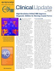
Dear Friends,<br />
I am pleased to share with you highlights of the 2011–2012 academic year. Our<br />
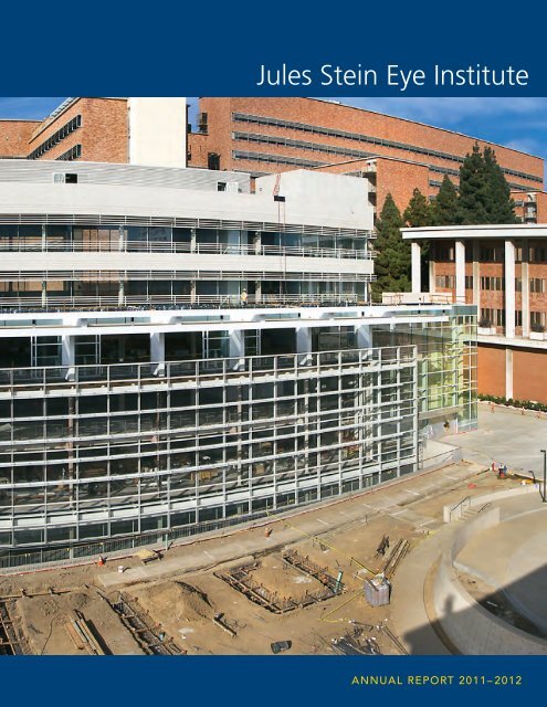
physical growth as an <strong>Institute</strong> is highlighted on the cover of this year’s <strong>Annual</strong> <strong>Report</strong>:<br />
the imposing outer edifice of the Edie and Lew Wasserman Building, awarded an<br />
architectural prize for design and significance, is close to completion.<br />
The <strong>Jules</strong> <strong>Stein</strong> <strong>Eye</strong> <strong>Institute</strong>’s vision-science campus has been built upon a foundation<br />
of excellent patient care and groundbreaking research. This year, Steven D. Schwartz,<br />
MD, chief of the Retina Division, began a safety study that involved injecting human<br />
embryonic stem cells for the first time into the eyes of legally blind patients.<br />
As in past years, <strong>Jules</strong> <strong>Stein</strong> <strong>Eye</strong> <strong>Institute</strong> faculty were recognized for their many achievements.<br />
Lynn K. Gordon, MD, PhD, was named the Vernon O. Underwood Family Chair<br />
in Ophthalmology, and Xian-Jie Yang, PhD, was named the Ernest G. Herman<br />
Endowed Chair in Ophthalmology. Prestigious honors were bestowed upon other<br />
faculty members by the American Academy of Ophthalmology, the International Society<br />
of Magnetic Resonance in Medicine, and additional professional groups. Vital research<br />
grants providing needed funding were awarded and renewed by the National <strong>Eye</strong><br />
<strong>Institute</strong>, the Foundation Fighting Blindness, and other key organizations.<br />
Among many exceptional philanthropic gifts to the <strong>Jules</strong> <strong>Stein</strong> <strong>Eye</strong> <strong>Institute</strong> was a<br />
$750,000 pledge from the Skirball Foundation to establish the Audrey and Jack Skirball<br />
Ocular Inflammatory Disease Fellowship. Contributions from David and Randi Fett and<br />
Theo and Wendy Kolokotrones will enable improvements in the <strong>Institute</strong>’s website.<br />
Significant bequests were also received, including from the estates of Ms. Helen V.<br />
Chaplin and Ms. Esther Shandler.<br />
As we anticipate the exciting changes ahead for the <strong>Jules</strong> <strong>Stein</strong> <strong>Eye</strong> <strong>Institute</strong>, we have<br />
not forgotten the exceptional donors and friends who have contributed to our achievements,<br />
including the late Dame Elizabeth Taylor who made a major gift through<br />
The Elizabeth Taylor AIDS Foundation to benefit the Herb Ritts, Jr. Memorial Vision Fund,<br />
providing necessary monies for AIDS-related vision care, research, and education<br />
at UCLA.<br />
It is our privilege to honor Miss Taylor’s legacy, and that of our countless donors, who<br />
have given so generously to ensure the <strong>Jules</strong> <strong>Stein</strong> <strong>Eye</strong> <strong>Institute</strong> maintains its standing<br />
as one of the world’s preeminent eye research centers leading the charge to preserve<br />
and restore vision.<br />
Sincerely,<br />
Bartly J. Mondino, MD<br />
Bradley R. Straatsma Professor of Ophthalmology<br />
Director, <strong>Jules</strong> <strong>Stein</strong> <strong>Eye</strong> <strong>Institute</strong><br />
Chairman, Department of Ophthalmology<br />
David Geffen School of Medicine at UCLA
Events<br />
The 2011–2012 academic year brought exciting<br />
advances to the <strong>Jules</strong> <strong>Stein</strong> <strong>Eye</strong> <strong>Institute</strong>. Of special<br />
significance, <strong>Institute</strong> researchers are conducting<br />
safety studies investigating the use of embryonic<br />
stem cells on patients with Stargardt macular<br />
dystrophy and dry age-related macular degeneration.<br />
Advancing research is just one of the many<br />
ways the <strong>Jules</strong> <strong>Stein</strong> <strong>Eye</strong> <strong>Institute</strong> strives to preserve<br />
and restore vision.<br />
Legally Blind Patients<br />
Receive Stem Cell Transplants<br />
On July 12, 2011, surgeons at the <strong>Jules</strong> <strong>Stein</strong> <strong>Eye</strong><br />
<strong>Institute</strong> began a safety study that involved injecting<br />
human embryonic stem cells for the first time into the<br />
eyes of legally blind patients.<br />
Both patients, one with Stargardt macular dystrophy<br />
and the other with dry age-related macular degeneration,<br />
underwent outpatient transplantation surgeries and<br />
recovered uneventfully, according to the lead surgeon,<br />
Steven D. Schwartz, MD, Ahmanson Professor of<br />
Ophthalmology and chief of the Retina Division.<br />
Dr. Schwartz is the principal investigator on two clinical<br />
trials, one for each eye disease. Each trial will include<br />
12 patients who are legally blind and will determine<br />
the safety of stem cell therapy. The patients’ ability to<br />
tolerate the surgical procedure itself went well.<br />
Human embryonic stem cells can differentiate into<br />
any cell type. The stem cell-derived retinal pigment<br />
epithelial (RPE) cells that were transplanted during<br />
surgery were differentiated in Advanced Cell Technology,<br />
Inc. (ACT) labs. ACT is a leader in the field of<br />
regenerative medicine and has been working for the<br />
last decade on developing a stem cell therapy to treat<br />
eye diseases. Each patient received a relatively low<br />
dose of the transplanted RPE cells (50,000) into the<br />
subretinal space of the treated eye.<br />
The dosing of the first patients in these trials, which<br />
are being closely watched by scientists and stem cell<br />
therapy advocates around the world, was hailed by ACT<br />
company officials as an important milestone in the<br />
therapeutic use of stem cells and may pave the way for<br />
a new therapeutic approach to treating eye diseases.<br />
Currently, both eye diseases are untreatable. The dry<br />
form of macular degeneration, the most common form<br />
of the disease, is the leading cause of blindness in<br />
the developed world, especially among people over the<br />
age of 55. As many as 30 million people in the United<br />
States and Europe currently suffer from this disease,<br />
and the number of people affected is expected to<br />
double over the next 20 years as the population ages.<br />
Stargardt disease causes progressive vision loss,<br />
usually starting when patients are between 10 to<br />
20 years of age.<br />
In both conditions, the layer of RPE cells located<br />
beneath the retina deteriorates and atrophies. These<br />
cells support, protect, and provide nutrition for lightsensitive<br />
photoreceptors in the eye. Over time, the<br />
death of the RPE cells and eventual loss of the photoreceptors<br />
can lead to blindness as central vision is<br />
gradually destroyed. Doctors are hoping the transplanted<br />
RPE cells will implant and begin functioning.<br />
Noted Robert Lanza, MD, chief scientific officer of ACT,<br />
“The great promise of human embryonic stem cells is<br />
finally being put to the test with the initiation of these<br />
two clinical trials. It’s time to start moving these exciting<br />
new stem cell therapies out of the laboratory and into<br />
the clinic.”<br />
Dr. Steven Schwartz peers into a microscope during surgery to<br />
transplant highly specialized cells derived from human embryonic<br />
stem cells into the eyes of the first patients enrolled in two clinical<br />
trials that are testing the promise of stem cell therapy.<br />
Highlights | Events 3
<strong>Institute</strong> Brings a<br />
Pediatric-Friendly<br />
Experience to<br />
the Surgical Area<br />
On May 16, 2012, <strong>Institute</strong><br />
Director Bartly J. Mondino, MD,<br />
introduced the <strong>Jules</strong> <strong>Stein</strong> Play<br />
Room, a dedicated children’s<br />
space located on the surgical floor<br />
of the <strong>Institute</strong>, and noted, “Today<br />
we celebrate the expansion of a<br />
pediatric-friendly experience from<br />
our clinics to our surgical suites.”<br />
Knowing a visit to the doctor can<br />
be an intimidating process for a<br />
child, the <strong>Institute</strong>, in cooperation<br />
with committed partners, has<br />
taken steps to ease the process<br />
for pediatric patients undergoing<br />
ophthalmic surgery by introducing<br />
a new children’s play area as<br />
well as trained specialists to<br />
guide families through the<br />
operative procedure.<br />
Bright and lively, the Play Room<br />
is filled with books, toys,<br />
puzzles, educational flash cards,<br />
and blocks to entertain youngsters<br />
during consultation and<br />
prior to surgery. Through the<br />
generosity of Wyndham Worldwide<br />
and the Starlight Children’s<br />
Foundation, the <strong>Jules</strong> <strong>Stein</strong> Play<br />
Room also includes a Fun Center<br />
mobile entertainment unit that<br />
offers a gaming system, DVD<br />
player, and television.<br />
A young visitor enjoys the new <strong>Jules</strong><br />
<strong>Stein</strong> Play Room located on the surgical<br />
floor of the <strong>Institute</strong>.<br />
4 Highlights | Events<br />
The <strong>Jules</strong> <strong>Stein</strong> <strong>Eye</strong> <strong>Institute</strong> is also<br />
incorporating the Chase Child Life<br />
Program for pediatric patients and<br />
their families, which offers Child<br />
Life Specialists, personnel who are<br />
trained personnel to help children<br />
and their parents better cope with<br />
the surgical experience.<br />
Child Life Specialists lead tours of<br />
the surgical area and accompany<br />
patients into surgery. They support<br />
the family by answering questions<br />
and providing educational information<br />
and resources so that parents<br />
and children alike can know what<br />
to expect. It’s especially reassuring<br />
for pediatric patients to have the<br />
familiar face of their Child Life<br />
Specialist with them as they go<br />
into surgery, and it reduces the<br />
parents’ anxiety to know their child<br />
is feeling more secure at that<br />
vulnerable time. The Chase Child<br />
Life Program, a no-cost offering,<br />
will be available once a week with<br />
the goal of expanding the service<br />
into a daily program.<br />
Through the generous support of caring partners, JSEI’s pediatric patients will enjoy a<br />
more comforting surgical experience. Left to right: Alison Sadock, corporate accounts<br />
manager, Starlight Children’s Foundation; Amy Bullock, director, Chase Child Life<br />
Program, Mattel Children’s Hospital, UCLA; Dr. Bartly Mondino, director, JSEI;<br />
Marti Winer, vice president of event services, Wyndham Worldwide; Mary Falvey,<br />
executive vice president, chief human resources officer, Wyndham Worldwide; Shannon<br />
O’Kelley, chief operating officer, UCLA Hospital System; Christine Archuleta, child life<br />
specialist, UCLA Operating Room Services; Paul Falcone, chief human resources officer,<br />
Starlight Children’s Foundation; Jacqueline Hart-Ibrahim, global chief executive officer,<br />
Starlight Children’s Foundation.
The Edie and Lew<br />
Wasserman Building<br />
Wins Architectural Award<br />
The Edie and Lew Wasserman<br />
Building, currently under construction<br />
in <strong>Stein</strong> Plaza, won a Community<br />
Impact Award at the 2011<br />
Los Angeles Business Council’s<br />
Los Angeles Architectural Awards<br />
ceremony. This prestigious award<br />
recognized the project’s breadth<br />
of investment, the anticipated<br />
achievements resulting from its<br />
completion, and its architectural<br />
excellence and significance.<br />
The Wasserman facility, which<br />
will house the Edie and Lew<br />
Wasserman <strong>Eye</strong> Research Center,<br />
is scheduled to be completed in<br />
March 2014. The facility was<br />
designed by Richard Meier &<br />
Partners Architects, the same<br />
architectural firm that created the<br />
Getty Center. Casey Wasserman,<br />
president and chief executive<br />
officer of the Wasserman Foundation,<br />
spearheaded the project,<br />
which honors his grandparents.<br />
Highlights | Events 5
Honors<br />
Each year, as part of their ongoing academic<br />
pursuits, faculty members achieve notable<br />
recognition for their accomplishments and<br />
contributions. They give prestigious lectures<br />
around the world, participate in influential<br />
professional and community organizations, and<br />
serve as writers and editors for a wide range of<br />
scientific journals. In some cases, special<br />
honors are bestowed.<br />
6 Highlights | Honors Section Name<br />
<strong>Jules</strong> <strong>Stein</strong> <strong>Eye</strong> <strong>Institute</strong> glaucoma<br />
specialists, Drs. Simon K. Law and<br />
Anne L. Coleman, were honored along<br />
with other <strong>Institute</strong> faculty members<br />
for their achievements in science.<br />
American Academy of<br />
Ophthalmology Awards<br />
UCLA ophthalmology faculty were honored by the<br />
American Academy of Ophthalmology for their outstanding<br />
contributions to the Academy, its scientific and<br />
educational programs, and to the field of ophthalmology.<br />
The awards were announced at the Academy’s annual<br />
meeting in Orlando, Florida in October 2011.<br />
Life Achievement Award:<br />
Joseph Caprioli, MD<br />
Senior Achievement Award:<br />
Bartly J. Mondino, MD<br />
Achievement Award:<br />
Simon K. Law, MD<br />
Secretariat Awards:<br />
Anne L. Coleman, MD, Fran and Ray Stark Professor of<br />
Ophthalmology, received a nomination by the secretary<br />
for Communications and an additional nomination by<br />
the secretary for Member Services.<br />
Bartly J. Mondino, MD, Bradley R. Straatsma Professor<br />
of Ophthalmology, received a nomination by the secretaries<br />
for State Affairs and Federal Affairs and the<br />
senior secretary for Advocacy.
Honors and Awards<br />
Anthony J. Aldave, MD, Associate<br />
Professor of Ophthalmology, was<br />
awarded the 2011 Gold Medal<br />
from the Indian Intraocular Implant<br />
and Refractive Society and the<br />
2012 W. Bruce Jackson Lectureship<br />
Award.<br />
Lynn K. Gordon, MD, PhD,<br />
Associate Professor of Ophthalmology,<br />
was named as the<br />
Vernon O. Underwood Family<br />
Chair in Ophthalmology, effective<br />
March 1, 2012.<br />
Wayne L. Hubbell, PhD,<br />
<strong>Jules</strong> <strong>Stein</strong> Professor of Ophthalmology,<br />
was elected as a Fellow<br />
of the International Society of<br />
Magnetic Resonance in Medicine.<br />
David Sarraf, MD, Associate<br />
Clinical Professor of Ophthalmology,<br />
was invited to become a<br />
member of the Gass Club at the<br />
2011 American Academy of<br />
Ophthalmology <strong>Annual</strong> Meeting.<br />
The invitation-only association is<br />
comprised of a small group (less<br />
than 30) of elite retinologists who<br />
meet annually to discuss interesting<br />
retinal cases.<br />
Steven D. Schwartz, MD,<br />
Ahmanson Professor of Ophthalmology,<br />
was a keynote lecturer at<br />
the UCLA IMED Seminar Series<br />
with Dr. Robert Lanza.<br />
Barry A. Weissman, OD, PhD,<br />
Professor of Ophthalmology,<br />
was the recipient of the 2012<br />
Legend Award, Cornea and<br />
Contact Lens Section, American<br />
Optometric Association.<br />
Xian-Jie Yang, PhD, Professor of<br />
Ophthalmology, was named as<br />
the Ernest G. Herman Endowed<br />
Chair in Ophthalmology, effective<br />
March 1, 2012.<br />
<strong>Jules</strong> <strong>Stein</strong> <strong>Eye</strong> <strong>Institute</strong> Rated Best in the West<br />
The <strong>Jules</strong> <strong>Stein</strong> <strong>Eye</strong> <strong>Institute</strong> continues in its position as one of<br />
the top five American eye care centers—and the best in the<br />
Western United States for the 22nd consecutive year—according<br />
to U.S. News & World <strong>Report</strong>’s 2011–2012 Best Hospitals<br />
rankings. Additionally, Ronald Reagan UCLA Medical Center is<br />
the only hospital in Los Angeles and the Southern California<br />
region that appears on the magazine’s “Honor Roll,” a place<br />
reserved for medical centers with high levels of expertise in<br />
multiple specialties. The Ronald Reagan UCLA Medical Center<br />
was ranked the number one hospital in California.<br />
Highlights | Honors 7
Research<br />
Research is a key component of the <strong>Institute</strong>’s<br />
mission and a high priority for faculty who often<br />
devote their life’s work to furthering our knowledge<br />
of specific vision processes and eye diseases.<br />
Major research grants are routinely awarded each<br />
year to support this effort. In 2011–2012, faculty<br />
members received important awards from both<br />
public and private organizations. New grants and<br />
grant renewals will enable faculty to further<br />
ongoing vision-science investigations that have<br />
shown promise and to undertake clinical trials that<br />
have direct application to some of the country’s<br />
most common ophthalmic problems.<br />
Drs. Kouros Nouri-Mahdavi and JoAnn Giaconi, along with other<br />
members of the Glaucoma Division, are leading the way in the study<br />
of sophisticated imaging tools that can be used to detect early signs<br />
of glaucoma-related damage.<br />
8 Highlights | Research<br />
Glaucoma Imaging Study<br />
Kouros Nouri-Mahdavi, MD, assistant professor of<br />
ophthalmology, is conducting research projects to<br />
investigate utility of different imaging techniques for<br />
improving detection of glaucoma or its progression.<br />
These studies aim to determine the performance of<br />
various testing modalities available on the newer<br />
spectral-domain optical coherence tomography<br />
(SD-OCT) for discrimination of glaucomatous eyes<br />
from normal eyes.<br />
Because of the higher resolution and the larger<br />
amount of data obtained by SD-OCTs, their use is<br />
expected to lead to better clinical performance and a<br />
higher rate of detection of early glaucoma or its<br />
progression. If the newer imaging devices are proven<br />
to have a better performance, they can potentially be<br />
useful for screening purposes and be especially<br />
valuable for evaluating cases that are suspected to<br />
show early glaucomatous damage where the visual<br />
field is frequently noncon-tributory. On the other end of<br />
the glaucoma spectrum, macular imaging is emerging<br />
as a promising modality for detection of progression in<br />
advanced glaucoma, since the retinal nerve fiber layer<br />
and optic nerve head in patients with advanced<br />
glaucoma are already too damaged to demonstrate any<br />
evidence of progression.
Effect of Corneal<br />
Preservation Time on<br />
Long-Term Graft Success<br />
Anthony J. Aldave, MD, associate<br />
professor of ophthalmology, and<br />
Sophie X. Deng, MD, PhD,<br />
assistant professor of ophthalmology,<br />
are conducting the Cornea<br />
Preservation Time Study (CPTS).<br />
This multicenter, nationwide study<br />
is evaluating whether the donor<br />
cornea preservation time affects<br />
the success of a corneal transplant<br />
surgery. Donor corneas are placed<br />
into a preservation solution and<br />
stored until surgery. Currently, the<br />
Food and Drug Administration<br />
regulations allow corneas to be<br />
stored up to 14 days before<br />
surgery. Although it is common<br />
practice to use corneas stored<br />
more than 7 days in other countries,<br />
it is not routinely done in the<br />
United States.<br />
This study aims to demonstrate<br />
that transplant surgery with<br />
corneas preserved from 8–17 days<br />
is not inferior to corneas preserved<br />
from 1–7 days. To accomplish this,<br />
participants will be randomized to<br />
one of two groups. The surgery will<br />
be identical to surgery performed<br />
on patients not included in the<br />
study. The study will examine the<br />
number of complications and graft<br />
failures for both groups. There is<br />
no research to suggest there is<br />
any greater risk for transplant<br />
failure. As the United States<br />
population lives longer and there<br />
is an increase in persons needing<br />
transplants, the cornea donor<br />
supply is uncertain. This study<br />
hopes to provide scientific evidence<br />
that may help increase the cornea<br />
supply available for transplants<br />
in the United States. This study<br />
is funded by the National <strong>Eye</strong><br />
<strong>Institute</strong> and coordinated through<br />
Case Western Reserve University.<br />
Longitudinal Studies of<br />
the Ocular Complications<br />
of AIDS<br />
Gary N. Holland, MD, Jack H.<br />
Skirball Professor of Ocular<br />
Inflammatory Diseases, has been<br />
an investigator on the Longitudinal<br />
Studies of Ocular Complications<br />
of AIDS (LSOCA) since its commencement<br />
in September 1998.<br />
This multicenter, prospective,<br />
observational cohort study is<br />
designed to collect data on<br />
ophthalmic conditions associated<br />
with AIDS that have been seen<br />
since the introduction of highly<br />
active antiretroviral therapies.<br />
Historically, the focus of LSOCA<br />
was to evaluate cytomegalovirus<br />
(CMV) retinitis in the population,<br />
including risk factors and outcomes<br />
for patients. Left untreated,<br />
CMV retinitis is a disorder that<br />
can lead to retinal destruction<br />
and blindness.<br />
In recent years, Dr. Holland has<br />
initiated research evaluating<br />
neuroretinal disorders within the<br />
cohort. He has investigated the<br />
relationship of HIV-related visual<br />
abnormalities, such as decreased<br />
contrast sensitivity and diminished<br />
color vision in patients without<br />
opportunist infections, like CMV.<br />
He demonstrated that there is an<br />
association between retinal nerve<br />
fiber layer thickness and abnormalities<br />
of vision in people infected<br />
with HIV. The findings suggest<br />
abnormalities of contrast sensitivity<br />
and of color vision appear to be<br />
more common among HIV-infected<br />
individuals than in the general<br />
population and are not correlated<br />
with immune function. It has been<br />
hypothesized that these abnormalities<br />
are related to the retinal<br />
microvasculopathy of HIV disease.<br />
Continued research on this subject<br />
may lead to better methods for<br />
monitoring the health of HIVinfected<br />
individuals.<br />
Safety and Effectiveness<br />
of the Calhoun Vision<br />
Light Adjustable Lens for<br />
Treating Postoperative<br />
Sphere and Cylinder<br />
Kevin M. Miller, MD, Kolokotrones<br />
Professor of Clinical Ophthalmology,<br />
has recently started a study to<br />
evaluate the use of a light adjustable<br />
intraocular lens (IOL) for<br />
reducing astigmatism and improving<br />
uncorrected visual acuity after<br />
IOL implantation. This randomized,<br />
prospective multicenter clinical<br />
trial is designed to evaluate the<br />
safety and effectiveness of a light<br />
adjustable lens (LAL).<br />
Currently, accurate calculation of<br />
IOL power is difficult and the<br />
position of the IOL in the eye is<br />
inherently inaccurate. Despite<br />
measurements of the eye prior to<br />
cataract surgery, several factors<br />
including preexisting astigmatism<br />
and unpredictable healing can<br />
result in suboptimal patient vision<br />
after the lens implantation. Standard<br />
monofocal IOLs allow some<br />
patients to have good distance<br />
vision, but the majority of patients<br />
are left dependent on glasses. The<br />
LAL may eliminate the patient’s<br />
dependence on glasses because<br />
this IOL allows the doctor to adjust<br />
the power after it is implanted in<br />
the eye.<br />
The LAL contains photosensitive<br />
material called macromers. These<br />
materials are sensitive to ultraviolet<br />
light; resulting in a change to the<br />
shape of the LAL and, in turn, the<br />
power of the lens. The LAL can be<br />
implanted in the eye using standard<br />
surgical procedures. After the<br />
eye has healed following surgery,<br />
the residual refractive error can be<br />
measured and the lens shape can<br />
be corrected using ultraviolet light.<br />
The study will consist of up to 600<br />
eyes across the country and is<br />
being funded by Calhoun Vision.<br />
Highlights | Research 9
Adaptive Robotics for Ocular Surgery<br />
Jean-Pierre Hubschman, MD, assistant professor of ophthalmology, is<br />
collaborating with UCLA engineers to design the platform for an intraocular<br />
robotic surgical device. The long-term goal is to enable automation for<br />
ocular surgery. Current surgical procedures have significant potential for<br />
inadvertent tissue manipulation possibly resulting in intraocular trauma.<br />
They have developed a dual arm, intraocular robotic surgical system<br />
designed for performing intraocular surgeries with a primary focus on<br />
cataract surgery. Preliminary results have shown an optimization of<br />
vibration reduction, a controlled remote center of motion for both robotic<br />
arms, the ability to mount multiple surgical instruments to either arm with<br />
automated surgical instrument replacement, and a vision-based object<br />
tracking system to monitor anatomical positions and instrument positions<br />
during surgery.<br />
It is believed that this more refined tracking system will decrease the<br />
incidence of trauma to ocular tissues by increasing the surgeon’s ability to<br />
visualize the environment and by creating restrictions on the proximity of<br />
surgical tools to vulnerable ocular tissues. After development, a comparison<br />
between manual cataract extraction and robotic cataract extraction will<br />
be conducted. The research aims to decrease negative outcomes in the<br />
most frequently performed surgical procedure, totaling more than 3 million<br />
operations every year in the United States.<br />
10 Highlights | Research<br />
Dr. Jean-Pierre Hubschman with a<br />
prototype of the intraocular robotic<br />
surgical platform.
Education<br />
Education at the <strong>Jules</strong> <strong>Stein</strong> <strong>Eye</strong> <strong>Institute</strong> is multifaceted,<br />
ranging from teaching medical students,<br />
residents, and fellows to leading national conferences.<br />
In the course of their educational duties, faculty<br />
members mentor, counsel, lecture, and demonstrate.<br />
They are responsible for hundreds of clinical and<br />
scientific publications each year and entrusted with<br />
developing and sharing new approaches to science<br />
and medicine that will ultimately result in improved<br />
patient care. This year we honored faculty at the<br />
<strong>Annual</strong> Clinical and Research Seminar and hosted a<br />
course to educate both new and seasoned physicians<br />
on clinical research methods.<br />
<strong>Annual</strong> Clinical and<br />
Research Seminar<br />
The <strong>Institute</strong>’s most prestigious<br />
academic event of each year, the<br />
Clinical and Research Seminar,<br />
was held on June 8, 2012.<br />
Sponsored by the Department of<br />
Ophthalmology Association, it<br />
provided an opportunity for<br />
discussion of emerging vision<br />
research and a celebration of<br />
teaching and faculty volunteerism.<br />
This year’s seminar featured the<br />
43rd <strong>Jules</strong> <strong>Stein</strong> Lecture, the 10th<br />
Bradley R. Straatsma Lecture, and<br />
the 10th Thomas H. Pettit Lecture.<br />
A number of volunteer and clinical<br />
faculty received awards of distinction.<br />
The S. Rodman Irvine Prize,<br />
which recognizes excellence<br />
among Department of Ophthalmology<br />
faculty, was awarded to<br />
Norman Shorr, MD. The Faculty<br />
Teaching Award to honor contributions<br />
to residency education was<br />
presented to Uday Devgan, MD,<br />
chief of ophthalmology at Olive<br />
<strong>View</strong>–UCLA Medical Center.<br />
43RD JULES STEIN LECTURER<br />
Mark S. Blumenkranz, MD<br />
Chairman, Department<br />
of Ophthalmology<br />
Inaugural Director,<br />
Byers <strong>Eye</strong> <strong>Institute</strong><br />
Stanford University<br />
10TH BRADLEY R. STRAATSMA<br />
LECTURER<br />
Cesar T. Chavez, MD<br />
Private Practice<br />
North San Diego County/<br />
Imperial Valley<br />
10TH THOMAS H. PETTIT<br />
LECTURE<br />
George M. Rajacich, MD<br />
Medical Director and<br />
Chief Executive Officer<br />
Valley <strong>Eye</strong> Center,<br />
Darin <strong>Eye</strong> Center,<br />
Mazzocco Ambulatory<br />
Surgery Center<br />
Highlights | Education 11
17th <strong>Annual</strong> Vision Science<br />
Conference<br />
The 17th annual Vision Science<br />
Conference, co- sponsored by the<br />
National <strong>Institute</strong>s of Health/<br />
National <strong>Eye</strong> <strong>Institute</strong> Vision<br />
Science Training Grant and the<br />
<strong>Jules</strong> <strong>Stein</strong> <strong>Eye</strong> <strong>Institute</strong>, was held<br />
October 28–30, 2011. More than<br />
80 basic scientists and clinical<br />
researchers gathered at UCLA’s<br />
Lake Arrowhead Conference<br />
Center to participate in scientific<br />
discussions and memorable<br />
networking events. Guest speakers<br />
presented a variety of fascinating<br />
topics. Scientific Keynote Speaker<br />
Andrew Huberman, PhD, from the<br />
Department of Neurosciences and<br />
Neurobiology at the University of<br />
California at San Diego, gave an<br />
informative lecture entitled,<br />
“Genetic Approaches: Understanding<br />
How the Visual System Wires<br />
up, Works, and Can be Repaired”;<br />
Valentyna Abramenko, PhD, from<br />
the Big Bear Solar Observatory,<br />
presented a fascinating talk about<br />
what we know about our closest<br />
star; and Laurie Shaker-Irwin,<br />
PhD, MS, stimulated discussion<br />
with her topic, “Responsible<br />
Conduct of Research—Ethics<br />
and Oversight.”<br />
12 Highlights | Education<br />
Aesthetic <strong>Eye</strong>lid and<br />
Facial Rejuvenation Course<br />
The Orbital and Oculoplastic<br />
Surgery Division held its “Aesthetic<br />
<strong>Eye</strong>lid and Facial Rejuvenation”<br />
course June 15–16, 2012, at<br />
the <strong>Jules</strong> <strong>Stein</strong> <strong>Eye</strong> <strong>Institute</strong>. The<br />
event attracted ophthalmologists,<br />
dermatologists, and cosmetic<br />
surgeons from around the world.<br />
The two-day event combined<br />
surgical demonstrations, a cadaver<br />
dissection, and didactic lectures<br />
that informed participants of the<br />
latest advances in the field of<br />
aesthetic and reconstructive<br />
surgery for the eyelids and face.<br />
Don Kikkawa, MD, Shiley <strong>Eye</strong><br />
Center, professor of Clinical<br />
Ophthalmology, division chief of<br />
Ophthalmic Plastic and Reconstructive<br />
Surgery, gave this year’s<br />
Robert Axelrod, MD, Memorial<br />
Lecture, “Perfecting Your Skills<br />
with the Aesthetic Patient.”<br />
Dr. Kikkawa conducted his<br />
residency at the <strong>Jules</strong> <strong>Stein</strong><br />
<strong>Eye</strong> <strong>Institute</strong>.<br />
Fall Medical Forum<br />
Discusses Pediatric<br />
Blindness<br />
On September 20, 2011, Sherwin<br />
J. Isenberg, MD, Laraine and David<br />
Gerber Professor of Ophthalmology,<br />
discussed issues affecting<br />
childhood eye health in a medical<br />
forum held at the <strong>Jules</strong> <strong>Stein</strong> <strong>Eye</strong><br />
<strong>Institute</strong>. In his lecture, “Fighting<br />
Blindness in Children.” Dr. Isenberg<br />
explained that for the 1.4 million<br />
children who are blind throughout<br />
the world, at least 40% of the<br />
cases are preventable. Two of the<br />
main causes of pediatric vision<br />
loss—corneal scarring and retinopathy<br />
of prematurity—are avoidable<br />
with proper treatment. Dr. Isenberg<br />
spoke about the development of<br />
an effective, inexpensive, and<br />
available eyedrop that can prevent<br />
and treat many of the infections<br />
that cause corneal scarring and<br />
how he and his team are establishing<br />
protocols in Ethiopia to<br />
deliver this medicine to children<br />
with trachoma, the number-one<br />
cause of infectious blindness.<br />
Faculty, fellows, and graduate students at the 17th <strong>Annual</strong> Vision Science Conference at UCLA’s Lake Arrowhead Conference Center.
Philanthropy<br />
“Do everything as in the eye of another.”<br />
—Lucius Annaeus Seneca<br />
Established in 1966 through the remarkable<br />
insight and generous philanthropy of Dr. and Mrs.<br />
<strong>Jules</strong> <strong>Stein</strong>, the <strong>Jules</strong> <strong>Stein</strong> <strong>Eye</strong> <strong>Institute</strong> at UCLA<br />
continues to advance and expand its programs<br />
and facilities. Private support is critical for scientific<br />
innovations, exceptional education and<br />
training, and the finest, most compassionate<br />
therapeutic approaches.<br />
This fiscal year, hundreds of donors supported<br />
sight-saving endeavors at the <strong>Institute</strong>. Additionally<br />
the <strong>Institute</strong> received several bequests including<br />
gifts from the estates of Ms. Helen V. Chaplin and<br />
Ms. Esther Shandler.<br />
Audrey and Jack Skirball Ocular<br />
Inflammatory Disease Fellowship<br />
The Skirball Foundation made a $750,000 pledge<br />
to establish the Audrey and Jack Skirball Ocular<br />
Inflammatory Disease Fellowship at UCLA’s <strong>Jules</strong><br />
<strong>Stein</strong> <strong>Eye</strong> <strong>Institute</strong>.<br />
Bartly J. Mondino, MD, <strong>Institute</strong> director remarked,<br />
“The Skirball Foundation’s gift will have a significant<br />
impact on fellows specializing in ocular inflammatory<br />
disease and greatly benefit their careers in ophthalmology.<br />
Such generosity will ultimately help patients<br />
suffering from debilitating eye conditions. This is<br />
truly a wonderful way to honor Audrey and Jack<br />
Skirball’s memory.”<br />
The creation of the Skirball Fellowship will underwrite<br />
the training of fellows specializing in ocular inflammatory<br />
disease, and the <strong>Jules</strong> <strong>Stein</strong> <strong>Eye</strong> <strong>Institute</strong> is poised<br />
to be a leading training center in this arena. More<br />
specialists are urgently needed to address increasingly<br />
complex diagnostic techniques and therapies. Since<br />
clinical fellows are required to participate in research<br />
projects, the fund also will spur new and promising<br />
areas of investigation. This fellowship is the first in<br />
ocular inflammatory disease at UCLA.<br />
The Skirball Foundation has been supporting the<br />
<strong>Institute</strong> for more than 40 years, due in part to<br />
Mr. Skirball’s enduring friendship with <strong>Jules</strong> <strong>Stein</strong> and<br />
From left to right, Mr. Jack Skirball, Mrs. Audrey Skirball,<br />
Rabbi Uri D. Herscher (1983).<br />
Lew Wasserman. Since 1969, the Skirball Foundation<br />
has continued to underwrite vision-science programs,<br />
particularly those in the UCLA Ocular Inflammatory<br />
Disease Center (OIDC). The Skirball Foundation Fund<br />
was established in 1990 to support research, education,<br />
and patient care programs within the OIDC and, as<br />
an endowment, serves as a significant ongoing<br />
resource for them. In 2001, the Skirball Foundation<br />
created a current-expenditure fund to further expand<br />
studies within the OIDC, and in 2007, it established the<br />
Jack H. Skirball Endowed Chair in Ocular Inflammatory<br />
Diseases, a distinguished position to which Gary N.<br />
Holland, MD, chief of the Cornea and Uveitis Division,<br />
was appointed in 2009.<br />
Born in 1896 in Homestead, Pennsylvania, Mr. Skirball<br />
was ordained a rabbi in 1921. After serving congregations<br />
in the Midwest, he took leave of the rabbinate<br />
in 1933 to manage Educational Films Corporation, a<br />
pioneer in audiovisual education. Mr. Skirball then went<br />
on to feature-film production as president of Skirball<br />
Productions, which was responsible for movies such as<br />
Alfred Hitchcock’s Saboteur (1942) and Shadow of a<br />
Doubt (1943). In the 1950s, he began a third successful<br />
career, this time as a real estate developer. Through<br />
Mr. Skirball’s film career and relationship with the<br />
Music Corporation of America (MCA, Inc.), he met<br />
and became lifelong friends with Dr. <strong>Stein</strong> and<br />
Mr. Lew Wasserman.<br />
(continued on next page)<br />
Highlights | Philanthropy 13
Audrey and Jack Skirball continued<br />
Mrs. Skirball was born in Alabama<br />
in 1915 and during World War II<br />
moved to California to join the<br />
Signal Corps, the branch of the<br />
United States Military responsible<br />
for communications. She married<br />
Mr. Skirball in 1952, and during<br />
their 33-year marriage they were<br />
actively involved in charitable<br />
endeavors, most notably building<br />
the Skirball Cultural Center, the<br />
largest Jewish cultural institution in<br />
the world today. Rabbi Uri D.<br />
Herscher, founding president and<br />
chief executive officer of the<br />
Skirball Cultural Center and a<br />
trustee of the Foundation, noted, “I<br />
first met Jack and Audrey Skirball<br />
in 1964. Throughout the years,<br />
they made me aware of their<br />
devotion to help cure eyesightrelated<br />
diseases, and they had the<br />
confidence that the <strong>Jules</strong> <strong>Stein</strong><br />
<strong>Eye</strong> <strong>Institute</strong> would develop the<br />
expertise to do so.” The creation of<br />
the Audrey and Jack Skirball<br />
Ocular Inflammatory Disease<br />
Fellowship will serve as a testament<br />
to Mr. and Mrs. Skirball’s<br />
lifetime of philanthropy and<br />
dedication to fighting blindness.<br />
14 Highlights | Philanthropy<br />
Donors Support<br />
Modernization of<br />
<strong>Institute</strong> Website<br />
Wendy and Theo Kolokotrones<br />
and Randi and David Fett have<br />
pledged generous gifts to support<br />
an update of the <strong>Jules</strong> <strong>Stein</strong><br />
<strong>Eye</strong> <strong>Institute</strong>’s website. <strong>Institute</strong><br />
Director Bartly J. Mondino, MD,<br />
noted that their contributions<br />
would strengthen the goals to<br />
modernize the site and expand<br />
user functionalities for both online<br />
visitors and <strong>Institute</strong> staff and<br />
faculty. “I am grateful for the<br />
dedication of Wendy and Theo<br />
Koloktrones and Randi and David<br />
Fett to advance our online capabilities.<br />
A state-of-the-art website will<br />
reflect the <strong>Institute</strong>’s position as<br />
one of the world’s preeminent eye<br />
research centers.”<br />
Proposed changes include an<br />
overhaul of multiple databases,<br />
improved site navigation, decentralized<br />
page editing, enhanced video<br />
streaming, integration of social<br />
media, text enlargement for the<br />
visually impaired, non-English ver-<br />
sions of the website, an <strong>Institute</strong>-<br />
wide calendar, an interactive portal<br />
for <strong>Institute</strong> alumni, and a robust<br />
Intranet for staff and faculty use.<br />
Also under consideration is the<br />
development of smart-phone<br />
applications that communicate with<br />
the website.<br />
The Kolokotrones family has been a<br />
loyal supporter of the <strong>Institute</strong> since<br />
1994. In 2004, they established a<br />
chair to benefit a cataract surgeon<br />
and scientist. Chair holder Kevin M.<br />
Miller, MD, Kolokotrones Professor<br />
of Clinical Ophthalmology and chief<br />
of the Comprehensive Ophthalmology<br />
Division, oversees the website<br />
development team. “Wendy and<br />
Theo Kolokotrones have supported<br />
my work for years and have been<br />
generous to the <strong>Institute</strong> in many<br />
ways,” said Dr. Miller, who has been<br />
the faculty director of the website<br />
since its first design more than<br />
15 years ago.<br />
Dr. David Fett is an <strong>Institute</strong><br />
alumnus and clinical faculty mem-<br />
ber. His wife Randi graduated from<br />
UCLA in 1984 with a degree in<br />
chemistry, and Dr. Fett has a long<br />
history with UCLA, since his grand-<br />
father graduated from the University<br />
in 1932. Most recently, the<br />
Fetts established a fellowship in<br />
the Orbital and Ophthalmic Plastic<br />
Surgery Division under the auspices<br />
of Robert Alan Goldberg, MD.<br />
According to Dr. Miller, the groundwork<br />
for the website is currently<br />
being laid, and the first order of<br />
business will be to rebuild the<br />
database—a fundamental element<br />
for all aspects of the project.<br />
Dr. Kevin M. Miller, Kolokotrones<br />
Professor of Clinical Ophthalmology, has<br />
been the faculty director of the <strong>Institute</strong>’s<br />
website since its first design more than<br />
15 years ago.
Dame Elizabeth Taylor<br />
Pioneering Voice for<br />
HIV/AIDS Awareness<br />
The UCLA community mourned<br />
the loss of legendary film star<br />
Dame Elizabeth Taylor who<br />
passed away in March 2011.<br />
Miss Taylor was a pioneering voice<br />
for HIV/AIDS awareness. In 1994,<br />
she began supporting the HIVrelated<br />
eye investigations of<br />
Gary N. Holland, MD, Jack H.<br />
Skirball Professor of Ocular<br />
Inflammatory Diseases and Chief<br />
of the Cornea and Uveitis Division<br />
at UCLA’s <strong>Jules</strong> <strong>Stein</strong> <strong>Eye</strong> <strong>Institute</strong>.<br />
Dr. Holland and his colleagues<br />
were the first to describe the<br />
ocular manifestations of AIDS, and<br />
he has continued to be involved<br />
in related research. Miss Taylor<br />
recognized that many people with<br />
AIDS suffer from eye complications<br />
during their illness. Diseases<br />
such as cytomegalovirus (CMV)<br />
retinitis, an infection of the retina,<br />
can result in permanent vision loss<br />
in patients whose immune systems<br />
are compromised.<br />
In 2006, Miss Taylor made a major<br />
gift through The Elizabeth Taylor<br />
AIDS Foundation to benefit the<br />
Herb Ritts, Jr. Memorial Vision<br />
Fund, providing necessary monies<br />
for AIDS-related vision care,<br />
research, and education at UCLA.<br />
Miss Taylor and Mr. Ritts had been<br />
close friends.<br />
The Herb Ritts, Jr. Memorial Vision<br />
Fund was established by family<br />
and friends of the beloved and<br />
celebrated American photographer.<br />
In the late 1990s, Mr. Ritts collaborated<br />
with Dr. Holland to raise<br />
awareness of AIDS-related eye<br />
disease. At the time of the gift,<br />
Miss Taylor said, “It is my hope<br />
that my donation will inspire others<br />
to give to this important cause,<br />
which honors our dear friend<br />
Herb Ritts and continues Herb’s<br />
long- standing support of HIV/<br />
AIDS research.”<br />
More recently, Miss Taylor helped<br />
to support a conference at UCLA.<br />
It was attended by HIV specialists<br />
and ophthalmologists from around<br />
the world who met to formulate<br />
strategies for treating the growing<br />
problem of AIDS-related CMV<br />
retinitis in resource-poor areas,<br />
such as Southeast Asia and Africa.<br />
Dr. Holland remembers Miss Taylor<br />
as a passionate advocate of these<br />
endeavors. “It was a privilege to<br />
know and collaborate with Dame<br />
Elizabeth for many years. I appreciated<br />
her interest in our work and<br />
the confidence she had in our<br />
research and clinical activities.<br />
Elizabeth’s support has truly helped<br />
to advance the understanding of<br />
AIDS-related blindness—both in<br />
the United States and in the<br />
developing world. Her enthusiasm<br />
was inspiring, and she will be<br />
missed greatly.”<br />
To make a donation to the<br />
Herb Ritts, Jr. Memorial Vision<br />
Fund, please contact the<br />
<strong>Institute</strong>’s Development Office at<br />
(310) 206-6035.<br />
Dame Elizabeth Taylor<br />
Dr. Gary Holland, Jack H. Skirball<br />
Professor of Ocular Inflammatory<br />
Diseases<br />
Highlights | Philanthropy 15
The Ahmanson Foundation<br />
Awards Grant to<br />
UCLA Mobile <strong>Eye</strong> Clinic<br />
The Ahmanson Foundation<br />
awarded the UCLA Mobile <strong>Eye</strong><br />
Clinic (MEC) at UCLA’s <strong>Jules</strong> <strong>Stein</strong><br />
<strong>Eye</strong> <strong>Institute</strong> a generous grant for<br />
advanced diagnostic equipment to<br />
expand services.<br />
The MEC is a 40-foot-long coach<br />
staffed by ophthalmologists,<br />
technicians, and volunteers that<br />
travels four days a week to<br />
underserved areas in Los Angeles—<br />
schools, health clinics, community<br />
centers, homeless shelters—<br />
providing high-quality eye care at<br />
no cost. The new diagnostic<br />
equipment provides significant<br />
enhancements to the MEC, specifically<br />
allowing ophthalmologists to<br />
more effectively screen for diabetic<br />
retinopathy and glaucoma. In<br />
addition, portable instruments that<br />
can be used outside the vehicle<br />
were purchased in order to screen<br />
patients with disabilities and<br />
provide additional support at larger<br />
events such as health fairs. Anne L.<br />
Coleman, MD, PhD, director of the<br />
MEC, noted, “The equipment the<br />
grant enabled us to obtain is<br />
essential for the advancement of<br />
clinical care and, ultimately, the<br />
benefit of patients. We are so<br />
grateful for The Ahmanson’s<br />
Foundation’s wonderful support of<br />
the MEC.”<br />
16 Highlights | Philanthropy<br />
The MEC was established in 1975<br />
by an anonymous donor who was a<br />
friend and associate of Dr. <strong>Jules</strong><br />
<strong>Stein</strong>. The individual had an urgent<br />
eye problem that brought him to<br />
the <strong>Jules</strong> <strong>Stein</strong> <strong>Eye</strong> <strong>Institute</strong>. He<br />
was so impressed with his care<br />
that he created the MEC with the<br />
advice of his ophthalmologist,<br />
Robert Christensen, MD, to meet<br />
the tremendous unmet need for<br />
eye care in the community.<br />
The MEC’s mission has been<br />
consistent from its inception, and<br />
this high quality, reliable resource<br />
has grown over the years, becoming<br />
well known throughout the<br />
region. This year alone, the MEC<br />
staff and ophthalmic personnel<br />
saw approximately 5,000 patients.<br />
Private philanthropy is critical to<br />
sustaining the MEC and allowing<br />
those in need to benefit from its<br />
important services. Bartly J.<br />
Mondino, MD, director of the<br />
<strong>Institute</strong>, commented, “The<br />
Ahmanson Foundation has been a<br />
loyal friend for many years, and its<br />
recent grant demonstrates a<br />
steadfast commitment to helping<br />
those in need. We are lucky to<br />
count the Foundation as a partner<br />
in our goal of ensuring a lifetime of<br />
good vision for all.”<br />
The Ahmanson Foundation, incorporated<br />
as a private foundation in<br />
1952, was established by financier<br />
Howard F. Ahmanson and his wife<br />
Dorothy Ahmanson. Its corpus<br />
was augmented in later years by<br />
his two nephews Robert H.<br />
Ahmanson and William H.<br />
Ahmanson. The Foundation serves<br />
Los Angeles County by funding<br />
cultural projects in the arts and<br />
humanities, education at all levels,<br />
health care, programs related to<br />
homelessness and underserved<br />
populations, as well as a wide<br />
range of human services. In 1997,<br />
the Foundation established the<br />
UCLA Center for <strong>Eye</strong> Epidemiology<br />
to support research and clinical<br />
studies to further knowledge of the<br />
development, treatment, and<br />
prevention of eye disease. In 2005,<br />
it established the Ahmanson Chair<br />
in Ophthalmology, a distinguished<br />
position to which Steven D.<br />
Schwartz, MD, chief of the Retina<br />
Division, was appointed in 2007.<br />
The UCLA Mobile <strong>Eye</strong> Clinic<br />
travels four days a week to<br />
underserved areas in Los<br />
Angeles—schools, health clinics,<br />
community centers, homeless<br />
shelters—providing high-quality<br />
eye care at no cost.
17 Highlights | Philanthropy<br />
Thank You<br />
The <strong>Jules</strong> <strong>Stein</strong> <strong>Eye</strong> <strong>Institute</strong> is grateful for<br />
the generous and steadfast support of its<br />
research, education, patient care, and<br />
outreach activities. This investment will<br />
influence ophthalmology and related<br />
disciplines at UCLA and throughout the<br />
broader vision community. Thank you for<br />
your commitment to these important<br />
endeavors.<br />
Highlights | Section Name 17
Major Gifts over $25,000<br />
American Geriatrics Society, Inc.<br />
Arthritis Foundation, Pacific Region<br />
Bruce Ford and Anne Smith Bundy<br />
Foundation<br />
Helen V. Chaplin Family Trust<br />
The Carl & Roberta Deutsch<br />
Foundation<br />
Andrea and Terry Donahue<br />
Dr. and Mrs. David Fett<br />
The Foundation Fighting Blindness<br />
Friends of the Congressional<br />
Glaucoma Caucus Foundation,<br />
Inc.<br />
Laraine Gerber<br />
Carol and Timothy W. Hannemann<br />
Mary Ann and Thomas C. Hays<br />
William & Margaret Fern Holmes<br />
Family Foundation<br />
<strong>Jules</strong> and Doris <strong>Stein</strong> UCLA<br />
Support Group<br />
The Karl Kirchgessner Foundation<br />
Wendy and Theo Kolokotrones<br />
Knights Templar <strong>Eye</strong> Foundation,<br />
Inc.<br />
Walter Lantz Foundation<br />
California Community Foundation<br />
Cynthia and Edward Lasker<br />
Fund<br />
Bert Levy<br />
MacDonald Family Foundation<br />
Macula Vision Research<br />
Foundation<br />
Richard Malm<br />
The Mann Center for Education<br />
and Family Development<br />
Susan and Morris Mark<br />
Merz Pharmaceuticals, LLC<br />
Ruth and George E. Moss<br />
Gerald Oppenheimer Family<br />
Foundation<br />
William R. Payden<br />
Research to Prevent Blindness, Inc.<br />
Mark Schulman & Esther Schulman<br />
Foundation, a Support<br />
Foundation of the Jewish<br />
Community Foundation of<br />
Greater Los Angeles<br />
Esther Shandler Trust<br />
18 Highlights | Philanthropy<br />
The Skirball Foundation<br />
Jerome and Joan Snyder<br />
The Fran and Ray Stark Foundation<br />
Vision of Children, Sam and Vivian<br />
Hardage, Co-Founders<br />
Plus numerous anonymous<br />
contributors<br />
The following individuals were<br />
honored with a tribute gift this<br />
past year:<br />
In Honor of…<br />
Barry Roy Binder, Esq.<br />
Jeffrey L. Eginton<br />
Devin Freeman<br />
Robert A. Goldberg, MD<br />
Michael B. Gorin, MD, PhD<br />
Kevin M. Miller, MD<br />
Ernest L. Neu<br />
Beverly Scarano<br />
Barry A. Weissman, OD, PhD<br />
In Memory of…<br />
Simon Abdallah<br />
Marvin Altshuler<br />
Lenore Fenster<br />
Marvin Fischer<br />
Peggy L. Giambrocco<br />
Ione J. Kanne<br />
Michael William Lenvin<br />
Betty L. Lewis<br />
Kenneth Jay Marcus, Esq.<br />
Ernest L. Neu<br />
Joan Penders<br />
Anne Rae Rosenberg<br />
Richard W. Sallop<br />
Esther Shandler<br />
Oscar Z. Simmons<br />
Nancy S. Wang, MD<br />
Endowed Professorships<br />
and Fellowships<br />
Endowed Professorships<br />
Ahmanson Chair in<br />
Ophthalmology<br />
Established in 2005 by The<br />
Ahmanson Foundation as an<br />
administrative chair for the<br />
Retina Division Chief to further<br />
research, education, and clinical<br />
care programs.<br />
Steven D. Schwartz, MD<br />
2007–Present<br />
Leonard Apt Endowed Chair in<br />
Pediatric Ophthalmology<br />
Established in 2003 by Dr. Leonard<br />
Apt, Professor Emeritus of Ophthalmology<br />
and Founding Chief of<br />
the Division of Pediatric Ophthalmology<br />
and Strabismus, with a<br />
gift from the trust of Frederic G.<br />
Rappaport, Dr. Apt’s nephew.<br />
Joseph L. Demer, MD, PhD<br />
2005–Present<br />
Karen and Frank Dabby<br />
Endowed Chair<br />
in Ophthalmology<br />
Established in 2007 by Dr. and<br />
Mrs. Dabby as a term chair to<br />
support the activities of a distinguished<br />
faculty member in the area<br />
of orbital disease.<br />
Robert A. Goldberg, MD<br />
2008–Present<br />
Charles Kenneth Feldman<br />
Chair in Ophthalmology<br />
Established in 1982 by various<br />
donors in memory of Charles<br />
Kenneth Feldman, an entertainment<br />
industry executive.<br />
Robert D. Yee, MD<br />
Professor 1984–1987<br />
Hillel Lewis, MD<br />
Scholar 1989–1993<br />
Gabriel H. Travis, MD<br />
2001–Present
Laraine and David Gerber<br />
Chair in Ophthalmology<br />
Established in 1998 as a term<br />
chair by Mr. and Mrs. Gerber and<br />
converted to a permanent chair in<br />
2007 with an additional pledge.<br />
Joseph L. Demer, MD, PhD<br />
2000–2004<br />
Sherwin J. Isenberg, MD<br />
2004–Present<br />
Brindell and Milton Gottlieb<br />
Chair in Pediatric<br />
Ophthalmology<br />
Established in 2005 by Mr. and<br />
Mrs. Gottlieb as an administrative<br />
chair for the Division of Pediatric<br />
Ophthalmology and Strabismus,<br />
in honor of the late Dr. Arthur<br />
L. Rosenbaum.<br />
Arthur L. Rosenbaum, MD<br />
2008–June 2010<br />
Dolly Green Chair<br />
of Ophthalmology<br />
Established in 1980 by<br />
Ms. Dorothy (Dolly) Green.<br />
Dean Bok, PhD<br />
1984–Present<br />
Ernest G. Herman Endowed<br />
Chair in Ophthalmology<br />
Initiated in 2007 by Mr. Ernest<br />
G. Herman to support a vision<br />
scientist or a clinician investigator.<br />
Xian-Jie Yang, PhD<br />
2012–Present<br />
Karl Kirchgessner Foundation<br />
Chair in Vision Science<br />
Established in 2001 as a term<br />
chair by a colleague of Dr. <strong>Jules</strong><br />
<strong>Stein</strong>’s to promote basic-science<br />
research initiatives.<br />
Debora B. Farber, PhD, DPhhc<br />
2001–Present<br />
Kolokotrones Chair<br />
in Ophthalmology<br />
Established in 2004 by Wendy and<br />
Theo Kolokotrones to support the<br />
teaching and research of a cataract<br />
surgeon and scientist.<br />
Kevin M. Miller, MD<br />
2005–Present<br />
Grace and Walter Lantz<br />
Endowed Chair<br />
in Ophthalmology<br />
Established in 1991 as a term<br />
chair by Mr. and Mrs. Lantz and<br />
converted to a permanent chair in<br />
2010 with an additional pledge.<br />
J. Bronwyn Bateman, MD<br />
Grace and Walter Lantz Scholar<br />
1993–1995<br />
Sherwin J. Isenberg, MD<br />
Grace and Walter Lantz Scholar<br />
1993–1995<br />
Professor 1996–2004<br />
Joseph L. Demer, MD, PhD<br />
Professor 2004–2005<br />
David May II Endowed Chair<br />
in Ophthalmology<br />
Established in 1998 as a term<br />
chair by the family of Mr. David<br />
May II, a founding member of the<br />
<strong>Institute</strong>’s Board of Trustees, to<br />
perpetuate in memoriam Mr. May’s<br />
association with the <strong>Jules</strong> <strong>Stein</strong><br />
<strong>Eye</strong> <strong>Institute</strong> and converted to<br />
a permanent chair with an additional<br />
pledge from the Wilbur May<br />
Foundation.<br />
Gary N. Holland, MD<br />
1999–2004<br />
Joseph Caprioli, MD<br />
2004–Present<br />
Oppenheimer Brothers<br />
Chair in Ophthalmology<br />
Established in 2002 as a term<br />
chair by the Oppenheimer Brothers<br />
Foundation.<br />
Joseph Horwitz, PhD<br />
2003–Present<br />
Harold and Pauline Price<br />
Chair in Ophthalmology<br />
Established in 2000 by the Louis<br />
and Harold Price Foundation and<br />
converted to a permanent chair in<br />
2006 with an additional pledge.<br />
Michael B. Gorin, MD, PhD<br />
2006–Present<br />
Jack H. Skirball Endowed<br />
Chair in Ocular<br />
Inflammatory Diseases<br />
Initiated in 2007 by The Skirball<br />
Foundation in honor of Jack H.<br />
Skirball’s long-standing friendship<br />
with Dr. <strong>Jules</strong> <strong>Stein</strong> and<br />
Lew Wasserman.<br />
Gary N. Holland, MD<br />
2009–Present<br />
Jerome and Joan Snyder<br />
Chair in Ophthalmology<br />
Established in 2007 by Mr. and<br />
Mrs. Snyder to support the activities<br />
of a distinguished faculty<br />
member who directs the Ophthalmology<br />
Residency Program,<br />
ensuring that UCLA’s accredited<br />
program continues to offer rigorous<br />
and comprehensive instruction for<br />
individuals of the highest caliber.<br />
Anthony C. Arnold, MD<br />
2008–Present<br />
The Fran and Ray Stark<br />
Foundation Chair<br />
in Ophthalmology<br />
Established in 1992 as a term<br />
chair by the Fran and Ray Stark<br />
Foundation and converted to a<br />
permanent chair in 2007 with an<br />
additional commitment.<br />
Joseph Caprioli, MD<br />
1997–2004<br />
Anne L. Coleman, MD, PhD<br />
2004–Present<br />
Highlights | Philanthropy 19
<strong>Jules</strong> <strong>Stein</strong> Chair<br />
in Ophthalmology<br />
Established in 1982 as a memorial<br />
tribute to Dr. <strong>Jules</strong> <strong>Stein</strong> by his<br />
many friends, with the leadership<br />
of Mr. Samuel Goldwyn, Jr.<br />
Wayne L. Hubbell, PhD<br />
1983–Present<br />
Bradley R. Straatsma, MD,<br />
Endowed Chair<br />
in Ophthalmology<br />
Established in 1994 to honor<br />
Bradley R. Straatsma, MD, JD,<br />
Founding Director of the<br />
<strong>Jules</strong> <strong>Stein</strong> <strong>Eye</strong> <strong>Institute</strong>.<br />
Bartly J. Mondino, MD<br />
2000–Present<br />
Vernon O. Underwood Family<br />
Chair in Ophthalmology<br />
Established in 1995 as a term<br />
chair by Mrs. Adrienne Underwood<br />
Pingree in memory of her late<br />
husband, Mr. Vernon O. Underwood.<br />
John R. Heckenlively, MD<br />
1997–2004<br />
Gary N. Holland, MD<br />
2004–2009<br />
Lynn K. Gordon, MD, PhD<br />
2012–Present<br />
Edith and Lew Wasserman<br />
Chair in Ophthalmology<br />
Established in 1977 by Edie<br />
and Lew Wasserman to honor<br />
Dr. <strong>Jules</strong> <strong>Stein</strong>.<br />
Manfred Spitznas, MD<br />
1979–1981<br />
Bartly J. Mondino, MD<br />
Scholar 1984–1988<br />
Professor 1988–2000<br />
Ben J. Glasgow, MD<br />
2003–Present<br />
20 Highlights | Philanthropy<br />
Endowed Fellowships<br />
Rosalind W. Alcott Fellowship<br />
Established in 1978 by the<br />
Rosalind W. Alcott Charitable<br />
Remainder Trust for the training of<br />
outstanding postdoctoral fellows.<br />
Christopher Gee, MD<br />
2011–2012<br />
Leonard Apt Endowed<br />
Fellowship in Pediatric<br />
Ophthalmology<br />
Established in 2002 by Leonard<br />
Apt, MD, Founding Chief of the<br />
Division of Pediatric Ophthalmology<br />
and Strabismus, to support<br />
outstanding clinical fellows in the<br />
field of pediatric ophthalmology<br />
and strabismus.<br />
Jason Peragallo, MD<br />
2011–2012<br />
Thelma and William Brand<br />
Director’s Fund<br />
Established in 2004 with a trust<br />
from Mr. William F. Brand to benefit<br />
worthy students at the <strong>Jules</strong> <strong>Stein</strong><br />
<strong>Eye</strong> <strong>Institute</strong>.<br />
Steven and Nancy Cooperman<br />
Fellowship Fund<br />
Established in 1988 by the<br />
Coopermans to support eye<br />
research and education, with<br />
emphasis on clinical ophthalmology.<br />
Jennifer Huang, MD<br />
2011–2012<br />
Klara Spinks Fleming<br />
Fellowship Fund<br />
Established in 1985 by<br />
Klara Spinks Fleming to support<br />
cataract research.<br />
Michael Kapamajian, MD<br />
2011–2012<br />
Frances Howard Goldwyn<br />
Fellowship<br />
Established in 1977 by Mr. Samuel<br />
Goldwyn, Jr., with gifts from<br />
Mrs. Goldwyn’s estate and<br />
Dr. and Mrs. <strong>Jules</strong> <strong>Stein</strong>.<br />
Jennifer Huang, MD<br />
Amelia Sheh, MD<br />
2011–2012<br />
Elsa and Louis Kelton Fellowship<br />
Endowed by the Keltons in 1982<br />
to support postdoctoral research<br />
and training.<br />
Kirsta Schoeff, DO<br />
2011–2012<br />
Bert Levy Research<br />
Fellowship Fund<br />
Established in 1995 by Mr. Bert<br />
Levy to enhance the educational<br />
opportunities of vision-science<br />
scholars and advance research in<br />
neuro-ophthalmology.<br />
David May II Fellowship Fund<br />
Established in 1992 by the family<br />
of Mr. David May II to support<br />
advanced study and research in<br />
ophthalmology and vision science.<br />
Sara Akbari, MD<br />
R. Duncan Johnson, MD<br />
2011–2012<br />
John and Theiline McCone<br />
Fellowship<br />
Established in 1989 by the<br />
McCones to support and enhance<br />
education programs and fellowship<br />
training in macular disease.<br />
Carolyn Pan, MD<br />
Adriana Ramirez, MD<br />
Vinod Voleti, MD<br />
2011–2012<br />
Abe Meyer Memorial<br />
Fellowship Fund<br />
Established in 1969 by various<br />
donors to support clinical fellows at<br />
the <strong>Jules</strong> <strong>Stein</strong> <strong>Eye</strong> <strong>Institute</strong>.<br />
Joseph Lin, MD<br />
2011–2012
Adelaide <strong>Stein</strong> Miller<br />
Research Fellowship<br />
Established in 1977 by<br />
Mr. Charles Miller as a tribute<br />
to Dr. <strong>Jules</strong> <strong>Stein</strong>’s sister.<br />
Jason Peragallo, MD<br />
2011–2012<br />
Harold and Pauline Price<br />
Fellowship<br />
Established in 1986 by the Louis<br />
and Harold Price Foundation to<br />
support research and education in<br />
ophthalmology and vision care.<br />
Vinod Voleti, MD<br />
2011–2012<br />
Frederic G. Rappaport<br />
Fellowship in Retina/Oncology<br />
Established in 2004 by Mrs. Jeanne<br />
A. Rappaport as a memorial to her<br />
son Frederic.<br />
Christopher Gee, MD<br />
2011–2012<br />
Dr. Jack Rubin Memorial<br />
Fellowship<br />
Established by the family of<br />
Dr. Jack Rubin to support postdoctoral<br />
fellows.<br />
Kirsta Schoeff, DO<br />
2011–2012<br />
Sanford and Erna Schulhofer<br />
Fellowship Fund<br />
Established in 1986 by<br />
Mr. Sanford Schulhofer to support<br />
postdoctoral research and training<br />
in vision science.<br />
David Isaacs, MD<br />
2011–2012<br />
Lee and Mae Sherman<br />
Fellowship Fund<br />
Established in 1981 by the<br />
Sherman family to support postdoctoral<br />
fellows.<br />
Michael Kapamajian, MD<br />
2011–2012<br />
Audrey and Jack Skirball<br />
Ocular Inflammatory<br />
Disease Fellowship<br />
Established in 2011 by<br />
The Skirball Foundation to support<br />
the training of fellows specializing<br />
in ocular inflammatory disease.<br />
<strong>Jules</strong> <strong>Stein</strong> Research Fellowship<br />
Established in 1982 by various<br />
donors to honor the memory of<br />
Charles Kenneth Feldman.<br />
Alla Kukuyev, MD<br />
2011–2012<br />
Endowments for Research,<br />
Education, and Patient Care<br />
The Annenberg Foundation Fund<br />
J. Richard Armstrong and<br />
Ardis Armstrong Fund<br />
Elsie B. Ballantyne Regents Fund<br />
Elsie B. Ballantyne UCLA<br />
Foundation Fund<br />
Virginia Burns Oppenheimer<br />
Endowment Fund<br />
Card Family Research Fund<br />
Edward and Hannah Carter Fund<br />
Anthony Eannelli Fund<br />
Katherine L. Gardner<br />
Research Fund<br />
Emma B. Gillespie Fund<br />
Audrey Hayden-Gradle Trust<br />
Marie and Jerry Hornstein Family<br />
Endowed Macular Degeneration<br />
Research Fund<br />
Michael Huffington Ophthalmology<br />
Scholarship Fund<br />
Stella F. Joseph Fund<br />
JSEI Maintenance Fund<br />
Herman King Fund<br />
The Karl Kirchgessner Foundation<br />
Ophthalmology Endowment<br />
Fund<br />
Sara Kolb Memorial Fund<br />
John and Theiline McCone<br />
Macular Disease Research Fund<br />
William, Richard, and<br />
Roger Meyer Fund<br />
Chesley Jack Mills Trust<br />
Patricia Pearl Morrison<br />
Research Fund<br />
Gerald Oppenheimer Family<br />
Foundation Center for the<br />
Prevention of <strong>Eye</strong> Disease<br />
Endowment Fund<br />
Emily G. Plumb Estate and Trust<br />
Herb Ritts, Jr. Memorial<br />
Vision Fund<br />
Arna Saphier Macular<br />
Degeneration Fund<br />
Albert Sarnoff Endowed<br />
Cataract Fund<br />
Richard B. Shapiro Vision Fund<br />
The Skirball Foundation Fund<br />
Arthur Spitzer Fund<br />
Dr. William F. <strong>Stein</strong> and Esther<br />
Elizabeth <strong>Stein</strong> Memorial Fund<br />
Raymond and Ruth Stotter<br />
Vision Science Research Fund<br />
Bradley R. Straatsma<br />
Research Fund<br />
Barbara P. Taylor Fund<br />
UCLA Center for<br />
<strong>Eye</strong> Epidemiology<br />
Paul J. Vicari Endowed<br />
Cataract Research Fund<br />
Uncle Claude Fund<br />
Anne H. West Estate Fund<br />
Daniel B. Whipple Fund<br />
Pat and Joe Yzurdiaga<br />
Endowed Cataract Fund<br />
Highlights | Philanthropy 21
Community Outreach<br />
Much of the <strong>Jules</strong> <strong>Stein</strong> <strong>Eye</strong> <strong>Institute</strong>’s reputation<br />
springs from its innovative vision research, which<br />
translates into first-class patient care, including<br />
care of those in underserved communities.<br />
Members of the <strong>Institute</strong>’s family—<strong>Jules</strong> <strong>Stein</strong> <strong>Eye</strong><br />
<strong>Institute</strong> Affiliates volunteers, donors, staff, faculty,<br />
fellows, and residents—have combined their<br />
talents to provide eye care to those who would<br />
normally find it difficult to afford vision screenings,<br />
contact lenses, eyeglasses, medical eye examinations,<br />
and surgery.<br />
<strong>Jules</strong> <strong>Stein</strong> <strong>Eye</strong> <strong>Institute</strong> Volunteers<br />
Deliver <strong>Eye</strong> Care to 2,000 Attendees at<br />
CareNow/LA Free Clinic<br />
During a four-day span in October 2011, more than<br />
a dozen <strong>Jules</strong> <strong>Stein</strong> <strong>Eye</strong> <strong>Institute</strong> ophthalmologists<br />
volunteered for shifts and worked alongside ophthalmic<br />
residents to provide free eye care to needy patients at<br />
the CareNow/LA Free Clinic. Out of the approximately<br />
2,000 attendees who sought eye services, the staff<br />
of the UCLA Mobile <strong>Eye</strong> Clinic provided ophthalmic<br />
screening to roughly 500 patients at risk for diseases<br />
such as cataracts, diabetic retinopathy, macular<br />
22 Highlights | Community Outreach<br />
degeneration, and glaucoma. Also on hand was an<br />
optometrist to assist those who needed eyeglassrelated<br />
services.<br />
<strong>Institute</strong> ophthalmologists at the CareNow/LA Free<br />
Clinic saw many people whose vision has been blurry<br />
for years. For many of these patients, having gone so<br />
long without services placed them at risk for serious<br />
complications, including blindness. After dilating one<br />
young man’s eyes, physicians found a choroidal melanoma,<br />
an intraocular tumor that offers the best prognosis<br />
if detected and treated early. Approximately 150<br />
attendees at the Clinic were referred for further evaluation<br />
and/or treatment, and ten patients later received<br />
no-cost surgical services.<br />
(To learn more about how <strong>Jules</strong> <strong>Stein</strong> <strong>Eye</strong> <strong>Institute</strong><br />
ophthalmologists, technicians, and volunteers serve<br />
eye-care needs in the community with the UCLA<br />
Mobile <strong>Eye</strong> Clinic, please refer to the article on the<br />
Ahmanson Foundation grant to the Mobile <strong>Eye</strong> Clinic<br />
on page 16.)<br />
Some of the approximately 2,000 attendees who sought eye services during the four-day long CareNow/LA Free Clinic.
<strong>Jules</strong> <strong>Stein</strong> <strong>Eye</strong> <strong>Institute</strong><br />
Affiliates Programs—<br />
A Year in Review<br />
The <strong>Jules</strong> <strong>Stein</strong> <strong>Eye</strong> <strong>Institute</strong> Affiliates<br />
is a broad-based volunteer network<br />
established in 1990 to “support the<br />
<strong>Institute</strong>’s three-tiered curriculum of<br />
research, education, and patient<br />
care.” The Affiliates sponsors sev-<br />
eral different vision education and<br />
patient care programs throughout<br />
Los Angeles, all of which are sup-<br />
ported entirely by volunteer efforts<br />
and funded by membership dues.<br />
Patient Services<br />
PRESCHOOL VISION SCREENING<br />
The Affiliates Preschool Vision<br />
Screening program marked its<br />
twelfth anniversary in 2012. This<br />
essential program, founded by<br />
Leonard Apt, MD, founding<br />
chief of the Division of Pediatric<br />
Ophthalmology and Strabismus<br />
and supported by Mrs. Glorya<br />
Kaufman and the <strong>Jules</strong> and Doris<br />
<strong>Stein</strong> UCLA Support Group,<br />
provides free vision screenings to<br />
the Los Angeles community. Under<br />
the supervision of five volunteer<br />
optometrists, Affiliates volunteers<br />
visited 25 local preschools during<br />
the 2011–2012 school year to<br />
screen 830 children, three-and-<br />
a-half to five years of age, for<br />
simple refractive errors and eye<br />
muscle problems.<br />
SHARED VISION PROGRAM<br />
The Affiliates Shared Vision<br />
program continues to collect and<br />
recycle donated eyeglasses for<br />
those in need. Glasses are given<br />
to clinic missions conducted by<br />
nonprofit groups in Africa, Central<br />
America, and other developing<br />
nations. <strong>Institute</strong> faculty and staff<br />
involved in international outreach<br />
activities also assist with distributing<br />
eyeglasses to new patients.<br />
Dr. Louis Rosenberg, volunteer optometrist for the Affiliates’ Preschool Vision<br />
Screening Program, greets a young student at a local screening.<br />
MAGNIVISION PROGRAM<br />
The Affiliates MagniVision program<br />
provides financial and volunteer<br />
support for the <strong>Jules</strong> <strong>Stein</strong> <strong>Eye</strong><br />
<strong>Institute</strong>’s Vision Rehabilitation<br />
Center (VRC). Volunteers work on<br />
site at the VRC and train low-vision<br />
patients on the use of magnifiers<br />
and various vision aids. Financial<br />
assistance from the Affiliates<br />
enables the VRC to purchase new<br />
assistive and magnification devices<br />
for its lending library and supports<br />
its general needs.<br />
Highlights | Community Outreach 23
Vision Education<br />
Vision IN-School (VIS) is a vision<br />
education program offered free of<br />
charge to fourth-to-sixth-grade<br />
students throughout Los Angeles.<br />
The curriculum is fun and interactive,<br />
covering the anatomy of the<br />
eye, eye safety, and optical illusions.<br />
VIS volunteers visited seven dif-<br />
ferent schools this past year,<br />
presenting the curriculum to close<br />
to 200 elementary students. The<br />
program emphasizes eye safety<br />
and injury prevention in hopes of<br />
inspiring children to protect their<br />
precious gift of sight.<br />
Community Outreach<br />
Affiliates volunteers participated<br />
in various campus events to raise<br />
awareness and funds for visionrelated<br />
programs. Two successful<br />
sponsorship events were held<br />
to attract funding for the Make<br />
Surgery Bearable (MSB) program.<br />
This initiative provides plush<br />
Dr. Teddy bears to each pediatric<br />
patient undergoing eye surgery<br />
at the <strong>Jules</strong> <strong>Stein</strong> <strong>Eye</strong> <strong>Institute</strong>.<br />
The bears are tagged with the<br />
name of the donor or donor’s<br />
designee and go a long way to<br />
make children feel secure during<br />
what could otherwise be a frightening<br />
time.<br />
Dayna and Jordan Ballenberg recently<br />
collected more than 200 pairs of<br />
eyeglasses to support the JSEI Affiliates’<br />
Shared Vision eyeglass recycling<br />
program as part of their B’nai Mitzvah<br />
service project.<br />
24 Highlights | Community Outreach<br />
The Affiliates participated in the<br />
Foundation Fighting Blindness’s<br />
fourth annual 2011 VisionWalk to<br />
raise awareness and vital support<br />
to advance retinal eye disease<br />
research. Affiliates volunteers<br />
also participated in the summer<br />
2011 eyeSmart eyeCheck<br />
com munity health initiative, providing<br />
free vision screenings for<br />
low-income patients.<br />
The 2011 FFB VisionWalk, held<br />
on campus at UCLA’s Dickson<br />
Court, raised over $110,000 for<br />
retinal eye disease research.<br />
The Make Surgery Bearable program<br />
brings joy to pediatric patients<br />
(shown: Dr. Sherwin Isenberg).
Faculty
anthony J. aldave, mD<br />
Associate Professor of Ophthalmology<br />
Director of the Cornea Service<br />
Member of the <strong>Jules</strong> <strong>Stein</strong> <strong>Eye</strong> <strong>Institute</strong><br />
ReseaRch summaRy<br />
Discovering the Genetic Basis<br />
of the Corneal Dystrophies<br />
The Cornea Genetics Laboratory, under Dr. Aldave’s<br />
direction, is involved in the search for the genetic basis<br />
of inherited corneal disorders such as keratoconus,<br />
posterior polymorphous corneal dystrophy, and posterior<br />
amorphous corneal dystrophy.<br />
26 Faculty | Aldave<br />
Public Service<br />
Chair, International Advisory Committee of<br />
Tissue Banks International<br />
Vice Chair, American Academy of Ophthalmology<br />
Knowledge Base Development Project,<br />
Cornea and External Disease Panel<br />
Vice Chair, American Academy of Ophthalmology<br />
Ethics Committee<br />
Associate Examiner, American Board of Ophthalmology<br />
Member, Cornea Society Board of Directors<br />
Reviewer for many scientific journals<br />
Honors<br />
2011 Gold Medal, Indian Intraocular Implant and<br />
Refractive Society<br />
2012 W. Bruce Jackson Lectureship Award<br />
Research Grants<br />
National <strong>Eye</strong> <strong>Institute</strong>: Cloning the Gene for Posterior<br />
Polymorphous Corneal Dystrophy (received an ARRA<br />
Administrative Supplement), 9/30/05–3/29/12
anthony c. arnold, mD<br />
Jerome and Joan Snyder Professor of<br />
Clinical Ophthalmology<br />
Chief of the Neuro-Ophthalmology Division<br />
Director of the UCLA Optic Neuropathy Center<br />
Member of the <strong>Jules</strong> <strong>Stein</strong> <strong>Eye</strong> <strong>Institute</strong><br />
ReseaRch summaRy<br />
Ischemic and Inflammatory Diseases<br />
of the Optic Nerve<br />
Dr. Arnold directs a neuro-ophthalmology research<br />
program concerned with diseases of the optic nerve.<br />
The overall goals of the program are the development<br />
of new techniques for imaging the optic nerve and its<br />
blood supply; an improved understanding and classification<br />
of ischemic and inflammatory optic nerve diseases;<br />
and the development and evaluation of new therapeutic<br />
modalities for these diseases.<br />
Dr. Arnold was a principal investigator in the National<br />
<strong>Eye</strong> <strong>Institute</strong>-sponsored clinical study of optic nerve<br />
sheath decompression surgery for nonarteritic anterior<br />
ischemic optic neuropathy, and he was on the study’s<br />
Visual Field Data Analysis Committee. He is a primary<br />
advisor for an international multicenter study of risk<br />
factors for nonarteritic anterior ischemic optic neuropathy.<br />
Ongoing additional research studies include clinical<br />
characteristics of ischemic optic neuropathy in young<br />
patients; improved differentiation of arteritic from<br />
nonarteritic anterior ischemic optic neuropathy; identification<br />
of ischemic aspects of other rare optic<br />
neuropathies, such as diabetic papillopathy, uremic optic<br />
neuropathy, and chemotherapy-induced optic neuropathy<br />
after bone marrow transplantation; and classification<br />
of unusual optic neuropathies, such as ethambutolinduced<br />
optic neuropathy and focal congenital optic<br />
nerve hypoplasia. A study of differentiation of optic disc<br />
drusen from papilledema has recently been completed.<br />
A major thesis entitled, The Spectrum of Optic Disc<br />
Ischemia, has been submitted to the American<br />
Ophthalmological Society.<br />
Public Service<br />
Faculty, Stanford/Bay Area Basic Science Course in<br />
Neuro-Ophthalmology<br />
Faculty, Lancaster Course in Ophthalmology, Colby College<br />
Board Director, American Board of Ophthalmology<br />
Chair, ACGME Residency Review Committee<br />
for Ophthalmology<br />
Chair, ACGME Milestones Committee for Ophthalmology<br />
Reviewer for many scientific journals<br />
Faculty | Arnold 27
Richard s. Baker, mD<br />
Associate Professor of Ophthalmology<br />
Provost and Dean of the College of Medicine,<br />
Charles R. Drew University of Medicine and Science<br />
Associate Dean of the David Geffen<br />
School of Medicine at UCLA<br />
Member of the <strong>Jules</strong> <strong>Stein</strong> <strong>Eye</strong> <strong>Institute</strong><br />
ReseaRch summaRy<br />
Ophthalmic Epidemiology<br />
and Health Services Research<br />
Dr. Baker’s primary areas of research interest are in<br />
the fields of ophthalmic epidemiology, health services<br />
research, and health information technology including<br />
telemedicine. Current projects in ophthalmic epidemiology<br />
include statistical analysis of national and statewide<br />
databases to produce definitive population-based<br />
estimates of the distribution and the determinants of<br />
major ophthalmic diseases and their treatments.<br />
As director of the Charles Drew Center for Health<br />
Services Research, Dr. Baker works closely with collaborators<br />
at the <strong>Jules</strong> <strong>Stein</strong> <strong>Eye</strong> <strong>Institute</strong> and across UCLA<br />
on multiple projects related to improving access to care,<br />
optimizing the quality of care, and eliminating health<br />
disparities in diverse and underserved populations.<br />
28 Faculty | Baker<br />
Public Service<br />
Board Member, California Medical Association<br />
Board Member, Los Angeles County Medical Association<br />
Board Member, Association of<br />
Minority Health Professions Schools<br />
Co-Founder and Board Member, Los Angeles <strong>Eye</strong> <strong>Institute</strong><br />
Chairman, Council for Scientific and Clinical Affairs,<br />
California Medical Association<br />
Member, South Los Angeles Health Care<br />
Leadership Roundtable<br />
Reviewer for multiple NIH and AHRQ<br />
Special Emphasis Panels<br />
Reviewer for many scientific journals<br />
Research Grants<br />
Office of Minority Health, Department of Health and Human<br />
Services: Charles R. Drew University of Medicine and Science,<br />
Graduate Medical Education Project for the Improvement of<br />
Health Disparities in Medicine, 2/1/11–1/31/12
suraj P. Bhat, PhD<br />
Associate Professor of Ophthalmology<br />
Member of the <strong>Jules</strong> <strong>Stein</strong> <strong>Eye</strong> <strong>Institute</strong><br />
Member of the Molecular Biology <strong>Institute</strong><br />
ReseaRch summaRy<br />
Molecular Biology of Vision<br />
Dr. Bhat’s laboratory studies the regulation of gene<br />
activity during differentiation and development of the<br />
vertebrate eye. This involves isolation and characterization<br />
of genes and gene products, identification of the<br />
regulatory elements and factors, and elucidation of their<br />
mechanisms employing both in vivo and in vitro paradigms<br />
with manipulated gene sequences.<br />
Two areas of research currently under investigation<br />
are focused on gaining deeper insight into molecular<br />
mechanisms that developmentally predispose the eye to<br />
visual impairment through ocular lens pathologies such<br />
as cataracts, and through retinal diseases including<br />
age-related macular degeneration (AMD).<br />
One area of attention is the study of the developmental<br />
and tissue-specific control of the heat shock promoter<br />
of the αB-crystallin gene and its involvement in cataractogenesis.<br />
Another is the elucidation of the physiological<br />
function of the αB-crystallin protein in the ocular<br />
lens, in the neuroretina and retinal pigment epithelium<br />
(RPE), and in the brain. Technically this work involves<br />
gene manipulations and the study of their consequences<br />
on the phenotype, both in vitro (cultured cells) as well as<br />
in vivo (transgenic animals).<br />
Studies on the regulation of the expression of the small<br />
heat shock protein gene, αB-crystallin are focused<br />
on heat-shock transcription factor 4 (HSF4), which<br />
Dr. Bhat’s laboratory has reported to be the predominant<br />
heat shock transcription factor of the developing<br />
ocular lens and whose post-natal expression correlates<br />
with the most prevalent form of early childhood lamellar<br />
cataracts. Dr. Bhat’s laboratory has generated mouse<br />
models of this cataract, thus enabling first-time investigation<br />
of this childhood pathology.<br />
Studies on the function of the αB-crystallin protein in<br />
the lens and the RPE (in particular its relation to AMD)<br />
are focused on elucidating its “noncrystallin” function,<br />
which is relevant both in the transparent and nontransparent<br />
physiology. These investigations in Dr. Bhat’s<br />
laboratory have led to the discovery of the secretion<br />
of αB-crystallin from the RPE in lipoprotein vesicles<br />
known as exosomes, and initiated studies on<br />
elucidation of intercellular communication (via<br />
exosomes) in the RPE, in health, and in disease.<br />
Public Service<br />
Member, Joint Working Group INDO-US Collaboration in<br />
Vision Research<br />
Assessor, National Health and Medical Research Council,<br />
Australia<br />
Editor, Molecular Vision<br />
Editorial Board Member, Developmental Neuroscience<br />
Editorial Board Member, International Journal of Biochemistry<br />
and Molecular Biology, e-Century Publishing Corporation, USA<br />
Reviewer for many scientific journals<br />
Faculty | Bhat 29
30 Faculty | Bok<br />
Dean Bok, PhD<br />
Dolly Green Professor of Ophthalmology<br />
Distinguished Professor of Neurobiology<br />
Member of the <strong>Jules</strong> <strong>Stein</strong> <strong>Eye</strong> <strong>Institute</strong><br />
Member of the Brain Research <strong>Institute</strong><br />
ReseaRch summaRy<br />
Cell and Molecular Biology<br />
of the Retina<br />
Dr. Bok’s research interests involve the cell and molecular<br />
biology of the normal and diseased retina. In one<br />
research area, he is identifying and characterizing<br />
genes specific to retinal pigment epithelium (RPE) and<br />
exploring interactions that take place between RPE and<br />
retinal photoreceptors. The RPE performs a multitude<br />
of functions in the retina, including the transport of<br />
nutrients, ions, and fluid; the uptake and processing of<br />
vitamin A; and the daily removal of outer segment disc<br />
membranes that have been discarded by the photoreceptors.<br />
A second area of research involves the study<br />
of animal models of human retinitis pigmentosa and<br />
macular degeneration.<br />
Dr. Bok is using the techniques of cell and molecular<br />
biology to determine the proteins responsible for<br />
photoreceptor degeneration. One of the proteins under<br />
study in mice and humans is rds/peripherin. Because of<br />
a gene mutation, this protein is defective in a strain of<br />
mice called rds. As a result, the photoreceptors fail to<br />
form their light-sensitive organelles and eventually die.<br />
Dr. Bok and his collaborators have prevented blindness<br />
in these mice by injecting an artificial gene for rds/<br />
peripherin that performs normally. They are currently<br />
placing human rds/peripherin mutations into mice in<br />
order to study the mechanisms that cause photoreceptor<br />
death. Attempts are being made to slow the<br />
process of photoreceptor degeneration by delivery of<br />
neurotrophic factors into the retina by nonpathogenic<br />
viruses. Finally, with new information regarding the<br />
genetics of age-related macular degeneration, Dr. Bok<br />
and collaborators are studying mechanisms whereby<br />
the alternative complement pathway of the immune<br />
system contributes to this disease.<br />
Public Service<br />
Scientific Advisory Board Member, E. Matilda Ziegler<br />
Foundation for the Blind, the Karl Kirchgessner Foundation,<br />
the Foundation Fighting Blindness, and the Macula Vision<br />
Research Foundation<br />
External Advisory Board Member, Center of Biomedical<br />
Research Excellence, University of Oklahoma<br />
Health Sciences Center; and the Macular Telangiectasia<br />
Project, Lowy Medical Research <strong>Institute</strong>, LTD<br />
Editorial Board Member, International Review of Cytology<br />
Reviewer for many scientific journals<br />
Research Grants<br />
Macula Vision Research Foundation: Identification and<br />
Cellular Localization of Gene Products that Affect<br />
Photoreceptor Survival in Inherited Retinal Degeneration,<br />
4/1/08–3/31/12<br />
National <strong>Eye</strong> <strong>Institute</strong>: Development of Complement<br />
Modulating Therapeutics for AMD (Principal Investigator:<br />
Gregory S. Hageman, PhD, with other investigators),<br />
8/1/06–7/31/11<br />
National <strong>Eye</strong> <strong>Institute</strong>: RDS Mutations; Gene Therapy for<br />
ADRP, Macular Degeneration, and Pattern Dystrophy<br />
(Principal Investigator: Alfred S. Lewin, PhD, with other<br />
investigators), 9/1/07–8/31/11<br />
California <strong>Institute</strong> for Regenerative Medicine: Development<br />
of a Stem Cell-Based Transplantation Strategy for Treating<br />
Age-Related Macular Degeneration (Principal Investigator:<br />
Gabriel Travis, MD, with other investigators),<br />
11/1/09–10/31/12
Joseph caprioli, mD<br />
David May II Professor of Ophthalmology<br />
Chief of the Glaucoma Division<br />
Member of the <strong>Jules</strong> <strong>Stein</strong> <strong>Eye</strong> <strong>Institute</strong><br />
ReseaRch summaRy<br />
Causes and New Treatments<br />
for Glaucoma<br />
Dr. Caprioli’s long-term objective in his clinical and basic<br />
research is to identify those individuals at greatest risk<br />
for visual loss and to implement new treatment strategies<br />
to prevent blindness. Currently, the only tool that<br />
physicians have to treat glaucoma is reduction of intra-<br />
ocular pressure. This treatment does not prevent visual<br />
loss in a substantial proportion of patients whose<br />
damage progresses quickly. The development of<br />
effective neuroprotective avenues of treatment will<br />
be a hallmark advance to eliminate blindness from<br />
this disease.<br />
evaluation of methods to measure Rates of<br />
Glaucomatous Optic Nerve Damage<br />
Accurate assessment of optic nerve and nerve fiber<br />
layer is important to the early detection and timely<br />
treatment of glaucoma. Studies are underway to<br />
develop novel structural measures of the optic nerve<br />
and nerve fiber layer, which are sensitive and specific<br />
for early and progressive glaucomatous optic nerve<br />
damage. The goals of this work include identifying<br />
clinically implementable techniques to measure the<br />
rate of progressive damage. It is unlikely that a single<br />
structural or functional technique will be best throughout<br />
the course of the disease, and different methods<br />
will need to be applied at different stages to best<br />
measure disease progression.<br />
molecular mechanisms of Retinal Ganglion<br />
cell Damage and Neuroprotective approaches<br />
to Treatment<br />
Basic science research under Dr. Caprioli’s direction<br />
involves the mechanisms of glaucomatous optic nerve<br />
damage. The stress protein response in a glaucoma<br />
model is being intensively studied. An important goal in<br />
this research is to identify neuroprotective drugs that<br />
prevent the death of retinal ganglion cells in mammalian<br />
models of glaucoma, and to apply these findings to<br />
clinical trials of human glaucoma.<br />
Public Service<br />
Chair, American Academy of Ophthalmology, Committee on<br />
Practice Improvement<br />
Clinical Volunteer, Venice Family Clinic<br />
Editorial Board Member, American Journal of Ophthalmology,<br />
Investigative Ophthalmology and Visual Science, and<br />
Journal of Glaucoma<br />
Reviewer for many scientific journals<br />
Honors<br />
2011 Life Achievement Award, American Academy<br />
of Ophthalmology<br />
Research Grants<br />
National <strong>Eye</strong> <strong>Institute</strong>: The Neuroprotective Effect of HSP72<br />
Induction in Experimental Glaucoma (Principal Investigator:<br />
Natik Piri, PhD), 9/30/09–8/31/14<br />
Outcome Sciences, Inc: Comparing the Effectiveness of<br />
Treatment Strategies for Primary Open-Angle Glaucoma,<br />
4/29/11–4/28/13<br />
Massachusetts <strong>Eye</strong> and Ear Infirmary: A Novel Computer<br />
Algorithm to Predict Visual Field Function Based on<br />
Structural Imaging in Glaucoma, 7/15/10–7/15/11<br />
Faculty | Caprioli 31
Richard casey, mD<br />
Clinical Professor of Ophthalmology<br />
Associate Member of the <strong>Jules</strong> <strong>Stein</strong> <strong>Eye</strong> <strong>Institute</strong><br />
ReseaRch summaRy<br />
Cornea External Disease<br />
Dr. Casey’s research is focused on understanding the<br />
causes of corneal disease and developing or improving<br />
treatments for patients with these disorders. He is<br />
collaborating with the Glaucoma and Ophthalmic<br />
Pathology Divisions on clinical research projects to<br />
understand the nature of comorbid conditions, such<br />
as glaucoma and dry eye disease. For dry eye disease,<br />
Dr. Casey is engaged in research to evaluate the ocular<br />
surface of patients undergoing corneal transplantation<br />
surgery to establish previously undescribed clinicalpathologic<br />
correlation with tear insufficiency and<br />
corneal epithelial abnormalities. The goal of these<br />
investigations is to improve the success of corneal<br />
transplantation in high-risk patients with the comorbid<br />
conditions of glaucoma and separately tear insufficiency.<br />
Public health services/health access<br />
Dr. Casey has elected to dedicate a significant portion<br />
of his professional career to improving access to<br />
health care in specific underserved communities of<br />
Los Angeles. He has focused on assessing unmet<br />
needs in vision health and developing innovative<br />
strategies to meet these deficits. He has established<br />
the Los Angeles Ophthalmology Medical Group, a<br />
comprehensive eye care service entity, which is currently<br />
engaged in a partnership to provide eye care<br />
services to facilities of the Los Angeles County,<br />
Department of Health Services in South Los Angeles.<br />
32 Faculty | Casey<br />
Public Service<br />
Lead Physician, Martin Luther King, Jr. Multi-Service<br />
Ambulatory Care Center<br />
President, Los Angeles Ophthalmology<br />
Medical Group, Inc.
anne L. coleman, mD, PhD<br />
Fran and Ray Stark Professor of Ophthalmology<br />
Professor of Epidemiology<br />
Director of the UCLA Center for <strong>Eye</strong> Epidemiology and<br />
the UCLA Mobile <strong>Eye</strong> Clinic<br />
Member of the <strong>Jules</strong> <strong>Stein</strong> <strong>Eye</strong> <strong>Institute</strong><br />
Vice Chair of Academic Affairs,<br />
Department of Ophthalmology<br />
ReseaRch summaRy<br />
Glaucoma, Cataract, and<br />
Age-Related Macular Degeneration<br />
Dr. Coleman’s research is directed toward the diagnosis,<br />
treatment, and societal impact of glaucoma, cataracts,<br />
and age-related macular degeneration, including the<br />
study of lifestyle limitations imposed on patients with<br />
these kinds of eye diseases. Clinical projects include<br />
studies that analyze the genetic markers in the trabecular<br />
meshwork of patients undergoing glaucoma<br />
surgery, the prevention of visual impairment and<br />
blindness in school-age children, comparative effectiveness<br />
research, and glaucomatous visual field and optic<br />
nerve progression.<br />
Public Service<br />
Member, Awards Committee, Association for<br />
Research in Vision and Ophthalmology<br />
Chair, Planning Committee, National <strong>Eye</strong> Health<br />
Education Program, National <strong>Eye</strong> <strong>Institute</strong><br />
Secretary for Quality Care, American Academy<br />
of Ophthalmology<br />
Director, H. Dunbar Hoskins Jr., MD Center for<br />
Quality of <strong>Eye</strong> Care<br />
Consultant, Ophthalmic Devices Panel,<br />
Food and Drug Administration<br />
Chair, Program Committee, American Glaucoma Society<br />
Member, Board of Trustees, Helen Keller International<br />
Executive Editor, American Journal of Ophthalmology<br />
Director, UCLA Center for Community Ophthalmology<br />
and Vision Health<br />
Honors<br />
2011 Secretariat Award, American Academy<br />
of Ophthalmology<br />
Research Grants<br />
Gerald Oppenheimer Family Foundation Center for the<br />
Prevention of <strong>Eye</strong> Disease: Prevention of Visual Impairment<br />
and Blindness In School-Age Children (Co-Principal<br />
Investigator), 1/1/10–12/31/12<br />
Outcome Sciences, Inc: ARRA: Evidence Development for<br />
Topics Identified as National Priorities for Comparative<br />
Effectiveness Research, 7/19/10–7/18/14<br />
Faculty | Coleman 33
Joseph L. Demer, mD, PhD<br />
Leonard Apt Professor of Pediatric Ophthalmology<br />
Professor of Neurology<br />
Chief of the Pediatric Ophthalmology and<br />
Strabismus Division<br />
Member of the <strong>Jules</strong> <strong>Stein</strong> <strong>Eye</strong> <strong>Institute</strong><br />
ReseaRch summaRy<br />
Motility and Vision<br />
Dr. Demer studies the role of the brain and extraocular<br />
muscles in the control of eye movements and visual<br />
perception. He is directing a National <strong>Eye</strong> <strong>Institute</strong><br />
research project aimed at developing an understanding<br />
of the role of orbital connective tissues and nerves in<br />
the development of binocular coordination disorders,<br />
such as strabismus, and is developing new technologies<br />
for magnetic resonance imaging of extraocular muscles<br />
and nerves. This research has contributed to the<br />
knowledge of the functional anatomy of extraocular<br />
muscles and connective tissues, allowing development<br />
of new types of surgeries.<br />
Dr. Demer’s research also employs novel micro- and<br />
nano-technological techniques to study the biomechanical<br />
properties of the extraocular muscles and associated<br />
tissues, as well as optical and x-ray imaging of the<br />
effects of physiological forces in the tissues.<br />
34 Faculty | Demer<br />
Public Service<br />
Editorial Board Member, Investigative Ophthalmology<br />
and Visual Science<br />
Associate Editor, Strabismus<br />
Grant Reviewer, United States Public Health Service<br />
Scientific Advisory Committee Member, Knights Templar<br />
<strong>Eye</strong> Foundation and <strong>Eye</strong> Sight Foundation of Alabama<br />
Council Member, International Strabismological Association<br />
National <strong>Eye</strong> <strong>Institute</strong> Board of Scientific Counselors,<br />
ad hoc member<br />
Reviewer for many scientific journals<br />
Research Grants<br />
National <strong>Eye</strong> <strong>Institute</strong>: Biomechanical Analysis in<br />
Strabismus Surgery, 5/1/06–4/30/12<br />
National <strong>Eye</strong> <strong>Institute</strong>/Children’s Hospital Boston:<br />
Genetic and Anatomic Basis of the Fibrosis Syndrome,<br />
12/1/08–11/30/11<br />
Research to Prevent Blindness: Walt and Lilly Disney Award<br />
for Amblyopia Research, 7/1/04–12/31/12
sophie X. Deng, mD, PhD<br />
Assistant Professor of Ophthalmology<br />
Member of the <strong>Jules</strong> <strong>Stein</strong> <strong>Eye</strong> <strong>Institute</strong><br />
Member of the UCLA Jonsson<br />
Comprehensive Cancer Center<br />
ReseaRch summaRy<br />
Limbal Stem Cell Deficiency<br />
Dr. Deng’s research is focused on improving the current<br />
diagnosis and treatments for patients with limbal stem<br />
cell deficiency. One of the ongoing clinical studies that<br />
Dr. Deng conducts shows correlated cellular changes<br />
in the cornea and limbus, using laser scanning in vivo<br />
confocal microscopy in patients with limbal stem cell<br />
deficiency. Damages to the limbal stem cells could be<br />
detected using this new technology and correlated with<br />
clinical presentation. This new technique could allow for<br />
a better understanding of the pathophysiology of limbal<br />
stem cell deficiency, a timely diagnosis and monitoring<br />
of disease progression.<br />
Dr. Deng’s laboratory studies the microenvironment/<br />
niche of the limbal stem cells to elucidate those factors<br />
that govern the fate of limbal stem cells. The role of<br />
the Wnt signal transduction pathway in human limbal<br />
stem/progenitor cells is under investigation. Recent<br />
study in her laboratory reveals that activation of the<br />
Wnt/ß-catenin promotes self-renewal of limbal stem<br />
cells. Use of small molecules to modulate Wnt signal-<br />
ing is being investigated to increase the efficiency of<br />
ex vivo expansion of limbal stem cells for transplantation.<br />
The second ongoing project in Dr. Deng’s laboratory is<br />
to achieve patient-specific therapy by regenerating<br />
autologous limbal stem cells in a xenobiotic-free cultur-<br />
ing system for transplantation.<br />
Public Service<br />
Member, American Academy of Ophthalmology<br />
Ophthalmic Technology Assessment Committee,<br />
Cornea and Anterior Segment Disorders Panel<br />
Research Grants<br />
California <strong>Institute</strong> for Regenerative Medicine: Regeneration<br />
of Functional Human Corneal Epithelial Progenitor Cells,<br />
3/1/11–2/28/13<br />
Faculty | Deng 35
Gordon L. Fain, PhD<br />
Distinguished Professor of the Departments of<br />
Integrative Biology/Physiology and of Ophthalmology<br />
Member of the <strong>Jules</strong> <strong>Stein</strong> <strong>Eye</strong> <strong>Institute</strong><br />
ReseaRch summaRy<br />
Physiology of Photoreceptors<br />
in the Vertebrate <strong>Eye</strong><br />
Dr. Fain’s primary research interest is in the physiology<br />
of photoreceptors in the vertebrate eye. A vertebrate<br />
photoreceptor uses a photopigment called rhodopsin<br />
and a G-protein cascade to produce the electrical<br />
response that signals a change in light intensity.<br />
Powerful new techniques have made it possible to<br />
understand the working of this cascade in extraordinary<br />
detail. Practically all the protein molecules involved in<br />
the cascade in a photoreceptor—from the pigment<br />
molecule to the G-protein and channels, but also<br />
including a large number of control proteins—are<br />
expressed only in the photoreceptors and nowhere else<br />
in the body. This has enabled scientists to use genetic<br />
techniques to create mice in which these proteins<br />
have been knocked out, over- or under-expressed, or<br />
replaced with proteins of modified structure.<br />
Dr. Fain’s laboratory uses electrical recording to study<br />
the effects of such genetic alterations on the light<br />
responses of mouse rods, in order to understand<br />
the role of these proteins in the visual cascade. His<br />
research team is especially interested in modulatory<br />
enzymes and their function in light and dark adaptation.<br />
Dr. Fain also has a long-standing interest in mechanisms<br />
of photoreceptor degeneration in genetically<br />
inherited disease. His laboratory is presently the only<br />
laboratory measuring changes in calcium from mouse<br />
rods with fluorescent dyes. Findings have implicated<br />
both increases and decreases in calcium concentration<br />
as contributing factors in the triggering of apoptosis.<br />
Such information may provide insight into the mechanisms<br />
of retinal degeneration.<br />
36 Faculty | Fain<br />
Research Grants<br />
National <strong>Eye</strong> <strong>Institute</strong>: Physiology of Photoreceptors,<br />
8/1/08–7/31/12
Debora B. Farber, PhD, DPhhc<br />
Karl Kirchgessner Professor of Ophthalmology<br />
Distinguished Professor of Ophthalmology<br />
Member of the <strong>Jules</strong> <strong>Stein</strong> <strong>Eye</strong> <strong>Institute</strong><br />
Member of the Brain Research <strong>Institute</strong><br />
Member of the Molecular Biology <strong>Institute</strong><br />
ReseaRch summaRy<br />
Retinal Biochemistry,<br />
Molecular Biology, and<br />
Genetics of Retinal Degenerations<br />
Dr. Farber’s research focuses on the isolation and<br />
characterization of genes involved in inherited retinal<br />
diseases. Her team has cloned several genes encoding<br />
enzymes and proteins that play a key role in vision,<br />
including the β-PDE gene, that leads to blindness in<br />
mice and dogs, and causes one type of autosomal<br />
recessive retinitis pigmentosa (arRP) (utilizing gene<br />
therapy methods, they rescued mice photoreceptors<br />
by delivering the normal gene to these cells); RP1<br />
(responsible for a type of autosomal dominant RP); the<br />
gene causing disease in the rd7 mouse—a model for<br />
Enhanced S-Cone Syndrome; the β-transducin gene,<br />
disrupted in the Rd4 mouse, another model of retinal<br />
disease; and the mouse homologue of the gene<br />
causing X-linked juvenile retinochisis (Xlrs1).<br />
Dr. Farber’s group is also working on the mechanisms<br />
that regulate transcription and expression of retinal<br />
genes. They found that the transcription factor SP4<br />
controls the activity of the β-PDE promoter and that<br />
mutations in both SP4 and β-transducin cause digenic<br />
arRP and cone-rod dystrophy (arCRD). Other projects<br />
include the identification of cone genes (mutations in<br />
7R and ZBED4 cause arRP and arCRD); the characterization<br />
of animal models of ocular albinism, which are<br />
affected with permanent visual impairment; and the<br />
study of microvesicles released by mouse embryonic<br />
stem cells that transfer RNA or protein to other cells,<br />
in vitro.<br />
Public Service<br />
Scientific Advisory Board Member, the Foundation<br />
Fighting Blindness; the Center for Vision Research,<br />
State University of New York at Syracuse;<br />
the Vision of Children Foundation; and an Advisor for<br />
the Canadian Retinitis Pigmentosa Foundation<br />
Editorial Board Member, Molecular Vision;<br />
The Open Ophthalmology Journal; Journal of<br />
Ocular Biology, Diseases, and Informatics; and Stem Cells<br />
and Cloning Advances and Applications<br />
Reviewer for many scientific journals<br />
Research Grants<br />
National <strong>Eye</strong> <strong>Institute</strong>: Molecular Mechanisms in<br />
Retinal Degeneration, 7/1/06–11/30/12<br />
Hope for Vision: Characterization of the Interaction<br />
Between ZBED4, a Novel Retinal Protein and SAFB1,<br />
11/1/09–10/31/11<br />
Foundation Fighting Blindness: Clinical and Genetic<br />
Characterization of Stargardt and Stargardt-Like Macular<br />
Dystrophies: The Potential Role of AMD Risk Factors on the<br />
Severity and Progression of These Macular Dystrophies<br />
(P.I. Michael Gorin and other investigators), 6/1/11–6/30/13<br />
Faculty | Farber 37
Joann a. Giaconi, mD<br />
Assistant Clinical Professor of Ophthalmology<br />
Member of the <strong>Jules</strong> <strong>Stein</strong> <strong>Eye</strong> <strong>Institute</strong><br />
ReseaRch summaRy<br />
Glaucoma<br />
Dr. Giaconi’s research focuses on the treatment of<br />
glaucoma and the effects of glaucoma surgery on the<br />
cornea and vision. She is currently working on a project<br />
examining the overtreatment or undertreatment of<br />
glaucoma in the veteran population. She is also enrolling<br />
patients in two clinical studies at the <strong>Jules</strong> <strong>Stein</strong><br />
<strong>Eye</strong> <strong>Institute</strong>. One study is examining the effect of<br />
various glaucoma surgeries on the corneal endothelium,<br />
which is the layer of cells that keeps the cornea clear,<br />
and the other is investigating the postoperative eye<br />
pressure course following Ahmed valve implant surgery.<br />
38 Faculty | Giaconi<br />
Public Service<br />
Member, Glaucoma Panel, American Academy of<br />
Ophthalmology Knowledge Base Development Project<br />
Secretary, Women in Ophthalmology<br />
President, Los Angeles Society of Ophthalmology<br />
Member, American Academy of Ophthalmology<br />
Liaisons Committee<br />
Volunteer, <strong>Eye</strong> Care America<br />
Reviewer for many scientific journals<br />
Research Grants<br />
American Glaucoma Society: Evaluation of Evidence-Based<br />
Glaucoma Practices at the VA, 9/1/10–8/31/12
Ben J. Glasgow, mD<br />
Edith and Lew Wasserman Professor of Ophthalmology<br />
Professor of Pathology and Laboratory Medicine<br />
Chief of the Ophthalmic Pathology Division<br />
Member of the <strong>Jules</strong> <strong>Stein</strong> <strong>Eye</strong> <strong>Institute</strong><br />
ReseaRch summaRy<br />
Ophthalmic Pathology<br />
Dr. Glasgow’s research interests are primarily in the<br />
field of ophthalmic pathology. His major focus is the role<br />
of human lacrimal gland proteins in the protection and<br />
maintenance of the eye. His laboratory is investigating<br />
the structure-function relationship of tear lipocalin, the<br />
principal lipid carrier protein of tears. Currently, the<br />
laboratory has developed a technique called sitedirected<br />
tryptophan fluorescence to probe and report<br />
information regarding molecular motion and solution<br />
structure. By studying the molecular mechanisms of tear<br />
proteins, Dr. Glasgow is seeking to learn the normal<br />
functions of tear lipocalin and its role in maintaining the<br />
health of the ocular surface and in the prevention of dry<br />
eye diseases. It is hoped that this research will lead to<br />
new treatments for dry eye and have broad application<br />
to numerous other members of this protein family that<br />
transport small, insoluble molecules through the body.<br />
Faculty | Glasgow 39
Robert alan Goldberg, mD<br />
Karen and Frank Dabby Professor of Ophthalmology<br />
Chief of the Orbital and Ophthalmic<br />
Plastic Surgery Division<br />
Director of the UCLA Orbital Disease Center<br />
Co-Director of the UCLA Aesthetic Center<br />
Member of the <strong>Jules</strong> <strong>Stein</strong> <strong>Eye</strong> <strong>Institute</strong><br />
ReseaRch summaRy<br />
Diseases and Therapy<br />
of the <strong>Eye</strong>lid and Orbit<br />
Research into the various surgical approaches to<br />
Graves orbitopathy (thyroid eye disease) has resulted<br />
in new techniques that include less invasive small<br />
incision surgical approaches. In addition, detailed<br />
clinical information gathered from patients with Graves<br />
orbitopathy is being recorded in a shared database as a<br />
way to understand the natural history and response to<br />
treatment of this multifaceted disease, which is a cause<br />
of significant visual loss and discomfort. Multicenter<br />
studies are underway. Dr. Goldberg is also investigating<br />
the underlying causes of thyroid-related orbitopathy.<br />
The goal of this research is to develop better tests to<br />
monitor disease activity, as well as new treatments to<br />
address the basic cause of the disease.<br />
Research into orbital and eyelid anatomy, currently<br />
in progress, is resulting in improved techniques and<br />
approaches to deep orbital disease. Included is the use<br />
of high-resolution magnetic resonance imaging and<br />
high-resolution dynamic ultrasonography to evaluate<br />
motility problems following trauma and orbital surgery,<br />
and three-dimensional analysis of orbital anatomy.<br />
Improved understanding of eyelid and orbital anatomy is<br />
the basis for developing improved surgical techniques.<br />
Instrumentation and devices that allow less invasive<br />
surgical approaches, such as hyaluronic acid gels, are<br />
being developed and studied. In collaboration with the<br />
Department of Engineering, custom materials for orbital<br />
reconstruction are investigated.<br />
Ongoing research related to the reconstruction of the<br />
ocular surface in severe trauma or cicatrizing disease<br />
includes the design and evaluation of improved instrumentation<br />
and surgery techniques. It is hoped that new<br />
treatment approaches can solve problems caused when<br />
damaged eyelids, conjunctiva, and support tissues fail to<br />
provide a supportive environment for the cornea.<br />
40 Faculty | Goldberg<br />
Public Service<br />
Assistant Vice President of Professional Education,<br />
California Academy of Ophthalmology<br />
Fellow and Chair, International Committee, American Society<br />
of Ophthalmic Plastic and Reconstructive Surgeons<br />
Fellowship Program Director, American Academy of<br />
Cosmetic Surgery and American Society of<br />
Ophthalmic Plastic and Reconstructive Surgery<br />
Editorial Board Member, Archives of Ophthalmology,<br />
Ophthalmic Plastic and Reconstructive Surgery,<br />
Aesthetic Surgery Journal, and Archives of Facial Plastic Surgery<br />
Section Editor, American Academy of Ophthalmology,<br />
ONE Network
Lynn K. Gordon, mD, PhD<br />
Vernon O. Underwood Family Chair in Ophthalmology<br />
Associate Dean for Academic Diversity,<br />
David Geffen School of Medicine at UCLA<br />
Chair of the College of Applied Anatomy,<br />
David Geffen School of Medicine at UCLA<br />
Member of the <strong>Jules</strong> <strong>Stein</strong> <strong>Eye</strong> <strong>Institute</strong><br />
ReseaRch summaRy<br />
Retinal Cell Biology and<br />
Inflammatory Disease<br />
Dr. Gordon’s laboratory is involved in two primary areas<br />
of research. One project identifies that the cell line<br />
ARPE-19 uses the FAK signal transduction pathway<br />
to accomplish contraction of collagen gels, an in vitro<br />
correlate of proliferative vitreoretinopathy (PVR). PVR<br />
is observed in up to 10% of individuals following repair<br />
of retinal detachments and may lead to recurrent<br />
tractional retinal detachment and result in loss of vision.<br />
Modulation of gel contraction is accomplished by<br />
altering the expression levels of EMP2, and this<br />
modulation is mediated through a direct interaction<br />
between EMP2 and FAK, resulting in FAK activation.<br />
The laboratory, in collaboration with others at UCLA, has<br />
recently developed a designer antibody fragment that<br />
has demonstrated efficacy in in vitro studies in the PVR<br />
model and in other animal models in vivo. Successful<br />
completion of this work may lead to the development of<br />
new therapy or prevention of PVR.<br />
The second area of interest of Dr. Gordon’s group is the<br />
developmental role of programmed death 1 (PD-1),<br />
a molecule that is known to play an important role in<br />
immune regulation in retinal formation. PD-1 has a<br />
major function as a negative regulator in the immune<br />
system. Although previous studies identified PD-1<br />
expression in the lymphoid system, Dr. Gordon and her<br />
colleagues have recently identified its expression in<br />
neuronal cells of the retina. This observation raises<br />
the possibility of a developmental role for PD-1 in<br />
maturation of the ganglion cell layer and retinal remodeling<br />
process as well as a possible role for PD-1 in<br />
degenerative neuronal diseases. By understanding<br />
the role of PD-1 in the retina we may understand how<br />
to improve the visual outcome for patients with optic<br />
nerve diseases.<br />
Public Service<br />
Member, Dermatologic and Ophthalmic Drugs<br />
Advisory Committee, Federal Drug Administration<br />
Member, Scientific Review Committee, Fight for Sight<br />
Committee Member, American Academy of Ophthalmology,<br />
Basic and Clinical Science Course Section 5, Neuroscience<br />
Member, Association for Research in Vision and<br />
Ophthalmology’s Diversity Issues Committee<br />
Chair, Neuro-Ophthalmology Research Committee,<br />
Member of the Board, North American<br />
Neuro-Ophthalmology Society<br />
Editorial Board Member, Investigative Ophthalmology and<br />
Visual Science, Journal of Neuro-Ophthalmology, and<br />
Ocular Immunology and Inflammation<br />
President, Women in Ophthalmology<br />
Honors<br />
Named as the Vernon O. Underwood Family<br />
Chair in Ophthalmology<br />
Research Grants<br />
National <strong>Eye</strong> <strong>Institute</strong>, Novel Therapies to Prevent Blindness<br />
Caused by Proliferative Vitreoretinopathy, 4/1/10–3/31/14<br />
Faculty | Gordon 41
michael B. Gorin, mD, PhD<br />
Harold and Pauline Price Professor of Ophthalmology<br />
Chief of the Division of Retinal Disorders and<br />
Ophthalmic Genetics<br />
Member of the <strong>Jules</strong> <strong>Stein</strong> <strong>Eye</strong> <strong>Institute</strong><br />
ReseaRch summaRy<br />
Hereditable <strong>Eye</strong> Disorders<br />
and Molecular Genetics<br />
of Age-Related Maculopathy<br />
Dr. Gorin’s primary research focus is molecular genetics<br />
of hereditable eye disorders, specifically age-related<br />
maculopathy (ARM). His research group was the first to<br />
identify specific regions of the genome that contributed<br />
to ARM development in families, leading to discovery of<br />
gene variations that contribute to the risk of develop-<br />
ing ARM.<br />
Dr. Gorin investigates the molecular genetics of complex<br />
disorders such as cystoid macular edema, agerelated<br />
cataracts, and glaucoma, as well as monogenic<br />
disorders such as hereditary retinal degenerations,<br />
glaucoma, cataracts, and ocular syndromes. He is<br />
pursuing studies to identify genetic variations that<br />
contribute to the severity, complications, and therapeutic<br />
responses of these conditions. Dr. Gorin and his clinical<br />
team, including Ms. Ariadna Martinez, a certified genetic<br />
counselor who specializes in hereditary eye disorders,<br />
works with the clinical and research human genetics<br />
group at UCLA to explore use of new technologies,<br />
including next-generation sequencing for clinical utility<br />
in ophthalmic genetics.<br />
Research also focuses on the neurobiology of ocular<br />
pain and photophobia (sensitivity to light) to understand<br />
the basic biology and neural pathways that contribute to<br />
photophobia so that new therapeutic strategies can be<br />
developed. Based upon preliminary work, opportunities<br />
have emerged to investigate the pathogenesis of light<br />
sensitivity for individuals who suffer from migraines and/<br />
or mild traumatic brain injury.<br />
Clinical research efforts are directed towards developing<br />
methods to monitor and quantify retinal function in<br />
progressive retinal disorders (such as diabetic retinopathy,<br />
Stargardt disease, and retinitis pigmentosa) and in<br />
patients with potential ocular toxicities from systemic<br />
medications. Applied research interests include bioinformatics<br />
in clinical ophthalmic practice and public health<br />
issues pertaining to ocular disease.<br />
42 Faculty | Gorin<br />
Public Service<br />
Medical Director, <strong>Jules</strong> <strong>Stein</strong> <strong>Eye</strong> <strong>Institute</strong>’s Electronic<br />
Medical Record System<br />
Member, Medical Education Committee,<br />
David Geffen School of Medicine at UCLA<br />
Editorial Board Member, Current <strong>Eye</strong> Research and<br />
Experimental <strong>Eye</strong> Research<br />
Advisory Board Member,<br />
American Health Assistance Foundation<br />
Member, Association of University Professors of<br />
Ophthalmology, Consortium of Medical Education Directors<br />
Member, Special National <strong>Institute</strong>s of Health Study Sections<br />
for the National <strong>Eye</strong> <strong>Institute</strong>, National <strong>Institute</strong> on Aging<br />
(Claude Pepper Grants), National Human Genome Research,<br />
Center for Inherited Disease Research<br />
Scientific Advisory Committee Member, the American<br />
Health Assistance Foundation and the Knights Templar<br />
<strong>Eye</strong> Research Foundation<br />
Founding Member of the von Hippel-Lindau Center<br />
of Excellence at UCLA Medical Center<br />
Listed by Castle Connolly Medical, Ltd. as one of America’s<br />
top doctors since 2006<br />
Reviewer for many scientific journals<br />
Research Grants<br />
National <strong>Eye</strong> <strong>Institute</strong>: Genetics of Age-Related Maculopathy<br />
(received an ARRA Administrative Supplement),<br />
4/1/07–3/31/13<br />
National <strong>Eye</strong> <strong>Institute</strong>: <strong>Jules</strong> <strong>Stein</strong> <strong>Eye</strong> <strong>Institute</strong> Core Grant<br />
for Vision Research, Module Director, 7/1/10–6/30/13<br />
Neuro Kinetics, Inc.: Rapid, Noninvasive, Regional Functional<br />
Imaging of Diabetic Retina, 9/1/10–11/30/11<br />
Foundation Fighting Blindness: Clinical and Genetic<br />
Characterization of Stargardt and Stargardt-like<br />
Macular Dystrophies: The Potential Role of AMD<br />
Risk Factors on the Severity and Progression of<br />
these Macular Dystrophies, Principal Investigator,<br />
6/1/11–6/30/13
David Rex hamilton, mD, Facs<br />
Associate Professor of Ophthalmology<br />
Director of the UCLA Laser Refractive Center<br />
Member of the <strong>Jules</strong> <strong>Stein</strong> <strong>Eye</strong> <strong>Institute</strong><br />
ReseaRch summaRy<br />
Corneal Biomechanics<br />
and Tomography<br />
Dr. Hamilton’s research interests are in the areas of<br />
corneal biomechanics and tomography (3D imaging<br />
of the cornea), screening for corneal ectatic disorders,<br />
and refractive surgical techniques to treat patients<br />
with complications from previous refractive surgery.<br />
He is also interested in the clinical study of intraocular<br />
lenses (IOLs) for the treatment of high myopia (Phakic<br />
IOLs) and presbyopia (multifocal and accommodating<br />
“premium” IOLs). Dr. Hamilton is actively involved in<br />
training residents and fellows in the surgical treatment<br />
of refractive errors and cataracts.<br />
Public Service<br />
Editorial Board Member, Ophthalmology, Journal of<br />
Refractive Surgery, and Journal of Cataract and<br />
Refractive Surgery<br />
Member, Refractive Leadership Council, Alcon Laboratories<br />
Member, American Academy of Ophthalmology,<br />
Preferred Practice Pattern Committee for Refractive Surgery<br />
Member, American Academy of Ophthalmology,<br />
ONE Network, Refractive Surgery<br />
Research Grants<br />
Departmental Unrestricted Research Grant, 2011<br />
Faculty | Hamilton 43
Gary N. holland, mD<br />
Jack H. Skirball Professor of Ocular<br />
Inflammatory Diseases<br />
Chief of the Cornea and Uveitis Division<br />
Director of the Ocular Inflammatory Disease Center<br />
Director of the <strong>Jules</strong> <strong>Stein</strong> <strong>Eye</strong> <strong>Institute</strong><br />
Clinical Research Center<br />
Member of the <strong>Jules</strong> <strong>Stein</strong> <strong>Eye</strong> <strong>Institute</strong><br />
ReseaRch summaRy<br />
Uveitis and Cornea-External<br />
Ocular Diseases<br />
Dr. Holland’s research interests involve infectious and<br />
inflammatory diseases of the eye. A major focus of<br />
current research is ocular toxoplasmosis, the most<br />
common retinal infection in the general population.<br />
Dr. Holland is conducting various epidemiological<br />
and laboratory investigations in collaboration with<br />
investigators at the National <strong>Institute</strong>s of Health, the<br />
U.S. Centers for Disease Control and Prevention, and<br />
other universities in the United States, Brazil, and<br />
Europe to understand the sources of infection, course<br />
of disease, response to treatment, and disease outcomes.<br />
Patient genetic and parasite-related factors<br />
that influence the risk of ocular involvement are<br />
being studied.<br />
Since 1981, Dr. Holland has been involved in the study<br />
of HIV-related eye disease. He is participating in a large,<br />
NIH-supported multicenter study to investigate how the<br />
ocular manifestations of HIV disease have changed<br />
since the introduction of potent antiretroviral drugs. In<br />
particular, he is using data generated in the study to<br />
investigate HIV-related “neuroretinal disorder” (NRD),<br />
a degenerative condition of the retina that appears to<br />
be the cause of subtle changes in vision known to occur<br />
in HIV-infected individuals, even among those whose<br />
immune function has improved because of antiretroviral<br />
drugs. In addition, he is investigating abnormalities of<br />
retinal blood vessels and abnormal blood flow as pos-<br />
sible contributing factors for development of NRD.<br />
Studies performed by Dr. Holland have shown that these<br />
vascular changes are markers of non-ocular, sometimes<br />
life-threatening diseases among people with AIDS.<br />
In conjunction with members of the Department of<br />
Pediatrics–Rheumatology Division, Dr. Holland has<br />
established a program to provide care for children with<br />
uveitis. He is investigating risk factors for development<br />
of vision-threatening complications among children with<br />
44 Faculty | Holland<br />
juvenile idiopathic arthritis and uveitis, and is<br />
studying the most effective techniques for evaluation<br />
and treatment of uveitis in children.<br />
Dr. Holland is also investigating birdshot chorioretinopathy,<br />
a chronic autoimmune disease of the<br />
eye, in conjunction with Drs. Ralph D. Levinson,<br />
Steven Nusinowitz, and researchers in Paris, France.<br />
Public Service<br />
Associate Editor, American Journal of Ophthalmology<br />
Executive Committee Member, American Uveitis Society<br />
Steering Committee Member, Studies of the<br />
Ocular Complications of AIDS<br />
International Council Member, International<br />
Ocular Inflammation Society<br />
Research Grants<br />
Advanced Cell Technology, Academic Research Organization<br />
for Research with Retinal Cells Derived from Stem Cells for<br />
Age-Related Macular Degeneration, 5/31/11–5/30/13<br />
Advanced Cell Technology, Academic Research Organization<br />
for Research with Retinal Cells Derived from Stem Cells for<br />
Stargardt Macular Dystrophy, 4/25/11–4/24/13<br />
National <strong>Eye</strong> <strong>Institute</strong>, Multicenter Uveitis Steroid Treatment<br />
Trial (MUST), 12/1/10–11/30/17<br />
National <strong>Eye</strong> <strong>Institute</strong>, UCLA/AUPO Introduction to<br />
Clinical Research Course, 3/1/11–4/30/12<br />
amfAR AIDS Research, International Conference on<br />
AIDS-related CMV Retinitis, 8/1/10–7/31/11<br />
Elizabeth Taylor AIDS Foundation, International Conference<br />
on AIDS-related CMV Retinitis, 8/1/10–7/31/11
Joseph horwitz, PhD<br />
Oppenheimer Brothers Professor of Ophthalmology<br />
Distinguished Professor of Ophthalmology<br />
Member of the <strong>Jules</strong> <strong>Stein</strong> <strong>Eye</strong> <strong>Institute</strong><br />
ReseaRch summaRy<br />
Biochemistry and Biophysics<br />
of the Crystalline Lens<br />
Dr. Horwitz is conducting research on the biochemical<br />
and biophysical properties of normal and cataractous<br />
lens proteins. In his laboratory, techniques have been<br />
developed for the microdissection of single human<br />
cataractous lenses and for separating, with the aid of a<br />
microscope, opaque areas and adjacent normal areas.<br />
The cataractous and normal lens sections are then<br />
studied with the aid of a high-performance liquid<br />
chromatography system that separates the chemical<br />
substances. This work should provide valuable information<br />
about the lens proteins, and contribute directly to<br />
understanding the processes involved in the development<br />
of cataracts. In addition, Dr. Horwitz is investigating<br />
the molecular chaperone properties of the lens’<br />
α-crystallin, a protein that plays an important role in<br />
keeping the eye lens clear during normal aging.<br />
Alpha-crystallin is also involved in age-related macular<br />
degeneration, as well as in many other neurodegenerative<br />
diseases.<br />
Public Service<br />
Ad hoc Member, National <strong>Institute</strong>s of Health,<br />
Anterior <strong>Eye</strong> Disease Study Section<br />
Reviewer for many scientific journals<br />
Faculty | Horwitz 45
Wayne L. hubbell, PhD<br />
<strong>Jules</strong> <strong>Stein</strong> Professor of Ophthalmology<br />
Distinguished Professor of Chemistry and Biochemistry<br />
Co-Chief of the Vision Science Division<br />
Associate Director of the <strong>Jules</strong> <strong>Stein</strong> <strong>Eye</strong> <strong>Institute</strong><br />
ReseaRch summaRy<br />
Molecular Basis of Phototransduction<br />
in the Vertebrate Retina<br />
Dr. Hubbell’s research is focused on understanding the<br />
complex relationship between molecular structure,<br />
plasticity, and conformational changes that control<br />
protein function in the visual system. Of particular<br />
interest are proteins that behave as “molecular switches,”<br />
that is proteins whose structures are switched to an<br />
active state by a physical or chemical signal. Examples<br />
include rhodopsin, the membrane-bound photoreceptor<br />
protein of the retina, and transducin and arrestin, pro-<br />
teins that associate with rhodopsin during function.<br />
The overall goal is to determine the structure of these<br />
proteins in their native environment, monitor the changes<br />
in structure that accompany the transition to an active<br />
state, and to understand the role of protein flexibility<br />
in function.<br />
To investigate these and other proteins, Dr. Hubbell’s<br />
laboratory has developed the technique of site-directed<br />
spin labeling, a novel and powerful approach to the<br />
exploration of protein structure and dynamics. By<br />
changing the genetic code, a specific attachment point<br />
in the protein is created for a nitroxide spin label probe.<br />
Analysis of the electron paramagnetic resonance (EPR)<br />
spectrum of the spin label provides information about<br />
the local environment in the protein. With a sufficiently<br />
large set of labeled proteins, global information on<br />
structure is obtained and changes in the structure<br />
during function can be followed in real time. While<br />
determination of static protein structure is important<br />
to understanding function, current research has highlighted<br />
a crucial role for protein flexibility (dynamics),<br />
which has not been previously appreciated. To explore<br />
molecular flexibility in proteins of the visual system,<br />
Dr. Hubbell’s group is developing novel methods using<br />
time-domain and high-pressure EPR.<br />
46 Faculty | Hubbell<br />
Public Service<br />
Member, National Academy of Sciences<br />
Member, American Academy of Arts and Sciences<br />
Honors<br />
Elected as a Fellow of the International Society of<br />
Magnetic Resonance in Medicine<br />
Research Grants<br />
National <strong>Eye</strong> <strong>Institute</strong>: Molecular Basis of<br />
Membrane Excitation, 5/1/05–4/30/13<br />
National <strong>Eye</strong> <strong>Institute</strong>: Core Grant for Vision Research<br />
at the <strong>Jules</strong> <strong>Stein</strong> <strong>Eye</strong> <strong>Institute</strong> (received an ARRA<br />
Administrative Supplement), 3/1/10–2/28/15
Jean-Pierre hubschman, mD<br />
Assistant Professor of Ophthalmology<br />
Member of the <strong>Jules</strong> <strong>Stein</strong> <strong>Eye</strong> <strong>Institute</strong><br />
ReseaRch summaRy<br />
Advanced Vitreoretinal Surgical<br />
Interventions and Robotics<br />
Dr. Hubschman’s clinical research focuses on the<br />
development and evaluation of new vitreoretinal<br />
surgical techniques and robotics for ophthalmic surgery.<br />
Automated surgery utilizing robotics promises to<br />
increase surgical precision and accuracy, and improve<br />
access to medical care. His publications include<br />
research papers as well as a book chapter about the<br />
feasibility of robotic surgery in ophthalmology. Currently,<br />
he is also investigating the use of the terahertz laser for<br />
the evaluation of ocular tissue hydration.<br />
Public Service<br />
Reviewer for many scientific journals<br />
Research Grants<br />
Advanced Cell Technology: Research with Retinal Cells<br />
Derived from Stem Cells for Stargardt Macular Dystrophy<br />
(Principal Investigator: Steven D. Schwartz, MD),<br />
3/23/11–3/22/13<br />
Advanced Cell Technology: Research with Retinal Cells<br />
Derived from Stem Cells for Age-Related Macular<br />
Degeneration (Principal Investigator: Steven D. Schwartz, MD),<br />
4/5/11–4/5/13<br />
National <strong>Institute</strong>s of Health: A Multicenter, Randomized<br />
Trial of Lutein, Zeaxanthin, and Omega-3 Long-Chain<br />
Polyunsaturated Fatty Acids in Age-Related Macular<br />
Degeneration (Principal Investigator: Steven D. Schwartz, MD),<br />
1/1/06–12/31/12<br />
Genentech, Inc.: A Phase III, Multicenter, Randomized,<br />
Double-Masked Study Comparing the Efficacy and Safety<br />
of 0.5 mg and 2.0 mg of Ranibizumab in Patients with<br />
Subfoveal Neovascular Age-Related Macular Degeneration,<br />
10/13/09–3/1/13<br />
Faculty | Hubschman 47
sherwin J. Isenberg, mD<br />
Laraine and David Gerber Professor of<br />
Pediatric Ophthalmology<br />
Professor of Pediatrics<br />
Vice-Chairman of the UCLA Department<br />
of Ophthalmology<br />
Chief of the Ophthalmology Division,<br />
Harbor-UCLA Medical Center, Torrance<br />
Member of the <strong>Jules</strong> <strong>Stein</strong> <strong>Eye</strong> <strong>Institute</strong><br />
ReseaRch summaRy<br />
Pediatric Ophthalmology, Amblyopia,<br />
and Ophthalmic Pharmacology<br />
Dr. Isenberg’s research activities have concentrated on<br />
various aspects of surgical and medical diseases of<br />
children’s eyes. The goal is to decrease the frequency<br />
of blindness in children worldwide. In a series of studies<br />
of newborns, Dr. Isenberg has characterized a number<br />
of elements: the type and source of bacteria of the<br />
external eye; the evolution of iris structural changes;<br />
the development of the macula, which is the source<br />
of central vision; and pupillary responses after birth.<br />
Recent publications have characterized the produc-<br />
tion and nature of tears of infants and the develop-<br />
ment of the cornea in the first year of life. He has also<br />
reported the ocular signs in newborns whose mothers<br />
abuse cocaine, facilitating the diagnosis of newborn<br />
cocaine intoxication.<br />
In another avenue of research, povidone-iodine eye<br />
drops have been found to treat bacterial conjunctivitis<br />
successfully in a three-year, international study with the<br />
University of the Philippines. The eye drops were also<br />
found to be safer and more effective in preventing eye<br />
infections than the currently used agents. Dr. Isenberg<br />
and other investigators have now proven that the<br />
povidone-iodine eye drops can treat the number one<br />
cause of preventable pediatric blindness in the world—<br />
corneal infections due to bacteria. These studies,<br />
conducted in children and adults in India and the<br />
Philippines, should reduce the number of 400,000<br />
children now blind from corneal infections. A new study<br />
investigating fungal infections of the eye, which blind<br />
approximately 10,000 children annually, has been<br />
completed at three sites in India.<br />
48 Faculty | Isenberg<br />
Lastly, a new device that reports blood gases from<br />
the conjunctiva, such as oxygen and carbon dioxide,<br />
is being developed. Preliminary trials have been<br />
completed in animals and in adults undergoing<br />
cardiac bypass surgery. The hope is to apply the<br />
device to the eyes of premature newborns. The<br />
continuous readout of tissue blood gas levels should<br />
enable the pediatrician to prevent damage to the<br />
baby’s brain and keep the oxygen at an appropriate<br />
level, minimizing the possibility of blindness from<br />
retinopathy of prematurity.<br />
Public Service<br />
Member, Medical Advisory Board and Board of Directors;<br />
and Research Committee Chair, Blind Children’s Center<br />
President, Costenbader Pediatric Ophthalmology Society<br />
Vice President Elect, American Association for<br />
Pediatric Ophthalmology and Strabismus
allan e. Kreiger, mD<br />
Professor of Ophthalmology Emeritus (Active Recall)<br />
Founding Chief of the Retina Division<br />
Member of the <strong>Jules</strong> <strong>Stein</strong> <strong>Eye</strong> <strong>Institute</strong><br />
ReseaRch summaRy<br />
Retinal Disease and<br />
Vitreoretinal Surgery<br />
Dr. Kreiger is interested in the expanding field of<br />
vitreoretinal surgery. He has worked to clarify the<br />
indications for improved outcomes of operations on<br />
complex forms of vitreous and retinal diseases, including<br />
diabetic retinopathy, complicated rhegmatogenous<br />
retinal detachment, and ocular trauma. He has designed<br />
numerous surgical instruments and has developed a<br />
wide array of surgical techniques. He is particularly<br />
interested in the surgical incisions made in the pars<br />
plana during vitrectomy and has reported several basic<br />
science and clinical investigations that define normal<br />
healing as well as the complications that can occur<br />
when healing is abnormal. His most recent work<br />
examined the epidemiology of proliferative vitreoretinopathy,<br />
the most complex form of retinal detachment.<br />
In this work, the risk of visual loss in the fellow<br />
eye was surveyed and found to be much higher than<br />
previously suspected.<br />
Public Service<br />
Reviewer for many scientific journals<br />
Faculty | Kreiger 49
simon K. Law, mD, PharmD<br />
Associate Clinical Professor of Ophthalmology<br />
Member of the <strong>Jules</strong> <strong>Stein</strong> <strong>Eye</strong> <strong>Institute</strong><br />
ReseaRch summaRy<br />
Optic Disc Evaluation<br />
Dr. Law’s principal research interest focuses on the<br />
structural appearance of the optic disc in different<br />
ocular diseases, including patients with high myopia.<br />
Assessment of optic disc size is an important component<br />
of the diagnostic evaluation for glaucoma. Patients<br />
with high myopia are at greater risk of developing<br />
glaucoma, and they also have an atypical optic disc<br />
that makes diagnosis difficult. The purpose of Dr. Law’s<br />
research is to characterize the appearance of the optic<br />
disc in eyes with high myopia and to identify the related<br />
risk factors for development of glaucoma.<br />
Glaucoma Tube shunt Procedure<br />
To prevent intraocular pressure spikes after an Ahmed<br />
valve or tube shunt procedure for glaucoma, Dr. Law<br />
is evaluating the effect of using antiglaucoma medications<br />
at different times following surgery. Patients are<br />
randomized to receive antiglaucoma medications either<br />
at an early phase or at the standard time following the<br />
surgical procedure.<br />
acupuncture and Glaucoma<br />
Dr. Law is conducting a prospective study to evaluate<br />
the effect of acupuncture in glaucoma. Acupuncture<br />
is a popular alternative medicine based on traditional<br />
Eastern medical theory. Its effect on glaucoma has not<br />
been objectively and scientifically studied.<br />
50 Faculty | Law<br />
Public Service<br />
Expert Reviewer, Medical Board of California<br />
Pharmacy Editor, eMedicine online Ophthalmology Journal<br />
Member of the Committee on Aging,<br />
American Academy of Ophthalmology<br />
Subcommittee Member, American Academy of<br />
Ophthalmology Basic and Clinical Science Course<br />
Reviewer for many scientific journals<br />
Honors<br />
2011 Achievement Award, American Academy<br />
of Ophthalmology
Ralph D. Levinson, mD<br />
Health Sciences Clinical Professor of Ophthalmology<br />
Member of the <strong>Jules</strong> <strong>Stein</strong> <strong>Eye</strong> <strong>Institute</strong><br />
ReseaRch summaRy<br />
Ocular Inflammatory Diseases<br />
Dr. Levinson’s research interest is ocular inflammatory<br />
diseases. He is the primary investigator on international<br />
research projects in both the clinical aspects of uveitis<br />
and the basic mechanisms and immunogenetics of<br />
ocular inflammation. Current projects include a collaborative<br />
longitudinal study of a chronic inflammatory<br />
disease, birdshot chorioretinopathy, with investigators in<br />
France. The study focuses on the interrelationship of<br />
disease factors, as well as the course of disease and<br />
response to treatment.<br />
Dr. Levinson is also studying the genetic components of<br />
the several forms of ocular inflammation in collaboration<br />
with investigators in the United States, France, Mexico,<br />
and Japan.<br />
Public Service<br />
Member, ONE Uveitis Committee, American Academy<br />
of Ophthalmology<br />
Member, Uveitis Panel, Academy of Ophthalmology<br />
Practicing Ophthalmologists Curriculum<br />
Reviewer for many scientific journals<br />
Research Grants<br />
MacDonald Family Foundation: Immunologic and Clinical<br />
Studies of <strong>Eye</strong> Disease at the <strong>Jules</strong> <strong>Stein</strong> <strong>Eye</strong> <strong>Institute</strong>,<br />
12/1/08–12/31/11<br />
Faculty | Levinson 51
colin a. mccannel, mD<br />
Associate Professor of Clinical Ophthalmology<br />
Member of the <strong>Jules</strong> <strong>Stein</strong> <strong>Eye</strong> <strong>Institute</strong><br />
ReseaRch summaRy<br />
Vitreoretinal Surgery<br />
Dr. McCannel has a longstanding interest in the<br />
management of vitreoretinal conditions, particularly<br />
complex retinal detachments, complications of diabetic<br />
retinopathy, macular holes and epimacular membranes,<br />
and age-related macular degeneration. His clinical<br />
research efforts are directed at the improvement of<br />
vitreoretinal surgical techniques and outcomes, including<br />
the prevention of endophthalmitis following intravitreal<br />
injections.<br />
Dr. McCannel devotes time to educational research as<br />
well. Currently, he is investigating the utility of virtual<br />
reality surgery simulation in teaching ophthalmic surgery.<br />
He has several ongoing protocols that assess ophthalmic<br />
surgical simulation in surgical teaching.<br />
52 Faculty | McCannel<br />
Public Service<br />
Moderator, “Retina Talk” online discussion forum,<br />
American Association of Retina Specialists<br />
Editorial Committee Member, Retina and Vitreous<br />
Basic and Clinical Science Course, Section 12,<br />
American Academy of Ophthalmology<br />
Reviewer of the Retina/Vitreous Section of the<br />
Practicing Ophthalmologists Curriculum for the<br />
Maintenance of Certification Exam Study Kit<br />
Medical Information Technology Committee Member,<br />
American Academy of Ophthalmology<br />
Reviewer for many scientific journals
Tara a. mccannel, mD, PhD<br />
Assistant Professor of Ophthalmology<br />
Director of the Ophthalmic Oncology Center<br />
Member of the <strong>Jules</strong> <strong>Stein</strong> <strong>Eye</strong> <strong>Institute</strong><br />
ReseaRch summaRy<br />
Metastatic Ocular Melanoma<br />
Dr. McCannel’s primary research interest is metastatic<br />
ocular melanoma. Under her direction, the Ophthalmic<br />
Oncology Laboratory is studying molecular markers<br />
in ocular melanoma to provide prognostic information<br />
to patients and advance understanding of metastatic<br />
disease. Discovery of candidate genes from tissue<br />
of patients undergoing surgical treatment for ocular<br />
melanoma is being explored. This information will<br />
be important to establish a better understanding of<br />
the biology of metastatic ocular melanoma and help<br />
develop better treatments for this cancer. New modalities<br />
are being investigated to predict, detect, and<br />
ultimately treat choroidal melanoma metastasis.<br />
surgical approaches to<br />
Vitreoretinal Disease and cancer<br />
Dr. McCannel is both a vitreoretinal surgeon and an<br />
ophthalmic oncologist. She manages the spectrum of<br />
vitreoretinal disease in addition to the surgical management<br />
of ocular melanoma, allowing patients to benefit<br />
maximally from her wide range of surgical prognostic<br />
and therapeutic expertise.<br />
Vitrectomy with silicone oil placement is a novel therapeutic<br />
strategy, which was discovered at UCLA to<br />
reduce radiation exposure to the healthy structures of<br />
the eye during plaque surgery for melanoma treatment.<br />
Dr. McCannel offers this potentially sight-saving tech-<br />
nique to patients who need radiation treatment.<br />
health Psychology and Ocular melanoma<br />
Central to incorporating an integrative approach to<br />
health care, the concerns and wishes of the patient as<br />
a whole are important aspects of cancer management.<br />
In collaboration with the UCLA Department of Health<br />
Psychology and the Jonsson Comprehensive Cancer<br />
Center, Dr. McCannel works closely with health psychologists<br />
who are interested in providing clinical care<br />
while researching factors, which predict psychological<br />
adjustment to cancer.<br />
Public Service<br />
Reviewer for many scientific journals<br />
Research Grants<br />
American Association of Cancer Research,<br />
Career Development Award for Translational<br />
Cancer Research: High Resolution Cytogenetic Study of<br />
Archival Metastatic Choroidal Melanoma, 7/1/08–12/30/11<br />
Faculty | McCannel 53
Kevin m. miller, mD<br />
Kolokotrones Professor of Clinical Ophthalmology<br />
Chief of the Comprehensive Ophthalmology Division<br />
Member of the <strong>Jules</strong> <strong>Stein</strong> <strong>Eye</strong> <strong>Institute</strong><br />
ReseaRch summaRy<br />
Cataract and Refractive Surgery<br />
Dr. Miller’s research interests are in cataract and<br />
refractive surgery, intraocular lenses, artificial iris<br />
implants, ophthalmic optics, surgical devices, and<br />
surgical outcomes.<br />
Dr. Miller’s clinical practice focuses primarily on refractive<br />
cataract surgery and the surgical correction of<br />
presbyopia and astigmatism with premium technology<br />
intraocular lenses. He developed an astigmatism<br />
management service for treating corneal astigmatism at<br />
the time of cataract surgery that optimizes postoperative<br />
uncorrected visual acuity. He developed a nomogram for<br />
peripheral corneal relaxing incisions and participated in<br />
the clinical trial of the world’s most popular toric intra-<br />
ocular lens. He described a nonparametric multivariate<br />
technique for comparing astigmatism outcomes between<br />
treatment groups. In addition to refractive cataract<br />
surgery, he also performs DSEK, LASIK, and other<br />
cornea-based laser refractive procedures.<br />
Dr. Miller runs several clinical trials of artificial iris<br />
implants to treat congenital and acquired aniridia.<br />
The multicenter Ophtec 311 clinical trial is investigat-<br />
ing a colored iris reconstruction lens. Dr. Miller has an<br />
individual device exemption from the FDA to study<br />
Morcher GmbH artificial iris implants. He is the only<br />
surgeon in the United States who is permitted by the<br />
FDA to implant Morcher devices at this time. He is also<br />
studying a custom artificial iris implant from Dr. Schmidt<br />
Intraocularlinsen, a subsidiary of HumanOptics AG.<br />
All of these devices are showing promising results<br />
in patients who suffer from congenital and acquired<br />
iris defects.<br />
54 Faculty | Miller<br />
Public Service<br />
Course Director, Southern California Cataract Surgery<br />
Course for Residents and Fellows<br />
American Academy of Ophthalmology, Skills Transfer<br />
Course Advisory Committee<br />
American Academy of Ophthalmology/Physician<br />
Consortium for Performance Improvement/National<br />
Committee for Quality Assurance <strong>Eye</strong> Care Work Group,<br />
Surgical Management Subgroup Committee<br />
American Academy of Ophthalmology, <strong>Annual</strong> Meeting<br />
Program Committee, Cataract Subcommittee<br />
Column Editor, American Society of Cataract and<br />
Refractive Surgery, <strong>Eye</strong>World Magazine<br />
American Society of Cataract and Refractive Surgery,<br />
Retina Clinical Committee<br />
World Congress of Ophthalmology 2012 Biannual Meeting<br />
Program Planning Committee, Cataract Section<br />
International Editorial Board, Oftalmologia Em Foco<br />
Faculty of 1000, Post-Publication Peer Review,<br />
Lens Disorders Section<br />
American Society of Cataract and Refractive Surgery Skills<br />
Transfer Subcommittee<br />
Reviewer for many scientific journals<br />
Research Grants<br />
Physical Optics Corp./NIH: Clinical Testing of the<br />
Tracking Adaptive-Optic Scanning Laser Ophthalmoscope,<br />
1/15/11–9/29/11
Bartly J. mondino, mD<br />
Bradley R. Straatsma Professor of Ophthalmology<br />
Chairman of the UCLA Department of Ophthalmology<br />
Director of the <strong>Jules</strong> <strong>Stein</strong> <strong>Eye</strong> <strong>Institute</strong><br />
Member of the UCLA Brain Research <strong>Institute</strong><br />
ReseaRch summaRy<br />
Cornea and External Ocular Diseases<br />
and Immunological Disorders<br />
Dr. Mondino’s research activity is focused on corneaexternal<br />
ocular diseases, with particular emphasis on<br />
immunological disorders. He has studied the role of<br />
lymphocytes and the complement system in the<br />
immunopathogenesis of anterior segment diseases.<br />
In addition, a model of staphylococcal hypersensitivity<br />
lesions of the cornea was explored as well as the<br />
immune response to staphylococcal endophthalmitis.<br />
Other research interests included corneal dystrophies,<br />
peripheral corneal ulcers, bullous diseases of the skin<br />
and mucous membranes, collagen shields, and contact<br />
lens-related corneal ulcers.<br />
Public Service<br />
Medical Advisory Board Member, Braille <strong>Institute</strong><br />
Board of Trustees Member, Association of<br />
University Professors of Ophthalmology<br />
Executive Vice-President, Association of<br />
University Professors of Ophthalmology<br />
Editorial Board Member, Ophthalmic Surgery,<br />
Lasers and Imaging<br />
Editor, Association of University Professors of<br />
Ophthalmology, News & <strong>View</strong>s<br />
Board of Directors Member, National Alliance for<br />
<strong>Eye</strong> and Vision Research<br />
Honors<br />
2011 Senior Achievement Award,<br />
American Academy of Ophthalmology<br />
2011 Secretariat Award,<br />
American Academy of Ophthalmology<br />
Research Grants<br />
Research to Prevent Blindness: Departmental Unrestricted<br />
Grant Award (annual), 2009–2012<br />
National <strong>Eye</strong> <strong>Institute</strong>: UCLA/AUPO Introduction to<br />
Clinical Research Course, 2010–2012<br />
Faculty | Mondino 55
Kouros Nouri-mahdavi, mD, msc<br />
Assistant Professor of Ophthalmology<br />
Member of the <strong>Jules</strong> <strong>Stein</strong> <strong>Eye</strong> <strong>Institute</strong><br />
ReseaRch summaRy<br />
Role of Structural and Functional<br />
Measurements for Detection of<br />
Glaucoma and Its Progression<br />
Dr. Nouri-Mahdavi’s research is focused on improving<br />
methods to detect early glaucoma and glaucoma<br />
progression with spectral-domain optical coherence<br />
tomography (SD-OCT) and various perimetry techniques.<br />
More specifically, he is interested in detection<br />
of glaucoma progression in patients with advanced<br />
disease. SD-OCT has revolutionized glaucoma imaging.<br />
Dr. Nouri-Mahdavi is currently exploring the role of<br />
macular imaging for detection of glaucoma progression.<br />
He is also interested in exploring the structure-function<br />
relationships in eyes with angle-closure glaucoma<br />
versus eyes with primary open-angle glaucoma. Three<br />
cross-sectional and longitudinal SD-OCT imaging<br />
studies enrolling open-angle and angle-closure glaucoma<br />
patients along with normal control subjects are<br />
ongoing in the Glaucoma Division.<br />
Another area of research and clinical interest for<br />
Dr. Nouri-Mahdavi is surgical outcomes in glaucoma<br />
and newer surgical techniques. The effect of cataract<br />
surgery on intraocular pressure control in advanced<br />
glaucoma patients is of significant interest to general<br />
ophthalmologists and glaucoma specialists alike and<br />
will soon be the focus of an upcoming investigation by<br />
the research team in the Glaucoma Division.<br />
56 Faculty | Nouri-Mahdavi<br />
Public Service<br />
Member, Research Committee, American Glaucoma Society<br />
Volunteer, third annual Los Angeles Sports Arena free clinic<br />
sponsored by CareNow, October 20, 2011<br />
Volunteer for <strong>Eye</strong>Care America<br />
Editorial Board Member,<br />
Journal of Ophthalmic and Vision Research<br />
Reviewer for many scientific journals<br />
Honors<br />
First Distinguished Alumni Lecture, Shiley <strong>Eye</strong> Center<br />
Research Grants<br />
American Glaucoma Society Mentoring for Advancement of<br />
Physician-Scientists (MAPS) Award: Evaluating the Role of<br />
Pattern ERG in Glaucoma, 9/1/10–8/31/11<br />
American Glaucoma Society Young Clinician Scientist Award:<br />
Optimizing Imaging of the Retinal Nerve Fiber Layer with<br />
Spectral-Domain Optical Coherence Tomography,<br />
4/1/11–12/31/12<br />
Gerald Oppenheimer Family Foundation Center for<br />
Prevention of <strong>Eye</strong> Disease Award: Optimizing Macular<br />
OCT Imaging for Detection of Glaucoma, 1/1/12–12/31/13
steven Nusinowitz, PhD<br />
Associate Professor of Ophthalmology<br />
Co-Director of the Visual Physiology Laboratory<br />
Director of the Live Imaging and<br />
Functional Evaluation (LIFE) Core<br />
Member of the <strong>Jules</strong> <strong>Stein</strong> <strong>Eye</strong> <strong>Institute</strong><br />
ReseaRch summaRy<br />
Mechanisms of Retinal Degeneration<br />
Dr. Nusinowitz’s primary research interest is focused on<br />
understanding the cellular contributions to noninvasive<br />
measures of visual function and defining the sites and<br />
mechanisms of disease action in inherited retinal and<br />
visual pathway disorders. His main approach to gaining<br />
an understanding of the site and underlying mechanism<br />
of disease action in humans is to study the patterns of<br />
electrophysiological and psychophysical responses<br />
obtained from mice in which the disruption of different<br />
cells or pathways in the visual system are specifically<br />
targeted by genetic manipulation. By comparing the<br />
patterns of responses in human disease with the<br />
patterns of responses from rodents with targeted<br />
cellular disruption, Dr. Nusinowitz is able to test hypotheses<br />
about the underlying pathophysiology in human<br />
disease and to provide a mechanism for the development<br />
of specific diagnostic tools that are sufficiently<br />
sensitive for early detection and better diagnosis of<br />
clinical disease.<br />
Public Service<br />
Editorial Board Service, Current <strong>Eye</strong> Research and<br />
Investigative Ophthalmology and Visual Science<br />
Grant Reviewer, Foundation Fighting Blindness and<br />
Medical Research Council of Canada<br />
Scientific Advisor: The Mouse Mutant Resource,<br />
The Jackson Laboratory; Novartis Pharmaceuticals;<br />
Allergan, Inc.; and Revision Therapeutics, Inc.<br />
Reviewer for many scientific journals<br />
Research Grants<br />
California <strong>Institute</strong> for Regenerative Medicine:<br />
Development of a Stem Cell-Based Transplantation Strategy<br />
for Treating Age-Related Macular Degeneration (Principal<br />
Investigator: Gabriel H. Travis, MD, with other investigators),<br />
11/1/09–10/31/12<br />
National <strong>Institute</strong>s of Health/National <strong>Eye</strong> <strong>Institute</strong>:<br />
<strong>Jules</strong> <strong>Stein</strong> <strong>Eye</strong> <strong>Institute</strong> Core Grant for Vision Research,<br />
for shared-use core facilities among investigators at<br />
the <strong>Jules</strong> <strong>Stein</strong> <strong>Eye</strong> <strong>Institute</strong> (Module Co-Director),<br />
3/1/10–2/28/15<br />
UCLA/Foundation Fighting Blindness Center Grant:<br />
Module I: Clinical Characterization and Correlation With<br />
Molecular Genetic Studies of Individuals With Suspected<br />
Stargardt Disease (Co-Investigator), 7/1/10–6/30/13<br />
Research to Prevent Blindness (Unrestricted Department<br />
Grant): Characterizing Visual Function in HIV-infected<br />
Patients (Principal Investigator), 4/25/11–4/1/12<br />
Faculty | Nusinowitz 57
stacy L. Pineles, mD<br />
Assistant Professor of Ophthalmology<br />
Member of the <strong>Jules</strong> <strong>Stein</strong> <strong>Eye</strong> <strong>Institute</strong><br />
ReseaRch summaRy<br />
Pediatric Neuro-Ophthalmology and<br />
Neurologic Causes of Strabismus<br />
Dr. Pineles’ research interests include evaluating the<br />
surgical outcomes of strabismus surgery and studying<br />
pediatric optic nerve diseases. With her dual training in<br />
pediatric ophthalmology and neuro-ophthalmology, she<br />
has a special interest in pediatric neuro-ophthalmic<br />
diseases, as well as adult patients with neurologic<br />
causes of strabismus.<br />
58 Faculty | Pineles<br />
Public Service<br />
Member, Department of Ophthalmology Residency<br />
Training Committee and Department of Ophthalmology<br />
Residency Selection Committee<br />
Associate Residency Director, Department of Ophthalmology<br />
Member, Research Committee, American Academy of<br />
Pediatric Ophthalmology and Strabismus<br />
Member, Professional Education Committee,<br />
American Academy of Pediatric Ophthalmology<br />
and Strabismus<br />
Reviewer for many scientific journals<br />
Research Grants<br />
Jaeb Center for Health Research: A Randomized Clinical Trial<br />
of Observation Versus Occlusion Therapy for Intermittent<br />
Exotropia, 2/28/11–12/31/13
Natik Piri, PhD<br />
Associate Professor of Ophthalmology<br />
Member of the <strong>Jules</strong> <strong>Stein</strong> <strong>Eye</strong> <strong>Institute</strong><br />
ReseaRch summaRy<br />
Retinal Ganglion Cell Degeneration<br />
in Optic Neuropathies<br />
and Neuroprotection<br />
The main directions in Dr. Piri’s research are defining<br />
the mechanisms leading to retinal ganglion cell (RGC)<br />
degeneration in glaucomatous neuropathy; developing<br />
strategies for preserving RGCs against neurodegeneration;<br />
and identifying and characterizing the genes critical<br />
for RGC function and integrity. Different types of RGCs<br />
have been identified based on their morphological and<br />
physiological characteristics, yet current knowledge<br />
of RGC molecular biology is very limited. Dr. Piri has<br />
initiated a study to identify the genes expressed in<br />
RGCs. Characterization of RGC-expressed genes is<br />
fundamental to a better understanding of normal RGC<br />
physiology and pathophysiology.<br />
Another area of investigation focuses on understanding<br />
the degeneration of RGCs and their axons, which is a<br />
hallmark of glaucoma. Dr. Piri’s laboratory is analyzing<br />
retinal gene expression profiles from the glaucoma<br />
model with the aim of identifying factors involved in the<br />
initiation and execution of RGC apoptosis. Study results<br />
have implicated several members of the crystallin<br />
superfamily in this process, including alpha crystallins.<br />
Dr. Piri is also studying the involvement of oxidative<br />
stress and proteins of the thioredoxin system, particularly<br />
in RGC degeneration in the glaucoma model, and<br />
the neuroprotective effects of these proteins against<br />
glaucomatous RGC death.<br />
Public Service<br />
Reviewer for many scientific journals<br />
Research Grants<br />
National <strong>Eye</strong> <strong>Institute</strong>: The Neuroprotective Effect of<br />
HSP72 Induction in Experimental Glaucoma,<br />
9/30/09–8/31/14<br />
Faculty | Piri 59
David sarraf, mD<br />
Associate Clinical Professor of Ophthalmology<br />
Member of the <strong>Jules</strong> <strong>Stein</strong> <strong>Eye</strong> <strong>Institute</strong><br />
ReseaRch summaRy<br />
Age-Related Macular Degeneration<br />
and Crystalline Maculopathy<br />
Dr. Sarraf has credits in approximately 50 publications<br />
(including chapters, review papers, case reports,<br />
case series, and scientific papers) and has published<br />
40 PubMed articles. His most important contribution<br />
involves wet age-related macular degeneration, specifically<br />
the subject of retinal pigment epithelial (RPE)<br />
detachment and RPE tears. Dr. Sarraf was one of the<br />
first to describe RPE tears and to determine the risk<br />
factors leading to RPE tears after anti-vascular<br />
endothelial growth factor (VEGF) therapy. He recently<br />
published a new grading system for RPE tears and<br />
was one of the first to describe evolving RPE tears<br />
both pre- and post-anti-VEGF therapy. Dr. Sarraf has<br />
received a financial grant to conduct a prospective trial<br />
(nearing completion) comparing low-dose and highdose<br />
Lucentis therapy for fibrovascular pigment<br />
epithelial detachments (PEDs). This study has allowed<br />
further insight into therapy for PEDs and the risk<br />
factors and mechanisms leading to RPE tears.<br />
Dr. Sarraf has also identified novel retinal diseases,<br />
specifically as pertains to crystalline retinopathy.<br />
He was the first to describe West African crystalline<br />
maculopathy and Triamcinolone-associated crystalline<br />
maculopathy. Studies on both topics were published in<br />
the Archives of Ophthalmology.<br />
60 Faculty | Sarraf<br />
Public Service<br />
Director, <strong>Annual</strong> <strong>Jules</strong> <strong>Stein</strong> <strong>Eye</strong> <strong>Institute</strong> and<br />
Doheny <strong>Eye</strong> <strong>Institute</strong> Comprehensive Ophthalmology<br />
Review Course<br />
Co-Director, Los Angeles Imaging Conference<br />
for Retinal Specialists<br />
Co-Director, Western Retina Study Meeting,<br />
Los Angeles Imaging Conference for Retinal Specialists<br />
Reviewer for many scientific journals<br />
Honors<br />
Invited to become a member of the Gass Club<br />
Research Grants<br />
Southern California Desert Retina Consultants: Ranibizumab<br />
(Lucentis) for Treating Submacular Vascularized PED,<br />
7/2/10–6/30/12
steven D. schwartz, mD<br />
Ahmanson Professor of Ophthalmology<br />
Chief of the Retina Division<br />
Director of the UCLA Diabetic <strong>Eye</strong> Disease and<br />
Retinal Vascular Center<br />
Director of the Macula Center<br />
Professor-in-Residence of Ophthalmology<br />
Member of the <strong>Jules</strong> <strong>Stein</strong> <strong>Eye</strong> <strong>Institute</strong><br />
ReseaRch summaRy<br />
Stem Cell Research<br />
Dr. Schwartz is leading two new clinical trials testing<br />
the use of stem cell-derived retinal pigment epithelial<br />
cells to address vision loss in people suffering from<br />
Stargardt macular dystrophy and dry age-related<br />
macular degeneration.<br />
Retinal Diseases<br />
Dr. Schwartz’s primary areas of research include early<br />
diagnosis and treatment of diseases such as retinopathy<br />
of prematurity (ROP), diabetic eye disease, and<br />
macular degeneration. Additionally, his focus includes<br />
development and evaluation of novel medical device<br />
technologies, imaging technologies, surgical equipment<br />
(including surgical robots), and drug-delivery systems,<br />
with particular emphasis on diagnostic and treatment<br />
applications. Dr. Schwartz’s clinical research focuses on<br />
trials of novel pharmacotherapeutic agents to discover<br />
treatments for both wet and dry age-related macular<br />
degeneration, ROP, and diabetic retinopathy.<br />
Through innovative teleophthalmological approaches to<br />
screen for eye diseases, such as diabetic retinopathy<br />
and ROP, Dr. Schwartz is dedicated to improving both<br />
the quality of and access to specialized ophthalmology<br />
care. Currently, a collaborative program with UCLA’s<br />
Gonda Diabetes Center and Venice Family Clinic is<br />
underway, in which screening for diabetic retinopathy<br />
is conducted with a nonmydriatic camera (a camera<br />
that does not require dilation of the eyes) as part of<br />
each patient’s regular diabetes treatment. Results are<br />
telecommunicated to specialists at the <strong>Jules</strong> <strong>Stein</strong><br />
<strong>Eye</strong> <strong>Institute</strong> for interpretation and follow up.<br />
Public Service<br />
Board Member, American Society of Retinal Specialists<br />
Program Committee Member, Association for Research in<br />
Vision and Ophthalmology<br />
Diabetic <strong>Eye</strong> Disease Screening, Venice Family Clinic<br />
Honors<br />
2012 Southern California Super Doctors<br />
Keynote lecturer at the UCLA IMED Seminar Series<br />
with Dr. Robert Lanza<br />
Research Grants<br />
Advanced Cell Technology, Inc.: Research with Retinal Cells<br />
Derived from Stem Cells for Age-Related Macular<br />
Degeneration, 4/5/11–4/4/13<br />
Advanced Cell Technology, Inc.: Research with Retinal Cells<br />
Derived from Stem Cells for Stargardt Macular Dystrophy,<br />
3/23/11–3/22/13<br />
Allergan Sales, LLC: DEX PS DDS Applicator System<br />
in the Treatment of Patients with Diabetic Macular Edema,<br />
8/25/05–12/31/13<br />
Emmes Corp.: Age-Related <strong>Eye</strong> Disease Study II,<br />
1/1/06–12/31/12<br />
Gen Vec, Inc.: GV-000.000: Gene Transfer Product<br />
Candidates in Clinical Development, 2/27/09–2/26/24<br />
Genentech, Inc.: Ranibizumab Injection, 10/5/07–8/31/12<br />
Lowy Medical Research <strong>Institute</strong>/NEI: Macular Telangiectasia,<br />
9/1/05–8/31/13<br />
ThromboGenics, Inc.: Resolution of Vitreomacular Adhesion<br />
(VMA) Associated with Neovascular Age-Related Macular<br />
Degeneration (AMD) with Intravitreal Microplasm,<br />
6/30/09–9/1/13<br />
Faculty | Schwartz 61
hui sun, PhD<br />
Associate Professor of Physiology and Ophthalmology<br />
Member of the <strong>Jules</strong> <strong>Stein</strong> <strong>Eye</strong> <strong>Institute</strong><br />
ReseaRch summaRy<br />
Molecular Mechanism of<br />
Vitamin A Transport for Vision;<br />
Identification of New Therapeutic<br />
Targets for Blinding Diseases<br />
Dr. Sun’s laboratory studies the molecular mechanism<br />
of vitamin A transport for vision and new therapeutic<br />
targets for blinding diseases such as age-related<br />
macular degeneration. Vitamin A deficiency is the<br />
leading cause of blindness in third world countries, and<br />
age-related macular degeneration is a leading cause of<br />
blindness in the developed world. Vitamin A is essential<br />
for vision because it is the precursor for the chromophore<br />
of photoreceptor proteins and also plays critical<br />
roles in eye development. Plasma retinol binding protein<br />
(RBP) is the principal carrier of vitamin A in the blood<br />
and is essential for mobilizing the hepatic vitamin A<br />
store. Dr. Sun’s laboratory identified the long-sought<br />
RBP receptor as a multitransmembrane protein of<br />
previously unknown function. It functions simultaneously<br />
as a membrane receptor and a membrane transporter<br />
that mediates cellular uptake of vitamin A. The RBP/<br />
RBP receptor system represents a rare example in<br />
eukaryotic cells of a small molecule delivery system that<br />
involves an extracellular carrier protein but does not<br />
depend on endocytosis. Human genetic studies found<br />
that the RBP receptor is essential for the formation of<br />
the human eye and many other organs, consistent with<br />
the critical role of vitamin A in embryonic development.<br />
Dr. Sun’s laboratory is using a variety of techniques to<br />
study this membrane transport system.<br />
62 Faculty | Sun<br />
Public Service<br />
Ad hoc Reviewer, National Science Foundation,<br />
National <strong>Eye</strong> <strong>Institute</strong> (United States), Health Research<br />
Board (Ireland), and Medical Research Council<br />
(United Kingdom)<br />
Reviewer for many scientific journals<br />
Research Grants<br />
National <strong>Eye</strong> <strong>Institute</strong>: Molecular Mechanism of<br />
Vitamin A Uptake for Vision, 9/30/07–8/31/12<br />
Howard Hughes Medical <strong>Institute</strong>, 9/1/09–8/31/15<br />
Research to Prevent Blindness Ophthalmic Research Award,<br />
1/1/12–12/31/14
Gabriel h. Travis, mD<br />
Charles Kenneth Feldman Professor of Ophthalmology<br />
Associate Director of the <strong>Jules</strong> <strong>Stein</strong> <strong>Eye</strong> <strong>Institute</strong><br />
Co-Chief of the Vision Science Division<br />
ReseaRch summaRy<br />
Biochemistry of Vertebrate<br />
Photoreceptors and Mechanisms<br />
of Retinal Degeneration<br />
Dr. Travis’ laboratory uses biochemical and genetic<br />
approaches to study the visual cycle and its role in<br />
retinal and macular degenerations. Vision in vertebrates<br />
is mediated by two types of light-sensitive cells: rods<br />
and cones. These cells contain light-detecting molecules<br />
called opsin pigments. Detection of a single light<br />
particle bleaches the opsin pigment. Restoring light<br />
sensitivity to a bleached opsin involves an enzymatic<br />
pathway called the visual cycle. Mutations in the genes<br />
for many proteins of the visual cycle cause inherited<br />
blinding diseases.<br />
One project in Dr. Travis’ laboratory studies the function<br />
of a transporter protein in rods and cones called ABCA4.<br />
Mutations in the human ABCA4 gene cause recessive<br />
Stargardt macular degeneration and cone-rod dystrophy.<br />
Dr. Travis’ group generated mice with a null mutation in<br />
this gene. Biochemical analysis of the phenotype in these<br />
ABCA4 “knockout” mice led them to the function of<br />
ABCA4 in photoreceptors, and the biochemical etiology<br />
of Stargardt disease. This understanding suggested a<br />
pharmacological strategy to reverse the biochemical<br />
defect in patients with Stargardt disease and agerelated<br />
macular degeneration. A phase II clinical trial is<br />
currently underway to test a drug based on this strategy<br />
as a treatment for age-related macular degeneration.<br />
Another ongoing project in Dr. Travis’ laboratory characterizes<br />
Rpe65, which catalyzes the critical isomeriza-<br />
tion step in the visual cycle. Previously, Dr. Travis and<br />
co-workers identified Rpe65 as the retinoid isomerase.<br />
Still another project in Dr. Travis’ laboratory concerns<br />
the mechanism of visual-pigment regeneration in cone<br />
photoreceptors. Despite the importance of cones, little<br />
is known about how visual pigments are replenished<br />
to permit sustained vision under daylight conditions.<br />
Recent results from Dr. Travis’ group point to the<br />
existence of a new enzymatic pathway for regenerating<br />
visual pigments in cones. His group is currently working<br />
to purify and clone the enzymes that define this new<br />
biochemical pathway.<br />
Public Service<br />
Scientific Advisory Panel Member, the Karl Kirchgessner<br />
Foundation Vision Science Program<br />
Grant Reviewer, National <strong>Institute</strong>s of Health:<br />
The Biology and Diseases of the Posterior <strong>Eye</strong><br />
Study Section<br />
Reviewer, Howard Hughes Medical <strong>Institute</strong> Investigators<br />
Reviewer for many scientific journals<br />
Research Grants<br />
California <strong>Institute</strong> for Regenerative Medicine:<br />
Development of a Stem Cell-Based Transplantation Strategy<br />
for Treating Age-Related Macular Degeneration<br />
(Principal Investigator, with other investigators),<br />
11/1/09–10/31/12<br />
National <strong>Eye</strong> <strong>Institute</strong>: The Role of Muller Cells in<br />
Visual Pigment Regeneration, 3/1/08–2/28/13<br />
National <strong>Eye</strong> <strong>Institute</strong>: Vision Science Training Grant<br />
for the <strong>Jules</strong> <strong>Stein</strong> <strong>Eye</strong> <strong>Institute</strong> (Principal Investigator),<br />
9/30/05–9/29/12<br />
Bruce Ford and Anne Smith Bundy Foundation,<br />
8/16/11–8/15/12<br />
Faculty | Travis 63
Federico G. Velez, mD<br />
Assistant Clinical Professor of Ophthalmology<br />
Member of the <strong>Jules</strong> <strong>Stein</strong> <strong>Eye</strong> <strong>Institute</strong><br />
ReseaRch summaRy<br />
Strabismus and<br />
Childhood <strong>Eye</strong> Disorders<br />
Dr. Velez’s primary research interest is studying the<br />
mechanisms of congenital and acquired forms of<br />
strabismus. He has participated in the development of<br />
guidelines for preoperative assessment and surgical<br />
approaches to patients with convergent (esotropia),<br />
divergent (exotropia), and vertical forms of strabismus,<br />
and has developed new techniques to treat pediatric<br />
patients with eyelid abnormalities and cataracts.<br />
Dr. Velez has recently completed studies that apply<br />
bioengineering technology to the correction of ocular<br />
motility disorders. He has also identified a new ocular<br />
motility disorder in patients with the human immunodeficiency<br />
virus.<br />
64 Faculty | Velez<br />
Public Service<br />
Editorial Board Advisory Panel Member, Treatment<br />
Strategies—Pediatrics, The Cambridge Research Centre<br />
Editorial Board Member, Journal of the American Association<br />
of Pediatric Ophthalmology and Strabismus, Journal of the<br />
Colombian Society of Ophthalmology, and Video Journal<br />
of Ophthalmology<br />
Member, American Academy of Ophthalmology, American<br />
Association for Pediatric Ophthalmology and Strabismus,<br />
Latin American Council of Strabismus, Latin American<br />
Pediatric Ophthalmology Society, Colombian Society of<br />
Ophthalmology, and Colombian Society of Pediatric<br />
Ophthalmology and Strabismus<br />
Reviewer for many scientific journals<br />
Research Grants<br />
<strong>Jules</strong> <strong>Stein</strong> <strong>Eye</strong> <strong>Institute</strong>: Ocular Motility Disorders in<br />
Patients Infected with the Human Immunodeficiency Virus,<br />
6/1/11–5/31/12
Barry a. Weissman, OD, PhD<br />
Professor of Ophthalmology<br />
Member of the <strong>Jules</strong> <strong>Stein</strong> <strong>Eye</strong> <strong>Institute</strong><br />
ReseaRch summaRy<br />
Corneal Contact Lenses and<br />
Corneal Oxygen Transport<br />
Dr. Weissman continues to study the optics and physiological<br />
tolerance of contact lens systems. He has<br />
specified a model that predicts optical changes that are<br />
induced when a hydrogel (soft) contact lens “wraps”<br />
onto a human cornea. He has also published an optical<br />
model of piggyback contact lens systems. Dr. Weissman<br />
investigates the severe complications occasionally<br />
encountered with contact lens wear, such as neovascularization,<br />
abrasion, and corneal infection, and he has<br />
published several recent large cohort series. He is<br />
interested in systems for oxygen supply to the corneas<br />
of contact lens wearers, and in the ability of contact<br />
lenses and emerging ophthalmic devices to transmit<br />
oxygen, and he developed a model to predict the tear<br />
layer oxygen under different contact lens designs,<br />
including piggyback lens systems. Keratoconus, a<br />
corneal disease treated with contact lenses, is another<br />
area of interest. Dr. Weissman was the principal investigator<br />
for the UCLA center of the National <strong>Eye</strong> <strong>Institute</strong>sponsored<br />
Collaborative Longitudinal Evaluation of<br />
Keratoconus (CLEK) study.<br />
Public Service<br />
Board Member and Past President, Los Angeles County<br />
Optometric Society<br />
Board of Trustees Member, California Optometric Association<br />
Honors<br />
2012 Legend Award, Cornea and Contact Lens Section,<br />
American Optometric Association<br />
Faculty | Weissman 65
David s. Williams, PhD<br />
<strong>Jules</strong> and Doris <strong>Stein</strong> Research to Prevent Blindness<br />
Professor of Ophthalmology<br />
Professor of Neurobiology<br />
Member of the <strong>Jules</strong> <strong>Stein</strong> <strong>Eye</strong> <strong>Institute</strong><br />
ReseaRch summaRy<br />
Cell Biology of the Retina and<br />
Inherited Retinal Disease<br />
Dr. Williams’ laboratory focuses on the cell biology of<br />
photoreceptor and retinal pigment epithelium cells.<br />
His group is especially interested in proteins that func-<br />
tion in transport and compartmentalization within these<br />
cells. These proteins include those that underlie Usher<br />
syndrome. A translational area of his research involves<br />
gene therapy experiments aimed at preventing the<br />
blindness that ensues from Usher syndrome type 1B.<br />
66 Faculty | Williams<br />
Public Service<br />
Scientific Advisory Board Member,<br />
Foundation Fighting Blindness<br />
Executive Board Member, Sustainability Council of<br />
New Zealand<br />
Committee Member, ARVO/International Society for<br />
Ocular Cell Biology<br />
Associate Editor, Visual Neuroscience<br />
Research Grants<br />
National <strong>Eye</strong> <strong>Institute</strong>: Retinal Cell Biology of Myosin VIIA,<br />
7/1/09–6/30/14<br />
National <strong>Eye</strong> <strong>Institute</strong>, ARRA Administrative Supplement:<br />
Kinesin in Photoreceptor Cells, 9/30/09–9/29/12<br />
National Neurovision Research <strong>Institute</strong>, Foundation<br />
Fighting Blindness: Wynn-Gund Translational Research<br />
Acceleration Program Award: MY07A Gene Therapy for<br />
Usher 1B, 7/1/08–6/30/13<br />
California <strong>Institute</strong> for Regenerative Medicine: Development<br />
of a Stem Cell-Based Transplantation Strategy for Treating<br />
Age-Related Macular Degeneration (Principal Investigator:<br />
Gabriel H. Travis, MD, with other investigators),<br />
11/1/09–10/31/12
Xian-Jie yang, PhD<br />
Ernest G. Herman Professor of Ophthalmology<br />
Member of the <strong>Jules</strong> <strong>Stein</strong> <strong>Eye</strong> <strong>Institute</strong><br />
ReseaRch summaRy<br />
Development and Disease Therapy<br />
of the Retina<br />
Dr. Yang is interested in the molecular and cellular<br />
mechanisms underlying retinal development and<br />
disease. Her research efforts are directed toward<br />
understanding how retinal progenitor (precursor) cells<br />
become different types of mature retinal neurons during<br />
formation of the retina. To achieve these goals, her<br />
laboratory uses a variety of molecular and cellular<br />
approaches to study genes involved in cell-to-cell<br />
communication and neuronal differentiation. As important<br />
research tools Dr. Yang utilizes special laboratorybased<br />
viruses to mediate gene transfer and advanced<br />
transgenic technologies. In addition, Dr. Yang’s laboratory<br />
is developing stem cell-based cell therapy for<br />
retinal degenerative diseases. Her research will<br />
enhance researchers’ capabilities to manipulate retinal<br />
progenitor and stem cells, thereby contributing to the<br />
effort to combat retinal degenerative diseases.<br />
Public Service<br />
Ad Hoc Grant Reviewer, Foundation Fighting Blindness,<br />
National Science Foundation, Medical Research Council<br />
and Wellcome Trust in the United Kingdom, the Research<br />
Grant Council of Hong Kong, Biomedical Research Council<br />
and National Medical Research Council of Singapore,<br />
Israel Science Foundation, and the Nation Science<br />
Foundation of China<br />
Standing Member, National <strong>Institute</strong>s of Health, Biology<br />
of the Visual System study section<br />
Editorial Board Member, Visual Neuroscience,<br />
Translational Vision Science & Technology<br />
Reviewer for many scientific journals<br />
Honors<br />
Named as the Ernest G. Herman Endowed Chair<br />
in Ophthalmology<br />
Research Grants<br />
National <strong>Eye</strong> <strong>Institute</strong>: Hedgehog Signaling in Photoreceptor<br />
Differentiation and Maintenance, 12/1/09–11/30/13<br />
California <strong>Institute</strong> for Regenerative Medicine: Development<br />
of a Stem Cell-Based Transplantation Strategy for Treating<br />
Age-Related Macular Degeneration (Principal Investigator:<br />
Gabriel H. Travis, MD, with other investigators),<br />
11/1/09–10/31/12<br />
Faculty | Yang 67
68 Faculty<br />
INsTITuTe memBeRs<br />
BaseD aT OTheR sITes<br />
James W. Bisley, PhD<br />
Assistant Professor of Neurobiology and Psychology<br />
Member of the <strong>Jules</strong> <strong>Stein</strong> <strong>Eye</strong> <strong>Institute</strong><br />
Member of the Brain Research <strong>Institute</strong><br />
ReseaRch summaRy<br />
Cognitive Processing of<br />
Visual Information<br />
Dr. Bisley’s research revolves around understanding the<br />
neural mechanisms underlying the cognitive processing<br />
of visual information. These cognitive processes include<br />
visual perception, visual memory, and visual attention.<br />
His recent work has focused on how the responses of<br />
neurons in the posterior parietal cortex are involved in<br />
the allocation of visual attention to neurons in visual<br />
cortices and how they guide eye movements in goaldirected<br />
visual search.<br />
Nicholas c. Brecha, PhD<br />
Professor of Neurobiology and Medicine<br />
Vice Chair of the Department of Neurobiology<br />
Member of the <strong>Jules</strong> <strong>Stein</strong> <strong>Eye</strong> <strong>Institute</strong><br />
Member of the Brain Research <strong>Institute</strong><br />
Member of CURE: Center for Digestive Diseases<br />
ReseaRch summaRy<br />
Functional and Structural Organization<br />
of the Mammalian Retina<br />
Dr. Brecha’s research focuses on the elucidation of the<br />
structural organization of the outer and inner retina for<br />
understanding visual information processing by the<br />
retina. Morphological studies have defined cell types<br />
and classes, and microcircuitry organization in the retina,<br />
and neurochemical studies have investigated the action<br />
of neurotransmitters and neuroactive peptides in retinal<br />
microcircuits. Experimental work has clarified the<br />
functional role of neuropeptides in the inner retina and<br />
supports the current hypothesis that certain neuropeptides<br />
are modulators of retinal neurons and circuitry<br />
that influence light and dark adaptation; they also<br />
influence retinal circuitry that mediates form vision.<br />
Other experimental work has investigated the photoreceptor<br />
synaptic triad, a specialized synaptic complex<br />
that is the site of initial transfer of visual information<br />
from photoreceptors and is critically important for visual<br />
processing. Experimental studies are testing the idea<br />
that a vesicular mechanism underlies transmitter<br />
release from horizontal cells in this triad to mediate<br />
feedback and feed forward signaling, which is critically<br />
important for the formation of visual receptive fields.<br />
These investigations are fundamental steps in establishing<br />
the retina’s functional organization and provide<br />
the basis for understanding the pathophysiology of<br />
retinal dysfunction.<br />
Patrick T. Dowling, mD, mPh<br />
Chairman of the UCLA Department of Family Medicine<br />
Kaiser Permanente Endowed Professor of<br />
Community Medicine<br />
Member of the <strong>Jules</strong> <strong>Stein</strong> <strong>Eye</strong> <strong>Institute</strong><br />
Health Care Policy and Access for<br />
Underserved Populations<br />
Dr. Dowling is a member of the California Community<br />
Foundation’s Board of Directors and the Board of<br />
Trustees at the Charles Drew University of Medicine and<br />
Science. He has received numerous grants for international<br />
medical graduate training, residency training<br />
and family medicine training. He has made unparalleled<br />
contributions to the community and to the training of<br />
physicians to support the community at a primary level.<br />
He is a prior Commissioner of Public Health for the<br />
Los Angeles County Department of Health and currently<br />
has funding for an innovative program to increase<br />
the number of Hispanic physicians in California.<br />
Dr. Dowling worked closely with Dr. Anne Coleman<br />
on the Remote Area Medical Program, which provided<br />
care to the homeless and indigent in the Los Angeles<br />
community. Dr. Dowling’s presence as a member of the<br />
<strong>Jules</strong> <strong>Stein</strong> <strong>Eye</strong> <strong>Institute</strong> adds a new dimension that is<br />
being developed and pioneered by Dr. Coleman at the<br />
<strong>Institute</strong>’s Center for <strong>Eye</strong> Epidemiology.
antoni Ribas, mD<br />
Professor of Medicine, Surgery, and Molecular and<br />
Medical Pharmacology<br />
Member of the <strong>Jules</strong> <strong>Stein</strong> <strong>Eye</strong> <strong>Institute</strong><br />
ReseaRch summaRy<br />
The Immune System and<br />
Oncogene-Targeted Therapies<br />
in the Treatment of Cancer<br />
Dr. Antoni Ribas conducts research focused on melanoma,<br />
an aggressive form of skin and eye cancer. His<br />
research aims at developing more effective and less<br />
toxic therapies for patients with advanced melanoma<br />
by studying how the immune system can be effectively<br />
used to fight cancer and also how targeted therapies<br />
can specifically block cancer genes. His projects include<br />
laboratory and clinical translational research in adoptive<br />
cell transfer therapy using T-cell receptor engineered<br />
lymphocytes designed to seek out melanoma cells, with<br />
the application of molecular imaging and advanced<br />
monitoring of the immune system to better study how<br />
it can fight cancer. Another way to engage an anticancer<br />
immune response is through the use of immune<br />
modulating antibodies, in particular by releasing the<br />
immune breaks CTLA4 and PD-1. He is also testing,<br />
both in the laboratory and the clinic, novel targeted<br />
therapies blocking cancer-causing events that result in<br />
melanoma, as well as the potential clinical applications<br />
of nanoparticle delivery of siRNA, which interferes with<br />
gene expression related to cancer. Dr. Ribas’ goal is to<br />
bring new concepts from the laboratory to the clinic to<br />
help patients with advanced melanoma.<br />
Dario L. Ringach, PhD<br />
Professor of Neurobiology and Psychology,<br />
Biomedical Engineering Program<br />
Member of the <strong>Jules</strong> <strong>Stein</strong> <strong>Eye</strong> <strong>Institute</strong><br />
ReseaRch summaRy<br />
Visual Perception and Neurophysiology<br />
Dr. Ringach’s research focuses on the relationship<br />
between eye movements and visual perception, as well<br />
as how motor planning and execution, such as reaching,<br />
grasping, navigating, and adjusting body posture, is<br />
influenced by visual information and impaired vision.<br />
In collaboration with a team of neurosurgeons at UCLA,<br />
Dr. Ringach’s laboratory is also recording the brain<br />
activity of patients with epilepsy who are undergoing<br />
clinical evaluation for surgical treatment. This unique<br />
opportunity is shedding new light into the processes<br />
involved in object recognition and perception.<br />
Guido a. Zampighi, PhD<br />
Professor of Neurobiology<br />
Member of the <strong>Jules</strong> <strong>Stein</strong> <strong>Eye</strong> <strong>Institute</strong><br />
ReseaRch summaRy<br />
Intercellular Junctions<br />
and Communication<br />
A fundamental property of cells organized in tissues is<br />
their ability to communicate with each other via highly<br />
specialized areas of plasma membrane contact, called<br />
junctions. Dr. Zampighi is studying the gap junction and<br />
the chemical synapse, two specialized junctions that<br />
mediate electrical and chemical transmission, respectively.<br />
The gap junction is composed of specialized<br />
channels containing small hydrophilic pores that span<br />
the two plasma membranes and the intervening<br />
extracellular space (the cell-to-cell channel). He is<br />
investigating the high-resolution structure of the<br />
channels as well as the mechanisms of assembly,<br />
insertion, and retrieval of the connexin proteins that<br />
form them. The chemical synapse is composed of a<br />
presynaptic neuron filled with small diameter vesicles<br />
that contain a high concentration of neurotransmitters<br />
and a postsynaptic neuron with receptors. He is study-<br />
ing the mechanisms by which the synaptic vesicles<br />
attach (“dock”) to the presynaptic membrane. He uses<br />
structural (electron microscopy), biochemical, and<br />
electrophysiological techniques in an attempt to identify,<br />
purify, and characterize the channels and transporters of<br />
both junctions in an effort to increase understanding of<br />
the mechanisms mediating cell communication.<br />
Faculty 69
70 Faculty<br />
PROFessIONaL ReseaRch seRIes<br />
Novruz ahmedli, PhD<br />
Associate Research Ophthalmologist<br />
ReseaRch summaRy<br />
Studies on Müller Cells<br />
Zbed4 is a multifunctional protein that plays a key role<br />
in the cells where it is expressed. Dr. Ahmedli’s results<br />
indicate that this important protein appears early in<br />
embryonic life and while in mouse retina it is detected<br />
only in Müller cells, in humans it is expressed in cones<br />
and Müller cells. In addition, he has found that the length<br />
and shape of Müller cell processes depend on the levels<br />
of Zbed4. Dr. Ahmedli’s work aims to identify the pathway,<br />
which is essential for proper functioning of Zbed4<br />
in Müller cells.<br />
christian altenbach, PhD<br />
Research Ophthalmologist<br />
ReseaRch summaRy<br />
Structure and Function of Rhodopsin<br />
The membrane protein rhodopsin is a critical first step<br />
in visual transduction, converting light energy into a<br />
chemical form in the photoreceptor cell of the eye. To<br />
understand this process on a detailed molecular level,<br />
Dr. Altenbach is using site-directed spin labeling and<br />
electron paramagnetic resonance spectroscopy to study<br />
the structure of rhodopsin in the absence of light, as<br />
well as the changes in structure caused by light.<br />
Barry L. Burgess, Bs<br />
Research Specialist IV<br />
ReseaRch summaRy<br />
Ocular Melanoma Molecular Genetics<br />
Mr. Burgess provides research support for the<br />
Ophthalmic Oncology Center under the direction of<br />
Tara A. McCannel, MD, PhD. His research interests<br />
include investigation of cytogenetic abnormalities of<br />
ocular melanoma and gene discovery for the metastatic<br />
form of this cancer. He has developed highly characterized<br />
cell lines from both primary ocular melanomas and<br />
metastatic lesions to complement the Center’s study<br />
of biopsy material. His research goals are to identify<br />
biochemical pathways that lead to a metastatic outcome<br />
and to find targets for therapeutic intervention<br />
that may preempt the progression of metastatic disease.<br />
Rajendra Gangalum, PhD<br />
Assistant Research Specialist II<br />
ReseaRch summaRy<br />
Function and Regulation of<br />
Small Heat Shock Protein αB-crystallin<br />
in Health and Disease<br />
Dr. Gangalum’s research seeks to gain understanding<br />
of the physiological function of αB-crystallin in the<br />
developing ocular lens and nonocular tissues.<br />
αB-crystallin has been shown to associate with pathologies<br />
such as cataracts, cancer, age-related macular<br />
degeneration (AMD), and other neurodegenerative dis-<br />
eases. Dr. Gangalum has discovered that αB-crystallin<br />
is a Golgi-associated membrane protein, secreted into<br />
extracellular medium via exosomes from retinal pigment<br />
epithelial cells. These findings explain how αB-crystallin<br />
is detected in the protein-lipid deposits know as drusen<br />
in AMD. Dr. Gangalum has generated the transgenic<br />
mice model of childhood cataract to study the molec-<br />
ular basis of its pathology. Cataracts in infants are<br />
debilitating, because opacities in the lens hinder the<br />
transmission of light to the retina, which impairs the<br />
development of visual cortex in the brain. This model<br />
system is the only paradigm available for future investigations<br />
on early childhood cataracts.<br />
yekaterina Gribanova, mD<br />
Assistant Research Specialist III<br />
ReseaRch summaRy<br />
Characterization of Novel<br />
Retinal Protein 7R<br />
A novel retinal protein, 7R, has been linked to autosomal<br />
recessive retinitis pigmentosa, a disease leading<br />
to incurable blindness. Dr. Gribanova is using<br />
immunohistochemical analysis, in situ proximity ligation<br />
assay, co-immunoprecipitation, immunoelectron cryomicroscopy,<br />
and gradient flotation fractionation combined<br />
with solid-phase immunoadsorption to determine<br />
and characterize 7R function in photoreceptor cells.<br />
These methods will identify 7R’s interacting partners<br />
and indicate whether 7R is contributing to the sorting<br />
and transport of phototransduction proteins to the outer<br />
segment of photoreceptor cells.
Qingling huang, mD<br />
Assistant Research Ophthalmologist<br />
ReseaRch summaRy<br />
Structure and Function<br />
of Alpha-crystallin<br />
Dr. Huang’s research is focused on understanding<br />
the function and structure of alpha-crystallin. Alphacrystallin<br />
is one of the major structure proteins of the<br />
eye lens. This protein is a member of the small heatshock<br />
proteins and is capable of protecting other<br />
proteins from denaturation and aggregations. This<br />
protein is extremely important because it is overexpressed<br />
in many neurological diseases.<br />
Joanna J. Kaylor, PhD<br />
Assistant Research Ophthalmologist<br />
ReseaRch summaRy<br />
Characterization and Identification of<br />
the Enzymes of the Cone Visual Cycle<br />
Bright light vision is solely mediated by the cone<br />
photoreceptor cells of the retina. Recent biochemical<br />
evidence supports the existence of a new metabolic<br />
pathway in the retina for the regeneration of cone opsin<br />
visual pigment. Dr. Kaylor is using biochemistry and<br />
molecular biology techniques to characterize and<br />
identify the enzymes responsible. Her work has led to<br />
the identification of the first vitamin A retinol isomerase,<br />
dihydroceramide desaturase-1 (DES1). Understanding<br />
the role of DES1 in vision is the current focus of<br />
her research.<br />
Jacky m.K. Kwong, PhD<br />
Associate Research Ophthalmologist<br />
ReseaRch summaRy<br />
Degeneration of Retinal Ganglion Cells<br />
and Neuroprotection<br />
Dr. Kwong’s research goal is to identify novel neuroprotective<br />
therapies for glaucoma. To understand the<br />
loss of retinal ganglion cells in glaucoma, his research<br />
focuses on the response and the cell death pathway of<br />
retinal ganglion cells in animal models related to optic<br />
nerve injury and glaucoma. Dr. Kwong and his colleagues<br />
recently found retinal ganglion cell-specific<br />
proteins. He is utilizing these proteins to visualize retinal<br />
ganglion cells and to investigate their function in retinal<br />
ganglion cells during degeneration. He is also applying<br />
pharmacologic techniques to evaluate therapies that<br />
enhance endogenous neuroprotective responses<br />
against glaucomatous, excitotoxic, and axonal damage<br />
to nerve cells, and is utilizing multidisciplinary methods<br />
to understand the protective mechanisms.<br />
anna matynia, PhD<br />
Assistant Research Ophthalmologist<br />
ReseaRch summaRy<br />
Neural and Molecular Basis<br />
of Photoallodynia<br />
Dr. Matynia is investigating the mechanisms of photoallodynia,<br />
a condition in which normal levels of light<br />
produce or enhance ocular or headache pain. Using<br />
a combination of behavioral, molecular, and cellular<br />
approaches in genetic mouse models, the goal is to<br />
identify mechanisms of light-pain association for<br />
different causes of photoallodynia, including corneal,<br />
retinal, and central causes such as dry eye injury,<br />
achromatopsia, and migraine respectively. The research<br />
findings will form the basis of treatment strategies for<br />
this condition.<br />
Faculty 71
72 Faculty<br />
michael D. Olson, OD, PhD<br />
Associate Research Ophthalmologist<br />
ReseaRch summaRy<br />
Comprehensive Ophthalmology<br />
Dr. Olson’s research activities focus on the visual and<br />
surgical outcomes following cataract surgery. This<br />
includes the surgical correction of refractive errors,<br />
evaluating the safety and efficacy of specialty use<br />
ocular implants, and functional visual outcomes following<br />
ocular trauma cases and complicated cataract<br />
surgery. He is currently investigating three iris implants:<br />
the HumanOptic Artificial Iris, which is a customized,<br />
handmade color match of the patient’s fellow eye;<br />
Morcher GmbH aniridia implants; and the Ophtec<br />
Reconstruction Lens for the treatment of eyes with<br />
congenital aniridia, acquired iris defects, or complete<br />
aniridia. He is also investigating a unique intraocular<br />
lens whose power can be modified after implantation,<br />
correcting the residual refractive error, both spherical<br />
and astigmatic, to improve uncorrected visual acuity.<br />
maria carolina Ortube, mD<br />
Assistant Research Ophthalmologist<br />
Clinical Director of Research Studies,<br />
Retinal Disorders and Ophthalmic Genetics Division<br />
ReseaRch summaRy<br />
Ocular Genetics<br />
Dr. Ortube is a fellowship-trained specialist in pediatric<br />
ophthalmology, strabismus, and pediatric genetic conditions.<br />
She is an investigator in four clinical and translational<br />
research projects related to ocular genetics.<br />
The genetics of inherited eye disorders and Stargardt<br />
protocols provide clinical characterization of affected<br />
individuals and at-risk family members. The Genetics<br />
of Age-Related Maculopathy study focuses on the<br />
genetic and environmental risk factors that contribute<br />
to age-related maculopathy. The protocols use state-ofthe-art<br />
imaging and functional technologies in conjunction<br />
with molecular genetic testing to identify causative<br />
genes and mutations. Dr. Ortube has a special interest<br />
in children born with craniofacial ocular disorders.<br />
She is also investigating a simple, noninvasive, rapid<br />
method for widespread screening of diabetics using the<br />
pupillary light reflex. This study aims to identify those<br />
who may require medical attention and/or therapy for<br />
diabetic retinopathy. Dr. Ortube is a co-investigator in<br />
collaborative research efforts with the University of<br />
Pittsburgh and Neurokinetics, Inc.<br />
Roxana a. Radu, mD<br />
Associate Research Ophthalmologist<br />
ReseaRch summaRy<br />
Retinoids Metabolism in the <strong>Eye</strong><br />
and Underlying Mechanisms of<br />
Macular Degeneration<br />
Dr. Radu’s research focuses on the visual cycle and<br />
underlying biochemical and molecular mechanisms of<br />
macular degeneration. She studies the in vivo association<br />
of the complement negative regulator genes and<br />
age-related macular degeneration (AMD) development.<br />
The mechanism by which dysfunction of complement<br />
factor H (CFH, one of the complement regulatory<br />
genes) causes AMD is not known. To investigate the<br />
relationship between abnormal build-up of vitamin A-<br />
based toxic compounds such as A2E and the complement<br />
system, Dr. Radu will generate a mouse lacking<br />
both genes ABCA4 and CFH. This complex mouse<br />
genetic model will advance understanding of the<br />
relationship between lipofuscin accumulation, complement<br />
activation, and photoreceptor degeneration in<br />
AMD, and it will be a valuable tool for developing new<br />
treatments for this disease.<br />
Kiyo sakagami, PhD<br />
Assistant Research Ophthalmologist<br />
ReseaRch summaRy<br />
Hedgehog Signaling and PTEN/PI3K<br />
Signaling on Retinal Development<br />
Dr. Sakagami’s research aims to understand how<br />
extracellular signals coordinate retinal cell fate and<br />
behavior during development, using genetic strategies<br />
for conditional mutagenesis in the mouse. The more<br />
specific goals of her research are to understand how<br />
hedgehog signaling regulates bHLH genes to determine<br />
cell cycle and cell fate decisions and to investigate<br />
the potential role of PTEN/PI3K signaling on<br />
retinal formation.
Ned Van eps, PhD<br />
Assistant Research Ophthalmologist<br />
ReseaRch summaRy<br />
The Molecular Mechanism<br />
of Transducin Activation<br />
The conversion of light energy into rod cell impulse<br />
responses requires signal transfer between a photoreceptor,<br />
rhodopsin, and a rod cell protein called<br />
transducin. Dr. Van Eps is studying the structural<br />
changes in transducin that are necessary for signal<br />
relay between the two proteins. The techniques of<br />
site-directed spin labeling and electron paramagnetic<br />
resonance are used to follow transducin conforma-<br />
tional changes that are important for its function and<br />
catalytic cycle.<br />
Quan yuan, PhD<br />
Assistant Research Ophthalmologist<br />
ReseaRch summaRy<br />
Biochemistry of Visual Cycle<br />
and Retinal Degeneration<br />
Dr. Yuan’s research focuses on the biochemical mechanism<br />
of key proteins involved in the retinoid visual cycle.<br />
Retinal Pigment Epithelium Specific Protein 65kD<br />
(Rpe65) is one of the most important enzymes in the<br />
visual cycle that converts all-trans-retinyl ester to<br />
11-cis-retinol and regenerates the visual pigment. He<br />
has investigated the biochemistry behind the interaction<br />
of Rpe65 with ER membrane and resolved the puzzle<br />
of Rpe65 membrane association. He is also investigating<br />
the chemical mechanism that regulates Rpe65<br />
enzymatic activity, using chemical and state-of-the-art<br />
instrumental approaches. In other studies, Dr. Yuan<br />
and his colleagues are identifying candidate enzymes<br />
involved in the putative secondary isomerase pathway<br />
in cone dominant species. Another research interest<br />
is elucidating the biochemistry of ABCA4 protein in<br />
Stargardt disease and age-related macular degeneration.<br />
PROFessIONaL cLINIcaL seRIes<br />
John D. Bartlett, mD<br />
Assistant Clinical Professor of Ophthalmology<br />
Clinical Director, University Ophthalmology Associates<br />
Cataract Surgery<br />
Dr. Bartlett provides clinical supervision to resident<br />
physicians at the University Ophthalmology Associates<br />
and teaches medical students during their ophthalmology<br />
surgical subspecialties clinical rotation. He also<br />
is responsible for the continued development of the<br />
resident cataract surgery educational curriculum.<br />
Gavin G. Bahadur, mD<br />
Associate Physician Diplomate<br />
ReseaRch summaRy<br />
Comprehensive Ophthalmology,<br />
including Cataract, Pterygium,<br />
and Glaucoma Surgery<br />
Dr. Bahadur teaches medical students during their<br />
ophthalmology surgical subspecialties clinical rotation.<br />
He is a full-time comprehensive ophthalmologist at the<br />
<strong>Jules</strong> <strong>Stein</strong> <strong>Eye</strong> Center in Santa Monica.<br />
Laura Bonelli, mD<br />
Clinical Instructor of Ophthalmology<br />
ReseaRch summaRy<br />
Neuro-Ophthalmology and<br />
Comprehensive Ophthalmology<br />
Dr. Bonelli provides clinical supervision to resident<br />
physicians at the University Ophthalmology Associates<br />
and teaches medical students during their ophthalmology<br />
surgical subspecialties clinical rotation. She<br />
is collaborating on a study to learn and better understand<br />
giant cell arteritis (GCA), an inflammation of the<br />
lining of the arteries. GCA frequently causes blurred<br />
or double vision and, if left untreated, may result in<br />
loss of vision. She is also a co-investigator for the<br />
National <strong>Eye</strong> <strong>Institute</strong>-sponsored study of idiopathic<br />
intracranial hypertension.<br />
Faculty 73
74 Faculty<br />
Vicki chan, mD<br />
Associate Physician Diplomate<br />
Glaucoma and<br />
Comprehensive Ophthalmology<br />
Dr. Chan teaches residents, fellows, and medical<br />
students at the <strong>Jules</strong> <strong>Stein</strong> <strong>Eye</strong> <strong>Institute</strong> and at<br />
Harbor-UCLA Medical Center.<br />
melissa W. chun, OD<br />
Associate Clinical Professor of Ophthalmology<br />
Director of the UCLA Vision Rehabilitation Center<br />
ReseaRch summaRy<br />
Vision Rehabilitation<br />
Dr. Chun’s clinical research interests are in the areas of<br />
vision rehabilitation outcomes and training techniques<br />
that maximize visual function. She is a member of the<br />
Low Vision Research Network, a nationwide collaboration<br />
of low vision specialists for multicenter clinical<br />
studies. She is currently participating in the Low Vision<br />
Rehabilitation Outcomes Study, a multicenter pilot<br />
study that utilizes surveys and questionnaires to assess<br />
outcome and effectiveness of low vision rehabilitation.<br />
Rachel Feit-Leichman, mD<br />
Associate Physician Diplomate<br />
Cataract Surgery<br />
Dr. Feit-Leichman divides her time between super-<br />
vising residents and providing patient care at the<br />
<strong>Jules</strong> <strong>Stein</strong> <strong>Eye</strong> <strong>Institute</strong>’s Urgent Care Clinic, University<br />
Ophthalmology Associates; and teaching cataract<br />
surgery and overseeing residents at the ophthal-<br />
mology clinic of the Harbor-UCLA Medical Center.<br />
Dr. Feit-Leichman is also active in striving to improve<br />
patient access to eye care in the greater Los Angeles<br />
County Healthcare System.<br />
catherine J. hwang, mD, mPh<br />
Associate Physician Diplomate<br />
ReseaRch summaRy<br />
Thyroid <strong>Eye</strong> Disease, Ocular Surface<br />
Disease, <strong>Eye</strong>lid Disorders<br />
Dr. Hwang’s research includes studies involving thyroid<br />
eye disease (Graves disease), ocular surface disease,<br />
and eyelid disorders such as blepharospasm. Dr. Hwang<br />
has a dedicated thyroid eye disease clinic to investigate<br />
the clinical course of the disease and treatments, as<br />
well as to provide education and patient support.<br />
The clinic is the first in the country to provide an<br />
orbital specialist, Dr. Hwang, and a rheumatologist,<br />
Dr. Ben-Artzi, who work together to evaluate and treat<br />
patients affected with thyroid eye disease. Dr. Hwang<br />
will be starting a blepharospasm clinic in order to better<br />
educate patients, form support groups, and aid in<br />
clinical studies. In addition, Dr. Hwang collaborates<br />
with researchers from other departments, such as<br />
interventional radiology and head and neck surgery,<br />
on various projects.<br />
susan s. Ransome, mD<br />
Clinical Instructor of Ophthalmology<br />
ReseaRch summaRy<br />
Cytomegalovirus Retinitis<br />
Dr. Ransome is participating in several clinical research<br />
studies involving HIV-infected patients, some of whom<br />
have AIDS and cytomegalovirus retinitis. In one study,<br />
investigators are following patients over time to see<br />
what types of eye problems develop in HIV-infected<br />
individuals in the era of potent antiretroviral therapies.<br />
Dr. Ransome is also following patients with HIV who<br />
have diabetes to see whether there is increased risk<br />
of development or progression of diabetic retinopathy<br />
when subjects are treated for abdominal lipodystrophy<br />
with Egrifta (tesamorelin).
meryl L. shapiro-Tuchin, mD<br />
Assistant Clinical Professor of Ophthalmology<br />
Director of the Ophthalmology Inpatient Consultation Service<br />
Comprehensive Ophthalmology<br />
Dr. Shapiro-Tuchin provides clinical supervision to<br />
resident physicians while they are attending patients<br />
at University Ophthalmology Associates clinics. She<br />
also functions as Director of the Ophthalmology<br />
Inpatient Consultation Service, assisting resident<br />
physicians in their evaluation of inpatients admitted<br />
to the David Geffen School of Medicine at UCLA.<br />
mehryar “Ray” Taban, mD, Facs<br />
Assistant Clinical Professor of Ophthalmology<br />
Associate Physician Diplomate<br />
Aesthetic and Reconstructive<br />
Oculoplastic Surgery<br />
Dr. Taban provides clinical supervision to resident<br />
physicians and oculoplastic fellows at the <strong>Jules</strong> <strong>Stein</strong><br />
<strong>Eye</strong> <strong>Institute</strong>. He focuses on nonsurgical and surgical<br />
management of disorders of the eyelid, orbital, and<br />
lacrimal system.<br />
Irena Tsui, mD<br />
Associate Physician Diplomate<br />
Adult and Pediatric Retina<br />
Dr. Tsui provides clinical supervision to resident and<br />
fellow physicians at the Ronald Reagan UCLA Medical<br />
Center. She leads the Retinopathy of Prematurity<br />
Service and manages complex retinal diseases affecting<br />
inpatients. Dr. Tsui’s clinical and research interests<br />
include diabetic retinopathy, and her publications have<br />
focused on surgical techniques and wide-angle imaging<br />
in central retinal vein occlusions.<br />
emeRITus FacuLTy<br />
Leonard apt, mD<br />
Professor of Ophthalmology Emeritus (Active Recall)<br />
Founding Chief of the Division of Pediatric Ophthalmology<br />
and Strabismus<br />
Member of the <strong>Jules</strong> <strong>Stein</strong> <strong>Eye</strong> <strong>Institute</strong><br />
michael O. hall, PhD<br />
Professor of Ophthalmology Emeritus (Active Recall)<br />
Founding Member of the <strong>Jules</strong> <strong>Stein</strong> <strong>Eye</strong> <strong>Institute</strong><br />
Robert s. hepler, mD<br />
Professor of Ophthalmology Emeritus (Active Recall)<br />
Founding Chief of the Neuro-Ophthalmology Division<br />
Member of the <strong>Jules</strong> <strong>Stein</strong> <strong>Eye</strong> <strong>Institute</strong><br />
Bradley R. straatsma, mD, JD<br />
Professor of Ophthalmology Emeritus (Active Recall)<br />
Founding Chairman of the Department of Ophthalmology<br />
Founding Director of the <strong>Jules</strong> <strong>Stein</strong> <strong>Eye</strong> <strong>Institute</strong><br />
marc O. yoshizumi, mD<br />
Professor of Ophthalmology Emeritus<br />
Member of the <strong>Jules</strong> <strong>Stein</strong> <strong>Eye</strong> <strong>Institute</strong><br />
Richard W. young, PhD<br />
Professor of Neurobiology Emeritus<br />
Member of the <strong>Jules</strong> <strong>Stein</strong> <strong>Eye</strong> <strong>Institute</strong><br />
LecTuReRs<br />
Kathleen L. Boldy, VmD<br />
Lecturer in Ophthalmology<br />
Faculty 75
Programs
Patient Care Services<br />
The <strong>Institute</strong>’s program of care for patients encompasses<br />
the full range of eye diseases. Nationally and<br />
internationally renowned faculty, along with highly<br />
skilled clinical fellows and physician residents, provide<br />
integrated consultation and treatment, including new<br />
diagnostic and therapeutic procedures that have been<br />
made available through recent scientific advances.<br />
<strong>Institute</strong> ophthalmologists are supported by optometrists,<br />
orthoptists, technicians, and nurses. Care is<br />
delivered in distinctive subspecialty treatment centers,<br />
service areas, and clinical laboratories, as well as in<br />
specially equipped ophthalmic surgical suites and a<br />
dedicated inpatient unit. The newly established UCLA<br />
<strong>Jules</strong> <strong>Stein</strong> <strong>Eye</strong> Center, Santa Monica, offers the<br />
premier service of the <strong>Jules</strong> <strong>Stein</strong> <strong>Eye</strong> <strong>Institute</strong> in a<br />
convenient neighborhood location.<br />
Faculty Consultation Service<br />
<strong>Institute</strong> faculty members provide direct consultation<br />
and treatment, including emerging therapies, to patients<br />
through the Ophthalmology Faculty Consultation<br />
Service. Faculty members have extensive, advanced<br />
training in ophthalmic subspecialties, providing refer-<br />
ring physicians and patients with a valuable resource<br />
for special eye problems.<br />
Inpatient Consultation Service<br />
<strong>Jules</strong> <strong>Stein</strong> <strong>Eye</strong> <strong>Institute</strong>, Westwood<br />
The Ophthalmology Inpatient Consultation Service,<br />
operating 24 hours a day through the Ronald Reagan<br />
UCLA Medical Center, provides consultation and<br />
treatment to pediatric and adult patients admitted to<br />
the hospital’s medical, surgical, and psychiatric inpatient<br />
services. Led by Dr. Meryl L. Shapiro-Tuchin, the<br />
consultation team consists of physician-residents.<br />
Subspecialty coverage from faculty is provided<br />
as needed.<br />
78 Programs | Patient Care Services<br />
<strong>Jules</strong> <strong>Stein</strong> <strong>Eye</strong> Center, Santa Monica<br />
The Ophthalmology Inpatient Consultation Service, operating<br />
through the <strong>Jules</strong> <strong>Stein</strong> <strong>Eye</strong> Center, Santa Monica,<br />
provides consultation and treatment to pediatric and<br />
adult patients admitted to the hospital’s medical,<br />
surgical, and psychiatric inpatient services. Led by<br />
Dr. Laura Bonelli, the consultation team consists of<br />
physician-residents. Subspecialty coverage from faculty<br />
is provided as needed.<br />
Surgical Services<br />
Ophthalmic surgery of all types, from cataract extraction<br />
to removal of ocular and orbital tumors, is performed in<br />
the <strong>Institute</strong>’s dedicated operating rooms. Additionally,<br />
laser vision-correction surgeries, such as laser-assisted<br />
in situ keratomileusis (LASIK), and certain aesthetic and<br />
retinal laser surgeries and injection procedures, are<br />
performed in designated outpatient procedure rooms.<br />
Faculty members perform surgical procedures according<br />
to their specific ophthalmic specialty. They are<br />
joined by other medical specialists, including anesthesiologists,<br />
nurses, and technicians to ensure the<br />
highest quality preoperative and postoperative care.<br />
University Ophthalmology Associates<br />
Comprehensive and subspecialty eye care in all medical<br />
and surgical areas of ophthalmology is offered through<br />
University Ophthalmology Associates. Dr. John D.<br />
Bartlett is medical director and ophthalmologists<br />
who are members of the UCLA Medical Group staff<br />
the practice.<br />
<strong>Jules</strong> <strong>Stein</strong> <strong>Eye</strong> Center, Santa Monica<br />
The <strong>Jules</strong> <strong>Stein</strong> <strong>Eye</strong> Center, under the direction of<br />
Dr. Colin A. McCannel, offers the world-renowned<br />
comprehensive and subspecialty eye care of the<br />
<strong>Jules</strong> <strong>Stein</strong> <strong>Eye</strong> <strong>Institute</strong> at a convenient new location<br />
in Santa Monica. Established in 2011, the <strong>Jules</strong> <strong>Stein</strong><br />
<strong>Eye</strong> Center features well-equipped exam rooms, an<br />
optical shop, convenient on-site parking for easy access,<br />
and testing facilities offering a wide range of examinations<br />
including visual field, corneal map (corneal<br />
topography), intraocular lens measurement, retinal<br />
imaging—including fluorescein angiography—and others.
Nearly all the evaluation, diagnosis, testing, and treatment<br />
services available at the <strong>Jules</strong> <strong>Stein</strong> <strong>Eye</strong> <strong>Institute</strong><br />
in Westwood are offered at the <strong>Jules</strong> <strong>Stein</strong> <strong>Eye</strong> Center,<br />
Santa Monica, and mostly by the same experts in<br />
retinal disorders, corneal disorders, glaucoma, neuro-<br />
ophthalmology, and laser refraction surgery. For surgeries<br />
and seldom-needed tests requiring specialized<br />
laboratories, patients would be referred to the main<br />
facility in Westwood. The <strong>Jules</strong> <strong>Stein</strong> <strong>Eye</strong> Center is<br />
located at 1807 Wilshire Boulevard, Suite 203, Santa<br />
Monica, California, 90403. The telephone number is<br />
(310) 829-0160, the fax number is (310) 829-0170,<br />
and the email address is jsec_sm@jsei.ucla.edu.<br />
UCLA Mobile <strong>Eye</strong> Clinic<br />
The UCLA Mobile <strong>Eye</strong> Clinic, a 39-foot-long bus<br />
specially outfitted with eye examination equipment, is<br />
supported by charitable contributions to the <strong>Jules</strong> <strong>Stein</strong><br />
<strong>Eye</strong> <strong>Institute</strong>. The Mobile <strong>Eye</strong> Clinic’s staff of trained<br />
ophthalmic personnel, led by Dr. Anne L. Coleman,<br />
provides general eye care to over 4,000 underserved<br />
adults and children annually throughout Southern<br />
California. Services include ophthalmic examination and<br />
refraction, diagnosis of potential or existing eye disorders,<br />
treatment of some ocular diseases, and appropriate<br />
referral of patients who need additional care.<br />
Summary of Patient Care Statistics<br />
2010–2011 2011–2012<br />
Faculty Consultation Service<br />
Patient visits 72,223 82,157<br />
University Ophthalmology Associates<br />
Patient visits 17,369* 20,499<br />
Inpatient Consultation Service<br />
Patient evaluations 283 540<br />
Clinical Laboratories<br />
Procedures 32,956 33,574<br />
Surgery Services<br />
Number of procedures 9,413 11,348<br />
Mobile <strong>Eye</strong> Clinic<br />
Number of patients seen 4,969 4,897<br />
Ocular abnormalities 38% 64%<br />
Number of trips 177 180<br />
*Revised<br />
Research and Treatment<br />
Centers<br />
The Ophthalmology Treatment Centers provide<br />
subspecialty care from faculty physicians who are<br />
actively involved in related research, enabling emerging<br />
and experimental treatment options to be developed<br />
for a gamut of eye disorders. In addition to comprehensive<br />
treatment, the centers provide both patients and<br />
physicians with expert diagnostic and consultation<br />
services for diseases that are difficult to identify and<br />
treat. Ophthalmology faculty work closely with other<br />
specialists, both within the <strong>Jules</strong> <strong>Stein</strong> <strong>Eye</strong> <strong>Institute</strong><br />
and in other UCLA clinical departments, to create a<br />
multidisciplinary team customized for each patient’s<br />
unique medical needs.<br />
Aesthetic Center<br />
The Aesthetic Center, under the direction of Dr. Robert<br />
Alan Goldberg, provides services to patients who are<br />
interested in enhancing their appearance through<br />
aesthetic surgery. Drs. Catherine J. Hwang and<br />
Mehryar (Ray) Taban also see patients. Established<br />
in 1998, the Center has earned a reputation for high<br />
quality, individualized care delivered by surgeons trained<br />
in both ophthalmic and plastic surgery.<br />
Surgical services include upper and lower eyelid<br />
blepharoplasty, endoscopic forehead lifting, endo-<br />
scopic midface surgery, lifting of the neck and face,<br />
liposuction, fat transfer, sclerotherapy, dermal filler and<br />
other cosmetic injections to smooth facial lines. Minimal<br />
incision approaches are utilized to provide the optimal<br />
aesthetic result. A major goal of the Center is to con-<br />
duct research focused on improving understanding<br />
of skin processes, such as aging and healing, and<br />
on developing new techniques and substances for<br />
aesthetic surgery. Center physicians have pioneered<br />
surgical techniques to enhance the normal function and<br />
appearance of the eyes and face, and often receive<br />
referrals for complex plastic surgery cases.<br />
Programs | Research and Treatment Centers 79
UCLA Center for <strong>Eye</strong> Epidemiology<br />
The UCLA Center for <strong>Eye</strong> Epidemiology, under the<br />
direction of Dr. Anne L. Coleman, was established in<br />
1998 to promote interdisciplinary investigations into<br />
blinding diseases of public health importance. It is<br />
supported by private donations including an endowment<br />
established by The Ahmanson Foundation. The Center<br />
maintains and improves vision health through public<br />
health research and intervention, and serves as a<br />
coordinating body for expanding and sharing information.<br />
Center members have expertise in epidemiology, bio-<br />
statistics, health policy, public health, and international<br />
health. Members draw on their diverse backgrounds and<br />
complementary skills to promote an understanding of<br />
issues related to vision health as it affects individuals,<br />
communities, and society. The Center encourages<br />
collaborative research among faculty and investigators<br />
from various UCLA departments and other institutions<br />
around the world to advance knowledge related to the<br />
causes and prevention of specific eye diseases.<br />
Center to Prevent Childhood Blindness<br />
The Center to Prevent Childhood Blindness, under<br />
the direction of Dr. Sherwin J. Isenberg, is com-<br />
mitted to reducing pediatric blindness. UCLA physi-<br />
cians and basic scientists, including Drs. Leonard Apt<br />
(co-director) , Gary N. Holland, Steven Nusinowitz, and<br />
Irwin Weiss, collaborate on research, education, and<br />
patient care programs designed to increase awareness<br />
and help treat pediatric blindness. Significant emphasis<br />
is on the development and evaluation of ophthalmic<br />
medical and surgical options for children.<br />
Center members are developing a new noninvasive<br />
method of measuring blood gases from the surface of<br />
the eye, which may be critical in preventing retinopathy<br />
of prematurity, a leading cause of blindness in premature<br />
newborns. In another avenue of research, the<br />
Center developed a very inexpensive antiseptic solution<br />
to treat pediatric corneal infections in underdeveloped<br />
areas, and completed a study showing its effective-<br />
ness in treating corneal ulcers that now blind more<br />
than 400,000 children worldwide. A second study,<br />
which evaluated the solution’s effectiveness in treating<br />
fungal corneal infections, a major cause of pediatric<br />
blindness in tropical countries, is now undergoing<br />
statistical evaluation.<br />
80 Programs | Research and Treatment Centers<br />
Clinical Research Center<br />
The <strong>Jules</strong> <strong>Stein</strong> <strong>Eye</strong> <strong>Institute</strong>’s Clinical Research Center<br />
(CRC) was established in 1998, to provide core support<br />
to faculty members who are conducting patient-based<br />
research studies. This support involves vital, behind-thescenes<br />
activities that facilitate the clinical research<br />
process. Dr. Gary N. Holland serves as director of the<br />
CRC; additional members of the Board of Directors<br />
include Drs. Joseph Caprioli, Michael B. Gorin,<br />
Ralph D. Levinson, and Steven D. Schwartz. The CRC<br />
has a full-time administrator, Ms. Ellen Haupt, and an<br />
in-house statistician, Dr. Fei Yu. CRC staff members<br />
interact with granting agencies and government<br />
regulatory bodies; assist with the preparation of grant<br />
applications; participate in the design and management<br />
of clinical studies; and perform data collection and<br />
analysis functions.<br />
<strong>Institute</strong> faculty members are currently conducting<br />
more than 60 clinical research studies (listed in the<br />
Appendices). Patients can volunteer to participate in<br />
studies that contribute to a better understanding of<br />
ocular disorders or that evaluate new, potentially better<br />
treatments for various diseases of the eye.<br />
Contact Lens Center<br />
The Contact Lens Center, under the direction of<br />
Dr. Barry A. Weissman, was created through a reorganization<br />
of the contact lens service in 2002 to provide<br />
patients with an expanded treatment program and<br />
facilities. The Center serves patients with all ophthalmic<br />
diagnoses that can be treated with contact lenses,<br />
including nearsightedness and farsightedness, regular<br />
and irregular astigmatism, and presbyopia. The Center<br />
also treats patients who have had eye diseases that are<br />
only optically or therapeutically approached with contact<br />
lenses (eg, aphakia, keratoconus, post-corneal transplants,<br />
corneal trauma, and infection).<br />
The Center is one of several across the nation that<br />
participated in the landmark Collaborative Longitudinal<br />
Evaluation of Keratoconus (CLEK) Study sponsored<br />
by the National <strong>Eye</strong> <strong>Institute</strong>. Other research conducted<br />
by faculty at the Center includes contact lens wear<br />
complications, such as neovascularization, abrasion, and<br />
corneal infection; and systems of oxygen supply to the<br />
corneas of contact lens wearers.
Diabetic <strong>Eye</strong> Disease<br />
and Retinal Vascular Center<br />
Under the direction of Dr. Steven D. Schwartz, the<br />
Diabetic <strong>Eye</strong> Disease and Retinal Vascular Center<br />
provides diabetic patients with comprehensive ophthalmic<br />
care. Established more than a decade ago, the<br />
Center has contributed significantly to the understanding,<br />
treatment, and prevention of diabetic eye disease.<br />
Current focus is on innovation in technologies and<br />
techniques that expand the standard of treatment,<br />
such as new lasers and laser strategies, refinement<br />
of microsurgical techniques specific to diabetic eye<br />
diseases, and nontraditional treatment approaches.<br />
The Center’s treatment philosophy is based upon the<br />
systemic nature of diabetes. Patient care is coordinated<br />
with other UCLA departments to address the special<br />
needs of diabetics that lie outside the field of ophthalmology.<br />
Center treatment interventions include laser<br />
and ophthalmic surgery. Recognizing the special care<br />
needed for diabetics in any surgical situation, Center<br />
physicians perform all eye surgeries for diabetics,<br />
including those specific to the disease, as well as<br />
vitrectomy, cataract surgery, and retinal reattachment.<br />
<strong>Eye</strong> Trauma and Emergency Center<br />
The <strong>Eye</strong> Trauma and Emergency Center, under the<br />
direction of Dr. Robert Alan Goldberg, provides<br />
immediate response to ophthalmic emergencies<br />
through an eye trauma team available 24 hours a day<br />
for consultative, medical, and surgical care involving<br />
both primary and secondary ocular repairs. Ophthal-<br />
mic emergency care has been provided by the UCLA<br />
Department of Ophthalmology since its inception.<br />
In 1980, the <strong>Eye</strong> Trauma and Emergency Center was<br />
formally established to encompass all levels of ocular<br />
trauma within the UCLA hospital system, including<br />
support to affiliated institutions.<br />
Patients are commonly referred to the Center for such<br />
ocular traumas as ruptured globe, intraocular foreign<br />
bodies, acute orbital hypertension, retinal detachment,<br />
chemical burns of the cornea and conjunctiva, and<br />
acute vitreous hemorrhage. The Center offers complete<br />
evaluation and treatment of the traumatically injured<br />
eye, including vitreoretinal and/or orbital and ophthal-<br />
mic plastic surgery, anterior segment surgery, and<br />
medical follow up. Primary surgical repairs are performed<br />
immediately for new trauma while secondary<br />
repairs are usually scheduled.<br />
Gerald Oppenheimer Family Foundation<br />
Center for the Prevention of <strong>Eye</strong> Disease<br />
Established with a generous pledge from Gail and<br />
Gerald H. Oppenheimer, the Gerald Oppenheimer<br />
Family Foundation Center for the Prevention of <strong>Eye</strong><br />
Disease is committed to the discovery of agents and<br />
methods to prevent ophthalmic diseases. Areas of<br />
study supported by the Foundation include genetic<br />
and environmental factors that may cause eye disease,<br />
and pharmacologic and natural agents to prevent eye<br />
disease. The Center complements an expanding array<br />
of research in many other fields at UCLA, where<br />
rigorous scientific methods are being applied to study<br />
novel approaches to health care.<br />
UCLA Laser Refractive Center<br />
The UCLA Laser Refractive Center is under the direc-<br />
tion of Dr. David Rex Hamilton. Founded in 1991,<br />
the Center specializes in refractive surgery of both<br />
the cornea and lens of the eye, including clinical and<br />
research applications of new laser technology. The<br />
Center is one of a few in the United States to pioneer<br />
investigations into laser eye surgery.<br />
The UCLA Laser Refractive Center offers all cornea<br />
refractive procedures, including LASIK and LASEK/<br />
PRK; astigmatic keratotomy; conductive keratoplasty,<br />
and intracorneal ring implantation. Advanced intraocular<br />
lens (IOL) procedures are also available, including<br />
presbyopia-correcting IOLs (multifocal and accommodating),<br />
toric IOLs, and phakic IOLs. Patients referred<br />
to the Center undergo a complete ocular examination<br />
that includes corneal topographic mapping, wavefront<br />
analysis, and corneal biomechanical measurements<br />
to identify conditions that may interfere with surgical<br />
correction of refractive errors. Candidates for laser or<br />
intraocular lens surgery receive intensive education<br />
to understand the benefits, risks, and alternatives to<br />
surgery. Participation in clinical trials for new refractive<br />
devices and techniques to treat nearsightedness,<br />
farsightedness, and presbyopia may be an option for<br />
qualified patients.<br />
Programs | Research and Treatment Centers 81
Macular Disease Center<br />
The Macular Disease Center, under the direction of<br />
Dr. Steven D. Schwartz, was created in 1994 in<br />
response to the growing, national incidence of macular<br />
degeneration. The Center’s mission is threefold: provide<br />
high-quality patient care, utilizing standard and devel-<br />
oping treatments; offer associated rehabilitation ser-<br />
vices, such as low-vision aids and counseling, in order<br />
to enhance quality of life for patients; and promote<br />
collaborative translational research between clinicians<br />
and basic science researchers into the cause of<br />
macular disease.<br />
Patients with the atrophic or dry form of macular<br />
disease are evaluated and often considered for clinical<br />
trials, such as the stem cell programs, and their cases<br />
are followed at the Macular Disease Center in conjunction<br />
with services offered by the Vision Rehabilitation<br />
Center. Patients with the exudative or wet form of<br />
macular disease are diagnosed and treated with<br />
cutting-edge therapies. Treatment options for wet<br />
macular disease include therapy and participation in<br />
clinical studies that are matched to the patient’s disease<br />
status. A patient coordinator is available to answer ques-<br />
tions and provide information on an ongoing basis.<br />
Ocular Inflammatory Disease Center<br />
The Ocular Inflammatory Disease Center, under the<br />
direction of Dr. Gary N. Holland, was established in<br />
1985 to coordinate research, educational activities, and<br />
patient-care services related to a broad spectrum of<br />
inflammatory eye disorders, including uveitis, infectious<br />
corneal ulcers, endophthalmitis, autoimmune diseases<br />
of the cornea and ocular surface, and the ophthalmic<br />
manifestations of HIV disease. The Center has a long<br />
history of participating in clinical studies and drug-<br />
therapy investigations that have furthered the understanding<br />
and treatment of these diseases.<br />
Center faculty members were the first to describe<br />
cytomegalovirus (CMV) retinitis as an ophthalmic<br />
manifestation of AIDS; today the Center is a nationally<br />
recognized site of expertise for AIDS-related ophthalmic<br />
disease. Other special programs have been developed<br />
in the following areas: ocular toxoplasmosis, uveitis in<br />
children, birdshot chorioretinopathy, immunogenetics of<br />
inflammatory eye diseases, unusual corneal infections,<br />
and chemical mediators of intraocular inflammation.<br />
82 Programs | Research and Treatment Centers<br />
Ophthalmic Oncology Center<br />
The Ophthalmic Oncology Center, under the direction of<br />
Dr. Tara A. McCannel, is renowned for its expertise in<br />
teaching, research, and clinical management of adult<br />
posterior segment tumors. Established in the early<br />
1980s by the <strong>Institute</strong>’s Founding Director, Dr. Bradley<br />
R. Straatsma, the Center is internationally recognized<br />
for the diagnosis and management of ocular melanoma.<br />
The Center serves as a hub for national, long-term<br />
studies investigating ocular melanoma, and played an<br />
important role in the Collaborative Ocular Melanoma<br />
Study (COMS) sponsored by the National <strong>Eye</strong> <strong>Institute</strong>.<br />
The Center’s clinical goal is successful treatment of<br />
the primary eye tumor. Patients diagnosed with ocular<br />
melanoma may be offered highly sophisticated treatment<br />
plans coordinated with UCLA radiation physicists<br />
and radiation oncologists. Patients may also qualify to<br />
participate in clinical research studies in partnership<br />
with the UCLA Jonsson Comprehensive Cancer Center.<br />
The Center’s primary research goal is to gain a fundamental<br />
understanding of the molecular biology of<br />
metastatic ocular melanoma. In addition to providing<br />
genetic prognostic information to patients on their risk<br />
of tumor metastasis, the Center has identified key<br />
genes associated with metastatic tumors.<br />
Optic Neuropathy Center<br />
Under the direction of Dr. Anthony C. Arnold, the<br />
Optic Neuropathy Center provides multidisciplinary<br />
consultation, diagnosis, and treatment for patients with<br />
complex diseases involving the optic nerve. Established<br />
in 1991, the Center incorporates specialized facilities<br />
and equipment for diagnostic testing and offers con-<br />
sultation from neuro-ophthalmologists, orbital surgeons,<br />
neurologists, neuroradiologists, and neurosurgeons.<br />
Patients referred to the Optic Neuropathy Center<br />
receive advanced diagnostic testing of the orbit and<br />
optic nerve to assist in evaluation. Extensive analysis<br />
of diagnostic data, together with information from the<br />
patient’s medical history and comprehensive physical<br />
examination, enables Center physicians to determine<br />
the nature of the disease and devise a treatment plan.<br />
Treatment may be medical or surgical depending on the<br />
nature of the referral and the patient’s disease status.<br />
As part of the Center’s clinical research effort, physicians<br />
are refining disease classifications along with<br />
diagnostic and treatment approaches to further the field<br />
of optic neuropathy and improve options for patients<br />
with these complex disorders.
Orbital Disease Center<br />
The Orbital Disease Center, under the direction of<br />
Dr. Robert Alan Goldberg, was founded in 1991.<br />
The Center brings multidisciplinary expertise to the<br />
treatment and study of orbital diseases arising from<br />
trauma, cancer, inflammation, and infection. Care is<br />
organized around a team of experts in ophthalmology,<br />
neuroradiology, neurosurgery, head and neck surgery,<br />
radiation oncology, and craniofacial surgery, bringing<br />
to the treatment of orbital diseases a depth of knowl-<br />
edge and experience not available elsewhere in<br />
Southern California.<br />
The Center provides both medical and surgical management<br />
of orbital diseases; however, surgery is the primary<br />
treatment approach. The team performs procedures in<br />
the <strong>Institute</strong>’s modern operating rooms that are not<br />
usually available in the community, including orbital<br />
decompression microsurgery for orbital apical tumors,<br />
optic canal decompression, and bony reconstruction<br />
to address traumatic or congenital defects. The Center<br />
has an active program on Graves disease. New surgical<br />
techniques are evaluated for Graves patients and basic<br />
science research is carried out to advance understanding<br />
about the disease.<br />
Vision Genetics Center<br />
Initially established in 1978 as the UCLA Retinitis<br />
Pigmentosa Registry, the Vision Genetics Center, under<br />
the direction of Dr. Michael B. Gorin, is an integrated<br />
clinical and research program that addresses the full<br />
spectrum of hereditary eye disorders. Center members,<br />
representing multiple disciplines, combine their efforts<br />
to understand the underlying molecular basis of a broad<br />
range of ocular conditions, including those involving<br />
the development of the eye, cornea, lens, extraocular<br />
muscles, retina, vitreous, and optic nerve. Areas of<br />
interest include both simple inherited conditions caused<br />
by alterations in single genes, as well as conditions in<br />
which multiple genetic variations and other risk factors<br />
play a role.<br />
The Center offers patients and their family members<br />
state-of-the art diagnosis and care of the hereditary eye<br />
or vision condition. Services include genetic counseling<br />
and, when appropriate, DNA testing either as a clinical<br />
diagnostic service, a research program, or in combination.<br />
The Center’s faculty members collaborate with a<br />
number of genetic research groups around the world<br />
to ensure that patients can participate in the most<br />
advanced research for their particular condition.<br />
Vision Rehabilitation Center<br />
The Vision Rehabilitation Center is under the direction<br />
of Dr. Melissa W. Chun with Dr. Steven D. Schwartz as<br />
medical advisor. The Center was established to provide<br />
rehabilitation to maximize visual function and quality of<br />
life of patients with low vision, defined as best corrected<br />
vision of 20/70 or worse in the better eye. These<br />
outcomes are accomplished with a rehabilitation plan<br />
tailored to each patient’s specific needs and goals.<br />
The Center provides assistance in the form of patient<br />
consultation and training, including reading and computer<br />
training, as well as evaluation with the latest<br />
low-vision devices that can help patients adapt to their<br />
visual restrictions. The Center utilizes a wide array of<br />
technologically advanced devices, such as magnifiers,<br />
telescopes, and digital and computer technology.<br />
Customized for each patient’s individual needs, ser-<br />
vices may range from simple solutions to specialized<br />
approaches. One unique feature of the Center is a<br />
special “lending library” of select low-vision devices that<br />
enables patients to try them at home or in the office<br />
prior to purchase. When appropriate, referrals are<br />
provided to assist independent living through occupational<br />
therapy and orientation and mobility training.<br />
Programs | Research and Treatment Centers 83
Clinical Laboratories<br />
The Ophthalmology Clinical Laboratories provide<br />
precise measurements, photographs, and quantitative<br />
studies of the eye and the visual system. Quantitative<br />
information of this type enhances patient care by<br />
increasing the accuracy of diagnosis and enlarging<br />
the parameters employed to assess the clinical course<br />
and effectiveness of treatment. Additionally, the clinical<br />
laboratories expand the scope of treatment alternatives,<br />
promote clinical research, and generally augment the<br />
effectiveness of ophthalmic disease management.<br />
The laboratories are available to all ophthalmologists<br />
in the community.<br />
Corneal Diagnostic Laboratory<br />
The Corneal Diagnostic Laboratory, under the direction<br />
of Dr. Anthony J. Aldave, offers a comprehensive array<br />
of corneal imaging modalities. Services include imaging<br />
of the anterior and posterior corneal surfaces with the<br />
Marco OPD-Scan III and Bausch and Lomb Orbscan<br />
topographers and the Ziemer GALILEI Dual Scheimpflug<br />
Analyzer, and imaging of the corneal endothelium for<br />
assessment of corneal endothelial cell morphology<br />
and density using the KONAN CellChek XL specular<br />
microscope. Full-thickness confocal microscopic<br />
imaging of the cornea, a very useful tool in the diagnosis<br />
of suspected fungal, acanthamoebic and other<br />
parasitic infections of the cornea, is performed with<br />
the Heidelberg HRT3 confocal microscope. This<br />
instrument can also perform optical pachymetry to<br />
noninvasively measure LASIK residual bed thicknesses<br />
and flap thicknesses as well as evaluate the LASIK<br />
interface for possible infections, diffuse lamellar<br />
keratitis, and ingrowth.<br />
84 Programs | Clinical Laboratories<br />
Glaucoma Photography Laboratory<br />
The Glaucoma Photography Laboratory, under the<br />
direction of Dr. Joseph Caprioli, provides specialized<br />
photographs for new and follow-up glaucoma patients<br />
to assist the ophthalmologist in the management of<br />
patients with this disease. The GDX Nerve Fiber<br />
Analyzer utilizes polarized light in place of dilation to<br />
measure the thickness of the nerve fiber layer. This<br />
test is particularly useful in diagnosing new glaucoma.<br />
Heidelberg retinal tomography, using confocal laser<br />
light, measures additional parameters of the optic nerve,<br />
and provides more information on the nerve fiber layer.<br />
Optical coherence tomography utilizes reflected light<br />
to measure the nerve fiber layer as well as to measure<br />
macular holes as a staging procedure for surgical repair.<br />
An ophthalmic fundus camera photographs the optic<br />
nerve in stereo. The Laboratory is conducting clinical<br />
studies to evaluate the effectiveness of each photographic<br />
modality in terms of predictive accuracy and<br />
early detection of glaucoma.<br />
Ocular Motility<br />
Clinical and Basic Science Laboratory<br />
The Ocular Motility Clinical and Basic Science Laboratory,<br />
under the direction of Dr. Joseph L. Demer,<br />
records and quantitatively analyzes eye movement<br />
abnormalities resulting from ocular and neurological<br />
disorders, such as ocular myasthenia gravis. Several<br />
types of tests are performed. The Hess test utilizes<br />
specialized eye charts and lenses to assist in the diag-<br />
nosis of a number of problems, including double vision.<br />
Magnetic scleral search coil techniques are utilized in<br />
clinical research studies to detect fine movements not<br />
evident through normal visual examination. Another test<br />
involves the visual recording of eye movement using a<br />
video camera. The Laboratory also engages in basic<br />
science research to further understanding of eye<br />
movement as well as diseases of the eye, brain, and<br />
muscles, and related tissues of the inner ear.<br />
Under the direction of Dr. Joseph L. Demer<br />
(standing), the Ocular Motility Clinical and<br />
Basic Science Laboratory engages in basic<br />
science research to further understanding<br />
of eye movement and diseases of the eye,<br />
brain, muscles, and related tissues of the<br />
inner ear.
Ophthalmic Photography<br />
Clinical Laboratory<br />
The Ophthalmic Photography Clinical Laboratory, under<br />
the direction of Dr. Tara A. McCannel, provides a wide<br />
array of photographic techniques important in patient<br />
care, research, and teaching. The primary purpose of<br />
ophthalmic photography in patient care is to record the<br />
present state of the eye, and in cases of abnormality, to<br />
establish a baseline and monitor the patient’s condition<br />
over time. Patient care services include photographic<br />
documentation of anterior segment diseases involving<br />
corneal problems like growths, infection, and trauma;<br />
photographs of ocular motility to record abnormalities<br />
in eye movement; fundus photography, which captures<br />
pictures of the retina; and diagnostic testing using<br />
fluorescein and indocyanine green angiography, which<br />
records the dynamics of blood flow in the eye. The<br />
Laboratory also supports the research and teaching<br />
activities of the <strong>Jules</strong> <strong>Stein</strong> <strong>Eye</strong> <strong>Institute</strong> by preparing<br />
and duplicating graphic materials for presentation<br />
and publication.<br />
Ophthalmic Ultrasonography<br />
Clinical Laboratory<br />
The Ophthalmic Ultrasonography Clinical Laboratory,<br />
directed by Dr. Steven D. Schwartz, performs clinical<br />
examinations that are useful in diagnosing both ocular<br />
and orbital eye diseases. Diagnostic examinations<br />
include standardized A-scan, B-scan, and biomicroscopy.<br />
Standardized A-scan is useful in tissue differentiation<br />
and is commonly employed to diagnose ocular and<br />
orbital tumors, including choroidal melanoma. B-scan<br />
provides location and contour information and is par-<br />
ticularly useful in differentiating vitreous membranes<br />
from retinal detachment. Ultrasound biomicroscopy<br />
provides exquisitely detailed, high-resolution views of<br />
the anterior segment of the eye and is a critical tool for<br />
the evaluation of ocular pathology, especially in opaque<br />
corneas. Biometry and intraocular lens calculations are<br />
also performed in the Laboratory, under the direction of<br />
Dr. Ralph Levinson. Biometry measures the axial eye<br />
length, anterior chamber depth, and lens thickness;<br />
intraocular lens calculations are performed to determine<br />
the power of the lens implant for cataract patients.<br />
Perimetry Laboratory<br />
The Perimetry Laboratory, under the direction of<br />
Dr. Joseph Caprioli, performs visual field examinations<br />
that determine the sensitivity of central and peripheral<br />
vision. Examinations are conducted with advanced<br />
Humphrey automated perimetry equipment. Testing<br />
detects visual field deficits associated with certain kinds<br />
of eye diseases such as glaucoma, retinal disorders,<br />
and neuro-ophthalmic conditions. Utilizing pinpoints<br />
of light around a perimetry bowl, the test evaluates<br />
different areas of the field of vision. Test results are<br />
computerized and compared to a range of normal<br />
values by age group. Patterns of diminished fields of<br />
vision are related to specific eye diseases. Perimetry<br />
testing is employed for diagnostic purposes and to<br />
monitor visual field sensitivity over time, especially for<br />
glaucoma patients. Both standard and shortwave auto-<br />
mated techniques are available, in addition to frequencydoubling<br />
perimetry and motion-detection perimetry.<br />
Visual Physiology Clinical Laboratory<br />
The Visual Physiology Clinical Laboratory, under<br />
the direction of Drs. Michael B. Gorin and Steven<br />
Nusinowitz, quantitatively evaluates the function of<br />
the retina and visual pathways. Patients are referred<br />
for functional testing to confirm a specific diagnosis<br />
or, in cases where the etiology is unknown, to rule out<br />
alternative diagnostic possibilities. Electrophysiological<br />
tests, including both the full-field and multifocal electroretinograms<br />
(ERG and mfERG), the electro-oculogram<br />
(EOG), and visually evoked cortical potentials (VECP),<br />
record electrical signals from different layers of the<br />
visual system to identify the site responsible for visual<br />
symptoms. Psychophysical tests require the participation<br />
of the patient in specific tasks to evaluate visual<br />
functions like color blindness, contrast sensitivity, and<br />
visual acuity. In many cases, both electrophysical and<br />
psychophysical tests are performed together to obtain<br />
the optimum amount of information for diagnosis.<br />
Programs | Clinical Laboratories 85
Training Programs<br />
The <strong>Jules</strong> <strong>Stein</strong> <strong>Eye</strong> <strong>Institute</strong> and the UCLA<br />
Department of Ophthalmology jointly provide comprehensive<br />
training in ophthalmology and vision science to<br />
medical students, residents, and clinical and research<br />
fellows. The programs encompass the gamut of<br />
ophthalmic and vision science education, representing<br />
every level of training and incorporating a full range of<br />
subjects in the study of the eye. The residency program<br />
is rated one of the top in the country. A large<br />
patient population with diverse vision problems offers<br />
innumerable training opportunities for both residents<br />
and clinical fellows. The availability of more than<br />
15 research laboratories ensures a wide choice of<br />
vision-science projects for all trainees. Predoctoral and<br />
postdoctoral research fellows particularly benefit from<br />
the wealth of new and unfolding research generated by<br />
vision scientists at the <strong>Institute</strong>.<br />
UCLA Medical Student Program<br />
Each academic year, the Department of Ophthalmology<br />
and the <strong>Institute</strong> extend instruction to UCLA medical<br />
students in their second, third, and fourth years of<br />
enrollment. Through lectures, demonstrations, discussions,<br />
and clinical practice, the students have numerous<br />
training opportunities from which to gain knowledge<br />
and experience in ophthalmology.<br />
All second-year medical students participate in a<br />
four-day program that encompasses the ophthalmology<br />
portion of Fundamentals of Clinical Medicine. Third-year<br />
medical students complete a one-week rotation in<br />
ophthalmology, and the fourth-year medical student<br />
program is made up of various elective programs.<br />
Elective courses provide intensive exposure to clinical<br />
ophthalmology and basic visual sciences.<br />
<strong>Jules</strong> <strong>Stein</strong> <strong>Eye</strong> <strong>Institute</strong> residents practice cataract surgery<br />
techniques at Alcon Laboratories.<br />
86 Programs | Training Programs<br />
Medical Student Research Program<br />
At the <strong>Jules</strong> <strong>Stein</strong> <strong>Eye</strong> <strong>Institute</strong>, medical students have<br />
taken clinical and laboratory research electives for<br />
decades; however, there has been no formal program<br />
wherein a medical student could obtain salary and<br />
research support. The Medical Student Research<br />
Program allows select medical students to familiarize<br />
themselves with laboratory or clinical vision science<br />
research, in the hopes that this will enhance their desire<br />
to undertake a career in ophthalmology, with a focus on<br />
academic ophthalmology. Each year, a committee selects<br />
one or two medical student researchers to receive salary<br />
and research support for 6–12 months in the laboratory<br />
or clinical research area of the student’s mentor. The<br />
goal of the program is to encourage medical students<br />
to pursue careers in academic ophthalmology.<br />
UCLA Ophthalmology<br />
Residency Program<br />
The Department of Ophthalmology conducts an accredited<br />
three-year residency program for 24 residents;<br />
eight new residents begin training each July. The full<br />
breadth of ophthalmology training is offered, including<br />
experience in general ophthalmology and ophthalmic<br />
subspecialties. Training incorporates the resources of<br />
the UCLA <strong>Jules</strong> <strong>Stein</strong> <strong>Eye</strong> <strong>Institute</strong>, Harbor-UCLA<br />
Medical Center, Olive <strong>View</strong>-UCLA Medical Center, and<br />
the Veterans Affairs Greater Los Angeles Healthcare<br />
System at West Los Angeles and Sepulveda. Every<br />
resident has exposure to each medical center during<br />
the course of training, thereby ensuring clinical experience<br />
with a wide range of problems and patient<br />
populations. Certification by the American Board of<br />
Ophthalmology is a natural objective of the program.<br />
Clinical Rotations<br />
Clinical rotations at the <strong>Jules</strong> <strong>Stein</strong> <strong>Eye</strong> <strong>Institute</strong> include<br />
both general ophthalmology and subspecialties. In<br />
general, ophthalmology residents work as a team,<br />
handling clinics, emergencies, and walk-in patients.<br />
They also serve as an ophthalmology consult service<br />
for inpatients in the UCLA Medical Center. Residents<br />
assigned to subspecialty service rotations are provided<br />
with intensive exposure to the various divisions within<br />
the Department, working closely with faculty members<br />
in a private practice environment. At the UCLA-affiliated<br />
medical centers, residents work in teams that provide<br />
both general and subspecialty patient care.
Didactic Education<br />
Residents receive didactic education in the classroom<br />
on an ongoing basis. Once each week, all residents<br />
attend a mandatory half-day program that includes<br />
faculty lectures that, over the course of the three-year<br />
program, cover each of the required subjects in the<br />
American Academy of Ophthalmology Basic and<br />
Clinical Sciences Course. These lectures are followed<br />
by Grand Rounds, which consist of presentation and<br />
discussion of specific patient cases, and faculty lectures<br />
on clinical topics related to ophthalmic subspecialties.<br />
Throughout the week, clinical conferences in ophthalmic<br />
subspecialties are held where problems are presented<br />
and discussed.<br />
Surgery Training<br />
Residents begin to perform surgery in their first year<br />
of training and continue to operate throughout their<br />
residency. Surgical cases are assigned commensurate<br />
with level of training and experience. First-year residents<br />
begin in the <strong>Institute</strong>’s Microsurgery Laboratory,<br />
an organized surgical course that includes computerized<br />
surgical simulators. This facility is available to<br />
residents throughout their training. Residents first assist<br />
on selected surgical cases; by the end of their residency,<br />
they are performing procedures independently.<br />
Research<br />
An understanding of and an appreciation for research<br />
are major prerequisites for assimilating future developments<br />
in ophthalmology. Accordingly, ophthalmic<br />
research is an integral component of residency training.<br />
Residents are expected to undertake independent<br />
investigation or to participate in ongoing clinical or<br />
basic science research projects in ophthalmology. They<br />
present the results of their work at the <strong>Jules</strong> <strong>Stein</strong><br />
<strong>Eye</strong> <strong>Institute</strong> Clinical and Research Seminar during<br />
their second and third years of residency. They are<br />
also encouraged to report their studies at regional and<br />
national meetings and publish their results in scientific<br />
journals. Residents with special clinical or research<br />
interests have an opportunity to use elective time to<br />
increase their exposure to a particular area of ophthalmology.<br />
This time can be spent with full-time or volunteer<br />
faculty at UCLA or at other institutions.<br />
<strong>Eye</strong>STAR Program<br />
For physicians who are interested in academic careers<br />
and professional leadership as clinician-scientists, the<br />
<strong>Jules</strong> <strong>Stein</strong> <strong>Eye</strong> <strong>Institute</strong> offers an Ophthalmology<br />
Specialty Training and Advanced Research Program,<br />
referred to as <strong>Eye</strong>STAR, which offers vision-science<br />
training combined with an ophthalmology residency.<br />
Appointees complete a residency program leading to<br />
certification in ophthalmology, as well as laboratory<br />
research experience leading to a doctorate, or postdoctoral<br />
training in the event that the trainee already has a<br />
doctorate. <strong>Eye</strong>STAR trainees work under the guidance<br />
of a faculty advisory panel representing the trainee’s<br />
clinical and research interests.<br />
The unique program began in 1995 and is geared to<br />
physicians committed to academic careers in ophthalmology,<br />
combining basic science with clinical practice<br />
in a five-year or six-year curriculum. <strong>Eye</strong>STAR graduates<br />
are trained to compete not just with clinical<br />
scientists but also with top basic scientists from all<br />
institutions. Trainees select their faculty mentors<br />
from the Vision Research Division of the <strong>Jules</strong> <strong>Stein</strong><br />
<strong>Eye</strong> <strong>Institute</strong> or from the David Geffen School of<br />
Medicine at UCLA, College of Letters and Sciences,<br />
School of Public Health, Clinical Scholars Program, and<br />
RAND Graduate School.<br />
<strong>Eye</strong>STAR is recognized by the National <strong>Eye</strong> <strong>Institute</strong><br />
and the Association of University Professors of<br />
Ophthalmology as a model training program for<br />
clinician-scientists in ophthalmology.<br />
Programs | Training Programs 87
UCLA Clinical Ophthalmology and<br />
Vision-Science Fellowship Programs<br />
The <strong>Jules</strong> <strong>Stein</strong> <strong>Eye</strong> <strong>Institute</strong> offers particularly wellqualified<br />
persons the opportunity to receive fellowship<br />
training in specific areas of clinical ophthalmology or<br />
vision-science research.<br />
Following successful completion of a residency program,<br />
a clinical fellowship combines outpatient, inpatient,<br />
and surgical experience in an ophthalmic subspecialty.<br />
The clinical fellow assumes increasing responsibility for<br />
patient care under the supervision of faculty members<br />
responsible for the program. In addition to receiving<br />
training from faculty, the fellow instructs medical<br />
students and residents. Research is considered an<br />
important aspect of specialty training and a major<br />
prerequisite for assimilating future developments in<br />
ophthalmology. Clinical fellows are expected to undertake<br />
independent investigation or to participate in one<br />
of the ongoing research projects in a field related to<br />
their specialty.<br />
Vision-science fellowship training is laboratory-based<br />
and offers both predoctoral and postdoctoral opportunities<br />
to trainees in specific areas of vision science that<br />
encompass a wide range of topics. Trainees work under<br />
the supervision of <strong>Institute</strong> faculty members who are<br />
engaged in basic science research and have active<br />
laboratories. The scope and nature of the training<br />
program for each predoctoral or postdoctoral fellow is<br />
developed by the trainee and his/her faculty mentor.<br />
Sylvia Yoo receives hands-on instruction at the Basic Cataract<br />
Surgery Course held October 1, 2011.<br />
88 Programs | Training Programs<br />
Clinical and vision science fellowships offered by<br />
the <strong>Institute</strong> are described below.<br />
Fellowship in Comprehensive Ophthalmology<br />
The Comprehensive Ophthalmology Division offers a<br />
one-year fellowship under the direction of Dr. Kevin M.<br />
Miller. The fellowship prepares graduates of residency<br />
training programs for careers in academic comprehensive<br />
ophthalmology, emphasizing the latest techniques<br />
in cataract surgery and combined cataract-refractive<br />
surgery. Fellows gain clinical experience by working<br />
under the program director in the comprehensive<br />
ophthalmology consultation suite and University<br />
Ophthalmology Associates, performing independent and<br />
supervised surgery and supervising residents. Teaching<br />
is an integral part of the fellowship experience. Fellows<br />
are expected to be instructors in courses offered by<br />
the Comprehensive Ophthalmology Division, instruct<br />
medical students, present cases at Grand Rounds,<br />
and participate in courses offered during the annual<br />
American Society of Cataract and Refractive Surgery<br />
and American Academy of Ophthalmology meetings.<br />
Fellows are also expected to undertake several clinical<br />
research projects during the year and are required to<br />
present the results of one study at one of the meetings.<br />
Fellowship in Contact Lens Practice<br />
This one-year fellowship, under the direction of<br />
Drs. Barry A. Weissman and Melissa W. Chun, offers<br />
optometrists and ophthalmologists advanced training<br />
in contact lens care. Fellows participate in patient care<br />
in the <strong>Jules</strong> <strong>Stein</strong> <strong>Eye</strong> <strong>Institute</strong> Contact Lens Center.<br />
Working with optometrists, ophthalmology residents,<br />
and ophthalmology fellows of various subspecialties<br />
provides training opportunities for routine and specialized<br />
contact lens and comprehensive ophthalmology<br />
services in a multidisciplinary setting. Specialized<br />
services include complicated contact lens fittings for<br />
all types of astigmatism, adult and pediatric aphakia,<br />
presbyopia, postsurgical corneas, irregular corneas<br />
secondary to trauma, and diseased corneas. Fellows<br />
are encouraged to participate in ongoing research in<br />
contact lens care and to initiate personal research<br />
activities related to patient care and/or laboratory study.<br />
In this way, fellows become versed in current scientific<br />
thought related to a variety of contact lens topics, such<br />
as immunology and microbiology of contact lens wear,<br />
contact lens optics, and oxygen delivery through contact<br />
lens materials.
Fellowship in Cornea–External Ocular Diseases<br />
and Refractive Surgery<br />
Under the direction of Drs. Anthony J. Aldave, Bartly J.<br />
Mondino, Gary N. Holland, Sophie X. Deng, Barry A.<br />
Weissman, and David Rex Hamilton, one-year fellowships<br />
are offered in the study of diseases of the cornea,<br />
external eye, anterior segment, and refractive surgery.<br />
Clinical experience consists of participation in the<br />
cornea faculty practices, including surgery, and in the<br />
care of emergency cornea cases at the <strong>Jules</strong> <strong>Stein</strong><br />
<strong>Eye</strong> <strong>Institute</strong>. Fellows work in the microsurgical laboratory,<br />
and assist in teaching microsurgical skills to<br />
ophthalmology residents. Under the direction of faculty,<br />
they also perform primary surgical procedures in the<br />
UCLA Laser Refractive Center. In addition to in-depth<br />
training at the <strong>Institute</strong>, fellows supervise patient care in<br />
the cornea clinics at the West Los Angeles Veterans<br />
Affairs Healthcare Center. Experience and knowledge<br />
concerning contact lens fitting, contact lens management,<br />
and related aspects of corneal physiology are<br />
also obtained. Fellows typically complete an original<br />
clinical or laboratory research project, and frequently<br />
co-author a book chapter or review during their training.<br />
Fellowship in Glaucoma<br />
Under the direction of Drs. Joseph Caprioli, Anne L.<br />
Coleman, JoAnn A. Giaconi, Simon K. Law, and<br />
Kouros Nouri-Mahdavi, the one-year or two-year<br />
glaucoma fellowship provides clinical and laboratory<br />
experience in glaucoma diagnosis and management.<br />
Clinical experience is gained by examining patients in<br />
the consultation suite and participating in their clinical<br />
and surgical management. Fellows work in the glaucoma<br />
microsurgical laboratory, participate in microsurgery<br />
courses, assist in the glaucoma clinic, and<br />
develop expertise in the various diagnostic techniques<br />
used in glaucoma treatment through preceptor-type<br />
relationships with faculty. They participate in glaucoma<br />
teaching at the <strong>Jules</strong> <strong>Stein</strong> <strong>Eye</strong> <strong>Institute</strong> and affiliated<br />
institutions, present cases at teaching rounds, and prepare<br />
presentations for regularly scheduled glaucoma<br />
conferences. Fellows also undertake at least one<br />
research project, which may be a clinical study or an<br />
applied research project in the laboratory, in cooperation<br />
with the faculty advisor.<br />
Fellowship in Medical Retina and Genetics<br />
This one-year fellowship, under the directorship of<br />
Dr. Michael B. Gorin, provides clinical knowledge<br />
pertaining to the diagnosis and management of a broad<br />
array of retinal disorders including age-related macular<br />
degeneration, diabetic retinopathy, retinal vascular<br />
disease, inflammatory retinopathies, drug-related<br />
toxic retinopathies, and hereditary retinal and macular<br />
degenerations and dystrophies. Fellows are instructed<br />
in the proper use and interpretation of noninvasive<br />
diagnostic tools, and training includes genetic counseling<br />
and the proper use of molecular genetic diagnos-<br />
tics. Fellows will gain experience with a diverse set of<br />
interventional skills including a wide spectrum of retinal<br />
laser procedures and periocular and intravitreal injection<br />
of various classes of drugs.<br />
Fellows divide their time among the clinical practices<br />
of Drs. Gorin, David Sarraf, and Colin A. McCannel<br />
within the Division of Retinal Disorders and Ophthal-<br />
mic Genetics as well as in the Retinal Diagnostics<br />
Unit and the Visual Physiology Laboratory directed by<br />
Dr. Steven Nusinowitz at UCLA. Dr. McCannel’s clinic<br />
provides exposure to surgical retinal decision-making<br />
and management issues. The fellows also provide care<br />
and teach residents in retina subspecialty clinics at two<br />
UCLA-affiliated hospitals. Fellows are strongly encouraged<br />
to engage in translational clinical research and/or<br />
clinical trials and descriptive retrospective studies and<br />
develop an in-depth working knowledge of the current<br />
scientific literature of medical and genetic retina. They<br />
are expected to participate in genetic and imaging<br />
conferences as well as other relevant meetings.<br />
Fellowship in Neuro-Ophthalmology<br />
The one-year fellowship in neuro-ophthalmology, under<br />
the direction of Dr. Anthony C. Arnold, involves a<br />
close preceptor-preceptee relationship, participation in<br />
teaching rounds, and work in the private consultation<br />
suite. The David Geffen School of Medicine at UCLA<br />
maintains major clinical and research programs in<br />
neurology, neurosurgery, and neuroradiology. Fellows<br />
attend the weekly Neurology and Neurosurgery Grand<br />
Rounds, take an active part in seeing relevant inpatient<br />
consultations throughout the medical center, and assist<br />
in selected surgical procedures of interest to neuroophthalmologists.<br />
Attendance at the weekly neuroradiology<br />
teaching conferences is encouraged. Time is<br />
allotted for scientific reading and for research activities.<br />
Participation in clinical research, such as studies of eye<br />
movement disorders and disturbances of visual pathways,<br />
is expected.<br />
Programs | Training Programs 89
Fellowship in Ophthalmic Pathology<br />
Under the direction of Dr. Ben J. Glasgow, this fellowship<br />
provides preparation for an academic career in<br />
ophthalmic pathology. One-year, two-year, and threeyear<br />
training programs are available depending on the<br />
background of the applicant. Training encompasses<br />
many aspects of ophthalmic pathology. Fellows may<br />
choose between a research or clinical-based curriculum.<br />
Clinical fellows gain expertise in surgical pathology;<br />
autopsy pathology; cytology, including fine-needle<br />
aspiration; electron microscopy; immunohistochemistry;<br />
DNA in situ hybridization; Southern blot analysis; and<br />
polymerase chain reaction techniques for diagnostic<br />
work. The fellowship programs are individualized<br />
according to the credentials and capabilities of<br />
each fellow.<br />
Fellowship in Orbital and Ophthalmic<br />
Plastic Surgery<br />
Fellowships in orbital and ophthalmic plastic surgery,<br />
under the overall supervision of Drs. Henry I. Baylis,<br />
Robert Alan Goldberg, Jonathan Hoenig, and<br />
Norman Shorr, provide training for ophthalmologists<br />
who are interested in specializing in orbital and adnexal<br />
disorders, and in aesthetic and reconstructive orbitofacial<br />
surgery. The fellowship program is approved<br />
by the American Society of Ophthalmic Plastic and<br />
Reconstructive Surgery and the American Academy<br />
of Cosmetic Surgery. Fellows participate in orbital<br />
and ophthalmic plastic surgery outpatient consultation,<br />
inpatient care, and surgical procedures at the<br />
<strong>Jules</strong> <strong>Stein</strong> <strong>Eye</strong> <strong>Institute</strong> and affiliated hospitals. They<br />
also participate extensively in the continuing education<br />
and research activities of the Orbital and Ophthalmic<br />
Plastic Surgery Division. In addition to publishing results<br />
of original research in peer-reviewed scientific journals,<br />
fellows complete a formal thesis that partially satisfies<br />
the membership requirements of the American Society<br />
of Ophthalmic Plastic and Reconstructive Surgery.<br />
One to two international fellows also participate in the<br />
program annually.<br />
90 Programs | Training Programs<br />
Fellowship in Pediatric Ophthalmology<br />
and Strabismus<br />
The division of Pediatric Ophthalmology and Strabismus<br />
offers one-year fellowships, under the directorship of<br />
Drs. Sherwin J. Isenberg and Joseph L. Demer.<br />
Clinical experience consists of supervised participation<br />
in the ophthalmic care of pediatric patients seen at the<br />
<strong>Jules</strong> <strong>Stein</strong> <strong>Eye</strong> <strong>Institute</strong>, Harbor-UCLA Medical Center,<br />
and Olive <strong>View</strong>-UCLA Medical Center. Specific activi-<br />
ties include participation in University Ophthalmology<br />
Associates, the nursery and neonatal intensive care<br />
units, ophthalmic plastic and reconstructive surgery,<br />
and the pediatric retinal service. Other activities in<br />
pediatric ophthalmology include experience in the<br />
private consultation suites and participation in pediatric<br />
cases that are handled through other services. Fellows<br />
may collaborate with vision scientists, including biochemists,<br />
physiologists, pathologists, and anatomists,<br />
on research projects of mutual interest.<br />
Fellowship in Uveitis and Inflammatory<br />
<strong>Eye</strong> Diseases<br />
This one-year fellowship, under the direction of<br />
Drs. Gary N. Holland and Ralph D. Levinson, offers<br />
comprehensive training in the evaluation and management<br />
of uveitis and other inflammatory eye diseases.<br />
Fellows participate in faculty practices at the <strong>Jules</strong> <strong>Stein</strong><br />
<strong>Eye</strong> <strong>Institute</strong>, as well as uveitis clinics at two UCLAaffiliated<br />
hospitals, assisting with diagnostic evaluations,<br />
emergency cases, management of immunomodulatory<br />
therapies, and perioperative care of patients undergoing<br />
surgical procedures. Research is an integral part of the<br />
fellowship program. Fellows may become involved in<br />
patient-based or laboratory-based projects, including<br />
special research programs in the Ocular Inflammatory<br />
Disease Center and collaborations with investigators<br />
at other institutions. Fellows typically complete and publish<br />
one or two original research articles and frequently<br />
prepare a book chapter or review on a subject of interest.<br />
Support is provided for fellows to participate in the<br />
activities of related subspecialty organizations, such as<br />
the annual meetings of the American Uveitis Society.
Fellowship in Vitreoretinal Diseases and Surgery<br />
Under the direction of Allan E. Kreiger as director and<br />
Steven D. Schwartz as co-director, the Vitreoretinal<br />
Diseases and Surgery Fellowship in the Department<br />
of Ophthalmology at the <strong>Jules</strong> <strong>Stein</strong> <strong>Eye</strong> <strong>Institute</strong> is a<br />
two-year program designed to provide medical and<br />
surgical training and clinical and vision science research<br />
opportunities related to vitreoretinal disease. Major com-<br />
ponents of the fellowship relate to diabetic retinopathy,<br />
diseases of the macula and retina, hereditary retinal<br />
degenerations, ocular trauma, ophthalmic oncology,<br />
rhegmatogenous retinal disease, vitreoretinal surgery,<br />
pediatric retinal disease, and diagnostic imaging. Clinical<br />
training includes the prevention, diagnosis, and treatment<br />
of retinal, choroidal, vitreous, and related ocular disease.<br />
Fellows participate in retinal clinics and surgical procedures<br />
at the <strong>Jules</strong> <strong>Stein</strong> <strong>Eye</strong> <strong>Institute</strong> and four UCLAaffiliated<br />
hospitals. Research is encouraged, whether<br />
clinical, laboratory, or translational. Other activities<br />
include vitreoretinal disease teaching at the <strong>Jules</strong> <strong>Stein</strong><br />
<strong>Eye</strong> <strong>Institute</strong> and affiliated institutions and case presentations<br />
at teaching sessions. The program also includes the<br />
participation of several international fellows.<br />
Fellowships in Vision Science<br />
Predoctoral and postdoctoral fellowships in vision<br />
science are offered to individuals who have an interest<br />
in specific research areas being pursued by <strong>Institute</strong><br />
faculty in highly specialized laboratory environments.<br />
These fellowships are supported either by individual<br />
funds available to <strong>Institute</strong> professors or as part of<br />
a special program offered under the auspices of a<br />
National <strong>Eye</strong> <strong>Institute</strong> Vision Science Training Grant<br />
and directed by Dr. Gabriel H. Travis.<br />
Predoctoral fellows take a defined program of core<br />
courses and carry out eye-related research, obtaining<br />
doctorates in about six years. Fellows are required to<br />
present their research at informal and formal seminars,<br />
and are encouraged to participate in national and<br />
international meetings and publish scientific papers.<br />
They gain a broad background in the vision sciences by<br />
interacting with members of adjacent laboratories and<br />
collaborating with faculty members other than their<br />
own preceptors. Postdoctoral research fellowships are<br />
offered for one to three years. Each one is unique with<br />
research programs established according to mutual<br />
agreement between trainees and mentors. Research<br />
areas for postdoctoral fellows include molecular biology,<br />
genetics, biophysics, biomechanics, cell biology, eye<br />
development, and biochemistry. Upon completion of<br />
their fellowships, trainees usually pursue careers in<br />
academia or industry.<br />
International Fellowship and Exchange Program<br />
To promote and encourage research and education<br />
interaction with ophthalmology institutions throughout<br />
the world, the <strong>Jules</strong> <strong>Stein</strong> <strong>Eye</strong> <strong>Institute</strong> offers an<br />
International Ophthalmology Fellowship and Exchange<br />
Program consisting of one-year to two-year fellowships<br />
under the supervision of specific <strong>Institute</strong> faculty.<br />
Candidates for these fellowships are nominated by<br />
prestigious institutions outside the United States, and<br />
often hold academic positions within their own countries.<br />
Fellows participate in the clinical and research<br />
activities of ophthalmic subspecialties according to their<br />
training needs.<br />
Vision Science Fellows are supervised by <strong>Institute</strong> faculty<br />
members who are engaged in basic science research and have<br />
active laboratories.<br />
Programs | Training Programs 91
92 Programs | Training Programs
Appendices
Volunteer and Consulting Faculty<br />
Volunteer Faculty<br />
in Ophthalmology<br />
Clinical Professor<br />
of Ophthalmology<br />
Henry I. Baylis, MD<br />
Founding Chief of the<br />
Orbital and Ophthalmic<br />
Plastic Surgery Division<br />
Bruce B. Becker, MD<br />
Michael S. Berlin, MD<br />
William P. Chen, MD<br />
Leland M. Garrison, MD<br />
John D. Hofbauer, MD<br />
Kenneth J. Hoffer, MD<br />
C. Richard Hulquist, MD<br />
Barry M. Kerman, MD<br />
Roger A. Kohn, MD<br />
Howard R. Krauss, MD<br />
Benjamin C. Kwan, MD<br />
Ezra Maguen, MD<br />
Robert K. Maloney, MD<br />
Samuel Masket, MD<br />
Albert T. Milauskas, MD<br />
Anthony B. Nesburn, MD<br />
Leon G. Partamian, MD<br />
Yaron S. Rabinowitz, MD<br />
Teresa O. Rosales, MD<br />
Robert J. Schechter, MD<br />
Alan L. Shabo, MD<br />
Norman Shorr, MD<br />
Roger W. Sorenson, MD<br />
94 Appendices | Volunteer and Consulting Faculty<br />
Associate Clinical Professor<br />
of Ophthalmology<br />
Kevin J. Belville, MD<br />
W. Benton Boone, MD<br />
Harvey Brown, MD<br />
Andrew E. Choy, MD<br />
Peter J. Cornell, MD<br />
Uday Devgan, MD<br />
Paul B. Donzis, MD<br />
David R. Fett, MD<br />
Donald S. Fong, MD, MPH<br />
Donald I. Goldstein, MD<br />
Michael J. Groth, MD<br />
Thomas A. Hanscom, MD<br />
Andrew Henrick, MD<br />
Edwin P. Hill, MD<br />
David F. Kamin, MD<br />
Brian L. Lee, MD<br />
Jonathan I. Macy, MD<br />
Joan E. McFarland, MD<br />
John F. Paschal, MD<br />
Gene J. Pawlowski, MD<br />
George M. Rajacich, MD<br />
Michael Reynard, MD<br />
David S. Robbin, MD<br />
David E. Savar, MD<br />
Timothy V. Scott, MD<br />
Albert Sheffer, MD<br />
James D. Shuler, MD<br />
Yossi Sidikaro, MD, PhD<br />
Matthew Sloan, MD<br />
Ronald J. Smith, MD<br />
Alfred Solish, MD, MS<br />
Kenneth D. <strong>Stein</strong>sapir, MD<br />
William C. Stivelman, MD<br />
Hector L. Sulit, MD<br />
Kamal A. Zakka, MD<br />
Assistant Clinical Professor<br />
of Ophthalmology<br />
David H. Aizuss, MD<br />
Malvin B. Anders, MD<br />
Richard K. Apt, MD<br />
Reginald G. Ariyasu, MD, PhD<br />
Arthur A. Astorino, MD<br />
John D. Bartlett, MD<br />
Mark A. Baskin, MD<br />
Arthur Benjamin, MD<br />
Katherine L. Bergwerk, MD<br />
Betsy E. Blechman, MD<br />
Cynthia A. Boxrud, MD<br />
Amarpreet S. Brar, MD<br />
Almira W. Cann, MD, PhD<br />
Arnett Carraby, MD<br />
Vicki K. Chan, MD<br />
Andrew M. Chang, MD<br />
Candice Chen, MD<br />
Thomas B-H. Choi, MD<br />
Milton W. Chu, MD<br />
Robert A. Clark, MD<br />
Charles A. Cooper, MD<br />
Yadavinder P. Dang, MD<br />
Jonathan M. Davidorf, MD<br />
John L. Davidson, MD<br />
Sanford S. Davidson, MD<br />
Louise Cooley Davis, MD<br />
Farid Eghbali, OD<br />
Troy R. Elander, MD<br />
Naomi L. Ellenhorn, MD<br />
Calvin T. Eng, MD<br />
Robert E. Engstrom, MD<br />
Doreen T. Fazio, MD<br />
Sanford G. Feldman, MD<br />
Laura E. Fox, MD<br />
Ronald P. Gallemore, MD<br />
George H. Garcia, MD<br />
Kathryn M. Gardner, MD<br />
W. James Gealy, Jr., MD<br />
Damien Goldberg, MD<br />
Richard Havunjian, MD<br />
Man M. Singh Hayreh, MD<br />
Matthew L. Hecht, MD<br />
Jonathan A. Hoenig, MD<br />
David Hollander, MD<br />
Jeffrey Hong, MD<br />
Morton P. Israel, MD<br />
Steven J. Jacobson, MD<br />
Batool Jafri, MD<br />
Véronique H. Jotterand, MD<br />
J. David Karlin, MD<br />
David S. Katzin, MD<br />
Jerome R. Klein, MD<br />
Craig H. Kliger, MD<br />
Steven Leibowitz, MD
Assistant Clinical Professor<br />
of Ophthalmology continued<br />
Robert T. Lin, MD<br />
Joanne E. Low, MD<br />
Bryant J. Lum, MD<br />
Michael C. Lynch, MD<br />
M. Polly McKinstry, MD<br />
Ashish M. Mehta, MD<br />
David R. Milstein, MD<br />
Ronald L. Morton, MD<br />
Roger L. Novack, MD, PhD<br />
Alpa A.S. Patel, MD<br />
James H. Peace, MD<br />
Cheryl J. Powell, MD<br />
Firas Rahhal, MD<br />
Laurence N. Roer, MD<br />
Barry S. Seibel, MD<br />
Meryl Shapiro-Tuchin, MD<br />
David M. Shultz, MD<br />
Eliot B. Siegel, MD<br />
Lance M. Siegel, MD<br />
John D. Slaney, MD<br />
Robert J. Smyth, MD<br />
Kenneth O. Sparks, MD<br />
Sadiqa Stelzner, MD<br />
Mehryar “Ray” Taban, MD<br />
Robert C. Tarter, MD<br />
Debra G. Tennen, MD<br />
Teddy Y. Tong, MD<br />
Henry E. Ullman, MD<br />
Sidney J. Weiss, MD<br />
Scott Whitcup, MD<br />
Jeffrey V. Winston, MD<br />
David L. Wirta, MD<br />
Barry J. Wolstan, MD<br />
Wilson C. Wu, MD, PhD<br />
Michael C. Yang, MD<br />
Patrick C. Yeh, MD<br />
Richard H. Yook, MD<br />
Clinical Instructor<br />
in Ophthalmology<br />
Gavin G. Bahadur, MD<br />
Eduardo Besser, MD<br />
Maria Braun, MD<br />
Neil D. Brourman, MD<br />
Stephen S. Bylsma, MD<br />
Andrew Caster, MD<br />
Joseph H. Chang, MD<br />
Hajir Dadgostar, MD<br />
Paul J. Dougherty, MD<br />
Sean Dumars, MD<br />
Daniel Ebroon, MD<br />
Brad S. Elkins, MD<br />
Satvinder Gujral, MD<br />
Lawrence M. Hopp, MD, MS<br />
Aarchan Joshi, MD<br />
Anisha J. Judge, MD<br />
Rajesh Khanna, MD<br />
Julie A. King, MD<br />
Mark H. Kramar, MD<br />
Daniel Krivoy, MD<br />
Laurie C. McCall, MD<br />
David Paikal, MD<br />
Jayantkumar Patel, MD<br />
Susan Ransome, MD<br />
Steven H. Rauchman, MD<br />
Richard H. Roe, MD<br />
Aaron Savar, MD<br />
Kayar Shah, MD<br />
Mark Silverberg, MD<br />
Abraham Soroudi, MD<br />
Sharon N. Spooner-Dailey, MD<br />
Dana P. Tannenbaum, MD<br />
William L. Trotter, MD<br />
Mathew Wang, MD<br />
Peter H. Win, MD<br />
Consulting Members of the<br />
<strong>Jules</strong> <strong>Stein</strong> <strong>Eye</strong> <strong>Institute</strong><br />
Robert W. Baloh, MD<br />
Professor of Neurology and Surgery<br />
(Head and Neck)<br />
Ferdinand V. Coroniti, PhD<br />
Professor, Department of Physics<br />
and Astronomy<br />
David Eisenberg, DPhil<br />
Investigator, Howard Hughes<br />
Medical <strong>Institute</strong><br />
Director, UCLA-DOE <strong>Institute</strong><br />
for Genomics and Proteomics<br />
Professor, Departments of<br />
Chemistry & Biochemistry and<br />
Biological Chemistry Molecular<br />
Biology <strong>Institute</strong><br />
Alan M. Fogelman, MD<br />
Castera Professor and<br />
Executive Chair, Department<br />
of Medicine<br />
Alan D. Grinnell, PhD<br />
Professor of Physiology and<br />
Physiological Science<br />
Director, Jerry Lewis Neuromuscular<br />
Research Center<br />
Director, Ahmanson Laboratory<br />
of Neurobiology<br />
Sherman M. Mellinkoff, MD<br />
Professor Emeritus of Medicine<br />
Former Dean, UCLA School<br />
of Medicine<br />
C. Kumar Patel, PhD<br />
Professor, Department of<br />
Physics and Astronomy<br />
Antoni Ribas, MD<br />
Associate Professor, Departments<br />
of Hematology/Oncology and<br />
Surgical Oncology<br />
Assistant Director for<br />
Clinical Programs, UCLA<br />
Human Gene Medicine Program<br />
Director, JCCC Cell and<br />
Gene Therapy Core Facility<br />
Leonard H. Rome, PhD<br />
Senior Associate Dean for Research<br />
Professor of Biological Chemistry<br />
Peter C. Whybrow, MD<br />
Judson Braun Professor and<br />
Executive Chair, Department<br />
of Psychiatry and<br />
Biobehavioral Sciences<br />
Director, Neuropsychiatric <strong>Institute</strong><br />
Physician in Chief,<br />
Neuropsychiatric Hospital<br />
Appendices | Volunteer and Consulting Faculty 95
Residents and Fellows<br />
Residents<br />
Third-Year Residents<br />
2009–2012<br />
Robert M. Beardsley, MD<br />
Jamison J. Engle, MD<br />
Shaheen P. Karim, MD (<strong>Eye</strong>STAR)<br />
Hanna Y. Kim, MD<br />
John D. Pitcher, MD<br />
Joanne C. Wen, MD<br />
Allen B. Yeroushalmi, MD<br />
Sandy X. Zhang-Nunes, MD<br />
Second-Year Residents<br />
2010–2013<br />
Wanda Hu, MD<br />
Crystal Hung, MD<br />
Helen Lee Kornmann, MD, PhD<br />
(<strong>Eye</strong>STAR)<br />
Ehsan Rahimy, MD<br />
David C. Reed, MD<br />
Neeta Varshney, MD<br />
Sylvia H. Yoo, MD<br />
First-Year Residents<br />
2011–2014<br />
Meena George, MD<br />
Anthony Joseph, MD<br />
Tina Ku, MD<br />
Robert Kule, MD<br />
Christine Lin, MD<br />
C. Nathaniel Roybal, MD, PhD<br />
(<strong>Eye</strong>STAR)<br />
Anitra Thomas, MD<br />
Dong Dawn Yang, MD<br />
<strong>Eye</strong>STAR Trainees<br />
Shaheen P. Karim, MD<br />
Diana Katsman, MD<br />
Helen Lee Kornmann, MD, PhD<br />
C. Nathaniel Roybal, MD, PhD<br />
Mauricio E. Vargas, MD, PhD<br />
Clinical Fellows<br />
Corneal and External Ocular<br />
Diseases and Refractive Surgery<br />
Sara Akbari, MD<br />
Roger Duncan Johnson, MD<br />
Glaucoma<br />
Jennifer Huang, MD<br />
Michael Kapamajian, MD<br />
Amelia Chen Sheh, MD<br />
Orbital and Ophthalmic<br />
Plastic Surgery<br />
David Isaacs, MD<br />
Joseph Lin, MD<br />
96 Appendices | Residents and Fellows<br />
Pediatric Ophthalmology<br />
and Strabismus<br />
Jason Peragallo, MD<br />
Kirsta Schoeff, DO<br />
Uveitis and Inflammatory<br />
<strong>Eye</strong> Disease<br />
Alla Kukuyev, MD<br />
Vitreoretinal Diseases<br />
and Surgery<br />
Christopher Gee, MD<br />
Adriana Ramirez, MD<br />
Carolyn Pan, MD<br />
Vinod Voleti, MD<br />
Specialized Clinical Fellow<br />
Vivian Phan, OD (Contact Lens)<br />
International Fellows<br />
Comprehensive Ophthalmology<br />
Ahmed E.M.M. Daifalla, MSc, MD<br />
Egypt<br />
Hamad Elzarrug, MD<br />
Libya<br />
Glaucoma<br />
Parham Azarbod, Bsc, MBBS, MRCS,<br />
FRCOphth<br />
United Kingdom<br />
Hamid Hosseini, MD<br />
Iran<br />
Joon Mo Kim, MD<br />
Korea<br />
Haksu Kyung, MD, PhD<br />
Korea<br />
Sasan Moghimi, MD<br />
Iran<br />
Nariman Nassiri, MD<br />
Iran<br />
Naveed Nilforushan, MD<br />
Iran<br />
Orbital and Ophthalmic<br />
Plastic Surgery<br />
Jocelyne Kohn, MD<br />
Chile<br />
Konstantinos Papageorgiou, MD<br />
Greece<br />
Pediatric Ophthalmology<br />
Abubaker Affan, MD<br />
Libya<br />
Zia Chaudhuri, MBBS, MS, FRCS<br />
(Glasg)<br />
India<br />
Karen Hendler, MD<br />
Israel<br />
Pathology (<strong>Eye</strong>)<br />
Po-Ting Yeh, MD<br />
Taiwan<br />
Visual Physiology<br />
Letícia D. Alves, MD<br />
Brazil<br />
Vitreoretinal Diseases<br />
and Surgery<br />
Valentina Franco-Cardenas, MD<br />
Mexico<br />
Gad Heilweil, MD<br />
Israel<br />
Kentaro Nishida, MD<br />
Japan<br />
Postdoctoral<br />
Research Fellows<br />
Negin Ashki, PhD<br />
Emilie Colin, PhD<br />
Jeremy Cook, PhD<br />
Jun Deng, PhD<br />
Julian Esteve-Rudd, PhD<br />
Oluwatoyin Fafowora, MD, MPH<br />
Sheyla Gonzalez-Garrido, PhD<br />
Sonia Guha, PhD<br />
Mei Jiang, PhD<br />
Miyeon Kim, PhD<br />
Gergana Kodjebacheva, PhD<br />
Carrie Louie, PhD<br />
John McCoy, PhD<br />
Hua Mei, PhD<br />
Shawn Morales, PhD<br />
Martin Nakatsu, PhD<br />
Yu “Christie” Qin, PhD<br />
Agrani Rump, PhD<br />
Shanta Sarfare, PhD<br />
Samuel Strom, PhD<br />
Deepti Trivedi, PhD<br />
Thu Thuy Truong, PhD<br />
Zhongyu Yang, PhD<br />
Jang “Lawrence” Yoo, PhD<br />
Alejandra Young, PhD<br />
Predoctoral<br />
Research Fellows<br />
Kelly Cadenas<br />
Anita Dushyanth<br />
Jun Isobe<br />
Michael Lerch<br />
Carlos Lopez<br />
Sheryll Mangahas<br />
Allison Sargoy<br />
Kwang Sup “Andrew” Shin<br />
Binbin Xie<br />
Yibo Yu
Educational Offerings<br />
Ophthalmology and<br />
Vision Science<br />
Training Programs<br />
Seventeenth <strong>Annual</strong><br />
Vision Science Conference<br />
October 28–30, 2011<br />
At this annual event, sponsored jointly<br />
by the <strong>Jules</strong> <strong>Stein</strong> <strong>Eye</strong> <strong>Institute</strong> and the<br />
National <strong>Eye</strong> <strong>Institute</strong> Vision Science<br />
Training Grant, pre- and postdoctoral<br />
fellows and faculty discuss a wide range<br />
of topics in vision science research.<br />
Ophthalmology Basic and<br />
Clinical Science Course<br />
Course Chairman<br />
Bartly J. Mondino, MD<br />
This course is a major segment of the<br />
education program for ophthalmology<br />
residents, as well as a review<br />
course for ophthalmologists. Sections<br />
are presented each year in a rotation<br />
designed to provide complete review<br />
of all sections in a three-year period.<br />
First-year residents participate in a<br />
more intensive curriculum in order<br />
to obtain a comprehensive foundation<br />
of ophthalmologic knowledge.<br />
In 2011–2012, the following course<br />
components were offered:<br />
Glaucoma<br />
September 7, 2011–November 9, 2011<br />
Section Chairman<br />
Joseph Caprioli, MD<br />
External Disease and Cornea<br />
November 16, 2011–February 8, 2012<br />
Section Chairman<br />
Gary N. Holland, MD<br />
Pathology<br />
February 22, 2012–April 4, 2012<br />
Section Chairman<br />
Ben J. Glasgow, MD<br />
Lens and Cataract<br />
April 11, 2012–May 30, 2012<br />
Section Chairman<br />
Kevin M. Miller, MD<br />
Ophthalmology<br />
Clinical Conferences<br />
Coordinators<br />
Anthony C. Arnold, MD<br />
Gary N. Holland, MD<br />
The Clinical Conferences are offered<br />
in conjunction with the regular weekly<br />
Ophthalmology Basic and Clinical<br />
Science Course. These conferences<br />
review patient care activities of the<br />
UCLA Department of Ophthalmology,<br />
present general topics in ophthalmic<br />
science, and promote discussion of relevant<br />
aspects of ophthalmic pathology<br />
and pharmacy.<br />
Vision Science Seminar Series<br />
Coordinators<br />
Sophie X. Deng, MD, PhD<br />
David S. Williams, PhD<br />
This seminar series, conducted<br />
throughout the academic year, allows<br />
faculty within the <strong>Jules</strong> <strong>Stein</strong> <strong>Eye</strong><br />
<strong>Institute</strong> to present their research to<br />
other members of the <strong>Institute</strong>, thereby<br />
fostering the exchange of knowledge<br />
and cooperation. The series frequently<br />
includes presentations by eminent visitors<br />
to the UCLA campus.<br />
Phacoemulsification Course<br />
October 1, 2011<br />
March 17, 2012<br />
Course Director<br />
Kevin M. Miller, MD<br />
This course is a key component of the<br />
residency-training program, as well as<br />
a resource for practicing ophthalmologists.<br />
Both classroom and laboratory<br />
instruction are offered, covering in<br />
detail the procedural and anatomical<br />
components of modern sutureless<br />
phacoemulsification.<br />
Study Groups<br />
Focusing on specific topics in clinical<br />
ophthalmology, study groups meet<br />
regularly under the leadership of faculty<br />
members who are acknowledged<br />
specialists in their respective fields.<br />
The study groups are an integral part<br />
of the residency and clinical fellowship<br />
training programs and serve as<br />
an informal resource for practicing<br />
ophthalmologists in the community.<br />
Fluorescein Angiography<br />
Conference<br />
Arranged by the clinical fellows in<br />
vitreoretinal studies, this conference<br />
convenes periodically to review current<br />
angiograms representing disease entities,<br />
unusual abnormalities, and controversial<br />
interpretations of angiographic<br />
findings. Steven D. Schwartz, MD, and<br />
other members of the Retina Division<br />
supervise the conference.<br />
Glaucoma Weekly Conference<br />
This conference is designed to teach<br />
residents and fellows a basic understanding<br />
of the pathophysiology and<br />
clinical care of glaucoma. Faculty,<br />
fellows, and residents all participate<br />
in case and subject presentations<br />
and discussions. The conferences are<br />
coordinated by Joseph Caprioli, MD.<br />
Neuro-Ophthalmology Conference<br />
This conference meets bimonthly and<br />
includes full-time and volunteer clinical<br />
faculty and visitors from the community<br />
who discuss neuro-ophthalmology<br />
cases presented by fellows and residents.<br />
The coordinator for the year was<br />
Anthony C. Arnold, MD.<br />
Oculoplastics Conference<br />
This conference meets bimonthly and<br />
includes full-time and volunteer clinical<br />
faculty and visitors from the community<br />
who discuss oculoplastics and orbital<br />
cases presented by fellows and residents.<br />
The coordinator for the year was<br />
Robert Alan Goldberg, MD.<br />
Ophthalmic Pathology Conference<br />
Faculty and residents meet daily to<br />
review pathological findings from<br />
current ophthalmology cases. The<br />
coordinator for the year was Ben J.<br />
Glasgow, MD.<br />
Appendices | Educational Offerings 97
Pediatric Ophthalmology and<br />
Strabismus Conference<br />
At monthly meetings rotating among<br />
Harbor-UCLA Medical Center, Olive<br />
<strong>View</strong>-UCLA Medical Center, and the<br />
<strong>Jules</strong> <strong>Stein</strong> <strong>Eye</strong> <strong>Institute</strong>, difficult pediatric<br />
ophthalmology and strabismus<br />
cases are presented and discussed.<br />
These conferences were coordinated<br />
for the year by Sherwin J. Isenberg,<br />
MD, at Harbor-UCLA Medical Center;<br />
Federico Velez, MD, at Olive <strong>View</strong>-<br />
UCLA Medical Center; and Joseph L.<br />
Demer, MD, PhD, at the <strong>Jules</strong> <strong>Stein</strong><br />
<strong>Eye</strong> <strong>Institute</strong>.<br />
Pediatric Rheumatology and<br />
Uveitis Conference<br />
Members of the Uveitis Service meet<br />
each week with members of the Rheumatology<br />
Service from the Department<br />
of Pediatrics to discuss patient-care<br />
issues and research topics of mutual<br />
interest. The conference is attended by<br />
faculty, clinical fellows from the Departments<br />
of Ophthalmology and Pediatrics,<br />
and research staff. The conference is<br />
coordinated by Gary N. Holland, MD.<br />
98 Appendices | Educational Offerings<br />
Continuing Education<br />
Programs<br />
Aesthetic <strong>Eye</strong>lid and<br />
Orbitofacial Surgery Course<br />
June 15–16, 2012<br />
Robert Axelrod Memorial Lecturer<br />
Don O. Kikkawa, MD<br />
University of California, San Diego<br />
Held by the Orbital and Ophthalmic<br />
Plastic Surgery Division, this event<br />
attracted ophthalmologists, dermatologists,<br />
and cosmetic surgeons from<br />
around the country and Asia. Surgical<br />
demonstrations, a cadaver dissection,<br />
and didactic lectures informed participants<br />
of the latest advances in the field<br />
of aesthetic and reconstructive surgery<br />
for the eyelids and face.<br />
Advances in <strong>Eye</strong> Care<br />
January 29, 2012<br />
Course Chair<br />
Melissa W. Chun, OD<br />
Optometrists from across California<br />
traveled to Los Angeles to attend the<br />
14th <strong>Annual</strong> <strong>Jules</strong> <strong>Stein</strong> <strong>Eye</strong> <strong>Institute</strong><br />
and Southern California College of<br />
Optometry Joint Optometric Symposium<br />
focusing on advances in eye<br />
care. The program included lectures by<br />
featured speakers from the <strong>Jules</strong> <strong>Stein</strong><br />
<strong>Eye</strong> <strong>Institute</strong> and other institutions.<br />
Comprehensive Ophthalmology<br />
Review Course<br />
February 23–26, 2012<br />
Course Directors<br />
David Sarraf, MD<br />
John Irvine, MD<br />
The <strong>Jules</strong> <strong>Stein</strong> <strong>Eye</strong> <strong>Institute</strong> and the<br />
Doheny <strong>Eye</strong> <strong>Institute</strong> sponsored the<br />
Sixth <strong>Annual</strong> Comprehensive Ophthalmology<br />
Review Course. Developed to<br />
serve ophthalmology-training programs<br />
in Southern California, the program<br />
concentrated on the epidemiology, clinical<br />
presentation, diagnosis, and management<br />
of ophthalmological disease.<br />
JSEI Clinical and<br />
Research Seminar<br />
June 8, 2012<br />
Coordinators<br />
Anthony C. Arnold, MD<br />
Robert A. Goldberg, MD<br />
Bartly J. Mondino, MD<br />
Xian-Jie Yang, PhD<br />
Geared to physicians and basic<br />
scientists, this seminar is an intensive<br />
course in which UCLA and guest<br />
faculty present current concepts and<br />
recent advances in ophthalmology.<br />
The <strong>Jules</strong> <strong>Stein</strong> Lecture, the Bradley<br />
R. Straatsma Lecture, and the Thomas<br />
H. Pettit Lecture, which commemorate<br />
each doctor’s contributions to ophthalmic<br />
science at UCLA and throughout<br />
the United States, are held in conjunction<br />
with this seminar and are among<br />
the academic highlights of the year.<br />
43rd <strong>Jules</strong> <strong>Stein</strong> Lecturer<br />
Mark S. Blumenkranz, MD<br />
Professor and Chairman<br />
Byers <strong>Eye</strong> <strong>Institute</strong> at Stanford<br />
Department of Ophthalmology<br />
Stanford University School of Medicine<br />
Palo Alto, California<br />
10th Bradley R. Straatsma Lecturer<br />
Cesar T. Chavez, MD<br />
Chavez <strong>Eye</strong> Care<br />
El Centro, California<br />
10th Thomas H. Pettit Lecturer<br />
George M. Rajacich, MD<br />
Associate Clinical Professor<br />
of Ophthalmology<br />
David Geffen School of Medicine<br />
at UCLA<br />
Los Angeles, California
Research Contracts and Grants<br />
Fiscal Year 2011–2012<br />
Vision Science Grants Total Award<br />
Anthony J. Aldave, MD<br />
Cloning/Gene/Posterior Corneal Dystrophy<br />
National <strong>Eye</strong> <strong>Institute</strong>: ARRA Administrative Supplement<br />
Duration: 9/1/09–3/29/12 $997,435<br />
Dean Bok, PhD<br />
Identification and Cellular Localization of Gene Products that Affect<br />
Photoreceptor Survival in Inherited Retinal Degeneration<br />
Macula Vision Research Foundation<br />
Duration: 4/1/08–3/31/13 $250,000<br />
Initiating Triggers for Age-Related Macular Degeneration and Complement<br />
Regulator Gene Responses as a Function of Cultured RPE Complement Factor<br />
Doheny <strong>Eye</strong> <strong>Institute</strong><br />
Duration: 7/1/11–6/30/12 $100,000<br />
Joseph Caprioli, MD<br />
Comparing the Effectiveness of Treatment Strategies for<br />
Primary Open-Angle Glaucoma<br />
Outcome Sciences, Inc.<br />
Duration: 4/29/11–4/28/13 $115,962<br />
A Novel Computer Algorithm to Predict Visual Field Function<br />
Based on Structural Imaging in Glaucoma<br />
Massachusetts <strong>Eye</strong> and Ear Infirmary<br />
Duration: 7/15/10–7/15/11 $2,560<br />
Anne L. Coleman, MD, PhD<br />
Immune Response Gene Polymorphisms and AMD: Examining HLA-KIR Epistasis<br />
Outcome Sciences, Inc.<br />
Duration: 7/1/2011–6/30/12 $80,759<br />
ARRA: Evidence Development for Topics Identified as National Priorities for<br />
Comparative Effectiveness Research<br />
Outcome Sciences, Inc.<br />
Duration: 7/19/10–7/18/14 $106,551<br />
Latourette, Robin: Fixation Stability During Perimetry in Advanced Glaucoma<br />
Fight For Sight, Inc. (Prevent Blindness America)<br />
Duration: 1/1/12–12/31/12 $2,100<br />
Joseph L. Demer, MD, PhD<br />
Biomechanical Analysis in Strabismus Surgery<br />
National <strong>Eye</strong> <strong>Institute</strong><br />
Duration: 5/1/06–4/30/12 $2,647,114<br />
Walt and Lilly Disney Award for Amblyopia Research<br />
Research to Prevent Blindness<br />
Duration: 7/1/04–12/31/12 $75,000<br />
Genetic and Anatomic Basis of the Fibrosis Syndrome<br />
NEI/Boston Children’s Hospital<br />
Duration: 12/1/08–11/30/11 $102,153<br />
Total awards for NIH, Clinical, and PI-Initiated Research include indirect cost.<br />
Appendices | Research Contracts and Grants 99
Sophie X. Deng, MD, PhD<br />
Regeneration of Functional Human Corneal Epithelial Progenitor Cells<br />
California <strong>Institute</strong> for Regenerative Medicine<br />
Duration: 3/1/12–2/28/13 $1,621,019<br />
Gordon L. Fain, PhD<br />
Physiology of Photoreceptors<br />
National <strong>Eye</strong> <strong>Institute</strong><br />
Duration: 8/1/08–7/31/12 $1,000,000<br />
Debora B. Farber, PhD, DPhhc<br />
Molecular Mechanisms in Retinal Degeneration<br />
National <strong>Eye</strong> <strong>Institute</strong><br />
Duration: 7/1/06–11/30/12 $1,864,094<br />
Characterization of the Interaction between ZBED4, a Novel Retinal Protein, and SAFB1<br />
Hope for Vision<br />
Duration: 11/1/09–10/31/11 $60,000<br />
JoAnn A. Giaconi, MD<br />
Evaluation of Evidence-Based Glaucoma Practices at the VA<br />
American Glaucoma Society<br />
Duration: 9/1/10–8/31/12 $10,000<br />
Lynn K. Gordon, MD, PhD<br />
Novel Therapies to Prevent Blindness Caused by Proliferative Vitreoretinopathy<br />
National <strong>Eye</strong> <strong>Institute</strong><br />
Duration: 4/1/10–3/31/15 $1,420,140<br />
Novel Therapies to Prevent Blindness Caused by Ocular Trauma<br />
and Proliferative Vitreoretinopathy<br />
A.P. Giannini Foundation<br />
Duration: 4/1/09–3/31/12 $129,000<br />
Michael B. Gorin, MD, PhD<br />
Genetics in Age-Related Maculopathy<br />
National <strong>Eye</strong> <strong>Institute</strong><br />
Duration: 4/1/07–3/31/13 $7,067,724<br />
Genetics in Age-Related Maculopathy<br />
National <strong>Eye</strong> <strong>Institute</strong>: ARRA Administrative Supplement<br />
Duration: 9/30/09–9/29/11 $730,926<br />
UCLA <strong>Jules</strong> <strong>Stein</strong> <strong>Eye</strong> <strong>Institute</strong> Center Grant<br />
Foundation Fighting Blindness<br />
Duration: 7/1/10–6/30/13 $419,308<br />
Diabetic Retinopathy Diagnosis Device<br />
Neuro Kinetics Inc.<br />
Duration: 9/1/10–1/31/13 $119,786<br />
Gary N. Holland, MD<br />
Studies of Ocular Complications of AIDS (SOCA)<br />
National <strong>Eye</strong> <strong>Institute</strong><br />
Duration: 8/1/05–7/31/12 $2,768,461<br />
Total awards for NIH, Clinical, and PI-Initiated Research include indirect cost.<br />
100 Appendices | Research Contracts and Grants
Longitudinal Studies of Ocular Complications of AIDS (LSOCA)<br />
National <strong>Eye</strong> <strong>Institute</strong><br />
Duration: 8/1/03–7/31/10 $1,017,221<br />
Multicenter Uveitis Steroid Treatment (MUST) Trial<br />
National <strong>Eye</strong> <strong>Institute</strong><br />
Duration: 12/1/10–11/30/17 $983,080<br />
International Consensus Meeting on CMV Retinitis<br />
amfAR Aids Research<br />
Duration: 8/1/10–7/31/11 $25,000<br />
UCLA/AUPO Introduction to Clinical Research Course<br />
National <strong>Eye</strong> <strong>Institute</strong><br />
Duration: 3/01/11–4/30/12 $43,744<br />
Academic Research Organization for Research with Retinal Cells Derived from<br />
Stem Cells for Stargardt Macular Dystrophy<br />
Advanced Cell Technology<br />
Duration: 4/25/11–4/24/13 $428,740<br />
Academic Research Organization for Research with Retinal Cells Derived from<br />
Stem Cells for Age-Related Macular Degeneration<br />
Advanced Cell Technology<br />
Duration: 5/31/11–5/30/13 $428,740<br />
Wayne L. Hubbell, PhD<br />
Molecular Basis of Membrane Excitation<br />
National <strong>Eye</strong> <strong>Institute</strong><br />
Duration: 5/1/10–4/30/13 $2,795,834<br />
Core Grant for Vision Research at the <strong>Jules</strong> <strong>Stein</strong> <strong>Eye</strong> <strong>Institute</strong><br />
National <strong>Eye</strong> <strong>Institute</strong><br />
Duration: 3/1/10–2/28/15 $2,737,632<br />
Core Grant for Vision Research at the <strong>Jules</strong> <strong>Stein</strong> <strong>Eye</strong> <strong>Institute</strong><br />
National <strong>Eye</strong> <strong>Institute</strong>: ARRA Administrative Supplement<br />
Duration: 3/1/10–2/28/15 $696,196<br />
Ralph D. Levinson, MD<br />
Immunologic and Clinical Studies of <strong>Eye</strong> Disease at the <strong>Jules</strong> <strong>Stein</strong> <strong>Eye</strong> <strong>Institute</strong><br />
MacDonald Family Foundation<br />
Duration: 12/1/08–12/01/13 $250,000<br />
Tara A. McCannel, MD, PhD<br />
High Resolution Cytogenetic Study of Archival Metastatic Choroidal Melanoma<br />
American Association for Cancer Research<br />
Duration: 7/1/08–12/31/11 $100,000<br />
Kevin M. Miller, MD<br />
Clinical Testing of the Tracking Adaptive-Optic Scanning Laser Ophthalmoscope<br />
Physical Optics Corp./NIH<br />
Duration: 1/15/11–9/29/12 $55,751<br />
Total awards for NIH, Clinical, and PI-Initiated Research include indirect cost.<br />
Appendices | Research Contracts and Grants 101
Bartly J. Mondino, MD<br />
RPB Unrestricted Grant<br />
Research to Prevent Blindness, Inc.<br />
Duration: 1/1/12–12/31/12 $100,000<br />
Kouros Nouri-Mahdavi, MD<br />
To Establish an Electrophysiology Lab for Evaluating the Role of Pattern ERG in<br />
Functional Assessment of Glaucoma<br />
American Glaucoma Society<br />
Duration: 9/1/10–8/31/11 $10,000<br />
Optimizing Imaging of the Retinal Nerve Fiber Layer with Spectral-Domain<br />
Optical Coherence Tomography<br />
American Glaucoma Society<br />
Duration: 4/1/11–12/31/11 $40,000<br />
Steven Nusinowitz, PhD<br />
Ocular SHV: Role of Virus and IL-2 Optic Neuritis<br />
Cedars-Sinai Burns and Allen Research Inst./NIH<br />
Duration: 9/30/06–8/31/11 $32,000<br />
Stacy L. Pineles, MD<br />
A Randomized Clinical Trial of Observation vs. Occlusion Therapy for<br />
Intermittent Exotropia<br />
Jaeb Center for Health Research<br />
Duration: 2/28/11–12/31/13 $27,296<br />
Binocular Summation in Strabismus<br />
NIH/National <strong>Eye</strong> <strong>Institute</strong><br />
Duration: 9/01/11–8/31/12 $1,081,280<br />
Sweep Visual Evoked Potential for<br />
Use in Diagnosing and Managing Vision Loss from Amblyopia<br />
Knights Templar <strong>Eye</strong> Foundation, Inc.<br />
Duration: 7/01/11–6/30/12 $40,000<br />
Natik I. Piri, PhD<br />
The Neuroprotective Effect of HSP72 Induction in Experimental Glaucoma<br />
National <strong>Eye</strong> <strong>Institute</strong><br />
Duration: 9/30/09–8/31/12 $1,304, 704<br />
David Sarraf, MD<br />
Ranibizumab (Lucentis) for Treating Submacular Vascularized PED<br />
Southern California Desert Retina Consultants<br />
Duration: 7/2/10–6/30/12 $119,304<br />
Gabriel H. Travis, MD<br />
The Role of Muller Cells in Visual Pigment Regeneration<br />
National <strong>Eye</strong> <strong>Institute</strong><br />
Duration: 3/1/08–2/28/13 $1,882,958<br />
Vision Science Training Grant to Researchers at the <strong>Jules</strong> <strong>Stein</strong> <strong>Eye</strong> <strong>Institute</strong><br />
National <strong>Eye</strong> <strong>Institute</strong><br />
Duration: 9/30/05–9/29/12 $1,181,713<br />
Total awards for NIH, Clinical, and PI-Initiated Research include indirect cost.<br />
102 Appendices | Research Contracts and Grants
Biochemical and Genetic Analysis of the Visual Cycle<br />
National <strong>Eye</strong> <strong>Institute</strong><br />
Duration: 9/9/05–7/31/11 $1,888,105<br />
Development of a Stem Cell-Based Transplantation Strategy for Treating<br />
Age-Related Macular Degeneration<br />
California <strong>Institute</strong> for Regenerative Medicine<br />
Duration: 11/01/09–10/31/12 $5,492,964<br />
Bruce Ford and Anne Smith Bundy Foundation Grant<br />
Bruce Ford and Anne Smith Bundy Foundation<br />
Duration: 8/16/11–8/15/12 $95, 821<br />
Federico G. Velez, MD<br />
Development of an Electrical-Stimulation Device to Prevent Strabismic Amblyopia<br />
Gerald Oppenheimer Family Foundation Center for the Prevention of <strong>Eye</strong> Disease<br />
Duration: 1/1/10–12/31/10 $30,000<br />
David S. Williams, PhD<br />
Doris and <strong>Jules</strong> <strong>Stein</strong> Research to Prevent Blindness Professorship<br />
Research to Prevent Blindness<br />
Duration: 1/1/08–12/21/12 $570,380<br />
MY07A Gene Therapy for Usher 1B-UCLA<br />
National Neurovision Research <strong>Institute</strong><br />
Foundation Fighting Blindness, Inc.<br />
Wynn-Gund Translational Research Acceleration Program Award<br />
Duration: 7/1/08–6/30/13 $1,325,415<br />
Retinal Cell Biology of Myosin VIIA<br />
National <strong>Eye</strong> <strong>Institute</strong><br />
Duration: 7/1/09–6/30/14 $1,863,862<br />
Kinesin in Photoreceptor Cells<br />
National <strong>Eye</strong> <strong>Institute</strong>: ARRA Administrative Supplement<br />
Duration: 9/30/09–9/29/12 $770,000<br />
Xian-Jie Yang, PhD<br />
Hedgehog Signaling in Photoreceptor Differentiation and Maintenance<br />
National <strong>Eye</strong> <strong>Institute</strong><br />
Duration: 12/1/09–11/30/12 $1,509,200<br />
Postdoctoral Fellow Grants<br />
Shawn Morales, PhD<br />
Lynn K. Gordon, MD, PhD (Mentor)<br />
Novel Therapies to Prevent Blindness Caused by Ocular Trauma and<br />
Proliferative Vitreoretinopathy<br />
A.P. Giannini Foundation<br />
Duration: 4/1/09–3/31/12 $129,000<br />
Total awards for NIH, Clinical, and PI-Initiated Research include indirect cost.<br />
Appendices | Research Contracts and Grants 103
Alejandra Young, PhD<br />
Debora B. Farber, PhD, DPhhc (Mentor)<br />
Interactions of the Melanosomal G-Protein-Coupled Receptor<br />
OA1 and Gai Proteins in the Retinal Pigment Epithelium<br />
Vision of Children Foundation<br />
Duration: 6/1/10–5/31/12 $222,757<br />
<strong>Eye</strong>STAR Graduate Student Grants<br />
Diana Katsman, MD, and Debora B. Farber, PhD, DPhhc<br />
Activation of Retinal Regenerative Potential by<br />
Embryonic Stem Cell-Derived Microvesicles<br />
Hope for Vision<br />
Duration: 1/15/10–1/14/12 $150,000<br />
Clinical Trials<br />
Lynn Gordon, M.D., Ph.D.<br />
A Multicenter, Double-Blind, Randomized, Placebo-Controlled Study of Weight-Reduction<br />
and/or Low Sodium Diet Plus Acetazolamide vs. Diet Plus Placebo I<br />
St. Luke’s College of Health Sciences<br />
Duration: 4/8/2010–1/31/12 $137,091<br />
A Phase 1 Open Label, Dose Escalation Trial of QPI-1007 Delivered by a Single Intravitreal<br />
Injection to Patients with Optic Nerve Atrophy<br />
Quark Pharmaceuticals<br />
Duration: 3/17/10–3/16/15 $248,024<br />
Case-Crossover Study of PDE5 Inhibitor Exposure as a Potential “Trigger Factor”<br />
for Acute NAION<br />
ICON Clinical Research, Inc.<br />
Duration: 6/24/09–11/15/12 $43,580<br />
Jean-Pierre Hubschman, MD<br />
A Phase III, Multicenter, Randomized, Double-Masked Study Comparing<br />
the Efficacy and Safety of 0.5 mg and 2.0 mg of Ranibizumab<br />
Genentech<br />
Duration: 10/13/09–3/1/13 $845,625<br />
Steven D. Schwartz, MD<br />
Research with Retinal Cells Derived from Stem Cells for<br />
Stargardt Macular Dystrophy<br />
Advanced Cell Technology<br />
Duration: 3/23/11–3/22/13 $762,184<br />
Research with Retinal Cells Derived from Stem Cells for<br />
Age-Related Macular Degeneration<br />
Advanced Cell Technology<br />
Duration: 4/5/11–4/5/13 $835,693<br />
Applicator System in the Treatment of Patients with Diabetic Macular Edema<br />
Allergan Sales, LLC<br />
Duration: 8/25/05–12/31/13 $1,604,947<br />
Total awards for NIH, Clinical, and PI-Initiated Research include indirect cost.<br />
104 Appendices | Research Contracts and Grants
Age-Related <strong>Eye</strong> Disease Study II<br />
Emmes Corp.<br />
Duration: 1/1/06–12/31/12 $249,007<br />
Ranibizumab Injection<br />
Genentech, Inc.<br />
Duration: 10/5/07–8/31/12 $553,500<br />
Gene Transfer Product Candidates in Clinical Development<br />
Gen Vec, Inc.<br />
Duration: 2/27/09–2/26/24 $39,537<br />
A Natural History Study of Macular Telangiectasia<br />
Lowy Medical Research <strong>Institute</strong>/NEI<br />
Duration: 9/1/05–8/31/10 $185,695<br />
Resolution of Vitreomacular Adhesion Associated with Neovascular<br />
Age-Related Macular Degeneration with Intravitreal Microplasmin<br />
ThromboGenics, Inc.<br />
Duration: 6/30/09–6/29/11 $42,495<br />
Total awards for NIH, Clinical, and PI-Initiated Research include indirect cost.<br />
Appendices | Research Contracts and Grants 105
Clinical Research Studies<br />
Cornea and External <strong>Eye</strong><br />
Analysis of the Corneal and<br />
Limbal Epithelial Changes in<br />
Limbal Stem Cell Deficiency using<br />
In Vivo Confocal Microscopy<br />
Investigators are working to establish a<br />
system for diagnosing limbal stem cell<br />
deficiency at a cellular level by correlating<br />
the information from impression<br />
cytology tests, confocal microscopy<br />
pictures, and medical records. Investigators:<br />
Sophie X. Deng, MD, PhD; and<br />
Anthony J. Aldave, MD<br />
Genetic Basis of Posterior<br />
Polymorphous Corneal Dystrophy<br />
Funded by the National <strong>Eye</strong> <strong>Institute</strong>,<br />
this study seeks to identify the gene(s)<br />
responsible for posterior polymorphous<br />
dystrophy, an inherited corneal<br />
endothelial disorder that may result in<br />
irreversible corneal swelling and loss of<br />
vision. Investigators: Anthony J. Aldave,<br />
MD; and Gary N. Holland, MD<br />
Identifying Novel Genes for Fuchs<br />
Corneal Endothelial Dystrophy<br />
In this multicenter study, investigators<br />
are working to identify the<br />
gene(s) responsible for Fuchs corneal<br />
endo thelial dystrophy, an inherited<br />
disorder that may result in irreversible<br />
corneal swelling and loss of vision.<br />
Investigators: Anthony J. Aldave, MD;<br />
Gary N. Holland, MD; and Bartly J.<br />
Mondino, MD<br />
Keratoprosthesis<br />
Implantation in Patients with<br />
Corneal Opacification<br />
This study aims to determine the success<br />
rate of keratoprosthesis (artificial<br />
corneal) transplantation for visual<br />
rehabilitation in patients with corneal<br />
opacification. Investigators: Anthony J.<br />
Aldave, MD; and Gary N. Holland, MD<br />
106 Appendices | Clinical Research Studies<br />
Effect of Corneal Preservation<br />
Time on Long-Term Graft Success<br />
The purpose of this study is to evaluate<br />
the effect of preservation time on<br />
corneal transplants. It will compare the<br />
preservation time up to the FDA limit<br />
of 14 days and hopes to determine<br />
that longer preservation time does not<br />
adversely impact graft success and<br />
endothelial cell density. Investigators:<br />
Anthony J. Aldave, MD; and Sophie X.<br />
Deng, MD, PhD<br />
<strong>Eye</strong> Infections and<br />
Inflammations<br />
Corneal Endothelial Cell Changes<br />
in Patients with Uveitis<br />
The purpose of this study is to analyze<br />
the involvement of the corneal<br />
endothelium in uveitis and the effect<br />
of various types of keratic precipitates<br />
on the corneal endothelium. Investigators:<br />
Gary N. Holland, MD; Anthony J.<br />
Aldave, MD; Joseph Caprioli, MD; and<br />
Ralph D. Levinson, MD<br />
Factors Related to the Severity of<br />
Ocular Toxoplasmosis<br />
Toxoplasmosis is a common parasitic<br />
disease that can cause a visionthreatening<br />
infection of the retina.<br />
Individuals with and without ocular toxoplasmosis<br />
are being evaluated with<br />
a blood test to determine whether<br />
(1) people can have a genetic predisposition<br />
to severe disease when<br />
infected with the parasite, or (2) there<br />
is a particular strain of parasite that<br />
causes more severe disease than<br />
others. Investigators: Gary N. Holland,<br />
MD; and Ralph D. Levinson, MD<br />
Longitudinal Studies of the Ocular<br />
Complications of AIDS (LSOCA)<br />
LSOCA is a multicenter, National <strong>Eye</strong><br />
<strong>Institute</strong> supported, epidemiological<br />
study designed to investigate the<br />
nature of HIV-related eye diseases<br />
since the introduction of potent antiretroviral<br />
therapies. More than 2,000<br />
people with AIDS are being followed<br />
nationwide. Investigator: Gary N.<br />
Holland, MD<br />
Multicenter Uveitis Steroid<br />
Treatment (MUST)<br />
Investigators are comparing two currently<br />
available treatments for uveitis.<br />
Systemic treatment utilizing medications<br />
taken orally, by injection, or by<br />
intravenous infusion is being compared<br />
to treatment with an intraocular<br />
implant containing corticosteroid.<br />
Investigators: Gary N. Holland, MD;<br />
Jean-Pierre Hubschman, MD; and<br />
Ralph D. Levinson, MD<br />
Natural Killer Cell Receptor<br />
Genes and AIDS-Related<br />
CMV Retinitis<br />
<strong>Institute</strong> faculty members are studying<br />
why some people with AIDS develop<br />
CMV retinitis, an infection of the retina,<br />
while others do not. This study aims to<br />
determine whether KIR genes, which<br />
control natural killer cell activities and<br />
other immune functions, differ between<br />
HIV-infected individuals who develop<br />
CMV retinitis and those who do not,<br />
despite similar risk factors. Investigators:<br />
Gary N. Holland, MD; and Ralph D.<br />
Levinson, MD<br />
A Comprehensive Analysis of<br />
Visual Function in Patients<br />
Diagnosed with HIV<br />
The main goal of this study is to determine<br />
the sequence of events that lead<br />
to visual disturbances in HIV-infected<br />
patients. Investigators will obtain<br />
measures of visual function with a<br />
series of established clinical electrophysiological<br />
and psychophysical tests<br />
commonly used to evaluate the function<br />
at different sites within the retina.<br />
Results of this study may enable better<br />
understanding and measurements<br />
of how vision is affected in subjects<br />
diagnosed with HIV. Investigators:<br />
Steven Nusinowitz, PhD; and Gary N.<br />
Holland, MD
Corneal Endothelial Cell Changes<br />
in Children with Uveitis<br />
This is a prospective study to evaluate<br />
the cornea, specifically endothelial<br />
cells, in children with uveitis. Uveitis is<br />
a general term meaning inflammation<br />
inside the eye. Investigators will be<br />
comparing these changes for children<br />
with anterior uveitis who have received<br />
a glaucoma drainage tube and those<br />
who have not. Investigators: Gary N.<br />
Holland, MD; Joseph Caprioli, MD;<br />
JoAnn Giaconi, MD; Simon K. Law, MD,<br />
PharmD; and Ralph D. Levinson, MD<br />
A Comprehensive Analysis of<br />
Visual Function in Patients<br />
Diagnosed with Birdshot<br />
Chorioretinopathy<br />
The main goal in this study is to investigate<br />
the relationship between vision<br />
dysfunction (eg, symptoms, visual<br />
field changes, contrast sensitivity, and<br />
color vision changes) experienced<br />
by patients diagnosed with birdshot<br />
chorioretinopathy and the location<br />
of defects in the retina as identified<br />
by electrophysiological testing.<br />
Investigators: Gary N. Holland, MD;<br />
Ralph D. Levinson, MD; and Steven<br />
Nusinowitz, PhD<br />
General Ophthalmology<br />
Clinical Testing of the Tracking<br />
Adaptive Scanning Laser<br />
Ophthalmoscope (TASLO)<br />
This clinical study evaluates the<br />
capabilities of the TASLO in eye health<br />
and disease. This may lead to earlier<br />
detection of eye disease and a better<br />
understanding of anatomy and abnormal<br />
conditions of the eye. Investigators:<br />
Kevin M. Miller, MD; Michael B.<br />
Gorin, MD, PhD; and Michael B. Olson,<br />
OD, PhD<br />
Glaucoma and Optic Nerve<br />
Clinical Measurements of the<br />
Optic Nerve in Glaucoma<br />
The goal of this study is to develop<br />
novel structural measures of the optic<br />
nerve and nerve fiber layer, which are<br />
sensitive and specific for early and<br />
progressive glaucomatous optic nerve<br />
damage. Investigators: Joseph Caprioli,<br />
MD; Anne L. Coleman, MD, PhD; and<br />
Simon K. Law, MD, PharmD<br />
Earlier Intraocular Pressure<br />
Control after Ahmed Glaucoma<br />
Valve Implantation<br />
The purpose of this study is to evaluate<br />
the occurrence rate of the high<br />
pressure phase and the final pressure<br />
outcomes between subjects treated<br />
with glaucoma medications prior to the<br />
onset of the high pressure phase and<br />
subjects who start glaucoma medications<br />
at the onset of the high pressure<br />
phase. Investigators: Simon K. Law,<br />
MD, PharmD; Joseph Caprioli, MD;<br />
Anne L. Coleman, MD, PhD; and JoAnn<br />
Giaconi, MD<br />
Effect of Yoga on Glaucoma<br />
The purpose of this study is to examine<br />
the practice of yoga and its ability to<br />
improve a patient’s vision by relieving<br />
stress and reducing eye pressure.<br />
Investigator: Anne L. Coleman,<br />
MD, PhD<br />
Glaucoma Drainage Devices<br />
and Filtering Surgery<br />
with Antimetabolites<br />
This study looks at the effect of<br />
two surgical procedures—glaucoma<br />
implant surgery and filtering surgery<br />
with antimetabolites—on the corneal<br />
endothelium. It evaluates the possibility<br />
of surgical damage to the corneal<br />
endothelium, which may result in loss<br />
of corneal clarity. Investigators: Anne<br />
L. Coleman, MD, PhD; Joseph Caprioli,<br />
MD; JoAnn Giaconi, MD; and Simon K.<br />
Law, MD, PharmD<br />
Glaucomatous Cupping and<br />
Visual Field Abnormalities in<br />
Chinese Young Adults<br />
The glaucoma-like syndrome is a condition<br />
where patients appear to have<br />
signs of glaucoma but are actually<br />
normal. This glaucoma-like syndrome<br />
is unusually common in young adult<br />
Chinese patients who normally would<br />
not be expected to show signs of eye<br />
disease. The purpose of the study is to<br />
determine the prevalence and characteristics<br />
of the glaucoma-like syndrome<br />
in Chinese young adults. Investigator:<br />
Simon K. Law, MD, PharmD<br />
Ocular Hypertension<br />
Treatment Study<br />
Since topical hypotensive medications<br />
are safe and effective in delaying or<br />
preventing primary open angle glaucoma,<br />
this study is examining whether<br />
other forms of treatment can be<br />
deferred with little or no penalty. Investigator:<br />
Anne L. Coleman, MD, PhD<br />
Optic Nerve Appearance<br />
in Age-Related<br />
Macular Degeneration<br />
In order to evaluate the relationship<br />
between macular degeneration and<br />
optic nerve change, digital imaging<br />
technology and photography are being<br />
used to assess the structural appearance<br />
of the optic nerve in patients<br />
with age-related macular degeneration.<br />
Investigator: Simon K. Law,<br />
MD, PharmD<br />
Vision-Related Quality of Life and<br />
Ocular Dominance<br />
This study is designed to evaluate how<br />
quality of life is impacted by glaucoma<br />
in relation to eye dominance. It aims<br />
to determine whether quality of life is<br />
affected more by glaucoma if it primarily<br />
affects the dominant eye. Investigators:<br />
Simon K. Law, MD, PharmD;<br />
Joseph Caprioli, MD; Anne L. Coleman,<br />
MD, PhD; and JoAnn Giaconi, MD<br />
Appendices | Clinical Research Studies 107
Ocular Biometric Measurements<br />
in Angle-Closure Glaucoma<br />
The purpose of this study is to determine<br />
the potential contributing factors<br />
in angle closure patients of different<br />
ethnicities and to determine predictive<br />
factors for this type of glaucoma.<br />
Investigators: Kouros Nouri-Mahdavi,<br />
MD; Naveed Nilforushan, MD; Joseph<br />
Caprioli, MD; Anne L. Coleman, MD,<br />
PhD; and Simon K. Law, MD, PharmD<br />
Glaucoma Imaging Study<br />
This study is evaluating different imaging<br />
techniques and their use in improving<br />
open-angle glaucoma detection.<br />
Investigators: Kouros Nouri-Mahdavi,<br />
MD; and Joseph Caprioli, MD<br />
Role of Pattern Electroretinogram<br />
(PERG) in Glaucoma<br />
This study is researching an electrophysiological<br />
test called pattern<br />
electroretinogram (PERG). The goal<br />
is to determine the role of PERG in<br />
estimating the risk of future glaucoma<br />
progression and the reversibility of<br />
glaucomatous damage after treatment.<br />
The latter could help clinicians better<br />
determine to what extent eye pressure<br />
needs to be lowered to prevent<br />
disease progression. Investigators:<br />
Kouros Nouri-Mahdavi, MD; Joseph<br />
Caprioli, MD; Anne L. Coleman, MD,<br />
PhD; JoAnn Giaconi, MD; and Simon K.<br />
Law, MD, PharmD<br />
Lens and Cataract<br />
Post-Approval Study of<br />
ACRYSOF ® IQ Toric High Cylinder<br />
Power Intraocular Lens<br />
This is a post-approval study of an<br />
intraocular lens. The primary objective<br />
of this study is to evaluate an FDAapproved<br />
intraocular lens (Models<br />
SN6AT6-SN6AT9) for patients with<br />
corneal astigmatism. Investigators:<br />
Kevin M. Miller, MD; and D. Rex<br />
Hamilton, MD<br />
108 Appendices | Clinical Research Studies<br />
Morcher Iris Diaphragm<br />
After obtaining a compassionate use<br />
Individual Device Exemption from the<br />
U.S. Food and Drug Administration<br />
to use the Morcher Iris Diaphragm<br />
implant in patients with partial or<br />
complete aniridia, the <strong>Institute</strong> is now<br />
evaluating its preliminary effectiveness.<br />
The implant is designed to limit<br />
the amount of light coming into the<br />
eye, similar to a natural iris, reducing<br />
symptoms of light sensitivity and glare.<br />
Investigators: Kevin M. Miller, MD; and<br />
Michael B. Olson, OD, PhD<br />
Ophtec Iris Reconstruction Lens<br />
The <strong>Institute</strong> is participating in a multicenter<br />
clinical investigation designed<br />
to evaluate the safety and effectiveness<br />
of the Ophtec Model 311 Iris<br />
Reconstruction Lens for the treatment<br />
of visual disturbances related<br />
to the absence of a portion of, or the<br />
entire, human iris. Investigators: Kevin<br />
M. Miller, MD; and Michael B. Olson,<br />
OD, PhD<br />
Safety and Effectiveness of the<br />
Calhoun Vision Light Adjustable<br />
Lens for Treating Postoperative<br />
Sphere and Cylinder<br />
This is a prospective, randomized<br />
controlled multicenter clinical study to<br />
evaluate the safety and effectiveness<br />
of Light Adjustable Lens in subjects<br />
with preoperative corneal astigmatism.<br />
Investigators: Kevin M. Miller, MD;<br />
Anthony J. Aldave, MD; and D. Rex<br />
Hamilton, MD<br />
Macula, Retina, and Vitreous<br />
Air, Perfluoropropane, and<br />
Sulfur Hexafluoride Gas<br />
Disappearance Variability<br />
after Vitrectomy<br />
The purpose of this study is to evaluate<br />
different gases used in vitrectomy<br />
surgeries and to understand why<br />
there is variability in the time that<br />
the different gases remain in the eye<br />
after vitrectomy surgery. Investigators:<br />
Jean-Pierre Hubschman, MD; and<br />
Steven D. Schwartz, MD<br />
BLOCK Retinopathy of<br />
Prematurity Study<br />
Investigators are studying the safety of<br />
complete blockage of vascular endothelial<br />
growth factor, a factor that stimulates<br />
blood vessel growth in the body,<br />
to decrease abnormal blood vessel<br />
activity for the treatment of retinopathy<br />
of prematurity. Investigators: Steven<br />
D. Schwartz, MD; and Jean-Pierre<br />
Hubschman, MD<br />
Clinical Characterization, Genetic<br />
Testing, and Visual Function in<br />
Patients with Stargardt Disease<br />
Investigators are doing a comprehensive<br />
analysis of visual function<br />
in patients diagnosed with Stargardt<br />
disease, an early onset form of macular<br />
degeneration caused by a number of<br />
mutations in the ABCR gene. They are<br />
performing molecular genetic testing<br />
to confirm the Stargardt diagnosis<br />
and better understand the diversity of<br />
the condition. Investigators: Michael<br />
B. Gorin, MD, PhD; Debora B. Farber,<br />
PhD, DPhhc; Steven Nusinowitz, PhD;<br />
and Maria Carolina Ortube, MD<br />
Dexamethasone Injections<br />
in the Treatment of Diabetic<br />
Macular Edema<br />
Faculty members are participating in a<br />
study to assess the safety and efficacy<br />
of 70 mg and 350 mg dexamethasone<br />
posterior segment drug delivery<br />
systems in the treatment of patients<br />
with diabetic macular edema. Investigators:<br />
Steven D. Schwartz, MD; Allan<br />
E. Kreiger, MD; and Tara A. McCannel,<br />
MD, PhD<br />
Genetics of Age-Related<br />
Maculopathy (GARM I)<br />
In this multicenter study with the<br />
University of Pittsburgh, investigators<br />
are evaluating the genetic and<br />
environmental risks that contribute to<br />
age-related maculopathy (ARM). The<br />
study is designed to identify genes<br />
that alter susceptibility to ARM and<br />
determine the extent to which variants<br />
in these genes and other factors affect<br />
one’s risk of developing the condition.<br />
Investigators: Michael B. Gorin, MD,<br />
PhD; and Maria Carolina Ortube, MD
Genetics of Age-Related<br />
Maculopathy (GARM II)<br />
The purpose of this study is to identify<br />
the hereditary and exposure risk factors<br />
that lead to the development of<br />
age-related maculopathy (ARM) in<br />
a cohort with a family history of the<br />
disease. The study is intended for<br />
individuals (49 and older) who have<br />
at least one parent with this condition,<br />
the spouses or partners of these<br />
individuals, and the parents themselves.<br />
Investigators: Michael B. Gorin, MD,<br />
PhD; and Maria Carolina Ortube, MD<br />
Heavy Metals in Surgically<br />
Removed Human Ocular Tissue<br />
Faculty members are examining the<br />
concentration of heavy metals in ocular<br />
tissue compared to blood concentrations.<br />
High volumes of specific heavy<br />
metals in the vitreous have been found<br />
to cause toxicity in the retina. Information<br />
about the concentrations of<br />
heavy metals in common vitreoretinal<br />
diseases could lead to a new physiopathological<br />
approach. Investigators:<br />
Jean-Pierre Hubschman, MD; and<br />
Steven D. Schwartz, MD<br />
Incidence of Late Macular<br />
Degeneration in Older Women<br />
The goal of this research is to determine<br />
the incidence of late age-related<br />
macular degeneration (AMD), the<br />
rate of progression of AMD, and the<br />
association of specific risk factors such<br />
as diabetes mellitus and prior cataract<br />
surgery with late AMD and its progression<br />
in older women. Investigator: Anne<br />
L. Coleman, MD, PhD<br />
Investigational Drug for Treatment<br />
of Vascular Pigment Epithelial<br />
Detachment (vPED)<br />
The purpose of this study is to see if<br />
the treatment of a vPED, a complication<br />
of macular degeneration, with<br />
ranibizumab is safe and effective<br />
with the regular dose (0.5 mg) or<br />
the high dose (2.0 mg). Investigators:<br />
David Sarraf, MD; and Colin A.<br />
McCannel, MD<br />
Natural History Study of<br />
Macular Telangiectasia<br />
Investigators are collecting data about<br />
macular telangiectasia with the goal<br />
of acquiring more knowledge of and<br />
developing a treatment for this rare<br />
retinal disease. Investigators: Steven<br />
D. Schwartz, MD; Michael B. Gorin, MD,<br />
PhD; Jean-Pierre Hubschman, MD;<br />
Allan E. Kreiger, MD; Tara A. McCannel,<br />
MD, PhD; and David Sarraf, MD<br />
Ocular Hazards from Currently<br />
Available Light Curing Units<br />
The purpose of this study is to assess<br />
the potential of currently available<br />
curing lights to cause retinal damage<br />
to dental personnel, to evaluate the<br />
amount of exposure to curing lights by<br />
dental personnel, and to assess current<br />
levels of retinal degenerative changes<br />
in dental workers that are possibly<br />
induced by curing lights. Investigators:<br />
Jean-Pierre Hubschman, MD; and<br />
Steven D. Schwartz, MD<br />
P200CAF Autofluorescence<br />
Ultra-Widefield Scanning Laser<br />
Ophthalmoscope in Subjects<br />
with Vitreoretinal Disease<br />
This study utilizes an ultra-widefield<br />
scanning laser ophthalmoscope with<br />
autofluorescence (SLO-AF) to take<br />
pictures of the retina in a noninvasive<br />
way. Images acquired with the SLO-AF<br />
are compared with those from other<br />
currently available instruments to evaluate<br />
the potentially improved resolution<br />
for diagnosis of retinal eye diseases.<br />
Investigators: Steven D. Schwartz, MD;<br />
Jean-Pierre Hubschman, MD; Allan<br />
E. Kreiger, MD; and Tara A. McCannel,<br />
MD, PhD<br />
Pars Plana Vitrectomy with and<br />
without ILM Peel<br />
This study is evaluating and comparing<br />
possible differences in the vision, as<br />
well as the thickness and shape of the<br />
back of the eye, following pars plana<br />
vitrectomy surgery with and without<br />
internal limiting membrane peeling in<br />
patients with complications of diabetic<br />
retinopathy. Investigator: Jean-Pierre<br />
Hubschman, MD<br />
Rapid, Noninvasive, Regional<br />
Functional Imaging of the Retina<br />
In this study funded by the NIH,<br />
<strong>Institute</strong> investigators are monitoring<br />
the responses of the pupil to light as a<br />
method for detecting regional losses<br />
of function of the retina. The ultimate<br />
goal of this research is to develop a<br />
simple, noninvasive, rapid method for<br />
widespread screening of diabetics in<br />
order to identify those who may require<br />
medical attention and/or therapy for<br />
diabetic retinopathy. Investigators:<br />
Michael B. Gorin, MD, PhD; and Maria<br />
Carolina Ortube, MD<br />
Resolution of Vitreomacular<br />
Adhesion Associated with<br />
Neovascular Age-Related<br />
Macular Degeneration with<br />
Intravitreal Microplasmin<br />
Faculty members are evaluating the<br />
safety and efficacy of intravitreal injection<br />
of microplasmin on age-related<br />
macular degeneration (AMD) with<br />
focal vitreomacular adhesion (VMA).<br />
Previous research has shown that<br />
intravitreal microplasmin may offer<br />
physicians a safe agent for resolution<br />
of focal VMA in AMD patients<br />
without surgery. Investigators: Steven<br />
D. Schwartz, MD; and Jean-Pierre<br />
Hubschman, MD<br />
Understanding the Genetics of<br />
Inherited <strong>Eye</strong> Disorders<br />
The <strong>Institute</strong> is participating in a study<br />
to search for the gene(s) responsible<br />
for inherited disorders that are either<br />
specific to the eye or have eye findings<br />
as part of the medical condition. This<br />
study provides for the clinical characterization<br />
of affected individuals and<br />
at-risk family members, in conjunction<br />
with molecular genetic testing, to identify<br />
the causative genes and mutations.<br />
Investigators: Anthony J. Aldave, MD;<br />
Michael B. Gorin, MD, PhD; and Maria<br />
Carolina Ortube, MD<br />
Appendices | Clinical Research Studies 109
Vitamin Supplementation as<br />
Treatment for Dry Age-Related<br />
Macular Degeneration<br />
This study explores the effects of oral<br />
supplementation of lutein and zeaxanthin<br />
and/or omega-3 long chain<br />
polyunsaturated fatty acids, called<br />
DHA and EPA, on the development<br />
of age-related macular degeneration<br />
and vision loss. Investigators: Steven<br />
D. Schwartz, MD; Michael B. Gorin, MD,<br />
PhD; Jean-Pierre Hubschman, MD;<br />
Allan E. Kreiger, MD; Tara A. McCannel,<br />
MD, PhD; and David Sarraf, MD<br />
Research with Retinal Cells<br />
Derived from Stem Cells<br />
for Dry Age-Related<br />
Macular Degeneration (AMD)<br />
This study is evaluating the safety of<br />
surgical procedures used to implant<br />
MA09-hRPE cells, assessing the<br />
number of cells to be transplanted in<br />
future studies, and evaluating measures<br />
for determining the effectiveness<br />
of future stem cell therapy for AMD.<br />
Investigators: Steven D. Schwartz, MD;<br />
Jean-Pierre Hubschman, MD; and<br />
Allan E. Kreiger, MD<br />
Research with Retinal Cells<br />
Derived from Stem Cells for<br />
Stargardt Macular Dystrophy<br />
This study is evaluating the safety of<br />
surgical procedures used to implant<br />
MA09-hRPE cells, assessing the number<br />
of cells to be transplanted in future<br />
studies, and evaluating measures for<br />
determining the effectiveness of future<br />
stem cell therapy for Stargardt macular<br />
dystrophy. Investigators: Steven D.<br />
Schwartz, MD; Jean-Pierre Hubschman,<br />
MD; and Allan E. Kreiger, MD<br />
A Safety and Tolerability Trial<br />
of CNTF in Patients with<br />
MacTel Type 2<br />
This study is assessing the safety of<br />
the NT-501 implant in patients with<br />
macular telangiectasia type 2. The<br />
device, an implant, is a small capsule<br />
of cells that is placed inside the eye.<br />
This allows a controlled, sustained<br />
release of CNTF directly to the retina.<br />
Investigators: Steven D. Schwartz, MD;<br />
Jean-Pierre Hubschman, MD; and<br />
Allan E. Kreiger, MD<br />
110 Appendices | Clinical Research Studies<br />
Microcystic Maculopathy<br />
Associated with Tamoxifen,<br />
Paclitaxel, and Docetaxel Therapy<br />
Using Spectral-Domain Optical<br />
Coherence Tomography Imaging<br />
We are evaluating chemotherapeutics<br />
used in the treatment of breast,<br />
lung, stomach, and prostate cancer<br />
for eye complications. They are given<br />
via injection and work by preventing<br />
microtubule function thus arresting<br />
mitotic division. These drugs have been<br />
associated with microcystic maculopathy.<br />
These findings have only been<br />
noted in sporadic case reports and this<br />
will be a prospective cohort study to<br />
define this complication. Investigators:<br />
David Sarraf, MD<br />
Neuro-Ophthalmology<br />
Different Dose Levels of an<br />
Injected Drug for Acute<br />
Nonarteritic Anterior Ischemic<br />
Optic Neuropathy<br />
This study will test whether an<br />
experimental drug is safe for humans<br />
when it is injected into the eye and<br />
will determine the highest dose that<br />
can be safely injected. Investigators:<br />
Lynn K. Gordon, MD, PhD; and Colin A.<br />
McCannel, MD<br />
Exposure as a Potential<br />
“Trigger Factor” for Acute NAION<br />
The objective for this study is to<br />
examine whether the use of PDE5<br />
inhibitors trigger the onset of acute<br />
nonarteritic anterior ischemic optic<br />
neuropathy (NAION), a rare visual<br />
disorder believed to be a consequence<br />
of disruption in optic nerve perfusion,<br />
presenting as partial loss of vision.<br />
Investigators: Lynn K. Gordon, MD,<br />
PhD; Gary N. Holland, MD; and Ralph<br />
D. Levinson, MD<br />
Idiopathic Intracranial<br />
Hypertension (IIH) with<br />
Mild Visual Loss<br />
Several treatments are used to treat<br />
IIH, including medication, a supervised<br />
dietary program, and surgery. Because<br />
these treatments have never been<br />
systematically studied, it is not known<br />
which are truly effective. This study will<br />
test the effectiveness of two treatments<br />
of IIH: a supervised dietary program<br />
with acetazolamide and a dietary<br />
program with a placebo. Investigators:<br />
Stacy L. Pineles, MD; Lynn K. Gordon,<br />
MD, PhD; and Laura Bonelli, MD<br />
Ocular Melanoma<br />
Molecular and Cytogenetic<br />
Studies of Ocular Melanoma<br />
The goal of this research is to study<br />
ocular melanoma tumor tissue and<br />
to identify key molecular and genetic<br />
features that could help predict those<br />
patients who may be at high risk<br />
for metastasis. Investigators: Tara A.<br />
McCannel, MD, PhD; Lynn K. Gordon,<br />
MD, PhD; and Bradley R. Straatsma,<br />
MD, JD<br />
Optical Coherence Tomography<br />
of Regional Abnormalities<br />
Associated with Choroidal Nevus,<br />
Choroidal Melanoma, and<br />
Choroidal Melanoma Treated with<br />
Iodine-125 Brachytherapy<br />
In this study, optical coherence therapy<br />
(OCT) imaging is performed during<br />
regularly scheduled visits on patients<br />
with choroidal nevus, choroidal melanoma,<br />
and choroidal melanoma treated<br />
with iodine-125 brachytherapy. The<br />
purpose is threefold: to study the structure<br />
and function of the retina overlying<br />
the tumor and the macula, to evaluate<br />
the effects of radiation on the retina,<br />
and to compare OCT imaging to other<br />
imaging procedures. Investigators: Tara<br />
A. McCannel, MD, PhD; and Bradley R.<br />
Straatsma, MD, JD
PET/CT Imaging for Early<br />
Detection of Ocular Melanoma<br />
This research involves the use of<br />
combined positron emission tomography<br />
(PET)/computed tomography (CT)<br />
scans in subjects with ocular melanoma.<br />
It may ultimately provide new<br />
knowledge that will be used to develop<br />
better ways of monitoring for tumor<br />
spread and allow for early treatment if<br />
metastasis is found. Investigators: Tara<br />
A. McCannel, MD, PhD; and Bradley R.<br />
Straatsma, MD, JD<br />
Orbital and Ophthalmic<br />
Plastic Surgery<br />
Volumetric Analysis of Orbital<br />
Images (CT and MRI Sequences)<br />
with Mimics Image Processing<br />
Software<br />
This study is evaluating the volumetric<br />
changes of orbital tissues using a validated<br />
3D image processing software.<br />
Orbital diseases such as thyroid eye<br />
disease, myositis, inflammatory and<br />
neoplastic conditions are characterized<br />
by significant soft tissue changes. Their<br />
assessment with Mimics software is<br />
helpful in understanding the clinical<br />
course of these diseases, as well as<br />
defining the etiologic and pathogenetic<br />
mechanisms involved. Investigator:<br />
Robert A. Goldberg, MD<br />
Ultrasound Study of <strong>Eye</strong>lid<br />
Mobility Before and After<br />
Blepharoplasty and<br />
Injectable Fillers<br />
This study uses a high-resolution<br />
ultrasound machine to capture realtime<br />
films of the tissues immediately<br />
surrounding the eye. Patients in each<br />
of the following three arms are being<br />
studied: pre/post botulinum toxin<br />
injection, pre/post synthetic filler injection,<br />
and pre/post periocular cosmetic<br />
surgery, including fat repositioning or<br />
fat transfer. Data gathered is being<br />
used to study the dispersion of filler<br />
injections, to gain more detailed and<br />
accurate insight into this cosmetic field.<br />
Investigator: Robert A. Goldberg, MD<br />
Studies on Tissue in<br />
Autoimmune Diseases<br />
This study aims to determine the cause<br />
of eye problems in Graves disease<br />
and other autoimmune diseases.<br />
Examination is being done of material<br />
removed from orbits during surgical<br />
therapy for Graves disease or other<br />
problems requiring surgery on the<br />
tissue surrounding the eyes; of thyroid<br />
tissue removed during the course of<br />
surgical therapy; or of blood drawn for<br />
laboratory tests. Investigator: Robert A.<br />
Goldberg, MD<br />
Quality of Life Study in Patients<br />
with Graves Disease<br />
This study is assessing the quality of<br />
life in patients with Graves disease<br />
orbitopathy before and after medical<br />
treatment or medical procedures.<br />
Investigator: Robert A. Goldberg, MD<br />
Pediatrics and Strabismus<br />
Biomechanical Analysis in<br />
Strabismus Surgery<br />
This study aims to develop new diagnostic<br />
tests and computer models that<br />
will lead to improvements in strabismus<br />
surgery. Tests of binocular alignment<br />
and eye movements, as well as magnetic<br />
resonance imaging of the extraocular<br />
muscles, are being performed<br />
in the <strong>Institute</strong>’s Clinical and Basic<br />
Science Ocular Motility Laboratory<br />
before and after strabismus surgery. To<br />
date, this research has fundamentally<br />
contributed to the knowledge of the<br />
functional anatomy of the extraocular<br />
muscles and connective tissues, and<br />
allowed discovery of causes of common<br />
strabismus and development of<br />
new types of surgeries. Investigator:<br />
Joseph L. Demer, MD, PhD<br />
Contact Lens Study<br />
The purpose of this study is to identify<br />
rates of complications and correlations<br />
to modality of wear, care system,<br />
hygiene, or other factors associated<br />
with contact lens complications. Investigators:<br />
Vivian Phan, OD; Sharon Y.<br />
Lee, OD; Bartly J. Mondino, MD; and<br />
Barry A. Weissman, OD, PhD<br />
Genetic and Anatomic Studies of<br />
<strong>Eye</strong> Movement Disorders<br />
This collaborative National <strong>Eye</strong><br />
<strong>Institute</strong>-funded study is conducting<br />
magnetic resonance imaging of the<br />
extraocular muscles. This procedure<br />
clarifies the phenotypes and mechanisms<br />
of congenital cranial dysinnervation<br />
syndromes whose hereditary<br />
properties have been characterized<br />
using modern molecular genetics.<br />
Patients with these syndromes have<br />
severe forms of strabismus. Investigator:<br />
Joseph L. Demer, MD, PhD<br />
Optic Nerve in Amblyopia<br />
Amblyopia is a major cause of childhood<br />
visual loss. This study uses high<br />
resolution, surface-coil magnetic resonance<br />
imaging to study optic nerve size<br />
in amblyopia. It tests the theory that<br />
the optic nerve is smaller than normal<br />
in amblyopia and that optic nerve size<br />
may be a limiting factor in restoration<br />
of vision by amblyopia treatment. Investigator:<br />
Joseph L. Demer, MD, PhD<br />
A Randomized Clinical Trial of<br />
Observation Versus Occlusion<br />
Therapy for Intermittent Exotropia<br />
Although occlusion treatment, or<br />
patching of the eye, is a widely used<br />
treatment for intermittent exotropia<br />
(IXT), there have been no randomized<br />
clinical trials evaluating its effectiveness.<br />
This study is being conducted<br />
to assess the natural history of IXT<br />
and to establish the effectiveness of<br />
patching in its treatment. Investigators:<br />
Stacey L. Pineles, MD; and Federico G.<br />
Velez, MD<br />
Appendices | Clinical Research Studies 111
Sweep Visual Evoked Potential for<br />
Use in Amblyopia and Pediatric<br />
Optic Nerve Disorders<br />
Using a new technique, investigators<br />
are measuring vision in preverbal<br />
children to diagnose and follow optic<br />
nerve diseases. Currently, treatment<br />
decisions are based on clinical examinations<br />
that are insensitive and reveal<br />
vision loss well after permanent damage<br />
has taken place. This technique<br />
allows more accurate examinations,<br />
leading to provision of treatments at<br />
the first signs of vision loss, thereby<br />
decreasing the risk of permanent damage.<br />
Investigators: Stacey L. Pineles,<br />
MD; Joseph L. Demer, MD, PhD; Sherwin<br />
Isenberg, MD; and Federico<br />
G. Velez, MD<br />
Prospective Study to Determine<br />
the Proportion of Patients with<br />
Isolated Third, Fourth, and Sixth<br />
Nerve Palsies of Microvascular<br />
Versus Nonmicrovascular Etiology<br />
Currently, magnetic resonance imaging<br />
scanning is only recommended<br />
in atypical cases (ie, young age, no<br />
vascular risk factors). The purpose of<br />
this multicenter study is to determine<br />
whether central nervous system abnormalities<br />
are detected in patients who<br />
otherwise would not have neuroimaging.<br />
Investigator: Stacey L. Pineles, MD<br />
Optical Coherence Tomography<br />
in the Newborn <strong>Eye</strong><br />
The purpose of this study is to better<br />
characterize the retina and optic<br />
nerve in newborns using spectraldomain<br />
optical coherence tomography<br />
(SD-OCT). SD-OCT has been used<br />
by doctors for more than five years to<br />
help diagnose and treat adults with eye<br />
diseases, but it has never been studied<br />
in newborns, where it could potentially<br />
help in the diagnoses of glaucoma,<br />
optic nerve hypoplasia, foveal hypoplasia,<br />
and colobomata, among many<br />
other disorders. Investigator: Sherwin J.<br />
Isenberg, MD<br />
112 Appendices | Clinical Research Studies<br />
Prevention of Visual Impairment<br />
In School-Age Children<br />
In this community-based participatory<br />
intervention to promote the use<br />
of eyeglasses in schools, first- and<br />
second-grade students with refractive<br />
errors receive two pairs of eyeglasses,<br />
with one pair staying at home and the<br />
other in the classroom. School nurses<br />
collaborate with teachers in monitoring<br />
the use of eyeglasses in the classroom,<br />
and parents receive eye care education.<br />
Investigators: Gergana Kodjebacheva,<br />
PhD; and Anne L. Coleman, MD, PhD<br />
A Randomized Trail of Levadopa<br />
as Treatment for Residual<br />
Amblyopia (ATS17)<br />
The objective of this study is to compare<br />
the efficacy and safety of oral<br />
levodopa and patching versus oral<br />
placebo and patching in children 7<br />
to
Publications of the Full-Time Faculty<br />
Clark RA, Demer JL. Enhanced<br />
vertical rectus contractility by magnetic<br />
resonance imaging in superior<br />
oblique palsy. Arch Ophthalmol. 2011<br />
Jul;129(7):904–8.<br />
Hubschman JP, Son J, Allen B,<br />
Schwartz SD, Bourges JL. Evaluation<br />
of the motion of surgical instruments<br />
during intraocular surgery. <strong>Eye</strong> (Lond).<br />
2011 Jul;25(7):947–53.<br />
Januszyk K, Fleissner MR, Atchabahian<br />
L, Shieh FK, Altenbach C, Martin SL,<br />
Guo F, Hubbell WL, Clubb RT. Sitedirected<br />
spin labeling electron para-<br />
magnetic resonance study of the<br />
ORF1 protein from a mouse L1 retrotransposon.<br />
Protein Sci. 2011 Jul;<br />
20(7): 1231–43.<br />
Kalai T, Fleissner MR, Jeko J,<br />
Hubbell WL, Hideg K. Synthesis of<br />
new spin labels from Cu-free click<br />
conjugation. Tetrahedron Letts 2011,<br />
52:2747–2749.<br />
Nouri-Mahdavi K, Nilforushan N,<br />
Razeghinejad MR, Amini H, Perera<br />
SA. Glaucoma in a patient with nanophthalmos.<br />
J Ophthalmic Vis Res. 2011<br />
Jul;6(3):208–14.<br />
Caprioli J, Mock D, Bitrian E, Afifi AA,<br />
Yu F, Nouri-Mahdavi K, Coleman AL.<br />
A method to measure and predict rates<br />
of regional visual field decay in glaucoma.<br />
Invest Ophthalmol Vis Sci. 2011<br />
Jul 1;52(7):4765–73.<br />
Nakatsu MN, Ding Z, Ng MY, Truong<br />
TT, Yu F, Deng SX. Wnt/β-catenin signaling<br />
regulates proliferation of human<br />
cornea epithelial stem/progenitor cells.<br />
Invest Ophthalmol Vis Sci. 2011 Jul 1;<br />
52(7):4734–41.<br />
Holbrook JT, Colvin R, van Natta ML,<br />
Thorne JE, Bardsley M, Jabs DA; Studies<br />
of Ocular Complications of AIDS<br />
(SOCA) Research Group (Holland<br />
GN). Evaluation of the United States<br />
public health service guidelines for<br />
discontinuation of anticytomegalovirus<br />
therapy after immune recovery in<br />
patients with cytomegalovirus retinitis.<br />
Am J Ophthalmol. 2011 Oct;152(4):<br />
628–637.e1. Epub 2011 Jul 13.<br />
Publications are ordered chronologically July 1, 2011–June 30, 2012.<br />
Phillips JB, Blanco-Sanchez B, Lentz<br />
JJ, Tallafuss A, Khanobdee K,<br />
Sampath S, Jacobs ZG, Han PF,<br />
Mishra M, Titus TA, Williams DS,<br />
Keats BJ, Washbourne P, Westerfield<br />
M. Harmonin (Ush1c) is required in<br />
zebrafish Müller glial cells for photoreceptor<br />
synaptic development and<br />
function. Dis Model Mech. 2011 Nov;<br />
4(6):786–800. Epub 2011 Jul 14.<br />
Haller JA, Bandello F, Belfort R Jr,<br />
Blumenkranz MS, Gillies M, Heier J,<br />
Loewenstein A, Yoon YH, Jiao J, Li XY,<br />
Whitcup SM; Ozurdex GENEVA Study<br />
Group, Li J (McCannel CA, Schwartz<br />
SD). Dexamethasone intravitreal<br />
implant in patients with macular edema<br />
related to branch or central retinal vein<br />
occlusion twelve-month study results.<br />
Ophthalmology. 2011 Dec; 118(12):<br />
2453–60. Epub 2011 Jul 20.<br />
Saghizadeh M, Gribanova Y, Akhmedov<br />
NB, Farber DB. ZBED4, a cone and<br />
Müller cell protein in human retina, has<br />
a different cellular expression in mouse.<br />
Mol Vis. 2011;17:2011–8. Epub 2011<br />
Jul 21.<br />
Morales SA, Telander D, Notterpek L,<br />
Wadehra M, Braun J, Gordon LK.<br />
Rewiring integrin-mediated signaling<br />
and cellular response with the peripheral<br />
myelin protein 22 and epithelial<br />
membrane protein 2 components of<br />
the tetraspan web. Invest Ophthalmol<br />
Vis Sci. 2011 Jul 23;52(8):5465–72.<br />
Kawaguchi R, Yu J, Ter-Stepanian<br />
M, Zhong M, Cheng G, Yuan Q, Jin M,<br />
Travis GH, Ong D, Sun H. Receptormediated<br />
cellular uptake mechanism<br />
that couples to intracellular storage.<br />
ACS Chem Biol. 2011 Oct 21; 6(10):<br />
1041–51. Epub 2011 Jul 27.<br />
Limb GA, Martin KR; Sixth ARVO/<br />
Pfizer Ophthalmics Research <strong>Institute</strong><br />
Conference Working Group (Piri N).<br />
Current prospects in optic nerve protection<br />
and regeneration: sixth ARVO/<br />
Pfizer Ophthalmics Research <strong>Institute</strong><br />
conference. Invest Ophthalmol Vis Sci.<br />
2011 Jul 29;52(8):5941–54.<br />
Gangaputra SS, Altaweel MM, Peng Q,<br />
Friedman DS, Rao PK, Foster CS, Kim<br />
RY, Reed SB, Srivastava SK, Wong IG,<br />
Kempen JH; MUST Trial Research<br />
Group (Holland GN, Levinson RD,<br />
Hubschman JP). Morphologic assessment<br />
for glaucoma in the Multicenter<br />
Uveitis Steroid Treatment (MUST) trial.<br />
Ocul Immunol Inflamm. 2011 Aug;<br />
19(4): 267–74.<br />
Gorin MB. Genetic insights into agerelated<br />
macular degeneration: Controversies<br />
addressing risk, causality, and<br />
therapeutics. Mol Aspects Med. 2012<br />
Aug;33(4):467–86.<br />
Grillo A, Levinson RD, Gordon, LK.<br />
Practical Diagnostic Approach to<br />
Uveitis. Exp. Rev. Ophthalmol. 2011<br />
Aug; 6:449–459. Review.<br />
Law SK, Riddle J. Management of<br />
cataracts in patients with glaucoma.<br />
Int Ophthalmol Clin. 2011 Summer;<br />
51(3):1–18. Review.<br />
Velez FG, Laursen JK, Pineles SL.<br />
Risk factors for consecutive exotropia<br />
after vertical rectus transposition for<br />
esotropic Duane retraction syndrome.<br />
J AAPOS. 2011 Aug;15(4):326–30.<br />
Nouri-Mahdavi K, Zarei R, Caprioli J.<br />
Influence of visual field testing fre-<br />
quency on detection of glaucoma<br />
progression with trend analyses. Arch<br />
Ophthalmol. 2011 Dec;129(12):<br />
1521–7. Epub 2011 Aug 8.<br />
Wen JC, McCannel CA, Mochon AB,<br />
Garner OB. Bacterial dispersal associated<br />
with speech in the setting of intra-<br />
vitreous injections. Arch Ophthalmol.<br />
2011 Dec;129(12):1551–4. Epub<br />
2011 Aug 8.<br />
Miyake N, Demer JL, Shaaban S,<br />
Andrews C, Chan WM, Christiansen SP,<br />
Hunter DG, Engle EC. Expansion of<br />
the CHN1 strabismus phenotype.<br />
Invest Ophthalmol Vis Sci. 2011 Aug<br />
11; 52(9):6321–8.<br />
Truong TT, Huynh K, Nakatsu MN,<br />
Deng SX. SSEA4 is a potential<br />
negative marker for the enrichment<br />
of human corneal epithelial stem/<br />
progenitor cells. Invest Ophthalmol Vis<br />
Sci. 2011 Aug 11;52(9):6315–20.<br />
Appendices | Publications of the Full-Time Faculty 113
Nouri-Mahdavi K, Nassiri N, Giangiacomo<br />
A, Caprioli J. Detection of<br />
visual field progression in glaucoma<br />
with standard achromatic perimetry:<br />
a review and practical implications.<br />
Graefes Arch Clin Exp Ophthalmol.<br />
2011 Nov;249(11):1593–616. Epub<br />
2011 Aug 26. Review.<br />
Amini R, Barocas VH, Kavehpour HP,<br />
Hubschman JP. Computational simulation<br />
of altitude change-induced intraocular<br />
pressure alteration in patients<br />
with intravitreal gas bubbles. Retina.<br />
2011 Sep;31(8):1656–63.<br />
Caprioli J, Varma R. Intraocular pressure:<br />
modulation as treatment for<br />
glaucoma. Am J Ophthalmol. 2011<br />
Sep;152(3):340–344.e2. Review.<br />
Chiang A, Kaiser RS, Avery RL, Dugel<br />
PU, Eliott D, Shah SP, Heier J, Duker J,<br />
Hu AY, Schwartz SD, Scott IU, Pollack<br />
JS, Faia LJ, Williams GA. Endophthalmitis<br />
in microincision vitrectomy: outcomes<br />
of gas-filled eyes. Retina. 2011<br />
Sep;31(8):1513–7.<br />
Demer JL, Clark RA, da Silva Costa<br />
RM, Kung J, Yoo L. Expanding repertoire<br />
in the oculomotor periphery:<br />
selective compartmental function in<br />
rectus extraocular muscles. Ann N Y<br />
Acad Sci. 2011 Sep;1233:8–16.<br />
Pineles SL, Liu GT, Acebes X, Arruga<br />
J, Nasta S, Glaser R, Pramick M, Fogt F,<br />
Roux PL, Gausas RE. Presence of<br />
Erdheim-Chester disease and Langerhans<br />
cell histiocytosis in the same<br />
patient: a report of 2 cases. J Neuroophthalmol.<br />
2011 Sep;31(3):217–23.<br />
Review.<br />
Pineles SL, Rosenbaum AL,<br />
Kekunnaya R, Velez FG. Medial rectus<br />
recession after vertical rectus transposition<br />
in patients with esotropic Duane<br />
syndrome. Arch Ophthalmol. 2011 Sep;<br />
129(9):1195–8.<br />
Telander DG, Hielweil G, Schwartz<br />
SD, Butler FK. Diagnostic and therapeutic<br />
challenges. Retina. 2011 Sep;<br />
31(8):1726–31.<br />
114 Appendices | Publications of the Full-Time Faculty<br />
Ausayakhun S, Skalet AH, Jirawison<br />
C, Ausayakhun S, Keenan JD, Khouri<br />
C, Nguyen K, Kalyani PS, Heiden D,<br />
Holland GN, Margolis TP. Accuracy<br />
and reliability of telemedicine for diagnosis<br />
of cytomegalovirus retinitis. Am J<br />
Ophthalmol. 2011 Dec;152(6):1053–<br />
1058.e1. Epub 2011 Sep 8.<br />
Perez-Rovira A, Cabido R, Trucco E,<br />
McKenna SJ, Hubschman JP.<br />
RERBEE: robust efficient registration<br />
via bifurcations and elongated<br />
elements applied to retinal fluorescein<br />
angiogram sequences. IEEE Trans Med<br />
Imaging. 2012 Jan;31(1):140–50.<br />
Epub 2011 Sep 8.<br />
Tsai TH, Demer JL. Nonaneurysmal<br />
cranial nerve compression as cause of<br />
neuropathic strabismus: evidence from<br />
high-resolution magnetic resonance<br />
imaging. Am J Ophthalmol. 2011 Dec;<br />
152(6):1067–1073.e2. Epub 2011<br />
Sep 8.<br />
Tsang SH, Woodruff ML, Lin CS,<br />
Jacobson BD, Naumann MC, Hsu CW,<br />
Davis RJ, Cilluffo MC, Chen J, Fain GL.<br />
Effect of the ILE86TER mutation in<br />
the γ subunit of cGMP phosphodiesterase<br />
(PDE6) on rod photoreceptor<br />
signaling. Cell Signal. 2012 Jan; 24(1):<br />
181–8. Epub 2011 Sep 8.<br />
Young A, Jiang M, Wang Y, Ahmedli NB,<br />
Ramirez J, Reese BE, Birnbaumer L,<br />
Farber DB. Specific interaction of<br />
Gαi3 with the Oa1 G-protein coupled<br />
receptor controls the size and density<br />
of melanosomes in retinal pigment epithelium.<br />
PLoS One. 2011;6(9):e24376.<br />
Epub 2011 Sep 8.<br />
Fleissner MR, Bridges MD, Brooks EK,<br />
Cascio D, Kálai T, Hideg K, Hubbell<br />
WL. Structure and dynamics of a<br />
conformationally constrained nitroxide<br />
side chain and applications in EPR<br />
spectroscopy. Proc Natl Acad Sci U S A.<br />
2011 Sep 27;108(39):16241–6. Epub<br />
2011 Sep 12.<br />
Fain GL. Adaptation of mammalian<br />
photoreceptors to background light:<br />
putative role for direct modulation of<br />
phosphodiesterase. Mol Neurobiol.<br />
2011 Dec;44(3):374–82. Epub 2011<br />
Sep 16. Review.<br />
Fawzi NL, Fleissner MR, Anthis NJ,<br />
Kálai T, Hideg K, Hubbell WL, Clore<br />
GM. A rigid disulfide-linked nitroxide<br />
side chain simplifies the quantitative<br />
analysis of PRE data. J Biomol NMR.<br />
2011 Sep;51(1–2):105–14. Epub<br />
2011 Sep 27.<br />
Casey J, Kawaguchi R, Morrissey M,<br />
Sun H, McGettigan P, Nielsen JE,<br />
Conroy J, Regan R, Kenny E, Cormican<br />
P, Morris DW, Tormey P, Chróinín MN,<br />
Kennedy BN, Lynch S, Green A, Ennis<br />
S. First implication of STRA6 mutations<br />
in isolated anophthalmia, microphthalmia,<br />
and coloboma: a new dimension<br />
to the STRA6 phenotype. Hum Mutat.<br />
2011 Dec;32(12):1417–26. doi:<br />
10.1002/humu.21590. Epub 2011<br />
Sep 29.<br />
Johnson LV, Forest DL, Banna CD,<br />
Radeke CM, Maloney MA, Hu J,<br />
Spencer CN, Walker AM, Tsie MS,<br />
Bok D, Radeke MJ, Anderson DH.<br />
Cell culture model that mimics drusen<br />
formation and triggers complement<br />
activation associated with age-related<br />
macular degeneration. Proc Natl<br />
Acad Sci USA. 2011 Nov 8; 108(45):<br />
18277–82. Epub 2011 Oct.<br />
Liu T, Hu AY, Kaines A, Yu F, Schwartz<br />
SD, Hubschman JP. A pilot study of<br />
normative data for macular thickness<br />
and volume measurements using cirrus<br />
high-definition optical coherence<br />
tomography. Retina. 2011 Oct; 31(9):<br />
1944–50.<br />
Pitcher JD 3rd, McCannel CA. Characterization<br />
of the fluidic properties of<br />
a syringe-based portable vitrectomy<br />
device. Retina. 2011 Oct; 31(9):<br />
1759–64.<br />
Taban M, Jarullazada I, Mancini R,<br />
Hwang C, Goldberg RA. Facial asymmetry<br />
and nasal septal deviation in<br />
acquired nasolacrimal duct obstruction.<br />
Orbit. 2011 Oct;30(5):226–9.<br />
Williams DS, Lopes VS. The many<br />
different cellular functions of MYO7A<br />
in the retina. Biochem Soc Trans. 2011<br />
Oct; 39(5):1207–10. Review.
Li X, Bykhovskaya Y, Haritunians T,<br />
Siscovick D, Aldave A, Szczotka-Flynn<br />
L, Iyengar SK, Rotter JI, Taylor KD,<br />
Rabinowitz YS. A genome-wide association<br />
study identifies a potential novel<br />
gene locus for keratoconus, one of the<br />
commonest causes for corneal transplantation<br />
in developed countries. Hum<br />
Mol Genet. 2012 Jan 15;21(2):421–9.<br />
Epub 2011 Oct 6.<br />
Marathe OS, Wu J, Lee SP, Yu F,<br />
Burgess BL, Leu M, Straatsma BR,<br />
McCannel TA. Ocular response of<br />
choroidal melanoma with monosomy<br />
3 versus disomy 3 after iodine-125<br />
brachytherapy. Int J Radiat Oncol Biol<br />
Phys. 2011 Nov 15;81(4):1046–8.<br />
Epub 2010 Oct 6.<br />
Jacobson SG, Cideciyan AV, Gibbs D,<br />
Sumaroka A, Roman AJ, Aleman TS,<br />
Schwartz SB, Olivares MB, Russell RC,<br />
<strong>Stein</strong>berg JD, Kenna MA, Kimberling<br />
WJ, Rehm HL, Williams DS. Retinal<br />
disease course in Usher syndrome 1B<br />
due to MYO7A mutations. Invest Ophthalmol<br />
Vis Sci. 2011 Oct 7; 52(11):<br />
7924–36.<br />
Hinkle DM, Dacey MS, Mandelcorn<br />
E, Kalyani P, Mauro J, Bates JH,<br />
Soukasian SH, Holland GN, Foster CS,<br />
Fraunfelder FT, Davis JL, Fraunfelder<br />
FW. Bilateral uveitis associated with<br />
fluoroquinolone therapy. Cutan Ocul<br />
Toxicol. 2012 Jun;31(2):111–6. Epub<br />
2011 Oct 8.<br />
Fish A, George S, Terrien E, Eccles A,<br />
Baker R, Ogunyemi O. Workflow con-<br />
cerns and workarounds of readers in<br />
an urban safety net teleretinal screening<br />
study. AMIA Annu Symp Proc. 2011;<br />
2011:417–26. Epub 2011 Oct 22.<br />
Gangaputra S, Kalyani PS, Fawzi AA,<br />
Van Natta ML, Hubbard LD, Danis RP,<br />
Thorne JE, Holland GN; Studies of<br />
the Ocular Complications of AIDS<br />
Research Group. Retinal vessel caliber<br />
among people with acquired immunodeficiency<br />
syndrome: relationships<br />
with disease-associated factors and<br />
mortality. Am J Ophthalmol. 2012<br />
Mar; 153(3):434–444.e1. Epub 2011<br />
Oct 22.<br />
Kalyani PS, Fawzi AA, Gangaputra S,<br />
van Natta ML, Hubbard LD, Danis RP,<br />
Thorne JE, Holland GN; Studies of<br />
the Ocular Complications of AIDS<br />
Research Group. Retinal vessel caliber<br />
among people with acquired immunodeficiency<br />
syndrome: relationships<br />
with visual function. Am J Ophthalmol.<br />
2012 Mar;153(3):428–433.e1. Epub<br />
2011 Oct 22.<br />
Ogunyemi O, Terrien E, Eccles A, Patty<br />
L, George S, Fish A, Teklehaimanot S,<br />
Ilapakurthi R, Aimiuwu O, Baker R.<br />
Teleretinal screening for diabetic<br />
retinopathy in six Los Angeles urban<br />
safety-net clinics: initial findings.<br />
AMIA Annu Symp Proc. 2011; 2011:<br />
1027–35. Epub 2011 Oct 22.<br />
Yoo L, Reed J, Gimzewski JK, Demer<br />
JL. Mechanical interferometry imaging<br />
for creep modeling of the cornea.<br />
Invest Ophthalmol Vis Sci. 2011 Oct<br />
28; 52(11):8420–4.<br />
Burdon KP, Macgregor S, Bykhovskaya<br />
Y, Javadiyan S, Li X, Laurie KJ,<br />
Muszynska D, Lindsay R, Lechner J,<br />
Haritunians T, Henders AK, Dash D,<br />
Siscovick D, Anand S, Aldave A,<br />
Coster DJ, Szczotka-Flynn L, Mills<br />
RA, Iyengar SK, Taylor KD, Phillips T,<br />
Montgomery GW, Rotter JI, Hewitt AW,<br />
Sharma S, Rabinowitz YS, Willoughby<br />
C, Craig JE. Association of polymorphisms<br />
in the hepatocyte growth factor<br />
gene promoter with keratoconus.<br />
Invest Ophthalmol Vis Sci. 2011 Oct<br />
31; 52(11): 8514–9.<br />
Aiello LP, Vignati L, Sheetz MJ, Zhi X,<br />
Girach A, Davis MD, Wolka AM, Shahri<br />
N, Milton RC; PKC-DRS and PKC-<br />
DRS2 Study Groups (Schwartz SD).<br />
Oral protein kinase c β inhibition<br />
using ruboxistaurin: efficacy, safety,<br />
and causes of vision loss among 813<br />
patients (1,392 eyes) with diabetic<br />
retinopathy in the Protein Kinase C β<br />
Inhibitor-Diabetic Retinopathy Study<br />
and the Protein Kinase C β Inhibitor-<br />
Diabetic Retinopathy Study 2. Retina.<br />
2011 Nov; 31(10):2084–94.<br />
Cunningham ET Jr, Hubbard LD,<br />
Danis RP, Holland GN. Proportionate<br />
topographic areas of retinal zones 1,<br />
2, and 3 for use in describing infectious<br />
retinitis. Arch Ophthalmol. 2011<br />
Nov;129(11):1507–8.<br />
Gangalum RK, Jing Z, Nagaoka Y,<br />
Jiang M, Bhat SP, Bhat SP. Purification<br />
of BAC DNA for high-efficiency<br />
transgenesis. Biotechniques. 2011<br />
Nov; 51(5):335–6, 338.<br />
Kodjebacheva G, Brown ER, Estrada L,<br />
Yu F, Coleman AL. Uncorrected refractive<br />
error among first-grade students<br />
of different racial/ethnic groups in<br />
southern California: results a year after<br />
school-mandated vision screening.<br />
J Public Health Manag Pract. 2011<br />
Nov–Dec;17(6):499–505.<br />
Sejpal K, Yu F, Aldave AJ. The Boston<br />
keratoprosthesis in the management<br />
of corneal limbal stem cell deficiency.<br />
Cornea. 2011 Nov;30(11):1187–94.<br />
Woo M, Weissman BA. Effective<br />
optics of piggyback soft contact<br />
lenses. Contact Lens Spectrum.<br />
November 2011.<br />
Bhat SP, Gangalum RK. Secretion of<br />
αB-Crystallin via exosomes: New clues<br />
to the function of human retinal pigment<br />
epithelium. Commun Integr Biol.<br />
2011 Nov 1;4(6):739–41.<br />
Yoo L, Kim H, Shin A, Gupta V, Demer<br />
JL. Creep behavior of passive bovine<br />
extraocular muscle. J Biomed Biotechnol.<br />
2011;2011:526705. Epub 2011<br />
Nov 2.<br />
Rahimy E, Sarraf D. Paraneoplastic<br />
and Related Retinopathies. Review of<br />
Ophthalmology. 2011 Nov 7.<br />
Chiu CJ, Conley YP, Gorin MB,<br />
Gensler G, Lai CQ, Shang F, Taylor A.<br />
Associations between genetic polymorphisms<br />
of insulin-like growth<br />
factor axis genes and risk for agerelated<br />
macular degeneration. Invest<br />
Ophthalmol Vis Sci. 2011 Nov 25;<br />
52(12):9099–107.<br />
Brézin AP, Monnet D, Cohen JH,<br />
Levinson RD. HLA-A29 and birdshot<br />
chorioretinopathy. Ocul Immunol<br />
Inflamm. 2011 Dec;19(6):397–400.<br />
Review.<br />
Goldberg RA, Lew H. Cosmetic out-<br />
come of posterior approach ptosis surgery<br />
(an American Ophthalmological<br />
Society thesis). Trans Am Ophthalmol<br />
Soc. 2011 Dec;109:157–67.<br />
Appendices | Publications of the Full-Time Faculty 115
Isenberg SJ. Foveal development<br />
in the premature infant: the motion<br />
picture. Ophthalmology. 2011 Dec;<br />
118(12): 2313–4.<br />
McKay GJ, Patterson CC, Chakravarthy<br />
U, Dasari S, Klaver CC, Vingerling JR,<br />
Ho L, de Jong PT, Fletcher AE, Young<br />
IS, Seland JH, Rahu M, Soubrane G,<br />
Tomazzoli L, Topouzis F, Vioque J,<br />
Hingorani AD, Sofat R, Dean M,<br />
Sawitzke J, Seddon JM, Peter I,<br />
Webster AR, Moore AT, Yates JR,<br />
Cipriani V, Fritsche LG, Weber BH,<br />
Keilhauer CN, Lotery AJ, Ennis S, Klein<br />
ML, Francis PJ, Stambolian D, Orlin<br />
A, Gorin MB, Weeks DE, Kuo CL,<br />
Swaroop A, Othman M, Kanda A,<br />
Chen W, Abecasis GR, Wright AF,<br />
Hayward C, Baird PN, Guymer RH,<br />
Attia J, Thakkinstian A, Silvestri G.<br />
Evidence of association of APOE with<br />
age-related macular degeneration: a<br />
pooled analysis of 15 studies. Hum<br />
Mutat. 2011 Dec;32(12):1407–16.<br />
Pineles SL, Deitz LW, Velez FG. Postoperative<br />
outcomes of patients initially<br />
overcorrected for intermittent exotropia.<br />
J AAPOS. 2011 Dec;15(6):527–31.<br />
Reddy S, Goldman DR, Hubschman<br />
JP, Kaines A, Sarraf D. Cocaine and<br />
Choroidal Infarction, Revisiting the<br />
Triangular Sign of Amalric. Retinal<br />
Cases and Brief <strong>Report</strong>s. Winter 2011.<br />
Wiaux C, Baghdasaryan E, Lee OL,<br />
Bourges JL, Deng SX, Yu F, Aldave<br />
AJ. Outcomes after Descemet stripping<br />
endothelial keratoplasty in glaucoma<br />
patients with previous trabeculectomy<br />
and tube shunt implantation. Cornea.<br />
2011 Dec;30(12):1304–11.<br />
Sakagami K, Chen B, Nusinowitz S,<br />
Wu H, Yang XJ. PTEN regulates retinal<br />
interneuron morphogenesis and synaptic<br />
layer formation. Mol Cell Neurosci.<br />
2012 Feb;49(2):171–83. Epub 2011<br />
Dec 4.<br />
Zarei-Ghanavati S, Ramirez-Miranda<br />
A, Deng SX. Limbal lacuna: a novel<br />
limbal structure detected by in vivo<br />
laser scanning confocal microscopy.<br />
Ophthalmic Surg Lasers Imaging. 2011<br />
Dec 8; 42 Online:e129–31.<br />
116 Appendices | Publications of the Full-Time Faculty<br />
Deng SX, Sejpal KD, Tang Q, Aldave<br />
AJ, Lee OL, Yu F. Characterization of<br />
limbal stem cell deficiency by in vivo<br />
laser scanning confocal microscopy:<br />
a microstructural approach. Arch<br />
Ophthalmol. 2012 Apr;130(4):440–5.<br />
Epub 2011 Dec 12.<br />
Harkewicz R, Du H, Tong Z, Alkuraya H,<br />
Bedell M, Sun W, Wang X, Hsu YH,<br />
Esteve-Rudd J, Hughes G, Su Z, Zhang<br />
M, Lopes VS, Molday RS, Williams DS,<br />
Dennis EA, Zhang K. Essential Role<br />
of ELOVL4 Protein in Very Long Chain<br />
Fatty Acid Synthesis and Retinal Function.<br />
J Biol Chem. 2012 Mar 30;<br />
287(14):11469–80. Epub 2011<br />
Dec 24.<br />
Kwong JM, Quan A, Kyung H, Piri N,<br />
Caprioli J. Quantitative analysis of<br />
retinal ganglion cell survival with<br />
Rbpms immunolabeling in animal<br />
models of optic neuropathies. Invest<br />
Ophthalmol Vis Sci. 2011 Dec 28;<br />
52(13):9694–702.<br />
Hsieh YW, Yang XJ. Transfection of<br />
primary embryonic chicken retinal cells<br />
using cationic lipid. Methods Mol Biol.<br />
2012;884:183–92.<br />
Hwang CJ, Khadavi NM, Papageorgiou<br />
K, Said J, Chong K, Lee D, Smith TJ,<br />
Goldberg RA, Douglas RS. Histopathol<br />
ogy of brow fat in thyroidassociated<br />
orbitopathy. Ophthal Plast<br />
Reconstr Surg. 2012 Jan;28(1):27–9.<br />
Khessali H, Mojadidi MK, Gevorgyan<br />
R, Levinson R, Tobis J. The effect of<br />
patent foramen ovale closure on visual<br />
aura without headache or typical aura<br />
with migraine headache. JACC Cardiovasc<br />
Interv. 2012;5(6):682–7.<br />
Lee SY, Kim YH, Johnson D, Mondino<br />
BJ, Weissman BA. Contact lens complications<br />
in an urgent-care population:<br />
the University of California, Los Angeles,<br />
contact lens study. <strong>Eye</strong> Contact<br />
Lens. 2012 Jan;38(1):49–52.<br />
Louttit MD, Kopplin LJ, Igo RP Jr,<br />
Fondran JR, Tagliaferri A, Bardenstein<br />
D, Aldave AJ, Croasdale CR, Price MO,<br />
Rosenwasser GO, Lass JH, Iyengar<br />
SK; FECD Genetics Multi-Center<br />
Study Group. A multicenter study to<br />
map genes for Fuchs endothelial corneal<br />
dystrophy: baseline characteristics<br />
and heritability. Cornea. 2012 Jan;<br />
31(1):26–35.<br />
McCannel CA. Improved intraoperative<br />
fundus visualization in corneal edema:<br />
the Viscoat trick. Retina. 2012 Jan;<br />
32(1): 189–90.<br />
Williams DS, Lopes VS. Gene therapy<br />
strategies for Usher syndrome type<br />
1B. Adv Exp Med Biol. 2012;723:<br />
235–42. Review.<br />
Papageorgiou KI, Mancini R, Garneau<br />
HC, Chang SH, Jarullazada I, King A,<br />
Forster-Perlini E, Hwang C, Douglas R,<br />
Goldberg RA. A three-dimensional<br />
construct of the aging eyebrow: the<br />
illusion of volume loss. Aesthet Surg J.<br />
2012 Jan 1;32(1):46–57.<br />
Yoo L, Gupta V, Lee C, Kavehpore P,<br />
Demer JL. Viscoelastic properties of<br />
bovine orbital connective tissue and<br />
fat: constitutive models. Biomech<br />
Model Mechanobiol. 2011 Dec; 10(6):<br />
901–14. Epub 2011 Jan 5.<br />
McCannel TA, Chang MY, Burgess BL.<br />
Multi-year follow-up of fine-needle<br />
aspiration biopsy in choroidal melanoma.<br />
Ophthalmology. 2012 Mar;<br />
119(3):606–10. Epub 2012 Jan 9.<br />
Caprioli J, Mock D, Bitrian E, Afifi A,<br />
Yu F, Nouri-Mahdavi K, Coleman A.<br />
Author response: On alternative methods<br />
for measuring visual field decay:<br />
Tobit linear regression. Invest Ophthalmol<br />
Vis Sci. 2012 Jan 10;53(1):118.<br />
Sofat R, Casas JP, Webster AR, Bird<br />
AC, Mann SS, Yates JR, Moore AT,<br />
Sepp T, Cipriani V, Bunce C, Khan JC,<br />
Shahid H, Swaroop A, Abecasis G,<br />
Branham KE, Zareparsi S, Bergen AA,<br />
Klaver CC, Baas DC, Zhang K, Chen<br />
Y, Gibbs D, Weber BH, Keilhauer<br />
CN, Fritsche LG, Lotery A, Cree AJ,<br />
Griffiths HL, Bhattacharya SS, Chen<br />
LL, Jenkins SA, Peto T, Lathrop M,<br />
Leveillard T, Gorin MB, Weeks DE,<br />
Ortube MC, Ferrell RE, Jakobsdottir<br />
J, Conley YP, Rahu M, Seland JH,<br />
Soubrane G, Topouzis F, Vioque J,<br />
Tomazzoli L, Young I, Whittaker J,<br />
Chakravarthy U, de Jong PT, Smeeth L,<br />
Fletcher A, Hingorani AD. Complement<br />
factor H genetic variant and agerelated<br />
macular degeneration: effect<br />
size, modifiers and relationship to<br />
disease subtype. Int J Epidemiol. 2012<br />
Feb;41(1):250–62. Epub 2012 Jan 13.
Kalyani PS, Holland GN, Fawzi AA,<br />
Arantes TE, Yu F, Sadun AA; Studies<br />
of the Ocular Complications of AIDS<br />
Research Group. Association between<br />
retinal nerve fiber layer thickness<br />
and abnormalities of vision in people<br />
with human immunodeficiency virus<br />
infection. Am J Ophthalmol. 2012 Apr;<br />
153(4):734–42, 742.e1. Epub 2012<br />
Jan 15.<br />
Ausayakhun S, Keenan JD, Ausayakhun<br />
S, Jirawison C, Khouri CM, Skalet AH,<br />
Heiden D, Holland GN, Margolis TP.<br />
Clinical features of newly diagnosed<br />
cytomegalovirus retinitis in northern<br />
Thailand. Am J Ophthalmol. 2012<br />
May;153(5):923–931.e1. Epub 2012<br />
Jan 20.<br />
Schwartz SD, Hubschman JP,<br />
Heilwell G, Franco-Cardenas V, Pan<br />
CK, Ostrick RM, Mickunas E, Gay R,<br />
Klimanskaya I, Lanza R. Embryonic<br />
stem cell trials for macular degeneration:<br />
a preliminary report. Lancet. 2012<br />
Feb 25;379(9817):713–20. Epub<br />
2012 Jan 24.<br />
Yellore VS, Rayner SA, Nguyen CK,<br />
Gangalum RK, Jing Z, Bhat SP,<br />
Aldave AJ. Analysis of the role of<br />
ZEB1 in the pathogenesis of posterior<br />
polymorphous corneal dystrophy. Invest<br />
Ophthalmol Vis Sci. 2012 Jan 25;<br />
53(1):273–8.<br />
Chang MY, Kamrava M, Demanes DJ,<br />
Leu M, Agazaryan N, Lamb J, Moral<br />
JN, Almanzor R, McCannel TA. Intraoperative<br />
ultrasonography-guided<br />
positioning of iodine 125 plaque<br />
brachytherapy in the treatment of<br />
choroidal melanoma. Ophthalmology.<br />
2012 May;119(5):1073–7. Epub 2012<br />
Jan 31.<br />
Rudnisky CJ, Belin MW, Todani A,<br />
Al-Arfaj K, Ament JD, Zerbe BJ, Ciolino<br />
JB; Boston Type 1 Keratoprosthesis<br />
Study Group (Aldave AJ). Risk factors<br />
for the development of retroprosthetic<br />
membranes with Boston keratoprosthesis<br />
type 1: multicenter study<br />
results. Ophthalmology. 2012 May;<br />
119(5):951–5. Epub 2012 Feb 22.<br />
Wang L, Deng SX, Lu L. Role of CTCF<br />
in EGF-induced migration of immortalized<br />
human corneal epithelial cells.<br />
Invest Ophthalmol Vis Sci. 2012 Feb<br />
23; 53(2):946–51.<br />
Papageorgiou KI, Hwang CJ, Chang<br />
SH, Jarullazada I, Chokron Garneau H,<br />
Ang MJ, King AJ, Mancini R, Douglas<br />
RS, Goldberg RA. Thyroid-associated<br />
periorbitopathy: eyebrow fat and soft<br />
tissue expansion in patients with<br />
thyroid-associated orbitopathy. Arch<br />
Ophthalmol. 2012 Mar;130(3):319–28.<br />
Pineles SL, Arnold AC. Fluorescein<br />
angiographic identification of optic<br />
disc drusen with and without optic disc<br />
edema. J Neuroophthalmol. 2012 Mar;<br />
32(1):17–22.<br />
Engelhardt M, Tosha C, Lopes VS,<br />
Chen B, Nguyen L, Nusinowitz S,<br />
Williams DS. Functional and morphological<br />
analysis of the subretinal injection<br />
of retinal pigment epithelium cells.<br />
Vis Neurosci. 2012 Mar;29(2):83–93.<br />
Epub 2012 Mar 6.<br />
Harris A, Topouzis F, Wilson MR, Founti<br />
P, Kheradiya NS, Anastasopoulos E,<br />
Gong G, Yu F, Jonescu-Cuypers CP,<br />
Pappas T, Koskosas A, Coleman AL.<br />
Association of the Optic Disc Structure<br />
With the Use of Antihypertensive Medications:<br />
The Thessaloniki <strong>Eye</strong> Study.<br />
J Glaucoma. 2012 Mar 8.<br />
Frick KD, Drye LT, Kempen JH, Dunn<br />
JP, Holland GN, Latkany P, Rao NA,<br />
Sen HN, Sugar EA, Thorne JE, Wang<br />
RC, Holbrook JT; Multicenter Uveitis<br />
Steroid Treatment-MUST Trial Research<br />
Group (Hubschman JP, Levinson<br />
RD). Associations among visual acuity<br />
and vision- and health-related quality of<br />
life among patients in the multicenter<br />
uveitis steroid treatment trial. Invest<br />
Ophthalmol Vis Sci. 2012 Mar 9; 53(3):<br />
1169–76.<br />
Nymark S, Frederiksen R, Woodruff<br />
ML, Cornwall MC, Fain GL. Bleaching<br />
of mouse rods: microspectrophotometry<br />
and suction-electrode recording.<br />
J Physiol. 2012 May 1;590(Pt 10):<br />
2353–64. Epub 2012 Mar 25.<br />
Li X, McClellan ME, Tanito M, Garteiser<br />
P, Towner R, Bissig D, Berkowitz BA,<br />
Fliesler SJ, Woodruff ML, Fain GL,<br />
Birch DG, Khan MS, Ash JD, Elliott<br />
MH. Loss of caveolin-1 impairs retinal<br />
function due to disturbance of subretinal<br />
microenvironment. J Biol Chem.<br />
2012 May 11;287(20):16424–34.<br />
Epub 2012 Mar 26.<br />
Strong R, Miller RA, Astle CM, Baur<br />
JA, de Cabo R, Fernandez E, Guo W,<br />
Javors M, Kirkland JL, Nelson JF,<br />
Sinclair DA, Teter B, Williams D, Zaveri<br />
N, Nadon NL, Harrison DE. Evaluation<br />
of Resveratrol, Green Tea Extract,<br />
Curcumin, Oxaloacetic Acid, and<br />
Medium-Chain Triglyceride Oil on Life<br />
Span of Genetically Heterogeneous<br />
Mice. J Gerontol A Biol Sci Med Sci.<br />
2012 Mar 26.<br />
Gasymov OK, Abduragimov AR,<br />
Glasgow BJ. Cation-π interactions<br />
in lipocalins: structural and functional<br />
implications. Biochemistry. 2012 Apr<br />
10; 51(14):2991–3002. Epub 2012<br />
Mar 28.<br />
Rao NA, Saraswathy S, Pararajasegaram<br />
G, Bhat SP. Small heat shock protein<br />
αA-crystallin prevents photoreceptor<br />
degeneration in experimental autoimmune<br />
uveitis. PLoS One. 2012; 7(3):<br />
e33582. Epub 2012 Mar 30.<br />
Goldman DR, Johnson MW, Sarraf D.<br />
Stellate Neuroretinitis Associated With<br />
Posterior Scleritis. Retinal Cases and<br />
Brief <strong>Report</strong>s. Spring 2012.<br />
Klionsky DJ, Piri N. Guidelines for<br />
the use and interpretation of assays<br />
for monitoring autophagy. Autophagy.<br />
2012;8:1–100.<br />
Kurbanyan K, Sejpal KD, Aldave AJ,<br />
Deng SX. In vivo confocal microscopic<br />
findings in Lisch corneal dystrophy.<br />
Cornea. 2012 Apr;31(4):437–41.<br />
Sen HN, Drye LT, Goldstein DA, Larson<br />
TA, Merrill PT, Pavan PR, Sheppard<br />
JD, Burke A, Srivastava SK, Jabs DA;<br />
Multicenter Uveitis Steroid Treatment<br />
(MUST) Trial Research Group (Holland<br />
GN, Hubschman JP, Levinson RD).<br />
Hypotony in patients with uveitis: The<br />
Multicenter Uveitis Steroid Treatment<br />
(MUST) Trial. Ocul Immunol Inflamm.<br />
2012 Apr;20(2):104–12.<br />
Zarei-Ghanavati S, Ramirez-Miranda A,<br />
Yu F, Hamilton DR. Corneal deformation<br />
signal waveform analysis in keratoconic<br />
versus post-femtosecond laser in<br />
situ keratomileusis eyes after statistical<br />
correction for potentially confounding<br />
factors. J Cataract Refract Surg. 2012<br />
Apr;38(4):607–14.<br />
Appendices | Publications of the Full-Time Faculty 117
118 Appendices | Publications of the Full-Time Faculty
Dean AW, Glasgow BJ. Mass spectrometric<br />
identification of phospholipids<br />
in human tears and tear lipocalin.<br />
Invest Ophthalmol Vis Sci. 2012 Apr 2;<br />
53(4):1773–82.<br />
Munemasa Y, Chang CS, Kwong JM,<br />
Kyung H, Kitaoka Y, Caprioli J, Piri N.<br />
The neuronal EGF-related gene Nell2<br />
interacts with Macf1 and supports<br />
survival of retinal ganglion cells after<br />
optic nerve injury. PLoS One. 2012;<br />
7(4):e34810. Epub 2012 Apr 4.<br />
Aldave AJ, Sangwan VS, Basu S,<br />
Basak SK, Hovakimyan A, Gevorgyan<br />
O, Kharashi SA, Jindan MA, Tandon R,<br />
Mascarenhas J, Malyugin B, Padilla<br />
MD, Maskati Q, Agarwala N, Hutauruk<br />
J, Sharma M, Yu F. International results<br />
with the Boston type I keratoprosthesis.<br />
Ophthalmology. 2012 Aug; 119(8):<br />
1530–8. Epub 2012 Apr 17.<br />
Ou Y, Caprioli J. Surgical manage-<br />
ment of pediatric glaucoma. Dev<br />
Ophthalmol. 2012;50:157–72. Epub<br />
2012 Apr 17. Review.<br />
Mullins RF, Kuehn MH, Radu RA,<br />
Enriquez GS, East JS, Schindler EI,<br />
Travis GH, Stone EM. Autosomal<br />
recessive retinitis pigmentosa due to<br />
ABCA4 mutations: clinical, pathologic,<br />
and molecular characterization. Invest<br />
Ophthalmol Vis Sci. 2012 Apr 18;<br />
53(4):1883–94.<br />
von Euw E, Atefi M, Attar N, Chu C,<br />
Zachariah S, Burgess BL, Mok S, Ng C,<br />
Wong DJ, Chmielowski B, Lichter DI,<br />
Koya RC, McCannel TA, Izmailova E,<br />
Ribas A. Antitumor effects of the<br />
investigational selective MEK inhibitor<br />
TAK733 against cutaneous and uveal<br />
melanoma cell lines. Mol Cancer. 2012<br />
Apr 19;11(1):22.<br />
Wiggs JL, Yaspan BL, Hauser MA,<br />
Kang JH, Allingham RR, Olson LM,<br />
Abdrabou W, Fan BJ, Wang DY,<br />
Brodeur W, Budenz DL, Caprioli J,<br />
Crenshaw A, Crooks K, Delbono E,<br />
Doheny KF, Friedman DS, Gaasterland<br />
D, Gaasterland T, Laurie C, Lee RK,<br />
Lichter PR, Loomis S, Liu Y, Medeiros<br />
FA, McCarty C, Mirel D, Moroi SE,<br />
Musch DC, Realini A, Rozsa FW,<br />
Schuman JS, Scott K, Singh K, <strong>Stein</strong><br />
JD, Trager EH, Vanveldhuisen P,<br />
Vollrath D, Wollstein G, Yoneyama S,<br />
Zhang K, Weinreb RN, Ernst J, Kellis<br />
M, Masuda T, Zack D, Richards JE,<br />
Pericak-Vance M, Pasquale LR, Haines<br />
JL. Common variants at 9p21 and<br />
8q22 are associated with increased<br />
susceptibility to optic nerve degenera-<br />
tion in glaucoma. PLoS Genet. 2012;<br />
8(4): e1002654. Epub 2012 Apr 26.<br />
Nouri-Mahdavi K, Mock D, Hosseini<br />
H, Bitrian E, Yu F, Afifi A, Coleman AL,<br />
Caprioli J. Pointwise rates of visual<br />
field progression cluster according to<br />
retinal nerve fiber layer bundles. Invest<br />
Ophthalmol Vis Sci. 2012 Apr 30;<br />
53(4):2390–4.<br />
Hosseini H, Lotfi M, Esfahani MH,<br />
Nassiri N, Khalili MR, Razeghinejad MR,<br />
Nouri-Mahdavi K. Effect of intravitreal<br />
bevacizumab on retrobulbar blood flow<br />
in injected and uninjected fellow eyes<br />
of patients with neovascular agerelated<br />
macular degeneration. Retina.<br />
2012 May;32(5):967–71.<br />
Pitcher JD 3rd, Liu T, Prasad PS,<br />
Schwartz SD, Hubschman JP. Shortduration<br />
focal pattern grid photocoagulation<br />
for macular edema secondary to<br />
branch retinal vein occlusion. Semin<br />
Ophthalmol. 2012 May;27(3–4):69–72.<br />
Smith RJ, McCannel CA, Gordon LK,<br />
Hollander DA, Giaconi JA, Stelzner SK,<br />
Devgan U, Bartlett J, Mondino BJ.<br />
Evaluating teaching methods of cataract<br />
surgery: validation of an evaluation<br />
tool for assessing surgical technique of<br />
capsulorhexis. J Cataract Refract Surg.<br />
2012 May;38(5):799–806.<br />
Tsui I, Franco-Cardenas V, Hubschman<br />
JP, Yu F, Schwartz SD. Ultra wide<br />
field fluorescein angiography can<br />
detect macular pathology in central<br />
retinal vein occlusion. Ophthalmic<br />
Surg Lasers Imaging. 2012<br />
May–Jun;43(3):257–62.<br />
Weissman BA. Advances in Technologies<br />
Associated with Contact Lens<br />
Care. Contact Lens Spectrum. May<br />
2012.<br />
Da Cruz S, Parone PA, Lopes VS,<br />
Lillo C, McAlonis-Downes M, Lee SK,<br />
Vetto AP, Petrosyan S, Marsala M,<br />
Murphy AN, Williams DS, Spiegelman<br />
BM, Cleveland DW. Elevated PGC-1α<br />
activity sustains mitochondrial biogenesis<br />
and muscle function without<br />
extending survival in a mouse model of<br />
inherited ALS. Cell Metab. 2012 May<br />
2; 15(5):778–86.<br />
Coleman AL. Advances in glaucoma<br />
treatment and management: surgery.<br />
Invest Ophthalmol Vis Sci. 2012 May<br />
4; 53(5):2491–4.<br />
Nilforushan N, Naghibi A, Shokrollahi<br />
S, Soltan Sanjari M, Nouri-Mahdavi K.<br />
Intraocular Pressure-Lowering Effect of<br />
0.005% Latanoprost with 2 Different<br />
Dosing Regimens. J Ocul Pharmacol<br />
Ther. 2012 May 4.<br />
Wu M, Zhang X, Bian Q, Taylor A,<br />
Liang JJ, Ding L, Horwitz J, Shang F.<br />
Oligomerization with wt αA- and<br />
αB-crystallins reduces proteasomemediated<br />
degradation of C-terminally<br />
truncated αA-crystallin. Invest Ophthalmol<br />
Vis Sci. 2012 May 4; 53(6):<br />
2541–50.<br />
Lin JL, Servat JJ, Bernardino CR,<br />
Goldberg RA, Levin F. Bilateral Corneal<br />
Perforations and Autoproptosis as<br />
Self-Induced Manifestations of Ocular<br />
Munchausen’s Syndrome. Orbit. 2012<br />
Aug;31(4):252–5. Epub 2012 May 9.<br />
Nilforushan N, Nassiri N, Moghimi S,<br />
Law SK, Giaconi J, Coleman AL,<br />
Caprioli J, Nouri-Mahdavi K.<br />
Structure-function relationships<br />
between spectral-domain OCT and<br />
standard achromatic perimetry. Invest<br />
Ophthalmol Vis Sci. 2012 May 9;53(6):<br />
2740–8.<br />
Chan CK, Abraham P, Sarraf D. Highdose<br />
ranibizumab therapy for vascularized<br />
pigment epithelial detachment.<br />
<strong>Eye</strong> (Lond). 2012 Jun;26(6):882–5.<br />
doi: 10.1038/eye.2012.90. Epub 2012<br />
May 11.<br />
Clark RA, Demer JL. Differential lateral<br />
rectus compartmental contraction<br />
during ocular counter-rolling. Invest<br />
Ophthalmol Vis Sci. 2012 May 14;<br />
53(6):2887–96.<br />
Appendices | Publications of the Full-Time Faculty 119
Fu M, Brewer S, Olafsen T, Wu AM,<br />
Gordon LK, Said J, Braun J, Wadehra<br />
M. Positron Emission Tomography<br />
Imaging of Endometrial Cancer Using<br />
Engineered Anti-EMP2 Antibody Fragments.<br />
Mol Imaging Biol. 2012 May 15.<br />
Ruiz A, Mark M, Jacobs H, Klopfenstein<br />
M, Hu J, Lloyd M, Habib S, Tosha C,<br />
Radu RA, Ghyselinck NB, Nusinowitz<br />
S, Bok D. Retinoid content, visual<br />
responses, and ocular morphology are<br />
compromised in the retinas of mice<br />
lacking the retinol-binding protein<br />
receptor, STRA6. Invest Ophthalmol Vis<br />
Sci. 2012 May 17;53(6):3027–39.<br />
Webb TR, Parfitt DA, Gardner JC,<br />
Martinez A, Bevilacqua D, Davidson AE,<br />
Zito I, Thiselton DL, Ressa JH, Apergi M,<br />
Schwarz N, Kanuga N, Michaelides M,<br />
Cheetham ME, Gorin MB, Hardcastle<br />
AJ. Deep intronic mutation in OFD1,<br />
identified by targeted genomic nextgeneration<br />
sequencing, causes a severe<br />
form of X-linked retinitis pigmentosa<br />
(RP23). Hum Mol Genet. 2012 Aug 15;<br />
21(16): 3647–54. Epub 2012 May 22.<br />
Akpek EK, Aldave AJ, Aquavella JV.<br />
The Use of Precut, γ-Irradiated Corneal<br />
Lenticules in Boston Type 1 Keratoprosthesis<br />
Implantation. Am J Ophthalmol.<br />
2012 May 23.<br />
Younger JR, Johnson RD, Holland GN,<br />
Page JP, Nepomuceno RL, Glasgow<br />
BJ, Aldave AJ, Yu F, Litak J, Mondino<br />
BJ; UCLA Cornea Service. Microbiologic<br />
and Histopathologic Assessment<br />
of Corneal Biopsies in the Evaluation<br />
of Microbial Keratitis. Am J Ophthalmol.<br />
2012 May 23.<br />
Sham CW, Chan AM, Kwong JM,<br />
Caprioli J, Nusinowitz S, Chen B, Lee<br />
JG, Gandhi NM, Francisco LM, Sharpe<br />
AH, Chen L, Braun J, Gordon LK.<br />
Neuronal Programmed Cell Death-1<br />
Ligand Expression Regulates Retinal<br />
Ganglion Cell Number in Neonatal and<br />
Adult Mice. J Neuroophthalmol. 2012<br />
May 24.<br />
120 Appendices | Publications of the Full-Time Faculty<br />
Alsuhaibani AH, Bosley TM, Goldberg<br />
RA, Al-Faky YH. Entropion in children<br />
with isolated peripheral facial nerve<br />
paresis. <strong>Eye</strong> (Lond). 2012 May 25.<br />
Mokhonov VV, Theendakara VP,<br />
Gribanova YE, Ahmedli NB, Farber DB.<br />
Sequence-Specific Binding of Recombinant<br />
Zbed4 to DNA: Insights into<br />
Zbed4 Participation in Gene Transcription<br />
and Its Association with Other Proteins.<br />
PLoS One. 2012;7(5):e35317.<br />
Epub 2012 May 31.<br />
Francis DJ, Hubbell WL, Klug CS.<br />
Probing protein secondary structure<br />
using EPR: Investigating a dynamic<br />
region of visual arrestin. Appl Magn<br />
Res. Epub June 2012.<br />
Sham CW, Levinson RD. Uveitis exacerbation<br />
after varicella-zoster vaccination<br />
in an adult. Arch Ophthalmol. 2012<br />
Jun;130(6):793–4.<br />
Sugar EA, Jabs DA, Ahuja A, Thorne<br />
JE, Danis RP, Meinert CL; Studies<br />
of the Ocular Complications of AIDS<br />
Research Group (Holland GN). Incidence<br />
of cytomegalovirus retinitis in<br />
the era of highly active antiretroviral<br />
therapy. Am J Ophthalmol. 2012 Jun;<br />
153(6): 1016–24.e5.<br />
Chaudhuri Z, Demer JL. Medial Rectus<br />
Recession Is as Effective as Lateral<br />
Rectus Resection in Divergence<br />
Paralysis Esotropia MR Recession<br />
as Effective as LR Resection in DPE.<br />
Arch Ophthalmol. 2012 Jun 11:1–5.<br />
Yang Z, Liu Y, Borbat P, Zweier JL,<br />
Freed JH, Hubbell WL. Pulsed ESR<br />
Dipolar Spectroscopy for Distance<br />
Measurements in Immobilized Spin<br />
Labeled Proteins in Liquid Solution.<br />
J Am Chem Soc. 2012 Jun 20; 134(24):<br />
9950–2. Epub 2012 Jun 11.<br />
Morales SA, Telander DG, Mareninov S,<br />
Nagy A, Wadehra M, Braun J, Gordon<br />
LK. Anti-EMP2 diabody blocks epithelial<br />
membrane protein 2 (EMP2) and<br />
FAK mediated collagen gel contraction<br />
in ARPE-19 cells. Exp <strong>Eye</strong> Res. 2012<br />
Jun 19;102C:10–16.<br />
Chiang A, Garg SJ, Alshareef RA,<br />
Pitcher JD 3rd, Hu AY, Spirn MJ,<br />
Hsu J, Lane RG, Regillo CD, Ho AC,<br />
Schwartz SD. Removal of posterior<br />
segment retained lens material using<br />
the OZil phacoemulsification handpiece<br />
versus Fragmatome during pars<br />
plana vitrectomy. Retina. 2012 Jun 20.<br />
Azarbod P, Mock D, Bitrian E, Afifi AA,<br />
Yu F, Nouri-Mahdavi K, Coleman AL,<br />
Caprioli J. Validation of pointwise<br />
exponential regression to measure the<br />
decay rates of glaucomatous visual<br />
fields. Invest Ophthalmol Vis Sci. 2012<br />
Jun 28.<br />
Bykhovskaya Y, Li X, Epifantseva I,<br />
Haritunians T, Siscovick D, Aldave A,<br />
Szczotka-Flynn L, Iyengar SK, Taylor<br />
KD, Rotter JI, Rabinowitz YS. Variation<br />
in the Lysyl Oxidase (LOX) Gene<br />
Is Associated with Keratoconus in<br />
Family-Based and Case-Control Studies.<br />
Invest Ophthalmol Vis Sci. 2012<br />
Jun 28; 53(7):4152–7.<br />
Fafowora O, Gorin MB. Genetics<br />
of Age-Related Maculopathy in<br />
Genetic Diseases of the <strong>Eye</strong> (2nd ed.);<br />
Traboulsi, ed. Oxford University Press.<br />
January 2012;543–73.<br />
Holland GN. “How do I treat a patient<br />
with ocular toxoplasmosis?” (Question<br />
26). In Curbside Consultation in Uveitis:<br />
49 Clinical Questions. Foster CS., ed.<br />
Thorofare, NJ, Slack, Inc., pp. 97–102,<br />
2012.
Appendices | Publications of the Full-Time Faculty 121
Giving Opportunities<br />
The <strong>Jules</strong> <strong>Stein</strong> <strong>Eye</strong> <strong>Institute</strong>, established<br />
in 1966, is dedicated to the<br />
preservation of sight and the prevention<br />
of blindness. Today, UCLA’s vision<br />
scientists are extending the boundaries<br />
of current knowledge and approaching<br />
the goal for a lifetime of good vision<br />
for everyone, due in large part to a<br />
strong tradition of philanthropy from<br />
private sources.<br />
Contributions from individuals,<br />
corporations, and foundations provide<br />
faculty with the resources necessary<br />
to consistently record noteworthy<br />
achievements in research, education,<br />
and patient care.<br />
The <strong>Institute</strong> offers a variety of giving<br />
options to those who wish to contribute<br />
to this tradition of excellence.<br />
How to Support the<br />
<strong>Jules</strong> <strong>Stein</strong> <strong>Eye</strong> <strong>Institute</strong><br />
Outright Gifts<br />
Outright gifts of cash, securities, or<br />
other property provide the <strong>Institute</strong><br />
with much-needed financial assistance.<br />
Outright gifts have an immediate<br />
impact on faculty research, education,<br />
and patient care programs because<br />
they are used to support a variety of<br />
current needs.<br />
Gift Pledges<br />
A pledge is a formal statement of<br />
intention to make a gift to the <strong>Institute</strong>.<br />
It may be followed by an immediate gift,<br />
or may simply confirm your intention to<br />
make a gift in the future. Many donors<br />
choose to complete their gift pledge<br />
by making regular payments over a<br />
five-year period. This method often<br />
allows donors to give more generously<br />
than they may have originally considered.<br />
Whenever possible, full payment<br />
of pledges is encouraged within five<br />
years of the original commitment.<br />
122 Giving Opportunities<br />
Securities<br />
A gift of long-term appreciated<br />
marketable securities helps you save<br />
taxes twice. Such a gift will provide an<br />
income-tax charitable deduction and<br />
capital gains tax savings.<br />
Matching Gifts<br />
Many corporations have demonstrated<br />
their support by matching or multiplying<br />
their employees’ gifts to the <strong>Institute</strong><br />
up to a set amount. Before making a<br />
gift, you may want to ask whether your<br />
employer participates in a matching<br />
gift program. Certain restrictions apply<br />
to matching gifts; please consult your<br />
company’s personnel office.<br />
Real Estate<br />
When you give a gift of your home or<br />
real property to the <strong>Institute</strong>, you may<br />
claim an income-tax charitable deduction<br />
based on the full market value of<br />
the gift, avoid capital gains taxes, and<br />
eliminate certain costs associated with<br />
the transfer of real property. Gifts of<br />
real estate can also provide income<br />
to you.<br />
Bequests<br />
In writing a will, living trust, or other<br />
planned-giving arrangements, donors<br />
can specify that they would like their<br />
estate to benefit the <strong>Institute</strong>.<br />
Charitable Gift Annuity<br />
Donors can transfer money, securities,<br />
or real estate in trust to the <strong>Institute</strong><br />
and receive income for themselves<br />
or a loved one for life. Donors may<br />
receive immediate tax benefits, and<br />
the <strong>Institute</strong> ultimately receives the<br />
trust property.<br />
Tribute Gifts<br />
Contributions may be made in memory,<br />
honor, or celebration of a loved one, or<br />
to commemorate a special occasion.<br />
Donations can be used for unrestricted<br />
program support or be directed to any<br />
area of the <strong>Institute</strong>.<br />
Endowments<br />
A gift of an endowment demonstrates<br />
a long-term commitment to the<br />
<strong>Institute</strong>, since the fund is maintained<br />
in perpetuity. A portion of the annual<br />
investment income is used to support<br />
clinical, educational, and scientific<br />
initiatives and the remaining investment<br />
yield is returned to principal; thus,<br />
over the years, the fund can grow and<br />
provide continuous support.<br />
An endowment serves as an enduring<br />
legacy as it can bear the donor’s<br />
name, or can honor a loved one. These<br />
funds can be made payable for up to<br />
five years. Giving opportunities exist<br />
for endowed chairs, endowed fellowships,<br />
and endowed funds for research,<br />
education, and patient care.<br />
However you choose to support the<br />
<strong>Institute</strong>, you will be embarking on a<br />
partnership with one of the world’s<br />
preeminent eye research centers. Such<br />
an investment will greatly expand our<br />
understanding of the causes of eye<br />
diseases, expose alternative treatment<br />
options, and ultimately prevent blindness.<br />
Your gift can make a difference.<br />
For further information,<br />
please contact:<br />
<strong>Jules</strong> <strong>Stein</strong> <strong>Eye</strong> <strong>Institute</strong><br />
Development Office<br />
100 <strong>Stein</strong> Plaza<br />
University of California, Los Angeles<br />
Los Angeles, California 90095-7000<br />
Phone: (310) 206-6035<br />
Fax: (310) 794-1665<br />
giving@jsei.ucla.edu<br />
To make an unrestricted gift online,<br />
please visit: giving.ucla.edu/jsei
The emblem of the <strong>Jules</strong> <strong>Stein</strong> <strong>Eye</strong><br />
<strong>Institute</strong> is adapted from the schematic<br />
eye used by Sir Isaac Newton in his<br />
classic treatise on human vision—<br />
“Opticks”—published in 1704. The<br />
horizontal lines extending from the<br />
surface of the eye represent Newton’s<br />
concept of the major colors that are in<br />
the spectrum of light.<br />
<strong>Jules</strong> <strong>Stein</strong> <strong>Eye</strong> <strong>Institute</strong><br />
<strong>Annual</strong> <strong>Report</strong> 2011–2012<br />
DIRECTOR<br />
Bartly J. Mondino, MD<br />
FACULTY ADVISOR<br />
Debora B. Farber, PhD, DPhhc<br />
MANAGING EDITOR<br />
Tina-Marie Gauthier<br />
EDITORS<br />
Teresa Closson<br />
Sara V. Nguyen<br />
Nazbanoo Pahlavi<br />
Debbie Sato<br />
Lauren I. Tomlinson<br />
GUEST WRITERS<br />
David Barrad<br />
Dan Gordon<br />
Cynthia Lee<br />
PHOTOGRAPHY<br />
Reed Hutchinson PhotoGraphics<br />
J. Charles Martin<br />
ASUCLA Photography<br />
DESIGN<br />
Robin Weisz/Graphic Design<br />
PRINTING<br />
Marina Graphic Center<br />
©2012, by the Regents of the University<br />
of California.<br />
All rights reserved.<br />
This report covers the period<br />
July 1, 2011, through June 30, 2012.<br />
Requests for additional copies<br />
of the publication<br />
<strong>Jules</strong> <strong>Stein</strong> <strong>Eye</strong> <strong>Institute</strong><br />
<strong>Annual</strong> <strong>Report</strong> 2011–2012<br />
may be sent to:<br />
Office of the Managing Editor<br />
<strong>Jules</strong> <strong>Stein</strong> <strong>Eye</strong> <strong>Institute</strong><br />
100 <strong>Stein</strong> Plaza, UCLA<br />
Box 957000<br />
Los Angeles, California 90095–7000<br />
Phone: (310) 206-7178<br />
<strong>View</strong> the <strong>Annual</strong> <strong>Report</strong> online at:<br />
jsei.org/annual_report.htm<br />
For more information on the <strong>Institute</strong>,<br />
see our website: www.jsei.org<br />
Printed on recycled paper using soy-based inks
<strong>Jules</strong> <strong>Stein</strong> <strong>Eye</strong> <strong>Institute</strong><br />
ANNUAL REPORT 2011–2012<br />
David Geffen School of Medicine at UCLA<br />
University of California, Los Angeles