View May 2006 Clinical Update - Jules Stein Eye Institute
View May 2006 Clinical Update - Jules Stein Eye Institute
View May 2006 Clinical Update - Jules Stein Eye Institute

You also want an ePaper? Increase the reach of your titles
YUMPU automatically turns print PDFs into web optimized ePapers that Google loves.
<strong>Jules</strong> <strong>Stein</strong> <strong>Eye</strong> <strong>Institute</strong><br />
U C L A<br />
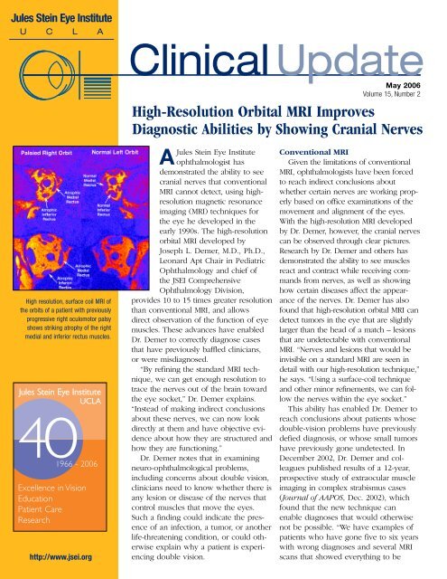
High resolution, surface coil MRI of<br />
the orbits of a patient with previously<br />
progressive right oculomotor palsy<br />
shows striking atrophy of the right<br />
medial and inferior rectus muscles.<br />
<strong>Jules</strong> <strong>Stein</strong> <strong>Eye</strong> <strong>Institute</strong><br />
UCLA<br />
401966<br />
- <strong>2006</strong><br />
Excellence in Vision<br />
Education<br />
Patient Care<br />
Research<br />
http://www.jsei.org<br />
<strong>Clinical</strong> <strong>Update</strong><br />
<strong>May</strong> <strong>2006</strong><br />
Volume 15, Number 2<br />
High-Resolution Orbital MRI Improves<br />
Diagnostic Abilities by Showing Cranial Nerves<br />
A<strong>Jules</strong> <strong>Stein</strong> <strong>Eye</strong> <strong>Institute</strong><br />
ophthalmologist has<br />
demonstrated the ability to see<br />
cranial nerves that conventional<br />
MRI cannot detect, using highresolution<br />
magnetic resonance<br />
imaging (MRI) techniques for<br />
the eye he developed in the<br />
early 1990s. The high-resolution<br />
orbital MRI developed by<br />
Joseph L. Demer, M.D., Ph.D.,<br />
Leonard Apt Chair in Pediatric<br />
Ophthalmology and chief of<br />
the JSEI Comprehensive<br />
Ophthalmology Division,<br />
provides 10 to 15 times greater resolution<br />
than conventional MRI, and allows<br />
direct observation of the function of eye<br />
muscles. These advances have enabled<br />
Dr. Demer to correctly diagnose cases<br />
that have previously baffled clinicians,<br />
or were misdiagnosed.<br />
“By refining the standard MRI technique,<br />
we can get enough resolution to<br />
trace the nerves out of the brain toward<br />
the eye socket,” Dr. Demer explains.<br />
“Instead of making indirect conclusions<br />
about these nerves, we can now look<br />
directly at them and have objective evidence<br />
about how they are structured and<br />
how they are functioning.”<br />
Dr. Demer notes that in examining<br />
neuro-ophthalmological problems,<br />
including concerns about double vision,<br />
clinicians need to know whether there is<br />
any lesion or disease of the nerves that<br />
control muscles that move the eyes.<br />
Such a finding could indicate the presence<br />
of an infection, a tumor, or another<br />
life-threatening condition, or could otherwise<br />
explain why a patient is experiencing<br />
double vision.<br />
Conventional MRI<br />
Given the limitations of conventional<br />
MRI, ophthalmologists have been forced<br />
to reach indirect conclusions about<br />
whether certain nerves are working properly<br />
based on office examinations of the<br />
movement and alignment of the eyes.<br />
With the high-resolution MRI developed<br />
by Dr. Demer, however, the cranial nerves<br />
can be observed through clear pictures.<br />
Research by Dr. Demer and others has<br />
demonstrated the ability to see muscles<br />
react and contract while receiving commands<br />
from nerves, as well as showing<br />
how certain diseases affect the appearance<br />
of the nerves. Dr. Demer has also<br />
found that high-resolution orbital MRI can<br />
detect tumors in the eye that are slightly<br />
larger than the head of a match – lesions<br />
that are undetectable with conventional<br />
MRI. “Nerves and lesions that would be<br />
invisible on a standard MRI are seen in<br />
detail with our high-resolution technique,”<br />
he says. “Using a surface-coil technique<br />
and other minor refinements, we can follow<br />
the nerves within the eye socket.”<br />
This ability has enabled Dr. Demer to<br />
reach conclusions about patients whose<br />
double-vision problems have previously<br />
defied diagnosis, or whose small tumors<br />
have previously gone undetected. In<br />
December 2002, Dr. Demer and colleagues<br />
published results of a 12-year,<br />
prospective study of extraocular muscle<br />
imaging in complex strabismus cases<br />
(Journal of AAPOS, Dec. 2002), which<br />
found that the new technique can<br />
enable diagnoses that would otherwise<br />
not be possible. “We have examples of<br />
patients who have gone five to six years<br />
with wrong diagnoses and several MRI<br />
scans that showed everything to be
2<br />
JSEI <strong>Clinical</strong> <strong>Update</strong> <strong>May</strong> <strong>2006</strong><br />
High resolution MRI of the brainstem of the same patient shows a neuroma of the right oculomotor<br />
nerve in the interpeduncular region that enhances strikingly with gadolinium contrast. This tumor was<br />
missed on three conventional MRI scans before referral to JSEI.<br />
normal, and we went in and on the<br />
first scan found a tumor that<br />
explained everything,” he says. “In<br />
other cases, we have been able to<br />
demonstrate that an injury to the eye<br />
socket has directly damaged a nerve<br />
that has paralyzed a muscle, and we<br />
accurately forecast whether the<br />
patient will recover without treatment<br />
— potentially sparing unnecessary<br />
surgery.” Dr. Demer’s group<br />
also used high-resolution orbital MRI<br />
to discover tissues within the eye<br />
socket, previously unknown, that led<br />
to a fundamentally new understanding<br />
of eye muscle operation. The<br />
discovery earned Dr. Demer the<br />
Friedenwald Award in 2003, the<br />
highest scientific honor bestowed by<br />
the Association for Research in<br />
Vision and Ophthalmology.<br />
Research Leads to Refinements<br />
Since 1991, Dr. Demer has<br />
received funding from the National<br />
<strong>Eye</strong> <strong>Institute</strong> to develop MRI techniques<br />
for the eye muscles. He has<br />
advanced the technology through<br />
targeted application of MRI to the<br />
eye and its socket. His group has<br />
used surface coil devices — special<br />
hardware akin to radio antennae,<br />
with greater sensitivity to receiving<br />
the radio signals from specific areas<br />
of tissue that are then used to construct<br />
an image. Methods developed<br />
by Dr. Demer’s team prevent the eye<br />
from moving during the scan — a<br />
problem that often leads to blurred<br />
MRI pictures.<br />
Just as important as the refinement<br />
of MRI to get better-quality<br />
pictures has been Dr. Demer’s<br />
research in interpreting the images.<br />
“We’re seeing things that haven’t<br />
been described in the textbooks<br />
before because they couldn’t be<br />
detected,” he explains. Dr. Demer<br />
has focused on three clinical<br />
applications: the diagnosis of<br />
strabismus; determination of<br />
neuro-ophthalmological conditions,<br />
including whether abnormalities in<br />
eye movements are indications of<br />
serious neurological disease; and<br />
reconstruction of injured eye<br />
sockets after severe injury or after<br />
an eye has been removed because<br />
it harbors a melanoma.<br />
Dr. Demer recently received a<br />
five-year National <strong>Eye</strong> <strong>Institute</strong><br />
grant renewal to make further<br />
refinements in high-resolution<br />
orbital MRI. Although far superior<br />
to conventional MRI, the approach<br />
still requires a longer scanning<br />
time than a computed assisted<br />
tomography (CAT) scan, and is<br />
unable to detect certain tissues<br />
that are either too small or lack<br />
sufficient contrast with adjacent<br />
tissues. “You can always improve<br />
the pictures,” Dr. Demer says. “But<br />
every year we are getting better at<br />
this, and we will continue to<br />
refine it.”<br />
Sharing Expertise<br />
Although the hardware needed<br />
for high-resolution orbital MRI is<br />
widely available, the intensive professional<br />
education and training<br />
required to successfully employ the<br />
technology has limited its use outside<br />
UCLA. Dr. Demer is working to<br />
change that. <strong>Clinical</strong> fellows have<br />
come from all over the United States<br />
and abroad to train with Dr. Demer<br />
and bring the new approach back to<br />
their institutions. Dr. Demer has<br />
taught at centers in the eastern<br />
United States, Australia and England,<br />
and offers courses at professional<br />
meetings. “Right now it’s a matter of<br />
passing on the expertise, as well as<br />
educating ophthalmologists to appreciate<br />
what kind of information they<br />
could be getting so that they order<br />
the tests and insist that they be<br />
done,” he says.<br />
Dr. Demer routinely receives<br />
referrals from across the country<br />
and as far away as South Africa to<br />
evaluate complicated cases. He<br />
offers referring ophthalmologists the<br />
option of a consultation in which<br />
Dr. Demer conducts a clinical examination<br />
and runs the high-resolution<br />
orbital MRI scanner, then interprets<br />
the results. He recommends referrals<br />
and consultations for complex forms<br />
of strabismus, orbital tumors, and<br />
trauma or inflammatory conditions<br />
that can result from sinus-surgery<br />
complications. “For any condition in<br />
which the ophthalmologist needs to<br />
know what’s going on inside the<br />
eye socket,” Dr. Demer explains,<br />
“this technique can quite often provide<br />
decisive information without<br />
pain or risk to the patient.”
<strong>May</strong> <strong>2006</strong> JSEI <strong>Clinical</strong> <strong>Update</strong><br />
A New Lacrimal Stent Promises Early Results<br />
Lacrimal surgery (dacryocystorhinostomy,<br />
DCR) has continued<br />
to evolve. The Orbital and<br />
Ophthalmic Plastic Surgery Service<br />
at the <strong>Jules</strong> <strong>Stein</strong> <strong>Eye</strong> <strong>Institute</strong> has<br />
favored the endonasal approach for<br />
more than 10 years. The endonasal<br />
approach avoids a cutaneous<br />
incision with the associated risks<br />
of scarring and muscle weakness,<br />
and also allows a more precise<br />
anatomically customized surgery.<br />
The latest technology, including<br />
endoscopes, custom instrumentation<br />
for removal of bone and soft<br />
tissue, and specialized silicone<br />
stents, has improved the ability to<br />
remove the bone and soft tissue in<br />
order to create an opening between<br />
the lacrimal sac and the nose.<br />
Today, this can be accomplished<br />
under monitored local anesthesia<br />
with minimal blood loss and rapid<br />
recovery, and no cutaneous incision.<br />
However, the surgery still has<br />
a failure rate in the range of 5 percent<br />
to 10 percent, because the<br />
newly created opening can be<br />
closed by scar tissue.<br />
Robert Alan Goldberg, M.D.,<br />
chief of the <strong>Institute</strong>’s Orbital and<br />
Ophthalmic Plastic Surgery<br />
Division, has developed a new type<br />
of lacrimal stent made of hydrogel<br />
— a specialized plastic that absorbs<br />
water. The stent is currently an<br />
experimental device, and surgeries<br />
are performed under the auspices<br />
of the institutional review board<br />
at UCLA.*<br />
Dr. Goldberg explains, “When it<br />
comes out of the package, before it<br />
touches water, the stent is small<br />
enough to easily fit into the surgical<br />
opening. Once the stent is positioned<br />
in the newly created DCR<br />
ostium, it begins to absorb water and<br />
expand.” Over the first 24 hours, the<br />
stent expands to seven times its orig-<br />
1<br />
Figure 1. Hydrogel<br />
lacrimal stent in dry<br />
form, out of the<br />
package (top), and<br />
following hydration<br />
for 48 hours (bottom)<br />
Figure 2. Stent in<br />
position at the end<br />
of surgery<br />
Figure 3. Healed DCR<br />
surgery immediately<br />
after stent removal,<br />
showing wide-open<br />
surgical ostium (dotted<br />
lines) with the internal<br />
punctum draining into<br />
the nose<br />
inal size, and it also becomes much<br />
softer (Fig. 1). The enlarged, soft<br />
stent gently presses the mucosal<br />
edges together, acting like a surgical<br />
staple (Fig. 2). The soft, smooth, wet<br />
surface of the stent is very biocompatible<br />
and seems to promote rapid<br />
mucosal epithelialization of the<br />
newly created lacrimal opening.<br />
After two to four weeks, the<br />
lacrimal mucosa has healed in the<br />
desired position. An ostium has<br />
been created from the lacrimal sac<br />
to the nose, providing a pathway<br />
for drainage of tears, and protecting<br />
against infection. At this point, the<br />
stent is removed by visualizing it in<br />
the nose and sliding it out, utilizing<br />
office endoscopy.<br />
2<br />
3<br />
Dr. Goldberg finds the initial<br />
results very encouraging: large<br />
lacrimal ostia have been noted, with<br />
excellent healing of the mucosal<br />
edges, in all cases performed to<br />
date (Fig. 3). He notes, “If these<br />
excellent early results are verified<br />
experimentally with longer followup<br />
and with additional patients<br />
enrolled, then the hydrogel lacrimal<br />
stent may be a significant step<br />
forward in our goal of designing<br />
minimally invasive endonasal DCR<br />
surgical technique with a high<br />
success rate.”<br />
* Dr. Goldberg does not have any<br />
commercial interest in the products cited<br />
in this article.<br />
33
4<br />
JSEI <strong>Clinical</strong> <strong>Update</strong> <strong>May</strong> <strong>2006</strong><br />
Optic Disc Imaging in Perimetrically Normal <strong>Eye</strong>s of Glaucoma<br />
Patients with Unilateral Field Loss<br />
Ophthalmologists have become<br />
increasingly interested in diagnosing<br />
glaucoma before functional<br />
deficits manifest. Existing evidence<br />
that modern imaging techniques can<br />
detect glaucomatous damage before<br />
visual field loss occurs makes it<br />
relevant to compare them in preperimetric<br />
glaucoma. Perimetrically<br />
unaffected eyes of glaucoma<br />
patients have been found to be at<br />
high risk for developing visual field<br />
abnormalities. Recognition of morphological<br />
damage is important for<br />
an early diagnosis of the disease.<br />
A clinical study performed at the<br />
<strong>Jules</strong> <strong>Stein</strong> <strong>Eye</strong> <strong>Institute</strong> by Federico<br />
Badala, M.D.; Kouros Nouri-Mahdavi,<br />
M.D; Behrooz Koucheki, M.D.; Hector<br />
Fontana, M.D.; Anita Manassakorn,<br />
M.D., international fellows at the<br />
<strong>Institute</strong>; and Simon K. Law, M.D.,<br />
Pharm.D., Assistant Professor of<br />
Ophthalmology; and Joseph Caprioli,<br />
M.D., Professor of Ophthalmology<br />
and Chief of the Glaucoma Division,<br />
compared the diagnostic precision of<br />
<strong>Jules</strong> <strong>Stein</strong> <strong>Eye</strong> <strong>Institute</strong> UCLA<br />
100 <strong>Stein</strong> Plaza<br />
Box 957000<br />
Los Angeles, CA 90095-7000<br />
U.S.A.<br />
0.4<br />
StratusOCT<br />
0.2<br />
HRT II<br />
Disc Photos<br />
0.0<br />
GDx-VCC<br />
0.0 0.2 0.4 0.6 0.8 1.0<br />
1 - Specificity<br />
optical coherence tomography<br />
(StratusOCT), confocal scanning laser<br />
ophthalmoscopy (HRT II), scanning<br />
laser polarimetry (GDx-VCC ), and<br />
clinical evaluation of the optic disc in<br />
pre-perimetric glaucoma.<br />
The research team studied data<br />
from 46 open-angle glaucoma<br />
patients with glaucomatous visual<br />
field loss with achromatic perimetry<br />
in one eye and a normal visual field<br />
in the fellow eye, and 46 normal<br />
controls. The perimetrically unaffected<br />
eyes of open-angle glaucoma<br />
patients with unilateral field loss well<br />
JSEI Continuing Education Programs<br />
<strong>May</strong> 19-20, <strong>2006</strong><br />
<strong>Jules</strong> <strong>Stein</strong> <strong>Eye</strong> <strong>Institute</strong><br />
<strong>Clinical</strong> and Research Seminar and 40th Anniversary Celebration<br />
RBP Auditorium<br />
37th <strong>Jules</strong> <strong>Stein</strong> Lecturer George B. Bartley, MD<br />
3rd Bradley R. Straatsma Lecturer Uwe Pleyer, MD<br />
3rd Thomas H. Pettit Lecturer Todd P. Margolis, MD, PhD<br />
Sensitivity<br />
1.0<br />
0.8<br />
0.6<br />
<strong>2006</strong><br />
represent preperimetric glaucoma.<br />
Open-angle glaucoma is predominantly<br />
a bilateral disease and the<br />
“pre-perimetric” eyes are at high risk<br />
for developing visual field defects.<br />
Furthermore, the rim area and the<br />
retinal nerve fiber layer (RNFL) have<br />
been found to be thinner in this<br />
subset of eyes than in a normal<br />
control group in previous studies.<br />
Receiver operator characteristic<br />
(ROC) curves, sensitivities at fixed<br />
specificities and likelihood ratios were<br />
used to compare the performance of<br />
StratusOCT, HRT II, GDx-VCC, and<br />
clinical evaluation of optic disc<br />
stereophotographs to distinguish<br />
perimetrically unaffected eyes of glaucoma<br />
patients from normal eyes. The<br />
larger the area under the ROC curve,<br />
the better the diagnostic accuracy.<br />
The present study suggests that<br />
StratusOCT can detect evidence of<br />
early glaucomatous damage sooner<br />
than other imaging techniques or<br />
clinical evaluation of the optic disc<br />
by experienced observers in eyes at<br />
high risk for developing visual field<br />
loss. This is the first report in which<br />
early structural glaucomatous damage<br />
is more frequently diagnosed with an<br />
imaging device than with expert<br />
clinical evaluation of optic disc<br />
stereophotographs. The inferior<br />
and superior average RNFL thicknesses<br />
were the parameters that<br />
best distinguished eyes with<br />
preperimetric glaucomatous<br />
damage from normal eyes.<br />
First Class Mail<br />
U.S. Postage<br />
PAID<br />
UCLA