View May 2006 Clinical Update - Jules Stein Eye Institute
View May 2006 Clinical Update - Jules Stein Eye Institute
View May 2006 Clinical Update - Jules Stein Eye Institute

Create successful ePaper yourself
Turn your PDF publications into a flip-book with our unique Google optimized e-Paper software.
2<br />
JSEI <strong>Clinical</strong> <strong>Update</strong> <strong>May</strong> <strong>2006</strong><br />
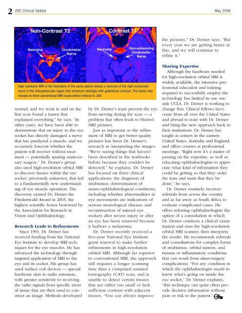
High resolution MRI of the brainstem of the same patient shows a neuroma of the right oculomotor<br />
nerve in the interpeduncular region that enhances strikingly with gadolinium contrast. This tumor was<br />
missed on three conventional MRI scans before referral to JSEI.<br />
normal, and we went in and on the<br />
first scan found a tumor that<br />
explained everything,” he says. “In<br />
other cases, we have been able to<br />
demonstrate that an injury to the eye<br />
socket has directly damaged a nerve<br />
that has paralyzed a muscle, and we<br />
accurately forecast whether the<br />
patient will recover without treatment<br />
— potentially sparing unnecessary<br />
surgery.” Dr. Demer’s group<br />
also used high-resolution orbital MRI<br />
to discover tissues within the eye<br />
socket, previously unknown, that led<br />
to a fundamentally new understanding<br />
of eye muscle operation. The<br />
discovery earned Dr. Demer the<br />
Friedenwald Award in 2003, the<br />
highest scientific honor bestowed by<br />
the Association for Research in<br />
Vision and Ophthalmology.<br />
Research Leads to Refinements<br />
Since 1991, Dr. Demer has<br />
received funding from the National<br />
<strong>Eye</strong> <strong>Institute</strong> to develop MRI techniques<br />
for the eye muscles. He has<br />
advanced the technology through<br />
targeted application of MRI to the<br />
eye and its socket. His group has<br />
used surface coil devices — special<br />
hardware akin to radio antennae,<br />
with greater sensitivity to receiving<br />
the radio signals from specific areas<br />
of tissue that are then used to construct<br />
an image. Methods developed<br />
by Dr. Demer’s team prevent the eye<br />
from moving during the scan — a<br />
problem that often leads to blurred<br />
MRI pictures.<br />
Just as important as the refinement<br />
of MRI to get better-quality<br />
pictures has been Dr. Demer’s<br />
research in interpreting the images.<br />
“We’re seeing things that haven’t<br />
been described in the textbooks<br />
before because they couldn’t be<br />
detected,” he explains. Dr. Demer<br />
has focused on three clinical<br />
applications: the diagnosis of<br />
strabismus; determination of<br />
neuro-ophthalmological conditions,<br />
including whether abnormalities in<br />
eye movements are indications of<br />
serious neurological disease; and<br />
reconstruction of injured eye<br />
sockets after severe injury or after<br />
an eye has been removed because<br />
it harbors a melanoma.<br />
Dr. Demer recently received a<br />
five-year National <strong>Eye</strong> <strong>Institute</strong><br />
grant renewal to make further<br />
refinements in high-resolution<br />
orbital MRI. Although far superior<br />
to conventional MRI, the approach<br />
still requires a longer scanning<br />
time than a computed assisted<br />
tomography (CAT) scan, and is<br />
unable to detect certain tissues<br />
that are either too small or lack<br />
sufficient contrast with adjacent<br />
tissues. “You can always improve<br />
the pictures,” Dr. Demer says. “But<br />
every year we are getting better at<br />
this, and we will continue to<br />
refine it.”<br />
Sharing Expertise<br />
Although the hardware needed<br />
for high-resolution orbital MRI is<br />
widely available, the intensive professional<br />
education and training<br />
required to successfully employ the<br />
technology has limited its use outside<br />
UCLA. Dr. Demer is working to<br />
change that. <strong>Clinical</strong> fellows have<br />
come from all over the United States<br />
and abroad to train with Dr. Demer<br />
and bring the new approach back to<br />
their institutions. Dr. Demer has<br />
taught at centers in the eastern<br />
United States, Australia and England,<br />
and offers courses at professional<br />
meetings. “Right now it’s a matter of<br />
passing on the expertise, as well as<br />
educating ophthalmologists to appreciate<br />
what kind of information they<br />
could be getting so that they order<br />
the tests and insist that they be<br />
done,” he says.<br />
Dr. Demer routinely receives<br />
referrals from across the country<br />
and as far away as South Africa to<br />
evaluate complicated cases. He<br />
offers referring ophthalmologists the<br />
option of a consultation in which<br />
Dr. Demer conducts a clinical examination<br />
and runs the high-resolution<br />
orbital MRI scanner, then interprets<br />
the results. He recommends referrals<br />
and consultations for complex forms<br />
of strabismus, orbital tumors, and<br />
trauma or inflammatory conditions<br />
that can result from sinus-surgery<br />
complications. “For any condition in<br />
which the ophthalmologist needs to<br />
know what’s going on inside the<br />
eye socket,” Dr. Demer explains,<br />
“this technique can quite often provide<br />
decisive information without<br />
pain or risk to the patient.”