HercepTest⢠Interpretation Manual - Breast - Dako
HercepTest⢠Interpretation Manual - Breast - Dako
HercepTest⢠Interpretation Manual - Breast - Dako

You also want an ePaper? Increase the reach of your titles
YUMPU automatically turns print PDFs into web optimized ePapers that Google loves.
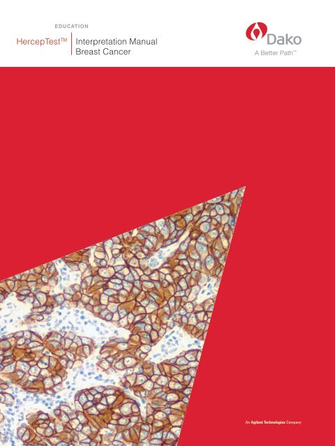
EDUCATION<br />
HercepTest TM<br />
<strong>Interpretation</strong> <strong>Manual</strong><br />
<strong>Breast</strong> Cancer
EDUCATION<br />
HercepTest TM<br />
Table of Contents
Contents<br />
5 Introduction<br />
6 HER2 Overview<br />
6 HER2 Protein and HER2 Family<br />
6 HER2 Testing IHC and FISH<br />
7 HER2 Testing Algorithm<br />
8 The HercepTest TM Kit<br />
9 HER2 IQFISH pharmDx TM Kit<br />
9 Hybridizer Instrument for In Situ Hybridization (FISH)<br />
10 Checklist<br />
10 HercepTest TM Training Checklist<br />
11 Recommendations<br />
11 Recommended Data Tracking for HercepTest TM Immunostaining<br />
12 Technical Considerations<br />
12 Technical Considerations for Optimal HercepTest TM Performance<br />
12 Protocol Recommendations<br />
13 Tissue Processing Considerations<br />
13 Tissue Processing Recommendations<br />
14 Guidelines<br />
14 Review of HercepTest TM Scoring Guidelines<br />
14 Validation of the Assay<br />
15 <strong>Interpretation</strong> Guide for 1+ Cell Line<br />
16 Guidelines for Scoring<br />
17 <strong>Interpretation</strong><br />
17 Recommendations for <strong>Interpretation</strong> of HercepTest TM – <strong>Breast</strong> Cancer<br />
17 Steps for HercepTest TM <strong>Interpretation</strong><br />
19 Staining Patterns<br />
24 Artifacts<br />
24 Interpreting Artifacts<br />
28 Effects of Fixation<br />
29 Effects of Insufficient Target Retrieval<br />
30 Effects of Excessive Tissue Drying<br />
31 Staining Images<br />
31 HER2 Expression in Various Diagnostic Entities<br />
46 Troubleshooting Guide<br />
46 Troubleshooting Guideline for HercepTest TM<br />
49 Bibliography
4<br />
HercepTest TM <strong>Interpretation</strong> <strong>Manual</strong> – <strong>Breast</strong> Cancer<br />
ROW Version
Introduction<br />
HercepTest TM <strong>Interpretation</strong> <strong>Manual</strong><br />
HercepTest is a semi-quantitative<br />
immunohistochemical assay to determine HER2<br />
protein overexpression in breast cancer tissues<br />
routinely processed for histological evaluation<br />
and formalin-fixed, paraffin-embedded cancer<br />
tissue from patients with adenocarcinoma of<br />
the stomach, including the gastroesophageal<br />
junction*. HercepTest is indicated as an aid<br />
in the assessment of breast and gastric cancer<br />
patients for whom Herceptin ® (trastuzumab)<br />
treatment is being considered (see Herceptin ®<br />
package insert).<br />
HercepTest TM <strong>Interpretation</strong> Guidelines<br />
Prior to HercepTest TM , immunohistochemistry<br />
was practiced largely as a subjective method,<br />
ideally suited for qualitative analysis. HercepTest TM ,<br />
however, changed this paradigm, as the<br />
determination of positivity was no longer a simple<br />
yes or no answer. Patients are now evaluated<br />
using immunohistochemistry technology applied<br />
as a semi-quantitative tool with a scoring system<br />
reflective of intensity of staining in conjunction with<br />
percentage of stained tumor cells. This shift in<br />
application introduced a change in the way<br />
immunohistochemistry was viewed.<br />
Most metastatic breast cancer tissue specimens<br />
tested for HER2 overexpression are scored with<br />
either 0 or 3+ staining intensity. While the majority<br />
of cases are clear-cut, a small percentage of the<br />
remaining 1+ and 2+ scored samples may be more<br />
difficult to interpret. In this manual, we will focus<br />
on these equivocal samples. In addition, we will<br />
review images of sample artifacts and discuss how<br />
to best interpret such cases.<br />
HER2 IQFISH pharmDx TM<br />
Despite the high quality of HercepTest TM , clinical<br />
response of weakly positive specimens has<br />
remained an area of uncertainty within HER2<br />
assessment. HER2 IQFISH pharmDx TM complements<br />
HercepTest TM by quantitatively determining HER2<br />
gene amplification and clarifying equivocal cases.<br />
HercepTest TM and HER2 IQFISH pharmDx TM<br />
enhance patient care by aiding in proper<br />
determination of the appropriate course of treatment.<br />
Photomicrographs<br />
The included photomicrographs are breast<br />
carcinoma unless otherwise noted.<br />
This HercepTest TM <strong>Interpretation</strong> <strong>Manual</strong> for<br />
breast cancer is provided as a tool to help guide<br />
pathologists and laboratorians to achieve correct<br />
and reproducible results.<br />
The goal of this manual is to familiarize you with<br />
the requirements for scoring breast carcinomas<br />
stained with HercepTest TM . Example cases of<br />
various staining intensities of HER2 expression are<br />
provided for reference. The HercepTest TM package<br />
insert guidelines will be reviewed and technical tips<br />
for ensuring high-quality staining in your laboratory<br />
will be given. Reviewing this HercepTest TM<br />
<strong>Interpretation</strong> <strong>Manual</strong> will provide a solid foundation<br />
for evaluating slides stained with HercepTest TM .<br />
* Van Cutsem E, Kang Y, Chung H, Shen L, Sawaki A, Lordick F, et<br />
al. Efficacy results from the ToGA trial: A phase III study of<br />
trastuzumab added to standard chemotherapy in first-line human<br />
epidermal growth factor receptor 2 positive advanced gastric<br />
cancer. J Clin Oncol 2009;27:18s, (suppl; abstr LBA2409).<br />
(http://media.asco.org/silver).<br />
<strong>Dako</strong> is a registered trademark of <strong>Dako</strong> Denmark A/S. HercepTest TM<br />
and Herceptin ® are trademarks owned by Genentech, Inc. and/or<br />
F. Hoffman-La Roche Ltd.; HercepTest TM is subject to an exclusive<br />
trademark license to <strong>Dako</strong> Denmark A/S.<br />
HercepTest TM <strong>Interpretation</strong> <strong>Manual</strong> – <strong>Breast</strong> Cancer<br />
ROW Version<br />
5
HER2 Overview<br />
HER2 Protein and HER2 Family<br />
The gene encoding HER2 is located on chromosome<br />
17 and is a member of the EGF/erbB growth<br />
factor receptor gene family, which also includes<br />
epidermal growth factor receptor (EGFR, or<br />
HER1), HER3/erbB3 and HER4/erbB4. All of<br />
these genes encode transmembrane growth<br />
factor receptors, which are tyrosine kinase type<br />
1 receptors with growth stimulating potential.<br />
Activation of HER family members generally<br />
occurs when the ligand and a dimer of the same<br />
monomer or other member of the HER family<br />
are bound together. HER2 has no known<br />
ligand. Once activation has occurred, tyrosine<br />
autophoshorylation of cytoplasmic signal proteins<br />
transmit signals to the nucleus, thus regulating<br />
aspects of cell growth, division, differentiation<br />
and migration.<br />
Overexpression of HER2 receptors results in<br />
receptors transmitting excessive signals for cell<br />
proliferation to the nucleus. This may lead to<br />
more aggressive growth of the transformed cell.<br />
Data supports the hypothesis that the HER2-<br />
overexpression cells directly contribute to the<br />
pathogenesis and clinical aggressiveness of<br />
tumors.* This overexpression is associated with<br />
poor prognosis, including reduced relapse-free<br />
and overall survival.<br />
Ligand<br />
Growth Signal<br />
HER Heterodimer<br />
Figure 1: Representation of HER family<br />
HER2 Homodimer<br />
HER2 Testing IHC and FISH<br />
Immunohistochemistry (IHC) measures the level of<br />
HER2 receptor overexpression, while fluorescence<br />
in situ hybridization (FISH) quantifies the level of<br />
HER2 gene amplification. Together they are the<br />
most commonly used methods of determining<br />
HER2 status in routine diagnostic settings.<br />
HER2 DNA (Target for ISH)<br />
HER2 Protein (Target for IHC)<br />
Nucleus<br />
HER2<br />
mRNA<br />
Cytoplasm<br />
Cell Membrane<br />
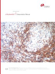
Amplified Result, Score ≥ 2<br />
<strong>Breast</strong> cancer specimen stained<br />
with HER2 IQFISH pharmDx TM .<br />
Figure 2: IHC and ISH targets for HER2 testing<br />
<strong>Breast</strong> Cancer Cell<br />
Positive Result, Score 3+<br />
<strong>Breast</strong> cancer specimen stained<br />
with HercepTest TM .<br />
* Robert W. Carlson, MD; Susan J. Moench, et al. HER2 Testing in <strong>Breast</strong> Cancer: NCCN Task Force Report and Recommendations: Journal of the<br />
National Comprehensive Cancer Network July 2005. | Edith A. Perez, Vera J. Suman, et al. HER2 Testing by Local, Central and Reference Laboratories in<br />
Specimens from the North Central Cancer Treatment Group N9831 Intergroup Adjuvant Trial. Journal of Clinical Oncology July 1, 2006. | Martine J.<br />
Piccart-Gebhart, MD., Ph.D., Marion Proctor, M. Sci., et al. Trastuzumab after Adjuvant Chemotherapy in HER2-Positive <strong>Breast</strong> Cancer. New England<br />
Journal of Medicine October 20, 2005.<br />
6<br />
HercepTest TM <strong>Interpretation</strong> <strong>Manual</strong> – <strong>Breast</strong> Cancer<br />
ROW Version
HER2 Testing Algorithm<br />
Tumor Sample<br />
HER2 IHC<br />
0<br />
Negative<br />
1+<br />
Negative<br />
2+<br />
3+<br />
Weakly Positive* Positive<br />
HER2 ISH<br />
Negative<br />
Non-Amplified<br />
Positive<br />
Amplified<br />
Report to Oncologist for Herceptin ®<br />
Consideration<br />
Figure 3: Current clinical practices for selection of patients for Herceptin ® treatment.<br />
* For Herceptin ® – Weakly positive cases (2+) may be considered equivocal and reflexed to ISH testing.<br />
NCCN Practice Guidelines in Oncology, CAP Conference Summary Laboratories performing HER2 testing should meet quality assurance standards.<br />
HercepTest TM <strong>Interpretation</strong> <strong>Manual</strong> – <strong>Breast</strong> Cancer<br />
ROW Version<br />
7
The HercepTest TM Kit<br />
HercepTest TM is a semi-quantitative<br />
immunohistochemical kit system for determination<br />
of HER2 protein overexpression in breast cancer<br />
tissues routinely processed for histological evaluation<br />
and in formalin-fixed, paraffin-embedded cancer<br />
tissue from patients with adenocarcinoma of the<br />
stomach, including gastro-esophageal junction.<br />
Following incubation with the primary antibody to<br />
human HER2 protein, this kit employs a ready-to-use<br />
Visualization Reagent based on dextran technology.<br />
This reagent consists of both secondary goat<br />
anti-rabbit molecules and horseradish peroxidase<br />
molecules linked to a common dextran polymer<br />
backbone, thus eliminating the need for sequential<br />
application of link antibody and peroxidase<br />
conjugate. The enzymatic conversion of the<br />
subsequently added chromogen results in formation<br />
of a visible reaction product at the antigen site.<br />
The specimen may then be counterstained and<br />
coverslipped. Control cell line slides are provided.<br />
HercepTest TM is a complete kit and includes:<br />
• Peroxidase-Blocking Reagent<br />
• Rabbit Anti-Human HER2 Protein<br />
• Visualization Reagent<br />
• Negative Control Reagent<br />
• DAB Buffered Substrate<br />
• DAB Chromogen<br />
• Epitope Retrieval Solution (10x)<br />
• Wash Buffer (10x) (not included in SK001)<br />
• User-Fillable Bottles (only included in SK001)<br />
Recommended hematoxylin counterstain:<br />
(not provided)<br />
• Mayer’s Hematoxylin for <strong>Dako</strong> Autostainer/<br />
Autostainer Plus, Code S3301<br />
• Mayer’s Hematoxylin for Automated Link<br />
Platforms, Code SK308<br />
Three HercepTest TM kit configurations<br />
are available:<br />
K5204<br />
35 Tests<br />
HercepTest TM for manual use<br />
K5207<br />
50 Tests<br />
HercepTest TM for the <strong>Dako</strong><br />
Autostainer<br />
SK001<br />
50 Tests<br />
HercepTest TM for Automated<br />
Link Platforms<br />
Step<br />
1<br />
Water bath 40 minutes,<br />
95-99 °C.<br />
Step<br />
4<br />
Step<br />
5<br />
Step<br />
2<br />
Application of peroxidase block.<br />
Incubate for 5 minutes.<br />
Step<br />
3<br />
Application of primary antibody.<br />
Incubate for 30 minutes.<br />
Application of<br />
HRP-labeled polymer.<br />
Incubate for 30 minutes.<br />
Application of<br />
chromogenic substrate.<br />
Incubate for 10 minutes.<br />
HER2 antibody Tissue proteins HER2 protein Peroxidase Block<br />
Secondary antibody Dextran backbone HRP enzyme DAB<br />
Figure 4: HercepTest TM procedure<br />
8<br />
HercepTest TM <strong>Interpretation</strong> <strong>Manual</strong> – <strong>Breast</strong> Cancer<br />
ROW Version
HER2 IQFISH pharmDx TM Kit<br />
HER2 IQFISH pharmDx kit is a direct fluorescence<br />
in situ hybridization (FISH) assay designed to<br />
quantitatively determine HER2 gene amplification<br />
in formalin-fixed, paraffin-embedded (FFPE) breast<br />
cancer tissue specimens and FFPE specimens<br />
from patients with adenocarcinoma of the stomach,<br />
including gastroesophageal junction. HER2<br />
IQFISH pharmDx kit is indicated as an aid in<br />
the assessment of breast and gastric patients for<br />
whom Herceptin ® (trastuzumab) treatment is being<br />
considered (see Herceptin ® package insert).<br />
For breast cancer patient, results from the HER2<br />
IQFISH pharmDx Kit are intended for use as an<br />
adjunct to the clinicopathologic information currently<br />
used for estimating prognosis in stage II, nodepositive<br />
breast cancer patients.<br />
K5731<br />
HER2 IQFISH pharmDx TM Kit<br />
(22 x 22 mm target area)<br />
20 Tests<br />
Hybridizer Instrument<br />
for In Situ Hybridization (FISH)<br />
Hybridizer is a hands-free, denaturation and<br />
hybridization instrument. The system allows for<br />
semi-automation of FISH by eliminating manual<br />
steps in the hands-on intensive manual procedure.<br />
S2450 Hybridizer 120 volt<br />
S2451 Hybridizer 240 volt<br />
The assays includes a chromosone 17 reference<br />
probe to correct for HER2 signal number in the event<br />
of chromosone 17 aneusomy.<br />
• CEN-17 PNA probes directly labeled with<br />
fluorescein (FITC) targets the centromeric region<br />
of the chromosome (green signals)<br />
• HER2 DNA probe directly labeled with Texas Red<br />
fluorochrome targets the HER2 amplicon (red<br />
signals)<br />
• Results are expressed as a ratio of HER2 gene<br />
copies (red signals) per number of chromosome<br />
17 copies (green signals)<br />
HER2 IQFISH pharmDx TM is a complete kit and<br />
includes<br />
• Pre-Treatment Solution 20x<br />
• Pepsin, Ready-to-Use<br />
• Pepsin Diluent (10x)<br />
• HER2/CEN-17 IQISH Probe Mix<br />
• Stringent Wash Buffer 20x<br />
• Fluorescence Mounting Medium,<br />
containing DAPI<br />
• Wash Buffer 20x<br />
• Coverslip Sealant<br />
Figure 5: <strong>Dako</strong> Hybridizer instrument<br />
HercepTest TM <strong>Interpretation</strong> <strong>Manual</strong> – <strong>Breast</strong> Cancer<br />
ROW Version<br />
9
Checklist<br />
HercepTest TM Training Checklist<br />
10<br />
HercepTest TM <strong>Interpretation</strong> <strong>Manual</strong> – <strong>Breast</strong> Cancer<br />
ROW Version
Recommendations<br />
Recommended Data Tracking for HercepTest TM Immunostaining<br />
HercepTest TM Testing<br />
Use HercepTest TM data to determine an<br />
average number of percent positive cases.<br />
15-20%<br />
positive<br />
If the average percent positive cases falls within 15-<br />
20%, report results: Continue to use HercepTest TM by<br />
following the protocol. Continue to monitor results.<br />
20%<br />
positive<br />
Review Patient Demographics<br />
If patient demographics are normal,<br />
review HercepTest TM procedures.<br />
Normal patient<br />
demographics<br />
High number<br />
of recurrent<br />
cases<br />
If patient demographics consist of a large number<br />
of recurrent cases, >20% positive can be expected.<br />
In this case, report results and continue to use<br />
HercepTest TM by following protocol. Continue<br />
to monitor results and note any changes in the<br />
percent positive associated with changes in<br />
patient demographics.<br />
Review Technical Procedures<br />
Page<br />
Technical considerations<br />
for optimal HercepTest TM performance ............12<br />
Protocol recommendations ........................12<br />
Tissue processing considerations. ................13<br />
Tissue processing recommendations .............13<br />
Review <strong>Interpretation</strong> Procedures<br />
Page<br />
Review of HercepTest TM scoring guidelines .......14<br />
Validation of the assay ............................14<br />
Guidelines for scoring .............................16<br />
Recommendations for interpretation<br />
of HercepTest TM ...................................17<br />
Staining patterns ..................................19<br />
Interpreting artifacts ...............................24<br />
Staining Images ...................................31<br />
Table 1<br />
HercepTest TM <strong>Interpretation</strong> <strong>Manual</strong> – <strong>Breast</strong> Cancer<br />
ROW Version<br />
11
Technical Considerations<br />
Technical Considerations for Optimal HercepTest TM Performance<br />
While accurate and consistent interpretation can be achieved, technical issues relating to<br />
the performance of HercepTest TM are not always easy to identify. If cumulative laboratory<br />
test results fall outside the expected range of 15-20% positive, evaluate the patient<br />
demographics and then address any technical problems.<br />
Technical problems may arise in two areas, those<br />
involving sample collection and preparation prior<br />
to performing the test, and those involving the<br />
actual performance of the test itself. Technical<br />
problems relating to the performance of the test<br />
generally are related to procedural deviations and<br />
can be controlled and eliminated through training<br />
and, where necessary, clarification of the<br />
product instructions.<br />
Protocol Recommendations<br />
Pre-treatment Using Water Bath<br />
Water Bath:<br />
Heat HercepTest TM Epitope Retrieval Solution in a<br />
calibrated water bath capable of maintaining the<br />
required temperature of 95-99 °C. For best results,<br />
fill a container suitable for holding slides with<br />
diluted epitope retrieval (1:10) solution. Place<br />
container with epitope retrieval solution in a water<br />
bath and bring the temperature of the water bath<br />
and the epitope retrieval solution to 95-99 °C. Add<br />
the tissue sections mounted on slides to the container<br />
and bring the temperature of the epitope retrieval<br />
solution back to 95 °C before starting the timer.<br />
Incubation Time:<br />
Incubate the slides for 40 (±1) minutes in the<br />
preheated epitope retrieval solution. Remove the<br />
container with the slides from the water bath, but<br />
keep them in the epitope retrieval solution while<br />
allowing them to cool for 20 (±1) minutes at room<br />
temperature. After cooling, decant the epitope<br />
retrieval solution and rinse in wash buffer. For optimal<br />
performance, soak sections in wash buffer for 5-20<br />
minutes after epitope retrieval and prior to staining.<br />
Pre-treatment Using PT Link<br />
Preheat the diluted epitope retrieval solution (1:10)<br />
in the <strong>Dako</strong> PT Link tank to 85 °C. Place the room<br />
temperature, deparaffinized sections in Autostainer<br />
racks and immerse the slides into the preheated<br />
epitope retrieval solution. Let the PT Link warm up<br />
to 97 °C and incubate for 40 (±1) minutes at 97 °C.<br />
Leave the sections to cool in the PT Link until the<br />
temperature reaches 85 °C. Remove the PT Link<br />
tanks with the sections from the PT Link and leave<br />
the tanks on the table for 10 minutes with the lid off<br />
for further cooling. Prepare a jar/tank, eg. the PT<br />
Link Rinse Station, with diluted <strong>Dako</strong> Wash Buffer<br />
and soak sections for 5-20 minutes after epitope<br />
retrieval and prior to staining. Dedicated PT Link<br />
equipment must be used for HercepTest TM .<br />
Proper Incubations<br />
All incubation times should be performed according<br />
to the package insert. Stay within ±1 minute of all<br />
incubation times. If staining must be interrupted,<br />
slides may be kept in wash buffer following<br />
incubation of the primary antibody for up to one hour<br />
at room temperature (20-25 °C).<br />
Automated Staining<br />
<strong>Dako</strong> recommends the use of HercepTest TM on<br />
an Autostainer Link or a <strong>Dako</strong> Autostainer. Use of<br />
HercepTest TM on alternative automated platforms<br />
has not been validated and may give erroneous<br />
results.<br />
12<br />
HercepTest TM <strong>Interpretation</strong> <strong>Manual</strong> – <strong>Breast</strong> Cancer<br />
ROW Version
Wash Buffer<br />
Dilute the recommended wash buffer 1:10 using<br />
distilled or deionized water. Store unused diluted<br />
solution at 2-8 °C up to one month. Discard diluted,<br />
solution if cloudy in appearance.<br />
Storage of Reagents<br />
Reagents should be stored at 2-8 °C. Do not<br />
use after the expiration date stamped on the<br />
outside package.<br />
Tissue Processing Considerations<br />
Procedural deviations related to sample<br />
handling and processing can affect<br />
HercepTest TM results.<br />
Some of the variables that affect outcome are<br />
as follows:<br />
• Specimens drying prior to fixation<br />
• Type of fixative; only neutral buffered formalin<br />
is recommended<br />
• Temperature, age, storage, pH of fixative<br />
• Length of fixation, specimen size, ratio of<br />
size to fixative volume<br />
• Length of time in alcohol after primary fixation<br />
• Processing time, temperature pressure,<br />
and chemicals used<br />
• Storage of paraffin blocks<br />
• Storage of cut sections<br />
• Section thickness<br />
Tissue Processing Recommendations<br />
Validated Fixatives<br />
• Neutral Buffered Formalin<br />
• Bouin’s Solution<br />
Fixation Times<br />
Neutral Buffered Formalin:<br />
• 18-24 hours<br />
Time to fixation and duration of fixation, if available,<br />
should be recorded for each sample.<br />
Bouin’s:<br />
• 1-12 hours depending on tissue thickness<br />
Tissues fixed in Bouin’s solution must be washed<br />
in 70% ethanol to remove picrates prior to aqueous<br />
washes. Bouin’s solution may not be optimal, if<br />
FISH testing is needed.<br />
Specimen Thickness<br />
Tissue samples submitted for processing and<br />
embedding should not exceed 3-4 mm in thickness.<br />
Processing and Embedding<br />
After fixation, tissues are dehydrated in a series of<br />
alcohols and xylene followed by infiltration by melted<br />
paraffin held at no more than 60 °C. Properly fixed<br />
and embedded tissues expressing the HER2<br />
protein will keep indefinitely prior to sectioning and<br />
slide mounting if stored in a cool place, 15-25 °C.<br />
Overheating of tissues during embedding or<br />
overheating of sections during drying can induce<br />
detrimental effects on immunostaining and,<br />
therefore, should be avoided.<br />
The slides required for HER2 protein evaluation<br />
and tumor presence should be prepared at the<br />
same time. To preserve antigenicity, tissue sections,<br />
mounted on slides, should be stained within<br />
four-to-six weeks of sectioning when held at room<br />
temperature, 20-25 °C. Tissue specimens should<br />
be cut into sections of 4-5 µm thickness.<br />
To achieve reproducible results, each laboratory<br />
performing HercepTest should monitor its rate<br />
of positivity. If the positive rate exceeds 20%, a<br />
complete review of interpretation and technical<br />
procedures should be done.<br />
HercepTest TM <strong>Interpretation</strong> <strong>Manual</strong> – <strong>Breast</strong> Cancer<br />
ROW Version<br />
13
Guidelines<br />
Review of HercepTest TM Scoring Guidelines for <strong>Breast</strong> Tissue<br />
HercepTest TM is a semi-quantitative immunohistochemical assay to determine<br />
HER2 protein overexpression in breast cancer tissues routinely processed for<br />
histological evaluation.<br />
For the determination of HER2 protein<br />
overexpression, only the membrane staining<br />
intensity and pattern should be evaluated using<br />
the scale presented on page 16. Slide evaluation<br />
should be performed using a light microscope.<br />
Verify that the negative tissue control slide from<br />
the same staining run demonstrates no reactivity.<br />
Validation of the Assay<br />
Included in each HercepTest TM kit are control<br />
slides representing different levels of HER2<br />
protein expression: MDA-231(0), MDA-175 (1+)<br />
and SK-BR-3 (3+). The first step of interpretation<br />
is to evaluate the control cell lines. The control<br />
cell lines have been provided for qualifying the<br />
procedure and reagents, not as an interpretation<br />
reference. No staining of the 0 control cell line,<br />
MDA-231, partial brown membrane rimming in<br />
the 1+ control cell line, MDA-175, (refer to the<br />
<strong>Interpretation</strong> Guide for 1+ Cell Line on next<br />
page), and presence of complete intense brown<br />
membrane staining (rimming) in the 3+ control<br />
cell line, SK-BR-3, indicates a valid assay. If any<br />
of the control cell lines perform outside of these<br />
criteria, all results with the patient specimens<br />
should be considered invalid.<br />
Next, the positive tissue control slide known<br />
to contain the HER2 antigen, stained with<br />
HercepTest TM and fixed and processed similarly<br />
to the patient slides, should be evaluated for<br />
indication of correctly prepared tissues and<br />
proper staining technique. The ideal positive<br />
tissue control is weakly positive staining tissue.<br />
The presence of a brown reaction product at the<br />
cell membrane is indicative of positive reactivity.<br />
Figure 6<br />
Figure 7<br />
Figure 6: 0 control cell line, MDA-231, stained with HercepTest TM .<br />
No staining of the membrane is observed. (20x magnification).<br />
Figure7: 3+ control cell line, SK-BR-3, stained with HercepTest TM .<br />
A strong staining of the entire membrane is observed.<br />
(20x magnification).<br />
14<br />
HercepTest TM <strong>Interpretation</strong> <strong>Manual</strong> – <strong>Breast</strong> Cancer<br />
ROW Version
<strong>Interpretation</strong> Guide for 1+ Cell Line<br />
The 1+ control cell line can display different<br />
categories of HER2-specific cellular staining. Cells<br />
displaying a partial brown membrane rimming,<br />
where the immunostaining is punctate and<br />
discontinuous (Fig. 8, 1a), are the true indicators<br />
of a valid staining run. In some cells, the partial<br />
brown membrane rimming is more borderline (but<br />
still considered positive) consisting of a punctate<br />
and discontinuous immunostaining of both<br />
membrane and cytoplasm (Fig. 8, 1b). The<br />
borderline cells depicted here may reflect the<br />
difference in quality between images and true<br />
microscopy. In a normal IHC staining run of the<br />
1+ control cell line, few cells will display a<br />
circumferential brown cell membrane staining<br />
(Fig. 8, 2). In addition, in some cells dot-like<br />
immunostaining can be observed in the<br />
Golgi region of the cytoplasm (Fig. 8, 3).<br />
1a<br />
Figure 8<br />
1b<br />
3 3<br />
1a<br />
1a<br />
2<br />
1a<br />
3<br />
3<br />
1b<br />
The different categories of HER2-specific cellular<br />
stainings may be reflected in the different<br />
appearances of acceptable 1+ cellular staining<br />
runs, e.g. low (Fig. 9) and moderate (Fig. 10).<br />
Figure 9<br />
Figure 10<br />
Figure 8: The 1+ control cell line, MDA-175 (20x), may display different categories of HER2-specific cellular stainings. Only the<br />
HER2 specific staining displayed as a partial brown membrane rimming – is used to validate the staining run. Note: The image only<br />
represents approximately 50% of a 20x microscope visual field.<br />
Figure 9: 1+ control cell line, MDA-175 (20x), acceptable staining run with punctate and discontinuous membrane staining in a small<br />
number of cells. The “low-limit appearance” may reflect the difference in quality between images and true microscopy. Note: The<br />
image only represents approximately 50% of a 20x microscope visual field.<br />
Figure 10: 1+ control cell line, MDA-175 (20x), acceptable staining run with punctate and discontinuous membrane staining in a<br />
moderate number of cells. Note The image only represents approximately 50% of a 20x microscope visual field.<br />
HercepTest TM <strong>Interpretation</strong> <strong>Manual</strong> – <strong>Breast</strong> Cancer<br />
ROW Version<br />
15
Guidelines for Scoring<br />
Use of the attached scoring system has proved<br />
reproducible both within and among laboratories.<br />
<strong>Dako</strong> recommends that scoring always be performed<br />
within the context of the pathologist’s past experience<br />
and best judgment in interpreting IHC stains. Only<br />
patients with invasive breast carcinoma should be<br />
scored. In cases with carcinoma in situ and invasive<br />
carcinoma in the same specimen, only the invasive<br />
component should be scored. Figure 9 shows<br />
examples of staining patterns.<br />
Score to HER2 Protein<br />
Report Overexpression Assessment Staining Pattern<br />
0 Negative No staining is observed, or membrane staining is observed in<br />
10% of tumor cells.<br />
The cells exhibit incomplete membrane staining.<br />
2+ Weakly Positive* A weak to moderate complete membrane staining is observed in<br />
>10% of tumor cells.<br />
3+ Positive A strong complete membrane staining is observed in<br />
>10% of tumor cells.<br />
Score: 0<br />
Score: 2+<br />
Score: 1+<br />
Score: 3+<br />
Figure 11: Examples of staining patterns for tissue scored 0, 1+, 2+, and 3+, at (40x magnification).<br />
* Weakly positive cases (2+): may be considered equivocal and reflexed to ISH testing.<br />
16<br />
HercepTest TM <strong>Interpretation</strong> <strong>Manual</strong> – <strong>Breast</strong> Cancer<br />
ROW Version
<strong>Interpretation</strong><br />
Recommendations for <strong>Interpretation</strong> of HercepTest TM – <strong>Breast</strong> Cancer<br />
<strong>Dako</strong> emphasizes that scoring of HercepTest TM must be performed in accordance with the<br />
guidelines established in the package insert and within the context of best practices and<br />
the pathologist’s experience and best medical judgment. This manual will highlight areas of<br />
interpretation potentially problematic for HercepTest TM users.<br />
The original Immunohistochemical assay (CTA)<br />
used by Genentech for the Herceptin ® clinical trials<br />
utilized a scoring system later adopted by <strong>Dako</strong> as<br />
an integrated part of HercepTest TM .<br />
Steps for HercepTest TM <strong>Interpretation</strong><br />
<strong>Manual</strong> or Automated <strong>Interpretation</strong><br />
1 Evaluate the control cell lines to validate the<br />
assay run.<br />
2 Next, evaluate the positive and negative<br />
control slides.<br />
3 An H&E stained section of the tissue sample is<br />
recommended for the first evaluation. (The tumor<br />
may not be obvious when looking at the sample<br />
stained with HercepTest TM . An H&E stain allows<br />
the pathologist to verify the presence of the<br />
invasive tumor).<br />
<strong>Manual</strong> <strong>Interpretation</strong> with<br />
Conventional Microscopy<br />
1 Evaluate the HER2 sections for estimation of the<br />
percentage of tumor cells showing membrane<br />
staining at low power first, 4x magnification. The<br />
majority of strongly positive cases will be obvious<br />
at 4x magnification. Invasive (infiltrating) breast<br />
cancer tumor cells are the only component that<br />
should be scored. In situ breast cancer cells<br />
should not be scored.<br />
2 To verify the percentage of stained tumor cells<br />
and completeness of membrane staining, use<br />
10x magnification. Well-preserved and<br />
well-stained areas of the specimen should be<br />
used to make a determination of the percent of<br />
positive infiltrating tumor cells.<br />
3 If determination of equivocal 1+/2+ cases is<br />
difficult using 10x magnification, confirm score<br />
using 20x or 40x magnification.<br />
4 If there is complete membrane staining at a<br />
weak to moderate intensity in greater than 10%<br />
of the tumor cells, the score of the specimens is<br />
2+. This is usually accompanied by incomplete<br />
membrane staining of the majority of the remaining<br />
tumor cells.<br />
5 In the majority of 3+ cases, staining is usually<br />
homogeneous with approximately 80% of<br />
the tumor cells positive with intense<br />
membrane staining.<br />
HercepTest TM <strong>Interpretation</strong> <strong>Manual</strong> – <strong>Breast</strong> Cancer<br />
ROW Version<br />
17
18<br />
HercepTest TM <strong>Interpretation</strong> <strong>Manual</strong> – <strong>Breast</strong> Cancer<br />
ROW Version
Staining Patterns<br />
Heterogeneous Staining<br />
Heterogeneous staining patterns occur less<br />
frequently as true biological entities. Consequently,<br />
when present, this staining pattern may represent<br />
artifacts of tissue preparation.<br />
DCIS Cases<br />
HercepTest TM has no indication for ductal<br />
carcinoma in situ (DCIS) at this time. Staining<br />
of DCIS should be disregarded.<br />
• The pathologist’s experience and judgment<br />
is important in the evaluation of<br />
heterogeneous staining.<br />
• Review these cases at a low power on<br />
the microscope.<br />
• If the staining pattern is an artifact, the best<br />
representative area(s) should be graded. There<br />
must be >10% of the infiltrative tumor cells<br />
demonstrating complete membrane staining for<br />
the score to be at least 2+ or greater.<br />
• In the absence of clear evidence for biological<br />
heterogeneity, the best representative area(s)<br />
should be scored, as long as >10% of the<br />
infiltrative tumor cells in these areas demonstrate<br />
complete membrane staining at a moderate<br />
to strong intensity. Focus on the most<br />
well-preserved and well-stained areas to make<br />
the determination.<br />
Focal Staining<br />
Focal staining is usually 1+. Focal staining usually<br />
occurs in 10% of the infiltrative tumor cells demonstrate complete<br />
membrane staining). (4x magnification).<br />
Figure 13: <strong>Breast</strong> carcinoma with example of heterogeneous<br />
staining. Characteristic feature: 2+ score on lower left, 1+ score<br />
on upper middle, and negative on normal tissue on lower right.<br />
(4x magnification).<br />
HercepTest TM <strong>Interpretation</strong> <strong>Manual</strong> – <strong>Breast</strong> Cancer<br />
ROW Version<br />
19
Artificial Heterogeneous Staining<br />
Heterogeneous staining may occur as a<br />
consequence of suboptimal performance<br />
of the immunohistochemical test.<br />
• Incomplete spreading of reagent<br />
Figure 14<br />
Figure 15<br />
Figure 14: <strong>Breast</strong> carcinoma with example of heterogeneous<br />
staining due to incomplete spreading of reagent. Characteristic<br />
feature: 3+ score on the lower part and 0 score on the upper.<br />
(10x magnification).<br />
Figure 15: <strong>Breast</strong> carcinoma with example of heterogeneous<br />
staining due to incomplete spreading of hematoxylin.<br />
Characteristic feature: Weak counterstain to the left, appropriate<br />
counterstain to the right. (10x magnification).<br />
20<br />
HercepTest TM <strong>Interpretation</strong> <strong>Manual</strong> – <strong>Breast</strong> Cancer<br />
ROW Version
Background Staining<br />
Background staining is defined as diffuse, nonspecific<br />
staining of a specimen. It is caused by<br />
several factors. These factors include, but are not<br />
limited to, pre-analytic fixation and processing of<br />
the specimen, incomplete removal of paraffin from<br />
sections, and incomplete rinsing of slides.<br />
The use of fixatives other than Neutral Buffered<br />
Formalin or Bouin’s solution may be a source of<br />
background staining. Background staining with<br />
HercepTest TM is rare. This artifact may occur in<br />
2-3% of cases. Background has been reported in<br />
breast tissues with abundant hyalinized stroma.<br />
Figure 16<br />
Possible Cause of Background<br />
• Improper drying of slides (use a humid chamber<br />
for primary antibody/negative control and<br />
labeled polymer HRP reagent incubations when<br />
the assay is performed manually)<br />
• Improper deparaffinization procedure<br />
• Use of a different wash buffer than recommended<br />
(Code S3006 is recommended)<br />
• Incomplete rinsing of reagents from slides<br />
Figure 17<br />
The non-specific background staining of the<br />
negative test specimen is useful in ascertaining<br />
the level of background staining in the positive test<br />
specimen. If background staining is significant, the<br />
specific staining must be interpreted with caution.<br />
Figure 18<br />
Example of high, non-specific background. Score: 0<br />
Characteristic feature: Diffuse smudgy brown stain in<br />
background stroma and cells.<br />
Figure 16: <strong>Breast</strong> carcinoma, brown staining is apparent.<br />
(4x magnification).<br />
Figure 17: <strong>Breast</strong> carcinoma, diffuse non-specific background<br />
staining can be seen. (10x magnification).<br />
Figure 18: <strong>Breast</strong> carcinoma, minimal membrane staining is seen.<br />
0 score is apparent. (20x magnification).<br />
HercepTest TM <strong>Interpretation</strong> <strong>Manual</strong> – <strong>Breast</strong> Cancer<br />
ROW Version<br />
21
Homogeneous Staining<br />
Properly fixed breast cancer tissue with HER2<br />
protein overexpression should reveal relative<br />
uniformity of immunostaining in individual tumor<br />
cells. In cases where variability in fixation of<br />
the tissue is present, the tissue may not<br />
appear homogeneous.<br />
This phenomenon may be caused by:<br />
1 Fixatives other than Neutral Buffered Formalin<br />
or Bouin’s solution.<br />
2 Use of a steamer or microwave rather than a<br />
water bath for epitope retrieval.<br />
• In the majority of cases, breast tumor<br />
specimens stain homogenously for HER2.<br />
• Evaluation of homogeneous staining should<br />
be based on an overall (average) of all the<br />
infiltrative tumor cells. Review average staining<br />
of the whole section.<br />
• Carefully evaluate:<br />
• The percent of infiltrative tumor cells<br />
showing complete membrane staining.<br />
• The intensity of staining:<br />
• If >10% of the infiltrative tumor cells<br />
exhibit complete membrane staining and<br />
there is a moderate intensity of staining,<br />
the score would be at least 2+.<br />
• If it is difficult to determine whether >10%<br />
of the infiltrative tumor cells show complete<br />
membrane staining, the score should be<br />
no greater than 1+.<br />
Figure 19<br />
Staining of Normal Epithelium<br />
Overexpression of HER2 on tumor cells is relative<br />
to a baseline level of expression on normal<br />
breast epithelium.<br />
• Normal breast tissue rarely overexpresses<br />
HER2. Staining of normal ducts may be<br />
observed occasionally.<br />
• The sensitivity of HercepTest TM has been<br />
established under controlled conditions to stain<br />
normal breast epithelium between 0-1+.<br />
• If normal epithelium is staining >1+, the test<br />
should be repeated and the protocol should<br />
be observed closely.<br />
Figure 20<br />
Figure 19: <strong>Breast</strong> carcinoma with no staining of normal ducts on<br />
the left and 3+ homogeneous staining on the right.<br />
(10x magnification).<br />
Figure 20: <strong>Breast</strong> carcinoma with no staining of normal ducts on<br />
the left and 3+ homogeneous staining on the right.<br />
(20x magnification).<br />
22<br />
HercepTest TM <strong>Interpretation</strong> <strong>Manual</strong> – <strong>Breast</strong> Cancer<br />
ROW Version
Cytoplasmic Staining – Homogeneous<br />
Diffuse homogeneous staining is specifically<br />
confined to the cytoplasm. Score 0<br />
Cytoplasmic Staining – “Dot” Artifact<br />
The dot artifact is specific to the cytoplasm.<br />
This artifact is associated with tumors having<br />
neuroendocrine differentiation. Score 0<br />
Figure 21<br />
Figure 24<br />
Figure 22<br />
Figure 25<br />
Figure 23<br />
Figure 26<br />
Figure 21: <strong>Breast</strong> carcinoma, brown staining is apparent. (4x magnification).<br />
Figure 22: In this breast carcinoma, homogeneous non-specific cytoplasmic staining can be seen. (20x magnification).<br />
Figure 23: <strong>Breast</strong> carcinoma with no membranous staining seen. (40x magnification).<br />
Figure 24: <strong>Breast</strong> carcinoma with dot artifact. (4x magnification).<br />
Figure 25: <strong>Breast</strong> carcinoma exhibits brown, dot artifact staining. (10x magnification).<br />
Figure 26: <strong>Breast</strong> carcinoma with brown dots representing cytoplasmic staining, not membrane staining. (20x magnification).<br />
HercepTest TM <strong>Interpretation</strong> <strong>Manual</strong> – <strong>Breast</strong> Cancer<br />
ROW Version<br />
23
Artifacts<br />
Interpreting Artifacts<br />
Edge Artifact<br />
Commonly, edge artifacts are linked to the<br />
preanalytic handling of the tissue. Often the<br />
method of surgical extraction is the cause<br />
(see Crushing and Thermal artifact sections).<br />
This phenomenon is more frequently observed<br />
with the advent of stereotactic needle biopsies.<br />
This artifact occurs in 3-5% of cases.<br />
Figure 27<br />
• Inadequate processing of thick tissue samples<br />
may mimic edge artifact by rendering the<br />
central portion of the tissue sub-optimally fixed<br />
relative to the peripheral areas. In these<br />
circumstances, the immunoreactivity based on<br />
the sub-optimal central portion may be mistakenly<br />
interpreted as false-negative as optimal fixation<br />
is only present at the periphery.<br />
• Frequently, increased staining is observed around<br />
the periphery of the tissue specimen, known as<br />
the “edge effect”.<br />
• The edge effect represents artifact due to<br />
tissue drying prior to fixation.<br />
• If the positive reaction is only at the edge of<br />
the tissue section (i.e. a few layers of staining<br />
at the periphery and ending abruptly with<br />
penetration into the centrally located tumor),<br />
grading at the edge of the tissue specimen<br />
should be avoided.<br />
Figure 28<br />
Figure 29<br />
Figure 27: <strong>Breast</strong> carcinoma, edge artifact, is obvious.<br />
(10x magnification).<br />
Figure 28: <strong>Breast</strong> carcinoma, edge artifact. (20x magnification).<br />
Figure 29: <strong>Breast</strong> carcinoma, edge artifact. (40x magnification).<br />
24<br />
HercepTest TM <strong>Interpretation</strong> <strong>Manual</strong> – <strong>Breast</strong> Cancer<br />
ROW Version
Retraction Artifact<br />
Retraction artifact is edge artifact on a cellular level<br />
and can be observed in diagnostic entities such as<br />
basal cell carcinoma. Unfortunately, in many<br />
infiltrating breast carcinomas, the desmoplastic status<br />
may cause retraction of the epithelial cells from the<br />
stroma. This creates small spaces where antibody<br />
and chromogen can pool around the epithelial cells<br />
forming circumferential deposition of the brown<br />
stain. This artifact requires thorough examination<br />
of the intercellular areas (i.e. cell-to-cell interfaces<br />
not the cell-to-stroma interface). Retraction artifacts<br />
occur in 2-5% of cases.<br />
Figure 31<br />
Figure 32<br />
Well preserved tissue<br />
(Figure 31)<br />
Area with<br />
retraction artifact<br />
(Figures 32 & 33)<br />
Figure 30<br />
Figure 33<br />
Retraction of the epithelial cells from the stroma with deposition<br />
of the chromogen circumferentially around clusters of cells but<br />
little to no immunoreactivity in the cell-to-cell interface. Score 1+<br />
Figure 30:<br />
<strong>Breast</strong> carcinoma. (4x magnification).<br />
Figure 31: Immunoreactivity in the well-preserved area of breast<br />
carcinoma is 1+. (10x magnification).<br />
Figure 32: <strong>Breast</strong> carcinoma with non-specific immunoreactivity<br />
is apparent as retraction artifact. (20x magnification).<br />
Figure 33: <strong>Breast</strong> carcinoma with non-specific immunoreactivity<br />
is confirmed. (40x magnification).<br />
HercepTest TM <strong>Interpretation</strong> <strong>Manual</strong> – <strong>Breast</strong> Cancer<br />
ROW Version<br />
25
Thermal Artifact<br />
This artifact occurs at the preanalytic stage. The<br />
surgical removal of tissue with an electrocautery<br />
instrument is detrimental to the preservation of<br />
the tissue. This is especially true and inversely<br />
proportional to the size of the tissue (i.e. the<br />
smaller the biopsy the more damage incurred).<br />
The frequency is dependent upon the surgeon<br />
and his/her preferred method of tissue procurement.<br />
Thermal artifacts may occur in 3-5% of cases. Figure 34<br />
Example of Thermal Artifact<br />
Figure 34: <strong>Breast</strong> carcinoma with thermal artifact can best be<br />
seen on the H&E. The majority of the injury can be seen at the<br />
edge. As heat transfers through the tissue, less and less can be<br />
seen. (10x magnification).<br />
Figure 35<br />
Figure 35: <strong>Breast</strong> carcinoma with burning around the edges is<br />
slightly apparent. Central part of the lesion is the best preserved<br />
area. 1+ score is apparent. (4x magnification).<br />
Figure 36: <strong>Breast</strong> carcinoma with thermal artifact. Score:<br />
1+ Non-specific deposition of the chromogen in a pattern<br />
consistent with specific HER2 is localized in areas with typical<br />
morphologic features of the thermal injury. The centrally located<br />
tumor is HER2 negative. (10x magnification).<br />
Figure 36<br />
26<br />
HercepTest TM <strong>Interpretation</strong> <strong>Manual</strong> – <strong>Breast</strong> Cancer<br />
ROW Version
Crush Artifact<br />
Crush artifact is related closely to edge artifact.<br />
This artifact may be encountered more often in<br />
stereotactic needle biopsies. It is presumed that<br />
the tissue injury occurs during the extraction of the<br />
tissue from the needle rather than from the actual<br />
biopsy process. Regardless, the compression of<br />
the tissues along the edges of the core can produce<br />
a linear staining that has to be interpreted as artifact.<br />
This artifact occurs in less than 1% of cases.<br />
• Inadvertent crushing of the tissue occasionally<br />
occurs during sectioning resulting in<br />
morphologically distorted cellular architecture.<br />
• When compared to surrounding cells, stronger<br />
staining may be observed in crushed cells.<br />
Crushed cells typically demonstrate condensed<br />
nuclei. Crushed cells should be avoided<br />
in grading.<br />
• Deposition of the chromogen is characteristic<br />
in areas where the cells are crushed while the<br />
central well-preserved cells are devoid<br />
of immunoreactivity.<br />
Decalcification Artifact<br />
The spinal vertebrae and other areas of the<br />
human skeleton are sites of metastatic carcinoma.<br />
Interventional radiology has facilitated access to<br />
domains of the body and has provided another<br />
source of specimens that can be tested for<br />
analytes such as HER2. However, in order to<br />
render the tissue soft enough to cut on a histologist’s<br />
microtome (at 4-5 microns) the tissue has to be<br />
decalcified. This traditionally is accomplished by<br />
exposing the bony tissue to a variety of available<br />
decalcification solutions. This renders the tissue<br />
soft enough to obtain good histologic section<br />
but also renders the tissue less than optimal for<br />
immunostains. The use of HercepTest TM on<br />
decalcified tissues has not been validated and is<br />
not recommended.<br />
Carcinoma has darker staining on crushed areas.<br />
Score 1+<br />
Figure 37<br />
Figure 37: <strong>Breast</strong> carcinoma showing crush artifact.<br />
(40x magnification).<br />
HercepTest TM <strong>Interpretation</strong> <strong>Manual</strong> – <strong>Breast</strong> Cancer<br />
ROW Version<br />
27
Effects of Fixation<br />
Standardization of fixation is very important<br />
when using HercepTest TM . These stains<br />
have been fixed for 18-24 hours and for one<br />
week, respectively.<br />
Figure 38A<br />
Figure 38B<br />
Figure 39A<br />
Figure 38A: <strong>Breast</strong> carcinoma shows a strong 3+ staining with<br />
the appropriate fixation time.<br />
Figure 38B: <strong>Breast</strong> carcinoma shows a noticeably weaker<br />
staining, but still 3+ after the extended fixation.<br />
Figure 39A: <strong>Breast</strong> carcinoma shows 2+ staining with the<br />
appropriate fixation time.<br />
Figure 39B: <strong>Breast</strong> carcinoma shows negative staining after<br />
the extended fixation.<br />
Figure 39B<br />
28<br />
HercepTest TM <strong>Interpretation</strong> <strong>Manual</strong> – <strong>Breast</strong> Cancer<br />
ROW Version
Effects of Insufficient Target Retrieval<br />
It is important to adhere to the target retrieval<br />
procedure described in the Instructions for Use<br />
for HercepTest TM . The stains displayed to the left<br />
are sections from the same tissue, but exposed<br />
to appropriate epitope retrieval and insufficient<br />
epitope retrieval, respectively.<br />
Figure 40A<br />
Figure 40B<br />
Figure 40A: <strong>Breast</strong> carcinoma displaying a 2+ staining when<br />
appropriate epitope retrieval is used (40 min at 95-99 °C).<br />
(20x magnification).<br />
Figure 40B :<strong>Breast</strong> carcinoma displaying a 1+ staining when<br />
insufficient epitope retrieval is used (20 min at 90 °C).<br />
(20x magnification).<br />
HercepTest TM <strong>Interpretation</strong> <strong>Manual</strong> – <strong>Breast</strong> Cancer<br />
ROW Version<br />
29
Effects of Excessive Tissue Drying<br />
Loss of Specific Staining<br />
Excessive heating for more than 1 hour at<br />
≥ 60 °C may cause a significant decrease or<br />
loss of the specific membrane-associated<br />
HER2 immunoreactivity.<br />
The decreased HER2 immunostaining is likely<br />
caused by the destruction of the epitope(s)<br />
recognized by the HER2 antibodies.<br />
• Use proper procedure for tissue drying: The<br />
drying temperature should be 60 °C for a<br />
maximum of 1 hour, 37 °C overnight, or room<br />
temperature for 12 hours or longer.<br />
• Use validated equipment (oven, thermometer)<br />
when conducting the tissue drying.<br />
Figure 41A<br />
Figure 41B<br />
Figure 42A<br />
Figure 41A: <strong>Breast</strong> carcinoma displaying a 2+ staining after<br />
appropriate tissue drying.<br />
Figure 41B: <strong>Breast</strong> carcinoma displaying a noticeably weaker<br />
1+ staining after excessive tissue drying.<br />
Figure 42A: <strong>Breast</strong> carcinoma displaying a strong 3+ staining<br />
after appropriate tissue drying.<br />
Figure 42B: <strong>Breast</strong> carcinoma displaying a negative staining<br />
after excessive tissue drying.<br />
Figure 42B<br />
30<br />
HercepTest TM <strong>Interpretation</strong> <strong>Manual</strong> – <strong>Breast</strong> Cancer<br />
ROW Version
Staining Images<br />
HER2 Expression in Various<br />
Diagnostic Entities<br />
Figure 45<br />
Figure 43<br />
Figure 46<br />
Figure 44<br />
Figure 47<br />
Figure 43: Example of poorly differentiated ductal carcinoma. Score 0 (40x magnification).<br />
Figure 44: Example of well differentiated ductal carcinoma. Score 1+ (40x magnification).<br />
Figure 45: Example of moderately differentiated ductal carcinoma. Score 2+ (40x magnification).<br />
Figure 46: Example of poorly differentiated ductal carcinoma. Score 3+ (40x magnification).<br />
Figure 47: Example of intraductal carcinoma (DCIS). Score 0 (40x magnification).<br />
HercepTest TM <strong>Interpretation</strong> <strong>Manual</strong> – <strong>Breast</strong> Cancer<br />
ROW Version<br />
31
HercepTest TM Score 0<br />
No staining is seen in this invasive ductal carcinoma.<br />
Figure 48<br />
Figure 49<br />
Figure 48: <strong>Breast</strong> carcinoma. Score 0<br />
(4x magnification).<br />
Figure 49: <strong>Breast</strong> carcinoma. Score 0<br />
(10x magnification).<br />
Figure 50<br />
Figure 50: <strong>Breast</strong> carcinoma. Score 0<br />
(20x magnification).<br />
32<br />
HercepTest TM <strong>Interpretation</strong> <strong>Manual</strong> – <strong>Breast</strong> Cancer<br />
ROW Version
HercepTest TM Score 1+<br />
The infiltrating tumor cells are weakly stained and do not demonstrate complete membrane staining.<br />
Figure 51<br />
Figure 52<br />
Figure 51: <strong>Breast</strong> carcinoma. Score 1+<br />
(4x magnification).<br />
Figure 52: <strong>Breast</strong> carcinoma. Score 1+<br />
(10x magnification).<br />
Figure 53<br />
Figure 53: <strong>Breast</strong> carcinoma. Score 1+<br />
(20x magnification).<br />
HercepTest TM <strong>Interpretation</strong> <strong>Manual</strong> – <strong>Breast</strong> Cancer<br />
ROW Version<br />
33
HercepTest TM Score 1+<br />
The infiltrating tumor cells are weakly stained and do not demonstrate complete membrane staining.<br />
Figure 54<br />
Figure 55<br />
Figure 54: <strong>Breast</strong> carcinoma. Score 1+<br />
(4x magnification).<br />
Figure 55: <strong>Breast</strong> carcinoma. Score 1+<br />
(10x magnification).<br />
Figure 56<br />
Figure 56: <strong>Breast</strong> carcinoma. Score 1+<br />
(20x magnification).<br />
34<br />
HercepTest TM <strong>Interpretation</strong> <strong>Manual</strong> – <strong>Breast</strong> Cancer<br />
ROW Version
HercepTest TM Score 1+<br />
The infiltrating tumor cells are weakly stained and do not demonstrate complete membrane staining.<br />
Figure 57<br />
Figure 58<br />
Figure 57: <strong>Breast</strong> carcinoma. Score 1+<br />
(4x magnification).<br />
Figure 58: <strong>Breast</strong> carcinoma. Score 1+<br />
(10x magnification).<br />
Figure 59<br />
Figure 59: <strong>Breast</strong> carcinoma. Score 1+<br />
(20x magnification).<br />
HercepTest TM <strong>Interpretation</strong> <strong>Manual</strong> – <strong>Breast</strong> Cancer<br />
ROW Version<br />
35
HercepTest TM Score 1+<br />
The infiltrating tumor cells are weakly stained and do not demonstrate complete membrane staining.<br />
Figure 60<br />
Figure 61<br />
Figure 60: <strong>Breast</strong> carcinoma. Score 1+<br />
(4x magnification).<br />
Figure 61: <strong>Breast</strong> carcinoma. Score 1+<br />
(10x magnification).<br />
Figure 62<br />
Figure 62: <strong>Breast</strong> carcinoma. Score 1+<br />
(20x magnification).<br />
36<br />
HercepTest TM <strong>Interpretation</strong> <strong>Manual</strong> – <strong>Breast</strong> Cancer<br />
ROW Version
HercepTest TM Score 2+<br />
These infiltrating tumor cells exhibit complete membrane staining; the intensity is moderate.<br />
Figure 63<br />
Figure 64<br />
Figure 63: <strong>Breast</strong> carcinoma. Score 2+<br />
(4x magnification).<br />
Figure 64: <strong>Breast</strong> carcinoma. Score 2+<br />
(10x magnification).<br />
Figure 65<br />
Figure 65: <strong>Breast</strong> carcinoma. Score 2+<br />
(20x magnification).<br />
HercepTest TM <strong>Interpretation</strong> <strong>Manual</strong> – <strong>Breast</strong> Cancer<br />
ROW Version<br />
37
HercepTest TM Score 2+<br />
These infiltrating tumor cells exhibit complete membrane staining; the intensity is moderate.<br />
Figure 66<br />
Figure 67<br />
Figure 66: <strong>Breast</strong> carcinoma. Score 2+<br />
(4x magnification).<br />
Figure 67: <strong>Breast</strong> carcinoma. Score 2+<br />
(10x magnification).<br />
Figure 68<br />
Figure 68: <strong>Breast</strong> carcinoma. Score 2+<br />
(20x magnification).<br />
38<br />
HercepTest TM <strong>Interpretation</strong> <strong>Manual</strong> – <strong>Breast</strong> Cancer<br />
ROW Version
HercepTest TM Score 2+<br />
These infiltrating tumor cells exhibit complete membrane staining; the intensity is moderate.<br />
Figure 69<br />
Figure 70<br />
Figure 69: <strong>Breast</strong> carcinoma. Score 2+<br />
(4x magnification).<br />
Figure 70: <strong>Breast</strong> carcinoma. Score 2+<br />
(10x magnification).<br />
Figure 71<br />
Figure 71: <strong>Breast</strong> carcinoma. Score 2+<br />
(20x magnification).<br />
HercepTest TM <strong>Interpretation</strong> <strong>Manual</strong> – <strong>Breast</strong> Cancer<br />
ROW Version<br />
39
HercepTest TM Score 2+<br />
These infiltrating tumor cells exhibit complete membrane staining; the intensity is moderate.<br />
Figure 72<br />
Figure 73<br />
Figure 72: <strong>Breast</strong> carcinoma. Score 2+<br />
(4x magnification).<br />
Figure 73: <strong>Breast</strong> carcinoma. Score 2+<br />
(10x magnification).<br />
Figure 74<br />
Figure 74: <strong>Breast</strong> carcinoma. Score 2+<br />
(20x magnification).<br />
40<br />
HercepTest TM <strong>Interpretation</strong> <strong>Manual</strong> – <strong>Breast</strong> Cancer<br />
ROW Version
HercepTest TM Score 2+<br />
These infiltrating tumor cells exhibit complete membrane staining; the intensity is moderate.<br />
Figure 75<br />
Figure 76<br />
Figure 75: <strong>Breast</strong> carcinoma. Score 2+<br />
(4x magnification).<br />
Figure 76: <strong>Breast</strong> carcinoma. Score 2+<br />
(10x magnification).<br />
Figure 77<br />
Figure 77: <strong>Breast</strong> carcinoma. Score 2+<br />
(20x magnification).<br />
HercepTest TM <strong>Interpretation</strong> <strong>Manual</strong> – <strong>Breast</strong> Cancer<br />
ROW Version<br />
41
HercepTest TM Score 3+<br />
The majority of infiltrating tumor cells exhibit intense, complete membrane staining.<br />
Figure 78<br />
Figure 79<br />
Figure 78: <strong>Breast</strong> carcinoma. Score 3+<br />
(4x magnification).<br />
Figure 79: <strong>Breast</strong> carcinoma. Score 3+<br />
(10x magnification).<br />
Figure 80<br />
Figure 80: <strong>Breast</strong> carcinoma. Score 3+<br />
(20x magnification).<br />
42<br />
HercepTest TM <strong>Interpretation</strong> <strong>Manual</strong> – <strong>Breast</strong> Cancer<br />
ROW Version
HercepTest TM Score 3+<br />
The majority of infiltrating tumor cells exhibit intense, complete membrane staining.<br />
Figure 81<br />
Figure 82<br />
Figure 81: <strong>Breast</strong> carcinoma. Score 3+<br />
(4x magnification).<br />
Figure 82: <strong>Breast</strong> carcinoma. Score 3+<br />
(10x magnification).<br />
Figure 83<br />
Figure 83: <strong>Breast</strong> carcinoma. Score 3+<br />
(20x magnification).<br />
HercepTest TM <strong>Interpretation</strong> <strong>Manual</strong> – <strong>Breast</strong> Cancer<br />
ROW Version<br />
43
HercepTest TM Score 3+<br />
The majority of infiltrating tumor cells exhibit intense, complete membrane staining.<br />
Figure 84<br />
Figure 85<br />
Figure 84: <strong>Breast</strong> carcinoma. Score 3+<br />
(4x magnification).<br />
Figure 85: <strong>Breast</strong> carcinoma. Score 3+<br />
(10x magnification).<br />
Figure 86<br />
Figure 86: <strong>Breast</strong> carcinoma. Score 3+<br />
(20x magnification).<br />
44<br />
HercepTest TM <strong>Interpretation</strong> <strong>Manual</strong> – <strong>Breast</strong> Cancer<br />
ROW Version
HercepTest TM Score 3+<br />
The majority of infiltrating tumor cells exhibit intense, complete membrane staining.<br />
Figure 87<br />
Figure 88<br />
Figure 87: <strong>Breast</strong> carcinoma. Score 3+<br />
(4x magnification).<br />
Figure 88: <strong>Breast</strong> carcinoma. Score 3+<br />
(10x magnification).<br />
Figure 89<br />
Figure 89: <strong>Breast</strong> carcinoma. Score 3+<br />
(20x magnification).<br />
HercepTest TM <strong>Interpretation</strong> <strong>Manual</strong> – <strong>Breast</strong> Cancer<br />
ROW Version<br />
45
Troubleshooting Guide<br />
Troubleshooting Guideline for HercepTest TM<br />
Problem<br />
Probable Cause<br />
Suggested Action<br />
Reference<br />
1. No staining<br />
of slides<br />
2. Weak staining<br />
of slides<br />
1a. Programming error. Reagents Check programming grid to verify that the<br />
not used in proper order. staining run was programmed correctly.<br />
1b. Reagent vials were not loaded Check the Reagent Map to verify the proper<br />
in the correct locations in the location of reagent vials.<br />
reagent racks.<br />
1c. Insufficient reagent on Ensure that enough reagent is loaded into<br />
tissue section.<br />
the reagent vials prior to commencing the<br />
run. Refer to the Reagent Map for volumes<br />
required. Ensure that spreading of reagent<br />
is optimal.<br />
1d. Sodium azide in<br />
Use fresh preparation of Wash Buffer<br />
Wash Solution.<br />
provided in the kit.<br />
1e. Excessive heating for more Air dry the tissue sections at room<br />
than one hour at ≥ 60 °C temperature for a minimum of 12 hours<br />
may cause a significant or until dry. Alternatively, dry at 37 °C<br />
decrease or loss of the overnight or dry at 60 °C for a maximum<br />
specific membrane-associated of one hour. Drying of tissue sections at<br />
HER2 immunoreactivity. elevated temperatures must only be<br />
performed in a calibrated oven with<br />
uniform heat distribution.<br />
2a. Inadequate epitope retrieval. Verify that Epitope Retrieval Solution<br />
reaches 95-99 °C for full 40 minutes and is<br />
allowed to cool for an additional 20 minutes.<br />
HercepTest TM <strong>Interpretation</strong><br />
<strong>Manual</strong> Artificial<br />
Heterogeneous Staining<br />
(page 20)<br />
HercepTest TM <strong>Interpretation</strong><br />
<strong>Manual</strong> Excessive Tissue<br />
Drying. Loss of specific<br />
staining (page 30)<br />
HercepTest TM <strong>Interpretation</strong><br />
<strong>Manual</strong> Effects of<br />
Insufficient Target<br />
Retrieval. (page 29)<br />
2b. Inadequate reagent<br />
incubation times.<br />
2c. Inappropriate fixation<br />
method used.<br />
2d. Excessive heating for more<br />
than one hour at ≥ 60 °C<br />
may cause a significant<br />
decrease or loss of the specific<br />
membrane-associated<br />
HER2 immunoreactivity.<br />
Review Staining Procedure instructions.<br />
Ensure that patient tissue is not over-fixed<br />
or that an alternative fixative was not used.<br />
Air dry the tissue sections at room<br />
temperature for a minimum of 12 hours or<br />
until dry. Alternatively, dry at 37 °C overnight<br />
or at 60 °C for a maximum of one hour.<br />
Drying of tissue sections at elevated<br />
temperatures must only be performed<br />
in a calibrated oven with uniform heat<br />
distribution.<br />
See<br />
Instructions for Use<br />
HercepTest TM <strong>Interpretation</strong><br />
<strong>Manual</strong> Effects of Fixation.<br />
(page 28)<br />
HercepTest TM <strong>Interpretation</strong><br />
<strong>Manual</strong> Effects of<br />
Excessive Tissue Drying.<br />
Loss of specific staining<br />
(page 30)<br />
46<br />
HercepTest TM <strong>Interpretation</strong> <strong>Manual</strong> – <strong>Breast</strong> Cancer<br />
ROW Version
Problem<br />
Probable Cause<br />
Suggested Action<br />
Reference<br />
3. Excessive<br />
background<br />
staining of slides.<br />
4. Tissue detaches<br />
from slides.<br />
5. Excessively strong<br />
specific staining.<br />
3a. Paraffin incompletely<br />
removed.<br />
3b. Starch additives used in<br />
mounting sections to slides.<br />
3c. Slides not thoroughly rinsed.<br />
3d. Sections dried during<br />
staining procedure.<br />
3e. Sections dried while loading<br />
the Autostainer.<br />
3f. Inappropriate fixation<br />
method used.<br />
3g. Non-specific binding of<br />
reagents to tissue.<br />
3h. Excessive heating of tissue.<br />
Refer to 2d.<br />
4a. Use of incorrect slides.<br />
5a. Inappropriate fixation<br />
method used.<br />
5b. Use of improper heat source<br />
for epitope retrieval, e.g.<br />
steamer, microwave oven<br />
or autoclave.<br />
5c. Reagent incubation times<br />
too long.<br />
5d. Inappropriate wash<br />
solution used.<br />
Use fresh clearing solutions and follow<br />
procedure as outlined in i Instructions for<br />
Use, section B.1<br />
Avoid using starch additives for adhering<br />
sections to glass sides. Many additives<br />
are immunoreactive.<br />
Ensure that the Autostainer is properly<br />
primed prior to running. Check to make sure<br />
that adequate buffer is provided for entire run.<br />
Use fresh solutions of buffers and washes.<br />
Verify that the appropriate volume of<br />
reagent is applied to slides. Make sure<br />
the Autostainer is run with the hood in<br />
the closed position and is not exposed to<br />
excessive heat or drafts.<br />
Ensure sections remain wet with buffer<br />
while loading and prior to initiating run.<br />
Ensure that approved fixative was used.<br />
Alternative fixative may cause excessive<br />
background staining.<br />
Check fixation method of the specimen<br />
and presence of necrosis.<br />
Use corrective procedure for drying<br />
tissue sections.<br />
Use silanized slides, such as <strong>Dako</strong><br />
Silanized Slides, Code S3003, SuperFrost<br />
Plus or poly-L-lysine coated slides.<br />
Ensure that only approved fixatives and<br />
fixation methods are used.<br />
Ensure that only an approved procedure<br />
for target retrieval is applied. Refer to the<br />
Instructions for Use.<br />
Review Staining Procedure instructions.<br />
Use only the Wash Buffer that is<br />
recommended for the kit.<br />
HercepTest TM <strong>Interpretation</strong><br />
<strong>Manual</strong> Background<br />
Staining (page 21)<br />
HercepTest TM<br />
<strong>Interpretation</strong> <strong>Manual</strong><br />
Background Staining<br />
(page 21)<br />
HercepTest TM<br />
<strong>Interpretation</strong> <strong>Manual</strong><br />
Background Staining<br />
(page 21)<br />
HercepTest TM <strong>Interpretation</strong><br />
<strong>Manual</strong> Background<br />
Staining (page 21)<br />
HercepTest TM <strong>Interpretation</strong><br />
<strong>Manual</strong> Background<br />
Staining (page 21)<br />
HercepTest TM <strong>Interpretation</strong><br />
<strong>Manual</strong> Background<br />
Staining (page 21)<br />
HercepTest TM <strong>Interpretation</strong> <strong>Manual</strong> – <strong>Breast</strong> Cancer<br />
ROW Version<br />
47
Troubleshooting Guideline for HercepTest TM<br />
Problem<br />
Probable Cause<br />
Suggested Action<br />
Reference<br />
6. Weak staining of the<br />
1+ Control Slide<br />
Cell Line.<br />
7. Other artifacts,<br />
miscellaneous.<br />
6a. Incorrect epitope retrieval<br />
protocol followed.<br />
6b. Lack of reaction with<br />
Substrate-Chromogen<br />
Solution (DAB).<br />
6c. Degradation of Control Slide.<br />
7a. Heterogeneous Staining<br />
7b. Cytoplasmic Staining<br />
7c. Edge Artifacts<br />
7d. Retraction Artifacts<br />
7e. Thermal Artifacts<br />
7f. Crush Artifacts<br />
7g. Decalcification Artifacts<br />
Immerse the slides in the pre-heated<br />
Epitope Retrieval Solution. Bring temperature<br />
of the Epitope Retrieval Solution back to<br />
95-99 °C and pre-treat for a full 40 minutes.<br />
Ensure that the full 10 minute incubation<br />
time is used. Ensure that only one drop of<br />
DAB Chromogen was added to 1 mL of<br />
DAB Buffered Substrate.<br />
Check kit expiration date and kit storage<br />
conditions on outside of package.<br />
HercepTest TM <strong>Interpretation</strong><br />
<strong>Manual</strong> Heterogeneous<br />
Staining (page 19)<br />
HercepTest TM <strong>Interpretation</strong><br />
<strong>Manual</strong> Cytoplasmic<br />
Staining (page 23)<br />
HercepTest TM <strong>Interpretation</strong><br />
<strong>Manual</strong> Edge Artifacts<br />
(page 24)<br />
HercepTest TM <strong>Interpretation</strong><br />
<strong>Manual</strong> Retraction Artifacts<br />
(page 25)<br />
HercepTest TM <strong>Interpretation</strong><br />
<strong>Manual</strong> Thermal Artifacts<br />
(page 26)<br />
HercepTest TM <strong>Interpretation</strong><br />
<strong>Manual</strong> Crush Artifacts<br />
(page 27)<br />
HercepTest TM <strong>Interpretation</strong><br />
<strong>Manual</strong> Decalcification<br />
Artifacts (page 27)<br />
48<br />
HercepTest TM <strong>Interpretation</strong> <strong>Manual</strong> – <strong>Breast</strong> Cancer<br />
ROW Version
Bibliography<br />
• Alpert LI, Chao D: Detection of HER-2 overexpression: a new<br />
laboratory challenge. Med Lab Observer, June 1999, 29-37.<br />
• Bartlett JMS, Going JJ, Mallon EA, Watters AD, Reeves JR, Stanton P,<br />
Richmond J, Donald B, Ferrier R, Cooke TG. Evaluating HER2<br />
amplification and overexpression in breast cancer.<br />
J Pathol 2001; 195: 422-428<br />
• Baselga J, Cortés J, Kim SB, Im SA, Hegg R, Im YH, et al. Pertuzumab<br />
plus trastuzumab plus docetaxel for metastatic breast cancer.<br />
N Engl J Med 2012;366:109-19.<br />
• Birner P, Oberhuber G, Stani J, et al: Evaluation of the United States<br />
Food and Drug Administration-approved scoring and test system of<br />
HER2 protein expression in breast cancer. Clin Cancer Res 2001;7:1669<br />
• Blackwell K., Mills D, Gianni L, et al. EMILIA: Primary results from<br />
EMILIA, a phase III study of trastuzumab emtansine (T-DM1) and<br />
lapatinib (L) in HER2-positive locally advanced or metastatic breast<br />
cancer (MBC) previously treated with trastuzumab (T) and a taxane.<br />
J Clin Oncol, 2012; 30 (suppl; abstr LBA1)<br />
• Bloom Kenneth, Harrington Douglas, Enhanced Accuracy of HER-2/<br />
nue Immunohistochemical Scoring Using Digital Microscopy.<br />
Am J Clin Pathol. 2004; 121(5):619-630.<br />
• Brown RE, Bernath AM, Lewis GO. HER-2/neu protein-receptorpositive<br />
breast carcinoma: An immunologic perspective.<br />
Ann Clin Lab Sec 2000; 30: 249<br />
• Burris HA. Docetaxel (Taxotere) plus trastuzumab (Herceptin) in<br />
breast cancer. Sem Oncol 2001; 28: 38<br />
• Burstein HJ, Kuter I, Campos SM, Gelman RS, Tribou L, Parker LM,<br />
Manola J, Younger J, Matulonis U, Bunnell CA, Partridge AH,<br />
Richardson PG, Clarke K, Shulman LN, Winer EP. Clinical activity of<br />
trastuzumab and vinorelbine in women with HER2-overexpressing<br />
metastatic breast cancer. J Clin Oncol 2001;19 (10): 2722-2730<br />
• Burstein HJ: Recent Readings in the Oncology Literature- HER2<br />
Testing: Defining the Gold-Standard. July 21, 1999 (Medscape<br />
Oncology Journal Scan Special Feature).<br />
• Carlsson J, Nordgren H, Sjostrom J, Wester K, Villman K, Bengtsson<br />
NO, Ostenstad B, Lundqvist H, Blomqvist C. HER2 expression in breast<br />
cancer primary tumours and corresponding metastases. Original<br />
data and literature review Br J Cancer. 2004;90(12):2344-8.<br />
• Check W. More than one way to look for HER2.<br />
CAP Today 1999; 13(3) 1, 40-54.<br />
• Couturier J, Vincent-Salomon A, Nicolas A, et al: Strong<br />
correlation between results of fluorescent in situ hybridization and<br />
immunohistochemistry for the assessment of the ERBB2 (HER-2/neu)<br />
gene status in breast carcinoma. Mod Pathol 2000;13:1238<br />
• Dowsett M, Cooke T, Ellis I, Gullick WJ, Gusterson B, Mallon E, Walker<br />
R. Assessment of HER2 status in breast cancer: why, when and how?<br />
Eur J Cancer 2000; 36: 170<br />
• Espinoza F, A Anguiano: The HercepTest assay: Another perspective<br />
(Letter+Reply). J Clin Oncol 1999;17(7): 2293-2294.<br />
• Esteva FJ, Valero V, Booser D, Guerra LT, Murray JL, Pasztai L,<br />
Cristofanilli M, Arun B, Esmaeli B, Fritsche HA, Sneige N, Smith TL,<br />
Hortobagyi GN. Phase II study of weekly docetaxel and trastuzumab<br />
for patients with HER-2-overexpressing metastatic breast cancer.<br />
J Clin Oncol 2002; 20:1800-1808<br />
• Field AS, Charberlain NL, Tran D, Morey AL. Suggestions for HER2/<br />
neu testing in breast carcinoma, based on a comparison of<br />
immunohistochemistry and fluorescence in situ hybridization.<br />
Pathology 2001; 33: 278<br />
• Fitzgibbons PL, Page DL, Weaver D, Thor AD, Allred DC, Clark GM,<br />
Ruby SG, O’Malley F, Simpson JF, Connolly JL, Hayes DF, Edge<br />
SB, Lichter A, Schnitt SJ. Prognostic factors in breast cancer:<br />
College of American Pathologists Consensus Statement 1999.<br />
Arch Pathol Lab Med 2000; 124: 966.<br />
• Fornier M, Esteva FJ, Seidman AD. Trastuzumab in combination with<br />
chemotherapy for the treatment of metastatic breast cancer.<br />
Sem Oncol 2000; 27: 38.<br />
• Hanna W, Kahn HJ, Trudeau M. Evaluation of HER-2/Erb-B2 on breast<br />
cancer cell lines: From bench to bedside. Mod Pathol 1999; 12(8): 827<br />
• Hoang MP, Sahin AA, Ordonez NG, et al: HER-2/neu gene<br />
amplification compared with HER-2/neu protein overexpression and<br />
interobserver reproducibility in invasive breast carcinoma.<br />
Am J Clin Pathol 2000;113:852<br />
• Jacobs TW, Barnes M, Yaziji H, et al.: HER2/neu protein expression<br />
in breast cancer determined by immunohistochemistry (IHC): A study<br />
of inter-laboratory agreement. Mod Pathol 1999 ;12:23A, (abstr).<br />
• Jacobs TW, Gown AM,Yaziji H, Barnes MJ, Schnitt SJ: Comparison of<br />
fluorescence in situ hybridization and immunohistochemistry for the<br />
evaluation of HER-2/neu in breast cancer.<br />
J Clin Oncol 1999;17(7): 1974-1982.<br />
• Jacobs TW, Gown AM, Yaziji H, Barnes MJ, Schnitt SJ: Specificity of<br />
HercepTest in determining HER-2/neu status of breast cancers<br />
using the United States Food and Drug Administration-approved<br />
scoring system. J Clin Oncol 1999;17(7):1983-1987.<br />
• Koeppen HKW, Wright BD, Burt AD, Quirke P, McNicol AM, Dybdal<br />
NO, Sliwkowski MX, Hillan KJ. Overexpression of HER2/neu in solid<br />
tumours: an immunohistochemical survey. Histopathol 2001; 38: 96<br />
HercepTest TM <strong>Interpretation</strong> <strong>Manual</strong> – <strong>Breast</strong> Cancer<br />
ROW Version<br />
49
• Lal P, Tan LK, Chen B. Correlation of HER-2 status with estrogen and<br />
progesterone receptors and histologic features in 3,655 invasive<br />
breast carcinomas. Am J Clin Pathol. 2005;123(4):541<br />
• Lebeau A, Deimling D, Kaltz C, et al: Her-2/neu analysis in archival<br />
tissue samples of human breast cancer: comparison of<br />
immunohistochemistry and fluorescence in situ hybridization.<br />
J Clin Oncol 2001;19:354<br />
• Lohrisch C, Piccart M. An overview of HER2. Sem Oncol 2001; 28,<br />
No 6, Suppl 18: 3-11<br />
• Lottner C, Schwarz S, Diermeier S, Hartmann A, Knuechel R,<br />
Hofstaedter F, Brockhoff G. Simultaneous detection of HER2 neu<br />
gene amplification and protein overexpression in paraffin-embedded<br />
breast cancer. J Pathol 2005;205(5):577-84<br />
• Maia DM: Immunohistochemical assays for HER2 overexpression<br />
(Letter). J Clin Oncol 17(5):1650, 1999.<br />
• Masood S, Bui MM. Assessment of HER-2/neu overexpression in<br />
primary breast cancers and their metastatic lesions: An<br />
immunohistochemical study. Ann Clin Laboratory Sci 2000; 30 (3): 259.<br />
• McNeil C: How should HER2 status be determined?<br />
J Natl Cancer Inst 91(2):111, 1999.<br />
• Paik S, Bryant J, Tan-Chiu E, Romond E, Hiller W, Park K, Brown A,<br />
Yothers G, Anderson S, Smith R, Wickerham DL, Wolmark N.<br />
Real-world performance of HER2 testing — National Surgical Adjuvant<br />
<strong>Breast</strong> and Bowel Project Experience. JNCI 2002; 94: 852-854<br />
• Parton M, Dowsett M, Ashley S, Hills M, Lowe F, Smith IE. High<br />
incidence of HER-2 positivity in inflammatory breast cancer.<br />
<strong>Breast</strong> 2004;13(2):97-103.<br />
• Perez EA, Roche PC, Jenkins RB, Reynolds CA, Halling KC, Ingle JN,<br />
Wold LE. HER2 testing in patients with breast cancer: Poor correlation<br />
between weak positivity by immunohistochemistry and gene<br />
amplification by fluorescence in situ hybridization.<br />
Mayo Clin Proc 2002; 77: 148-154<br />
• Persons DL, Bui MM, Lowery MC, et al: Fluorescence in situ hybridization<br />
(FISH) for detection of HER-2/neu amplification in breast cancer: a<br />
multicenter portability study. Ann Clin Lab Sci 2000;30:41<br />
• Regitnig P, Schippinger W, Lindbauer M, Samonigg H, Lax SF.<br />
Change of HER-2/neu status in a subset of distant metastases from<br />
breast carcinomas. J Pathol. 2004 Aug;203(4):918-26.<br />
• Roche PC, Suman VJ, Jenkins RB, Davidson NE, Martino S, Kaufman<br />
PA, Addo FK, Murphy B, Ingle JN, Perez EA. Concordance between<br />
local and central laboratory HER-2 testing in the breast intergroup<br />
trial N9831. JNCI 2002; 94: 855-857<br />
• Roche PC, Ingle JN: Increased HER2 with U.S. Food and Drug<br />
Administration-approved antibody. J Clin Oncol 17:434, 1999 (letter)<br />
• Ropke M, Askaa J, Key M. HER2/neu. CAP Today 1999; 13 (7):11-12<br />
• Ross J, Fletcher JA. The HER-2/neu oncogene: prognostic factor,<br />
predictive factor and target for therapy. Sem Canc Biol 1999; 9: 125.<br />
• Rudlowski C, Friedrichs N, Faridi A, Fuzesi L, Moll R, Bastert G, Rath<br />
W,Buttner R. Her-2/neu gene amplification and protein expression in<br />
primary male breast cancer. <strong>Breast</strong> Cancer Res Treat. 2004;84(3):215-23.<br />
• Sahin AA. Biologic and clinical significance of HER-2/neu (cerbB-2)<br />
in breast cancer. Adv Anat. Pathol. 2000; 7 (3): 158.<br />
• Simon R, Nocito A, Hubscher T, Bucher C, Torhorst J, Schraml p,<br />
Bubendorf L, Mihatsch MM, Moch H, Wilber K, Schotzau A, Kononen<br />
J, Sauter G. Patterns of HER-2/neu amplification and overexpression<br />
in primary and metastatic breast cancer. J Natl Cancer Inst 2001; 93: 1141<br />
• Tripathy D, Plante M: HER-2 as a Predictive Marker — Data from<br />
CALGB, NSABP, and SWOG. San Antonio <strong>Breast</strong> Cancer<br />
Symposium, December 12, 1998 (Medscape Daily Summary).<br />
• Van Poznak C, Tan L, Panageas KS, Arroyo CD, Hudis C, Norton L,<br />
Seidman AD. Assessment of molecular markers of clinical sensitivity<br />
to single-agent taxane therapy for metastatic breast cancer.<br />
J Clin Oncol. 2002; 20 (9): 2319<br />
• Vera-Roman JM, Rubio-Martinez LA. Comparative assays for the<br />
HER-2/neu oncogene status in breast cancer. Arch Pathol Lab Med.<br />
2004;128(6):627-33.<br />
• Wang S, Saboorian MH, Frenkel EP, Haley BB, Siddiqui MT, Gokaslan<br />
S, Wians FH, Hynan L, Ashfaq R. Assessment of HER-2/neu status in<br />
breast cancer: Automated cellular imaging system-assisted quantitation<br />
of immunohistochemical assay achieves high accuracy in comparison<br />
with fluorescence in situ hybridization assay as the standard.<br />
Am J Clin Pathol 2001; 116: 495<br />
• Wisecarver JL: HER-2/neu testing comes of age.<br />
Am J Clin Pathol 1999; 111(3):299-301.<br />
• Wolff AC, Hammond ME, Hicks DG, Dowsett M, McShane LM,<br />
Allison KH, et al. Recommendations for human epidermal growth<br />
factor receptor 2 testing in breast cancer: American Society of<br />
Clinical Oncology/College of American Pathologists clinical practice<br />
guideline update. J Clin Oncol 2013; 31:3997-4013.<br />
Acknowledgements<br />
<strong>Dako</strong> would like to thank Dr. Froilan Espinoza at Quest Diagnostics and<br />
Dr. Jim Thompson at Impath Laboratories for their generosity in contributing<br />
to this project. Impath Laboratories and Quest Diagnostics process a<br />
large volume of HercepTest TM slides every month. Dr. Espinoza and Dr.<br />
Thompson contributed by offering their expertise and providing many of<br />
the images throughout this manual.<br />
50<br />
HercepTest TM <strong>Interpretation</strong> <strong>Manual</strong> – <strong>Breast</strong> Cancer
Relentless in our commitment<br />
to fighting cancer. Together.<br />
Corporate Headquarters<br />
Denmark<br />
+45 44 85 95 00<br />
www.dako.com<br />
Represented in more<br />
than 100 countries<br />
Australia<br />
+61 2 9922 0700<br />
Austria<br />
+43 1 408 43 34 0<br />
Belgium<br />
+32 (0) 16 38 72 20<br />
Brazil<br />
+55 11 50708300<br />
Canada<br />
+1 905 335 3256<br />
China<br />
+86 21 3612 7091<br />
Denmark<br />
+45 44 85 97 56<br />
Finland<br />
+358 9 348 73 950<br />
France<br />
+33 1 64 53 61 44<br />
Germany<br />
+49 40 69 69 470<br />
Ireland<br />
+353 1 479 0568<br />
Italy<br />
+39 02 58 078 1<br />
Japan<br />
+81 3 5802 7211<br />
Korea<br />
+82 2 402 6775<br />
The Netherlands<br />
+31 20 42 11 100<br />
Norway<br />
+47 23 14 05 40<br />
Poland<br />
+48 58 661 1879<br />
Spain<br />
+34 93 499 05 06<br />
Sweden<br />
+46 8 556 20 600<br />
Switzerland<br />
+41 41 760 11 66<br />
United Kingdom<br />
+44 (0)1 353 66 99 11<br />
United States of America<br />
+1 805 566 6655<br />
28630 17SEP14 - ROW Version<br />
52<br />
HercepTest TM <strong>Interpretation</strong> <strong>Manual</strong> – <strong>Breast</strong> Cancer