Assessment of Prognostic and Predictive Factors in Breast ... - Dako
Assessment of Prognostic and Predictive Factors in Breast ... - Dako
Assessment of Prognostic and Predictive Factors in Breast ... - Dako

You also want an ePaper? Increase the reach of your titles
YUMPU automatically turns print PDFs into web optimized ePapers that Google loves.
<strong>Assessment</strong> <strong>of</strong> <strong>Prognostic</strong> <strong>and</strong> <strong>Predictive</strong><br />
<strong>Factors</strong> <strong>in</strong> <strong>Breast</strong> Cancer by Immunohistochemistry<br />
D. Craig Allred, MD<br />
<strong>Breast</strong> Center, Baylor College <strong>of</strong> Medic<strong>in</strong>e,<br />
Houston Texas<br />
Most decisions for treatment <strong>of</strong> breast<br />
cancer are made on the basis <strong>of</strong> prognostic<br />
factors such as tumor, nodal, <strong>and</strong><br />
metastasis stag<strong>in</strong>g variables. In addition<br />
to these traditional variables, predictive<br />
factors such as status <strong>of</strong> hormone<br />
receptors are play<strong>in</strong>g <strong>in</strong>creas<strong>in</strong>gly important<br />
roles. Pathologists today are evaluat<strong>in</strong>g<br />
progesterone- <strong>and</strong> estrogen- receptors<br />
primarily by immunohistochemical methods<br />
which have largely replaced the earlier<br />
biochemical lig<strong>and</strong>-b<strong>in</strong>d<strong>in</strong>g tests.<br />
Although many studies suggest that these<br />
immunohistochemical tests are at least as<br />
good <strong>in</strong> predict<strong>in</strong>g patient outcomes as<br />
the older biochemical assays, there rema<strong>in</strong><br />
important methodological issues to resolve<br />
before immunohistochemistry achieves the<br />
cl<strong>in</strong>ical validation necessary to justify its<br />
rout<strong>in</strong>e use.<br />
Most important among these unresolved<br />
issues are st<strong>and</strong>ardization <strong>of</strong> test methodology<br />
<strong>and</strong> how to <strong>in</strong>terpretation <strong>of</strong><br />
results. 1 Some laboratories have gone<br />
to considerable efforts to validate their<br />
methods <strong>of</strong> test<strong>in</strong>g <strong>and</strong> evaluat<strong>in</strong>g results,<br />
whereas other laboratories have not<br />
adequately addressed these issues <strong>and</strong><br />
might not even be aware <strong>of</strong> this necessity.<br />
Unless laboratories are prepared to validate<br />
their own tests or use validated procedures<br />
developed by others, they run the risk<br />
<strong>of</strong> report<strong>in</strong>g mean<strong>in</strong>gless <strong>and</strong> potentially<br />
harmful results.<br />
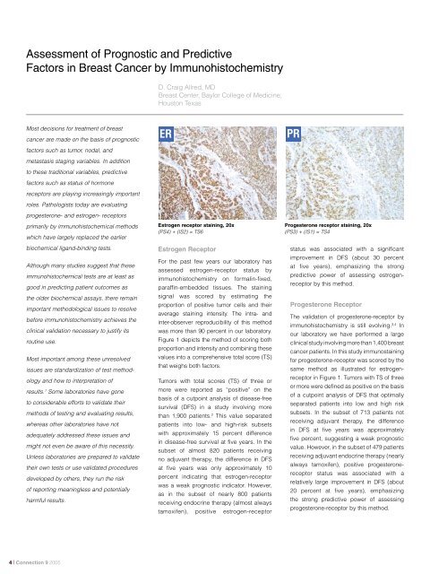
ER<br />
Estrogen receptor sta<strong>in</strong><strong>in</strong>g, 20x<br />
(PS4) + (IS2) = TS6<br />
Estrogen Receptor<br />
For the past few years our laboratory has<br />
assessed estrogen-receptor status by<br />
immunohistochemistry on formal<strong>in</strong>-fixed,<br />
paraff<strong>in</strong>-embedded tissues. The sta<strong>in</strong><strong>in</strong>g<br />
signal was scored by estimat<strong>in</strong>g the<br />
proportion <strong>of</strong> positive tumor cells <strong>and</strong> their<br />
average sta<strong>in</strong><strong>in</strong>g <strong>in</strong>tensity. The <strong>in</strong>tra- <strong>and</strong><br />
<strong>in</strong>ter-observer reproducibility <strong>of</strong> this method<br />
was more than 90 percent <strong>in</strong> our laboratory.<br />
Figure 1 depicts the method <strong>of</strong> scor<strong>in</strong>g both<br />
proportion <strong>and</strong> <strong>in</strong>tensity <strong>and</strong> comb<strong>in</strong><strong>in</strong>g these<br />
values <strong>in</strong>to a comprehensive total score (TS)<br />
that weighs both factors.<br />
Tumors with total scores (TS) <strong>of</strong> three or<br />
more were reported as “positive” on the<br />
basis <strong>of</strong> a cutpo<strong>in</strong>t analysis <strong>of</strong> disease-free<br />
survival (DFS) <strong>in</strong> a study <strong>in</strong>volv<strong>in</strong>g more<br />
than 1,900 patients. 2 This value separated<br />
patients <strong>in</strong>to low- <strong>and</strong> high-risk subsets<br />
with approximately 15 percent difference<br />
<strong>in</strong> disease-free survival at five years. In the<br />
subset <strong>of</strong> almost 820 patients receiv<strong>in</strong>g<br />
no adjuvant therapy, the difference <strong>in</strong> DFS<br />
at five years was only approximately 10<br />
percent <strong>in</strong>dicat<strong>in</strong>g that estrogen-receptor<br />
was a weak prognostic <strong>in</strong>dicator. However,<br />
as <strong>in</strong> the subset <strong>of</strong> nearly 800 patients<br />
receiv<strong>in</strong>g endocr<strong>in</strong>e therapy (almost always<br />
tamoxifen), positive estrogen-receptor<br />
PR<br />
Progesterone receptor sta<strong>in</strong><strong>in</strong>g, 20x<br />
(PS3) + (IS1) = TS4<br />
status was associated with a significant<br />
improvement <strong>in</strong> DFS (about 30 percent<br />
at five years), emphasiz<strong>in</strong>g the strong<br />
predictive power <strong>of</strong> assess<strong>in</strong>g estrogenreceptor<br />
by this method.<br />
Progesterone Receptor<br />
The validation <strong>of</strong> progesterone-receptor by<br />
immunohistochemistry is still evolv<strong>in</strong>g. 3,4 In<br />
our laboratory we have performed a large<br />
cl<strong>in</strong>ical study <strong>in</strong>volv<strong>in</strong>g more than 1,400 breast<br />
cancer patients. In this study immunosta<strong>in</strong><strong>in</strong>g<br />
for progesterone-receptor was scored by the<br />
same method as illustrated for estrogenreceptor<br />
<strong>in</strong> Figure 1. Tumors with TS <strong>of</strong> three<br />
or more were def<strong>in</strong>ed as positive on the basis<br />
<strong>of</strong> a cutpo<strong>in</strong>t analysis <strong>of</strong> DFS that optimally<br />
separated patients <strong>in</strong>to low <strong>and</strong> high risk<br />
subsets. In the subset <strong>of</strong> 713 patients not<br />
receiv<strong>in</strong>g adjuvant therapy, the difference<br />
<strong>in</strong> DFS at five years was approximately<br />
five percent, suggest<strong>in</strong>g a weak prognostic<br />
value. However, <strong>in</strong> the subset <strong>of</strong> 479 patients<br />
receiv<strong>in</strong>g adjuvant endocr<strong>in</strong>e therapy (nearly<br />
always tamoxifen), positive progesteronereceptor<br />
status was associated with a<br />
relatively large improvement <strong>in</strong> DFS (about<br />
20 percent at five years), emphasiz<strong>in</strong>g<br />
the strong predictive power <strong>of</strong> assess<strong>in</strong>g<br />
progesterone-receptor by this method.<br />
| Connection 9 2005
Allred Scor<strong>in</strong>g Guidel<strong>in</strong>e for ER/PR pharmDx<br />
Proportion<br />
Score<br />
<br />
0 to 1 100<br />
<br />
1 100 to<br />
1 10 <br />
1 10 to<br />
1 3 <br />
1 3 to 2 3<br />
<br />
2 3 to 1<br />
Intensity<br />
Score<br />
<br />
<br />
(TS range 0, 28)<br />
<br />
Figure 1. Scor<strong>in</strong>g Guidel<strong>in</strong>es for Immunohistochemical Sta<strong>in</strong><strong>in</strong>g <strong>of</strong> Estrogen-receptor<br />
Conclusions<br />
There is still no consensus today concern<strong>in</strong>g<br />
methodology for assess<strong>in</strong>g<br />
progesterone- <strong>and</strong> estrogen-receptor<br />
status by immunohistochemistry. Cl<strong>in</strong>ical<br />
laboratories <strong>of</strong>fer<strong>in</strong>g these tests us<strong>in</strong>g their<br />
own <strong>in</strong>-house methods should perform<br />
rigorous validation studies, or should<br />
follow procedures from other laboratories<br />
that have performed such studies. For<br />
laboratories where such validation studies<br />
are impractical, an alternative that is now<br />
available is to use one <strong>of</strong> the FDA-cleared<br />
immunohistochemistry tests that have been<br />
cl<strong>in</strong>ically validated by calibration to patient<br />
outcomes. Almost certa<strong>in</strong>ly a new class <strong>of</strong><br />
immunohistochemistry tests, some <strong>of</strong> which<br />
are still <strong>in</strong> development, will be evaluated<br />
<strong>and</strong> <strong>in</strong>terpreted us<strong>in</strong>g multivariate analysis<br />
to identify a mean<strong>in</strong>gful prognostic <strong>in</strong>dex.<br />
That <strong>in</strong>dex will be more powerful than the<br />
<strong>in</strong>dividual factors <strong>in</strong> identify<strong>in</strong>g patients at<br />
risk for disease recurrence.<br />
Correspondence should be directed to:<br />
D. Craig Allred, MD<br />
<strong>Breast</strong> Center, Baylor College <strong>of</strong> Medic<strong>in</strong>e<br />
dcallred@breastcenter.tmc.edu<br />
K1903<br />
K1904<br />
Related Products<br />
ER/PR pharmDx TM for manual use<br />
ER/PR pharmDx TM for use on <strong>Dako</strong> Autosta<strong>in</strong>er / Autosta<strong>in</strong>er Plus<br />
ER/PR pharmDx TM kits were developed <strong>and</strong> validated for use with<br />
the follow<strong>in</strong>g companion reagents from <strong>Dako</strong>.<br />
Materials required, but not supplied <strong>in</strong>clude:<br />
S3006<br />
S2003<br />
S1699/S1700<br />
Wash Buffer<br />
Dual Endogenous Enzyme Block<br />
Target Retrieval Solution<br />
Calibrated pressure cooker with the capability <strong>of</strong> reach<strong>in</strong>g <strong>and</strong> ma<strong>in</strong>ta<strong>in</strong><strong>in</strong>g<br />
a temperature <strong>of</strong> 125ºC for 5 m<strong>in</strong>utes<br />
References<br />
1. Allred DC, Harvey JM, Berardo M, <strong>and</strong> Clark GM.<br />
<strong>Prognostic</strong> <strong>and</strong> predictive factors <strong>in</strong> breast cancer by<br />
immunohistochemical analysis. Mod Pathol 11: 155-<br />
168, 1998.<br />
2. Clark GM, Harvey JM, Osborne CK, <strong>and</strong> Allred DC.<br />
Estrogen receptor status (ER) determ<strong>in</strong>ed by immunohistochemistry<br />
(IHC) is superior to biochemical lig<strong>and</strong>b<strong>in</strong>d<strong>in</strong>g<br />
(LB) assay for evaluat<strong>in</strong>g breast cancer patients<br />
(abstract). Proc Am Soc Cl<strong>in</strong> Oncol 16:129A, 1997.<br />
3. Berardo M, Clark GM, de Moor C, Osborne CK,<br />
Weig<strong>and</strong> RA, <strong>and</strong> Allred DC. <strong>Prognostic</strong> <strong>and</strong><br />
predictive properties <strong>of</strong> immunohistochemical<br />
progesterone receptors <strong>in</strong> breast cancer (abstract).<br />
Proc Am Soc Cl<strong>in</strong> Oncol 14:110A, 1995.<br />
4. Mohs<strong>in</strong> SK, Weiss H, Havighurst T, Clark GM, Berardo<br />
M, Roanh LD, To TV, Zho Q, Love RR, <strong>and</strong> Allred DC.<br />
Progesterone receptor by immunohistochemistry <strong>and</strong><br />
cl<strong>in</strong>ical outcome <strong>in</strong> breast cancer: a validation study.<br />
Mod Pathol 17: 1545-1554, 2004.<br />
Connection 9 2006 |