Calarco et al 2007 - University of Toronto
Calarco et al 2007 - University of Toronto
Calarco et al 2007 - University of Toronto
- No tags were found...
Create successful ePaper yourself
Turn your PDF publications into a flip-book with our unique Google optimized e-Paper software.
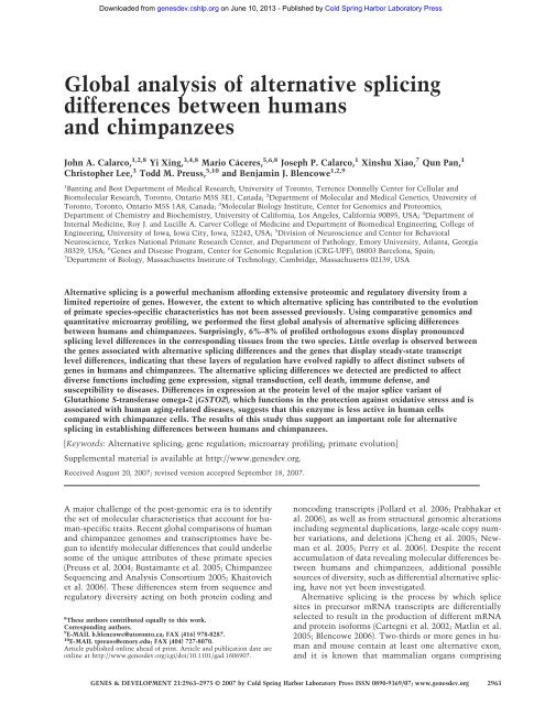
Downloaded from genesdev.cshlp.org on June 10, 2013 - Published by Cold Spring Harbor Laboratory PressGlob<strong>al</strong> an<strong>al</strong>ysis <strong>of</strong> <strong>al</strong>ternative splicingdifferences b<strong>et</strong>ween humansand chimpanzeesJohn A. <strong>C<strong>al</strong>arco</strong>, 1,2,8 Yi Xing, 3,4,8 Mario Cáceres, 5,6,8 Joseph P. <strong>C<strong>al</strong>arco</strong>, 1 Xinshu Xiao, 7 Qun Pan, 1Christopher Lee, 3 Todd M. Preuss, 5,10 and Benjamin J. Blencowe 1,2,91 Banting and Best Department <strong>of</strong> Medic<strong>al</strong> Research, <strong>University</strong> <strong>of</strong> <strong>Toronto</strong>, Terrence Donnelly Center for Cellular andBiomolecular Research, <strong>Toronto</strong>, Ontario M5S 3E1, Canada; 2 Department <strong>of</strong> Molecular and Medic<strong>al</strong> Gen<strong>et</strong>ics, <strong>University</strong> <strong>of</strong><strong>Toronto</strong>, <strong>Toronto</strong>, Ontario M5S 1A8, Canada; 3 Molecular Biology Institute, Center for Genomics and Proteomics,Department <strong>of</strong> Chemistry and Biochemistry, <strong>University</strong> <strong>of</strong> C<strong>al</strong>ifornia, Los Angeles, C<strong>al</strong>ifornia 90095, USA; 4 Department <strong>of</strong>Intern<strong>al</strong> Medicine, Roy J. and Lucille A. Carver College <strong>of</strong> Medicine and Department <strong>of</strong> Biomedic<strong>al</strong> Engineering, College <strong>of</strong>Engineering, <strong>University</strong> <strong>of</strong> Iowa, Iowa City, Iowa, 52242, USA; 5 Division <strong>of</strong> Neuroscience and Center for Behavior<strong>al</strong>Neuroscience, Yerkes Nation<strong>al</strong> Primate Research Center, and Department <strong>of</strong> Pathology, Emory <strong>University</strong>, Atlanta, Georgia30329, USA; 6 Genes and Disease Program, Center for Genomic Regulation (CRG-UPF), 08003 Barcelona, Spain;7 Department <strong>of</strong> Biology, Massachus<strong>et</strong>ts Institute <strong>of</strong> Technology, Cambridge, Massachus<strong>et</strong>ts 02139, USAAlternative splicing is a powerful mechanism affording extensive proteomic and regulatory diversity from <strong>al</strong>imited repertoire <strong>of</strong> genes. However, the extent to which <strong>al</strong>ternative splicing has contributed to the evolution<strong>of</strong> primate species-specific characteristics has not been assessed previously. Using comparative genomics andquantitative microarray pr<strong>of</strong>iling, we performed the first glob<strong>al</strong> an<strong>al</strong>ysis <strong>of</strong> <strong>al</strong>ternative splicing differencesb<strong>et</strong>ween humans and chimpanzees. Surprisingly, 6%–8% <strong>of</strong> pr<strong>of</strong>iled orthologous exons display pronouncedsplicing level differences in the corresponding tissues from the two species. Little overlap is observed b<strong>et</strong>weenthe genes associated with <strong>al</strong>ternative splicing differences and the genes that display steady-state transcriptlevel differences, indicating that these layers <strong>of</strong> regulation have evolved rapidly to affect distinct subs<strong>et</strong>s <strong>of</strong>genes in humans and chimpanzees. The <strong>al</strong>ternative splicing differences we d<strong>et</strong>ected are predicted to affectdiverse functions including gene expression, sign<strong>al</strong> transduction, cell death, immune defense, andsusceptibility to diseases. Differences in expression at the protein level <strong>of</strong> the major splice variant <strong>of</strong>Glutathione S-transferase omega-2 (GSTO2), which functions in the protection against oxidative stress and isassociated with human aging-related diseases, suggests that this enzyme is less active in human cellscompared with chimpanzee cells. The results <strong>of</strong> this study thus support an important role for <strong>al</strong>ternativesplicing in establishing differences b<strong>et</strong>ween humans and chimpanzees.[Keywords: Alternative splicing; gene regulation; microarray pr<strong>of</strong>iling; primate evolution]Supplement<strong>al</strong> materi<strong>al</strong> is available at http://www.genesdev.org.Received August 20, <strong>2007</strong>; revised version accepted September 18, <strong>2007</strong>.A major ch<strong>al</strong>lenge <strong>of</strong> the post-genomic era is to identifythe s<strong>et</strong> <strong>of</strong> molecular characteristics that account for human-specifictraits. Recent glob<strong>al</strong> comparisons <strong>of</strong> humanand chimpanzee genomes and transcriptomes have begunto identify molecular differences that could underliesome <strong>of</strong> the unique attributes <strong>of</strong> these primate species(Preuss <strong>et</strong> <strong>al</strong>. 2004; Bustamante <strong>et</strong> <strong>al</strong>. 2005; ChimpanzeeSequencing and An<strong>al</strong>ysis Consortium 2005; Khaitovich<strong>et</strong> <strong>al</strong>. 2006). These differences stem from sequence andregulatory diversity acting on both protein coding and8 These authors contributed equ<strong>al</strong>ly to this work.Corresponding authors.9 E-MAIL b.blencowe@utoronto.ca; FAX (416) 978-8287.10 E-MAIL tpreuss@emory.edu; FAX (404) 727-8070.Article published online ahead <strong>of</strong> print. Article and publication date areonline at http://www.genesdev.org/cgi/doi/10.1101/gad.1606907.noncoding transcripts (Pollard <strong>et</strong> <strong>al</strong>. 2006; Prabhakar <strong>et</strong><strong>al</strong>. 2006), as well as from structur<strong>al</strong> genomic <strong>al</strong>terationsincluding segment<strong>al</strong> duplications, large-sc<strong>al</strong>e copy numbervariations, and del<strong>et</strong>ions (Cheng <strong>et</strong> <strong>al</strong>. 2005; Newman<strong>et</strong> <strong>al</strong>. 2005; Perry <strong>et</strong> <strong>al</strong>. 2006). Despite the recentaccumulation <strong>of</strong> data reve<strong>al</strong>ing molecular differences b<strong>et</strong>weenhumans and chimpanzees, addition<strong>al</strong> possiblesources <strong>of</strong> diversity, such as differenti<strong>al</strong> <strong>al</strong>ternative splicing,have not y<strong>et</strong> been investigated.Alternative splicing is the process by which splicesites in precursor mRNA transcripts are differenti<strong>al</strong>lyselected to result in the production <strong>of</strong> different mRNAand protein is<strong>of</strong>orms (Cartegni <strong>et</strong> <strong>al</strong>. 2002; Matlin <strong>et</strong> <strong>al</strong>.2005; Blencowe 2006). Two-thirds or more genes in humanand mouse contain at least one <strong>al</strong>ternative exon,and it is known that mamm<strong>al</strong>ian organs comprisingGENES & DEVELOPMENT 21:2963–2975 © <strong>2007</strong> by Cold Spring Harbor Laboratory Press ISSN 0890-9369/07; www.genesdev.org 2963
Downloaded from genesdev.cshlp.org on June 10, 2013 - Published by Cold Spring Harbor Laboratory Press<strong>C<strong>al</strong>arco</strong> <strong>et</strong> <strong>al</strong>.many speci<strong>al</strong>ized cell types such as the brain are associatedwith relatively complex <strong>al</strong>ternative splicing patterns(Xu <strong>et</strong> <strong>al</strong>. 2002; Johnson <strong>et</strong> <strong>al</strong>. 2003; Yeo <strong>et</strong> <strong>al</strong>.2004a). Surprisingly, comparisons <strong>of</strong> human and mous<strong>et</strong>ranscript sequences have reve<strong>al</strong>ed that 5% substitution rates were an<strong>al</strong>yzed for <strong>al</strong>ternativesplicing differences by RT–PCR assays using poly(A) +RNA from human and chimpanzee tissues (see main text ford<strong>et</strong>ails). (Right panel) In the quantitative <strong>al</strong>ternative splicingmicroarray pr<strong>of</strong>iling strategy, labeled cDNA from the samepoly(A) + RNA samples were hybridized to an <strong>al</strong>ternative splicingmicroarray designed to monitor inclusion levels <strong>of</strong> cass<strong>et</strong>t<strong>et</strong>ype<strong>al</strong>ternative exons. Each <strong>al</strong>ternative splicing event is monitoredby a s<strong>et</strong> <strong>of</strong> six oligonucleotide probes (black horizont<strong>al</strong>lines) (Pan <strong>et</strong> <strong>al</strong>. 2004). Predictions for <strong>al</strong>ternative splicing differencesb<strong>et</strong>ween the corresponding human and chimpanzee tissueswere v<strong>al</strong>idated by RT–PCR assays (see Table 1; Figs. 4, 5;Supplementary Fig. 1).2964 GENES & DEVELOPMENT
Downloaded from genesdev.cshlp.org on June 10, 2013 - Published by Cold Spring Harbor Laboratory PressSurveying primate <strong>al</strong>ternative splicing<strong>et</strong> <strong>al</strong>. 2003; Yeo <strong>et</strong> <strong>al</strong>. 2004b, <strong>2007</strong>; for reviews, see Matlin<strong>et</strong> <strong>al</strong>. 2005; Blencowe 2006; Chasin <strong>2007</strong>). Consequently,we expected that frequent nucleotide changes in exonsand the flanking intron regions would be predictive <strong>of</strong>splicing differences b<strong>et</strong>ween humans and chimpanzees.Role <strong>of</strong> nucleotide substitutions in generatingdifferences b<strong>et</strong>ween human and chimpanzeesplicing patternsHigh substitution rates were rarely found in the <strong>al</strong>ignedorthologous exons and flanking intron sequences: Ofthese regions, 0.3% displayed substitution rates <strong>of</strong> >5%,and substitution rates >10% were found in 5% substitution rates are predictive <strong>of</strong> splicingdifferences, semiquantitative RT–PCR assays wereperformed using primers specific for neighboring exonsequences and poly(A) + RNA samples from human orchimpanzee front<strong>al</strong> cortex and heart tissues. These tissueRNA samples were pooled, in each case, from sever<strong>al</strong>adult individu<strong>al</strong>s (Supplementary Table 2). Of 31 regionswith >5% substitution rates selected for an<strong>al</strong>ysis, 14contained exons that displayed evidence <strong>of</strong> <strong>al</strong>ternativesplicing in humans from an<strong>al</strong>ysis <strong>of</strong> the correspondingEST/cDNA (complementary DNA) sequences. Five <strong>of</strong>the 31 regions displayed splicing level differences b<strong>et</strong>weenhumans and chimpanzees in at least one <strong>of</strong> th<strong>et</strong>wo tissues (see below). Interestingly, <strong>al</strong>l five <strong>of</strong> theseis<strong>of</strong>orm ratio differences involved exons included amongthe 14 <strong>al</strong>ternative exons identified in the test s<strong>et</strong>. At thisv<strong>al</strong>idation rate, 25%in differenceb<strong>et</strong>ween the human and chimpanzee front<strong>al</strong> cortex and/or heart. Of these events, 30 (81%) were v<strong>al</strong>idated ashaving the expected differenti<strong>al</strong> splicing patterns (Figs.4, 5, below; Table 1; addition<strong>al</strong> data not shown). Thus,GENES & DEVELOPMENT 2965
Downloaded from genesdev.cshlp.org on June 10, 2013 - Published by Cold Spring Harbor Laboratory Press<strong>C<strong>al</strong>arco</strong> <strong>et</strong> <strong>al</strong>.Figure 2. Quantitative <strong>al</strong>ternative splicing microarray pr<strong>of</strong>ilingreve<strong>al</strong>s the extent <strong>of</strong> <strong>al</strong>ternative splicing differences b<strong>et</strong>weenhuman and chimpanzee orthologous exons, as well as the degree<strong>of</strong> divergence b<strong>et</strong>ween splicing patterns over different evolutionarytime periods. (A) Color spectrum plots indicating thenumber and magnitude <strong>of</strong> <strong>al</strong>ternative splicing differences b<strong>et</strong>weenhuman and chimpanzee front<strong>al</strong> cortex and heart tissues.The Y-axes indicate the number <strong>of</strong> <strong>al</strong>ternative splicing eventspr<strong>of</strong>iled; these are sorted according to the magnitude <strong>of</strong> the absolutev<strong>al</strong>ue <strong>of</strong> the percent exon inclusion level (%in) differenceb<strong>et</strong>ween the human and chimpanzee tissue being compared.The magnitude <strong>of</strong> the percentage inclusion difference is indicatedby the color sc<strong>al</strong>e on the right. (B) Cumulative distributionplot displaying the distribution <strong>of</strong> percentage inclusion differenceswhen comparing microarray data for 217 conserved<strong>al</strong>ternative splicing events b<strong>et</strong>ween the following pairs <strong>of</strong> tissues:human and chimpanzee front<strong>al</strong> cortex (blue line), humanand chimpanzee heart (red line), human front<strong>al</strong> cortex andmouse cortex (green line), and human and mouse heart (purpleline). (C) Spearman correlation coefficients are shown for pairwisecomparisons b<strong>et</strong>ween <strong>al</strong>ternative splicing levels (blacknumbers) and transcript levels (purple numbers) for the s<strong>et</strong> <strong>of</strong>217 orthologous genes an<strong>al</strong>yzed in human, chimpanzee, andmouse tissues in B. Double arrows indicate the pairs <strong>of</strong> speciescompared. (Hs) Homo sapiens; (Pt) Pan troglodytes; (Mm) Musmusculus.despite the remarkable degree <strong>of</strong> conservation b<strong>et</strong>weenthe coding regions <strong>of</strong> the human and chimpanzee genomes,a substanti<strong>al</strong> number <strong>of</strong> <strong>al</strong>ternative exons thatare not associated with high substitution rates in the<strong>al</strong>ternative exons and flanking intron regions displaypronounced splicing level differences b<strong>et</strong>ween the twospecies.Evidence for stabilizing selection pressure acting topreserve the majority <strong>of</strong> splicing levels <strong>of</strong> orthologoushuman, chimpanzee, and mouse <strong>al</strong>ternative exonsThe similarity b<strong>et</strong>ween the splicing levels for the majority<strong>of</strong> the pr<strong>of</strong>iled orthologous exons could reflect stabilizingselection pressure acting to conserve the inclusionlevels <strong>of</strong> most human and chimpanzee orthologous exons.However, it is <strong>al</strong>so possible that insufficient evolutionarytime has accumulated to result in a higher incidence<strong>of</strong> pronounced inclusion level differences than observedabove. To investigate these possibilities, wecompared the extent <strong>of</strong> divergence b<strong>et</strong>ween <strong>al</strong>ternativesplicing pr<strong>of</strong>iles representing a longer evolutionary timespan, namely the 80- to 90-million-year period separatingthe common ancestor <strong>of</strong> human or chimpanzee andmouse. Percent inclusion v<strong>al</strong>ues for 217 <strong>al</strong>ternativesplicing events conserved b<strong>et</strong>ween humans and themouse were obtained from RT–PCR-v<strong>al</strong>idated, quantitative<strong>al</strong>ternative splicing microarray pr<strong>of</strong>iling data frommouse heart and brain cortex tissues (Fagnani <strong>et</strong> <strong>al</strong>. <strong>2007</strong>)and compared with percent inclusion v<strong>al</strong>ues from thehuman and chimpanzee datas<strong>et</strong>s described above (Fig.2B,C; see the Supplement<strong>al</strong> Materi<strong>al</strong>).Notably, <strong>al</strong>though the splicing levels <strong>of</strong> the mouse exonsare <strong>al</strong>so very similar to the orthologous exons in thecorresponding human and chimpanzee tissues, they havediverged to a significantly greater extent in both tissues(P
Downloaded from genesdev.cshlp.org on June 10, 2013 - Published by Cold Spring Harbor Laboratory PressSurveying primate <strong>al</strong>ternative splicingTable 1. Genes associated with different cellular functions with experiment<strong>al</strong>ly v<strong>al</strong>idated splicing level differences b<strong>et</strong>weenhumans and chimpanzeesFunction<strong>al</strong> category Gene name GO processes/known featuresAlternativesplicing-<strong>al</strong>teredprotein domain(s)Sign<strong>al</strong>ingInositol hexaphosphate kinaseInositol trisphosphate-3 kinaseIHPK2 (IHPK2)activityHypoth<strong>et</strong>ic<strong>al</strong> protein LOC220686 Phoshpatidylinositol-4 kinase activity PI4KADP-ribosylation factor-like 3 (ARL3) G-protein-coupled receptor protein GTPasesign<strong>al</strong>ing pathwayGene regulation TBP-associated factor 6 (TAF6) TFIID complex, regulator <strong>of</strong>NotranscriptionSRp40 (SFRS5) Splicing regulation RRM, RSPoly(A)-binding protein cytoplasmic 4 RNA binding, poly(A) bindingND(PABPC4)Immune response Toll/Interleukin-1 receptor 8 (TIR8) Immune response NDHerpesvirus entry mediator (HVEM/ Immune responseTM, cytoplasmicTNFRSF14)OtherGlutathione S-transferase omega Glutathione transferase activity, ND2 (GSTO2)m<strong>et</strong>abolism <strong>of</strong> xenobioticsRoundabout 1 homolog (ROBO1) Axon guidance, dyslexia candidate NDgeneFSHD region gene 1 (FRG1)Facioscapulohumor<strong>al</strong> diseaseActin cross-linkingcandidate-Adducin (ADD3)C<strong>al</strong>modulin binding, structur<strong>al</strong>Spectrin bindingconstituent <strong>of</strong> cytoskel<strong>et</strong>onZonula Occludens protein (ZO-1) Tight junction protein NDBridging Integrator 1 (BIN1) Regulation <strong>of</strong> endocytosis BARC<strong>al</strong>bindin 2 (CALB2) C<strong>al</strong>cium ion binding EF handChr. 14 ORF 153 Uncharacterized NDUnc-84 homolog (UNC84B)Microtubule binding, mitotic spindleorganization, and biogenesisNDFunction<strong>al</strong> annotations from the Gene Ontology database or from literature searches are listed. Protein domains predicted to be <strong>al</strong>teredby differenti<strong>al</strong> <strong>al</strong>ternative splicing are <strong>al</strong>so listed. In most cases, differenti<strong>al</strong> <strong>al</strong>ternative splicing was d<strong>et</strong>ected in both front<strong>al</strong> cortex andheart; exceptions are in IHPK2, front<strong>al</strong> cortex only; BIN1, front<strong>al</strong> cortex only; CALB2, heart only; and UNC84B, heart only. (IHPK)Inositol hexaphosphate kinase domain; (PI4K) phosphatidylinositol-4 kinase domain; (GTPase) Ras-like GTPase domain; (RRM) RNArecognition motif; (RS), arginine/serine-rich domain; (TM) transmembrane domain; (Cytoplasmic) cytoplasmic sign<strong>al</strong>ing domain;(Actin cross-linking) actin cross-linking protein fold; (Spectrin binding) C-termin<strong>al</strong> region <strong>of</strong> DD3 proxim<strong>al</strong> to a defined spectrininteractingdomain; (BAR) BIN/amphiphysin/Rvs domain involved in endocytosis; (EF hand) c<strong>al</strong>cium-binding domain; (ND) no knowndomain is affected by <strong>al</strong>ternative splicing.the microarray pr<strong>of</strong>iled genes. A comparable proportion<strong>of</strong> orthologous genes has been found to display at leasttw<strong>of</strong>old transcript level differences b<strong>et</strong>ween correspondinghuman and chimpanzee tissues (Preuss <strong>et</strong> <strong>al</strong>. 2004;Khaitovich <strong>et</strong> <strong>al</strong>. 2005). Remarkably, however, despite apar<strong>al</strong>lel divergence in <strong>al</strong>ternative splicing and transcription<strong>al</strong>patterns during the evolution <strong>of</strong> humans andchimpanzees, primarily nonoverlapping subs<strong>et</strong>s <strong>of</strong> genesare associated with these changes (Fig. 3). By sortinggenes according to the magnitude <strong>of</strong> the microarray-d<strong>et</strong>ectedpercent inclusion <strong>al</strong>ternative splicing level differences(Fig. 3, columns 1 and 3), it is evident that relativelyfew genes display coincident changes in transcription<strong>al</strong>levels (Fig. 3 in cf. columns 1 and 2, and columns3 and 4). Among the 160–240 splicing events with predictedpercent inclusion differences >20% in front<strong>al</strong> cortexand/or heart tissues, only 25% are predicted to <strong>al</strong>sohave tw<strong>of</strong>old or greater steady-state transcript levelchanges. This observation extends previous results indicatingthat predominantly different subs<strong>et</strong>s <strong>of</strong> genes areregulated in a cell-specific/tissue-specific or activity-dependentmanner at the transcription<strong>al</strong> and <strong>al</strong>ternativesplicing levels (Le <strong>et</strong> <strong>al</strong>. 2004; Pan <strong>et</strong> <strong>al</strong>. 2004; Li <strong>et</strong> <strong>al</strong>.2006; Fagnani <strong>et</strong> <strong>al</strong>. <strong>2007</strong>; Ip <strong>et</strong> <strong>al</strong>. <strong>2007</strong>). In particular, thepresent results show that these two layers <strong>of</strong> regulationhave evolved rapidly to affect different subs<strong>et</strong>s <strong>of</strong> genes,and indicate that <strong>al</strong>ternative splicing has served as anaddition<strong>al</strong> mechanism for diversifying gene regulationduring the 5–7 million years <strong>of</strong> evolution separating humansand chimpanzees.Alternative splicing differences b<strong>et</strong>ween humans andchimpanzees affect transcripts from genes associatedwith diverse cellular functionsGenes with <strong>al</strong>ternative splicing differences b<strong>et</strong>ween humansand chimpanzees d<strong>et</strong>ected using the m<strong>et</strong>hods describedabove, and confirmed by the RT–PCR assays, areassociated with diverse biologic<strong>al</strong> processes includinggene expression, sign<strong>al</strong>ing pathways, immune defense,GENES & DEVELOPMENT 2967
Downloaded from genesdev.cshlp.org on June 10, 2013 - Published by Cold Spring Harbor Laboratory Press<strong>C<strong>al</strong>arco</strong> <strong>et</strong> <strong>al</strong>.Figure 3. Transcript and AS level differences b<strong>et</strong>ween humansand chimpanzees involve largely distinct subs<strong>et</strong>s <strong>of</strong> genes. Thecolor spectrum plots compare splicing level differences andtranscript level differences for the same s<strong>et</strong> <strong>of</strong> genes expressed inthe front<strong>al</strong> cortex and heart. For each tissue comparison, the leftcolumn shows splicing differences (measured as the magnitude<strong>of</strong> percent inclusion difference, columns 1 and 3) and the rightcolumn measures differences in gene transcript level (measuredas the magnitude <strong>of</strong> the hyperbolic arcsine [arcsinh] difference,columns 2 and 4). An hyperbolic arcsine difference <strong>of</strong> ∼0.4 correspondsto a 1.5-fold change in expression level, and a difference<strong>of</strong> ∼0.7 corresponds to a tw<strong>of</strong>old difference in expressionlevel.Alternative splicing differences are consistent b<strong>et</strong>weenhuman and chimpanzee individu<strong>al</strong>sSince the an<strong>al</strong>yses described above employed samples <strong>of</strong>poly(A) + mRNA pooled, in each case, from sever<strong>al</strong> humanindividu<strong>al</strong>s and from sever<strong>al</strong> chimpanzee individu<strong>al</strong>s(Supplementary Table 2), it was important to assessthe extent to which the <strong>al</strong>ternative splicing differencesmight be explained by variations b<strong>et</strong>ween individu<strong>al</strong>sfrom each species. Individu<strong>al</strong> samples comprising thepools <strong>of</strong> tissue mRNAs were therefore an<strong>al</strong>yzed separatelyfor splicing level differences. In each case examined,similar <strong>al</strong>ternative splicing differences were observedb<strong>et</strong>ween the sever<strong>al</strong> human and chimpanzee individu<strong>al</strong>s(Fig. 5B; data not shown). These data indicat<strong>et</strong>hat it is highly unlikely that the over<strong>al</strong>l differences insplicing levels measured using the pooled samples areattributed to differences associated with individu<strong>al</strong>variation within each species. This conclusion was furthersupported by an an<strong>al</strong>ysis <strong>of</strong> <strong>al</strong>ternative splicing leveldifferences in primary fibroblasts and lymphoblastoidcells, <strong>al</strong>so from multiple addition<strong>al</strong> individu<strong>al</strong>s fromeach species (Supplementary Fig. 1). Again, in <strong>al</strong>l casesexamined, similar species-associated splicing level differenceswere d<strong>et</strong>ected b<strong>et</strong>ween the different individu<strong>al</strong>sas d<strong>et</strong>ected in the tissue samples (Supplementary Fig. 1;see below). The results from this last experiment furtherand disease (Table 1). In most cases the <strong>al</strong>ternative splicingdifferences were observed in both front<strong>al</strong> cortex andheart tissues (Figs. 4, 5) as well as in cell lines fromsimilar origins from both species (Supplementary Fig. 1;see below). An example <strong>of</strong> an <strong>al</strong>ternative splicing differenced<strong>et</strong>ected using the comparative genomics approachinvolves transcripts encoding the TATA-box-bindingprotein-associated factor 6 (TAF6) (Fig. 4; SupplementaryFig. 1; see Table 1 and below for addition<strong>al</strong> examples andinformation). Examples <strong>of</strong> <strong>al</strong>ternative splicing differencesd<strong>et</strong>ected in the microarray data affect transcriptsencoding the herpesvirus entry mediator receptor(HVEM/TNFRSF14), the spectrin-binding protein -Adducin(ADD3), ADP-ribosylation factor-like 3 (ARL3),serine/arginine (SR)-repeat family protein splicing factor<strong>of</strong> 40 kDa (SRp40/SFRS5), and GSTO2 (Figs. 4, 5; seeTable 1 and below for addition<strong>al</strong> examples and information).To assess wh<strong>et</strong>her these observed differences arespecific<strong>al</strong>ly associated with the human or chimpanzeelineage, RT–PCR assays were <strong>al</strong>so performed using RNAextracted from front<strong>al</strong> cortex and heart tissue from rhesusmacaques, an Old World monkey, as an outgroupcomparison. The results <strong>of</strong> this an<strong>al</strong>ysis are summarizedbelow and in Supplementary Table 3, and representativeexamples are shown in Figure 5A.Figure 4. Examples <strong>of</strong> <strong>al</strong>ternative splicing differences b<strong>et</strong>weenhumans and chimpanzees confirmed by semiquantitative RT–PCR and sequencing. RT–PCR assays were performed usingprimers specific for sequences in constitutive exons flankingthe <strong>al</strong>ternative exons predicted to be differenti<strong>al</strong>ly spliced by thecomparative genomic or <strong>al</strong>ternative splicing microarray pr<strong>of</strong>ilingan<strong>al</strong>yses. Corresponding tissues from human (Hs) and chimpanzee(Pt) are indicated. Major splice is<strong>of</strong>orms that include andskip <strong>al</strong>ternative exons (black boxes) are indicated on the right <strong>of</strong>each panel. Diagrams below each gel illustrate the predictedconsequence <strong>of</strong> <strong>al</strong>ternative splicing changes at the protein and/or transcript levels for TAF6, ADD3, the SR-repeat family proteinsplicing factor <strong>of</strong> 40 kDa (SFRS5/SRp40) and GSTO2 (referto Table 1 and main text for d<strong>et</strong>ails). Protein domains are labeledas follows: (H) head domain; (N) neck domain; (C) C-termin<strong>al</strong> tail region known to interact with spectrin; (RRM1)RNA recognition motif 1; (RRM2) RNA recognition motif 2;(RS) arginine/serine-rich domain; (GST) glutathione S-transferasedomain. The stop sign indicates the insertion <strong>of</strong> a PTC. Theasterisk indicates an addition<strong>al</strong> TAF6 splice is<strong>of</strong>orm d<strong>et</strong>ected inhuman but not chimpanzee tissue, as confirmed by sequencing.2968 GENES & DEVELOPMENT
Downloaded from genesdev.cshlp.org on June 10, 2013 - Published by Cold Spring Harbor Laboratory PressSurveying primate <strong>al</strong>ternative splicingFigure 5. Comparisons <strong>of</strong> AS levels in RNA samplesfrom macaque, human, and chimpanzee individu<strong>al</strong>s. (A)RT–PCR assays were performed using rhesus macaqu<strong>et</strong>ot<strong>al</strong> RNA from front<strong>al</strong> cortex and heart samples from anumber <strong>of</strong> different individu<strong>al</strong>s. RT–PCR primers weredesigned to anne<strong>al</strong> to the exons neighboring each <strong>al</strong>ternativeexon, resulting in the amplification <strong>of</strong> two products(the is<strong>of</strong>orms including and skipping the <strong>al</strong>ternativeexon, as indicated in each <strong>of</strong> the panels). Asterisksdenote novel is<strong>of</strong>orms observed primarily in one speciesbut not the other. The transcripts shown correspond tothe ADD3 (top panel), GSTO2 (second panel), HVEM/TNFRS14 (third panel), and ARL3 (bottom panel) genes.The macaque is used as an outgroup to define the ancestr<strong>al</strong>splicing pattern for each gene. Examples <strong>of</strong>chimpanzee lineage-specific splicing differences (ADD3and ARL3 transcripts) and human lineage-specific splicingdifferences (GSTO2 and TNFRSF14) are shown (see<strong>al</strong>so Supplementary Table 3 for addition<strong>al</strong> examples).(B) RT–PCR experiments were performed using tot<strong>al</strong>RNA from each <strong>of</strong> the individu<strong>al</strong> samples that werepooled for an<strong>al</strong>ysis in the comparative genomic and microarrayexperiments. The same <strong>al</strong>ternative splicingevents displayed for macaque experiments in A are <strong>al</strong>soshown in B for comparison. The labels for each gel lanerepresent the macaque, human, and chimpanzee individu<strong>al</strong>slisted in Supplementary Table 2.indicate that the majority <strong>of</strong> the splicing level differenceswe observed are unlikely related to possible contributionsfrom environment<strong>al</strong> or physiologic<strong>al</strong> differences,such as di<strong>et</strong> or stress, since the corresponding celllines from both species were cultured in par<strong>al</strong>lel for sever<strong>al</strong>weeks under identic<strong>al</strong> growth conditions, prior toharvesting.Examples <strong>of</strong> v<strong>al</strong>idated human and chimpanzee<strong>al</strong>ternative splicing differencesThe <strong>al</strong>ternative splicing difference d<strong>et</strong>ected in TAF6transcripts is human lineage specific and is pronouncedin both the front<strong>al</strong> cortex and heart (Fig. 4). TAF6 is asubunit <strong>of</strong> the gener<strong>al</strong> transcription factor TFIID, whichis involved in gene activation (Sauer <strong>et</strong> <strong>al</strong>. 1995). It hasbeen reported that TAF6 is<strong>of</strong>orms are associated with thecontrol <strong>of</strong> apoptosis and cell cycle arrest (Wang <strong>et</strong> <strong>al</strong>.1997, 2004; Bell <strong>et</strong> <strong>al</strong>. 2001). The <strong>al</strong>ternative splicing differencefound b<strong>et</strong>ween humans and chimpanzees lies inthe 5 untranslated region (UTR), and could thereforeaffect TAF6 regulation at the post-transcription<strong>al</strong> ortranslation<strong>al</strong> levels. Consistent with this propos<strong>al</strong> areprevious observations <strong>of</strong> <strong>al</strong>ternative splicing events in 5UTRs that affect translation<strong>al</strong> efficiency (Wang <strong>et</strong> <strong>al</strong>.1999; Singh <strong>et</strong> <strong>al</strong>. 2005). Such a difference impactingTAF6 expression could, in turn, account for some <strong>of</strong> thedifferences in transcription<strong>al</strong> pr<strong>of</strong>iles that have been observedb<strong>et</strong>ween humans and chimpanzees (Preuss <strong>et</strong> <strong>al</strong>.2004; Khaitovich <strong>et</strong> <strong>al</strong>. 2006).The variation in <strong>al</strong>ternative splicing <strong>of</strong> SRp40, whichis <strong>al</strong>so human lineage specific, is intriguing in light <strong>of</strong>the known roles <strong>of</strong> SR family members in both constitutiveand regulated splicing (Graveley 2000; Sanford <strong>et</strong><strong>al</strong>. 2005; Lin and Fu <strong>2007</strong>). These proteins gener<strong>al</strong>ly functionin splicing by binding to exonic enhancer sequencesvia their RNA recognition motifs (RRMs) (Fig. 4). The<strong>al</strong>ternative splicing difference in SRp40 transcripts resultsin the increased inclusion <strong>of</strong> a highly conservedpremature termination codon (PTC)-containing exon (locatedb<strong>et</strong>ween exons 4 and 5) in human transcripts, inboth front<strong>al</strong> cortex and heart tissue, and correspondinglyreduced levels <strong>of</strong> the shorter transcript encoding the fulllengthprotein, particularly in the heart. Sever<strong>al</strong> RNAbindingproteins including SR family members havebeen shown previously to regulate their own expressionlevels by activating the splicing <strong>of</strong> PTC-containing is<strong>of</strong>ormsthat are subsequently targ<strong>et</strong>ed by the process <strong>of</strong>nonsense-mediated mRNA decay (Lareau <strong>et</strong> <strong>al</strong>. <strong>2007</strong>; Ni<strong>et</strong> <strong>al</strong>. <strong>2007</strong> and references within). Individu<strong>al</strong> SR familymembers are essenti<strong>al</strong> and tightly regulated proteins(Graveley 2000; Sanford <strong>et</strong> <strong>al</strong>. 2005; Lin and Fu <strong>2007</strong>). Itis therefore possible that the differenti<strong>al</strong> expression <strong>of</strong>the two SRp40 is<strong>of</strong>orms described above could be relatedto some <strong>of</strong> the splicing differences we observed b<strong>et</strong>weenhumans and chimpanzees, including those that are notassociated with nucleotide substitutions.The <strong>al</strong>ternative splicing difference affecting GSTO2transcripts results in pronounced skipping <strong>of</strong> exon 4 <strong>of</strong>GENES & DEVELOPMENT 2969
Downloaded from genesdev.cshlp.org on June 10, 2013 - Published by Cold Spring Harbor Laboratory Press<strong>C<strong>al</strong>arco</strong> <strong>et</strong> <strong>al</strong>.this gene, specific<strong>al</strong>ly in human front<strong>al</strong> cortex tissue(Fig. 3; Supplementary Table 3). GSTO2 and its closelyrelated par<strong>al</strong>og GSTO1 are important enzymes in thebiotransformation <strong>of</strong> endogenous and exogenous compoundsand in protection against oxidative stress(Schmuck <strong>et</strong> <strong>al</strong>. 2005), which plays an important role indefense against aging-associated diseases including cancersand neurodegeneration. In this regard, it is interestingto note that in some studies single nucleotide polymorphismsin GSTO2 and GSTO1 have been tentativelylinked to certain human cancers and the age at ons<strong>et</strong> <strong>of</strong>famili<strong>al</strong> Alzheimer’s and Parkinson’s disease (Li <strong>et</strong> <strong>al</strong>.2003; Whitbread <strong>et</strong> <strong>al</strong>. 2005; Pongstaporn <strong>et</strong> <strong>al</strong>. 2006; seeDiscussion).An important question raised by the present and previousstudies on <strong>al</strong>ternative splicing is the extent towhich differences in splice variant levels d<strong>et</strong>ected at th<strong>et</strong>ranscript level correspond to actu<strong>al</strong> differences at theprotein level. It should be borne in mind that comparisons<strong>of</strong> datas<strong>et</strong>s from large-sc<strong>al</strong>e protein mass spectrom<strong>et</strong>ryan<strong>al</strong>yses and from microarray pr<strong>of</strong>iling <strong>of</strong> mRNAfrom the same mamm<strong>al</strong>ian tissues have reve<strong>al</strong>ed goodover<strong>al</strong>l correlations b<strong>et</strong>ween steady-state protein and transcriptabundance (Kislinger <strong>et</strong> <strong>al</strong>. 2006). To investigate thisquestion in the context <strong>of</strong> splice variants that differ b<strong>et</strong>weenhumans and chimpanzees, we next asked wh<strong>et</strong>herthe <strong>al</strong>ternative splicing level difference involving skipping<strong>of</strong> the frame-preserving exon 4 <strong>of</strong> GSTO2 transcriptscould result in species-specific differences in theexpression <strong>of</strong> GSTO2 variants at the protein level (Fig. 6).Expression <strong>of</strong> the function<strong>al</strong> splice variant <strong>of</strong> GSTO2differs b<strong>et</strong>ween humans and chimpanzees at theprotein levelTo initi<strong>al</strong>ly assess the stability <strong>of</strong> the GSTO2 proteinvariants, expression vectors containing c-myc epitop<strong>et</strong>aggedGSTO2 cDNAs, with and without exon 4, wer<strong>et</strong>ransiently expressed in HeLa cells. RT–PCR experimentsusing primers specific to constitutive exon sequences5 and 3 to exon 4 indicated that comparablelevels <strong>of</strong> the two splice variant transcripts are produced(Fig. 6A). Surprisingly, however, Western blotting <strong>of</strong> theprotein lysates from the transfected cells with an antimycepitope antibody reve<strong>al</strong>ed that only the GSTO2variant containing exon 4 results in significant proteinexpression (Fig. 6B,C); minor levels <strong>of</strong> the GSTO2 variantlacking exon 4 could be d<strong>et</strong>ected, but only after prolongedexposure <strong>of</strong> the blot (Fig. 6C; data not shown).These results indicate that GSTO2 splice variants lackingexon 4 are unstable at the protein level, and thatlevels <strong>of</strong> active GSTO2 are therefore likely d<strong>et</strong>erminedby the relative expression <strong>of</strong> the splice variant containingexon 4. Also in support <strong>of</strong> this propos<strong>al</strong> are recentfindings indicating that residues overlapping exon 4 <strong>of</strong>GSTO2 are important for both the stability and activity<strong>of</strong> this class <strong>of</strong> enzymes in vitro (Schmuck <strong>et</strong> <strong>al</strong>. 2005).From comparisons <strong>of</strong> the levels <strong>of</strong> the GSTO2 variantsb<strong>et</strong>ween different tissues and cell types it is apparentthat increased skipping <strong>of</strong> exon 4 can result in reducedsteady-state levels <strong>of</strong> exon 4-containing transcripts inhumans compared with chimpanzees and other nonhumanprimates, <strong>al</strong>though in some cell and tissue sourcesdifferences in over<strong>al</strong>l GSTO2 transcript levels may <strong>al</strong>soaccount in part for increased levels <strong>of</strong> this variant inchimpanzees (Figs. 4, 5; Supplementary Fig. 1). Westernblotting <strong>of</strong> lysates from lymphoblastoid cells from twohuman and two chimpanzee individu<strong>al</strong>s confirms thatthe exon 4-containing splice variant is expressed athigher levels in the chimpanzee than human cells (Fig.6D). This result therefore suggests that GSTO2 is likelymore active in chimpanzee cells compared with the correspondinghuman cells.DiscussionThis study represents the first large-sc<strong>al</strong>e comparativean<strong>al</strong>ysis <strong>of</strong> <strong>al</strong>ternative splicing patterns in humans andFigure 6. Increased skipping <strong>of</strong> exon 4 in GSTO2 transcriptsreduces over<strong>al</strong>l levels <strong>of</strong> function<strong>al</strong> GSTO2 proteinin humans compared with chimpanzees. (A) RT–PCR experiments were performed to measure transcriptabundance <strong>of</strong> individu<strong>al</strong> myc-tagged GSTO2 is<strong>of</strong>ormstransfected into HeLa cells. Primers were designed toanne<strong>al</strong> to exons neighboring the <strong>al</strong>ternative exon in orderto amplify is<strong>of</strong>orms including and skipping exon 4.-Actin transcript levels were measured to norm<strong>al</strong>izefor input <strong>of</strong> tot<strong>al</strong> RNA b<strong>et</strong>ween samples. (B) Westernblotting experiments on the same transfected samplesfrom A. Anti-c-myc antibodies were used to d<strong>et</strong>ect proteinexpression <strong>of</strong> individu<strong>al</strong> GSTO2 splice variants.-Tubulin protein levels were measured to control forloading input. (C) Measure <strong>of</strong> the relative protein/mRNA abundance for each <strong>of</strong> the transfected samples inA and B. Measurements represent the average <strong>of</strong> threeindependent transfection experiments, and standard deviationsare shown. (D) Western blotting using samplesfrom human and chimpanzee lymphoblastoid cells andantibodies to d<strong>et</strong>ect endogenous GSTO2. -Tubulin proteinlevels were measured to control for loading input.2970 GENES & DEVELOPMENT
Downloaded from genesdev.cshlp.org on June 10, 2013 - Published by Cold Spring Harbor Laboratory PressSurveying primate <strong>al</strong>ternative splicingchimpanzees, and indeed b<strong>et</strong>ween any pair <strong>of</strong> speciesseparated by a relatively short period <strong>of</strong> evolutionary history.The results indicate that at least 4% <strong>of</strong> genes inhumans and chimpanzees have one or more cass<strong>et</strong>te <strong>al</strong>ternativeexons that display pronounced splicing leveldifferences b<strong>et</strong>ween the two species (refer to the Supplement<strong>al</strong>Materi<strong>al</strong>). This is comparable with the proportions(2%–8%) <strong>of</strong> orthologous human and chimpanzeeprotein coding genes reported to have significant transcriptlevel differences in corresponding tissues (Preuss<strong>et</strong> <strong>al</strong>. 2004; Khaitovich <strong>et</strong> <strong>al</strong>. 2005). However, the majority<strong>of</strong> orthologous human and chimpanzee genes withsplicing level differences do not overlap genes that displaytranscript level differences. These results indicat<strong>et</strong>hat <strong>al</strong>ternative splicing has evolved rapidly to substanti<strong>al</strong>lyincrease the number <strong>of</strong> protein coding genes with<strong>al</strong>tered patterns <strong>of</strong> regulation b<strong>et</strong>ween humans andchimpanzees. Furthermore, the <strong>al</strong>ternative splicing differencesidentified b<strong>et</strong>ween humans and chimpanzeesare predicted to affect a diverse range <strong>of</strong> critic<strong>al</strong> functions,including the ability <strong>of</strong> cells to protect against oxidativedamage.Our result reve<strong>al</strong>ing a difference in the level <strong>of</strong> expression<strong>of</strong> the active splice variant <strong>of</strong> GSTO2 b<strong>et</strong>ween humansand chimpanzees has interesting implications forhow some <strong>al</strong>ternative splicing differences could impactthe evolution <strong>of</strong> important physiologic<strong>al</strong> and phenotypicdifferences b<strong>et</strong>ween two species. Although GSTO2 transcriptsthat include and skip exon 4 have similar stabilities,only minor levels <strong>of</strong> protein expression were d<strong>et</strong>ectedfrom the exon 4-skipped splice variant. This indicatesthat skipping <strong>of</strong> this exon leads to the expression <strong>of</strong>an unstable protein. Thus, even though it is unlikelythat the exon 4-skipped splice variant <strong>of</strong> GSTO2 has asignificant function<strong>al</strong> role at the protein level, differenti<strong>al</strong>inclusion <strong>of</strong> exon 4 appears to impact, at least in part,the relative expression levels <strong>of</strong> GSTO2 enzyme b<strong>et</strong>weenhumans and chimpanzees. This finding raises an importantquestion: What is the extent to which <strong>al</strong>ternativesplicing events result in the expression <strong>of</strong> stable andfunction<strong>al</strong>ly distinct protein products? Recent structur<strong>al</strong>modeling <strong>of</strong> sever<strong>al</strong> splice variants from genes in the 1%<strong>of</strong> the genome surveyed by the ENCODE Consortiumhas predicted that many <strong>al</strong>ternative splicing events areunlikely to result in correct protein folding, since the<strong>al</strong>ternatively spliced regions are expected to <strong>of</strong>ten disruptcore structur<strong>al</strong> domains (Tress <strong>et</strong> <strong>al</strong>. <strong>2007</strong>). Based onthis observation, it was concluded that a large proportion<strong>of</strong> splice variants d<strong>et</strong>ected in sequenced transcripts maynot have function<strong>al</strong> roles. An <strong>al</strong>ternative possibility,which is supported by the results in the present study, isthat some <strong>al</strong>ternative splicing events that do not lead toexpression <strong>of</strong> function<strong>al</strong> or active protein could neverthelessimpact over<strong>al</strong>l levels <strong>of</strong> function<strong>al</strong> protein, eitheras a consequence <strong>of</strong> regulated splicing decisions within aspecies, or as a consequence <strong>of</strong> the evolution <strong>of</strong> differencesin splicing levels b<strong>et</strong>ween species.In regard to the above, what are the possible function<strong>al</strong>consequences <strong>of</strong> <strong>al</strong>tered splicing levels <strong>of</strong> GSTO2 exon4? As mentioned earlier, GSTO2 functions in the biotransformation<strong>of</strong> toxic compounds and in the protectionagainst oxidative stress, and SNPs in the GSTO2 gene, aswell as in the par<strong>al</strong>ogous GSTO1 gene, have been tentativelylinked to human aging-associated diseases includingcancer (Pongstaporn <strong>et</strong> <strong>al</strong>. 2006), and Alzheimer’s andParkinson’s diseases (Li <strong>et</strong> <strong>al</strong>. 2003). GSTO2 appears tobe more highly expressed in front<strong>al</strong> cortex comparedwith heart, and it is <strong>al</strong>so d<strong>et</strong>ected in other cells and tissues(Wang <strong>et</strong> <strong>al</strong>. 2005; Whitbread <strong>et</strong> <strong>al</strong>. 2005). Moreover,the differences we d<strong>et</strong>ected b<strong>et</strong>ween human and chimpanzeeGSTO2 transcripts appear to be quite specific,since we did not d<strong>et</strong>ect differences b<strong>et</strong>ween the splicingor expression patterns <strong>of</strong> GSTO1 (data not shown),which nevertheless is closely related to GSTO2, <strong>al</strong>thoughit has distinct substrate specificities (Schmuck <strong>et</strong><strong>al</strong>. 2005; Wang <strong>et</strong> <strong>al</strong>. 2005). Reduced expression levels <strong>of</strong>GSTO2 in human cells would therefore be expected tospecific<strong>al</strong>ly impact GSTO2-dependent functions. Intriguingin this regard are apparent major differences b<strong>et</strong>weenhumans and chimpanzees in relation to aging-associatedneurodegeneration. Norm<strong>al</strong> brain aging in humansis associated with the aberrant phosphorylation <strong>of</strong>the microtubule protein Tau, resulting in intraneuron<strong>al</strong>accumulations and formation <strong>of</strong> paired helic<strong>al</strong> filamentsand neur<strong>of</strong>ibrillary tangles, a process that is greatly acceleratedin Alzheimer’s disease (H<strong>of</strong> and Morrison2004). Comparable norm<strong>al</strong> and pathologic<strong>al</strong> changeshave not been identified in studies <strong>of</strong> aged chimpanzeesor other nonhuman primates (W<strong>al</strong>ker and Cork 1999). Itis therefore possible that differences in gene regulationthat impact the levels <strong>of</strong> proteins that function in theprotection against oxidative damage, including GSTO2,could be linked to aging-associated norm<strong>al</strong> and diseaserelateddifferences b<strong>et</strong>ween humans and chimpanzees.A major ch<strong>al</strong>lenge for the future will be to definitivelyestablish which differences in the expression levels <strong>of</strong>splice variants are associated with function<strong>al</strong> and phenotypicattributes <strong>of</strong> species, including humans andchimpanzees. This ch<strong>al</strong>lenge equ<strong>al</strong>ly applies to the growinglist <strong>of</strong> differences that have been d<strong>et</strong>ected recently atthe levels <strong>of</strong> gene structure and composition (Bustamante<strong>et</strong> <strong>al</strong>. 2005; Cheng <strong>et</strong> <strong>al</strong>. 2005; Chimpanzee Sequencingand An<strong>al</strong>ysis Consortium 2005; Newman <strong>et</strong> <strong>al</strong>.2005; Perry <strong>et</strong> <strong>al</strong>. 2006), as well as at other levels <strong>of</strong> generegulation (Pollard <strong>et</strong> <strong>al</strong>. 2006; Prabhakar <strong>et</strong> <strong>al</strong>. 2006).Another important question in relation to <strong>al</strong>l <strong>of</strong> thesestudies is the degree to which evolutionary changes ingene structure or expression are required to establish majordifferences in morphologic<strong>al</strong> and phenotypic characteristics.While the accumulation <strong>of</strong> sm<strong>al</strong>l-effect changesat multiple loci probably underlie many species-specificdifferences, differences in the expression <strong>of</strong> individu<strong>al</strong>genes that are integr<strong>al</strong>ly involved in development<strong>al</strong> processescan <strong>al</strong>so result in the evolution <strong>of</strong> major morphologic<strong>al</strong>changes (McGregor <strong>et</strong> <strong>al</strong>. <strong>2007</strong>). Likewise, the differenceswe d<strong>et</strong>ected in the levels <strong>of</strong> splicing involving6%–8% <strong>of</strong> orthologous cass<strong>et</strong>te <strong>al</strong>ternative exons, whichin gener<strong>al</strong> are highly consistent b<strong>et</strong>ween individu<strong>al</strong>sfrom each species, could have a significant impact onmorphologic<strong>al</strong> and other phenotypic differences b<strong>et</strong>weenGENES & DEVELOPMENT 2971
Downloaded from genesdev.cshlp.org on June 10, 2013 - Published by Cold Spring Harbor Laboratory Press<strong>C<strong>al</strong>arco</strong> <strong>et</strong> <strong>al</strong>.humans and chimpanzees. The results <strong>of</strong> the presentstudy provide the basis for future investigations directedat elucidating the function<strong>al</strong> and phenotypic consequences<strong>of</strong> <strong>al</strong>ternative splicing differences b<strong>et</strong>ween humansand chimpanzees.Materi<strong>al</strong>s and m<strong>et</strong>hodsComparative genomic an<strong>al</strong>ysis <strong>of</strong> human and chimpanzeeexons/flanking intronsHuman mRNA and EST sequences were <strong>al</strong>igned to the humangenome sequence as described previously (Kim <strong>et</strong> <strong>al</strong>. <strong>2007</strong>) usinghuman UniGene EST data (January 2006) (ftp://ftp.ncbi.nih.gov/repository/UniGene) and human genome assembly versionhg17 (http://hgdownload.cse.ucsc.edu/goldenPath/hg17). Intern<strong>al</strong>exons were d<strong>et</strong>ected as genomic regions flanked by twoconsensus splice sites. Orthologous chimpanzee sequenceswere identified using the <strong>University</strong> <strong>of</strong> C<strong>al</strong>ifornia at Santa Cruzhuman versus chimpanzee pairwise genome <strong>al</strong>ignments (http://hgdownload.cse.ucsc.edu/goldenPath/hg17/vsPanTro2). The percentnucleotide divergence rate (number <strong>of</strong> substituted nucleotideswithin each region divided by the tot<strong>al</strong> number <strong>of</strong> nucleotideswithin the region) was d<strong>et</strong>ermined for regions includingeach orthologous exon and the upstream and downstream 150bp <strong>of</strong> flanking intronic sequence. Regions associated with elevatedsubstitution rates were manu<strong>al</strong>ly inspected for sequencingerrors, and to ensure correct <strong>al</strong>ignment to the orthologousgenes and not to more distantly related par<strong>al</strong>ogs.Tissue samples, cell lines, and cell cultureTissue samples were obtained post-mortem from nine humans(Homo sapiens), six common chimpanzees (Pan troglodytes),and six rhesus macaques (Macaca mulatta). Information on thesex, age, and origin <strong>of</strong> the samples from each individu<strong>al</strong> is providedin Supplementary Table 2. Tissue samples were immediatelyfrozen in liquid nitrogen after dissection and kept at−80°C. Brain cortic<strong>al</strong> tissue samples were taken from the front<strong>al</strong>pole (FP) <strong>of</strong> the left hemisphere <strong>of</strong> each species. Mouse braincortex and heart tissue samples were obtained from approximatelyfive mice, dissected immediately after sacrificing andflash-frozen in liquid nitrogen. Primary fibroblast and lymphoblastcell lines from human and chimpanzee individu<strong>al</strong>s were akind gift from Stephen Scherer (The Hospit<strong>al</strong> for Sick Children,<strong>Toronto</strong>, Ontario, Canada). The lymphoblast cell lines (humanand chimpanzee) were grown in RPMI medium supplementedwith 15% FCS, sodium pyruvate, L-glutamine, and antibiotics.Fibroblasts were grown in minim<strong>al</strong> essenti<strong>al</strong> medium supplementedwith 10% FCS and antibiotics. HeLa cells (AmericanType Culture Collection) were used for <strong>al</strong>l experiments involvingthe GSTO2 overexpression constructs and were grown inDMEM supplemented with 10% FBS and antibiotics. All cellswere incubated at 37°C and 5% CO 2 .Microarray hybridization, data extraction, and an<strong>al</strong>ysisTot<strong>al</strong> RNA was extracted from 1–2 g <strong>of</strong> each tissue sample usingthe Trizol reagent (Invitrogen) as per the manufacturer’s recommendations.Portions <strong>of</strong> the tot<strong>al</strong> RNA samples from the sam<strong>et</strong>issue from each <strong>of</strong> the individu<strong>al</strong>s were pooled and poly(A) +mRNA was purified from these samples using oligo-dT celluloseresin (New England Biolabs), as described previously (Pan <strong>et</strong><strong>al</strong>. 2004). cDNA synthesized from the pooled poly(A) + RNAsamples was separately labeled with cyanine 3 and cyanine 5fluorescent dyes (Amersham Pharmacia), and hybridized in dyeswap experiments to a custom human oligonucleotide microarraymanufactured by Agilent Technologies, Inc., as describedpreviously (Pan <strong>et</strong> <strong>al</strong>. 2004). Microarrays were washed andscanned with a Genepix 4000A scanner (Axon Instruments), andimages were processed and norm<strong>al</strong>ized as described previously(Pan <strong>et</strong> <strong>al</strong>. 2004). Processed intensity v<strong>al</strong>ues from the microarrayscans were input into the GenASAP <strong>al</strong>gorithm (Pan <strong>et</strong> <strong>al</strong>. 2004;Shai <strong>et</strong> <strong>al</strong>. 2006) to obtain confidence-ranked percent inclusionlevel predictions for 5183 unique cass<strong>et</strong>te <strong>al</strong>ternative splicingevents. Addition<strong>al</strong> d<strong>et</strong>ails <strong>of</strong> data an<strong>al</strong>ysis procedures are givenin the Supplement<strong>al</strong> Materi<strong>al</strong>.RT–PCR assays and quantificationPooled poly(A) + RNA samples were norm<strong>al</strong>ized for RNA concentrationby comparing the amplified band intensities to a portion<strong>of</strong> the coding region <strong>of</strong> the human -actin transcript. Inaddition, sm<strong>al</strong>l <strong>al</strong>iquots <strong>of</strong> tot<strong>al</strong> RNA from each <strong>of</strong> the human,chimpanzee, and macaque individu<strong>al</strong>s were an<strong>al</strong>yzed by RT–PCR assays (Fig. 5; Supplementary Fig. 1). In each RT–PCR reaction,0.2 ng <strong>of</strong> poly(A) + RNA or 20 ng <strong>of</strong> tot<strong>al</strong> RNA were usedas input and cDNA synthesis and amplification was performedusing the One-Step RT–PCR kit (Qiagen) as per the manufacturer’srecommendations, with the following changes: Reactionswere performed in a 10-µL volume, and 0.3 µCi <strong>of</strong> - 32 P-dCTP was added to the reaction. The number <strong>of</strong> amplificationcycles was 22 for -actin and 30 for <strong>al</strong>l other transcripts an<strong>al</strong>yzed.All reaction products were resolved by using 6% denaturingpolyacrylamide gels. The gels were subsequently driedand an<strong>al</strong>yzed using a Typhoon Trio PhosphorImager and s<strong>of</strong>tware(Amersham). Percent inclusion levels from RT–PCR reactionswere c<strong>al</strong>culated as the percent <strong>of</strong> the is<strong>of</strong>orm including an<strong>al</strong>ternative exon over the tot<strong>al</strong> abundance <strong>of</strong> the is<strong>of</strong>orms includingand excluding the <strong>al</strong>ternative exon. In the case <strong>of</strong> candidatesfrom the comparative genomic sequence an<strong>al</strong>ysis, thepresence <strong>of</strong> novel bands on gels were <strong>al</strong>so considered and v<strong>al</strong>idatedby sequencing. All primer sequences used in this study areavailable on request.PlasmidsThe full-length GSTO2 ORF was amplified by PCR from a plasmid(a kind gift from Dr. Richard Weinshilboum, Mayo ClinicCollege <strong>of</strong> Medicine, Rochester, MN) using the followingprimers: 5-CGGAAGCTTATGTCTGGGGATGCGACCAGG-3 and 5-CGGGGATCCTCAGCACAGCCCAAAGTCAAAG-3. The insert was subcloned into pCMV-Myc (Rosonina <strong>et</strong> <strong>al</strong>.2005) using HindIII and BamHI sites to generate pCMV-Myc-GSTO2. The construct expressing the cDNA corresponding tothe GSTO2 is<strong>of</strong>orm lacking exon 4 was generated by PCR amplificationusing pCMV-Myc-GSTO2 and the following primers:5-ATTCTTGAGTATCAGAACACCACCTTCTT-3 and 5-CTTACAAAATAGCTCCAATAACATCTTTTGG-3. The amplifiedproduct was then ligated with T4 DNA ligase (Fermentas)to generate pCMV-Myc-GSTO2-4.Transfection and Western blottingHeLa cells were transfected with 24 µg <strong>of</strong> either pCMV-Myc-GSTO2 or pCMV-Myc-GSTO2-4 with Lip<strong>of</strong>ectamine 2000 (Invitrogen)as recommended by the manufacturer. Cells were harvested48 h post-transfection. Tot<strong>al</strong> RNA was prepared as describedabove from both the transfected HeLa cells and thehuman and chimpanzee lymphoblast and primary fibroblastlines. Protein lysates were prepared by the addition <strong>of</strong> 100–2002972 GENES & DEVELOPMENT
Downloaded from genesdev.cshlp.org on June 10, 2013 - Published by Cold Spring Harbor Laboratory PressSurveying primate <strong>al</strong>ternative splicingµL <strong>of</strong> RIPA buffer (150 mM NaCl, 50 mM Tris-HCl at pH 7.5,500 µM EDTA, 100 µM EGTA, 0.1% SDS, 1% Triton X-100, 1%sodium deoxycholate) to cell pell<strong>et</strong>s, followed by lysis. Thirtymicrograms <strong>of</strong> protein lysate from each transfected sample or100 µg <strong>of</strong> human and chimpanzee lymphoblast cell lysates wererun on 12% SDS–polyacrylamide gels. Immunoblotting wasperformed using anti-c-myc (Sigma), anti-GSTO2 (a kind gift <strong>of</strong>Dr. Richard Weinshilboum), or anti--tubulin (Sigma) antibodiesand chemiluminescence reagents and secondary antibodiesat dilutions recommended by the manufacturers.AcknowledgmentsWe thank Arne<strong>et</strong> S<strong>al</strong>tzman, Ofer Shai, Matthew Fagnani, andChristine Misquitta for help with data processing and an<strong>al</strong>ysis.We <strong>al</strong>so thank Chris Burge and James Thomas for support, SteveScherer and The Centre for Applied Genomics (Hospit<strong>al</strong> for SickChildren, <strong>Toronto</strong>) for technic<strong>al</strong> assistance, and the staff <strong>of</strong> theYerkes Nation<strong>al</strong> Primate Research Center <strong>of</strong> Emory <strong>University</strong>and the Brain and Tissue Bank for Development<strong>al</strong> Disorders atthe <strong>University</strong> <strong>of</strong> Maryland for their assistance in obtainingtissue samples. Chris Burge, Jim Ingles, Quaid Morris, MathieuGabut, Arne<strong>et</strong> S<strong>al</strong>tzman, Matthew Fagnani, and Sidrah Ahmadkindly provided critic<strong>al</strong> comments and helpful suggestions onthe manuscript. Research in B.J.B’s laboratory was supported bygrants from the Canadian Institutes <strong>of</strong> He<strong>al</strong>th Research andNation<strong>al</strong> Cancer Institute <strong>of</strong> Canada, and in part by a grant fromGenome Canada funded through the Ontario Genomics Institute.T.M.P. is supported by a James S. McDonnell Foundationgrant (JSMF 21002093), the Center for Behavior<strong>al</strong> Neuroscienceunder the Science and Technology Center program <strong>of</strong> the Nation<strong>al</strong>Science Foundation (IBN-9876754), and the Yerkes Nation<strong>al</strong>Primate Research Center under Nation<strong>al</strong> Center for ResearchResources grant RR00165. C.L. and Y.X. were supportedby NIH grant U54-RR021813 and a Dreyfus FoundationTeacher-Scholar Award. J.A.C. was supported by a scholarshipfrom the Natur<strong>al</strong> Science and Engineering Research Council <strong>of</strong>Canada, and M.C. was supported in part by the Ramón y Caj<strong>al</strong>Program (Ministerio de Educación y Ciencia, Spain)ReferencesAst, G. 2004. How did <strong>al</strong>ternative splicing evolve? Nat. Rev.Gen<strong>et</strong>. 5: 773–782.Bell, B., Scheer, E., and Tora, L. 2001. Identification <strong>of</strong>hTAF(II)80 links apoptotic sign<strong>al</strong>ing pathways to transcriptionfactor TFIID function. Mol. Cell 8: 591–600.Blencowe, B.J. 2006. Alternative splicing: New insights fromglob<strong>al</strong> an<strong>al</strong>yses. Cell 126: 37–47.Brudno, M., Gelfand, M.S., Spengler, S., Zorn, M., Dubchak, I.,and Conboy, J.G. 2001. Computation<strong>al</strong> an<strong>al</strong>ysis <strong>of</strong> candidateintron regulatory elements for tissue-specific <strong>al</strong>ternativepre-mRNA splicing. Nucleic Acids Res. 29: 2338–2348.Bustamante, C.D., Fledel-Alon, A., Williamson, S., Nielsen, R.,Hubisz, M.T., Glanowski, S., Tanenbaum, D.M., White, T.J.,Sninsky, J.J., Hernandez, R.D., <strong>et</strong> <strong>al</strong>. 2005. Natur<strong>al</strong> selectionon protein-coding genes in the human genome. Nature 437:1153–1157.Cartegni, L., Chew, S.L., and Krainer, A.R. 2002. Listening tosilence and understanding nonsense: Exonic mutations thataffect splicing. Nat. Rev. Gen<strong>et</strong>. 3: 285–298.Chasin, L.A. <strong>2007</strong>. Searching for splicing motifs. In: Alternativesplicing in the postgenomic era (eds. B.J. Blencowe and B.R.Graveley), pp. 86–107. Landes Bioscience, Georg<strong>et</strong>own, TX.Cheng, Z., Ventura, M., She, X., Khaitovich, P., Graves, T., Osoegawa,K., Church, D., DeJong, P., Wilson, R.K., Paabo, S., <strong>et</strong><strong>al</strong>. 2005. A genome-wide comparison <strong>of</strong> recent chimpanzeeand human segment<strong>al</strong> duplications. Nature 437:88–93.Chimpanzee Sequencing and An<strong>al</strong>ysis Consortium. 2005. Initi<strong>al</strong>sequence <strong>of</strong> the chimpanzee genome and comparisonwith the human genome. Nature 437: 69–87.Fagnani, M., Barash, Y., Ip, J., Misquitta, C., Pan, Q., S<strong>al</strong>tzman,A.L., Shai, O., Lee, L., Rozenhek, A., Mohammad, N., <strong>et</strong> <strong>al</strong>.<strong>2007</strong>. Function<strong>al</strong> coordination <strong>of</strong> <strong>al</strong>ternative splicing in themamm<strong>al</strong>ian centr<strong>al</strong> nervous system. Genome Biol. 8: R108.doi: 10.1186/gb-<strong>2007</strong>-8-6-r108.Gazave, E., Marques-Bon<strong>et</strong>, T., Fernando, O., Charlesworth, B.,and Navarro, A. <strong>2007</strong>. Patterns and rates <strong>of</strong> intron divergenceb<strong>et</strong>ween humans and chimpanzees. Genome Biol. 8: R21.doi: 10.1186/gb-<strong>2007</strong>-8-2-r21.Graveley, B.R. 2000. Sorting out the complexity <strong>of</strong> SR proteinfunctions. RNA 6: 1197–1211.H<strong>of</strong>, P.R. and Morrison, J.H. 2004. The aging brain: Morphomolecularsenescence <strong>of</strong> cortic<strong>al</strong> circuits. Trends Neurosci. 27:607–613.Ip, J.Y., Tong, A., Pan, Q., Topp, J.D., Blencowe, B.J., and Lynch,K.W. <strong>2007</strong>. Glob<strong>al</strong> an<strong>al</strong>ysis <strong>of</strong> <strong>al</strong>ternative splicing during T-cell activation. RNA 13: 563–572.Johnson, J.M., Castle, J., Garr<strong>et</strong>t-Engele, P., Kan, Z., Loerch,P.M., Armour, C.D., Santos, R., Schadt, E.E., Stoughton, R.,and Shoemaker, D.D. 2003. Genome-wide survey <strong>of</strong> human<strong>al</strong>ternative pre-mRNA splicing with exon junction microarrays.Science 302: 2141–2144.Khaitovich, P., Hellmann, I., Enard, W., Nowick, K., Leinweber,M., Franz, H., Weiss, G., Lachmann, M., and Paabo, S. 2005.Par<strong>al</strong>lel patterns <strong>of</strong> evolution in the genomes and transcriptomes<strong>of</strong> humans and chimpanzees. Science 309: 1850–1854.Khaitovich, P., Enard, W., Lachmann, M., and Paabo, S. 2006.Evolution <strong>of</strong> primate gene expression. Nat. Rev. Gen<strong>et</strong>. 7:693–702.Kim, N., Alekseyenko, A.V., Roy, M., and Lee, C. <strong>2007</strong>. TheASAP II database: An<strong>al</strong>ysis and comparative genomics <strong>of</strong><strong>al</strong>ternative splicing in 15 anim<strong>al</strong> species. Nucleic Acids Res.35: D93–D98. doi: 10.1093/nar/gk1884.Kislinger, T., Cox, B., Kannan, A., Chung, C., Hu, P., Ignatchenko,A., Scott, M.S., Gramolini, A.O., Morris, Q., H<strong>al</strong>l<strong>et</strong>t,M.T., <strong>et</strong> <strong>al</strong>. 2006. Glob<strong>al</strong> survey <strong>of</strong> organ and organelle proteinexpression in mouse: Combined proteomic and transcriptomicpr<strong>of</strong>iling. Cell 125: 173–186.Lareau, L.F., Inada, M., Green, R.E., Wengrod, J.C., and Brenner,S.E. <strong>2007</strong>. Unproductive splicing <strong>of</strong> SR genes associated withhighly conserved and ultraconserved DNA elements. Nature446: 926–929.Le, K., Mitsouras, K., Roy, M., Wang, Q., Xu, Q., Nelson, S.F.,and Lee, C. 2004. D<strong>et</strong>ecting tissue-specific regulation <strong>of</strong> <strong>al</strong>ternativesplicing as a qu<strong>al</strong>itative change in microarray data.Nucleic Acids Res. 32: e180. doi: 10.1093/nar/gnh173.Li, Y.J., Oliveira, S.A., Xu, P., Martin, E.R., Stenger, J.E., Scherzer,C.R., Hauser, M.A., Scott, W.K., Sm<strong>al</strong>l, G.W., Nance,M.A., <strong>et</strong> <strong>al</strong>. 2003. Glutathione S-transferase omega-1 modifiesage-at-ons<strong>et</strong> <strong>of</strong> Alzheimer disease and Parkinson disease.Hum. Mol. Gen<strong>et</strong>. 12: 3259–3267.Li, H.R., Wang-Rodriguez, J., Nair, T.M., Yeakley, J.M., Kwon,Y.S., Bibikova, M., Zheng, C., Zhou, L., Zhang, K., Downs,T., <strong>et</strong> <strong>al</strong>. 2006. Two-dimension<strong>al</strong> transcriptome pr<strong>of</strong>iling:Identification <strong>of</strong> messenger RNA is<strong>of</strong>orm signatures in prostatecancer from archived paraffin-embedded cancer specimens.Cancer Res. 66: 4079–4088.GENES & DEVELOPMENT 2973
Downloaded from genesdev.cshlp.org on June 10, 2013 - Published by Cold Spring Harbor Laboratory Press<strong>C<strong>al</strong>arco</strong> <strong>et</strong> <strong>al</strong>.Lin, S. and Fu, X.-D. <strong>2007</strong>. SR proteins and related factors in<strong>al</strong>ternative splicing. In: Alternative splicing in the postgenomicera (eds. B.J. Blencowe and B.R. Graveley), pp. 108–123. Landes Bioscience, Georg<strong>et</strong>own, TX.Matlin, A.J., Clark, F., and Smith, C.W. 2005. Understanding<strong>al</strong>ternative splicing: Towards a cellular code. Nat. Rev. Mol.Cell Biol. 6: 386–398.McGregor, A.P., Orgogozo, V., Delon, I., Zan<strong>et</strong>, J., Srinivasan,D.G., Payre, F., and Stern, D.L. <strong>2007</strong>. Morphologic<strong>al</strong> evolutionthrough multiple cis-regulatory mutations at a singlegene. Nature 448: 587–590.Modrek, B. and Lee, C.J. 2003. Alternative splicing in the human,mouse and rat genomes is associated with an increasedfrequency <strong>of</strong> exon creation and/or loss. Nat. Gen<strong>et</strong>. 34: 177–180.Newman, T.L., Tuzun, E., Morrison, V.A., Hayden, K.E., Ventura,M., McGrath, S.D., Rocchi, M., and Eichler, E.E. 2005.A genome-wide survey <strong>of</strong> structur<strong>al</strong> variation b<strong>et</strong>ween humanand chimpanzee. Genome Res. 15: 1344–1356.Ni, J.Z., Grate, L., Donohue, J.P., Preston, C., Nobida, N.,O’Brien, G., Shiue, L., Clark, T.A., Blume, J.E., and Ares Jr.,M. <strong>2007</strong>. Ultraconserved elements are associated with homeostaticcontrol <strong>of</strong> splicing regulators by <strong>al</strong>ternative splicingand nonsense-mediated decay. Genes & Dev. 21: 708–718.Nurtdinov, R.N., Artamonova, I.I., Mironov, A.A., and Gelfand,M.S. 2003. Low conservation <strong>of</strong> <strong>al</strong>ternative splicing patternsin the human and mouse genomes. Hum. Mol. Gen<strong>et</strong>. 12:1313–1320.Pan, Q., Shai, O., Misquitta, C., Zhang, W., S<strong>al</strong>tzman, A.L.,Mohammad, N., Babak, T., Siu, H., Hughes, T.R., Morris,Q.D., <strong>et</strong> <strong>al</strong>. 2004. Reve<strong>al</strong>ing glob<strong>al</strong> regulatory features <strong>of</strong>mamm<strong>al</strong>ian <strong>al</strong>ternative splicing using a quantitative microarrayplatform. Mol. Cell 16: 929–941.Pan, Q., Bakowski, M.A., Morris, Q., Zhang, W., Frey, B.J.,Hughes, T.R., and Blencowe, B.J. 2005. Alternative splicing<strong>of</strong> conserved exons is frequently species-specific in humanand mouse. Trends Gen<strong>et</strong>. 21: 73–77.Pan, Q., S<strong>al</strong>tzman, A.L., Kim, Y.K., Misquitta, C., Shai, O.,Maquat, L.E., Frey, B.J., and Blencowe, B.J. 2006. Quantitativemicroarray pr<strong>of</strong>iling provides evidence against widespreadcoupling <strong>of</strong> <strong>al</strong>ternative splicing with nonsense-mediatedmRNA decay to control gene expression. Genes & Dev.20: 153–158.Perry, G.H., Tchinda, J., McGrath, S.D., Zhang, J., Picker, S.R.,Caceres, A.M., Iafrate, A.J., Tyler-Smith, C., Scherer, S.W.,Eichler, E.E., <strong>et</strong> <strong>al</strong>. 2006. Hotspots for copy number variationin chimpanzees and humans. Proc. Natl. Acad. Sci. 103:8006–8011.Pollard, K.S., S<strong>al</strong>ama, S.R., Lambert, N., Lambot, M.A., Coppens,S., Pedersen, J.S., Katzman, S., King, B., Onodera, C.,Siepel, A., <strong>et</strong> <strong>al</strong>. 2006. An RNA gene expressed during cortic<strong>al</strong>development evolved rapidly in humans. Nature 443:167–172.Pongstaporn, W., Rochanawutanon, M., Wilailak, S., Linasamita,V., Weerakiat, S., and P<strong>et</strong>mitr, S. 2006. Gen<strong>et</strong>ic <strong>al</strong>terationsin chromosome 10q24.3 and glutathione S-transferaseomega 2 gene polymorphism in ovarian cancer. J. Exp.Clin. Cancer Res. 25: 107–114.Prabhakar, S., Noonan, J.P., Paabo, S., and Rubin, E.M. 2006.Accelerated evolution <strong>of</strong> conserved noncoding sequences inhumans. Science 314: 786.Preuss, T.M., Caceres, M., Oldham, M.C., and Geschwind, D.H.2004. Human brain evolution: Insights from microarrays.Nat. Rev. Gen<strong>et</strong>. 5: 850–860.Rosonina, E., Ip, J.Y., <strong>C<strong>al</strong>arco</strong>, J.A., Bakowski, M.A., Emili, A.,McCracken, S., Tucker, P., Ingles, C.J., and Blencowe, B.J.2005. Role for PSF in mediating transcription<strong>al</strong> activatordependentstimulation <strong>of</strong> pre-mRNA processing in vivo.Mol. Cell. Biol. 25: 6734–6746.Sanford, J.R., Ellis, J., and Caceres, J.F. 2005. Multiple roles <strong>of</strong>arginine/serine-rich splicing factors in RNA processing. Biochem.Soc. Trans. 33: 443–446.Sauer, F., Hansen, S.K., and Tjian, R. 1995. Multiple TAFIIsdirecting synergistic activation <strong>of</strong> transcription. Science 270:1783–1788.Schmuck, E.M., Board, P.G., Whitbread, A.K., T<strong>et</strong>low, N., Cavanaugh,J.A., Blackburn, A.C., and Masoumi, A. 2005.Characterization <strong>of</strong> the monom<strong>et</strong>hylarsonate reductase anddehydroascorbate reductase activities <strong>of</strong> omega class glutathion<strong>et</strong>ransferase variants: Implications for arsenic m<strong>et</strong>abolismand the age-at-ons<strong>et</strong> <strong>of</strong> Alzheimer’s and Parkinson’sdiseases. Pharmacogen<strong>et</strong>. Genomics 15: 493–501.Shai, O., Morris, Q.D., Blencowe, B.J., and Frey, B.J. 2006. Inferringglob<strong>al</strong> levels <strong>of</strong> <strong>al</strong>ternative splicing is<strong>of</strong>orms using agenerative model <strong>of</strong> microarray data. Bioinformatics 22:606–613.Singh, S., Bevan, S.C., Patil, K., Newton, D.C., and Marsden,P.A. 2005. Extensive variation in the 5-UTR <strong>of</strong> DicermRNAs influences translation<strong>al</strong> efficiency. Biochem. Biophys.Res. Commun. 335: 643–650.Sorek, R. and Ast, G. 2003. Intronic sequences flanking <strong>al</strong>ternativelyspliced exons are conserved b<strong>et</strong>ween human andmouse. Genome Res. 13: 1631–1637.Sorek, R., Shamir, R., and Ast, G. 2004. How prev<strong>al</strong>ent is function<strong>al</strong><strong>al</strong>ternative splicing in the human genome? TrendsGen<strong>et</strong>. 20: 68–71.Stadler, M.B., Shomron, N., Yeo, G.W., Schneider, A., Xiao, X.,and Burge, C.B. 2006. Inference <strong>of</strong> splicing regulatory activitiesby sequence neighborhood an<strong>al</strong>ysis. PLoS Gen<strong>et</strong>. 2:e191. doi: 10.1372/journ<strong>al</strong>.pgen.0020191.Sugn<strong>et</strong>, C.W., Srinivasan, K., Clark, T.A., O’Brien, G., Cline,M.S., Wang, H., Williams, A., Kulp, D., Blume, J.E., Haussler,D., <strong>et</strong> <strong>al</strong>. 2006. Unusu<strong>al</strong> intron conservation near tissueregulatedexons found by splicing microarrays. PLoS Comput.Biol. 2: e4. doi: 10.1371/journ<strong>al</strong>.pcbi.0020004.Tress, M.L., Martelli, P.L., Frankish, A., Reeves, G.A., Wesselink,J.J., Yeats, C., Olason, P.L., Albrecht, M., Hegyi, H.,Giorg<strong>et</strong>ti, A., <strong>et</strong> <strong>al</strong>. <strong>2007</strong>. The implications <strong>of</strong> <strong>al</strong>ternativesplicing in the ENCODE protein complement. Proc. Natl.Acad. Sci. 104: 5495–5500.W<strong>al</strong>ker, L.C. and Cork, L.C. 1999. The neurobiology <strong>of</strong> aging innonhuman primates. Lippincott Williams & Wilkins, Philadelphia.Wang, S., Dibened<strong>et</strong>to, A.J., and Pittman, R.N. 1997. Genes inducedin programmed cell death <strong>of</strong> neuron<strong>al</strong> PC12 cells anddeveloping sympath<strong>et</strong>ic neurons in vivo. Dev. Biol. 188:322–336.Wang, Y., Newton, D.C., Robb, G.B., Kau, C.L., Miller, T.L.,Cheung, A.H., H<strong>al</strong>l, A.V., VanDamme, S., Wilcox, J.N., andMarsden, P.A. 1999. RNA diversity has pr<strong>of</strong>ound effects onthe translation <strong>of</strong> neuron<strong>al</strong> nitric oxide synthase. Proc. Natl.Acad. Sci. 96: 12150–12155.Wang, W., Nahta, R., Huper, G., and Marks, J.R. 2004. TAFII70is<strong>of</strong>orm-specific growth suppression correlates with its abilityto complex with the GADD45a protein. Mol. Cancer Res.2: 442–452.Wang, L., Xu, J., Ji, C., Gu, S., Lv, Y., Li, S., Xu, Y., Xie, Y., andMao, Y. 2005. Cloning, expression and characterization <strong>of</strong>human glutathione S-transferase omega 2. Int. J. Mol. Med.16: 19–27.Whitbread, A.K., Masoumi, A., T<strong>et</strong>low, N., Schmuck, E., Cog-2974 GENES & DEVELOPMENT
Downloaded from genesdev.cshlp.org on June 10, 2013 - Published by Cold Spring Harbor Laboratory PressSurveying primate <strong>al</strong>ternative splicinggan, M., and Board, P.G. 2005. Characterization <strong>of</strong> the omegaclass <strong>of</strong> glutathione transferases. M<strong>et</strong>hods Enzymol. 401: 78–99.Xing, Y. and Lee, C. 2006. Alternative splicing and RNA selectionpressure—Evolutionary consequences for eukaryotic genomes.Nat. Rev. Gen<strong>et</strong>. 7: 499–509.Xu, Q., Modrek, B., and Lee, C. 2002. Genome-wide d<strong>et</strong>ection <strong>of</strong>tissue-specific <strong>al</strong>ternative splicing in the human. NucleicAcids Res. 30: 3754–3766.Yeo, G., Holste, D., Kreiman, G., and Burge, C.B. 2004a. Variationin <strong>al</strong>ternative splicing across human tissues. GenomeBiol. 5: R74.Yeo, G., Hoon, S., Venkatesh, B., and Burge, C.B. 2004b. Variationin sequence and organization <strong>of</strong> splicing regulatory elementsin vertebrate genes. Proc. Natl. Acad. Sci. 101:15700–15705.Yeo, G.W., Van Nostrand, E., Holste, D., Poggio, T., and Burge,C.B. 2005. Identification and an<strong>al</strong>ysis <strong>of</strong> <strong>al</strong>ternative splicingevents conserved in human and mouse. Proc. Natl. Acad.Sci. 102: 2850–2855.Yeo, G.W., Nostrand, E.L., and Liang, T.Y. <strong>2007</strong>. Discovery andan<strong>al</strong>ysis <strong>of</strong> evolutionarily conserved intronic splicing regulatoryelements. PLoS Gen<strong>et</strong>. 3: e85. doi: 10.1371/journ<strong>al</strong>.pgen.0030085.Zhang, X.H., Heller, K.A., Hefter, I., Leslie, C.S., and Chasin,L.A. 2003. Sequence information for the splicing <strong>of</strong> humanpre-mRNA identified by support vector machine classification.Genome Res. 13: 2637–2650.GENES & DEVELOPMENT 2975


