Biochemical Factors Influencing Measurement of Cardiac Troponin I ...
Biochemical Factors Influencing Measurement of Cardiac Troponin I ...
Biochemical Factors Influencing Measurement of Cardiac Troponin I ...

Create successful ePaper yourself
Turn your PDF publications into a flip-book with our unique Google optimized e-Paper software.
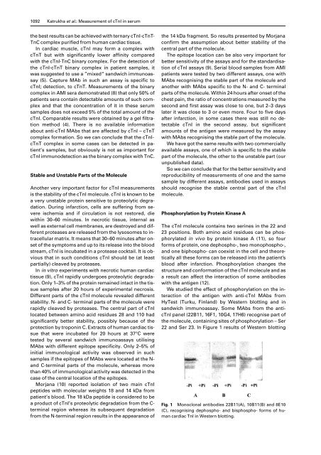
1092 Katrukha et al.: <strong>Measurement</strong> <strong>of</strong> cTnI in serumthe best results can be achieved with ternary cTnI-cTnT-TnC complex purified from human cardiac tissue.In cardiac muscle, cTnI may form a complex withcTnT but with significantly lower affinity comparedwith the cTnI-TnC binary complex. For the detection <strong>of</strong>the cTnI-cTnT binary complex in patient samples, itwas suggested to use a ”mixed“ sandwich immunoassay(5). Capture MAb in such an assay is specific tocTnI; detection, to cTnT. <strong>Measurement</strong>s <strong>of</strong> the binarycomplex in AMI sera demonstrated (8) that only 50% <strong>of</strong>patients sera contain detectable amounts <strong>of</strong> such complexand that the concentration <strong>of</strong> it in these serumsamples does not exceed 5% <strong>of</strong> the total amount <strong>of</strong> thecTnI. Comparable results were obtained by a gel filtrationmethod (4). There is no available informationabout anti-cTnI MAbs that are affected by cTnI – cTnTcomplex formation. So we can conclude that the cTnIcTnTcomplex in some cases can be detected in patient’ssamples, but obviously is not as important forcTnI immunodetection as the binary complex with TnC.Stable and Unstable Parts <strong>of</strong> the MoleculeAnother very important factor for cTnI measurementsis the stability <strong>of</strong> the cTnI molecule. cTnI is known to bea very unstable protein sensitive to proteolytic degradation.During infarction, cells are suffering from severeischemia and if circulation is not restored, diewithin 30–60 minutes. In necrotic tissue, internal aswell as external cell membranes, are destroyed and differentproteases are released from the lysosomes to intracellularmatrix. It means that 30–60 minutes after onset<strong>of</strong> the symptoms and up to its release into the bloodstream, cTnI is incubated in a protease cocktail. It is obviousthat in such conditions cTnI should be (at leastpartially) cleaved by proteases.In in vitro experiments with necrotic human cardiactissue (9), cTnI rapidly undergoes proteolytic degradation.Only 1–3% <strong>of</strong> the protein remained intact in the tissuesamples after 20 hours <strong>of</strong> experimental necrosis.Different parts <strong>of</strong> the cTnI molecule revealed differentstability. N- and C- terminal parts <strong>of</strong> the molecule wererapidly cleaved by proteases. The central part <strong>of</strong> cTnIlocated between amino acid residues 28 and 110 hadsignificantly better stability, possibly because <strong>of</strong> theprotection by troponin C. Extracts <strong>of</strong> human cardiac tissuethat were incubated for 20 hours at 37°C weretested by several sandwich immunoassays utilisingMAbs with different epitope specificity. Only 2–5% <strong>of</strong>initial immunological activity was observed in suchsamples if the epitopes <strong>of</strong> MAbs were located at the N-and C-terminal parts <strong>of</strong> the molecule, whereas morethan 40% <strong>of</strong> immunological activity was detected in thecase <strong>of</strong> the central location <strong>of</strong> the epitopes.Morjana (10) reported isolation <strong>of</strong> two main cTnIpeptides with molecular weights 18 and 14 kDa frompatient’s blood. The 18 kDa peptide is considered to bea product <strong>of</strong> cTnI’s proteolytic degradation from the C-terminal region whereas its subsequent degradationfrom the N-terminal region results in the appearance <strong>of</strong>the 14 kDa fragment. So results presented by Morjanaconfirm the assumption about better stability <strong>of</strong> thecentral part <strong>of</strong> the molecule.The epitope location can be also very important forbetter sensitivity <strong>of</strong> the assays and for the standardisation<strong>of</strong> cTnI assays (9). Serial blood samples from AMIpatients were tested by two different assays, one withMAbs recognising the stable part <strong>of</strong> the molecule andanother with MAbs specific to the N- and C- terminalparts <strong>of</strong> the molecule. Within 24 hours after onset <strong>of</strong> thechest pain, the ratio <strong>of</strong> concentrations measured by thesecond and first assay was close to one, but 2–3 dayslater it was close to 3 or even more. Four to five daysafter infarction, in some cases there was still no detectablecTnI in the second assay, but significantamounts <strong>of</strong> the antigen were measured by the assaywith MAbs recognising the stable part <strong>of</strong> the molecule.We have got the same results with two commerciallyavailable assays, one <strong>of</strong> which is specific to the stablepart <strong>of</strong> the molecule, the other to the unstable part (ourunpublished data).So we can conclude that for the better sensitivity andreproducibility <strong>of</strong> measurements <strong>of</strong> one and the samesample by different assays, antibodies used in assaysshould recognise the stable central part <strong>of</strong> the cTnImolecule.Phosphorylation by Protein Kinase AThe cTnI molecule contains two serines in the 22 and23 positions. Both amino acid residues can be phosphorylatedin vivo by protein kinase A (11), so fourforms <strong>of</strong> protein, one dephospho-, two monophospho-,and one biphospho- can coexist in the cell and theoreticallyall these forms can be released into the patient’sblood after infarction. Phosphorylation changes thestructure and conformation <strong>of</strong> the cTnI molecule and asa result can affect the interaction <strong>of</strong> some antibodieswith the antigen (12).We studied the effect <strong>of</strong> phosphorylation on the interaction<strong>of</strong> the antigen with anti-cTnI MAbs fromHyTest (Turku, Finland) by Western blotting and insandwich immunoassay. Some MAbs from the anticTnIpanel (22B11, 16F1, 10G4, 17H6) recognise part <strong>of</strong>the molecule, containing sites <strong>of</strong> phosphorylation – Ser22 and Ser 23. In Figure 1 results <strong>of</strong> Western blottingFig. 1 Monoclonal antibodies 22B11(A), 10B11(B) and 8E10(C), recognising dephospho- and bisphospho- forms <strong>of</strong> humancardiac TnI in Western blotting.