Spiegelberg:Sonde 1 Gebrauchsinformation. Sorgfältig lesen!
Spiegelberg:Sonde 1 Gebrauchsinformation. Sorgfältig lesen!
Spiegelberg:Sonde 1 Gebrauchsinformation. Sorgfältig lesen!

Sie wollen auch ein ePaper? Erhöhen Sie die Reichweite Ihrer Titel.
YUMPU macht aus Druck-PDFs automatisch weboptimierte ePaper, die Google liebt.
<strong>Spiegelberg</strong>: <strong>Sonde</strong> 1<br />
<strong>Gebrauchsinformation</strong>. Sorgfältig <strong>lesen</strong>!<br />
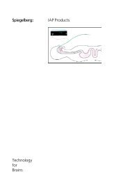
Methode<br />
Das Luftkammer-System ist ein Hohlkörper<br />
aus Kunststoff, der über einen<br />
Schlauch mit einem Druckaufnehmer<br />
verbunden ist. Der Druckaufnehmer befindet<br />
sich zusammen mit der Meßelektronik<br />
und einer Vorrichtung zur Füllung<br />
der Luftkammer in dem Hirndruck-<br />
Meßgerät.<br />
Zur epiduralen Druckmessung wird die<br />
Luftkammer auf der Dura des Patienten<br />
plaziert. Der intrakranielle Druck wird<br />
über die dünne Wand der Luftkammer<br />
auf die Luft in der Kammer übertragen<br />
und vom Druckaufnehmer in elektrische<br />
Signale umgesetzt.<br />
Auf dem Display des Gerätes werden<br />
der Mitteldruck und während der ersten<br />
zehn Minuten nach dem Einschalten<br />
auch die Pulsamplitude angezeigt.<br />
Am Monitorausgang stehen sowohl der<br />
Mitteldruck als auch das pulsatile Signal<br />
zur Verfügung.<br />
Das Hirndruck-Meßgerät öffnet den<br />
Druckaufnehmer einmal pro Stunde zur<br />
Atmosphäre und stellt den Nullpunkt<br />
ein. Danach wird die Luftkammer mit<br />
dem Volumen gefüllt, das für die Druckübertragung<br />
erforderlich ist.<br />
Empfohlene Technik<br />
Gerader Hautschnitt paramedian 2 bis<br />
3 cm lang. Setzen des Bohrloches in<br />
das Zentrum. Bohrlochdurchmesser: 11<br />
mm. Notfalls kann ein Stufentrepan von<br />
9 mm Durchmesser verwendet werden.<br />
Mit einer 4-mm-Stanze kann dann eine<br />
Erweiterung auf die "Kleeblatt"-Form<br />
erfolgen. Entfernen der Knochenreste<br />
auf der Dura, Brechen der Bohrkante<br />
am Unterrand mit dem scharfen Löffel,<br />
Ablösen der Dura zirkulär mindestens<br />
10 mm mit einem stumpfen Häkchen.<br />
Eventuell Blutstillung mit Knochenwachs.<br />
Überprüfen der <strong>Sonde</strong><br />
<strong>Sonde</strong> am Hirndruck-Meßgerät<br />
konnektieren. Das Schlauchende und<br />
der Konnektor werden dabei unsteril.<br />
Gerät einschalten und Entleerung und<br />
Füllung der Luftkammer beobachten.<br />
Die Druckanzeige muß bei etwa 0<br />
mmHg stehen. Die restliche Prüfung erfolgt<br />
mit dem palpierenden Finger<br />
(feucht). Leichten, pulsierenden Druck<br />
auf die Luftkammer ausüben. Am Monitor<br />
Pulsation beobachten.<br />
Mögliche Fehlerquellen sind:<br />
Adhärenzen der Meßmembran, dadurch<br />
artifizieller Druck. Solche<br />
Adhärenzen können durch Manipulationen<br />
an der Membran gelöst werden.<br />
Undichtigkeit der <strong>Sonde</strong>, rote Warnanzeige<br />
blinkt, <strong>Sonde</strong> muß ausgetauscht<br />
werden.<br />
Keine Pulsation am Monitor zu beobachten.<br />
Schlauch abgeknickt oder verlegt.<br />
Plazieren der <strong>Sonde</strong> 1<br />
Nach Prüfung <strong>Sonde</strong> am Meßgerät<br />
dekonnektieren, <strong>Sonde</strong> befeuchten,<br />
im Bohrloch plazieren. Dabei möglichst<br />
mit anatomischer Pinzette, Gewebs-<br />
Pinzette etc. arbeiten, an der<br />
Verstärkungsplatte greifen. Eventuell<br />
Dissektor oder stumpfes Häkchen zu<br />
Hilfe nehmen. <strong>Sonde</strong> am Hirndruck-<br />
Meßgerät konnektieren, Gerät einschalten,<br />
Hirndruck messen. Möglichst<br />
am Monitor Pulsation beobachten. Die<br />
Amplitude des Hirndruckpulses sollte in<br />
plausiblem Verhältnis zum mittleren<br />
Hirndruck stehen. Beim Gesunden hat<br />
die ICP-Welle eine Amplitude von 1-4<br />
mmHg. Sie steigt bei Drucksteigerungen<br />
auf 10-20 mmHg und mehr an.<br />
Wenn der gemessene ICP bei niedriger<br />
Amplitude zu hoch erscheint, können<br />
ungenügende Duraablösung, ein abgeknickter<br />
Schlauch oder verkippte<br />
Lage der <strong>Sonde</strong> im Bohrloch dafür verantwortlich<br />
sein.<br />
Je nach Situation den ICP bis zu zehn<br />
Minuten messen. Bei deutlichem<br />
Druckanstieg ohne gleichzeitigen Anstieg<br />
der Amplitude kann eine<br />
epidurale Blutung vorliegen. Dann<br />
<strong>Sonde</strong> revidieren und erneut Druckverlauf<br />
abwarten.<br />
Luftschlauch senkrecht aus der Wunde<br />
leiten, Wundverschluß mit etwa vier<br />
Einzelnähten. Annähen der Haltelasche,<br />
dabei Bilden einer Schlaufe.<br />
SND13.1.11/FV530P<br />
Entfernung der <strong>Sonde</strong> 1<br />
Nach Dekonnektion vom Hirndruck-Meßgerät<br />
die beiden mittleren Einzelnähte lösen,<br />
Haltelasche lösen. Am Luftschlauch<br />
<strong>Sonde</strong> senkrecht aus dem Bohrloch ziehen.<br />
Dabei den Schlauch kurz fassen und<br />
einmal kräftig ziehen. Sekundärnaht.<br />
Warnung<br />
Diese <strong>Sonde</strong> ist zur einmaligen Verwendung<br />
zur Hirndruckmessung mit dem<br />
<strong>Spiegelberg</strong> Monitor HDM 13.x, HDM 26.x<br />
oder HDM 29.x bestimmt. Nicht<br />
resterilisieren. Nicht wiederverwenden.<br />
Bei Wiederverwendung besteht ein<br />
Infektionsrisiko.<br />
Niemals Kochsalzlösung oder ein anderes<br />
flüssiges Medium einfüllen. Nicht verwenden<br />
wenn Packung beschädigt.<br />
Technische Daten<br />
Bestell Nr.<br />
Material<br />
Füllvolumen<br />
Außendurchmesser<br />
Länge<br />
Anwendungsdauer<br />
Doppelt verpackt<br />
EO sterilisiert<br />
Zur einmaligen Verwendung<br />
SND13.1.11/FV530P<br />
Pellethane<br />
0,05 - 0,1 ml<br />
2 mm<br />
1500 mm<br />
kurzzeitig<br />
bis zu 30 Tagen<br />
Hersteller<br />
<strong>Spiegelberg</strong><br />
GmbH & Co. KG<br />
Tempowerkring 4<br />
21079 Hamburg<br />
Germany<br />
Telefon: 040-790-178-0<br />
Telefax: 040-790-178-10<br />
Email: Info@<strong>Spiegelberg</strong>.de<br />
0297<br />
Version: 7.1 / 2013-05-31
<strong>Spiegelberg</strong>: Probe 1<br />
Directions for use. Read carefully!<br />
Method<br />
The air-pouch system consists of a<br />
hollow body connected to a pressure<br />
transducer by tubing. The pressure<br />
transducer, the electronic hardware,<br />
and the device for filling the air-pouch<br />
are integrated in the Brain-Pressure<br />
Monitor.<br />
For epidural pressure measurement the<br />
air- pouch is placed on the dura of the<br />
patient. The intracranial pressure is<br />
transmitted across the thin pouch wall<br />
to the air volume in the pouch and<br />
transformed into an electric signal by<br />
the pressure transducer.<br />
On the digital display the mean<br />
pressure is shown. At the monitor<br />
output both the mean pressure and<br />
the pulsatile signal are available.<br />
Once every hour the Brain-Pressure<br />
Monitor opens the pressure transducer<br />
to atmospheric pressure for zero<br />
adjustment. The air-pouch is then filled<br />
with the exact air volume required for<br />
accurate pressure transmission.<br />
Recommended Technique<br />
Straight paramedian skin incision,<br />
length 2 to 3 cm. Central placement of<br />
burr-hole. Burr-hole diameter 11 mm.<br />
Remove bone fragments on the dura,<br />
smoothen burr-edge at lower rim with<br />
curette, strip dura on circular area 10<br />
mm around the hole with small blunt<br />
hook. If necessary, apply bone cera for<br />
control of hemorrhage.<br />
Probe Check<br />
Connect probe to Brain-Pressure Monitor.<br />
Note that the tube end and the<br />
connector are non-sterile now. Switch<br />
on monitor and watch emptying and<br />
filling of air-pouch. The pressure<br />
reading must be around zero.<br />
Remaining checks with palpating<br />
finger (moist). Apply a slightly pulsating<br />
pressure on the air-pouch. Watch the<br />
undulating signal on the monitor.<br />
Possible sources of error are:<br />
Pressure artifacts caused by adherence<br />
of probe membrane. Resolve by<br />
manipulating membrane.<br />
Leaky probe, red warning indicator<br />
appears. Probe should be replaced.<br />
Placement of Probe 1<br />
Disconnect probe from monitor after<br />
probe check (membrane is depressurized),<br />
moisten probe, place probe in<br />
burr-hole. Use dissecting forceps,<br />
tissue forceps etc. If possible, grasp<br />
probe at base plate. If necessary assist<br />
with dissector or small blunt hook.<br />
Connect probe to Brain-Pressure Monitor,<br />
switch on monitor, read brain<br />
pressure. It is advised to check for<br />
pulsation on a patient monitor. The<br />
amplitude of the brain-pressure should<br />
be plausible in relation to the mean<br />
pressure. In healthy man the ICP-wave<br />
has an amplitude of 1 to 4 mmHg. With<br />
elevated ICP the amplitude rises to 10-<br />
20 mmHg and even more. If the<br />
measured ICP appears to be high with<br />
a low amplitude, the dura might not be<br />
stripped from the bone sufficiently. Also<br />
a kinked tubing or a tilted probe could<br />
be the cause. Depending on the<br />
situation continue to read the ICP for<br />
up to 10 minutes. A distinct rise of the<br />
mean ICP which is not accompanied by<br />
a rise of the amplitude can indicate an<br />
epidural hematoma. In that case<br />
remove the hematoma and read ICP<br />
anew.<br />
Removal of Probe 1<br />
Disconnect probe from the Brain-Pressure<br />
Monitor. Loosen the two central<br />
interrupted sutures, detach suture flap.<br />
Remove probe from burr-hole vertically by<br />
giving the air tube a strong pull.<br />
Secondary suture.<br />
Warning<br />
This probe is designed and is intended for<br />
single use for the measurement of ICP with<br />
the <strong>Spiegelberg</strong> Monitor HDM 13.x, HDM<br />
26.x, or HDM 29.x. Do not resterilize. Do<br />
not reuse. With reuse an infection risk<br />
exists. Do not fill with saline or other liquid<br />
media. Do not use if package is damaged.<br />
Technical Information<br />
Order No. SND13.1.11/FV530P<br />
Material Pellethane<br />
Filling volume 0,05 - 0,1 ml<br />
Outer diameter 2 mm<br />
Length 1500 mm<br />
Duration of use short term<br />
not more than 30 days<br />
Double packed<br />
EO sterilized<br />
For single use<br />
No pulsation on the monitor. Tubing<br />
kinked or clogged.<br />
Direct air tube vertically out of wound,<br />
close with approximately four<br />
interrupted sutures. Affix suture flap,<br />
form loop.<br />
SND13.1.11/FV530P<br />
Manufacturer<br />
<strong>Spiegelberg</strong><br />
GmbH & Co. KG<br />
Tempowerkring 4<br />
21079 Hamburg<br />
Germany<br />
Phone: +49-40-790-178-0<br />
Fax: +49-40-790-178-10<br />
Email: Info@<strong>Spiegelberg</strong>.de<br />
0297<br />
Version: 7.1 / 2013-05-31