The pyrenoid ultrastructure in Oocystis lacustris CHODAT ... - Fottea
The pyrenoid ultrastructure in Oocystis lacustris CHODAT ... - Fottea
The pyrenoid ultrastructure in Oocystis lacustris CHODAT ... - Fottea

Create successful ePaper yourself
Turn your PDF publications into a flip-book with our unique Google optimized e-Paper software.
150 St o y n e va et al.: <strong>The</strong> <strong>pyrenoid</strong> <strong>ultrastructure</strong><br />
et Be n D e r l i e v, Eremosphaera De Ba ry and<br />
Siderocelis Fo t t is done.<br />
Material and methods<br />
<strong>Oocystis</strong> <strong>lacustris</strong> material was obta<strong>in</strong>ed from selected<br />
phytoplankton samples from Lake Tanganyika dated<br />
June–July 2003 when it formed dense populations<br />
(St o y n e va et al. 2007) and fixed <strong>in</strong> acid Lugol’s<br />
solution. For detailed description of localities, sampl<strong>in</strong>g<br />
and methods refer to St o y n e va et al. (2007). For TEM<br />
study cells were fixed a) <strong>in</strong> 3% glutaraldehyd <strong>in</strong> 0,1 M<br />
cacodylate buffer and b) <strong>in</strong> 1% aqueous O s O 4 <strong>in</strong> 0,1 M<br />
cacodylatbuffer, dehydrated <strong>in</strong> acetone and embedded<br />
<strong>in</strong> Spurr’s res<strong>in</strong>e¸ ultrath<strong>in</strong> sections were sta<strong>in</strong>ed with<br />
uranyl acetate and lead citrate (re y n o l D S 1963).<br />
Electron micrographs were taken with a Tecnai 12<br />
(FEI) microscope equipped with a Gatan ccd camera.<br />
Results<br />
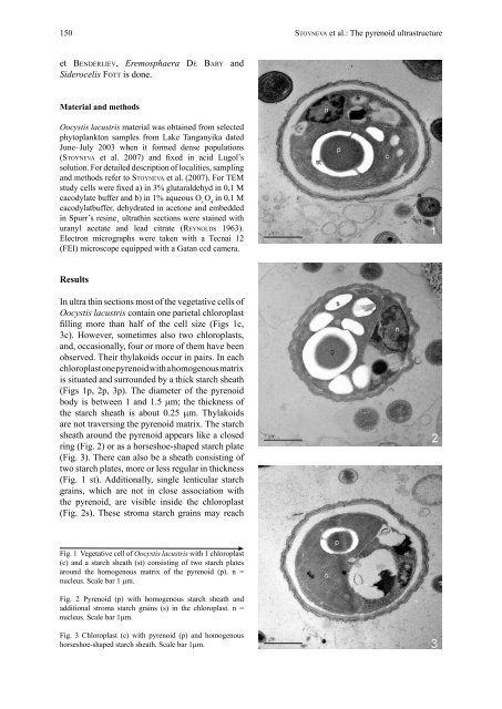
In ultra th<strong>in</strong> sections most of the vegetative cells of<br />
<strong>Oocystis</strong> <strong>lacustris</strong> conta<strong>in</strong> one parietal chloroplast<br />
fill<strong>in</strong>g more than half of the cell size (Figs 1c,<br />
3c). However, sometimes also two chloroplasts,<br />
and, occasionally, four or more of them have been<br />
observed. <strong>The</strong>ir thylakoids occur <strong>in</strong> pairs. In each<br />
chloroplast one <strong>pyrenoid</strong> with a homogenous matrix<br />
is situated and surrounded by a thick starch sheath<br />
(Figs 1p, 2p, 3p). <strong>The</strong> diameter of the <strong>pyrenoid</strong><br />
body is between 1 and 1.5 µm; the thickness of<br />
the starch sheath is about 0.25 µm. Thylakoids<br />
are not travers<strong>in</strong>g the <strong>pyrenoid</strong> matrix. <strong>The</strong> starch<br />
sheath around the <strong>pyrenoid</strong> appears like a closed<br />
r<strong>in</strong>g (Fig. 2) or as a horseshoe-shaped starch plate<br />
(Fig. 3). <strong>The</strong>re can also be a sheath consist<strong>in</strong>g of<br />
two starch plates, more or less regular <strong>in</strong> thickness<br />
(Fig. 1 st). Additionally, s<strong>in</strong>gle lenticular starch<br />
gra<strong>in</strong>s, which are not <strong>in</strong> close association with<br />
the <strong>pyrenoid</strong>, are visible <strong>in</strong>side the chloroplast<br />
(Fig. 2s). <strong>The</strong>se stroma starch gra<strong>in</strong>s may reach<br />
Fig. 1 Vegetative cell of <strong>Oocystis</strong> <strong>lacustris</strong> with 1 chloroplast<br />
(c) and a starch sheath (st) consist<strong>in</strong>g of two starch plates<br />
around the homogenous matrix of the <strong>pyrenoid</strong> (p). n =<br />
nucleus. Scale bar 1 µm.<br />
Fig. 2 Pyrenoid (p) with homogenous starch sheath and<br />
additional stroma starch gra<strong>in</strong>s (s) <strong>in</strong> the chloroplast. n =<br />
nucleus. Scale bar 1µm.<br />
Fig. 3 Chloroplast (c) with <strong>pyrenoid</strong> (p) and homogenous<br />
horseshoe-shaped starch sheath. Scale bar 1µm.