The cyanobacterial genus Macrospermum Jiří KOMÁREK - Fottea
The cyanobacterial genus Macrospermum Jiří KOMÁREK - Fottea
The cyanobacterial genus Macrospermum Jiří KOMÁREK - Fottea

You also want an ePaper? Increase the reach of your titles
YUMPU automatically turns print PDFs into web optimized ePapers that Google loves.
<strong>Fottea</strong>, Olomouc, 8(1): 79–86, 2008 79<br />
<strong>The</strong> <strong>cyanobacterial</strong> <strong>genus</strong> <strong>Macrospermum</strong><br />
<strong>Jiří</strong> Ko m á r e k<br />
Academy of Sciences of the Czech Republic, Institute of Botany, Dukelská 135, CZ-379 82 Třeboň, and University<br />
of South Bohemia, Faculty of Sciences, Branišovská 31, CZ-370 05 České Budějovice, Czech Republic, e-mail:<br />
komarek@butbn.cas.cz<br />
Abstract: This small tropical <strong>cyanobacterial</strong> group, containing Anabaena volzii Le m m e r m a n n and a few related<br />
species (A. fuellebornii Sc h m i d l e, A. unispora Ga r d n e r, A. mysorensis Go n z a lv e s & ka m at ), differs substantially<br />
phenotypically from all other planktic or benthic Anabaena types, mainly by the subsymmetric structure of the<br />
trichomes, type of akinete formation and restricted ecology. <strong>The</strong> taxonomic uniformity of all other Anabaenalike<br />
clusters (typical benthic Anabaena, planktic Anabaena subg. Dolichospermum, Trichormus, Aphanizomenon,<br />
Cuspidothrix) was already supported by molecular analyses. All of them also have their typical morphological<br />
markers, which are clearly different from the “Anabaena volzii – cluster”. <strong>The</strong>refore, this group can not be<br />
classified in any of the mentioned revised genera and must be described as a separate generic entity of heterocytous<br />
cyanobacteria (although they have not been sequenced to date). <strong>The</strong> new <strong>genus</strong> <strong>Macrospermum</strong> is therefore defined<br />
in my article with 4 related, morphologically distinguishable species. <strong>The</strong> generic name is selected according to<br />
the unusually large akinetes.<br />
Key words: Cyanobacteria, taxonomy, Anabaena, <strong>Macrospermum</strong>, pantropical <strong>genus</strong>, akinete formation<br />
Introduction<br />
Sc h m i d l e (1902) described a <strong>cyanobacterial</strong><br />
species Anabaena fuellebornii from Africa. This<br />
metaphytic species was later found from several<br />
localities in tropical Africa (Chad, Guinea,<br />
Tanzania – co m p è r e 1967, 1974, Bo u r r e l ly<br />
1975), tropical Asia (Burma – sk u j a 1949) and<br />
America (Brazil, Cuba – ko m á r e k 2005, orig.<br />
data). In 1904, Le m m e r m a n n described Anabaena<br />
volzii, having a similar trichome structure, from<br />
plankton and benthos in Singapore. This species<br />
was found also in numerous localities over the<br />
whole tropical region in Asia (S. China, India,<br />
Indonesia, Singapore – Ge i t l e r 1932, ja o 1948,<br />
Fr i t s c h 1949, Gu p ta 1956, de s i k a c h a ry 1959),<br />
Africa (Chad, Guinea, Mozambique, Tanzania<br />
– Bo u r r e l ly 1975, ri n o 1972) and Central<br />
America, particularly in the Caribbean district<br />
(Cuba, Guadeloupe, Venezuela – Bo u r r e l ly &<br />
ma n G u i n 1952, ya c u B s o n 1974, ko m á r e k 1984,<br />
2005). <strong>The</strong> number of similar species was enlarged<br />
by Anabaena unispora Ga r d n e r 1927 known in<br />
typical form from Puerto Rico and Cuba, and also<br />
by Anabaena mysorensis, described by Go n z a lv e s<br />
& ka m at (1959) from India (Mysore State).<br />
All of these species have a unique, very specific<br />
morphology, which is different from all other<br />
Anabaena species. <strong>The</strong> filaments are nearly<br />
symmetric, uniserial, unbranched and isopolar,<br />
with two heterocytes localized in the subapical<br />
portions of the trichomes, slightly distant from<br />
the terminal parts. One central, third heterocyte,<br />
localised ± in the centre of a trichome, sometimes<br />
occurs in fully developed trichomes. Unusually<br />
large, solitary, oval or ellipsoid akinetes (very<br />
exceptionally occurring in pairs) develop after<br />
fusion of several vegetative cells joined to the<br />
heterocyte on the external side of the heterocyte.<br />
<strong>The</strong> filaments have a typical symmetrical or<br />
subsymmetrical structure (in the second case the<br />
heterocytes are a little shifted from the central<br />
or subterminal positions). Exceptions from this<br />
trichome structure can occur, but they are very<br />
rare (Fig. 1). Of course, the asymmetry appears<br />
after trichome disintegration and in developing<br />
filaments.<br />
<strong>The</strong> cluster of “Anabaena volzii”-like<br />
species contains now four tropical species. <strong>The</strong><br />
only non-confirmed record from the temperate<br />
zone is “A. unispora” from Michigan, recorded<br />
by pr e s c o t t (1962), but with several unclear<br />
features. <strong>The</strong> species of this cluster differ from<br />
one another by the form of the cells (particularly
80 ko m á r e k: <strong>The</strong> <strong>cyanobacterial</strong> <strong>genus</strong> <strong>Macrospermum</strong><br />
in the apical parts of trichomes), and the form<br />
and colour of akinete epispores. Anabaena volzii<br />
f. recta ki s e l e v 1931, characterised by straight<br />
trichomes, and A. volzii var. crassa (ra o) Fr i t s c h<br />
1949 seem to be in the variation range of the<br />
typical species. All transitions between straight,<br />
waved and coiled filaments occurred in our studied<br />
populations; also, the dimensions in all described<br />
populations only slightly deviated and overlapped<br />
with all transient forms (Tab. 1).<br />
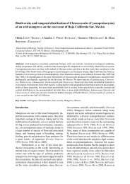
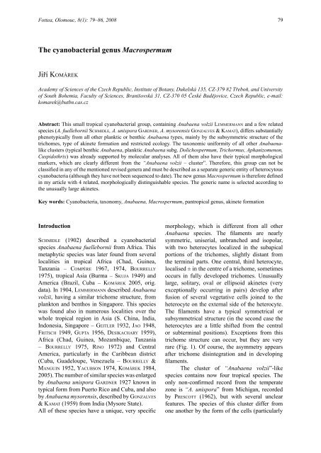
Fig. 1. Scheme of the slightly subsymmetric to symmetric trichomes of “Anabaena/Aphanizomenon” (= <strong>Macrospermum</strong>) volzii<br />
(populations from Cuba) and their development: A – variability of cell width in trichomes and position of heterocytes (H) in<br />
filaments; B – variability (average limits from 48 filaments) in the position of heterocytes (H) and akinetes (S) in trichomes<br />
with two (48 measurements) and three (14 measurements) heterocytes; C – typical filament with two heterocytes and two<br />
akinetes at low magnification; D – scheme of a typical filament with fully developed akinetes and average width of vegetative<br />
cells, necridic cells (n), akinetes (S), heterocytes (H) and with the number of cells in corresponding segments of trichomes; m =<br />
morphological centre of a trichome, s = geometrical centre of a trichome. (After Ko m á r e k 1984, sub Aphanizomenon volzii.)
<strong>Fottea</strong>, Olomouc, 8(1): 79–86, 2008 81<br />
Thus, the Anabaena volzii-type differs from<br />
the other Anabaena-like species (both planktic<br />
or benthic) by the specific trichome structure<br />
(in all Anabaena-types is strictly metameric),<br />
from Aphanizomenon and Cuspidothrix by the<br />
morphology of cells, type of life (formation of<br />
mats), position and form of akinetes, and form<br />
of colonies. This type also differs from other<br />
nostocalean genera by the special subsymmetric<br />
structure of the trichomes and the position of the<br />
heterocytes and akinetes (Fig. 2). No species from<br />
the whole described Anabaena volzii cluster has<br />
been isolated in culture up to now, nor have they<br />
yet been sequenced. However, their morphology<br />
and life form differ substantially from all genera,<br />
as shown by generic analyses and confirmed<br />
by the corresponding phenotypic markers<br />
(Anabaena, Aphanizomenon, Cylindrospermum,<br />
Cylindrospermopsis, Cuspidothrix, Trichormus,<br />
Anabaenopsis, and others – cf. e.g., Gu G G e r &<br />
al. 2002a, b, ra j a n i e m i et al. 2005, etc). Because<br />
the generic classification of this characteristic<br />
morphological group into any existing <strong>genus</strong> is not<br />
possible, the description of a new generic entity<br />
is necessary (e.g., for the prepared monographic<br />
elaboration of the heterocytous cyanobacteria for<br />
Süsswasserflora von Mitteleuropa).<br />
Results<br />
<strong>The</strong> main generic phenotypic diacritical feature<br />
of the new <strong>genus</strong> <strong>Macrospermum</strong> is the structure<br />
of the trichomes: <strong>The</strong> trichomes are isopolar with<br />
a nearly symmetric or subsymmetric structure<br />
(Ko m á r e k 1984). <strong>The</strong> heterocytes develop in the<br />
apical parts of trichomes, slightly distant from<br />
the ends. <strong>The</strong> third heterocyte develops in old<br />
trichomes ± in the centre, or slightly shifted from<br />
the geometric middle of a trichome. Akinetes<br />
develop after fusion of several vegetative cells,<br />
attached to a marginal heterocyte, usually on the<br />
side towards the ends of a trichome, exceptionally<br />
at both sides of marginal heterocytes. <strong>The</strong> akinete<br />
was never observed at the central heterocyte.<br />
Trichome width in Anabaena (= <strong>Macrospermum</strong>)<br />
volzii is always a little greater in parts with<br />
heterocytes, while being slightly narrowed<br />
between heterocytes and towards the ends (Fig.<br />
1).<br />
Usually, only two akinetes develop in a<br />
trichome, very rarely in pairs. <strong>The</strong>y are formed<br />
after fusion of few neighbouring vegetative cells<br />
and are extremely large (oval or ellipsoidal) in<br />
comparison with akinetes of other genera. <strong>The</strong><br />
epispore is smooth or sculptured; differences<br />
in surface structure of akinetes is considered as<br />
differential feature between morphospecies.<br />
All described species are distributed<br />
in tropical regions (with one exception – see<br />
discussion; pr e s c o t t 1962). <strong>The</strong>y appear in<br />
aquatic habitats, marshes, ponds, the littoral of<br />
lakes and in paddy fields. <strong>The</strong>y form free colonies<br />
(disintegrating fine mats) on water plants, or<br />
small floating clusters in the metaphyton. Solitary<br />
trichomes or small groups of filaments can occur<br />
less frequently and secondarily also in the plankton.<br />
Facultatively rare aerotope-like inclusions occur<br />
in cells of Anabaena (<strong>Macrospermum</strong>) volzii.<br />
Formal description of the <strong>genus</strong> <strong>Macrospermum</strong>:<br />
Cyanobacterial, heterocytous, filamentous <strong>genus</strong>.<br />
Filaments are free-living, solitary, in small<br />
irregular clusters or in fine macroscopic mats.<br />
Trichomes have symmetric to subsymmetric<br />
structure with two subapical heterocytes and<br />
sometimes with one ± central heterocyte; they<br />
are nearly straight or irregularly coiled with fine,<br />
colourless, diffluent and indistinct slime, uniserial,<br />
unbranched, ± cylindrical, constricted at crosswalls,<br />
sometimes narrowed to the ends and in<br />
distinct central parts. Cells cylindrical or slightly<br />
barrel-shaped, ± isodiametric or rather longer than<br />
wide, with blue-green, homogeneous content with<br />
scarce granules and facultatively with solitary<br />
aerotopes; terminal cells are rounded, conical or<br />
narrowed and bluntly pointed. Heterocytes always<br />
solitary, intercalar, cylindrical, usually wider than<br />
vegetative cells, usually two in subapical position<br />
in a trichome, or with the third heterocyte ± in<br />
the middle of a trichome. Akinetes widely oval,<br />
developing from several neighbouring cells, large,<br />
solitary, rarely in pairs, always attached to outer<br />
heterocytes; in one trichome develop usually<br />
only two akinetes, outside from heterocytes,<br />
rarely also at the “inner” side of a heteocyte.<br />
Reproduction by fragmentation of trichomes and<br />
by akinetes. Type species: <strong>Macrospermum</strong> volzii<br />
(le m m e r m a n n ) comb. nova (= Anabaena volzii<br />
le m m e r m a n n 1904). This species was selected as<br />
the type-species instead of the older “Anabaena<br />
fuellebornii” (1902), because it is the best known<br />
and most distributed species from the whole new<br />
<strong>genus</strong>.<br />
Diagnosis: <strong>Macrospermum</strong> <strong>genus</strong> nova –<br />
Genus cyanobacteriis heterocytosis. Filamenta
82 ko m á r e k: <strong>The</strong> <strong>cyanobacterial</strong> <strong>genus</strong> <strong>Macrospermum</strong><br />
solitaria, libere natantia vel in strata irregularia,<br />
macroscopica aggregata, sine vaginis, vel cum<br />
muco incolore, tenue, amorpho circumdatae.<br />
Trichomata uniseriata, plus minusve recta vel<br />
irregulariter flexuosa, not ramosa, symmetrica<br />
vel subsymmetrica, ad dissepimenta constricta,<br />
ad apices paucim attenuata vel cylindrica,<br />
cum heterocytis duobus subapicalis, raro cum<br />
heterocyta tertia centrali. Cellulae cylindricae<br />
vel paucim barriliformes, plus minusve<br />
isodiametricae vel longior quam latae, contentu<br />
aerugineo, homogeneo, cum granulis sparsis<br />
et aerotopis solitariis facultativis; cellula<br />
terminalis cylindrica vel conica, apice rotundata.<br />
Heterocytae intercalares, solitariae, cylindricae,<br />
plerumque latior quam cellulae vegetativae, in<br />
trichomatibus subapicaliter dispositae, rare plus<br />
una heterocyta centralis. Akinetes late ovales,<br />
intercalares, cum heterocytis conjunctae, valde ad<br />
eos partes externis dispositae, solitariae, rarissime<br />
binae, cum episporio laevi vel ornati, praecipue<br />
duas in una trichoma. Reproductio trichomatibus<br />
fragmentatione et akinetis germinatione. - Typus<br />
generis: <strong>Macrospermum</strong> volzii (le m m e r m a n n )<br />
comb. nova (syn.: Anabaena volzii le m m e r m a n n<br />
1904).<br />
List of species:<br />
<strong>Macrospermum</strong> volzii (le m m e r m a n n ) comb. nova<br />
(Fig. 3)<br />
Basionym: Anabaena volzii Le m m e r m a n n Abh. Nat.<br />
Ver. Bremen 18(1): 153, 1904.<br />
Syn: Anabaena volzii f. recta I. ki s e l e v Tr. Sr.-Aziat.<br />
Gos. Univ. (Tashkent), Ser. XIIa, 9: 74, 1931; incl.;<br />
Anabaena unispora var. crassa ra o Proc. Ind. Acad.<br />
Sci. 6(6B): 362, 1937; incl.; Anabaena volzii var. crassa<br />
(ra o) Fr i t s c h J. Ind. Bot. Soc. 28(3): 155, 1949; incl.<br />
Anabaena volzii var. unispora (Ga r d n e r) Bo u r r e l ly<br />
sensu Bo u r r e l ly Bull. Inst. France Afr. Nord., ser.<br />
A, 19(4): 1049, 1957; incl.; Aphanizomenon volzii<br />
(le m m e r m a n n ) ko m á r e k Acta Bot. Cubana 18: 9,<br />
1984.<br />
Diacritical characters: Cells cylindrical, 4.5-<br />
14 x 4-5.8 µm; apical cells slightly elongated,<br />
narrowed and bluntly pointed; akinetes with<br />
smooth, colourless or brownish epispore, (20)32-<br />
48 x (13)15-21 µm. With pantropical distribution<br />
and in central Asia (e. ki s e l e va 1931, i. ki s e l e v<br />
1931 from el e n k i n 1938).<br />
<strong>Macrospermum</strong> fuellebornii (sc h m i d l e) comb.<br />
nova (Fig. 4)<br />
Basionym: Anabaena fuellebornii sc h m i d l e Engler’s<br />
Bot. Jahrb. 32: 61, 1892.<br />
Diacritical characters: Cells slightly barrelshaped,<br />
3.8-8.2 x 4.8-7.4 µm; apical cells slightly<br />
narrowed, rounded; akinetes with granular-dotted,<br />
brownish epispore, 25-45 x (14.3)16.5-19(21.6)<br />
µm. With pantropical distribution.<br />
<strong>Macrospermum</strong> mysorense (Go n z a lv e s et<br />
ka m at ) comb. nova (Fig. 5)<br />
Basionym: Anabaena mysorensis Go n z a lv e s et ka m at<br />
Hydrobiologia 13: 237, 1959.<br />
Diacritical characters: Cells cylindrical, up to<br />
twice as long as wide, 6.4-12.3 x 5.8-7.5 µm;<br />
apical cells probably cylindrical and rounded<br />
(not described in the original diagnosis); akinetes<br />
ellipsoidal to oval, having epispore with pointed,<br />
up to 3,2-4,5 µm long, spines, 35.8-51,6 x 12.5-<br />
19.4 µm. Known only from India (Mysore State).<br />
<strong>Macrospermum</strong> unisporum (Ga r d n e r) comb.<br />
nova (Fig. 6)<br />
Basionym: Anabaena unispora Ga r d n e r Mem N.Y.<br />
Bot. Garden 7: 59, 1927.<br />
Syn.: Anabaena volzii var. unispora (Ga r d n e r)<br />
Bo u r r e l ly Bull. Inst. Franc. Afr. Nord, Ser. A, 19(4):<br />
1049, 1957.<br />
Diacritical characters: Cells cylindrical, mostly<br />
isodiametric, infrequently up to twice as long than<br />
wide, 4-10.2 x 4-5.4 µm; apical cells ± cylindrical,<br />
rounded; akinetes with smooth, brown epispore,<br />
(18)20-40(43) x (8)12.5-20.5 µm. Known from<br />
tropical America, particularly from the Caribbean<br />
district (Cuba, Puerto Rico); the records from<br />
Michigan (pr e s c o t t 1950) need confirmation.<br />
<strong>The</strong> key to the species identification:<br />
1a Ripe akinetes with smooth epispore ..................... 2<br />
1b Ripe akinetes with sculptured epispore ................ 3<br />
2a Vegetative cells isodiametric or (usually) longer than<br />
wide, apical cells elongated, narrowed, end cell bluntly<br />
pointed ................................................... M. volzii<br />
2b Vegetative cells commonly isodiametric or only<br />
slightly longer than wide, apical cells cylindrical, not<br />
narrowed, end cell cylindrical and rounded ..................<br />
............................................................... M. unisporum<br />
3a Surface of akinetes granular-dotted (brownish), cells<br />
± isodiamatric, end cells cylindrical to slightly barrelshaped,<br />
apical cells conical rounded .............................<br />
............................................................. M. fuellebornii<br />
3b Surface of akinetes with pointed spines, cells<br />
cylindrical, longer as wide (up to twice), apical cells
<strong>Fottea</strong>, Olomouc, 8(1): 79–86, 2008 83<br />
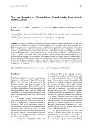
Fig. 2. Scheme of the trichome structure of <strong>Macrospermum</strong> in comparison with other nostocalean genera with paraheterocytic<br />
origin of akinetes; circles indicate the places of heterocyte origin: A = place of akinete development. (Partly from ko m á r e k &<br />
an a G n o s t i d i s 1989.)<br />
cylindrical rounded (?) .......................... M. mysorense<br />
Discussion<br />
<strong>The</strong> modern taxonomy of <strong>cyanobacterial</strong> genera is<br />
based primarily on molecular sequencing, joined<br />
with a combined evaluation of phenotypic, biochemical<br />
and ecological markers. However, typical<br />
characteristic phenotype markers were found<br />
in all generic units, being defined and supported<br />
by phylogenetic studies. <strong>The</strong> groups of genera,<br />
which evidently do not belong to clusters already<br />
revised by molecular sequencing and which morphologically<br />
evidently belong beyond the range<br />
of morphological variation of complexly revised<br />
generic entities remain taxonomically problematic.<br />
<strong>The</strong> group of the <strong>genus</strong> <strong>Macrospermum</strong> is just<br />
a typical case of such clusters. <strong>The</strong> various species<br />
were described as members of the traditional
84 ko m á r e k: <strong>The</strong> <strong>cyanobacterial</strong> <strong>genus</strong> <strong>Macrospermum</strong><br />
Fig. 3. <strong>Macrospermum</strong> volzii: a – original drawing after Le m m e r m a n n (1904, sub Anabaena volzii); b – populations from Cuba after ko m á r e k (1984, sub Aphanizomenon volzii); Fig. 4.<br />
<strong>Macrospermum</strong> fuellebornii: a – ends of trichomes; b – variability of heterocytes; c – position of a heterocyte with joined akinete; d – variability of akinetes (from Ko m á r e k 2005, sub Anabaena<br />
fuellebornii); Fig. 5. <strong>Macrospermum</strong> mysorense (after Go n z a lv e s & ka m at 1959 from India – Mysore, sub Anabaena mysorensis); Fig. 6. <strong>Macrospermum</strong> unisporum: a – original drawing after<br />
Ga r d n e r (1927) from Puerto Rico; b-e – variability of trichome ends, vegetative cells, heterocytes and akinetes from the Cuban population, after Ko m á r e k (2005), (all sub Anabaena unispora).
<strong>Fottea</strong>, Olomouc, 8(1): 79–86, 2008 85<br />
<strong>genus</strong> Anabaena, but their morphology is substantially<br />
different (subsymmetric structure of trichomes)<br />
from the updated and revised metameric<br />
Anabaena-like clusters. This includes typical benthic<br />
Anabaena species, based on the type-species<br />
A. oscillarioides Bo ry ex Bo r n e t et Fl a h a u lt<br />
1888, planktic Anabaena subg. Dolichospermum<br />
with the type-species A. flos-aquae (ly n G B y e)<br />
Br é B i s s o n ex Bo r n e t et Fl a h a u lt 1888, as well<br />
as the <strong>genus</strong> Trichormus with apoheterocytic formation<br />
of akinetes with type species T. variabilis<br />
(Kü t z i n G ex Bo r n e t et Fl a h a u lt) ko m á r e k et<br />
an a G n o s t i d i s 1989 (sooner “Anabaena variabilis”<br />
Kü t z i n G ex Bo r n e t et Fl a h a u lt 1886).<br />
<strong>Macrospermum</strong>, by having a symmetric or<br />
subsymmetric structure of trichomes is similar<br />
mainly to the genera Cylindrospermum, Aphanizomenon,<br />
Cuspidothrix and Cylindrospermopsis<br />
(cf. Fig. 2). Cylindrospermum and Cylindrospermopsis<br />
differ by the development of heterocytes<br />
from terminal cells, Aphanizomenon and Cuspidothrix<br />
by the type of akinete formation and the<br />
life form. However, the phylogenetic position of<br />
<strong>Macrospermum</strong> seems to be rather near this group<br />
of genera than to the Anabaena–like clusters, to<br />
which all the <strong>Macrospermum</strong> species were traditionally<br />
classified.<br />
All of the species morphologically congruent<br />
with the <strong>genus</strong> <strong>Macrospermum</strong> are described<br />
from aquatic biotopes in tropical regions. To this<br />
can be added also the rice fields in Tadzhikistan<br />
near Samarkand (central Asia; see ki s e l e v in<br />
ho l l e r B a c h & al. 1953 and de s i k a c h a ry 1959).<br />
<strong>The</strong> only exception is “Anabaena unispora” recorded<br />
by pr e s c o t t (1962) from Michigan, USA.<br />
However, this specimen has cells up to twice as<br />
long than the type and the akinetes develop “near<br />
the middle of the filaments, with the dimensions<br />
20-34 x 11-15 µm”. <strong>The</strong> identity of these populations<br />
with typical <strong>Macrospermum</strong> unisporum is<br />
therefore problematic and should be confirmed.<br />
Thus, the <strong>genus</strong> <strong>Macrospermum</strong> is described,<br />
because the group of “Anabaena volzii”<br />
remained out of any generic classification after<br />
molecular confirmation of the most related anabaenacean<br />
genera.<br />
Acknowledgement<br />
This study was supported by the grant GA AS CR<br />
no. IAA600050704. <strong>The</strong> author thanks Dr. Jaroslava<br />
Komárková for critical remarks and Dr. Keith Edwards<br />
for language correction.<br />
References<br />
Bo u r r e l ly, p. (1961): Quelques algues d’eau douce<br />
de la région de Konakry. – Bull. Res. Counc.<br />
Israel, Botany, 10(D): 9-14.<br />
Bo u r r e l ly, P. (1975): Quelques algues d’eau douce de<br />
Guinée. – Bull. Mus. Nat. d’Hist. Natur. 276:<br />
1-71.<br />
Bo u r r e l ly, p. & ma n G u i n, E. (1952): Algues d’eau<br />
douce de la Guadeloupe. – 281 pp., Paris<br />
co m p è r e , p. (1967): Algues du Sahara et de la région<br />
du lac Tchad. – Bull. Jard. Bot. Nat. Belg.,<br />
Bruxelles, 37: 109–288.<br />
co m p è r e , p. (1974): Algues de la région du lac Tchad.<br />
II. Cyanophycées. – Cah. O.R.S.T.O.M., Sér.<br />
Hydrobiol. 8(3/4): 165–198<br />
de s i k a c h a ry, t.v. (1959): Cyanophyta. - In: ICAR<br />
Monographs on Algae, 686 pp., New Delhi.<br />
el e n k i n, a.a. (1938): Monographia algarum<br />
cyanophycearum aquidulcium et terrestrium in<br />
finibus URSS inventarum. Pars spec., Fasc. 1. –<br />
984 pp., Izd. AN SSSR, Moskva-Leningrad.<br />
Fr i t s c h, F.e. (1949): <strong>The</strong> <strong>genus</strong> Anabaena with special<br />
reference to the species recorded from India<br />
and the adjacent Asiatic mainland. – J. Ind. Bot.<br />
Soc. 28: 135–161.<br />
Ga r d n e r, n.l. (1927): New Myxophyceae from Porto<br />
Rico. – Mem. N.Y. Bot. Garden 7: 1–144.<br />
Ge i t l e r, l. (1932): Cyanophyceae. – In Rabenhorst‘s<br />
Kryptogamenflora von Deutschland, Österreich<br />
und der Schweiz 14: 1–1196, Akad. Verlagsges.,<br />
Leipzig.<br />
Go l l e r B a c h, m.m., ko s i n s k a j a, e.k. & po l j a n s k i j,<br />
v.i. (1953): Sinezelenye vodorosli. [Blue-green<br />
algae]. – In: Opredelitel‘ presnovodnych<br />
vodoroslej SSSR 2: 1–652, Izd. “Sovetskaja<br />
nauka”, Moskva.<br />
Go n z a lv e s, e.a. & ka m at , n.d. (1959): Two new<br />
species of Anabaena from Western India. –<br />
Hydrobiologia 13(3): 236–238.<br />
Gu G G e r, m., ly r a, c., su o m i n e n, i., ts i t k o, i.,<br />
hu m B e r t , j.-F., sa l k i n o j a-sa l o n e n, m. &<br />
si v o n e n, K. (2002a): Cellular fatty acids<br />
as chemotaxonomic markers of the genera<br />
Anabaena, Aphanizomenon, Microcystis, Nostoc<br />
and Planktothrix (Cyanobacteria). – Internat. J.<br />
Syst. Evol. Microbiol. 52: 1007–1015.<br />
Gu G G e r, m., ly r a, c., he n r i k s e n, p., co u t é, a.,<br />
hu m B e r t , j.-F. & si v o n e n, K. (2002b):<br />
Phylogenetic comparison of the <strong>cyanobacterial</strong><br />
genera Anabaena and Aphanizomenon. –<br />
Internat. J. Syst. Evol. Microbiol. 52: 1–14.<br />
Gu p ta, a.B. (1956): A contribution to the algal flora of<br />
the Allahabad District. – J. Res. D.A.V. College,<br />
Kanpur, 3(1): 76–81.<br />
ja o, c.-c. (1948): Studies on the fresh-water algae of<br />
China. – Sinensia 18(2): 39–61.
86 ko m á r e k: <strong>The</strong> <strong>cyanobacterial</strong> <strong>genus</strong> <strong>Macrospermum</strong><br />
ki s e l e va, e. (1931): Beitrag zur Kenntnis der<br />
Mikroflora der Reisfelder in der Umgebung von<br />
Samarkand. – J. Soc. Bot. Russ., Leningrad,<br />
16(4): 375.<br />
ko m á r e k, j. (1984): Sobre las cyanofíceas de Cuba: (1)<br />
Aphanizomenon volzii, (2) especies de Fortiea.<br />
v Acta Bot. Cubana, La Habana, 18: 30pp.<br />
ko m á r e k, j. (2005): Studies on the Cyanophytes<br />
(Cyanobacteria, Cyanoprokaryota) of Cuba:<br />
(11) Freshwater Anabaena species. – Preslia<br />
77: 211–234.<br />
ko m á r e k, j. & ko m á r k o v á, j. (2006): Diversity of<br />
Aphanizomenon-like cyanobacteria. – Czech<br />
Phycology 6: 1–32.<br />
le m m e r m a n n , e. (1904): über die von Herrn Dr.<br />
Walter Volz auf seiner Weltreise gesammelte<br />
Süsswasseralgen. – Abh. Nat. Ver. Bremen<br />
18(1): 143–174.<br />
pr e s c o t t, G.W. (1962): Algae of the Western Great<br />
Lakes area. – 977 pp., Rev. 2 nd edit. W.M.C.Brown<br />
Co. Dubuque, Iowa.<br />
Ra j a n i e m i, P., Ko m á r e k, J., Hr o u z e k, P., Wi l l a m e,<br />
R., Ka š t o v s k á, K., Ho F F m a n n, L. & Si v o n e n,<br />
K. (2005): Taxonomic consequences from the<br />
combined molecular and phenotype evaluation<br />
of selected Anabaena and Aphanizomenon<br />
strains. – Algological Studies 117: 371–391.<br />
ri n o, j.a. (1972): Contribuçao para o conhecimento<br />
das algas de agua doce de Moçambique III. –<br />
Rev. Cienc. Biol., Ser. A, 5: 121–264.<br />
sc h m i d l e, W. (1902): Schizophyceae, Conjugatae,<br />
Chlorophyceae. – In: en G l e r (ed.), Beiträge<br />
zur Flora von Africa – XXII, Bot. Jahrb. 30:<br />
240–254.<br />
sk u j a, h. (1949): Zur Süsswasseralgen-Flora Burmas.<br />
– Nora Acta Reg. Soc. Sci. Upsal., Uppsala, Ser.<br />
4,14, 5: 1–188.<br />
ya c u B s o n, S. (1974): El fitoplancton de la laguna de<br />
San Javier del Valle (Estado Mérida), Venezuela.<br />
– Rev. Algol., N.S. 11(1-2): 91–131.<br />
© Czech Phycological Society (2008)<br />
Received September10, 2007<br />
Accepted December 19, 2007