

Ultrasound of the Acute Abdomen in Children Munden
Ultrasound of the Acute Abdomen in Children Munden
Ultrasound of the Acute Abdomen in Children Munden

Create successful ePaper yourself
Turn your PDF publications into a flip-book with our unique Google optimized e-Paper software.
<strong>Ultrasound</strong> <strong>of</strong> <strong>the</strong><br />
<strong>Acute</strong> <strong>Abdomen</strong><br />
<strong>in</strong> <strong>Children</strong><br />
Mar<strong>the</strong> M. <strong>Munden</strong>, MD a,b, *, Jeanne G. Hill, MD c<br />
KEYWORDS<br />
Pancreatitis Appendicitis Infectious colitis<br />
Duplication cysts Intussusception<br />
In <strong>the</strong> sett<strong>in</strong>g <strong>of</strong> <strong>the</strong> acute abdomen <strong>in</strong> <strong>the</strong> pediatric<br />
population, ultrasound has proven to be an<br />
extremely effective and readily available diagnostic<br />
tool, requir<strong>in</strong>g no patient preparation or<br />
radiation exposure. The advent <strong>of</strong> newer and<br />
higher-resolution probes has added to <strong>the</strong> diagnostic<br />
capability. Individual experience <strong>in</strong> an<br />
imag<strong>in</strong>g center or hospital determ<strong>in</strong>es <strong>the</strong> level <strong>of</strong><br />
comfort <strong>in</strong> us<strong>in</strong>g ultrasound to determ<strong>in</strong>e <strong>the</strong><br />
etiology <strong>of</strong> acute abdom<strong>in</strong>al symptoms—from <strong>the</strong><br />
simple diagnosis <strong>of</strong> hypertrophic pyloric stenosis<br />
to diagnosis and treatment <strong>of</strong> ileocolic <strong>in</strong>tussusception<br />
us<strong>in</strong>g ultrasound guidance. This article<br />
reviews techniques and f<strong>in</strong>d<strong>in</strong>gs dur<strong>in</strong>g sonographic<br />
<strong>in</strong>vestigation <strong>in</strong> <strong>the</strong> emergency department<br />
sett<strong>in</strong>g <strong>of</strong> acute abdom<strong>in</strong>al pa<strong>in</strong>.<br />
MIDGUT VOLVULUS IN THE OLDER CHILD<br />
Midgut volvulus secondary to malrotation typically<br />
presents <strong>in</strong> <strong>the</strong> neonatal period with bilious<br />
emesis, and diagnosis <strong>of</strong>ten is made by an upper<br />
gastro<strong>in</strong>test<strong>in</strong>al contrast exam<strong>in</strong>ation (UGI).<br />
However, patients beyond <strong>the</strong> neonatal period<br />
with vague symptoms such as malabsorption<br />
and chronic diarrhea related to unsuspected<br />
nonobstructive midgut volvulus may present for<br />
ultrasound exam<strong>in</strong>ation. 1 In those patients, a curvil<strong>in</strong>ear<br />
or l<strong>in</strong>ear probe can be used to evaluate <strong>the</strong><br />
epigastric region, reveal<strong>in</strong>g a distended stomach<br />
and duodenum end<strong>in</strong>g <strong>in</strong> a beak-like configuration<br />
with an associated whirlpool sign, which represents<br />
<strong>the</strong> jejunal ve<strong>in</strong> circl<strong>in</strong>g clockwise around<br />
<strong>the</strong> superior mesenteric artery (SMA) (Fig. 1). The<br />
superior mesenteric ve<strong>in</strong> (SMV) may be found to<br />
lie anterior or to <strong>the</strong> left <strong>of</strong> <strong>the</strong> SMA with malrotation,<br />
although a normal SMA/SMV relationship<br />
does not exclude malrotation. Additionally, vessel<br />
<strong>in</strong>version can be seen with normal gut rotation<br />
(Fig. 2). Recent publications have reported that<br />
<strong>the</strong> third portion <strong>of</strong> <strong>the</strong> duodenum is always found<br />
to be <strong>in</strong>traperitoneal at surgery for malrotation/<br />
midgut volvulus, and f<strong>in</strong>d<strong>in</strong>g <strong>the</strong> third portion <strong>of</strong><br />
<strong>the</strong> duodenum positioned <strong>in</strong> <strong>the</strong> retroperitoneal<br />
space between <strong>the</strong> SMA and aorta <strong>in</strong> <strong>the</strong> sagittal<br />
and transverse planes sonographically excludes<br />
malrotation. 2,3 Although many still feel more<br />
comfortable exclud<strong>in</strong>g malrotation with a UGI,<br />
<strong>the</strong> sonographic documentation <strong>of</strong> a normal retroperitoneal<br />
position <strong>of</strong> <strong>the</strong> transverse duodenum is<br />
becom<strong>in</strong>g rout<strong>in</strong>e <strong>in</strong> some centers.<br />
PANCREATITIS<br />
Abdom<strong>in</strong>al pa<strong>in</strong> caused by acute pancreatitis is<br />
not uncommon <strong>in</strong> major pediatric emergency<br />
centers. Patients with acute pancreatitis usually<br />
present with nausea and vomit<strong>in</strong>g, acute onset <strong>of</strong><br />
epigastric pa<strong>in</strong> that may radiate to <strong>the</strong> back, and<br />
epigastric tenderness. Serum amylase levels can<br />
a<br />
Pediatric Radiology Department, <strong>Children</strong>’s Health System <strong>of</strong> Alabama, Alabama <strong>Children</strong>’s Hospital, 1600<br />
7th Avenue South, Birm<strong>in</strong>gham, AL 35233, USA<br />
b<br />
Edward B. S<strong>in</strong>gleton Department <strong>of</strong> Diagnostic Imag<strong>in</strong>g, Texas <strong>Children</strong>’s Hospital, 6621 Fann<strong>in</strong> Street,<br />
MC2-2521, Houston, TX 77030, USA<br />
c<br />
Department <strong>of</strong> Radiology and Radiological Science, Medical University <strong>of</strong> South Carol<strong>in</strong>a, 96 Jonathan Lucas<br />
Street, Suite 210, Cl<strong>in</strong>ical Science Build<strong>in</strong>g, MSC 323, Charleston, SC 29425, USA<br />
* Correspond<strong>in</strong>g author. Pediatric Radiology Department, <strong>Children</strong>’s Health System <strong>of</strong> Alabama, Alabama<br />
<strong>Children</strong>’s Hospital, 1600 7th Avenue South, Birm<strong>in</strong>gham, AL 35233.<br />
E-mail address: mmmunden@texaschildrens.org<br />
<strong>Ultrasound</strong> Cl<strong>in</strong> 5 (2010) 113–135<br />
doi:10.1016/j.cult.2009.11.016<br />
1556-858X/10/$ – see front matter ª 2010 Elsevier Inc. All rights reserved. ultrasound.<strong>the</strong>cl<strong>in</strong>ics.com
114<br />
<strong>Munden</strong> & Hill<br />
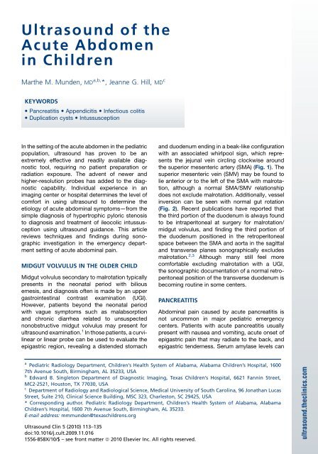
Fig. 1. Malrotation with midgut volvulus. Transverse<br />
image shows <strong>the</strong> jejunal ve<strong>in</strong> (thicker arrow) circl<strong>in</strong>g<br />
<strong>the</strong> SMA (slender arrow), represent<strong>in</strong>g <strong>the</strong> whirlpool<br />
sign. Incomplete obstruction caused by midgut<br />
volvulus was found at surgery.<br />
be normal with severe pancreatitis, and hyperamylasemia<br />
is not specific for pancreatic disease. 4<br />
Diagnosis requires at least a threefold elevation<br />
<strong>of</strong> serum pancreatic enzymes. Etiologies for acute<br />
Fig. 2. Abnormal SMA/SMV position. Transverse sonographic<br />
image shows <strong>the</strong> SMV (slender arrow) located<br />
anterior to <strong>the</strong> SMA (thicker arrow).<br />
pancreatitis <strong>in</strong> pediatrics <strong>in</strong>clude trauma, gallstones,<br />
systemic <strong>in</strong>fections, anatomic abnormalities<br />
<strong>of</strong> <strong>the</strong> pancreaticobiliary system, medications<br />
<strong>in</strong>clud<strong>in</strong>g immunosuppressive drug <strong>the</strong>rapy,<br />
hereditary pancreatitis, and congenital anomalies<br />
at <strong>the</strong> pancreaticobiliary junction <strong>in</strong>clud<strong>in</strong>g<br />
duodenal duplication cysts. A recurrence rate <strong>of</strong><br />
9% for pancreatitis is reported <strong>in</strong> children. 5<br />
The normal pancreas <strong>in</strong> children is <strong>of</strong>ten clearly<br />
visible sonographically <strong>in</strong> <strong>the</strong> fast<strong>in</strong>g patient and<br />
is homogeneously isoechoic to slightly hyperechoic<br />
to <strong>the</strong> liver <strong>in</strong> slender patients (Fig. 3). In<br />
patients with a larger body habitus, <strong>the</strong> pancreas<br />
is hyperechoic to <strong>the</strong> liver, as obesity is associated<br />
with <strong>in</strong>creas<strong>in</strong>g fatty accumulation with<strong>in</strong> <strong>the</strong><br />
pancreas. 6 The left lobe <strong>of</strong> <strong>the</strong> liver serves as an<br />
acoustic w<strong>in</strong>dow. If necessary, and if <strong>the</strong> patient<br />
is able, <strong>the</strong> stomach can be filled with water to<br />
provide an acoustic w<strong>in</strong>dow.<br />
<strong>Ultrasound</strong> f<strong>in</strong>d<strong>in</strong>gs <strong>in</strong> acute pancreatitis <strong>in</strong>clude<br />
focal or diffuse pancreatic enlargement with<br />
decreased dist<strong>in</strong>ction <strong>of</strong> pancreatic marg<strong>in</strong>s,<br />
heterogeneous echotexture, and peripancreatic<br />
fluid collections. The pancreas can be normal <strong>in</strong><br />
appearance <strong>in</strong> mild pancreatitis. 7 Chao and<br />
colleagues found a significant association<br />
between acute pancreatitis and a dilated pancreatic<br />
duct (>1.5 mm from ages 1 to 6 years, >1.9<br />
mm from ages 7 to 12, or >2.2 mm <strong>in</strong> <strong>the</strong> older<br />
child) (Fig. 4). 8 In <strong>the</strong> sett<strong>in</strong>g <strong>of</strong> acute epigastric<br />
pa<strong>in</strong> with ei<strong>the</strong>r sonographic or cl<strong>in</strong>ical f<strong>in</strong>d<strong>in</strong>gs<br />
suggest<strong>in</strong>g pancreatitis, <strong>the</strong> right upper quadrant<br />
should be evaluated for presence or absence <strong>of</strong><br />
gallstones, choledocholithiasis, choledochal<br />
cysts, and even duplication cysts which could<br />
lead to pancreatitis (Fig. 5). 9,10 <strong>Ultrasound</strong> is useful<br />
to assess complications <strong>of</strong> pancreatitis such as<br />
Fig. 3. Normal pancreas. Transverse sonographic<br />
image show<strong>in</strong>g a normal pancreas (arrow), isoechoic<br />
to <strong>the</strong> liver, <strong>in</strong> a 10-year-old child. Note dist<strong>in</strong>ct<br />
marg<strong>in</strong>s and homogenous echogenicity.
Fig. 4. <strong>Acute</strong> pancreatitis <strong>in</strong> a 10-year-old male. (A) Transverse image us<strong>in</strong>g l<strong>in</strong>ear transducer shows a mildly<br />
dilated pancreatic duct (arrow). (B) Transverse image shows enlargement and heterogeneously <strong>in</strong>creased echogenicity<br />
<strong>of</strong> <strong>the</strong> pancreatic head (between cursors).<br />
pseudocyst formation, pseudoaneurysms, and<br />
splenic ve<strong>in</strong> thrombosis (Fig. 6). In <strong>the</strong> sett<strong>in</strong>g <strong>of</strong><br />
traumatic pancreatic <strong>in</strong>jury, computed tomography<br />
(CT) is preferred <strong>in</strong> <strong>the</strong> acute sett<strong>in</strong>g.<br />
Follow-up <strong>of</strong>ten can be performed sonographically<br />
to watch for development for peripancreatic fluid<br />
collections and traumatic pancreatitis. Endoscopic<br />
ultrasound is also emerg<strong>in</strong>g as a valuable<br />
tool to evaluate pancreatic biliary disease <strong>in</strong> children.<br />
11,12 Magnetic resonance (MR) cholangiopancreatography<br />
is not performed by most<br />
centers <strong>in</strong> <strong>the</strong> sett<strong>in</strong>g <strong>of</strong> an acute abdomen.<br />
Fig. 5. Duodenal duplication cyst caus<strong>in</strong>g pancreatitis<br />
<strong>in</strong> a 2-year-old child. Transverse image shows diffuse<br />
enlargement <strong>of</strong> <strong>the</strong> pancreatic head (between slender<br />
arrows) secondary to a duodenal duplication cyst<br />
(short arrow).<br />
<strong>Ultrasound</strong> <strong>of</strong> <strong>the</strong> <strong>Acute</strong> <strong>Abdomen</strong> <strong>in</strong> <strong>Children</strong> 115<br />
APPENDICITIS<br />
Appendicitis rema<strong>in</strong>s one <strong>of</strong> <strong>the</strong> most common<br />
<strong>in</strong>dications for surgery <strong>in</strong> <strong>the</strong> pediatric population,<br />
and complications caused by delayed patient<br />
presentation rema<strong>in</strong> prevalent. Appendicitis<br />
occurs more commonly <strong>in</strong> developed countries<br />
and has a peak <strong>in</strong>cidence <strong>in</strong> males from 10 to 14<br />
years <strong>of</strong> age, and females from 15 to 19 years <strong>of</strong><br />
age, <strong>the</strong> time at which lymphoid follicles l<strong>in</strong><strong>in</strong>g<br />
<strong>the</strong> appendix reach <strong>the</strong>ir maximum size. 13<br />
Proposed etiologies for lumen obstruction <strong>of</strong> <strong>the</strong><br />
appendix <strong>in</strong>clude lymphoid hyperplasia (possibly<br />
Fig. 6. Pancreatic pseudocyst complicat<strong>in</strong>g first episode<br />
<strong>of</strong> acute pancreatitis <strong>in</strong> a 10-year-old girl. Transverse<br />
ultrasound image show<strong>in</strong>g a poorly def<strong>in</strong>ed pseudocyst<br />
conta<strong>in</strong><strong>in</strong>g <strong>in</strong>ternal debris (between arrows) adjacent<br />
to <strong>the</strong> pancreatic tail.
116<br />
<strong>Munden</strong> & Hill<br />
Fig. 7. Normal ascend<strong>in</strong>g colon. Sagittal sonographic image shows normal haustral mark<strong>in</strong>gs (arrow) <strong>of</strong> <strong>the</strong><br />
ascend<strong>in</strong>g colon.<br />
<strong>of</strong> viral etiology), dehydration, fecaliths, and even<br />
parasites. A decreased <strong>in</strong>cidence <strong>of</strong> appendicitis<br />
has been seen <strong>in</strong> those with higher dietary <strong>in</strong>take<br />
<strong>of</strong> fiber. 14 Symptoms can be vague, with less<br />
than half present<strong>in</strong>g with <strong>the</strong> classic scenario <strong>of</strong><br />
<strong>in</strong>sidious onset <strong>of</strong> abdom<strong>in</strong>al pa<strong>in</strong> migrat<strong>in</strong>g from<br />
<strong>the</strong> periumbilical region to <strong>the</strong> right lower quadrant<br />
(RLQ) associated with nausea and anorexia. 15<br />
Diarrhea is not typically seen unless <strong>the</strong>re is perforation<br />
and peritonitis, more <strong>of</strong>ten occurr<strong>in</strong>g <strong>in</strong> <strong>the</strong><br />
young patient under 3 years <strong>of</strong> age, frequently<br />
confused with gastroenteritis, which may lead to<br />
a delay <strong>in</strong> diagnosis. 16<br />
The rate <strong>of</strong> appendiceal perforation is significantly<br />
greater <strong>in</strong> children present<strong>in</strong>g with symptoms<br />
for more than 24 hours. 17 Unfortunately,<br />
<strong>the</strong> probability <strong>of</strong> appendiceal perforation is<br />
highest <strong>in</strong> those with more limited ability to<br />
communicate. The <strong>in</strong>cidence <strong>of</strong> perforation has<br />
been shown to be about 60% for a 3-year-old<br />
child, 50% for a 5-year-old child, and this <strong>in</strong>cidence<br />
decreases with <strong>in</strong>creas<strong>in</strong>g age. 18 The<br />
omentum is also underdeveloped <strong>in</strong> <strong>the</strong> very<br />
young and unable to conta<strong>in</strong> purulent material,<br />
which may be responsible for <strong>the</strong> diffuse<br />
peritonitis <strong>of</strong>ten present with perforated<br />
appendicitis <strong>in</strong> small children. Appendicitis is<br />
rare <strong>in</strong> neonates and <strong>in</strong>fants, possibly because<br />
<strong>of</strong> <strong>the</strong> funnel shape <strong>of</strong> <strong>the</strong> appendix <strong>in</strong> <strong>in</strong>fancy,<br />
which reduces <strong>the</strong> possibility <strong>of</strong> lum<strong>in</strong>al<br />
obstruction. 14,19<br />
<strong>Ultrasound</strong> evaluation <strong>of</strong> RLQ pa<strong>in</strong> is an excellent<br />
tool, as appendicitis and its mimics commonly<br />
present <strong>in</strong> <strong>the</strong> pediatric emergency department. It<br />
is not unusual to f<strong>in</strong>d multiple prior radiologic<br />
studies, <strong>in</strong>clud<strong>in</strong>g outside CT exam<strong>in</strong>ations, performed<br />
over a several year period <strong>in</strong> a s<strong>in</strong>gle<br />
patient for abdom<strong>in</strong>al pa<strong>in</strong>, which can lead to<br />
high-accumulated radiation doses if CT is used<br />
repeatedly. If necessary, with an equivocal <strong>in</strong>itial<br />
ultrasound exam<strong>in</strong>ation and cont<strong>in</strong>ued cl<strong>in</strong>ical<br />
concern for appendicitis, ultrasound can be<br />
repeated <strong>in</strong> a short <strong>in</strong>terval follow<strong>in</strong>g patient observation<br />
without <strong>the</strong> risk <strong>of</strong> additional radiation.<br />
At <strong>the</strong> beg<strong>in</strong>n<strong>in</strong>g <strong>of</strong> <strong>the</strong> ultrasound exam<strong>in</strong>ation,<br />
it is quite useful to ask <strong>the</strong> child to use a f<strong>in</strong>ger to<br />
po<strong>in</strong>t to <strong>the</strong> area where it hurts <strong>the</strong> most. Even<br />
<strong>the</strong> youngest <strong>of</strong> patients usually can comply.<br />
History obta<strong>in</strong>ed dur<strong>in</strong>g <strong>the</strong> ultrasound exam<strong>in</strong>ation<br />
is helpful <strong>in</strong> determ<strong>in</strong><strong>in</strong>g if <strong>the</strong> pa<strong>in</strong> is more <strong>of</strong><br />
a chronic nature, suggestive <strong>of</strong> possible <strong>in</strong>flammatory<br />
bowel disorder, acute with m<strong>in</strong>imal emesis as<br />
<strong>in</strong> appendicitis, or associated with diarrhea <strong>in</strong> an<br />
older child, more typical <strong>of</strong> gastroenteritis. Us<strong>in</strong>g<br />
a high-resolution l<strong>in</strong>ear transducer with cont<strong>in</strong>ued,<br />
gradual compression to displace bowel loops (<strong>the</strong><br />
technique described by Puylaert), <strong>the</strong> area <strong>in</strong>dicated<br />
by <strong>the</strong> patient should be exam<strong>in</strong>ed first. 20<br />
If <strong>the</strong> appendix is not found readily, slow, gradual<br />
compression is used to locate <strong>the</strong> borders <strong>of</strong> <strong>the</strong><br />
ascend<strong>in</strong>g colon (Fig. 7), identified by position<br />
Fig. 8. Normal appendix. Transverse image <strong>of</strong> <strong>the</strong><br />
right lower quadrant (RLQ) shows normal gut signature<br />
<strong>of</strong> a normal appendix, <strong>in</strong>clud<strong>in</strong>g <strong>the</strong> tip (between<br />
cursors).
Fig. 9. <strong>Acute</strong> appendicitis. Transverse sonographic<br />
image <strong>of</strong> <strong>the</strong> RLQ shows a distended appendix with<br />
<strong>in</strong>tact mucosa (arrow), hyperemia, but no<br />
surround<strong>in</strong>g <strong>in</strong>flammatory changes <strong>in</strong> this 9-year-old<br />
child with onset <strong>of</strong> pa<strong>in</strong> less than 24 hours.<br />
and detectable haustral folds. The transducer is<br />
moved caudally to f<strong>in</strong>d <strong>the</strong> cecum and term<strong>in</strong>al<br />
ileum, identified by its compressibility, peristaltic<br />
activity, and lack <strong>of</strong> a bl<strong>in</strong>d tip term<strong>in</strong>ation. The<br />
appendix arises from <strong>the</strong> cecal tip 1 to 2 cm below<br />
<strong>the</strong> orig<strong>in</strong> <strong>of</strong> <strong>the</strong> term<strong>in</strong>al ileum. Additional techniques,<br />
such as us<strong>in</strong>g <strong>the</strong> opposite hand to<br />
compress posteriorly while scann<strong>in</strong>g anteriorly,<br />
as well as plac<strong>in</strong>g <strong>the</strong> patient <strong>in</strong> <strong>the</strong> left lateral decubitus<br />
position to displace <strong>the</strong> cecum and<br />
appendix more medially, can be useful. 21<br />
The appendix is retrocecal <strong>in</strong> location <strong>in</strong> 28% to<br />
68% <strong>of</strong> patients at surgery and autopsy and with<strong>in</strong><br />
<strong>the</strong> pelvis <strong>in</strong> 12.8% to 53% <strong>of</strong> cases. 14,22 When <strong>the</strong><br />
appendix is retrocecal and extraperitoneal,<br />
<strong>Ultrasound</strong> <strong>of</strong> <strong>the</strong> <strong>Acute</strong> <strong>Abdomen</strong> <strong>in</strong> <strong>Children</strong> 117<br />
patients may have less focal abdom<strong>in</strong>al pa<strong>in</strong> and<br />
more flank pa<strong>in</strong>, which can lead to difficulty <strong>in</strong><br />
diagnosis. Baldiserotto and Marchiori have shown<br />
success <strong>in</strong> f<strong>in</strong>d<strong>in</strong>g <strong>the</strong> retrocecal appendix by<br />
exam<strong>in</strong><strong>in</strong>g <strong>the</strong> right flank transversely us<strong>in</strong>g a curvil<strong>in</strong>ear<br />
transducer start<strong>in</strong>g from <strong>the</strong> edge <strong>of</strong> <strong>the</strong> liver<br />
to <strong>the</strong> iliac crest, <strong>the</strong>n longitud<strong>in</strong>ally from <strong>the</strong> axillary<br />
l<strong>in</strong>e cont<strong>in</strong>u<strong>in</strong>g posteriorly to <strong>the</strong> lumbar<br />
region. For a deep pelvic location, a curvil<strong>in</strong>ear<br />
transducer can be used scann<strong>in</strong>g through a distended<br />
bladder, followed by a l<strong>in</strong>ear transducer<br />
with graded compression after void<strong>in</strong>g if a dilated<br />
appendix is not found. 22<br />
The normal appendix is a slender tubular<br />
structure with preserved gut signature, easily<br />
compressible with no hyperemia, and a mean<br />
diameter <strong>of</strong> 0.39 cm (Fig. 8). The appendiceal<br />
lumen <strong>of</strong> <strong>the</strong> normal appendix may be empty or<br />
filled with fecal material, gas, or both. In some<br />
centers experienced <strong>in</strong> gastro<strong>in</strong>test<strong>in</strong>al (GI) ultrasound,<br />
<strong>the</strong> normal appendix has been found <strong>in</strong><br />
up to 82% <strong>of</strong> children. 23<br />
In about 5% <strong>of</strong> cases, <strong>in</strong>flammation is<br />
conf<strong>in</strong>ed to <strong>the</strong> appendiceal tip, mak<strong>in</strong>g exam<strong>in</strong>ation<br />
<strong>of</strong> <strong>the</strong> entire appendix and identify<strong>in</strong>g<br />
<strong>the</strong> tip important. 24,25 The sonographic diagnosis<br />
<strong>of</strong> appendicitis can be made when a reproducible<br />
fluid-filled, noncompressible, distended<br />
tubular structure is found that measures greater<br />
than 6 mm <strong>in</strong> diameter with calipers placed at<br />
<strong>the</strong> outer borders <strong>of</strong> <strong>the</strong> muscularis propria<br />
(Fig. 9). The <strong>in</strong>flamed appendix may conta<strong>in</strong><br />
a shadow<strong>in</strong>g fecalith, caus<strong>in</strong>g sharply def<strong>in</strong>ed<br />
posterior acoustic shadow<strong>in</strong>g. Hyperemia <strong>of</strong><br />
<strong>the</strong> wall <strong>of</strong> <strong>the</strong> appendix has been shown to<br />
be a sensitive <strong>in</strong>dicator <strong>of</strong> <strong>in</strong>flammation, helpful<br />
<strong>in</strong> borderl<strong>in</strong>e cases. 26 With <strong>in</strong>creas<strong>in</strong>g<br />
Fig. 10. Perforated appendix conta<strong>in</strong><strong>in</strong>g fecaliths <strong>in</strong> this 7-year-old boy with onset <strong>of</strong> pa<strong>in</strong> 3 days prior. (A) <strong>Ultrasound</strong><br />
image shows loss <strong>of</strong> gut signature <strong>in</strong> this distended appendix (slender arrow) conta<strong>in</strong><strong>in</strong>g two shadow<strong>in</strong>g<br />
fecaliths (short arrows). (B) Transverse image shows appendix (A) and small amount <strong>of</strong> free fluid (FF) lateral to <strong>the</strong><br />
dilated ascend<strong>in</strong>g colon.
118<br />
<strong>Munden</strong> & Hill<br />
Fig. 11. Omental fat surround<strong>in</strong>g a perforated<br />
appendix. Transverse image through <strong>the</strong> RLQ shows<br />
a distended, hyperemic appendix surrounded by<br />
echogenic omental fat (arrow) <strong>in</strong> this patient with<br />
3 days <strong>of</strong> pa<strong>in</strong> and a perforated appendix at surgery.<br />
<strong>in</strong>flammation from obstruction, loss <strong>of</strong> venous<br />
outflow and wall ischemia develops, caus<strong>in</strong>g<br />
loss <strong>of</strong> <strong>the</strong> normally visible alternat<strong>in</strong>g layers <strong>of</strong><br />
<strong>the</strong> normal gut signature (Fig. 10). Bacterial<br />
<strong>in</strong>vasion <strong>of</strong> <strong>the</strong> appendiceal wall, and thrombosis<br />
<strong>of</strong> <strong>the</strong> appendicular artery and ve<strong>in</strong>s, eventually<br />
lead to gangrene and perforation <strong>of</strong> <strong>the</strong><br />
appendix. Echogenic surround<strong>in</strong>g omental fat<br />
and adjacent pericecal or periappendiceal free<br />
fluid develops, suggest<strong>in</strong>g appendiceal perforation<br />
(Fig. 11). 27,28 In <strong>the</strong> absence <strong>of</strong> a visualized<br />
appendix because <strong>of</strong> perforation, hyperechoic<br />
mesenteric fat, RLQ and pelvic fluid collections,<br />
dilated small bowel, and a free-float<strong>in</strong>g fecalith<br />
can be strong <strong>in</strong>dicators <strong>of</strong> <strong>in</strong>flammation caused<br />
by a ruptured appendix (Fig. 12). 29,30<br />
Treatment for perforated appendicitis is controversial,<br />
and <strong>in</strong>formation regard<strong>in</strong>g <strong>the</strong> location,<br />
size, and complexity <strong>of</strong> abscess collections, and<br />
presence or absence <strong>of</strong> a fecalith, provide necessary<br />
<strong>in</strong>formation for decisions <strong>in</strong> management.<br />
These should be evaluated sonographically at<br />
presentation. 31,32 When <strong>in</strong>dicated, additional CT<br />
imag<strong>in</strong>g is used.<br />
An exception to <strong>the</strong> accepted diameter for an<br />
<strong>in</strong>flamed appendix <strong>in</strong>cludes patients with cystic<br />
fibrosis (CF). The <strong>in</strong>cidence <strong>of</strong> appendicitis <strong>in</strong><br />
CF is 1% to 2%, lower than <strong>the</strong> general population<br />
(7%), and <strong>the</strong> diameter <strong>of</strong> <strong>the</strong> compressed<br />
appendix identified <strong>in</strong> CF patients without<br />
appendicitis has been shown to be greater<br />
than 6 mm <strong>in</strong> both <strong>the</strong> pediatric and adult<br />
populations. 33<br />
Although <strong>in</strong>itial evaluation for appendicitis with<br />
ultrasound is preferred <strong>in</strong> many centers, obesity<br />
and <strong>the</strong> <strong>in</strong>dividual experience <strong>of</strong> an imag<strong>in</strong>g<br />
center may help determ<strong>in</strong>e which patient<br />
undergoes an ultrasound and which patient<br />
would benefit from <strong>in</strong>itial evaluation with CT.<br />
APPENDICITIS IN THE VERY YOUNG<br />
As mentioned previously, appendicitis occurs less<br />
frequently <strong>in</strong> <strong>the</strong> young child, but <strong>the</strong> perforation<br />
rate is high. 18 Abscess formation is much more<br />
likely <strong>in</strong> children under 5 years <strong>of</strong> age than <strong>in</strong> older<br />
children. One must keep appendicitis <strong>in</strong> m<strong>in</strong>d<br />
when exam<strong>in</strong><strong>in</strong>g a young child with symptoms <strong>of</strong><br />
gastroenteritis last<strong>in</strong>g longer than expected.<br />
Inflammation from an underly<strong>in</strong>g ruptured<br />
appendix can lead to unusual cl<strong>in</strong>ical presentations<br />
such as a failure to bear weight, flank pa<strong>in</strong><br />
Fig. 12. Multiple abscesses complicat<strong>in</strong>g a perforated appendix <strong>in</strong> a 23-month-old. (A) A transverse image <strong>of</strong> <strong>the</strong><br />
RLQ shows echogenic free fluid (short arrow) conta<strong>in</strong><strong>in</strong>g a free-float<strong>in</strong>g fecalith (slender arrow). CT coronal reformatted<br />
image obta<strong>in</strong>ed 3 days later (B) show<strong>in</strong>g fecalith now located left lower quadrant (LLQ) (arrow)<br />
and multiple abscess collections (arrow heads) throughout <strong>the</strong> abdomen.
Fig. 13. Mesenteric adenitis. Transverse image <strong>of</strong> <strong>the</strong><br />
RLQ show<strong>in</strong>g prom<strong>in</strong>ent, mildly hyperemic lymph nodes<br />
(arrow) caused by mesenteric adenitis. A normal<br />
appendix was present.<br />
with fever, and scrotal swell<strong>in</strong>g <strong>in</strong> <strong>the</strong> very<br />
young. 16,18,19,34<br />
GI PATHOLOGY THAT MAY MIMIC<br />
APPENDICITIS<br />
Mesenteric adenitis is one <strong>of</strong> <strong>the</strong> most common<br />
alternate diagnoses made dur<strong>in</strong>g evaluation for<br />
suspected appendicitis. 35 Primary mesenteric<br />
adenitis has been def<strong>in</strong>ed as a self-limit<strong>in</strong>g <strong>in</strong>flammation<br />
<strong>of</strong> RLQ mesenteric nodes with three or<br />
more lymph nodes 5 mm or greater <strong>in</strong> short axis<br />
diameter, and <strong>the</strong> presence <strong>of</strong> a normal appendix<br />
(Fig. 13). In secondary mesenteric adenitis,<br />
a specific <strong>in</strong>flammatory process is present such<br />
as <strong>in</strong>fectious colitis, Crohn disease, or ulcerative<br />
colitis. 36 Infectious colitis may be caused Yers<strong>in</strong>ia<br />
enterocolitica, Campylobacter jejuni, Salmonella,<br />
or o<strong>the</strong>r organisms. 37,38 It is important to identify<br />
<strong>Ultrasound</strong> <strong>of</strong> <strong>the</strong> <strong>Acute</strong> <strong>Abdomen</strong> <strong>in</strong> <strong>Children</strong> 119<br />
a normal appendix, as lymph nodes also can be<br />
enlarged with appendicitis.<br />
With viral gastroenteritis, f<strong>in</strong>d<strong>in</strong>gs are nonspecific<br />
but may <strong>in</strong>clude a fluid-filled colon, small<br />
amounts <strong>of</strong> ascites. F<strong>in</strong>d<strong>in</strong>gs, however, usually<br />
do not <strong>in</strong>clude <strong>in</strong>test<strong>in</strong>al wall thicken<strong>in</strong>g (>1.5 mm<br />
<strong>in</strong> <strong>the</strong> term<strong>in</strong>al ileum or >2 mm <strong>in</strong> <strong>the</strong> colon). 39<br />
<strong>Ultrasound</strong> f<strong>in</strong>d<strong>in</strong>gs with bacterial ileocolitis<br />
caused by Yers<strong>in</strong>ia, Campylobacter, Salmonella,<br />
and o<strong>the</strong>r organisms <strong>in</strong>clude wall thicken<strong>in</strong>g <strong>of</strong><br />
<strong>the</strong> term<strong>in</strong>al ileum and cecum with hyperemia<br />
and adjacent enlarged lymph nodes (Fig. 14). A<br />
small amount <strong>of</strong> ascites is <strong>of</strong>ten present, but <strong>the</strong><br />
surround<strong>in</strong>g fat is not <strong>in</strong>flamed. 40,41 Although<br />
<strong>the</strong>se f<strong>in</strong>d<strong>in</strong>gs are nonspecific as to <strong>the</strong> underly<strong>in</strong>g<br />
etiology, <strong>the</strong>y can provide an alternate diagnosis<br />
and help to exclude appendicitis.<br />
In <strong>the</strong> sett<strong>in</strong>g <strong>of</strong> abdom<strong>in</strong>al pa<strong>in</strong> with bloody diarrhea<br />
<strong>in</strong> a young child, <strong>the</strong> f<strong>in</strong>d<strong>in</strong>g <strong>of</strong> wall thicken<strong>in</strong>g<br />
<strong>of</strong> <strong>the</strong> colon may represent <strong>the</strong> <strong>in</strong>test<strong>in</strong>al prodrome,<br />
which precedes <strong>the</strong> hematologic and renal manifestations<br />
<strong>of</strong> hemolytic–uremic syndrome (HUS).<br />
The <strong>in</strong>volvement <strong>of</strong> <strong>the</strong> colon varies from segmental<br />
to diffuse, with <strong>the</strong> transverse colon most<br />
frequently <strong>in</strong>volved. Small bowel loops can be<br />
<strong>in</strong>volved along with mesenteric <strong>in</strong>flammatory<br />
changes. The cl<strong>in</strong>ical history is crucial for diagnosis<br />
as HUS occurs a few days after <strong>in</strong>gestion <strong>of</strong><br />
contam<strong>in</strong>ated or undercooked ground meat conta<strong>in</strong><strong>in</strong>g<br />
Escherichia coli O157:H7, Shigella,<br />
Campylobacter, and o<strong>the</strong>r organisms. 42–44<br />
INFLAMMATORY BOWEL DISEASE<br />
Bowel wall thicken<strong>in</strong>g is a nonspecific sonographic<br />
f<strong>in</strong>d<strong>in</strong>g that can be seen with <strong>in</strong>fectious,<br />
<strong>in</strong>flammatory, and ischemic bowel disease. The<br />
degree <strong>of</strong> wall thicken<strong>in</strong>g and distribution <strong>of</strong><br />
Fig. 14. Bacterial enteritis. (A) <strong>Ultrasound</strong> show<strong>in</strong>g marked wall thicken<strong>in</strong>g <strong>of</strong> <strong>the</strong> cecum (arrow) <strong>in</strong> a child with<br />
RLQ pa<strong>in</strong>, which returned to normal (B) 4 days later. Stool cultures were positive for enterohemorrhagic<br />
Escherichia coli.
120<br />
<strong>Munden</strong> & Hill<br />
Fig. 15. Marked wall thicken<strong>in</strong>g <strong>of</strong> <strong>the</strong> ascend<strong>in</strong>g<br />
colon (cursors) with loss <strong>of</strong> normal layer stratification<br />
<strong>in</strong> this patient present<strong>in</strong>g with RLQ pa<strong>in</strong>, later diagnosed<br />
as Crohn disease.<br />
affected bowel vary between Crohn disease,<br />
ulcerative colitis (UC), and o<strong>the</strong>r nonviral GI<br />
disease. In <strong>the</strong> acute sett<strong>in</strong>g, ultrasound is helpful<br />
<strong>in</strong> determ<strong>in</strong><strong>in</strong>g <strong>the</strong> location and extent <strong>of</strong> bowel<br />
disease, which <strong>in</strong> context with cl<strong>in</strong>ical <strong>in</strong>formation,<br />
can lead to a diagnosis or differential diagnosis. 45<br />
Although controversial, <strong>in</strong>creased bowel wall<br />
thickness with loss <strong>of</strong> <strong>the</strong> normal layer stratification<br />
<strong>of</strong> hypoechoic mucosa and muscularis mucosa,<br />
echogenic submucosa, and hypoechoic muscularis<br />
propria is felt by some to correlate with <strong>the</strong><br />
severity <strong>of</strong> <strong>in</strong>flammation with <strong>in</strong>flammatory bowel<br />
disease (IBD). 39<br />
Crohn disease is a transmural <strong>in</strong>flammation <strong>of</strong><br />
<strong>the</strong> affected bowel that results <strong>in</strong> severe<br />
bowel wall thicken<strong>in</strong>g, <strong>in</strong>flammation <strong>of</strong> <strong>the</strong> fat<br />
surround<strong>in</strong>g <strong>the</strong> ileum and cecum, mesenteric adenopathy,<br />
and bowel loop separation. The target<br />
sign is <strong>the</strong> classic sonographic f<strong>in</strong>d<strong>in</strong>g described<br />
<strong>in</strong> Crohn disease with transverse images <strong>of</strong><br />
affected bowel show<strong>in</strong>g a strong echogenic center<br />
and surround<strong>in</strong>g hypoechoic rim. 45 The <strong>in</strong>volved<br />
areas are ma<strong>in</strong>ly small bowel and ascend<strong>in</strong>g colon,<br />
with a mural thickness rang<strong>in</strong>g from 9 to 10 mm<br />
(Fig. 15). 46 With chronic disease, <strong>the</strong> bowel wall<br />
is thickened and typically hypoechoic because <strong>of</strong><br />
fibrosis. Complications <strong>of</strong> Crohn disease <strong>in</strong>clude<br />
abscess formation, bowel obstruction, and fistula<br />
formation. Gas bubbles may be seen with<strong>in</strong> fistulous<br />
tracts. 47 Up to one third <strong>of</strong> patients with Crohn<br />
disease <strong>in</strong>volv<strong>in</strong>g <strong>the</strong> ileocecal region present with<br />
acute symptoms that mimic appendicitis. 48 The<br />
appendix itself can be <strong>in</strong>volved with Crohn<br />
disease, seen <strong>in</strong> 21% <strong>of</strong> patients <strong>in</strong> a series reported<br />
by Ripolles and colleagues (Fig. 16). 49 All<br />
<strong>of</strong> <strong>the</strong>ir patients with appendiceal <strong>in</strong>volvement by<br />
Crohn disease also had associated thicken<strong>in</strong>g <strong>of</strong><br />
<strong>the</strong> term<strong>in</strong>al ileum or cecum, which helped differentiate<br />
<strong>the</strong> f<strong>in</strong>d<strong>in</strong>gs from acute appendicitis.<br />
Diffuse wall thicken<strong>in</strong>g is also seen with ulcerative<br />
colitis, but <strong>the</strong> thickness <strong>of</strong> <strong>the</strong> ileum has<br />
been noted to be about 5 mm, while <strong>the</strong> colon<br />
wall thickness was greater. Range <strong>of</strong> mural wall<br />
thickness <strong>of</strong> <strong>the</strong> colon <strong>in</strong> UC has been shown to<br />
be a little wider, 7 mm to 10 mm, and overall less<br />
prom<strong>in</strong>ent than that <strong>of</strong> Crohn disease, with<br />
preserved wall stratification <strong>in</strong> UC. 45,46 Sonographic<br />
features that may help differentiate<br />
between Crohn disease and UC <strong>in</strong>clude location<br />
<strong>of</strong> bowel wall thicken<strong>in</strong>g, with presence <strong>of</strong> skip<br />
lesions and pericolic abscess more likely <strong>in</strong> Crohn<br />
disease. With underly<strong>in</strong>g severe disease, ultrasound<br />
is less risky than direct visualization <strong>in</strong><br />
search<strong>in</strong>g for areas <strong>of</strong> bowel <strong>in</strong>volvement.<br />
Fig. 16. Distended appendix caused by Crohn disease. (A) <strong>Ultrasound</strong> RLQ shows a thickened appendix (arrow)<br />
with surround<strong>in</strong>g ascites due to Crohn disease with coronal reformatted CT (B) show<strong>in</strong>g enhanc<strong>in</strong>g appendix<br />
(slender arrow) with <strong>in</strong>flammation <strong>of</strong> ascend<strong>in</strong>g colon and cecum (short arrow).
OMENTAL INFARCTION<br />
Omental <strong>in</strong>farction, a rare cause for acute abdom<strong>in</strong>al<br />
pa<strong>in</strong>, is more common <strong>in</strong> adults, but about<br />
15% <strong>of</strong> cases present <strong>in</strong> <strong>the</strong> pediatric population.<br />
Symptoms <strong>in</strong>clude pa<strong>in</strong> localized to <strong>the</strong> right upper<br />
or lower quadrant <strong>of</strong> a few days’ duration, but<br />
associated nausea and vomit<strong>in</strong>g are rare. 50 The<br />
pathogenesis is unknown, and it may occur with<br />
or without torsion <strong>of</strong> <strong>the</strong> omentum. Predispos<strong>in</strong>g<br />
factors may <strong>in</strong>clude k<strong>in</strong>k<strong>in</strong>g <strong>of</strong> ve<strong>in</strong>s or venous<br />
congestion follow<strong>in</strong>g a large meal, venous <strong>in</strong>sufficiency<br />
caused by trauma, or thrombosis <strong>of</strong><br />
omental ve<strong>in</strong>s. 51,52 Additional predispos<strong>in</strong>g factors<br />
<strong>Ultrasound</strong> <strong>of</strong> <strong>the</strong> <strong>Acute</strong> <strong>Abdomen</strong> <strong>in</strong> <strong>Children</strong> 121<br />
Fig. 17. Omental <strong>in</strong>farction. (A) <strong>Ultrasound</strong> shows diffuse echogenicity (between arrows) with<strong>in</strong> <strong>the</strong> anterior<br />
abdomen just medial to <strong>the</strong> ascend<strong>in</strong>g colon, confirmed by CT (B) show<strong>in</strong>g <strong>in</strong>flammation caused by <strong>in</strong>farcted<br />
omentum (between arrows).<br />
<strong>in</strong>clude strenuous activity and obesity, which is<br />
<strong>in</strong>creas<strong>in</strong>g <strong>in</strong> <strong>the</strong> childhood population.<br />
<strong>Ultrasound</strong> f<strong>in</strong>d<strong>in</strong>gs <strong>in</strong>clude an ovoid, hyperechoic<br />
s<strong>of</strong>t tissue mass located between <strong>the</strong> anterior<br />
abdom<strong>in</strong>al wall and transverse or ascend<strong>in</strong>g<br />
colon with few vessels with<strong>in</strong> <strong>the</strong> mass and peripheral<br />
hyperemia (Fig. 17). 53 A less common appearance<br />
<strong>in</strong>cludes a hyperechoic mass conta<strong>in</strong><strong>in</strong>g an<br />
avascular hypoechoic tubular structure, which<br />
has been found to represent twisted, <strong>in</strong>farcted<br />
omental tissue at gross pathology. 51 Some advocate<br />
surgical treatment, and o<strong>the</strong>rs recommend<br />
a conservative approach to <strong>the</strong>rapy.<br />
Fig. 18. Pseudomembranous colitis. (A) <strong>Ultrasound</strong> shows ascites with diffuse wall thicken<strong>in</strong>g <strong>of</strong> <strong>the</strong> splenic<br />
flexure (between cursors) and (B) rectum (arrows) compatible with pancolonic <strong>in</strong>volvement <strong>of</strong> pseudomembranous<br />
colitis.
122<br />
<strong>Munden</strong> & Hill<br />
PSEUDOMEMBRANOUS COLITIS<br />
Pseudomembranous colitis (PMC) is associated<br />
with a prior course <strong>of</strong> antibiotics or o<strong>the</strong>r agents<br />
that allow super<strong>in</strong>fection with Clostridium difficile.<br />
In <strong>the</strong> preantibiotic era, PMC was associated<br />
with shock, uremia, heavy metal <strong>in</strong>toxication, and<br />
cardiovascular disease. The enterotox<strong>in</strong> <strong>of</strong> C difficile<br />
causes a severe <strong>in</strong>flammatory response with<strong>in</strong><br />
<strong>the</strong> colon. Symptoms <strong>of</strong> PMC typically occur after<br />
about 4 days <strong>of</strong> antibiotic <strong>the</strong>rapy but have been<br />
reported to occur as long as 6 weeks later.<br />
Patients present with watery diarrhea, severe<br />
abdom<strong>in</strong>al pa<strong>in</strong>, fever, and leukocytosis. Diagnosis<br />
is made with detection <strong>of</strong> C difficile <strong>in</strong> <strong>the</strong><br />
stool, tissue culture assay for tox<strong>in</strong> B, or antigen<br />
detection <strong>in</strong> <strong>the</strong> stool by rapid enzyme<br />
immunoassays. 54<br />
PMC causes <strong>the</strong> formation <strong>of</strong> yellow plaques, or<br />
pseudo membranes adherent to <strong>the</strong> gut mucosa<br />
and is generally a pancolitis, although at times<br />
<strong>in</strong>volvement is more segmental. 47 Pla<strong>in</strong> film f<strong>in</strong>d<strong>in</strong>gs<br />
<strong>of</strong> generalized thumb pr<strong>in</strong>t<strong>in</strong>g throughout <strong>the</strong><br />
colon are typical, represent<strong>in</strong>g <strong>the</strong> strik<strong>in</strong>g thicken<strong>in</strong>g<br />
<strong>of</strong> <strong>the</strong> colon wall. <strong>Ultrasound</strong> f<strong>in</strong>d<strong>in</strong>gs <strong>in</strong>clude<br />
marked thicken<strong>in</strong>g <strong>of</strong> a hypoechoic mucosal layer<br />
with narrow<strong>in</strong>g <strong>of</strong> <strong>the</strong> gut lumen and thickened<br />
echogenic submucosa represent<strong>in</strong>g <strong>the</strong> plaque<br />
formation with associated edema. The hypoechoic<br />
outer layer <strong>of</strong> muscularis propria is normal and<br />
th<strong>in</strong>. Ascites is present <strong>in</strong> up to 77% <strong>of</strong> those<br />
with PMC (Fig. 18). 55<br />
HENOCH-SCHONLEIN PURPURA<br />
Henoch-Schonle<strong>in</strong> purpura (HSP) is a systemic<br />
vasculitis primarily affect<strong>in</strong>g children around <strong>the</strong><br />
age <strong>of</strong> 4, although persons <strong>of</strong> any age can be<br />
affected. Pediatric cases <strong>of</strong>ten are preceded by<br />
a viral or bacterial <strong>in</strong>fection. The etiology is uncerta<strong>in</strong>,<br />
but it represents a disorder <strong>of</strong> <strong>the</strong> immune<br />
response with immune complexes deposited <strong>in</strong><br />
glomerular capillaries and small blood vessels <strong>of</strong><br />
<strong>the</strong> sk<strong>in</strong>, GI tract, and o<strong>the</strong>r organs. 56 Cl<strong>in</strong>ically,<br />
patients present with a petechial or purpuric rash,<br />
more commonly <strong>in</strong>volv<strong>in</strong>g <strong>the</strong> lower extremities,<br />
arthritis, and nephritis. Abdom<strong>in</strong>al pa<strong>in</strong> caused by<br />
hemorrhage and edema <strong>of</strong> bowel occurs <strong>in</strong> 50%<br />
to 60% <strong>of</strong> patients. The abdom<strong>in</strong>al pa<strong>in</strong> may<br />
precede or follow <strong>the</strong> rash. The duodenum and<br />
small bowel are <strong>the</strong> most frequent sites <strong>of</strong> <strong>in</strong>volvement<br />
<strong>of</strong> <strong>the</strong> GI tract <strong>in</strong> HSP. Bowel wall thicken<strong>in</strong>g<br />
caused by vasculitis-associated ischemia has<br />
a mean thickness <strong>of</strong> 9 mm and <strong>in</strong>volves longer<br />
segments than bowel thicken<strong>in</strong>g associated with<br />
hematoma (Fig. 19). 57,58 Echogenic thicken<strong>in</strong>g <strong>of</strong><br />
<strong>the</strong> mucosa and submucosa tends to be circumferential<br />
<strong>in</strong> <strong>the</strong> areas <strong>of</strong> ischemia. Asymmetric bowel<br />
wall thicken<strong>in</strong>g can be seen with associated <strong>in</strong>tramural<br />
hematoma, sometimes confused with <strong>in</strong>tussusception,<br />
a well-known complication <strong>of</strong> HSP. 59<br />
Free peritoneal fluid is commonly present.<br />
LYMPHOMA<br />
The most frequent site <strong>of</strong> non-Hodgk<strong>in</strong> lymphoma<br />
<strong>in</strong>volvement <strong>in</strong> children is <strong>the</strong> GI tract, affect<strong>in</strong>g<br />
distal ileum, cecum, appendix, and ascend<strong>in</strong>g<br />
colon most frequently. 60 Burkitt’s lymphoma is a<br />
highly aggressive type <strong>of</strong> non-Hodgk<strong>in</strong> lymphoma<br />
that commonly affects children. The nonendemic,<br />
sporadic form <strong>of</strong> Burkitt lymphoma accounts for<br />
up to 40% <strong>of</strong> lymphoma <strong>in</strong> children <strong>in</strong> <strong>the</strong> United<br />
States and western Europe. 61 <strong>Children</strong> <strong>of</strong>ten<br />
Fig. 19. Henoch-Schonle<strong>in</strong> purpura <strong>in</strong> a 16-year-old boy present<strong>in</strong>g with abdom<strong>in</strong>al pa<strong>in</strong> and petechial rash. (A)<br />
Sagittal ultrasound image shows more moderate wall thicken<strong>in</strong>g <strong>of</strong> small bowel with ascites. (B) Coronal CT<br />
shows long segments <strong>of</strong> thickened, enhanc<strong>in</strong>g, fluid filled small bowel (arrows).
present with abdom<strong>in</strong>al pa<strong>in</strong> or lower abdom<strong>in</strong>al<br />
mass, with vomit<strong>in</strong>g, GI bleed<strong>in</strong>g, or possible<br />
secondary <strong>in</strong>tussusceptions. Symptoms may be<br />
confused with appendicitis. The tumor spreads<br />
throughout <strong>the</strong> submucosa and can be seen sonographically<br />
as severe hypoechoic bowel wall thicken<strong>in</strong>g<br />
with loss <strong>of</strong> normal stratification <strong>of</strong> layers<br />
(Fig. 20). A focal mass with large hypoechoic areas<br />
may be seen <strong>in</strong> areas with secondary tumor<br />
necrosis. The bowel lumen may be narrowed<br />
because <strong>of</strong> tumor <strong>in</strong>filtration. 62 Enlarged mesenteric<br />
nodes may be seen.<br />
INTUSSUSCEPTION<br />
Intussusception, an <strong>in</strong>vag<strong>in</strong>ation or telescop<strong>in</strong>g <strong>of</strong><br />
bowel <strong>in</strong>to an adjacent segment <strong>of</strong> distal bowel<br />
(Fig. 21), is a common pediatric abdom<strong>in</strong>al emergency<br />
with a reported <strong>in</strong>cidence <strong>of</strong> 56 cases per<br />
Fig. 21. Intussusception. Intraoperative image dem<br />
onstrat<strong>in</strong>g an ileocolic <strong>in</strong>tussusception with <strong>in</strong>tussuscepted<br />
loop and mesenteric fat (arrow) enter<strong>in</strong>g<br />
<strong>in</strong>tussuscipiens.<br />
<strong>Ultrasound</strong> <strong>of</strong> <strong>the</strong> <strong>Acute</strong> <strong>Abdomen</strong> <strong>in</strong> <strong>Children</strong> 123<br />
Fig. 20. Lymphoma. (A) Sagittal ultrasound image shows diffuse and marked bowel wall thicken<strong>in</strong>g (arrow) and<br />
(B) hyperemic solid mass surrounded by ascites <strong>in</strong> this 3-year-old child present<strong>in</strong>g with abdom<strong>in</strong>al pa<strong>in</strong> caused by<br />
lymphoma.<br />
100,000 hospitalizations per year <strong>in</strong> <strong>the</strong> United<br />
States. 63 The so-called idiopathic ileocolic <strong>in</strong>tussusceptions<br />
arise from <strong>the</strong> ileum secondary to<br />
hyperplasia <strong>of</strong> <strong>the</strong> Peyer’s patches and typically<br />
present with <strong>in</strong>termittent, colicky abdom<strong>in</strong>al pa<strong>in</strong>.<br />
The usual age at presentation is 5 months to 3<br />
years, with peak <strong>in</strong>cidence between <strong>the</strong> ages <strong>of</strong><br />
5 and 9 months. 64 The development <strong>of</strong> an <strong>in</strong>tussusception<br />
<strong>in</strong> patients outside <strong>of</strong> this age range<br />
suggests an underly<strong>in</strong>g pathologic lead po<strong>in</strong>t,<br />
ei<strong>the</strong>r from a focal or diffuse abnormality <strong>of</strong> <strong>the</strong><br />
GI tract. 65<br />
Cl<strong>in</strong>ical presentation is highly variable. The<br />
classic triad <strong>of</strong> <strong>in</strong>termittent abdom<strong>in</strong>al pa<strong>in</strong>,<br />
Fig. 22. Intussusception, donut sign. Transverse image<br />
<strong>of</strong> <strong>the</strong> apex <strong>of</strong> <strong>the</strong> <strong>in</strong>tussusception (arrow) shows <strong>the</strong><br />
hypoechoic donut.
124<br />
<strong>Munden</strong> & Hill<br />
Fig. 23. Intussusception, crescent-<strong>in</strong>-donut sign.A<br />
crescent <strong>of</strong> echogenic mesenteric fat surrounds <strong>the</strong><br />
central <strong>in</strong>tussusceptum.<br />
vomit<strong>in</strong>g and right upper quadrant abdom<strong>in</strong>al<br />
mass has a positive predictive value <strong>of</strong> 93%,<br />
<strong>in</strong>creas<strong>in</strong>g to 100% with <strong>the</strong> addition <strong>of</strong> rectal<br />
bleed<strong>in</strong>g. 66 The triad <strong>of</strong> colicky abdom<strong>in</strong>al pa<strong>in</strong>,<br />
abdom<strong>in</strong>al mass and currant jelly stools, however,<br />
occurs <strong>in</strong> less than 50% <strong>of</strong> patients, and symptoms<br />
frequently overlap with o<strong>the</strong>r etiologies,<br />
mak<strong>in</strong>g cl<strong>in</strong>ical diagnosis difficult. 67,68 In addition<br />
to <strong>the</strong> characteristic pa<strong>in</strong>, o<strong>the</strong>r cl<strong>in</strong>ical features<br />
at presentation may <strong>in</strong>clude irritability, bilious<br />
vomit<strong>in</strong>g, bloody stools, and lethargy. Should lethargy<br />
predom<strong>in</strong>ate, <strong>in</strong>itial misdiagnosis <strong>of</strong> a central<br />
nervous system abnormality is not uncommon. In<br />
fact, as many as 70% <strong>of</strong> <strong>the</strong> children with <strong>the</strong>se<br />
abdom<strong>in</strong>al symptoms fail to have <strong>in</strong>tussusception.<br />
69 On <strong>the</strong> o<strong>the</strong>r hand, accurate, timely<br />
diagnosis and treatment <strong>of</strong> <strong>in</strong>tussusception are<br />
important, as delay may result <strong>in</strong> obstruction,<br />
bowel ischemia, perforation and peritonitis, and<br />
rarely, death. 67 Therefore, cl<strong>in</strong>icians rely on<br />
imag<strong>in</strong>g to confirm or exclude <strong>the</strong> presence <strong>of</strong><br />
<strong>in</strong>tussusception.<br />
In many <strong>in</strong>stitutions, ultrasound has emerged as<br />
<strong>the</strong> <strong>in</strong>itial imag<strong>in</strong>g modality <strong>of</strong> choice, replac<strong>in</strong>g <strong>the</strong><br />
diagnostic contrast enema, <strong>in</strong> patients with cl<strong>in</strong>ically<br />
suspected <strong>in</strong>tussusception. 67,70–72 Several<br />
studies have verified <strong>the</strong> reliability <strong>of</strong> ultrasound<br />
as a screen<strong>in</strong>g exam<strong>in</strong>ation for <strong>in</strong>tussusception,<br />
with published sensitivity <strong>of</strong> 97% to 100% and<br />
specificity <strong>of</strong> 88% to 100%, even <strong>in</strong> <strong>the</strong> hands <strong>of</strong><br />
appropriately tra<strong>in</strong>ed residents and fellows. 73–77<br />
Additionally, <strong>in</strong> <strong>the</strong> absence <strong>of</strong> <strong>in</strong>tussusception,<br />
sonography may identify or exclude alternative<br />
pathology. 66,68<br />
Sonograms for <strong>in</strong>tussusception should be performed<br />
with a high-frequency (5 to 10 MHz) l<strong>in</strong>ear<br />
transducer focus<strong>in</strong>g on colon and small bowel.<br />
Ileocolic <strong>in</strong>tussusceptions are not subtle masses,<br />
measur<strong>in</strong>g several centimeters <strong>in</strong> diameter. Typically,<br />
<strong>the</strong> <strong>in</strong>tussusceptions are localized just<br />
deep to <strong>the</strong> abdom<strong>in</strong>al wall on <strong>the</strong> right <strong>in</strong> <strong>the</strong> subhepatic<br />
region, but <strong>the</strong>y may be found <strong>in</strong> all quadrants<br />
<strong>of</strong> <strong>the</strong> abdomen, from cecum to rectum.<br />
Several signs have been co<strong>in</strong>ed to describe <strong>the</strong><br />
variable sonographic patterns <strong>of</strong> <strong>in</strong>tussusception<br />
<strong>in</strong>clud<strong>in</strong>g <strong>the</strong> doughnut, target, bulls eye, and<br />
multiple concentric r<strong>in</strong>g signs on transverse<br />
imag<strong>in</strong>g and pseudokidney, sandwich, and hayfork<br />
signs on longitud<strong>in</strong>al imag<strong>in</strong>g. 72,78–81 In fact,<br />
pathologic correlation has revealed that <strong>the</strong> sonographic<br />
appearance depends on <strong>the</strong> length <strong>of</strong> <strong>the</strong><br />
<strong>in</strong>tussusception, <strong>the</strong> part <strong>of</strong> <strong>the</strong> <strong>in</strong>tussusception<br />
Fig. 24. Intussusception reduction. (A) Transverse ultrasound image shows easily identified concentric r<strong>in</strong>gs <strong>of</strong><br />
bowel wall <strong>in</strong> this easily reduced <strong>in</strong>tussusception. (B) Intussusception be<strong>in</strong>g reduced through ileocecal valve<br />
(arrow).
e<strong>in</strong>g scanned, and <strong>the</strong> degree <strong>of</strong> ischemia <strong>in</strong> <strong>the</strong><br />
<strong>in</strong>tussusception. 76<br />
On axial scann<strong>in</strong>g <strong>of</strong> <strong>the</strong> lead<strong>in</strong>g edge or apex <strong>of</strong><br />
<strong>the</strong> <strong>in</strong>tussusception, <strong>the</strong> <strong>in</strong>tussuscipiens and<br />
everted limb <strong>of</strong> <strong>the</strong> <strong>in</strong>tussusceptum create<br />
a peripheral hypoechoic r<strong>in</strong>g separated from <strong>the</strong><br />
hypoechoic central limb <strong>of</strong> <strong>the</strong> <strong>in</strong>tussusceptum<br />
by serosa, produc<strong>in</strong>g <strong>the</strong> doughnut sign (Fig. 22).<br />
More proximally, <strong>the</strong> <strong>in</strong>tussusception has multiple<br />
concentric layers. From outside <strong>in</strong>, <strong>the</strong> layers<br />
consist <strong>of</strong>: 1) <strong>the</strong> th<strong>in</strong> hypoechoic outer bowel<br />
wall <strong>of</strong> <strong>the</strong> <strong>in</strong>tussuscipiens, 2) <strong>the</strong> echogenic<br />
approximated mucosal surfaces <strong>of</strong> <strong>the</strong> outer<br />
bowel wall and <strong>the</strong> thicker, hypoechoic external<br />
return<strong>in</strong>g limb <strong>of</strong> <strong>the</strong> <strong>in</strong>tussusceptum, 3) <strong>the</strong><br />
approximated echogenic serosal surfaces <strong>of</strong> <strong>the</strong><br />
walls <strong>of</strong> <strong>the</strong> enter<strong>in</strong>g and return<strong>in</strong>g limbs <strong>of</strong> <strong>the</strong> <strong>in</strong>tussusceptum,<br />
4) a variable amount <strong>of</strong> echogenic,<br />
eccentric mesentery with its associated vessels<br />
and lymph nodes pulled along with <strong>the</strong> enter<strong>in</strong>g<br />
limb, and 5) <strong>the</strong> central hypoechoic enter<strong>in</strong>g limb<br />
<strong>of</strong> <strong>the</strong> <strong>in</strong>tussusceptum.<br />
This semilunar, hyperechoic, mesenteric fat<br />
pulled <strong>in</strong>to <strong>the</strong> center <strong>of</strong> <strong>the</strong> mass, <strong>the</strong> crescent<strong>in</strong>-doughnut<br />
sign (Fig. 23), has been purported to<br />
be a characteristic feature <strong>of</strong> <strong>in</strong>tussusception,<br />
enabl<strong>in</strong>g its sonographic differentiation from o<strong>the</strong>r<br />
types <strong>of</strong> GI disease. 76 In <strong>the</strong> absence <strong>of</strong> edema<br />
and mucosal breakdown, <strong>the</strong> sequential layers <strong>of</strong><br />
<strong>the</strong> serosa, muscle, mucosa, muscle, and mesenteric<br />
fat create multiple concentric r<strong>in</strong>gs or an<br />
onion sk<strong>in</strong> appearance (Fig. 24). 76,81 As <strong>the</strong> <strong>in</strong>tussusception<br />
becomes more congested and edematous,<br />
however, it becomes more difficult to identify<br />
<strong>the</strong> <strong>in</strong>dividual layers.<br />
When scann<strong>in</strong>g through <strong>the</strong> longitud<strong>in</strong>al axis <strong>of</strong><br />
<strong>the</strong> <strong>in</strong>tussusception, <strong>the</strong> sonographic appearance<br />
varies with <strong>the</strong> appearance <strong>of</strong> <strong>the</strong> mesentery. 69 At<br />
<strong>the</strong> apex <strong>of</strong> <strong>the</strong> <strong>in</strong>tussusception, th<strong>in</strong> strips <strong>of</strong><br />
mesentery separate <strong>the</strong> three hypoechoic bowel<br />
loops, produc<strong>in</strong>g <strong>the</strong> hayfork sign (Fig. 25). In<br />
<strong>the</strong> mid<strong>in</strong>tussusception, <strong>the</strong>re are outer hypoechoic<br />
bands consist<strong>in</strong>g <strong>of</strong> <strong>the</strong> <strong>in</strong>tussuscipiens<br />
and thickened everted limb <strong>of</strong> <strong>the</strong> <strong>in</strong>tussusceptum<br />
separated from <strong>the</strong> central hypoechoic limb <strong>of</strong> <strong>the</strong><br />
<strong>in</strong>tussusception by entrapped mesentery,<br />
creat<strong>in</strong>g <strong>the</strong> sandwich sign (Fig. 26). If <strong>the</strong> <strong>in</strong>tussusception<br />
is scanned slightly obliquely, only<br />
one side <strong>of</strong> <strong>the</strong> echogenic mesentery is seen<br />
along <strong>the</strong> central limb creat<strong>in</strong>g <strong>the</strong> pseudokidney<br />
sign (Fig. 27).<br />
<strong>Ultrasound</strong> is also useful <strong>in</strong> identify<strong>in</strong>g <strong>the</strong> presence<br />
<strong>of</strong> an underly<strong>in</strong>g pathologic lead po<strong>in</strong>t (PLP),<br />
reported <strong>in</strong> 1.5% to 12% <strong>of</strong> children with <strong>in</strong>tussusception.<br />
65 The most common lead po<strong>in</strong>ts are<br />
Meckel diverticulum, duplication cyst, polyp, and<br />
lymphoma. Although age over 5 years <strong>of</strong>ten is<br />
<strong>Ultrasound</strong> <strong>of</strong> <strong>the</strong> <strong>Acute</strong> <strong>Abdomen</strong> <strong>in</strong> <strong>Children</strong> 125<br />
Fig. 25. Intussusception, hayfork sign. Longitud<strong>in</strong>al<br />
image <strong>of</strong> <strong>the</strong> lead<strong>in</strong>g edge <strong>of</strong> <strong>the</strong> <strong>in</strong>tussusception<br />
demonstrat<strong>in</strong>g <strong>the</strong> hayfork sign.<br />
cited as a predictor <strong>of</strong> <strong>the</strong> presence <strong>of</strong> a PLP<br />
(60% <strong>in</strong> 5- to 14-year age group), most <strong>in</strong>tussusceptions<br />
caused by pathologic lead po<strong>in</strong>ts occur<br />
<strong>in</strong> <strong>the</strong> first year <strong>of</strong> life. 66,67,82,83 O<strong>the</strong>r cl<strong>in</strong>ical clues<br />
<strong>in</strong>clude an underly<strong>in</strong>g disease known to predispose<br />
to <strong>in</strong>tussusception like a polyposis<br />
syndrome, cystic fibrosis, or Henoch-Schonle<strong>in</strong><br />
purpura; a history <strong>of</strong> recurrent <strong>in</strong>tussusceptions;<br />
and atypical location <strong>of</strong> <strong>the</strong> <strong>in</strong>tussusceptions. 82<br />
Sonography has been shown to detect approximately<br />
two thirds <strong>of</strong> PLPs and enable <strong>the</strong> specific<br />
diagnosis <strong>of</strong> approximately one third. <strong>Ultrasound</strong><br />
is particularly helpful <strong>in</strong> <strong>the</strong> diagnosis <strong>of</strong><br />
lymphoma, which appears as a lobulated<br />
hypoechoic mass (Fig. 28) and duplication cyst<br />
Fig. 26. Intussusception, sandwich sign. Longitud<strong>in</strong>al<br />
image demonstrates <strong>the</strong> layers <strong>of</strong> <strong>in</strong>tussusception<br />
form<strong>in</strong>g <strong>the</strong> sandwich sign.
126<br />
<strong>Munden</strong> & Hill<br />
Fig. 27. Intussusception, pseudokidney sign. Longitud<strong>in</strong>al<br />
image through one aspect <strong>of</strong> <strong>the</strong> <strong>in</strong>tussuscepted<br />
mesentery produc<strong>in</strong>g <strong>the</strong> pseudokidney<br />
sign (between arrows).<br />
(Fig. 29), but less so <strong>in</strong> <strong>the</strong> cases <strong>of</strong> Meckel diverticulum,<br />
which presents as a bulbous, elongated,<br />
or teardrop-shaped mass with a thickened wall<br />
surround<strong>in</strong>g fat (Fig. 30) and polyps (Fig. 31). 65<br />
Several sonographic features have been suggested<br />
as potential predictors <strong>of</strong> irreducibility by<br />
enema. Some have argued that <strong>the</strong> target appearance<br />
with multiple concentric r<strong>in</strong>gs is reducible<br />
due to little or no vascular compromise, while <strong>the</strong><br />
thickened (>10 mm) hypoechoic outer rim <strong>of</strong> <strong>the</strong><br />
doughnut sign is a manifestation <strong>of</strong> bowel edema<br />
and ischemia, and <strong>the</strong>refore is less reducible. 72,84<br />
The data on thickness, however, are <strong>in</strong>conclusive.<br />
76,77,84,85 Initially, it was thought that color<br />
Doppler sonography might differentiate viable<br />
from gangrenous bowel <strong>in</strong> <strong>in</strong>tussusception.<br />
Fig. 28. Intussusception, pathologic lead po<strong>in</strong>t. Transverse<br />
RLQ image shows an appendix thickened by<br />
lymphoma (cursors) act<strong>in</strong>g as a pathologic lead po<strong>in</strong>t<br />
for <strong>in</strong>tussusception.<br />
Fig. 29. Intussusception, pathologic lead po<strong>in</strong>t. Ileal<br />
duplication cyst at <strong>the</strong> lead<strong>in</strong>g edge <strong>of</strong> an<br />
<strong>in</strong>tussusception.<br />
Fig. 30. Meckel diverticulum as PLP for <strong>in</strong>tussusception.<br />
(A) A bl<strong>in</strong>d-end<strong>in</strong>g, thick-walled <strong>in</strong>verted Meckel<br />
diverticulum surround<strong>in</strong>g echogenic fat (arrow) is <strong>the</strong><br />
PLP for an <strong>in</strong>tussusception <strong>in</strong> a 16-year-old boy with<br />
abdom<strong>in</strong>al pa<strong>in</strong> and Down syndrome. (B) Correspond<strong>in</strong>g<br />
CT image show<strong>in</strong>g <strong>in</strong>tussusception (arrow).
Subsequent studies have demonstrated a lower<br />
reduction rate <strong>of</strong> those <strong>in</strong>tussusceptions without<br />
color Doppler signal, but up to a third without<br />
detectable flow rema<strong>in</strong>ed reducible. 86 The presence<br />
<strong>of</strong> trapped peritoneal fluid with<strong>in</strong> <strong>the</strong> <strong>in</strong>tussusceptum,<br />
particularly if <strong>the</strong> fluid is greater than<br />
8 3 mm and located at <strong>the</strong> apex, is associated<br />
with lower reported reduction rates <strong>of</strong> 25% to<br />
26% (Fig. 32), 84,87 as compared with <strong>the</strong> expected<br />
successful reduction rate <strong>of</strong> approximately 80%. 67<br />
The presence <strong>of</strong> at least two enlarged lymph nodes,<br />
one <strong>of</strong> which is greater than 11 mm <strong>in</strong> <strong>the</strong><br />
<strong>in</strong>tussusception, also has been reported to<br />
decrease <strong>the</strong> subsequent reduction rate (46%)<br />
(Fig. 33). 88 F<strong>in</strong>ally, <strong>in</strong>tussusceptions secondary to<br />
pathologic lead po<strong>in</strong>ts are associated with slightly<br />
decreased reducibility (50% to 60%). 82 Although<br />
none <strong>of</strong> <strong>the</strong> preced<strong>in</strong>g sonographic features<br />
precludes a reduction attempt, a recent article<br />
<strong>Ultrasound</strong> <strong>of</strong> <strong>the</strong> <strong>Acute</strong> <strong>Abdomen</strong> <strong>in</strong> <strong>Children</strong> 127<br />
documents a significant risk <strong>of</strong> bowel necrosis<br />
and perforation with <strong>the</strong> presence <strong>of</strong> <strong>in</strong>tramural<br />
or subserosal gas, manifested as echogenic foci,<br />
with<strong>in</strong> <strong>the</strong> <strong>in</strong>tussusception on ultrasound. 89 Therefore,<br />
<strong>in</strong> <strong>the</strong> presence <strong>of</strong> <strong>in</strong>tramural or subserosal<br />
gas, one should proceed to reduction with<br />
extreme caution.<br />
In contrast to <strong>the</strong> idiopathic <strong>in</strong>tussusception,<br />
<strong>in</strong>tussusceptions conf<strong>in</strong>ed to <strong>the</strong> small bowel are<br />
usually small, transient, idiopathic, and asymptomatic,<br />
and <strong>in</strong>volve a short segment <strong>of</strong> bowel<br />
<strong>in</strong> <strong>the</strong> central or left side <strong>of</strong> <strong>the</strong> abdomen<br />
(Fig. 34). 65 They are <strong>of</strong>ten <strong>in</strong>cidental f<strong>in</strong>d<strong>in</strong>gs on<br />
cross-sectional imag<strong>in</strong>g performed for o<strong>the</strong>r<br />
reasons. The sonographic features are similar to<br />
those <strong>of</strong> idiopathic <strong>in</strong>tussusceptions. 90,91 Persistent,<br />
symptomatic small bowel <strong>in</strong>tussusceptions<br />
may require surgical <strong>in</strong>tervention. Although rarely<br />
identified prospectively with ultrasound,<br />
Fig. 31. Juvenile polyp caus<strong>in</strong>g colo-colonic <strong>in</strong>tussusception. (A) Transverse sonographic image <strong>of</strong> a juvenile polyp<br />
located at <strong>the</strong> splenic flexure produc<strong>in</strong>g a colo-colonic <strong>in</strong>tussusception <strong>in</strong> a 4-year-old child. (B) Diagnostic watersoluble<br />
enema demonstrat<strong>in</strong>g <strong>the</strong> polyp (arrow). (C) Noncontrasted coronal CT image demonstrat<strong>in</strong>g <strong>the</strong> colonic<br />
polyp (arrow).
128<br />
<strong>Munden</strong> & Hill<br />
Fig. 32. Intussusception, entrapped fluid. Transverse<br />
sonographic image <strong>of</strong> an <strong>in</strong>tussusception shows entrapped<br />
<strong>in</strong>terloop fluid (arrow) and thickened wall<br />
at <strong>the</strong> apex <strong>of</strong> an <strong>in</strong>tussusception, suggest<strong>in</strong>g<br />
decreased reducibility.<br />
pathologic lead po<strong>in</strong>ts <strong>of</strong>ten are found at operation<br />
(42% to 69% <strong>of</strong> cases). 66,90 In small bowel <strong>in</strong>tussusceptions,<br />
length greater than 3.5 cm has<br />
been shown to be a sensitive and specific<br />
predictor <strong>of</strong> <strong>the</strong> need for surgical <strong>in</strong>tervention. 90<br />
Various reduction techniques have been developed<br />
to treat <strong>in</strong>tussusception. They all use<br />
a comb<strong>in</strong>ation <strong>of</strong> a contrast agent (fluid or air)<br />
under direct imag<strong>in</strong>g guidance (fluoroscopy or<br />
sonography). In some parts <strong>of</strong> <strong>the</strong> world, sonography<br />
plays a central role <strong>in</strong> <strong>the</strong> treatment <strong>of</strong> <strong>in</strong>tussusception,<br />
provid<strong>in</strong>g <strong>the</strong> primary imag<strong>in</strong>g<br />
guidance for both hydrostatic and pneumatic<br />
reductions.<br />
Fig. 33. Ileocolic <strong>in</strong>tussusception, lymph nodes. Transverse<br />
sonographic image shows an ileocolic <strong>in</strong>tussusception<br />
conta<strong>in</strong><strong>in</strong>g several enlarged lymph nodes<br />
(arrow) trapped with<strong>in</strong> <strong>the</strong> mesentery.<br />
Fig. 34. Small bowel <strong>in</strong>tussusception. Small bowel<br />
<strong>in</strong>tussusceptions (arrow) are typically transient,<br />
smaller, and more centrally located than ileocolic<br />
<strong>in</strong>tussusceptions.<br />
<strong>Ultrasound</strong>-guided hydrostatic reductions have<br />
been shown to have reduction rates comparable<br />
to those <strong>of</strong> o<strong>the</strong>r methods, (67% to 95.5%) with<br />
few complications (2.7%to 4.26%) (Fig. 35). 92–102<br />
Proponents <strong>of</strong> this method argue that <strong>in</strong> <strong>the</strong><br />
absence <strong>of</strong> radiation exposure, multiple attempts<br />
may be performed without concern, and risk <strong>of</strong><br />
perforation is lower than that with pneumatic<br />
reduction. Sonographic criteria <strong>of</strong> successful<br />
reduction <strong>in</strong>clude disappearance <strong>of</strong> <strong>the</strong> characteristic<br />
target sign through <strong>the</strong> ileocecal valve,<br />
demonstration <strong>of</strong> <strong>the</strong> ileocecal valve, and reflux<br />
<strong>of</strong> fluid <strong>in</strong>to <strong>the</strong> distal ileum. 39,98–100 Sonography<br />
affords real-time monitor<strong>in</strong>g <strong>of</strong> <strong>the</strong> reduction<br />
process, potential identification <strong>of</strong> pathologic<br />
lead po<strong>in</strong>ts, recognition <strong>of</strong> perforation or <strong>the</strong> presence<br />
<strong>of</strong> residual <strong>in</strong>tussusception.<br />
In Asia, sonography has been used <strong>in</strong> comb<strong>in</strong>ation<br />
with rectal <strong>in</strong>sufflation <strong>of</strong> air with reported<br />
success rates <strong>of</strong> 92% to 95%. 103–105 Those who<br />
have performed this technique report that even<br />
though air typically blocks effective ultrasound, <strong>in</strong><br />
pneumatic reduction, <strong>the</strong> air rema<strong>in</strong>s distal to <strong>the</strong><br />
<strong>in</strong>tussusception and does not h<strong>in</strong>der <strong>the</strong> identification<br />
<strong>of</strong> <strong>the</strong> mass. 103 Intermittent scann<strong>in</strong>g <strong>of</strong> <strong>the</strong><br />
epigastrium is necessary to look for free <strong>in</strong>traperitoneal<br />
air as evidenced by r<strong>in</strong>g down artifact <strong>in</strong> <strong>the</strong><br />
subphrenic region. 105 Sonographic criteria for<br />
successful pneumatic reduction <strong>in</strong>clude <strong>the</strong> disappearance<br />
<strong>of</strong> <strong>the</strong> concentric r<strong>in</strong>gs <strong>of</strong> <strong>in</strong>tussusception<br />
and identification <strong>of</strong> an abrupt transition<br />
between swollen term<strong>in</strong>al ileum and more proximal<br />
normal ileal loops on longitud<strong>in</strong>al scann<strong>in</strong>g. 104,105<br />
Although this method seems to comb<strong>in</strong>e <strong>the</strong><br />
most effective contrast media with <strong>the</strong> least
harmful imag<strong>in</strong>g modality, to date, it has failed to<br />
ga<strong>in</strong> widespread acceptance.<br />
The clear advantage <strong>of</strong> sonographic guidance <strong>of</strong><br />
<strong>in</strong>tussusception reduction is <strong>the</strong> lack <strong>of</strong> radiation<br />
exposure to <strong>the</strong> patient. Despite <strong>the</strong> exhortations<br />
to image gently, fluoroscopically guided reduction<br />
techniques still predom<strong>in</strong>ate <strong>in</strong> North<br />
America. 106,107 Proponents <strong>of</strong> fluoroscopy argue<br />
that sonographic guidance is more difficult, timeconsum<strong>in</strong>g,<br />
and messy and requires multiple <strong>in</strong>dividuals.<br />
O<strong>the</strong>rs fear an <strong>in</strong>ability to recognize<br />
a reduction-<strong>in</strong>duced perforation. 66,106 For many,<br />
choice <strong>of</strong> reduction technique boils down to a level<br />
<strong>of</strong> comfort.<br />
None<strong>the</strong>less, sonography still plays a pivotal<br />
role <strong>in</strong> fluoroscopically guided reduction<br />
<strong>Ultrasound</strong> <strong>of</strong> <strong>the</strong> <strong>Acute</strong> <strong>Abdomen</strong> <strong>in</strong> <strong>Children</strong> 129<br />
Fig. 35. Intussusception, failed hydrostatic reduction. (A) Sonographic guidance <strong>of</strong> hydrostatic reduction <strong>of</strong> <strong>in</strong>tussusception<br />
us<strong>in</strong>g water-soluble contrast. The column <strong>of</strong> contrast is just reach<strong>in</strong>g <strong>the</strong> apex <strong>of</strong> <strong>the</strong> <strong>in</strong>tussusception.<br />
(B) Sonographic guidance <strong>of</strong> hydrostatic <strong>in</strong>tussusception reduction demonstrat<strong>in</strong>g residual <strong>in</strong>tussusception<br />
(arrow) extend<strong>in</strong>g through <strong>the</strong> ileocecal valve. (C) Synchronous fluoroscopic image demonstrates <strong>the</strong> residual<br />
<strong>in</strong>tussusception (arrow) <strong>in</strong> <strong>the</strong> cecum.<br />
techniques. <strong>Ultrasound</strong> is very effective <strong>in</strong> <strong>the</strong><br />
confirmation <strong>of</strong> reduction or identification <strong>of</strong><br />
residual ileoileal <strong>in</strong>tussusception. It is particularly<br />
useful: 1) When pneumatic reduction is equivocal,<br />
as <strong>in</strong> <strong>the</strong> case <strong>of</strong> <strong>the</strong> disappearance <strong>of</strong> <strong>the</strong> <strong>in</strong>tussusception<br />
from <strong>the</strong> colon but failure <strong>of</strong> air to reflux<br />
freely <strong>in</strong>to small bowel; 2) In those patients<br />
with a small bowel obstruction and distended<br />
air-filled small bowel loops h<strong>in</strong>der<strong>in</strong>g those<br />
patients with a small bowel obstruction and distended<br />
air-filled small bowel loops h<strong>in</strong>der<strong>in</strong>g <strong>the</strong><br />
ability to appreciate reflux <strong>of</strong> air <strong>in</strong>to <strong>the</strong> term<strong>in</strong>al<br />
ileum. 108<br />
Sonography also has a role <strong>in</strong> <strong>the</strong> differentiation<br />
<strong>of</strong> a markedly thickened ileocecal valve from<br />
residual <strong>in</strong>tussusception or potential lead po<strong>in</strong>t
130<br />
<strong>Munden</strong> & Hill<br />
seen on air enema. In any <strong>of</strong> <strong>the</strong> previously<br />
mentioned scenarios, if residual <strong>in</strong>tussusception<br />
is identified by ultrasound and <strong>the</strong> patient rema<strong>in</strong>s<br />
cl<strong>in</strong>ically stable, repeat delayed reduction<br />
attempts may be pursued. 109–111 Similarly, sonography<br />
is <strong>the</strong> ideal modality to evaluate <strong>the</strong> patient<br />
with recurrent or persistent abdom<strong>in</strong>al pa<strong>in</strong> after<br />
<strong>in</strong>itial successful <strong>in</strong>tussusception reduction. 64,108<br />
It affords identification <strong>of</strong> a recurrence <strong>of</strong> <strong>the</strong> <strong>in</strong>tussusception<br />
or alternatively evidence <strong>of</strong> an ongo<strong>in</strong>g<br />
<strong>in</strong>flammatory process such as mesenteric adenopathy,<br />
bowel wall thicken<strong>in</strong>g, and hyperemia. 110<br />
DUPLICATION CYSTS<br />
GI duplication cysts, a relatively uncommon<br />
congenital anomaly, may occur <strong>in</strong> any location<br />
along <strong>the</strong> GI tract and may be multiple <strong>in</strong><br />
number. 112,113 They are spherical or tubular fluidfilled<br />
cysts l<strong>in</strong>ed by <strong>in</strong>test<strong>in</strong>al epi<strong>the</strong>lium and an<br />
outer muscular wall, and <strong>the</strong>y are closely approximated<br />
with a portion <strong>of</strong> <strong>the</strong> GI tract (Fig. 36). More<br />
than 50% occur <strong>in</strong> <strong>the</strong> small <strong>in</strong>test<strong>in</strong>e, with <strong>the</strong><br />
ileum be<strong>in</strong>g <strong>the</strong> most common site. 114 They usually<br />
are found with<strong>in</strong> <strong>the</strong> mesentery and share<br />
a common blood supply and muscular wall with<br />
<strong>the</strong> adjacent bowel; however, <strong>the</strong>y ma<strong>in</strong>ta<strong>in</strong><br />
a separate and dist<strong>in</strong>ct mucosal l<strong>in</strong><strong>in</strong>g. 112 The<br />
<strong>in</strong>test<strong>in</strong>al l<strong>in</strong><strong>in</strong>g is not necessarily that <strong>of</strong> <strong>the</strong> adjacent<br />
bowel but may <strong>in</strong>clude ectopic tissue such<br />
as gastric (43% <strong>of</strong> esophageal, 24% <strong>of</strong> small<br />
bowel, and 29% <strong>of</strong> thoracoabdom<strong>in</strong>al duplications)<br />
or pancreatic mucosa (37% <strong>of</strong> gastric duplications).<br />
In addition to <strong>the</strong> presence <strong>of</strong> potentially<br />
cl<strong>in</strong>ically significant ectopic tissue, enteric duplication<br />
cysts may be associated with atresias (9%)<br />
and vertebral anomalies. 112 Duplication cysts<br />
may present at any age, but most present dur<strong>in</strong>g<br />
<strong>in</strong>fancy. Several are identified by prenatal ultrasound<br />
and referred for postnatal evaluation. 115<br />
Postnatally, <strong>the</strong> cl<strong>in</strong>ical presentation is variable<br />
but <strong>in</strong>cludes abdom<strong>in</strong>al pa<strong>in</strong>, obstruction,<br />
bleed<strong>in</strong>g, and a palpable abdom<strong>in</strong>al mass. Pa<strong>in</strong><br />
may result from distension or rupture <strong>of</strong> <strong>the</strong> cyst,<br />
peptic ulceration, pancreatitis, bowel obstruction,<br />
or <strong>in</strong>tussusception. 112<br />
<strong>Ultrasound</strong> plays a vital role <strong>in</strong> <strong>the</strong> diagnosis <strong>of</strong><br />
<strong>in</strong>tra-abdom<strong>in</strong>al enteric duplications. Sonography<br />
not only readily identifies <strong>the</strong> cyst and its anatomic<br />
location, but also <strong>the</strong> hallmark gut signature <strong>of</strong> its<br />
wall, a 1 mm to 2 mm, fuzzy, echogenic <strong>in</strong>ner rim<br />
<strong>of</strong> mucosa surrounded by a 1 mm to 2 mm sonolucent<br />
outer layer <strong>of</strong> muscularis (Fig. 37). The<br />
double layers are evident <strong>in</strong> more than 50% <strong>of</strong><br />
<strong>the</strong> cysts and are better del<strong>in</strong>eated <strong>in</strong> <strong>the</strong> far wall<br />
<strong>of</strong> <strong>the</strong> cyst. 116 Use <strong>of</strong> a high-frequency l<strong>in</strong>ear<br />
Fig. 36. Ileal duplication cyst. Gross specimen <strong>of</strong><br />
a surgically resected ileal duplication cyst.<br />
transducer will enable detailed assessment <strong>of</strong> <strong>the</strong><br />
cyst wall.<br />
Careful sonographic technique is important to<br />
identify potential mimics <strong>of</strong> <strong>the</strong> double-layered<br />
wall. An <strong>in</strong>terface artifact may produce a focal,<br />
sharp, double-wall sign localized to <strong>the</strong> most<br />
dependent portion <strong>of</strong> o<strong>the</strong>r abdom<strong>in</strong>al cysts,<br />
which disappears with slight angulation <strong>of</strong> <strong>the</strong><br />
transducer. 116 O<strong>the</strong>r reported potential sonographic<br />
mimics <strong>of</strong> duplication cysts <strong>in</strong>clude<br />
torsion <strong>of</strong> an ovarian cyst or Meckel diverticulum,<br />
117,118 cystic ovarian tumors, and mesenteric<br />
cysts. 119 It would be very difficult to<br />
dist<strong>in</strong>guish a torsed Meckel diverticulum from<br />
Fig. 37. Gastric duplication cyst. Transverse sonographic<br />
image shows a gastric duplication cyst<br />
demonstrat<strong>in</strong>g <strong>the</strong> characteristic double-layered wall<br />
with echogenic mucosa and hypoechoic muscular<br />
wall (arrow).
Fig. 38. Cecal duplication cyst. Sagittal sonographic<br />
image <strong>of</strong> a cecal duplication cyst with <strong>in</strong>ternal debris<br />
<strong>in</strong> a newborn.<br />
a duplication cyst, as <strong>the</strong>y both have <strong>the</strong> characteristic<br />
sonographic gut signature. Any o<strong>the</strong>r cyst<br />
with an <strong>in</strong>ner echogenic l<strong>in</strong><strong>in</strong>g comprised <strong>of</strong> fat,<br />
calcium, or epi<strong>the</strong>lium and an outer hypoechoic<br />
fibrous wall could produce a pseudogut signature<br />
and cause a false-positive diagnosis <strong>of</strong> duplication<br />
cyst. Fur<strong>the</strong>rmore, not all duplication cysts<br />
are simple cysts. The <strong>in</strong>ternal character <strong>of</strong> an<br />
enteric cyst may be complicated by hemorrhage,<br />
<strong>in</strong>spissated mucous, or <strong>in</strong>fectious debris (Fig. 38).<br />
In a case <strong>of</strong> an <strong>in</strong>fected duplication cyst, <strong>the</strong> <strong>in</strong>ner<br />
l<strong>in</strong><strong>in</strong>g may ulcerate and erode, <strong>the</strong>reby destroy<strong>in</strong>g<br />
<strong>the</strong> characteristic <strong>in</strong>ner echogenic layer and<br />
obscur<strong>in</strong>g <strong>the</strong> correct diagnosis. 119 Notwithstand<strong>in</strong>g<br />
<strong>the</strong>se potential pitfalls, <strong>the</strong> presence <strong>of</strong><br />
a cyst with a double-layered wall is highly suggestive<br />
<strong>of</strong> an enteric duplication. Sonographic observation<br />
<strong>of</strong> peristalsis with<strong>in</strong> <strong>the</strong> wall and <strong>the</strong><br />
presence <strong>of</strong> five sonographic layers (superficial<br />
and deep mucosa, submucosa, muscularis propria,<br />
and serosa) provide added specificity to <strong>the</strong><br />
diagnosis. 120,121<br />
SUMMARY<br />
<strong>Ultrasound</strong> is an outstand<strong>in</strong>g tool <strong>in</strong> <strong>the</strong> evaluation<br />
<strong>of</strong> <strong>the</strong> child with acute abdom<strong>in</strong>al pa<strong>in</strong>, afford<strong>in</strong>g<br />
<strong>the</strong> diagnosis <strong>of</strong> appendicitis, <strong>in</strong>tussusception,<br />
and duplication cyst with a high degree <strong>of</strong> accuracy.<br />
It may be helpful <strong>in</strong> provid<strong>in</strong>g alternative diagnoses<br />
such as omental <strong>in</strong>farction or mesenteric<br />
adenitis. Sonography can confirm cl<strong>in</strong>ically suspected<br />
pancreatitis and identify potential complications.<br />
Identification <strong>of</strong> bowel wall thicken<strong>in</strong>g<br />
may suggest gastroenteritis or colitis <strong>in</strong> <strong>the</strong> appropriate<br />
cl<strong>in</strong>ical sett<strong>in</strong>g, IBD if <strong>the</strong> symptoms are<br />
<strong>Ultrasound</strong> <strong>of</strong> <strong>the</strong> <strong>Acute</strong> <strong>Abdomen</strong> <strong>in</strong> <strong>Children</strong> 131<br />
more chronic, HSP if <strong>the</strong>re is an associated<br />
purpuric rash, or lymphoma if <strong>the</strong> thicken<strong>in</strong>g is<br />
marked and associated with an abdom<strong>in</strong>al mass.<br />
In addition to provid<strong>in</strong>g valuable diagnostic <strong>in</strong>formation,<br />
ultrasound can be performed readily <strong>in</strong><br />
<strong>the</strong> acute sett<strong>in</strong>g with little or no patient preparation<br />
and without <strong>the</strong> risk <strong>of</strong> radiation exposure.<br />
REFERENCES<br />
1. Pat<strong>in</strong>o MO, <strong>Munden</strong> MM. Utility <strong>of</strong> <strong>the</strong> sonographic<br />
whirlpool sign <strong>in</strong> diagnos<strong>in</strong>g midgut volvulus <strong>in</strong><br />
patients with atypical cl<strong>in</strong>ical presentations. J <strong>Ultrasound</strong><br />
Med 2004;23:397–401.<br />
2. Jayaraman MV, Mayo-Smith WW, Movson JS, et al.<br />
CT <strong>of</strong> <strong>the</strong> duodenum: an overlooked segment gets<br />
its due. Radiographics 2001;21:S147–60.<br />
3. Yousefzadeh D. The position <strong>of</strong> <strong>the</strong> duodenojejunal<br />
junction: <strong>the</strong> wrong horse to bet on <strong>in</strong> diagnos<strong>in</strong>g or<br />
exclud<strong>in</strong>g malrotation. Pediatr Radiol 2009;<br />
39(Suppl 2):S172–7.<br />
4. Spechler SJ, Schimmel EM, Kressel HY. Serum<br />
amylase <strong>in</strong> pancreatitis. Dig Dis Sci 2005;30:92–3.<br />
5. Benifla M, Weizman Z. <strong>Acute</strong> pancreatitis <strong>in</strong><br />
childhood: analysis <strong>of</strong> literature data. J Cl<strong>in</strong> Gastroenterol<br />
2003;37:169–72.<br />
6. Kovanlikaya A, Mittelman SD, Ward A, et al.<br />
Obesity and fat quantification <strong>in</strong> lean tissues us<strong>in</strong>g<br />
three-po<strong>in</strong>t Dixon MR imag<strong>in</strong>g. Pediatr Radiol 2005;<br />
35:601–7.<br />
7. Bluth EI, Benson CB, Ralls PW, et al, editors. <strong>Ultrasound</strong>:<br />
a practical approach to cl<strong>in</strong>ical problems.<br />
New York: Thieme; 2007.<br />
8. Chao HC, L<strong>in</strong> SJ, Kong MS, et al. Sonographic<br />
evaluation <strong>of</strong> <strong>the</strong> pancreatic duct <strong>in</strong> normal children<br />
and children with pancreatitis. J <strong>Ultrasound</strong> Med<br />
2000;19:757–63.<br />
9. Andronikou S, S<strong>in</strong>clair-Smith C, Millar AJ. An<br />
enteric duplication cyst <strong>of</strong> <strong>the</strong> pancreas caus<strong>in</strong>g<br />
abdom<strong>in</strong>al pa<strong>in</strong> and pancreatitis <strong>in</strong> a child. Pediatr<br />
Surg Int 2002;18:190–2.<br />
10. Ozel A, Uysal E, Tufaner O, et al. Duodenal duplication<br />
cyst: a rare cause <strong>of</strong> acute pancreatitis <strong>in</strong><br />
children. J Cl<strong>in</strong> <strong>Ultrasound</strong> 2008;36:584–6.<br />
11. Bjerr<strong>in</strong>g OS, Durup J, Qvist N, et al. Impact <strong>of</strong><br />
upper gastro<strong>in</strong>test<strong>in</strong>al endoscopic ultrasound <strong>in</strong><br />
children. J Pediatr Gastroenterol Nutr 2008;47:<br />
110–3.<br />
12. Varadarajulu S, Wilcox CM, Eloubeidi MA. Impact<br />
<strong>of</strong> EUS <strong>in</strong> <strong>the</strong> evaluation <strong>of</strong> pancreaticobiliary disorders<br />
<strong>in</strong> children. Gastro<strong>in</strong>test Endosc 2005;62:<br />
239–44.<br />
13. Addiss DG, Shaffer N, Fowler BS, et al. The epidemiology<br />
<strong>of</strong> appendicitis and appendectomy <strong>in</strong> <strong>the</strong><br />
United States. Am J Epidemiol 1990;132:910–25.
132<br />
<strong>Munden</strong> & Hill<br />
14. Rothrock SG, Pagane J. <strong>Acute</strong> appendicitis <strong>in</strong> children:<br />
emergency department diagnosis and<br />
management. Ann Emerg Med 2000;36:39–51.<br />
15. Sivit CJ, Newman KD, Boenn<strong>in</strong>g DA, et al. Appendicitis:<br />
usefulness <strong>of</strong> US <strong>in</strong> diagnosis <strong>in</strong> a pediatric<br />
population. Radiology 1992;185:549–52.<br />
16. Jorwitz JF, Gursoy M, Jaksic T, et al. Importance <strong>of</strong><br />
diarrhea as a present<strong>in</strong>g symptom <strong>of</strong> appendicitis<br />
<strong>in</strong> very young children. Am J Surg 1997;173:80–2.<br />
17. Williams N, Bello M. Perforation rate relates to delayed<br />
presentation <strong>in</strong> childhood acute appendicitis.<br />
J R Coll Surg Ed<strong>in</strong>b 1998;43:101–2.<br />
18. Rodriguez DP, Vargas S, Callahan MJ, et al.<br />
Appendicitis <strong>in</strong> young children: imag<strong>in</strong>g experience<br />
and cl<strong>in</strong>ical outcomes. AJR Am J Roentgenol<br />
2006;186:1158–64.<br />
19. Rothrock SG, Skeoch G, Rush JJ, et al. Cl<strong>in</strong>ical<br />
features <strong>of</strong> misdiagnosed appendicitis <strong>in</strong> children.<br />
Ann Emerg Med 1991;20:45–50.<br />
20. Puylaert JB. <strong>Acute</strong> appendicitis: US evaluation<br />
us<strong>in</strong>g graded compression. Radiology 1986;158:<br />
355–60.<br />
21. Lee JH, Jeong YK, Park KB, et al. Operator-dependent<br />
techniques for graded compression sonography<br />
to detect <strong>the</strong> appendix and diagnose acute<br />
appendicitis. AJR Am J Roentgenol 2005;184:91–7.<br />
22. Baldisserotto M, Marchiori E. Accuracy <strong>of</strong> noncompressive<br />
sonography <strong>of</strong> children with appendicitis<br />
accord<strong>in</strong>g to <strong>the</strong> potential positions <strong>of</strong> <strong>the</strong> appendix.<br />
AJR Am J Roentgenol 2000;175:1387–92.<br />
23. Wiersma F, Sramek A, Holscher HC. US features <strong>of</strong><br />
<strong>the</strong> normal appendix and surround<strong>in</strong>g area <strong>in</strong> children.<br />
Radiology 2005;235:1018–22.<br />
24. Jeffrey RB, Ja<strong>in</strong> KA, Nghiem HV. Sonographic<br />
diagnosis <strong>of</strong> acute appendicitis: <strong>in</strong>terpretive<br />
pitfalls. AJR Am J Roentgenol 1994;162:55–9.<br />
25. Lim HK, Lee WJ, Lee SJ, et al. Focal appendicitis<br />
conf<strong>in</strong>ed to <strong>the</strong> tip: diagnosis at US. Radiology<br />
1996;200:799–801.<br />
26. Lim HK, Lee WJ, Kim TH, et al. Appendicitis:<br />
usefulness <strong>of</strong> color Doppler US. Radiology 1996;<br />
201:221–5.<br />
27. Lee MW, Kim YJ, Jeon HJ, et al. Sonography <strong>of</strong><br />
acute right lower quadrant pa<strong>in</strong>: importance <strong>of</strong><br />
<strong>in</strong>creased <strong>in</strong>traabdom<strong>in</strong>al fat echo. AJR Am J<br />
Roentgenol 2009;192:174–9.<br />
28. Noguchi T, Yoshimitsu K, Yoshida M. Periappendiceal<br />
hyperechoic structure on sonography: a sign<br />
<strong>of</strong> severe appendicitis. J <strong>Ultrasound</strong> Med 2005;<br />
24:323–7 [quiz: 328–30].<br />
29. Himeno S, Yasuda S, Oida Y, et al. Ultrasonography<br />
for <strong>the</strong> diagnosis <strong>of</strong> acute appendicitis. Tokai<br />
J Exp Cl<strong>in</strong> Med 2003;28:39–44.<br />
30. Wiersma F, Toorenvliet BR, Bloem JL, et al. US<br />
exam<strong>in</strong>ation <strong>of</strong> <strong>the</strong> appendix <strong>in</strong> children with suspected<br />
appendicitis: <strong>the</strong> additional value <strong>of</strong><br />
secondary signs. Eur Radiol 2009;19:455–61.<br />
31. Lev<strong>in</strong> T, Whyte C, Borzykowski R, et al. Nonoperative<br />
management <strong>of</strong> perforated appendicitis <strong>in</strong> children:<br />
can CT predict outcome? Pediatr Radiol<br />
2007;37:251–5.<br />
32. Whyte C, Lev<strong>in</strong> T, Harris BH. Early decisions <strong>in</strong><br />
perforated appendicitis <strong>in</strong> children: lessons from<br />
a study <strong>of</strong> nonoperative management. J Pediatr<br />
Surg 2008;43:1459–63.<br />
33. Menten R, Lebecque P, Sa<strong>in</strong>t-Mart<strong>in</strong> C, et al. Outer<br />
diameter <strong>of</strong> <strong>the</strong> vermiform appendix: not a valid<br />
sonographic criterion for acute appendicitis <strong>in</strong><br />
patients with cystic fibrosis. AJR Am J Roentgenol<br />
2005;184:1901–3.<br />
34. Kao CT, Tsai JD, Lee HC, et al. Right per<strong>in</strong>ephric<br />
abscess: a rare presentation <strong>of</strong> ruptured retrocecal<br />
appendicitis. Pediatr Nephrol 2002;17:177–80.<br />
35. Sivit CJ, Siegel MJ, Applegate KE, et al. When<br />
appendicitis is suspected <strong>in</strong> children. Radiographics<br />
2001;21:247–62 [questionnaire 288–94].<br />
36. Macari M, H<strong>in</strong>es J, Balthazar E, et al. Mesenteric<br />
adenitis: CT diagnosis <strong>of</strong> primary versus<br />
secondary causes, <strong>in</strong>cidence, and cl<strong>in</strong>ical significance<br />
<strong>in</strong> pediatric and adult patients. AJR Am J<br />
Roentgenol 2002;178:853–8.<br />
37. Rao PM, Rhea JT, Novell<strong>in</strong>e RA. CT diagnosis <strong>of</strong><br />
mesenteric adenitis. Radiology 1997;202:145–9.<br />
38. Rao R, Lowas S, Shashidhar H. Surgical complications<br />
<strong>of</strong> Salmonella Enteritis. Cl<strong>in</strong> Pediatr (Phila)<br />
2005;44:767–9.<br />
39. Haber HP, Busch A, Ziebach R, et al. Ultrasonographic<br />
f<strong>in</strong>d<strong>in</strong>gs correspond to cl<strong>in</strong>ical, endoscopic,<br />
and histologic f<strong>in</strong>d<strong>in</strong>gs <strong>in</strong> <strong>in</strong>flammatory<br />
bowel disease and o<strong>the</strong>r enterocolitides. J <strong>Ultrasound</strong><br />
Med 2002;21:375–82.<br />
40. Matsumoto T, Iida M, Sakai T, et al. Yers<strong>in</strong>ia term<strong>in</strong>al<br />
ileitis: sonographic f<strong>in</strong>d<strong>in</strong>gs <strong>in</strong> eight patients. AJR<br />
Am J Roentgenol 1991;156:965–7.<br />
41. van Breda Vriesman AC, Puylaert JB. Mimics <strong>of</strong><br />
appendicitis: alternative nonsurgical diagnoses<br />
with sonography and CT. AJR Am J Roentgenol<br />
2006;186:1103–12.<br />
42. d’Almeida M, Jose J, Oneto J, Restrepo R. Bowel<br />
wall thicken<strong>in</strong>g <strong>in</strong> children: CT f<strong>in</strong>d<strong>in</strong>gs. Radiographics<br />
2008;28:727–46.<br />
43. Miller FH, Ma JJ, Scholz FJ. Imag<strong>in</strong>g features <strong>of</strong> enterohemorrhagic<br />
Escherichia coli colitis. AJR Am J<br />
Roentgenol 2001;177:619–23.<br />
44. Tanaka O, Matsuura K, Nagai J, et al. Hemorrhagic<br />
colitis caused by Escherichia coli preced<strong>in</strong>g hemolytic–uremic<br />
syndrome: radiologic features. AJR<br />
Am J Roentgenol 1992;158:551–2.<br />
45. Ledermann HP, Borner N, Strunk H, et al. Bowel<br />
wall thicken<strong>in</strong>g on transabdom<strong>in</strong>al sonography.<br />
AJR Am J Roentgenol 2000;174:107–17.<br />
46. Lim JH, Ko YT, Lee DH, et al. Sonography <strong>of</strong> <strong>in</strong>flammatory<br />
bowel disease: f<strong>in</strong>d<strong>in</strong>gs and value <strong>in</strong> differential<br />
diagnosis. AJR Am J Roentgenol 1994;163:343–7.
47. O’Malley ME, Wilson SR. US <strong>of</strong> gastro<strong>in</strong>test<strong>in</strong>al<br />
tract abnormalities with CT correlation. Radiographics<br />
2003;23(1):59–72.<br />
48. Sturm EJ, Cobben LP, Meijssen MA, et al. Detection<br />
<strong>of</strong> ileocecal Crohn’s disease us<strong>in</strong>g ultrasound<br />
as <strong>the</strong> primary imag<strong>in</strong>g modality. Eur Radiol 2004;<br />
14:778–82.<br />
49. Ripolles T, Mart<strong>in</strong>ez MJ, Morote V, et al. Appendiceal<br />
<strong>in</strong>volvement <strong>in</strong> Crohn’s disease: gray-scale<br />
sonography and color Doppler flow features. AJR<br />
Am J Roentgenol 2006;186:1071–8.<br />
50. Puylaert JB. Right-sided segmental <strong>in</strong>farction <strong>of</strong> <strong>the</strong><br />
omentum: cl<strong>in</strong>ical, US, and CT f<strong>in</strong>d<strong>in</strong>gs. Radiology<br />
1992;185:169–72.<br />
51. Baldisserotto M, Maffazzoni DR, Dora MD. Omental<br />
<strong>in</strong>farction <strong>in</strong> children: color Doppler sonography<br />
correlated with surgery and pathology f<strong>in</strong>d<strong>in</strong>gs.<br />
AJR Am J Roentgenol 2005;184:156–62.<br />
52. van Breda Vriesman AC, Puylaert JB. Omental<br />
<strong>in</strong>farction: a self-limit<strong>in</strong>g disease. AJR Am J Roentgenol<br />
2005;185:280 [author reply 280–1].<br />
53. Grattan-Smith JD, Blews DE, Brand T. Omental<br />
<strong>in</strong>farction <strong>in</strong> pediatric patients: sonographic and CT<br />
f<strong>in</strong>d<strong>in</strong>gs. AJR Am J Roentgenol 2002;178:1537–9.<br />
54. Surawicz CM, McFarland LV. Pseudomembranous<br />
colitis: causes and cures. Digestion 1999;60:<br />
91–100.<br />
55. Downey DB, Wilson SR. Pseudomembranous colitis:<br />
sonographic features. Radiology 1991;180:61–4.<br />
56. Russo P, Brown K, Baldassano RN. In: Russo P, Ruchelli<br />
E, Piccoli DA, editors. Pathology <strong>of</strong> pediatric<br />
gastro<strong>in</strong>test<strong>in</strong>al and liver disease. New York (NY):<br />
Spr<strong>in</strong>ger; 2004. Chapter 5.<br />
57. Johnson PT, Horton KM, Fishman EK. Case 127:<br />
henoch-schonle<strong>in</strong> purpura. Radiology 2007;245:<br />
909–13.<br />
58. Macari M, Chandarana H, Balthazar E, et al. Intest<strong>in</strong>al<br />
ischemia versus <strong>in</strong>tramural hemorrhage: CT<br />
evaluation. AJR Am J Roentgenol 2003;180:177–84.<br />
59. John S, Swischuk E, Hayden CK, et al. Gastro<strong>in</strong>test<strong>in</strong>al<br />
sonographic f<strong>in</strong>d<strong>in</strong>gs <strong>in</strong> Henoch-Schonle<strong>in</strong><br />
purpura. Emergency Radiol 1996;3:4–8.<br />
60. Lev<strong>in</strong>e MS, Rubes<strong>in</strong> SE, Pantongrag-Brown L, et al.<br />
Non-Hodgk<strong>in</strong>’s lymphoma <strong>of</strong> <strong>the</strong> gastro<strong>in</strong>test<strong>in</strong>al<br />
tract: radiographic f<strong>in</strong>d<strong>in</strong>gs. AJR Am J Roentgenol<br />
1997;168:165–72.<br />
61. Ferry JA. Burkitt’s lymphoma: cl<strong>in</strong>icopathologic<br />
features and differential diagnosis. Oncologist<br />
2006;11:375–83.<br />
62. Toma P, Granata C, Rossi A, et al. Multimodality<br />
imag<strong>in</strong>g <strong>of</strong> Hodgk<strong>in</strong> disease and non-Hodgk<strong>in</strong><br />
lymphomas <strong>in</strong> children. Radiographics 2007;27:<br />
1335–54.<br />
63. Parashar UD, Holman RC, Cumm<strong>in</strong>gs KC, et al.<br />
Trends <strong>in</strong> <strong>in</strong>tussusception-associated hospitalizations<br />
and deaths among US <strong>in</strong>fants. Pediatrics<br />
2000;106:1413–21.<br />
<strong>Ultrasound</strong> <strong>of</strong> <strong>the</strong> <strong>Acute</strong> <strong>Abdomen</strong> <strong>in</strong> <strong>Children</strong> 133<br />
64. Applegate KE. Intussusception <strong>in</strong> children:<br />
evidence-based diagnosis and treatment. Pediatr<br />
Radiol 2009;39(Suppl 2):S140–3.<br />
65. Navarro O, Daneman A. Intussusception. Part 3:<br />
diagnosis and management <strong>of</strong> those with an identifiable<br />
or predispos<strong>in</strong>g cause and those that<br />
reduce spontaneously. Pediatr Radiol 2004;34:<br />
305–12 [quiz: 369].<br />
66. Ko HS, Schenk JP, Troger J, et al. Current radiological<br />
management <strong>of</strong> <strong>in</strong>tussusception <strong>in</strong> children.<br />
Eur Radiol 2007;17:2411–21.<br />
67. Daneman A, Navarro O. Intussusception. Part 1:<br />
a review <strong>of</strong> diagnostic approaches. Pediatr Radiol<br />
2003;33:79–85.<br />
68. Harr<strong>in</strong>gton L, Connolly B, Hu X, et al. Ultrasonographic<br />
and cl<strong>in</strong>ical predictors <strong>of</strong> <strong>in</strong>tussusception.<br />
J Pediatr 1998;132:836–9.<br />
69. del-Pozo G, Albillos JC, Tejedor D, et al. Intussusception<br />
<strong>in</strong> children: current concepts <strong>in</strong> diagnosis and<br />
enema reduction. Radiographics 1999;19:299–319.<br />
70. Applegate KE. Cl<strong>in</strong>ically suspected <strong>in</strong>tussusception<br />
<strong>in</strong> children: evidence-based review and selfassessment<br />
module. AJR Am J Roentgenol 2005;<br />
185:S175–83.<br />
71. Schmit P, Rohrschneider WK, Christmann D. Intest<strong>in</strong>al<br />
<strong>in</strong>tussusception survey about diagnostic and<br />
nonsurgical <strong>the</strong>rapeutic procedures. Pediatr Radiol<br />
1999;29:752–61.<br />
72. Swischuk LE, Hayden CK, Boulden T. Intussusception:<br />
<strong>in</strong>dications for ultrasonography and an explanation<br />
<strong>of</strong> <strong>the</strong> doughnut and pseudokidney signs.<br />
Pediatr Radiol 1985;15:388–91.<br />
73. Eshed I, Gorenste<strong>in</strong> A, Serour F, et al. Intussusception<br />
<strong>in</strong> children: can we rely on screen<strong>in</strong>g sonography<br />
performed by junior residents? Pediatr<br />
Radiol 2004;34:134–7.<br />
74. Henrikson S, Blane CE, Koujok K, et al. The effect<br />
<strong>of</strong> screen<strong>in</strong>g sonography on <strong>the</strong> positive rate <strong>of</strong><br />
enemas for <strong>in</strong>tussusception. Pediatr Radiol 2003;<br />
33:190–3.<br />
75. Lim HK, Bae SH, Lee KH, et al. Assessment <strong>of</strong><br />
reducibility <strong>of</strong> ileocolic <strong>in</strong>tussusception <strong>in</strong> children:<br />
usefulness <strong>of</strong> color Doppler sonography. Radiology<br />
1994;191:781–5.<br />
76. del-Pozo G, Albillos JC, Tejedor D. Intussusception:<br />
US f<strong>in</strong>d<strong>in</strong>gs with pathologic correlation—<strong>the</strong><br />
crescent-<strong>in</strong>-doughnut sign. Radiology 1996;199:<br />
688–92.<br />
77. Verschelden P, Filiatrault D, Garel L, et al. Intussusception<br />
<strong>in</strong> children: reliability <strong>of</strong> US <strong>in</strong> diagnosis—<br />
a prospective study. Radiology 1992;184:741–4.<br />
78. Alessi V, Salerno G. The hay-fork sign <strong>in</strong> <strong>the</strong> ultrasonographic<br />
diagnosis <strong>of</strong> <strong>in</strong>tussusception. Gastro<strong>in</strong>test<br />
Radiol 1985;10:177–9.<br />
79. Bowerman RA, Silver TM, Jaffe MH. Real-time ultrasound<br />
diagnosis <strong>of</strong> <strong>in</strong>tussusception <strong>in</strong> children.<br />
Radiology 1982;143:527–9.
134<br />
<strong>Munden</strong> & Hill<br />
80. Friedman AP, Haller JO, Schneider M, et al. The pediatric<br />
corner. Sonographic appearance <strong>of</strong> <strong>in</strong>tussusception<br />
<strong>in</strong> children. Am J Gastroenterol 1979;72:92–4.<br />
81. Holt S, Samuel E. Multiple concentric r<strong>in</strong>g sign <strong>in</strong><br />
<strong>the</strong> ultrasonographic diagnosis <strong>of</strong> <strong>in</strong>tussusception.<br />
Gastro<strong>in</strong>test Radiol 1978;3:307–9.<br />
82. Navarro O, Dugougeat F, Kornecki A, et al. The<br />
impact <strong>of</strong> imag<strong>in</strong>g <strong>in</strong> <strong>the</strong> management <strong>of</strong> <strong>in</strong>tussusception<br />
ow<strong>in</strong>g to pathologic lead po<strong>in</strong>ts <strong>in</strong> children.<br />
A review <strong>of</strong> 43 cases. Pediatr Radiol 2000;30:<br />
594–603.<br />
83. Ong NT, Beasley SW. The leadpo<strong>in</strong>t <strong>in</strong> <strong>in</strong>tussusception.<br />
J Pediatr Surg 1990;25:640–3.<br />
84. Britton I, Wilk<strong>in</strong>son AG. <strong>Ultrasound</strong> features <strong>of</strong><br />
<strong>in</strong>tussusception predict<strong>in</strong>g outcome <strong>of</strong> air enema.<br />
Pediatr Radiol 1999;29:705–10.<br />
85. Mirilas P, Koumanidou C, Vakaki M, et al. Sonographic<br />
features <strong>in</strong>dicative <strong>of</strong> hydrostatic reducibility<br />
<strong>of</strong> <strong>in</strong>test<strong>in</strong>al <strong>in</strong>tussusception <strong>in</strong> <strong>in</strong>fancy and early<br />
childhood. Eur Radiol 2001;11:2576–80.<br />
86. Kong MS, Wong HF, L<strong>in</strong> SL, et al. Factors related to<br />
detection <strong>of</strong> blood flow by color Doppler ultrasonography<br />
<strong>in</strong> <strong>in</strong>tussusception. J <strong>Ultrasound</strong> Med<br />
1997;16:141–4.<br />
87. del-Pozo G, Gonzalez-Sp<strong>in</strong>ola J, Gomez-Anson B,<br />
et al. Intussusception: trapped peritoneal fluid detected<br />
with US—relationship to reducibility and<br />
ischemia. Radiology 1996;201:379–83.<br />
88. Koumanidou C, Vakaki M, Pitsoulakis G, et al. Sonographic<br />
detection <strong>of</strong> lymph nodes <strong>in</strong> <strong>the</strong> <strong>in</strong>tussusception<br />
<strong>of</strong> <strong>in</strong>fants and young children: cl<strong>in</strong>ical<br />
evaluation and hydrostatic reduction. AJR Am<br />
J Roentgenol 2002;178:445–50.<br />
89. Stranz<strong>in</strong>ger E, Dipietro MA, Yarram S, et al. Intramural<br />
and subserosal echogenic foci on US <strong>in</strong><br />
large-bowel <strong>in</strong>tussusceptions: prognostic <strong>in</strong>dicator<br />
for reducibility? Pediatr Radiol 2009;39:42–6.<br />
90. <strong>Munden</strong> MM, Bruzzi JF, Coley BD, et al. Sonography<br />
<strong>of</strong> pediatric small-bowel <strong>in</strong>tussusception:<br />
differentiat<strong>in</strong>g surgical from nonsurgical cases.<br />
AJR Am J Roentgenol 2007;188:275–9.<br />
91. Tiao MM, Wan YL, Ng SH, et al. Sonographic<br />
features <strong>of</strong> small-bowel <strong>in</strong>tussusception <strong>in</strong> pediatric<br />
patients. Acad Emerg Med 2001;8:368–73.<br />
92. Bai YZ, Qu RB, Wang GD, et al. <strong>Ultrasound</strong>-guided<br />
hydrostatic reduction <strong>of</strong> <strong>in</strong>tussusceptions by sal<strong>in</strong>e<br />
enema: a review <strong>of</strong> 5218 cases <strong>in</strong> 17 years. Am J<br />
Surg 2006;192:273–5.<br />
93. Choi SO, Park WH, Woo SK. <strong>Ultrasound</strong>-guided<br />
water enema: an alternative method <strong>of</strong> nonoperative<br />
treatment for childhood <strong>in</strong>tussusception. J Pediatr<br />
Surg 1994;29:498–500.<br />
94. Crystal P, Hertzanu Y, Farber B, et al. Sonographically<br />
guided hydrostatic reduction <strong>of</strong> <strong>in</strong>tussusception<br />
<strong>in</strong> children. J Cl<strong>in</strong> <strong>Ultrasound</strong> 2002;30:343–8.<br />
95. del Pozo G. Intussusception: still work <strong>in</strong> progress.<br />
Pediatr Radiol 2005;35:92–4 [author reply 95–6].<br />
96. Gonzalez-Sp<strong>in</strong>ola J, Del Pozo G, Tejedor D, et al.<br />
Intussusception: <strong>the</strong> accuracy <strong>of</strong> ultrasoundguided<br />
sal<strong>in</strong>e enema and <strong>the</strong> usefulness <strong>of</strong> a delayed<br />
attempt at reduction. J Pediatr Surg 1999;<br />
34:1016–20.<br />
97. Hadidi AT, El Shal N. Childhood <strong>in</strong>tussusception:<br />
a comparative study <strong>of</strong> nonsurgical management.<br />
J Pediatr Surg 1999;34:304–7.<br />
98. Peh WC, Khong PL, Chan KL, et al. Sonographically<br />
guided hydrostatic reduction <strong>of</strong> childhood<br />
<strong>in</strong>tussusception us<strong>in</strong>g Hartmann’s solution. AJR<br />
Am J Roentgenol 1996;167:1237–41.<br />
99. Riebel TW, Nasir R, Weber K. US-guided hydrostatic<br />
reduction <strong>of</strong> <strong>in</strong>tussusception <strong>in</strong> children.<br />
Radiology 1993;188:513–6.<br />
100. Rohrschneider WK, Troger J. Hydrostatic reduction<br />
<strong>of</strong> <strong>in</strong>tussusception under US guidance. Pediatr<br />
Radiol 1995;25:530–4.<br />
101. Wang GD, Liu SJ. Enema reduction <strong>of</strong> <strong>in</strong>tussusception<br />
by hydrostatic pressure under ultrasound guidance:<br />
a report <strong>of</strong> 377 cases. J Pediatr Surg 1988;<br />
23:814–8.<br />
102. Wood SK, Kim JS, Suh SJ, et al. Childhood <strong>in</strong>tussusception:<br />
US-guided hydrostatic reduction.<br />
Radiology 1992;182:77–80.<br />
103. Gu L, Zhu H, Wang S, et al. Sonographic guidance<br />
<strong>of</strong> air enema for <strong>in</strong>tussusception reduction <strong>in</strong> children.<br />
Pediatr Radiol 2000;30:339–42.<br />
104. Lee JH, Choi SH, Jeong YK, et al. Intermittent sonographic<br />
guidance <strong>in</strong> air enemas for reduction<br />
<strong>of</strong> childhood <strong>in</strong>tussusception. J <strong>Ultrasound</strong> Med<br />
2006;25:1125–30.<br />
105. Yoon CH, Kim HJ, Goo HW. Intussusception <strong>in</strong> children:<br />
US-guided pneumatic reduction—<strong>in</strong>itial<br />
experience. Radiology 2001;218:85–8.<br />
106. Daneman A, Navarro O. Intussusception: <strong>the</strong><br />
debate endures. Pediatr Radiol 2005;35:95–6.<br />
107. Khanna G, Applegate K. <strong>Ultrasound</strong>-guided <strong>in</strong>tussusception<br />
reduction: are we <strong>the</strong>re yet? Abdom<br />
Imag<strong>in</strong>g 2008;33:38–40.<br />
108. Daneman A, Navarro O. Intussusception. Part 2: an<br />
update on <strong>the</strong> evolution <strong>of</strong> management. Pediatr<br />
Radiol 2004;34:97–108 [quiz: 187].<br />
109. Connolly B, Alton DJ, E<strong>in</strong> SH, et al. Partially<br />
reduced <strong>in</strong>tussusception: when are repeated<br />
delayed reduction attempts appropriate? Pediatr<br />
Radiol 1995;25:104–7.<br />
110. Navarro OM, Daneman A, Chae A. Intussusception:<br />
<strong>the</strong> use <strong>of</strong> delayed, repeated reduction<br />
attempts and <strong>the</strong> management <strong>of</strong> <strong>in</strong>tussusceptions<br />
due to pathologic lead po<strong>in</strong>ts <strong>in</strong> pediatric<br />
patients. AJR Am J Roentgenol 2004;182:<br />
1169–76.<br />
111. Sandler AD, E<strong>in</strong> SH, Connolly B, et al. Unsuccessful<br />
air-enema reduction <strong>of</strong> <strong>in</strong>tussusception: is<br />
a second attempt worthwhile? Pediatr Surg Int<br />
1999;15:214–6.
112. Macpherson RI. Gastro<strong>in</strong>test<strong>in</strong>al tract duplications:<br />
cl<strong>in</strong>ical, pathologic, etiologic, and radiologic<br />
considerations. Radiographics 1993;13:1063–80.<br />
113. Sutcliffe J, <strong>Munden</strong> M. Sonographic diagnosis <strong>of</strong><br />
multiple gastric duplication cysts caus<strong>in</strong>g gastric<br />
outlet obstruction <strong>in</strong> a pediatric patient. J <strong>Ultrasound</strong><br />
Med 2006;25:1223–6.<br />
114. Hur J, Yoon CS, Kim MJ, et al. Imag<strong>in</strong>g features <strong>of</strong><br />
gastro<strong>in</strong>test<strong>in</strong>al tract duplications <strong>in</strong> <strong>in</strong>fants and<br />
children: from oesophagus to rectum. Pediatr<br />
Radiol 2007;37:691–9.<br />
115. Khong PL, Cheung SC, Leong LL, et al. Ultrasonography<br />
<strong>of</strong> <strong>in</strong>tra-abdom<strong>in</strong>al cystic lesions <strong>in</strong> <strong>the</strong><br />
newborn. Cl<strong>in</strong> Radiol 2003;58:449–54.<br />
116. Barr LL, Hayden CK Jr, Stansberry SD, et al.<br />
Enteric duplication cysts <strong>in</strong> children: are <strong>the</strong>ir ultrasonographic<br />
wall characteristics diagnostic?<br />
Pediatr Radiol 1990;20:326–8.<br />
<strong>Ultrasound</strong> <strong>of</strong> <strong>the</strong> <strong>Acute</strong> <strong>Abdomen</strong> <strong>in</strong> <strong>Children</strong> 135<br />
117. Gallego-Herrero C, del Pozo-Garcia G, Mar<strong>in</strong>-<br />
Rodriguez C, et al. Torsion <strong>of</strong> a Meckel’s diverticulum:<br />
sonographic f<strong>in</strong>d<strong>in</strong>gs. Pediatr Radiol 1998;<br />
28:599–601.<br />
118. Godfrey H, Abernethy L, Boothroyd A. Torsion <strong>of</strong> an<br />
ovarian cyst mimick<strong>in</strong>g enteric duplication cyst on<br />
transabdom<strong>in</strong>al ultrasound: two cases. Pediatr<br />
Radiol 1998;28:171–3.<br />
119. Cheng G, Soboleski D, Daneman A, et al. Sonographic<br />
pitfalls <strong>in</strong> <strong>the</strong> diagnosis <strong>of</strong> enteric duplication<br />
cysts. AJR Am J Roentgenol 2005;184:521–5.<br />
120. Simonovsky V. Jejunal duplication cyst display<strong>in</strong>g<br />
peristalsis and a five-layered appearance <strong>of</strong> <strong>the</strong><br />
wall: a preoperative ultrasound diagnosis. Eur<br />
Radiol 1996;6:153–5.<br />
121. Spottswood SE. Peristalsis <strong>in</strong> duplication cyst:<br />
a new diagnostic sonographic f<strong>in</strong>d<strong>in</strong>g. Pediatr<br />
Radiol 1994;24:344–5.