

Treatment of the Non-Functional Contracted Hand - University of ...
Treatment of the Non-Functional Contracted Hand - University of ...
Treatment of the Non-Functional Contracted Hand - University of ...

You also want an ePaper? Increase the reach of your titles
YUMPU automatically turns print PDFs into web optimized ePapers that Google loves.
Nick Pappas, MD<br />
Keith Baldwin, MD, MPH<br />
Mary Ann Keenan, MD<br />
Neuro-Orthopaedics Service<br />
Department <strong>of</strong> Orthopaedic Surgery<br />
<strong>University</strong> <strong>of</strong> Pennsylvania<br />
Philadelphia, PA 19104<br />
No pharmaceutical or industry support<br />
was given for this study. The authors<br />
received no financial compensation.<br />
Corresponding Author:<br />
Nick Pappas, M.D.<br />
Department <strong>of</strong> Orthopaedic Surgery<br />
<strong>University</strong> <strong>of</strong> Pennsylvania<br />
3400 Spruce Street; 2 Silverstein<br />
Philadelphia, PA 19104<br />
nick.pappas@uphs.upenn.edu<br />
<strong>Treatment</strong> <strong>of</strong> <strong>the</strong> <strong>Non</strong>-<strong>Functional</strong> <strong>Contracted</strong><br />
<strong>Hand</strong><br />
The spastic hand poses a unique challenge to <strong>the</strong> hand surgeon. Those patients who develop a clenched fist deformity<br />
experience both pain and significant hygiene problems. When conservative management for this condition fails, surgery<br />
can <strong>of</strong>ten be helpful. Which procedure should be performed depends on <strong>the</strong> presence or absence <strong>of</strong> volitional control<br />
<strong>of</strong> <strong>the</strong> extremity. If a patient has active function <strong>of</strong> <strong>the</strong> hand, a flexor tendon leng<strong>the</strong>ning procedure might be beneficial.<br />
In <strong>the</strong> absence <strong>of</strong> volitional control, a superficialis to pr<strong>of</strong>undus transfer (STP) is <strong>of</strong>ten recommended.<br />
Introduction<br />
Clenched fist deformity is a condition <strong>of</strong>ten<br />
seen in <strong>the</strong> spastic upper extremity <strong>of</strong> patients<br />
with upper motor neuron syndromes (UMN).<br />
The deformity is caused by spastic flexor<br />
digitorum superficialis (FDS), flexor digitorum<br />
pr<strong>of</strong>undus (FDP), flexor carpi ulnaris (FCU),<br />
and flexor carpi radialis (FCR) muscles. It <strong>of</strong>ten<br />
occurs in concert with a thumb-in-palm (TIP)<br />
deformity, which is caused by hyperactivity <strong>of</strong><br />
<strong>the</strong> flexor pollicis longus (FPL) and/or intrinsic<br />
<strong>the</strong>nar muscles, namely <strong>the</strong> flexor pollicis brevis<br />
(FPB) and adductor pollicis (AP) 1 .<br />
A clenched fist in concert with a TIP deformity<br />
is not only painful but also can cause significant<br />
hygiene problems. When nonoperative<br />
management for this condition fails, surgery can<br />
<strong>of</strong>ten be helpful. Which procedure should be<br />
performed depends on <strong>the</strong> presence or absence<br />
<strong>of</strong> volitional control <strong>of</strong> <strong>the</strong> extremity. If a patient<br />
has active function <strong>of</strong> <strong>the</strong> hand, a flexor tendon<br />
leng<strong>the</strong>ning procedure might be beneficial 1-3 .<br />
However, in <strong>the</strong> absence <strong>of</strong> volitional control,<br />
a superficialis to pr<strong>of</strong>undus transfer (STP) with<br />
concomitant wrist fusion is recommended to<br />
ameliorate problems such as pain, poor hygiene,<br />
nail bed infections, skin maceration and malodor<br />
within <strong>the</strong> palm 1,4-6 .<br />
While both <strong>the</strong> clenched fist and TIP<br />
deformity are corrected at <strong>the</strong> time <strong>of</strong> <strong>the</strong> STP,<br />
<strong>the</strong> resulting leng<strong>the</strong>ning <strong>of</strong> <strong>the</strong> extrinsic thumb<br />
and finger flexors <strong>of</strong>ten unmasks intrinsic<br />
spasticity. Post-operatively, intrinsic hand<br />
deformities can be seen, necessitating additional<br />
surgical procedures such as intrinsic releases<br />
or a Matev <strong>the</strong>nar slide. A neurectomy <strong>of</strong> <strong>the</strong><br />
ulnar motor nerve done distal to Guyon’s canal<br />
has been shown to prevent an intrinsic plus<br />
deformity from hypertonicity <strong>of</strong> <strong>the</strong> lumbrical<br />
and interossei muscles 7 . In addition to <strong>the</strong><br />
ulnar motor neurectomy, a recurrent median<br />
nerve neurectomy may be performed at <strong>the</strong><br />
time <strong>of</strong> STP to relieve spasticity in <strong>the</strong> median<br />
innervated intrinsic <strong>the</strong>nar muscles, namely <strong>the</strong><br />
opponens pollicis and superficial head <strong>of</strong> <strong>the</strong><br />
flexor pollicis brevis.<br />
90 UNIVERSITY OF PENNSYLVANIA ORTHOPAEDIC JOURNAL<br />
Surgical Technique<br />
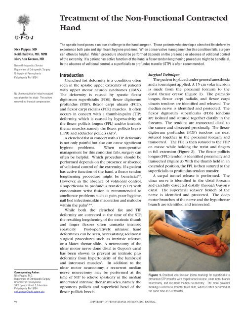
The patient is placed under general anes<strong>the</strong>sia<br />
and a tourniquet applied. A 15 cm volar incision<br />
is made from <strong>the</strong> proximal forearm to <strong>the</strong><br />
distal <strong>the</strong>nar crease (Figure 1). The palmaris<br />
longus, flexor carpi radialis, and flexor carpi<br />
ulnaris tendons are identified and released. The<br />
median nerve is identified and protected. The<br />
flexor digitorum superficialis (FDS) tendons<br />
are isolated and sutured toge<strong>the</strong>r distally in <strong>the</strong><br />
forearm. The tendons are transected distal to<br />
<strong>the</strong> suture and dissected proximally. The flexor<br />
digitorum pr<strong>of</strong>undus (FDP) tendons are next<br />
sutured toge<strong>the</strong>r in <strong>the</strong> proximal forearm and<br />
transected. The FDS is <strong>the</strong>n sutured to <strong>the</strong> FDP<br />
en masse while holding <strong>the</strong> wrist and fingers<br />
in full extension (Figure 2). The flexor pollicis<br />
longus (FPL) tendon is identified proximally and<br />
transected (Figure 3). With <strong>the</strong> thumb held in an<br />
extended position, <strong>the</strong> FPL is <strong>the</strong>n sutured to <strong>the</strong><br />
superficialis to pr<strong>of</strong>undus tendon transfer.<br />
A carpal tunnel release is performed. The<br />
ulnar nerve is identified in <strong>the</strong> distal forearm<br />
and carefully dissected distally through Guyon’s<br />
canal. The superficial sensory branch <strong>of</strong> <strong>the</strong><br />
nerve is identified and protected. The deep<br />
motor branches <strong>of</strong> <strong>the</strong> nerve and <strong>the</strong> hypo<strong>the</strong>nar<br />
branch are identified and transected.<br />
Figure 1. Standard volar incision (distal marking) for superficialis to<br />
pr<strong>of</strong>undus (STP) transfer with carpal tunnel release, ulnar motor branch<br />
neurectomy, and recurrent median neurectomy. The more proximal<br />
marking is used for a pronator teres slide, which is <strong>of</strong>ten performed at<br />
<strong>the</strong> same time as STP transfer.
Figure 2. The flexor digitorum superficialis tendons (red arrow) are sutured to <strong>the</strong> flexor<br />
digitorum pr<strong>of</strong>undus tendons (yellow arrow) en masse.<br />
Figure 3. The flexor pollicis longus (FPL) tendon is identified (red arrow) proximally and<br />
transected.<br />
If a recurrent median neurectomy is also to be performed,<br />
dissection is carried out to expose <strong>the</strong> <strong>the</strong>nar muscles <strong>of</strong> <strong>the</strong><br />
thumb. The recurrent motor branch <strong>of</strong> <strong>the</strong> median nerve is<br />
identified and <strong>the</strong>n transected (Figure 4).<br />
The tourniquet is <strong>the</strong>n released, hemostasis obtained and<br />
<strong>the</strong> incisions are closed. The tourniquet is <strong>the</strong>n re-inflated and<br />
<strong>the</strong> wrist is arthrodesed in 10 degrees <strong>of</strong> extension using a<br />
dorsal plate 8 .<br />
Post-operative Management<br />
Post-operatively, <strong>the</strong> patient is immobilized in dorsal and<br />
volar splints with <strong>the</strong> thumb and fingers in full extension out<br />
to <strong>the</strong> DIP joints. This splint is used for 6 weeks, after which<br />
a removable volar wrist splint is applied and passive range<br />
<strong>of</strong> motion <strong>of</strong> <strong>the</strong> thumb and fingers begun. The volar wrist<br />
splint is used for an additional 6 weeks to protect <strong>the</strong> wrist<br />
TREATMENT OF THE NON-FUNCTIONAL CONTRACTED HAND 91<br />
VOLUME 21, MAY 2011<br />
Figure 4. The recurrent motor branch <strong>of</strong> <strong>the</strong> median nerve is identified (red arrow).<br />
arthrodesis. Patients are routinely seen at 2 weeks, 6 weeks,<br />
and 12 weeks after surgery. Radiographs are obtained at 6<br />
and 12 weeks to assess <strong>the</strong> status <strong>of</strong> <strong>the</strong> wrist arthrodesis.<br />
Office visits are <strong>the</strong>n done at 3 month intervals or as indicated<br />
by <strong>the</strong> overall treatment <strong>of</strong> <strong>the</strong> multiple limb deformities seen<br />
in <strong>the</strong>se UMN patients.<br />
Discussion<br />
Spastic clenched fist with TIP deformity can be very difficult<br />
for <strong>the</strong> hand surgeon to treat and <strong>the</strong>re is no agreement as<br />
to which surgical procedure is most appropriate 9 . In <strong>the</strong><br />
non-functional hand, extrinsic tightness can be effectively<br />
addressed by an STP. However, previously unappreciated<br />
intrinsic spasticity can <strong>of</strong>ten cause recurrent deformity 10,11 .<br />
Therefore, <strong>the</strong> treating surgeon should address this potential<br />
problem at <strong>the</strong> time <strong>of</strong> <strong>the</strong> index procedure. In <strong>the</strong> patient<br />
with a spastic TIP deformity, intrinsic <strong>the</strong>nar muscle tightness<br />
is <strong>of</strong>ten corrected via a Matev <strong>the</strong>nar slide, which typically<br />
involves release <strong>of</strong> <strong>the</strong> flexor pollicis brevis, adductor pollicis,<br />
and first dorsal interosseus muscles 12,13 . However, given<br />
<strong>the</strong> potential morbidity <strong>of</strong> this procedure, which involves<br />
a lengthy surgical dissection and potential wound healing<br />
complications, <strong>the</strong> senior author on this paper has been<br />
performing a recurrent median nerve neurectomy instead.<br />
Only if <strong>the</strong>re is a spastic intrinsic TIP deformity after <strong>the</strong><br />
initial index procedure is a Matev <strong>the</strong>nar slide considered.<br />
The logic behind <strong>the</strong> recurrent median neurectomy is that<br />
by transecting <strong>the</strong> recurrent median nerve, one reduces<br />
<strong>the</strong> tone <strong>of</strong> superficial head <strong>of</strong> <strong>the</strong> flexor pollicis brevis and<br />
opponens pollicis, which can hopefully prevent an intrinsic<br />
TIP deformity. In a recent study <strong>of</strong> 23 patients with upper<br />
motor neuron syndrome who underwent a STP transfer, it was<br />
demonstrated that a recurrent median neurectomy at <strong>the</strong> time<br />
<strong>of</strong> <strong>the</strong> STP transfer reduced <strong>the</strong> rate <strong>of</strong> post-operative intrinsic<br />
TIP deformity from 45.5% to 16.7% 14 . No complications were<br />
noted from performing this additional procedure.
92 PAPPAS ET AL<br />
However, one should be aware that for <strong>the</strong> recurrent<br />
median neurectomy to be effective, <strong>the</strong> patient must have<br />
a dynamic TIP deformity due to muscle spasticity. Patients<br />
with a fixed TIP secondary to capsular or intrinsic <strong>the</strong>nar<br />
muscle scarring will not benefit from <strong>the</strong> procedure and will<br />
require alternate procedures such as a capsular release or<br />
Matev <strong>the</strong>nar slide. Examination <strong>of</strong> <strong>the</strong> thumb following <strong>the</strong><br />
STP procedure while <strong>the</strong> patient is still under anes<strong>the</strong>sia will<br />
reveal <strong>the</strong> presence <strong>of</strong> a fixed thumb contracture. Since <strong>the</strong><br />
majority <strong>of</strong> deformities in patients with upper motor neuron<br />
syndrome are <strong>the</strong> result <strong>of</strong> both static and dynamic forces,<br />
we recommend doing <strong>the</strong> recurrent median neurectomy to<br />
address <strong>the</strong> dynamic portion <strong>of</strong> <strong>the</strong> intrinsic TIP. If needed, a<br />
release <strong>of</strong> <strong>the</strong> contracture can be done as an adjunct during<br />
<strong>the</strong> initial procedure.<br />
<strong>Treatment</strong> <strong>of</strong> <strong>the</strong> non-functional contracted hand clearly<br />
poses a unique challenge to <strong>the</strong> hand surgeon. Only time<br />
and future studies will elucidate <strong>the</strong> best means <strong>of</strong> affording a<br />
long-lasting surgical correction.<br />
References<br />
1. Keenan, MAE. Upper Extremity Dysfunction After Stroke or Brain Injury. Green’s Operative<br />
<strong>Hand</strong> Surgery, 5 th ed. Editors Green et al. Philadelphia: Elsevier, 2005. pp.1235-1270.<br />
2. Keenan MAE. Management <strong>of</strong> <strong>the</strong> spastic upper extremity in <strong>the</strong> neurologically impaired<br />
adult. Clin Orthop Relat Res. 1988 Aug;(233):116-25.<br />
UNIVERSITY OF PENNSYLVANIA ORTHOPAEDIC JOURNAL<br />
3. Pinzur MS. Flexor origin release and functional prehension in adult spastic hand deformity. J<br />
<strong>Hand</strong> Surg Br. 1991 May;16(2):133-6.<br />
4. Keenan MAE, Korchek JI, Botte MJ, and Garland DE. Results <strong>of</strong> transfer <strong>of</strong> <strong>the</strong> flexor<br />
digitorum superficialis tendons to flexor digitorum pr<strong>of</strong>undus tendons in adults with acquired<br />
spasticity <strong>of</strong> <strong>the</strong> hand. J Bone Joint Surg. 1987:69A:1127-1132.<br />
5. Braun RM, Vise GT, Roper B. Preliminary experience with a superficialis-to-pr<strong>of</strong>undus<br />
transfer in <strong>the</strong> hemiplegic upper extremity. J Bone Joint Surg 1974;56A:466-472.<br />
6. Botte MJ, Keenan MA, Korchek JI. Modified technique for <strong>the</strong> superficialis-to-pr<strong>of</strong>undus<br />
transfer in <strong>the</strong> treatment <strong>of</strong> adults with clenched fist deformity. J <strong>Hand</strong> Surg 1987;12A:639-640.<br />
7. Pomerance JF, Keenan MA. Correction <strong>of</strong> Severe Spastic Flexion Contractures in <strong>the</strong><br />
<strong>Non</strong>functional <strong>Hand</strong>. J <strong>Hand</strong> Surg (Am) 1996; 21(5): 828-33.<br />
8. Jebson PJ, Adams BD. Wrist arthrodesis: review <strong>of</strong> current techniques. JAAOS. 2001<br />
Jan-Feb; 9(1):53-60.<br />
9. Smeulders M, Coester A, Kreulen M. Surgical <strong>Treatment</strong> for Thumb-in-Palm Deformity in<br />
Patients with Cerebral Palsy. Cochrane Database Syst Rev 2005;(4): CD004093.<br />
10. Keenan MA, Todderud ER Henderson R, Botte MJ. Management <strong>of</strong> intrinsic spasticity in<br />
<strong>the</strong> hand with phenol injection or neurectomy <strong>of</strong> <strong>the</strong> motor branch <strong>of</strong> <strong>the</strong> ulnar nerve. J <strong>Hand</strong><br />
Surg 1987;12A:734-739<br />
11. Botte MJ, Keenan MA, Gellman H, Garland DE, Waters RL. Surgical Management <strong>of</strong> Spastic<br />
Thumb-in-Palm Deformity in Adults with Brain Injury. J <strong>Hand</strong> Surg (Am) 1989; 14A: 174-82.<br />
12. Matev I. Surgery <strong>of</strong> <strong>the</strong> spastic thumb-in-palm deformity. J <strong>Hand</strong> Surg Br. 1991 May;16(2):127-<br />
32.<br />
13. Matev I. Surcial <strong>Treatment</strong> <strong>of</strong> Spastic Thumb-In-Palm Deformity. J Bone Joint Surg Br. 1963<br />
Nov;45:703-8.<br />
14. Pappas N, Baldwin K, Keenan MA. Efficacy <strong>of</strong> median nerve recurrent branch neurectomy<br />
as an adjunct to ulnar motor nerve neurectomy and wrist arthrodesis at <strong>the</strong> time <strong>of</strong> superficialis<br />
to pr<strong>of</strong>undus transfer in prevention <strong>of</strong> intrinsic spastic thumb-in-palm deformity. J <strong>Hand</strong> Surg<br />
Am. 2010 Aug;35(8):1310-6.