Case Study: Titin Ig domains - Theoretical Biophysics Group
Case Study: Titin Ig domains - Theoretical Biophysics Group
Case Study: Titin Ig domains - Theoretical Biophysics Group

You also want an ePaper? Increase the reach of your titles
YUMPU automatically turns print PDFs into web optimized ePapers that Google loves.
<strong>Case</strong> <strong>Study</strong>: <strong>Titin</strong> <strong>Ig</strong> <strong>domains</strong><br />
Mu Gao and Eric Lee<br />
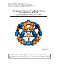
A Molecular Rubber Band<br />
In the extreme sport of bungee jumping, a daring athlete leaps from a great height and freefalls<br />
while a tethered cord tightens and stretches to absorb the energy from the descent. The<br />
bungee cord protects the jumper from serious injury, because its elasticity allows it to extend<br />
and provide a cushioning force that opposes gravity during the fall. Amazingly, nature also<br />
uses elasticity to dampen biological forces at the molecular level, such as during extension of<br />
a muscle fiber under stress. The molecular bungee cord that serves this purpose in the human<br />
muscle fiber is the protein titin, which functions to protect muscle fibers from damage due to<br />
overstretching. In this case study, we will examine how experimental and theoretical studies<br />
yield insight into the molecular mechanism for titin’s elasticity.<br />
1
These are the files you will need to complete the exercises in this case study.<br />
2
The Muscle Fiber and <strong>Titin</strong><br />
Striated muscle, such as a skeletal or cardiac muscle, is a complex of elongated fibers arranged<br />
in parallel order and held together with connective tissue. These muscle fibers are typically<br />
2 to 3 cm long and 100 µm in diameter. Each fiber consists of thread-like fibrils (1-2 µm<br />
diameter) known as myofibrils, which primarily contain thick filaments (12-18 nm diameter)<br />
and thin filaments (5-8 nm diameter). The thick filaments are structures built primarily from<br />
overlapping myosin proteins, which have globular heads that form cross bridges connected to<br />
thin filaments formed mainly by actin bundles. Viewed lengthwise, the myofibril is seen as a<br />
series of bands of alternating composition, separated by line-like regions (Fig 1a and b). The A<br />
band is a set of stacked thick filaments centered on the M line, overlapped on both sides by thin<br />
filaments, which compose the I band. In between each I band, a dense vertical region called the<br />
Z line divides the myofibrils into a functional unit called the sarcomere, which is the smallest<br />
self-contained contractile unit (resting length 2 to 3 µm) of the striated muscle.<br />
Figure 1: (a) The electron microscope image of striated muscle (photograph courtesy of Dr. Roger<br />
Craig, University of Massachusetts) and schematic views of (b) the muscle sarcomere and (c) the<br />
structural components of the giant protein titin. <strong>Titin</strong> <strong>Ig</strong> and FnIII <strong>domains</strong> are shown in circles and<br />
ovals, respectively. Five <strong>Ig</strong> <strong>domains</strong> and one FnIII domain with known atomic structure are labeled.<br />
One very specialized structure in a sarcomere is the giant muscle protein titin, which is the<br />
largest protein identified in nature, with a molecular weight of nearly ∼4 MDa and a contour<br />
length of ∼1 µm [1, 2]. Anchored at the Z and M lines, a string-like titin molecule spans half<br />
of the sarcomere in close contact with the thick filament along the A band, but flexible along<br />
the I band (Fig 1b and c). The major structural components of titin are bead-like β-sandwich<br />
protein modules, most notably the immunoglobulin-like <strong>domains</strong>, or <strong>Ig</strong> <strong>domains</strong>, named after<br />
immunoglobulin where the homologous structural motif was first identified. The stable core of<br />
an <strong>Ig</strong> domain is formed from strands of β-sheets, connected through flexible loops from which<br />
protrusions form binding sites for antigens in immunoglobulins. However, in titin the <strong>Ig</strong> <strong>domains</strong><br />
are not involved in immunological recognition. Instead, they contribute to the passive stiffness<br />
of a stretched sarcomere.<br />
3
<strong>Titin</strong> is the molecular spring in a muscle sarcomere<br />
Muscle force generation is dependent on the contractile and stretching motions that take place in<br />
the sarcomere. The contractile movement generates active force while the stretching movement<br />
generates passive force. The combination of active and passive forces within muscle fibers<br />
ultimately gives rise to the muscular force required for various physical motions, such as your<br />
heart beating, walking, or turning this page (not yet!). At the molecular level, the contractile<br />
motion is due to the sliding of myosin filaments over the actin filaments. Triggered by the<br />
release of calcium ions upon muscle excitation and powered by the hydrolysis of ATPs, a series<br />
of conformation changes occur in myosin cross-bridges that associate with actin filaments. These<br />
conformational changes induce a power stroke event that pulls the thin filament past the thick<br />
filament and towards the central M-line at a rate of up to 15 µm/s, exerting the active force.<br />
In contrast, when sarcomere is relaxed and further stretched beyond its slack length, a passive<br />
force develops to dampen the force of extension and restore the sarcomere to its resting length.<br />
An essential characteristic of striated muscle is its elasticity, a mechanical property necessary<br />
for the reversible contractile and stretching movement of sarcomere. This elasticity permits<br />
myofibrils to be stretched to twice their resting length without damaging their structure. The<br />
physiological sarcomere length ranges from 1.9 to 2.5 µm in cardiac muscle and 2.2 to 3.8 µm<br />
in soleus, a skeletal calf muscle. Investigations of myofibrils during contraction and relaxation<br />
suggest that neither actin nor myosin filaments confer the elasticity seen during force generation,<br />
since the length of both filaments appear unchanged during the contraction-extension cycle.<br />
Instead, the elastic property of sarcomere appears to arise from another set of muscular filaments,<br />
composed of the protein titin. It is the lengthening of titin molecules that generates the passive<br />
force which is crucial for maintaining the integrity of the sarcomere.<br />
Electron micrographs and gene sequence analysis reveal that the architecture of titin is built<br />
upon a linear assembly of 300 immunoglobulin-like (<strong>Ig</strong>) or fibronectin III-like (FN-III) <strong>domains</strong>,<br />
in addition to several uncharacterized segments. In the A-band or Z-line regions, titin intimately<br />
associates with proteins located in the thick and thin filaments, respectively. The only part of<br />
titin that appears to move freely is its I-band region, which provides the major contribution<br />
to passive elastic properties for the sarcomere. <strong>Titin</strong> I-band <strong>domains</strong> of variable composition,<br />
called isoforms, have been found in different types of muscle tissue, indicating that the specific<br />
distribution of domain types within the titin spring determine its stiffness and extensibility.<br />
Molecular fine tuning for the titin spring<br />
Approximately ninety percent of titin’s mass by composition lie in the globular <strong>Ig</strong> or FN-III<br />
<strong>domains</strong> (Fig 1c). Each domain consists of ∼100 residues folded into a β-sandwich motif. The<br />
titin A-band is built from highly conserved <strong>Ig</strong> or FnIII <strong>domains</strong> that are arranged in regular<br />
patterns correlating to the structural composition of associated thick filaments. The repeating<br />
patterns seen in both titin and myosin leads to the hypothesis that the titin A-band may regulate<br />
the assembly of myosin in the early stage of muscle development. In contrast, the components<br />
of titin I-band exhibit prominent passive elasticity that characterizes the spring-like property<br />
of the sarcomere. The termini of titin I-band are composed of two well conserved <strong>Ig</strong> segments,<br />
the distal (15 <strong>Ig</strong> <strong>domains</strong>) and the proximal (22 <strong>Ig</strong> <strong>domains</strong>) segments, respectively. The two<br />
segments are connected by three isoform-dependent segments: (i) a variable length <strong>Ig</strong> segment,<br />
4
(ii) PEVK region, named due to the abundance of proline (P), glutamic acid (E), valine (V), and<br />
lysine (K), and (iii) N2 (N2A and/or N2B) regions. The PEVK and N2 regions, rich in charged<br />
residues and prolines, do not form stable secondary structures and unfold completely upon<br />
stress, elongating two to four times their folded length during muscle extension. In contrast to<br />
the flexible PEVK and N2 <strong>domains</strong>, <strong>Ig</strong> <strong>domains</strong> appear to be designed to resist extreme tension<br />
and even unfold reversibly at a higher load as the stretch in the sarcomere continues. Thus, the<br />
resistance to stress and the reversible unfolding of <strong>Ig</strong> <strong>domains</strong> provide a mechanism to prevent<br />
over-stretching of muscle.<br />
Figure 2: (a) Structure of <strong>Titin</strong> <strong>Ig</strong> domain I27, (b) and (c) Structural alignment and protein sequence comparison<br />
of five human titin <strong>Ig</strong> <strong>domains</strong>: I1 (PDB code: 1G1C), I27 (1TIT), Z1 (1RJ0), Z2 (1RJ0), M5 (1TNM).<br />
Details of how to align these five <strong>Ig</strong> <strong>domains</strong> and reproduce the snapshot (c) with the program VMD are given<br />
in Exercise 1.<br />
The composition of a bungee cord determines how much it will stretch during a jump. For<br />
example, sheathed bungee cords typically have a rubber core surrounded by a nylon wrapping<br />
and stretch to around twice their resting length during a jump. In contrast, all rubber cords<br />
consist of many tiny strands of rubber wound together to form one solid cord, and typically<br />
stretch to four times their resting length during a jump. The elasticity of I-band titin follows a<br />
similar scheme where its composition determines whether it is stiff or elastic. Instead of rubber<br />
strands, titin is built from protein segments with different folds, and the ratio of these folds<br />
determine to what extent the sarcomere can stretch and still reversibly recover to equilibrium<br />
length without permanent damage.<br />
The complete sequence of the human titin gene contains 363 exons that potentially encode<br />
a total of 38,138 amino acid residues [3]. One third of titin exons are differentially expressed,<br />
5
giving rise to variants with variable domain compositions. For example, the titin isoform found<br />
in the human soleus, a leg muscle, contains an extended (ten times longer) PEVK region, and<br />
four times as many <strong>Ig</strong> <strong>domains</strong> compared to the cardiac titin isoform [4]. Thus, these titin<br />
isoforms possess fine-tuned elastic responses for different types of muscle. The elastic behavior<br />
of coiled PEVK or other unique <strong>domains</strong> appear easy to understand because they do not form<br />
stable structures. In order to understand why <strong>Ig</strong> <strong>domains</strong> are mechanically so stable, however,<br />
it is necessary to look more closely at their detailed atomic structures and investigate how they<br />
respond to external force.<br />
Exercise 1: 3D Structures of <strong>Titin</strong> <strong>Ig</strong> Domains. In this VMD exercise you will inspect<br />
and align the atomic structures of five titin <strong>Ig</strong> <strong>domains</strong> shown in Fig. 2. To load the coordinates<br />
of these proteins, start a new VMD session and open the state file titin-<strong>Ig</strong>.vmd.<br />
• After loading, all structures other than I27 are in hidden mode. Select the New Cartoon<br />
representation to view the structure of I27. You should get a view similar to what is shown<br />
in Fig. 2a.<br />
• Make all other <strong>Ig</strong> <strong>domains</strong> visible, and align all five titin <strong>domains</strong> using VMD. This is<br />
accomplished by choosing the Multiple Alignment tool from the Extensions menu on the<br />
VMD Main window. Then, under the Alignment tab, choose Run Structural Alignment.<br />
Fig. 2b shows the aligned <strong>domains</strong> using the default parameters. The resulting assemblage<br />
of aligned structures were colored by the degree of structural similarity (Qa). The color<br />
scale ranges from blue (most similar) to red (least similar). You can clearly see that all five<br />
titin <strong>Ig</strong> <strong>domains</strong> surveyed here have highly conserved β-sandwich structures.<br />
• Can you think of another protein family for which a structural alignment would reveal the<br />
conservation of functional <strong>domains</strong>?<br />
• Generate a sequence comparison from the structural alignment of our five titin <strong>Ig</strong> <strong>domains</strong><br />
as shown in Fig. 2c. The highlighted blocks show regions where structure is conserved with<br />
Qa value ≥0.8.<br />
• Comparison studies between sequence alignments of proteins with known structure have<br />
shown that conserved sequences lead to conserved structure. Can the same be stated vice<br />
versa, i.e., that conserved structure implies conserved sequence?<br />
<strong>Ig</strong> <strong>domains</strong> possess a β-sandwich motif<br />
<strong>Titin</strong> I27 is the first structurally determined <strong>Ig</strong> domain from the I-band region responsible for<br />
regulating passive elasticity for muscle sarcomere [5]. Two β-sheets of I27, the ABED sheet and<br />
the A’GFC sheets, are tightly connected at the terminal regions through a straightened loop<br />
between the A- and the A’-strands, as shown in Fig. 3. To prevent spontaneous unraveling, two<br />
sets of inter-strand hydrogen bonds firmly lock the terminal regions. The two sets of hydrogen<br />
bonds include two bonds between a pair of charged residues Lys6 and Glu24 on the A- and the<br />
B-strands, and five bonds between the A’- and G-strands. Each set may expand by acquiring<br />
an additional dynamic hydrogen bond at their edges.<br />
6
Figure 3: The structure of titin I27 in various representations. (a) The hydrogen bonding at two terminal<br />
regions. (b) The cartoon representation rotated 90 ◦ with respect to (a). (c) Prolines in loop regions. The<br />
hydrogen bonding network of the β-sheets can be disrupted by proline residues, known as the β-breaker. VMD<br />
can be used to display all proline residues of I27, and their effect on secondary structure (see Exercise 2). (d)<br />
Schematic diagram of I27.<br />
Exercise 2: Structural Characteristics of β-sheets. In this exercise we will explore a<br />
few structural characteristics of I27.<br />
• With the previous VMD session still loaded, hide all other <strong>Ig</strong> <strong>domains</strong> except I27. Create<br />
a new representation for backbone atoms (use the selection text backbone and within<br />
1.5 of betasheet) and choose the Hbond drawing method. The hydrogen bonds are not<br />
apparent in this selection with the default settings. Therefore to view a greater number<br />
hydrogen bonds, try increasing the distance cut-off and angle cut-off settings (a distance<br />
cut-off of 5.0 should be adequate). Create a new CPK representation for backbone atoms<br />
between A- and B-strands (residue 3 to 6, 24 to 26) and between A’- and G-strands (residue<br />
9 to 15, 83 to 87). Compare your hydrogen bonds to Fig. 3a.<br />
• Most β-strands of I27 are aligned in anti-parallel fashion, where nearly all the polar amide<br />
groups from one strand are hydrogen bonded at 90 ◦ angle to the backbone carbonyl oxygen<br />
of the opposing strand whose directionality is reversed. Parallel β-strands also exist in<br />
nature but are more rare, and feature hydrogen bonding to neighboring strands at a diagonal<br />
angle. Find a pair of β-strands arranged in parallel fashion on I27.<br />
• Based on the representation you created above, what role do you think hydrogen bonds<br />
may play in stabilizing the β-sheets?<br />
• Using the same structure you loaded above, find all prolines in I27. Where are the proline<br />
residues located in titin I27? What unique structural feature of proline makes it distinct<br />
from all the other amino acids? Knowing that, does this explain their positions in I27?<br />
The rigid molecular structure of a titin <strong>Ig</strong>-fold is the determinant of its ability to bear<br />
mechanical stress. A typical titin <strong>Ig</strong> fold consists of ∼100 residues packed into two β-sheets<br />
7
approximately 40 ˚A long. The three-dimensional structures of five known titin <strong>Ig</strong> <strong>domains</strong>, I1<br />
and I27 (I-band), Z1 and Z2 (N-terminus Z-line), and M5 (C-terminus M-line), each exhibit an<br />
eight- or nine-stranded β-sandwich arranged mostly in antiparallel fashion. An exception exists<br />
for the A’-and G-strands, which are cross-linked in parallel (Fig. 2). Structural alignment of<br />
these five <strong>domains</strong> reveal that there is a high structural homology between <strong>Ig</strong> <strong>domains</strong>. See<br />
Exercise 1 for details on performing structural alignment of proteins using the program VMD.<br />
The structural and sequence comparison suggest that all titin <strong>Ig</strong> <strong>domains</strong> belong to a common<br />
<strong>Ig</strong> I-set superfamily, which also include <strong>Ig</strong> <strong>domains</strong> found at many cell surface receptors.<br />
Exercise 3: I27 Contact Map. In this exercise we will use VMD to examine physical<br />
interactions between β-sheets in I27 titin. VMD’s Contact Map tool, accessed from the Extensions<br />
menu, provides a method for viewing residue-residue contacts between two sets of selected atoms.<br />
The Cα to Cα distances are produced on the plot, with black as the highest intensity corresponding<br />
to 0.0 ˚A distance, a linear gray scale for 0.0 to 10.0 ˚A distance, and white when equal to or greater<br />
than 10 ˚A.<br />
• Pair scanning: Scroll over the contact map while holding down button 2 (middle button on<br />
Linux and Windows PC, right button on MacOSX) to view the resid number and alphacarbon<br />
distances between the residue pair selected.<br />
• The zoom slider bars on the left side sets the residue numbering scale for both the vertical<br />
and horizontal axes. The Fit All option will scale the axes for the window size.<br />
• We have provided a contact map of I27 titin <strong>Ig</strong> domain. Contact between β-strands B4 and<br />
B5 (C and F from Fig. 3) are shown in the figure as an example (labeled as 1).<br />
• Generate this contact map on your own in VMD for I27. (Hint: Make sure only titin I27 is<br />
loaded into VMD.) Can you identify the contacts labeled by 2 and 3 in the figure?<br />
• How do contacts between adjacent β-sheets show up in the contact map?<br />
8
Probing the mechanical stability of titin <strong>Ig</strong> <strong>domains</strong> in vitro<br />
The mechanical properties of titin <strong>Ig</strong> <strong>domains</strong> have been investigated experimentally with atomic<br />
force microscopy (AFM) or optical tweezers, techniques that can be used to apply force as weak<br />
as several pN to a single molecule and resolve corresponding conformational changes at the<br />
˚A level. Combined with protein engineering, these sensitive methods permit the controlled<br />
stretching of a molecule made up of identical protein repeats, providing a unique opportunity<br />
to study the mechanical properties of individual titin <strong>domains</strong>. In an AFM experiment, a<br />
cantilever probe is situated on the surface of a protein molecule anchored on the substrate. The<br />
substrate is retracted at a constant velocity, causing the cantilever tip to bend. The bending<br />
deflects a laser beam pointed to the tip, and the force applied to the molecule can be measured<br />
through the deflection. Fig. 4 shows a diagram of stretching seven titin <strong>Ig</strong> <strong>domains</strong>, along<br />
with a characteristic sawtooth pattern on the force-extension profile obtained from the AFM<br />
experiment [6]. The spacing between force peaks matches the contour length of a fully unfolded<br />
<strong>Ig</strong> domain, indicating that the <strong>Ig</strong> <strong>domains</strong> unfold in a one-by-one fashion. One may guess that<br />
the force peaks induced critical conformational changes that lead to domain unfolding. However,<br />
it is not clear from experiments alone what these conformational changes are and how the protein<br />
responds to mechanical force.<br />
Figure 4: Probing the mechanical properties of individual titin <strong>Ig</strong> <strong>domains</strong> using atomic force microscopy<br />
[6]. The molecule stretched consists of seven I1 and I27 <strong>domains</strong> linked alternately. (c) Shows<br />
one sawtooth peak in detail. The stages of extension are defined by 1) the phase where force builds<br />
as the protein provides resistance to mechanical stretching, and 2) the moment where force bearing<br />
elements rupture, leading to a sharp decline of force measured when continued extension.<br />
9
Unraveling the terminal β-strands initiates the I27 unfolding process<br />
Obviously the answer to above questions lies at the β-sandwich structure of titin <strong>Ig</strong> <strong>domains</strong><br />
that define their mechanical response upon external stress. To address these important questions,<br />
computational studies have provided insights into the structure-function relationship of<br />
<strong>Ig</strong> <strong>domains</strong> at the atomic level [7–9]. Fig. 5 illustrates the conformational changes to I27 during<br />
stretching of the domain. The N-terminus was fixed and the C-terminus attached to a harmonic<br />
spring that was pulled at its other end at various constant velocities. At first, the terminal regions<br />
of the protein straightened with the applied force, and the two β-sheets slid slightly with<br />
respect to each other, reaching an extension of ∼10 ˚A. At this initial stage, the overall secondary<br />
structure of I27 remained intact. Upon further extension by ∼5 ˚A, the increasing force caused<br />
two terminal β-strands, the A-strand of ABED sheet and the A’-strands of the A’GFC sheet,<br />
to unravel, leading to a rapid unfolding process and decrease of the measured force. The force<br />
was measured by monitoring the spring extension.<br />
Figure 5: The mechanical unfolding pathway of titin I27. The trajectory of this unfolding process is<br />
provided for study. See Exercise 4 for details.<br />
The unfolding process of an <strong>Ig</strong> domain during the SMD simulation can be divided into three<br />
stages: i) extension from 0 ˚A to 10 ˚A where the molecule is straightened without losing its<br />
secondary structure, ii) extension from 10 ˚A to 15 ˚A where a pronounced peak force is required<br />
to initiate the disruption of the secondary structure, namely detachment of A- and A’-strands,<br />
and iii) extension beyond 15 ˚A where the remaining β-strands unravel rapidly. It is important<br />
to note that a large force is not necessary to unfold the protein if a weaker force is applied for a<br />
longer duration. For example, the peak force required for unfolding the protein decreases 50%<br />
as the stretching velocities are slowed 20 fold from 0.5 ˚A/ps to 0.01 ˚A/ps. Under physiological<br />
conditions a stretching force as weak as 20 pN can unfold a titin <strong>Ig</strong> domain within one second.<br />
10
Exercise 4: Forced Unfolding of I27. The forced unfolding of titin <strong>Ig</strong> domain I27 has been<br />
simulated by fixing the N-terminus and pulling the C-terminus with an attached harmonic spring.<br />
A 3ns trajectory and a related data file from an SMD simulation at the pulling velocity of 0.05<br />
˚A/ps are provided for study. To start, launch VMD and load the state file I27-unfolding.vmd.<br />
• Play the trajectory and identify which part of the protein becomes unfolded first. A few<br />
snapshots of the unfolding pathway are shown in Fig. 5.<br />
• The applied force and resulting extension was recorded in the data file<br />
I37-smd-cv-0.05Aps.dat. Make plots of time versus force and extension versus<br />
force. Correlate the force peak to the disruption of the secondary structures of the protein.<br />
• During the simulation, the Cα atom of the N-terminus was held fixed, while the Cα atom<br />
of the C-terminus was pulled along the vector connecting these two atoms. What would<br />
happen if one applies the force in a direction orthogonal to the original pulling direction?<br />
Terminal inter-strand hydrogen bonds characterize the stability of <strong>Ig</strong><br />
<strong>domains</strong><br />
The atomic details obtained from simulations on the unfolding of I27 reveal key structural<br />
changes that explains the distinct force peak observed in AFM experiments as shown in Fig. 4.<br />
The force peak correlates to separating two pairs of β-strands near the terminal regions of the<br />
<strong>Ig</strong> domain, namely the A- and B-strands and the A’- and G-strand. The interactions between<br />
these β-strands contribute significantly to the mechanical stability of this domain. In particular,<br />
the force peak corresponded to the rupture of inter-strand hydrogen bonds between the A- and<br />
the B-strands and between the A’- and the G-strands as shown in Fig. 6.<br />
Figure 6: The disruption of the A-B and A’-G inter-strand hydrogen bonds of I27 correlates with the<br />
unfolding force peak. See also the attached movie clip I27-smd.mpg. Details of hydrogen bond analysis<br />
is discussed in the Exercise 5 below.<br />
11
Prior to the separation of the A- and the A’-strands at 10 ˚A extension, all hydrogen bonds<br />
between the ABE and the A’GF β-sheets are well maintained. As the force reaches its peak<br />
during the extension to 12 ˚A, two hydrogen bonds between the A- and the B-strands break,<br />
closely followed by the concurrent burst of hydrogen bonds between the A’- and the G-strands at<br />
∼14 ˚A. At this phase, the hydrogen bonds between other β-strands, such as between the B- and<br />
E-strands, and between the G- and F-strands, remain intact. However, as the A- and A’-strands<br />
fully extend, the remaining hydrogen bonds are sequentially disrupted in an unzipping fashion,<br />
requiring a much lower force to break than the major force peak. During the unfolding process<br />
water molecules play an important role by forming new hydrogen bonds with the original protein<br />
backbone atoms that have ruptured inter-strand hydrogen bonds.<br />
Insights gained from atomic details provide guidance for modulating the mechanical stability<br />
of I27 by mutating key amino acids. A point mutation disrupting the inter-strand hydrogen<br />
bonds, for example, by replacing one hydrogen bond forming amino acid on G-strand with a<br />
proline, destabilizes the stability of the module. AFM experiments of these point mutants have<br />
indeed shown decreased mechanical stability [10].<br />
Mechanical properties of a titin complex<br />
Up to this point our examination of titin has been restricted to individual <strong>Ig</strong>-<strong>domains</strong>. <strong>Titin</strong><br />
however, interacts with a variety of proteins along its ∼1 µm long length. These interactions<br />
provide structural support for the development and function of myofibrils and also regulatory<br />
roles such as responding to stretching forces developed in titin. The majority of these interactions<br />
occur at the A-band where titin interacts with myosin filaments, and at the terminal ends where<br />
it is anchored at the sarcomeric Z-line and M-line and binds with dozens of proteins. At the<br />
very N-terminal region of titin, a sensor complex is formed based on two titin <strong>Ig</strong> <strong>domains</strong>,<br />
Z1 and Z2. The core part of this complex relies on the anchoring of Z1 and Z2 through the<br />
ligand telethonin [11, 12]. Telethonin is one of the most abundant transcripts in striated muscle.<br />
Studies have revealed that the presence of telethonin, in conjunction with titin Z1Z2, is required<br />
for progression of muscle growth, and it is highly specific for both titin Z1 and Z2, but not for<br />
Z1 or Z2 individually [13]. Point mutations to telethonin resulting in premature stop codons<br />
have been correlated to a form of limb girdle muscular dystrophy (type 2A) [14–16], suggesting<br />
that telethonin plays a major role in the stabilization of N-terminal titin at the Z-disc.<br />
The crystal structure of telethonin in complex with titin Z1 and Z2 has revealed a novel<br />
binding motif between <strong>Ig</strong> <strong>domains</strong> and a ligand [17]. Unlike typical ligand-binding through<br />
insertion of the ligand into a receptor binding pocket, in this crystal structure the receptor Z1Z2<br />
<strong>domains</strong> of two antiparallel titin N-termini are surprisingly joined together by an N-terminal<br />
fragment of the ligand telethonin through β-strand cross-linking (see Fig 7), a structural motif<br />
that appears in pathological fibril formation.<br />
12
Exercise 5: Mechanical stability and interstrand hydrogen bonds. In order to<br />
understand how the force-bearing β-strands of I27 contribute to the mechanical stability of the<br />
protein, we monitor the conformational changes connected with the rupture and subsequent unfolding<br />
of the protein. For this purpose we analyze a SMD simulation at a pulling velocity of 0.01<br />
˚A/ps. The corresponding trajectory reveals the key unfolding step, the detachment of the A’- and<br />
A-strands. To start, launch VMD and load the state file I27-hbond.vmd.<br />
• Find all inter-strand hydrogen bonds formed between A- and B-strands and between A’and<br />
G-strands (refer to Fig 3), and monitor the change of the bonding length (the distance<br />
between the hydrogen and the oxygen of a hydrogen bond) during the trajectory. You<br />
should obtain plots similar to what shown in Fig. 6. Which set of hydrogen bonds break<br />
first?<br />
• The applied force and resulting extension was recorded in the data file<br />
I27-smd-cv-0.01Aps.dat. Make plots of time versus force and extension versus<br />
force, and correlate the force peak to the breakage of the hydrogen bonds.<br />
• Hydrogen bonds may form between bonding pairs as they align during the stretching process.<br />
Identify an inter-strand hydrogen bond that was broken initially but re-formed briefly when<br />
an alternate bonding pair aligns.<br />
One can further analyze the hydrogen bond energy with the following empirical formula used by<br />
CHARMM22:<br />
EHBond = ( A<br />
r 6 AD<br />
− B<br />
r4 ) cos<br />
AD<br />
2 (θA−H−D) cos 2 (θAA−A−H)<br />
SW (r 2 AD, r 2 hon, r 2 hoff ) SW (cos 2 (θAD), cos 2 (θhon), cos 2 (θhoff ))<br />
where rAD, θA−H−D, θAA−A−H are the distance between the acceptor (A) and the donor (D), the<br />
angles formed by connecting the acceptor, the hydrogen (H) and the donor, and the angle formed<br />
by connecting the acceptor antecedent (AA), the acceptor and the hydrogen, respectively. SW is<br />
a switch function defined as<br />
⎧<br />
⎪⎨<br />
SW (R, Ron, Roff ) =<br />
⎪⎩<br />
0 if R > Roff<br />
(R 2 − R 2 off )(R2 − R 2 off )2 (R 2 − R 2 off −3(R2 − R 2 on )<br />
(R 2 off − R2 on )3 if Roff > R > Ron<br />
1 if R < Ron<br />
A tcl script hbond.tcl for calculating and visualizing the hydrogen bond energy using the above<br />
formula is provided. With the trajectory still loaded, open the VMD TkConsole and type source<br />
hbond.tcl. When the calculation is completed, all inter-strand hydrogen bonds will appear in<br />
bond representation. The strength of the bond is indicated by the color scale transiting gradually<br />
from green (stronger) to blue (weaker) and eventually broken. A MPEG movie clip I27-smd.mpg<br />
of this visualization is available for view.<br />
• Display water molecules that are in the proximity (within 2.5 ˚A) of partner atoms forming<br />
AB and A’G inter-strand hydrogen bonds.<br />
• Make sure your selection of water molecules updated along the trajectory, and observe how<br />
water molecules participate the unfolding process.<br />
13
Figure 7: Structure of titin Z1Z2-telethonin complex. (A) Cartoon representation of two separate<br />
N-terminal titin Z1Z2 <strong>Ig</strong>-<strong>domains</strong> joined by one N-terminal telethonin molecule fragment (residues 1 to<br />
89). Two titin molecules are individually colored blue and red (ZA and ZB, respectively). Telethonin is<br />
colored in yellow. The β-strands involved in critical force bearing contacts for each titin <strong>Ig</strong>-domain (AB<br />
and A’G) and between each titin Z2 module and telethonin are labeled (GB and GD). (B) Detailed<br />
schematic of β-strand alignment for the T2T complex, with force bearing hydrogen bonds shown for<br />
β-strand pairs AB and A’G for titin <strong>domains</strong>, and hydrogen bonds linking the G-strands on the <strong>Ig</strong><br />
<strong>domains</strong> to the A,B,C, and D-strands on telethonin. See Exercise 6 to inspect the complex with VMD<br />
We have shown in earlier sections that single titin <strong>Ig</strong>-<strong>domains</strong> such as I27 are well suited for<br />
bearing a mechanical force. The discovery of titin Z1Z2 <strong>Ig</strong>-<strong>domains</strong> in complex with telethonin<br />
naturally poses questions: Are the force bearing properties of these <strong>Ig</strong>-<strong>domains</strong> affected by<br />
their interaction with telethonin? What are the mechanical features of force bearing protein<br />
complexes, as opposed to individual proteins, and why are they important?<br />
Using a similar computational approach described above in studies of titin I27, we now<br />
investigate the Z1Z2-telethonin complex in order to answer the questions posed above. It turns<br />
out that the mechanical stability of the complex is much stronger than individual <strong>Ig</strong> <strong>domains</strong> [18].<br />
Destabilizing the complex by stretching the titin Z2 terminal ends of the complex requires higher<br />
peak force than that required for unfolding single titin <strong>Ig</strong>-<strong>domains</strong> such as I27 (a force peak of<br />
nearly 1900 pN for Z1Z2-telethonin as seen in Fig. 8 vs. a force peak of 1500 pN for I27 as<br />
seen in Fig. 5). Whereas previous studies have revealed that the unraveling of the β-strands<br />
for titin <strong>Ig</strong>-<strong>domains</strong> constitutes the dominant unfolding barrier, in the case of Z1Z2 in complex<br />
with telethonin, the force peak observed corresponded to a detachment of one virtually intact<br />
Z2 domain from a β-strands of telethonin (see Fig. 8 a-d).<br />
14
This discovery sheds light on a key architectural strategy that the titin Z1Z2-telethonin complex<br />
employs to yield extraordinary resistance to mechanical stress. The major force bearing<br />
component of this complex is an extensive intermolecular hydrogen bonding network formed<br />
across β-strands between telethonin and Z1Z2 <strong>domains</strong>, and not intramolecularly between termini<br />
β-strands of individual Z1 or Z2 <strong>domains</strong>. This shift to a stronger force bearing interface<br />
reduces the chance of the unraveling the individual <strong>Ig</strong>-<strong>domains</strong>, thus stabilizing the complex.<br />
This example demonstrates how β-strand cross-linking via hydrogen bonds serves as an important<br />
mechanism, in effect a molecular glue, for augmenting the ability of protein complexes<br />
designed to resist mechanical stress.<br />
Figure 8: Mechanical stability of the titin Z1Z2-telethonin complex. The force vs. extension profile in<br />
the plot illustrates that a force peak of nearly 2000 pN is required to detach a Z2 domain from telethonin<br />
under constant velocity SMD simulation with a pulling velocity of 0.05˚A/ps. (a)-(d) represent the<br />
initial, inter-molecule β-strands detachment, further separation, and subsequent unraveling states,<br />
respectively. The color code is the same as defined in Fig. 7, except that the four G-strands forming<br />
β-strands with telethonin are colored in grey. A movie taken from our simulation illustrating this<br />
process has been provided under Exercise 6.<br />
15
Exercise 6: The mechanical function of a molecular glue. In the previous<br />
section we discussed the forced unfolding of the titin Z1Z2-telethonin complex.<br />
We have included a movie, z1z2-tlt-smd.mpg, generated from the simulation illustrating<br />
the detachment of a titin Z2 domain from telethonin. Here we will examine<br />
the structural feature that contributes to the superior resistance to mechanical stress<br />
the complex exhibits. To start, launch VMD and load the state file z1z2-tlt.vmd.<br />
Outlook<br />
• The initial view will be of the Z1Z2-telethonin complex in the drawing<br />
method structure similar to that shown in Fig. 7. The two individual<br />
titin chains, ZA and ZB, have been colored separately, but each contains<br />
two titin <strong>domains</strong>, Z1 and Z2. Use the Sequence Viewer under the menu<br />
Extensions > Analysis in order to identify Z1 and Z2, and their relative<br />
orientation to each other.<br />
• Are the two titin chains aligned parallel or antiparallel? What implications<br />
to the force bearing ability of the complex do you think would occur for<br />
each scenario?<br />
• <strong>Titin</strong> binds to telethonin via β-strand hydrogen bond crosslinking. This<br />
requires β-strands from both titin and telethonin to align lengthwise, in<br />
order to permit the β-sheet to form between the G-strands of the titin<br />
<strong>domains</strong> and the ABCD-strands of telethonin. Refer to Fig. 7 for a diagram<br />
with the orientation of the β-strands, and make a new selection under Graphical<br />
Representations of the backbone atoms for one of the titin G-strands and its associated<br />
binding strand on telethonin using the CPK drawing method and coloring method name. For<br />
example, you can make a selection for the G-strand on domain Z1 of molecule ZA, and the<br />
A-strand of telethonin. You will probably need to keep the Sequence Viewer window open<br />
to verify the proper selections.<br />
• Create a third representation which combines the two selections you made above but this<br />
time, select Hbonds as the drawing method, and coloring method to colorID 1. Increase<br />
the cutoff angle (to 35) and distance (to 4.0) settings until you can comfortably see<br />
the dashed red lines representing hydrogen bonds. Hide all selections under representations<br />
except for the three you have just made, and then use Display > Reset View in order<br />
to center the screen. You should now be able to see the numerous hydrogen bonds that<br />
stabilize the interface between the titin <strong>domains</strong> and telethonin.<br />
Beside immunoglobulin-like <strong>domains</strong>, titin also contains fibronectin type III like <strong>domains</strong>, which<br />
has a double β-sheet architecture that is similar to the structure of <strong>Ig</strong> <strong>domains</strong>. FnIII <strong>domains</strong><br />
were best characterized in the extracellular matrix protein fibronectin that forms elastic fibrils<br />
connecting cells to extracellular matrix via the integrin receptors at the cell surface. A subtle<br />
structural difference between <strong>Ig</strong> and FnIII <strong>domains</strong> is that the two β-sheets in a FnIII is loosely<br />
connected due to the absence of the A’-strand as I27 exhibits. As a result, the FnIII domain may<br />
unravel one β-sheet while the other β-sheet remains virtually intact upon stress. This partial<br />
unraveling leads the module to a stable mechanical intermediate with an extension of ∼100 ˚A.<br />
16
In addition to provide elasticity, the intermediate has profound biological function in regulating<br />
adhesion between neighboring fibronectin molecules that assemble into fibrils [19].<br />
References<br />
[1] L. Tskhovrebova and J. Trinick. <strong>Titin</strong>: Properties and family relationships. Nat. Rev. Mol.<br />
Cell. Biol., 4(9):679–689, 2003.<br />
[2] H. L. Granzier and S. Labeit. The giant protein titin a major player in myocardial mechanics,<br />
signaling and disease. Circulation Res., 94:284–295, 2004.<br />
[3] A. Freiburg, K. Trombitas, W. Hell, O. Cazorla, F. Fougerousse, T. Centner, B. Kolmerer,<br />
C. Witt, J. Beckmann, C. Gregorio, H. Granzier, and S. Labeit. Series of exon-skipping<br />
events in the elastic spring region of titin as the structural basis for myofibrillar elastic<br />
diversity. Circ. Res., 86:1114–1121, 2000.<br />
[4] S. Labeit and B. Kolmerer. <strong>Titin</strong>s, giant proteins in charge of muscle ultrastructure and<br />
elasticity. Science, 270:293–296, 1995.<br />
[5] Sabina Improta, Anastasia Politou, and Annalisa Pastore. Immunoglobulin-like modules<br />
from titin I-band: extensible components of muscle elasticity. Structure, 4:323–337, 1996.<br />
[6] H. Li, W. Linke, A. F. Oberhauser, M. Carrion-Vazquez, J. G. Kerkvliet, H. Lu, P. E.<br />
Marszalek, and J. M. Fernandez. Reverse engineering of the giant muscle protein titin.<br />
Nature, 418:998–1002, 2002.<br />
[7] Hui Lu, Barry Isralewitz, André Krammer, Viola Vogel, and Klaus Schulten. Unfolding<br />
of titin immunoglobulin <strong>domains</strong> by steered molecular dynamics simulation. Biophys. J.,<br />
75:662–671, 1998.<br />
[8] Mu Gao, Matthias Wilmanns, and Klaus Schulten. Steered molecular dynamics studies of<br />
titin I1 domain unfolding. Biophys. J., 83:3435–3445, 2002.<br />
[9] Mu Gao, Hui Lu, and Klaus Schulten. Unfolding of titin <strong>domains</strong> studied by molecular<br />
dynamics simulations. J. Muscle Res. Cell Mot., 23:513–521, 2002.<br />
[10] H. Li, C. V. Mariano, A. F. Oberhauser, P. E. Marszalek, and J. M. Fernandez. Point<br />
mutations alter the mechanical stability of immunoglobulin modules. Nature Struct. Biol.,<br />
7:1117–1120, 2001.<br />
[11] G. Valle, G. Faulkner, A. DeAntoni, P. Pacchioni, A. Pallavicini, D. Pandolfo, N. Tiso,<br />
S. Toppo, S. Trevisan, and G. Lanfranchi. Telethonin, a novel sarcomeric protein of heart<br />
and skeletal muscle. FEBS Lett., 415:163–168, 1997.<br />
[12] C. C. Gregorio, K. Trombitas, T. Centner, B. Kolmerer, G. Stier, K. Kunke, K. Suzuki,<br />
F. Obermayr, B. Herrmann, H. Granzier, H. Sorimachi, and S. Labeit. The NH2 terminus<br />
of titin spans the Z-disc: Its interaction with a novel 19-kD ligand (T-cap) is required for<br />
sarcomeric integrity. J. Cell Biol., 143(4):1013–1027, 1998.<br />
17
[13] G. Faulkner, G. Lanfranchi, and G. Valle. Telethonin and other new proteins of the Z-disc<br />
of skeletal muscle. Life, 51:275–282, 2001.<br />
[14] M. Itoh-Satoh, T. Hayashi, H. Nishi, Y. Koga, T. Arimura, T. Koyanagi, M. Takahashi,<br />
S. Hohda, K. Ueda, T. Nouchi, M. Hiroe, F. Marumo, T. Imaizumi, M. Yasunami, and<br />
A. Kimura. <strong>Titin</strong> mutations as the molecular basis for dilated cardiomyopathy. Biochem.<br />
Biophys. Res. Commun., 291(2):385–393, 2002.<br />
[15] C. Bonnemann and N. Laing. Myopathies resulting from mutations in sarcomeric proteins.<br />
Curr. Op. Neur., 17:529–537, 2004.<br />
[16] S. Laval and K. Bushby. Limb-girdle muscular dystrophies - from genetics to molecular<br />
pathology. Neuropath. & Appl. Neurobiol., 30:91–105, 2004.<br />
[17] P. J. Zou, N. Pinotsis, M. Marino, S. Lange, A. Popov, I. Mavridis, M. Gautel, O. M.<br />
Mayans, and M. Wilmanns. Molecular basis of telethonin-mediated linkage of the Nterminus<br />
of titin within the sarcomeric Z-disc. Nature, 2005, in press.<br />
[18] Eric H. Lee, Mu Gao, Nikos Pinotsis, Matthias Wilmanns, and Klaus Schulten. Mechanical<br />
strength of the titin Z1Z2/telethonin complex. Structure, 2005. in press.<br />
[19] Mu Gao, David Craig, Olivier Lequin, Iain D. Campbell, Viola Vogel, and Klaus Schulten.<br />
Structure and functional significance of mechanically unfolded fibronectin type III1<br />
intermediates. Proc. Natl. Acad. Sci. USA, 100:14784–14789, 2003.<br />
18