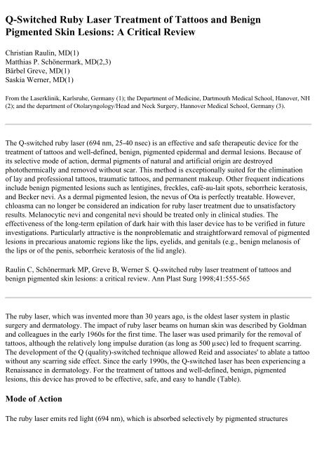
Q-Switched Ruby Laser Treatment of Tattoos and Benign Pigmented ...
Q-Switched Ruby Laser Treatment of Tattoos and Benign Pigmented ...
Q-Switched Ruby Laser Treatment of Tattoos and Benign Pigmented ...

Create successful ePaper yourself
Turn your PDF publications into a flip-book with our unique Google optimized e-Paper software.
Q-<strong>Switched</strong> <strong>Ruby</strong> <strong>Laser</strong> <strong>Treatment</strong> <strong>of</strong> <strong>Tattoos</strong> <strong>and</strong> <strong>Benign</strong><br />
<strong>Pigmented</strong> Skin Lesions: A Critical Review<br />
Christian Raulin, MD(1)<br />
Matthias P. Schönermark, MD(2,3)<br />
Bärbel Greve, MD(1)<br />
Saskia Werner, MD(1)<br />
From the <strong>Laser</strong>klinik, Karlsruhe, Germany (1); the Department <strong>of</strong> Medicine, Dartmouth Medical School, Hanover, NH<br />
(2); <strong>and</strong> the department <strong>of</strong> Otolaryngology/Head <strong>and</strong> Neck Surgery, Hannover Medical School, Germany (3).<br />
The Q-switched ruby laser (694 nm, 25-40 nsec) is an effective <strong>and</strong> safe therapeutic device for the<br />
treatment <strong>of</strong> tattoos <strong>and</strong> well-defined, benign, pigmented epidermal <strong>and</strong> dermal lesions. Because <strong>of</strong><br />
its selective mode <strong>of</strong> action, dermal pigments <strong>of</strong> natural <strong>and</strong> artificial origin are destroyed<br />
photothermically <strong>and</strong> removed without scar. This method is exceptionally suited for the elimination<br />
<strong>of</strong> lay <strong>and</strong> pr<strong>of</strong>essional tattoos, traumatic tattoos, <strong>and</strong> permanent makeup. Other frequent indications<br />
include benign pigmented lesions such as lentigines, freckles, café-au-lait spots, seborrheic keratosis,<br />
<strong>and</strong> Becker nevi. As a dermal pigmented lesion, the nevus <strong>of</strong> Ota is perfectly treatable. However,<br />
chloasma can no longer be considered an indication for ruby laser treatment due to unsatisfactory<br />
results. Melanocytic nevi <strong>and</strong> congenital nevi should be treated only in clinical studies. The<br />
effectiveness <strong>of</strong> the long-term epilation <strong>of</strong> dark hair with this laser device has to be verified in future<br />
investigations. Particularly attractive is the nonproblematic <strong>and</strong> straightforward removal <strong>of</strong> pigmented<br />
lesions in precarious anatomic regions like the lips, eyelids, <strong>and</strong> genitals (e.g., benign melanosis <strong>of</strong><br />
the lips or <strong>of</strong> the penis, seborrheic keratosis <strong>of</strong> the lid angle).<br />
Raulin C, Schönermark MP, Greve B, Werner S. Q-switched ruby laser treatment <strong>of</strong> tattoos <strong>and</strong><br />
benign pigmented skin lesions: a critical review. Ann Plast Surg 1998;41:555-565<br />
The ruby laser, which was invented more than 30 years ago, is the oldest laser system in plastic<br />
surgery <strong>and</strong> dermatology. The impact <strong>of</strong> ruby laser beams on human skin was described by Goldman<br />
<strong>and</strong> colleagues in the early 1960s for the first time. The laser was used primarily for the removal <strong>of</strong><br />
tattoos, although the relatively long impulse duration (as long as 500 µsec) led to frequent scarring.<br />
The development <strong>of</strong> the Q (quality)-switched technique allowed Reid <strong>and</strong> associates' to ablate a tattoo<br />
without any scarring side effect. Since the early 1990s, the Q-switched laser has been experiencing a<br />
Renaissance in dermatology. For the treatment <strong>of</strong> tattoos <strong>and</strong> well-defined, benign, pigmented<br />
lesions, this device has proved to be effective, safe, <strong>and</strong> easy to h<strong>and</strong>le (Table).<br />
Mode <strong>of</strong> Action<br />
The ruby laser emits red light (694 nm), which is absorbed selectively by pigmented structures
(melanosomes <strong>and</strong> melanin-containing cells) <strong>and</strong> exogenously introduced pigments (metallic <strong>and</strong><br />
organic color particles, dirt, <strong>and</strong> other erogenous substances). Oxyhemoglobin as well as hemoglobin<br />
show only a marginal absorption at this wavelength. The main absorption spectrum for these<br />
chromophores lies between 400 <strong>and</strong> 600 nm, leading to minimal thermal injury.<br />
The quality-switched mechanism, which was investigated with guinea pig <strong>and</strong> human skin, is based<br />
on the principle <strong>of</strong> selective photothermolysis. Because the duration <strong>of</strong> exposure to the energy is<br />
between 20 <strong>and</strong> 50 nsec, which is well below the thermal relaxation time <strong>of</strong> the target tissue<br />
(approximately 0.5-1 µsec for melanosomes), the damage to the surrounding tissue is minimal.<br />
The applied ruby laser beam causes extreme heat (as high as 1000°C) in a very short interval (25-40<br />
nsec). This "flash <strong>of</strong> heat" leads to a rupture <strong>of</strong> melanosome-carrying cells (melanocytes <strong>and</strong><br />
keratinocytes) in a depth <strong>of</strong> as much as 2 mm. Histological studies have shown that tattoo particles<br />
are located mainly in the macrophages <strong>and</strong> fibroblasts <strong>of</strong> the upper dermis. Only a very small portion<br />
may be found in the deeper interstitium. The laser beam, which is absorbed by dark pigments, leads<br />
to a fast, thermally induced expansion <strong>of</strong> the pigment-carrying cells (so-called "shock waves") <strong>and</strong><br />
thus to the loss <strong>of</strong> their integrity. These released pigments are phagocytosed by invading<br />
inflammatory cells <strong>and</strong> removed via the lymphatic system. Some smaller portion <strong>of</strong> the pigmented<br />
fragments are ablated transdermally by a crusting process. A chemical alteration <strong>of</strong> nonphysiological<br />
pigments by the affecting heat, which alters the optical refraction index, is another mode <strong>of</strong> action <strong>of</strong><br />
the ruby laser that has been discussed recently.<br />
Application <strong>of</strong> the Q-switched <strong>Ruby</strong> <strong>Laser</strong><br />
The Q-switched ruby laser that we use (<strong>Laser</strong>ase, Lambda Photometrics Ltd., Harpenden, UK)<br />
utilizes a pulse duration <strong>of</strong> 25 nsec. The fluence may be adjusted between 0.5 <strong>and</strong> 40 J per square<br />
centimeter, <strong>and</strong> the spot size can be between 2 <strong>and</strong> 4 mm . The laser pulses are applied with a<br />
frequency <strong>of</strong> one pulse per second in a slightly overlapping fashion. Therefore, large areas can be<br />
treated within one treatment session. Immediately after the application, the skin shows a whitish<br />
discoloration (Fig 1B), which is transient <strong>and</strong> usually disappears within 5 to 20 minutes. The cause <strong>of</strong><br />
this phenomenon is the induction <strong>of</strong> "steam" through the consecutive heating <strong>of</strong> intra- <strong>and</strong><br />
extracellular fluid. The expansive tendency <strong>of</strong> the steam leads to a rise <strong>of</strong> the epidermis, which in turn<br />
causes an alteration <strong>of</strong> the epidermal refractory index <strong>and</strong> thus a whitish color (see Fig 1B).<br />
Consequently, erythema appears in the treated area, which tapers <strong>of</strong>f within 24 hours. If higher<br />
energies are used or facial areas are treated, a transient swelling may occur that lasts no longer than<br />
36 hours.<br />
Within the next few days, delicate crusts <strong>and</strong> sometimes small blisters evolve that heal within the<br />
following 2 weeks. Patients should be taught not to manipulate the lesions to prohibit infections <strong>and</strong><br />
scarring. As a prophylaxis against infection, dressings with sulfadiazine-silver (Flammazine) or<br />
polyvidon-iodide (Betaisodona) may be applied.
Fig 1. (A) Amateur tattoos on both thighs. (B) Whitish discoloration immediately after the laser<br />
treatment. (C) Discrete hypopigmentation after five treatments with the Q-switched ruby laser<br />
The typical sensation experienced during lasering is a tingling, somewhat burning, low-range pain<br />
that normally does not need any anesthetic treatment. However, for very sensitive patients <strong>and</strong><br />
children, <strong>and</strong> for most facial applications, a topical anesthetic may be used (e.g., a lidocain-prilocain<br />
cream such as EMLA).<br />
For most patients, several consecutive treatments are necessary. The interval between sessions should<br />
be at least 4 weeks. During the entire therapeutic period <strong>and</strong> approximately 4 weeks beyond, patients<br />
should be advised to avoid direct sun exposure <strong>and</strong> tanning booths to minimize the risk <strong>of</strong> pigmentary
chances. Sun blockers with a high ultraviolet (UV) protection factor should be used if UV exposure is<br />
unavoidable. Hypo- <strong>and</strong>/or hyperpigmentation may be observed in some patients. Most <strong>of</strong> these<br />
changes are transient <strong>and</strong> will fade within several weeks. The risk <strong>of</strong> scarring or lasting pigmentary<br />
disturbances is very low for this type <strong>of</strong> laser.<br />
Indications<br />
<strong>Tattoos</strong><br />
Techniques for removal <strong>of</strong> pr<strong>of</strong>essional <strong>and</strong> lay tattoos have improved steadily during the past 10<br />
years. The more conventional therapeutic measures include dermabrasion, surgical excision,<br />
salabrasion, <strong>and</strong> cryotherapy. All <strong>of</strong> these methods bear a considerable risk for atrophic <strong>and</strong><br />
hypertrophic scarring, hypo- <strong>and</strong> hyperpigmentation, <strong>and</strong> incomplete removal <strong>of</strong> the pigment. The<br />
carbon dioxide <strong>and</strong> the argon lasers, which operate in a continuous mode, led to nonspecific tissue<br />
destruction <strong>and</strong> thus to posttherapeutic scars.<br />
The introduction <strong>of</strong> the Q-switched ruby laser, which allows selective destruction <strong>of</strong> tattoo pigments<br />
with minimal damage to the surrounding tissue, has led to scarless removal <strong>of</strong> tattoos. Lay <strong>and</strong><br />
pr<strong>of</strong>essional tattoos are <strong>of</strong>ten composed <strong>of</strong> different colors. Black <strong>and</strong> blue-black pigments can be<br />
removed effectively by the Q-switched ruby laser because these pigments absorb the ruby laser light<br />
well (absorption maximum, 600-800 nm). Green colors respond somewhat variously, <strong>and</strong> yellow <strong>and</strong><br />
red pigments cannot be removed by the ruby laser. These colors absorb light in the range <strong>of</strong> 500 to<br />
580 nm. Therefore, a combination <strong>of</strong> different laser systems must be applied to target effectively a<br />
complex, multicolored tattoo. The Q-switched ND:YAG laser seems<br />
to be particularly effective in the freqtienc@1-doubled mode (532 nm) for the treatment <strong>of</strong> red <strong>and</strong><br />
pink tattoos. Yellow <strong>and</strong> orange pigments respond very well to the pulsed dye laser (510 nm). An<br />
alternative to the ruby laser for the treatment <strong>of</strong> black tattoos seems to be the Q-switched Nd: YAG<br />
laser (1,064 nm). Finally, black <strong>and</strong> green pigments also may be treated effectively with the Qswitched<br />
alex<strong>and</strong>rite laser.<br />
The number <strong>of</strong> treatment sessions required differs from patient to patient (i.e., from tattoo to tattoo)<br />
<strong>and</strong> depends on the amount <strong>of</strong> pr<strong>of</strong>essionalism used to pierce the tattoo. Lay tattoos are <strong>of</strong>ten pierced<br />
with black ink only (Fig 1A) <strong>and</strong> can be removed with three to five treatments (Fig 1C). Pr<strong>of</strong>essional<br />
tattoos, however, contain a higher pigment density <strong>and</strong> more stable pigmentary compounds (heavy<br />
metals, organic pigments), <strong>and</strong> thus need more treatments (usually more than eight, but sometimes as<br />
many as 20 treatments are required to remove pr<strong>of</strong>essional tattoos). Older tattoos are more responsive<br />
to ruby laser treatment than newer tattoos. This may be because <strong>of</strong> the completed phagocytosis <strong>of</strong> the<br />
pigment <strong>and</strong> to the more fragile, more easily disrupted pigment crystals. To treat tattoos with the ruby<br />
laser, fluences between 5 <strong>and</strong> 20 J per square centimeter are applied.<br />
Sun-tanned or dark-colored skin (Fitzpatrick types IV-VI) may respond with some delay because<br />
melanin absorbs the major part <strong>of</strong> the applied light. Thus the light energy is filtered away by the<br />
pigmented, epidermal cells, which lay above the dermal tattoo pigments. To surmount this effect, the<br />
Q-switched ND:YAG laser may be used, which-due to its longer wavelength (1,064 nm)-penetrates
deeper into the skin <strong>and</strong> interferes only minimally with the melanin-containing structures.<br />
Because <strong>of</strong> the interaction with melanin, approximately 50% <strong>of</strong> all patients show some<br />
posttherapeutic hypopigmentation, which is almost always transient <strong>and</strong> tapers <strong>of</strong>f within 2 to 6<br />
months. Hyperpigmentation, however, is a rare side effect <strong>of</strong> ruby laser treatment. A predilection site<br />
for pigmentary changes is the ventral part <strong>of</strong> the forearms (personal observation). Patients who have<br />
dark pigment seem to be especially susceptible to posttherapeutic pigmentary changes. The highly<br />
selective mode <strong>of</strong> action <strong>of</strong> the ruby laser leads to a comparably low risk <strong>of</strong> scarring (
The penetration <strong>of</strong> artifacts (e.g., dirt, dust, s<strong>and</strong>, or plastic particles) into the skin during accidents or<br />
explosions leads to so-called "traumatic tattoos." After wound healing <strong>and</strong> reepithelialization is<br />
complete, it is difficult to remove the foreign particles from the dermis. Several techniques (e.g.,<br />
surgical excision, dermabrasion, electrosurgery, <strong>and</strong> carbon dioxide or argon lasers) have been<br />
described. Most <strong>of</strong> these measures, however, bear a considerable risk <strong>of</strong> scarring <strong>and</strong> may not lead to<br />
complete removal <strong>of</strong> all pigments. In contrast, the Q-switched ruby laser has proved to be a safe <strong>and</strong><br />
very effective therapeutic device for the removal <strong>of</strong> traumatic tattoos. Depending on the depth <strong>and</strong> the<br />
structure <strong>of</strong> the injected pigments, 3 to 20 sessions may be needed for complete removal (Fig 3).<br />
Normally, fluencies between 5 <strong>and</strong> 10 J per square centimeter are applied. Similar results may be<br />
achieved with the Q-switched ND:YAG laser (1,064 nm) or the Q-switched alex<strong>and</strong>rite laser.<br />
Fig 3. (A) Traumatic tattoo after a car accident in<br />
1971.<br />
Drug-Induced Hyperpigmentation<br />
(B) The lesion is removed completely after five<br />
treatments..<br />
After long-time <strong>and</strong>/or high-close treatment with minocycline, cutaneous hyperpigmentation has been<br />
noted to occur. The exact mechanism for this pigmentary change, however, is not fully understood.<br />
The application <strong>of</strong> Q-switched lasers (e.g., ruby or ND:YAG) may lead to a complete removal <strong>of</strong><br />
these cosmetically disturbing lesions within 5 to 10 sessions.<br />
<strong>Benign</strong> <strong>Pigmented</strong> Lesions<br />
Due to the selective absorption <strong>of</strong> the ruby laser light by melanin, several benign pigmented lesions<br />
can be treated quite effectively with this laser.<br />
Hyperpigmented Scars <strong>and</strong> Postinflammatory Hyperpigmentations<br />
Sometimes operation <strong>and</strong> trauma, especially burns <strong>and</strong> scaldings, result in cosmetically disfiguring<br />
hyperpigmented scars. The conservative treatment options for these lesions include cryotherapy,<br />
dermabrasion, <strong>and</strong> the application <strong>of</strong> bleach or scar ointments. However, most <strong>of</strong> these measures do<br />
not lead to satisfying results. <strong>Treatment</strong> with the Q-switched ruby laser can lead to a dramatic<br />
improvement <strong>of</strong> the scar zones. Fluencies between 4 <strong>and</strong> 10 J per square centimeter <strong>and</strong> several
treatment sessions (three to nine) may be used. Also, postinflammatory hyperpigmentary lesions (e.<br />
g., purpura jaune d'ocre) are a considerable responsive target for ruby laser therapy <strong>and</strong> can be<br />
lightened with this treatment (Fig 4). As an alternative, the frequency-doubled Q-switched ND:YAG<br />
laser (532 nm) can be applied. In a direct-comparison trial, however, the ruby laser showed a<br />
somewhat higher clearance rate than the ND:YAG laser.<br />
Fig 4. (A) Purpura jaune d'ocre on the left ankle<br />
<strong>of</strong> a 44-year old female patient.<br />
Ephelides <strong>and</strong> <strong>Benign</strong> Lentigines<br />
(B) Complete removal after five treatments.<br />
The Q-switched ruby laser can be used safely <strong>and</strong> effectively to remove cosmetically disturbing<br />
freckles (Fig 5) <strong>and</strong> benign lentigines <strong>of</strong> the h<strong>and</strong>s <strong>and</strong> face (Fig 6). Normally, the lesions are<br />
removed within one to two sessions. Patients should be informed before treatment that ephelides<br />
especially tend to recur at a considerably high rate. Sun exposure <strong>and</strong> tanning booths should be<br />
avoided for at least 6 weeks, <strong>and</strong> high-potency sun blockers should be used.
Fig 5. Ephelides <strong>of</strong> the arm. The forearm was treated once with the Q-switched ruby laser; the upper<br />
arm was not treated.<br />
Fig 6. (A) Senile lentigines on the back <strong>of</strong> the<br />
h<strong>and</strong>.<br />
<strong>Benign</strong> Melanosis <strong>of</strong> the Lip <strong>and</strong> <strong>of</strong> the Penis<br />
(B) Clinical picture after two laser treatments.<br />
Before these lesions are treated with the Q-switched ruby laser, it must be made explicitly clear that<br />
they are indeed benign (e.g., by a preceding biopsy). The benign melanosis responds very well to<br />
laser treatment, <strong>and</strong> disappears within one to three sessions. Fluencies between 6 <strong>and</strong> 10 J per square<br />
centimeter have been applied. Peutz-Jeghers syndrome-associated lentigines <strong>of</strong> the lips <strong>and</strong> the oral<br />
mucosa have also been treated effectively with the ruby laser.<br />
The benign penile melanosis is a rare condition that also has been removed successfully with the ruby<br />
laser. An invasive approach to this lesion (excision, dermabrasion) bears a considerable risk <strong>of</strong><br />
cosmetically or even functionally disturbing sequelae. This can be surmounted by application <strong>of</strong> the<br />
Q-switched ruby laser.<br />
Seborrheic Keratosis<br />
The treatment <strong>of</strong> choice for pigmented, seborrheic keratotic lesions is still excochleation with a sharp<br />
spoon. Alternatively, cryosurgical measures with liquid nitrogen or the Q-switched ruby laser may be
used. Seborrheic keratosis in delicate locations (e.g., at the lid angle) <strong>and</strong> multiple lesions can be<br />
removed very safely <strong>and</strong> with excellent cosmetic results (Fig 7). One to three sessions <strong>and</strong> high<br />
energy fluencies (as high as 40 J per square centimeter) are needed with the Q-switched ruby laser to<br />
remove seborrheic keratotic lesions completely. Another laser system that is used for this indication<br />
is the carbon dioxide laser. However, in a recent study including 32 patients <strong>and</strong> 690 seborrheic<br />
lesions, 25% <strong>of</strong> all patients who were treated with the carbon dioxide laser in continuous mode<br />
suffered from posttherapeutic, atrophic scarring. The carbon dioxide laser in the ultrapulse mode <strong>and</strong><br />
the Erbium:YAG laser seem to be more gentle <strong>and</strong> therefore have less side effects (personal<br />
observation).<br />
Fig 7. (A) Seborrheic keratosis <strong>of</strong> the face.<br />
Café-au-lait Spots<br />
(B) Cosmetically satisfactory result after two<br />
treatment sessions.<br />
These lesions appear in conjunction with genetically determined diseases (e.g., neur<strong>of</strong>ibromatosis) as<br />
<strong>of</strong>ten solitary, light- to medium-brown spots. They can be removed with the Q-switched ruby laser<br />
within three to eight treatment sessions (Figs 8). The effective fluencies lie between 7 <strong>and</strong> 20 J per<br />
square centimeter. The interval between sessions should be 4 to 6 weeks. However, some patients<br />
should be treated in shorter intervals to prohibit early repigmentation. The combined application <strong>of</strong>
the Q-switched mode (25-40 nsec) <strong>and</strong> the normal <strong>and</strong> long-pulse mode (270 µsec to 1 msec) allows<br />
the delivery <strong>of</strong> higher fluencies <strong>and</strong> thus the targeting <strong>of</strong> more deeply seated pigments (e.g., in<br />
follicles). Thus a higher clearance rate may be achieved (personal observation). Patients with darker<br />
skin types (Fitzpatrick IV <strong>and</strong> V) can develop posttherapeutic hypopigmentation that taper <strong>of</strong>f within<br />
a few months. Transient Hyperpigmentations have also been described. Therefore, a treatment trial<br />
should be carried out before the entire lesion area is treated. Recurring spots are not uncommon,<br />
especially if the melanocytes are activated due to insufficiently blocked sunlight. Recurrences occur<br />
in 40% to 50% <strong>of</strong> the patients (own unpublished data).<br />
Resistant lesions can be treated with other laser systems. Tse <strong>and</strong> colleagues describe a clearance rate<br />
<strong>of</strong> 30% with the frequency-doubled Q-switched ND:YAG laser (532 nm) compared with a 51%<br />
clearance rate by the Q-switched ruby laser. In contrast, the "normal" Q-switched Nd: YAG laser<br />
(1,064 nm) is, due to its higher wavelength, less effective for the treatment <strong>of</strong> epidermal pigmented<br />
lesions. The dye laser (510 nm) has been used for the treatment <strong>of</strong> 14 patients with café-au-lait spots;<br />
35% <strong>of</strong> the patients showed a 75% lightening whereas 42% <strong>of</strong> the patients seemed to be resistant.<br />
Somewhat better results could have been achieved by Fitzpatrick <strong>and</strong> associates.<br />
Fig 8. (A) Periorbital café-au-lait spot.<br />
Becker Nevus <strong>and</strong> Hypertrichosis<br />
(B) Complete removal <strong>of</strong> the lesion after five<br />
laser treatments.<br />
The Becker nevus is a hyperpigmented, <strong>of</strong>ten hypertrichotic lesion <strong>of</strong> young, mostly male adults. It<br />
can be treated effectively with the Q-switched ruby laser. Fluencies between 7 <strong>and</strong> 20 J per square<br />
centimeter are applied. Three to 10 treatment sessions at monthly intervals may be necessary.<br />
Resistant as well as recurring lesions have been described. Hypo- <strong>and</strong> hyperpigmentation are possible<br />
side effects <strong>of</strong> the treatment.<br />
For the treatment <strong>of</strong> cosmetically disturbing hypertrichosis, two highly energetic flashlamps with a<br />
continuous wavelength spectrum have been introduced recently (PhotoDerm VL/PL <strong>and</strong> EpiLight;<br />
ESC Medical Systems, Yokneam, Israel). Both devices lead to a removal <strong>of</strong> hair. The Q-switched<br />
ruby laser seems to be ineffective in the long-term removal <strong>of</strong> hypertrichosis. A delayed hair growth,<br />
however, has been described after the application <strong>of</strong> a normally pulsed ruby laser (270 µsec). In our<br />
h<strong>and</strong>s, the longpulsed ruby laser (1 msec) also led to a retarded growth <strong>of</strong> hairs but not to a reduction
<strong>of</strong> total hair number. Preliminary results suggest that the long-pulsed alex<strong>and</strong>rite laser (755 nm;<br />
impulse duration as high as 20 msec) is a very effective therapeutic alternative.<br />
Chloasma <strong>and</strong> Melasma<br />
These typically circumscribed, patchy facial hyperpigmentations that appear normally in younger<br />
women are difficult to treat. Hydrochinon-containing external therapeutics may have some lightening<br />
effect. The etiopathology shows increased melanin synthesis with epidermal <strong>and</strong> dermal depositions<br />
<strong>of</strong> melanin. These should be responsive to ruby laser treatment. Surprisingly, only 15% to 20% <strong>of</strong><br />
patients were responsive to the ruby laser. Some lesions became even worse <strong>and</strong> resulted in<br />
considerable additional pigmentation (personal observations). Therefore, we dispense with ruby laser<br />
treatment for chloasma. The dye laser (510 nm) seems to be equally ineffective. Grekin <strong>and</strong><br />
coworkers report a response rate <strong>of</strong> only 20%.<br />
Nevus <strong>of</strong> Ota<br />
This lesion is a rare, congenital oculodermal melanosis. The typical nevus <strong>of</strong> Ota is a blackblue mole<br />
that manifests in the area <strong>of</strong> the first <strong>and</strong> second branch <strong>of</strong> the trigeminal nerve. The Mongolian race<br />
has a much higher incidence <strong>of</strong> nevus <strong>of</strong> Ota than whites. Conservative therapeutic measures include<br />
dermabrasion, surgical excision, <strong>and</strong> cryotherapy. The results, however, have been mostly<br />
unsatisfactory. Argon laser treatment <strong>of</strong> this lesion has led to changes in the dermal texture <strong>and</strong> to a<br />
permanent loss <strong>of</strong> pigmentation. In contrast, treatment with the Q-switched ruby laser bears only a<br />
minimal risk <strong>of</strong> scarring <strong>and</strong> <strong>of</strong> pigmentary changes, which are almost always transient. To achieve<br />
complete removal, 8 to 10 sessions at 1- to 2-month intervals are needed. The applied fluencies range<br />
between 5 <strong>and</strong> 10 j per square. Also, other laser systems have been used quite successfully for the<br />
treatment <strong>of</strong> nevus <strong>of</strong> Ota. Dye laser therapy has led to an 80% lightening when combined with a<br />
flashlamppumped Q-switched alex<strong>and</strong>rite laser. In a histological study, it has been shown that the<br />
dermal melanocytes are targeted <strong>and</strong> destroyed by both the Q-switched ruby laser <strong>and</strong> the Q-switched<br />
ND:YAG laser. Clinically, however, ruby laser treatment led to a higher clearance rate (57%) than<br />
ND:YAG laser treatment (42%).<br />
Melanocytic Nevi <strong>and</strong> Congenital Nevi<br />
<strong>Laser</strong> treatment <strong>of</strong> congenital melanocytic nevi is a controversial subject. On the one h<strong>and</strong>,<br />
recurrence <strong>of</strong> lesions after laser treatment is frequent. On the other h<strong>and</strong>, the effects <strong>of</strong> laser<br />
irradiation on cellular biological behavior <strong>and</strong> the possible mutagenic responses <strong>of</strong> nevomelanocytes<br />
are still unclear.<br />
Congenital <strong>and</strong> immediate postpartum-appearing nevi have an incidence <strong>of</strong> approximately 1% among<br />
newborns. Without treatment, small- <strong>and</strong> middle-size nevi bear a risk <strong>of</strong> malignant transformation <strong>of</strong><br />
approximately 1% to 5%. Five percent <strong>of</strong> large nevi may develop into malignant melanoma during a<br />
person's lifetime. The therapeutic options for these large lesions include cryotherapy, dermabrasion,<br />
successive excision with combined exp<strong>and</strong>er techniques, <strong>and</strong> autologous transplantation.
Fig 9. (A) Superficical melanocytic nevus <strong>of</strong> the right cheek. (B) Histology <strong>of</strong> the lesion before<br />
treatment indicates superficially seated neval cell nests (H&E, original magnification x160 before<br />
53% reduction). (C) Complete removal after one treatment with the ruby laser (2.5 years after<br />
treatment).<br />
Generally, excision is the therapy <strong>of</strong> choice in removing suspect congenital <strong>and</strong> melanocytic nevi.<br />
Only in select patients should ruby laser therapy be considered (Figs 9A, C). As a general rule, nevi<br />
should not be treated uncritically with the Q-switched ruby laser. Clinical <strong>and</strong> histological studies<br />
show that the Q-switched ruby laser appears to be effective in removing superficial portions <strong>of</strong><br />
congenital melanocytic nevi. Deeper dermal melanocytes <strong>and</strong> nests <strong>of</strong> nonpigmented melanocytes<br />
remained unaltered. Therefore a therapy <strong>of</strong> melanocytic nevi <strong>and</strong> congenital nevi with the Q-switched
uby laser should only be performed in controlled studies. In these patients careful diagnostic<br />
procedure, especially epiluminescence microscopy <strong>and</strong> a biopsy, must be carried out before any kind<br />
<strong>of</strong> laser treatment is begun (Fig 9B). If there is the slightest suspicion <strong>of</strong> a premalignant alteration, an<br />
excision in toto should be performed. Longterm results <strong>of</strong> effective ruby laser therapy have to be<br />
evaluated in this situation before recommendations can be made.<br />
Indications <strong>and</strong> <strong>Treatment</strong> Parameters for the Q-<strong>Switched</strong> <strong>Ruby</strong> <strong>Laser</strong><br />
Indication Fluency (J/cm²) No. <strong>of</strong> <strong>Treatment</strong>s<br />
<strong>Tattoos</strong> (black, black-blue, green)<br />
Lay tattoos 5-20 3-8<br />
Pr<strong>of</strong>essional tattoos 5-20 8-20<br />
Permanent makeup 3-20 5-15<br />
Traumatic tattoos 5-10 3-20<br />
Minocycline-induced hyperpigmentations 4-10 3-9<br />
<strong>Benign</strong> pigmented lessons<br />
Hyperpigmented scars <strong>and</strong> postinflammatory<br />
hyperpigmentations<br />
4-10 3-9<br />
Ephelides <strong>and</strong> benign lentigines 6-10 1-2<br />
Labial <strong>and</strong> penile melanosis 6-10 1-3<br />
Seborrheic keratosis 10-40 2-3<br />
Café-au-lait spots 7-20 3-8<br />
Becker's nevus 7-20 3-10<br />
Nevus <strong>of</strong> Ota 5-10 8-10<br />
Congenital <strong>and</strong> immediate postpartum-appearing nevi have an incidence <strong>of</strong> approximately 1% among<br />
newborns. Without treatment, small- <strong>and</strong> middle-size nevi bear a risk <strong>of</strong> malignant transformation <strong>of</strong><br />
approximately 1% to 5%. Five percent <strong>of</strong> large nevi may develop into malignant melanoma during a<br />
person's lifetime. The therapeutic options for these large lesions include cryotherapy, dermabrasion,<br />
successive excision with combined exp<strong>and</strong>er techniques, <strong>and</strong> autologous transplantation.<br />
Generally, excision is the therapy <strong>of</strong> choice in removing suspect congenital <strong>and</strong> melanocytic nevi.<br />
Only in select patients should ruby laser therapy be considered (Figs 9A, C). As a general rule, nevi<br />
should not be treated uncritically with the Q-switched ruby laser. Clinical <strong>and</strong> histological studies<br />
show that the Q-switched ruby laser appears to be effective in removing superficial portions <strong>of</strong><br />
congenital melanocytic nevi. Deeper dermal melanocytes <strong>and</strong> nests <strong>of</strong> nonpigmented melanocytes
emained unaltered. Therefore a therapy <strong>of</strong> melanocytic nevi <strong>and</strong> congenital nevi with the Q-switched<br />
ruby laser should only be performed in controlled studies. In these patients careful diagnostic<br />
procedure, especially epiluminescence microscopy <strong>and</strong> a biopsy, must be carried out before any kind<br />
<strong>of</strong> laser treatment is begun (Fig 9B). If there is the slightest suspicion <strong>of</strong> a premalignant alteration, an<br />
excision in toto should be performed. Longterm results <strong>of</strong> effective ruby laser therapy have to be<br />
evaluated in this situation before recommendations can be made.<br />
(Literatur bei den Verfassern)<br />
Copyright (c) 1997-2002 PD Dr. med. Christian Raulin. Alle Rechte vorbehalten.<br />
Fragen, Anregungen und Kritik bitte an den Webmaster.<br />
Letzte Änderung: Freitag, 07. Juli 2000<br />
Webdesign und Pflege by ISD<br />
Homepage<br />
Seitenanfang