The Principles of Clinical Cytogenetics - Extra Materials - Springer
The Principles of Clinical Cytogenetics - Extra Materials - Springer
The Principles of Clinical Cytogenetics - Extra Materials - Springer

Create successful ePaper yourself
Turn your PDF publications into a flip-book with our unique Google optimized e-Paper software.
<strong>The</strong> <strong>Principles</strong> <strong>of</strong><br />
<strong>Clinical</strong><br />
<strong>Cytogenetics</strong><br />
SECOND EDITION<br />
EDITED BY<br />
Steven L. Gersen<br />
Martha B. Keagle<br />
Includes Updated<br />
ISCN Guidelines
<strong>The</strong> <strong>Principles</strong> <strong>of</strong> <strong>Clinical</strong> <strong>Cytogenetics</strong>
<strong>The</strong> <strong>Principles</strong> <strong>of</strong><br />
<strong>Clinical</strong> <strong>Cytogenetics</strong><br />
Second Edition<br />
Edited by<br />
Steven L. Gersen, PhD<br />
AmeriPath Inc.<br />
and<br />
Martha B. Keagle, MEd<br />
University <strong>of</strong> Connecticut, Storrs, CT
© 2005 Humana Press Inc.<br />
999 Riverview Drive, Suite 208<br />
Totowa, New Jersey 07512<br />
All rights reserved. No part <strong>of</strong> this book may be reproduced, stored in a retrieval system, or transmitted in any form or by any<br />
means, electronic, mechanical, photocopying, micr<strong>of</strong>ilming, recording, or otherwise without written permission from the<br />
Publisher.<br />
All authored papers, comments, opinions, conclusions, or recommendations are those <strong>of</strong> the author(s), and do not necessarily<br />
reflect the views <strong>of</strong> the publisher.<br />
This publication is printed on acid-free paper. ∞<br />
ANSI Z39.48-1984 (American Standards Institute) Permanence <strong>of</strong> Paper for Printed Library <strong>Materials</strong>.<br />
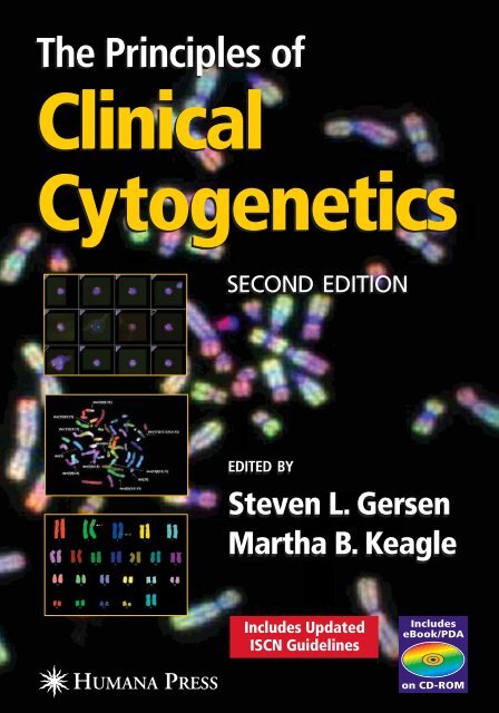
Cover illustration: <strong>The</strong> cover images are courtesy <strong>of</strong> Applied Imaging, Inc.<br />
Top inset: from Chapter 7, Fig. 11 (Instrumentation in the cytogenetics laboratory), by Steven L Gersen and Lotte Downey.<br />
Middle inset: M-FISH <strong>of</strong> a metaphase from a patient with CML in transformation to blast crisis. Image submitted by Anette<br />
Grand, Inge-Lise Frost Andersen, and Mette Klarskov Andersen.<br />
Bottom inset: M-FISH karyotype demonstrating a complex cryptic rearrangement in a child with multiple anomalies and<br />
developmental delay. Image submitted by Charles Lee.<br />
Background image: Comparative genomic hybridization (CGH), submitted by James Ashman.<br />
Cover design by Patricia F. Cleary.<br />
For additional copies, pricing for bulk purchases, and/or information about other Humana titles, contact Humana at<br />
the above address or at any <strong>of</strong> the following numbers: Tel.: 973-256-1699; Fax: 973-256-8341; E-mail: humana@humanapr.com;<br />
Website: http://humanapress.com<br />
Photocopy Authorization Policy:<br />
Authorization to photocopy items for internal or personal use, or the internal or personal use <strong>of</strong> specific clients, is granted by<br />
Humana Press Inc., provided that the base fee <strong>of</strong> US $25.00 per copy is paid directly to the Copyright Clearance Center at 222<br />
Rosewood Drive, Danvers, MA 01923. For those organizations that have been granted a photocopy license from the CCC, a<br />
separate system <strong>of</strong> payment has been arranged and is acceptable to Humana Press Inc. <strong>The</strong> fee code for users <strong>of</strong> the Transactional<br />
Reporting Service is: [1-58829-300-9/05 $25.00].<br />
Printed in the United States <strong>of</strong> America. 10 9 8 7 6 5 4 3 2<br />
Library <strong>of</strong> Congress Cataloging in Publication Data<br />
<strong>The</strong> principles <strong>of</strong> clinical cytogenetics / edited by Steven L. Gersen and Martha B. Keagle.— 2nd ed.<br />
p. ; cm.<br />
Includes bibliographical references and index.<br />
ISBN 1-58829-300-9 (alk. paper) eISBN 1-59259-833-1<br />
1. Human chromosome abnormalities—Diagnosis. 2. Human cytogenetics.<br />
[DNLM: 1. Chromosomes—genetics. 2. Cytogenetic Analysis—methods. QH 600 P957 2005] I.<br />
Keagle, Martha B.<br />
RB44.P75 2005<br />
616'.042—dc22 2004005167
v<br />
Preface<br />
In the summer <strong>of</strong> 1989, one <strong>of</strong> us (SLG), along with his mentor, Dorothy Warburton,<br />
attended the Tenth International Workshop on Human Gene Mapping. <strong>The</strong> meeting was<br />
held at Yale University in celebration <strong>of</strong> the first such event, which also took place there.<br />
This meeting was not open to the general public; one had to have contributed to mapping<br />
a gene to be permitted to attend. <strong>The</strong> posters, <strong>of</strong> course, were therefore all related to<br />
gene mapping, and many were covered with pretty, colorful pictures <strong>of</strong> a novel, fluorescent<br />
application <strong>of</strong> an old technology, in situ hybridization. Walking through the room, Dorothy<br />
remarked that, because <strong>of</strong> this new FISH technique, chromosomes, which had become<br />
yesterday’s news, were once again “back in style.”<br />
Approximately three years later, a commercial genetics company launched a FISH assay<br />
for prenatal ploidy detection. A substantial number <strong>of</strong> cytogeneticists across the country<br />
reacted with a combination <strong>of</strong> outrage and panic. Many were concerned that physicians<br />
would be quick to adopt this newfangled upstart test and put us all on the unemployment<br />
line. <strong>The</strong>y did not at the time realize what Dorothy instinctively already knew—that FISH<br />
would not spell the doom <strong>of</strong> the cytogenetics laboratory, but it would, rather, take it to new<br />
heights. In the early 1990s we didn’t know where FISH would end up being performed,<br />
but because <strong>of</strong> the number <strong>of</strong> FISH applications that require metaphase chromosomes, it<br />
has landed, either literally or functionally, squarely in the cytogenetics laboratory, securing<br />
its place in an increasingly “molecularized” laboratory environment. Add to this the explosion<br />
<strong>of</strong> cytogenetic and FISH data to become available in oncology in recent years, and it<br />
becomes apparent that chromosomes are here to stay.<br />
This brings us to the revision <strong>of</strong> <strong>The</strong> <strong>Principles</strong> <strong>of</strong> <strong>Clinical</strong> <strong>Cytogenetics</strong>. After the first<br />
edition was printed, it seemed possible that we had achieved our goal <strong>of</strong> assembling the<br />
basic concepts <strong>of</strong> clinical cytogenetics for the “end user” physician or student who needed to<br />
understand what we do, and that perhaps no update would be necessary. However, FISH and<br />
cancer cytogenetics continued to march on, and new data have become available even for<br />
such basic concepts as chromosome rearrangements, sex chromosome abnormalities, and<br />
autosomal aneuploidy. Combine these with all that has been learned about uniparental<br />
disomy and imprinting in the last five years, plus the regulatory changes we are all subject<br />
to, and it becomes obvious that what was needed was not a second printing, but a second<br />
edition.<br />
Our one concern was that, as this edition <strong>of</strong> <strong>The</strong> <strong>Principles</strong> <strong>of</strong> <strong>Clinical</strong> <strong>Cytogenetics</strong> went<br />
to press, the nomenclature committee had met but had not set any date for a revision <strong>of</strong> the<br />
ISCN, the nomenclature that forms the core <strong>of</strong> reporting in clinical cytogenetics. <strong>The</strong> best<br />
guess was that this would be available sometime in 2005, ten years after the last revision and<br />
a year after this book will have been published. Though we weren't comfortable with the<br />
notion that part <strong>of</strong> this book could be out <strong>of</strong> date shortly after its printing, most if not all<br />
updates were expected to involve details <strong>of</strong> FISH nomenclature that could not have been
vi Preface<br />
envisioned when ISCN 1995 was released, and we therefore decided not to delay the publication<br />
<strong>of</strong> this update merely to wait for that one.<br />
Our expectations were correct. ISCN 2005 was released late last year, and while some<br />
aspects <strong>of</strong> cytogenetic nomenclature have been updated, most improvements center around<br />
FISH. It was therefore possible to incorporate certain necessary changes into Chapter 3 <strong>of</strong><br />
this revised second edition and render it up to date.<br />
This edition <strong>of</strong> <strong>The</strong> <strong>Principles</strong> <strong>of</strong> <strong>Clinical</strong> <strong>Cytogenetics</strong> is organized much like the first,<br />
though there are several important changes. First, because <strong>of</strong> its increasing importance, the<br />
FISH chapter is now its own section in the book. Next, the increasing importance <strong>of</strong> cancer<br />
cytogenetics has prompted us to separate this subject into two distinct chapters, covering<br />
hematological disorders and solid tumors, also as a separate section. Because the ever-increasing<br />
popularity <strong>of</strong> computerized imaging and karyotyping systems has resulted in a waning<br />
popularity and likely eventual elimination <strong>of</strong> standard photography in the cytogenetics<br />
lab, this topic has been eliminated. Finally, two new chapters have been added, covering<br />
chromosome instability and the cytogenetics <strong>of</strong> infertility.<br />
We would like to take this opportunity to thank the authors who contributed to this book,<br />
and to the readers who made the first edition a success. We hope this edition will prove to be<br />
equally valuable.<br />
Steven L. Gersen, PhD<br />
Martha Keagle, MEd
vii<br />
Preface to First Edition<br />
<strong>The</strong> study <strong>of</strong> human chromosomes plays a role in the diagnosis, prognosis, and monitoring<br />
<strong>of</strong> treatment involving conditions seen not only by medical geneticists and genetic counselors,<br />
but also by pediatricians, obstetrician/gynecologists, perinatologists, hematologists,<br />
oncologists, endocrinologists, pathologists, urologists, internists, and family practice physicians.<br />
In addition, cytogenetic testing is <strong>of</strong>ten an issue for hospital laboratory personnel and<br />
managed care organizations.<br />
Few esoteric clinical laboratory disciplines have the potential to affect such a broad range<br />
<strong>of</strong> medical specialists, yet cytogenetics is <strong>of</strong>ten less well understood than most “specialized”<br />
testing.<br />
One can attribute this to several causes:<br />
• <strong>The</strong> cytogenetics laboratory is essentially the only setting in which living cells are required<br />
for traditional testing (fluorescence in situ hybridization [FISH] provides an<br />
exception to this rule). This unusual sample requirement is a potential source <strong>of</strong> confusion.<br />
• <strong>Cytogenetics</strong> is still perceived, and rightly so, to be as much “art” as it is science in an<br />
era when most clinical testing is becoming more and more automated or “high tech.”<br />
• Genetics in general still does not receive sufficient emphasis in the training <strong>of</strong> medical<br />
personnel.<br />
This issue has been complicated in recent years because, in an era <strong>of</strong> molecular medicine,<br />
chromosome analysis has become somewhat less <strong>of</strong> a stand alone discipline; as genes are<br />
mapped to chromosomes, traditional cytogenetics is <strong>of</strong>ten augmented with DNA analysis<br />
and/or FISH. <strong>The</strong> latter, <strong>of</strong>ten referred to as “molecular cytogenetics,” represents the single<br />
most significant advance in this field in decades, and has become such an integral part <strong>of</strong> the<br />
typical cytogenetics laboratory, with such a wide variety <strong>of</strong> applications, that it warrants its<br />
own chapter in <strong>The</strong> <strong>Principles</strong> <strong>of</strong> <strong>Clinical</strong> <strong>Cytogenetics</strong>.<br />
It is impossible to completely separate the relationships that exist today between the cytogenetics<br />
and the molecular genetics laboratories, from cases involving fragile-X-syndrome to<br />
those dealing with cancer patients, and for this reason, relevant molecular concepts are discussed<br />
in several chapters.<br />
Entire volumes have been devoted to some <strong>of</strong> the topics covered in <strong>The</strong> <strong>Principles</strong> <strong>of</strong><br />
<strong>Clinical</strong> <strong>Cytogenetics</strong>; these <strong>of</strong>ten serve as references or how-to manuals for those involved in<br />
providing genetics services, and in most cases provide a greater level <strong>of</strong> detail than is needed<br />
here. <strong>The</strong> purpose <strong>of</strong> the present book is to provide a comprehensive description <strong>of</strong> the basic<br />
concepts involved in chromosome analysis in a single volume, while at the same time producing<br />
a summary <strong>of</strong> sufficient depth to be <strong>of</strong> value to the practicing genetics pr<strong>of</strong>essional. We hope<br />
that it will serve as a valuable reference to any health care provider, from the individual who<br />
utilizes cytogenetics routinely to someone who has need <strong>of</strong> it on rare occasions.
viii Preface to First Edition<br />
<strong>The</strong> <strong>Principles</strong> <strong>of</strong> <strong>Clinical</strong> <strong>Cytogenetics</strong> is divided into four sections. <strong>The</strong> first section provides<br />
an historical perspective and explanation <strong>of</strong> the concepts involved, including a detailed<br />
description <strong>of</strong> cytogenetic nomenclature and examples <strong>of</strong> its use. <strong>The</strong> second section is an<br />
overview <strong>of</strong> the processes involved. <strong>The</strong> purpose <strong>of</strong> this section is to provide a fundamental<br />
understanding <strong>of</strong> the labor-intensive nature <strong>of</strong> chromosome analysis. It is not, however, a “laboratory<br />
manual”; detailed protocols for laboratory use are available elsewhere and are not appropriate<br />
in this setting. <strong>The</strong> third section comprises the main focus <strong>of</strong> this book, namely, the<br />
various applications <strong>of</strong> chromosome analysis in clinical settings and the significance <strong>of</strong> abnormal<br />
results. <strong>The</strong> final section connects cytogenetics to the broader field <strong>of</strong> clinical genetics,<br />
with discussions <strong>of</strong> synergistic technologies and genetic counseling.<br />
We gratefully acknowledge the hard work and attention to detail provided by the individuals<br />
who authored each chapter <strong>of</strong> <strong>The</strong> <strong>Principles</strong> <strong>of</strong> <strong>Clinical</strong> <strong>Cytogenetics</strong>, and thank our<br />
publisher for supporting this effort.<br />
Steven L. Gersen, PhD<br />
Martha B. Keagle, MEd
ix<br />
Contents<br />
Preface .....................................................................................................................................v<br />
Preface to First Edition ........................................................................................................ vii<br />
Contributors........................................................................................................................... xi<br />
eBook/PDA Instructions ..................................................................................................... xiii<br />
Section I Basic Concepts and Background.......................................... 1<br />
1 History <strong>of</strong> <strong>Clinical</strong> <strong>Cytogenetics</strong><br />
Steven L. Gersen ........................................................................................................... 3<br />
2 DNA, Chromosomes, and Cell Division<br />
Martha B. Keagle ......................................................................................................... 9<br />
3 Human Chromosome Nomenclature: An Overview and Definition <strong>of</strong> Terms<br />
Avirachan Tharapel ................................................................................................... 27<br />
Section II Examining and Analyzing Chromosomes........................ 59<br />
Editors’ Foreword<br />
Steven L. Gersen and Martha B. Keagle .................................................................. 61<br />
4 Basic Laboratory Procedures<br />
Martha B. Keagle and Steven L. Gersen .................................................................. 63<br />
5 <strong>The</strong> Fundamentals <strong>of</strong> Microscopy<br />
Christopher McAleer .................................................................................................. 81<br />
6 Quality Control and Quality Assurance<br />
Michael Watson and Steven L. Gersen ..................................................................... 93<br />
7 Instrumentation in the <strong>Cytogenetics</strong> Laboratory<br />
Steven L. Gersen and Lotte Downey ....................................................................... 113<br />
Section III <strong>Clinical</strong> <strong>Cytogenetics</strong> ...................................................... 131<br />
8 Autosomal Aneuploidy<br />
Jin-Chen C. Wang .................................................................................................... 133
x Contents<br />
9 Structural Chromosome Rearrangements<br />
Kathleen Kaiser-Rogers and Kathleen Rao ............................................................ 165<br />
10 Sex Chromosomes and Sex Chromosome Abnormalities<br />
Cynthia M. Powell .................................................................................................... 207<br />
11 <strong>Cytogenetics</strong> <strong>of</strong> Infertility<br />
Linda Marie Randolph ............................................................................................. 247<br />
12 Prenatal <strong>Cytogenetics</strong><br />
Linda Marie Randolph ............................................................................................. 267<br />
13 <strong>Cytogenetics</strong> <strong>of</strong> Spontaneous Abortion<br />
Solveig M. V. Pflueger ............................................................................................. 323<br />
14 Chromosome Instability<br />
Xiao-Xiang Zhang .................................................................................................... 347<br />
Section IV Cancer <strong>Cytogenetics</strong> ....................................................... 363<br />
15 <strong>Cytogenetics</strong> <strong>of</strong> Hematologic Neoplasms<br />
Rizwan C. Naeem...................................................................................................... 365<br />
16 <strong>Cytogenetics</strong> <strong>of</strong> Solid Tumors<br />
Jonathan A. Fletcher ............................................................................................... 421<br />
Section V Fluorescence In Situ Hybridization ................................ 453<br />
17 Fluorescence In Situ Hybridization (FISH)<br />
Daynna J. Wolff and Stuart Schwartz .................................................................... 455<br />
Section VI Beyond Chromosomes .................................................... 491<br />
Editors’ Foreword<br />
Steven L. Gersen and Martha B. Keagle ................................................................ 493<br />
18 Fragile X: From <strong>Cytogenetics</strong> to Molecular Genetics<br />
Dana C. Crawford and Patricia N. Howard-Peebles ............................................. 495<br />
19 Genomic Imprinting and Uniparental Disomy<br />
Jin-Chen C. Wang .................................................................................................... 515<br />
20 Genetic Counseling<br />
Sarah Hutchings Clark ............................................................................................ 541<br />
Index ................................................................................................................................... 559
Contributors<br />
SARAH HUTCHINGS CLARK, MS, CGC • New England Fertility Institute, Stamford, CT<br />
DANA C. CRAWFORD, PhD • Department <strong>of</strong> Genome Sciences, University <strong>of</strong> Washington,<br />
Seattle, WA<br />
LOTTE DOWNEY • Applied Imaging, Santa Clara, CA<br />
JONATHAN A. FLETCHER, MD • Department <strong>of</strong> Pathology, Brigham and Women’s Hospital,<br />
Boston, MA<br />
STEVEN L. GERSEN, PhD • AmeriPath, Inc.<br />
PATRICIA N. HOWARD-PEEBLES, PhD • Howard-Peebles Consulting, Fairview, TX,<br />
and Genetics & IVF Institute, Fairfax, VA (Retired)<br />
KATHLEEN KAISER-ROGERS, PhD • Department <strong>of</strong> Pediatrics, University <strong>of</strong> North Carolina at<br />
Chapel Hill, Chapel Hill, NC<br />
MARTHA B. KEAGLE, MEd • University <strong>of</strong> Connecticut, Storrs, CT<br />
CHRISTOPHER MCALEER • Cellomics Inc., Pittsburgh, PA<br />
RIZWAN C. NAEEM, MD • Department <strong>of</strong> Pediatrics and Pathology, Texas Children’s<br />
Cancer Center, Baylor College <strong>of</strong> Medicine, Houston, TX<br />
SOLVEIG M.V. PFLUEGER, PhD, MD • Department <strong>of</strong> Pathology, Baystate Medical Center, Tufts<br />
University School <strong>of</strong> Medicine, Springfield, MA<br />
CYNTHIA M. POWELL, MD • Department <strong>of</strong> Pediatrics, Division <strong>of</strong> Genetics and Metabolism,<br />
University <strong>of</strong> North Carolina at Chapel Hill, Chapel Hill, NC<br />
LINDA MARIE RANDOLPH, MD • Genetic Resources Medical Group Inc. and Childrens<br />
Hospital Los Angeles, Los Angeles, CA<br />
KATHLEEN RAO, PhD • Department <strong>of</strong> Pediatrics, University <strong>of</strong> North Carolina<br />
at Chapel Hill, Chapel Hill, NC<br />
STUART SCHWARTZ, PhD • Department <strong>of</strong> Genetics and Center for Human Genetics,<br />
Case Western Reserve University and University Hospitals <strong>of</strong> Cleveland, Cleveland, OH<br />
AVIRACHAN THARAPEL, PhD • Department <strong>of</strong> Pediatric and Ob/Gyn Genetics,<br />
University <strong>of</strong> Tennessee, Memphis, TN<br />
JIN-CHEN C. WANG, MD • Specialty Laboratories, Valencia, CA<br />
MICHAEL S. WATSON, phd • American College <strong>of</strong> Medical Genetics, Bethesda, MD<br />
DAYNNA J. WOLFF, PhD • Department <strong>of</strong> Pathology and Laboratory Medicine,<br />
Medical University <strong>of</strong> South Carolina, Charleston, SC<br />
XIAO-XIANG ZHANG, MD, PhD • DIANON Systems, A LabCorp Co., Oklahoma City, OK<br />
xi
Value-Added eBook/PDA on CD-ROM<br />
This book is accompanied by a value-added CD-ROM that contains an eBook version <strong>of</strong> the volume<br />
you have just purchased. This eBook can be viewed on your computer, and you can synchronize<br />
it to your PDA for viewing on your handheld device. <strong>The</strong> eBook enables you to view this volume on<br />
only one computer and PDA. Once the eBook is installed on your computer, you cannot download,<br />
install, or e-mail it to another computer; it resides solely with the computer to which it is installed.<br />
<strong>The</strong> license provided is for only one computer. <strong>The</strong> eBook can only be read using Adobe ® Reader ®<br />
6.0 s<strong>of</strong>tware, which is available free from Adobe Systems Incorporated at www.Adobe.com. You<br />
may also view the eBook on your PDA using the Adobe ® PDA Reader ® s<strong>of</strong>tware that is also available<br />
free from Adobe.com.<br />
You must follow a simple procedure when you install the eBook/PDA that will require you to<br />
connect to the Humana Press website in order to receive your license. Please read and follow the<br />
instructions below:<br />
1. Download and install Adobe ® Reader ® 6.0 s<strong>of</strong>tware<br />
You can obtain a free copy <strong>of</strong> the Adobe ® Reader ® 6.0 s<strong>of</strong>tware at www.adobe.com<br />
*Note: If you already have the Adobe ® Reader ® 6.0 s<strong>of</strong>tware installed, you do not need to reinstall it.<br />
2. Launch Adobe ® Reader ® 6.0 s<strong>of</strong>tware<br />
3. Install eBook: Insert your eBook CD into your CD-ROM drive<br />
PC: Click on the “Start” button, then click on “Run”<br />
At the prompt, type “d:\ebookinstall.pdf” and click “OK”<br />
*Note: If your CD-ROM drive letter is something other than d: change the above command<br />
accordingly.<br />
MAC: Double click on the “eBook CD” that you will see mounted on your desktop.<br />
Double click “ebookinstall.pdf”<br />
4. Adobe ® Reader ® 6.0 s<strong>of</strong>tware will open and you will receive the message<br />
“This document is protected by Adobe DRM” Click “OK”<br />
*Note: If you have not already activated the Adobe ® Reader ® 6.0 s<strong>of</strong>tware, you will be prompted to do so.<br />
Simply follow the directions to activate and continue installation.<br />
Your web browser will open and you will be taken to the Humana Press eBook registration page. Follow<br />
the instructions on that page to complete installation. You will need the serial number located on the<br />
sticker sealing the envelope containing the CD-ROM.<br />
If you require assistance during the installation, or you would like more information regarding<br />
your eBook and PDA installation, please refer to the eBookManual.pdf located on your cd. If you<br />
need further assistance, contact Humana Press eBook Support by e-mail at: ebooksupport@humanapr.<br />
com or by phone at 973-256-1699.<br />
*Adobe and Reader are either registered trademarks or trademarks <strong>of</strong> Adobe Systems Incorporated<br />
in the United States and/or other countries.<br />
xiii
History <strong>of</strong> <strong>Clinical</strong> <strong>Cytogenetics</strong> 1<br />
I<br />
Basic Concepts and Background
History <strong>of</strong> <strong>Clinical</strong> <strong>Cytogenetics</strong> 3<br />
History <strong>of</strong> <strong>Clinical</strong> <strong>Cytogenetics</strong><br />
From: <strong>The</strong> <strong>Principles</strong> <strong>of</strong> <strong>Clinical</strong> <strong>Cytogenetics</strong>, Second Edition<br />
Edited by: S. L. Gersen and M. B. Keagle © Humana Press Inc., Totowa, NJ<br />
3<br />
1<br />
Steven L. Gersen, PhD<br />
<strong>The</strong> beginning <strong>of</strong> human cytogenetics is generally attributed to Walther Flemming, an Austrian<br />
cytologist and pr<strong>of</strong>essor <strong>of</strong> anatomy, who published the first illustrations <strong>of</strong> human chromosomes in<br />
1882. Flemming also referred to the stainable portion <strong>of</strong> the nucleus as chromatin and first used the<br />
term mitosis (1). In 1888, Waldeyer introduced the word chromosome, from the Greek words for<br />
“colored body” (2), and several prominent scientists <strong>of</strong> the day began to formulate the idea that<br />
determinants <strong>of</strong> heredity were carried on chromosomes. After the “rediscovery” <strong>of</strong> Mendelian inheritance<br />
in 1900, Sutton (and, independently at around the same time, Boveri) formally developed a<br />
“chromosome theory <strong>of</strong> inheritance” (3,4). Sutton combined the disciplines <strong>of</strong> cytology and genetics<br />
when he referred to the study <strong>of</strong> chromosomes as cytogenetics.<br />
Owing in part to improvements in optical lenses, stains, and tissue manipulation techniques during<br />
the late 19th and early 20th centuries, the study <strong>of</strong> cytogenetics continued, with an emphasis placed<br />
by some on determining the correct number <strong>of</strong> chromosomes, as well as the sex chromosome configuration,<br />
in humans. Several reports appeared, with differing estimates <strong>of</strong> these. For example, in<br />
1912, von Winiwarter concluded that men have 47 chromosomes and women have 48 (5). <strong>The</strong>n, in<br />
1923, Painter studied (meiotic) chromosomes derived from the testicles <strong>of</strong> several men who had been<br />
incarcerated, castrated, and ultimately hanged in the Texas State Insane Asylum. Based on this work,<br />
Painter definitively reported the human diploid chromosome number to be 48 (double the 24 bivalents<br />
he saw), even though, 2 years earlier, he had preliminarily reported that some <strong>of</strong> his better samples<br />
produced a diploid number <strong>of</strong> 46 (6). At this time, Painter also proposed the X and Y sex chromosome<br />
mechanism in man. One year later, Levitsky formulated the term karyotype to refer to the<br />
ordered arrangement <strong>of</strong> chromosomes (7).<br />
Despite continued technical improvements, there was clearly some difficulty in properly visualizing<br />
or discriminating between individual chromosomes. Even though Painter’s number <strong>of</strong> 48 human<br />
chromosomes was reported somewhat conservatively, it was increasingly treated as fact with the<br />
passage <strong>of</strong> time and was “confirmed” several times over the next few decades. For example, in 1952,<br />
Hsu reported that, rather than depending on histologic sections, examination <strong>of</strong> chromosomes could<br />
be facilitated if one studied cells grown with tissue culture techniques published by Fisher (8). Hsu<br />
then demonstrated the value <strong>of</strong> this method by using it to examine human embryonic cell cultures,<br />
from which he produced both mitotic metaphase drawings and an ideogram (9) <strong>of</strong> all 48 human<br />
chromosomes!<br />
As with other significant discoveries, correcting this inaccuracy required an unplanned event—a<br />
laboratory error. Its origin can be found in the addendum that appears at the end <strong>of</strong> Hsu’s paper:<br />
It was found after this article had been sent to press that the well-spread metaphases were the result<br />
<strong>of</strong> an accident. Instead <strong>of</strong> being washed in isotonic saline, the cultures had been washed in hypotonic<br />
solution before fixation.
4 Steven Gersen<br />
<strong>The</strong> hypotonic solution caused water to enter the cells via osmosis, which swelled the cell membranes<br />
and separated the chromosomes, making them easier to visualize. This accident was the key<br />
that unlocked the future <strong>of</strong> human cytogenetics. Within 1 year, Hsu and Pomerat, realizing the potential<br />
<strong>of</strong> this fortuitous event, reported a “hypotonic shock” procedure (10). By 1955, Ford and<br />
Hamerton had modified this technique and had also worked out a method for pretreating cells grown<br />
in culture with colchicine so as to destroy the mitotic spindle apparatus and thus accumulate dividing<br />
cells in the metaphase (11). Joe Hin Tjio, an American-born Indonesian, learned about these procedures<br />
and worked with Hamerton and Ford to further improve upon them.<br />
In November 1955, Tjio was invited to Lund, Sweden to work on human embryonic lung fibroblast<br />
cultures in the laboratory <strong>of</strong> his colleague, Levan, a Spaniard who had learned the colchicine<br />
and hypotonic method in Hsu’s laboratory at the Sloan-Kettering Institute in New York. Tjio and<br />
Levan optimized the colchicine/hypotonic method for these cells, and in January 1956 (after carefully<br />
reviewing images from decades <strong>of</strong> previously reported work), they diplomatically reported that<br />
the human diploid chromosome number appeared to be 46, not 48 (12). <strong>The</strong>y referenced anecdotal<br />
data from a colleague who had been studying liver mitoses from aborted human embryos in the<br />
spring 1955, but temporarily abandoned the research “because the workers were unable to find all the<br />
48 human chromosomes in their material; as a matter <strong>of</strong> fact, the number 46 was repeatedly counted<br />
in their slides.” Tjio and Levan concluded their paper<br />
. . . we do not wish to generalize our present findings into a statement that the chromosome number<br />
<strong>of</strong> man is 2n=46, but it is hard to avoid the conclusion that this would be the most natural explanation<br />
<strong>of</strong> our observations.<br />
What was dogma for over 30 years had been overturned in one now classic paper. Ford and<br />
Hamerton soon confirmed Tjio and Levan’s finding (13). <strong>The</strong> era <strong>of</strong> clinical cytogenetics was at<br />
hand. It would take 3 more years to arrive, however, and it would begin with the identification <strong>of</strong> four<br />
chromosomal syndromes.<br />
<strong>The</strong> concept that an abnormality involving the chromosomes could have a phenotypic effect was<br />
not original. In 1932, Waardenburg made the suggestion that Down syndrome could perhaps be the<br />
result <strong>of</strong> a chromosomal aberration (14), but the science <strong>of</strong> the time could neither prove nor disprove<br />
his idea; this would take almost three decades. In 1958, Lejeune studied the chromosomes <strong>of</strong> fibroblast<br />
cultures from patients with Down syndrome, and in 1959, Lejeune and colleagues described an<br />
extra chromosome in each <strong>of</strong> these cells (15). <strong>The</strong> trisomy was reported to involve one <strong>of</strong> the smallest<br />
pairs <strong>of</strong> chromosomes and would eventually be referred to as trisomy 21. Lejeune had proved<br />
Waardenburg’s hypothesis by reporting the first example <strong>of</strong> a chromosomal syndrome in man, and in<br />
December 1962, he received one <strong>of</strong> the first Joseph Kennedy Jr. Foundation International Awards for<br />
his work (see Fig. 1).<br />
Three more chromosomal syndromes, all believed to involve the sex chromosomes, were also<br />
described in 1959. Ford et al. reported that females with Turner syndrome have 45 chromosomes,<br />
apparently with a single X chromosome and no Y (16), and Jacobs and Strong demonstrated that<br />
men with Klinefelter syndrome have 47 chromosomes, with the additional chromosome belonging<br />
to the group that contained the X chromosome (17). A female with sexual dysfunction was also<br />
shown by Jacobs to have 47 chromosomes and was believed to have an XXX sex chromosome<br />
complement (18).<br />
<strong>The</strong> sex chromosome designation <strong>of</strong> these syndromes was supported by (and helped explain) a<br />
phenomenon that had been observed 10 years earlier. In 1949, Murray Barr was studying fatigue in<br />
repeatedly stimulated neural cells <strong>of</strong> the cat (19). Barr observed a small stained body on the periphery<br />
<strong>of</strong> some interphase nuclei, and his records were detailed enough for him to realize that this was<br />
present only in the nuclei <strong>of</strong> female cats. This object, referred to as sex chromatin (now known as X<br />
chromatin or the Barr body), is actually the inactivated X chromosome present in nucleated cells <strong>of</strong><br />
all normal female mammals but absent in normal males. <strong>The</strong> observation that Turner syndrome,
History <strong>of</strong> <strong>Clinical</strong> <strong>Cytogenetics</strong> 5<br />
Fig. 1. Jérôme Lejeune receives a Joseph P. Kennedy, Jr. Foundation International Award for demonstrating that<br />
Down syndrome results from an extra chromosome. (Photo provided by the John F. Kennedy Library, Boston, MA.)<br />
Klinefelter syndrome, and putative XXX patients had 0, 1, and 2 Barr bodies, respectively, elucidated<br />
the mechanism <strong>of</strong> sex determination in humans, confirming for the first time that it is the presence or<br />
absence <strong>of</strong> the Y chromosome that determines maleness, not merely the number <strong>of</strong> X chromosomes<br />
present, as in Drosophila. In 1961, the single active X chromosome mechanism <strong>of</strong> X-dosage compensation<br />
in mammals was developed by Lyon (20) and has been known since then as the Lyon hypothesis.<br />
It was not long after Lejeune et al.’s report <strong>of</strong> the chromosomal basis <strong>of</strong> Down syndrome that other<br />
autosomal abnormalities were discovered. In the April 9, 1960 edition <strong>of</strong> <strong>The</strong> Lancet, Patau et al.<br />
described two similar infants with an extra “D group” chromosome who had multiple anomalies quite<br />
different from those seen in Down syndrome (21). In the same journal, Edwards et al. described “A<br />
New Trisomic Syndrome” in an infant girl with yet another constellation <strong>of</strong> phenotypic abnormalities<br />
and a different autosomal trisomy (22). <strong>The</strong> former became known as Patau’s syndrome or “D trisomy”<br />
and the latter as Edward’s syndrome or “E trisomy.” Patau et al.’s article incredibly contains<br />
a typographical error and announces that the extra chromosome “belongs to the E group” and Edwards<br />
reported that “the patient was … trisomic for the no. 17 chromosome,” but we now know these<br />
syndromes to be trisomies 13 and 18, respectively.<br />
Also in 1960, Nowell and Hungerford reported the presence <strong>of</strong> the “Philadelphia chromosome” in<br />
chronic myelogenous leukemia, demonstrating, for the first time, an association between chromosomes<br />
and cancer (23).<br />
In 1963 and 1964, Lejeune et al. reported that three infants with the cri du chat (“cat cry”) syndrome<br />
<strong>of</strong> phenotypic anomalies, which includes severe mental retardation and a characteristic kitten-like<br />
mewing cry, had a deletion <strong>of</strong> the short arm <strong>of</strong> a B-group chromosome, designated as chromosome 5<br />
(24,25). Within two years, Jacobs et al. described “aggressive behavior, mental subnormality and the<br />
XYY male” (26), and the chromosomal instabilities associated with Bloom syndrome and Fanconi<br />
anemia were reported (27,28).<br />
Additional technical advancements had facilitated the routine study <strong>of</strong> patient karyotypes. In 1960,<br />
Nowell observed that the kidney bean extract phytohemagglutinin, used to separate red and white<br />
blood cells, stimulated lymphocytes to divide. He introduced its use as a mitogen (23,29), permitting<br />
a peripheral blood sample to be used for chromosome analysis. This eliminated the need for bone
6 Steven Gersen<br />
marrow aspiration, which had previously been the best way to obtain a sufficient number <strong>of</strong> spontaneously<br />
dividing cells. It was now feasible to produce mitotic cells suitable for chromosome analysis<br />
from virtually any patient.<br />
Yet, within nine years <strong>of</strong> the discovery <strong>of</strong> the number <strong>of</strong> chromosomes in humans, only three<br />
autosomal trisomies, four sex chromosome aneuploidies, a structural abnormality (a deletion), an<br />
acquired chromosomal abnormality associated with cancer, and two chromosome breakage disorders<br />
had been described as recognizable “chromosomal syndromes.” A new clinical laboratory discipline<br />
had been created; was it destined to be restricted to the diagnosis <strong>of</strong> a few abnormalities?<br />
This seemed likely. Even though certain pairs were distinguishable by size and centromere position,<br />
individual chromosomes could not be identified, and, as a result, patient-specific chromosome<br />
abnormalities could be observed but not defined. Furthermore, the existence <strong>of</strong> certain abnormalities,<br />
such as inversions involving a single chromosome arm (so-called paracentric inversions) could be<br />
hypothesized but not proven, because they could not be visualized. Indeed, it seemed that without a<br />
way to definitively identify each chromosome (and more importantly, regions <strong>of</strong> each chromosome),<br />
this new field <strong>of</strong> medicine would be limited in scope to the study <strong>of</strong> a few disorders.<br />
For three years, clinical cytogenetics was so relegated. <strong>The</strong>n, in 1968, Torbjörn Caspersson observed<br />
that when plant chromosomes were stained with fluorescent quinacrine compounds, they did not<br />
fluoresce uniformly, but rather produced a series <strong>of</strong> bright and dull areas across the length <strong>of</strong> each<br />
chromosome. Furthermore, each pair fluoresced with a different pattern, so that previously indistinguishable<br />
chromosomes could now be recognized (30).<br />
Caspersson and colleagues then turned their attention from plants to the study <strong>of</strong> human chromosomes.<br />
<strong>The</strong>y hypothesized that the quinacrine derivative quinacrine mustard (QM) would preferentially<br />
bind to guanine residues, and that C-G-rich regions <strong>of</strong> chromosomes should therefore produce<br />
brighter “striations,” as they initially referred to them, whereas A-T-rich regions would be dull.<br />
Although it ultimately turned out that it is the A-T-rich regions that fluoresce brightly and that ordinary<br />
quinacrine dihydrochloride works as well as QM, by 1971 Caspersson and co-workers had successfully<br />
produced and reported a unique “banding” pattern for each human chromosome pair (31,32)<br />
(see Fig. 2).<br />
For the first time, each human chromosome could be positively identified. <strong>The</strong> method, however,<br />
was cumbersome. It required a relatively expensive fluorescence microscope and a room that could<br />
be darkened, and the fluorescence tended to fade or “quench” after a few minutes, making real-time<br />
microscopic analysis difficult.<br />
<strong>The</strong>se difficulties were overcome a year later, when Drets and Shaw described a method <strong>of</strong> producing<br />
similar chromosomal banding patterns using an alkali and saline pretreatment followed by<br />
staining with Giemsa, a compound developed for identification, in blood smears, <strong>of</strong> the protozoan<br />
that causes malaria (33). Even though some <strong>of</strong> the chromosome designations proposed by Drets and<br />
Shaw have been changed (essentially in favor <strong>of</strong> those advocated by Caspersson), this method, and<br />
successive variations <strong>of</strong> it, facilitated widespread application <strong>of</strong> clinical cytogenetic techniques.<br />
Although the availability <strong>of</strong> individuals with the appropriate training and expertise limited the number<br />
and capacity <strong>of</strong> laboratories that could perform these procedures (in some ways still true today),<br />
the technology itself was now within the grasp <strong>of</strong> any facility.<br />
What followed was a cascade <strong>of</strong> defined chromosomal abnormalities and syndromes: aneuploidies,<br />
deletions, microdeletions, translocations, inversions (including the paracentric variety), insertions<br />
and mosaicisms, plus an ever-increasing collection <strong>of</strong> rearrangements and other cytogenetic anomalies<br />
associated with neoplasia, and a seemingly infinite number <strong>of</strong> patient- and family-specific rearrangements.<br />
Thanks to the host <strong>of</strong> research applications made possible by the precise identification <strong>of</strong> smaller<br />
and smaller regions <strong>of</strong> the karyotype, genes began to be mapped to chromosomes at a furious pace.<br />
<strong>The</strong> probes that resulted from such research have given rise to the discipline <strong>of</strong> molecular cytogenetics,<br />
which utilizes the techniques <strong>of</strong> fluorescence in situ hybridization (FISH). In recent years, this
History <strong>of</strong> <strong>Clinical</strong> <strong>Cytogenetics</strong> 7<br />
Fig. 2. <strong>The</strong> first photograph <strong>of</strong> a Q-banded cell published by Caspersson and co-workers in 1970. <strong>The</strong> figure<br />
was originally labeled “Quinacrine mustard treated human metaphase chromosomes (male) from leukocyte<br />
culture. Fluorescence microscope. ×2000.” (Reprinted from ref. 31 with permission from Elsevier.)<br />
exciting development and the many innovative procedures derived from it have created even more<br />
interest in the human karyotype.<br />
This brings us to the present. More than 1 million cytogenetic and molecular cytogenetic analyses<br />
are now performed annually in over 400 laboratories worldwide (34,35), and this testing is now <strong>of</strong>ten<br />
the standard <strong>of</strong> care. Pregnant women over the age <strong>of</strong> 35 or those with certain serum-screening results<br />
are routinely <strong>of</strong>fered prenatal cytogenetic analysis, and many also have prenatal ploidy analysis via<br />
FISH. For children with phenotypic and/or mental difficulties and for couples experiencing reproductive<br />
problems, cytogenetics has become a routine part <strong>of</strong> their clinical work-up, and FISH has<br />
permitted us to visualize changes that are too subtle to be detected with standard chromosome analysis.<br />
<strong>Cytogenetics</strong> and FISH also provide information vital to the diagnosis, prognosis, therapy, and<br />
monitoring <strong>of</strong> treatment for a variety <strong>of</strong> cancers.<br />
It was really not so long ago that we had 48 chromosomes. One has to wonder whether Flemming,<br />
Waldeyer, Tjio, Levan, Hsu, or Lejeune could have predicted the modern widespread clinical use <strong>of</strong><br />
chromosome analysis. However, perhaps it is even more exciting to wonder what lies ahead for<br />
medical cytogenetics and molecular cytogenetics now that we have entered the 21st century.<br />
REFERENCES<br />
1. Flemming, W. (1882) In Zellsubstanz, Kern und Zellteilung. Vogel, Leipzig.<br />
2. Waldeyer, W. (1888) Über Karyokineze und ihre Beziehung zu den Befruchtungsvorgängen. Arch. Mikr. Anat. 32, 1.<br />
3. Sutton, W.S. (1903) <strong>The</strong> chromosomes in heredity. Biol. Bull. Wood’s Hole 4, 231.<br />
4. Boveri, T. (1902) Über mehrpolige Mitosen als Mittel zur Analyse des Zellkerns. Verh. Phys-med. Ges. (Würzburg,<br />
N.F.) 35, 67–90.<br />
5. Von Winiwarter, H. (1912) Études sur la spermatogenèse humaine. I. Cellule de Sertoli. II. Hétérochromosome et<br />
mitoses de l’epitheleum seminal. Arch. Biol. (Liege) 27, 91–189.<br />
6. Painter, T.S. (1923) Studies in mammalian spermatogenesis. II. <strong>The</strong> spermatogenesis <strong>of</strong> man. J. Exp. Zool. 37, 291–336.<br />
7. Levitsky G.A. (1924) Materielle Grundlagen der Vererbung. Staatsverlag, Kiew.<br />
8. Fisher, A. (1946) Biology <strong>of</strong> Tissue Cells. Cambridge University Press, Cambridge.
8 Steven Gersen<br />
9. Hsu, T.C. (1952) Mammalian chromosomes in vitro. I. <strong>The</strong> karyotype <strong>of</strong> man. J. Heredity 43, 167–172.<br />
10. Hsu, T.C. and Pomerat, C.M. (1953) Mammalian Chromosomes in vitro. II. A method for spreading the chromosomes<br />
<strong>of</strong> cells in tissue culture. J. Heredity 44, 23–29.<br />
11. Ford, C.E. and Hamerton, J.L. (1956) A colchicine, hypotonic citrate, squash sequence for mammalian chromosomes.<br />
Stain Technol. 31, 247.<br />
12. Tjio, H.J. and Levan, A. (1956) <strong>The</strong> chromosome numbers <strong>of</strong> man. Hereditas 42, 1–6.<br />
13. Ford. C.E. and Hamerton, J.L. (1956) <strong>The</strong> chromosomes <strong>of</strong> man. Nature 178, 1020–1023.<br />
14. Waardenburg, P.J. (1932) Das menschliche Auge und seine Erbanlagen. Bibliogr. Genet. 7.<br />
15. Lejeune, J. Gautier, M., and Turpin, R. (1959) Étude des chromosomes somatiques de neuf enfants mongoliens. C.R.<br />
Acad. Sci. 248, 1721–1722.<br />
16. Ford, C.E., Miller, O.J., Polani, P.E., Almeida, J.C., de and Briggs, J.H. (1959) A sex-chromosome anomaly in a case <strong>of</strong><br />
gonadal dysgenesis (Turner’s syndrome). Lancet I, 711–713.<br />
17. Jacobs, P.A. and Strong, J.A. (1959) A case <strong>of</strong> human intersexuality having a possible XXY sex-determining mechanism.<br />
Nature 183, 302–303.<br />
18. Jacobs, P.A. Baikie, A.G., MacGregor, T.N., and Harnden, D.G. (1959) Evidence for the existence <strong>of</strong> the human<br />
“superfemale.” Lancet ii, 423–425.<br />
19. Barr, M.L. and Bertram, L.F. (1949) A morphological distinction between neurones <strong>of</strong> the male and the female and the<br />
behavior <strong>of</strong> the nucleolar satellite during accelerated nucleoprotein synthesis. Nature 163, 676–677.<br />
20. Lyon, M.F. (1961) Gene action in the X-chromosome <strong>of</strong> the mouse. Nature 190, 372–373.<br />
21. Patau, K., Smith, D.W., <strong>The</strong>rman, E., and Inhorn, S.L. (1960) Multiple congenital anomaly caused by an extra chromosome.<br />
Lancet i, 790–793.<br />
22. Edwards, J.H., Harnden, D.G., Cameron, A.H., Cross, V.M., and Wolff, O.H. (1960) A new trisomic syndrome. Lancet<br />
I, 711–713.<br />
23. Nowell, P.C. and Hungerford, D.A. (1960) A minute chromosome in human chronic granulocytic leukemia. Science<br />
132, 1497.<br />
24. Lejeune, J., Lafourcade, J., Berger, R., et al. (1963) Trois cas de délétion partielle du bras court d’un chromosome 5.<br />
C.R. Acad. Sci. (Paris) 257, 3098–3102.<br />
25. Lejeune, J., Lafourcade, J., Grouchy, J. de, et al. (1964) Délétion partielle du bras court du chromosome 5.<br />
Individualisation d’un nouvel état morbide. Semin. Hôp. Paris 18, 1069–1079.<br />
26. Jacobs, P.A., Brunton, M., Melville, M.M., Brittain, R.P., and McClermont, W.F. (1965) Aggressive behavior, mental<br />
subnormality and the XYY male. Nature 208, 1351–1352.<br />
27. Schroeder, T.M., Anschütz, F., and Knopp, F. (1964) Spontane chromosomenaberrationen bei familiärer Panmyelopathie.<br />
Hum. Genet. I, 194–196.<br />
28. German, J. Archibald, R., and Bloom, D. (1965) Chromosomal breakage in a rare and probably genetically determined<br />
syndrome <strong>of</strong> man. Science 148, 506.<br />
29. Nowell, P.C. (1960) Phytohaemagglutinin: an initiator <strong>of</strong> mitosis in cultures <strong>of</strong> normal human leukocytes. Cancer Res.<br />
20, 462–466.<br />
30. Caspersson, T., Farber, S., Foley, G.E., et al. (1968) Chemical differentiation along metaphase chromosomes. Exp. Cell<br />
Res. 49, 219–222.<br />
31. Caspersson, T., Zech, L., and Johansson, C. (1970) Differential binding <strong>of</strong> alkylating fluorochromes in human chromosomes.<br />
Exp. Cell Res. 60, 315–319.<br />
32. Caspersson, T., Lomakka, G., and Zech, L. (1971) <strong>The</strong> 24 fluorescence patterns <strong>of</strong> the human metaphase chromosomes—distinguishing<br />
characters and variability. Hereditas 67, 89–102.<br />
33. Drets, M.E. and Shaw, M.W. (1971) Specific banding patterns in human chromosomes. Proc. Natl. Acad. Sci. USA 68,<br />
2073–2077.<br />
34. Rebolloso, F. (ed.) (1998) 1997–1998 AGT International Laboratory Directory. Association <strong>of</strong> Genetic Technologists,<br />
Lenexa, KS.<br />
35. Rebolloso, F. (ed.) (2003) 2003 AGT International Laboratory Directory. Association <strong>of</strong> Genetic Technologists,<br />
Lenexa, KS.
DNA, Chromosomes, and Cell Division 9<br />
INTRODUCTION<br />
DNA, Chromosomes, and Cell Division<br />
From: <strong>The</strong> <strong>Principles</strong> <strong>of</strong> <strong>Clinical</strong> <strong>Cytogenetics</strong>, Second Edition<br />
Edited by: S. L. Gersen and M. B. Keagle © Humana Press Inc., Totowa, NJ<br />
9<br />
2<br />
Martha B. Keagle, MEd<br />
<strong>The</strong> molecule deoxyribonucleic acid (DNA) is the raw material <strong>of</strong> inheritance and ultimately<br />
influences all aspects <strong>of</strong> the structure and functioning <strong>of</strong> the human body. A single molecule <strong>of</strong> DNA,<br />
along with associated proteins, comprises a chromosome. Chromosomes are located in the nuclei <strong>of</strong><br />
all human cells (with the exception <strong>of</strong> mature red blood cells), and each human cell contains 23<br />
different pairs <strong>of</strong> chromosomes.<br />
Genes are functional units <strong>of</strong> genetic information that reside on each <strong>of</strong> the 23 pairs <strong>of</strong> chromosomes.<br />
<strong>The</strong>se units are linear sequences <strong>of</strong> nitrogenous bases that code for protein molecules necessary<br />
for the proper functioning <strong>of</strong> the body. <strong>The</strong> genetic information contained within the<br />
chromosomes is copied and distributed to newly created cells during cell division. <strong>The</strong> structure <strong>of</strong><br />
DNA provides the answer to how it is precisely copied with each cell division and to how proteins are<br />
synthesized.<br />
DNA STRUCTURE<br />
James Watson and Francis Crick elucidated the molecular structure <strong>of</strong> DNA in 1953 using X-ray<br />
diffraction data collected by Rosalind Franklin and Maurice Wilkins and model building techniques<br />
advocated by Linus Pauling (1,2). Watson and Crick proposed the double helix: a twisted, spiral<br />
ladder structure consisting <strong>of</strong> two long chains wound around each other and held together by hydrogen<br />
bonds. DNA is composed <strong>of</strong> repeating units—the nucleotides. Each nucleotide consists <strong>of</strong> a<br />
deoxyribose sugar, a phosphate group, and one <strong>of</strong> four nitrogen-containing bases: adenine (A), guanine<br />
(G), cytosine (C), or thymine (T). Adenine and guanine are purines with a double-ring structure,<br />
whereas cytosine and thymine are smaller pyrimidine molecules with a single ring structure. Two<br />
nitrogenous bases positioned side by side on the inside <strong>of</strong> the double helix form one rung <strong>of</strong> the<br />
molecular ladder. <strong>The</strong> sugar and phosphate groups form the backbone, or outer structure <strong>of</strong> the helix.<br />
<strong>The</strong> fifth (5') carbon <strong>of</strong> one deoxyribose molecule and the third (3') carbon <strong>of</strong> the next deoxyribose<br />
are joined by a covalent phosphate linkage. This gives each strand <strong>of</strong> the helix a chemical orientation<br />
with the two strands running opposite or antiparallel to one another.<br />
Biochemical analyses performed by Erwin Chargaff showed that the nitrogenous bases <strong>of</strong> DNA<br />
were not present in equal proportions and that the proportion <strong>of</strong> these bases varied from one species to<br />
another (3). Chargaff noted, however, that concentrations <strong>of</strong> guanine and cytosine were always equal,<br />
as were the concentrations <strong>of</strong> adenine and thymine. This finding became known as Chargaff’s rule.<br />
Watson and Crick postulated that in order to fulfill Chargaff’s rule and to maintain a uniform shape to<br />
the DNA molecule, there must be a specific complementary pairing <strong>of</strong> the bases: adenine must always<br />
pair with thymine and guanine must always pair with cytosine. Each strand <strong>of</strong> DNA, therefore, contains<br />
a nucleotide sequence that is complementary to its partner. <strong>The</strong> linkage <strong>of</strong> these complementary
10 Martha Keagle<br />
Fig. 1. DNA structure. Schematic representation <strong>of</strong> a DNA double helix, unwound to show the complementarity<br />
<strong>of</strong> bases and the antiparallel structure <strong>of</strong> the phosphate (P) and sugar (S) backbone strands.<br />
nitrogenous basepairs holds the antiparallel strands <strong>of</strong> DNA together. Two hydrogen bonds link the<br />
adenine and thymine pairs, whereas three hydrogen bonds link the guanine and cytosine pairs (see<br />
Fig. 1). <strong>The</strong> complementarity <strong>of</strong> DNA strands is what allows the molecule to replicate faithfully. <strong>The</strong><br />
sequence <strong>of</strong> bases is critical for DNA function because genetic information is determined by the<br />
order <strong>of</strong> the bases along the DNA molecule.<br />
DNA SYNTHESIS<br />
<strong>The</strong> synthesis <strong>of</strong> a new molecule <strong>of</strong> DNA is called replication. This process requires many enzymes<br />
and c<strong>of</strong>actors. <strong>The</strong> first step <strong>of</strong> the process involves breakage <strong>of</strong> the hydrogen bonds that hold the<br />
DNA strands together. DNA helicases and single-strand binding proteins work to separate the strands<br />
and keep the DNA exposed at many points along the length <strong>of</strong> the helix during replication. <strong>The</strong> area<br />
<strong>of</strong> DNA at the active region <strong>of</strong> separation is a Y-shaped structure referred to as a replication fork.<br />
<strong>The</strong>se replication forks originate at structures called replication bubbles, which, in turn, are at DNA<br />
sequences called replication origins. <strong>The</strong> molecular sequence <strong>of</strong> the replication origins has not been<br />
completely characterized. Replication takes place on both strands, but nucleotides can only be added<br />
to the 3' end <strong>of</strong> an existing strand. <strong>The</strong> separated strands <strong>of</strong> DNA serve as templates for production <strong>of</strong><br />
complementary strands <strong>of</strong> DNA following Chargaff’s rules <strong>of</strong> basepairing.
DNA, Chromosomes, and Cell Division 11<br />
Fig. 2. Semiconservative replication. Complementary nucleotides are added directly to the 3' end <strong>of</strong> the<br />
leading strand, whereas the lagging strand is copied by the formation <strong>of</strong> Okazaki fragments.<br />
<strong>The</strong> process <strong>of</strong> DNA synthesis differs for the two strands <strong>of</strong> DNA because <strong>of</strong> its antiparallel structure.<br />
Replication is straightforward on the leading strand. <strong>The</strong> enzyme DNA polymerase I facilitates<br />
the addition <strong>of</strong> complementary nucleotides to the 3' end <strong>of</strong> a newly forming strand <strong>of</strong> DNA. In order to<br />
add further nucleotides, DNA polymerase I requires the 3' hydroxyl end <strong>of</strong> a base-paired strand.<br />
DNA synthesis on the lagging strand is accomplished by the formation <strong>of</strong> small segments <strong>of</strong> nucleotides<br />
called Okazaki fragments (4). After separation <strong>of</strong> the strands, the enzyme DNA primase uses<br />
ribonucleotides to form a ribonucleic acid primer.<br />
<strong>The</strong> structure <strong>of</strong> ribonucleic acid (RNA) is similar to that <strong>of</strong> DNA, except that each nucleotide in<br />
RNA has a ribose sugar instead <strong>of</strong> deoxyribose and the pyrimidine thymine is replaced by another<br />
pyrimidine, uracil (U). RNA also differs from DNA in that it is a single-stranded molecule. This RNA<br />
primer is at the beginning <strong>of</strong> each Okazaki segment to be copied, provides a 3' hydroxyl group, and is<br />
important for the efficiency <strong>of</strong> the replication process. <strong>The</strong> ribonucleic acid primer then attracts DNA<br />
polymerase I. DNA polymerase I brings in the nucleotides and also removes the RNA primer and any<br />
mismatches that occur during the process. Okazaki fragments are later joined by the enzyme DNA<br />
ligase. <strong>The</strong> process <strong>of</strong> replication is semiconservative because the net result is creation <strong>of</strong> two identical<br />
DNA molecules, each consisting <strong>of</strong> a parent DNA strand and a newly synthesized DNA strand. <strong>The</strong><br />
new DNA molecule grows as hydrogen bonds form between the complementary bases (see Fig. 2).
12 Martha Keagle<br />
Fig. 3. Transcription. A DNA molecule is copied into mRNA with the help <strong>of</strong> RNA polymerase.<br />
PROTEIN SYNTHESIS<br />
<strong>The</strong> genetic information <strong>of</strong> DNA is stored as a code, a linear sequence <strong>of</strong> nitrogenous bases in<br />
triplets. <strong>The</strong>se triplets code for specific amino acids that are subsequently linked together to form<br />
protein molecules. <strong>The</strong> process <strong>of</strong> protein synthesis involves several types <strong>of</strong> ribonucleic acid.<br />
<strong>The</strong> first step in protein synthesis is transcription. During this process, DNA is copied into a<br />
complementary piece <strong>of</strong> messenger RNA (mRNA). Transcription is controlled by the enzyme RNA<br />
polymerase, which functions to link ribonucleotides together in a sequence complementary to the<br />
DNA template strand. <strong>The</strong> attachment <strong>of</strong> RNA polymerase to a promoter region, a specific sequence<br />
<strong>of</strong> bases that varies from gene to gene, starts transcription. RNA polymerase moves <strong>of</strong>f the template<br />
strand at a termination sequence to complete the synthesis <strong>of</strong> a mRNA molecule (see Fig. 3).<br />
Messenger RNA is modified at this point by the removal <strong>of</strong> introns—segments <strong>of</strong> DNA that do not<br />
code for an mRNA product. In addition, some nucleotides are removed from the 3' end <strong>of</strong> the molecule,<br />
and a string <strong>of</strong> adenine nucleotides are added. This poly(A) tail helps in the transport <strong>of</strong> mRNA
DNA, Chromosomes, and Cell Division 13<br />
Fig. 4. Messenger RNA processing. <strong>The</strong> transcribed strand <strong>of</strong> DNA is modified to produce a mature mRNA<br />
transcript.<br />
molecules to the cytoplasm. Another modification is the addition <strong>of</strong> a cap to the 5' end <strong>of</strong> the mRNA,<br />
which serves to aid in attachment <strong>of</strong> the mRNA to the ribosome during translation. <strong>The</strong>se alterations<br />
to mRNA are referred to as mRNA processing (see Fig. 4). At this point, mRNA, carrying the information<br />
necessary to synthesize a specific protein, is transferred from the nucleus into the cytoplasm<br />
<strong>of</strong> the cell, where it then associates with ribosomes. Ribosomes, composed <strong>of</strong> ribosomal RNA (rRNA)<br />
and protein, are the site <strong>of</strong> protein synthesis. Ribosomes consist <strong>of</strong> two subunits that come together<br />
with mRNA to read the coded instructions on the mRNA molecule.<br />
<strong>The</strong> next step in protein synthesis is translation. A chain <strong>of</strong> amino acids is synthesized during<br />
translation by using the newly transcribed mRNA molecule as a template, with the help <strong>of</strong> a third<br />
ribonucleic acid, transfer RNA (tRNA). Leder and Nirenberg (5) and Khorana (6) determined that<br />
three nitrogen bases on an mRNA molecule constitute a codon. With 4 nitrogenous bases, there are<br />
64 possible three-base codons. Sixty-one <strong>of</strong> these code for specific amino acids, and the other three<br />
are “stop” codons that signal the termination <strong>of</strong> protein synthesis. <strong>The</strong>re are only 20 amino acids, but<br />
61 codons. <strong>The</strong>refore, most amino acids are coded for by more than one mRNA codon. This redundancy<br />
is referred to as degeneracy <strong>of</strong> the DNA code.<br />
Transfer RNA molecules contain anticodons—nucleotide triplets that are complementary to the<br />
codons on mRNA. Each tRNA molecule has attached to it the specific amino acid for which it codes.
14 Martha Keagle<br />
Fig. 5. Translation. Transfer RNA molecules bring in specific amino acids according to the triplet codon<br />
instructions <strong>of</strong> mRNA that are read at the ribosomes.<br />
Ribosomes read mRNA one codon at a time. Transfer RNA molecules transfer the specific amino<br />
acids to the synthesizing protein chain (see Fig. 5). <strong>The</strong> amino acids are joined to this chain by<br />
peptide bonds. This process is continued until a stop codon is reached. <strong>The</strong> new protein molecule is<br />
then released into the cell milieu and the ribosomes split apart (see Fig. 6).<br />
DNA ORGANIZATION<br />
Human chromatin consists <strong>of</strong> a single continuous molecule <strong>of</strong> DNA complexed with histone and<br />
nonhistone proteins. <strong>The</strong> DNA in a single human diploid cell, if stretched out, would be approximately<br />
2 m in length (7) and therefore must be condensed considerably to fit within the cell nucleus.<br />
<strong>The</strong>re are several levels <strong>of</strong> DNA organization that allow for this.<br />
<strong>The</strong> DNA helix itself is the first level <strong>of</strong> condensation. Next, two molecules <strong>of</strong> each <strong>of</strong> the histones<br />
H2A, H2B, H3, and H4 form a protein core, the octamer. <strong>The</strong> DNA double helix winds twice around<br />
the octamer to form a 10-nm nucleosome, the basic structural unit <strong>of</strong> chromatin. Adjacent nucleosomes<br />
are pulled together by a linker segment <strong>of</strong> the histone H1. Repeated, this gives the chromatin<br />
the appearance <strong>of</strong> “beads on a string.” Nucleosomes are further coiled into a 30-nm solenoid, with<br />
each turn <strong>of</strong> the solenoid containing about six nucleosomes. <strong>The</strong> solenoids are packed into DNA<br />
looped domains attached to a nonhistone protein matrix. Attachment points <strong>of</strong> each loop are fixed
DNA, Chromosomes, and Cell Division 15<br />
Fig. 6. Overview <strong>of</strong> protein synthesis. DNA is transcribed to mRNA, which is modified to a mature transcript<br />
and then transferred to the cytoplasm <strong>of</strong> the cell. <strong>The</strong> codons are read at the ribosomes and translated with<br />
the help <strong>of</strong> tRNA. <strong>The</strong> chain <strong>of</strong> amino acids produced during translation is joined by peptide bonds to form a<br />
protein molecule.<br />
along the DNA. <strong>The</strong> looped domains coil further to give rise to highly compacted units, the chromosomes,<br />
which are visible with the light microscope only during cell division. Chromosomes reach<br />
their greatest extent <strong>of</strong> condensation during the mitotic metaphase (see Fig. 7).<br />
CHROMOSOME STRUCTURE<br />
A chromosome consists <strong>of</strong> two sister chromatids, each <strong>of</strong> which is comprised <strong>of</strong> a contracted and<br />
compacted double helix <strong>of</strong> DNA. <strong>The</strong> centromere, telomere, and nucleolar organizing regions are<br />
functionally differentiated areas <strong>of</strong> the chromosomes (see Fig. 8).
16 Martha Keagle<br />
Fig. 7. <strong>The</strong> levels <strong>of</strong> DNA organization. (Reprinted from ref. 21 with permission from Elsevier).<br />
<strong>The</strong> Centromere<br />
<strong>The</strong> centromere is a constriction visible on metaphase chromosomes where the two sister chromatids<br />
are joined together. <strong>The</strong> centromere is essential to the survival <strong>of</strong> a chromosome during cell<br />
division. Interaction with the mitotic spindle during cell division occurs at the centromeric region.<br />
Mitotic spindle fibers are the functional elements that separate the sister chromatids during cell<br />
division.<br />
Human chromosomes are grouped based on the position <strong>of</strong> the centromere on the chromosome.<br />
<strong>The</strong> centromere is located near the middle in metacentric chromosomes, near one end in acrocentric<br />
chromosomes, and it is between the middle and end in submetacentric chromosomes. <strong>The</strong> kinetochore<br />
apparatus is a complex structure consisting <strong>of</strong> proteins that function at the molecular level to<br />
attach the chromosomes to the spindle fibers during cell division. Although the kinetochore is located
DNA, Chromosomes, and Cell Division 17<br />
in the region <strong>of</strong> the centromere, it should not be confused with the centromere. <strong>The</strong> latter is the DNA<br />
at the site <strong>of</strong> the spindle-fiber attachment.<br />
<strong>The</strong> Nucleolar Organizer Regions<br />
<strong>The</strong> satellite stalks <strong>of</strong> human acrocentric chromosomes contain the nucleolar organizer regions<br />
(NORs), so-called because this is where nucleoli form in interphase cells. NORs are also the site <strong>of</strong><br />
ribosomal RNA genes and production <strong>of</strong> rRNA. In humans, there are theoretically 10 nucleolar organizer<br />
regions, although all may not be active during any given cell cycle.<br />
<strong>The</strong> Telomeres<br />
<strong>The</strong> telomeres are the physical end <strong>of</strong> chromosomes. Telomeres act as protective caps to chromosome<br />
ends, preventing end-to-end fusion <strong>of</strong> chromosomes and DNA degradation resulting after chromosome<br />
breakage. Nonhistone proteins complex with telomeric DNA to protect the ends <strong>of</strong><br />
chromosomes from nucleases located within the cell (9). <strong>The</strong> telomeric region also plays a role in<br />
synapsis during meiosis. Chromosome pairing appears to be initiated in the subtelomeric regions (10).<br />
Telomeres contain tandem repeats <strong>of</strong> the nitrogenous base sequence TTAGGG over 3–20 kb at the<br />
chromosome ends (11). At the very tip <strong>of</strong> the chromosome, the two strands do not end at the same<br />
point, resulting in a short G-rich tail that is single-stranded. Because <strong>of</strong> this, DNA synthesis breaks<br />
down at the telomeres and telomeres replicate differently than other types <strong>of</strong> linear DNA. <strong>The</strong> enzyme<br />
telomerase synthesizes new copies <strong>of</strong> the telomere TTAGGG repeat using an RNA template that is a<br />
component <strong>of</strong> the telomerase enzyme. <strong>The</strong> telomerase also counteracts the progressive shortening <strong>of</strong><br />
chromosomes that results from many cycles <strong>of</strong> normal DNA replication. Telomere length gradually<br />
decreases with the aging process and with increased numbers <strong>of</strong> cell divisions in culture. <strong>The</strong> progressive<br />
shortening <strong>of</strong> human telomeres appears to be a tumor-suppressor mechanism (12). <strong>The</strong> maintenance<br />
<strong>of</strong> telomeric DNA permits the binding <strong>of</strong> telomeric proteins that form the protective cap at<br />
chromosome ends and regulate telomere length (12). Cells that have defective or unstable telomerase<br />
will exhibit shortening <strong>of</strong> chromosomes, leading to chromosome instability and cell death.<br />
TYPES OF DNA<br />
Fig. 8. <strong>The</strong> functional and structural components <strong>of</strong> metaphase chromosomes.<br />
DNA is classified into three general categories: unique sequence, highly repetitive sequence<br />
DNA (>10 5 copies), and middle repetitive sequence DNA (10 2 –10 4 copies). Unique sequence or
18 Martha Keagle<br />
single-copy DNA is the most common class <strong>of</strong> DNA, comprising about 75% <strong>of</strong> the human genome<br />
(13). This DNA consists <strong>of</strong> nucleotide sequences that are represented only once in a haploid set.<br />
Genes that code for proteins are single-copy DNA. Repetitive or repeated sequence DNA makes up<br />
the remaining 25% <strong>of</strong> the genome (13) and is classified according to the number <strong>of</strong> repeats and<br />
whether the repeats are tandem or interspersed among unique sequence DNA.<br />
Repetitive, tandemly arranged DNA was first discovered with a cesium chloride density gradient.<br />
Repetitive, tandem sequences were visualized as separate bands in the gradient. This DNA was termed<br />
satellite DNA (14). Satellite DNA is categorized, based on the length <strong>of</strong> sequences that make up the<br />
tandem array and the total length <strong>of</strong> the array, as α-satellite, minisatellite, and microsatellite DNA.<br />
Alpha-satellite DNA is a repeat <strong>of</strong> a 171-basepair sequence organized in a tandem array <strong>of</strong> up to a<br />
million basepairs or more in total length. Alpha-satellite DNA is generally not transcribed and is<br />
located in the heterochromatin associated with the centromeres <strong>of</strong> chromosomes (see below). <strong>The</strong><br />
size and number <strong>of</strong> repeats <strong>of</strong> satellite DNA is chromosome-specific (15). Although α-satellite DNA<br />
is associated with centromeres, its role in centromere function has not been determined. A centromeric<br />
protein, CENP-B, has been shown to bind to a 17-basepair portion <strong>of</strong> some α-satellite DNA,<br />
but the functional significance <strong>of</strong> this has not been determined (16).<br />
Minisatellites have repeats that are 20–70 basepairs in length, with a total length <strong>of</strong> a few thousand<br />
basepairs. Microsatellites have repeat units <strong>of</strong> 2, 3, or 4 basepairs and the total length is usually less<br />
than a few hundred basepairs. Minisatellites and microsatellites vary in length among individuals<br />
and, as such, are useful markers for gene mapping and identity testing.<br />
<strong>The</strong> genes for 18S and 28S ribosomal RNAs are middle repetitive sequences. Several hundred<br />
copies <strong>of</strong> these genes are tandemly arranged on the short arms <strong>of</strong> the acrocentric chromosomes.<br />
Dispersed repetitive DNA is classified as either short or long. <strong>The</strong> terms SINES (short interspersed<br />
elements) and LINES (long interspersed elements) were introduced by Singer (17). SINES range in<br />
size from 90 to 500 basepairs. One class <strong>of</strong> SINES is the Alu sequence. Many Alu sequences are<br />
transcribed and are present in nuclear pre-mRNA and in some noncoding regions <strong>of</strong> mRNA. Alu<br />
sequences have high G-C content and are found predominantly in the Giemsa-light bands <strong>of</strong> chromosomes<br />
(18). LINES can be as large as 7000 bases. <strong>The</strong> predominant member <strong>of</strong> the LINEs family is a<br />
sequence called L1. L1 sequences have high A-T content and are predominantly found in the Giemsa-dark<br />
bands <strong>of</strong> chromosomes (17). See Chapters 3 and 4.<br />
CHROMATIN<br />
<strong>The</strong>re are two fundamental types <strong>of</strong> chromatin in eukaryotic cells: euchromatin and heterochromatin.<br />
Euchromatin is loosely organized, extended, and uncoiled. This chromatin contains active,<br />
early replicating genes and stains lightly with GTG banding techniques.<br />
<strong>The</strong>re are two special types <strong>of</strong> heterochromatin that warrant special mention: facultative heterochromatin<br />
and constitutive heterochromatin. Both are genetically inactive, late replicating during the<br />
synthesis (S) phase <strong>of</strong> mitosis, and are highly contracted.<br />
Constitutive Heterochromatin<br />
Constitutive heterochromatin consists <strong>of</strong> simple repeats <strong>of</strong> nitrogenous bases that are generally located<br />
around the centromeres <strong>of</strong> all chromosomes and at the distal end <strong>of</strong> the Y chromosome. <strong>The</strong>re are no<br />
transcribed genes located in constitutive heterochromatin, which explains that fact that variations in<br />
constitutive heterochromatic chromosome regions apparently have no effect on the phenotype. Chromosomes<br />
1, 9, 16, and Y have variably sized constitutive heterochromatic regions. <strong>The</strong> heterochromatic<br />
regions <strong>of</strong> these chromosomes stain differentially with various special staining techniques, revealing<br />
that the DNA structure <strong>of</strong> these regions is not the same as the structure <strong>of</strong> the euchromatic regions on the<br />
same chromosomes. <strong>The</strong> only established function <strong>of</strong> constitutive heterochromatin is the regulation <strong>of</strong><br />
crossing over—the exchange <strong>of</strong> genes from one sister chromatid to the other during cell division (19).
DNA, Chromosomes, and Cell Division 19<br />
Facultative Heterochromatin<br />
One X chromosome <strong>of</strong> every female cell is randomly inactivated. <strong>The</strong> inactivated X is condensed<br />
during the interphase and replicates late during the synthesis stage <strong>of</strong> the cell cycle. It is termed<br />
facultative heterochromatin. Because these regions are inactivated, it has been proposed that facultative<br />
heterochromatin regulates gene function (20).<br />
CELL DIVISION<br />
Fig. 9. <strong>The</strong> cell cycle: gap 1, Synthesis, gap 2, and mitosis.<br />
An understanding <strong>of</strong> cell division is basic to an understanding <strong>of</strong> cytogenetics. Dividing cells are<br />
needed in order to study chromosomes using traditional cytogenetic techniques, and many cytogenetic<br />
abnormalities result from errors in cell division.<br />
<strong>The</strong>re are two types <strong>of</strong> cell division: mitosis and meiosis. Mitosis is the division <strong>of</strong> somatic cells,<br />
whereas meiosis is a special type <strong>of</strong> division that occurs only in gametic cells.<br />
<strong>The</strong> Cell Cycle<br />
<strong>The</strong> average mammalian cell cycle lasts about 17–18 hours and is the transition <strong>of</strong> a cell from one<br />
interphase through cell division and back to the interphase (21). <strong>The</strong> cell cycle is divided into four<br />
major stages. <strong>The</strong> first three stages, gap 1 (G1), synthesis (S), and gap 2 (G2), comprise the interphase.<br />
<strong>The</strong> fourth and final stage <strong>of</strong> the cell cycle is mitosis (M) (see Fig. 9).<br />
<strong>The</strong> first stage, G1, is the longest and typically lasts about 9 hours (21). Chromosomes exist as<br />
single chromatids during this stage. Cells are metabolically active during G1, and this is when protein<br />
synthesis takes place. A cell might be permanently arrested at this stage if its does not undergo further<br />
division. This arrested phase is referred to as gap zero (G0).<br />
Gap 1 is followed by the synthesis phase, which lasts about 5 hours in mammalian cells (21). This<br />
is when DNA synthesis occurs. <strong>The</strong> DNA replicates itself and the chromosomes then consist <strong>of</strong> two<br />
identical sister chromatids.<br />
Some DNA replicates early in the S phase and some replicates later. Early replicating DNA contains<br />
a higher portion <strong>of</strong> active genes than late replicating DNA. By standard G-banding techniques,<br />
the light staining bands usually replicate early, whereas the dark staining bands and the inactive X<br />
chromosome in females replicate late in the S phase.<br />
Gap 2 lasts about 3 hours (21). During this phase, the cell prepares to undergo cell division. <strong>The</strong><br />
completion <strong>of</strong> G2 represents the end <strong>of</strong> the interphase.<br />
<strong>The</strong> final step in the cell cycle is mitosis. This stage lasts only 1–2 hours in most mammalian cells.<br />
Mitosis is the process by which cells reproduce themselves, creating two daughter cells that are<br />
genetically identical to one another and to the original parent cell. Mitosis is itself divided into stages<br />
(see Fig. 10).
20 Martha Keagle<br />
Fig. 10. Mitosis. Schematic representation <strong>of</strong> two pairs <strong>of</strong> chromosomes undergoing cell division: (a) interphase,<br />
(b) prophase, (c) metaphase, (d) anaphase, (e) telophase, (f) cytokinesis, and (g) interphase <strong>of</strong> the next<br />
cell cycle.<br />
MITOSIS<br />
Prophase<br />
Chromosomes are at their greatest elongation and are not visible as discrete structures under the<br />
light microscope during the interphase. During the prophase, chromosomes begin to coil, become<br />
more condensed, and begin to become visible as discrete structures. Nucleoli are visible early in the<br />
prophase, but disappear as the stage progresses.<br />
Prometaphase<br />
Prometaphase is a short period between the prophase and the metaphase during which the nuclear<br />
membrane disappears and the spindle fibers begin to appear. Chromosomes attach to the spindle<br />
fibers at their kinetochores.
DNA, Chromosomes, and Cell Division 21<br />
Metaphase<br />
During metaphase, the mitotic spindle is completed, the centrioles divide and move to opposite poles,<br />
and chromosomes line up on the equatorial plate. Chromosomes reach their maximum state <strong>of</strong> contraction<br />
during this phase. It is metaphase chromosomes that are traditionally studied in cytogenetics.<br />
Anaphase<br />
Centromeres divide longitudinally and the chromatids separate during this stage. Sister chromatids<br />
migrate to opposite poles as anaphase progresses.<br />
Telophase<br />
<strong>The</strong> final stage <strong>of</strong> the mitosis is telophase. <strong>The</strong> chromosomes uncoil and become indistinguishable<br />
again, the nucleoli reform, and the nuclear membrane is reconstructed. Telophase is usually followed<br />
by cytokinesis, or cytoplasmic division. Barring errors in DNA synthesis or cell division, the products<br />
<strong>of</strong> mitosis are two genetically identical daughter cells, each <strong>of</strong> which contains the complete set <strong>of</strong><br />
genetic material that was present in the parent cell. <strong>The</strong> two daughter cells enter interphase and the<br />
cycle is repeated.<br />
MEIOSIS<br />
Meiosis takes place only in the ovaries and testes. A process involving one duplication <strong>of</strong> the DNA<br />
and two cell divisions (meiosis I and meiosis II) reduces the number <strong>of</strong> chromosomes from the diploid<br />
number (2n = 46) to the haploid number (n = 23). Each gamete produced contains only one copy<br />
<strong>of</strong> each chromosome. Fertilization restores the diploid number in the zygote.<br />
Meiosis I<br />
Meiosis I is comprised <strong>of</strong> several substages: prophase I, metaphase I, anaphase I, and telophase I<br />
(see Fig. 11).<br />
Prophase I<br />
Prophase I is a complex stage that is further subdivided as follows.<br />
LEPTOTENE<br />
In leptotene, there are 46 chromosomes, each comprised <strong>of</strong> two chromatids. <strong>The</strong> chromosomes<br />
begin to condense but are not yet visible by light microscopy. Once leptotene takes place, the cell is<br />
committed to meiosis.<br />
ZYGOTENE<br />
Zygotene follows leptotene. Homologous chromosomes, which in zygotene appear as long threadlike<br />
structures, pair locus for locus. This pairing is called synapsis. A tripartite structure, the synaptonemal<br />
complex, can be seen with electron microscopy. <strong>The</strong> synaptonemal complex is necessary for<br />
the phenomenon <strong>of</strong> crossing-over that will take place later in prophase I.<br />
Synapsis <strong>of</strong> the X and Y chromosomes in males occurs only at the pseudoautosomal regions. <strong>The</strong>se<br />
regions are located at the distal short arms and are the only segments <strong>of</strong> the X and Y chromosomes containing<br />
homologous loci. <strong>The</strong> nonhomologous portions <strong>of</strong> these chromosomes condense to form the sex vesicle.<br />
PACHYTENE<br />
Synapsis is complete during pachytene. Chromosomes continue to condense and now appear as<br />
thicker threads. <strong>The</strong> paired homologs form structures called bivalents, sometimes referred to as tetrads<br />
because they are composed <strong>of</strong> four chromatids.<br />
<strong>The</strong> phenomenon <strong>of</strong> crossing-over takes place during pachytene. Homologous or like segments <strong>of</strong><br />
DNA are exchanged between nonsister chromatids <strong>of</strong> the bivalents. <strong>The</strong> result <strong>of</strong> crossing-over is a
22 Martha Keagle<br />
Fig. 11. Schematic representation <strong>of</strong> two chromosome pairs undergoing meiosis I: (a) prophase I,<br />
(b) metaphase I, (c) anaphase I, (d) telophase I, and (e) products <strong>of</strong> meiosis I.<br />
reshuffling or recombination <strong>of</strong> genetic material between homologs, creating new combinations <strong>of</strong><br />
genes in the daughter cells.<br />
DIPLOTENE<br />
In diplotene, chromosomes continue to shorten and thicken and the homologous chromosomes<br />
begin to repel each other. Repelling continues until the homologous chromosomes are held together<br />
only at points where crossing-over took place. <strong>The</strong>se points are referred to as chiasmata. In males, the<br />
sex vesicle disappears and the X and Y chromosomes associate end to end.<br />
DIAKINESIS<br />
Chromosomes reach their greatest contraction during this last stage <strong>of</strong> prophase.<br />
Metaphase I<br />
Metaphase I is characterized by disappearance <strong>of</strong> the nuclear membrane and formation <strong>of</strong> the<br />
meiotic spindle. <strong>The</strong> bivalents line up on the equatorial plate with their centromeres randomly oriented<br />
toward opposite poles.
DNA, Chromosomes, and Cell Division 23<br />
Fig. 12. Schematic representation <strong>of</strong> two chromosome pairs undergoing meiosis II: (a) prophase II,<br />
(d) metaphase II, (c) anaphase II, (d) telophase II, and (e) products <strong>of</strong> meiosis.<br />
Anaphase I<br />
During anaphase I, the centromeres <strong>of</strong> each bivalent separate and migrate to opposite poles.<br />
Telophase I<br />
In telophase, the two haploid sets <strong>of</strong> chromosomes reach opposite poles and the cytoplasm divides.<br />
<strong>The</strong> result is two cells containing 23 chromosomes, each composed <strong>of</strong> two chromatids.<br />
Meiosis II<br />
<strong>The</strong> cells move directly from telophase I to metaphase II with no intervening interphase or<br />
prophase. Meiosis II proceeds much like mitotic cell division except that each cell contains only 23<br />
chromosomes (see Fig. 12).<br />
<strong>The</strong> 23 chromosomes line up on the equatorial plate in metaphase II, the chromatids separate and<br />
move to opposite poles in anaphase II, and cytokinesis occurs in telophase II. <strong>The</strong> net result is four<br />
cells, each <strong>of</strong> which contains 23 chromosomes, each consisting <strong>of</strong> a single chromatid. Owing to the
24 Martha Keagle<br />
Fig. 13. Spermatogenesis and oögenesis. <strong>The</strong> events <strong>of</strong> spermatogenesis and oögenesis are the same, but the<br />
timing and net results are different. Oögenesis begins prenatally and is arrested in meiosis I until the postpubertal<br />
life <strong>of</strong> a woman.<br />
effects crossing-over and random assortment <strong>of</strong> homologs, each <strong>of</strong> the new cells differ genetically<br />
from one another and from the original cell.<br />
Spermatogenesis and Oögenesis<br />
<strong>The</strong> steps <strong>of</strong> spermatogenesis and oögenesis are the same in human males and females, however,<br />
the timing is very different (see Fig. 13).<br />
Spermatogenesis<br />
Spermatogenesis takes place in the seminiferous tubules <strong>of</strong> the male testes. <strong>The</strong> process is continuous<br />
and each meiotic cycle <strong>of</strong> a primary spermatocyte results in the formation <strong>of</strong> four nonidentical<br />
spermatozoa. Spermatogenesis begins with sexual maturity and occurs throughout the postpubertal<br />
life <strong>of</strong> a man.<br />
<strong>The</strong> spermatogonia contain 46 chromosomes. Through mitotic cell division, they give rise to primary<br />
spermatocytes. <strong>The</strong> primary spermatocytes enter meiosis I and give rise to the secondary spermatocytes,<br />
which contain 23 chromosomes, each consisting <strong>of</strong> two chromatids. <strong>The</strong> secondary<br />
spermatocytes undergo meiosis II and give rise to spermatids. Spermatids contain 23 chromosomes,
DNA, Chromosomes, and Cell Division 25<br />
each consisting <strong>of</strong> a single chromatid. <strong>The</strong> spermatids differentiate to become spermatozoa, or mature<br />
sperm.<br />
Oögenesis<br />
Oögenesis in human females begins in prenatal life. Ova develop from oögonia within the follicles<br />
in the ovarian cortex. At about the third month <strong>of</strong> fetal development, the oögonia, through mitotic cell<br />
division, begin to develop into diploid primary oöcytes. Meiosis I continues to diplotene, where it is<br />
arrested until sometime in the postpubertal reproductive life <strong>of</strong> a woman. This suspended diplotene is<br />
referred to as dictyotene.<br />
Subsequent to puberty, several follicles begin to mature with each menstrual cycle. Meiosis I<br />
rapidly proceeds with an uneven distribution <strong>of</strong> the cytoplasm in cytokinesis <strong>of</strong> meiosis I, resulting in<br />
a secondary oöcyte containing most <strong>of</strong> the cytoplasm, and a first polar body. <strong>The</strong> secondary oöcyte,<br />
which has been ovulated, begins meiosis II. Meiosis II continues only if fertilization takes place. <strong>The</strong><br />
completion <strong>of</strong> meiosis II results in a haploid ovum and a second polar body. <strong>The</strong> first polar body<br />
might undergo meiosis II or it might degenerate. Only one <strong>of</strong> the potential four gametes produced<br />
each menstrual cycle is theoretically viable.<br />
Fertilization<br />
<strong>The</strong> chromosomes <strong>of</strong> the egg and sperm produced in meiosis II are each surrounded by a nuclear<br />
membrane within the cytoplasm <strong>of</strong> the ovum. <strong>The</strong> pronuclei fuse to form the diploid nucleus <strong>of</strong> the<br />
zygote and the first mitotic division begins.<br />
REFERENCES<br />
1. Watson, J.D. and Crick, F.H.C. (1953) A structure for deoxyribose nucleic ccid. Nature 171, ##–##.<br />
2. Watson, J.D. and Crick, F.H.C. (1953) <strong>The</strong> structure <strong>of</strong> DNA. Cold Spring Harbor Symp. Quant. Biol. 18, 123–131.<br />
3. Chargaff, E. (1951) Structure and function <strong>of</strong> nucleic acids as cell constituents. Fed. Proc. 10, 654–659.<br />
4. Okazaki, R. Okazaki, T., Sakabe, K., Sugimoto, K., and Sugino, A. (1968) Mechanism <strong>of</strong> DNA chain growth, I. Possible<br />
discontinuity and unusual secondary structure <strong>of</strong> newly synthesized chains. Proc. Natl. Acad. Sci. USA 59, 598–605.<br />
5. Leder, P. and Nirenberg, M. (1964) RNA codewords and protein synthesis. Science 145, 1399–1407.<br />
6. Khorana, H.G. (1968) Synthesis in the study <strong>of</strong> nucleic acids. Biochem. J. 109, 709–725.<br />
7. Sharma, T. (ed.) (1990). Trends in Chromosome Research. Narosa Publishing House, New Delhi.<br />
8. Jorde, L.B., Garey, J.C., and White, R.L. (1995) Medical Genetics. Mosby, St. Louis, MO, p. 23.<br />
9. Zakian, V.A. (1989) Structure and function <strong>of</strong> telomeres. Annu. Rev. Genet. 23, 579–604.<br />
10. Lese, C.M. and Ledbetter, D.H. (1998) <strong>The</strong> means to an end: exploring human telomeres. J Assoc. Genet. Technol.<br />
24(5), 165–170.<br />
11. Moyzis, R.K. Buckingham, J.M., Cram, L.S., Dani, M., Deaven, L.L., Jones, M.D., Meyne, J., Ratcliffe, R.L., and Wu,<br />
J. (1988) A highly conserved repetitive DNA sequence, (TTAGGG)n present at the telomeres <strong>of</strong> human chromosomes.<br />
Proc. Natl. Acad. Sci. USA 85, 6622–6626.<br />
12. Smith, S. and De Lange, T. (1997) TRF1, a mammalian telomeric protein. Trends Genet. 13, 21–26.<br />
13. Spradling, A. Penman, S., Campo, M.S., and Bishop, J.O. (1974) Repetitious and unique sequences in the heterogeneous<br />
nuclear and cytoplasmic messenger RNA <strong>of</strong> mammalian and insect cells. Cell 3, 23–30.<br />
14. Hsu, T.C. (1979) Human and Mammalian <strong>Cytogenetics</strong>: An Historical Perspective. <strong>Springer</strong>-Verlag, New York.<br />
15. Willard, H.F. and Waye, J.S. (1987) Hierarchal order in chromosome-specific human alpha satellite DNA. Trends<br />
Genet. 3, 192–198.<br />
16. Willard, H.F. (1990) Centromeres <strong>of</strong> mammalian chromosomes. Trends Genet. 6, 410–416.<br />
17. Singer, M.F. (1982) SINEs and LINEs: highly repeated short and long interspersed sequences in mammalian genomes.<br />
Cell 28, 433–434.<br />
18. Korenberg, J.R. and Rykowski, M.C. (1988) Human genome organization: Alu, LINEs and the molecular structure <strong>of</strong><br />
metaphase chromosome bands. Cell 53, 391–400.<br />
19. Miklos, G. and John, B. (1979) Heterochromatin and satellite DNA in man: properties and prospects. Am. J. Hum.<br />
Genet. 31, 264–280.<br />
20. <strong>The</strong>rman, E. and Susman, M. (1993) Human Chromosomes: Structure, Behavior, and Effects. Springler-Verlag, New<br />
York.<br />
21. Barch M.J., Knutsen T., and Spurbeck J.L. (eds.) (1997) <strong>The</strong> AGT Cytogenetic Laboratory Manual. Raven-Lippincott,<br />
Philadelphia.
Human Chromosome Nomenclature 27<br />
INTRODUCTION<br />
Human Chromosome Nomenclature<br />
An Overview and Definition <strong>of</strong> Terms<br />
From: <strong>The</strong> <strong>Principles</strong> <strong>of</strong> <strong>Clinical</strong> <strong>Cytogenetics</strong>, Second Edition<br />
Edited by: S. L. Gersen and M. B. Keagle © Humana Press Inc., Totowa, NJ<br />
27<br />
3<br />
Avirachan Tharapel, PhD<br />
Advancements in methodology and discovery <strong>of</strong> the diploid human chromosome number invigorated<br />
further research in human cytogenetics (1,2). <strong>The</strong> eventful years that followed witnessed the<br />
birth <strong>of</strong> a new specialty—human cytogenetics—which provided answers to many intriguing phenomena<br />
in medicine. Little was known at the time that human cytogenetics would form the backbone<br />
<strong>of</strong> present-day “human genetics,” providing answers to questions regarding human reproduction,<br />
behavior, aging, and disease while generating knowledge that could be applied to the treatment and<br />
prevention <strong>of</strong> many disorders.<br />
<strong>The</strong> discovery <strong>of</strong> the chromosomal etiology <strong>of</strong> Down syndrome, Turner syndrome, Klinefelter<br />
syndrome, Edwards syndrome, and Patau syndrome further added to the knowledge that variations<br />
from the normal diploid chromosome number and structure can cause severe phenotypic malformations<br />
and mental impairment. <strong>The</strong> investigators responsible for these early discoveries came from<br />
both sides <strong>of</strong> the Atlantic. Working independently, they devised their own terminology and nomenclature<br />
to describe chromosome abnormalities. Confusion in the scientific literature was the result.<br />
<strong>The</strong> need for guidelines and standardization <strong>of</strong> terminology thus became imperative. At a conference<br />
held in Denver, CO, 14 attendees from different countries argued for 3 days. In the end, they agreed<br />
upon guidelines for describing human chromosomes and chromosome abnormalities. This historic<br />
document is called the Denver Conference (1960): A Proposed Standard System <strong>of</strong> Nomenclature <strong>of</strong><br />
Human Mitotic Chromosomes (3).<br />
Although the basic principles adopted in Denver have prevailed to date, new technologies and<br />
ever-increasing knowledge in human cytogenetics necessitated periodic revision and update <strong>of</strong> the<br />
nomenclature document (see Table 1). <strong>The</strong> Chicago Conference nomenclature (4) was widely used<br />
from 1966 to 1971 during the prebanding era (see Fig. 1). At the Paris Conference (5, 6) the document<br />
was expanded so that banded chromosomes (see Fig. 2) could be described. With the Stockholm<br />
Conference in 1977, the proceedings came to be known as the International System for Human Cytogenetic<br />
Nomenclature or ISCN (7). Each ISCN is identified by the year <strong>of</strong> its publication (8–12).<br />
ISCN 1995 established a uniform code for designating both constitutional (congenital) and acquired<br />
chromosome abnormalities as well as one for describing and reporting results obtained from in situ<br />
hybridization methodologies (11). <strong>The</strong> document currently in use is An International System for Human<br />
Cytogenetic Nomenclature (2005), abbreviated ISCN 2005 as agreed upon by the conferees in<br />
Vancouver, BC in December 2004 (12).<br />
ISCN 1995 had a certain uniqueness. It provided a new 850-band-level idiogram based on actual<br />
measurements <strong>of</strong> bands. For comparative purposes, it included G- and R-banded composite photographs<br />
<strong>of</strong> chromosomes at band resolutions ranging from about 400 to 850 bands. It introduced specific<br />
ways to accurately describe Robertsonian translocations, whole-arm translocations, and uniparental
28 Avirachan Tharapel<br />
Table 1<br />
International Conferences on Human Chromosome Nomenclature<br />
Conference/Document Year <strong>of</strong> Publication<br />
Denver Conference 1960<br />
London Conference 1963 (13)<br />
Chicago Conference 1966<br />
Paris Conference 1971<br />
Paris Conference (Supplement) 1975<br />
Stockholm—1977 ISCN 1978<br />
Paris—1980 ISCN 1981<br />
ISCN 1985<br />
Cancer Supplement ISCN 1991<br />
Memphis—1994 ISCN 1995<br />
Vancouver—2004 ISCN 2005<br />
disomy. Keeping up with technical developments, this document for the first time established nomenclature<br />
guidelines for the description <strong>of</strong> fluorescence in situ hybridization (FISH). In ISCN 2005 the<br />
idiograms have been updated to include 300 and 700 band levels and the in situ hybridization nomenclature<br />
has been expanded, including introduction <strong>of</strong> array CGH nomenclature (see Chapter 17). In the<br />
following pages, I have attempted to simplify the use and point out the highlights <strong>of</strong> ISCN 2005. <strong>The</strong><br />
examples that appear in this chapter are based on the dictates <strong>of</strong> this nomenclature document. However,<br />
for a detailed understanding <strong>of</strong> ISCN 2005, the reader is requested to refer to the original document<br />
(12).<br />
HUMAN CHROMOSOMES<br />
Of the 46 chromosomes in a normal human somatic cell, 44 are autosomes and 2 are sex chromosomes.<br />
<strong>The</strong> autosomes are designated as pairs 1–22. <strong>The</strong> numbers are assigned in descending order <strong>of</strong><br />
the length, size, and centromere position <strong>of</strong> each chromosome pair. In a normal female the sex chromosomes<br />
are XX, and in a normal male, they are XY.<br />
Until the advent <strong>of</strong> certain specialized staining techniques, arbitrary identification <strong>of</strong> individual<br />
chromosome pairs was based on the size and position <strong>of</strong> the centromere (4). Variability in the centromere<br />
position <strong>of</strong> different chromosomes allowed them to be classified into three basic categories.<br />
A chromosome with its centromere in the middle is metacentric, one with the centromere closer to<br />
one end is sub-metacentric, and one with the centromere almost at one end is acrocentric (see Fig. 3).<br />
Based on decreasing relative size and centromere position, a karyotype comprised <strong>of</strong> seven groups<br />
labeled A through G was devised. <strong>The</strong> X chromosome belonged to the third or “C” group, whereas<br />
the Y was <strong>of</strong>ten placed separately. Although still used occasionally, these letter group names are now<br />
considered obsolete.<br />
Chromosome Banding and Identification<br />
Unequivocal identification <strong>of</strong> individual chromosomes and chromosome regions became possible<br />
with the technical developments <strong>of</strong> the late 1960s (refer to Chapters 1 and 4). When chromosome<br />
preparations are treated with dilute solutions <strong>of</strong> proteolytic enzymes (trypsin, pepsin, etc.) or salt<br />
solutions (2X SSC) and treated with a chromatin stain such as Giemsa, alternating dark and light<br />
stained demarcations called bands appear along the length <strong>of</strong> each chromosome. <strong>The</strong> banding patterns<br />
produced are specific for each chromosome pair, thus enabling the identification not only <strong>of</strong><br />
individual chromosomes but also <strong>of</strong> regions within each chromosome. Methods commonly used to<br />
produce these discriminative banding patterns include Giemsa or G-banding, quinacrine mustard or<br />
Q-banding, reverse or R-banding and constitutive heterochromatin or C-banding, each with its own
Human Chromosome Nomenclature 29<br />
Fig. 1. Unbanded metaphase spread (bottom) and corresponding karyotype (top) per the Chicago conference.<br />
uniqueness. In the United States and Canada, the most frequently used methods for routine cytogenetic<br />
analysis are G- and Q-bands (see Fig. 2), whereas in other countries (France, for example),<br />
R-banding is more common. Additional banding methods are occasionally employed to exemplify<br />
specific abnormalities or chromosome regions. Abbreviations commonly used to denote the various<br />
banding techniques appear in Table 2.<br />
Chromosome Regions and Band Designations<br />
<strong>The</strong> chromosomal details revealed by the new banding techniques necessitated the introduction <strong>of</strong><br />
additional terminology and modifications <strong>of</strong> certain existing ones. This task was accomplished by a
30 Avirachan Tharapel<br />
Fig. 2. Normal 46,XY male karyotype. Characteristic G-band pattern (a) and fluorescent Q-banding (b). <strong>The</strong><br />
same cell was used for both methodologies to demonstrate the complementary banding patterns.<br />
standing committee appointed at the Fourth International Congress <strong>of</strong> Human Genetics in Paris. <strong>The</strong><br />
recommendations <strong>of</strong> the committee were published as Paris Conference (1971): Standardization in<br />
Human <strong>Cytogenetics</strong>. Through a diagrammatic representation <strong>of</strong> banding pattern, the document elucidated<br />
the typical band morphology for each chromosome (5) (see Fig. 4). <strong>The</strong> Paris Conference
Human Chromosome Nomenclature 31<br />
Fig. 3. Examples <strong>of</strong> metacentric, submetacentric, and acrocentric chromosomes.<br />
Table 2<br />
Frequently Used Banding Methods and <strong>The</strong>ir Abbreviations<br />
Banding Method Abbreviation<br />
Q-bands Q<br />
Q-bands by quinacrine derivatives<br />
and fluorescence microscopy<br />
QFQ<br />
G-bands G<br />
G-bands by trypsin and Giemsa GTG<br />
C-bands C<br />
C-bands by barium hydroxide and Giemsa CBG<br />
R-bands R<br />
R-bands by acridine orange RFA<br />
and fluorescence microscopy<br />
R-bands by BrdU and Giemsa RBG<br />
Telomere bands or T-bands T<br />
(1971) introduced a numbering system helpful in designating specific bands and regions. New terminology<br />
and abbreviations were introduced to help explain chromosome abnormalities in a more meaningful<br />
way. Other conferences then followed, with the latest held in Memphis in 1994. Descriptions<br />
<strong>of</strong> human chromosomes and their abnormalities utilize a series <strong>of</strong> symbols and abbreviations. A<br />
partial list <strong>of</strong> recommended symbols and abbreviations in ISCN 2005 appear in Table 3.<br />
<strong>The</strong> centromere “cen” divides a chromosome into a short or “p” arm (from the French petit) and a<br />
long or “q” arm. For descriptive purposes, the centromere is composed <strong>of</strong> two portions. <strong>The</strong> portion<br />
<strong>of</strong> the centromere lying between its middle and the first band on the short arm is designated as “p10.”<br />
Similarly, the portion <strong>of</strong> the centromere lying between its middle and the first band on the long arm is<br />
designated as “q10.” <strong>The</strong> designations p10 and q10 allow us to describe accurately the nature and<br />
organization <strong>of</strong> centromeres in isochromosomes, whole-arm translocations, and Robertsonian translocations<br />
(see below). Each arm ends in a terminus (“ter,” thus “pter” and “qter”), where telomeres<br />
are present to prevent the chromosomes from having “sticky ends.”<br />
Each chromosome arm is divided into regions. This division is based on certain landmarks present<br />
on each chromosome. By definition, a landmark is “a consistent and distinct morphologic area <strong>of</strong> a<br />
chromosome that aids in the identification <strong>of</strong> that chromosome.” A region is an area that lies between<br />
two landmarks. <strong>The</strong> two regions immediately adjacent to the centromere are designated as “1” (p1<br />
and q1), the next distal as “2,” and so on. Regions are divided into bands and the bands into subbands<br />
(see Fig. 5). A band is that part <strong>of</strong> a chromosome that is distinctly different from the adjacent area by<br />
virtue <strong>of</strong> being lighter or darker in staining intensity. Sequential numbering <strong>of</strong> chromosome arms and<br />
bands helps make the designation <strong>of</strong> specific bands easy. For example, the terminal band on the long
32 Avirachan Tharapel<br />
Fig. 4. A composite karyotype <strong>of</strong> G-banded chromosomes (left) along with the corresponding 1971 Paris<br />
Conference idiograms (right).<br />
arm <strong>of</strong> chromosome 2 can be written as 2q37 to mean chromosome 2, long arm, region 3, band 7 and<br />
is referred to as “two q three-seven,” not “two q thirty-seven”.<br />
Karyotype Descriptions<br />
Karyotype descriptions follow certain basic rules. When designating a karyotype, the first item specified<br />
is the total number <strong>of</strong> chromosomes, including the sex chromosomes present in that cell, followed<br />
by a comma and the sex chromosomes in that order. Thus, a normal female karyotype is written as<br />
46,XX and a normal male karyotype as 46,XY. <strong>The</strong> characters are contiguous, without spaces between<br />
items. Chromosome abnormalities, when present, follow the sex chromosome designation using abbreviations<br />
or symbols denoting each abnormality (see Table 3). <strong>The</strong>se are listed in a specific order: Sex<br />
chromosome abnormalities are described first, followed by autosomal changes in numerical order. For<br />
each chromosome described, numerical changes are listed before structural abnormalities.<br />
Most karyotypes can be described using the “short system for designating structural chromosome<br />
abnormalities,” which is used in this chapter. However, it should be noted that for certain complex<br />
rearrangements this can produce ambiguity. ISCN therefore provides for a “detailed system”, in which<br />
abnormal chromosomes can be described from end to end, with all structural changes “spelled out” in<br />
detail. Some examples are provided throughout the chapter; the reader is encouraged to refer to the<br />
original document (12) for additional information.
Human Chromosome Nomenclature 33<br />
Table 3<br />
Selected List <strong>of</strong> Symbols and Abbreviations Used in Karyotype Designations<br />
Abbreviation or Symbol Description<br />
add Additional material, origin unknown<br />
arrow (_ or →) From – to, when using detailed system<br />
[ ] square brackets Number <strong>of</strong> cells in each clone<br />
cen Centromere<br />
chi Chimera<br />
single colon (:) Break<br />
double colon (:) Break and reunion<br />
comma (,) Separates chromosome number, sex chromosomes, and abnormalities<br />
del Deletion<br />
der Derivative chromosome<br />
dic Dicentric<br />
dmin Double minute(s)<br />
dup Duplication<br />
fis Fission<br />
fra Fragile site<br />
h Heterochromatin<br />
i Isochromosome<br />
inv Inversion<br />
ins Insertion<br />
mar Marker chromosome<br />
mat Maternal origin<br />
minus sign (–) Loss<br />
mos Mosaic<br />
multiplication sign (×) Multiple copies; also designates copy number with ish<br />
p Short arm <strong>of</strong> chromosome<br />
pat Paternal origin<br />
Ph or Ph1 Philadelphia chromosome<br />
plus sign (+) Gain<br />
q Long arm <strong>of</strong> chromosome<br />
question mark (?) Uncertainty <strong>of</strong> chromosome identification or abnormality<br />
r Ring chromosome<br />
rcp Reciprocal<br />
rec Recombinant chromosome<br />
rob Robertsonian translocation<br />
s Satellite<br />
slash (/) Separates cell lines or clones<br />
semicolon (;) Separates chromosomes and breakpoints in rearrangements involving<br />
more than one chromosome<br />
stk Satellite stalk<br />
t Translocation<br />
upd Uniparental disomy<br />
Note: For a complete listing <strong>of</strong> symbols and abbreviations, refer to ISCN 2005 (12).<br />
<strong>The</strong> remainder <strong>of</strong> this chapter discusses the current method <strong>of</strong> using the ISCN nomenclature to<br />
describe chromosome abnormalities. A section on interpretation <strong>of</strong> karyotype descriptions follows.<br />
NUMERICAL ABNORMALITIES OF CHROMOSOMES<br />
<strong>The</strong> term “numerical abnormality” refers to gain or loss <strong>of</strong> chromosomes. As outlined above, all<br />
such abnormalities are presented in numerical order with the exception <strong>of</strong> the X and Y, which are
34 Avirachan Tharapel<br />
Fig. 5. Idiogram showing the G-banding pattern for normal human chromosomes at different band resolutions.<br />
From left to right these are 300, 400, 550, 700, and 850 bands. (Reproduced from ISCN 2005 with permission<br />
<strong>of</strong> S. Karger AG, Basel.)<br />
always listed first. To designate an additional or a missing chromosome plus (+) and minus (–) signs<br />
are placed before the specific chromosome number. Thus, –7,+18 would mean a missing chromosome<br />
7 and an extra chromosome 18. Note that these abnormalities are presented in numerical order,<br />
regardless <strong>of</strong> whether they involve gain or loss <strong>of</strong> a chromosome. A + sign can also be used to denote<br />
additional copies <strong>of</strong> derivative chromosomes or accessory marker chromosomes, e.g., +der(6) or<br />
+mar (see below).
Human Chromosome Nomenclature 35<br />
Fig. 5. (continued)<br />
Numerical Abnormalities Involving the Sex Chromosomes<br />
<strong>The</strong>se can be constitutional (congenital) or acquired. ISCN 2005 provides special ways to distinguish<br />
between the two. As shown in the examples below, the + and—signs are not needed to designate<br />
constitutional sex chromosome aneuploidies.<br />
Constitutional Sex Chromosome Aneuploidies<br />
45,X Classical monosomy X or Turner syndrome<br />
47,XXY Classical Klinefelter syndrome<br />
47,XXX A female with three X chromosomes<br />
48,XXYY Variant <strong>of</strong> Klinefelter syndrome with two X and two Y chromosomes
36 Avirachan Tharapel<br />
Fig. 5. (continued)<br />
Acquired Sex Chromosome Aneuploidies<br />
<strong>The</strong>se involve chromosome changes seen in certain leukemias and solid tumors and are restricted<br />
to the affected tissues:<br />
45,X,–X<br />
This describes a normal female with two X chromosomes but with the loss <strong>of</strong> one X chromosome in<br />
her tumor cells.<br />
47,XX,+X<br />
This is a normal female with two X chromosomes and gain <strong>of</strong> an extra X chromosome in her tumor<br />
cells.<br />
45,X,–Y<br />
This is a normal male with XY chromosomes and loss <strong>of</strong> the Y chromosome in his tumor cells.<br />
48,XY,+X,+Y<br />
This describes a male with acquired X and Y chromosomes in his tumor cells.<br />
48,XXYc,+X
Human Chromosome Nomenclature 37<br />
Fig. 5. (continued)
38 Avirachan Tharapel<br />
Fig. 5. (continued)<br />
Here, we have a patient with Klinefelter syndrome who has an acquired X chromosome in his tumor<br />
cells. <strong>The</strong> letter “c” is placed next to XXY to show that the patient’s sex chromosome complement is<br />
XXY and not XY or XXXY.<br />
46,Xc,+X<br />
This is a Turner syndrome patient (45,X) with gain <strong>of</strong> an X chromosome in her tumor cells.<br />
Numerical Abnormalities <strong>of</strong> the Autosomes<br />
<strong>The</strong> situation here is similar to that involving the sex chromosomes, with the exception that (+)<br />
and (–) signs are used to designate constitutional abnormalities:<br />
47,XY,+18 Male with trisomy 18<br />
48,XX,+18+21 Female with both trisomy 18 and trisomy 21<br />
45,XY,–21 Male with monosomy 21<br />
46,XY,+21c,–21 Male trisomy 21 patient with loss <strong>of</strong> one chromosome 21 in his tumor cells<br />
48,XX,+21c,+21 Female with trisomy 21 and gain <strong>of</strong> an additional chromosome 21 in her tumor cells<br />
Mosaics and Chimeras<br />
An individual with two or more cell types, differing in chromosome number or structure is either<br />
a mosaic or a chimera. If the two cell types originated from a single zygote, the individual is a mosaic<br />
(mos). If the cell types originated from two or more zygotes that subsequently fused, the individual is<br />
a chimera (chi). In designating mosaic or chimeric karyotypes, a slash (/) is used to separate the cell<br />
lines. <strong>The</strong> actual number <strong>of</strong> cells detected in each clone can be given within square brackets [ ]. <strong>The</strong>
Human Chromosome Nomenclature 39<br />
Fig. 5. (continued)
40 Avirachan Tharapel<br />
Fig. 5. (continued)
Human Chromosome Nomenclature 41<br />
Fig. 5. (continued)
42 Avirachan Tharapel<br />
Fig. 5. (continued)<br />
largest clone is recorded first, then the next largest, and so on. Whenever a normal cell line is present,<br />
it is always recorded last, irrespective <strong>of</strong> the number <strong>of</strong> normal cells detected.<br />
Examples are as follows:<br />
mos 45,X[4]/46,XX[16]<br />
This is a Turner mosaic with two cell lines. Analysis <strong>of</strong> 20 cells showed that this individual has 4<br />
cells that are 45,X and 16 cells that are 46,XX.<br />
mos 45,X[5]/47,XYY[5]/46,XY[10]<br />
This represents a mosaic with three cell lines.<br />
mos 47,XX,+13[15]/46,XX[5]
Human Chromosome Nomenclature 43<br />
Fig. 5. (continued)<br />
This is a mosaic with both trisomy 13 and normal cell lines.<br />
In a chimera where the two cell lines are normal (46,XX and 46,XY) and both are present in equal<br />
proportions, either one <strong>of</strong> them can be listed first. If one cell line is larger than the other, the larger<br />
clone is listed first.<br />
chi 46,XX[10]/46,XY[10]<br />
This describes a chimera with female and male cells in equal number.<br />
chi 47,XX+21[15]/46,XY[5]<br />
This is a chimera with both female and male cell lines. <strong>The</strong> female cell line shows trisomy 21, whereas<br />
the male cell line is normal.<br />
chi 69,XXX[20]/46,XY[5]<br />
This represents a chimera with triploid and diploid cell lines. <strong>The</strong> triploid line is XXX, whereas the<br />
diploid line is XY.<br />
Use <strong>of</strong> the abbreviations chi and mos is optional, as the presence <strong>of</strong> chimerism or mosaicism is<br />
usually evident from the karyotype.
44 Avirachan Tharapel<br />
Fig. 5. (continued)<br />
STRUCTURAL CHROMOSOME ABNORMALITIES<br />
This category <strong>of</strong> abnormalities includes several subclasses that will be discussed under separate<br />
headings. Again, as previously stated, all chromosomes involved in abnormalities are designated in<br />
numerical order, except for the X and Y, which are listed first.<br />
When designating an abnormality that is limited to a single chromosome, the abbreviation for that<br />
abnormality is used, followed by the chromosome number in parentheses [e.g., r(X), del(2), ins(4),<br />
dup(5)]. If two or more chromosomes are involved in a rearrangement, as with translocations, a<br />
semicolon (;) is used to separate chromosome numbers within parentheses [e.g., t(3;4), t(2;5;10;) or<br />
t(15;17)]. Again, chromosomes are listed in numerical order unless a sex chromosome is involved<br />
[e.g., t(X;1) or t(Y;15)]. If, in the same cell, a specific chromosome is involved in both a numerical<br />
and a structural rearrangement, the numerical abnormality is designated first [e.g., +13,t(13;14)].<br />
For ease <strong>of</strong> reference, the abnormalities covered will be presented in alphabetical order. For a<br />
thorough description <strong>of</strong> the mechanisms and clinical significance <strong>of</strong> structural chromosome abnormalities,<br />
see Chapter 9.<br />
Additional Material, Origin Unknown (add)<br />
When a chromosome has additional material attached to it, the origin <strong>of</strong> this material might not be<br />
identifiable with conventional banding methods. This is especially likely if the abnormality is subtle<br />
and originated de novo or is acquired. <strong>The</strong> abbreviation “add” (from the Latin additio) is used.<br />
46,XX,add(17)(p13)
Human Chromosome Nomenclature 45<br />
Fig. 5. (continued)<br />
Additional material <strong>of</strong> unknown origin is attached to chromosome 17 at band p13.<br />
46,XX,add(9)(q22)<br />
Additional material <strong>of</strong> unknown origin attached to chromosome 9 at band q22. <strong>The</strong> region 9q22 →<br />
qter is missing and has been replaced by this material.<br />
Deletions (del)<br />
A deletion is an aberration in which a part <strong>of</strong> a chromosome is lost. Deletions can be either terminal,<br />
where all chromosomal material from the breakpoint on is lost, or interstitial, in which an internal<br />
section <strong>of</strong> one arm is missing. To introduce the reader to the detailed system <strong>of</strong> the nomenclature,<br />
a few <strong>of</strong> the following abnormalities will be presented using both the short and detailed systems.<br />
Terminal Deletions<br />
46,XY,del(1)(q32) (short system)<br />
46,XY,del(1)(pter → q32:) (detailed system)<br />
This karyotype describes a terminal deletion involving the long arm <strong>of</strong> chromosome 1. <strong>The</strong> colon present in
46 Avirachan Tharapel<br />
Fig. 5. (continued)
Human Chromosome Nomenclature 47<br />
Interstitial Deletions<br />
46,XY,del(1)(p21p32) (short system)<br />
46,XY,del(1)(pter → p21::p32 → qter) (detailed system)<br />
Breakage and reunion are represented in the detailed system by a double colon (::). Here, this occurred<br />
involving bands 1p21 and 1p32. <strong>The</strong> segment between them has been deleted.<br />
Derivative and Recombinant Chromosomes<br />
Derivative Chromosomes (der)<br />
A structurally rearranged chromosome generated by events involving two or more chromosomes<br />
or the result <strong>of</strong> multiple events within a single chromosome is a derivative chromosome. Thus, each<br />
unbalanced product <strong>of</strong> a translocation event is a derivative chromosome. <strong>The</strong> identity <strong>of</strong> a derivative<br />
chromosome is determined by its centromere.<br />
Examples are as follows:<br />
46,XY,der(3)t(3;6)(p21;q23)<br />
<strong>The</strong> derivative chromosome 3 in this example is the result <strong>of</strong> a translocation between the short arm <strong>of</strong><br />
chromosome 3 at band p21 and the long arm <strong>of</strong> chromosome 6 at band q23. <strong>The</strong> der(3) replaces one normal<br />
chromosome 3, and both chromosomes 6 are normal. This unbalanced karyotype results in monosomy<br />
(loss) <strong>of</strong> region 3p21 → pter and trisomy (gain) <strong>of</strong> 6q23 → qter. This karyotype is the product <strong>of</strong> adjacent-<br />
1 segregation (see Chapter 9).<br />
45,XY,der(3)t(3;6)(p21;q23),–6<br />
<strong>The</strong> der(3) is same as in the above example and again replaces one <strong>of</strong> the normal chromosomes 3. However,<br />
there is only one normal chromosome 6 in the case, resulting in monosomy for both 3p21 → pter and 6pter<br />
→ q23. This is the result <strong>of</strong> 3:1 segregation (see Chapter 9).<br />
47,XY,+der(3)t(3;6)(p21;q23)mat<br />
<strong>The</strong> der(3) is the same as in the above examples. A 3:1 segregation in the mother resulted in a normal 3 and<br />
the derivative 3 to be retained in the ovum. <strong>The</strong> father contributed a normal 3 as well. <strong>The</strong> patient is,<br />
therefore, trisomic for both3p21 → qter and 6q23 → qter.<br />
Recombinant Chromosomes (rec)<br />
Recombinant chromosomes are also structurally rearranged chromosomes. <strong>The</strong>y arise de novo<br />
from meiotic crossing-over between homologous chromosomes when one is structurally abnormal,<br />
<strong>of</strong>ten, in an inversion heterozygote.<br />
Take, for example, an individual with the karyotype 46,XY,inv(3)(p21q27). As described below,<br />
this man has one chromosome 3 with a pericentric inversion involving the segment between bands<br />
p21 and q27. During meiosis, crossing-over within the inverted segment could result in two recombinant<br />
chromosomes, each <strong>of</strong> which has a duplication <strong>of</strong> one part <strong>of</strong> the chromosome and deletion <strong>of</strong><br />
another part; this is described in detail in Chapter 9:<br />
46,XY,rec(3)dup(3p)inv(3)(p21q27)<br />
One normal chromosome 3 has been replaced by a recombinant chromosome 3. <strong>The</strong> segment 3p21 → pter<br />
is duplicated, and the segment from 3q27 → qter is deleted. <strong>The</strong> key to interpreting this karyotype is<br />
“dup(3p)”; dup indicates a duplication (see Table 3).<br />
46,XY,rec(3)dup(3q)inv(3)(p21q27)<br />
Here, the other possible recombinant chromosome is present, resulting in duplication <strong>of</strong> the segment 3q27<br />
→ qter and loss <strong>of</strong> the segment 3p21 → pter. In this case, note “dup(3q).”<br />
Fragile Sites<br />
As discussed in Chapters 14 and 18, fragile sites exist in many areas <strong>of</strong> the human karyotype.<br />
Although the fragile site responsible for fragile X syndrome is no longer diagnosed via cytogenetic<br />
analysis, the nomenclature occasionally can still be seen. A male would be described as 46,Y,fra(X)<br />
(q27.3), and a female would be 46,X,fra(X)(q27.3). Other fragile sites are described in the same way<br />
[e.g., 46,XY,fra(12)(q13.1)].<br />
Insertions (ins)<br />
An insertion is a structural rearrangement in which a part <strong>of</strong> a chromosome is typically interstitially<br />
repositioned into a different area <strong>of</strong> the karyotype. Insertions can occur within a chromosome or<br />
between two chromosomes. <strong>The</strong>y can also be direct, in which the inserted segment retains its orien-
48 Avirachan Tharapel<br />
tation relative to the centromere, or inverted, where the inserted segment has been “flipped over.”<br />
Although the symbols “dir” and “inv” can be used to distinguish between the two, they are optional,<br />
as the orientation <strong>of</strong> the inserted material is typically evident from the nomenclature.<br />
Insertion Within a Chromosome<br />
In these cases, only one chromosome need be described. <strong>The</strong> first band listed is the break at the point<br />
<strong>of</strong> insertion, followed by the breakpoints that define the inserted segment itself. No punctuation is used:<br />
46,XX,ins(3)(p21q27q32)<br />
This represents a direct insertion. <strong>The</strong> long-arm segment between bands 3q27 and 3q32 has broken away and<br />
has been inserted into the short arm <strong>of</strong> the same chromosome at band p21. <strong>The</strong> orientation <strong>of</strong> the inverted<br />
segment has not changed (i.e., band q27 is still proximal to the centromere relative to band q32).<br />
46,XX,ins(3)(p21q32q27)<br />
In this case, the inserted segment is inverted; band q32 is now closer to the centromere than band q27.<br />
Insertion Between Two Chromosomes<br />
Here, both chromosomes are listed, with the recipient chromosome presented first, irrespective <strong>of</strong><br />
numerical order. As with other rearrangements, a semicolon separates the chromosome numbers.<br />
46,XX,ins(4;9)(q31;q12q13)<br />
<strong>The</strong> long-arm segment between bands 9q12 and 9q13 has been inserted, in its original orientation, into the<br />
long arm <strong>of</strong> chromosome 4 at band q31.<br />
Inversions (inv)<br />
A chromosomal aberration in which a segment <strong>of</strong> a chromosome is reversed in orientation but not<br />
relocated is called an inversion. <strong>The</strong>re are two types <strong>of</strong> inversion. Paracentric inversions involve only<br />
one arm <strong>of</strong> a chromosome, whereas pericentric inversions involve both arms <strong>of</strong> a chromosome and,<br />
therefore, include the centromere. <strong>The</strong> type <strong>of</strong> inversion does not have to be specified, as this will be<br />
evident from the breakpoints.<br />
Paracentric Inversions<br />
46,XY,inv(3)(q21q27)<br />
Break and reunion occurred at bands q21 and q27 in the long arm <strong>of</strong> chromosome 3. <strong>The</strong> segment lying<br />
between these breakpoints has been reattached with its bands in reverse (inverted) order.<br />
Pericentric Inversions<br />
46,XY,inv(2)(p21q31)<br />
Break and reunion occurred at bands p21 (short arm) and q31 (long arm) <strong>of</strong> chromosome 2. <strong>The</strong> segment<br />
between these bands, including the centromere, was reattached with its bands in inverted order.<br />
Isochromosomes (i)<br />
An abnormal chromosome in which one arm is duplicated (and the other lost) is an isochromosome,<br />
abbreviated as “i” in the nomenclature. <strong>The</strong> breakpoint in an isochromosome is assigned to the<br />
centromere, at band p10 or q10, depending on which arm is duplicated:<br />
46,XX,i(18)(p10)<br />
This describes an isochromosome for the short arm <strong>of</strong> chromosome 18, as evident by assigning the<br />
breakpoint to band p10.<br />
46,XX,i(18)(q10)<br />
This describes an isochromosome for the long arm <strong>of</strong> a chromosome 18; the breakpoint is assigned to q10.<br />
Isodicentric Chromosomes (idic)<br />
Unlike isochromosomes, isodicentric chromosomes contain two copies <strong>of</strong> the same centromere. One<br />
<strong>of</strong> the two centromeres might be inactive, in which case the chromosome is pseudodicentric (psu dic).<br />
<strong>The</strong> breakpoints in isodicentric chromosomes are usually on the band adjacent to the centromere on the<br />
opposite arm:<br />
46,XX,idic(18)(q11.2)<br />
Here, we have an isodicentric chromosome comprised <strong>of</strong> two copies <strong>of</strong> the entire short arm <strong>of</strong> chromosome<br />
18, two copies <strong>of</strong> the centromere, and two copies <strong>of</strong> the small portion <strong>of</strong> the long arm between the centromere<br />
and band q11.2.
Human Chromosome Nomenclature 49<br />
Marker Chromosomes (mar)<br />
Marker chromosomes (mar) are supernumerary, structurally abnormal chromosomes <strong>of</strong> which no<br />
part can be identified. If any part <strong>of</strong> such a chromosome is identifiable, it is not a marker but a derivative<br />
chromosome. <strong>The</strong> presence <strong>of</strong> a “mar” in a karyotype is always recorded by a plus (+) sign:<br />
47,XY,+mar<br />
This is a male karyotype with a marker chromosome.<br />
48,XY,+2mar<br />
This is a male karyotype with two marker chromosomes.<br />
48,XY,t(5;12)(q13;p12),+21,+mar<br />
This describes a male karyotype with a translocation involving chromosomes 5 and 12, an extra chromosome<br />
21, and a marker chromosome.<br />
Ring Chromosomes (r)<br />
A structurally abnormal chromosome with two breaks, one on the short arm and one on the long arm, in<br />
which the broken ends are attached to form a circular configuration is a ring chromosome. <strong>The</strong> net result is<br />
deletion <strong>of</strong> at least the terminal ends <strong>of</strong> both arms, and potentially more (or most) <strong>of</strong> either or both arms:<br />
46,X,r(X)<br />
This is a female karyotype with only one normal X chromosome and a ring X chromosome with no information<br />
on breakpoints.<br />
46,X,r(X)(p22q24)<br />
This describes a female karyotype with one normal X chromosome and a ring X chromosome with break<br />
and reunion at bands p22 and q24. <strong>The</strong> material distal to both breakpoints is lost.<br />
Translocations (t)<br />
<strong>The</strong> interchange or transfer <strong>of</strong> chromosomal segments between two nonhomologous chromosomes<br />
is defined as a translocation.<br />
Reciprocal Translocations<br />
If the translocation involves mutual exchange <strong>of</strong> segments between two chromosomes, it is referred to<br />
as a reciprocal translocation. To describe a reciprocal translocation, the abbreviation “rcp” can be substituted<br />
for the “t,” but this is generally not done, as all translocations are, in one sense, theoretically<br />
reciprocal, even if this is not readily apparent visually. As always, sex chromosomes are listed first, with<br />
autosomes presented in numerical order. If a translocation involves three or more chromosomes, the<br />
same rule applies to the first chromosome listed; however, in these rearrangements, the second chromosome<br />
specified will be the one that received the segment from the first and so on:<br />
46,XX,t(7;10)(q22;q24)<br />
Break and reunion occurred at bands 7q22 and 10q24. <strong>The</strong> segments distal to these bands were interchanged.<br />
<strong>The</strong> translocation event has not altered the total DNA content <strong>of</strong> this cell. <strong>The</strong>refore, the translocation is<br />
microscopically (cytogenetically) balanced.<br />
46,X,t(X;1)(p21;q32)<br />
Break and reunion occurred at bands Xp21 and 1q32. <strong>The</strong> segments distal to these bands were interchanged.<br />
<strong>The</strong> translocation is balanced. Note that the X chromosome is specified first.<br />
46,X,t(Y;15)(q11.23;q21.2)<br />
Break and reunion occurred at subbands Yq11.23 and 15q21.2. <strong>The</strong> segments distal to these bands were interchanged.<br />
This translocation is cytogenetically balanced. Here, again, the sex chromosome is specified first.<br />
46,XY,t(9;22)(q34;q11.2)<br />
Break and reunion has occurred at bands 9q34 and 22q11.2. <strong>The</strong> segments distal to these bands have been<br />
interchanged. This represents the typical “Philadelphia” rearrangement associated with CML and also seen<br />
in ALL and AML.<br />
46,XX,t(1;7;4)(q32;p15;q21)<br />
This is an example <strong>of</strong> a complex translocation involving three chromosomes. <strong>The</strong> segment on chromosome 1<br />
distal to band q32 has been translocated onto chromosome 7 at band p15, the segment on chromosome 7 distal<br />
to band p15 has been translocated onto chromosome 4 at band q21, and the segment on chromosome 4 distal to<br />
band q21 has been translocated onto chromosome 1 at band q32. <strong>The</strong> translocation is cytogenetically balanced.<br />
<strong>The</strong>se same general principles also apply to describing translocations involving more than three<br />
chromosomes.
50 Avirachan Tharapel<br />
Whole-Arm Translocations<br />
Whole-arm translocations are a type <strong>of</strong> reciprocal translocation in which the entire arms <strong>of</strong> two<br />
nonacrocentric chromosomes are interchanged. Such rearrangements are described by assigning the<br />
breakpoints to the arbitrary centromeric regions designated as p10 or q10, as the actual ultimate<br />
composition <strong>of</strong> the centromeres is not known. If both chromosomes have exchanged the same arms,<br />
so that the resultant rearranged chromosomes are still comprised <strong>of</strong> one short arm and one long arm,<br />
the breakpoint p10 is assigned to the chromosome with the lowest number (or a sex chromosome, if<br />
applicable). Consequently, the other chromosome will have the breakpoint at q10:<br />
46,XX,t(3;8)(p10;q10)<br />
This represents a balanced whole-arm translocation between chromosomes 3 and 8. In this example, the short<br />
arm <strong>of</strong> chromosome 3 and the long arm <strong>of</strong> chromosome 8 have been fused. Reciprocally, the long arm <strong>of</strong><br />
chromosome 3 has fused with the short arm <strong>of</strong> chromosome 8, but only one combination need be written. <strong>The</strong><br />
composition <strong>of</strong> the resultant centromeres is not known.<br />
46,XX,t(3;8)(p10;p10)<br />
This is a balanced whole-arm translocation in which the short arms <strong>of</strong> chromosomes 3 and 8 have been fused,<br />
as have both long arms <strong>of</strong> these chromosomes. Note that the breakpoints designate the short arms <strong>of</strong> both<br />
chromosomes. Here, again, the reciprocal product [t(3;8)(q10;q10)] need not be written, as its presence is<br />
obvious from the chromosome number <strong>of</strong> 46.<br />
Whole-arm translocations are not always balanced, as in the following examples:<br />
45,X,der(X;3)(p10;q10)<br />
Here, we have a derivative chromosome consisting <strong>of</strong> the short arm <strong>of</strong> an X and the long arm <strong>of</strong> chromosome<br />
3. <strong>The</strong> reciprocal product consisting <strong>of</strong> the long arm <strong>of</strong> the X and the short arm <strong>of</strong> 3 is missing. Note: <strong>The</strong> total<br />
chromosome number is 45, indicating the loss <strong>of</strong> the reciprocal product; no (–) sign is used. <strong>The</strong> net result is<br />
monosomy for both the long arm <strong>of</strong> X and short arm <strong>of</strong> 3.<br />
47,XX,+der(X;3)(p10;q10)<br />
This karyotype has an extra derivative chromosome consisting <strong>of</strong> the short arm <strong>of</strong> an X and the long arm <strong>of</strong><br />
chromosome 3, the same derivative chromosome as in the previous example. However, in this case, two<br />
normal X chromosomes and two normal chromosomes 3 are also present, and so the derivative chromosome<br />
is extra (note that the total number <strong>of</strong> chromosomes is 47). <strong>The</strong> net result is trisomy for both the short arm <strong>of</strong><br />
the X and the long arm <strong>of</strong> chromosome 3.<br />
Robertsonian Translocations<br />
Although long believed to originate through centric fusion <strong>of</strong> the long arms <strong>of</strong> acrocentric chromosomes<br />
(pairs 13, 14, 15, 21, and 22), recent data suggest this might not always be so (see Chapter 9).<br />
<strong>The</strong>y were first described by Robertson, whose name they have been given. <strong>The</strong> short arms, which all<br />
contain redundant copies <strong>of</strong> ribosomal genes, are lost in these rearrangements; this is <strong>of</strong> no clinical<br />
significance. Because Robertsonian translocations are still treated as a type <strong>of</strong> whole-arm translocation,<br />
they can be adequately described using the same nomenclature:<br />
45,XX,der(13;14)(q10;q10)<br />
This describes a Robertsonian translocation between chromosomes 13 and 14. <strong>The</strong> centromere origin is unknown,<br />
and so the breakpoints are designated as 13q10 and 14q10 to indicate that both long arms are involved.<br />
This derivative chromosome has replaced one chromosome 13 and one chromosome 14; there is no need to<br />
indicate the missing chromosomes. <strong>The</strong> karyotype now contains one normal 13, one normal 14, and the<br />
der(13;14). <strong>The</strong> short arms <strong>of</strong> the 13 and 14 are lost, which is why the abbreviation “der” is used instead <strong>of</strong> “t” to<br />
describe the translocation. One can also use “rob” to describe Robertsonian translocations. <strong>The</strong> loss <strong>of</strong> these<br />
short arms is not clinically significant and, therefore, this description represents a balanced Robertsonian translocation<br />
(an individual with this karyotype is referred to as a balanced carrier) even though only 45 chromosomes<br />
are present.<br />
46,XX,+13,der(13;14)(q10;q10)<br />
<strong>The</strong> derivative chromosome consists <strong>of</strong> the long arms <strong>of</strong> chromosomes 13 and 14, as in the above example.<br />
However, in this karyotype, there are two normal 13s and one normal 14, plus the der(13;14). <strong>The</strong> net result is<br />
trisomy for the long arm <strong>of</strong> chromosome 13, clinically identical to trisomy 13. <strong>The</strong> additional chromosome 13 is<br />
shown by the designation +13. Here, we have an example <strong>of</strong> both numerical and structural abnormalities that<br />
involve the same chromosome number, and so the numerical abnormality is designated first.
Human Chromosome Nomenclature 51<br />
Uniparental Disomy (upd)<br />
Representation <strong>of</strong> both maternally and paternally inherited genes is required in many areas <strong>of</strong> the<br />
genome in order for normal development to occur. This phenomenon is referred to as genomic<br />
imprinting and involves selective inactivation <strong>of</strong> certain genes by methylation. Uniparental disomy<br />
is a situation in which both homologs <strong>of</strong> a specific chromosome pair are inherited from the same<br />
parent and, in some cases, is associated with an abnormal phenotype. Uniparental disomy can occur,<br />
for example, in an embryo that starts out trisomic for a given chromosome and then loses one copy<br />
<strong>of</strong> this chromosome early enough in development to “rescue” what would have been a pregnancy<br />
doomed to abort spontaneously. If, by chance, the two remaining copies were inherited from one<br />
parent, the individual is said to have upd for that chromosome. For example, some patients with<br />
Prader–Willi/Angelman syndrome and no deletion <strong>of</strong> chromosome 15 have been shown to have upd<br />
for this chromosome. Inheriting two paternal chromosomes 15 results in Angelman syndrome,<br />
whereas receiving two maternal 15s results in Prader–Willi syndrome. See Chapter 19.<br />
Nomenclature examples are follows:<br />
46,XY,upd(15)pat<br />
This is a male patient with uniparental disomy for paternally derived chromosomes 15.<br />
46,XY,upd(22)pat[10]/47,XY,+22[6]<br />
This represents a mosaic male karyotype involving one cell line that contains two paternally derived chromosomes<br />
22 and the other with trisomy 22. Here, both cell lines are abnormal and, therefore, the larger<br />
clone is recorded first.<br />
46,XX,upd pat<br />
This describes a complete hydatidiform mole with XX sex chromosomes (very rare). All 46 chromosomes<br />
are paternally derived.<br />
46,XY,upd pat<br />
This describes a complete hydatidiform mole with XY sex chromosomes. All 46 chromosomes are paternally<br />
derived.<br />
46,XX,upd mat<br />
This is an ovarian teratoma. All 46 chromosomes are maternally derived.<br />
NEOPLASIA<br />
<strong>The</strong> basic rules for using the nomenclature apply when describing the karyotypes associated with<br />
cancer. However, special situations, requiring additional guidelines, might arise in these cases. <strong>The</strong>refore,<br />
special ISCN definitions and rules have been devised for use with neoplasia.<br />
Clones<br />
A clone is defined as two cells that share the same abnormality or abnormalities, unless the change<br />
involves loss <strong>of</strong> a chromosome, in which case three such cells are required (because <strong>of</strong> the possibility<br />
<strong>of</strong> coincidental random chromosome loss). During tumor progression, related subclones can evolve;<br />
related or unrelated clones are separated by slashes “/” and the number <strong>of</strong> cells observed for each is<br />
given in square brackets “[ ]”.<br />
Mainline, Stemline, Sideline, and Clonal Evolution<br />
<strong>The</strong>se terms can be confusing and are <strong>of</strong>ten misunderstood. <strong>The</strong> mainline (ml) is the term used to<br />
describe the most common clone (i.e., the one represented by the most cells). This is a quantitative<br />
issue only. It does not necessarily indicate the most basic clone in tumor progression, which is referred<br />
to as the stemline (sl). Clones that evolve from the stemline are referred to as sidelines (sdl):<br />
46,XY,t(9;22)(q34;q11.2)[5]/47,XY,+8,t(9;22)(q34;q11.2)[11]/46,XY,t(9;22)(q34;q11.2),i(17)(q10)[4]<br />
� � � �<br />
stemline mainline sidelines<br />
When more than one clone is present but no clear clonal progression is evident, the mainline is listed<br />
first, followed by each clone in order <strong>of</strong> relative size. When clonal evolution is present, the stemline is<br />
listed first, with sidelines listed in order <strong>of</strong> increasing complexity whenever possible, or by clone size<br />
when more than one sideline evolves independently from the stemline, as in the preceding example.
52 Avirachan Tharapel<br />
Composite Karyotype (cp)<br />
When a clone contains multiple abnormalities, a frequent occurrence is that not all changes are<br />
present in every cell, yet the interpretation can be made that these cells do, in fact, represent a single<br />
abnormal clone rather than an evolving process. To report such a phenomenon, the clone is described<br />
as a composite, using the abbreviation “cp” before the number in brackets. It should be noted that this<br />
can occasionally produce seemingly contradictory data, as some cells will contain additional copies<br />
<strong>of</strong> a chromosome that is missing in others.<br />
INTERPRETING A KARYOTYPE DESCRIPTION<br />
Receiving a cytogenetic report that contains the description <strong>of</strong> a patient’s karyotype can create confusion,<br />
particularly if complex rearrangements or multiple clones are present. Interpretation <strong>of</strong> the description<br />
<strong>of</strong> a karyotype can be facilitated by breaking this description into its component parts.<br />
First, determine whether more than one cell line is present. This will happen if constitutionally the<br />
patient is a mosaic or a chimera as is <strong>of</strong>ten the case with acquired cytogenetic abnormalities, particularly<br />
in patients whose neoplasm is progressing. Because the first item described is always the number <strong>of</strong><br />
chromosomes present, each clone or cell line present will start with this number, and each is separated<br />
by a slash (/). Each cell line can then be examined individually. If abnormalities present in the first clone<br />
listed are also present in another, the description can be simplified by using the abbreviation “idem” to<br />
indicate this; note that idem is only used when a single sideline is present. When more than one sideline<br />
is present, the abbreviations “sl” and “sdl” are used and each sideline is numbered (sdl1, sdl2, etc.).<br />
As discussed above, the sex chromosome complement follows the chromosome count. Sex chromosome<br />
abnormalities are listed first, followed by autosomal abnormalities in numerical order. When<br />
abnormalities involve the same chromosome, numerical changes are presented first, followed by<br />
structural abnormalities listed in alphabetical order, using the abbreviations listed in Table 3.<br />
Commas separate each abnormality listed, and so by examining the karyotype from comma to<br />
comma, the abnormalities involved can be interpreted.<br />
Consider the following example from a patient with AML:<br />
47,XY,del(5)(q13q33),+8,t(9;22)(q34;q11.2)[4]/48,idem,+9,i(17)(q10)[12]/46,XY[4]<br />
At first blush, receiving a report with this karyotype might be enough to scare away even the most<br />
confident clinician! However, let us break this karyotype down into its component parts, which will<br />
simplify its interpretation.<br />
<strong>The</strong> slashes, brackets, and listings <strong>of</strong> number <strong>of</strong> chromosomes tell us that three different clones are present:<br />
47,XY,del(5)(q13q33),+8,t(9;22)(q34;q11.2)[4]<br />
/48,idem,+9,i(17)(q10)[12]<br />
/46,XY[4]<br />
Of the 20 cells examined, the first clone has 47 chromosomes and is represented by 4 cells. <strong>The</strong> second<br />
clone has 48 chromosomes; 12 <strong>of</strong> these cells were observed. Finally, four normal 46,XY cells are present.<br />
Now, let us look again at the first cell line, the stemline in this case. It has an XY sex chromosome<br />
complement. It also has three cytogenetic abnormalities: It has one chromosome 5 with an interstitial<br />
deletion <strong>of</strong> the material between bands q13 and q33 (on the long arm):<br />
47,XY,del(5)(q13q33),+8,t(9;22)(q34;q11.2)[4]/48,idem,+9,i(17)(q10)[12]/46,XY[4]<br />
�<br />
It has an extra copy <strong>of</strong> chromosome 8,<br />
47,XY,del(5)(q13q33),+8,t(9;22)(q34;q11.2)[4]/48,idem,+9,i(17)(q10)[12]/46,XY[4]<br />
�<br />
and it has a translocation involving the long arms <strong>of</strong> chromosomes 9 and 22, at band q34 <strong>of</strong> chromosome<br />
9 and band q11.2 <strong>of</strong> chromosome 22:<br />
47,XY,del(5)(q13q33),+8,t(9;22)(q34;q11.2)[4]/48,idem,+9,i(17)(q10)[12]/46,XY[4]<br />
�<br />
Yes, this is the “Philadelphia” rearrangement, which is sometimes also seen in patients with AML.
Human Chromosome Nomenclature 53<br />
Table 4<br />
Selected List <strong>of</strong> Symbols and Abbreviations Used for In Situ Hybridization (ish) Nomenclature<br />
Abbreviation<br />
or Symbol Description<br />
– Absent on a specific chromosome<br />
+ Present on a specific chromosome<br />
++ Duplication on a specific chromosome<br />
x Precedes the number <strong>of</strong> signals seen<br />
. Period, separates cytogenetic results from ish results<br />
con Connected or adjacent signals<br />
ish Refers to in situ hybridization; when used by itself, ish refers to hybridization to chromosomes<br />
nuc ish Nuclear or interphase in situ hybridization<br />
pcp Partial chromosome paint<br />
sep Separated signals (which are usually adjacent)<br />
wcp Whole chromosome paint<br />
Note: For a complete listing <strong>of</strong> symbols and abbreviations, refer to ISCN 2005 (12).<br />
<strong>The</strong> second cell line contains the sex chromosomes and all <strong>of</strong> the abnormalities present in the first:<br />
47,XY,del(5)(q13q33),+8,t(9;22)(q34;q11.2)[4]/48,idem,+9,i(17)(q10)[12]/46,XY[4]<br />
�<br />
plus an additional copy <strong>of</strong> chromosome 9:<br />
47,XY,del(5)(q13q33),+8,t(9;22)(q34;q11.2)[4]/48,idem,+9,i(17)(q10)[12]/46,XY[4]<br />
�<br />
and an isochromosome for the long arm <strong>of</strong> chromosome 17:<br />
47,XY,del(5)(q13q33),+8,t(9;22)(q34;q11.2)[4]/48,idem,+9,i(17)(q10)[12]/46,XY[4].<br />
�<br />
Because this is the largest clone present (with 12 cells), it represents the mainline.<br />
Finally, as mentioned above, the third cell line represents cells with a normal male karyotype:<br />
47,XY,del(5)(q13q33),+8,t(9;22)(q34;q11.2)[4]/48,idem,+9,i(17)(q10)[12]/46,XY[4]<br />
�<br />
In the next example there are three cell lines; the stemline and two diverging sidelines.<br />
46,XY,inv(16)(p13q22)[7]/47,sl,+22[8]/46,sdl1,–7[5]<br />
<strong>The</strong> stemline consists <strong>of</strong> seven cells with an XX sex chromosome complement and an inverted chromosome16.<br />
<strong>The</strong> first sideline consists <strong>of</strong> eight cells that also have an XX sex chromosome complement<br />
and the inverted 16 (indicated by the abbreviation sl), but also have an additional chromosome 22. <strong>The</strong><br />
second sideline consists <strong>of</strong> five cells with an XX sex chromosome complement, the inverted 16, the<br />
additional chromosome 22 (indicated by the abbreviation sdl1), and monosomy 7.<br />
Thus, we see that by examining the components <strong>of</strong> a reported karyotype using the above-outlined<br />
rules, together with the abbreviations listed in Table 3 or in the nomenclature document itself (12),<br />
what initially might appear as an indecipherable compilation <strong>of</strong> numbers and symbols becomes a<br />
concise, universal method <strong>of</strong> describing the results <strong>of</strong> a patient’s chromosome analysis.<br />
FLUORESCENCE AND OTHER IN SITU HYBRIDIZATION<br />
Recent advances in human cytogenetics include the development and application <strong>of</strong> in situ hybridization<br />
(ish) protocols to incorporate and bind labeled, cloned DNA or RNA sequences to cytological<br />
preparations. <strong>The</strong>se techniques facilitate the localization <strong>of</strong> specific genes and DNA segments onto<br />
specific chromosomes, ordering the position and orientation <strong>of</strong> adjacent genes along a specific chromosome,<br />
identification <strong>of</strong> microduplications or microdeletions <strong>of</strong> loci that lie beyond the resolution <strong>of</strong><br />
conventional cytogenetics but manifest themselves as abnormal clinical phenotypes, and the detection<br />
<strong>of</strong> aneuploidies involving whole chromosomes or chromosomal regions (see Chapter 17). For these<br />
reasons, nomenclature to designate various ish applications was introduced in ISCN 1995. <strong>The</strong> symbols<br />
and abbreviations used in ish nomenclature are listed in Table 4.
54 Avirachan Tharapel<br />
Prophase or Metaphase Chromosome In Situ Hybridization (ish)<br />
Even though fluorescence microscopy is most commonly used to view in situ hybridization signals,<br />
the abbreviation ish, not FISH, is used in the karyotype description. If chromosome analysis was done<br />
prior to ish, the karyotype is first designated using conventional rules. A period (.) is then placed to<br />
record the end <strong>of</strong> the cytogenetic findings. This is then followed by the ish results. If a standard cytogenetic<br />
analysis was not done and only ish studies were done, the ish results are presented directly.<br />
When presenting an abnormal ish result, the abbreviation for that specific abnormality is recorded<br />
(e.g., ish del) followed by the chromosome number, the breakpoints, and a designation for the probe<br />
used, all listed in separate parentheses [e.g., ish del(4)(p16.p16)(D4S96–]. Whenever possible, Genome<br />
Data Base (GDB) designations for loci are used. <strong>The</strong>se consist <strong>of</strong> the letter “D” (for DNA), the chromosome<br />
<strong>of</strong> origin, the letter “S” (segment), and the GDB number <strong>of</strong> the probe. <strong>The</strong> above example uses the<br />
96th DNA segment assigned to chromosome 4, D4S96. <strong>The</strong> locus designation must be given using<br />
capital letters only. When a GDB designation is not available, a probe name can be used. If more than<br />
one probe from the same chromosome is used, these are listed in order from pter to qter. If probes from<br />
two different chromosomes are used, they are separated by a semicolon.<br />
Given below are a series <strong>of</strong> examples that illustrate the ish karyotype designations one might see,<br />
using as examples patients suspected <strong>of</strong> having various disorders.<br />
Patients with Possible DiGeorge/VCF Syndrome<br />
46,XX.ish 22q11.2(D22S75x2)<br />
This example illustrates the basic rules in describing an ish karyotype when both chromosome and ish<br />
results are normal. <strong>The</strong> test was performed on prophase/metaphase chromosomes. <strong>The</strong> probe used, D22S75,<br />
detects about 80% <strong>of</strong> deletions leading to DiGeorge/velocardi<strong>of</strong>acial (VCF) syndrome. First, the cytogenetics<br />
result 46,XX is recorded, followed by a period and then the abbreviation ish. A single space is left, after<br />
which the chromosome and region numbers are given together without parentheses, 22q11.2, followed by<br />
the GDB locus designation <strong>of</strong> the probe used, within parenthesis (D22S75), and the number <strong>of</strong> times the<br />
probe signal is observed (x2, because in a normal cell, neither chromosome 22 would have a deletion).<br />
46,XX.ish del(22)(q11.2q11.2)(D22S75–)<br />
This patient has a normal karyotype resulting from standard chromosome analysis, but a deletion in the<br />
DiGeorge region <strong>of</strong> chromosome 22, at band q11.2, was detected by ish using a probe for that locus, D22S75.<br />
Note: <strong>The</strong> chromosome 22 and the region tested are now placed within parentheses because an abnormality<br />
(no signal, indicated by a minus sign) is being described.<br />
46,XX,del(22)(q11.2q11.2).ish del(22)(q11.2q11.2) (D22S75–)<br />
Here, we have a karyotype in which a deletion was identified with standard chromosome analysis and<br />
confirmed with ish using a probe for locus D22S75.<br />
Patients with Possible Prader–Willi/Angelman Syndrome<br />
46,XX.ish 15q11.2q13(D15S11x2,GABRB3x2)<br />
This patient has normal chromosomes, but was suspected <strong>of</strong> having a microdeletion in the Prader–Willi/<br />
Angelman syndrome region. She was studied by in situ hybridization using probes D15S11 and GABRB3,<br />
both <strong>of</strong> which map to the region 15q11.2 → q13. Hybridization showed two copies each <strong>of</strong> the two probes,<br />
suggesting no deletion for either locus.<br />
46,XX.ish 15q11.2q13(D15S11x2,GABRB3x2D15S10x2,SNRPNx2,)<br />
Because <strong>of</strong> the negative ish results but continued clinical suspicion, the above patient was re-tested using<br />
the additional probes D15S10 and SNRPN. <strong>The</strong>re is still no deletion detected. Because <strong>of</strong> the high degree <strong>of</strong><br />
suspicion, this patient is a candidate for uniparental disomy analysis (see Chapter 19).<br />
46,XX,del(15)(q11.2q13).ish del(15)(q11.2q13)(D15S10–,SNRPN–)<br />
Here, a deletion <strong>of</strong> 15q11.2 → q13 was detected with cytogenetic analysis and was confirmed with ish. <strong>The</strong><br />
probes used were D15S10 and SNRPN. Both were absent from one chromosome 15. <strong>The</strong> ish “deletion” is<br />
denoted by a minus sign.<br />
Patients with Possible Williams Syndrome<br />
46,XX.ish 7q11.23(ELNx2)<br />
This patient had a diagnosis <strong>of</strong> Williams syndrome and normal chromosomes. In situ hybridization with<br />
a probe for the Elastin–Williams syndrome (ELN) locus produced hybridization at band 7q11.23 on both<br />
chromosomes 7. <strong>The</strong>re is no deletion.
Human Chromosome Nomenclature 55<br />
46,XX.ish del(7)(q11.23q11.23)(ELN–)<br />
As above, this is a patient with Williams syndrome and normal cytogenetic results. In situ hybridization<br />
with a probe for the ELN locus showed a deletion on one chromosome 7.<br />
46,XX.ish del(7)(q11.23q11.23)(ELN–x2)<br />
Again, this is a patient with Williams syndrome and normal cytogenetic results. However, in this case,<br />
ish with a probe for the ELN locus showed deletions on both chromosomes 7.<br />
Patients with Possible Charcot–Marie–Tooth Syndrome<br />
46,XX.ish 17p11.2(CMT1Ax2)<br />
This is a patient with Charcot–Marie–Tooth syndrome and normal chromosomes. In situ hybridization<br />
with a probe for the CMT1A locus showed normal hybridization on both chromosomes 17 and, thus, no<br />
deletion or duplication <strong>of</strong> the locus.<br />
46,XX,ish dup(17)(p11.2p11.2)(CMT1A++)<br />
This Charcot–Marie–Tooth syndrome patient also has normal chromosomes. In situ hybridization with a probe<br />
for the CMT1A locus showed duplication <strong>of</strong> the locus on one chromosome 17(++).<br />
Chromosome Abnormalities Identified with Whole Chromosome Paints<br />
In situ hybridization can be performed using a cocktail <strong>of</strong> chromosome-specific probes that will<br />
hybridize along the entire length <strong>of</strong> that chromosome pair, effectively “painting” them. Because the<br />
procedures used ensure that no other chromosomes are “painted,” these probes provide a way to<br />
identify or confirm the identity <strong>of</strong> chromosomal material:<br />
46,XX,add(20)(p13).ish dup(5)(p13p15.3)(wcp5+)<br />
In this patient, one chromosome 20 has extra material attached to it at band p13. By using a whole chromosome<br />
paint for chromosome 5 (wcp5), the extra segment was identified as a partial duplication <strong>of</strong> chromosome<br />
5. Subsequent analysis <strong>of</strong> the band morphology using Giemsa banded preparations allowed the duplicated<br />
material to be identified as the segment 5p13 → p15.3. <strong>The</strong> diagnosis is essentially made by going from Gbanding<br />
to ish and then back to G-banding.<br />
Both whole chromosome paints and locus-specific ish probes can be used in combination in order<br />
to determine the composition <strong>of</strong> an abnormal chromosome:<br />
46,X,r(X).ish r(X)(p22.3q13.2)(wcpX+,DXS1140+,DXZ1+,XIST+,DXZ4–)<br />
This is an example <strong>of</strong> a ring X that was identified with G-banding and then further defined by ish. First, a<br />
whole chromosome paint for the X confirmed the origin <strong>of</strong> the ring. Next, probes localized to Xp22.3<br />
(DXS1140), the X centromere (DXZ1), Xq13.2 (XIST), and Xq24 (DXZ4) were used. <strong>The</strong> last probe,<br />
DXZ4, produced no hybridization signal (–), narrowing down the portion <strong>of</strong> the X that was lost during<br />
formation <strong>of</strong> the ring.<br />
46,X,r(?).ish r(X)(DYZ3–, wcpX+)<br />
In this case, a small ring chromosome <strong>of</strong> indeterminate origin was detected with G-banding. Hybridization<br />
with the Y probe DYZ3 showed that the ring was not derived from the Y. Follow-up hybridization with a<br />
whole chromosome paint for the X showed that it originated from an X.<br />
47,XX,+mar.ish der(12)(wcp12+,D12Z1+)[10]/46,XX[10]<br />
This patient is a mosaic, with both normal cells and cells with an extra marker chromosome. <strong>The</strong> marker<br />
was identified with ish as being derived from chromosome 12, using a whole chromosome paint for chromosome<br />
12 and chromosome 12 centromere probe D12Z1.<br />
Painting probes that hybridize to specific parts <strong>of</strong> chromosomes have also been developed and are<br />
referred to as partial chromosome paints (pcp). Consider the following example:<br />
46,XX.ish inv(16)(p13.1q22)(pcp16q sp)<br />
In this case, what appeared to be a normal female karyotype with routine G-banding was found to have a<br />
pericentric inversion <strong>of</strong> chromosome 16 using ish. When a partial chromosome paint for band 16q22 was<br />
used, this band was shown to be split (sp) between the long and short arms.<br />
Identification <strong>of</strong> Cryptic Translocations Using ish<br />
Some translocations are beyond the limits <strong>of</strong> microscopic resolution. Take, for example, an individual<br />
who has a child with Miller-Dieker syndrome. Although routine chromosome analysis produced a normal<br />
karyotype, the child was shown to have a microdeletion for this locus on chromosome 17 using ish:<br />
46,XX.ish del(17)(p13.3p13.3)(D17S379–)
56 Avirachan Tharapel<br />
<strong>The</strong> patient and her husband wish to know if this condition could have been inherited as the result <strong>of</strong> a<br />
microscopically undetectable (cryptic) translocation carried by one <strong>of</strong> them. Both <strong>of</strong> their karyotypes are<br />
normal with standard chromosome analysis, but ish analysis demonstrates that the mother carries such a<br />
cryptic translocation.<br />
46,XX.ish t(16;17)(q24;p13.3)(D17S379+;wcp16+)<br />
After hybridizing the Miller–Dieker locus probe D17S379 to previously banded cells, one signal was observed<br />
in its proper location on the short arm <strong>of</strong> chromosome 17, but the other appeared on the long arm <strong>of</strong> chromosome<br />
16. Subsequent hybridization with a whole chromosome paint for chromosome 16 showed that part <strong>of</strong><br />
this chromosome is now on chromosome 17, confirming the presence <strong>of</strong> a reciprocal cryptic rearrangement.<br />
Sometimes, a translocation appears to be present with standard cytogenetics, but it is so subtle and<br />
must be confirmed with ish:<br />
46,XX,?t(4;7)(p16;q36).ish (wcp7+,D7S427+,D4S96–;wcp4+,D4S96+,D7S427–)<br />
Here, a cryptic translocation between the short arm <strong>of</strong> chromosome 4 and the long arm <strong>of</strong> chromosome 7 was<br />
suspected with G-banding, but was not a certainty via cytogenetics alone, hence the “?.” <strong>The</strong> presence <strong>of</strong> this<br />
rearrangement was confirmed with ish using probe D7S247 localized to 7qter, D4S96 localized 4pter, and whole<br />
chromosome paints for both chromosomes 4 and 7. <strong>The</strong> distal short arm <strong>of</strong> the der(4) was wcp7+, D7S247+, and<br />
D4S96–. <strong>The</strong> distal long arm <strong>of</strong> the der(7), on the other hand, was wcp4+, D4S96+, and D7S247–.<br />
Interphase or Nuclear In situ Hybridization (nuc ish)<br />
In situ hybridization can be performed on interphase nuclei to provide information concerning the<br />
number and/or relative positions <strong>of</strong> the probes (and therefore the loci) involved. Thus, it can be used<br />
as a screening method for the rapid detection <strong>of</strong> aneuploidies and gene rearrangements. Typically<br />
performed prior to or in the absence <strong>of</strong> standard chromosome analysis, interphase ish results are<br />
abbreviated nuc ish.<br />
Designation <strong>of</strong> the Number <strong>of</strong> Signals<br />
When designating interphase ish results, the abbreviation nuc ish is followed by a space, the chromosome<br />
band to which the probe is mapped, and then, in parentheses, by the GDB locus designation,<br />
a multiplication sign, and the number <strong>of</strong> signals detected:<br />
nuc ish Xcen(DXZ1x1,DYZ3x1)<br />
One copy <strong>of</strong> the X centromere probe DXZ1 is detected, as is one copy <strong>of</strong> the Y chromosome probe DYZ3.<br />
This implies the presence <strong>of</strong> one X and one Y chromosome, suggesting an XY sex chromosome complement.<br />
No other information is presented, and so this report cannot specify whether these sex chromosomes<br />
are normal. <strong>The</strong>se types <strong>of</strong> data are therefore generally used when the sex chromosome information itself is<br />
<strong>of</strong> value (e.g., when monitoring the progress <strong>of</strong> a mixed-gender bone marrow transplant).<br />
nuc ish Xcen(DXZ1x2)<br />
Here, two copies <strong>of</strong> the DXZ1 locus were detected. This implies the presence <strong>of</strong> two X chromosomes.<br />
nuc ish Xcen(DXZ1x3)<br />
Three copies <strong>of</strong> the DXZ1 locus are detected, implying the presence <strong>of</strong> three X chromosomes.
Human Chromosome Nomenclature 57<br />
nuc ish 13q14(Rb1x1)<br />
Only one copy <strong>of</strong> the retinoblastoma locus probe Rb1 is detected. This implies a deletion <strong>of</strong> the Rb1 gene<br />
from one chromosome 13.<br />
nuc ish Yp11.2(DYZ3x2)<br />
In this case, two copies <strong>of</strong> DYZ3 are detected, implying that an extra copy <strong>of</strong> this locus is present. It is not<br />
clear whether the extra copy is the result <strong>of</strong> the presence <strong>of</strong> two Y chromosomes or an isochromosome<br />
involving the short arm <strong>of</strong> the Y.<br />
nuc ish 4cen(D4Z1x2),4p16.3(D4S96x1)<br />
Here, we have an example <strong>of</strong> a structural abnormality identified with ish. Two copies <strong>of</strong> chromosome 4 are<br />
implied by two D4Z1 signals. However, only one copy <strong>of</strong> D4S96 is detected, implying a deletion <strong>of</strong> this locus<br />
from one chromosome 4. In the nomenclature, two or more probes for the same chromosome are separated by<br />
commas.<br />
nuc ish Xp22.3(STSx2),13q14(Rb1x3)<br />
This is an example <strong>of</strong> ploidy detection. Two copies <strong>of</strong> the steroid sulfatase locus on the X chromosome<br />
and three copies <strong>of</strong> the Rb1 locus are detected. This implies the presence <strong>of</strong> two X chromosomes and<br />
trisomy 13. Probes from different chromosomes are separated by commas.<br />
Designation <strong>of</strong> Relative Positions <strong>of</strong> the Signals<br />
Under normal conditions, if probes from two chromosomes are tested simultaneously, the signals<br />
are expected to appear separated. However, chromosome rearrangements, such as the BCR/ABL1<br />
gene fusion, can bring signals together:<br />
nuc ish 9q34(ABL1x2),22q11.2(BCRx2)<br />
Two ABL1 and two BCR loci seen and they are well separated. No gene rearrangement is evident.<br />
nuc ish 9q34(ABL1x2),22q11.2(BCRx2)(ABL1 con BCRx1)<br />
Here, signals from two ABL1 and two BCR loci are seen. However, one ABL1 signal and one BCR signal are<br />
juxtaposed (or connected, “con”), suggesting that they now reside on the same chromosome. This is the pattern<br />
observed when a t(9;22) or “Philadelphia rearrangement” is present.
58 Avirachan Tharapel<br />
nuc ish 9q34(ABL1x3),22q11.2(BCRx3)(ABL1 con BCRx2)<br />
Here, three ABL1 and three BCR signals are present. However, two pairs <strong>of</strong> BCR/ABL1 signals are juxtaposed.<br />
This is the pattern observed when both a t(9;22) and an additional der(22) [“Philadelphia chromosome”] are<br />
present.<br />
Sometimes, a rearrangement can be detected when signals that are normally juxtaposed become separated:<br />
nuc ish Xp22.3(STSx2,KALx2)<br />
STS and KAL are two loci on Xp22.3 that are adjacent to each other. Because they map to the same band, they<br />
are reported within parentheses. Under normal circumstances, ish signals will appear side by side, as they do<br />
here. However, the signals might appear to be independent <strong>of</strong> each other.<br />
nuc ish Xp22.3(STSx2,KALx2)(STS sep KALx1)<br />
In this example, one STS and one KAL locus are separated, most likely as the result <strong>of</strong> a rearrangement<br />
involving this area <strong>of</strong> the X chromosome. <strong>The</strong> nomenclature term “sep” is used to designate this change.<br />
ISCN 1995 made a good beginning concerning nomenclature for various in situ hybridization<br />
scenarios. However, it became obvious in clinical practice that this document required substantive<br />
revision in order to accommodate the technical explosion we are witnessing in this arena (see Chapter<br />
17). Subtelomeric metaphase in situ hybridization, multicolor chromosome painting (M-FISH), array<br />
CCG, dual fusion probes and break-apart probes represent some <strong>of</strong> the additions to ISCN 2005.<br />
REFERENCES<br />
1. Tjio, J.H. and Leven, A. (1956) <strong>The</strong> chromosome number in man. Hereditas 42, 1–16.<br />
2. Ford, C.E. and Hamerton, J.L. (1956) <strong>The</strong> chromosomes <strong>of</strong> man. Nature 178, 1010–1023.<br />
3. Denver Conference (1960): A proposed standard system <strong>of</strong> nomenclature <strong>of</strong> human mitotic chromosomes. Lancet i,<br />
1063–1065; also reprinted in ref. 4.<br />
4. Chicago Conference (1966): Standardization in human cytogenetics. Birth Defects: Original Article Series, Vol VIII,<br />
No. 2. <strong>The</strong> National Foundation, New York.<br />
5. Paris Conference (1971): Standardization in human cytogenetics. Birth Defects Original Article Series, Vol VII, No. 7.<br />
<strong>The</strong> National Foundation, New York, 1972; also in <strong>Cytogenetics</strong> 11, 313–362.<br />
6. Paris Conference (1971), Supplement (1975) Standardization in human cytogenetics. Birth Defects Original Article<br />
Series, Vol XI, No. 9. <strong>The</strong> National Foundation, New York, 1975; also in Cytogenet. Cell Genet. 15, 201–238.<br />
7. ISCN (1978): An international system for human cytogenetic nomenclature. Birth Defects Original Article Series, Vol<br />
XIV, No. 8. <strong>The</strong> National Foundation, New York, 1978; also in Cytogenet. Cell Genet. 21, 309–404.<br />
8. ISCN (1981): An international system for human cytogenetic nomenclature. Birth Defects Original Article Series, Vol<br />
XVII, No. 5. March <strong>of</strong> Dimes Birth Defects Foundation, New York; also in Cytogenet. Cell Genet. 31, 1–32.<br />
9. Harnden, D.G. and Klinger, H.P. (eds.) (1985) ISCN (1985): An International System for Human Cytogenetic Nomenclature.<br />
Karger, Basel, in collaboration with Cytogenet. Cell Genet.; also in Birth Defects Original Article Series Vol<br />
XXI, No. 1. March <strong>of</strong> Dimes Birth Defects Foundation, New York.<br />
10. Mitelman, F. (ed.) (1991) ISCN (1991): Guidelines for Cancer <strong>Cytogenetics</strong>, Supplement to An International System<br />
for Human Cytogenetic Nomenclature. Karger, Basel.<br />
11. Mitelman, F. (ed.) (1995) ISCN (1995): An International System for Human Cytogenetic Nomenclature. Karger, Basel.<br />
12. Shaffer, L. and Tommerup (eds.) (2005) ISCN (2005): An International System for Human Cytogenetic Nomenclature.<br />
Karger, Basel.<br />
13. London Conference on the normal human karyotype. (1963) <strong>Cytogenetics</strong> 2, 264–268.
Basic Laboratory Procedures 59<br />
II<br />
Examining and<br />
Analyzing Chromosomes
Basic Laboratory Procedures 61<br />
Editors’ Foreword to Section II<br />
Despite a prolific increase in machinery available to the clinical laboratorian, involving both the<br />
actual number <strong>of</strong> devices themselves as well as an ever-increasing variety <strong>of</strong> tasks they can perform,<br />
cytogenetics is still a relatively labor-intensive discipline. <strong>The</strong> basic tool <strong>of</strong> the cytogenetics technologist<br />
remains the microscope, and the number <strong>of</strong> steps involved in generating chromosomes for<br />
analysis has essentially not changed for decades.<br />
Although it is sometimes difficult to make a distinction between “how it’s done” and “how to do<br />
it,” the former is our goal in presenting the following four chapters. It is not our intention to prepare<br />
the reader for a career in the cytogenetics laboratory or to provide the cytogeneticist with yet another<br />
lab manual. Our aim, rather, is to impress upon the reader the amount <strong>of</strong> effort and attention to detail<br />
required in order to accurately diagnose a myriad <strong>of</strong> cytogenetic conditions from the variety <strong>of</strong> tissue<br />
types routinely submitted to the laboratory.<br />
However, given the diverse background <strong>of</strong> the individuals likely to be referring to this book, a<br />
certain amount <strong>of</strong> technical data has been retained for those who might find it interesting and/or<br />
informative.<br />
It is our hope that this section will leave the reader with a newfound respect for the effort required<br />
to perform this <strong>of</strong>ten critical aspect <strong>of</strong> patient care.<br />
61
Basic Laboratory Procedures 63<br />
INTRODUCTION<br />
Basic Laboratory Procedures<br />
Martha B. Keagle, MEd and Steven L. Gersen, PhD<br />
<strong>The</strong> study <strong>of</strong> chromosomes using traditional cytogenetic techniques requires cells that are actively dividing.<br />
Chromosomes are individually distinguishable under the light microscope only during cell division and<br />
are best examined during metaphase. Metaphase chromosomes can be obtained from specimens that contain<br />
spontaneously dividing cells or ones that are cultured and chemically induced to divide in vitro.<br />
Specimens that contain spontaneously proliferating cells include bone marrow, lymph nodes, solid<br />
tumors, and chorionic villi. If there are not enough naturally dividing cells for a chromosome analysis,<br />
these specimen types can also be cultured in the laboratory. Peripheral blood lymphocytes, tissue<br />
biopsies, and amniotic fluid samples are routinely cultured to obtain dividing cells; lymphocytes<br />
usually require the addition <strong>of</strong> a mitotic stimulant. <strong>The</strong> choice <strong>of</strong> specimen for chromosome analysis<br />
depends on clinical indications and whether the diagnosis is prenatal or postnatal.<br />
<strong>The</strong> individual details <strong>of</strong> culture initiation, maintenance, and cell harvest vary somewhat for the different<br />
sample types; however, the general steps and requirements are similar. <strong>The</strong>se are summarized below.<br />
OVERVIEW OF CELL CULTURE AND HARVEST<br />
Culture Initiation ⎯⎯⎯→ Culture Maintenance ⎯⎯⎯→ Cell Harvest<br />
• Living cells • Sterility • Arrest division<br />
• Sterility • Optimal temperature • Swell cells<br />
• Proper growth medium • Optimal pH • Fix cells<br />
• ± Mitotic stimulant • Optimal humidity • Prepare slide<br />
• Microbial inhibitors • Optimal time interval • Stain/band<br />
<strong>The</strong> most critical requirement is that living cells capable <strong>of</strong> cell division be received by the laboratory.<br />
<strong>The</strong> manner in which the sample is collected and subsequently handled will greatly influence<br />
whether or not the cells will grow and divide and the quality <strong>of</strong> the resulting metaphases. Specimen<br />
containers must be sterile and must be labeled with the patient’s name. <strong>The</strong> laboratory may reject<br />
specimens that are improperly labeled or unlabeled (see Chapter 6).<br />
SPECIMEN COLLECTION AND HANDLING<br />
Requirements: Peripheral Blood Specimens<br />
Peripheral blood samples should be collected in sterile syringes or vacuum tubes containing preservative-free<br />
sodium heparin. Vacuum tubes should be discarded if outdated. Peripheral blood cultures<br />
can be initiated several days after the blood is drawn; however, for best results, blood samples should<br />
be set up within 24 hours <strong>of</strong> collection. Temperature extremes must be avoided if samples are transported<br />
or stored. Specimens should be kept at room temperature or refrigerated above 4°C until they<br />
From: <strong>The</strong> <strong>Principles</strong> <strong>of</strong> <strong>Clinical</strong> <strong>Cytogenetics</strong>, Second Edition<br />
Edited by: S. L. Gersen and M. B. Keagle © Humana Press Inc., Totowa, NJ<br />
63<br />
4
64 Martha Keagle and Steven Gersen<br />
can be processed. Culture medium is sometimes added to small blood samples, as these have a tendency<br />
to dry up, especially if collected in large containers.<br />
A repeat sample should be requested if these requirements are not met (e.g., the sample is received<br />
clotted, on ice, more than 24 hours old, etc.). It is not always practicable or possible to obtain a new<br />
sample, and in such cases, the laboratory should attempt to salvage the original specimen. <strong>The</strong>re may be<br />
enough viable cells for a cytogenetic analysis, although the number and quality <strong>of</strong> cells may be compromised.<br />
Requirements: Bone Marrow Aspirates<br />
<strong>The</strong> collection requirements for bone marrow samples are essentially the same as for peripheral<br />
blood. Bone marrow aspirates should be collected in sterile syringes or vacuum tubes containing<br />
preservative-free sodium heparin and transported at room temperature. <strong>The</strong> first few milliliters <strong>of</strong> the<br />
bone marrow tap contain the highest proportion <strong>of</strong> cells and are the best sample for the cytogenetics<br />
laboratory. Blood dilutes the bone marrow sample in later taps and reduces the number <strong>of</strong> actively<br />
dividing cells present in the sample. <strong>The</strong> success <strong>of</strong> bone marrow culture is dependent on the number<br />
<strong>of</strong> actively dividing cells. Bone marrow specimens should be processed without delay upon receipt to<br />
avoid cell death.<br />
Requirements: Amniotic Fluid Specimens<br />
Amniocentesis can be performed from as early as 10 weeks gestation until term (see Chapter 12).<br />
From 15 to 30 mL <strong>of</strong> amniotic fluid should be obtained under sterile conditions and collected in a<br />
sterile container approved for cell culture. For amniocentesis performed earlier than 15 weeks, 1 mL<br />
<strong>of</strong> fluid is generally drawn for each week <strong>of</strong> gestation. <strong>The</strong> first few milliliters <strong>of</strong> an amniotic tap are<br />
the most likely to be contaminated with maternal cells and should not be submitted to the cytogenetics<br />
laboratory. Samples should be transported at room temperature. Temperature extremes and long<br />
transport times should be avoided.<br />
<strong>The</strong> amniocentesis procedure has an inherent, albeit small, risk <strong>of</strong> miscarriage and should not be<br />
repeated unless absolutely necessary. Every effort to salvage samples improperly collected or handled<br />
should be made to diminish the need for a repeat tap.<br />
Requirements: Solid Tissue Specimens<br />
Solid tissue sources include skin biopsies, chorionic villi, products <strong>of</strong> conception, and stillbirth<br />
biopsies. Products <strong>of</strong> conception and stillbirths are one-<strong>of</strong>-a-kind specimens that cannot be recollected,<br />
and repeat collection <strong>of</strong> chorionic villi increases the risk <strong>of</strong> abortion, although subsequent<br />
amniocentesis is an option here. Microbial contamination is a common problem for many types <strong>of</strong><br />
solid tissue samples. Unlike amniotic fluid, blood, bone marrow, and chorionic villi, most solid tissue<br />
specimens are not sterile prior to collection. In addition, viable cells might be few or even nonexistent.<br />
<strong>The</strong>se factors threaten the integrity <strong>of</strong> the sample and pose problems for the laboratory.<br />
Small samples should be collected and transported in sterile culture vessels containing growth or<br />
tissue culture medium (not formalin). Sterile saline is not optimal for this purpose, but should be used<br />
if no other option is available. If distance and timing permit the laboratory to receive and process the<br />
sample at once, it can be delivered with no liquid added at all. Larger samples can be sent to the<br />
laboratory in toto for dissection. Solid tissue samples should be transported and stored on ice until<br />
culture is established. Storing tissue specimens on ice slows the action <strong>of</strong> enzymes that degrade the<br />
tissue and slows microbial growth in the event <strong>of</strong> contamination.<br />
CULTURE INITIATION<br />
Growth Media<br />
All specimens for chromosome preparation are grown and maintained in an aqueous growth<br />
medium. Some media are formulated for specific cell types (e.g., AmnioMax ® or Chang ® for
Basic Laboratory Procedures 65<br />
amniocytes, Giant Cell Tumor Conditioned Medium for malignancies, PANDIS for breast tumors,<br />
etc.), whereas others are appropriate for a broad spectrum <strong>of</strong> cell types (e.g., RPMI 1640, MEM). All<br />
culture media are balanced salt solutions with a variety <strong>of</strong> additives, including salts, glucose, and a<br />
buffering system to maintain the proper pH. Phenol red is <strong>of</strong>ten used as a pH indicator in many<br />
media. If the medium becomes too acidic, it will turn yellow, whereas medium that is too basic<br />
becomes pink or purple.<br />
Commercial media are available either in powder forms that must be rehydrated or as ready-to-use<br />
aqueous solutions. Both complete and incomplete types are commercially available, but most commercial<br />
media are incomplete. Incomplete media do not contain all <strong>of</strong> the nutrients and additives<br />
necessary for cell growth. Incomplete culture medium must be supplemented with one or more additives<br />
before being used for cell culture:<br />
L-Glutamine<br />
L-Glutamine is an amino acid essential for cell growth. L-Glutamine is unstable and breaks down<br />
on storage to D-glutamine, a form that cannot be used by cells. L-Glutamine must therefore be stored<br />
frozen to retain its stability, and it is optimal to add it to the culture medium just prior to use. <strong>The</strong>re<br />
are some commercially available complete media that contain L-glutamine.<br />
Serum<br />
Serum is essential for good cell growth. Too little does not allow for maximum cell growth, but<br />
too much can have a detrimental effect. Fetal bovine serum (FBS) is preferred; culture medium is<br />
generally supplemented with 10–30% FBS.<br />
Antibiotics<br />
Microbial inhibitors are added to culture media to retard the growth <strong>of</strong> microorganisms. This is a<br />
stopgap measure at best and should never be relied upon to compensate for sloppy technique. Good<br />
sterile technique is always the best defense against contamination.<br />
Penicillin/streptomycin, kanamycin, and gentamicin are bacterial inhibitors commonly used in<br />
tissue culture. Fungicides routinely used include nystatin and amphotericin B. Fungicides can adversely<br />
affect cell growth and are generally only used when the potential for contamination outweighs this<br />
potentially negative effect.<br />
Bacterial contamination <strong>of</strong> cultures imparts a cloudy appearance to the culture medium. Fungal<br />
contamination presents to the unaided eye as “woolly” masses in the medium; when observed under<br />
an inverted microscope, it appears as branching hyphae. Mycoplasma and viral contamination can be<br />
hard to detect and treat. Mycoplasma should be suspected if the background level <strong>of</strong> chromosome<br />
breaks and rearrangements is higher than usual.<br />
Mitotic Stimulants (Mitogens)<br />
Some cells, particularly mature lymphocytes, do not spontaneously undergo cell division and must<br />
be stimulated to divide by the addition <strong>of</strong> an appropriate mitogen to the cell culture.<br />
Phytohemagglutinin (PHA) is an extract <strong>of</strong> red kidney beans that stimulates division primarily <strong>of</strong><br />
T-lymphocytes. Cell division starts 48 hours after the addition <strong>of</strong> PHA, with additional waves <strong>of</strong><br />
division at 24-hour intervals. <strong>The</strong> culture period for blood specimens is based on this knowledge. For<br />
routine peripheral blood cultures, 72 hours is usually optimal. Blood specimens from newborns might<br />
require a shorter culture period.<br />
Some leukemia and lymphoma studies require stimulation <strong>of</strong> B-lymphocytes. <strong>The</strong>re are a number<br />
<strong>of</strong> B-cell mitogens available, including Epstein–Barr virus, LPS (lipopolysaccharide from Escherichia<br />
coli), protein A, TPA (12–O-tetradecanoyl-phorbol-13-acetate) and pokeweed. A cocktail<br />
including PHA and interleukin-2 (IL-2) has proven successful as a lymphoid mitogen for bone marrow<br />
samples.
66 Martha Keagle and Steven Gersen<br />
Growth Factors<br />
A variety <strong>of</strong> additional growth factors are commercially available and are used by some laboratories<br />
to achieve optimal cell growth for different sample types. <strong>The</strong>se include giant cell tumor extract<br />
(GCT) for bone marrow culture and specially formulated amniotic fluid culture media.<br />
Culture Vessels<br />
<strong>The</strong> choice <strong>of</strong> culture vessel depends in part on the growth needs <strong>of</strong> the sample and in part on the<br />
individual preference <strong>of</strong> the laboratory. Blood and bone marrow samples consist <strong>of</strong> single free-floating<br />
cells. For such suspension cultures, sterile centrifuge tubes or tissue culture flasks (T-flasks) can<br />
be used. <strong>The</strong> cells from samples such as amniotic fluid, chorionic villi, skin biopsies, and other solid<br />
tissues need to attach to a surface to grow. Such samples can be grown in T-flasks or with an in situ<br />
method.<br />
Flask Method<br />
Cells are grown on the inner surface <strong>of</strong> T-flasks until adequate numbers <strong>of</strong> dividing cells are<br />
present. Cell growth is monitored using an inverted microscope. To remove the cells from the surface<br />
<strong>of</strong> the culture flask where they have been growing, the cultures are treated with an enzyme such as<br />
trypsin. This enzymatic treatment releases the individual cells into the fluid environment and permits<br />
their collection, harvest, or subculture, as needed.<br />
In Situ Method<br />
Amniotic fluid, chorionic villus (CVS) and other tissue samples can be grown directly on cover<br />
slips in small Petri dishes, in “flaskettes,” or in slide chambers. Growth <strong>of</strong> these cultures is also<br />
monitored with an inverted microscope. <strong>The</strong>y are harvested as “primary” cultures (those that have<br />
not been subcultured) when adequate numbers <strong>of</strong> dividing cells are present, and cells do not have to<br />
be enzymatically removed prior to harvest. <strong>The</strong>refore, the cells can be analyzed as they grow in situ.<br />
Advantages <strong>of</strong> the In Situ Method over the Flask Method<br />
<strong>The</strong> primary advantage <strong>of</strong> using the in situ method is that it provides information about the colony<br />
<strong>of</strong> origin <strong>of</strong> a cell. This is important when deciding whether an abnormality seen in some but not all<br />
cells represents true mosaicism (constitutional mosaicism) or an artifact <strong>of</strong> tissue culture (pseudomosaicism).<br />
True mosaicism is said to be present when there are multiple colonies from more than<br />
one culture with the same chromosomal abnormality. Pseudomosaicism is suggested if a single colony<br />
with all or some cells exhibiting a chromosomal abnormality is found. In such cases, all available<br />
colonies should be studied to rule out the possibility <strong>of</strong> true mosaicism. If only a single colony with a<br />
potentially viable abnormality is found, it might result in an equivocal diagnosis. Low-level mosaicism<br />
cannot be completely ruled out in such cases. <strong>Clinical</strong> correlation can help clarify the picture. A<br />
repeat amniocentesis might confirm the presence <strong>of</strong> true mosaicism, but cannot, <strong>of</strong> course, eliminate<br />
the results <strong>of</strong> the first study. See also Chapter 12.<br />
No inference can be made about the origin <strong>of</strong> cells when using the flask method, because cells<br />
from all colonies are mixed together after they are released from the growing surface. It is impossible<br />
to tell if multiple cells exhibiting the same chromosomal abnormality arose from one or multiple<br />
colonies. Thus, two or more cells exhibiting the same structural abnormality or having the same extra<br />
chromosome or three or more cells lacking the same chromosome must be treated as potential true<br />
mosaics if the flask method is used. However, it should be noted that the presence <strong>of</strong> multiple abnormal<br />
colonies in the same in situ culture might also represent an artifact. Guidelines for interpretation<br />
<strong>of</strong> mosaicism are available for both methods.<br />
Another advantage <strong>of</strong> the in situ method is that there is usually a shorter turnaround time (TAT)<br />
because only primary cultures are harvested. Flask cultures are <strong>of</strong>ten subcultured, adding days to the<br />
culture time.
Basic Laboratory Procedures 67<br />
Preparation <strong>of</strong> Specimens for Culture<br />
Amniotic fluid specimens, whole blood samples, and bone marrow samples arrive in the laboratory<br />
as single cells in a fluid environment. Whole blood or bone marrow can be added directly to the<br />
culture medium or the white blood cells can be separated from the other blood elements and used to<br />
inoculate the culture medium. Separation <strong>of</strong> the white blood cells is easily accomplished by centrifuging<br />
the sample or allowing it to rest undisturbed until the blood settles into three distinct layers.<br />
<strong>The</strong> lowest layer consists <strong>of</strong> the heavier red blood cells, the top layer consists <strong>of</strong> plasma, and the<br />
narrow middle layer, the buffy coat, consists <strong>of</strong> the desired white blood cells. <strong>The</strong> buffy coat can be<br />
removed and used to establish the suspension culture.<br />
Amniotic fluid contains a variety <strong>of</strong> cells that arise from the fetal skin, urinary and gastrointestinal<br />
tracts, and the amnion. <strong>The</strong>se are collectively referred to as amniocytes. Most <strong>of</strong> the cells in an<br />
amniotic fluid sample are dead or dying and are not suitable for cytogenetic analysis. Amniotic fluids<br />
are centrifuged at low speed (800–1000 rpm) to retrieve the small number <strong>of</strong> viable cells. <strong>The</strong> cell<br />
pellet is then used to establish the cultures. <strong>The</strong> supernatant can be used for a variety <strong>of</strong> biochemical<br />
tests including α-fetoprotein (AFP) and acetylcholinesterase (AChE) assays for open fetal defects.<br />
Solid tissue samples received in the cytogenetics laboratory are usually too large to culture directly<br />
and must be disaggregated before use. To obtain single cells, the sample must be finely minced using<br />
sterile scissors or scalpels, or, alternately, cell dispersion can be achieved by enzymatic digestion <strong>of</strong><br />
the sample using collagenase or trypsin.<br />
CULTURE MAINTENANCE<br />
After cultures have been initiated, they are allowed to grow under specific conditions <strong>of</strong> temperature,<br />
humidity, and pH until adequate numbers <strong>of</strong> dividing cells are present. <strong>The</strong> optimal temperature<br />
for human cell growth is 37°C and it is essential that incubators be maintained at this temperature.<br />
Cultures are maintained in either “open” or “closed” systems, depending on the type <strong>of</strong> incubator<br />
used.<br />
Open systems are those that allow the free exchange <strong>of</strong> gases between the atmosphere inside the<br />
culture vessel and the surrounding environment <strong>of</strong> the incubator. To facilitate the exchange <strong>of</strong> gases,<br />
the tops or caps <strong>of</strong> tissue culture vessels are loosely applied. A CO 2 incubator is required for open<br />
systems to maintain the 5% CO 2 level necessary to sustain the ideal pH <strong>of</strong> 7.2–7.4. A humidity level<br />
<strong>of</strong> 97% should be maintained to prevent cell death as a result <strong>of</strong> cultures drying out. This can be<br />
accomplished by placing pans <strong>of</strong> sterile water in the bottom <strong>of</strong> the incubator. A major disadvantage <strong>of</strong><br />
open systems is that they are susceptible to microbial contamination, especially fungi, because <strong>of</strong> the<br />
moist warm surfaces in the incubator. An open system is required for samples grown on cover slips<br />
by the in situ method.<br />
Closed systems are those in which the culture vessels are tightly capped to prevent exchange <strong>of</strong><br />
gases. Humidification is self-maintained, and CO 2 incubators are not required. Commercial media<br />
are buffered to the appropriate pH necessary to sustain short-term cultures such as those from blood<br />
and bone marrow samples. Long-term cultures from amniotic fluid and solid tissue specimens require<br />
the use <strong>of</strong> additional buffering systems to maintain the proper pH over the longer culture period.<br />
Microbial contamination is not as great a risk with closed systems.<br />
In the final analysis, the decision to use an open or closed system or a combination <strong>of</strong> both involves<br />
the type <strong>of</strong> sample being processed and the preference <strong>of</strong> the laboratory.<br />
Culture Maintenance and Growth Interval<br />
Once the culture requirements are met, the cells must be allowed time to grow and divide. <strong>The</strong><br />
time in culture varies depending on the cell type involved.<br />
Peripheral blood cultures require little maintenance once the growth requirements have been met.<br />
<strong>The</strong> culture vessels are placed in an incubator for a specified period <strong>of</strong> time, usually 72 hours.
68 Martha Keagle and Steven Gersen<br />
Likewise, bone marrow cultures need little attention once the culture has been initiated. Bone<br />
marrow contains actively dividing cells and, therefore, can be harvested directly, without any time in<br />
culture, or a 24- to 48-hour culture time can be used to increase the mitotic index. Longer culture<br />
periods are generally not advised because the abnormal cancerous cells might be lost over time or be<br />
diluted out by normal precursor cells that might be present. A short growth period usually provides a<br />
more accurate reflection <strong>of</strong> makeup <strong>of</strong> the tumor; however, there are exceptions, as some tumor cells<br />
are slow growing. Certain B-cell mitogens require increased culture times.<br />
Amniotic fluid and solid tissue specimens require longer culture periods and do not grow at predictable<br />
rates. Cell growth is monitored periodically until there are sufficient numbers <strong>of</strong> dividing<br />
cells present, indicating that the culture is ready for harvest. An inverted phase-contrast microscope<br />
is used to visualize the mitotic cells, which appear as small, refractile spheres. In situ amniotic fluid<br />
cultures are generally harvested at 6–10 days, sometimes earlier. For amniotic fluid and solid tissue<br />
specimens grown using the flask method, the culture interval might be 2 weeks or more.<br />
Amniotic fluid and solid tissue specimens cultured with either the in situ or flask method become<br />
depleted <strong>of</strong> required nutrients and additives during the culture period. Depleted medium must be<br />
removed and replenished with fresh medium. This process is called “feeding” the culture and is done<br />
on a regular basis throughout the culture maintenance period dependent on the number <strong>of</strong> cells growing,<br />
the length <strong>of</strong> time in culture, and the protocol <strong>of</strong> the laboratory. Exhausted medium becomes<br />
acidic and will appear yellow if the medium contains a pH indicator such as phenol red.<br />
CELL HARVEST<br />
After the cell cultures have grown for the appropriate period <strong>of</strong> time and there is a sufficient<br />
number <strong>of</strong> dividing cells, the cells are harvested. Harvest is the procedure <strong>of</strong> collecting the dividing<br />
cells at metaphase, their subsequent hypotonic treatment and fixation, and the placement <strong>of</strong><br />
the chromosomes on glass slides so they can be stained and microscopically examined. <strong>The</strong><br />
basic steps <strong>of</strong> cell harvest are the same for all specimen types, with minor variation. An example<br />
is shown in Fig. 1.<br />
Mitotic Inhibitor<br />
A mitotic inhibitor must be used to obtain adequate numbers <strong>of</strong> cells in the metaphase. Colcemid ® ,<br />
an analog <strong>of</strong> colchicine, is used in most cytogenetics laboratories. Colcemid binds to the protein<br />
tubulin, obstructing formation <strong>of</strong> the spindle fibers or destroying those already present. This prevents<br />
separation <strong>of</strong> the sister chromatids in anaphase, thus collecting the cells in the metaphase. Exposure<br />
time to Colcemid is a trade-<strong>of</strong>f between quantity and quality. A longer exposure results in more<br />
metaphases being collected, but they will be shorter because chromosomes condense as they progress<br />
through metaphase. Longer chromosomes are generally preferred for cytogenetic studies. Exposure<br />
time to colcemid varies by specimen type.<br />
Hypotonic Solution<br />
A hypotonic solution is added to the cells after exposure to Colcemid. <strong>The</strong> hypotonic solution<br />
has a lower salt concentration than the cell cytoplasm, allowing water to move into the cell by<br />
osmosis. This swells the cells and is critical for adequate spreading <strong>of</strong> the chromosomes on the<br />
microscope slide. Timing is crucial, as too long an exposure will cause the cells to burst. Too short<br />
an exposure to hypotonic solution will not swell the cells sufficiently, which results in poor spreading<br />
<strong>of</strong> the chromosomes.<br />
<strong>The</strong>re are a variety <strong>of</strong> acceptable hypotonic solutions, including 0.075M potassium chloride (KCl),<br />
0.8% sodium citrate, dilute balanced salt solutions, dilute serum, and mixtures <strong>of</strong> KCl and sodium<br />
citrate. Morphology <strong>of</strong> the chromosomes is affected by the hypotonic solution used. <strong>The</strong> choice <strong>of</strong><br />
hypotonic solution is based on specimen type and laboratory protocol.
Basic Laboratory Procedures 69<br />
Fig. 1. Overview <strong>of</strong> culture and harvest for chromosome analysis. This procedure, with minor variations, is<br />
utilized for all specimen types.<br />
Fixative<br />
A solution <strong>of</strong> three parts absolute methanol to one part glacial acetic acid is used to stop the action<br />
<strong>of</strong> the hypotonic solution and to fix the cells in the swollen state. This fixative also lyses any red<br />
blood cells present in the sample. <strong>The</strong> fixative must be prepared fresh before use because it readily<br />
absorbs water from the atmosphere, which adversely affects chromosome quality and staining.<br />
Slide Preparation<br />
<strong>The</strong> final step <strong>of</strong> the harvest procedure is slide preparation. Fixed cells from suspension cultures<br />
are dropped onto glass slides to allow for subsequent staining and analysis. <strong>The</strong> concentration<br />
<strong>of</strong> the cell suspension can be adjusted to achieve optimal results. Fixed cells from in situ<br />
cultures are not dropped because they are already attached to a cover slip or other solid surface.<br />
<strong>The</strong> prepared slides or cover slips are dried under conditions that favor optimal chromosome<br />
spreading and are checked with a phase contrast microscope for metaphase quality and number.<br />
A good slide preparation has sufficient numbers <strong>of</strong> metaphases that are not crowded on the slide,<br />
metaphases that are well spread with minimal overlapping <strong>of</strong> the chromosomes, and no visible<br />
cytoplasm.<br />
A number <strong>of</strong> variables affect the rate <strong>of</strong> evaporation <strong>of</strong> fixative from the slide, the spreading <strong>of</strong><br />
chromosomes, and the overall quality <strong>of</strong> the slide preparation. Ambient temperature and humidity<br />
and length <strong>of</strong> time in hypotonic treatment all affect spreading <strong>of</strong> chromosomes. Increased temperature<br />
and humidity enhance chromosome spreading, whereas cooler temperature and lower humidity<br />
decrease it. Longer exposure to hypotonic solution makes cells more fragile and increases spreading,<br />
but an inadequate exposure can result in cells that are difficult to burst. Every technologist must have<br />
an arsenal <strong>of</strong> techniques to effectively deal with these variables.<br />
Other variables in slide making include the height from which the cells are dropped, the use <strong>of</strong> wet<br />
or dry slides, the use <strong>of</strong> cold, room temperature, or warm slides, the use <strong>of</strong> steam, air, or flaming the<br />
slides, and the angle at which the slide and/or pipet is held.<br />
After slides are prepared they are aged overnight at 60°C or 1 hour at 90°C to enhance chromosome<br />
banding. <strong>The</strong>re are also techniques that allow chromosomes to be “aged” by brief exposure to<br />
ultraviolet (UV) light.
70 Martha Keagle and Steven Gersen<br />
CHROMOSOME STAINING AND BANDING<br />
Prior to the 1970s, human chromosomes were “solid” stained using orcein or other stains with an<br />
affinity for chromatin. <strong>The</strong> chromosomes were classified according to their overall length, centromere<br />
position, and the ratio <strong>of</strong> the short arm to long arm. Solid stains provided limited information. Simple<br />
aneuploidies could be recognized, but structural aberrations were difficult to characterize, and in<br />
some cases impossible to detect. In addition, it was not possible to specifically identify individual<br />
chromosomes. See Chapter 1.<br />
A large number <strong>of</strong> banding and staining techniques have since been developed. <strong>The</strong>se can be<br />
divided into two broad categories: those that produce specific alternating bands along the length <strong>of</strong><br />
each entire chromosome and those that stain only a specific region <strong>of</strong> some or all chromosomes.<br />
Methods that produce specific alternating bands along the length <strong>of</strong> the chromosomes create unique<br />
patterns for each individual chromosome pair. This property allows for the positive identification <strong>of</strong><br />
the individual chromosome pairs and permits characterization <strong>of</strong> structural abnormalities. <strong>The</strong>se banding<br />
techniques answer many questions by facilitating the numerical and structural examination <strong>of</strong> the<br />
entire karyotype.<br />
Those techniques that selectively stain specific regions <strong>of</strong> chromosomes are used in special circumstances<br />
when a particular piece <strong>of</strong> information cannot be answered using a routine banding<br />
method. <strong>The</strong>se special stains are typically utilized to obtain such specific data.<br />
Techniques That Create Bands Along the Length <strong>of</strong> the Chromosomes<br />
An important measurement associated with these methods is the level <strong>of</strong> banding resolution<br />
obtained. As chromosomes condense during mitosis, sub-bands begin to merge into larger landmarks<br />
along the chromosome. Obviously, as this progresses, the ability to visualize subtle abnormalities is<br />
reduced. Chromosomes with a greater number <strong>of</strong> visible bands and subbands (higher resolution) are,<br />
therefore, more desirable. Laboratories accomplish this in two ways: by optimizing the banding and<br />
staining procedures themselves so that a maximum number <strong>of</strong> sharp, crisp bands is produced, and by<br />
choosing (and in some cases manipulating cultures to produce) cells with longer, less condensed<br />
chromosomes.<br />
Cytogenetic nomenclature (see Chapter 3) utilizes approximations <strong>of</strong> the number <strong>of</strong> bands present<br />
per haploid set <strong>of</strong> chromosomes, estimates <strong>of</strong> the number <strong>of</strong> light and dark bands one would arrive at by<br />
counting these in one <strong>of</strong> each chromosome (the definition <strong>of</strong> a haploid set). Minimum estimates usually<br />
begin at approximately 400 bands. Well-banded, moderately high-resolution metaphases are usually in<br />
the 500- to 550-band range, whereas prometaphase cells can achieve resolutions <strong>of</strong> 850 or more bands.<br />
G-Banding (Giemsa Banding)<br />
G-banding is the most widely used routine banding method in the United States. GTG banding<br />
(G bands produced with trypsin and Giemsa) is one <strong>of</strong> several G-band techniques. With this<br />
method, prepared and “aged” slides are treated with the enzyme trypsin and then stained with<br />
Giemsa. This produces a series <strong>of</strong> light and dark bands that allows for the positive identification<br />
<strong>of</strong> each chromosome (see Fig. 2). <strong>The</strong> dark bands are A-T-rich, late replicating, heterochromatic<br />
regions <strong>of</strong> the chromosomes, whereas the light bands are C-G-rich, early replicating, euchromatic<br />
regions. <strong>The</strong> G-light bands are biologically more significant, because they represent the<br />
active regions <strong>of</strong> the chromosomes, whereas the G-dark bands contain relatively few active genes.<br />
<strong>The</strong>re are also G-banding techniques that actually utilize stains other than Giemsa, such as<br />
Wright’s and Leishman’s stains.<br />
Q-Banding (Quinacrine Banding)<br />
Q-banding is a fluorescent technique and was the first banding method developed for human chromosomes.<br />
Certain fluorochromes, such as quinacrine dihydrochloride, will bind to DNA and produce
Basic Laboratory Procedures 71<br />
Fig. 2. G-Banding (Giemsa banding). Note the light and dark bands along the length <strong>of</strong> each chromosome.<br />
(Image provided by Alma Ganezer.)<br />
distinct banding patterns <strong>of</strong> bright and dull fluorescence when excited with the proper wavelength <strong>of</strong><br />
light. Because adjacent A-T pairs are necessary to create binding sites, the brightly fluorescing regions<br />
are A-T rich. <strong>The</strong> Q-banding pattern is similar to the G-banding pattern with some notable<br />
exceptions. In particular, the large polymorphic pericentromeric regions <strong>of</strong> chromosomes 1 and 16,<br />
and the distal long arm <strong>of</strong> the Y fluoresce brightly; the distal long arm <strong>of</strong> the Y chromosome is the<br />
most fluorescent site in the human genome. Q-banding is, therefore, useful to confirm the presence <strong>of</strong><br />
Y material or when studying the cited polymorphic regions. (See Fig. 3.)<br />
Most fluorescent stains are not permanent and require the use <strong>of</strong> expensive fluorescence microscopes<br />
and a darkened room. Q-banding is, therefore, not conducive to routine work in most<br />
laboratories. For an example <strong>of</strong> the clinical use <strong>of</strong> Q-banding see Chapter 19, Fig. 2.<br />
R-Banding (Reverse Banding)<br />
R-banding techniques produce a banding pattern that is the opposite or reverse <strong>of</strong> the G-banding<br />
pattern. <strong>The</strong>re are fluorescent and nonfluorescent methods. <strong>The</strong> C-G-rich, euchromatic regions<br />
stain darkly or fluoresce brightly, whereas the A-T-rich, heterochromatic regions stain lightly or<br />
fluoresce dully. <strong>The</strong> euchromatic, R-band-positive regions are the more genetically active regions<br />
<strong>of</strong> the chromosomes. Many human chromosomes have euchromatic terminal ends that can be difficult<br />
to visualize with standard G-band techniques because the pale telomeres might fade into the<br />
background. R-banding is a useful technique for the evaluation <strong>of</strong> these telomeres. R-banding is<br />
typically used as an additional procedure in many countries, but is the standard method for routine<br />
banding in France (see Fig. 4).
72 Martha Keagle and Steven Gersen<br />
Fig. 3. Q-Banding. <strong>The</strong> fluorescence banding pattern is essentially the same as with G-banding. Note, however,<br />
the bright fluorescence on the long arm <strong>of</strong> the Y chromosome (arrow).<br />
Techniques that Stain Selective Chromosome Regions<br />
C-Banding (Constitutive Heterochromatin Banding)<br />
C-banding techniques selectively stain the constitutive heterochromatin around the centromeres,<br />
the areas <strong>of</strong> inherited polymorphisms present on chromosomes 1, 9, and 16, and the distal long arm <strong>of</strong><br />
the Y chromosome. C-band-positive areas contain highly repetitive, late replicating sequences <strong>of</strong><br />
α-satellite DNA. <strong>The</strong> function <strong>of</strong> constitutive heterochromatin is not understood, but it is stable and<br />
highly conserved evolutionarily.<br />
With CBG banding (C-bands, by barium hydroxide, using Giemsa), the DNA is selectively<br />
depurinated and denatured by barium hydroxide, and the fragments are washed away by incubation<br />
in a warm salt solution. Constitutive heterochromatin resists degradation and is, therefore, the only<br />
material left to bind with the Giemsa stain. <strong>The</strong> result is pale, almost ghostlike chromosomes with<br />
darkly stained areas around the centromeres, at the pericentromeric polymorphic regions <strong>of</strong> chromosomes<br />
1, 9, and 16, and at the distal Y long arm (see Fig. 5). C-banding is useful for determining the<br />
presence <strong>of</strong> dicentric and pseudodicentric chromosomes, and also for studying marker chromosomes<br />
and polymorphic variants.<br />
T-Banding (Telomere Banding)<br />
T-banding is an <strong>of</strong>fshoot <strong>of</strong> R-banding that results in only the terminal ends or telomeres <strong>of</strong> the<br />
chromosomes being stained. A harsher treatment <strong>of</strong> the chromosomes diminishes staining except at<br />
the heat-resistant telomeres. <strong>The</strong>re are fluorescent and nonfluorescent T-banding techniques.
Basic Laboratory Procedures 73<br />
Fig. 4. R-Banding (reverse banding). <strong>The</strong> light and dark bands are the opposite <strong>of</strong> those obtained with<br />
G-banding. R-Banding can also be performed with fluorescent staining. (Image courtesy <strong>of</strong> Dr. Sylvie Szpiro-<br />
Tapia, Laboratoire cerba, Val d’Oise, France.)<br />
Cd Staining (Centromeric dot or Kinetochore Staining)<br />
This technique produces a pair <strong>of</strong> dots at each centromere, one on each chromatid. <strong>The</strong>se are<br />
believed to represent the kinetochores or the chromatin associated with them. <strong>The</strong> dots are specific to<br />
the centromeric region and are not the same as C-bands. Only active or functional centromeres will<br />
stain with Cd staining, in contrast to C-banding, which will stain inactive as well as active centromeric<br />
regions. Cd staining can be used to differentiate functional from nonfunctional centromeres<br />
and to study Robertsonian translocations (centromere-to-centromere translocations <strong>of</strong> acrocentric<br />
chromosomes), ring chromosomes, and marker chromosomes.<br />
G-11 Banding (Giemsa at pH 11)<br />
This technique specifically stains the pericentromeric regions <strong>of</strong> all chromosomes, the heterochromatin<br />
regions <strong>of</strong> chromosomes 1, 9, and 16 and the distal Yq, and the satellites <strong>of</strong> the acrocentric<br />
chromosomes. An alkaline treatment <strong>of</strong> the chromosomes causes loss <strong>of</strong> the Giemsa binding sites.<br />
Optimal results are achieved at pH 11.6. At this high alkaline pH, only the azure component <strong>of</strong><br />
Giemsa binds with the majority <strong>of</strong> the chromosomes, staining them light blue. <strong>The</strong> eosin component<br />
<strong>of</strong> Giemsa binds specifically to the heteromorphic regions cited above, staining them magenta. G-11<br />
banding is used to delineate these heterochromatin polymorphisms.<br />
G-11 banding also has research applications. It is used to differentiate between human and rodent<br />
chromosomes in hybrid cells. <strong>The</strong> human chromosomes stain pale blue, whereas the rodent chromosomes<br />
stain magenta.
74 Martha Keagle and Steven Gersen<br />
Fig. 5. C-Banding. This technique stains the constitutive heterochromatin found in each chromosome (hence<br />
the term C-banding) and is useful for clarification <strong>of</strong> polymorphisms. Note the large heterochromatic regions in<br />
some <strong>of</strong> the chromosomes. (Image provided by Alma Ganezer.)<br />
NOR Staining (Silver Staining for Nucleolar Organizer Regions)<br />
This technique selectively stains the nucleolar organizer regions (NORs) located on the satellite<br />
stalks <strong>of</strong> the acrocentric chromosomes. <strong>The</strong>se regions contain the genes for ribosomal RNA and can<br />
be stained with silver nitrate. <strong>The</strong>oretically, there are 10 NORs per cell, 1 for each acrocentric chromosome.<br />
However, not all will usually stain at any one time because the silver stains the activity, not<br />
presence, <strong>of</strong> rRNA genes. NOR staining is useful for the identification <strong>of</strong> marker chromosomes and<br />
rearrangements or polymorphisms involving the acrocentric chromosomes. (See Fig. 6.)<br />
DAPI/DA Staining (4,6-Diamino-2-Phenole-Indole/Distamycin A)<br />
This stain combines DAPI, a fluorescent dye, with distamycin A, a nonfluorescent antibiotic. Both<br />
form stable bonds preferentially to similar, but not identical, A-T-rich, double-stranded regions <strong>of</strong><br />
DNA. Used together, DAPI/DA fluoresces certain A-T-rich areas <strong>of</strong> constitutive heterochromatin in<br />
the C-band regions <strong>of</strong> chromosomes 1, 9, and 16 the distal Yq, and the short arm <strong>of</strong> chromosome 15.<br />
Prior to the development <strong>of</strong> fluorescence in situ hybridization techniques, this was the stain that<br />
differentiated between satellite regions <strong>of</strong> any <strong>of</strong> the acrocentric chromosomes.<br />
DAPI/DA is used to identify rearrangements <strong>of</strong> chromosome 15, to confirm variations in the polymorphic<br />
regions <strong>of</strong> chromosomes 1, 9, and 16 and distal Yq, and to study marker chromosomes with satellites.<br />
Fluorescence In Situ Hybridization (FISH)<br />
<strong>The</strong> development <strong>of</strong> fluorescence in situ hybridization technology represents an important<br />
advancement in cytogenetics. FISH is a marriage <strong>of</strong> classical cytogenetics and molecular technologies<br />
and has a large number <strong>of</strong> applications. Chapter 17 deals with this topic in depth.
Basic Laboratory Procedures 75<br />
Fig. 6. NOR staining (silver staining). This procedure identifies active NORs found on the stalks <strong>of</strong> acrocentric<br />
chromosomes. Silver nitrate produces dark staining in these areas.<br />
HIGH-RESOLUTION STUDIES<br />
Chromosomes are routinely examined during metaphase, when they are at their most contracted<br />
state. Although this is <strong>of</strong>ten sufficient for chromosomal analysis, small structural abnormalities might<br />
not be detected in chromosomes <strong>of</strong> metaphase length. In such cases, longer, less contracted prophase<br />
or prometaphase chromosomes are needed. To achieve longer chromosomes, the cells can be synchronized<br />
and harvested earlier in the cell cycle or chemical elongation techniques can be used to<br />
prevent condensation <strong>of</strong> the chromosomes.<br />
Cell Synchronization Techniques<br />
Randomly dividing cells can be synchronized with knowledge <strong>of</strong> the average timing <strong>of</strong> the stages<br />
<strong>of</strong> the human cell cycle. <strong>The</strong> cells are blocked and then released at the appropriate time so that a large<br />
percentage <strong>of</strong> cells accumulate in prophase or prometaphase at the time <strong>of</strong> harvest. <strong>The</strong>re are several<br />
protocols for generating such synchronization.<br />
One method involves the addition <strong>of</strong> FUdR (5-fluorodeoxyuridine) to peripheral blood cultures<br />
prior to harvest. FUdR is an inhibitor <strong>of</strong> thymidylate synthetase, which plays an important role in<br />
the folic acid pathway. Folic acid is required for incorporation <strong>of</strong> thymidine during DNA synthesis.<br />
<strong>The</strong> addition <strong>of</strong> FUdR blocks cell division at the G1/S border. After 17 hours, the accumulated cells<br />
are released from the block by the addition <strong>of</strong> a high level <strong>of</strong> thymidine. <strong>The</strong> peak prometaphase<br />
index occurs 5–6 hours later and this is when the harvest is performed.
76 Martha Keagle and Steven Gersen<br />
(+) Amethopterin or methotrexate (MTX) can also be used to achieve cell synchrony, and BrdU<br />
(5-bromodeoxyuridine), an analog <strong>of</strong> thymidine, can be used to release the block.<br />
Chemical Elongation<br />
Ethidium bromide (EB) can be added to cultures prior to harvest to achieve longer chromosomes.<br />
Ethidium bromide acts by intercalating between the bases <strong>of</strong> DNA, thus preventing or slowing its<br />
contraction. This results in the collection <strong>of</strong> long, if not truly prometaphase, chromosomes. <strong>The</strong> procedure<br />
is technically very simple and is used routinely on blood and bone marrow cultures.<br />
<strong>The</strong> major drawback to using EB is that it is highly mutagenic. Extreme care must therefore be<br />
taken when utilizing this reagent. Newer, less toxic reagents that produce similar results have recently<br />
become available.<br />
In previous decades, before the introduction <strong>of</strong> molecular analysis for fragile X syndrome (see<br />
Chapter 18), the diagnosis <strong>of</strong> this disorder was made in the cytogenetics lab, using special culture<br />
conditions. Among these was the inclusion <strong>of</strong> FUdR, described above. Laboratories observed that<br />
one by-product <strong>of</strong> this procedure was longer chromosomes. Although the exact mechanism is not<br />
known, the addition <strong>of</strong> FUdR to blood cultures 24 hours prior to harvest does, in fact, seem to produce<br />
chromosomes <strong>of</strong> greater length, and this technique is used in several labs. One consideration,<br />
however, is that this can facilitate the expression <strong>of</strong> folate-sensitive fragile sites (see Chapter 14).<br />
Some laboratories employ an amniotic fluid harvest technique that includes overnight exposure to<br />
Colcemid. Many have also found that the addition <strong>of</strong> BrdU to these cultures also increases chromosome<br />
length, probably by replacing thymidine with a larger base, thereby reducing chromosome<br />
condensation.<br />
CULTURE FAILURE<br />
All culture failures must be investigated. <strong>The</strong> circumstances <strong>of</strong> the failure should be recorded as a<br />
part <strong>of</strong> an on-going quality assurance program (see Chapter 6). A record <strong>of</strong> failure rates for each<br />
specimen type in the laboratory must be kept as a baseline so that deviations from the norm can be<br />
detected. It is important to isolate the reason(s) for a culture failure so that steps can be taken to<br />
prevent future similar failures. Some culture failure is unavoidable, but adherence to strict standards<br />
and rigorous investigation <strong>of</strong> all failures should keep this number to a minimum.<br />
<strong>The</strong>re are many possible origins <strong>of</strong> culture failure. It can be the result <strong>of</strong> improper specimen collection<br />
or transport, improper laboratory technique, or the condition <strong>of</strong> the sample. <strong>The</strong>re are general<br />
sources <strong>of</strong> failure that apply to all sample types and specific ones that pertain to one or more <strong>of</strong> the<br />
sample types.<br />
Errors in sample collection and handling include failure to submit an adequate amount <strong>of</strong> sample,<br />
collection under nonsterile conditions resulting in microbial contamination, use <strong>of</strong> an inappropriate<br />
collection vessel or medium, failure to use an anticoagulant, use <strong>of</strong> an inappropriate or expired<br />
anticoagulant, delay in transport, and improper storage before and/or during transport <strong>of</strong> the sample.<br />
In the laboratory, errors can occur at any step from culture initiation to staining. Failure to follow<br />
proper protocol can cause loss <strong>of</strong> a culture. This is one reason for establishing multiple cultures for all<br />
samples and harvesting them at different times. Faulty media, sera, or other reagents can also result in<br />
culture failure. <strong>The</strong>refore, it is important to test all new lots <strong>of</strong> media and sera for sterility and ability<br />
to support cell growth before using these on patient samples. It is also important to maintain a log <strong>of</strong><br />
lot numbers <strong>of</strong> all reagents used and the date each was put into use, to help identify the source <strong>of</strong> any<br />
problem. During the culture period, improper temperature, CO 2 level, or pH <strong>of</strong> the culture can have<br />
deleterious results. <strong>The</strong> temperatures and CO 2 levels <strong>of</strong> all incubators must be therefore be monitored<br />
and recorded at least daily and samples should be split and grown in separate incubators in the event<br />
an incubator malfunctions. In general, all equipment used in the laboratory must be monitored at<br />
regular intervals and maintained to prevent malfunction.
Basic Laboratory Procedures 77<br />
Lack <strong>of</strong> viable cells or unsuitable cell type can compromise amniotic fluid samples. Samples from<br />
patients with advanced gestational age (20 weeks or older) could consist primarily <strong>of</strong> mature nondividing<br />
cells or dead cells. Some samples consist principally <strong>of</strong> epithelial cells, which typically produce<br />
few metaphases <strong>of</strong> poorer quality than the desired fibroblasts.<br />
Amniotic fluid samples are usually clear yellow in appearance. A brown fluid indicates prior<br />
bleeding into the amniotic cavity, which could suggest fetal death or threatened miscarriage. In such<br />
samples, there might be few, if any, viable cells present. Bloody taps containing large numbers <strong>of</strong> red<br />
blood cells can be problematic. <strong>The</strong> physical presence <strong>of</strong> large numbers <strong>of</strong> red blood cells can prevent<br />
the amniocytes from settling on and attaching to the growth surface <strong>of</strong> the culture vessel. In addition,<br />
the red cells utilize nutrients in the culture medium, thereby competing with the amniocytes.<br />
Patient factors can influence the success <strong>of</strong> peripheral blood and bone marrow samples. Disease<br />
conditions, immunosupression, and use <strong>of</strong> other drugs can affect both the number <strong>of</strong> lymphocytes<br />
present and their response to mitotic stimulants. <strong>The</strong> laboratory is not always made aware <strong>of</strong> these<br />
confounding factors. Bone marrow samples that have been contaminated with blood might not have<br />
adequate numbers <strong>of</strong> spontaneously dividing cells present. For this reason, it is important that the cytogenetics<br />
laboratory receive the first few milliliters <strong>of</strong> the bone marrow tap. Bone marrow samples are<br />
notorious for producing poor quality metaphases. <strong>The</strong>re are sometimes adequate numbers <strong>of</strong><br />
metaphases, but the chromosomes are so short and so poorly spread that analysis is difficult or impossible.<br />
In addition, metaphases <strong>of</strong> poor quality <strong>of</strong>ten represent an abnormal clone.<br />
<strong>The</strong> failure rate <strong>of</strong> solid tissues may be quite high and is <strong>of</strong>ten the result <strong>of</strong> the samples themselves.<br />
In the case <strong>of</strong> products <strong>of</strong> conception or stillbirths, the sample might not contain viable cells or the<br />
wrong tissue type might have been collected. Additionally, microbial contamination is a frequent<br />
contributing factor because many solid tissue samples are not sterile prior to collection.<br />
PRESERVATION OF CELLS<br />
Cells do not survive indefinitely in tissue culture. After a period <strong>of</strong> time, they become senescent<br />
and eventually die. At times, a sample might need to be saved for future testing, to look at retrospectively,<br />
or because it is unusual or interesting and might be <strong>of</strong> some value in the future. In such cases,<br />
the cells need to be kept alive and capable <strong>of</strong> division long term or indefinitely.<br />
Cultured cells can be kept alive by cryopreservation, the storage <strong>of</strong> cell in liquid nitrogen. <strong>The</strong><br />
freezing process is critical to cell survival. Rapid freezing will cause cell death due to formation <strong>of</strong><br />
ice crystals within the cells. Improper freezing can also denature proteins, alter the pH, and upset<br />
electrolyte concentrations. <strong>The</strong> cells must be cooled slowly so that water is lost before the cells<br />
freeze. <strong>The</strong> addition <strong>of</strong> 10% glycerol or dimethyl sulfoxide (DMSO) to the storage medium lowers<br />
the freezing points and aids in this process. One milliliter aliquots <strong>of</strong> the sample in storage medium<br />
are placed in cryogenic freezing tubes. <strong>The</strong> samples are then slowly frozen under controlled conditions<br />
at a rate <strong>of</strong> 1°C per minute to a temperature <strong>of</strong> –40°C. <strong>The</strong> sample can then be rapidly frozen to<br />
about –80°C. Alternately, the samples can be placed in a –70°C freezer for 1–4 hours. After this<br />
initial freezing has been accomplished, the cells are stored in liquid nitrogen at about –190°C.<br />
Thawing <strong>of</strong> the sample is also critical. Rapid thawing is necessary to prevent the formation <strong>of</strong> ice<br />
crystals.<br />
B-Lymphocytes can be transformed so that they will proliferate indefinitely in tissue culture by<br />
exposing them to Epstein–Barr virus (EBV). <strong>The</strong>se immortalized lymphoblastoid cell lines do not<br />
become senescent and can, therefore, be maintained indefinitely in culture.<br />
CHROMOSOME ANALYSIS<br />
Selection <strong>of</strong> the correct specimen for chromosome analysis and additional tests is not always<br />
straightforward, and the submission <strong>of</strong> an inappropriate sample to the laboratory can create frustration<br />
for both patient and clinician.
78 Martha Keagle and Steven Gersen<br />
This was not always as complex an issue as it is today. In the 1970s, prenatal diagnosis involved<br />
an amniotic fluid specimen, <strong>of</strong>ten obtained at exactly 17 weeks <strong>of</strong> gestation, for chromosome analysis<br />
and α-fetoprotein (AFP) testing. Other tests were available, but rare. <strong>The</strong> cytogenetic contribution<br />
to hematology/oncology essentially involved whether a bone marrow specimen was “positive or negative”<br />
for the “Philadelphia chromosome.” Constitutional chromosome analysis from peripheral blood<br />
implied that the patient had to be an adult or a child.<br />
Today’s prenatal caregivers and their patients must chose among traditional amniocentesis, early<br />
amniocentesis, chorionic villus sampling, or, sometimes, percutaneous umbilical blood sampling (see<br />
Chaper 12). A decision concerning whether ploidy analysis via FISH is warranted must be made, and<br />
acetylcholinesterase (AChE) is <strong>of</strong>ten a factor in the diagnosis <strong>of</strong> certain open fetal lesions, but AFP<br />
and AChE cannot be performed on all sample types. Many disorders can be also diagnosed by biochemical<br />
or molecular methods, and ethical dilemmas surround the potential to prenatally diagnose<br />
late-onset disorders such as Huntington’s disease. Screening for increased potential or predisposition to<br />
develop certain cancers or other diseases will create new moral and ethical pitfalls. Each <strong>of</strong> these<br />
might ultimately affect the number <strong>of</strong> cells available for chromosome analysis, and all <strong>of</strong> these issues<br />
can play a role in the timing and choice <strong>of</strong> sampling procedure.<br />
Today, the cytogenetics laboratory provides indispensable information for the diagnosis, prognosis,<br />
or monitoring <strong>of</strong> patients with a wide variety <strong>of</strong> hematological disorders and other neoplasms, using not<br />
only bone marrow, but in some cases blood, lymph node biopsies or tumor tissue or aspirates. Treatment<br />
decisions <strong>of</strong>ten rest on the results <strong>of</strong> a chromosome analysis, but some tissue types are only appropriate<br />
under certain conditions, and an incorrect selection here can delay a vital diagnosis.<br />
Instead <strong>of</strong> a child or an adult suspected <strong>of</strong> having a constitutional chromosome abnormality, a<br />
blood sample, therefore, could also be from a patient with leukemia or a fetus. <strong>The</strong>se must all be<br />
handled differently, and the information they provide is unique in each circumstance.<br />
Procedure<br />
After all <strong>of</strong> the appropriate laboratory manipulations and staining procedures have been performed,<br />
there are several steps involved in the clinical analysis <strong>of</strong> chromosomes. <strong>The</strong>se begin with the microscope,<br />
where selection <strong>of</strong> appropriate metaphases begins the process. Although technologists are<br />
trained to recognize well-spread, high-quality cells under low-power magnification, they must also<br />
remember to examine some poor quality metaphases when analyzing hematological samples, as these<br />
<strong>of</strong>ten represent abnormal clones.<br />
Under high power, the chromosome morphology and degree <strong>of</strong> banding (resolution) are evaluated.<br />
If these are appropriate, the number <strong>of</strong> chromosomes is counted, and the sex chromosome constitution<br />
is typically determined. <strong>The</strong> microscope stage coordinates <strong>of</strong> each metaphase are recorded, and<br />
in many laboratories, an “identifier” <strong>of</strong> the cell is also noted. This is typically the position <strong>of</strong> one or<br />
more chromosomes at some reference point(s) and serves to verify, should there be a need to relocate<br />
a cell, that the correct metaphase has been found. Any other characteristics <strong>of</strong> the metaphase being<br />
examined, such as a chromosome abnormality or quality <strong>of</strong> the banding and chromosome morphology<br />
are also noted.<br />
In the United States, certifying agencies such as the College <strong>of</strong> American Pathologists (CAP)<br />
require that a minimum number <strong>of</strong> metaphases be examined for each type <strong>of</strong> specimen, barring technical<br />
or clinical issues that can sometimes prevent this (see Chapter 6). <strong>The</strong>re are also requirements<br />
for a more detailed analysis (typically band by band) <strong>of</strong> a certain number <strong>of</strong> cells, as well as standards<br />
for the number <strong>of</strong> metaphases from which karyotypes are prepared. Regulations notwithstanding, it is<br />
clearly good laboratory practice to analyze every chromosome completely in several cells, and even<br />
more important to check all chromosomes in certain situations, such as when analyzing cancer specimens.<br />
Depending on the results obtained and/or initial diagnosis, additional cells might be examined<br />
in order to correctly identify all cell lines present.
Basic Laboratory Procedures 79<br />
Once the appropriate number <strong>of</strong> mitotic cells has been examined and analyzed, a representative<br />
sample must be selected for imaging and ultimate preparation <strong>of</strong> karyotypes. This will involve either<br />
traditional photography and manual arrangement <strong>of</strong> chromosomes (becoming increasingly rare) or computerized<br />
image capture and automated karyotyping (see Chapter 7). Many laboratories also image<br />
additional cells, to be included as references in the patient chart. Ultimately, summary information<br />
(patient karyotype, banding resolution, number <strong>of</strong> cells examined, analyzed and imaged, etc.) is recorded<br />
in the patient’s file and is used, either manually or via computer, in the clinical report.<br />
<strong>The</strong> final steps <strong>of</strong> the process typically involve a clerical review <strong>of</strong> all relevant clinical, technical,<br />
and clerical data, examination <strong>of</strong> the patient’s chart and karyotypes by the laboratory director (<strong>of</strong>ten<br />
preceded by the supervisor and/or other senior laboratory personnel), and generation <strong>of</strong> the formal<br />
clinical report. In addition to the appropriate physician and patient demographic information, this<br />
should include the number <strong>of</strong> metaphases that were examined microscopically, the banding resolution<br />
obtained for the specimen, the number <strong>of</strong> cells analyzed in detail, the number <strong>of</strong> karyotypes<br />
prepared, the patient’s karyotype, and a clinical interpretation <strong>of</strong> the results, including, where appropriate,<br />
recommendations for additional testing and/or genetic counseling.<br />
SUMMARY<br />
<strong>The</strong> purpose <strong>of</strong> this chapter is to provide a general overview <strong>of</strong> the many steps involved from receipt<br />
<strong>of</strong> a sample in the cytogenetics laboratory to the generation <strong>of</strong> a patient report and to impress upon the<br />
reader the labor-intensive nature <strong>of</strong> this work. Although the basic procedure is always the same, there<br />
are culturing and processing variations that are sample type dependent, choices <strong>of</strong> methodology that<br />
are diagnosis dependent, and microscopic analysis decisions that are results dependent. All <strong>of</strong> these, in<br />
turn, depend on individuals with the appropriate expertise and dedication to patient care.<br />
REFERENCES<br />
Because <strong>of</strong> the nature <strong>of</strong> this chapter, individual citations were not practical. In addition to the<br />
authors’ personal experience, the following were used as supplemental sources <strong>of</strong> information:<br />
• Barch, M.J., Knutsen, T. and Spurbeck, J.L. (eds.) (1997) <strong>The</strong> AGT Cytogenetic Laboratory<br />
Manual. Raven–Lippincott, Philadelphia.<br />
• Rooney, D.E. (ed.) (2001) Human <strong>Cytogenetics</strong>: Constitutional Analysis, 3rd ed. Oxford University<br />
Press, Oxford.<br />
• Rooney, D.E. and Czepulkowski, B.H. (eds.) (1992) Human <strong>Cytogenetics</strong>: A Practical Approach,<br />
Volume I: Constitutional Analysis. IRL/Oxford University Press, New York.<br />
• Verma, R.S. and Babu, A. (1995) Human Chromosomes. McGraw-Hill, New York.
Fundamentals <strong>of</strong> Microscopy 81<br />
INTRODUCTION<br />
<strong>The</strong> Fundamentals <strong>of</strong> Microscopy<br />
From: <strong>The</strong> <strong>Principles</strong> <strong>of</strong> <strong>Clinical</strong> <strong>Cytogenetics</strong>, Second Edition<br />
Edited by: S. L. Gersen and M. B. Keagle © Humana Press Inc., Totowa, NJ<br />
81<br />
5<br />
Christopher McAleer<br />
Microscopy is an essential technique for clinical cytogenetic analysis and is an integral part <strong>of</strong> the<br />
cytogenetics laboratory. Computer imaging (see Chapter 7) is a newer technology that has also<br />
become an essential component <strong>of</strong> most cytogenetics laboratories over the last decade.<br />
<strong>The</strong>re are many variables involved in obtaining good microscope images. By themselves, the individual<br />
processes might have little impact on a specimen image, but together they can have a significant<br />
influence on the final product. It is key to recognize that each step in microscopy tends to build<br />
upon those before it. Achievement <strong>of</strong> the best image quality thus requires a full understanding and<br />
application <strong>of</strong> many principles. This chapter will explore a process that many take for granted.<br />
BRIGHTFIELD MICROSCOPY<br />
<strong>The</strong> brightfield microscope is arguably the most important piece <strong>of</strong> equipment in the cytogenetics<br />
laboratory. Knowledge <strong>of</strong> its component parts and their proper use is fundamental. <strong>The</strong> basic components<br />
<strong>of</strong> a brightfield microscope are illustrated in Fig. 1.<br />
<strong>The</strong> Transmitted Light Source and Power Supply<br />
A transmitted light source is found in the base <strong>of</strong> a microscope, <strong>of</strong>ten in the rear, but occasionally<br />
in the front <strong>of</strong> the scope. <strong>The</strong> bulb housings <strong>of</strong> many microscopes will automatically center the light<br />
bulb, but some microscopes might require the bulb to be manually centered and focused.<br />
<strong>The</strong> power supply for transmitted light sources is typically located within the base <strong>of</strong> a microscope,<br />
but might be an external unit. In either instance, the intensity <strong>of</strong> the microscope light is usually<br />
controlled by regulating the bulb voltage through a rheostat.<br />
Even illumination and precise control <strong>of</strong> the light supply are important for good microscope images<br />
and are critical for the proper function <strong>of</strong> computer imaging systems. Proper alignment <strong>of</strong> a bulb<br />
and use <strong>of</strong> dispersion filters are requirements for illumination that is free <strong>of</strong> shadows. <strong>The</strong> quality and<br />
age <strong>of</strong> a light bulb, the setting <strong>of</strong> the microscope rheostat, and the quality <strong>of</strong> the power supplied to the<br />
microscope can quickly degrade images if they do not permit a stable supply <strong>of</strong> light.<br />
Microscope Filters for Brightfield Microscopy<br />
In general, the resolution <strong>of</strong> a microscope image improves with the quantity <strong>of</strong> focused light passing<br />
from the smallest details <strong>of</strong> a specimen into the objective lens. In addition, the color <strong>of</strong> light plays<br />
an important role in the resolving capacity <strong>of</strong> a microscope. Resolving power is the ability to visually<br />
distinguish two separate objects that lie in close proximity. As the distance between two such objects<br />
approaches the wavelength <strong>of</strong> light, the ability to see separate points becomes highly dependent on
82 Christopher McAleer<br />
Fig. 1. Schematic diagram <strong>of</strong> a brightfield microscope illustrating the basic components. (Reprinted with<br />
permission <strong>of</strong> Olympus America, Inc.)<br />
the wavelength used for viewing. For this reason, shorter wavelengths <strong>of</strong> light (green–blue) have<br />
greater resolving capabilities than longer ones (yellow–red).<br />
Resolving power is also influenced by the color and brightness difference (contrast) between<br />
two objects, as well as the contrast <strong>of</strong> the background on which the objects appear. When an image<br />
has a low range <strong>of</strong> contrast, it is more difficult for the eye to detect structures resolved by the<br />
microscope components. Stains used for G-banding act to increase the contrast range <strong>of</strong> a chromosome<br />
by absorbing transmitted light in areas where the stain has bound. This produces bands <strong>of</strong><br />
varying intensity, as regions with more stain allow less light to be detected. Green light enhances<br />
this absorption effect, as G-banding stains strongly absorb green wavelengths. This increases the<br />
overall contrast range <strong>of</strong> the image, allowing the eye to detect more <strong>of</strong> the subtle details <strong>of</strong> chromosome<br />
morphology and banding. For these reasons, a green filter is recommended for routine chromosome<br />
analysis.
Fundamentals <strong>of</strong> Microscopy 83<br />
Fig. 2. Cut-away view depicting the light path <strong>of</strong> a brightfield microscope. (Reprinted with permission <strong>of</strong><br />
Olympus America, Inc.)<br />
A variety <strong>of</strong> filters are available for improving the image contrast <strong>of</strong> cytogenetic specimens. “Green<br />
glass” filters are the least expensive option and will improve the contrast range <strong>of</strong> G-banded chromosomes.<br />
Green interference filters absorb all but a single wavelength to a narrow band <strong>of</strong> green wavelengths,<br />
but are more expensive. Because the optics <strong>of</strong> some microscopes rely upon the use <strong>of</strong><br />
monochromatic green light to produce quality images, investment in a green interference filter may<br />
be required. Interference filters are easily identified by their partial reflective quality and unusual tint<br />
(<strong>of</strong>ten orange) when viewed at an angle.<br />
Field Diaphragm, Condenser, and Condenser (Aperture) Diaphragm<br />
<strong>The</strong> field diaphragm, condenser, and condenser (aperture) diaphragm gather and focus the microscope<br />
light, passing it through the specimen and into the objective lens. <strong>The</strong>se components play an<br />
important role in image contrast and resolution.<br />
As light passes through a specimen, the light rays bend or diffract from their original path. It is<br />
important to understand that the smaller structures <strong>of</strong> a specimen diffract light to a greater degree<br />
relative to the diffraction <strong>of</strong> larger structures. To obtain well-resolved images, a microscope must<br />
gather as many <strong>of</strong> these highly diffracted light rays as possible for viewing (1).<br />
<strong>The</strong> process <strong>of</strong> seeing an image begins with the field diaphragm, which is used to control the<br />
area <strong>of</strong> specimen illumination. From here, light passes into the condenser, through the specimen,<br />
and into the small opening (aperture) <strong>of</strong> the objective lens. <strong>The</strong> path <strong>of</strong> light through a brightfield<br />
microscope is depicted in Fig. 2. <strong>The</strong> numerical apertures (NAs) <strong>of</strong> the condenser and objective are
84 Christopher McAleer<br />
measurements <strong>of</strong> their ability to gather highly diffracted light, thus a measure <strong>of</strong> a microscope’s<br />
resolving potential.<br />
How the condenser is used to illuminate the specimen will influence the number <strong>of</strong> light rays<br />
passing into an objective lens. A properly adjusted condenser will illuminate the small structures <strong>of</strong> a<br />
specimen from many different angles. This increases the likelihood <strong>of</strong> producing light rays with an<br />
angle <strong>of</strong> diffraction that will pass through the small opening <strong>of</strong> an objective and, thus, be present for<br />
viewing.<br />
Microscope condensers come in a variety <strong>of</strong> NAs, ranging from 1.4 to less than 1.0. Because the<br />
operating NA <strong>of</strong> an objective lens cannot be greater than that <strong>of</strong> the condenser, these components<br />
should have similar NA values.<br />
Condensers require correction for two basic groups <strong>of</strong> optical imperfections (aberrations) that are<br />
created as light passes through any lens. Monochromatic aberrations are those that can occur with<br />
any wavelength <strong>of</strong> light, whereas chromatic aberrations are problems unique to a specific range <strong>of</strong><br />
wavelengths. A microscope is equipped with one <strong>of</strong> three <strong>of</strong> condensers: abbe, aplanatic, or aplanatic/<br />
achromatic. <strong>The</strong>se differ in their ability to correct optical aberrations. Condensers are generally labeled<br />
with the type <strong>of</strong> optical correction they make and their NA.<br />
Abbe condensers are the most basic type <strong>of</strong> condenser and are not corrected for either type <strong>of</strong><br />
optical aberration. <strong>The</strong>y are not recommended for use in the cytogenetics laboratory.<br />
Aplanatic condensers are corrected solely for monochromatic aberrations and rely upon green<br />
light for the greatest degree <strong>of</strong> correction (2). <strong>The</strong> performance <strong>of</strong> aplanatic condensers is highest<br />
when a monochromatic green interference filter is used.<br />
Aplanatic/achromatic condensers are corrected for both types <strong>of</strong> optical aberration and do not<br />
require the use <strong>of</strong> green light for the correction (2).<br />
<strong>The</strong> aperture diaphragm <strong>of</strong> a condenser is adjusted to achieve a balance between the resolving<br />
power <strong>of</strong> the microscope and image contrast. When the aperture diaphragm is completely open, the<br />
small structures <strong>of</strong> a specimen are illuminated by light from the greatest number <strong>of</strong> angles and resolving<br />
capacity is at its highest. Unfortunately, the details <strong>of</strong> these structures are so well illuminated that<br />
they lose the “shadowing” or contrast variation that give such structures perspective.<br />
As the aperture diaphragm is closed, the structures <strong>of</strong> the specimen are illuminated from fewer<br />
angles, resulting in a loss <strong>of</strong> resolving power, but an improvement in the “shadowing” or contrast <strong>of</strong><br />
the image. Considering this, the aperture diaphragm should be set to produce a suitable balance<br />
between image contrast and resolution. Many microscope manufacturers recommend setting the condenser<br />
aperture between 66% and 75% open to achieve the best balance.<br />
Köhler Illumination<br />
Centering and focusing the condenser (Köhler illumination) are crucial for optimum image quality.<br />
<strong>The</strong> process begins by closing the field diaphragm so that light traveling through the condenser<br />
can be visually centered and focused. Once this is achieved, the field diaphragm is opened so that the<br />
light illuminates the specimen just beyond the field <strong>of</strong> view. Finally, the aperture diaphragm is adjusted<br />
to generate the desired image contrast.<br />
<strong>The</strong> Phase-Contrast Condenser<br />
Phase-contrast microscopes use a special condenser and objective lens to increase the contrast<br />
range <strong>of</strong> a specimen by darkening areas <strong>of</strong> greater density and lightening areas <strong>of</strong> lesser density.<br />
Phase contrast is <strong>of</strong>ten used for visualizing living or other unstained cells, but can be used successfully<br />
to increase the contrast range <strong>of</strong> G-banded specimens. A microscope equipped for phase contrast<br />
makes use <strong>of</strong> a special condenser and a phase objective lenses. Proper use <strong>of</strong> a phase-contrast<br />
microscope requires achievement <strong>of</strong> Köhler illumination, followed by an adjustment to align the<br />
phase components <strong>of</strong> the microscope. Of key importance is selection <strong>of</strong> a phase condenser setting<br />
that matches the “Ph” number <strong>of</strong> the phase objective.
Fundamentals <strong>of</strong> Microscopy 85<br />
Immersion Oil<br />
As light rays enter and exit a lens, they can reflect (refract) <strong>of</strong>f <strong>of</strong> its outer and inner surfaces. This<br />
occurs because the lens and air each have a different refractive property (refractive index).<br />
Most microscopes are engineered to reduce the incidence refraction to that occurring between the<br />
condenser and microscope slide and between the specimen and objective lens. Immersion oil can be<br />
used to greatly reduce the remaining refraction by removing the air from these spaces, filling them<br />
with a substance that has a nearly identical refractive index. As a result, more light rays will exit one<br />
surface and pass into the next than would occur if air occupied these spaces, thus increasing the<br />
resolving capacity <strong>of</strong> the microscope.<br />
A question that is <strong>of</strong>ten raised is whether oiling a condenser will actually improve the image<br />
quality to the extent that it is worth having to clean an oiled condenser. <strong>The</strong> highest operating NA <strong>of</strong><br />
an unoiled condenser and objective lens is slightly less than 1.0, as the refractive index <strong>of</strong> the air (1.0)<br />
prevents a higher operating NA (3). If the NA <strong>of</strong> a condenser or objective lens is less than 1.0, no<br />
benefit results from applying immersion oil.<br />
Even with higher NAs, the loss might not be <strong>of</strong> practical significance. Remembering that resolving<br />
capacity must be balanced with image contrast, the act <strong>of</strong> applying immersion oil increases the<br />
operating numerical aperture, but also lowers image contrast. In turn, closing the aperture diaphragm<br />
improves image contrast but also lowers the resolving capacity <strong>of</strong> the microscope. Thus, the application<br />
<strong>of</strong> immersion oil to a condenser might only be beneficial when a specimen can be viewed at a<br />
reasonable contrast while the aperture diaphragm <strong>of</strong> the condenser is nearly wide open.<br />
Immersion oil comes in several types that vary in viscosity and fluorescent properties. Each is<br />
formulated to have a refractive index <strong>of</strong> 1.5150 ± 0.0002 at 23°C (essentially the refractive index <strong>of</strong><br />
glass).<br />
Non-cover-slipped microscope slides should be cleaned <strong>of</strong> immersion oil at the completion <strong>of</strong><br />
microscopic analysis. Immersion oil can cause fading <strong>of</strong> unprotected G-banded chromosomes if left<br />
on the slides for prolonged periods <strong>of</strong> time.<br />
<strong>The</strong> Microscope Stage and Coordinate Location<br />
<strong>The</strong> microscope stage provides a flat, level surface for the microscope slide and a means <strong>of</strong> affixing<br />
the slide to the stage. Controls on a mechanical stage allow the microscope slide to be moved<br />
along x- and y-axes. Mechanical stages usually have a coordinate grid on each axis to precisely identify<br />
the location <strong>of</strong> an object on the slide. <strong>The</strong> microscope stage can also be moved in an up-and-down<br />
manner (z-axis) by using the coarse and fine focus controls.<br />
Coordinate Location<br />
Recording accurate coordinates is essential for documentation <strong>of</strong> cytogenetic findings. In most<br />
instances, notation <strong>of</strong> the x and y coordinates are used for this purpose.<br />
VERNIER GRIDS AND ENGLAND FINDERS ®<br />
When a metaphase is to be relocated at a microscope other than that used for the original analysis,<br />
a system <strong>of</strong> coordinate conversion between the two microscopes needs to be employed.<br />
Microscopes <strong>of</strong> the same manufacturer and model can <strong>of</strong>ten have their stages aligned so that the<br />
coordinates <strong>of</strong> one scope can be used at another. Vernier grids or England Finders ® allow for easy<br />
conversion <strong>of</strong> coordinates between similar microscopes whose stages cannot be aligned or when the<br />
microscopes are made by different manufacturers. This technique provides a printed grid whose value<br />
is read at one microscope and then simply relocated at the second.<br />
Microscope Slides, Cover Slips, and Mounting Media<br />
<strong>The</strong> microscope slides, cover slips, and mounting media play a significant role in the contrast and<br />
resolution <strong>of</strong> an image. Microscope slides and cover slips should be made from high-quality glass to
86 Christopher McAleer<br />
allow light to pass with the least generation <strong>of</strong> optical aberrations. A microscope slide with a thickness<br />
<strong>of</strong> 1.0 mm is well suited for cytogenetics microscopy. Cover-slip thickness can be 0.17–0.18 mm,<br />
depending on the recommendation <strong>of</strong> the microscope manufacturer. It is important to note that high-NA<br />
lenses have a very low tolerance to variance <strong>of</strong> slide, mounting medium, and cover-slip thickness<br />
(± 0.05 mm for NAs greater than 0.7) (3). Images that cannot be brought into good Köhler illumination<br />
are <strong>of</strong>ten a sign <strong>of</strong> a specimen whose thickness has exceeded the capacity <strong>of</strong> the microscope lenses.<br />
Magnification and Objective Lenses<br />
<strong>The</strong> objective lens (<strong>of</strong>ten referred to as the objective) plays the largest role in the magnification<br />
and resolution <strong>of</strong> the microscope image. Choice <strong>of</strong> objective <strong>of</strong>ten begins by selecting a lens that<br />
allows a specimen’s detail to be comfortably analyzed at the microscope. For most cytogenetic analyses,<br />
this is either a ×63 or ×100 lens.<br />
Magnification <strong>of</strong> the microscope image as it is projected onto a recording medium is also an<br />
important criterion for lens selection. Images that are too small as they emit onto an electronic chip <strong>of</strong><br />
a camera will have their resolution limited by the resolving capacity <strong>of</strong> the camera. This will result in<br />
a significant reduction in image quality when later viewed on an electronic monitor or hard-copy<br />
print. Because <strong>of</strong> this, ×100 objectives have the capacity to allow the greatest degree <strong>of</strong> enlargement<br />
by a computer imaging system (see Chapter 7). Although ×63 objectives can also be used with computer<br />
imaging systems, they will <strong>of</strong>ten require a secondary means <strong>of</strong> magnification in order to produce<br />
printed enlargements <strong>of</strong> equal feature size and quality.<br />
<strong>The</strong>re are a variety <strong>of</strong> objective lens types: plan, achromat, apochromat, and fluorite. Markings<br />
indicating the type <strong>of</strong> objective lens are engraved on the objective barrel.<br />
Plan Objective Lenses<br />
Plan objective lenses are corrected for flatness <strong>of</strong> field, a term that describes an image that is in<br />
focus at its center and its periphery. A flat field is critical for imaging.<br />
Achromat Objective Lenses<br />
As light passes through any lens, each wavelength comes into focus at a different distance from<br />
the lens (red at the farthest point, green in the middle, and blue at the nearest point). This phenomenon<br />
is referred to as chromatic aberration. Achromatic objectives are engineered to bring the red and<br />
blue wavelengths into focus at nearly the same point, greatly reducing the depth <strong>of</strong> the overall focal<br />
plane and producing sharper detail for all colors.<br />
Achromat objectives are <strong>of</strong>ten engineered for correction <strong>of</strong> monochromatic aberrations and require<br />
green light for the greatest degree <strong>of</strong> correction (4). Thus, use <strong>of</strong> a monochromatic green interference<br />
filter greatly reduces the incidence <strong>of</strong> both chromatic and monochromatic aberrations experienced<br />
with achromat objectives.<br />
Apochromat Objective Lenses<br />
Apochromat objectives are regarded as the highest-quality lenses. Apochromatic objectives bring<br />
three or four colors into focus at nearly the same focal plane. Apochromatic objectives also correct<br />
for monochromatic aberrations and do not rely upon green light for the correction (4).<br />
Fluorite Objective Lenses<br />
Fluorite objectives (also referred to as FL, Fluars, Ne<strong>of</strong>luars, or semiapochromat objectives) are<br />
lenses <strong>of</strong> intermediate quality between that <strong>of</strong> achromatic and apochromatic objectives and are more<br />
commonly used for fluorescent microscopy where maximum light gathering power is required.<br />
Tube Length<br />
<strong>The</strong> tube length is the distance from the top <strong>of</strong> the objective to the back lens <strong>of</strong> the eyepiece and is<br />
the distance at which an objective lens will bring an image into focus within the microscope. Tube
Fundamentals <strong>of</strong> Microscopy 87<br />
lengths have been standardized by some microscope manufacturers at 160 mm and by others at 170 mm<br />
(3). When appropriate, the tube length <strong>of</strong> an objective is engraved on its outer surface. Objectives<br />
with identical tube lengths can sometimes be exchanged between different microscopes, but should<br />
only be done after first consulting with the microscope manufacturer.<br />
<strong>The</strong> objectives <strong>of</strong> many modern microscopes have been engineered with infinity correction. <strong>The</strong><br />
tube length <strong>of</strong> an infinity-corrected system is established internally with a tube lens, allowing the<br />
objective to move independent <strong>of</strong> the eyepiece. A key feature <strong>of</strong> infinity-corrected systems is that a<br />
change in the tube length will not significantly impact the size <strong>of</strong> the image as it projects onto the<br />
back lens <strong>of</strong> the eyepiece or onto a camera. Remembering the importance <strong>of</strong> image size, infinitycorrected<br />
systems allow the addition <strong>of</strong> optional microscope components without significantly<br />
impacting image quality.<br />
Cover-Slip Correction<br />
A predictable working distance between the condenser, specimen, and objective lens is required<br />
for microscopy. Microscope slides are made with a specific thickness to place the specimen at the<br />
proper working distance between these components. When a slide is coated with mounting medium<br />
and a cover slip is attached, the objective lens must be corrected for this additional thickness.<br />
<strong>The</strong> correction factor is engraved on the objective immediately following the tube length (<strong>of</strong>ten<br />
0.17 or 0.18 mm). A cover slip and mounting medium <strong>of</strong> that thickness must be used in order for the<br />
objective to maintain its optimal resolving power. If the value is listed as “–,” the objective may be<br />
used with or without a cover slip.<br />
Non-Cover-Slip-Corrected Lenses<br />
Non-cover-glass (NCG)-corrected lenses are occasionally found in cytogenetics laboratories and<br />
are used exclusively when cover slips are not used.<br />
Numerical Aperture<br />
<strong>The</strong> NA <strong>of</strong> an objective is a statement <strong>of</strong> its resolving capacity. Objectives <strong>of</strong> differing magnification<br />
but identical NA values have similar resolving potential.<br />
Many factors can reduce the operating NA <strong>of</strong> an objective lens, resulting in a decrease in the<br />
microscope’s overall resolving capacity. <strong>The</strong>se include the alignment <strong>of</strong> the light bulb, the wavelength<br />
<strong>of</strong> light, the quality <strong>of</strong> the condenser, the setting <strong>of</strong> the aperture diaphragm, the presence <strong>of</strong><br />
immersion oil between the condenser and slide, and the characteristics <strong>of</strong> the microscope slide and<br />
mounting medium.<br />
Oil Immersion Lenses<br />
<strong>The</strong> designation “Oil” or “Oel” engraved on an objective denotes an oil immersion lens. <strong>The</strong>se<br />
lenses use oil as a means <strong>of</strong> increasing the refractive index <strong>of</strong> the space between the specimen and<br />
objective lens, thus increasing the operating NA and resolving capacity <strong>of</strong> the microscope.<br />
High-Dry Objective Lenses<br />
High-dry objective lenses are not used with immersion oil. High-dry lenses substantially sacrifice<br />
the resolving potential <strong>of</strong> a microscope and are not recommended for diagnostic use in a cytogenetics<br />
laboratory. <strong>The</strong>y are, however, commonly used to check the morphology <strong>of</strong> nonstained chromosomes.<br />
<strong>The</strong>y can also be used to check the banding characteristics <strong>of</strong> stained chromosomes before the<br />
application <strong>of</strong> immersion oil.<br />
<strong>The</strong> spaces between the condenser, specimen, and objective lens are filled with air when a highdry<br />
lens is used. <strong>The</strong>refore, the operating numerical aperture <strong>of</strong> high-dry lenses is limited to that <strong>of</strong><br />
air, 1.0. In practice, the operating NA <strong>of</strong> a high-dry objective is typically 0.95 or less, compared with<br />
the NA <strong>of</strong> 1.3–1.4 found in oil immersion lenses.
88 Christopher McAleer<br />
High-dry objectives typically have a very low tolerance to slide thickness (4). In addition, when a<br />
high-dry lens is used on a cover-slipped specimen, a correction collar should also be present to allow<br />
compensation for the additional thickness generated by the mounting medium and cover slip.<br />
Infinity Correction<br />
<strong>The</strong> symbol “∞“ engraved on an objective identifies it as an infinity-corrected lens. Infinity-corrected<br />
objectives project parallel light rays into the microscope. <strong>The</strong>se rays do not come into focus until they<br />
pass through a special tube lens, where they are brought into focus at the back lens <strong>of</strong> the microscope<br />
eyepiece. As the tube length for the objective is not fixed, a variety <strong>of</strong> features can be added to a<br />
microscope without significantly impacting image magnification. Infinity-corrected lenses are typically<br />
engineered for a specific microscope and are not usually interchangeable between microscopes.<br />
Objective-Correction Collar<br />
Objectives with a correction collar are <strong>of</strong>ten designed for use with materials that have a range <strong>of</strong><br />
refractive properties, as well as a variation in overall specimen thickness. For brightfield microscopy,<br />
it is very important to ensure that a correction collar is adjusted appropriately for the specimen preparation.<br />
Typically, correction collars are labeled with a scale that should be adjusted to the appropriate<br />
cover-slip or plating material thickness. Often, the correction collar is left in a specific position for<br />
cytogenetic analysis, as even a slightly misadjusted collar can result in a poorly resolved image.<br />
<strong>The</strong> quality <strong>of</strong> fluorescent images can also be impacted by use <strong>of</strong> a correction collar and one is<br />
sometimes used for controlling image contrast. Again, it is important to realize that the collar has been<br />
designed to allow optimization <strong>of</strong> the resolving capacity <strong>of</strong> a lens for the plating materials and overall<br />
thickness <strong>of</strong> the specimen. Although closing a collar might improve contrast, it might also lower the<br />
resolving capacity <strong>of</strong> the lens (see the Fluorescence Microscopy section for additional information).<br />
Optivar Lenses and Magnification Changers<br />
A microscope could have additional lenses that increase the total magnification <strong>of</strong> the image. A<br />
magnification changer or Optivar lens can appear as a rotating control or a sliding bar located between<br />
the objective lenses and the eyepieces. Optivar lenses allow microscopes using a mid-range objective<br />
(×63) with a high NA to increase the size <strong>of</strong> an image without the loss <strong>of</strong> resolution.<br />
Use <strong>of</strong> Optivar lenses to increase the magnification <strong>of</strong> an image beyond that provided by the<br />
objective should be done so with care, as image magnification that exceeds the resolving capacity <strong>of</strong><br />
a microscope will result in “empty magnification.”<br />
Eyepieces<br />
Microscope eyepieces increase the magnification <strong>of</strong> the microscope image and position the image<br />
so that it can be seen by each eye. Eyepieces may also be engineered with a variety <strong>of</strong> features,<br />
including those that correct chromatic aberrations (C, K), those that provide a wide field <strong>of</strong> view<br />
(WF), and those that allow viewing from a greater than standard distance (H) (thus allowing the<br />
microscopist to wear corrective eyeglasses) (5). Most eyepieces are adjustable so that the focus characteristics<br />
<strong>of</strong> each eye can be optimized for the individual viewing an image. This allows the images<br />
at each eyepiece to be brought into simultaneous focus, permitting individuals with vision deficits to<br />
be able to use the microscope without the need for corrective lenses. Finally, cross-hairs may also be<br />
present in an eyepiece to provide an indication <strong>of</strong> the image focus at the microscope compared to the<br />
image focus at a photographic camera.<br />
Beam Splitter<br />
A beam splitter is present on microscopes capable <strong>of</strong> photomicrography or electronic image capture.<br />
A beam splitter allows the light to be diverted between the eyes and the photographic port at<br />
various relative intensities.
Fundamentals <strong>of</strong> Microscopy 89<br />
FLUORESCENCE MICROSCOPY<br />
Microscopes used for brightfield microscopy can also be equipped for fluorescence microscopy. <strong>The</strong><br />
additional components for fluorescence microscopy include an epifluorescence lamp housing, a horizontal<br />
attachment for the fluorescent light path, fluorescence filters, and fluorescence objective lenses.<br />
Essentials <strong>of</strong> the Fluorescence Microscope<br />
Epifluorescence Lamp Housing and Microscope Attachment<br />
<strong>The</strong> housing for the epifluorescent light source is mounted on the rear <strong>of</strong> many microscopes and is<br />
located just above the housing for the brightfield light source. <strong>The</strong> fluorescence housing is mounted<br />
to an epifluorescent microscope attachment, which is used to direct fluorescent light into the microscope<br />
and to house the fluorescence filters. Most fluorescent lamp housings include bulb alignment<br />
controls and an adjustable collector lens to control the dispersion <strong>of</strong> the light across the microscope<br />
field <strong>of</strong> view. <strong>The</strong> epifluorescent microscope attachment also includes a light shutter to block the<br />
passage <strong>of</strong> light into the microscope, a field diaphragm to control the area <strong>of</strong> illumination, and a<br />
means <strong>of</strong> inserting infrared or neutral density filters.<br />
Filters<br />
Infrared (IR) filters can be used with electronic cameras to block IR light emitted by the lamp and<br />
keep it from from reaching the camera and producing a high background. IR filters can also reduce<br />
the overall intensity <strong>of</strong> the fluorescent light and should therefore be used on an “as-needed” basis. In<br />
addition, IR filters should not be used with fluorescent dyes that rely on infared or near-infared<br />
wavelengths for excitation.<br />
Neutral density (ND) filters allow the intensity <strong>of</strong> the fluorescent light <strong>of</strong> all wavelengths to be<br />
reduced (attenuated) by a fixed amount. Neutral density filters come in several attenuations and are<br />
labeled to indicate the degree <strong>of</strong> reduction. Neutral density filters are <strong>of</strong>ten used with fluorescent<br />
preparations that have unusually bright fluorescence intensity or that produce a great deal <strong>of</strong> fluorescent<br />
flare. Neutral density filters can dramatically reduce the intensity <strong>of</strong> a limited fluorescent light<br />
supply and should be used only when necessary.<br />
Fluorescence Filters and the Fluorescence Filter Housing<br />
<strong>The</strong> basic principal <strong>of</strong> fluorescence microscopy involves the excitation <strong>of</strong> a fluorochrome (fluorescent<br />
stain) with one wavelength <strong>of</strong> light, causing the emission <strong>of</strong> a second wavelength <strong>of</strong> light. <strong>The</strong><br />
wavelength used for excitation will vary for each fluorochrome, but will be <strong>of</strong> a higher energy (shorter<br />
wavelength) than the emission wavelength it produces (e.g., green excitation wavelengths can produce<br />
red emission wavelengths).<br />
Fluorescent filters include three basic components: the excitation filter, the dichroic mirror, and<br />
the emission filter (see Fig. 3). <strong>The</strong> excitation filter and dichroic mirror work together to produce<br />
precise excitation wavelengths and to direct the light down into the objective lens and onto the specimen.<br />
<strong>The</strong> resulting emission wavelengths then travel up through the objective and pass through the<br />
mirror and emission filter so that precise wavelengths reach the eyes <strong>of</strong> the microscopist or camera.<br />
Protocols for the various staining techniques will specify the fluorochromes and filters required<br />
for their specific use.<br />
Viewing Fluorochromes at the Microscope<br />
<strong>The</strong> best fluorescent stain and filter combination for specimens stained with more than one fluorochrome<br />
is one in which the color <strong>of</strong> each fluorochrome contrasts strongly against all other fluorochromes<br />
present.<br />
Single-emission fluorescence filters limit the wavelengths <strong>of</strong> light so that only one fluorochrome<br />
is visible at a time. Dual- and triple-emission filters allow multiple fluorochromes to be seen at the<br />
same time.
90 Christopher McAleer<br />
Fig. 3. Schematic <strong>of</strong> the basic components <strong>of</strong> a fluorescence microscope: (a) light source, (b) excitation<br />
filter, (c) dichroic mirror, and (d) emission or barrier filter.<br />
Single-emission filters <strong>of</strong>fer several advantages over dual- or triple-emission filters, including<br />
the use <strong>of</strong> peak excitation wavelengths for particular fluorochromes, resulting in stronger<br />
emission intensities. In addition, single-emission filters usually allow a wider band <strong>of</strong> wavelengths<br />
to emit to the eyes or camera, thus increasing the overall intensity <strong>of</strong> the fluorescent<br />
image. Finally, viewing only one fluorochrome at a time allows for the visualization <strong>of</strong> verylow-intensity<br />
fluorescence, without the signal becoming lost in the fluorescence <strong>of</strong> other fluorochromes.<br />
<strong>The</strong> disadvantage <strong>of</strong> single-emission filters is that the fluorescence filters must be<br />
frequently changed to allow all fluorochromes to be seen when multiple fluorochromes are used<br />
simultaneously. Scanning and analysis can be less tedious when dual- or triple-emission filters<br />
are used in such situations.<br />
Fluorescence Objective Lenses<br />
Fluorescence objective lenses are made <strong>of</strong> fluorite or quartz, not glass. This extends the useful<br />
range <strong>of</strong> the objective into the UV spectrum (a requirement for DAPI microscopy), as UV wavelengths<br />
are absorbed by glass.<br />
As previously mentioned, many fluorescence lenses are equipped with a correction collar<br />
designed to allow use <strong>of</strong> the objective with materials with a range <strong>of</strong> refractive properties and<br />
that must be adjusted to the thickness <strong>of</strong> the plating material. Other lenses are equipped with<br />
an aperture diaphragm. Although this diaphragm can be adjusted to reduce the intensity <strong>of</strong><br />
fluorescence and image flare, closing it will result in a significant loss <strong>of</strong> resolution and is<br />
therefore not recommended. A slight adjustment <strong>of</strong> the collector lens, use <strong>of</strong> an ND filter, or<br />
other controls present on the epifluorescent microscope attachment are better solutions to control<br />
image intensity.
Fundamentals <strong>of</strong> Microscopy 91<br />
Immersion Oil<br />
Immersion oil is very important for fluorescent imaging, as the fluorescent light source is very<br />
limited. Low-fluorescing oil is recommended to allow the contrast <strong>of</strong> the fluorescing objects to stand<br />
out against a dark background.<br />
Beam Splitters<br />
Because fluorescent specimens produce low-intensity images, it is very important to equip the<br />
microscope with a beam splitter that allows all light to be directed either to the eyes for microscopic<br />
analysis or to photographic ports for photography or electronic image capture.<br />
<strong>The</strong> Brightfield Condenser<br />
<strong>The</strong> brightfield condenser is not used for epifluorescent microscopy and can allow ambient light to<br />
interfere with the fluorescent image quality. For this reason, it is recommended that the condenser be<br />
blocked <strong>of</strong>f or defocused so that ambient light will not be passed into the objective lens.<br />
ACKNOWLEDGMENTS<br />
I would like to thank Dr. Albert Gough <strong>of</strong> Cellomics, Inc. and Andrew J. Hunt <strong>of</strong> B&B Microscopes,<br />
Pittsburgh, PA for his contribution to the information in this chapter. I would also like to<br />
thank the following individuals: Gerald Hints and Dorene Cantelmo <strong>of</strong> Olympus America, Sandra<br />
Doleman <strong>of</strong> Carl Zeiss Instruments, Lee Shuette <strong>of</strong> Nikon Instruments, John P. Vetter RBP, FBPA,<br />
and Paul Milman <strong>of</strong> Chroma Technologies.<br />
REFERENCES<br />
1. Carl Zeiss (1994) Microscopy from the Very Beginning. Carl Zeiss, Oberkochen, Germany<br />
2. Abramowitz, M. (1985) Microscope: Basics and Beyond, Volume 1. Olympus Corp., Lake Success, NY.<br />
3. Delly, J.G. (1988) Photography through the Microscope. Eastman Kodak, Rochester, NY.<br />
4. Abramowitz, M. Optics: A Primer. Olympus Corp., Lake Success, NY.<br />
5. Olympus Corp. Basics <strong>of</strong> the Optical Microscope. Olympus Corp., Lake Success, NY.
Quality Control and Quality Assurance 93<br />
INTRODUCTION<br />
Quality Control and Quality Assurance<br />
Michael S. Watson, PhD and Steven L. Gersen, PhD<br />
Upon receiving news that results <strong>of</strong> a chromosome analysis are abnormal (and even sometimes<br />
that they are normal), a patient will frequently ask, “How do I know that the lab didn’t make a<br />
mistake? How do I know that the sample they reported on was really mine? How can I be certain that<br />
this is all correct?” Most would be surprised to learn <strong>of</strong> the myriad <strong>of</strong> checks and balances that exist.<br />
Practice standards (American College <strong>of</strong> Medical Genetics [ACMG], 2003) based on the consensus<br />
<strong>of</strong> pr<strong>of</strong>essionals and common sense are the basis for oversight by regulatory agencies, to prevent<br />
clinical and clerical errors. <strong>The</strong>se comprise the area <strong>of</strong> laboratory medicine known as quality assurance<br />
and quality control (QA/QC). <strong>The</strong>se are supplemented by both total quality management (TQM)<br />
and complete quality improvement (CQI) programs that seek to minimize errors where the laboratory<br />
interfaces to referring physicians and their patients.<br />
<strong>The</strong> nature <strong>of</strong> clinical cytogenetics is such that it includes both quantitative and qualitative components<br />
<strong>of</strong> tests. Some aspects are generic to practices in laboratories <strong>of</strong> any kind, and others are specific<br />
to cytogenetic laboratory tests.<br />
A proper QA/QC program requires that policies for validation <strong>of</strong> protocols and reagents, training<br />
and credentials <strong>of</strong> individuals performing chromosome analysis, sample identification, safety for<br />
laboratory staff, and other compliance issues must all be in place. Laboratories are inspected periodically<br />
by various state and national entities, and most have institutional and internal regulations and<br />
guidelines as well.<br />
<strong>The</strong>re are many steps that occur between obtaining a specimen for chromosome analysis and the<br />
generation <strong>of</strong> a final clinical report. After collection <strong>of</strong> the specimen itself, accessioning, culturing,<br />
harvesting, slide preparation and staining, microscopic analysis, photography or electronic imaging,<br />
karyotype production, creation <strong>of</strong> a final report, and actual reporting <strong>of</strong> results are the path that specimens<br />
follow as they progress into and out <strong>of</strong> the cytogenetics laboratory. During this process, many<br />
variables can subject a specimen or data to a variety <strong>of</strong> conditions that must be managed in order to<br />
reach a proper diagnosis.<br />
Central to any QA/QC program is the laboratory’s Standard Operating Procedure (SOP) Manual.<br />
This <strong>of</strong>ten formidable document contains the policies and procedures that must be followed in order<br />
for the laboratory to perform chromosome analysis: physical space and mechanical requirements,<br />
sample amount and collection, transport requirements, personnel experience and credential requirements,<br />
and safety and protection requirements for those personnel. It includes sections on training<br />
and compliance with the various regulatory agencies that monitor and inspect laboratories, and,<br />
finally, it might contain a section pertaining to quality assurance and quality control. <strong>The</strong> majority <strong>of</strong><br />
these issues pertain to the analytic component <strong>of</strong> testing. <strong>The</strong> format <strong>of</strong> the SOP manual is specified<br />
by the National Accrediting Agency for <strong>Clinical</strong> Laboratory Sciences (NAACLS), which requires<br />
From: <strong>The</strong> <strong>Principles</strong> <strong>of</strong> <strong>Clinical</strong> <strong>Cytogenetics</strong>, Second Edition<br />
Edited by: S. L. Gersen and M. B. Keagle © Humana Press Inc., Totowa, NJ<br />
93<br />
6
94 Michael Watson and Steven Gersen<br />
that, in addition to containing a detailed protocol for every procedure the laboratory performs, the<br />
manual also includes the clinical and technical rationale behind each one.<br />
With the rapid growth <strong>of</strong> knowledge and expansion <strong>of</strong> genetic testing, the laboratory has become<br />
increasingly involved in ensuring that the preanalytic and postanalytic aspects <strong>of</strong> testing also are<br />
designed to ensure the appropriate use <strong>of</strong> tests and their results. <strong>The</strong>se commonly include issues <strong>of</strong><br />
analytical test validation, documentation <strong>of</strong> clinical validity, interpretation <strong>of</strong> test results, and educational<br />
materials that allow the laboratory’s clients to interface with it. <strong>The</strong>se aspects are commonly<br />
encompassed in a complete quality improvement program.<br />
Books could be written that address each <strong>of</strong> these issues in detail; entire chapters could be devoted<br />
to labels alone! Such detail is beyond the scope <strong>of</strong> this book, however; this chapter will provide an<br />
overview <strong>of</strong> the ways in which laboratories deal with many <strong>of</strong> these steps in order to ensure proper<br />
patient care.<br />
PREANALYTICAL TESTING COMPONENTS<br />
Before a test specimen arrives in the laboratory, there are a number <strong>of</strong> steps that must be done<br />
correctly to ensure that an accurate and useful test result is provided. Laboratories <strong>of</strong>ten develop and<br />
provide to their clients materials to guide them in understanding when to test, what to test, and how to<br />
order tests. Often considered outside <strong>of</strong> the day-to-day functioning <strong>of</strong> the laboratory, these are important<br />
to ensuring safe and effective testing.<br />
Test Validation<br />
Prior to initiating testing, there should be evidence <strong>of</strong> clinical validation <strong>of</strong> the test. This can be<br />
done by the laboratory developing the test or be apparent from the scientific literature and merely<br />
documented. With the advent <strong>of</strong> the 1992 modifications to the 1988 <strong>Clinical</strong> Laboratory Improvement<br />
Amendments (CLIA ’88) regulations, laboratories are required to validate all tests being introduced<br />
into service whether they were newly developed or long used in other laboratories.<br />
Furthermore, all new tests must be revalidated every 6 months. Approaches to validation vary for<br />
quantitative versus qualitative tests. Classical concepts such as sensitivity (the ability to detect a<br />
target when it is present) and specificity (the ability to not detect a target when it is not present) are<br />
common measures <strong>of</strong> analytical validity for quantitative tests (ACMG, 2003). <strong>The</strong>se are most <strong>of</strong>ten<br />
applied to fluorescence in situ hybridization (FISH) tests (see Chapter 17), particularly when interphase-based,<br />
but also are important when mosaicism is considered. Requirements for validation can<br />
vary with the regulatory status <strong>of</strong> a product. When a test is Food and Drug Administration (FDA)<br />
approved, the laboratory is expected to demonstrate that the test operates within the performance<br />
characteristics described by the manufacturer. When tests are not FDA approved or have been modified,<br />
the laboratory is expected to demonstrate their validity independently. For the more qualitative<br />
classical chromosome analysis, laboratories commonly validate their ability to process particular<br />
specimen types, perform particular tests, or to detect a particular abnormality by testing samples from<br />
individuals with those abnormalities.<br />
Submitting a Specimen<br />
Specimens are almost always collected by individuals who rely upon the laboratory to provide a<br />
requisition form and instructions for specimen collection and transport. Considering this, QA/QC<br />
begin through an interaction with the health care providers who collect and submit specimens for<br />
chromosome analysis.<br />
Collection Protocol<br />
A collection protocol from the cytogenetics laboratory is <strong>of</strong> critical importance, as it establishes<br />
the collection guidelines for individuals who are not intimately familiar with the operating procedures<br />
<strong>of</strong> the laboratory. A collection protocol should include the following:
Quality Control and Quality Assurance 95<br />
• Ideal volume <strong>of</strong> specimen for collection<br />
• Suitable transport containers, anticoagulants, or media<br />
• Transport temperature and the maximum permissible transport time to ensure optimum specimen growth<br />
• Confirmation <strong>of</strong> the identification <strong>of</strong> the patient from whom the specimen was collected<br />
• Specimen container labeling and requisition form requirements<br />
• Laboratory hours, phone numbers, contact individuals, and after-hours procedures<br />
Once established, it is important to keep copies <strong>of</strong> this protocol anywhere a specimen might be<br />
collected, including a hospital’s general laboratory, departmental clinics, and operating room suites,<br />
as well as outpatient clinics and referring physician’s <strong>of</strong>fices. It is also a good idea to routinely<br />
discuss collection protocols with the appropriate individuals, especially those who submit samples<br />
infrequently to the laboratory. Regular interaction will promote a complete understanding <strong>of</strong> collection<br />
requirements, as well as general expectations for a sample submitted for cytogenetic analysis. It<br />
will also provide an opportunity to discuss questions, concerns, or suggested improvements <strong>of</strong> collection<br />
or submission procedures.<br />
Specimen Labeling and Requisition Forms<br />
Accurate specimen identification is perhaps one <strong>of</strong> the most important policies to implement.<br />
Specimen labels should include at least two sources <strong>of</strong> identification, such as patient name, date <strong>of</strong><br />
birth, date <strong>of</strong> collection, and so forth for proper identification in the event <strong>of</strong> a labeling error.<br />
<strong>The</strong> requisition form is equally important, as it supplies the laboratory with the patient and clinical data<br />
associated with the specimen. When Medicare is to be billed for laboratory tests, the requisition (or an<br />
accompanying document) must also include an advanced beneficiary notification (ABN), which informs<br />
the patient that he or she will be billed should Medicare deny payment. Certain states or other regulatory<br />
agencies also require that informed patient consent be part <strong>of</strong> or accompany the requisition form.<br />
For obvious reasons, it is desirable to have a properly completed requisition accompany each<br />
specimen submitted to the laboratory, but it is also important for the laboratory to develop a policy<br />
for dealing with specimens that are not accompanied by a requisition or for requisitions that have not<br />
been filled out completely. Of special importance are those requests for chromosome analysis that are<br />
made verbally with the laboratory. In these instances, it is important for the laboratory to obtain<br />
written or electronic authorization for the study. <strong>The</strong> provision <strong>of</strong> sufficient clinical information to<br />
ensure that appropriate tests and analyses have been requested is a valuable cross-check.<br />
Rejection Criteria<br />
It is very important for individuals to clearly understand the minimum requirements for submission<br />
<strong>of</strong> a specimen for chromosome analysis or FISH and what circumstances would prevent a laboratory<br />
from performing analysis. <strong>The</strong> collection protocol and requisition forms should clearly state<br />
these requirements. Although extremely rare, circumstances can arise that prevent a laboratory from<br />
accepting a specimen for analysis.<br />
In the event <strong>of</strong> a problem with a sample, the laboratory should make immediate contact with the<br />
individual submitting the specimen, either to obtain clarification <strong>of</strong> the specimen identity or to discuss<br />
potential difficulties in obtaining a result. In most instances, both parties will elect to proceed,<br />
knowing that the success <strong>of</strong> the analysis might be impacted. In some instances, the problems are<br />
insurmountable and a repeat sample is needed. When this occurs, it is a requirement for the lab to<br />
carefully document the reason for rejection, as well as disposition <strong>of</strong> the specimen in the patient<br />
report and appropriate log.<br />
ANALYTICAL TESTING COMPONENTS<br />
<strong>The</strong> analytical phase <strong>of</strong> testing includes the actual processing and analysis <strong>of</strong> the specimen.<br />
Although specimen accessioning is <strong>of</strong>ten considered preanalytical, we include it here because labeling
96 Michael Watson and Steven Gersen<br />
and tracking <strong>of</strong> specimens through a test are among the most common causes <strong>of</strong> error in a clinical<br />
laboratory testing. This phase usually ends when a laboratory test result is apparent.<br />
Accessioning a Specimen<br />
Once a specimen has been received, an accession process is used to log it into the laboratory and<br />
to prepare it for analysis. During this time, an accession and/or laboratory number is assigned to a<br />
specimen, relevant patient and clinical data are entered into a log book and/or database, and the<br />
culture and analysis requirements for the studies requested are identified.<br />
Assessing the Condition <strong>of</strong> the Specimen and Requisition<br />
Once a specimen is received, the accessioning individual must check the sample and requisition<br />
for the appropriate labels, transport reagents (medium, anticoagulants, etc.), specimen condition<br />
(color, clotted, adequate sample size, transport temperature, etc.), and date <strong>of</strong> collection. When a<br />
problem is detected, the individual should follow the laboratory procedure for informing the “submitter”<br />
<strong>of</strong> the specimen and take appropriate actions. Problems with the specimen and action taken<br />
might also be documented.<br />
Accession Numbers and Patient Database<br />
It is important to assign a unique identifier to a specimen as it enters a laboratory, distinguishing it<br />
from other lab specimens, as well as distinguishing it from a patient’s previous studies. <strong>The</strong> lab<br />
number, patient data, and clinical information are then <strong>of</strong>ten transferred into a logbook or electronic<br />
database, creating a patient record that can be tracked and cross-referenced against previous or future<br />
studies. In addition, other data can be entered into a database record as a study progresses, allowing<br />
the laboratory to track the following:<br />
• Culture conditions<br />
• Results<br />
• Turnaround times<br />
• Dates and individuals issuing reports<br />
• Cytogenetic results versus the findings <strong>of</strong> patients with similar histories or abnormalities<br />
• Culture failures, labeling errors, transcription errors, and misdiagnoses<br />
• Incidence <strong>of</strong> submission problems<br />
Electronic databases need to be managed within a laboratory to ensure the accuracy <strong>of</strong> the data as<br />
well as patient confidentiality.<br />
Once a specimen has been logged into the cytogenetics laboratory, the individual who has done so<br />
should follow the procedure for preparing it for cell culture. This might include storing a specimen<br />
under appropriate conditions, creation <strong>of</strong> culture records and container labels, notification <strong>of</strong> the<br />
appropriate individuals, if others, that the samples have been received, and creation <strong>of</strong> a patient folder<br />
for paper records.<br />
It is also important to have a system for identifying specimens that require special handling such<br />
as an accelerated study, a preliminary report, or completion by a certain date to meet anticipated<br />
turnaround times. <strong>The</strong>se requirements should be clearly indicated on all appropriate forms and/or<br />
computer fields, and all individuals involved with the study should be notified.<br />
Specimen Labels<br />
<strong>The</strong> accuracy <strong>of</strong> any laboratory result requires correct specimen labeling. Up to this point, labels<br />
have been assigned to the specimen container and requisition form. After the accessioning process,<br />
specimen labels can take on many forms, involving a culture worksheet, culture flasks or Petri dishes,<br />
microscope slides, a microscope analysis worksheet, photographic film, photographic or electronically<br />
produced prints, karyotypes, and reports. With these things in mind, it is important for a labora-
Quality Control and Quality Assurance 97<br />
tory to establish a labeling policy so that patient identification can be cross-checked in the event <strong>of</strong> a<br />
labeling error.<br />
Specimen Culture, Harvesting, Slide Preparation, and Staining<br />
All equipment and supplies used for culture and harvesting <strong>of</strong> cells, preparation <strong>of</strong> slides, and<br />
banding and staining <strong>of</strong> chromosomes should be monitored in order to provide high-quality analyses.<br />
Cell Culture<br />
When preparing a specimen for culture, it is important, when possible, to generate duplicate or<br />
independently established cultures for all samples. In addition, it is also important to place duplicate<br />
cultures in separate incubators, each equipped with its own power, CO2 source (if utilized), and emergency<br />
alarm. A backup procedure must also be created that ensures that cultures will be maintained in<br />
the event <strong>of</strong> a power (emergency generator) or CO2 (automatic gas tank supply change) failure.<br />
As a specimen is added to culture medium, a culture is transferred between containers, or reagents<br />
are added to a specimen culture, it is important to take precautions to prevent contamination.<br />
Working with specimens within the area <strong>of</strong> a laboratory designated for biological hazardous materials<br />
and using sterile technique in laminar flow hoods will greatly reduce the risk <strong>of</strong> bacterial contamination<br />
<strong>of</strong> the specimen and exposure <strong>of</strong> staff to biohazards. In addition, using latex gloves,<br />
cleaning work surfaces with alcohol before and after use, and exposing container openings, pipets, or<br />
other measuring devices to a flame will also reduce the likelihood <strong>of</strong> contamination.<br />
Working with one specimen at a time and disposing <strong>of</strong> all used pipets or containers that come into<br />
contact with a specimen (before moving onto the next) will greatly reduce the likelihood <strong>of</strong> crosscontamination<br />
or improper identification. It is also important to note that the transfer <strong>of</strong> reagents into<br />
a culture should be done using a fresh pipette when there is any risk <strong>of</strong> contact with a specimen or<br />
specimen aerosol.<br />
Culture Protocols<br />
Cell and tissue culture begins with a protocol that outlines tested and reproducible steps to produce<br />
cells and metaphase chromosomes for analysis. <strong>The</strong> quality control <strong>of</strong> new reagent lots and<br />
similar changes in established protocols should be done prior to their use with patient specimens. For<br />
critical reagents that may be <strong>of</strong> variable quality from manufacturer to manufacturer or lot to lot, such<br />
as serum, prepurchase testing <strong>of</strong> multiple lots can ensure that the highest-quality reagent is available<br />
to the laboratory. <strong>The</strong> methods <strong>of</strong> QC testing should be appropriate to the reagent and method being<br />
tested and might include parallel testing <strong>of</strong> the current validated reagents/devices against the new lots<br />
<strong>of</strong> reagents/devices using nonclinical control specimens or reference materials. It is also important to<br />
track the history <strong>of</strong> protocol modifications, allowing a comparison <strong>of</strong> past culture techniques and<br />
successes. <strong>The</strong> format <strong>of</strong> a culture protocol is an important consideration and should comply with the<br />
requirements <strong>of</strong> the agency used for laboratory accreditation.<br />
Equipment Maintenance<br />
Consistency and reliability <strong>of</strong> laboratory procedures cannot be accomplished without well-maintained<br />
equipment, and there are many regulations that reflect this.<br />
Refrigerators, freezers, and water baths should be closely monitored daily for temperature and follow<br />
regular cleaning schedules. Centrifuges should be monitored for accurate speed semiannually. Laminar<br />
flow hoods should be cleaned before and after use and be equipped with an antibacterial light, or<br />
cover, to prevent contamination during periods <strong>of</strong> nonuse. Laminar flow hoods also should be checked<br />
and certified annually for airflow and bacterial contamination. pH meters should be cleaned and calibrated<br />
regularly. Balances should be kept clean <strong>of</strong> laboratory reagents and calibrated regularly to ensure<br />
proper weight measurements. Ovens need to be monitored daily for temperature. Trays for slide preparation<br />
and storage should be kept clean to reduce chemical contamination <strong>of</strong> staining reagents.
98 Michael Watson and Steven Gersen<br />
Incubator temperature and gas (CO2) concentration should be monitored continuously and documented<br />
daily. Incubators should be on a regular cleaning schedule and, as discussed above, should<br />
also be equipped with separate power and gas sources, as well as emergency alarms. Incubator gas<br />
and power supplies should also have a backup in the event <strong>of</strong> a failure, and the laboratory should<br />
maintain an emergency plan in the event <strong>of</strong> complete incubator failure. Records <strong>of</strong> equipment monitoring<br />
and maintenance should be documented in an equipment log.<br />
Automation <strong>of</strong> the harvesting procedure is used by many cytogenetics laboratories as a way <strong>of</strong><br />
increasing laboratory productivity and improving consistency (see Chapter 7). However, automation<br />
does not imply “care-free”! Laboratories that utilize such technology must closely follow the manufacturers’<br />
recommended operational guidelines and closely monitor the equipment for acceptable<br />
performance. A procedure for the use <strong>of</strong> automated equipment must be prepared that details the<br />
procedural steps for operation, appropriate reagents, calibration and cleaning requirements, and preventive<br />
maintenance. It is also important for individuals operating the equipment to receive proper<br />
training before using it on clinical specimens.<br />
Harvesting, Slide Making, and Staining<br />
<strong>The</strong> transition from cell/tissue culture to microscopically analyzable chromosomes is achieved by<br />
harvesting the dividing cells (mitotic arrest, osmotic swelling <strong>of</strong> cell membranes, and fixation),<br />
spreading <strong>of</strong> chromosomes on microscope slides, and staining chromosomes with one <strong>of</strong> various<br />
methods that produce an appropriate banding pattern (see Chapter 4). Each <strong>of</strong> these steps must be<br />
optimized to facilitate correct diagnoses.<br />
PROTOCOLS<br />
Harvesting, slide making, and banding/staining determine the quality <strong>of</strong> metaphase chromosomes<br />
made available by successful cell culture. Following validated protocols is very important for these<br />
procedures, but frequent modifications could be required to address changing laboratory conditions.<br />
It is important to note that these procedures can be especially sensitive to individual technique, particularly<br />
fixation and slide making, and that mastery <strong>of</strong> these skills requires individuals to observe<br />
and document minor variations in procedure or laboratory conditions that improve or detract from<br />
chromosome morphology.<br />
New protocols, procedural changes, introduction <strong>of</strong> new reagents, reagent concentrations or microscope<br />
slides, and so forth must be validated under controlled conditions. <strong>The</strong> method <strong>of</strong> validation<br />
should be one that is appropriate for the reagent or technique being tested and might include parallel<br />
testing <strong>of</strong> current versus new, testing on nonclinical control specimens, or direct analysis using reference<br />
materials. It is also important to track the history <strong>of</strong> harvesting, slide preparation, and staining<br />
protocol modifications in order to allow a comparison <strong>of</strong> past techniques to present successes. Documentation<br />
<strong>of</strong> proactive and reactive factors from these procedures is important to ensure quality<br />
metaphase chromosomes as well as to identify and track problems that reduce specimen quality.<br />
Slide Preparation<br />
<strong>The</strong> chromosomes present in harvested metaphases must be spread apart so that they can be microscopically<br />
analyzed. <strong>The</strong>y must lie flat, so that staining will be uniform and plane <strong>of</strong> focus will not be<br />
affected, and they must be aged (literally or artificially) in order for most banding and staining procedures<br />
to work properly.<br />
When all else has gone well with the tissue culture and harvesting procedures, poor slide preparation<br />
can result in scarce, poorly spread, or improperly aged metaphase spreads for staining and microscope<br />
analysis. Consider the following variables:<br />
• Harvesting method (centrifuge tubes versus in situ processing; see Chapter 4)<br />
• <strong>The</strong> humidity and temperature <strong>of</strong> the laboratory or drying chamber utilized (See Chapter 7)<br />
• <strong>The</strong> number <strong>of</strong> fixations and the method <strong>of</strong> fixing the specimen
Quality Control and Quality Assurance 99<br />
• <strong>The</strong> slide temperature<br />
• Wet or dry slide? How much water?<br />
• <strong>The</strong> angle <strong>of</strong> the slide during specimen application<br />
• <strong>The</strong> method <strong>of</strong> applying the specimen<br />
• <strong>The</strong> method <strong>of</strong> drying the slide<br />
• <strong>The</strong> slide-aging technique<br />
Each <strong>of</strong> these factors significantly contributes to the success <strong>of</strong> slide preparation. As theses can be<br />
variable from day to day or between individuals, close observation and documentation <strong>of</strong> technique<br />
can allow the highest pr<strong>of</strong>iciency <strong>of</strong> these skills.<br />
Banding and Staining<br />
Slide preparation and aging are deciding factors in the lab’s ability to successfully stain a specimen,<br />
by affecting adjustments to solution concentration, the time a slide is left in a staining solution,<br />
and so forth. Careful reagent preparation and documentation <strong>of</strong> adjustments made to staining procedures<br />
helps laboratories to refine techniques.<br />
<strong>The</strong> shelf life and storage conditions <strong>of</strong> banding and staining reagents are important considerations<br />
and should also be documented in a staining log. As reagents arrive in the laboratory, lot<br />
numbers should be recorded and compared with previous lots used. Reagent containers should be<br />
labeled with the reagent name, quantity, concentration, storage requirements, the date received, and<br />
an expiration date. Reagents that require refrigeration should have minimum and maximum permissible<br />
temperatures documented, and these should not be exceeded. Existing supplies <strong>of</strong> reagents<br />
should be rotated so that they are depleted before new supplies are used.<br />
Although good specimen staining is important for microscope analysis, it is also necessary to<br />
consider the microscope on which a specimen will be analyzed and the staining requirements <strong>of</strong> the<br />
recording media (photography or electronic image capture). When a laboratory has a variety <strong>of</strong> microscopes,<br />
each might have a light source, contrast or interference filters, objectives, or other lenses that<br />
produce images with a unique set <strong>of</strong> visual characteristics. Additional variables are introduced with<br />
the use fluorescence microscopy, such as excitation and barrier filters, and features such as the numerical<br />
aperture <strong>of</strong> lenses or bulb intensity could be critical. Individual taste will also play an<br />
important factor in identifying a staining intensity that is well suited for microscope analysis.<br />
For either photography or electronic capture <strong>of</strong> traditional G-banded images, it is important to<br />
identify staining intensities that produce the following:<br />
• Chromosome pale ends that contrast well against background areas<br />
• A wide range in mid-gray intensity<br />
• Dark bands in close proximity appear as distinct bands<br />
It is not unusual to find that a staining protocol might not be well suited for both microscope<br />
analysis and photography/electronic capture.<br />
Comparing the requirements <strong>of</strong> the individual performing the microscope analysis against the<br />
requirements <strong>of</strong> the recording media and documenting the ideal conditions in a staining log will help<br />
laboratories gain control <strong>of</strong> the many variables <strong>of</strong> a staining procedure.<br />
Specimen Analysis<br />
Any chromosome analysis must begin by identifying specific requirements for the specimen type<br />
being examined. Following this, the basic steps involved are microscope analysis itself (location <strong>of</strong><br />
metaphase spreads suitable for analysis, counting chromosomes and determining sex chromosome<br />
complement, and band-pattern analysis), photographic or computerized imaging, preparation <strong>of</strong><br />
karyotypes, and documentation and reporting <strong>of</strong> results. <strong>The</strong> procedure begins with a protocol that<br />
must be accessible and thoroughly understood by all individuals performing chromosome analysis.
100 Michael Watson and Steven Gersen<br />
Analysis Protocols<br />
An analysis protocol must identify the general requirements for each specimen type. <strong>The</strong> protocol<br />
should identify normal parameters and normal variants and should distinguish between true abnormality<br />
and artifact. <strong>The</strong> number <strong>of</strong> cells from which chromosomes are to be counted, sex chromosomes<br />
examined, and bands analyzed in detail must be clearly stated, including whether each type <strong>of</strong><br />
examination is to occur at the microscope, on an image, or via a karyotype. A protocol should set<br />
standards for the selection <strong>of</strong> suitable metaphase spreads, as well as the number <strong>of</strong> cultures (and<br />
colonies, when applicable) from which cells should be examined. When an abnormality is detected,<br />
the appropriate steps to take should be specified. Other items, such as an appropriate banding resolution<br />
level, maximum allowable number <strong>of</strong> overlapping chromosomes, random chromosome loss, and<br />
dealing with metaphases in close proximity, might also be included.<br />
A protocol should identify the procedures used to document each metaphase, as well as the data to<br />
be recorded on a microscope analysis worksheet, requirements for imaging, the number <strong>of</strong> cells to be<br />
karyotyped, the number <strong>of</strong> individuals who should take part in performing the analysis, and the individual<br />
who should verify the results.<br />
Finally, a protocol should establish the policies for the storage <strong>of</strong> microscope slides, both during<br />
analysis and once analysis has been completed.<br />
Personnel Requirements<br />
In addition to understanding specimen analysis requirements, it is important to identify personal<br />
criteria to be met for and by individuals performing the analysis. <strong>The</strong> experience level, credentials,<br />
and workload <strong>of</strong> each technologist are important considerations and the laboratory must be appropriately<br />
staffed to allow for complete, accurate, and timely results <strong>of</strong> all samples received. When possible,<br />
it is <strong>of</strong>ten recommended to split the analysis <strong>of</strong> a specimen between two individuals in some<br />
way, increasing the potential for detection <strong>of</strong> a subtle abnormality.<br />
Establishing goals for individuals or groups to meet, such as turnaround time and the number <strong>of</strong><br />
cases to be completed in a week, are important aspects <strong>of</strong> effective laboratory management. <strong>The</strong><br />
quality <strong>of</strong> analysis should not, however, be sacrificed in the attainment <strong>of</strong> these goals, and performance<br />
monitors should include frequent statistical analysis <strong>of</strong> failure rates and percentage <strong>of</strong> abnormal<br />
cases.<br />
Microscopy<br />
A significant part <strong>of</strong> quality microscopy lies within the training an individual receives on the<br />
components <strong>of</strong> a microscope and their proper use. One <strong>of</strong> first subjects detailed in any protocol for<br />
microscopy should, therefore, be training, including quality checks to identify equipment in need <strong>of</strong><br />
service or adjustment or individuals in need <strong>of</strong> additional training.<br />
<strong>The</strong> selection <strong>of</strong> a microscope for analysis and documentation <strong>of</strong> results (image production) is also<br />
a very important consideration. It is not unusual for a laboratory to have microscopes <strong>of</strong> various<br />
quality grades, and individuals need to understand the limiting factors <strong>of</strong> any given scope. “Newer”<br />
does not necessarily imply “better” in this instance, as many “veteran” microscopes can produce<br />
excellent images, and it is <strong>of</strong>ten the resolution <strong>of</strong> the objective (lens), not extraneous accessories, that<br />
is the key to image clarity. Also, keep in mind that good images are more likely to come from wellprepared<br />
microscope slides. Controlling the slide preparation process and using a microscope with<br />
the appropriate lenses and features will promote quality cytogenetic analysis and image documentation.<br />
For additional details on microscopy, see Chapter 5.<br />
General Analysis Requirements<br />
Analysis requirements have evolved as a mix <strong>of</strong> “conventional wisdom” and statistically validated<br />
needs for specific types <strong>of</strong> study. Pr<strong>of</strong>essional organizations have developed consensus-based standards<br />
for different types <strong>of</strong> analysis (ACMG, 2003) and regulatory bodies have typically used these
Quality Control and Quality Assurance 101<br />
as a guide when they specify minimum requirements for each sample type processed for chromosome<br />
analysis. Laboratories themselves and individual states <strong>of</strong> the United States frequently augment these.<br />
It should be noted that minimum requirements are just those; the standard <strong>of</strong> care frequently requires<br />
more rigid guidelines. It must also be remembered that most listed standards apply to chromosomally<br />
normal samples. Once an abnormality has been discovered, it is important to confirm its presence or<br />
absence in each cell examined and to identify additional procedures that might be necessary for<br />
correct diagnosis. It is also important to realize that a patient’s clinical indications could dictate that<br />
analytical resolution should be higher than the stated minimums<br />
Some general guidelines follow:<br />
Sample Type Basic Analysis Requirements<br />
PHA-stimulated Blood Chromosome count and sex chromosome complement determination<br />
(non-neoplastic disorders) <strong>of</strong> at least 20 cells, or at least 30 cells when a mosaic condition is<br />
suspected or detected. At least five metaphase cells should be completely<br />
analyzed and at least two karyotypes should be prepared. If<br />
more than one cell line is present, at least one karyotype must be prepared<br />
from each. A minimum resolution <strong>of</strong> 400–450 bands (500–550<br />
is preferable) should be attained; this number should greater for highresolution<br />
or focused studies <strong>of</strong> a specific chromosome pair.<br />
Amniotic fluid, Chromosome count and sex chromosome complement determination<br />
in situ method <strong>of</strong> 1 cell from each <strong>of</strong> at least 15 colonies; as many colonies as possible<br />
should be examined when a true mosaic condition is detected, or<br />
in some cases to confirm pseudomosaicism. Cells must originate from<br />
at least two independent cultures (initiated from more than one sample<br />
syringe or tube, when possible). At least five metaphases from independent<br />
colonies should be completely analyzed, and at least two<br />
karyotypes should be prepared. If more than one cell line is present, at<br />
least one karyotype must be prepared from each. A minimum resolution<br />
<strong>of</strong> 400–450 bands is recommended.<br />
Amniotic fluid, Chromosome count and sex chromosome complement determination<br />
flask method from at least 20 cells from at least 2 independent cultures as above.<br />
Other requirements are the same as for the in situ method.<br />
Chorionic villus samples Chromosome count and sex chromosome complement determination<br />
<strong>of</strong> least 20 cells from at least 2 independent cultures. Altough opinions<br />
differ, most laboratories examine both “direct” preparations and<br />
cultured cells when possible. Additional cells should be examined<br />
when mosaicism is detected, particularly when there are discrepancies<br />
between the direct and cultured preparations, <strong>of</strong>ten an indication<br />
<strong>of</strong> confined placental mosaicism (see Chapter 12). At least five<br />
metaphases should be completely analyzed, and at least two karyotypes<br />
should be prepared. If more than one cell line is present, at least<br />
one karyotype must be prepared from each. A minimum resolution <strong>of</strong><br />
400 bands should be achieved.<br />
Solid tissues Chromosome count and sex chromosome complement determination<br />
(non-neoplastic studies) <strong>of</strong> least 20 cells from at least 2 independent cultures. At least five<br />
metaphases should be completely analyzed, and at least two karyotypes<br />
should be prepared. If more than one cell line is present, at least<br />
one karyotype must be prepared from each. A minimum resolution <strong>of</strong><br />
400 bands should be achieved.
102 Michael Watson and Steven Gersen<br />
Neoplastic studies Thorough examination <strong>of</strong> 20 cells when possible. All metaphases<br />
(bone marrow, tumor should be analyzed, and at least two karyotypes should be prepared. If<br />
biopsy or aspirate, more than one cell line is present, at least one karyotype must be preunstimulated<br />
peripheral pared from each. <strong>The</strong> resolution should be at least 400 bands. When<br />
blood) fewer than 20 cells can be analyzed and an abnormality has been detected,<br />
the number <strong>of</strong> abnormal and normal (if any) cells is reported. When<br />
fewer than 20 cells can be examined and an abnormality is not detected,<br />
the number <strong>of</strong> cells studied is reported and additional procedures<br />
(FISH, molecular analysis) or a repeat study, when clinically appropriate,<br />
might be recommended. For studies <strong>of</strong> minimal residual disease<br />
or engraftment studies, additional metaphases can be examined.<br />
Fragile X syndrome Although guidelines were created for the diagnosis <strong>of</strong> this disorder<br />
via cytogenetic analysis, current standard <strong>of</strong> care now involves analysis<br />
via molecular methods. See Chapter 18.<br />
Analysis Worksheets<br />
Laboratories routinely use some form <strong>of</strong> worksheet to document microscopic analysis data. This is<br />
the technologist’s working document, but becomes part <strong>of</strong> the patient’s permanent laboratory chart<br />
and, as such, serves as an additional clinical and clerical cross-check.<br />
<strong>The</strong> analysis worksheet typically includes patient data (patient name, laboratory accession and case<br />
numbers), indication for study, and specimen type. <strong>The</strong> identification <strong>of</strong> each slide examined should be<br />
verified, and previous studies might be noted. <strong>The</strong> technologist performing the analysis and the date should<br />
both be recorded. <strong>The</strong> microscope being used is <strong>of</strong>ten indicated, and microscopic coordinates are recorded<br />
for each metaphase examined, along with other data (slide number, culture <strong>of</strong> origin, banding method, and<br />
identifiers for relocating the cell). <strong>The</strong> number <strong>of</strong> chromosomes and sex chromosome complement are<br />
typically noted, along with other relevant data such as quality <strong>of</strong> banding, abnormalities, polymorphisms,<br />
or chromosome breakage observed, whether the cell was analyzed and/or imaged, which cells should be<br />
considered for karyotype, and so forth. Finally, a summary <strong>of</strong> the results, including the patient’s karyotype,<br />
can be included, along with indications <strong>of</strong> clerical review.<br />
Photography, Imaging Systems, and Karyotype Production<br />
During or upon completion <strong>of</strong> the microscopic analysis, a specimen is ready to be photographed or<br />
imaged electronically, printed, and karyotyped. Photography or electronic capture are the tools used<br />
to record the microscope image, allowing the chromosomes to be documented and reanalyzed as<br />
necessary. Understanding how to operate, optimize, and maintain the materials and equipment used<br />
in these processes is necessary in order to achieve optimum results from any sample.<br />
Photography<br />
Like microscopy, good photography is not a difficult technique, but is one that relies heavily upon<br />
proper training. Technologists must understand the factors that go into successful transfer <strong>of</strong> a microscopic<br />
image to film and must be thoroughly familiar with their photomicrographic equipment, as<br />
well as with the chemicals, print papers, equipment, and procedures for developing film and preparing<br />
photographic prints. Knowledge <strong>of</strong> the resolution and contrast characteristics <strong>of</strong> print papers and<br />
related developer characteristics are important to those considering individualizing their darkroom<br />
capabilities. Photography could soon become a “lost art,” as more and more cytogenetics laboratories<br />
implement computerized imaging and karyotyping systems (see below).<br />
One <strong>of</strong> the most important quality controls in photographic processes is accurate labeling <strong>of</strong> film<br />
and developed prints. A protocol for the laboratory and its darkroom should include a system for<br />
labeling these items, as well as information that will allow a label to be cross-checked against the<br />
microscope analysis worksheet.
Quality Control and Quality Assurance 103<br />
Computerized Imaging Systems<br />
Computer-driven imaging systems are essentially the digital equivalent <strong>of</strong> photography; otherwise,<br />
the steps involved are similar. Instead <strong>of</strong> photographing a cell, it is electronically captured in<br />
digital form. Instead <strong>of</strong> developing film and using filters to produce prints with the appropriate contrast<br />
and background, the image is electronically enhanced to achieve a similar appearance, and a<br />
laser or other type <strong>of</strong> printer provides a hard copy. Finally, images are stored not as photographic<br />
negatives but as digital files on tape drives, optical disks, DVD-R, or other digital storage media.<br />
Finally, as with photography, an understanding <strong>of</strong> theory and hardware, generated by the appropriate<br />
amount <strong>of</strong> training, are required so that laboratory staff can utilize an imaging system properly<br />
and efficiently. (See Chapter 7.)<br />
Karyotype Production<br />
Although not used in all countries, the final laboratory manipulation required for chromosome analysis<br />
is typically the generation <strong>of</strong> the ordered arrangement <strong>of</strong> chromosomes known as a karyotype.<br />
If there was ever a perfect example <strong>of</strong> the value <strong>of</strong> training in laboratory medicine, it is this process.<br />
A bright individual with a modest comprehension <strong>of</strong> the theory behind cytogenetics and essentially<br />
normal pattern-recognition and motor skills can be taught the normal human karyotype well<br />
enough to perform this task in about a week. Yet, the comment most <strong>of</strong>ten made by visitors to a<br />
cytogenetics lab is typically, “<strong>The</strong>se chromosomes all look alike. How do you tell them apart? I’d<br />
never be able to do that.” In reality, all that is required is a sufficient number <strong>of</strong> images for repeated<br />
attempts, plus sufficient patience on the part <strong>of</strong> the individuals doing the training. By making attempt<br />
after attempt (and receiving the appropriate corrections each time), one eventually begins to recognize<br />
certain pairs, and then eventually all pairs. Mastery <strong>of</strong> the subtleties, sufficient to perform actual<br />
microscopic analysis, <strong>of</strong> course requires much more training, but in many laboratories, lab aides,<br />
interns, or other students are <strong>of</strong>ten employed to generate karyotypes. A good rule to follow when this<br />
occurs is that no such individual is permitted to karyotype an entire case without supervision or<br />
review by a trained technologist.<br />
Karyotype production is one method laboratories use to divide analyses between two or more<br />
technologists. This can be accomplished with a guideline that specifies that the technologist(s) who<br />
performed the microscopic analysis cannot prepare or review the karyotypes for that patient. When<br />
one adds to this a review by the laboratory supervisor or another senior individual, followed by final<br />
review by the laboratory director, it can be seen that a well-designed protocol can ensure that at least<br />
four or more trained “pairs <strong>of</strong> eyes” examine chromosomes from every patient, increasing the likelihood<br />
<strong>of</strong> detecting a subtle abnormality or clerical error.<br />
A special consideration in this area involves the use <strong>of</strong> the computerized imaging system to prepare<br />
patient karyotypes. In the past this essentially involved “cutting and pasting” the chromosomes<br />
with a trackball or mouse; however, pattern-recognition s<strong>of</strong>tware has improved to the point that many<br />
sophisticated systems can now arrange the chromosomes with little or no human input (see Chapter 7).<br />
This, <strong>of</strong> course, creates a quality concern. Laboratories deal with this by putting in place protocols<br />
that require appropriate review <strong>of</strong> all computer-generated karyotypes. When properly monitored,<br />
such systems can increase laboratory efficiency by markedly reducing the time required for karyotype<br />
production.<br />
POSTANALYTICAL TESTING COMPONENTS<br />
Preliminary and Final Reports<br />
Reporting the results <strong>of</strong> chromosome analysis can have a direct impact on the diagnosis and treatment<br />
<strong>of</strong> a patient. Considering this, it is important to establish a reporting procedure that accomplish<br />
the following:
104 Michael Watson and Steven Gersen<br />
• Summarize the findings <strong>of</strong> the laboratory<br />
• Crosscheck the findings against the various specimen labels for labeling errors<br />
• Interpret the test results, where appropriate<br />
• Establish a reporting process to outside individuals so that the data, individual issuing the report,<br />
individual receiving the data, and the report date are properly documented<br />
Preliminary Reports<br />
Although potentially risky, preliminary results are sometimes released by a laboratory before the<br />
full chromosome analysis has been completed. Preliminary reports are <strong>of</strong>ten issued verbally once<br />
enough data have been collected to formulate a likely indication <strong>of</strong> the final result or once the data<br />
already available are clinically critical and must be communicated to a physician. Once verified, it is<br />
important to follow an established procedure for reporting preliminary results. Individuals reporting<br />
the data should be qualified to interpret the preliminary findings and to give an indication <strong>of</strong> the<br />
possible outcome once a complete study has been conducted. It is important for this individual to<br />
document the microscope analysis data, the patient and cytogenetic data reported, date <strong>of</strong> the report,<br />
and individual receiving preliminary data. It is also vital to impress upon the person receiving the<br />
report what might change once the study is completed.<br />
Final Reports<br />
A final report will summarize and interpret the results <strong>of</strong> the study. Some states and regulatory<br />
agencies also require a statement describing the limitations <strong>of</strong> chromosome analysis, and many laboratories<br />
choose to include such a statement whether required or not. A procedure for the creation <strong>of</strong><br />
final reports should include a checklist to ensure that all appropriate procedures have been completed<br />
and that all data are clerically correct. Once completed, final reports can be generated electronically<br />
or on paper. If a preliminary report was provided, any variations from that should be stated.<br />
Once the final report has been completed, a record should be kept <strong>of</strong> the individuals to whom a<br />
report was issued, as well as the date(s) <strong>of</strong> issue. In most instances, a report is typed or printed<br />
electronically by a computer program and filed in a patient folder. Patient folders are retained in the<br />
laboratory or filed in an outside facility. Whether stored within a laboratory or at an <strong>of</strong>f-site facility,<br />
it is important to have access that allows prompt data retrieval.<br />
QUALITY ASSURANCE<br />
Laboratories can experience a variety <strong>of</strong> difficulties with samples themselves; some <strong>of</strong> these are<br />
inevitable and therefore are not preventable (insufficient volume, wrong sample type, no living cells<br />
present, etc.). Others may be the result <strong>of</strong> collection or transport <strong>of</strong> the specimen, incorrect labeling,<br />
or other human error at sample collection, during transport, or in the laboratory. <strong>The</strong>re are also a<br />
number <strong>of</strong> difficulties that can arise in the laboratory after an appropriate specimen has been received<br />
without incident.<br />
Any <strong>of</strong> these can result either in an incorrect diagnosis or in failure to reach one at all. <strong>The</strong>refore,<br />
it is very important for a laboratory to document all problems that arise and, by determining ways to<br />
prevent similar occurrences, improve overall quality.<br />
It is also important to monitor specific types <strong>of</strong> laboratory test outcome in order to judge one’s<br />
laboratory’s performance. This is most commonly done when a laboratory can expect a particular<br />
distribution <strong>of</strong> outcomes. In studies <strong>of</strong> products <strong>of</strong> conception, review <strong>of</strong> distribution <strong>of</strong> results can<br />
inform the laboratory <strong>of</strong> potential problems with tissues provided and dissected for study (e.g., if the<br />
male : female ratio is not close to 1). In leukemia and cancer testing, there may be subsets <strong>of</strong> cases for<br />
which there is an expectation <strong>of</strong> study success rate and abnormality detection rates. For instance,<br />
among patients entered in national cancer cooperative group studies, there is usually a groupwide<br />
expectation based on prior performance <strong>of</strong> laboratories in the group.
Quality Control and Quality Assurance 105<br />
Specimen Failures<br />
<strong>The</strong> inability <strong>of</strong> a laboratory to provide a diagnostic result is typically the result <strong>of</strong> one <strong>of</strong> two basic<br />
reasons: cells from the sample do not grow in culture and, therefore, no mitotic cells are produced, or<br />
a problem occurs in one <strong>of</strong> the many postculture steps, rendering the processed material useless. <strong>The</strong><br />
purpose <strong>of</strong> this subsection is not to convince the reader that problems are inevitable, but rather to<br />
impress upon him or her the amount <strong>of</strong> care and attention to detail required and the critical role<br />
quality assurance plays in the cytogenetics laboratory.<br />
Culture Failure<br />
As described in Chapter 4, the basic procedure for producing chromosomes for analysis from any<br />
tissue type requires living cells that can somehow be coaxed into active division. Without mitosis,<br />
there can be no chromosomes to process and examine.<br />
<strong>The</strong>re are several possible reasons for cell culture failure:<br />
• <strong>The</strong> sample did not contain any living cells. In some cases, this is clinically not surprising; it is<br />
frequently the case with products <strong>of</strong> conception obtained from fetal demise or in necrotic or aplastic<br />
bone marrow samples. Other times, one can deduce the cause (such as a delay in sample transport<br />
or exposure <strong>of</strong> the specimen to extremes <strong>of</strong> temperature during transport when an outside<br />
reference lab is used). In still other instances, no explanation is readily available. In these cases,<br />
the entire path the specimen followed between the point <strong>of</strong> collection and delivery to the laboratory<br />
is suspect and must be investigated.<br />
• An inappropriate specimen is submitted to the laboratory. This could involve peripheral blood<br />
with no circulating blasts being collected instead <strong>of</strong> bone marrow. (Without blasts in the periphery,<br />
there are no spontaneously dividing cells present and the unstimulated cultures used for hematopoietic<br />
disorders will not produce metaphases.) It might be the result <strong>of</strong> the wrong collection<br />
tube being used, or <strong>of</strong> products <strong>of</strong> conception being placed in formalin and then sent to the lab. <strong>The</strong><br />
specimen and the way it is collected must match the intended application <strong>of</strong> chromosome analysis.<br />
• An insufficient specimen is submitted to the laboratory. For example, “2 mL <strong>of</strong> extremely bloody<br />
amniotic fluid” or “0.5 mL <strong>of</strong> watery bone marrow” is the type <strong>of</strong> description that frequently<br />
accompanies a culture failure record. It should be pointed out, however, that all such samples<br />
should be submitted to the laboratory, which will do everything it can to generate a result, no<br />
matter how unlikely this may seem.<br />
• <strong>The</strong> laboratory suffers a catastrophic equipment failure. With proper precautions in place, this is<br />
unlikely. Specimens should be divided and multiple cultures, placed in separate incubators, should<br />
be initiated whenever possible. <strong>The</strong>re should also be appropriate backup power, redundant CO 2<br />
and alarm/warning systems in place, and all major equipment should be on a preventative maintenance<br />
schedule. Nevertheless, unusual hardware problems do occur.<br />
• Reagent failure. <strong>The</strong>re are rare but unfortunate examples <strong>of</strong> supplies that are supposedly quality<br />
controlled by the manufacturer being released (unknowingly) for purchase by laboratories without<br />
actually meeting the appropriate criteria. Improperly cleaned water storage tanks have poisoned<br />
entire lots <strong>of</strong> culture medium, and syringes made with natural rubber stoppers have periodically<br />
resulted in amniotic cell death on contact. Again, with proper precautions in place (testing all<br />
supplies before use and dividing all cultures between two lots <strong>of</strong> everything), this risk can be<br />
minimized.<br />
• Human error. Although also unlikely, it is always possible for a technologist to inadvertently<br />
prepare culture medium incorrectly, forget to add the appropriate mitogen, or utilize equipment<br />
improperly.
106 Michael Watson and Steven Gersen<br />
Every culture failure must be documented and the cause investigated to the extent possible. <strong>The</strong><br />
laboratory should keep records <strong>of</strong> these, along with periodic measurements <strong>of</strong> culture failures for<br />
each specimen type, as a way <strong>of</strong> detecting an increasing trend before it becomes a serious problem.<br />
Postculturing Errors<br />
<strong>The</strong>re are few things as frustrating to the cytogenetics laboratory as having seemingly good cell<br />
cultures or routine blood cultures produce no usable metaphases. Although these are admittedly rare<br />
events, they do occur and, as with culture failures, must be fully investigated and documented. Some<br />
examples are as follows:<br />
• Harvesting errors. As outlined in Chapter 4, there are a variety <strong>of</strong> steps in the harvest procedure<br />
and each provides the potential for error. If Colcemid ® is not added, an insufficient amount <strong>of</strong><br />
mitotic cells can be the result. If fixative is added before the hypotonic solution (unfortunately, an<br />
easy thing to do, but a mistake a good technologist makes only once), cells will not swell and<br />
chromosome separation is impossible. If a centrifugation step is omitted, all cells except those that<br />
have settled via gravity will be removed via aspiration or pour<strong>of</strong>f. Other errors, such as adding the<br />
wrong hypotonic, making any <strong>of</strong> the reagents incorrectly, or using incorrect timing can also render<br />
a harvest unusable. Finally, a catastrophic event that results in the loss <strong>of</strong> all material (e.g., spillage<br />
or breakage <strong>of</strong> a rack <strong>of</strong> tubes) will, <strong>of</strong> course, result in loss <strong>of</strong> usable material.<br />
• Problems with a robotic harvester. As described in Chapter 7, cultures that are processed with the<br />
in situ method are amenable to harvesting on a robotic fluid handling device. Although the concept<br />
when using such a machine is freeing up the technologist’s time for other vital functions, it is<br />
not good practice merely to load the cultures onto the harvester, press the start button, and walk<br />
away. Solution bottles must be filled with the proper reagents, lines must be free <strong>of</strong> clogs, and the<br />
computer program must be functioning correctly. All <strong>of</strong> these must be verified before a technologist<br />
leaves the machine alone, and periodic checkups until the cultures are in fixative is a good<br />
policy for the laboratory to adopt.<br />
• Slide making/culture drying errors. It has <strong>of</strong>ten been said that clinical cytogenetics is part art and<br />
part science. Producing high-quality metaphases during the slide making process is one example.<br />
This procedure is described in Chapter 4 and is also discussed in Chapter 7; suffice it to say that if<br />
not done properly, the laboratory’s ability to correctly analyze a patient’s chromosomes can be<br />
compromised.<br />
• Banding/staining errors. This is another example <strong>of</strong> the art <strong>of</strong> cytogenetics. Correct “aging” (actual<br />
or artificial via baking slides in an oven) and timing <strong>of</strong> each step in this process is critical to<br />
producing well-banded chromosomes (see Chapter 4), and a failure to interpret results and adjust<br />
parameters accordingly can ruin even the best <strong>of</strong> preparations.<br />
• Miscellaneous accidents or human error. Although each <strong>of</strong> the basic postculture steps has been<br />
covered, there are still strange things that can occur at any point in the process, from wiping the<br />
wrong side <strong>of</strong> a slide to breaking it completely.<br />
Labeling Errors<br />
<strong>The</strong> result <strong>of</strong> a labeling error can range from an incorrect laboratory number appearing on a report<br />
to the misdiagnosis <strong>of</strong> a specimen. Collection containers, requisition forms, computer databases,<br />
culture flasks, culture worksheets, microscope slides, and microscope analysis worksheets are all<br />
places where specimen labeling errors can occur. Regardless <strong>of</strong> the outcome, labeling errors lead to<br />
improper identification <strong>of</strong> or assign incorrect inaccurate information to a specimen and are, therefore,<br />
a significant concern <strong>of</strong> any laboratory. Processing specimens one at a time using controlled, standardized<br />
procedures serves to greatly reduce the likelihood <strong>of</strong> labeling errors. Nevertheless, it is
Quality Control and Quality Assurance 107<br />
important to remember that people make mistakes, and the laboratory must, therefore, implement a<br />
system that cross-checks the accuracy <strong>of</strong> the labels assigned to a patient as well as the data collected<br />
from a cytogenetic study. Each step that creates the possibility for misidentification should have a<br />
cross-check built into it, and some form <strong>of</strong> overall clerical review <strong>of</strong> a patient chart is frequently<br />
carried out before results are released.<br />
Misdiagnosis<br />
Perfection is always a goal in medicine, but it is never achieved. Every laboratory discipline strives<br />
to eliminate all mistakes, but given the fact that human beings are involved, each also has an “acceptable”<br />
error limit. A small cytogenetics laboratory processing 2000 samples per year that achieves a<br />
99.97% accuracy rate (far in excess <strong>of</strong> the performance <strong>of</strong> the typical excellent pathology lab) will<br />
make 6 misdiagnoses in a 10-year period.<br />
Misdiagnosis in the cytogenetics laboratory can occur in three ways: as the result <strong>of</strong> incorrect<br />
specimen labeling (described above), by incorrect interpretation <strong>of</strong> a chromosome abnormality, or by<br />
missing an abnormality that is present. Despite the many “pairs <strong>of</strong> eyes” that typically see each<br />
specimen in many labs, as described above, some things can occasionally still manage to get all the<br />
way through such a system undetected.<br />
<strong>The</strong> consequences <strong>of</strong> an incorrect interpretation <strong>of</strong> a chromosome abnormality can range from<br />
negligible to serious. Because <strong>of</strong> the chromosome morphology <strong>of</strong>ten produced by bone marrow aspirates<br />
or solid tumors or the complex abnormalities frequently present in such samples, it is <strong>of</strong>ten<br />
difficult, if not impossible, to correctly identify every change. It is not uncommon for a laboratory to<br />
receive serial bone marrow aspirates from a patient, only to discover that, because <strong>of</strong> improved resolution<br />
in the current sample, an abnormality can be more accurately characterized and that a previous<br />
interpretation was not quite correct. This is typically <strong>of</strong> little clinical consequence and can easily be<br />
addressed in the current clinical report. On the other hand, misidentification <strong>of</strong> a disease-specific<br />
rearrangement can lead to incorrect therapy and potentially disastrous results.<br />
Incorrect identification <strong>of</strong> a constitutional chromosome abnormality is less common than it used<br />
to be, because most such changes can be confirmed or further characterized via FISH (see Chapter 17).<br />
Many rearrangements are family-specific, and predicting the phenotype likely to result from an unbalanced<br />
translocation is never an exact science. Nationwide pr<strong>of</strong>iciency tests <strong>of</strong>ten result in numerous<br />
similar but different interpretations <strong>of</strong> the abnormalities presented in any given challenge, demonstrating<br />
that “getting it right” is subjective in the field <strong>of</strong> cytogenetics.<br />
Failure to identify a chromosome abnormality that is, in fact, present can be a serious issue should<br />
it ultimately be discovered. Such is not always the case. As discussed above, an abnormality might be<br />
detected in one bone marrow aspirate but not in a prior one, particularly if there is a difference in<br />
quality between the two. If the same laboratory receives both specimens, this can be detected and<br />
interpreted correctly, and it is possible that the referring physician(s) might comment that the patient’s<br />
treatment would have been different had the abnormality been detected earlier. However, it is not<br />
uncommon today for different labs to be used, and in such a scenario, the initial diagnostic failure<br />
might never be revealed.<br />
Perhaps the most serious example <strong>of</strong> a missed diagnosis is the unbalanced chromosome abnormality<br />
that is not detected in a prenatal sample. Failure to identify a balanced rearrangement could have<br />
consequences for the extended family, usually by resulting in the failure to identify other family<br />
members who are at risk for carrying it (see Chapters 9 and 20), but rarely has an impact on the<br />
current pregnancy. However, failure to identify an unbalanced abnormality will almost always result<br />
in the birth <strong>of</strong> an abnormal child and, should the parents believe that they would have interrupted the<br />
pregnancy had the abnormality been caught, can result in a lawsuit. <strong>The</strong> outcome <strong>of</strong> such cases <strong>of</strong>ten<br />
depends on whether the laboratory’s methods, quality systems, and results measure up to what is<br />
considered to be the standard <strong>of</strong> care (i.e., everything covered in this chapter) and whether the abnormality<br />
“should have been detected.” <strong>The</strong> latter <strong>of</strong>ten involves presenting uninvolved pr<strong>of</strong>essionals
108 Michael Watson and Steven Gersen<br />
with the karyotypes, to determine whether or not they can identify the abnormality (a biased process,<br />
because these individuals obviously must know that something is wrong), and soliciting their opinions<br />
as “expert witnesses” concerning whether or not the laboratory should have caught the abnormality<br />
or whether it was too subtle to detect. Regardless <strong>of</strong> the nature <strong>of</strong> the error that is detected, it<br />
is important to determine the cause <strong>of</strong> the problem and to put into place the necessary changes to<br />
minimize recurrence.<br />
THE LABORATORY QUALITY ASSURANCE PROGRAM<br />
In order to realize the benefits <strong>of</strong> having tracked the function <strong>of</strong> the laboratory and monitored its<br />
performance and problems, an organized process <strong>of</strong> review, communication, and staff education is<br />
required. This could involve subsets <strong>of</strong> the laboratory personnel but at certain times, it is part <strong>of</strong> the<br />
ongoing training and continuing education program that should be available to the entire staff.<br />
Oversight<br />
In addition to the numerous steps already described, cytogenetics labs, like all other clinical laboratories,<br />
are subject to many external guidelines, inspections, and tests that ensure and improve quality.<br />
<strong>The</strong>se vary from country to country and even from state to state in the United States. Oversight <strong>of</strong><br />
clinical laboratories is in two main forms. <strong>The</strong> Food and Drug Administration (FDA) regulates manufacturers<br />
<strong>of</strong> devices, some reagents, some s<strong>of</strong>tware, and testing kits sold to laboratories. Though the<br />
FDA has suggested that the regulation <strong>of</strong> laboratories is within the purview <strong>of</strong> its federal mandate<br />
because the laboratories make some <strong>of</strong> their own reagents, there is no precedent for their involvement<br />
at this level. <strong>The</strong> majority <strong>of</strong> direct laboratory oversight is focused on laboratory practices, including<br />
personnel requirements, general quality control and assurance, and quality control and assurance<br />
specific to the area <strong>of</strong> practice. <strong>Clinical</strong> cytogenetics is among the areas <strong>of</strong> CLIA ’88 regulation with<br />
specialty specific requirements (CFR§493.1276).<br />
United States<br />
ACCREDITATION, INSPECTIONS AND EXTERNAL PROFICIENCY TESTING<br />
Under the <strong>Clinical</strong> Laboratory Improvement Amendment <strong>of</strong> 1988 (CLIA ’88), every laboratory<br />
performing moderate- to high-complexity testing (i.e., every cytogenetics laboratory) must enroll in<br />
Health and Human Services approved external inspection and testing programs. In fact, virtually all<br />
clinical laboratories in the United States do so under the auspices <strong>of</strong> the CLIA-deemed program <strong>of</strong><br />
the College <strong>of</strong> American Pathologists (CAP). This accrediting organization inspects laboratories and<br />
provides the American College <strong>of</strong> Medical Genetics (ACMG)/CAP pr<strong>of</strong>iciency testing survey program,<br />
according to CLIA requirements, several times a year. A lab’s ability to perform and be reimbursed<br />
for testing depends on successful participation in each aspect <strong>of</strong> this process, as repeated<br />
failure can lead to loss <strong>of</strong> accreditation. As <strong>of</strong> this time, no areas <strong>of</strong> genetic testing have mandated<br />
performance requirements for their pr<strong>of</strong>iciency testing programs.<br />
<strong>The</strong> College <strong>of</strong> American Pathologists sends a team, typically from another laboratory, to inspect<br />
each facility every other year; during <strong>of</strong>f-years, the laboratory must conduct and report the results <strong>of</strong><br />
a self-inspection. Pr<strong>of</strong>iciency testing and interlaboratory comparison programs vary according to<br />
specialty; in cytogenetics, the pr<strong>of</strong>iciency tests generally consist <strong>of</strong> four unknowns in the form <strong>of</strong><br />
banded metaphase preparations plus sufficient clinical information for the lab to make a diagnosis. A<br />
fifth unknown, in the form <strong>of</strong> a peripheral blood sample, is also frequently submitted, but the reader<br />
will appreciate the logistical and medical challenges <strong>of</strong> this procedure; there are enough cytogenetics<br />
laboratories in the United States that care must be taken not to exsanguinate the individual (typically<br />
a carrier <strong>of</strong> some rearrangement) who has volunteered to be the test subject!<br />
State requirements can be quite variable. Several require participation in the CAP programs. However,<br />
this is <strong>of</strong>ten difficult for private-sector laboratories. One <strong>of</strong> the more rigorous programs is admin-
Quality Control and Quality Assurance 109<br />
istered by the New York State Department <strong>of</strong> Health, which conducts its own inspections and pr<strong>of</strong>iciency<br />
tests <strong>of</strong> all labs in the United States that process specimens from New York State residents.<br />
This body also has its own certification process (see below).<br />
LABORATORY STAFF QUALIFICATIONS<br />
Many US states require, either formally or informally, that the individual who signs chromosome<br />
analysis reports (typically the director <strong>of</strong> the cytogenetics lab) be Board-Certified in <strong>Clinical</strong> <strong>Cytogenetics</strong><br />
by the American Board <strong>of</strong> Medical Genetics (ABMG), a body that is recognized by both the<br />
American Board <strong>of</strong> Medical Specialties and the American Medical Association. It is similarly<br />
approved by the US Department <strong>of</strong> Health and Human Services under the CLIA ’88 regulations as<br />
among the boards required <strong>of</strong> laboratory directors. Such certification is awarded to a doctor (MD or<br />
PhD) who passes a comprehensive examination in General Genetics as well as a specialty exam (in<br />
this case, <strong>Clinical</strong> <strong>Cytogenetics</strong>). Both exams must be passed for an individual to be Board-Certified.<br />
Diplomates certified prior to 1993 when maintenance <strong>of</strong> certification became a requirement must<br />
recertify after 10 years. Other need not recertify but are encouraged to do so.<br />
Many technologists, supervisors, and even directors in clinical cytogenetics labs across the United<br />
States learned how to perform chromosome analysis on the job, and such experience was all that was<br />
needed in order to find employment. Today, degreed programs in cytogenetics exist in several colleges<br />
and universities, and a technologist can now be certified as <strong>Clinical</strong> Laboratory Specialist in<br />
<strong>Cytogenetics</strong> [CLSp(CG)] by the National Credentialing Agency for Laboratory Personnel (NCA).<br />
Initial certification results from passing an examination and lasts for 5 years. Recertification can be<br />
accomplished either via continuing education (every 2 years) or re-examination (every 4 years).<br />
<strong>The</strong> State <strong>of</strong> New York requires documentation <strong>of</strong> sufficient postdoctoral experience in order to<br />
receive a Certificate <strong>of</strong> Qualification (CQ) in various laboratory disciplines, including cytogenetics.<br />
Individuals, even ABMG Board-Certified ones, who do not have such a COQ in cytogenetics, are not<br />
permitted to sign chromosome analysis reports for samples from New York State residents.<br />
International QA/QC<br />
<strong>The</strong>re are cytogenetics laboratories located in North America, South America, Central America, eastern<br />
and western Europe, Africa, Australia, and Asia. Although a comprehensive listing would not be<br />
feasible here, the following are some examples <strong>of</strong> the way quality issues are handled around the world.<br />
CANADA<br />
<strong>Cytogenetics</strong> laboratories in Canada fall under provincial jurisdiction, and regulations vary from<br />
province to province. In some, quality control is legislated; in others, it is optional. However, many<br />
laboratories voluntarily follow in CAP guidelines and/or participate in CAP pr<strong>of</strong>iciency testing. <strong>The</strong>re<br />
is also no uniformity in the credentials needed to be a cytogenetics laboratory director, although<br />
many are certified by the Canadian College <strong>of</strong> Medical Genetics (CCMG).<br />
EUROPE<br />
For many years, only the United Kingdom had a comprehensive, formal quality control program<br />
in place. Criteria are similar to those found in the United States. However, because the British do not<br />
prepare hard-copy karyotypes, greater emphasis is given to slide quality.<br />
In November 1996, 24 geneticists representing 15 European countries met in Leuven, Belgium to<br />
discuss quality control and quality assurance guidelines for prenatal genetic diagnosis in Europe.<br />
This committee endorsed the formation <strong>of</strong> “pan-European external quality assessment (EQA) networks”<br />
and produced “Quality Guidelines and Standards for Genetic Laboratories/Clinics in Prenatal<br />
Diagnosis on Foetal Samples Obtained by Invasive Procedures—an attempt to establish a common<br />
European framework for quality assessment.”<br />
Since this meeting, the French have already modified their accreditation procedures under the<br />
auspices <strong>of</strong> the Ministry <strong>of</strong> Health, and formal national guidelines are under development. At this
110 Michael Watson and Steven Gersen<br />
point in time, the major difference between France and the United States is that only an MD can<br />
interpret and report cytogenetic results in France, whereas in the United States, many laboratory<br />
directors are PhDs<br />
Requirements and guidelines for other European countries vary widely at the present time. Some<br />
have detailed policies, some use those <strong>of</strong> other countries, and some have no formal guidelines in<br />
place, although individual laboratories may.<br />
JAPAN<br />
In 2001, the Japan <strong>Clinical</strong> Reference Laboratory Association, now the Japan Registered <strong>Clinical</strong><br />
Laboratories Association, polled 442 reference laboratories and found that 33 were conducting<br />
genetic tests. However, no distinction was made between cytogenetics and molecular analysis, and<br />
so the number <strong>of</strong> laboratories actually performing chromosome analysis remains unclear. It should<br />
also be noted that since the poll was administered independently <strong>of</strong> the Japanese Society <strong>of</strong> Human<br />
Genetics (JSHG), a portion <strong>of</strong> the facilities conducting such tests may not have been included.<br />
As <strong>of</strong> April 1, 2000, the JSHG had certified 43 facilities as cytogenetics laboratories. <strong>The</strong>se include<br />
both commercial laboratories and hospital laboratories. However, the Ministry <strong>of</strong> Health and Labor,<br />
which oversees clinical testing in Japan, does not recognize clinical cytogenetics laboratories as distinct<br />
entities despite the fact that the tests themselves are so recognized. Beginning in 2004, national<br />
laboratory inspection guidelines are to include a category for cytogenetics, although the cytogenetics<br />
laboratory itself continues to go unrecognized <strong>of</strong>ficially as a distinct entity, in contrast, for example,<br />
to the pathology laboratory.<br />
<strong>The</strong> JSHG has several policies that are designed to ensure that clinical cytogenetics laboratories<br />
achieve and maintain the highest possible standards. It has developed a Board that is comprised <strong>of</strong><br />
Medical Geneticists (MDs) and “Instructors <strong>of</strong> <strong>Clinical</strong> Cytogeneticists” (PhDs). This Board has<br />
formulated requirements for training and qualifications for certified clinical cytogeneticists (technologists)<br />
and instructors, and has created an examination for certifying those individuals who have<br />
completed the prescribed requirements.<br />
Candidates must go through a training period during which they must process at least 100 samples<br />
representing various tissue types. <strong>The</strong>y must demonstrate pr<strong>of</strong>iciency in all phases <strong>of</strong> specimen preparation,<br />
culture and harvest, and analysis. A candidate must also be a member <strong>of</strong> the JSHG, and must<br />
be listed as an author on at least three papers.<br />
<strong>The</strong> JSHG holds annual seminars designed to keep these certified individuals up to date, and<br />
regional seminars are also held.<br />
Despite these recent developments, only physicians are still allowed to make clinical interpretations<br />
<strong>of</strong> test results. Many reference laboratories get around this problem by retaining the services <strong>of</strong><br />
qualified MDs as technical advisors. <strong>The</strong> relationship between doctor and laboratory in Japan is<br />
certainly still quite different from that in the United States.<br />
AUSTRALIA<br />
In 1979, Australia established its National Pathology Accreditation Advisory Council (NPAAC).<br />
This body makes recommendations concerning the accreditation <strong>of</strong> pathology laboratories, introduces<br />
and maintains uniform standards <strong>of</strong> practice, and formulates guidelines and educational programs<br />
relating to the performance <strong>of</strong> laboratory testing throughout the commonwealth. Based on<br />
guidelines provided by the Human Genetics Society <strong>of</strong> Australia (HGSA), the NPAAC produced<br />
guidelines for cytogenetics labs in 2001 and recommended that these be reviewed in 3 years time.<br />
<strong>The</strong>se guidelines are quite extensive, dealing with every aspect <strong>of</strong> laboratory function from staffing<br />
and staffing levels to equipment, confidentiality, laboratory, chromosome analysis and karyotyping<br />
protocols, clinical reports, and quality. <strong>The</strong>re is also a section on FISH, and one on acceptable<br />
turnaround times.<br />
In Australia, the creation <strong>of</strong> karyotypes is recommended, but not considered essential. Qualifications<br />
<strong>of</strong> individuals directing and supervising a cytogenetics laboratory were created by the HGSA in
Quality Control and Quality Assurance 111<br />
1984, which created certification examinations in this field. Australia has its own confidentiality<br />
policy that is summarized in an NPAAC document entitled Guidelines for Data Communication. For<br />
more information, see www.health.gov.au/npaac/pdf/cytogen.pdf.<br />
Related Topics<br />
We have covered most issues involved in the generation <strong>of</strong> clinical results in the cytogenetics<br />
laboratory, which was the goal <strong>of</strong> this chapter. However, no such work would be complete without<br />
making mention <strong>of</strong> the ancillary QA/QC that must also be dealt with on an ongoing basis:<br />
• Safety. In decades past, laboratory design and protocols put the specimen first and the technologist<br />
second. Mouth pipetting was common, even with potentially toxic reagents (Giemsa stain, for<br />
example, is frequently dissolved in methanol). Gloves were not used, and “medical waste” was<br />
any garbage can that had come in contact with a specimen. <strong>Cytogenetics</strong> labs <strong>of</strong>ten reeked <strong>of</strong><br />
acetic acid (used in fixing samples; see Chapter 4). Laminar flow hoods (“sterile hoods”) were<br />
constructed with no separation between the specimen and the technologist and utilized a back-t<strong>of</strong>ront<br />
horizontal flow <strong>of</strong> filtered air. <strong>The</strong> sample was protected from microbial contamination as air<br />
blew over it directly into the technologist’s face! <strong>The</strong> reader is reminded that hepatitis existed long<br />
before AIDS.<br />
Today’s hoods feature split vertical airflow and protective glass windows. Pipetting devices are<br />
typically required, and, in the United States, MSDS (material safety data sheets) for every reagent<br />
used in the lab must be available to all employees. Acceptable concentrations <strong>of</strong> all volatile<br />
reagents are maintained via ventilation systems and are monitored, and universal precautions govern<br />
every process that involves contact with patient samples. Most laboratory inspections include<br />
a safety component. All laboratories should have general and specific laboratory, chemical, biological,<br />
and, if needed, radiation safety programs.<br />
• FISH. Many <strong>of</strong> the multitudinous and ever-expanding uses <strong>of</strong> this versatile technology are employed<br />
in the clinical cytogenetics laboratory (and many, we should remember, are used in other<br />
settings). Guidelines typically involve probe validation, use <strong>of</strong> controls, and training.<br />
• Reference laboratories. Not every cytogenetics lab performs every type <strong>of</strong> test on every type <strong>of</strong><br />
sample. Some specimens require additional noncytogenetic testing. Some laboratories experience<br />
backlogs or other similar difficulties, which require that some samples be sent to another lab to<br />
enable them to “catch up.” For these reasons, proper record keeping and other regulations exist to<br />
ensure proper handling and timely reporting <strong>of</strong> results for such specimens.<br />
• Ethics policies. Although most laboratories that perform prenatal testing consider themselves to<br />
be “pro choice” concerning a patient’s right to make informed decisions, many feel compelled to<br />
contribute only clinically relevant data to such a process. Prenatal analysis for “gender selection”<br />
does not fall into this category, and such studies are, therefore, <strong>of</strong>ten refused by laboratories with<br />
this type <strong>of</strong> policy. Because <strong>of</strong> the obvious difficulties faced by all involved with such issues, a<br />
written policy, created by an internal ethics committee, can be extraordinarily helpful.<br />
• Health Insurance Portability and Accountability Act <strong>of</strong> 1996 (HIPAA). <strong>The</strong> HIPAA privacy rules<br />
created new requirements for health care providers to protect the privacy and security <strong>of</strong> individually<br />
identifiable health information. This is defined as information that is created or received by a<br />
health care provider that relates to the past, present, or future physical or mental health <strong>of</strong> condition<br />
<strong>of</strong> an individual, the provision <strong>of</strong> care to an individual, or past, present, or future payment for<br />
the provision <strong>of</strong> health care to an individual, or information that identifies an individual. <strong>The</strong>y are<br />
fully in effect as <strong>of</strong> April 14, 2003. <strong>The</strong> requirement to comply is triggered when the medical<br />
geneticist <strong>of</strong> the institution at which he or she practices electronically transmits health information<br />
for billing or other purposes. Once required to comply, the requirements apply to all information,
112 Michael Watson and Steven Gersen<br />
including that in a nonelectronic form. <strong>The</strong>re are three main areas <strong>of</strong> requirement. <strong>The</strong>se include<br />
monitoring and control <strong>of</strong> the uses and disclosures <strong>of</strong> protected health information (PHI), providing<br />
patients with certain rights with respect to their PHI, and establishing and implementing certain<br />
administrative polices and procedures to ensure maintenance <strong>of</strong> privacy. Not all rules apply<br />
equally to clinicians and laboratories. For instance, because the laboratory is considered to have<br />
an indirect treatment relationship with the patient, it is considered exempt from the consent requirements<br />
that require distribution <strong>of</strong> a Notice <strong>of</strong> Privacy Practices and from obtaining the<br />
acknowledgement. In order to extend a clinician’s patient privacy protections to third parties, they<br />
might seek to enter into a “Business Associate Agreement.” However, where a geneticist is only<br />
analyzing specimens to provide treatment services, they should not need to sign such an agreement.<br />
<strong>The</strong>re is a wide range <strong>of</strong> information that could be considered “individually identifiable,” including<br />
names, social security numbers, geographic subdivisions smaller than a state, and so forth.<br />
Care also must be taken in the use <strong>of</strong> photographs. <strong>The</strong> HIPAA privacy rules are likely to evolve as<br />
their intent is interpreted over time. <strong>The</strong>y set the floor for the protection <strong>of</strong> individual’s information.<br />
About half <strong>of</strong> the states have enacted more specific genetic information privacy statutes.<br />
Consultation with local or institutional compliance <strong>of</strong>ficers for one’s specific needs is recommended.<br />
• Compliance training. Many labs, particularly those in commercial settings, find themselves subjected<br />
to an increasing number <strong>of</strong> restrictions designed to prevent kickbacks” or other potentially<br />
fraudulent finance-related practices. Although the average technologist is unlikely to be faced<br />
with decisions that may involve such regulations, training in this area is becoming common as a<br />
precaution.<br />
ACKNOWLEDGMENTS AND CONTRIBUTORS<br />
<strong>The</strong> authors would like to sincerely thank the following for their direction and generous contributions<br />
to this chapter:<br />
Christopher McAleer, Cellomics, Inc., Pittsburgh, PA<br />
Renee R. Fordyce-Boyer, M.S., CLSp(CG) <strong>of</strong> the University <strong>of</strong> Nebraska Medical Center, Omaha NE<br />
Sandra J. Bomgaars, CLSp (CG), Iowa Methodist Medical Center, Des Moines, IA<br />
Sally J. Kochmar, M.S., CLSp(CG), Magee Women’s Hospital, Pittsburgh PA<br />
Jay W. Moore, Ph. D., Genzyme Genetics, Tampa, FL<br />
In addition, we would also like to thank the following for their contribution to this chapter:<br />
Suzanne Merrick, CLSp(CG), Applied Imaging, Santa Clara, CA<br />
Dennis Hulseberg, CLSp(CG), Denise Wilson, CLSp(CG), and Carol Johnson, CLSp(CG), University<br />
<strong>of</strong> Iowa Hospitals and Clinics, Iowa City, IA<br />
Matt Williams, CLSp(CG), Stanford University Hospital, Stanford, CA<br />
Patricia Lewin, M.D., Laboratoire cerba, Val d’Oise, France<br />
Raleigh Hankins, Ph.D., Health Sciences Research Institute, Yokohama, Japan<br />
Lola Cartier, McGill University Health Center, Montreal, Canada<br />
Dr. Johji Inazawa, M.D., Ph.D., Tokyo Medical and Dental University, Tokyo, Japan
Automation in the <strong>Cytogenetics</strong> Laboratory 113<br />
INTRODUCTION<br />
Instrumentation in the <strong>Cytogenetics</strong> Laboratory<br />
Steven L. Gersen, PhD and Lotte Downey<br />
Ask anyone to envision a typical clinical laboratory, and a host <strong>of</strong> blinking, whirring, computercontrolled<br />
machines that analyze samples and spit out results usually comes to mind. Even traditionally<br />
labor-intensive settings, such as the cytology laboratory are frequently populated by automatic<br />
stainers, and machines that prepare and automatically analyze pap smears are becoming ever more<br />
popular.<br />
This image does not hold up when one takes a closer look at the modern cytogenetics laboratory.<br />
Although certain procedures have been automated in recent years, most processes are still performed<br />
manually. One message that the reader should take away from this chapter, therefore, is that although<br />
technology can be utilized in any setting, the world <strong>of</strong> cytogenetics is still essentially one <strong>of</strong> manual<br />
manipulation and diagnosis.<br />
Nevertheless, no description <strong>of</strong> the steps involved in producing a cytogenetic diagnosis would be<br />
complete without mention <strong>of</strong> the instrumentation that has been developed to assist the chromosome<br />
laboratory. Such instrumentation can help with both sample preparation and chromosome analysis<br />
and falls into several basic categories: robotic harvesters, environmentally controlled drying chambers,<br />
and computerized imaging systems, which can also include automation <strong>of</strong> certain microscopy<br />
steps. <strong>The</strong>re have also been devices developed to eliminate some <strong>of</strong> the manual steps involved in<br />
performing fluorescence in situ hybridization (FISH) analysis. It should be pointed out that some<br />
cytogenetics laboratories use all <strong>of</strong> these devices, most use one or two, and some do not use any.<br />
ROBOTIC HARVESTERS<br />
As described in Chapter 4, harvesting <strong>of</strong> mitotic cells for cytogenetic analysis involves exposing<br />
the cells to a series <strong>of</strong> reagents that separate the chromosomes, fix them, and prepare them for the<br />
banding and staining process. This traditionally involves pelleting the cells by centrifugation between<br />
steps, in order to aspirate one reagent and add another, a process that, by its very nature, is not<br />
amenable to any form <strong>of</strong> automation. However, the in situ method <strong>of</strong> culture and harvest <strong>of</strong> amniotic<br />
fluid (and other) specimens requires that the cells remain undisturbed in the vessel in which they<br />
were cultured. <strong>The</strong>refore, reagents are removed and added without the need to collect the cells in a<br />
tube that can be centrifuged. If the culture vessel is a Petri dish with a removable cover or a similar<br />
type <strong>of</strong> “chamber slide,” the harvest process does lend itself to automation.<br />
Webster defines a robot as “. . . an automatic apparatus or device that performs functions ordinarily<br />
ascribed to human beings. . . .” In this context, those functions are aspiration <strong>of</strong> the growth<br />
medium from the culture dish, addition <strong>of</strong> a hypotonic solution, and, after an appropriate incubation<br />
time, removal <strong>of</strong> the hypotonic solution and addition <strong>of</strong> several changes <strong>of</strong> fixative, each with its own<br />
duration. What is required, then, is a device that can both aspirate and dispense liquids, monitor the<br />
From: <strong>The</strong> <strong>Principles</strong> <strong>of</strong> <strong>Clinical</strong> <strong>Cytogenetics</strong>, Second Edition<br />
Edited by: S. L. Gersen and M. B. Keagle © Humana Press Inc., Totowa, NJ<br />
113<br />
7
114 Steven Gersen and Lotte Downey<br />
Fig. 1. Robotic harvester for processing in situ cultures. (Courtesy <strong>of</strong> Tecan U.S., Inc.)<br />
timing <strong>of</strong> each step, and control these steps correctly regardless <strong>of</strong> the number <strong>of</strong> cultures being<br />
processed at any one time (i.e., some form <strong>of</strong> computer control that can be “told” how many dishes<br />
there are and where on the device they are).<br />
Such automatic liquid-handling devices have been available for many years, and only a minor<br />
modification was required for Petri dishes to be accommodated. An example is shown in Fig. 1. Two<br />
<strong>of</strong> the racks that sit on the base <strong>of</strong> the robot are designed to accept 35mm Petri dishes. <strong>The</strong> arm moves<br />
along both the x- and y-axes and contains, in this case, three plastic tubes: one for aspiration, one for<br />
dispensing the hypotonic solution, and one for dispensing the fixative. <strong>The</strong> incubation times for each<br />
step are programmed into a computer that controls the robot, as is the number <strong>of</strong> dishes and their<br />
locations on the racks. After Colcemid treatment, the dishes are placed on the harvester. <strong>The</strong> robot<br />
will then aspirate the culture medium, add hypotonic solution, and proceed to the next dish. <strong>The</strong><br />
process continues until all dishes are filled with hypotonic solution. After the first dish has incubated<br />
for the proper amount <strong>of</strong> time, the hypotonic solution is aspirated and fixative is added; some protocols<br />
call for addition <strong>of</strong> a small amount <strong>of</strong> a “prefix” first. It should be pointed out that the computer<br />
program will not accept more dishes than it can process without perturbing the timing <strong>of</strong> these steps.<br />
<strong>The</strong> end result is culture dishes that contain fixative, which must then be removed in order to properly<br />
spread the chromosomes.<br />
A different approach would be to design an instrument specifically for this purpose, rather than<br />
modifying one that had already existed. Such a device is now available. In addition to being built<br />
specifically for cytogenetics laboratories, this instrument is programmed by the user directly, without<br />
the need for an external computer. This, along with the machine’s vertical design, dramatically<br />
reduces its footprint, conserving valuable bench space. (See Fig. 2.)<br />
A robotic processor has also been modified to facilitate automation <strong>of</strong> the blood/bone marrow<br />
harvest procedure. At the present time, this device is only available in Europe.<br />
DRYING CHAMBERS<br />
Again, as described in Chapter 4, the typical end product <strong>of</strong> the cytogenetic harvest is a centrifuge<br />
tube with fixed cells, both mitotic and nonmitotic. Spreading <strong>of</strong> chromosomes is achieved by placing
Automation in the <strong>Cytogenetics</strong> Laboratory 115<br />
Fig. 2. Multiprep robotic harvester. This device was designed specifically for cytogenetics laboratories,<br />
with enhancements such as automatic fixative mixing, integral fume extraction, multiple dispensing and aspiration<br />
probes to reduce the risk that blockage will ruin a harvest, and on-board programming, which eliminates<br />
the need for an external computer, reducing bench space requirements. (Courtesy <strong>of</strong> Genial Genetic Solutions.)<br />
one or more drops <strong>of</strong> this suspension on a number <strong>of</strong> microscope slides, and it is controlled by the<br />
height from which the suspension is dropped, the temperature and condition <strong>of</strong> the slide, and any<br />
number <strong>of</strong> manipulations while the slide is drying (including the ambient conditions in the laboratory).<br />
Results are monitored with phase-contrast microscopy, and any slide that is not satisfactory can<br />
be discarded and replaced; trained individuals can determine the adjustments necessary to improve<br />
drying and spreading. Provided that such adjustments are made properly and quickly, running out <strong>of</strong><br />
cell suspension is generally not a problem.<br />
This is not the case with in situ harvesting. Most cytogenetics laboratories initiate four to six<br />
cultures from each sample, depending on the condition <strong>of</strong> the specimen upon receipt. Regulations<br />
and good clinical sense require that cells from at least two <strong>of</strong> these are examined; in many cases, three<br />
cultures are required. When one considers that at least one culture or some other form <strong>of</strong> backup<br />
should be retained against an unexpected need for additional testing, it becomes evident that every<br />
culture dish must produce usable metaphases. <strong>The</strong> concept <strong>of</strong> discarding one and trying again, as is so<br />
<strong>of</strong>ten done when making slides from cell suspensions, does not apply. Further complication is introduced<br />
by the fact that the physical force generated by dropping the cells onto a glass slide is not<br />
available when in situ processing is used, and so spreading <strong>of</strong> chromosomes is accomplished solely<br />
by the manner in which the cultures are dried.
116 Steven Gersen and Lotte Downey<br />
Fig. 3. Benchtop drying chamber. (Courtesy <strong>of</strong> Percival Scientific, Inc.)<br />
As the 3:1 methanol:acetic acid fixative used in cytogenetics laboratories dries, it “pulls” the cell<br />
membrane across the slide or cover slip with it, allowing the chromosomes <strong>of</strong> mitotic cells to separate.<br />
If this process is viewed with a phase-contrast microscope, the metaphases appear to open much<br />
like a flower blossom. Clearly, the ambient temperature and humidity, as well as airflow over the<br />
cells (and possibly, as suggested by some studies, the barometric pressure) all affect the rate <strong>of</strong> drying;<br />
therefore, when utilizing in situ processing, controlling these parameters is the only way to<br />
control chromosome spreading (1).<br />
In fact, <strong>of</strong> greatest importance is not merely controlling conditions, but maintaining them with a<br />
high degree <strong>of</strong> consistency. With each change in any one parameter, drying and spreading <strong>of</strong> chromosomes<br />
changes; once the correct combination is achieved, it is <strong>of</strong> paramount importance that it be<br />
maintained throughout the entire harvest.<br />
<strong>The</strong>re are probably as many solutions to this situation as there are cytogenetics laboratories. Some<br />
have constructed enclosed chambers in which airflow, humidity, and temperature can be varied,<br />
although these are typically prone to failure whenever the air conditioning breaks, because it is easy<br />
to warm the air inside the chamber but extremely difficult to cool it. Some labs have designed climatecontrolled<br />
rooms; these frequently function well, but the drawbacks here are the need to maintain<br />
conditions while properly venting out fixative fumes (an engineering challenge, but certainly possible)<br />
and the potential to expose the technologist to uncomfortable conditions. Such rooms are also<br />
<strong>of</strong>ten costly to build.<br />
Recently, several companies have developed self-contained chambers specifically for the purpose<br />
<strong>of</strong> drying in situ cultures; an example is shown in Fig. 3. Initially developed for the culture <strong>of</strong> insect<br />
cells (which are grown at room temperature, and so the incubator must be capable <strong>of</strong> cooling as well
Automation in the <strong>Cytogenetics</strong> Laboratory 117<br />
Fig. 4. Floor model drying chamber. (Courtesy <strong>of</strong> <strong>The</strong>rmotron Industries.)<br />
as heating), this chamber has been modified to control humidity as well, and fans have been installed<br />
to allow for control <strong>of</strong> airflow over the cover slips. <strong>The</strong> advantages to this type <strong>of</strong> hardware are its<br />
ability to maintain conditions, quick recovery time after opening the chamber to insert or remove<br />
dishes, and potential for external venting if necessary. <strong>The</strong> disadvantage is the necessity to remove<br />
the fixative prior to placing the dishes in the chamber, creating the potential for drying to begin under<br />
noncontrolled conditions if there is any delay in getting the dishes into the chamber.<br />
A variation on this theme is shown in Fig. 4. Here, the entire drying process, including aspiration<br />
<strong>of</strong> fixative, can take place inside the chamber. <strong>The</strong> technologist sits at the unit and manipulates the<br />
processing with a glove-box approach. <strong>The</strong> drawback to this concept is the large size <strong>of</strong> the unit, and<br />
a somewhat more cumbersome and limiting setup; removing one or more cultures for examination<br />
(an absolute requirement) can be more intrusive to the workflow.<br />
<strong>The</strong>se condition-controlled chambers are gaining in popularity in cytogenetics laboratories, and<br />
some use them not only for in situ processing but also for routine slide making because <strong>of</strong> the consistency<br />
they provide.<br />
INSTRUMENTATION FOR FISH<br />
Although FISH (see Chapter 17) represents one <strong>of</strong> the most exciting and clinically significant<br />
developments <strong>of</strong> the last decade, most <strong>of</strong> the steps involved in preparing samples for analysis are<br />
unremarkable and <strong>of</strong>ten repetitive and, therefore, lend themselves to automation. When one considers<br />
the enormous increase in FISH sample volume most cytogenetics laboratories are experiencing,<br />
any device that can reduce the labor component <strong>of</strong> the process becomes indispensable.
118 Steven Gersen and Lotte Downey<br />
Fig. 5. VP 2000 processor. <strong>The</strong> device automates various laboratory protocols, such as a pretreatment or<br />
deparaffinization step prior to performing a FISH assay. (Courtesy <strong>of</strong> Vysis, Inc.)<br />
Pretreatment<br />
For many applications <strong>of</strong> FISH, the only thing one must do to prepare a sample for analysis is<br />
make one or more additional slides or, in some cases, destain a slide that has already been examined.<br />
However, newer applications <strong>of</strong> the technology (e.g., HER2 analysis for breast cancer or ploidy analysis<br />
for bladder cancer recurrence; see Chapter 17) utilize specimen types not routinely handled in the<br />
cytogenetics laboratory, such as slides cut from paraffin blocks or made from bladder wash/urine<br />
samples. Such sample types require deparaffinization or other pretreatment before any FISH procedure<br />
can be performed. Although not difficult or complicated, these procedures are repetitive and<br />
time-consuming. Fortunately for the laboratory, devices that automate such steps have been developed<br />
(see Fig. 5). <strong>The</strong>se devices also <strong>of</strong>fer the laboratory the flexibility <strong>of</strong> performing other FISH<br />
pretreatment procedures, and they can even be programmed to perform certain routine cytogenetic or<br />
cytological procedures, making them more cost efficient for certain institutions. This can be significant,<br />
as these instruments are not inexpensive.<br />
Hybridization<br />
As with any DNA hybridization procedure, FISH requires a series <strong>of</strong> heating and cooling steps to<br />
facilitate denaturation and renaturation/hybridization <strong>of</strong> probe and target DNA. Analogous to the<br />
thermocyclers utilized for the polymerase chain reaction (PCR) in the molecular genetics laboratory,<br />
devices are available that permit a technologist to add FISH probes to a sample slide, close the cover,<br />
initiate a preprogrammed series <strong>of</strong> temperature changes, and walk away. <strong>The</strong>se instruments can handle
Automation in the <strong>Cytogenetics</strong> Laboratory 119<br />
Fig. 6. HYBrite denaturation/hybridization system. Up to 12 slides can be placed in the device, which can be<br />
programmed to heat and cool as required for various FISH protocols. (Courtesy <strong>of</strong> Vysis, Inc.)<br />
a modest number <strong>of</strong> slides at one time to facilitate volume testing, and they can store several userdefined<br />
programs for analytical flexibility. (See Fig. 6.)<br />
<strong>The</strong> drawback to these devices is the large volume or frequent use <strong>of</strong> probes that require different<br />
programming, necessitating the purchase <strong>of</strong> more than one unit. <strong>The</strong>y have, however, come down in<br />
price in recent years.<br />
AUTOMATED IMAGING SYSTEMS<br />
Introduction<br />
<strong>The</strong> traditional method <strong>of</strong> imaging chromosomes has always been photomicrography. A photograph<br />
<strong>of</strong> metaphase chromosomes is taken, the film is developed and photographs are printed in a<br />
darkroom, and the chromosomes are cut out and arranged to form a karyotype. Although a standard<br />
technique for a long time, this process increases the already time-consuming nature <strong>of</strong> clinical cytogenetics.<br />
Because <strong>of</strong> increasing workload in cytogenetics laboratories around the world, automated<br />
imaging is increasing in popularity.<br />
Automated imaging systems dramatically reduce the time it takes to produce a karyotype, and therefore<br />
can be seen as one <strong>of</strong> the most important developments in automation <strong>of</strong> the cytogenetics laboratory.<br />
Furthermore, the growth in fluorescent techniques such as multicolor FISH, interphase FISH, and comparative<br />
genomic hybridization (CGH) can also be attributed to automated imaging. (See Chapter 17.)<br />
Currently, the primary application <strong>of</strong> an imaging system in a cytogenetics laboratory is still the<br />
production <strong>of</strong> karyotypes, either from brightfield (G-band) or fluorescence (Q-band or R-band)<br />
images, although the use <strong>of</strong> automated imaging systems in FISH (painting probes, single-locus probes,<br />
multicolor FISH, CGH, etc.) is rapidly gaining popularity. Rare event detection (e.g., automated
120 Steven Gersen and Lotte Downey<br />
metaphase finding or fluorescent spot counting) also represents a growing application for imaging<br />
systems in cytogenetics.<br />
Benefits<br />
Reduction in the time it takes to complete an analysis is unquestionably the major benefit <strong>of</strong> an<br />
automated imaging system. Laboratories can save operator time by automating metaphase scanning,<br />
karyotyping, and FISH applications, resulting in a faster turnaround time and higher throughput <strong>of</strong><br />
cases. Reduction in labor also translates to reduced costs.<br />
Another big advantage <strong>of</strong> digital images is easy and compact storage. Some states require storage<br />
<strong>of</strong> patient cases for up to 100 years! With current compression technologies and digital storage<br />
devices, this is easier and less space-consuming than with photographs. In addition, photographs can<br />
deteriorate over time, making them harder to re-examine if necessary.<br />
Automated imaging systems also provide consistency, especially when performing interphase<br />
FISH assays. Whereas manual spot counting can be highly subjective and error-prone, an automated<br />
system will use predefined parameters for spot counting and, using those parameters, will produce<br />
consistent results.<br />
Sharing <strong>of</strong> data is important in a clinical lab setting and is clearly facilitated by the use <strong>of</strong> digital<br />
images versus traditional photography. With the growing use <strong>of</strong> the Internet and electronic mail,<br />
digital images are more easily shared for consultation and discussion (2). However, with data sharing<br />
via the Internet comes the need for compression, and a more pressing need for patient record security.<br />
Partly to address this need for patient record security, the US Congress recently passed the Health<br />
Insurance Portability and Accountability Act (HIPAA) (3). See also Chapter 6.<br />
Although traditional photographic techniques <strong>of</strong>fer some degree <strong>of</strong> contrast and other image adjustment,<br />
automated imaging systems further <strong>of</strong>fer easy image enhancements, visualization techniques,<br />
and quantification, providing additional information. As stated earlier, recent advances in cytogenetic<br />
applications, especially in FISH applications such as M-FISH, CGH, and interphase FISH can<br />
be attributed to imaging systems.<br />
Limitations<br />
Of course, there are limitations to automation in the cytogenetics laboratory. Probably the greatest<br />
limitation is the interpretation <strong>of</strong> the karyotype or FISH results and the diagnosis based on the analysis<br />
<strong>of</strong> the image. This will still be a task for the Director. Another limitation is that despite image<br />
enhancement features, the quality <strong>of</strong> the final image is still dependent on the quality <strong>of</strong> the original<br />
microscope image (2). An image might be improved through background elimination, contrast, and<br />
color enhancement or even longer exposure times, but all <strong>of</strong> these will not make up for a poor image<br />
resulting from poor microscope configuration or slide preparation.<br />
Imaging System Components<br />
In general, an imaging system for cytogenetics contains the following components: a microscope<br />
with camera adapter, a camera, computer and s<strong>of</strong>tware, a printer, and an archival device. (See Fig. 7.)<br />
Microscope with Camera Adapter<br />
A detailed discussion <strong>of</strong> microscopes and microscopy can be found in Chapter 5. As the name<br />
already implies, the camera adapter is the device designed to attach a camera to a microscope. This<br />
adapter also permits the microscope image to be projected onto the photosensitive area <strong>of</strong> the camera.<br />
Camera<br />
Although a wide range <strong>of</strong> camera options are available (analog, digital, cooled, uncooled, monochrome,<br />
color), the most commonly used camera on automated imaging systems for the cytogenetic<br />
laboratory is a black-and-white, uncooled CCD (charge-coupled device) camera (4).
Automation in the <strong>Cytogenetics</strong> Laboratory 121<br />
Computer<br />
Fig. 7. CytoVision automated chromosome imaging system. (Courtesy <strong>of</strong> Applied Imaging.)<br />
Although both PC- and Macintosh-based systems have been available, the recent trend has been a<br />
move toward PC-based imaging systems. <strong>The</strong> computer(s) can be networked, allowing the actual<br />
analysis <strong>of</strong> the images to be performed <strong>of</strong>f-line and to facilitate data sharing.<br />
S<strong>of</strong>tware<br />
<strong>The</strong> s<strong>of</strong>tware for automated imaging systems for cytogenetics consists <strong>of</strong> at least two parts: acquisition<br />
or capture, and the actual analysis. <strong>The</strong>se can be two distinct steps or can be seamlessly integrated<br />
into one application. <strong>The</strong> acquisition step drives the camera in order to take a digital picture<br />
(capture an image). It also includes image enhancement features such as contrast adjustment, background<br />
subtraction, and shading correction. After image capture and enhancement, the user can analyze<br />
the image using the analysis applications <strong>of</strong> the s<strong>of</strong>tware.<br />
Although there are many commercial packages available for image analysis, cytogenetics s<strong>of</strong>tware,<br />
especially developed to address the specific requirements <strong>of</strong> a cytogenetics laboratory, includes<br />
several important features that are not available in conventional image analysis packages. Some <strong>of</strong><br />
these features include the automatic generation <strong>of</strong> karyotypes and the automated scoring <strong>of</strong> interphase<br />
FISH slides.<br />
Printer<br />
A high-quality print <strong>of</strong> the image is still important in the cytogenetics laboratory. Although the<br />
trend might be moving to a so-called paperless laboratory, a hard-copy print is <strong>of</strong>ten needed to for<br />
diagnostic and/or archival purposes. In addition to the high-resolution black and white images <strong>of</strong><br />
karyotypes prepared by all cytogenetics laboratories, a printer used for FISH applications must be<br />
capable <strong>of</strong> reproducing the range <strong>of</strong> colors generated by modern FISH s<strong>of</strong>tware.
122 Steven Gersen and Lotte Downey<br />
Archival Device<br />
As mentioned earlier, there is a need (<strong>of</strong>ten legally imposed) for long-term archiving <strong>of</strong> patient<br />
data. With the use <strong>of</strong> automated imaging systems, the data are in digital form and are easier to store.<br />
<strong>The</strong>re are three basic categories <strong>of</strong> archival devices for digital data: tape, optical disk, and magnetic<br />
disk. Which type <strong>of</strong> archival device is best depends on several factors, including the expected volume<br />
<strong>of</strong> data to store, the duration <strong>of</strong> storage, and how <strong>of</strong>ten the data need to be accessed in the future.<br />
Currently, DVDs are used more and more as the storage device <strong>of</strong> choice.<br />
<strong>Cytogenetics</strong> Applications <strong>of</strong> Automated Imaging<br />
Karyotyping<br />
<strong>The</strong> predominant application <strong>of</strong> automated imaging systems is karyotyping. Karyotyping involves<br />
separating and classifying the chromosomes based on the length <strong>of</strong> the chromosome, location <strong>of</strong> the<br />
centromere, and the banding pattern (see Chapter 3). Automated systems for karyotyping need to<br />
provide at least the following benefits to the user: ease <strong>of</strong> use, speed, image quality, and accuracy (4).<br />
Less automated systems require the user to “cut out” the chromosomes using the mouse and then<br />
to place them into a karyotype. In semiautomated systems, the system will “cut out” the chromosomes,<br />
and the user classifies them into a karyotype. On the other hand, a fully automated imaging<br />
system will capture the metaphase chromosomes (either brightfield for G-banding or fluorescent for<br />
Q-banding), separate or “cut out” the chromosomes, classify them, and arrange them in a karyotype<br />
(see Fig. 8). However, some metaphases contain very complex clusters <strong>of</strong> overlapping chromosomes,<br />
and the user might still need to intervene and manually separate the chromosomes using the mouse.<br />
A fully automated karyotyping system can also be used in conjunction with a so-called metaphase<br />
finding capability. This means that the system will automatically scan the slide in search <strong>of</strong> good<br />
metaphase spread that can be used for karyotyping. This application will be discussed later in this<br />
chapter.<br />
From a s<strong>of</strong>tware perspective, automated karyotyping systems need to include the following<br />
capabilities:<br />
• Separation <strong>of</strong> chromosomes. Chromosomes in a metaphase might be touching or overlapping, and<br />
the s<strong>of</strong>tware will not be able to classify the individual chromosomes until they are separated.<br />
<strong>Cytogenetics</strong> s<strong>of</strong>tware will include features such as “split,” “overlap” and “draw axis” to allow for<br />
the segmentation <strong>of</strong> such chromosomes.<br />
• Automatic classification <strong>of</strong> the chromosomes into a karyotype. Using pattern recognition, the system<br />
will assign the classification <strong>of</strong> each chromosome based on length <strong>of</strong> the chromosome, location<br />
<strong>of</strong> the centromere, and the banding pattern. However, in cases where there are chromosomal<br />
abnormalities, the system might not recognize a chromosome, so then the user can assign or change<br />
the classification.<br />
• Image enhancement to facilitate the interpretation <strong>of</strong> the banding pattern. Image enhancement<br />
features include the ability to change the contrast and brightness to bring out the banding pattern.<br />
Scanning and Metaphase Finding<br />
Finding metaphases acceptable for analysis is an integral part <strong>of</strong> cytogenetics. In normal samples,<br />
good quality metaphases are abundant. However, in some samples, such as in cancer cytogenetics,<br />
cells are <strong>of</strong>ten <strong>of</strong> poor quality, and metaphase spreads acceptable for analysis are few and hard to<br />
find. A system that will automatically scan a slide for metaphase spreads can greatly reduce the time<br />
spent by a technologist on these samples looking for those metaphases.<br />
<strong>The</strong> microscope in a metaphase finding system is outfitted with a motorized stage and focus drive<br />
for automated focusing. Although automatically scanning one slide saves the user time, it does not<br />
make much sense to continuously have to change slides for scanning. To increase the throughput <strong>of</strong><br />
the system, many suppliers add a stage or even slide loader to the system that holds multiple slides
Automation in the <strong>Cytogenetics</strong> Laboratory 123<br />
Fig. 8. An example <strong>of</strong> karyotyping s<strong>of</strong>tware. <strong>The</strong> original metaphase is in the upper right. (Courtesy <strong>of</strong><br />
Applied Imaging.)<br />
(see Fig. 9). Based on several parameters, the system images metaphase spreads (fluorescent or<br />
brightfield) and presents them to the user for review and analysis (see Fig. 10).<br />
Key factors for a metaphase finding system are the ability to recognize appropriate metaphases or<br />
cells, accuracy <strong>of</strong> relocation to a metaphase <strong>of</strong> interest, speed <strong>of</strong> scanning, and sensitivity (the percentage<br />
<strong>of</strong> metaphase cells found by the system).<br />
S<strong>of</strong>tware features important for metaphase finding include the following:<br />
• Definition <strong>of</strong> rare event classification parameters to ensure optimum scan results. <strong>The</strong> user can<br />
define the parameters that are utilized by the system to identify the rare event.<br />
• Ability to quickly relocate to a metaphase or rare cell for review.<br />
• Sort function to organize metaphases or cells after scanning based on specific parameters.<br />
Because <strong>of</strong> the general nature <strong>of</strong> the scanning system, it can also be used in other applications that<br />
require scanning for particular cells (rare events), such as FISH spot counting (see below) for detection<br />
<strong>of</strong> tumor cells in body fluids.<br />
Laboratories are using scanning systems more and more for streamlining their workflows. <strong>The</strong><br />
systems are set up to continuously scan slides for metaphases or rare events while technologists<br />
are analyzing the detected metaphases or cells on remote review stations. This increases throughput<br />
while using the technologists’ time where it is most valuable: analyzing cases.
124 Steven Gersen and Lotte Downey<br />
Fig. 9. Automated slide scanning and metaphase finding system. (Courtesy <strong>of</strong> MetsSystems Group, Inc.)<br />
Fig. 10. S<strong>of</strong>tware interface <strong>of</strong> a metaphase finding system, showing thumbnails <strong>of</strong> the metaphase spreads<br />
located by the system. (Courtesy <strong>of</strong> Applied Imaging.)
Automation in the <strong>Cytogenetics</strong> Laboratory 125<br />
FISH and Fluorescent Spot Counting<br />
Fluorescence in situ hybridization is based on fluorescently labeled probes that hybridize to unique<br />
DNA sequences along the chromosomes. <strong>The</strong>re are many different applications <strong>of</strong> FISH; see Chapter<br />
17 for more detail on this technology.<br />
Fluorescence in situ hybridization can be performed on either metaphase preparations or interphase<br />
cells. One <strong>of</strong> the applications is fluorescent spot counting used for translocation and copy<br />
number analysis performed on interphase cells (see Fig. 11). An example <strong>of</strong> an interphase FISH kit is<br />
the Vysis UroVysion ® kit for the detection <strong>of</strong> chromosomal abnormalities associated with the recurrence<br />
and progression <strong>of</strong> bladder cancer (see Chapter 17, Fig. 14).<br />
Generally, an imaging system for FISH needs to be able to capture low-light-level images, quantify<br />
the number <strong>of</strong> each fluorescent signal, and estimate the intensity ratio <strong>of</strong> the different signals.<br />
Because interphase cells are three-dimensional (3-D) structures, the fluorescent signals in interphase<br />
FISH and spot counting can be present in different focal planes. This means that to be able to<br />
see all signals, the user will need to focus on the different planes, making the presence <strong>of</strong> a motorized<br />
focus drive on an automated system imperative. <strong>The</strong> automated focusing allows for resolution <strong>of</strong> the<br />
multiple signals across a large focal depth. Images from different focal planes are captured, processed,<br />
and compiled into one pseudo-3-D image that shows all signals in focus. This 3-D image<br />
capture is <strong>of</strong>ten referred to as Z-stack.<br />
To visualize the different fluorochromes, the system uses different bandpass filters and a single,<br />
epi-illuminating light source (see Chapter 5, Fig. 3). An image is acquired for each fluorescent label<br />
used in the protocol, and the computer combines those into a color image. If the system is not equipped<br />
with an automated microscope with motorized filter block changing, a motorized filter wheel that<br />
will hold the different filters is highly desirable (5,6). (See Fig. 12.)<br />
<strong>The</strong> microscope focus, camera, and filter wheel are automatically controlled and synchronized by<br />
Z-stack s<strong>of</strong>tware for multiplane, multicolor fluorescence image capture. Images in different focal<br />
planes are acquired and combined in a focused color image to ensure that faint signals that would<br />
otherwise be omitted are incorporated in subsequent analyses.<br />
To ensure consistent scoring and analysis <strong>of</strong> interphase FISH, the s<strong>of</strong>tware should include the<br />
following:<br />
• Trainable classifiers to determine which cells to score, so users can “teach” the system to work<br />
with their own results and standards.<br />
• User-definable parameters to determine the scoring rules. Such parameters include spot size and<br />
spot separation distances (measured three dimensional) and the number <strong>of</strong> cells to score.<br />
• <strong>The</strong> ability to reprocess the images under different scoring rules without having to rescan the slide.<br />
• A reporting function that presents the results for review by the clinician. Reports should be<br />
customizable to reflect the user’s preferred data layout, and should include images <strong>of</strong> scored cells<br />
and different representations <strong>of</strong> the results, such as bar charts and scatter plots.<br />
M-FISH<br />
M-FISH, also referred to as multicolor FISH or multiplex FISH, can be viewed as fluorescent<br />
multicolor karyotyping and is mainly used for the detection and classification <strong>of</strong> interchromosomal<br />
aberrations (see Chapter 17). In this form <strong>of</strong> FISH, probes labeled with a combination <strong>of</strong> different<br />
fluors are hybridized with the chromosomes in a metaphase spread. Currently, 5 different fluorochromes<br />
are being used (7). <strong>The</strong> five different fluors give 31 (2n –1) color combinations, enough to<br />
uniquely identify the 24 different chromosomes in the human genome. However, lately it has been<br />
shown that the resolution <strong>of</strong> using a five-fluorochrome set is not high enough, and certain small<br />
aberrations might be missed (8). To improve resolution, a set <strong>of</strong> eight fluorochromes, to facilitate<br />
labeling each probe with a unique combination <strong>of</strong> two fluors, has been suggested. However, this<br />
would require nine filters (eight for the fluors and one for the DAPI counterstain) and would involve
126 Steven Gersen and Lotte Downey<br />
Fig 11. S<strong>of</strong>tware interface <strong>of</strong> a spot counting or interphase FISH system, showing thumbnails <strong>of</strong> cells and<br />
spots located by the system. (Courtesy <strong>of</strong> Applied Imaging.)<br />
some manual filter changing, as microscopes currently only accommodate eight-position filter wheels.<br />
<strong>The</strong>refore, experiments with a seven-fluorochrome set have been performed, with promising results (8).<br />
From a hardware perspective, the requirements <strong>of</strong> an automated system for M-FISH are similar to<br />
the requirements <strong>of</strong> an automated system for interphase FISH: the system should include the fluorescent<br />
epi-illuminating light source and a filter wheel containing the appropriate filters. In addition, the<br />
system could include a metaphase finding capability as well as motorized focusing.<br />
<strong>The</strong> s<strong>of</strong>tware for M-FISH (see Fig. 13) incorporates the following:<br />
• Sophisticated algorithms that analyze the images to determine the fluor combination a chromosome<br />
is labeled with and then assign a pseudocolor to each fluor combination. <strong>The</strong>se pseudocolors<br />
should be user-changeable to improve visualization <strong>of</strong> rearranged chromosomes.<br />
• Karyotyping capabilities so that the colored chromosomes can be arranged in a karyotype (see<br />
Chapter 17, Fig. 17).<br />
• Individual pseudocolor display <strong>of</strong> a single chromosome to facilitate visualization <strong>of</strong> chromosomal<br />
aberrations.<br />
High-Resolution Comparative Genomic Hybridization and Microarray CGH<br />
<strong>The</strong> last forms <strong>of</strong> FISH discussed here are HR-CGH (high-resolution comparative genomic<br />
hybridization), and microarray CGH. Whereas M-FISH is a useful technique for determining inter-
Automation in the <strong>Cytogenetics</strong> Laboratory 127<br />
Fig 12. Computer-controlled automated filter wheel. (Courtesy <strong>of</strong> Applied Imaging.)<br />
chromosomal rearrangements, CGH will give insight in losses or gains <strong>of</strong> DNA within a chromosome<br />
(see Chapter 17). In CGH, the probes are generated from two different sources: one from genetically<br />
normal cells and the other from the patient sample. <strong>The</strong> two different probe sets are labeled<br />
with different fluors. <strong>The</strong>se two pools <strong>of</strong> probes are then hybridized to a slide with normal metaphases.<br />
As the name indicates, the two probe sets will compete for hybridization to the corresponding loci.<br />
<strong>The</strong> ratio <strong>of</strong> the <strong>of</strong> patient DNA to normal DNA will indicate whether the patient DNA is normal (the<br />
ratio is 1 : 1) or whether there is an addition or deletion <strong>of</strong> DNA in any given region. When there is an<br />
addition, the ratio will increase; when there is a deletion, the ratio will decrease.<br />
Until recent, this technique was able to pick up additions and deletions in the order <strong>of</strong> 10 megabase<br />
pairs (Mbp). However, current work has increased the resolution to the order <strong>of</strong> 3 Mbp (9).<br />
Comparative genomic hybridization requires the use <strong>of</strong> a high-quality and quantitative FISH imaging<br />
system with a dedicated CGH suite. This s<strong>of</strong>tware suite will perform the following:<br />
• Accurately measure and average the ratio <strong>of</strong> the two fluors over multiple metaphases. This requires<br />
sophisticated algorithms.<br />
• Correct the measurements for unequal chromosome length.<br />
• Plot the ratios along the chromosome length for ease <strong>of</strong> interpretation, highlighting the areas <strong>of</strong><br />
statistically significant differences (see Chapter 17, Fig. 15).<br />
With microarray CGH, specific DNA targets are “printed” onto a microscope slide and CGH is<br />
performed in situ on the slide (see Chapter 17). A scanner reads the slide and sends the data to a<br />
computer for analysis (see Fig. 14).
128 Steven Gersen and Lotte Downey<br />
Fig. 13. M-FISH capture and analysis. <strong>The</strong> s<strong>of</strong>tware analyzes the signals produced by the various combinations<br />
<strong>of</strong> fluors, produces a false color for each chromosome, and arranges the chromosomes into a karyotype.<br />
See text for details. (Courtesy <strong>of</strong> Applied Imaging.)<br />
Discussion<br />
As discussed, automated imaging and metaphase finding systems can be used in many applications<br />
in a cytogenetics laboratory. More significantly, some cytogenetic applications, such as HR-CGH,<br />
array CGH, and M-FISH, would hardly be possible if there were no automated imaging systems<br />
available.<br />
Whereas such automation currently affects most <strong>of</strong> the possible applications in a cytogenetics<br />
laboratory, the next area automated systems are bound to impact is laboratory workflow, especially<br />
in large, high-volume laboratories. Scanning systems are used to locate and capture cells, whereas<br />
technologists analyze and review them on remote stations. This more efficient workflow will increase<br />
throughput and improve turnaround time while reducing cost.<br />
SUMMARY<br />
All laboratory procedures were essentially manual at one time. Primitive centrifuges were hand<br />
operated, and even the earliest microscopes (examples <strong>of</strong> “new technology” in their own right)<br />
utilized mirrors to gather sunlight or candlelight before the discovery <strong>of</strong> electricity and the invention<br />
<strong>of</strong> the electric light bulb. Today, however, the typical clinical laboratory is dominated by
Automation in the <strong>Cytogenetics</strong> Laboratory 129<br />
Fig. 14. GenePix microarray scanner. This benchtop unit features an eight-position emission filter wheel to<br />
allow the user flexibility in choosing fluorescent dyes. <strong>The</strong> image on the right shows a schematic <strong>of</strong> the light<br />
path through the scanner. (Courtesy <strong>of</strong> Axon Instruments, Inc.)<br />
technology, computers, and automated instrumentation. <strong>The</strong>se have improved laboratory practice<br />
in three basic ways:<br />
• Automation <strong>of</strong> tasks, which can free up technologist time, thereby improving efficiency and<br />
reducing costs<br />
• An increase in the speed (and sometimes accuracy) at that tasks can be performed<br />
• Performance <strong>of</strong> tasks which cannot be accomplished manually<br />
Nevertheless, the world <strong>of</strong> cytogenetics is still essentially one <strong>of</strong> manual manipulation and diagnosis.<br />
We have, however, seen that there are notable exceptions, assisting with both sample preparation<br />
and chromosome analysis. <strong>The</strong>se fall into several basic categories: robotic harvesters, environmentally<br />
controlled drying chambers, automation <strong>of</strong> certain aspects <strong>of</strong> the FISH procedure, and computerized<br />
imaging systems. We have further seen how the latter represents the single most significant<br />
example <strong>of</strong> automation in the cytogenetics laboratory. <strong>The</strong> major benefits <strong>of</strong> a computerized imaging<br />
system are a reduction in the amount <strong>of</strong> time required to complete each standard analysis and the<br />
ability to perform some FISH analyses (e.g., M-FISH) that require a computer. Whether automatically<br />
locating suitable metaphases, automating the karyotyping process, enabling the use <strong>of</strong> low-light<br />
fluorescence techniques, or eliminating the need for photomicrography and darkroom processing,<br />
computerized imaging systems can save valuable operator time.<br />
As technology continues to advance, there seems little doubt that most <strong>of</strong> the manual tasks required<br />
for chromosome analysis today will eventually be automated.<br />
REFERENCES<br />
1. Spurbeck, J.L., Zinmeister, A.R., Meyer, K.J., and Jalal, S.M. (1996) Dynamics <strong>of</strong> chromosome spreading. Am. J. Med.<br />
Genet. 61, 387–393.<br />
2. Drent P. (2003) Digital imaging—new opportunities for microscopy. Retrieved May 27, 2003 Nikon Microscopy,<br />
www.microscopyu.com/articles/photomicrography/digital/drentdigital.html (accessed May 27, 2003).<br />
3. US Department <strong>of</strong> Health and Human Services (2003) Fact sheet:rotecting the privacy <strong>of</strong> patients’ health information,<br />
www.hhs.gov/news/facts/privacy.html (accessed October 10, 2003).<br />
4. Gee S. (2001) Seeing the genome, part 1. <strong>Cytogenetics</strong>—the challenges for automated genetic image analysis systems.<br />
G.I.T. Imaging Microsc. 1, 4–7.
130 Steven Gersen and Lotte Downey<br />
5. Gee S. (2001) Seeing the genome, part 2. <strong>Cytogenetics</strong> in colour—automated imaging systems for fluorescent in situ<br />
hybridization (FISH). G.I.T. Imaging Microsc. 3, 4–7.<br />
6. Brenner M. and Dunlay T. (n.d.) Fluorescence in situ hybridization: hardware and s<strong>of</strong>tware implications in the research<br />
laboratory. Nikon Microscopy, www.microscopyu.com/articles/fluorescence/insitu/brennerinsitu.html (accessed May<br />
22, 2003).<br />
7. Speicher M.R., Gwyn Ballard S., and Ward D.C. (1996) Karyotyping human chromosomes by combinatorial multifluor<br />
FISH. Nature Genet. 12, 368–375.<br />
8. Az<strong>of</strong>eifa J., Fauth C., Kraus J., et al. (2000) An optimized probe set for the detection <strong>of</strong> small interchromosomal<br />
aberrations by use <strong>of</strong> 24-color FISH. Am. J. Hum. Genet. 66, 1684–1688.<br />
9. Kirch<strong>of</strong>f M., Gerdes T., Maahr J., et al. (1999) Deletions below 10 megabasepairs are detected in comparative genomic<br />
hybridization by standard reference intervals. Genes Chromosomes Cancer 25, 410–413.
Autosomal Aneuploidy 131<br />
III<br />
<strong>Clinical</strong> <strong>Cytogenetics</strong>
132 Jin-Chen Wang
Autosomal Aneuploidy 133<br />
INTRODUCTION<br />
From: <strong>The</strong> <strong>Principles</strong> <strong>of</strong> <strong>Clinical</strong> <strong>Cytogenetics</strong>, Second Edition<br />
Edited by: S. L. Gersen and M. B. Keagle © Humana Press Inc., Totowa, NJ<br />
133<br />
8<br />
Autosomal Aneuploidy<br />
Jin-Chen C. Wang, MD<br />
<strong>The</strong> term aneuploidy refers to cytogenetic abnormalities in which all or part <strong>of</strong> one or more chromosomes<br />
is added or deleted. Autosomal aneuploidy refers to all such abnormalities that do not<br />
involve the sex chromosomes. <strong>The</strong>se can be either numerical (the topic <strong>of</strong> this chapter) or structural,<br />
the vast majority being trisomies, and can be present only in some cells (mosaic aneuploidy) or in all<br />
cells (nonmosaic). <strong>The</strong> incidence <strong>of</strong> autosomal aneuploidy in newborns is estimated to be 0.2% (1).<br />
Many autosomal aneuploidies are incompatible with fetal survival and, therefore, have much higher<br />
incidences (approximately 27–30%) in spontaneous abortuses (2–4). <strong>The</strong>se are discussed below and<br />
covered in detail in Chapter 12.<br />
Cytogenetic studies <strong>of</strong> human oöcytes and sperm reveal that the overall frequency <strong>of</strong> abnormalities<br />
is approximately 20% and 10%, respectively (reviewed in ref. 5). More than 90% <strong>of</strong> the abnormalities<br />
observed in oöcytes and less than 50% <strong>of</strong> those seen in sperm are numerical. Studies using<br />
fluorescence in situ hybridization (FISH) (6) or primed in situ labeling (PRINS) (7,8), which do not<br />
require the presence <strong>of</strong> dividing cells, have shown that the frequency <strong>of</strong> autosomal aneuploidy in<br />
human sperm is relatively uniform for all chromosomes studied (chromosomes 3, 7, 8, 9, 10, 11, 13,<br />
16, 17, and 21), with a range <strong>of</strong> 0.26–0.34%. On the other hand, one study using FISH (9) reported a<br />
higher frequency <strong>of</strong> aneuploidy for chromosome 21 (0.29%) than for other chromosomes studied<br />
(0.08–0.19% for chromosomes 1, 2, 4, 9, 12, 15, 16, 18, and 20). It is therefore possible that meiotic<br />
nondisjunction is random for all autosomes, with the possible exception <strong>of</strong> chromosome 21. However,<br />
this is not the case for aneuploidy actually observed in spontaneous abortuses or liveborns.<br />
Trisomies for all autosomes have been reported in spontaneous abortuses (3,10,11), including<br />
trisomy 1, which has been reported in a clinically recognized pregnancy at 8–9 weeks <strong>of</strong> gestation<br />
(11) and in a clinically recognized in vitro fertilization (IVF) pregnancy at 6 weeks <strong>of</strong> gestation (12).<br />
However, no fetal pole ever developed in either case. <strong>The</strong> observed frequency <strong>of</strong> each trisomy, however,<br />
varies greatly. For example, trisomy 16 accounts for approximately 30% <strong>of</strong> all autosomal trisomies<br />
in abortuses (3). In liveborns, the only trisomies that have not been reported in either mosaic or<br />
nonmosaic form are those involving chromosomes 1 and 11, although trisomies other than 13, 18,<br />
and 21 are rare. Autosomal monosomies, on the other hand, are extremely rare in both liveborns and<br />
recognized abortuses.<br />
<strong>The</strong> supposition that, with the probable exception <strong>of</strong> trisomy 21, the frequencies <strong>of</strong> trisomy for<br />
each chromosome might be similar at the time <strong>of</strong> conception but differ greatly among abortuses and<br />
liveborns can be explained by the devastating effect <strong>of</strong> chromosomal imbalance. Many autosomal<br />
aneuploidies are so deleterious that they are lethal in the pre-embryonic stage and thus result in<br />
unrecognized and, therefore, unstudied spontaneous abortions. <strong>The</strong> lethality <strong>of</strong> a particular autosomal<br />
aneuploidy correlates with the gene content <strong>of</strong> the chromosome involved (10). Aneuploidies for
134 Jin-Chen Wang<br />
“gene-rich” chromosomes are less likely to survive. Trisomies 13, 18, and 21, which involve chromosomes<br />
that are less “gene rich,” are therefore relatively “mild” and fetuses can survive to term.<br />
This chapter addresses only those autosomal aneuploidies, both trisomies and monosomies, that have<br />
been observed in liveborns. Polyploidy, or changes in the number <strong>of</strong> complete sets <strong>of</strong> chromosomes, are<br />
also included, as are aneuploidies that are the result <strong>of</strong> supernumerary “marker” chromosomes.<br />
MECHANISM AND ETIOLOGY<br />
Errors in meiosis (nondisjunction) result in gametes that contain abnormal numbers <strong>of</strong> chromosomes<br />
and, following fertilization, produce aneuploid conceptuses. Using DNA markers, the parental<br />
origin <strong>of</strong> the additional chromosome in autosomal aneuploidies has been studied for trisomies 2, 7,<br />
13, 14, 15, 16, 18, 21, and 22 (2,13–26). <strong>The</strong>se studies suggest that most trisomies are <strong>of</strong> maternal<br />
origin, but that the proportion varies among different chromosomes and that, with the exception <strong>of</strong><br />
chromosomes 7 and 18, nondisjunction in maternal meiosis stage I accounts for the majority <strong>of</strong> cases<br />
(see Table 1).<br />
<strong>The</strong> association between autosomal aneuploidy and maternal age has long been recognized. In<br />
1933, Penrose demonstrated that maternal age was the key factor for the birth <strong>of</strong> Down syndrome<br />
children (27). Why aneuploidy is maternal-age-dependent and what constitutes the mechanism and<br />
etiology <strong>of</strong> chromosomal nondisjunction have been topics <strong>of</strong> much research, as summarized below.<br />
Nondisjunction can occur during either meiosis I (MI) or meiosis II (MII). In MI, homologous<br />
chromosomes pair and form bivalents (see Chapter 2). Malsegregation <strong>of</strong> homologous chromosomes<br />
can occur in one <strong>of</strong> two ways. <strong>The</strong> first involves nondisjunction <strong>of</strong> the bivalent chromosomes with<br />
both homologs going to the same pole (see Fig. 1d,e). This mechanism, as shown by Angell, can be<br />
a very rare occurrence (28). <strong>The</strong> second type <strong>of</strong> error involves premature separation <strong>of</strong> the sister<br />
chromatids <strong>of</strong> one homolog <strong>of</strong> a chromosome pair. Subsequent improper distribution <strong>of</strong> one <strong>of</strong> the<br />
separated chromatids results in its segregation with the other homolog <strong>of</strong> the chromosome pair (29)<br />
(see Fig. 2d,e). In MII, sister chromatids separate. Malsegregation occurs when both chromatids go<br />
to the same pole (see Fig. 3g,h). Cytogenetic studies <strong>of</strong> oöcytes, performed mostly on unfertilized or<br />
uncleaved specimens obtained from IVF programs, have provided conflicting results regarding<br />
whether the frequency <strong>of</strong> aneuploidy actually increases with maternal age (30–33). However, a FISH<br />
study <strong>of</strong> human oöcytes using corresponding polar bodies as internal controls demonstrated that nondisjunction<br />
<strong>of</strong> bivalent chromosomes (MI error) does increase with maternal age (34), and a recent<br />
study using multiplex FISH on fresh, noninseminated oöcytes also indicated an increase in premature<br />
separation <strong>of</strong> the sister chromatids in MI with increasing maternal age (35). More data are needed<br />
before a firm conclusion can be drawn. If confirmed, these latter studies will provide direct evidence<br />
that a maternal-age-dependent increase in the frequency <strong>of</strong> MI errors is the basis for the observation<br />
that the risk <strong>of</strong> having a trisomic <strong>of</strong>fspring is greater in older women. In contrast, the “relaxed selection”<br />
hypothesis assumes that older women are less likely to spontaneously abort trisomic conceptions<br />
(36,37). However, data obtained from fetal death (38) and from comparison <strong>of</strong> frequencies <strong>of</strong> trisomy<br />
21 between the time <strong>of</strong> chorionic villus sampling and the time <strong>of</strong> amniocentesis (39) suggest that<br />
selective miscarriage is actually enhanced with increasing maternal age.<br />
If maternal MI nondisjunction does increase in older women, what then causes this? Different<br />
mechanisms have been proposed. One example is the “production line” hypothesis (40). This theory<br />
proposes that oöcytes mature in adult life in the same order as the corresponding oögonia entered<br />
meiosis in fetal life. Oögonia that enter meiosis later in development might be more defective in the<br />
formation <strong>of</strong> chiasmata and, thus, more likely to undergo nondisjunction. <strong>The</strong> first direct cytological<br />
support for this hypothesis was provided by a study that examined the frequency <strong>of</strong> unpaired homologs<br />
in MI pachytene and diplotene in oöcytes obtained from abortuses at 13–24 weeks and 32–41<br />
weeks <strong>of</strong> gestation (41). Of the six chromosomes studied (chromosomes X, 7, 13, 16, 18, and 21), the<br />
rate <strong>of</strong> pairing failures in early specimens (0–1.2%) was significantly lower than that in later specimens
135<br />
Table 1<br />
Parental and Meiotic/Mitotic Origin <strong>of</strong> Autosomal Trisomies Determined by Molecular Studies (Number <strong>of</strong> Cases)<br />
Maternal Paternal<br />
Trisomy MI MII MI or MII Mitotic Total a MI MII MI or MII Mitotic Total a Reference<br />
2 4 1 6 1 13 5 5 26<br />
7 2 3 1 6 12 2 2<br />
13 4 17 21 1 1 1 3 20<br />
14 3 4 2 9 2 2 20<br />
15 21 3 3 27 5 5 16 (UPD study)<br />
10 10 2 2 18 (UPD study)<br />
17 2 10 29 4 1 5 20,26<br />
16 56 6 62 0 21<br />
18 11 17 56 1 6 24<br />
17 5 17<br />
16 >35 3 61 2 2 22<br />
21 9 1 22 3 13<br />
91 6 14<br />
128 38 188 2 7 9 15<br />
7 15 8 36 19 (paternal study only)<br />
174 58 79 311 9 15 8 32 20<br />
62 81 10 13 23<br />
67 22 97 4 4 10 25<br />
22 20 1 15 37 1 1 20,26<br />
a Total numbers might not add up because not all origins <strong>of</strong> error can be determined.<br />
Autosomal Aneuploidy 135
136 Jin-Chen Wang<br />
Fig. 1. Schematic representation <strong>of</strong> meiosis I nondisjunction: (a) prophase I, (b) metaphase I, (c) anaphase I,<br />
(d) telophase I, with both homologs <strong>of</strong> one chromosome pair segregating together, (e) products <strong>of</strong> meiosis I,<br />
(f) metaphase II, (g) anaphase II, (h) meiotic products—two gametes lack one chromosome and two gametes<br />
contain two copies <strong>of</strong> one chromosome.<br />
(1.3–5.5%). Further studies are needed to substantiate this observation. Another example is the “limited<br />
oöcyte pool” model (38). At the antral stage <strong>of</strong> each menstrual cycle, multiple follicles at various<br />
stages <strong>of</strong> development are present. When stimulated with high levels <strong>of</strong> plasma follicle-stimulating<br />
hormone (FSH), only one follicle, presumably the one at the most optimal stage, will complete MI<br />
and eventually achieve ovulation. <strong>The</strong> number <strong>of</strong> follicles in the antral stage decreases with increasing<br />
maternal age. When the number <strong>of</strong> these follicles is low, it is more likely that an oöcyte that is not<br />
at the optimal stage will be selected for ovulation. If such “less optimal” oöcytes are more likely to<br />
undergo MI nondisjunction, then the ovulated oöcytes <strong>of</strong> older women will have higher rates <strong>of</strong><br />
aneuploidy. Recent data, however, do not appear to support this hypothesis (42).<br />
One probable factor that predisposes gametes to nondisjunction is aberrant recombination (see<br />
Chapter 2, reviewed in ref. 43). Data on recombination patterns are available for trisomies 15, 16, 18,<br />
and 21. Studies <strong>of</strong> chromosome 15 nondisjunction in uniparental disomy (see Chapter 19) revealed<br />
that there was a mild reduction in recombination in association with maternal nondisjunction, with an
Autosomal Aneuploidy 137<br />
Fig. 2. Schematic representation <strong>of</strong> meiosis I error resulting from premature sister chromatid separation:<br />
(a) prophase I, (b) metaphase I, (c) anaphase I, with premature separation <strong>of</strong> centromere <strong>of</strong> one chromosome,<br />
(d) telophase I, with one prematurely separated chromatid segregating with its homologous chromosome, (e)<br />
products <strong>of</strong> meiosis I, (f) metaphase II, (g) anaphase II, (h) meiotic products—two gametes with a normal<br />
chromosome complement, one gamete lacking one chromosome, and one gamete containing two copies <strong>of</strong> one<br />
chromosome.<br />
excess <strong>of</strong> cases that have zero or one crossover and a deficiency <strong>of</strong> cases that have multiple crossovers<br />
(16). In contrast, in a study <strong>of</strong> trisomy 18, approximately one-third (5/16) <strong>of</strong> maternal MI<br />
nondisjunctions were associated with a complete absence <strong>of</strong> recombination, whereas the remaining<br />
MI and all MII nondisjunctions appeared to have normal rates <strong>of</strong> recombination (22). Studies <strong>of</strong><br />
trisomy 16 and trisomy 21 reported similar findings between the two. In trisomy 16, it was shown that<br />
recombination was reduced, but not absent, and that distribution <strong>of</strong> recombination was altered, with<br />
rare crossovers in the proximal regions <strong>of</strong> the chromosome (21). In trisomy 21, there was an overall<br />
reduction in recombination with an increase in both zero and one crossover in maternal MI nondisjunction<br />
(44). Lamb et al. (45) showed that in maternal MI nondisjunction for chromosome 21, the<br />
average number <strong>of</strong> recombination events was decreased, with approximately 35–45% <strong>of</strong> cases having<br />
no crossovers. When at least one crossover was present, it occurred largely at distal 21q. This
138 Jin-Chen Wang<br />
Fig. 3. Schematic representation <strong>of</strong> meiosis II nondisjunction: (a) prophase I, (b) metaphase I, (c) anaphase<br />
I, (d) telophase I, (e) products <strong>of</strong> meiosis I, (f) metaphase II, (g) anaphase II, with both sister chromatids<br />
segregating together, (h) meiotic products—two gametes with a normal chromosome complement, one gamete<br />
lacking one chromosome, and one gamete containing two copies <strong>of</strong> one chromosome.<br />
study, together with one on trisomy 16 (21), suggests that, at least for trisomies 16 and 21, distal<br />
chiasmata are less efficient in preventing nondisjunction in MI. In contrast, in maternal MII nondisjunction,<br />
the number <strong>of</strong> recombination events appeared to be increased, especially in proximal 21q.<br />
<strong>The</strong>se proximal recombinations could cause an “entanglement” effect. Entanglement <strong>of</strong> the two homologs<br />
can cause the bivalent to move to the same pole at MI, and then at MII, the two homologs<br />
finally separate, resulting in two disomic gametes, each having two chromatids with identical centromeres.<br />
Alternatively, the entanglement may disrupt sister chromatid cohesion resulting in premature<br />
separation <strong>of</strong> the sister chromatids at MI. If the two separated sister chromatids travel to the same<br />
pole at MI and again at MII, an apparent MII nondisjunction would be observed. Thus, these data<br />
suggest that all nondisjunction events could be initiated during MI. Lamb et al. showed that the<br />
alteration in recombination pattern was not maternal age dependent. <strong>The</strong>y proposed a “two-hit” model<br />
(45,46) and hypothesized that certain recombination configurations are less likely to be processed<br />
properly in older women. This could result from, for example, an age-dependent loss <strong>of</strong> spindle-
Autosomal Aneuploidy 139<br />
forming ability, thus explaining their observation for trisomy 21 that although an altered recombination<br />
pattern is not maternal age dependent, meiotic disturbance is age dependent (47). <strong>The</strong> same<br />
argument was used by Hassold et al. to explain their findings with trisomy 16 (21).<br />
<strong>The</strong> possibility <strong>of</strong> the presence <strong>of</strong> a genetic predisposition to nondisjunction has also been proposed.<br />
One study involving consanguineous families in Kuwait showed that the relative risk for the<br />
occurrence <strong>of</strong> Down syndrome was approximately four times greater for closely related parents (first<br />
cousins, first cousins once removed, second cousins) than for unrelated parents (48). As consanguinity<br />
is usually perpetuated in certain families or sections <strong>of</strong> the population, these results were taken as<br />
evidence for the existence <strong>of</strong> an autosomal recessive gene that facilitates meiotic nondisjunction in<br />
homozygous parents. Thus, in a subgroup <strong>of</strong> trisomy 21 patients, nondisjunction might be genetically<br />
determined.<br />
Our understanding <strong>of</strong> the mechanism and etiology <strong>of</strong> nondisjunction is not complete. It is possible<br />
that more than one mechanism contributes to the observed maternal age effect. Thus, the “two-hit,”<br />
“limited oöcyte pool,” and “production line” models, along with other hypothetical explanations,<br />
could explain a portion <strong>of</strong> the cases involving some chromosomes. Further studies are needed.<br />
Nondisjunction occurring at mitosis, on the other hand, will result in mosaicism, usually with both<br />
normal and abnormal cell lines.<br />
Discussion <strong>of</strong> autosomal aneuploidies in this chapter will be limited largely to those observed in<br />
liveborns only.<br />
AUTOSOMAL TRISOMIES<br />
Trisomy 21<br />
Incidence<br />
Trisomy 21 [47,XX or XY,+21] (see Fig. 4) was the first chromosomal abnormality described in<br />
humans (49). <strong>The</strong> phenotype was delineated by John Langdon Down (1828–1896) in 1866 and is<br />
referred to today as Down syndrome (50). It is the most common single known cause <strong>of</strong> mental<br />
retardation. <strong>The</strong> frequency in the general population is approximately 1 in 700. Down syndrome is<br />
more frequent in males, with a male-to-female ratio <strong>of</strong> 1.2 : 1. A recent study using multicolor FISH<br />
showed that among sperm disomic for chromosome 21, significantly more were Y-bearing than<br />
X-bearing (51). This finding was consistent with earlier reports showing an excess <strong>of</strong> males among<br />
trisomy 21 conceptuses that resulted from paternal meiotic errors (19). This preferential segregation<br />
<strong>of</strong> the extra chromosome 21 with the Y chromosome contributes to a small extent to the observed sex<br />
ratio in trisomy 21 patients. Other mechanisms, such as in utero selection against female trisomy 21<br />
fetuses, must also exist.<br />
Trisomy 21 accounts for approximately 95% <strong>of</strong> all cases <strong>of</strong> Down syndrome. Mosaicism and<br />
Robertsonian translocations (see Chapter 9) comprise the remaining 5%. As described previously,<br />
the incidence <strong>of</strong> trisomy 21 in newborns is closely associated with maternal age (see Table 2).<br />
Phenotype<br />
<strong>The</strong> clinical phenotype <strong>of</strong> Down syndrome has been well described (54,55). Briefly, there is a<br />
characteristic crani<strong>of</strong>acial appearance with upward-slanting palpebral fissures, epicanthal folds, flat<br />
nasal bridge, small mouth, thick lips, protruding tongue, flat occiput, and small, overfolded ears.<br />
Hands and feet are small and might demonstrate clinodactyly, hypoplasia <strong>of</strong> the midphalanx <strong>of</strong> the<br />
fifth finger, single palmar crease (see Fig. 5), and a wide space between the first and second toes.<br />
Hypotonia and small stature are common, and mental retardation is almost invariable. Cardiac anomalies<br />
are present in 40–50% <strong>of</strong> patients, most commonly endocardial cushion defects, ventricular septal<br />
defects (VSDs), patent ductus arteriosus (PDA), and auricular septal defects (ASDs). Other<br />
observed major malformations include duodenal atresia, annular pancreas, megacolon, cataracts, and<br />
choanal atresia. In addition, a 10- to 20-fold increase in the risk for leukemia has been observed in
140 Jin-Chen Wang<br />
Fig. 4. Trisomy 21 Down syndrome male karyotype (47,XY,+21).<br />
Table 2<br />
Maternal Age-Specific Risks for Trisomy 21 at Birth<br />
Maternal age Incidence at bbirth Maternal age Incidence at birth<br />
(Years) (1 in) (Years) (1 in)<br />
15 1580 33 570<br />
16 1570 34 470<br />
17 1565 35 385<br />
18 1555 36 307<br />
19 1540 37 242<br />
20 1530 38 189<br />
21 1510 39 146<br />
22 1480 40 112<br />
23 1450 41 85<br />
24 1400 42 65<br />
25 1350 43 49<br />
26 1290 44 37<br />
27 1210 45 28<br />
28 1120 46 21<br />
29 1020 47 15<br />
30 910 48 11<br />
31 800 49 8<br />
32 680 50 6<br />
Note: Data were based on eight pooled studies. Restriction <strong>of</strong> analysis to two studies with the most complete ascertainment<br />
yielded higher rates (53).<br />
Source: Modified from ref. 52.
Autosomal Aneuploidy 141<br />
Fig. 5. <strong>The</strong> hand <strong>of</strong> a Down syndrome child showing small hand, clinodactyly, only one crease in the fifth<br />
finger, and single palmar crease.<br />
Down syndrome patients <strong>of</strong> all ages, with a bimodal age <strong>of</strong> onset in the newborn period and again at<br />
3–6 years (56). Moreover, a transient myeloproliferative disorder (TMPD) in the newborn period,<br />
characterized by a high spontaneous remission rate with occasional relapse, occurs more frequently<br />
in children with Down syndrome. Of interest is the observation <strong>of</strong> the presence <strong>of</strong> a trisomy 21 clone<br />
in association with TMPD in 15 phenotypically normal children, at least four <strong>of</strong> whom were determined<br />
to be constitutional mosaics for Down syndrome (57).<br />
Overall, the clinical phenotype is typically milder in mosaic Down syndrome patients, but there is<br />
no clear correlation between the percentage <strong>of</strong> trisomy 21 cells and the severity <strong>of</strong> clinical presentation.<br />
This can be as severe in mosaic patients as in nonmosaic trisomy 21 individuals.<br />
Delineation <strong>of</strong> the regions <strong>of</strong> chromosome 21 responsible for the Down syndrome phenotype has<br />
been attempted using molecular methods to study patients with partial trisomy 21 who present clinically<br />
with various features <strong>of</strong> the syndrome (58–63). <strong>The</strong>se studies suggest that the genes for CuZn–<br />
superoxide dismutase (SOD1) and amyloid precursor protein (APP), located proximal to band<br />
21q22.1, can be excluded from a significant contribution to the Down syndrome phenotype, whereas<br />
parts <strong>of</strong> bands 21q22.2 and 21q22.3, including locus D21S55, can be the minimal region necessary<br />
for the generation <strong>of</strong> many Down syndrome features (see Chapter 3 for a discussion <strong>of</strong> band nomenclature).<br />
Studies by Korenberg et al. suggest that, instead <strong>of</strong> a single critical region, many chromosome<br />
21 regions are responsible for various Down syndrome features (64). <strong>The</strong>y used a panel <strong>of</strong> cell<br />
lines derived from 16 partial trisomy 21 individuals to construct a “phenotypic map” correlating 25<br />
Down syndrome features with regions <strong>of</strong> chromosome 21.
142 Jin-Chen Wang<br />
Recurrence<br />
<strong>The</strong> empirical recurrence risk is about 1% in women under 30 years <strong>of</strong> age and includes trisomies<br />
other than 21. For women over 30, the recurrence risk is not significantly different from the agespecific<br />
risk (65).<br />
One study <strong>of</strong> 13 families with two trisomy 21 children showed that three had a parent who was<br />
mosaic for trisomy 21 (by cytogenetic studies) and two had a parent who was potentially mosaic (by<br />
DNA polymorphism analysis) (66). In a family with three trisomy 21 children, Harris et al. reported<br />
that the mother was mosaic for trisomy 21 in lymphocytes and skin fibroblasts (67). In another singlecase<br />
report involving a family with four trisomy 21 children, the mother was found to have a trisomy<br />
21 cell line in an ovarian biopsy specimen (68). Thus, gonadal mosaicism in one parent is an important<br />
cause <strong>of</strong> recurrent trisomy 21 and should be looked for in families with more than one affected<br />
child. When present, the recurrence risk will be high and will depend on the proportion <strong>of</strong> trisomy 21<br />
cells in the gonad.<br />
<strong>The</strong> recurrence risk for mosaic trisomy 21 that results from mitotic nondisjunction should, in<br />
general, not be increased. However, several studies investigating the mechanism and origin <strong>of</strong> mosaic<br />
trisomy 21 have shown that in a relatively high proportion <strong>of</strong> cases (probably over 50%), the mosaicism<br />
results from the loss <strong>of</strong> one chromosome 21 during an early mitotic division in a zygote with<br />
trisomy 21 (69,70). In such cases, the recurrence risk for nondisjunction will be the same as for<br />
nonmosaic trisomy 21.<br />
Trisomy 18<br />
Incidence<br />
Trisomy 18 [47,XX or XY,+18] was first described by Edwards et al. (71). <strong>The</strong> incidence is 1 in<br />
6000–8000 births. It is more frequent in females, with a male-to-female ratio <strong>of</strong> 1 : 3–4. <strong>The</strong> risk for<br />
trisomy 18 also increases with maternal age.<br />
Phenotype<br />
<strong>The</strong> most common features <strong>of</strong> trisomy 18 include mental and growth deficiencies, neonatal hypotonicity<br />
followed by hypertonicity, crani<strong>of</strong>acial dysmorphism (prominent occiput, narrow bifrontal diameter,<br />
short palpebral fissures, small mouth, narrow palate, low-set malformed ears, micrognathia) (see<br />
Fig. 6), clenched hands with a tendency for the second finger to overlap the third and the fifth finger to<br />
overlap the fourth, short dorsiflexed hallux, hypoplastic nails, rocker bottom feet, short sternum, hernias,<br />
single umbilical artery, small pelvis, cryptorchidism, hirsutism, and cardiac anomalies (mainly<br />
VSD, ASD, and PDA). Recent studies show that median survival averages approximately 5 days, with<br />
1-week survival at 35–45% (72–75). Fewer than 10% <strong>of</strong> patients survive beyond the first year <strong>of</strong> life. A<br />
few patients over 10 years <strong>of</strong> age, all females with one exception (76), have been described (77,78),<br />
however, the presence <strong>of</strong> a normal cell line in these patients was not always investigated.<br />
Mosaic trisomy 18 patients have, in general, milder phenotypes. At least six mosaic trisomy 18<br />
patients, again all females, with normal intelligence and long-term survival have been reported (79–84).<br />
Two recent molecular studies, performed on a total <strong>of</strong> ten patients with partial trisomy 18, suggest<br />
that the region proximal to band 18q12 does not contribute to the syndrome, whereas two critical<br />
regions, one proximal (18q12.1 → q21.2) and one distal (18q22.3 → qter), could work in cooperation<br />
to produce the typical trisomy 18 phenotype (85,86). In addition, severe mental retardation in these<br />
patients could be associated with trisomy <strong>of</strong> the region 18q12.3 → q21.1.<br />
Recurrence<br />
<strong>The</strong>re are not enough data regarding the recurrence risk for trisomy 18. Single-case reports <strong>of</strong> trisomy<br />
18 in sibs (e.g., ref. 87) and <strong>of</strong> trisomy 18 and a different trisomy in sibs or in prior or subsequent<br />
abortuses (e.g., refs. 88 and 89) are recorded. For genetic counseling purposes, a risk figure <strong>of</strong> less than<br />
1% for another pregnancy with any trisomy is generally used and might be appropriate.
Autosomal Aneuploidy 143<br />
Fig. 6. Pr<strong>of</strong>ile <strong>of</strong> a trisomy 18 child showing prominent occiput, low-set malformed ear, and micrognathia.<br />
Trisomy 13<br />
Incidence<br />
Trisomy 13 [47,XX or XY,+13] was first described by Patau et al. in 1960 (90). <strong>The</strong> incidence is<br />
estimated to be 1 in 12,000 births. It is seen slightly more in females than in males. Again, the risk for<br />
trisomy 13 increases with maternal age.<br />
Phenotype<br />
<strong>The</strong> most prominent features <strong>of</strong> trisomy 13 include the holoprosencephaly spectrum (see Fig. 7),<br />
scalp defects, microcephaly with sloping forehead, large fontanels, capillary hemangioma (usually<br />
on the forehead), microphthalmia, cleft lip, cleft palate, abnormal helices, flexion <strong>of</strong> the fingers,<br />
polydactyly, hernias, single umbilical artery, cryptorchidism, bicornuate uterus, cardiac abnormalities<br />
in 80% <strong>of</strong> patients (mostly VSD, PDA, and ASD), polycystic kidneys, increased polymorphonuclear<br />
projections <strong>of</strong> neutrophils, and persistence <strong>of</strong> fetal hemoglobin. Prognosis is extremely poor,<br />
with a median survival <strong>of</strong> 2.5 days and a 6-month survival <strong>of</strong> 5% (74). Severe mental deficiencies,<br />
failure to thrive, and seizures are seen in those who survive. Mosaic trisomy 13 patients are, again, in<br />
general less severely affected; however, the degree is very variable and can be as severe as in<br />
nonmosaic trisomy 13 individuals.<br />
Development <strong>of</strong> a karyotype–phenotype correlation by studying partial trisomies for different segments<br />
<strong>of</strong> chromosome 13 has also been attempted (91,92). <strong>The</strong>se studies were based on cytogenetic
144 Jin-Chen Wang<br />
methods and suggested that the proximal segment (13pter → q14) contributes little to the trisomy 13<br />
phenotype, whereas the distal segment (all or part <strong>of</strong> 13q14 → qter) is responsible for the complete trisomy<br />
13 features. Molecular studies for a more accurate delineation <strong>of</strong> the breakpoints have not been done.<br />
Recurrence<br />
Again, no empirical recurrence risk data are available. <strong>The</strong> risk is likely to be very low. A less than<br />
1% risk is generally quoted for genetic counseling purposes, as with trisomy 18.<br />
Trisomy 8<br />
Fig 7. Trisomy 13 stillborn with midline cleft lip and holoprosencephaly.<br />
Trisomy 8 [47,XX or XY,+8] was first reported by Grouchy et al. in 1971 (93). It is rare, with an<br />
unknown incidence. More than 100 cases have been reported in the literature (94–97), most <strong>of</strong> them<br />
mosaics [47,+8/46]. <strong>The</strong> male-to-female ratio is 2–3 : 1.<br />
Growth and the degree <strong>of</strong> mental deficiency are variable. Mild to severe retardation is seen, and a<br />
proportion <strong>of</strong> patients have normal IQs. Crani<strong>of</strong>acial dysmorphism (see Fig. 8) includes prominent<br />
forehead, deep-set eyes, strabismus, broad nasal bridge, upturned nares, long upper lip, thick and<br />
everted lower lip, high arched or cleft palate, micrognathia, and large dysplastic ears with prominent<br />
antihelices. Skeletal abnormalities include a long, thin trunk, hemivertebrae, spina bifida, kyphoscoliosis,<br />
hip dysplasia, multiple joint contractures, camptodactyly, dysplastic nails, and absent or<br />
dysplastic patella. <strong>The</strong> presence <strong>of</strong> deep palmar and plantar furrows is characteristic. Renal and ureteral<br />
anomalies and congenital heart defects are common. A few cases <strong>of</strong> hematological malignancy
Autosomal Aneuploidy 145<br />
Fig. 8. An infant with mosaic trisomy 8. Note prominent forehead, strabismus, broad nasal bridge, upturned<br />
nares, long upper lip, and everted lower lip.<br />
have been reported in mosaic trisomy 8 patients (98,99). This is <strong>of</strong> particular interest because trisomy<br />
8 is a frequently acquired cytogenetic abnormality in myeloid disorders (see Chapter 15). When<br />
studied, the abnormal cells in these patients appeared to have developed from the trisomic cell population.<br />
<strong>The</strong> significance <strong>of</strong> this is not clear, but the possibility remains that constitutional trisomy 8<br />
could predispose individuals to myeloid neoplasia.<br />
<strong>The</strong>re is no direct correlation between the proportion <strong>of</strong> the trisomy 8 cells and the severity <strong>of</strong> the<br />
phenotype. <strong>The</strong> percentage <strong>of</strong> trisomic cells is usually greater in skin fibroblasts than in blood lymphocytes.<br />
In addition, the proportion in lymphocytes usually decreases with time.<br />
<strong>The</strong> risk for recurrence is not known.<br />
Trisomy 9<br />
<strong>The</strong> first cases <strong>of</strong> trisomy 9 in either nonmosaic [47,XX or XY,+9] or mosaic [47,+9/46] form<br />
were reported in 1973 (100,101). More than 40 cases <strong>of</strong> liveborns or term stillborns with trisomy 9<br />
have been reported. Most were mosaics (102–106). <strong>The</strong> male-to-female ratio is close to 1 : 1.<br />
<strong>Clinical</strong> features include crani<strong>of</strong>acial anomalies (high narrow forehead, short upward slanting<br />
palpebral fissures, deep-set eyes, microphthalmia, low-set malformed auricles, bulbous nose,<br />
prominent upper lip, micrognathia), skeletal malformations (abnormal position/function <strong>of</strong> various
146 Jin-Chen Wang<br />
joints, bone dysplasia, narrow chest, 13 ribs), overlapping fingers, hypoplastic external genitalia, and<br />
cryptorchidism. Cardiac anomalies are seen in more than 60% <strong>of</strong> cases, most frequently VSD. Renal<br />
malformations are present in 40% <strong>of</strong> patients. <strong>The</strong> majority <strong>of</strong> patients die in the early postnatal<br />
period. With rare exceptions, all survivors have severe mental deficiency. Mosaic patients tend to<br />
survive longer, but the proportion <strong>of</strong> trisomy 9 cells does not predict the severity <strong>of</strong> the condition or<br />
the length <strong>of</strong> survival. It is possible that a normal cell line could be present in some tissues in apparently<br />
nonmosaic patients.<br />
A recent study showed that the mean maternal age <strong>of</strong> women bearing trisomy 9 <strong>of</strong>fspring is significantly<br />
increased over that <strong>of</strong> the general population (103). This suggests that the occurrence <strong>of</strong><br />
trisomy 9 might also be associated with advanced maternal age. <strong>The</strong> risk for recurrence is not known.<br />
Trisomy 16<br />
Trisomy 16 is the most frequently observed autosomal aneuploidy in spontaneous abortuses (see<br />
Chapter 13). Full trisomy 16 is almost always lethal during early embryonic or fetal development,<br />
although a single case <strong>of</strong> a stillborn at 35 weeks gestation has been recorded (107).<br />
Mosaic trisomy 16 fetuses, however, can occasionally survive to term. At least ten such cases have<br />
been reported (108–114). Intrauterine growth retardation is invariable. An elevated maternal serum<br />
human chorionic gonadotropin (hCG) or α-fetoprotein level during pregnancy was noted in more<br />
than 50% <strong>of</strong> cases. Congenital cardiac defects (mainly VSD or ASD) were present in 60% <strong>of</strong> patients.<br />
Other clinical findings included postnatal growth retardation, mild developmental/speech delay, crani<strong>of</strong>acial<br />
asymmetry, ptosis, flat broad nasal bridge, low-set dysplastic ears, hypoplastic nipples,<br />
umbilical hernia, deep sacral dimple, scoliosis, nail hypoplasia, and single transverse palmar crease.<br />
Approximately 50% <strong>of</strong> the patients died within the first year <strong>of</strong> life. Long-term follow-up is not<br />
available; however, survival to more than 5 years to date has been observed (Hajianpour and Wang,<br />
personal observation).<br />
<strong>The</strong> risk for recurrence is probably negligible.<br />
Trisomy 20<br />
Although mosaic trisomy 20 is one <strong>of</strong> the most frequent autosomal aneuploidies detected prenatally, its<br />
occurrence in liveborns is very rare (115). <strong>The</strong> majority <strong>of</strong> prenatally diagnosed cases are not cytogenetically<br />
confirmed in postnatal life. It appears that in conceptuses capable <strong>of</strong> surviving to the second trimester,<br />
trisomy 20 cells are largely confined to extraembryonic tissues. Very few liveborns with documented<br />
mosaic trisomy 20 have been reported and all were phenotypically normal at birth (116–121). In cases<br />
with long-term follow-up, hypopigmentation was reported in three, but no major malformation or intellectual<br />
impairment was observed. No case <strong>of</strong> liveborn nonmosaic trisomy 20 has been recorded.<br />
Phenotypic abnormalities in abortuses with cytogenetically confirmed mosaic trisomy 20 include<br />
microcephaly, facial dysmorphism, cardiac defects, and urinary tract anomalies (megapelvis, kinky<br />
ureters, double fused kidney) (122).<br />
Trisomy 20 cells have been found in various fetal tissues, including kidney, lung, esophagus,<br />
small bowel, rectum, thigh, rib, fascia, and skin (115,122,123). Postnatally, they have been detected<br />
in cultured foreskin fibroblasts and urine sediments (116–121). <strong>The</strong> detection <strong>of</strong> trisomy 20 cells in<br />
newborn cord blood has been reported in one case, but subsequent study <strong>of</strong> peripheral blood at<br />
4 months <strong>of</strong> age produced only cytogenetically normal cells (118). <strong>The</strong>re are no other reports <strong>of</strong><br />
trisomy 20 cells in postnatal blood cultures.<br />
<strong>The</strong> risk for recurrence is probably negligible.<br />
Trisomy 22<br />
Trisomy 22 was first reported in 1971 (124). Since then, more than 20 liveborns have been reported<br />
in the literature (reviewed in ref. 125,126–129). Although most cases were apparently nonmosaic full
Autosomal Aneuploidy 147<br />
trisomies, the presence <strong>of</strong> an undetected, normal cell line confined to certain tissues cannot be<br />
excluded, as pointed out by Robinson and Kalousek (130).<br />
<strong>The</strong> most consistent phenotypic abnormalities include intrauterine growth retardation, low-set ears<br />
(frequently associated with microtia <strong>of</strong> varying degrees plus tags/pits), and midfacial hypoplasia.<br />
Other frequently seen abnormalities are microcephaly, hypertelorism with epicanthal folds, cleft palate,<br />
micrognathia, webbed neck, hypoplastic nails, anal atresia/stenosis, and hypoplastic genitalia.<br />
Cardiac defects, complex in some cases, are seen in 80% <strong>of</strong> patients. Renal hypoplasia/dysplasia are<br />
also common. Skin hypopigmentation (hypomelanosis <strong>of</strong> Ito) is usually present in mosaic cases.<br />
Most nonmosaic patients die in the first months <strong>of</strong> life. <strong>The</strong> longest survival reported is 3 years (131).<br />
That patient had severe growth and developmental delay and died a few days before his third birthday.<br />
Prolonged survival to over 20 years has been observed in mosaic patients.<br />
Trisomy 22 cells can be detected in both blood lymphocytes and skin fibroblasts. <strong>The</strong> risk for<br />
recurrence is unknown.<br />
Other Rare Autosomal Trisomies<br />
As noted in the Introduction, mosaic or nonmosaic autosomal trisomies for chromosomes other<br />
than 1 and 11 have been reported in liveborns. Trisomies are detected much more frequently in spontaneous<br />
abortuses or in prenatal diagnostic specimens, following which elective terminations are<br />
<strong>of</strong>ten performed. Thus, the occurrence <strong>of</strong> such trisomies in liveborns is extremely rare and only isolated<br />
case reports are available. <strong>The</strong> risks for recurrence for these rare trisomies are probably negligible.<br />
<strong>The</strong> following discussion will include cytogenetically confirmed postnatal cases only.<br />
A single case <strong>of</strong> liveborn mosaic trisomy 2 has been reported (132). <strong>The</strong> mosaicism was detected<br />
in amniocytes and confirmed postnatally in liver biopsy fibroblasts (4 <strong>of</strong> 100 cells) but not in blood,<br />
skin fibroblasts, or ascites fluid cells. At 16 months <strong>of</strong> age, the child had hypotonia, microcephaly,<br />
and growth and developmental delay. Another case <strong>of</strong> possible mosaic trisomy 2, detected at amniocentesis<br />
and observed in a single cell <strong>of</strong> a foreskin fibroblast culture following the birth <strong>of</strong> a<br />
dysmorphic child, was reported in an abstract (133). Three cases <strong>of</strong> mosaic trisomy 3 have been<br />
reported (10,134,135); one <strong>of</strong> these, a severely mentally retarded woman, was alive at age 32. <strong>Clinical</strong><br />
features in the three cases vary, except all had prominent forehead, ear and eye anomalies. One<br />
case each <strong>of</strong> postnatally confirmed mosaic trisomy 4 (136) and mosaic trisomy 5 (137) has been<br />
reported. In both cases, the trisomic cells were detected in prenatal amniocytes and confirmed postnatally<br />
in skin fibroblasts, but not in blood lymphocytes. Both patients had multiple congenital anomalies.<br />
One case <strong>of</strong> mosaic trisomy 6 has recently been reported (138). This child was born at 25 weeks<br />
<strong>of</strong> gestation. <strong>Clinical</strong> features included heart defects (ASD and peripheral pulmonary stenosis), large<br />
ears, cleft right hand, cutaneous syndactyly, overlapping toes <strong>of</strong> irregular shape and length, and epidermal<br />
nevi. Growth was considerably delayed, but development was relatively normal at age 2.<br />
Trisomy 6 cells were detected in skin fibroblasts but not in blood. At lease six cases <strong>of</strong> cytogenetically<br />
documented mosaic trisomy 7 in skin fibroblasts have been recorded (reviewed in ref. 139,<br />
140,141). All patients were phenotypically abnormal. Common features included growth and developmental<br />
delay, skin pigmentary dysplasia with hypopigmentation and hyperpigmentation, facial or<br />
body asymmetry, and facial dysmorphism. One mentally retarded male was 18 years old at time <strong>of</strong><br />
report. A few cases <strong>of</strong> liveborn mosaic trisomy 10 have been reported (reviewed in ref. 142,143). One<br />
patient was mosaic for trisomy 10 and monosomy X in skin fibroblasts, whereas only monosomy X<br />
cells were present in blood. This infant died at 7 weeks <strong>of</strong> age from heart failure. <strong>The</strong> common<br />
clinical phenotype included growth failure, crani<strong>of</strong>acial dysmorphism (prominent forehead,<br />
hypertelorism, upslanted palpebral fissures, blepharophimosis, dysplastic large ears, retrognathia),<br />
long slender trunk, deep palmar and plantar fissures, cardiac defects, and short survival.<br />
At least six cases <strong>of</strong> trisomy 12 have been reported in liveborns; all were mosaics (144–148). <strong>The</strong><br />
earliest reported case was that <strong>of</strong> an infertile man, whereas the most recent case involved an infant
148 Jin-Chen Wang<br />
with multiple malformations and pigmentary dysplasia who died at 2 monts <strong>of</strong> age. Phenotypic presentation<br />
was variable and included facial dysmorphism, scoliosis, ASD, PDA, dysplastic pulmonary<br />
and tricuspid valves, short stature, and mental retardation. Trisomy 12 cells have been found in lymphocytes,<br />
skin fibroblasts, urine sediments, and internal organs including liver, spleen, adrenal, and<br />
thymus.<br />
At least 15 cases <strong>of</strong> mosaic trisomy 14 have been reported in liveborns (reviewed in ref. 149,150).<br />
<strong>The</strong> most consistent phenotypic abnormalities were growth and mental retardation, broad nose, lowset<br />
dysplastic ears, micrognathia, congenital heart defects, and micropenis/cryptorchidism in males.<br />
One prenatally diagnosed patient had alobar holoprosencephaly and died at 36 days <strong>of</strong> age (150).<br />
Survival varied from days to more than 29 years. Trisomy 14 cells were detected in both lymphocytes<br />
and fibroblasts, with a generally higher percentage in lymphocytes. <strong>The</strong>re was no clear correlation<br />
between the proportion <strong>of</strong> trisomic cells and the severity <strong>of</strong> the phenotype. In patients with body<br />
asymmetry, trisomic cells were usually limited to the atrophic side.<br />
At least ten cases <strong>of</strong> liveborn trisomy 15 have been recorded (reviewed in refs. 151 and<br />
154;153,154), two <strong>of</strong> them were reportedly nonmosaics (155,156). In some cases, the trisomy 15 cell<br />
line was present only in skin fibroblasts and not in peripheral blood lymphocytes. <strong>The</strong> concurrent<br />
finding <strong>of</strong> maternal uniparental disomy 15 (see Chapter 19) in the normal cell line was reported in<br />
two <strong>of</strong> the cases (152,154). <strong>The</strong>se cases appeared to have the most severe phenotype. Phenotypic<br />
abnormalities include hypotonia, various crani<strong>of</strong>acial dysmorphisms, minor skeletal anomalies, congenital<br />
heart defects, and short survival.<br />
Two cases <strong>of</strong> confirmed mosaic trisomy 17 have been reported (157,158). <strong>The</strong> trisomic cells were<br />
not seen in lymphocytes, but were found in high percentage in skin fibroblasts. One patient, age 8 years<br />
at the time <strong>of</strong> reporting, had mental and growth retardation, microcephaly, minor dysmorphism, seizures,<br />
hearing loss, attention deficit hyperactivity disorder, and autistic behavior. <strong>The</strong> other patient<br />
had mild dysmorphic features and moderate neurological involvement that the authors suggested<br />
could be related to prematurity. Two cases <strong>of</strong> mosaic trisomy 19 are in the literature, one <strong>of</strong> them was<br />
a stillborn male and the other died on day 13. <strong>Clinical</strong> features were varied and included facial<br />
dysmorphism with no report <strong>of</strong> major malformation (159,160).<br />
AUTOSOMAL MONOSOMIES<br />
As noted in the Introduction, autosomal monosomies are extremely rare in either liveborns or abortuses,<br />
reflecting the severity <strong>of</strong> the genetic imbalance resulting from the loss <strong>of</strong> an entire chromosome.<br />
<strong>The</strong> only monosomies that have been reported are monosomy 21, mosaic monosomy 22, and a single<br />
case reported in an abstract <strong>of</strong> a possible mosaic monosomy 20 in a 3 1/2 year old boy with atypical speech/<br />
language delay, behavior problems, microcephaly, and patchy hypopigmentation <strong>of</strong> the skin (161).<br />
Monosomy 21<br />
Mosaic monosomy 21 was reported in four liveborns in the early literature (162–165). <strong>The</strong> most<br />
prominent features included intrauterine growth retardation, postnatal growth and mental retardation,<br />
hypertonia, facial dysmorphism with downward slanting palpebral fissures, large low-set ears, and<br />
micrognathia. A more recent report described pathological findings <strong>of</strong> an electively terminated<br />
20-week female fetus after mosaic monosomy 21 was diagnosed by repeated amniocenteses (166).<br />
<strong>The</strong> above-described facial abnormalities were present in this abortus. In addition, a complex cardiac<br />
malformation, malrotation <strong>of</strong> the bowel, uterus didelphys, small dysmature ovaries, and focal cystic<br />
dysplasia <strong>of</strong> the lung were noted.<br />
Approximately ten cases <strong>of</strong> apparently nonmosaic monosomy 21 have been reported in liveborns<br />
(reviewed in ref. 167,168,169). Some <strong>of</strong> these cases have subsequently been shown to represent<br />
partial monosomy 21 resulting from an undetected subtle translocation (170), explaining the observation<br />
that mosaic monosomy 21 is less commonly observed than apparently nonmosaic monosomy
Autosomal Aneuploidy 149<br />
21 and indicating that complete monosomy 21 is almost always incompatible with life. Most patients<br />
died before 2 years <strong>of</strong> age, although one male child survived to 11 years. <strong>The</strong> phenotypic features<br />
were similar to those observed in the mosaics and included intrauterine growth retardation, postnatal<br />
growth and mental deficiencies, microcephaly, hypertelorism with downward slanting palpebral fissures,<br />
large low-set ears, prominent nose, cleft lip/palate, and micrognathia. Abnormals muscle tone,<br />
mostly hypertonia, was common. Cardiac anomalies were present in a few cases.<br />
Monosomy 22<br />
Four cases <strong>of</strong> mosaic monosomy 22 in liveborns have been reported (171–174). All four were<br />
male. One was a 34-week premature infant with gastroschisis who died from intracranial hemorrhage<br />
shortly after birth. No dysmorphic features were noted, and an autopsy was not performed (173). Two<br />
patients had growth and developmental deficiencies, microcephaly, and mild facial dysmorphism. <strong>The</strong><br />
fourth patient was a 30-week premature infant with facial features <strong>of</strong> DiGeorge syndrome, hypertonicity,<br />
limited extension <strong>of</strong> major joints, and flexion contractures <strong>of</strong> all fingers.<br />
POLYPLOIDY<br />
Fig. 9. Karyotype <strong>of</strong> a triploid fetus (69,XXX).<br />
Polyploidies are numerical chromosome abnormalities with changes in the number <strong>of</strong> complete<br />
sets <strong>of</strong> chromosomes. <strong>The</strong>y are usually incompatible with fetal survival and are extremely rare in<br />
liveborns.<br />
Triploidy<br />
<strong>The</strong> chromosome number in triploidy is 3n=69 (see Fig. 9). It is estimated to occur in approximately<br />
1% <strong>of</strong> all human conceptions and is found in 17–18% <strong>of</strong> all chromosomally abnormal abortuses<br />
(175,176). Only very rarely do triploid conceptuses survive to term. Two distinct phenotypes<br />
have been recognized (177). One type presents as a relatively well-grown fetus with or without
150 Jin-Chen Wang<br />
microcephaly and an abnormally large and cystic placenta usually classified as partial hydatidiform<br />
moles. <strong>The</strong> parental origin <strong>of</strong> the extra haploid set <strong>of</strong> chromosomes in such cases is determined to be<br />
paternal (diandry) by analysis <strong>of</strong> cytogenetic heteromorphisms (177,178) or DNA polymorphisms<br />
(179). Diandry results from the fertilization <strong>of</strong> a normal ovum with either two sperm (dispermy) or a<br />
sperm that has a diploid chromosome complement resulting from a failure <strong>of</strong> meiotic division. <strong>The</strong><br />
other type is characterized by severe intrauterine growth retardation with relative macrocephaly and<br />
a small and noncystic placenta. <strong>The</strong> extra haploid set <strong>of</strong> chromosomes in such cases is maternal (digyny)<br />
(177–180). Digyny can result from a failure <strong>of</strong> the first maternal meiotic division, generating a diploid<br />
egg, or from retention <strong>of</strong> the second polar body. Although the occurrence <strong>of</strong> triploidy does not appear<br />
to be associated with maternal age, digyny can play a major role in the generation <strong>of</strong> triploidy in the<br />
advanced maternal age group (176). Early cytogenetic studies indicated that the majority <strong>of</strong> triploid<br />
conceptuses were diandric partial moles (178,181). Later studies based on DNA polymorphisms have<br />
suggested that a maternal contribution to triploidy could occur more frequently than was previously<br />
realized (179,182). Yet, in a most recent study <strong>of</strong> 87 informative cases <strong>of</strong> triploid spontaneous abortuses<br />
at 5–18 weeks <strong>of</strong> gestation, Zaragoza et al. (183) showed that approximately two-thirds are<br />
androgenetic in origin and that many, but not all, androgenetic triploids developed a partial molar<br />
phenotype. <strong>The</strong> sex chromosome complement in triploidy is either XXX or XXY, with XYY occurring<br />
only rarely. For example, the reported numbers <strong>of</strong> XXX : XXY : XYY cases in two studies<br />
performed on spontaneous abortuses were 82 : 92 : 2 (3) and 26 : 36 : 1 (176), and in one study<br />
performed on amniotic fluid cells, this ratio was 6 : 8 : 0 (184). It has been suggested that 69,XYY<br />
triploid conceptuses are incompatible with significant embryonic development (3).<br />
<strong>The</strong> observation that the phenotype <strong>of</strong> triploidy depends on the parental origin <strong>of</strong> the extra set <strong>of</strong><br />
chromosomes is an example <strong>of</strong> genomic imprinting, or the differential expression <strong>of</strong> paternally and<br />
maternally derived genetic material (185,186). It correlates well with observations obtained from<br />
mouse embryo studies using nuclear transplantation techniques, which demonstrated that maternal<br />
and paternal genomes function differently and are both required for normal development (187–189)<br />
(see Chapter 19).<br />
More than 50 cases <strong>of</strong> apparently nonmosaic triploidy, either 69,XXX or XXY, have been reported<br />
in liveborns. Most patients died shortly after birth. Eight patients with survival longer than 2 months<br />
have been reported (reviewed in ref. 190), with the longest being 10 months (191). <strong>The</strong> origin <strong>of</strong> the<br />
extra set <strong>of</strong> chromosomes was determined by cytogenetic polymorphisms or human leukocyte antigen<br />
(HLA) to be maternal in three cases and paternal in one case (192). One study based on DNA<br />
polymorphism in an infant who survived for 46 days indicated a maternal meiosis II failure as the<br />
origin <strong>of</strong> the triploid (reviewed in ref. 192). <strong>The</strong>se findings suggest that, in general, digynic triploids<br />
survive longer than diandric triploids. <strong>The</strong> most frequent phenotypic abnormalities include intrauterine<br />
growth retardation, hypotonia, crani<strong>of</strong>acial anomalies (macro/hydrocephalus, low-set dysplastic<br />
ears, broad nasal bridge), syndactyly, malformation <strong>of</strong> the extremities, adrenal hypoplasia, cardiac<br />
defects, and brain anomalies.<br />
Mosaic triploidy (diploid/triploid mixoploidy) has been reported in approximately 20 patients. Triploid<br />
cells were found in both lymphocytes and fibroblasts, although in a number <strong>of</strong> cases, the triploid<br />
cell line was limited to fibroblasts (193). Patients with such mixoploidy are less severely affected than<br />
nonmosaics and survival beyond 10 years has been observed. Usual clinical features include intrauterine<br />
growth retardation, psychomotor retardation, asymmetric growth, broad nasal bridge, syndactyly, genital<br />
anomalies, and irregular skin pigmentation (194). Truncal obesity was seen in some patients (195).<br />
Mitotic nondisjunction cannot readily explain the occurrence <strong>of</strong> diploid and triploid cell lines in<br />
the same individual. One possible mechanism is double fertilization <strong>of</strong> an ovum by two sperms; one<br />
sperm nucleus fuses with the ovum nucleus, producing the diploid line, followed by a second sperm<br />
fertilizing one <strong>of</strong> the early blastomeres producing the triploid line. Cytogenetic evidence for such a<br />
mechanism has been reported in at least one case (196). Another proposed mechanism supported by
Autosomal Aneuploidy 151<br />
molecular evidence is delayed incorporation <strong>of</strong> the second polar body into one <strong>of</strong> the early blastomeres.<br />
<strong>The</strong> triploid cell line in this case is digynic (197).<br />
Tetraploidy<br />
<strong>The</strong> chromosome number in tetraploidy is 4n=92. It is rarer than triploidy in spontaneous abortuses,<br />
seen in approximately 6–7% <strong>of</strong> such specimens with chromosome abnormalities (175,176).<br />
Tetraploid conceptuses usually abort spontaneously early in gestation and only rarely do they survive<br />
to term. A probable origin <strong>of</strong> tetraploidy is chromosome duplication in the zygote resulting from a<br />
failure <strong>of</strong> cytoplasmic cleavage during the first division. Other theoretically possible mechanisms<br />
require the occurrence <strong>of</strong> two independent, rare events and are, thus, highly unlikely.<br />
At least eight apparently nonmosaic tetraploid liveborns have been reported (reviewed in ref. 198).<br />
<strong>The</strong> sex chromosome complement was either XXXX or XXYY. No 92,XYYY or XXXY conceptus<br />
has been reported. <strong>The</strong> most frequent abnormalities were growth and developmental delay, hypotonia,<br />
crani<strong>of</strong>acial anomalies (short palpebral fissures, low-set malformed ears, high arched/cleft palate,<br />
micrognathia), and contracture/structural abnormalities <strong>of</strong> the limbs, hands, and feet. Cardiac<br />
defects were present in three cases. Urinary tract abnormalities, such as hypoplastic kidneys, have<br />
also been recorded. Most patients died before 1 years <strong>of</strong> age. One girl had survived to 22 months at<br />
the time <strong>of</strong> report (199).<br />
Mosaic tetraploidy (diploid/tetraploid mixoploidy) has been reported in 12 liveborns (reviewed in<br />
ref. 200,201). Tetraploid cells were seen in peripheral blood lymphocytes, skin fibroblasts, and bone<br />
marrow cells. In one severely malformed patient who died at 2 days <strong>of</strong> age, tetraploid cells were found<br />
in 95% <strong>of</strong> bone marrow cells (202). In two females, aged 11 and 21 years, with severe intellectual<br />
handicaps and skin pigmentary dysplasia, tetraploid cells were found only in skin fibroblasts (201). In<br />
lymphocytes, the proportion <strong>of</strong> tetraploid cells decreases with age (203). Overall, clinical features are<br />
similar to but less severe than those in nonmosaic tetraploidy patients. In addition to the longer survivals<br />
mentioned above (201), survivals to 6 years at the time <strong>of</strong> reporting have also been recorded (204).<br />
PARTIAL AUTOSOMAL ANEUPLOIDIES<br />
Partial duplication/deletion as a result <strong>of</strong> structural rearrangement is discussed in Chapter 9. Only<br />
those partial autosomal aneuploidies that result from the presence <strong>of</strong> a supernumerary chromosome<br />
will be presented in this chapter.<br />
Tetrasomy 5p<br />
Tetrasomy 5p [47,XX or XY,+i(5)(p10)] resulting from the presence <strong>of</strong> a supernumerary isochromosome<br />
for the entire short arm <strong>of</strong> chromosome 5 is rare and has been reported in only three liveborns,<br />
all <strong>of</strong> whom are mosaics with both normal and abnormal cell lines (205). <strong>The</strong> abnormal cell line was<br />
found in lymphocytes, skin fibroblasts, and chondrocytes. <strong>The</strong> phenotype appears to be similar to<br />
that <strong>of</strong> trisomy 5p. This includes hypotonia, seizures/abnormal electroencephalogram (EEG), psychomotor<br />
retardation, macrocephaly, facial dysmorphism, and respiratory difficulties. Skin hyperpigmentation<br />
was observed in one patient. Survival was variable; one patient died at 6 months and<br />
one was 5 years old at the time <strong>of</strong> reporting.<br />
Tetrasomy 8p<br />
Tetrasomy 8p [47,XX or XY,+i(8)(p10)] usually results from the presence <strong>of</strong> a supernumerary<br />
isochromosome for the entire short arm <strong>of</strong> chromosome 8. All cases reported are mosaics, with both<br />
normal and abnormal cell lines. <strong>The</strong> abnormal cell line was found in lymphocytes and skin fibroblasts.<br />
In some cases, the origin <strong>of</strong> the abnormal isochromosome was confirmed by molecular cytogenetic<br />
(FISH) studies (206–208). At least 11 cases have been reported (reviewed in ref. 208,209). A<br />
few patients died before the first year <strong>of</strong> life, but survival beyond 5 years was not uncommon. Weight
152 Jin-Chen Wang<br />
and head circumference were normal at birth. <strong>The</strong> most frequently observed phenotypic features<br />
include mental retardation, speech and motor delay, dilatation <strong>of</strong> cerebral ventricles, mild facial<br />
dysmorphism (depressed nasal bridge, short nose, upturned nares, low-set and posteriorly rotated<br />
ears), and vertebral abnormalities. Agenesis <strong>of</strong> the corpus callosum was noted in six patients and<br />
cardiac defects in five. Deep palmar and plantar creases have also been reported. <strong>The</strong> phenotype<br />
resembles, to some degree, that <strong>of</strong> mosaic trisomy 8.<br />
Tetrasomy 9p<br />
Tetrasomy 9p [47,XX or XY,+i(9)(p10)], resulting from the presence <strong>of</strong> a supernumerary isochromosome,<br />
has been reported in more than 20 liveborns (reviewed in ref. 210,211–213). <strong>The</strong> isochromosome<br />
consists <strong>of</strong> either the entire short arm <strong>of</strong> chromosome 9 as described above, the entire short arm<br />
and part <strong>of</strong> the heterochromatic region <strong>of</strong> the long arm, or the entire short arm and part <strong>of</strong> the long arm<br />
extending to the euchromatic region. No consistent phenotypic differences have been observed among<br />
the three types. Both mosaic and apparently nonmosaic patients have been reported. <strong>The</strong> tetrasomy 9p<br />
cells were seen in both lymphocytes and skin fibroblasts. In contrast to tetrasomy 12p (described later),<br />
the 9p isochromosomes were present only in lymphocytes in five patients (210,211,214,215) and in<br />
fibroblasts at a much lower percentage than in lymphocytes in two others (216,217). <strong>The</strong> mechanism for<br />
this observed tissue-limited mosaicism for different chromosomes is not clear.<br />
Survival is variable, ranging from a few hours to beyond 10 years. <strong>The</strong> most frequent phenotypic<br />
abnormalities include low birth weight, growth and developmental delay, crani<strong>of</strong>acial anomalies<br />
(microphthalmia, low-set malformed ears, bulbous tip <strong>of</strong> the nose, cleft lip/palate, micrognathia),<br />
short neck, skeletal anomalies, joint contracture, nail hypoplasia, and urogenital anomalies. Cardiac<br />
defects are present in over 50% <strong>of</strong> patients. Overall, nonmosaic patients are more severely affected.<br />
One patient who had the i(9p) present in 75% <strong>of</strong> lymphocytes but not in skin fibroblasts had only<br />
mild developmental delay and minor anomalies (210).<br />
Tetrasomy 12p<br />
Tetrasomy 12p (Pallister–Killian syndrome) results from the presence <strong>of</strong> a supernumerary isochromosome<br />
for the entire short arm <strong>of</strong> chromosome 12 [i(12)(p10) or i(12p)] (see Fig. 10). <strong>The</strong> syndrome<br />
was first described in 1977 by Pallister et al. in two adults, a 37-year-old man and a 19-year-old<br />
woman (218). In 1981, Killian and Teschler-Nicola reported a 3-year-old girl with similar clinical<br />
manifestations (219). Subsequently, more than 60 cases have been reported (reviewed in ref. 220,<br />
221), and many more have been observed but not reported in the literature. All cases were mosaics,<br />
with a normal cell line in addition to cells containing i(12p). Maternal age for reported cases has been<br />
shown to be significantly higher than that for the general population (222). This observation has been<br />
taken to suggest that the isochromosome arises from a meiotic error and that the normal cell line<br />
results from subsequent loss <strong>of</strong> the i(12p) from some cells. In six <strong>of</strong> seven cases studied by molecular<br />
analysis, the meiotic error was determined to be maternal (223,224). Tissue specificity and both the<br />
in vivo and in vitro age dependencies <strong>of</strong> the i(12p) have been well demonstrated (reviewed in ref. 225).<br />
<strong>The</strong> i(12p) is found in a high percentage <strong>of</strong> skin fibroblasts and amniocytes, but it is rarely seen in<br />
blood lymphocytes. <strong>The</strong> percentage <strong>of</strong> cells containing the isochromosome also decreases with age.<br />
<strong>The</strong> presence <strong>of</strong> tetrasomy 12p in 100% <strong>of</strong> bone marrow cells has been reported in at least two<br />
newborn infants (226,227) and in only 6% <strong>of</strong> marrow cells in a 3-year-old child (228). In lymphocytes,<br />
it has been found in fetal blood (225,229), but has never been seen beyond childhood. In a case<br />
reported by Ward et al., the i(12p) was present in 10% <strong>of</strong> lymphocytes initially, but was not seen in<br />
these cells when the patient was 2 months old (226). <strong>The</strong> isochromosome is more stable in skin<br />
fibroblasts and can be found in adults, usually at a lower percentage than in younger patients. When<br />
fibroblast cultures were examined, the percentage <strong>of</strong> cells containing the isochromosome decreased<br />
with increasing numbers <strong>of</strong> cell passages (223,225–227,230). One study using FISH showed that in<br />
lymphocytes, the i(12p) was present in a significantly higher proportion <strong>of</strong> interphase nuclei than in
Autosomal Aneuploidy 153<br />
Fig. 10. Tetrasomy 12p female karyotype.<br />
metaphase cells (231). <strong>The</strong>se authors proposed that lymphocytes containing i(12p) might fail to divide<br />
upon PHA stimulation. <strong>The</strong>se observations suggest that tissue-limited mosaicism in Pallister–Killian<br />
syndrome could result from differential selection against cells containing i(12p) in different tissues<br />
and that this selection can occur both in vivo and in vitro.<br />
Many patients die shortly after birth, but survival to adulthood is possible. <strong>Clinical</strong>ly, a distinct<br />
pattern <strong>of</strong> anomalies is observed in these patients. Growth parameters at birth are usually normal.<br />
Pr<strong>of</strong>ound hypotonia is present in the newborn period, whereas contractures develop later in life.<br />
Sparse scalp hair, especially bitemporally, is observed in infancy, with coarsening <strong>of</strong> facial features<br />
over time. Crani<strong>of</strong>acial dysmorphism includes prominent forehead, large malformed ears, hypertelorism,<br />
epicanthal folds, broad flat nasal bridge, short nose, upturned nares, long philtrum, thin<br />
upper lip, and high arched palate. Most patients have a generalized pigmentary dysplasia with areas<br />
<strong>of</strong> hyperpigmentation and hypopigmentation. Other abnormalities include short neck, macroglossia,<br />
micrognathia progressing to prognathia, accessory nipples, umbilical and inguinal hernias, and urogenital<br />
abnormalities. Severe mental retardation and seizure are seen in those who survive.<br />
All cases are sporadic. <strong>The</strong> recurrence risk is probably negligible.<br />
Tetrasomy 18p<br />
Tetrasomy 18p [47, XX or XY,+i(18)(p10)] results from the presence <strong>of</strong> a supernumerary isochromosome<br />
for the entire short arm <strong>of</strong> chromosome 18. <strong>The</strong> syndrome was first described by Froland et<br />
al. in 1963 (232), although identification <strong>of</strong> the marker as an i(18p) was not made until after the<br />
introduction <strong>of</strong> banding techniques in 1970. Confirmation <strong>of</strong> the origin <strong>of</strong> the marker has been possible<br />
in recent years by FISH studies. Of interest is the finding <strong>of</strong> a loss <strong>of</strong> approximately 80% <strong>of</strong><br />
chromosome 18 α-satellite DNA in the i(18p) in one case (233).<br />
At least 50 cases have been reported (234–238). Most are nonmosaics. <strong>The</strong> i(18p) is usually readily<br />
detectable in lymphocytes. Its presence in amniocytes (239) and cultured chorionic villus cells (233)<br />
has also been reported.
154 Jin-Chen Wang<br />
<strong>The</strong> most frequent clinical features include low birth weight, microcephaly, feeding problems,<br />
various degrees <strong>of</strong> psychomotor retardation, spasticity, seizures, crani<strong>of</strong>acial characteristics (ovalshaped<br />
face, arched eyebrows, strabismus, low-set dysplastic ears, small pinched nose, small triangular<br />
mouth, high arched palate, micrognathia), narrow shoulders and thorax, small iliac wings,<br />
scoliosis, camptodactyly, and simian creases. Cardiac defects including ASDs, VSDs, and PDA have<br />
been observed in some cases. Urogenital anomalies, including horseshoe kidneys, double ureter, and<br />
cryptorchidism have occasionally been seen.<br />
It is not clear whether patients with tetrasomy 18p are born to mothers <strong>of</strong> increased age. Most <strong>of</strong><br />
the reported cases are sporadic. <strong>The</strong> presence <strong>of</strong> i(18p) in maternal lymphocytes has been reported in<br />
three families. In two families, the mothers had an abnormal chromosome 18 with deletion <strong>of</strong> the<br />
short arm and a supernumerary i(18p) and, thus, were trisomic for 18p. <strong>The</strong> <strong>of</strong>fspring inherited the<br />
normal chromosome 18 and the i(18p) and were, therefore, tetrasomic for 18p (240,241). In the third<br />
family, the mother had low-level mosaicism for a supernumerary i(18p) and was mildly affected<br />
clinically. <strong>The</strong> child apparently had nonmosaic tetrasomy 18p and had the full clinical presentation <strong>of</strong><br />
the syndrome (242). In another recent report, the presence <strong>of</strong> an i(18p) in two maternal half-siblings<br />
was observed. No i(18p) was found in the mother’s lymphocytes or fibroblasts, raising the possibility<br />
<strong>of</strong> gonadal mosaicism (238). <strong>The</strong> recurrence risk in such families will be high.<br />
Other Partial Autosomal Aneuploidies<br />
Supernumerary Marker Chromosomes<br />
In addition to the tetrasomies described above, partial autosomal aneuploidies can result from the<br />
presence <strong>of</strong> small supernumerary marker chromosomes <strong>of</strong> cytogenetically indeterminate origin. <strong>The</strong><br />
frequency <strong>of</strong> such markers is approximately 0.7 per 1000 in newborns (243) and 0.8–1.5 per 1000 in<br />
prenatal specimens (244–246). Because their cytogenetic origins are not initially known, these markers<br />
might represent autosomal aneuploidy. Identification <strong>of</strong> such markers is now typically achieved<br />
using FISH; this is covered in Chapter 17.<br />
<strong>The</strong>se supernumerary markers are <strong>of</strong>ten classified as satellited or nonsatellited and are frequently<br />
present in mosaic form. <strong>The</strong>y are a heterogeneous group and the clinical significance <strong>of</strong> a marker<br />
depends on its origin and characteristics. Markers that contain only heterochromatin and/or the short<br />
arms <strong>of</strong> acrocentric chromosomes are typically <strong>of</strong> no phenotypic consequence. On the other hand,<br />
markers that contain euchromatin are generally not benign and can result in phenotypic abnormalities.<br />
Among these are the dicentric bisatellited markers that contain variable amounts <strong>of</strong> long-arm<br />
euchromatin <strong>of</strong> an acrocentric chromosome.<br />
Markers derived from all autosomes have been reported (reviewed in ref. 247,248–250). <strong>The</strong> most<br />
common marker is the so-called inverted duplication <strong>of</strong> chromosome 15, inv dup(15). This is an<br />
archaic misnomer that dates from an incorrect assessment <strong>of</strong> the mechanism <strong>of</strong> formation <strong>of</strong> such<br />
chromosomes and represents a heterogeneous group <strong>of</strong> small markers consisting <strong>of</strong> two copies <strong>of</strong> the<br />
short arm <strong>of</strong> chromosome 15, with or without variable amounts <strong>of</strong> long-arm material. <strong>The</strong>se account<br />
for approximately 40% <strong>of</strong> all marker chromosomes (249,251). <strong>The</strong> amount <strong>of</strong> long-arm euchromatin<br />
present in the marker dictates its phenotypic significance. A direct correlation has been observed<br />
between the presence <strong>of</strong> the Prader–Willi/Angelman syndrome regions (located at 15q11.2) on the<br />
marker and mental retardation or developmental delay (252–254). Of particular interest is the observation<br />
<strong>of</strong> a few patients with this type <strong>of</strong> marker who present clinically with Prader–Willi syndrome<br />
(253,255–258) or Angelman syndrome (258,259). Molecular studies performed on some <strong>of</strong> these<br />
patients indicate that the abnormal phenotype results not from the presence <strong>of</strong> the marker, but from<br />
either uniparental disomy <strong>of</strong> the two normal chromosomes 15 (253,258) or a deletion <strong>of</strong> 15q11-q13<br />
on one <strong>of</strong> the apparently cytogenetically normal 15s (259).<br />
Another type <strong>of</strong> marker chromosome that results in a clinically recognizable multiple congenital<br />
anomaly syndrome is the supernumerary bisatellited dicentric marker derived from chromosome 22.
Autosomal Aneuploidy 155<br />
This marker contains two copies <strong>of</strong> a small segment <strong>of</strong> proximal long-arm euchromatin (22q11.2),<br />
thus resulting in tetrasomy for 22q11.2. <strong>Clinical</strong>ly, these patients usually present with cat-eye syndrome<br />
(260–262). Characteristic features include crani<strong>of</strong>acial anomalies (vertical coloboma <strong>of</strong> the<br />
iris, which gives the syndrome its name, coloboma <strong>of</strong> the choroid or optic nerve, preauricular skin<br />
tags/pits, down-slanting palpebral fissures), and anal atresia with rectovestibular fistula. Cardiac<br />
defects are present in more than one-third <strong>of</strong> cases. Renal malformations include unilateral agenesis,<br />
unilateral or bilateral hypoplasia, or dysplasia. Other less frequent findings include microphthalmia,<br />
microtia, atresia <strong>of</strong> the external auditory canal, biliary atresia, and malrotation <strong>of</strong> the gut. Intelligence<br />
is usually low normal to mildly deficient.<br />
Other types <strong>of</strong> supernumerary marker, such as ring chromosomes derived from chromosome 22<br />
resulting in either trisomy or tetrasomy for 22q11.2, can also cause various features <strong>of</strong> the cat-eye<br />
syndrome. <strong>The</strong> critical region <strong>of</strong> this syndrome has been shown to lie within a 2.1-Mb DNA segment<br />
defined distally by locus D22S57 and containing the ATP6E (the E subunit <strong>of</strong> vacuolar H-ATPase)<br />
gene (263).<br />
<strong>Clinical</strong>ly definable entities have not been observed for other markers, as each is typically unique.<br />
However, this could change as data concerning the composition <strong>of</strong> marker chromosomes accumulates<br />
through the use <strong>of</strong> FISH and other molecular technologies.<br />
ACKNOWLEDGMENTS<br />
<strong>The</strong> author is grateful to Martha Keagle, Leven Chen, and Craig McKee for preparation <strong>of</strong> the<br />
diagrams and to Dr. Linda Randolph for providing patient photographs. Thanks are also given to Jo<br />
Ann Rieger for her aid in preparation <strong>of</strong> the manuscript.<br />
REFERENCES<br />
1. Jacobs, P.A., Browne, C., Gregson, N., Joyce, C., and White, H. (1992) Estimates <strong>of</strong> the frequency <strong>of</strong> chromosome<br />
abnormalities detectable in unselected newborns using moderate levels <strong>of</strong> banding. J. Med. Genet. 29, 103–108.<br />
2. Hassold, T.J. and Takaesu, N. (1989) Analysis <strong>of</strong> non-disjunction in human trisomic spontaneous abortions. In Molecular<br />
and Cytogenetic Studies <strong>of</strong> Non-Disjunction (Hassold, T.J. and Epstein, C.J., eds.), Alan R. Liss, New York,<br />
pp. 115–134.<br />
3. Warburton, D., Byrne, J. and Canki, N. (1991) Chromosome Anomalies and Prenatal Development: An Atlas. Oxford<br />
Monographs on Medical Genetics No. 21. Oxford University Press, New York.<br />
4. Jacobs, P.A., Hassold, T.J., Henry, A., Pettay, D., and Takaesu, N. (1987) Trisomy 13 ascertained in a survey <strong>of</strong><br />
spontaneous abortions. J. Med. Genet. 24, 721–724.<br />
5. Martin, R.H., Ko, E., and Rademaker, A. (1991) Distribution <strong>of</strong> Aneuploidy in human gametes: comparison between<br />
human sperm and oöcytes. Am. J. Med. Genet. 39, 321–331.<br />
6. Guttenbach, M., Schakowski, R., and Schmid, M. (1994) Incidence <strong>of</strong> chromosome 3,7,10,11,17 and X disomy in<br />
mature human sperm nuclei as determined by nonradioactive in situ hybridization. Hum. Genet. 93, 7–12.<br />
7. Girardet, A., Coignet, L., Andreo, B., Lefort, G., Charlieu, J.P., and Pellestor, F. (1996) Aneuploidy detection in human<br />
sperm nuclei using PRINS technique. Am. J. Med. Genet. 64, 488–492.<br />
8. Pellestor, F., Girardet, A., Coignet, L., Andreo, B., and Charlieu, J.P. (1996) Assesment <strong>of</strong> aneuploidy for chromosomes<br />
8, 9, 13, 16, and 21 in human sperm by using primed in situ labeling technique. Am. J. Hum. Genet. 58, 797–802.<br />
9. Spriggs, E.L., Rademaker, A.W., and Martin, R.H. (1996) Aneuploidy in human sperm: the use <strong>of</strong> multicolor FISH to<br />
test various theories <strong>of</strong> nondisjunction. Am. J. Hum. Genet. 58, 356–362.<br />
10. Kuhn, E.M., Sarto, G.E., Bates, B.G., and <strong>The</strong>rman, E. (1987) Gene-rich chromosome regions and autosomal trisomy.<br />
Hum. Genet. 77, 214–220.<br />
11. Hanna, J.S., Shires, P., and Matile, G. (1997) Trisomy 1 in a clinically recognized pregnancy. Am. J. Med. Genet. 68, 98.<br />
12. Dunn, T.M., Grunfeld, L., and Kardon, N.B. (2001) Trisomy 1 in clinically recognized IVF pregnancy. Am. J. Med.<br />
Genet. 99, 152–153.<br />
13. Warren, A.C., Chakravarti, A., Wong, C., et al. (1987) Evidence for reduced recombination on the nondisjoined chromosome<br />
21 in Down syndrome. Science 237, 652–654.<br />
14. Sherman, S.L., Takaesu, N., Freeman, S.B., et al. (1991) Trisomy 21: association between reduced recombination and<br />
nondisjunction. Am. J. Hum. Genet. 49, 608–620.<br />
15. Antonarakis, S.E., Petersen, M.B., McInnis, M.G., et al. (1992) <strong>The</strong> meiotic stage <strong>of</strong> nondisjunction in trisomy 21:<br />
determination by using DNA polymorphisms. Am. J. Hum. Genet. 50, 544–550.
156 Jin-Chen Wang<br />
16. Robinson, W.P., Bernasconi, F., Mutirangura, A., et al. (1993) Nondisjunction <strong>of</strong> chromosome 15: origin and recombination.<br />
Am. J. Hum. Genet. 53, 740–751.<br />
17. Ya-gang, X., Robinson, W.P., Spiegel, R., Binkert, F., Ruefenacht, U., and Schinzel, A.A. (1993) Parental origin <strong>of</strong> the<br />
supernumerary chromosome in trisomy 18. Clin. Genet. 44, 57–61.<br />
18. Mutirangura, A., Greenberg, F., Butler, M.G., et al. (1993) Multiplex PCR <strong>of</strong> three dinucleotide repeats in the Prader–<br />
Willi/Angelman critical region (15q11-q13): molecular diagnosis and mechanism <strong>of</strong> uniparental disomy. Hum. Mol.<br />
Genet. 2, 143–151.<br />
19. Petersen, M.B., Antonarakis, S.E., Hassold, T.J., et al. (1993) Paternal nondisjunction in trisomy 21: excess <strong>of</strong> male<br />
patients. Hum. Mol. Genet. 2, 1691–1695.<br />
20. Zaragoza, M.V., Jacobs, P.A., James, R.S., Rogan, P., Sherman, S., and Hassold, T. (1994) Nondisjunction <strong>of</strong> human<br />
acrocentric chromosomes: studies <strong>of</strong> 432 trisomic fetuses and liveborns. Hum. Genet. 94, 411–417.<br />
21. Hassold, T., Merrill, M., Adkins, K., Freeman, S., and Sherman, S. (1995) Recombination and maternal age-dependent<br />
nondisjunction: molecular studies <strong>of</strong> trisomy 16. Am. J. Hum. Genet. 57, 867–874.<br />
22. Fisher, J.M., Harvey, J.F., Morton, N.E., and Jacobs, P.A. (1995) Trisomy 18: studies <strong>of</strong> the parent and cell division <strong>of</strong><br />
origin and the effect <strong>of</strong> aberrant recombination on nondisjunction. Am. J. Hum. Genet. 56, 669–675.<br />
23. Zittergruen, M.M., Murray, J.C., Lauer, R.M., Burns, T.L., and Sheffield, V.C. (1995) Molecular analysis <strong>of</strong> nondisjunction<br />
in Down syndrome patients with and without atrioventricular septal defects. Circulation 92, 2803–2810.<br />
24. Eggermann, T., Nothen, M.M., Eiben, B., et al. (1996) Trisomy <strong>of</strong> human chromosome 18: molecular studies on parental<br />
origin and cell stage <strong>of</strong> nondisjunction. Hum. Genet. 97, 218–223.<br />
25. Yoon, P.W., Freeman, S.B., Sherman, S.L., et al. (1996) Advanced maternal age and the risk <strong>of</strong> Down syndrome<br />
characterized by the meiotic stage <strong>of</strong> the chromosomal error: a population based study. Am. J. Hum. Genet. 58, 628–633.<br />
26. Zaragoza, M.V., Millie, E., Redline, R.W., and Hassold, T.J. (1998) Studies <strong>of</strong> non-disjunction in trisomies 2, 7, 15,<br />
and 22: does the parental origin <strong>of</strong> trisomy influence placental morphology? J. Med. Genet. 35, 924–931.<br />
27. Penrose, L. (1933) <strong>The</strong> relative effects <strong>of</strong> paternal and maternal age in mongolism. J. Genet. 27, 219–224.<br />
28. Angell, R.R. (1997) First-meiotic-division nondidjunction in human oocytes. Am. J. Hum. Genet. 61, 23–32.<br />
29. Angell, R.R., Xian, J., Keith, J., Ledger, W., and Baird, D.T. (1994) First meiotic division abnormalities in human<br />
oöcytes: mechanism <strong>of</strong> trisomy formation. Cytogenet. Cell Genet. 65, 194–202.<br />
30. Angell, R.R., Xian, J., and Keith, J. (1993) Chromosome anomalies in human oöcytes in relation to age. Hum. Reprod.<br />
8, 1047–1054.<br />
31. Kumar, R.M., and Khuranna, A. (1995) <strong>The</strong> chromosome complement <strong>of</strong> human uncleaved oöcytes. J. Obstet. Gynaecol.<br />
21, 601–607.<br />
32. Lim, A.S., Ho, A.T., and Tsakok, M.F. (1995) Chromosomes <strong>of</strong> oöcytes failing in-vitro fertilization. Hum. Reprod. 10,<br />
2570–2575.<br />
33. Roberts, C.G. and O’Neill, C. (1995) Increase in the rate <strong>of</strong> diploidy with maternal age in unfertilized in-vitro fertilization<br />
oöcytes. Hum. Reprod. 10, 2139–2141.<br />
34. Dailey, T., Dale, B., Cohen, J., and Munne, S. (1996) Association between nondisjunction and maternal age in meiosis-<br />
II human oöcytes. Am J. Hum. Genet. 59, 176–184.<br />
35. Sandalinas, M., Márquez, C., and Munné, S. (2002) Spectral karyotyping <strong>of</strong> fresh, non-inseminated oocytes. Mol. Hum.<br />
Reprod. 8, 580–585.<br />
36. Hook, E.B. (1983) Down syndrome rates and relaxed selection at older maternal ages. Am. J. Hum. Genet. 35, 1307–1313.<br />
37. Stein, Z., Stein, W. and Susser, M. (1986) Attrition <strong>of</strong> trisomies as a screening device: an explanation for the association<br />
<strong>of</strong> trisomy 21 with maternal age. Lancet ii, 944–946.<br />
38. Warburton, D. (1989) <strong>The</strong> effect <strong>of</strong> maternal age on the frequency <strong>of</strong> trisomy: change in meiosis or in utero selection?<br />
In Molecular and Cytogenetic Studies <strong>of</strong> Non-Disjunction (Hassold T.J. and Epstein, C.J., eds.), Alan R. Liss, New<br />
York, pp. 165–181.<br />
39. Kratzer, P.G., Golbus, M.S., Schonberg, S.A., Heilbron, D.C., and Taylor, R.N. (1992) Cytogenetic evidence for<br />
enhanced selective miscarriage <strong>of</strong> trisomy 21 pregnancies with advancing maternal age. Am. J. Med. Genet. 44, 657–663.<br />
40. Henderson, S.A. and Edwards, R.G. (1968) Chiasma frequency and maternal age in mammals. Nature 218, 22–28.<br />
41. Cheng, E.Y., Chen, Y.J., and Gartler, S.M. (1995) A cytological evaluation <strong>of</strong> the production line hypothesis in human<br />
oögenesis using chromosome painting. Am J. Hum. Genet. 57, A51.<br />
42. Kline, J., Kinney, A., Reuss, M.L., et al. (2003) Trisomy and the size <strong>of</strong> the oocyte pool: No connection? Am. J. Hum.<br />
Genet. 73(5)(Suppl.), 203.<br />
43. Hassold, T., Sherman, S., and Hunt, P. (2000) Counting cross-overs: characterizing meiotic recombination in mammals.<br />
Hum. Mol. Genet. 9, 2409–2419.<br />
44. Sherman, S.L., Petersen, M.B., Freeman, S.B., et al. (1994) Non-disjunction <strong>of</strong> chromosome 21 in maternal meiosis I:<br />
evidence for a maternal age-dependent mechanism involving reduced recombination. Hum. Mol. Genet. 3, 1529–1535.<br />
45. Lamb, N.E., Feingold, E., Savage, A., et al. (1997) Characterization <strong>of</strong> susceptible chiasma configurations that increase<br />
the risk for maternal nondisjunction <strong>of</strong> chromosome 21. Hum. Mol. Genet. 6, 1391–1399.<br />
46. Lamb, N.E., Freeman, S.B., Savage-Austin, A., et al. (1996) Susceptible chiasmata configurations <strong>of</strong> chromosome 21<br />
predispose to non-disjunction in both maternal meiosis I and meiosis II. Nature Genet. 14, 400–405.
Autosomal Aneuploidy 157<br />
47. Hawley, R.S., Frazier, J.A., and Rasooly, R. (1994) Separation anxiety: the etiology <strong>of</strong> nondisjunction in flies and<br />
people. Hum. Mol. Genet. 3, 1521–1528.<br />
48. Alfi, O.S., Chang, R., and Azen, S.P. (1980) Evidence for genetic control <strong>of</strong> nondisjunction in man. Am. J. Hum. Genet.<br />
32, 477–483.<br />
49. Lejeune, J., Gautier, M., and Turpin, R. (1959) Les chromosomes humains en culture de tissus. C.R. Acad. Sci. 248,<br />
602–603.<br />
50. Down, J.L.H. (1866) Observations on an ethnic classification <strong>of</strong> idiots. Clin. Lect. Rep. London Hosp. 3, 259.<br />
51. Griffin, D.K., Abruzzo, M.A., Millie, E.A., Feingold, E., and Hassold, T.J. (1996) Sex ratio in normal and disomic<br />
sperm: evidence that the extra chromosome 21 preferentially segregates with the Y chromosome. Am. J. Hum. Genet.<br />
59, 1108–1113.<br />
52. Cuckle, H.S., Wald, N.J., and Thompson, S.G. (1987) Estimating a woman’s risk <strong>of</strong> having a pregnancy associated with<br />
Down’s syndrome using her age and serum alpha-fetoprotein level. Br. J. Obstet. Gynaecol. 94, 387–402.<br />
53. Hecht, C.A. and Hook, E.B. (1994) <strong>The</strong> imprecision in rates <strong>of</strong> Down syndrome by 1-year maternal age intervals: a<br />
critical analysis <strong>of</strong> rates used in biochemical screening. Prenat. Diagn. 14, 729–738.<br />
54. de Grouchy, J. and Turleau, C. (eds.) (1984) <strong>Clinical</strong> Atlas <strong>of</strong> Human Chromosomes. John Wiley, New York.<br />
55. Jones, K.L. (ed.) (1997) Smith’s Recognizable Patterns <strong>of</strong> Human Malformation. WB Saunders, Philadelphia.<br />
56. Fong, C.T. and Brodeur, G.M. (1987) Down’s syndrome and leukemia: epidemiology, genetics, cytogenetics and mechanisms<br />
<strong>of</strong> leukemogenesis. Cancer Genet. Cytogenet. 28, 55–76.<br />
57. Bhatt, S., Schreck, R., Graham, J.M., Korenberg, J.R., Hurvitz, C.G., and Fischel-Ghodsian, N. (1995) Transient leukemia<br />
with trisomy 21: description <strong>of</strong> a case and review <strong>of</strong> the literature. Am. J. Med. Genet. 58, 310–314.<br />
58. Pellissier, M.C., Laffage, M., Philip, N., Passage, E., Mattei, M.G., and Mattei, J.F. (1988) Trisomy 21q223 and Down’s<br />
phenotype correlation evidenced by in situ hybridization. Hum. Genet. 80, 277–281.<br />
59. McCormick, M.K., Schinzel, A., Petersen, M.B., et al. (1989) Molecular genetic approach to the characterization <strong>of</strong> the<br />
“Down syndrome region” <strong>of</strong> chromosome 21. Genomics 5, 325–331.<br />
60. Rahmani, Z., Blouin, J.L., Creau-Goldberg, N., et al. (1989) Critical role <strong>of</strong> the D21S55 region on chromosome 21 in<br />
the pathogenesis <strong>of</strong> Down syndrome. Proc. Natl. Acad. Sci. USA 86, 5958–5962.<br />
61. Delabar, J.M., <strong>The</strong>ophile, D., Rahmani, Z., et al.(1993) Molecular mapping <strong>of</strong> twenty-four features <strong>of</strong> Down syndrome<br />
on chromosome 21. Eur. J. Hum. Genet. 1, 114–124.<br />
62. Korenberg, J.R., Kawashima, H., Pulst, S.M., et al. (1990) Molecular definition <strong>of</strong> a region <strong>of</strong> chromosome 21 that<br />
causes features <strong>of</strong> the Down syndrome phenotype. Am. J. Hum. Genet. 47, 236–246.<br />
63. Barlow, G.M., Chen, X-N., Shi, Z.Y., et al. (2001) Down syndrome congenital heart disease: a narrowed region and a<br />
candidate gene. Genet. Med. 3, 91–101.<br />
64. Korenberg, J.R., Chen, X.N., Schipper, R., et al. (1994) Down syndrome phenotypes: the consequences <strong>of</strong> chromosomal<br />
imbalance. Proc. Natl. Acad. Sci. USA 91, 4997–5001.<br />
65. Gardner, R.J.M. and Sutherland, G.R. (eds.) (1996) Chromosome Abnormalities and Genetic Counseling. Oxford Monographs<br />
on Medical Genetics No. 29. Oxford University Press, New York.<br />
66. Pangalos, C.G., Talbot, C.C., Jr., Lewis, J.G., et al. (1992) DNA polymorphism analysis in families with recurrence <strong>of</strong><br />
free trisomy 21. Am. J. Hum. Genet. 51, 1015–1027.<br />
67. Harris, D.J., Begleiter, M.L., Chamberlin, J., Hankins, L., and Magenis, R.E. (1982) Parental trisomy 21 mosaicism.<br />
Am. J. Hum. Genet. 34, 125–133.<br />
68. Nielsen, K.G., Poulsen, H., Mikkelsen, M., and Steuber, E. (1988) Multiple recurrence <strong>of</strong> trisomy 21 Down syndrome.<br />
Hum. Genet. 78, 103–105.<br />
69. Niikawa, N. and Kajii, T. (1984) <strong>The</strong> origin <strong>of</strong> mosaic Down syndrome: four cases with chromosome markers. Am. J.<br />
Hum. Genet. 36, 123–130.<br />
70. Pangalos, C., Avramopoulos, D., Blouin, J., et al. (1994) Understanding the mechanism(s) <strong>of</strong> mosaic trisomy 21 by<br />
using DNA polymorphism analysis. Am. J. Hum. Genet. 54, 473–481.<br />
71. Edwards, J.H., Harnden, D.G., Cameron, A.H., Crosse, V.M., and Wolff, O.H. (1960) A new trisomic syndrome. Lancet<br />
1, 787–790.<br />
72. Carter, P., Pearn, J., Bell, J., Martin, N., and Anderson, N. (1985) Survival in trisomy 18. Clin. Genet. 27, 59–61.<br />
73. Young, I., Cook, J., and Mehta, L. (1986) Changing demography <strong>of</strong> trisomy 18. Arch. Dis. Child. 61, 1035–1036.<br />
74. Goldstein, H. and Nielsen, K. (1988) Rates and survival <strong>of</strong> individuals with trisomy 13 and 18. Clin. Genet. 34,<br />
366–372.<br />
75. Root, S. and Carey, J.C. (1994) Survival in trisomy 18. Am J. Med. Genet. 49, 170–174.<br />
76. Geiser, C.F. and Chindlera, N. (1969) Long survival in a male with trisomy 18 and Wilms tumor. Pediatrics 44, 111.<br />
77. Van Dyke, D.C., and Allen, M. (1990) <strong>Clinical</strong> management considerations in long-term survivors with trisomy 18.<br />
Pediatrics 85, 753–759.<br />
78. Mehta, L., Shannon, R.S., Duckett, D.P., and Young, I.D. (1986) Trisomy 18 in a 13 year old girl. J. Med. Genet. 23,<br />
256–257.<br />
79. Beratis, N.G., Kardon, N.B., Hsu, L.Y.F., Grossmann, D., and Hirschhorn, K. (1972) Prenatal mosaicism in trisomy 18.<br />
Pediatr. 50, 908–911.
158 Jin-Chen Wang<br />
80. Kohn, G. and Shohat, M. (1987) Trisomy 18 mosaicism in an adult with normal intelligence. Am. J. Med. Genet. 26,<br />
929–931.<br />
81. Gersdorf, E., Utermann, B., and Utermann, G. (1990) Trisomy 18 mosaicism in an adult woman with normal intelligence<br />
and history <strong>of</strong> miscarriage. Hum. Genet. 84, 298–299.<br />
82. Sarigol, S.S. and Rogers, D.G. (1994) Trisomy 18 mosaicism in a thirteen-year-old girl with normal intelligence, delayed<br />
pubertal development, and growth failure. Am. J. Med. Genet. 50, 94–95.<br />
83. Butler, M.G. (1994) Trisomy 18 mosaicism in a 24–year-old white woman with normal intelligence and skeletal abnormalities.<br />
Am. J. Med. Genet. 53, 92–93.<br />
84. Collins, A.L., Fisher, J., Crolla, J.A., and Cockwell, A.E. (1995) Further case <strong>of</strong> trisomy 18 mosaicism with a mild<br />
phenotype. Am. J. Med. Genet. 56, 121–122.<br />
85. Mewar, R., Kline, A.D., Harrison, W., Rojas, K., Greenberg, F., and Overhauser, J. (1993) <strong>Clinical</strong> and molecular evaluation<br />
<strong>of</strong> four patients with partial duplications <strong>of</strong> the long arm <strong>of</strong> chromosome 18. Am. J. Hum. Genet. 53, 1269–1278.<br />
86. Boghosian-Sell, L., Mewar, R., Harrison, W., et al. (1994) Molecular mapping <strong>of</strong> the Edwards syndrome phenotype to<br />
two noncontiguous regions on chromosome 18. Am. J. Hum. Genet. 55, 476–483.<br />
87. FitzPatrick, D.R. and Boyd, E. (1989) Recurrences <strong>of</strong> trisomy 18 and trisomy 13 after trisomy 21. Hum. Genet. 82, 301.<br />
88. Stene, J. and Stene, E. (1984) Risk for chromosome abnormality at amniocentesis following a child with a non-inherited<br />
chromosome aberration. Prenat. Diagn. 4, 81–95.<br />
89. Baty, B.J., Blackburn, B.L., and Carey, J.C. (1994) Natural history <strong>of</strong> trisomy 18 and trisomy 13: I. growth, physical<br />
assessment, medical histories, survival, and recurrence risk. Am. J. Med. Genet. 49, 175–188.<br />
90. Patau, K., Smith, D.W., <strong>The</strong>rman, E., Inhorn, S.L., and Wagner, H.P. (1960) Multiple congenital anomaly caused by an<br />
extra autosome. Lancet i, 790–793.<br />
91. Tharapel, S.A., Lewandowski, R.C., Tharapel, A.T., and Wilroy, R.S., Jr., (1986) Phenotype–karyotype correlation in<br />
patients trisomic for various segments <strong>of</strong> chromosome 13. J. Med. Genet. 23, 310–315.<br />
92. Helali, N., Iafolla, A.K., Kahler, S.G., and Qumsiyeh, M.B. (1996) A case <strong>of</strong> duplication <strong>of</strong> 13q32-qter and deletion<br />
<strong>of</strong> 18p11.32-pter with mild phenotype: Patau syndrome and duplications <strong>of</strong> 13q revisited. J. Med. Genet. 33,<br />
600–602.<br />
93. de Grouchy, J., Turleau, C., and Leonard, C. (1971) Etude en fluoresence d’une trisomie C mosaique probablement 8:<br />
46,XY/47,XY,?8+. Ann. Genet. 14, 69–72.<br />
94. Riccardi, V.M. (1977) Trisomy 8: an international study <strong>of</strong> 70 patients. Birth Defects 13, 171.<br />
95. Schinzel, A. (1983) Trisomy 8 mosaicism. In Catalogue <strong>of</strong> Unbalanced Chromosome Aberrations in Man, Walter de<br />
Gruyter, Berlin, pp. 325–328.<br />
96. Götze, A., Krebs, P., Stumm, M., Wieacker, P., and Allh<strong>of</strong>f, E.P. (1999) Trisomy 8 mosaicism in a patient with<br />
tetraamelia. Am. J. Med. Genet. 86, 497–498.<br />
97. Habecker-Green, J., Naeem, R., Goh, W., Pflueger, S., Murray, M., and Cohn, G. (1998) Reproduction in a patient with<br />
trisomy 8 mosaicism: case report and literature review. Am. J. Med Genet. 75, 382–385.<br />
98. Gafter, U., Shabtai, F., Kahn, Y., Halbrecht, I., and Djaldetti, M. (1976) Aplastic anemia followed by leukemia in<br />
congenital trisomy 8 mosaicism. ultrastructural studies <strong>of</strong> polymorphonuclear cells in peripheral blood. Clin. Genet. 9,<br />
134–142.<br />
99. Hasle, H., Clausen, N., Pedersen, B., and Bendix-Hansen, K. (1995) Myelodysplastic syndrome in a child with constitutional<br />
trisomy 8 mosaicism and normal phenotype. Cancer Genet. Cytogenet. 79, 79–81.<br />
100. Feingold, M. and Atkins, L. (1973) A case <strong>of</strong> trisomy 9. J. Med. Genet. 10, 184–187.<br />
101. Haslam, R.H.A., Broske, S.P., Moore, C.M., Thomas, G.H., and Neill, C.A. (1973) Trisomy 9 mosaicism with multiple<br />
congenital anomalies. J. Med. Genet. 10, 180–184.<br />
102. Arnold, G.L., Kirby, R.S., Stern, T.P., and Sawyer, J.R. (1995) Trisomy 9: review and report <strong>of</strong> two new cases. Am. J.<br />
Med. Genet. 56, 252–257.<br />
103. Wooldridge, J. and Zunich, J. (1995) Trisomy 9 syndrome: report <strong>of</strong> a case with Crohn disease and review <strong>of</strong> the<br />
literature. Am. J. Med. Genet. 56, 258–264.<br />
104. Stoll, C., Chognot, D., Halb, A., and Luckel, J.C. (1993) Trisomy 9 mosaicism in two girls with multiple congenital<br />
malformations and mental retardation. J. Med. Genet. 30, 433–435.<br />
105. Cantu, E., Eicher, D.J., Pai, G.S., Donahue, C.J., and Harley, R.A. (1996) Mosaic vs. nonmosaic trisomy 9: report <strong>of</strong> a<br />
liveborn infant evaluated by fluorescence in situ hybridization and review <strong>of</strong> the literature. Am J. Med. Genet. 62, 330–<br />
335.<br />
106. Saneto, R.P., Applegate, K.E., and Frankel, D.G. (1998) Atypical manifestations <strong>of</strong> two cases <strong>of</strong> trisomy 9 syndrome:<br />
rethinking development delay. Am. J. Med. Genet. 80, 42–45.<br />
107. Yancey, M.K., Hardin, E.L., Pacheco, C., Kuslich, C.D., and Donlon, T.A. (1996) Non-mosaic trisomy 16 in a third<br />
trimester fetus. Obstet. Gynecol. 87, 856–860.<br />
108. Gilbertson, N.J., Taylor, J.W., and Kovar, I.Z. (1990) Mosaic trisomy 16 in a live newborn infant. Arch. Dis. Child. 65,<br />
388–389.<br />
109. Devi, A.S., Velinov, M., Kamath, M.V., et al. (1993) Variable clinical expression <strong>of</strong> mosaic trisomy 16 in the newborn<br />
infant. Am. J. Med. Genet. 47, 294–298.
Autosomal Aneuploidy 159<br />
110. Lindor, N.M., Jalal, S.M., Thibodeau, S.N., Bonde, D., Sauser, K.L., and Karnes, P.S. (1993) Mosaic trisomy 16 in a<br />
thriving infant: maternal heterodisomy for chromosome 16. Clin. Genet. 44, 185–189.<br />
111. Garber, A., Carlson, D., Schreck, R., et al. (1994) Prenatal diagnosis and dysmorphic findings in mosaic trisomy 16.<br />
Prenat. Diagn. 14, 257–266.<br />
112. Pletcher, B.A., Sanz, M.M., Schlessel, J.S., et al. (1994) Postnatal confirmation <strong>of</strong> prenatally diagnosed trisomy 16<br />
mosaicism in two phenotypically abnormal liveborns. Prenat. Diagn. 14, 933–940.<br />
113. Hajianpour, M.J. (1995) Postnatally confirmed trisomy 16 mosaicism: follow-up on a previously reported patient.<br />
Prenat. Diagn. 15, 877–879.<br />
114. Hsu, WT., Shchepin, D.A., Mao, R., et al. (1998) Mosaic trisomy 16 ascertained through amniocentesis: evaluation <strong>of</strong><br />
11 new cases. Am. J. Med. Genet. 80, 473–480.<br />
115. Hsu, L.Y.F., Kaffe, S., and Perlis, T.E. (1991) A revisit <strong>of</strong> trisomy 20 mosaicism in prenatal diagnosis—an overview <strong>of</strong><br />
103 cases. Prenat. Diagn. 11, 7–15.<br />
116. Miny, P., Karabacak, Z., Hammer, P., Schulte-Vallentin, M., and Holzgreve, W. (1989) Chromosome analyses from<br />
urinary sediment: postnatal confirmation <strong>of</strong> a prenatally diagnosed trisomy 20 mosaicism. New Engl. J. Med. 320, 809.<br />
117. Park, J.P., Moeschler, J.B., Rawnsley, E., Berg, S.Z., and Wurster-Hill, D.H. (1989) Trisomy 20 mosaicism confirmed<br />
in a phenotypically normal liveborn Prenat. Diagn. 9, 501–504.<br />
118. Van Dyke, D.L., Roberson, J.R., Babu, V.R., Weiss, L., and Tyrkus, M. (1989) Trisomy 20 mosaicism identified<br />
prenatally and confirmed in foreskin fibroblasts. Prenat. Diagn. 9, 601–602.<br />
119. Brothman, A.R., Rehberg, K., Storto, P.D., Phillips, S.E., and Mosby, R.T. (1992) Confirmation <strong>of</strong> true mosaic trisomy<br />
20 in a phenotypically normal liveborn male. Clin. Genet. 42, 47–49.<br />
120. Warren, N.S., Soukup, S., King, J.L., and Dignan, P. S.J. (2001) Prenatal diagnosis <strong>of</strong> trisomy 20 by chorionic villus<br />
sampling (CVS): a case report with long-term outcome. Prenat. Diagn. 21, 1111–1113.<br />
121. Baty, B.J., Olson, S.B., Magenis, R.E., and Carey, J.C. (2001) Trisomy 20 mosaicism in two unrelated girls with skin<br />
hypopigmentation and normal intellectual development. Am. J. Med. Genet. 99, 210–216.<br />
122. Hsu, L.Y.F., Kaffe, S., and Perlis, T.E. (1987) Trisomy 20 mosaicism in prenatal diagnosis—a review and update.<br />
Prenat. Diagn. 7, 581–596.<br />
123. Punnett, H.H. and Kistenmacher, M.L. (1990) Confirmation <strong>of</strong> prenatally ascertained trisomy 20 mosaicism. Prenat.<br />
Diagn. 10, 136–137.<br />
124. Hsu, L.Y.F., Shapiro, L.R., Gertner, M., Lieber, E., and Hirschhorn, K. (1971) Trisomy 22: a clinical entity. J. Pediatr.<br />
79, 12–19.<br />
125. Bacino, C.A., Schreck, R., Fischel-Ghodsian, N., Pepkowitz, S., Prezant, T.R., and Graham, J.M., Jr. (1995) <strong>Clinical</strong><br />
and molecular studies in full trisomy 22: further delineation <strong>of</strong> the phenotype and review <strong>of</strong> the literature. Am. J. Med.<br />
Genet. 56, 359–365.<br />
126. Hirschhorn, K., Lucas, M., and Wallace, I. (1973) Precise identification <strong>of</strong> various chromosomal abnormalities. Ann.<br />
Hum. Genet. 36, 375–379.<br />
127. Pridjian, G., Gill, W.L., and Shapira, E. (1995) Goldenhar sequence and mosaic trisomy 22. Am. J. Med. Genet. 59,<br />
411–413.<br />
128. Ladonne, J., Gaillard, D., Carre-Pigeon, F., and Gabriel, R. (1996) Fryns syndrome phenotype and trisomy 22. Am. J.<br />
Med. Genet. 61, 68–70.<br />
129. Crowe, C.A., Schwartz, S., Black, C.J., and Jaswaney, V. (1997) Mosaic Trisomy 22: a case presentation and literature<br />
review <strong>of</strong> trisomy 22 phenotypes. Am. J. Med. Genet. 71, 406–413.<br />
130. Robinson, W.P. and Kalousek, D.K. (1996) Mosaicism most likely accounts for extended survival <strong>of</strong> trisomy 22. Am. J.<br />
Med. Genet. 62, 100.<br />
131. Kukolich, M.K., Kulharya, A., Jalal, S.M., and Drummond-Borg, M. (1989) Trisomy 22: no longer an enigma. Am. J.<br />
Med. Genet. 34, 541–544.<br />
132. Sago, H., Chen, E., Conte, W.J., Cox, V.A., Goldberg, J.D., Lebo, R.V., and Golabi, M. (1997) True trisomy 2 mosicism<br />
in amniocytes and newborn liver associated with multiple system abnormalities. Am. J. Med. Genets. 72, 343–346.<br />
133. Casey, J., Ketterer, D.M., Heisler, K.L., Daugherty, E.A., Prince, P.M., and Giles, H.R. (1990) Prenatal diagnosis <strong>of</strong><br />
trisomy 2 mosaicism confirmed in foreskin fibroblasts. Am. J. Hum. Genet. 47, A270.<br />
134. Metaxotou, C., Tsenghi, C., Bitzos, I., Strataki-Benetou, M., Kalpini-Mavrou, A., and Matsaniotis, N. (1981) Trisomy<br />
3 mosaicism in a live-born infant. Clin. Genet. 19, 37–40.<br />
135. Smith, S.C., Varela, M., Toebe, C., and Shapira, E. (1988) Trisomy 3 mosaicism: a case report. Am. J. Hum. Genet. 43, A71.<br />
136. Marion, J.P., Fernh<strong>of</strong>f, P.M., Korotkin J., and Priest, J.H. (1990) Pre- and postnatal diagnosis <strong>of</strong> trisomy 4 mosaicism.<br />
Am. J. Med. Genet. 37, 362–365.<br />
137. Sciorra, L.J., Hux, C., Day-Salvadore, D., et al. (1992) Trisomy 5 mosaicism detected prenatally with an affected<br />
liveborn. Prenat. Diagn. 12, 477–482.<br />
138. Miller, K.R., Muhlhaus, K., Herbst, R.A., Bohnhorst, B., Bohmer, S., and Arslan-Kirchner, M. (2001) Patient with<br />
trisomy 6 mosaicism. Am. J. Med. Genet. 100, 103–105.<br />
139. Magenis, E., Webb, M.J., Spears, B., and Opitz, J.M. (1999) Blaschkolinear malformation syndrome in complex trisomy-7<br />
mosaicism. Am. J. Med. Genet. 87, 375–383.
160 Jin-Chen Wang<br />
140. Kayser, M., Henderson, L.B., Kreutzman, J., Schreck, R., and Graham, J.M. (2000) Blaschkolinear skin pigmentary<br />
variation due to trisomy 7 mosaicism. Am. J. Med. Genet. 95, 281–284.<br />
141. Kivirikko, S., Salonen, R., Salo, A., and von Koskull, H. (2002) Prenatally detected trisomy 7 mosaicism in a<br />
dysmorphic child. Prenat. Diagn. 22, 541–544.<br />
142. deFrance, H.F., Beemer, F.A., Senders, R.C., and Schaminee-Main, S.C. (1985) Trisomy 10 mosaicism in a newborn<br />
boy; delineation <strong>of</strong> the syndrome. Clin. Genet. 27, 92–96.<br />
143. Mielke, G., Enders, H., Goelz, R., Klein-Vogler, U., Ulmer, R., and Trautmann, U. (1997) Prenatal detection <strong>of</strong> double<br />
aneuploidy trisomy 10/monosomy X in a liveborn twin with exclusively monosomy X in blood. Clin. Genet. 51, 275–277.<br />
144. Richer, C.L., Bleau, G., and Chapdelaine, A. (1977) Trisomy 12 mosaicism in an infertile man. Can. J. Genet. Cytol.<br />
19, 565–567.<br />
145. Leschot, N.J., Wilmsen-Linders, E.J., van Geijn, H.P., Samsom, J.F., and Smit, L.M. (1988) Karyotyping urine sediment<br />
cells confirms trisomy 12 mosaicism detected at amniocentesis. Clin. Genet. 34, 135–139.<br />
146. Von Koskull, H., Ritvanen, A., Ammala, P., Gahmberg, N., and Salonen, R. (1989) Trisomy 12 mosaicism in amniocytes<br />
and dysmorphic child despite normal chromosomes in fetal blood sample. Prenat. Diagn. 9, 433–437.<br />
147. English, C.J., Goodship, J.A., Jackson, A., Lowry, M., and Wolstenholme, J. (1994) Trisomy 12 mosaicism in a 7 year<br />
old girl with dysmorphic features and normal mental development. J. Med. Genet. 3, 253–254.<br />
148. DeLozier-Blanchet, C.D., Roeder, E., Denis-Arrue, R., et al. (2000) Trisomy 12 mosaicism confirmed in multiple<br />
organs from a liveborn child. Am. J. Med. Genet. 95, 444–449.<br />
149. Fujimoto, A., Allanson, J., Crowe, C.A., Lipson, M.H., and Johnson, V.P. (1992) Natural history <strong>of</strong> mosaic trisomy 14<br />
syndrome. Am. J. Med. Genet. 44, 189–196.<br />
150. Sepulveda, W., Monckeberg, M.J., and Be, C. (1998) Twin pregnancy discordant for trisomy 14 mosaicism: prenatal<br />
sonographic findings. Prenat. Diagn. 18, 481–484.<br />
151. Bühler, E.M., Bienz, G., Straumann, E., and Bösch, N. (1996) Delineation <strong>of</strong> a clinical syndrome caused by mosaic<br />
trisomy 15. Am. J. Med. Genet. 62, 109–112.<br />
152. Milunsky, J.M., Wyandt, H.E., Huang, X.L., Kang X.Z., Elias, E.R., and Milunsky, A. (1996) Trisomy 15 mosaicism<br />
and uniparental disomy (UPD) in a liveborn infant. Am. J. Med. Genet. 61, 269–273.<br />
153. Olander, E., Stamberg, J., Steinberg, L., and Wulfsberg, E.A. (2000) Third Prader–Willi syndrome phenotype due to<br />
maternal uniparental disomy 15 with mosaic trisomy 15. Am. J. Med. Genet. 93, 215–218.<br />
154. Gerard-Blanluet, M., Elbez, A., Bazin, A., Danan, C., Verloes, A., and Janaud, J.D. (2001) Mosaic trisomy 15 and<br />
hemihypertrophy. Ann. Genet. 44, 143–148.<br />
155. Coldwell, S., Fitzgerald, B., Semmens, J.M., Ede, R., and Bateman, C. (1981) A case <strong>of</strong> trisomy <strong>of</strong> chromosome 15. J.<br />
Med. Genet. 18, 146–148.<br />
156. Kuller, J.A. and Laifer, S.A. (1991) Trisomy 15 associated with nonimmune hydrops. Am. J. Perinatol. 8, 39–40.<br />
157. Shaffer, L.G., McCaskill, C., Hersh, J.H., Greenberg, F., and Lupski, J.R. (1996) A clinical and molecular study <strong>of</strong><br />
mosaicism for trisomy 17. Hum. Genet. 97, 69–72.<br />
158. Lesca, G., Boggio, D., Bellec, V., Magaud, J.P., and Till, M. (1999) Trisomy 17 mosaicism in amniotic fluid cells not<br />
found at birth in blood but present in skin fibroblasts. Prenat. Diagn. 19, 263–265.<br />
159. Chen, H., Yu, C.W., Wood, M.J., and Landry, K. (1981) Mosaic trisomy 19 syndrome. Ann. Genet. 24, 32–33.<br />
160. Rethore, M.O., Debray, P., Guesne, M.C., Amedee-Manesme, O., Iris, L., and Lejeune, J. (1981) A case <strong>of</strong> trisomy 19<br />
mosaicism. Ann. Genet. 24, 34–36.<br />
161. Wurster-Hill, D.H., Moeschler, J.M., Park, J.P., and McDermet, M.K. (1990) Mosaicism for monosomy 20 in a 30<br />
month old boy. Am. J. Hum. Genet. 47, A44.<br />
162. Richmond, H.G., MacArthur, P., and Hunter, D. (1973) A “G” deletion syndrome anti-mongolism. Acta Paediatr.<br />
Scand. 62, 216–220.<br />
163. Mikkelsen, M. and Vestermark, S. (1974) Karyotype 45,XX,–21/46,XX,21q- in an infant with symptoms <strong>of</strong> G-deletion<br />
syndrome I. J. Med. Genet. 11, 389–392.<br />
164. Olinici, C.D., Butnariu, J., Popescu, A., and Giurgiuman, M. (1977) Mosaic 45,XY, -21/46,XY in a child with G<br />
deletion syndrome I. Ann. Genet. 20, 115–117.<br />
165. Abeliovich, D., Carmi, R., Karplus, M., Bar-Ziv, J., and Cohen, M.M. (1979) Monosomy 21: a possible stepwise<br />
evolution <strong>of</strong> the karyotype. Am. J. Med. Genet. 4, 279–286.<br />
166. Ghidini, A., Fallet, S., Robinowitz, J., Lockwood, C., Dische, R., and Willner, J. (1993) Prenatal detection <strong>of</strong> monosomy<br />
21 mosaicism. Prenat. Diagn. 13, 163–169.<br />
167. Wisniewski, K., Dambska, M., Jenkins, E.C., Sklower, S., and Brown, W.T. (1983) Monosomy 21 syndrome: further<br />
delineation including clinical, neuropathological, cytogenetic and biochemical studies. Clin. Genet. 23, 102–110.<br />
168. Pellissier, M.C., Philip, N., Voelckel-Baeteman, M.A., Mattei, M.G., and Mattei, J.F. (1987) Monosomy 21: a new case<br />
confirmed by in situ hybridization. Hum. Genet. 75, 95–96.<br />
169. Garzicic, B., Guc-Scekic, M., Pilic-Radivojevic, G., Ignjatovic, M., and Vilhar, N. (1988) A case <strong>of</strong> monosomy 21.<br />
Ann. Genet. 31, 247–249.<br />
170. West, B.R. and Allen, E.F. (1998) Another previously described 21 monosomy case turns out to be an unbalanced<br />
translocation. Am. J. Med. Genet. 75, 438.
Autosomal Aneuploidy 161<br />
171. Moghe, M.S., Patel, Z.M., Peter, J.J., and Ambani, L.M. (1981) Monosomy 22 with mosaicism. J. Med. Genet. 18,<br />
71–73.<br />
172. Verloes, A., Herens, C., Lambotte, C., and Frederic, J. (1987) Chromosome 22 mosaic monosomy (46,XY/45,XY,–22).<br />
Ann. Genet. 30, 178–179.<br />
173. Lewinsky, R.M., Johnson, J.M., Lao, T.T., Winsor, E.J., and Cohen, H. (1990) Fetal gastroschisis associated with<br />
monosomy 22 mosaicism and absent cerebral diastolic flow. Prenat. Diagn. 10, 605–608.<br />
174. Pinto-Escalante, D., Ceballos-Quintal, J.M., Castillo-Zapata, I., and Canto-Herrera, J. (1998) Full mosaic monosomy<br />
22 in a child with DiGeorge syndrome facial appearance. Am. J. Med. Genet. 76, 150–153.<br />
175. Carr, D.H. and Gedeon, M. (1977) Population cytogenetics <strong>of</strong> human abortuses. In Population <strong>Cytogenetics</strong> (Hook<br />
E.B. and Porter I.H., eds.), Academic, New York, pp. 1–9.<br />
176. Neuber, M., Rehder, H., Zuther, C., Lettau, R., and Schwinger, E. (1993) Polyploidies in abortion material decrease<br />
with maternal age. Hum. Genet. 91, 563–566.<br />
177. McFadden, D.E. and Kalousek, D.K. (1991) Two different phenotypes <strong>of</strong> fetuses with chromosomal triploidy: correlation<br />
with parental origin <strong>of</strong> the extra haploid set. Am. J. Med. Genet. 38, 535–538.<br />
178. Jacobs, P.A., Szulman, A.E., Funkhouser, J., Matsuura, J.S., and Wilson, C.C. (1982) Human triploidy: relationship<br />
between parental origin <strong>of</strong> the additional haploid complement and development <strong>of</strong> partial hydatidiform mole. Ann.<br />
Hum. Genet. 46, 223–231.<br />
179. McFadden, D.E., Kwong, L.C., Yam, I.Y., and Langlois, S. (1993) Parental origin <strong>of</strong> triploidy in human fetuses: evidence<br />
for genomic imprinting. Hum. Genet. 92, 465–469.<br />
180. Dietzsch, E., Ramsay, M., Christianson, A.L., Henderson, B.D., and de Ravel, T.J.L. (1995) Maternal origin <strong>of</strong> extra<br />
haploid set <strong>of</strong> chromosomes in third trimester triploid fetuses. Am. J. Med. Genet. 58, 360–364.<br />
181. Uchida, I.A. and Freeman, V.C.P. (1985) Triploidy and chromosomes. Am. J. Obstet. Gynecol. 151, 65–69.<br />
182. Baumer, A., Balmer, D., Binkert, F., and Schinzel, A. (2000) Parental origin and mechanisms <strong>of</strong> formation <strong>of</strong> triploidy:<br />
a study <strong>of</strong> 25 cases. Eur. J. Hum. Genet. 8, 911–917.<br />
183. Zaragoza, M.V., Surti, U., Redline, R.W., Millie, E., Chakravarti, A., and Hassold, T.J. (2000) Parental origin and<br />
phenotype <strong>of</strong> triploidy in spontaneous abortions: predominance <strong>of</strong> diandry and association with the partial hydatidiform<br />
mole. Am. J. Hum. Genet. 66, 1807–1820.<br />
184. Gersen, S.L., Carelli, M.P., Klinger, K.W., and Ward, B.E. (1995) Rapid prenatal diagnosis <strong>of</strong> 14 cases <strong>of</strong> triploidy<br />
using FISH with multiple probes. Prenat. Diagn. 15, 1–5.<br />
185. Hall, J.G. (1990) Genomic imprinting: review and relevance to human diseases. Am. J. Hum. Genet. 46, 857–873.<br />
186. Engel, E. and DeLozier-Blanchet, C.D. (1991) Uniparental disomy, isodisomy, and imprinting: probable effects in man<br />
and strategies for their detection. Am. J. Med. Genet. 40, 432–439.<br />
187. McGrath, J. and Solter, D. (1984) Completion <strong>of</strong> mouse embryogenesis requires both the maternal and paternal genomes.<br />
Cell 37, 179–183.<br />
188. Surani, M.A.H., Barton, S.C., and Norris, M.L. (1984) Development <strong>of</strong> reconstituted mouse eggs suggests imprinting<br />
<strong>of</strong> the genome during gametogenesis. Nature. 308, 548–550.<br />
189. Surani, M.A.H., Barton, S.C., and Norris, M.L. (1986) Nuclear transplantation in the mouse: heritable differences<br />
between parental genomes after activation <strong>of</strong> the embryonic genome. Cell 45, 127–136.<br />
190. Niemann-Seyde, S.C., Rehder, H., and Zoll, B. (1993) A case <strong>of</strong> full triploidy (69,XXX) <strong>of</strong> paternal origin with unusually<br />
long survival time. Clin. Genet. 43, 79–82.<br />
191. Sherald, J., Bean, C., Bove, B., et al. (1986) Long survival in a 69,XXY triploid male. Am. J. Med. Genet. 25, 307–312.<br />
192. Hasegawa, T., Harada, N., Ikeda, K., et al. (1999) Digynic triploid infant surviving for 46 days. Am. J. Med. Genet. 87,<br />
306–310.<br />
193. Järvelä, I.E., Salo, M.K., Santavuori, P., and Salonen, R.K. (1993) 46,XX/69,XXX diploid–triploid mixoploidy with<br />
hypothyroidism and precocious puberty. J. Med. Genet. 30, 966–967.<br />
194. Tharapel, A.T., Wilroy, R.S., Martens, P.R., Holbert, J.M., and Summitt, R.L. (1983) Diploid–triploid mosaicism:<br />
delineation <strong>of</strong> the syndrome. Ann. Genet. 26, 229–233.<br />
195. Carakushansky, G., Teich, E., Ribeiro, M.G., Horowitz, D.D.G., and Pellegrini, S. (1994) Diploid/triploid mosaicism:<br />
further delineation <strong>of</strong> the phenotype. Am. J. Med. Genet. 52, 399–401.<br />
196. Dewald, G., Alvarez, M.N., Cloutier, M.D., Kelalis, P.P., and Gordon, H. (1975) A diploid-triploid human mosaic with<br />
cytogenetic evidence <strong>of</strong> double fertilization. Clin. Genet. 8, 149–160.<br />
197. Daniel, A., Wu, Z., Bennetts, B., et al. (2001) Karyotype, phenotype and parental origin in 19 cases <strong>of</strong> triploidy. Prenat.<br />
Diagn. 21, 1034–1048.<br />
198. Coe, S.J., Kapur, R., Luthardt, F., Rabinovitch, P., and Kramer, D. (1993) Prenatal diagnosis <strong>of</strong> tetraploidy: a case<br />
report. Am. J. Med. Genet. 45, 378–382.<br />
199. Lafer, C.Z. and Neu, R.L. (1988) A liveborn infant with tetraploidy. Am. J. Med. Genet. 31, 375–378.<br />
200. Wullich, B., Henn, W., Groterath, E., Ermis, A., Fuchs, S., and Zankl, M. (1991) Mosaic tetraploidy in a liveborn infant<br />
with features <strong>of</strong> the DiGeorge anomaly. Clin. Genet. 40, 353–357.<br />
201. Edwards, M.J., Park, J.P., Wurster-Hill, D.H., and Graham J.M., Jr. (1994) Mixoploidy in humans: two surviving cases<br />
<strong>of</strong> diploid–tetraploid mixoploidy and comparison with diploid–triploid mixoploidy. Am. J. Med. Genet. 52, 324–330.
162 Jin-Chen Wang<br />
202. Aughton, D.J., Saal, H.M., Delach, J.A., Rahman, Z.U., and Fisher, D. (1988) Diploid/tetraploid mosaicism in a liveborn<br />
infant demonstrable only in the bone marrow: case report and literature review. Clin. Genet. 33, 299–307.<br />
203. Quiroz, E., Orozco, A., and Salamanca, F. (1985) Diploid–tetraploid mosaicism in a malformed boy. Clin. Genet. 27,<br />
183–186.<br />
204. Wittwer, B.B. and Wittwer, H.B. (1985) Information about diploid-tetraploid mosaicism in a six-year-old male. Clin.<br />
Genet. 28, 567–568.<br />
205. Lorda-Sánchez, I., Villa, A., Urioste, M., et al. (1997) Tetrasomy 5p mosaicism due to an extra i(5p) in a severely<br />
affected girl. Am. J. Med. Genet. 68, 481–484.<br />
206. Fisher, A.M., Barber, J.C.K., Crolla, J.A., et al. (1993) Mosaic tetrasomy 8p: Molecular cytogenetic confirmation and<br />
measurement <strong>of</strong> glutathione reductase and tissue plasminogen activator levels. Am. J. Med. Genet. 47, 100–105.<br />
207. Schrander-Stumpel, C.T.R.M., Govaerts, L.C.P., Engelen, J.J.M., et al. (1994) Mosaic tetrasomy 8p in two patients:<br />
clinical data and review <strong>of</strong> the literature. Am. J. Med. Genet. 50, 377–380.<br />
208. Winters, J., Markello, T., Nance, W., and Jackson-Cook, C. (1995) Mosaic “tetrasomy” 8p: case report and review <strong>of</strong><br />
the literature. Clin. Genet. 48, 195–198.<br />
209. Napoleone, R.M., Varela, M., and Andersson, H.C. (1997) Complex congenital heart malformations in mosaic tetrasomy<br />
8p: Case report and review <strong>of</strong> the literature. Am. J. Med. Genet. 73, 330–333.<br />
210. Grass, F.S., Parke J.C., Jr., Kirkman, H.N., et al. (1993) Tetrasomy 9p: tissue-limited idic(9p) in a child with mild<br />
manifestations and a normal CVS result. Report and review. Am. J. Med. Genet. 47, 812–816.<br />
211. Melaragno, M.I., Brunoni, D., Patricio, F.R., et al. (1992). A patient with tetrasomy 9p, Dandy–Walker cyst and<br />
Hirschsprung disease. Ann. Genet. 35, 79–84.<br />
212. Leichtman, L.G., Zackowski, J.L., Storto, P.D., and Newlin, A. (1996) Non-mosaic tetrasomy 9p in a liveborn infant<br />
with multiple congenital anomalies: case report and comparison with trisomy 9p. Am. J. Med. Genet. 63, 434–437.<br />
213. Tonk, V.S. (1997) Moving towards a syndrome: a review <strong>of</strong> 20 cases and a new case <strong>of</strong> nonmosaic tetrasomy 9p with<br />
long-term survival. Clin. Genet. 52, 23–29.<br />
214. Cuoco, C., Gimelli, G., Pasquali, F., et al. (1982) Duplication <strong>of</strong> the short arm <strong>of</strong> chromosome 9. Analysis <strong>of</strong> five cases.<br />
Hum. Genet. 61, 3–7.<br />
215. Peters, J., Pehl, C., Miller, K., and Sandlin, C. (1982) Case report <strong>of</strong> mosaic partial tetrasomy 9p mimicking Klinefelter<br />
syndrome. Birth Defects 18, 287–293.<br />
216. Calvieri, F., Tozzi, C., Benincori, C., et al. (1988) Partial tetrasomy 9 in an infant with clinical and radiological evidence<br />
<strong>of</strong> multiple joint dislocations. Eur. J. Pediatr. 147, 645–648.<br />
217. Papenhausen, P., Riscile, G., Miller, K., Kousseff, B., and Tedescto, T. (1990) Tissue limited mosaicism in a patient<br />
with tetrasomy 9p. Am. J. Med. Genet. 37, 388–391.<br />
218. Pallister, P.D., Meisner, L.F., Elejalde, B.R., et al. (1977) <strong>The</strong> Pallister mosaic syndrome. Birth Defects: Original<br />
Article Series. 3B, 103–110.<br />
219. Killian, W. and Teschler-Nicola, M. (1981) Case report 72: mental retardation, unusual facial appearance, abnormal<br />
hair. Synd. Ident. 7, 6–17.<br />
220. Bielanska, M.M., Khalifa, M.M., and Duncan, A.M.V. (1996) Pallister–Killian syndrome: a mild case diagnosed by<br />
fluorescence in situ hybridization. Review <strong>of</strong> the literature and expansion <strong>of</strong> the phenotype. Am. J. Med. Genet. 65,<br />
104–108.<br />
221. Reynolds, J.F., Daniel, A., Kelly, T.E., et al. (1987) Isochromosome 12p mosaicism (Pallister mosaic aneuploidy or<br />
Pallister–Killian syndrome): report <strong>of</strong> 11 cases. Am. J. Med. Genet. 27, 257–274.<br />
222. Wenger, S.L., Steele, M.W., and Yu, W.D. (1988) Risk effect <strong>of</strong> maternal age in Pallister i (12p) syndrome. Clin.<br />
Genet. 34, 181–184.<br />
223. Schubert, R., Viersbach, R., Eggermann, T., Hansmann, M., and Schwanitz, G. (1997) Report <strong>of</strong> two new cases <strong>of</strong><br />
Pallister–Killian syndrome confirmed by FISH: tissue-specific mosaicism and loss <strong>of</strong> i(12p) by in vitro selection. Am.<br />
J. Med. Genet. 72, 106–110.<br />
224. Struthers, J.L., Cuthbert, C.D., and Khalifa, M.M. (1999) Parental origin <strong>of</strong> the isochromosome 12p in Pallister–Killian<br />
syndrome: molecular analysis <strong>of</strong> one patient and review <strong>of</strong> the reported cases. Am. J. Med. Genet. 84, 111–115.<br />
225. Priest, J.H., Rust, J.M., and Fernh<strong>of</strong>f, P.M. (1992) Tissue specificity and stability <strong>of</strong> mosaicism in Pallister–Killian<br />
+i(12p) syndrome: relevance for prenatal diagnosis. Am. J. Med. Genet. 42, 820–824.<br />
226. Ward, B.E., Hayden, M.W., and Robinson, A. (1988) Isochromosome 12p mosaicism (Pallister–Killian syndrome):<br />
newborn diagnosis by direct bone marrow analysis. Am. J. Med. Genet. 31, 835–839.<br />
227. Wenger, S.L., Boone, L.Y., and Steele, M.W. (1990) Mosaicism in Pallister i(12p) syndrome. Am. J. Med. Genet. 35,<br />
523–525.<br />
228. Peltomaki, P., Knuutila, S., Ritvanen, A., Kaitila, I., and de la Chapelle, A. (1987) Pallister–Killian syndrome: cytogenetic<br />
and molecular studies. Clin. Genet. 31, 399–405.<br />
229. Zakowski, M.F., Wright, Y., and Ricci, A., Jr. (1992) Pericardial agenesis and focal aplasia cutis in tetrasomy 12p<br />
(Pallister–Killian syndrome). Am. J. Med. Genet. 42, 323–325.<br />
230. Speleman, F., Leroy, J.G., Van Roy, N., et al. (1991) Pallister–Killian syndrome: characterization <strong>of</strong> the isochromosome<br />
12p by fluorescent in situ hybridization. Am. J. Med. Genet. 41, 381–387.
Autosomal Aneuploidy 163<br />
231. Reeser, S.L.T. and Wenger, S.L. (1992) Failure <strong>of</strong> PHA-stimulated i(12p) lymphocytes to divide in Pallister–Killian<br />
syndrome. Am. J. Med. Genet. 42, 815–819.<br />
232. Froland, A., Holst, G., and Terslev, E. (1963) Multiple anomalies associated with an extra small autosome. <strong>Cytogenetics</strong><br />
2, 99–106.<br />
233. Yu, L.C., Williams H., III, Wang, B.B.T., et al. (1993) Characterization <strong>of</strong> i(18p) in prenatal diagnosis by fluorescence<br />
in situ hybridization. Prenat. Diagn. 13, 355–361.<br />
234. Rivera, H., Moller, M., Hernandez, A., Enriquez-Guerra, M.A., Arreola, R., and Cantu, J.M. (1984) Tetrasomy 18p: a<br />
distinctive syndrome. Ann. Genet. 27, 187–189.<br />
235. Callen, D.F., Freemantle, C.J., Ringenbergs, M.L., et al. (1990) <strong>The</strong> Isochromosome 18p syndrome: confirmation <strong>of</strong><br />
cytogenetic diagnosis in nine cases by in situ hybridization. Am. J. Hum. Genet. 47, 493–498.<br />
236. Back, E., Toder, R., Voiculescu, I., Wildberg, A., and Schempp, W. (1994) De novo isochromosome 18p in two patients:<br />
cytogenetic diagnosis and confirmation by chromosome painting. Clin. Genet. 45, 301–304.<br />
237. Eggermann, T., Nothen, M.M., Eiben, B., et al. (1996) Trisomy <strong>of</strong> human chromosome 18: molecular studies on parental<br />
origin and cell stage <strong>of</strong> nondisjunction. Hum. Genet. 97, 218–223.<br />
238. Boyle, J., Sangha, K., Dill, F., Robinson, W.P., and Yong, S.L. (2001) Grandmaternal origin <strong>of</strong> an isochromosome 18p<br />
present in two maternal half-sisters. Am. J. Med. Genet. 101, 65–69.<br />
239. Göcke, H., Muradow, I., Zerres, K., and Hansmann, M. (1986) Mosaicism <strong>of</strong> isochromosome 18p: cytogenetic and<br />
morphological findings in a male fetus at 21 weeks. Prenat. Diagn. 6, 151–157.<br />
240. Taylor, K.M., Wolfinger, H.L. Brown, M.G., and Chadwick, D.L. (1975) Origin <strong>of</strong> a small metacentric chromosome:<br />
familial and cytogenetic evidence. Clin. Genet. 8, 364–369.<br />
241. Takeda, K., Okamura, T., and Hasegawa, T. (1989) Sibs with tetrasomy 18p born to a mother with trisomy 18p. J. Med.<br />
Genet. 26, 195–197.<br />
242. Abeliovich, D., Dagan, J., Levy, A., Steinberg, A., and Zlotogora, J. (1993) Isochromosome 18p in a mother and her<br />
child. Am. J. Med. Genet. 46, 392–393.<br />
243. Nielsen, J. and Wohlert, M. (1991) Chromosome abnormalities found among 34,910 newborn children: results from a<br />
13–year incidence study in Arhus, Denmark. Hum. Genet. 87, 81–83.<br />
244. Sachs, E.S., Van Hemel, J.O., Den Hollander, J.C., and Jahoda, M.G.J. (1987) Marker chromosomes in a series <strong>of</strong><br />
10,000 prenatal diagnoses. Cytogenetic and follow-up studies. Prenat. Diagn. 7, 81–89.<br />
245. Blennow, E., Bui, T-H., Krist<strong>of</strong>fersson, U., et al. (1994) Swedish survey on extra structurally abnormal chromosomes<br />
in 39,105 consecutive prenatal diagnoses: Prevalence and characterization by fluorescence in situ hybridization. Prenat.<br />
Diagn. 14, 1019–1028.<br />
246. Brondum-Nielsen, K. and Mikkelsen, M. (1995) A 10-year survey, 1980–1990, <strong>of</strong> prenatally diagnosed small supernumerary<br />
marker chromosomes, identified by FISH analysis. Outcome and follow-up <strong>of</strong> 14 cases diagnosed in a series <strong>of</strong><br />
12,699 prenatal samples. Prenat. Diagn. 15, 615–619.<br />
247. Crolla, J.A. (1998) FISH and molecular studies <strong>of</strong> autosomal supernumrary marker chromosomes excluding those<br />
derived from chromosome 15: II. Review <strong>of</strong> the literature. Am. J. Med. Genet. 75, 367–381.<br />
248. Callen, D.F., Eyre, H.J., Ringenbergs, M.L., Freemantle, C.J., Woodr<strong>of</strong>fe, P., and Haan, E.A. (1991) chromosomal<br />
origin <strong>of</strong> small ring marker chromosomes in man: Characterization by molecular genetics. Am. J. Hum.<br />
Genet. 48, 769–782.<br />
249. Blennow, E., Nielsen, K.B., Telenius, H., et al. (1995) Fifty probands with extra structurally abnormal chromosomes<br />
characterized by fluorescence in situ hybridization. Am. J. Med. Genet. 55, 85–94.<br />
250. Masuno, M., Imaizumi, K., Ishii, T., Kimura, J., and Kuroki, Y. (1999) Supernumerary ring chromosome 5 identified<br />
by FISH. Am. J. Med. Genet. 84, 381.<br />
251. Buckton, K.E., Spowart, G., Newton, M.S., and Evans, H.J. (1985) Forty four probands with an additional “marker”<br />
chromosome. Hum. Genet. 69, 353–370.<br />
252. Leana-Cox, J., Jenkins, L., Palmer, C.G., et al. (1994) Molecular cytogenetic analysis <strong>of</strong> inv dup(15) chromosomes,<br />
using probes specific for the Prader–Willi/Angelman syndrome region: <strong>Clinical</strong> implications. Am. J. Hum. Genet. 54,<br />
748–756.<br />
253. Cheng, S-D., Spinner, N.B., Zackai, E.H., and Knoll, J.H.M. (1994) Cytogenetic and molecular characterization <strong>of</strong><br />
inverted duplicated chromosomes 15 from 11 patients. Am. J. Hum. Genet. 55, 753–759.<br />
254. Crolla, J.A., Harvey, J.F., Sitch, F.L., and Dennis, N.R. (1995) Supernumerary marker 15 chromosomes: a clinical,<br />
molecular and FISH approach to diagnosis and prognosis. Hum. Genet. 95, 161–170.<br />
255. Wisniewski, L.P., Witt, M.E., Ginsberg-Fellner, F., Wilner, J., and Desnick, R.J. (1980) Prader–Willi syndrome and a<br />
bisatellited derivative <strong>of</strong> chromosome 15. Clin. Genet. 18, 42–47.<br />
256. Ledbetter, D.H., Mascarello, J.T., Riccardi, V.M., Harper, V.D., Airhart, S.D., and Strobel, R.J. (1982) Chromosome<br />
15 abnormalities and the Prader–Willi syndrome: a follow-up report <strong>of</strong> 40 cases. Am. J. Hum. Genet. 34, 278–285.<br />
257. Mattei, J.F., Mattei, M.G., and Giraud, F. (1983) Prader–Willi syndrome and chromosome 15. A clinical discussion <strong>of</strong><br />
20 cases. Hum. Genet. 64, 356–362.<br />
258. Robinson, W.P., Wagstaff, J., Bernasconi, F., et al. (1993) Uniparental disomy explains the occurrence <strong>of</strong> the Angelman<br />
or Prader–Willi syndrome in patients with an additional small inv dup(15) chromosome. J. Med. Genet. 30, 756–760.
164 Jin-Chen Wang<br />
259. Spinner, N.B., Zackai, E., Cheng, S.D., and Knoll, J.H. (1995) Supernumerary inv dup(15) in a patient with Angelman<br />
syndrome and a deletion <strong>of</strong> 15q11-q13. Am. J. Med. Genet. 57, 61–65.<br />
260. Schinzel, A., Schmid, W., Fraccaro, M., et al. (1981) <strong>The</strong> “cat eye syndrome”: dicentric small marker chromosome<br />
probably derived from a no.22 (tetrasomy 22pter to q11) associated with a characteristic phenotype. Report <strong>of</strong> 11<br />
patients and delineation <strong>of</strong> the clinical picture. Hum. Genet. 57, 148–158.<br />
261. McDermid, H.E., Duncan, A.M., Brasch, K.R., et al. (1986) Characterization <strong>of</strong> the supernumerary chromosome in cat<br />
eye syndrome. Science 232, 646–648.<br />
262. Liehr, T., Pfeiffer, R.A., and Trautmann, U. (1992) Typical and partial cat eye syndrome: identification <strong>of</strong> the marker<br />
chromosome by FISH. Clin. Genet. 42, 91–96.<br />
263. Mears, A.J., El-Shanti, H., Murray, J.C., McDermid, H.E., and Patil, S.R. (1995) Minute supernumerary ring chromosome<br />
22 associated with cat eye syndrome: further delineation <strong>of</strong> the critical region. Am. J. Hum. Genet. 57, 667–673.
Structural Chromosome Rearrangements 165<br />
INTRODUCTION<br />
Structural Chromosome Rearrangements<br />
Kathleen Kaiser-Rogers, PhD and Kathleen Rao, PhD<br />
<strong>The</strong> subject <strong>of</strong> structural chromosome rearrangements is an immense one, to which entire catalogs<br />
have been devoted. Indeed, there are theoretically an almost infinite number <strong>of</strong> ways in which chromosomes<br />
can reconfigure themselves from the normal 23-pair arrangement with which we are familiar.<br />
Although we tend to think <strong>of</strong> the resulting structural rearrangements in terms <strong>of</strong> chromosome<br />
pathology, some rearrangements are fairly innocuous. In fact, a few such benign rearrangements<br />
(such as certain pericentric inversions <strong>of</strong> chromosome 9) are seen frequently enough to be considered<br />
polymorphic variants <strong>of</strong> no clinical significance.<br />
In this chapter, we will discuss and provide examples <strong>of</strong> the ways in which chromosome rearrangements<br />
can occur. We begin with an overview <strong>of</strong> general concepts that relate to all structural<br />
rearrangements and their association with human pathology. Each category <strong>of</strong> structural rearrangement<br />
is then dealt with as a unique entity in the second half <strong>of</strong> the chapter.<br />
Mechanism <strong>of</strong> Formation<br />
<strong>The</strong> exchange <strong>of</strong> genetic material between sister chromatids and homologous chromosomes is a<br />
normal occurrence in somatic and germ cells. <strong>The</strong>se types <strong>of</strong> exchange ensure mixing <strong>of</strong> the gene<br />
pool and appear to be obligatory for normal cell division. It is only when exchanges occur between<br />
nonallelic chromosomal regions that structural rearrangements result. Because chromosome breakage<br />
can theoretically occur anywhere within the human genome and the involved chromosome(s) can<br />
recombine in innumerable ways, the number <strong>of</strong> potential rearrangements that can result is immense.<br />
In practice, however, there appear to be particular areas <strong>of</strong> the genome that are more susceptible to<br />
breakage and rearrangement than others. <strong>The</strong> presence <strong>of</strong> a DNA sequence that is repeated elsewhere<br />
in the genome, a fragile site (see Chapter 14) and/or a particular secondary DNA structure all appear<br />
to influence the likelihood that a particular chromosome region is involved in a structural rearrangement<br />
(1–6).<br />
Numerous studies have shown that many rearrangements occur secondary to recombination<br />
between nonallelic regions <strong>of</strong> homology. <strong>The</strong>se regions <strong>of</strong> homology sometimes represent high-copynumber<br />
repeats such as Alu or satellite DNA sequences. Recently, the role <strong>of</strong> low-copy repeats (LCRs)<br />
in the formation <strong>of</strong> structural rearrangements has begun to be elucidated. <strong>The</strong>re are now many<br />
examples in the literature <strong>of</strong> recurring duplications, deletions, inversions, translocations, isochromosomes,<br />
and marker chromosomes that form secondary to LCR-mediated nonallelic homologous recombination.<br />
<strong>The</strong> LCRs that serve as substrates for these recombination events range in size from 1<br />
to 500 kilobasepairs (kb) and share > 97% sequence identity. Although distributed throughout the<br />
genome, LCRs might appear preferentially within pericentromeric chromosomal regions. <strong>The</strong> ultimate<br />
size and types <strong>of</strong> rearrangement that result from these nonallelic homologous recombination<br />
From: <strong>The</strong> <strong>Principles</strong> <strong>of</strong> <strong>Clinical</strong> <strong>Cytogenetics</strong>, Second Edition<br />
Edited by: S. L. Gersen and M. B. Keagle © Humana Press Inc., Totowa, NJ<br />
165<br />
9
166 Kathleen Kaiser-Rogers and Kathleen Rao<br />
Fig. 1. Chromosome rearrangements can be produced by nonallelic homologous recombination between<br />
shared sequences or repeats <strong>of</strong> identical (direct repeats) or opposite (inverted repeats) orientation. Recombination<br />
between direct, nonallelic repeats on homologous chromosomes (A) or sister chromatids (B) can produce<br />
complementary duplications and deletions. Recombination between direct repeats located at different sites<br />
within a single chromatid can produce both deletions and acentric ring chromosomes (C). If, instead, recombination<br />
occurs between inverted repeats within a single chromatid, a chromosome inversion is produced (D).<br />
Translocations and other more complex rearrangements can occur secondary to recombination events between<br />
shared sequences that are located on different chromosomes (E). Shared sequences or repeats are designated by<br />
arrows, and the lowercase letters represent unique sequences.<br />
events appear to reflect the location, size, and orientation <strong>of</strong> the LCRs, as well as the number <strong>of</strong><br />
crossover events that occur within the LCR.<br />
Direct LCRs (those with the same orientation) located on the same chromosome can mediate both<br />
duplications and deletions as shown in Fig. 1. When a single, nonallelic, homologous recombination<br />
event involving homologous chromosomes (interchromosomal) or sister chromatids (intrachromosomal)<br />
is mediated by direct LCRs, complementary duplications and deletions occur (see Figs. 1A,B).<br />
Only deletions are predicted to occur, however, if nonallelic homologous recombination involving<br />
direct LCRs occurs within a single chromatid (intrachromatid; Fig. 1C). As shown in Fig. 1D, inversions<br />
can form secondary to intrachromatid recombination events within a pair <strong>of</strong> nonallelic homologous<br />
inverted LCRs. Nonallelic recombination events involving LCRs located on completely different<br />
chromosomes would be expected to produce translocations (see Fig. 1E) as well as other more complex<br />
rearrangements (7).<br />
<strong>The</strong> size <strong>of</strong> the inversions, duplications, and deletions produced by the recombination events<br />
described above are dependent on the length and proximity <strong>of</strong> the LCRs mediating the rearrangement.<br />
In general, the larger the rearranged region, the larger the LCR that mediates the recombination<br />
event. Single-gene rearrangements occur when the recombining homologous sequences flank or are<br />
within a single gene. <strong>The</strong>se rearrangements are submicroscopic, require molecular techniques for<br />
their identification, and typically result in Mendelian genetic disorders such as Charcot–Marie–Tooth<br />
disease type 1A, hereditary neuropathy with liability to pressure palsies (HNPP), hemophilia A, and<br />
many others (6). In contrast to single-gene rearrangements, recombination events that utilize nonal-
Structural Chromosome Rearrangements 167<br />
lelic homologous sequences that are separated by large regions <strong>of</strong> the genome (usually at least 3–5 Mb)<br />
or are located on different chromosomes altogether can produce cytogenetically visible rearrangements<br />
involving multiple genes. Included within this group are many <strong>of</strong> the recognized microdeletion<br />
and microduplication syndromes, as well as several recurring rearrangements such as the pericentric<br />
inversion 9 with breakpoints at p11 and q13 and the (4;8) translocation with breakpoints at 4p16 and<br />
8p23 (see inversions and reciprocal translocations below).<br />
Although LCRs appear to serve as the recombination substrates for many chromosomal rearrangements,<br />
high-copy-number repeats such as Alu or satellite DNA sequences also play a role. At least<br />
32 cases <strong>of</strong> single-gene disorders and 16 cases <strong>of</strong> cancer have been attributed to intrachromosomal<br />
Alu-mediated recombination events (8). Although much less common, interchromosomal Alu-Alu<br />
recombination events also appear to occur. This is evidenced by a report <strong>of</strong> an XX male who carried<br />
an XY translocation mediated by Alu repeats (9). Additionally, interchromosomal nonallelic recombination<br />
events mediated by high-copy satellite DNA sequences and/or other adjacent repetitive<br />
sequences located within the short arms <strong>of</strong> the acrocentric chromosomes are hypothesized to be responsible<br />
for the formation <strong>of</strong> at least some <strong>of</strong> the Robertsonian translocations (see Robertsonian translocations<br />
below).<br />
In contrast to the wealth <strong>of</strong> data supporting a role for both high- and low-copy repeat sequences in<br />
the formation <strong>of</strong> many constitutional chromosomal rearrangements, the data suggesting a role for<br />
fragile sites (see Chapter 14) is modest. In fact, the only fragile site with convincing evidence for a<br />
possible role in the formation <strong>of</strong> a constitutional chromosome abnormality is that <strong>of</strong> FRA11B.<br />
FRA11B is a rare heritable folate-sensitive fragile site located within the CBL2 oncogene in band<br />
11q23.3. In a subset <strong>of</strong> patients with Jacobsen syndrome secondary to a terminal chromosome 11<br />
long arm deletion, molecular studies demonstrated that the deletion breakpoints map to the chromosome<br />
11 homolog with the FRA11B premutation or full mutation and lie in the vicinity <strong>of</strong> the fragile<br />
site (10,11). <strong>The</strong> paucity <strong>of</strong> data suggesting a role for other fragile sites in the formation <strong>of</strong> constitutional<br />
chromosome rearrangements suggests that their involvement might be minimal.<br />
That DNA architecture could create “hot spots” for chromosome rearrangements has been supported<br />
by studies addressing the recurring (11;22) translocation (see Reciprocal Translocations below).<br />
<strong>The</strong> breakpoints involved in this translocation are not associated with regions <strong>of</strong> homology, but rather<br />
with AT-rich palindromic sequences (DNA sequences that contain two inverted regions complementary<br />
to each other) that are predicted to form hairpin-shaped secondary structures. <strong>The</strong>se hairpin<br />
structures are thought to be susceptible to nucleases that produce double-stranded breaks that serve as<br />
substrates for recombination and the formation <strong>of</strong> the resulting translocation. Despite the fact that the<br />
breakpoints <strong>of</strong> a number <strong>of</strong> translocations have now been examined, at this time palindromic<br />
sequences have been implicated in the formation <strong>of</strong> only one other translocation, a (17;22) translocation<br />
identified in a family with neur<strong>of</strong>ibromatosis type I (12).<br />
In contrast to the maternal bias noted for numerical chromosome abnormalities, approximately<br />
75% <strong>of</strong> structural chromosome rearrangements appear to be paternally derived (13,14). Exactly why<br />
the male bias for de novo structural rearrangements exists is currently unknown. It has been suggested,<br />
however, that the lifelong mitotic proliferation <strong>of</strong> spermatogonial cells, compared to the finite<br />
number <strong>of</strong> mitotic divisions responsible for oögonial cell production in the female embryo, might<br />
promote the accumulation <strong>of</strong> mutations. Additionally, studies on mouse and Drosophila suggest that<br />
male gametogenesis might be more sensitive to mutagens than oögenesis (15). It is interesting to<br />
note, however, that although structural rearrangements as a group are more commonly paternal in<br />
origin, there are some exceptions to this rule. For example, approximately 90% <strong>of</strong> de novo<br />
nonhomologous Robertsonian translocations and 80% <strong>of</strong> terminal chromosome 1 short arm deletions<br />
are maternal in origin (16,17). Several supernumerary isochromosomes and inverted duplicated chromosomes<br />
also appear to form primarily during maternal gametogenesis (18–21). No parental bias has<br />
been noted for several other types <strong>of</strong> rearrangements, including the interstitial microdeletions associated<br />
with DiGeorge and Williams syndrome (22,23). Although the differences noted in male versus
168 Kathleen Kaiser-Rogers and Kathleen Rao<br />
female gametogenesis are thought to affect our respective predispositions to producing specific types<br />
<strong>of</strong> de novo rearrangements, other factors, such as the effect <strong>of</strong> imprinting on fetal survival, have also<br />
been proposed to play a role (see Chapter 19).<br />
In theory, chromosome breakage, rearrangement, and reunion can occur during meiosis or mitosis.<br />
Meiotic errors, because they occur prior to conception, would be expected to be present in every<br />
cell <strong>of</strong> the resulting pregnancy. Postconception mitotic errors, in contrast, would be predicted to<br />
produce a mosaic pregnancy containing both normal and abnormal cells. Interestingly, with the exception<br />
<strong>of</strong> mitotically unstable chromosomes such as rings or dicentrics, structural chromosome rearrangements<br />
are rarely seen in mosaic form. Although this observation suggests that many structural<br />
rearrangements might be formed during meiosis, ascertainment bias likely plays a role as well.<br />
Because mosaic individuals typically have milder phenotypes than comparable nonmosaics, they are<br />
less likely to be ascertained and karyotyped. This would be especially true <strong>of</strong> individuals carrying<br />
mosaic balanced rearrangements. Additionally, mosaicism is difficult to detect, particularly when it<br />
is limited to a specific tissue or group <strong>of</strong> tissues, is present at a low level, and/or involves a subtle<br />
structural change.<br />
Differentiating Between Balanced and Unbalanced Structural Rearrangements<br />
Structural rearrangements are <strong>of</strong>ten divided into two general categories: balanced and unbalanced.<br />
Balanced rearrangements contain no net loss or gain <strong>of</strong> genetic information and the individuals who<br />
carry them are generally phenotypically normal. In contrast, additional and/or missing genetic material<br />
is present in individuals who carry unbalanced rearrangements. Just as modifications in the<br />
amount <strong>of</strong> the various ingredients added to any recipe cause changes in the final product, deviation<br />
from the normal disomic genetic complement results in a clinically affected individual.<br />
Although it is easy to define balanced and unbalanced rearrangements, distinguishing between a<br />
truly balanced and an unbalanced rearrangement using traditional cytogenetic techniques is <strong>of</strong>ten<br />
impossible. <strong>The</strong> maximum level <strong>of</strong> resolution obtained using standard microscopy <strong>of</strong> G-banded<br />
prometaphase chromosomes is reported to be 2–5 Mb or 2–5 × 10⁶ bp. This number will vary, however,<br />
depending on the quality <strong>of</strong> the chromosome preparations and the skill <strong>of</strong> the cytogeneticist<br />
examining the karyotype(s). <strong>The</strong> ability to resolve or identify a rearrangement will also be influenced<br />
by the degree to which the banding pattern, overall size, and centromere location <strong>of</strong> an involved<br />
chromosome is altered. Obviously, the more apparent the change, the more likely it is to be detected.<br />
A number <strong>of</strong> molecular cytogenetic techniques such as fluorescence in situ hybridization (FISH),<br />
24-color karyotyping, and comparative genomic hybridization (CGH) are currently being used to<br />
detect submicroscopic or otherwise cryptic rearrangements that cannot be detected using traditional<br />
cytogenetics (see Chapter 17).<br />
Associated Risks<br />
Once a structural chromosome rearrangement is detected, regardless <strong>of</strong> whether it is balanced or<br />
unbalanced, the subsequent steps to take depend on the type <strong>of</strong> specimen that was analyzed.<br />
For prenatal samples or children, parental karyotypes should be obtained to assess whether the<br />
rearrangement has been inherited or represents a de novo mutation. If neither parent is found to be a<br />
carrier <strong>of</strong> the rearrangement, the most likely scenario is that it represents a de novo abnormality rather<br />
than an inherited one. Because the possibility <strong>of</strong> gonadal mosaicism can never be excluded, this<br />
family would be given a very low risk <strong>of</strong> having another child with the same structural abnormality.<br />
Prenatal testing would also be <strong>of</strong>fered for all future pregnancies.<br />
In contrast to the very low recurrence risk quoted to a couple with a child or pregnancy carrying a<br />
de novo rearrangement, the risk <strong>of</strong> chromosomally abnormal conceptions for an adult who carries a<br />
balanced structural rearrangement is much higher. In fact, for some familial rearrangements, the risk<br />
can approach 50%. <strong>The</strong>refore, it is imperative that these families be identified so that they can be
Structural Chromosome Rearrangements 169<br />
given accurate genetic counseling regarding their reproductive risks and options. In situations where<br />
a familial rearrangement is identified, it must be remembered that it is not just the immediate family<br />
but distant relatives as well who could be at risk for having children with unbalanced karyotypes and<br />
associated mental and/or physical abnormalities. By systematically karyotyping the appropriate individuals<br />
in each generation, all those with elevated reproductive risks can be identified and appropriately<br />
counseled regarding their risks and options. Although there has been some debate regarding the<br />
appropriateness <strong>of</strong> karyotyping the phenotypically normal minors <strong>of</strong> balanced carriers, 50% <strong>of</strong> whom<br />
would be expected to be balanced carriers themselves, there is a consensus that these children should<br />
be referred for appropriate genetic counseling when they reach reproductive age.<br />
<strong>The</strong> situation becomes a bit more complex when chromosome analysis <strong>of</strong> a bone marrow or tumor<br />
specimen results in an apparently balanced rearrangement, not associated with any particular neoplasm,<br />
in all cells examined. In these cases, it is imperative to ascertain whether such a rearrangement<br />
represents a patient-specific acquired change (which can then be monitored during treatment, remission,<br />
relapse, or any change in disease aggression) or a constitutional abnormality present from birth.<br />
<strong>The</strong> reasons for this are tw<strong>of</strong>old. First, from the point <strong>of</strong> view <strong>of</strong> the physician treating the patient, the<br />
presence <strong>of</strong> any acquired cytogenetic change is significant (see Chapters 15 and 16). Alternatively,<br />
demonstrating that the rearrangement is constitutional can be considered “good news,” because this<br />
means that there are, in fact, no acquired chromosomal changes. Second, and equally important,<br />
however, is to consider the potential reproductive consequences for the extended family. Because it<br />
is necessary to focus on the treatment <strong>of</strong> the patient’s cancer, and because many <strong>of</strong> these patients are<br />
elderly and well beyond childbearing age, reproductive issues associated with a familial chromosome<br />
rearrangement are frequently overlooked. It should be clear from this chapter, however, that these<br />
issues must be addressed. Genetic counseling is covered in detail in Chapter 20.<br />
De Novo Rearrangements<br />
Every chromosome rearrangement was at one time a new or de novo rearrangement that carried<br />
the risks associated with an undefined entity. Children who carry unbalanced rearrangements, regardless<br />
<strong>of</strong> whether they represent new mutations or an unbalanced form <strong>of</strong> a familial rearrangement,<br />
almost inevitably demonstrate an abnormal phenotype. An imbalance is an imbalance regardless <strong>of</strong><br />
how it arose.<br />
In contrast, accurate predictions regarding the phenotype <strong>of</strong> a child or fetus that carries an<br />
apparently balanced de novo chromosome rearrangement are more difficult to make. In this situation,<br />
we have no idea what has occurred at the molecular level within the rearrangement and we<br />
have no family members with the rearrangement from whom inferences can be made. <strong>The</strong> risk<br />
for an abnormal phenotype is therefore always higher for an individual with an apparently balanced<br />
de novo rearrangement than for an individual who has inherited a similar rearrangement<br />
from a normal parent. Obviously, these individuals also carry a significantly higher risk for<br />
phenotypic abnormalities than their chromosomally normal counterparts. Several population<br />
studies have shown, for example, that the incidence <strong>of</strong> de novo apparently balanced rearrangements<br />
among the mentally retarded is approximately seven times that reported in newborns (24).<br />
Apparently balanced de novo rearrangements detected at amniocentesis have also been associated<br />
with a risk for congenital abnormalities that is tw<strong>of</strong>old to threefold higher than that observed<br />
within the general population (1).<br />
A number <strong>of</strong> different mechanisms are thought to be responsible for the abnormal phenotypes<br />
observed in children with apparently balanced de novo rearrangements. One possibility is that the<br />
translocation is not truly balanced. As discussed above, structural rearrangements that appear balanced<br />
at the microscopic level might actually contain large duplications and/or deletions at the<br />
molecular level. Another possibility is that the rearrangement is “balanced” but a break has occurred<br />
within a critical gene or its surrounding regulatory sequences such that the gene product or its
170 Kathleen Kaiser-Rogers and Kathleen Rao<br />
Fig 2. A pedigree <strong>of</strong> a family in which a balanced Robertsonian (13;14) translocation is segregating. Multiple<br />
spontaneous abortions (see individuals II-2, III-2, and III-4), abnormal children (III-5), and infertility are<br />
frequently observed in families segregating a balanced rearrangement.<br />
expression is altered. This scenario has been demonstrated in several patients with Duchenne muscular<br />
dystrophy, for example (25). A position effect, in which the expression <strong>of</strong> a specific gene or<br />
group <strong>of</strong> genes is altered when the chromosome segment containing them is moved to a different<br />
location, could also result in an abnormal phenotype. Such an effect has been demonstrated in<br />
several X;autosome translocation chromosomes in which inactivation seems to spread from the<br />
inactive X chromosome into neighboring autosomal segments. This phenomenon has been documented<br />
in Drosophila and plants as well. Finally, the possibility that an individual’s abnormal<br />
phenotype might be completely unrelated to his or her rearrangement must always be examined.<br />
Other nonchromosomal genetic disorders, prenatal exposures, birth trauma, and so forth must all<br />
be considered.<br />
Familial Rearrangements<br />
Balanced structural rearrangements can pass through multiple generations <strong>of</strong> a family without<br />
detection. When these families are ascertained, it is usually the result <strong>of</strong> the presence <strong>of</strong> infertility,<br />
multiple spontaneous pregnancy losses, and/or clinically abnormal family members (see Fig. 2).<br />
Meiotic events that result in cytogenetically unbalanced conceptions can explain the presence <strong>of</strong> all<br />
three occurrences within these families.<br />
During normal meiosis, homologous chromosomes pair utilizing a mechanism <strong>of</strong> formation<br />
thought to depend, at least in part, on interactions between their shared sequences. Under normal<br />
circumstances, all 23 pairs <strong>of</strong> homologous chromosomes align themselves to form 23 paired linear<br />
structures or bivalents that later separate and migrate to independent daughter cells (see Chapter 2).<br />
In cells carrying structurally rearranged chromosomes, pairing cannot occur in a simple linear fashion.<br />
Instead, complex pairing configurations are formed in an attempt to maximize pairing between
Structural Chromosome Rearrangements 171<br />
homologous regions that now differ with regard to their chromosomal location and/or orientation<br />
(see the sections Translocations, Inversions, Insertions, and Duplications). Chromosome malsegregation<br />
and/or particular recombination events within these complex configurations can then lead to<br />
unbalanced conceptions, many <strong>of</strong> which never implant or are spontaneously lost during gestation.<br />
Cytogeneticists are frequently asked to make predictions regarding a balanced carrier’s risk <strong>of</strong><br />
producing an abnormal liveborn child. Although this is a legitimate question it is in practice very<br />
difficult to answer accurately. One source <strong>of</strong> difficulty is the fact that, with very few exceptions, each<br />
family’s rearrangement is unique. <strong>The</strong>refore, unless a family is large and accurate information regarding<br />
the reproductive history and phenotype <strong>of</strong> each family member is available, typically no empiric<br />
data are available from which to obtain risk values. A second source <strong>of</strong> difficulty one encounters in<br />
assessing the reproductive risks associated with a particular balanced rearrangement is the breadth<br />
and complexity <strong>of</strong> the variables involved.<br />
One important factor that is considered when assessing the reproductive risks <strong>of</strong> a carrier parent is<br />
the extent <strong>of</strong> imbalance demonstrated by the potential segregants. In general, the smaller the imbalance,<br />
the less severe the phenotype and the more likely the survival. An additional rule <strong>of</strong> thumb is<br />
that the presence <strong>of</strong> extra genetic material is less deleterious than the absence <strong>of</strong> genetic material.<br />
Another variable to be considered is the quality <strong>of</strong> the genetic information involved. Some chromosomes,<br />
such as 16 and 19, are infrequently involved in unbalanced structural rearrangements. Presumably,<br />
this occurs because <strong>of</strong> the importance <strong>of</strong> maintaining a critical dosage for a gene or group <strong>of</strong><br />
genes on these chromosomes. Conversely, imbalances involving other chromosomes such as 13, 18,<br />
21, X, and Y appear to be more easily tolerated. In fact, a complete trisomy involving any <strong>of</strong> these<br />
chromosomes is survivable.<br />
Each family’s reproductive history can also provide important clues regarding the most likely<br />
outcome for an unbalanced pregnancy. As one might expect, those families or individuals who have<br />
had a liveborn child or children with congenital abnormalities, especially those where an unbalanced<br />
form <strong>of</strong> the familial rearrangement has been documented, are at highest risk for having unbalanced<br />
<strong>of</strong>fspring. In families or individuals in whom multiple spontaneous abortions and/or infertility are<br />
noted, the risk for liveborn unbalanced <strong>of</strong>fspring would be expected to be lower. In these families, it<br />
is assumed that the unbalanced conceptions are being lost very early as unrecognized pregnancies<br />
(infertility) or later during gestation. Interestingly, the sex <strong>of</strong> the carrier parent also, in some cases,<br />
influences the risk <strong>of</strong> having unbalanced <strong>of</strong>fspring. In situations where a sex bias does exist, the<br />
female carrier invariably possesses the higher risk. Why male carriers appear to produce fewer unbalanced<br />
<strong>of</strong>fspring than their female counterparts is not known. Perhaps fewer unbalanced segregants<br />
form during spermatogenesis relative to oögenesis, and/or the selective pressure against unbalanced<br />
gametes is greater in the male, and/or imprinting effects might cause the unbalanced embryos <strong>of</strong> male<br />
carriers to be less viable than those <strong>of</strong> their female counterparts. Male infertility could also play role<br />
(15,26) (see Chapter 11).<br />
On rare occasions, an abnormal phenotype is observed in an apparently balanced carrier <strong>of</strong> a<br />
familial rearrangement. Although some <strong>of</strong> these cases could simply represent coincidental events,<br />
other possible explanations exist as well. Very rarely, abnormal <strong>of</strong>fspring resulting from uniparental<br />
disomy, or the inheritance <strong>of</strong> both homologous chromosomes from a single parent, has been documented<br />
in the <strong>of</strong>fspring <strong>of</strong> balanced translocation carriers (27) (see Chapter 19). Incomplete transmission<br />
<strong>of</strong> a partially cryptic rearrangement has also been observed in the abnormal <strong>of</strong>fspring <strong>of</strong> a<br />
phenotypically normal carrier parent. Wagstaff and Herman, for example, describe a family in which<br />
an apparently balanced (3;9) translocation was thought to be segregating (28). After the birth <strong>of</strong> two<br />
phenotypically abnormal <strong>of</strong>fspring with apparently balanced karyotypes, molecular analysis demonstrated<br />
that the father’s apparently balanced (3;9) translocation was actually a more complex rearrangement<br />
involving a cryptic insertion <strong>of</strong> chromosome 9 material into chromosome 8. Abnormal<br />
segregation <strong>of</strong> this complex rearrangement led to a cryptic deletion <strong>of</strong> chromosome 9 material in one<br />
sibling and a duplication <strong>of</strong> the same material in the other.
172 Kathleen Kaiser-Rogers and Kathleen Rao<br />
Fig. 3. In the example here, the mother (top left) carries a recessive point mutation (asterisk) within a gene<br />
(black box) located on one chromosome homolog (hatched). <strong>The</strong> father (top right) carries a mutation in the<br />
same gene secondary to interruption via a translocation event. Because the second homolog in each parent<br />
contains a normal allele, both parents are phenotypically normal. This is also true for their first child (bottom<br />
left), who inherited the balanced translocation from her father and the normal hatched chromosome from her<br />
mother. Although their second child (bottom right) is also a balanced translocation carrier, she has inherited<br />
two mutated copies <strong>of</strong> the gene and therefore manifests the recessive disease. <strong>The</strong> allele she inherited from her<br />
mother contains a point mutation, whereas the comparable paternally inherited allele has been interrupted secondary<br />
to a translocation.<br />
Phenotypic discrepancies between child and parent could also be explained by the presence <strong>of</strong> a<br />
recessive allele that is inherited from a chromosomally normal parent. Although the parent is phenotypically<br />
normal because <strong>of</strong> the presence <strong>of</strong> a complementary normal allele on the homologous chromosome,<br />
the abnormal allele can be expressed in the <strong>of</strong>fspring, who has no normal allele. <strong>The</strong> affected<br />
child inherits two mutant alleles; one mutant allele is inherited secondary to the balanced chromosome<br />
rearrangement, whereas the other is inherited from the cytogenetically normal parent (see Fig. 3).<br />
DELETIONS<br />
Autosomal deletions that can be detected by traditional, high-resolution, or molecular cytogenetic<br />
methods produce monosomies that are generally associated with significant pathology. Some exceptions,<br />
however, do exist. Loss <strong>of</strong> the short arm material from acrocentric chromosomes during the<br />
formation <strong>of</strong> Robertsonian translocations, for example, has no impact on phenotype. Similarly, the<br />
striking size variation <strong>of</strong> heterochromatic regions in normal individuals suggests that loss <strong>of</strong> some, if<br />
not all, <strong>of</strong> this material is insignificant. <strong>The</strong>re have even been reports <strong>of</strong> “benign” deletions in regions
Structural Chromosome Rearrangements 173<br />
Fig. 4. A terminal deletion involving the distal short arm <strong>of</strong> chromosome 5 [del(5)(p15.3)]. Patients with<br />
similar deletions are said to have cri du chat or cat cry syndrome because <strong>of</strong> the characteristic cat-like cry<br />
present in many during infancy.<br />
Fig. 5. An interstitial deletion involving the long arm <strong>of</strong> chromosome 13 [del(13)(q21.3q33)].<br />
that have traditionally been considered euchromatic. Gardner and Sutherland catalog deletions <strong>of</strong> this<br />
type in bands 5p14, 11p12, 11q14, 13q21, and 16q21 (29).<br />
Among deletions <strong>of</strong> pathological significance, classic cytogenetic deletions that can be detected<br />
by routine methodology tend to be larger and associated with major malformations. Generally, large<br />
deletions have a more significant impact on phenotype and survival than smaller ones. <strong>The</strong> nature <strong>of</strong><br />
the deleted material, however, also plays an important role in determining whether or not a specific<br />
deletion is viable. Thus, deletions <strong>of</strong> large segments <strong>of</strong> the short arms <strong>of</strong> chromosomes 4 and 5 and <strong>of</strong><br />
the entire short arm <strong>of</strong> chromosome 18 are recurrent abnormalities among infants with major malformations,<br />
whereas deletions <strong>of</strong> similar size involving the short arms <strong>of</strong> chromosomes 17 and 19 are<br />
rarely, if ever, seen in liveborns (30).<br />
Classic deletions have traditionally been described as either terminal (see Fig. 4) or interstitial<br />
(see Fig. 5) based on chromosome banding patterns. A deletion is considered “terminal” if there is no<br />
discernable material beyond the site <strong>of</strong> initial breakage. Conversely, interstitial deletions have a proximal<br />
breakpoint, missing material, and a more distal breakpoint beyond which the chromosome continues<br />
with a normal banding pattern to its terminus. All stable chromosomes have telomeres<br />
comprised <strong>of</strong> the human consensus telomere sequence, (TTAGGG) n. Chromosomes with apparent<br />
terminal deletions are no exception and are assumed to have acquired “new” telomeres following the<br />
deletion event.<br />
Several mechanisms for acquiring or retaining a telomere have now been documented among<br />
chromosome deletions. One mechanism referred to as telomere healing involves the addition <strong>of</strong> a<br />
new (TTAGGG) n sequence at or near the deletion breakpoint (31–33). In these cases, a telomerase<br />
recognition site in the vicinity <strong>of</strong> the deletion breakpoint is bound by the enzyme telomerase, which
174 Kathleen Kaiser-Rogers and Kathleen Rao<br />
synthesizes a completely new telomere. <strong>The</strong>se particular deletions represent true terminal deletions.<br />
Other chromosomes with apparent terminal deletions have been shown to actually represent derivative<br />
chromosomes that have acquired their subtelomeric and telomeric regions from another chromosome<br />
secondary to a translocation event. <strong>The</strong>se translocation or “telomere capture” events are<br />
hypothesized to occur secondary to homologous recombination mediated by regions <strong>of</strong> shared<br />
homology that exist within the deleted chromosome and the subtelomeric region <strong>of</strong> a separate chromosome<br />
(34,35). Still other deletions appear to be terminal by cytogenetics analysis, but have been<br />
shown by molecular analysis to be interstitial. It is estimated that 7–20% <strong>of</strong> apparent terminal deletions<br />
fall into this category (36,37). Because a chromosome with an interstitial deletion retains its<br />
original telomere, there is no reason to synthesize or acquire a new one.<br />
<strong>The</strong> use <strong>of</strong> high-resolution banding and molecular cytogenetic techniques has led to the identification<br />
<strong>of</strong> a new class <strong>of</strong> cytogenetic abnormality variously referred to as chromosomal microdeletions,<br />
contiguous gene syndromes, and, more recently, segmental aneusomy syndromes (SAS). <strong>The</strong>se abnormalities<br />
are mostly very small interstitial deletions, <strong>of</strong>ten at or below the resolution <strong>of</strong> microscopic<br />
analysis, recur with appreciable frequency, and are associated with distinct clinical phenotypes. <strong>The</strong><br />
term “microdeletion” is descriptive, but it fails to include the minority category <strong>of</strong> “microduplications”<br />
(e.g., CMT1A; see also the section Duplications) and the variable etiologies for some <strong>of</strong> the<br />
disorders. <strong>The</strong> term “contiguous gene syndromes” was introduced in 1986 to describe the involvement<br />
<strong>of</strong> multiple contiguous genes in the production <strong>of</strong> a clinical phenotype (38). Although this<br />
terminology remains appropriate for some <strong>of</strong> the disorders in this new category, recent molecular investigations<br />
have shown that others are actually single-gene disorders or the result <strong>of</strong> imprinting defects or<br />
uniparental disomy (see Chapter 19). In an effort to more accurately characterize the pathogenesis <strong>of</strong><br />
these disorders, the term “segmental aneusomy syndrome” was proposed to imply that the phenotype is<br />
the result <strong>of</strong> “inappropriate dosage for a critical gene(s) within a genomic segment” (39).<br />
Williams syndrome is one example <strong>of</strong> an SAS that results from a small deletion. <strong>The</strong>se patients<br />
typically carry an approximately 1.5-Mb deletion within the proximal long arm <strong>of</strong> chromosome 7<br />
that encompasses a minimum <strong>of</strong> 16 different genes (40,41). At least some <strong>of</strong> these genes appear to<br />
be responsible for the cardiovascular abnormalities, growth and developmental delays, infantile<br />
hypercalcemia, and dysmorphic facial features that are associated with Williams syndrome. Deletion<br />
<strong>of</strong> the elastin gene (ELN), for example, has been implicated in the cardiovascular abnormalities.<br />
This gene is also presumed to play a causative role in some <strong>of</strong> the other features associated<br />
with this syndrome, including renal artery stenosis, hypertension, hoarse voice, premature sagging<br />
<strong>of</strong> the skin, and perhaps some <strong>of</strong> the facial features. Similarly, loss <strong>of</strong> LIM-kinase 1 (LIMK1), a<br />
novel kinase expressed in the brain, is predicted to explain some <strong>of</strong> the cognitive abnormalities in<br />
these patients (40,41). Presumably, some or all <strong>of</strong> the remaining 14 genes identified within the<br />
common Williams syndrome deletion also contribute to the physical features associated with this<br />
contiguous gene syndrome.<br />
Molecular studies <strong>of</strong> the Williams syndrome deletions have revealed the presence <strong>of</strong> flanking<br />
LCR sequences at the common breakpoint sites. <strong>The</strong>se LCR sequences appear to provide recombination<br />
sites for unequal meiotic and mitotic exchange events that produce the recurring Williams syndrome<br />
deletions (42–44). Recent evidence suggests that these unequal exchange events could be<br />
promoted, in some cases, by the presence <strong>of</strong> heterozygosity for a submicroscopic inversion that spans<br />
the same LCR sequences that mediate the common 1.5-Mb Williams syndrome deletion (45,46).<br />
Presumably, the inversion loop that forms to maximize homologous chromosome pairing in the heterozygous<br />
parent renders the paired chromosomes more susceptible to unequal crossing-over (see the<br />
section Inversions and Fig. 12).<br />
As noted for Williams syndrome, flanking LCR sequences have also been found at the deletion<br />
sites <strong>of</strong> several other SASs. Recombination events localized to these LCR sequences appear to account<br />
for the size consistency and the frequency <strong>of</strong> the deletions associated with these disorders as well. A<br />
partial listing <strong>of</strong> classic cytogenetic deletion or SASs can be found in Table 1.
Structural Chromosome Rearrangements 175<br />
In contrast to the size consistency and recurrent use <strong>of</strong> specific LCR sequences documented among<br />
many <strong>of</strong> the interstitial SAS deletions, other deletions appear to have multiple independent<br />
breakpoints and vary considerably in size. This size variability has been noted in association with<br />
multiple deletions including those that involve the short arms <strong>of</strong> chromosomes 1, 4, and 5 (17,47,48).<br />
Although the mechanism(s) responsible for these more variable deletions is currently unknown, recent<br />
evidence suggests that LCRs might also be involved in the formation <strong>of</strong> at least some (5,6,49).<br />
DUPLICATIONS<br />
<strong>The</strong> term “duplication” as applied to chromosome abnormalities implies the presence <strong>of</strong> an extra<br />
copy <strong>of</strong> a genomic segment resulting in a partial trisomy. A duplication can take many forms. It can be<br />
present in an individual as a “pure duplication,” uncomplicated by other imbalances (see Fig. 6), or in<br />
combination with a deletion or some other rearrangement. Examples <strong>of</strong> some types <strong>of</strong> rearrangement<br />
that involve duplications include isochromosomes, dicentrics, derivatives, recombinants, and markers.<br />
<strong>The</strong> origins and behavior <strong>of</strong> these abnormal chromosomes are discussed elsewhere in this chapter.<br />
Tandem duplications represent a contiguous doubling <strong>of</strong> a chromosomal segment. <strong>The</strong> extra material<br />
can be oriented in the same direction as the original (a direct duplication) or in opposition (an<br />
inverted duplication). Most cytogenetically detectable tandem duplications in humans appear to be<br />
direct (50).<br />
Autosomal duplications produce partial trisomies and associated phenotypic abnormalities. As<br />
mentioned in the Introduction, the phenotypes associated with duplications are typically less severe<br />
than those associated with comparable deletions. Very few duplications, however, have occurred<br />
with sufficient frequency or been associated with such a strikingly characteristic phenotype that they<br />
have been recognized as defined clinical syndromes (see Table 2). A few cases <strong>of</strong> distal 3q duplication<br />
have been reported in patients with features similar to Cornelia de Lange syndrome. However,<br />
these patients also have additional abnormalities not usually associated with the syndrome (30).<br />
Paternally derived duplications <strong>of</strong> distal 11p have also been associated, in some cases, with Beckwith–<br />
Wiedemann syndrome (51). More intriguing, and perhaps more significant, is the emerging recognition<br />
<strong>of</strong> recurring duplications that involve the same genomic segments that are associated with some<br />
<strong>of</strong> the established microdeletion syndromes. <strong>The</strong>se complementary microduplication/microdeletion<br />
syndromes are thought to represent the reciprocal products <strong>of</strong> recurring unequal exchange events that<br />
are mediated by flanking homologous LCR sequences. <strong>The</strong> causative unequal exchange events can<br />
occur following misalignment <strong>of</strong> either sister chromatids or homologs, as shown in Fig. 1. At this<br />
time, complementary microduplication/microdeletion syndromes have been documented for the<br />
Prader–Willi/Angelman, Smith–Magenis, and DiGeorge syndrome critical regions.<br />
Several proximal chromosome 17 short arm duplications involving the same loci that are deleted<br />
in Smith–Magenis syndrome have now been reported. <strong>The</strong> same LCRs that mediate the common<br />
Smith–Magenis syndrome deletion also appear to mediate the complementary duplication. Consistent<br />
clinical features in these duplication patients include growth and developmental delay, as well as<br />
dental and behavioral abnormalities (52).<br />
Likewise, a handful <strong>of</strong> patients with a proximal chromosome 22 duplication that is complementary<br />
to the DiGeorge syndrome deletion have now been reported in the literature (53–55). <strong>The</strong><br />
phenotypes reported in these patients are variable and range from nearly normal to severe. A number<br />
<strong>of</strong> the features observed in these duplication patients, such as mental retardation and developmental<br />
delay, palate abnormalities, conotruncal heart defects, absent thymus, and T-cell deficiency,<br />
are also associated with DiGeorge syndrome. In fact, it is interesting to note that several <strong>of</strong> the<br />
reported duplication patients were identified by a fluorescence in situ hybridization (FISH) (see<br />
Chapter 17) study that was requested because <strong>of</strong> suspected DiGeorge syndrome. Unexpectedly, an<br />
extra fluorescence signal representing the duplication, rather than a deletion, was seen within the<br />
proximal long arm <strong>of</strong> chromosome 22.
176<br />
Table 1<br />
Some Recurring Deletion Syndromes<br />
Deletion syndrome Deleted region Key clinical features<br />
Monosomy 1p36 1p36 a Mental retardation, growth delay, hypotonia, early puberty, deafness, eye problems, cardiomyopathy,<br />
seizures and/or abnormal EEGs, enlarged anterior fontanel, deep-set eyes, flat nasal bridge, or<strong>of</strong>acial<br />
clefting or palatal abnormalities, pointed chin, ear abnormalities<br />
Wolf–Hirschhorn 4p a Mental and growth retardation, microcephaly, hypertelorism, broad nasal bridge, down-turned mouth,<br />
cleft lip and/or palate, micrognathia, cryptorchidism, hypospadias<br />
Cri du chat 5p a Mental and growth retardation, catlike cry in infancy, microcephaly, round face, hypertelorism,<br />
downslanting palpebral fissures<br />
Williams 7q11.23 b Mental retardation, short stature, supravalvular aortic stenosis, hypercalcemia, friendly disposition,<br />
hoarse voice, periorbital fullness, stellate pattern in the iris, anteverted nares, long philtrum, full lips<br />
Potocki–Shaffer (DEFECT 11p11.2 a Mental retardation, biparental foramina, brachycephaly, turricephaly, multiple exostoses, micropenis,<br />
11 syndrome for Deletion, minor facial dysmorphism, including a high forehead, small upturned nose with broad tip,<br />
Enlarged Foramina, downturned mouth<br />
Exostoses, Cranial<br />
dysostosis, reTardation)<br />
Jacobsen 11q24.1-11qter a Mental and growth retardation, trigonocephaly, strabismus, cardiac defects, digit anomalies,<br />
thrombocytopenia<br />
Langer-Giedion 8q24.11-8q24.13 a Mental and growth retardation, multiple exostoses, cone-shaped epiphyses, fine scalp hair, bulbous nose,<br />
(Tricho-rhino-phalangeal prominent ears, simple but prominent philtrum, loose redundant skin in infancy<br />
syndrome Type II)<br />
Angelman Maternal deletion <strong>of</strong> Mental and growth retardation, inappropriate laughter, ataxia and jerky arm movements, seizures,<br />
15q11-15q13 a maxillary hypoplasia, deep-set eyes, large mouth with protruding tongue, widely spaced teeth,<br />
prognathia<br />
176 Kathleen Kaiser-Rogers and Kathleen Rao
177<br />
Prader–Willi Paternal deletion <strong>of</strong> Mental and growth retardation, hypotonia and feeding problems in infancy, later obesity associated with<br />
15q11-15q13 a hyperphagia, narrow bifrontal diameter, almond-shaped eyes, small hands and feet, hypogonadism,<br />
skin picking.<br />
Rubinstein–Taybi 16p13.3 b Mental retardation, postnatal growth retardation, hypotonia, broad thumbs and toes, cryptorchidism,<br />
abnormal facies with downward slanting palpebral fissures, heavy highly arched eyebrows, long eyelashes,<br />
prominent and/or beaked nose, hypoplastic maxilla with narrow palate<br />
Miller–Dieker 17p13.3 a Mental and growth retardation, lissencephaly, microcephaly, bitemporal depression, long philtrum, thin<br />
upper lip, mild micrognathia, ear dysplasia, anteverted nostrils<br />
Smith–Magenis 17p11.2 a Mental retardation, behavioral problems, hyperactivity, sleep disturbance, decreased pain sensitivity,<br />
short stature, brachycephaly, midface hypoplasia, prognathism, fingertip pads, hoarse voice<br />
Alagille 20p12 b Cholestasis, peripheral pulmonic stenosis, vertebral arch defects, posterior embryotoxon, abnormal<br />
facies including deep-set eyes, broad forehead, long straight nose, prominent chin, small low-set or<br />
malformed ears<br />
DiGeorge/velocardi<strong>of</strong>acial 22q11.2 a Learning disabilities, short stature, overt or submucous cleft palate, velopharyngeal incompetence,<br />
(Shprintzen) prominent nose with squared nasal root and narrow alar base, conotruncal cardiac defects, psychiatric<br />
disorders in some<br />
Monosomy 22q13.3 22q13.3 a Moderate to severe developmental delay, severe expressive speech delay, increased tolerance to pain,<br />
hypotonia, normal to accelerated growth, dysplastic toenails, large hands, minor dysmorphic features,<br />
including dolicocephaly, ptosis, abnormal ears, pointed chin<br />
Kallmann c Xp22.3 b Hypogonadotropic hypogonadism, eunuchoid habitus, anosmia or hyposmia, bimanual synkinesia<br />
Ichthyosis (X-linked) c Xp22.3 b Hypertrophic ichthyosis, corneal opacities without impairment <strong>of</strong> vision<br />
a Deletion is frequently visible.<br />
b Typically not visible using traditional cytogenetics.<br />
c This has been seen in association with several other X-linked disorders when it occurs as part <strong>of</strong> a contiguous gene syndrome.<br />
Structural Chromosome Rearrangements 177
178 Kathleen Kaiser-Rogers and Kathleen Rao<br />
Fig. 6. A duplication involving the distal long arm <strong>of</strong> chromosome 15 [dup(15)(q24q26.3)]. This duplication<br />
was initially observed in the bone marrow <strong>of</strong> a patient with mental retardation and leukemia. By obtaining a<br />
peripheral blood karyotype, we were able to demonstrate that the duplication was constitutional and apparently<br />
unrelated to his leukemia.<br />
Many patients have also been observed with duplications in the proximal long arm <strong>of</strong> chromosome<br />
15 that encompass the same region that is deleted in Prader–Willi and Angelman syndrome (PWS/<br />
AS) (see Fig. 7). <strong>The</strong>se duplications can be detected by FISH and are associated with a phenotype<br />
that includes mental retardation, decreased motor coordination, autism spectrum disorder, and mild<br />
to no dysmorphic features (56). <strong>The</strong> clinical significance <strong>of</strong> these particular duplications was initially<br />
difficult to assess because many affected patients appeared to have phenotypically normal relatives<br />
with the same apparent duplication. Molecular studies have now provided us with an explanation for<br />
the apparent absence <strong>of</strong> a genotype–phenotype correlation in these families. With few exceptions, the<br />
clinically affected individual(s) within these families carry a maternally derived duplication. In contrast,<br />
the duplicated chromosome in the normal relatives <strong>of</strong> these patients is typically paternally inherited.<br />
<strong>The</strong>se data suggest that imprinting within the PWS/AS region is not only responsible for the<br />
phenotypic differences we observe with maternal verses paternal deletions but also for the presence<br />
or absence <strong>of</strong> a clinical phenotype in patients with a duplication (see Chapter 19).<br />
INVERSIONS<br />
Inversions are intrachromosomal rearrangements formed when a chromosome breaks in two places<br />
and the material between the two breakpoints reverses orientation. Inversions can be <strong>of</strong> two types:<br />
pericentric or paracentric. In pericentric inversions, the breakpoints lie on either side <strong>of</strong> the centromere<br />
and formation <strong>of</strong> the inversion <strong>of</strong>ten changes the chromosome arm ratio (centromere position)<br />
and alters the banding pattern <strong>of</strong> the chromosome (see Figs. 8, 9, and 11). Paracentric inversions,<br />
on the other hand, have both breakpoints on the same side <strong>of</strong> the centromere or within a single chromosome<br />
arm (see Chapter 10, Fig. 5). In paracentric inversions, the centromere position does not<br />
change and the only clue to their presence is an alteration in the chromosome banding pattern. Prior<br />
to the development <strong>of</strong> banding techniques, the existence <strong>of</strong> paracentric inversions was theorized but<br />
could not be proven.<br />
In those studies in which parents <strong>of</strong> a proband with an inversion have been karyotyped, the inversion<br />
is found in a parent as <strong>of</strong>ten as 85–90% <strong>of</strong> the time (57,58). Most inversions <strong>of</strong> both types<br />
therefore appear to be inherited.<br />
Pericentric Inversions<br />
Both recurring and unique pericentric inversions have been reported in humans. Some recurring<br />
inversions are considered normal variants. In these polymorphic inversions, a block <strong>of</strong> heterochromatin<br />
normally situated in the proximal long arm <strong>of</strong> the chromosome is inverted into the short arm <strong>of</strong><br />
the chromosome. Such inversions are found in chromosomes 1, 9 (see Fig. 8), and 16. A second
179<br />
Table 2<br />
Some Recurring Duplication/Triplication Syndromes<br />
Duplication/ Duplicated/<br />
triplication syndrome triplicated region Key clinical features<br />
Duplication 3q ?3q26.3 A Cornelia de Lange-like phenotype that includes mental retardation postnatal growth<br />
retardation, long philtrum, palate anomalies, anteverted nares, clinodactyly, talipes, renal<br />
and cardiac abnormalities.<br />
Beckwith–Wiedemann 11p15.5 Macrosomia, macroglossia, organomegaly, omphalocele, ear creases, hypoglycemia, tumor<br />
(paternal) susceptibility; Beckwith-Wiedemann patients with cytogenetic duplications more likely to<br />
have learning difficulties.<br />
Pallister–Killian Mosaic tetrasomy 12p; usually Mental retardation, streaks <strong>of</strong> hyperpigmentation and hypopigmentation, sparse anterior scalp<br />
secondary to an extra hair, sparse eyebrows and eyelashes, prominent forehead, protruding lower lip, coarsening <strong>of</strong><br />
metacentric isochromosome face with age.<br />
Duplication <strong>of</strong> 15q11.2; typically <strong>of</strong> maternal Mild to severe intellectual impairment particularly with regard to language, autism spectrum<br />
proximal 15q origin and complementary to disorders, decreased motor coordination, hypotonia, reduced deep tendon reflexes, joint laxity,<br />
the Prader–Willi and Angelman mild or no dysmorphic features.<br />
syndrome deletions<br />
Pseudodicentric 15 Tetrasomy 15pter-15q13; the result Mental and growth retardation, autism, behavioral disturbance, seizures, low posterior hairline,<br />
(inverted duplicated 15) <strong>of</strong> the presence <strong>of</strong> an extra epicanthal folds, low-set ears, strabismus; the smaller pseudodicentric 15 chromosomes might<br />
pseudodicentric chromosome not cause phenotypic abnormalities.<br />
Duplication <strong>of</strong> 17p11.2; duplication is Mild to borderline mental retardation, behavioral problems, short stature, normal facies, dental<br />
proximal 17p complementary to the Smith– abnormalities including malocclusion and crowded teeth.<br />
Magenis syndrome deletion.<br />
Duplication <strong>of</strong> 22q11.2; duplication is Currently no clearly established phenotype recognized; some patients noted to have features that<br />
proximal 22q complementary to the DiGeorge overlapwith DiGeorge syndrome, including mental retardation and developmental delay,<br />
syndrome deletion abnormalities <strong>of</strong> the palate, conotruncal heart defects, absent thymus and corresponding T-cell<br />
deficiency; phenotype variable and ranges from mild to severe.<br />
Cat-eye Tetrasomy 22q11.2 (occasionally Usually mild mental retardation, coloboma <strong>of</strong> the iris, downslanting palpebral fissures,<br />
trisomy); usually secondary to an preauricular tags and/or fistulas, anal atresia.<br />
extra pseudodicentric or ring<br />
chromosome<br />
Structural Chromosome Rearrangements 179
180 Kathleen Kaiser-Rogers and Kathleen Rao<br />
Fig. 7. A duplication involving the proximal long arm <strong>of</strong> chromosome 15 [dup(15)(q11.2q13)] that appears<br />
to be complementary to the common deletions that are observed in Prader–Willi syndrome and Angelman<br />
syndrome patients. Duplication <strong>of</strong> the Prader–Willi/Angelman syndrome region was confirmed using FISH<br />
(not shown).<br />
Fig. 8. This benign inversion <strong>of</strong> chromosome 9 [inv(9)(p11q13)] represents a pericentric inversion with<br />
breakpoints in both chromosome arms. <strong>The</strong> material between the two breakpoints has been inverted, the block<br />
<strong>of</strong> heterochromatin that normally sits in the long arm has been shifted to the short arm, and the banding pattern<br />
has been subtly changed. Because the breakpoints have not occurred symmetrically with respect to the centromere,<br />
the short arm–to–long arm ratio <strong>of</strong> the inverted chromosome has been altered as well.<br />
group <strong>of</strong> apparently benign recurring inversions, which have breakpoints very near the centromere in<br />
both the long and short arms, are found in chromosomes 2, 3, and 10 and in the Y chromosome. <strong>The</strong>se<br />
variant forms have been observed in a large number <strong>of</strong> families and appear to segregate without<br />
deleterious effect. One group <strong>of</strong> investigators, however, has reported an increased risk for miscarriage<br />
among carriers <strong>of</strong> a pericentric inversion <strong>of</strong> chromosome 2 [inv(2)(p11q13); see Fig. 9] (59).<br />
Other inversions have been observed in many families but are not without consequence. Of particular<br />
note is the inversion <strong>of</strong> chromosome 8 with breakpoints at p23 and q22, which has been seen in<br />
families <strong>of</strong> Mexican-American descent (60).<br />
Unique inversions are those observed in a single individual or family. <strong>The</strong> clinical significance <strong>of</strong><br />
these inversions must therefore be determined on a case-by-case basis; as described below, some<br />
inversions can impart substantial reproductive risk, depending on the chromosome segment involved.<br />
Excluding the variant inversions discussed previously, the frequency <strong>of</strong> pericentric inversions in<br />
the human population has been estimated at 0.12–0.7% (29).<br />
Meiotic Behavior and Risks for Carriers <strong>of</strong> Pericentric Inversions<br />
In order to understand the reproductive risks <strong>of</strong> an inversion carrier (heterozygote), we must first<br />
consider the meiotic behavior <strong>of</strong> inverted chromosomes. During meiosis, homologous chromosomes
Structural Chromosome Rearrangements 181<br />
Fig. 9. Although this recurring pericentric inversion [inv(2)(p11q13)] is considered to be benign, individuals<br />
who carry this inversion might have a slightly increased risk for miscarriages.<br />
Fig. 10. Several models for meiotic pairing in a pericentric inversion heterozygote. (A) An inversion loop<br />
containing a single crossover and the resulting parental and recombinant chromosomes. Note that only the<br />
material that is distal to the inversion breakpoints has been duplicated/deleted in each recombinant chromosome.<br />
(B,C) Alternate models for pairing during which only partial pairing or synapsis occurs.<br />
pair in close association. During this pairing phase, genetic information is exchanged between<br />
homologs through a process known as crossing-over or recombination (see Chapter 2). Crossingover<br />
appears to be a necessary step for orderly chromosome segregation, and it is the mechanism that<br />
ensures human genetic individuality. A chromosome pair that consists <strong>of</strong> one normal chromosome<br />
and one chromosome with an inversion cannot achieve the intimate pairing <strong>of</strong> homologous regions<br />
necessary for normal meiosis through simple linear alignment. <strong>The</strong> classic model for pairing in an<br />
inversion heterozygote is the inversion or reverse loop demonstrated in Fig. 10A. In this model, the
182 Kathleen Kaiser-Rogers and Kathleen Rao<br />
Fig. 11. Left: A normal 16, an inverted chromosome 16, and a recombinant chromosome 16 [rec(16)dup(16q)<br />
inv(16)(p13.3q23)] resulting from recombination within the inversion loop <strong>of</strong> the parental inversion carrier.<br />
<strong>The</strong> recombinant chromosome 16 is missing the material distal to the short arm breakpoint and contains a<br />
duplication <strong>of</strong> the material distal to the breakpoint within the long arm. Right: A metaphase that has been<br />
hybridized with a FISH probe specific for the subtelomeric region in the long arm <strong>of</strong> chromosome 16. A signal<br />
is seen on the distal long arm <strong>of</strong> the normal chromosome 16 (small arrow) and on both arms <strong>of</strong> the recombinant<br />
chromosome 16 (large arrow), confirming the duplication <strong>of</strong> long arm material.<br />
inverted segment forms a loop that can then pair with homologous regions on the normal chromosome.<br />
<strong>The</strong> noninverted portions <strong>of</strong> the chromosome (the chromosome segments distal to the inversion<br />
breakpoints) pair linearly with homologous regions on the normal chromosome. An odd number<br />
<strong>of</strong> crossovers between the same two chromatids within the inversion loop will result in the production<br />
<strong>of</strong> recombinant chromosomes, whereas an even number <strong>of</strong> crossovers between the same two chromatids<br />
within the inversion loop should result in the production <strong>of</strong> normal or balanced chromosomes.<br />
Two types <strong>of</strong> recombinant chromosome are formed when crossing-over occurs between the inversion<br />
breakpoints. One recombinant will contain a duplication <strong>of</strong> the material distal to the breakpoint<br />
on the short arm and a deletion <strong>of</strong> the material distal to the breakpoint in the long arm. <strong>The</strong> second<br />
recombinant is complementary to the first and contains a short arm deletion and a long arm duplication<br />
(Figs. 10 and 11). Both recombinants are known as duplication-deficiency chromosomes.<br />
Alternate models for pairing in an inversion heterozygote are seen in Figs. 10B,C. In inversions<br />
with very small inverted segments (breakpoints are close to the centromere and the distal segments<br />
are large), the noninverted segments <strong>of</strong> both chromosomes could pair in linear fashion, with asynapsis<br />
or failure to pair in the small inverted segment. In this model, crossing-over can only take place in<br />
the noninverted segments <strong>of</strong> the chromosomes, and thus abnormal recombinant chromosomes are not<br />
formed. In the opposite situation, where the inverted segment is very large relative to the size <strong>of</strong> the<br />
entire chromosome and the distal segments are small, pairing could occur only between the inversion<br />
breakpoints and the distal material will remain unpaired. In this situation, a crossover between the inversion<br />
breakpoints would produce recombinant chromosomes in a manner similar to the reverse loop<br />
model discussed previously. Crossing-over could not take place in the segments distal to the inversion<br />
breakpoints because those regions do not pair.<br />
Careful examination <strong>of</strong> the recombinant chromosomes produced when crossing-over takes place<br />
between the breakpoints in a pericentric inversion reveals that the genetic imbalance always involves<br />
the material distal to the inversion breakpoints. Thus, large inversions have small distal segments and<br />
produce recombinant chromosomes with small duplications and deficiencies, whereas small inversions<br />
have large distal segments and produce recombinant chromosomes with large duplications and
Structural Chromosome Rearrangements 183<br />
deficiencies. In general, then, large inversions are associated with a greater risk <strong>of</strong> producing abnormal<br />
liveborn <strong>of</strong>fspring, because the recombinant chromosomes associated with them carry small<br />
duplications and deficiencies that have a greater probability <strong>of</strong> being compatible with survival. Furthermore,<br />
the larger the inversion, the greater the likelihood that a recombination event within the<br />
inversion loop will occur and form recombinant chromosomes. <strong>The</strong> opposite is true <strong>of</strong> small inversions<br />
with large distal segments, which are usually associated with a very low risk <strong>of</strong> liveborn abnormal<br />
<strong>of</strong>fspring.<br />
In addition to the size <strong>of</strong> the inverted segment, other factors must be considered when determining<br />
the reproductive risk associated with any given pericentric inversion. Because monosomies are generally<br />
more lethal than trisomies, an inversion that produces a recombinant with a very small monosomy<br />
might be associated with a relatively high risk <strong>of</strong> abnormal <strong>of</strong>fspring.<br />
<strong>The</strong> nature <strong>of</strong> the genetic material in the inverted chromosomes can also be important. For instance,<br />
both trisomy and partial monosomy <strong>of</strong> chromosomes 13, 18, and 21 are seen in liveborn infants with<br />
birth defects and mental retardation. Once the duplications and deficiencies associated with the<br />
recombinants from a particular inversion are identified, review <strong>of</strong> the medical literature for evidence<br />
that these duplications and/or deficiencies are compatible with survival can aid in predicting the magnitude<br />
<strong>of</strong> the risk associated with that particular inversion.<br />
Another clue to the level <strong>of</strong> risk associated with a given inversion is the manner in which the<br />
inversion was ascertained. If a balanced inversion is ascertained fortuitously (for instance during a<br />
prenatal chromosome study because <strong>of</strong> advanced maternal age), the risk associated with such an<br />
inversion is probably very low. On the other hand, an inversion that is ascertained through the birth <strong>of</strong><br />
an infant with anomalies secondary to the presence <strong>of</strong> a recombinant chromosome is associated with<br />
a much higher risk, because the important question <strong>of</strong> whether the recombinant <strong>of</strong>fspring is viable<br />
has already been answered. Careful examination <strong>of</strong> the family history in both types <strong>of</strong> ascertainment<br />
can provide additional important information in assessing risk.<br />
Gardner and Sutherland reviewed several studies that contain data about the risks associated with<br />
pericentric inversions and estimated the risk for an inversion heterozygote to have an abnormal child<br />
secondary to a recombinant chromosome (29). This risk was estimated to be 5–10% in families ascertained<br />
through an abnormal child and approximately 1% for families ascertained for any other reason.<br />
For families segregating very small inversions, the risk <strong>of</strong> having a liveborn recombinant child<br />
might be close to zero. In cases <strong>of</strong> recurring inversions, additional information about the risks can be<br />
gained from studying the literature. In the case <strong>of</strong> the inversion (8)(p23q22) mentioned earlier, for<br />
example, enough recombinant <strong>of</strong>fspring have been observed to derive an empiric risk <strong>of</strong> 6% for a<br />
heterozygote to have a liveborn recombinant child (61). Large inversions with distal segments that<br />
have been seen in liveborn children as monosomies or trisomies might be associated with high risk<br />
regardless <strong>of</strong> their mode <strong>of</strong> ascertainment in a particular family.<br />
Paracentric Inversions<br />
<strong>The</strong> presence <strong>of</strong> paracentric inversions in the human population was only appreciated after the<br />
advent <strong>of</strong> chromosome banding, and they are still reported less frequently than pericentric inversions.<br />
<strong>The</strong>ir incidence has been estimated at 0.09–0.49 per 1000 (57). Recurring paracentric inversions<br />
have been reported in the short arms <strong>of</strong> chromosomes 3 and 6 and in the long arms <strong>of</strong> chromosomes<br />
7, 11, and 14. A recurring 11(q21q23) inversion has been observed in a large number <strong>of</strong> families in<br />
the Netherlands (62) and in Canadian Hutterites (63). With the advent <strong>of</strong> FISH and other molecular<br />
techniques, a number <strong>of</strong> submicroscopic recurring inversions have also begun to be identified in the<br />
human genome.<br />
Recent data suggest that heterozygosity for some recurring submicroscopic inversions confer susceptibility<br />
to other nearby rearrangements involving the same chromosome. For example, a submicroscopic<br />
inversion polymorphism that spans the same LCR sequences that mediate the recurring
184 Kathleen Kaiser-Rogers and Kathleen Rao<br />
Fig. 12. <strong>The</strong> type <strong>of</strong> recombinant chromosome produced depends on which mechanism <strong>of</strong> chromosome<br />
exchange occurs within the paracentric inversion loop. A classic crossover within the inversion loop results in<br />
the formation <strong>of</strong> an acentric and a dicentric recombinant chromosome (top), whereas a U-type exchange produces<br />
only monocentric chromosomes (bottom).<br />
Williams syndrome deletion has been observed in the transmitting parent <strong>of</strong> some Williams syndrome<br />
parents (45,46). Similarly, heterozygosity for submicroscopic inversions that span the olfactory<br />
receptor gene clusters at both 4p16 and 8p23 and mediate the recurring 4;8 translocation has<br />
been seen in all five <strong>of</strong> the transmitting parents examined (7). <strong>The</strong> same 8p inversion polymorphism<br />
associated with this 4;8 translocation has also been observed in each <strong>of</strong> the nine mothers who transmitted<br />
a recurring inverted duplicated 8p or supernumerary derivative 8p chromosome to their <strong>of</strong>fspring<br />
(64). Recombination between the homologous PRKX and PRKY genes located on the short<br />
arm <strong>of</strong> the X and Y chromosome respectively leads to the formation <strong>of</strong> both XX males and XY<br />
females. This particular recombination event also appears to occur preferentially in association with<br />
a Yp inversion polymorphism (65).<br />
Meiotic Behavior and Risk for Carriers <strong>of</strong> Paracentric Inversions<br />
As with pericentric inversions, the classic solution to the problem <strong>of</strong> homologous pairing in<br />
paracentric inversions is the reverse loop. In this case, however, the centromeres are found in the<br />
segment distal to the inversion loop. On a theoretical basis, an odd number <strong>of</strong> crossovers within the<br />
inversion loop <strong>of</strong> a paracentric inversion should produce one dicentric and one acentric recombinant<br />
chromosome (see Fig. 12). <strong>The</strong> dicentric recombinant is genetically unstable because each <strong>of</strong> the two<br />
centromeres could potentially orient toward opposite poles <strong>of</strong> the dividing cell. <strong>The</strong> material between<br />
the two centromeres would remain stretched between the poles <strong>of</strong> the two reorganizing daughter<br />
nuclei or break. Thus, with each cell division, the dicentric recombinant chromosome has a new<br />
opportunity to contribute a different and possibly lethal genetic imbalance to a new generation <strong>of</strong><br />
cells. <strong>The</strong> acentric fragment, on the other hand, has no ability to attach to a spindle, because it lacks<br />
a centromere. Consequently, at cell division, it can be passively included in the daughter nuclei or be
Structural Chromosome Rearrangements 185<br />
lost. Dicentric and acentric recombinant chromosomes are almost always lethal and are rarely found<br />
in liveborns (see acentric chromosomes and dicentric chromosomes below).<br />
Although dicentric and acentric recombinants are very rarely seen, there have been several reports<br />
<strong>of</strong> monocentric recombinants among the children <strong>of</strong> paracentric inversion carriers. Pettenati et al., for<br />
example, identified 17 recombinant chromosomes among 446 inversions (57). Whereas 2 <strong>of</strong> these<br />
recombinant chromosomes were dicentric, all <strong>of</strong> the remaining 15 were monocentric with duplications<br />
and/or deletions. A variety <strong>of</strong> mechanisms have now been proposed for the formation <strong>of</strong> these<br />
abnormal monocentric chromosomes, including breakage <strong>of</strong> dicentric recombinants, unequal<br />
crossing-over, and abnormal U-loop exchanges similar to the one diagrammed in Fig. 12. All <strong>of</strong> these<br />
mechanisms involve abnormal processes <strong>of</strong> one type or another.<br />
<strong>The</strong>re is currently a fair amount <strong>of</strong> controversy surrounding the question <strong>of</strong> risk for liveborn children<br />
with abnormalities secondary to the presence <strong>of</strong> a familial balanced paracentric inversion. Much<br />
<strong>of</strong> this controversy might be based our inability, in some cases, to distinguish between a paracentric<br />
inversion and an intrachromosomal insertion using G-banded chromosomes (66). Indeed, if the distance<br />
between the original site <strong>of</strong> the inserted segment and the new point <strong>of</strong> insertion is small, the<br />
resulting insertion is more likely to be interpreted as an inversion rather than a relatively rare insertion.<br />
By inadvertently combining data from intrachromosomal insertion carriers, whose risk for a<br />
recombinant <strong>of</strong>fspring can approach 50%, with that from true paracentric inversion carriers, some<br />
studies might have overestimated the reproductive risks <strong>of</strong> paracentric inversion carriers. Generation<br />
<strong>of</strong> an accurate empiric risk estimate has been further complicated by ascertainment bias. Some express<br />
concern that not all <strong>of</strong> the associations <strong>of</strong> abnormal phenotypes with apparently balanced inherited<br />
paracentric inversions can be explained by the presence <strong>of</strong> misidentified intrachromosomal insertions,<br />
ascertainment bias, or coincidence (57,67). Others believe that familial paracentric inversions<br />
are relatively innocuous and carry a small risk for abnormal <strong>of</strong>fspring; Gardner and Sutherland estimate<br />
that the risk “lies in the range <strong>of</strong> 0.1 to 0.5%” (29,66,68,69). Clearly, many questions remain to be<br />
answered concerning the clinical significance <strong>of</strong> apparently balanced inherited paracentric inversions.<br />
DICENTRIC CHROMOSOMES<br />
Any chromosome exchange in which the involved donor and recipient chromosome segments<br />
each contain a centromere will result in the formation <strong>of</strong> a chromosome with two centromeres. <strong>The</strong>se<br />
chromosomes are referred to as dicentrics. <strong>The</strong> most common dicentric chromosomes are those that<br />
are derived from a Robertsonian translocation event (see the section Robertsonian Translocations).<br />
Recombination within a paracentric inversion loop is also a well-documented method by which a<br />
dicentric chromosome can form (see the section Inversions above).<br />
As one might suspect, the presence <strong>of</strong> two active centromeres in a single chromosome has the<br />
potential to wreak havoc during cell division. Normal segregation can only occur when the spindle<br />
apparatus from a single pole binds both centromeres <strong>of</strong> the dicentric chromosome. If, instead, spindles<br />
from both poles independently bind only one <strong>of</strong> each <strong>of</strong> the two centromeres, the chromosome will be<br />
simultaneously pulled in two opposing directions. As a result <strong>of</strong> this bipolar pulling, the chromosome<br />
could continue to straddle both daughter cells in a state <strong>of</strong> limbo until it is ultimately excluded from<br />
both. Alternatively, the chromosome could break, allowing some portion to go to each daughter cell.<br />
Regardless <strong>of</strong> which <strong>of</strong> these takes place, changes in the genetic content <strong>of</strong> the resulting sister cells<br />
will occur and mosaicism can result. Interestingly, not all dicentric chromosomes demonstrate mitotic<br />
instability. Some <strong>of</strong> these stable dicentric chromosomes appear to have closely spaced centromeres<br />
that function as a single large centromere (70–72). <strong>The</strong> presence <strong>of</strong> one active and one inactive centromere<br />
is also frequently observed among stable dicentric chromosomes. <strong>The</strong>se “pseudodicentric”<br />
chromosomes contain two copies <strong>of</strong> the centromeric heterochromatin, but only the centromere with<br />
the primary constriction appears to bind the appropriate centromere proteins required for activity<br />
(70,73). An example <strong>of</strong> a pseudoisodicentric chromosome 9 is shown in Fig. 13.
186 Kathleen Kaiser-Rogers and Kathleen Rao<br />
Fig. 13. A pseudoisodicentric chromosome involving the entire short arm and a portion <strong>of</strong> the long arm <strong>of</strong><br />
chromosome 9. It appears to have one constricted active centromere (upper dot) and one unconstricted inactive<br />
centromere (lower dot). This chromosome was found in each <strong>of</strong> the cells <strong>of</strong> a phenotypically abnormal infant<br />
with the following karyotype: 47,XY,+psu idic(9)(q21.1).<br />
ACENTRIC CHROMOSOMES<br />
Because the centromere is essential for chromosomal attachment to the spindle and proper segregation,<br />
chromosomes lacking this critical component are rapidly lost. <strong>The</strong>refore, although single<br />
cells with acentric chromosomes or fragments are occasionally observed, individuals with constitutional<br />
karyotypes that include a true acentric chromosome are never seen. More than 50 chromosomes<br />
with atypical centromeres have, however, been reported in the literature (74). <strong>The</strong>se atypical<br />
centromeres or neocentromeres have now been reported in association with at least 18 different<br />
chromosomes. Although clearly functional, the high incidence <strong>of</strong> mosaicism associated with<br />
neocentromere-containing chromosomes suggests that these centromeres <strong>of</strong>ten do not function as<br />
well as their normal counterparts.<br />
Like traditional centromeres, neocentromeres are denoted by the presence <strong>of</strong> a primary constriction.<br />
Interestingly, however, neocentromeres are located in noncentromeric regions and they interact<br />
with only a subset <strong>of</strong> the centromeric proteins that typically bind active centromeres. Furthermore,<br />
they do not react to stains specific for centromeric heterochromatin, nor do they hybridize to centromere<br />
specific FISH probes. <strong>The</strong>se staining differences suggested early on that the composition<br />
<strong>of</strong> these neocentromeres differed from that <strong>of</strong> a traditional centromere. Subsequent mapping and<br />
sequencing studies have since confirmed this. Current data suggest that neocentromeres do not contain<br />
DNA sequences that we typically associate with centromeres, such as α-satellite DNA. In fact,<br />
the DNA sequence within such an atypical centromere does not appear to be altered relative to the<br />
homologous region <strong>of</strong> the parental chromosome from which it was derived (75). <strong>The</strong>se data, in addition<br />
to DNA modeling studies, suggest that it is the conformation or secondary structure formed by<br />
the DNA, rather than the DNA sequence itself, that enables a chromosomal region to function as a<br />
neocentromere. It has been speculated that neocentromeres might represent ancient centromere<br />
sequences that have been reactivated as a consequence <strong>of</strong> chromosome rearrangement (76,77).<br />
ISOCHROMOSOMES<br />
An isochromosome consists <strong>of</strong> two copies <strong>of</strong> the same chromosome arm joined through a single<br />
centromere in such a way that the arms form mirror images <strong>of</strong> one another. Individuals with 46<br />
chromosomes, 1 <strong>of</strong> which is an isochromosome, are monosomic for the genes within the lost arm and<br />
trisomic for all genes present on the isochromosome. Tetrasomy for the involved chromosome segment<br />
is present when an isochromosome is present as an extra (supernumerary) chromosome. In<br />
general, the smaller the isochromosome, the smaller the imbalance and the more likely the survival <strong>of</strong><br />
the fetus or child that carries the isochromosome. It is therefore not surprising that, with few exceptions,<br />
the most frequently reported autosomal isochromosomes tend to involve chromosomes with
Structural Chromosome Rearrangements 187<br />
Fig. 14. Some <strong>of</strong> the mechanisms proposed for isochromosome formation. *Because recombination occurs<br />
during normal meiotic cell division, the arms <strong>of</strong> an isochromosome formed during meiosis would be identical<br />
only for markers close to the centromere.<br />
small arms. Some <strong>of</strong> the more common chromosome arms involved in isochromosome formation<br />
include 5p, 8p, 9p, 12p, 18p, and 18q. <strong>The</strong> relatively large isochromosome involving the long arm <strong>of</strong><br />
the X chromosome shown in Fig. 3 <strong>of</strong> Chapter 10 is the most common structural abnormality found in<br />
Turner syndrome patients.<br />
Over the years, a number <strong>of</strong> theories have been proposed to explain the mechanism <strong>of</strong> isochromosome<br />
formation (20,21,70,78–81). One <strong>of</strong> the more popular proposals has been that isochromosome<br />
formation is the result <strong>of</strong> centromere misdivision (see Fig. 14). Instead <strong>of</strong> splitting longitudinally to<br />
separate the two sister chromatids, the centromere was hypothesized to undergo a transverse split that<br />
separated the two arms from one another. Recent molecular studies, however, suggest that the breakage<br />
and reunion events required to form some isochromosomes might occur predominantly within<br />
the area adjacent to the centromere, rather than within the centromere itself (70,82,83). <strong>The</strong> resulting<br />
chromosome, which appears monocentric at the cytogenetic level, would actually have two closely<br />
spaced centromeres and would more appropriately be called an isodicentric chromosome. Recently,<br />
other theories that invoke exchanges between homologous chromosomes have also been challenged<br />
as common mechanisms <strong>of</strong> isochromosome formation. Molecular evidence indicating that at least<br />
some isochromosomes are formed from genetically identical arms, rather than homologous arms,<br />
suggests that one predominant mechanism <strong>of</strong> isochromosome formation might rely on sister chromatid<br />
exchange (81,82,84–87). Breakage and reunion involving the pericentromeric regions <strong>of</strong> sister<br />
chromatids, an event sometimes referred to as a sister chromatid U-type exchange, might therefore<br />
represent an important mechanism <strong>of</strong> isochromosome formation. Additional molecular studies suggest<br />
that most isochromosomes are maternal in origin and that nondisjunction appears to occur prior<br />
to isochromosome formation in the majority <strong>of</strong> cases involving supernumerary isochromosomes (19–21).<br />
Most <strong>of</strong> these nondisjunction events appear to be meiotic, rather than mitotic, in origin.<br />
From existing data, it is clear that multiple mechanisms <strong>of</strong> isochromosome formation are likely to<br />
exist. Precisely which mechanism is found to predominate could largely depend on the chromosomal<br />
origin <strong>of</strong> the isochromosome, whether the chromosome is present in a disomic karyotype or represents<br />
an extra or supernumerary chromosome, and whether formation occurs during meiosis or mitosis.<br />
Clearly, additional studies are needed to establish a more complete understanding <strong>of</strong> isochromosome<br />
formation.
188 Kathleen Kaiser-Rogers and Kathleen Rao<br />
Fig. 15. A ring chromosome 18 [r(18)(p11.2q23)]. This ring chromosome is the result <strong>of</strong> fusion between two<br />
broken arms. <strong>The</strong> chromosome material distal to the breakpoints in each arm has been lost because it lacks a<br />
centromere.<br />
RING CHROMOSOMES<br />
Autosomal ring chromosomes are rare and usually arise de novo (see Fig. 15). Reported frequencies<br />
range from 1 in 27,225 to 1 in 62,279 in consecutive newborn and prenatal diagnosis studies<br />
(58). Rings have been reported for all chromosome pairs, although those involving chromosomes 13<br />
and 18 are among the most common (88). When ring chromosomes replace a normal homolog in a<br />
karyotype, they <strong>of</strong>ten represent a partial monosomy for both long and short arm material. When rings<br />
are present as supernumerary chromosomes, partial trisomies result.<br />
Rings are traditionally thought to form as a result <strong>of</strong> breakage in both arms <strong>of</strong> a chromosome, with<br />
subsequent fusion <strong>of</strong> the ends and loss <strong>of</strong> the distal segments. Recent molecular studies, however, have<br />
suggested additional mechanisms. In a 1991 study, Callen et al. characterized 10 small supernumerary rings<br />
using FISH (89). <strong>The</strong>y found that some <strong>of</strong> the rings were missing specific satellite DNA sequences from<br />
one side <strong>of</strong> the centromere, suggesting that these rings originated from a “transverse misdivision <strong>of</strong> the<br />
centromere” combined with a U-type exchange on one <strong>of</strong> the chromosome arms. In other studies, investigators<br />
have demonstrated that some rings form by telomere fusion, with no detectable loss <strong>of</strong> genetic material<br />
(90). A number <strong>of</strong> ring chromosomes that are composed <strong>of</strong> discontinuous sequences have also been reported<br />
in the literature, suggesting still other mechanisms <strong>of</strong> ring formation. Some <strong>of</strong> these discontinuous ring<br />
chromosomes are believed to represent the “breakdown” products <strong>of</strong> larger rings, whereas others appear to<br />
derived from a structurally abnormal chromosome rather than a normal one (91–94).<br />
A novel class <strong>of</strong> ring chromosomes that lack a traditional centromere has recently been identified<br />
(74). In contrast to the traditional ring chromosomes described above, which are formed following<br />
breakage in both arms or within the centromere and one arm, these rings form following fusion <strong>of</strong> the<br />
ends <strong>of</strong> a chromosome fragment that lacks centromeric DNA. In the absence <strong>of</strong> a traditional centromere,<br />
a new centromere (neocentromere) forms from previously noncentromeric DNA within the<br />
resulting ring chromosome (see the section Acentric Chromosomes above).<br />
One <strong>of</strong> the more striking characteristics <strong>of</strong> ring chromosomes is their instability. This instability is<br />
thought to result from sister chromatid exchanges that occur in the ring chromosome before cell division.<br />
Such exchanges are normal events that, because <strong>of</strong> the unique structure <strong>of</strong> the ring chromosome, lead to<br />
the formation <strong>of</strong> double-sized dicentric rings and interlocking rings. Rings with even larger numbers <strong>of</strong><br />
centromeres are also occasionally seen. <strong>The</strong> centromeres <strong>of</strong> these multicentric and interlocking rings can<br />
orient toward opposite poles during cell division. This can lead to breakage <strong>of</strong> the ring at anaphase, with<br />
subsequent generation <strong>of</strong> new ring structures. Alternatively, the entire ring chromosome can be lost. This<br />
active process <strong>of</strong> creating new cells with altered genetic material is termed “dynamic mosaicism” (29,88).<br />
Not all ring chromosomes exhibit instability. Although the relationship between ring size and stability is<br />
not entirely clear, in most cases smaller rings appear to be more stable than large rings (29).<br />
In addition to mosaicism, the genetic content and breakpoints <strong>of</strong> the rings will also have a significant<br />
impact on the patient’s phenotype. A heterozygote with a partially deleted ring chromosome will
Structural Chromosome Rearrangements 189<br />
have clinical findings associated with a partial monosomy. <strong>The</strong> specific phenotype <strong>of</strong> the individual<br />
will depend on both the amount and the nature <strong>of</strong> the deleted material. Similarly, for a patient with a<br />
supernumerary ring chromosome, the size <strong>of</strong> the ring, its genetic content, and the proportion <strong>of</strong> cells<br />
that contain the ring will all influence phenotype.<br />
Another phenomenon that has the potential to impact on the phenotype <strong>of</strong> individuals with ring<br />
chromosomes is uniparental disomy (UPD) (see Chapter 19). Petersen et al. described a patient with<br />
mosaicism for a normal cell line and a cell line in which one normal copy <strong>of</strong> chromosome 21 was<br />
replaced by a ring (95). Uniparental isodisomy for chromosome 21 was present in the normal cell line.<br />
<strong>The</strong> authors suggested that the isodisomy developed when the normal 21 was duplicated in a cell that<br />
had lost the ring (“monosomy rescue”). Similarly, Crolla reported a patient with a supernumerary ring<br />
6 in which the normal copies <strong>of</strong> chromosome 6 showed paternal isodisomy (96). Rothlisberger et al.<br />
have reported a single case <strong>of</strong> mosaicism involving a cell line with a supernumerary ring derived from<br />
chromosome 1 and a normal cell line with maternal uniparental heterodisomy for chromosome 1 (97).<br />
<strong>The</strong> presence <strong>of</strong> uniparental heterodisomy (rather than isodisomy as described earlier) suggests that<br />
both <strong>of</strong> the abnormal cell lines in this patient could have arisen secondary to trisomy rescue events (see<br />
Chapter 19). Presumably, the original zygote had three copies <strong>of</strong> chromosome 1: one paternal chromosome<br />
1 and two different maternal chromosome 1’s. Conversion <strong>of</strong> the paternal chromosome 1 into a<br />
small ring would then produce a cell line with a survivable partial trisomy 1, rather than a lethal complete<br />
trisomy. Subsequent loss <strong>of</strong> the ring chromosome would then ultimately produce a disomic cell<br />
with the expected two copies <strong>of</strong> chromosome 1 and uniparental maternal heterodisomy for chromosome<br />
1. Given that current data suggest there are no maternally imprinted genes on chromosome 1 that influence<br />
phenotype, the resulting disomic cell line would be expected to demonstrate normal viability and,<br />
perhaps, a selective growth advantage compared to the cell line with partial trisomy 1.<br />
One recurring phenotype seen in ring chromosome heterozygotes is the “ring syndrome,” originally<br />
proposed by Cote et al. in 1981 (98). <strong>The</strong>se patients have 46 chromosomes, 1 <strong>of</strong> which is a ring<br />
chromosome with no detectable deletion. <strong>The</strong> ring is derived from one <strong>of</strong> the larger chromosomes in<br />
the karyotype, and the larger the chromosome, the more severe the phenotype. Typically, these<br />
patients have severe growth retardation without major malformations. Minor anomalies and mild to<br />
moderate mental retardation are <strong>of</strong>ten part <strong>of</strong> the picture. <strong>The</strong> ring syndrome is believed to result<br />
from instability <strong>of</strong> the ring chromosome. <strong>The</strong> larger chromosomes are thought to be more unstable<br />
than the smaller ones because they present more opportunities for sister chromatid exchange. <strong>The</strong><br />
breakage that occurs during cell division generates new ring structures, most <strong>of</strong> which represent a<br />
more serious genetic imbalance than the previous forms and are, thus, less viable. This results in<br />
increased cell death and contributes to growth failure and the disturbance <strong>of</strong> developmental pathways<br />
(99). Kosztolanyi has proposed that this phenomenon might also contribute to the severity <strong>of</strong> the<br />
phenotype in patients who have ring chromosomes with obvious deletions (99).<br />
A 1991 literature review discovered 32 reported cases in which a ring chromosome was inherited<br />
from a carrier parent. <strong>The</strong> authors concluded that no more than 1% <strong>of</strong> ring chromosomes are inherited.<br />
Among the 32 patients with inherited rings, half had a phenotype similar to the carrier parent,<br />
whereas approximately one-third were more severely affected (100). In over 90% <strong>of</strong> inherited ring<br />
chromosome cases, the carrier parent is the mother (29).<br />
In addition to the risks associated with ring instability, carriers <strong>of</strong> ring chromosomes might also be<br />
at risk for having children with other abnormalities involving the chromosome from which their ring<br />
is derived. <strong>The</strong>re are at least three reports <strong>of</strong> carriers <strong>of</strong> a ring chromosome 21 who had <strong>of</strong>fspring with<br />
trisomy 21 secondary to a translocation or tandem duplication <strong>of</strong> chromosome 21 (100).<br />
RECIPROCAL AUTOSOMAL TRANSLOCATIONS<br />
Reciprocal translocations represent one <strong>of</strong> the most common structural rearrangements observed<br />
in humans. Estimates <strong>of</strong> the population frequency range from 1/1000 to 1/673 (1,101). A reciprocal
190 Kathleen Kaiser-Rogers and Kathleen Rao<br />
Fig. 16. A balanced reciprocal translocation involving the short arm <strong>of</strong> chromosomes 1 and 9 [t(1;9)(p32.3;p21)].<br />
<strong>The</strong> translocated segments <strong>of</strong> each chromosome have been bracketed.<br />
translocation forms when two different chromosomes exchange segments. In the example shown in<br />
Fig. 16, a balanced translocation involving chromosomes 1 and 9 has occurred. <strong>The</strong> distal short arm<br />
<strong>of</strong> chromosome 1 has replaced the distal short arm material on chromosome 9, and vise versa. <strong>The</strong><br />
individual who carries this balanced translocation is clinically normal. His rearrangement was identified<br />
when his wife had prenatal karyotyping because <strong>of</strong> advanced maternal age and a fetus with the<br />
same (1;9) translocation was found.<br />
Although individuals who carry truly balanced reciprocal translocations are themselves clinically<br />
normal, they do have an increased risk for having children with unbalanced karyotypes secondary to<br />
meiotic malsegregation <strong>of</strong> their translocation. As discussed in the Introduction and in Chapter 2,<br />
during normal meiotic prophase all 23 sets <strong>of</strong> homologous chromosomes couple to produce 23 paired<br />
linear structures or bivalents that later separate and migrate to independent daughter cells. In a cell<br />
with a reciprocal translocation, 21 rather than 23 bivalents are formed. <strong>The</strong> remaining two derivative<br />
chromosomes involved in the reciprocal translocation and their normal homologs form a single pairing<br />
structure called a quadrivalent. <strong>The</strong> expected quadrivalent for the reciprocal (1;9) translocation<br />
described above is diagrammed in Fig. 17. Note that the four chromosomes within the quadrivalent<br />
have arranged themselves such that pairing between homologous regions is maximized.<br />
Segregation <strong>of</strong> the chromosomes within a quadrivalent can occur in multiple ways, most <strong>of</strong> which<br />
will result in chromosomally unbalanced gametes. Only a 2:2 segregation, during which the two<br />
alternate chromosomes within the quadrivalent travel together to the same daughter cell, yields chromosomally<br />
balanced gametes. In theory, 50% <strong>of</strong> the resulting gametes would carry a normal chromosome<br />
complement and the other 50% would be balanced translocation carriers. Each <strong>of</strong> the remaining<br />
segregation patterns for a reciprocal translocation produces unbalanced gametes. A 2:2 segregation,<br />
during which two chromosomes with adjacent rather than alternate centromeres migrate to the same<br />
daughter cell, produces gametes with partial trisomies and monosomies. <strong>The</strong> 3:1 and 4:0 segregations<br />
also occur, resulting in trisomies and monosomies or tetrasomies and nullisomies, respectively. Studies<br />
examining the sperm obtained from balanced reciprocal translocation carriers suggest that approximately<br />
equal numbers <strong>of</strong> alternate and adjacent segregants are generally formed and that these two<br />
groups represent the most common types <strong>of</strong> segregant. <strong>The</strong> remaining 3:1 and 4:0 segregants appear<br />
to be much rarer. Corresponding data are not available for female carriers, as large numbers <strong>of</strong> oocytes<br />
are much more difficult to obtain and study than spermatocytes. We do know, however, that female<br />
translocation carriers are capable <strong>of</strong> producing the same types <strong>of</strong> unbalanced 2:2, 3:1, and 4:0 segregants<br />
that have been documented in male carriers (102,103).
Structural Chromosome Rearrangements 191<br />
Fig. 17. <strong>The</strong> expected meiotic pairing configuration for the (1;9) translocation described in Figure 16. Each<br />
<strong>of</strong> the 2:2 and 3:1 segregants typically produced during meiotic cell division are shown.<br />
In addition to being inherited, reciprocal translocations can also occur as new or de novo mutations.<br />
As discussed in the Introduction, the risk for an abnormal outcome associated with a de novo<br />
apparently balanced rearrangement is always greater than that associated with an equivalent rearrangement<br />
that has been inherited from a normal parent. <strong>The</strong> actual risk associated with a de novo<br />
apparently “balanced” translocation has been reported to be approximately 6–9% (1). This is two to<br />
three times the overall rate <strong>of</strong> congenital abnormalities observed in the population.<br />
<strong>The</strong> (11;22) Translocation<br />
<strong>The</strong> (11;22) translocation, with breakpoints within bands 11q23.3 and 22q11.2, is unique because it<br />
represents the first recognized recurring constitutional reciprocal translocation in humans (see Fig. 18).<br />
Evidence for a second recurring translocation, a 4;8 translocation with breakpoints at 4p16 and<br />
8p23.1, has only recently been reported (see below).<br />
More than 100 apparently unrelated families with this (11;22) translocation have been reported to<br />
date. For many years, it was not known whether the ostensible recurrence <strong>of</strong> this translocation is best<br />
explained by (1) the efficient transmission <strong>of</strong> a single ancient unique translocation through multiple<br />
generations or (2) multiple independent translocation events between two susceptible regions. However,<br />
we now know that the latter is true. Mapping studies involving many different unrelated families<br />
have demonstrated that the translocation breakpoints cluster within long AT-rich palindromic<br />
sequences. [A palindrome is a DNA sequence that contains two inverted regions that are complementary<br />
to each other (104,105).] <strong>The</strong> breakpoints are located at the tip <strong>of</strong> the imperfect hairpin or cruciform<br />
structures that are predicted to form. Palindromic sequences that are predicted to form hairpinlike<br />
secondary structures have also been implicated in the formation <strong>of</strong> at least one other translocation, a<br />
nonrecurring (17;22) translocation (12). Although exactly how these structures promote this translocation<br />
is unknown, it has been suggested that they might be susceptible to nicking by hairpin-specific<br />
nucleases. Once nicked, these structures would then become susceptible to other nucleases that produce<br />
double-stranded breaks and further erosion <strong>of</strong> the palindromic DNA surrounding the initial nick
192 Kathleen Kaiser-Rogers and Kathleen Rao<br />
Fig. 18. A balanced reciprocal translocation involving the long arm <strong>of</strong> chromosomes 11 and 22 [t(11;22)(q23.3;q11.2)].<br />
This is the first recurring constitutional translocation reported in multiple, apparently unrelated families.<br />
site. Illegitimate (nonhomologous) recombination between the resulting double-stranded breaks, perhaps<br />
secondary to some existing repair mechanism, is then predicted to produce the recurring (11;22)<br />
translocation. Only future studies will determine whether this current model for the formation <strong>of</strong> the<br />
(11;22) translocation is accurate.<br />
<strong>The</strong> presence <strong>of</strong> multiple families with the same (11;22) translocation has made it possible to<br />
obtain good empiric data concerning viable segregants, expected phenotypes, and the various risks<br />
associated with this rearrangement. We know, for example, that a carrier’s empiric risk for having a<br />
liveborn child with an unbalanced karyotype is 2–10% (106,107). We also know that the unbalanced,<br />
liveborn <strong>of</strong>fspring <strong>of</strong> (11;22) translocation carriers inevitably have 47 chromosomes: 46 normal chromosomes<br />
plus an extra or supernumerary chromosome representing the derivative chromosome 22.<br />
<strong>The</strong>se individuals are therefore trisomic for the distal long arm <strong>of</strong> chromosome 11 and the proximal<br />
long arm <strong>of</strong> chromosome 22. Mental retardation, congenital heart disease, malformed ears with preauricular<br />
skin tags and /or pits, a high arched or cleft palate, micrognathia, anal stenosis or atresia, renal<br />
aplasia or hypoplasia, and genital abnormalities in males are common features shared by these unbalanced<br />
(11;22) segregants.<br />
Balanced carriers <strong>of</strong> the (11;22) translocation are phenotypically normal, with one possible exception.<br />
<strong>The</strong>re is a single, unconfirmed report in the literature indicating that female carriers might have<br />
a predisposition to breast cancer (108). Although, cytogenetically the breakpoints involved in this<br />
translocation appear to be identical to those identified in the acquired chromosome rearrangements<br />
seen in Ewing’s sarcoma, peripheral neuroepithelioma, and Askin tumor, molecular studies have<br />
shown that they differ (109–111) (see also Chapter 16). <strong>The</strong> gene(s) and mechanisms responsible for<br />
the development <strong>of</strong> these neoplasms therefore have provided no clues regarding the etiology <strong>of</strong> breast<br />
cancer development in these patients.<br />
<strong>The</strong> (4;8)Translocation<br />
At least 18 unrelated families with similar (4;8) translocations, or a chromosome derived from this<br />
translocation, have been reported in the literature (7,112,113). In each case, the breakpoints involved<br />
appear to correspond to bands 4p16 and 8p23. Most <strong>of</strong> these families have been ascertained secondary<br />
to the birth <strong>of</strong> a clinically abnormal child with the derivative chromosome 4, but not the complementary<br />
abnormal chromosome 8. <strong>The</strong>se children are monosomic for distal chromosome 4 short arm material<br />
and trisomic for a small amount <strong>of</strong> distal chromosome 8 short arm material. Despite the presence <strong>of</strong> 8p<br />
trisomy, these patients are clinically indistinguishable from Wolf-Hirshhorn patients with pure 4p deletions<br />
(see Table 1) (112). Both groups <strong>of</strong> patients demonstrate mental retardation, poor growth, hypotonia,<br />
heart defects, and an abnormal facies, including hypertelorism, prominent forehead, broad nasal<br />
bridge, large downturned mouth, cleft lip and/or palate, micrognathia, and dysplastic ears.
Structural Chromosome Rearrangements 193<br />
In contrast to the frequent reports <strong>of</strong> chromosomally unbalanced children who inherited the abnormal<br />
chromosome 4, only one chromosomally unbalanced child who inherited the abnormal chromosome<br />
8 has been reported. This child was reported to be less dysmorphic and have milder mental<br />
retardation than her third cousin and other unrelated individuals reported in the literature who inherited<br />
the derivative chromosome 4 (114).<br />
It has recently been demonstrated that this particular (4;8) translocation is a recurring one, mediated<br />
by nonallelic homologous recombination between olfactory receptor gene clusters located on<br />
both chromosomes (7). Among the six individuals whose translocation breakpoints were examined,<br />
the 8p23 breakpoint was confined to a single olfactory receptor cluster, whereas the 4p16 breakpoint<br />
was distributed between two different clusters. Interestingly, the translocation was <strong>of</strong> maternal origin<br />
in all five <strong>of</strong> the de novo cases examined and each transmitting mother was heterozygous for a submicroscopic<br />
inversion at both 4p16 and 8p23. <strong>The</strong> 4p16 inversion seen in each <strong>of</strong> these mothers spanned<br />
the two olfactory receptor clusters that serve as breakpoint sites for the (4;8) translocation. Similarly,<br />
the 8p23 inversion spanned the distal olfactory receptor cluster involved in the (4;8) translocation<br />
and a more proximally placed cluster. Presumably both <strong>of</strong> these submicroscopic inversions, like the<br />
(4;8) translocation, were also mediated by these clusters. Of note, heterozygosity for the same 8p23<br />
inversion polymorphism was also found in the transmitting parents <strong>of</strong> patients who carry several<br />
other types <strong>of</strong> recurring chromosome 8 rearrangement that are mediated by the same 8p23 olfactory<br />
receptor gene clusters implicated in the (4;8) translocation (64).<br />
Heterozygosity for a submicroscopic inversion has also been reported in association with several<br />
other chromosome rearrangements. In approximately 30% <strong>of</strong> the Williams syndrome families studied,<br />
a submicroscopic inversion that spans the same LCR sequences that mediate the common 1.5-Mb<br />
deletion was observed in the transmitting parent (45,46). This inversion was never seen within the<br />
nontransmitting parent or within any <strong>of</strong> the 26 unrelated control individuals who were examined.<br />
Similarly, as discussed previously, Jobling et al. have reported an association between the presence<br />
<strong>of</strong> a submicroscopic Y chromosome short arm inversion and short arm translocations between the X<br />
and Y chromosomes, leading to the formation <strong>of</strong> XX males and XY females (65). How heterozygosity<br />
for small inversions promotes the formation <strong>of</strong> these chromosome rearrangements is not currently<br />
known. It has been suggested, however, that these rearrangements might be the consequence <strong>of</strong> abnormal<br />
homologous chromosome pairing caused by the presence <strong>of</strong> heterozygosity for the associated<br />
inversion (see the section Paracentric Inversions above).<br />
ROBERTSONIAN TRANSLOCATIONS<br />
A Robertsonian translocation occurs when the long arms <strong>of</strong> any two acrocentric chromosomes<br />
(13,14,15,21,22) join to produce a single metacentric or submetacentric chromosome (see Figs. 19<br />
and 20). Although these translocations might in fact be reciprocal, the small complementary chromosome<br />
composed <strong>of</strong> short arm material is only occasionally seen, presumably because it is typically<br />
acentric and, therefore, lacks the stability conferred by a centromere (115). Balanced carriers <strong>of</strong><br />
Robertsonian translocations therefore typically have 45 chromosomes rather than the usual 46. <strong>The</strong><br />
only notable genetic material within the short arm region <strong>of</strong> each <strong>of</strong> these chromosomes is a nucleolar<br />
organizer region composed <strong>of</strong> multiple copies <strong>of</strong> the ribosomal RNA genes. Because this is redundant<br />
information, loss <strong>of</strong> this material from the two chromosomes involved in the translocation is therefore<br />
not clinically significant. It has been suggested that the close association <strong>of</strong> these nucleolar organizer<br />
regions within the cell nucleus could promote the formation <strong>of</strong> Robertsonian translocations.<br />
Since Robertsonian translocations were first described by WRB Robertson in 1916, we have<br />
come to recognize that these translocations are among the most common balanced structural<br />
rearrangements in the human population (116). Numerous studies examining both spontaneous<br />
abortions and liveborn individuals indicate a frequency <strong>of</strong> approximately 1/1000 (117–119).<br />
Although pairwise association <strong>of</strong> the 5 human acrocentric chromosomes can form 15 different
194 Kathleen Kaiser-Rogers and Kathleen Rao<br />
Fig. 19. This (13;14) translocation is the most common Robertsonian translocation observed in man<br />
[der(13;14)(q10;q10), sometimes described as der(13;14)(p11.2;p11.2); see Chapter 3].<br />
Fig. 20. Although less common than the (13;14) translocation, the Robertsonian (14;21) translocation is more<br />
clinically significant because the affected <strong>of</strong>fspring <strong>of</strong> such a carrier are more likely to survive to birth. <strong>The</strong>ir<br />
unbalanced <strong>of</strong>fspring will inevitably have three copies <strong>of</strong> chromosome 21 long arm material or Down syndrome,<br />
a chromosome abnormality that is more compatible with survival than trisomy 13 [der(14;21)(q10;q10)].<br />
Robertsonian translocations, these rearrangements do not occur with equal frequency and their<br />
mechanisms <strong>of</strong> formation appear to differ.<br />
Nonhomologous Robertsonian Translocations<br />
Approximately 95% <strong>of</strong> all Robertsonian translocations are formed between two nonhomologous<br />
or different chromosomes. Among this group, the (13;14) and (14;21) translocations are the most<br />
common and constitute approximately 75% and 10% <strong>of</strong> all nonhomologous Robertsonian translocations,<br />
respectively (29) (see Figs. 19 and 20). Molecular studies performed to explore the origins <strong>of</strong><br />
these rearrangements suggest that they occur predominantly during oögenesis (16,120).<br />
Despite the monocentric appearance <strong>of</strong> many <strong>of</strong> these chromosomes, most are, in fact, dicentric<br />
(121–123). <strong>The</strong>refore, the majority <strong>of</strong> these chromosomes appear to form as a result <strong>of</strong> short arm<br />
fusion rather than centromere fusion or a combination <strong>of</strong> both. A single pair <strong>of</strong> short arm breakpoint<br />
regions has been observed in most (13;14) and (14;21) translocations, whereas multiple short arm<br />
breakpoint regions are utilized during formation <strong>of</strong> each <strong>of</strong> the remaining types <strong>of</strong> Robertsonian<br />
translocation (121,124–126). Precisely where the breakpoint occurs within the short arm therefore<br />
seems to be dependent on the type <strong>of</strong> Robertsonian translocation being formed, and perhaps the<br />
mechanism responsible for the rearrangement. Although the mechanisms responsible for Robertsonian<br />
translocation formation are not currently known, recombination involving repetitive satellite III DNA
Structural Chromosome Rearrangements 195<br />
sequences and/or other repetitive DNA sequences located within the short arms <strong>of</strong> the acrocentric<br />
chromosomes has been proposed. Nonrandom suppression <strong>of</strong> one centromere appears to provide<br />
mitotic stability to some <strong>of</strong> the dicentric Robertsonian chromosomes (73,127). In other cases, both<br />
centromeres appear to be active. It is believed that because <strong>of</strong> their close proximity, both centromeres<br />
are able to function as one in these dicentric chromosomes (128). It should be noted that current<br />
nomenclature (see Chapter 3) still calls for the description <strong>of</strong> all Robertsonian translocations as<br />
monocentric and that this is used in most laboratories.<br />
Homologous Robertsonian Translocations<br />
In contrast to nonhomologous Robertsonian translocations, de novo whole-arm exchanges involving<br />
homologous or like chromosome pairs are very rare. <strong>The</strong>y appear to be predominantly<br />
monocentric (82,123) and several <strong>of</strong> them have been shown to form postmeiotically (87,129,130).<br />
Although historically all such rearrangements were collectively called homologous Robertsonian translocations,<br />
recent molecular studies have shown that approximately 90% <strong>of</strong> the chromosomes within this<br />
category might actually be isochromosomes composed <strong>of</strong> identical rather than unique homologous arms<br />
(82,83,86,131). Molecular studies exploring the parental origin <strong>of</strong> de novo homologous Robertsonian<br />
translocations suggest that no parental bias exists. Equal numbers <strong>of</strong> maternally and paternally derived<br />
isochromosomes have been reported, and true homologous Robertsonian translocations in balanced<br />
carriers appear to be composed <strong>of</strong> both a maternal homolog and a paternal homolog.<br />
Reproductive Risks for Carriers <strong>of</strong> Robertsonian Translocations<br />
Carriers <strong>of</strong> Robertsonian translocations are at risk for miscarriages and for <strong>of</strong>fspring with mental<br />
retardation and birth defects associated with aneuploidy, and rarely, uniparental disomy (UPD) or the<br />
inheritance <strong>of</strong> both copies <strong>of</strong> a chromosome pair from a single parent (see Chapter 19). <strong>The</strong> relative<br />
risk for each <strong>of</strong> these outcomes is a function <strong>of</strong> the sex <strong>of</strong> the heterozygous parent and/or the particular<br />
acrocentric chromosome involved. In theory, all chromosome segregations within the carrier parent<br />
<strong>of</strong> a homologous Robertsonian translocation and all malsegregations within nonhomologous<br />
Robertsonian carriers produce monosomic or trisomic conceptions. Because all potential monosomies<br />
and most <strong>of</strong> the potential trisomies are lethal during the first trimester, miscarriage is not uncommon.<br />
Only those Robertsonian translocation chromosomes containing chromosomes 21 or 13 are<br />
associated with an increased risk for having liveborn trisomic <strong>of</strong>fspring. Trisomy 22 occurring secondary<br />
to a Robertsonian translocation could also represent a rare possibility. Because their risk for<br />
aneuploidy is greater than that <strong>of</strong> the general population, it is recommended that all Robertsonian<br />
translocation carriers be <strong>of</strong>fered prenatal testing (see Chapter 12).<br />
Occasionally, abnormal <strong>of</strong>fspring with UPD have also been observed among the children <strong>of</strong> balanced<br />
Robertsonian translocation carriers (27). UPD has been reported in association with both de<br />
novo and familial, homologous, and nonhomologous translocations. Currently, the risk for UPD in a<br />
fetus with a balanced nonhomologous Robertsonian translocation is estimated to be 0.6%, whereas<br />
that for a fetus with a balanced homologous Robertsonian translocation is predicted to be approximately<br />
66% (132). Among liveborn <strong>of</strong>fspring with congenital anomalies who carry a balanced<br />
nonhomologous or homologous Robertsonian translocation, the risk for UPD has been reported to be<br />
4% and 100% (2/2 homologous Robertsonian cases studied), respectively (133). <strong>The</strong> higher incidence<br />
<strong>of</strong> UPD noted in association with the balanced homologous Robertsonian translocations parallels<br />
the observation that most <strong>of</strong> these translocations actually represent true isochromosomes. Because<br />
both arms <strong>of</strong> a true isochromosome are derived from a single chromosome, by definition UPD should<br />
be present in these balanced Robertsonian translocation carriers. Whether the risk for UPD varies<br />
depending on whether the translocation is familial or de novo is not currently known.<br />
Postzygotic correction <strong>of</strong> a trisomy through chromosome loss (trisomy rescue) is thought to represent<br />
the most likely mechanism for UPD, although monosomy correction and gamete complementation<br />
could occur as well (132–134). Current data indicate that UPD is most concerning when
196 Kathleen Kaiser-Rogers and Kathleen Rao<br />
Robertsonian translocations containing chromosomes 14 or 15 are involved, because both chromosomes<br />
appear to have imprinted regions. Maternal and paternal UPD for chromosome 15 result in<br />
Prader–Willi syndrome and Angelman syndrome, respectively (135,136). <strong>Clinical</strong>ly abnormal <strong>of</strong>fspring<br />
have also been documented in association with paternal and maternal UPD for chromosome<br />
14 (137–141). A single reported case <strong>of</strong> maternal UPD 14 in a normal individual has created uncertainty<br />
regarding the association between maternal UPD 14 and phenotype (138). Because UPD involving<br />
chromosomes 14 and 15 is associated with an adverse outcome, it has been proposed by the<br />
American College <strong>of</strong> Medical Genetics that prenatal UPD testing be <strong>of</strong>fered when a fetus carrying a<br />
balanced Robertsonian translocation involving one or both <strong>of</strong> these chromosomes is ascertained.<br />
Although an abnormal phenotype is not likely to be directly associated with UPD for chromosomes 13,<br />
21, and 22, residual disomy/trisomy mosaicism and recessive disease resulting from reduction to homozygosity<br />
through isodisomy might influence the phenotype <strong>of</strong> all uniparental disomy <strong>of</strong>fspring (134). <strong>The</strong>se<br />
etiologies for disease should be remembered when dealing with any fetus that carries a balanced Robertsonian<br />
translocation involving these chromosomes, especially if the fetus is clinically abnormal (see Chapter 19).<br />
As discussed in the Introduction, for some types <strong>of</strong> rearrangements the risk for unbalanced <strong>of</strong>fspring<br />
appears to be significantly higher for a female carrier than a male carrier. This appears to be<br />
the case for nonhomologous Robertsonian translocations involving chromosome 21. In female carriers<br />
<strong>of</strong> these translocations, an unbalanced karyotype is detected in 13–17% <strong>of</strong> second trimester pregnancies<br />
(29,142). For male carriers, the same risk appears to be less than 2%. Precisely why male<br />
carriers appear to produce fewer unbalanced <strong>of</strong>fspring than their female counterparts is not known.<br />
However, there is some recent evidence suggesting that female Robertsonian translocation carriers<br />
could produce greater numbers <strong>of</strong> unbalanced gametes than their male counterparts (143).<br />
JUMPING TRANSLOCATIONS<br />
<strong>The</strong> term “jumping translocation” refers to dynamic or changing translocations that are rarely<br />
observed in constitutional karyotypes. It is used most <strong>of</strong>ten to describe a type <strong>of</strong> mosaicism in which<br />
a specific donor chromosome segment is translocated to two or more different recipient sites over the<br />
course <strong>of</strong> multiple mitotic cell divisions (144–150). Jewett et al. have described an individual with<br />
four different cell lines in which long arm material <strong>of</strong> chromosome 15 was translocated to five different<br />
sites (150). Within the child’s main cell line, the chromosome 15 long arm segment was transferred<br />
to the distal long arm <strong>of</strong> chromosome 8 and the distal short arm <strong>of</strong> chromosome 7. In additional<br />
cell lines, this same segment was transferred to the long arm <strong>of</strong> chromosome 12, the short arm <strong>of</strong><br />
chromosome 6, or the short arm <strong>of</strong> chromosome 8.<br />
In other rare situations, families are described in which translocations involving a common donor<br />
chromosome segment but a different recipient chromosome are observed in parent and child (151,152).<br />
Tomkins, for example, describe a mother and daughter with different, apparently balanced translocations<br />
involving the same short arm segment <strong>of</strong> chromosome 11 (151). <strong>The</strong> mother carried an (11;22)<br />
translocation while the daughter carried a similar (11;15) translocation. In families like this, chromosome<br />
“jumping” appears to occur during gametogenesis rather than during mitosis, as described earlier.<br />
<strong>The</strong> breakpoints observed in jumping translocations frequently involve regions known to contain<br />
repetitive DNA sequences such as telomeres, centromeres, and nucleolar organizers (146,150,152,153).<br />
<strong>The</strong> location <strong>of</strong> breaks within these repetitive regions and the suspicion that evolutionary chromosome<br />
rearrangements have distributed inactive forms <strong>of</strong> these sequences throughout the genome suggest<br />
that recombination between homologous sequences might play a role. For now, however, the<br />
mechanism by which jumping translocations occur is unknown.<br />
INSERTIONS<br />
Insertions are complex three-break rearrangements that involve the excision <strong>of</strong> a portion <strong>of</strong> a<br />
chromosome from one site (two breaks) and its insertion into another site (one break). <strong>The</strong> orienta-
Structural Chromosome Rearrangements 197<br />
Fig. 21. Insertion. A portion <strong>of</strong> chromosome 11 short arm material has been inserted into the proximal long arm <strong>of</strong><br />
chromosome 5 to produce an apparently balanced, inverted, interchromosomal insertion [ins(5;11)(q13.1;p15.3p13)].<br />
<strong>The</strong> individual who carries this insertion was ascertained following the birth <strong>of</strong> a cytogenetically unbalanced<br />
child who inherited the derivative 5 but not the complementary derivative 11. (Courtesy <strong>of</strong> Dr. Frank S. Grass,<br />
Department <strong>of</strong> Pediatrics, Carolinas Medical Center.)<br />
tion <strong>of</strong> the chromosomal material that has been moved can remain the same in relation to the centromere<br />
(a direct insertion) or be reversed (an inverted insertion). When the material is inserted into<br />
a different chromosome, the insertion is considered interchromosomal, whereas with intrachromosomal<br />
insertions, material excised from one portion <strong>of</strong> a chromosome is reinserted into another<br />
portion <strong>of</strong> the same chromosome. An example <strong>of</strong> an interchromosomal insertion involving chromosomes<br />
5 and 11 is shown in Fig. 21.<br />
Three-break rearrangements, <strong>of</strong> which insertions are an example, are extremely rare. Chudley et<br />
al. estimated that they occur 10 times less frequently than two-break rearrangements, or in approximately<br />
1 in 5000 live births (154). Madan and Menko found only 27 reported cases <strong>of</strong> intrachromosomal<br />
insertions, whereas Van Hemel and Eussen found only 87 cases <strong>of</strong> interchromosomal insertions<br />
reported in the literature (155,156). Although these complex rearrangements are rare, they can be<br />
associated with a very high risk for abnormal reproductive outcome. <strong>The</strong> unbalanced <strong>of</strong>fspring <strong>of</strong><br />
insertion carriers typically have either a pure partial monosomy or a pure partial trisomy.<br />
Intrachromosomal Insertions<br />
Intrachromosomal insertions can occur within a single chromosome arm or between chromosome<br />
arms. Direct within-arm insertions have occasionally been mistaken for paracentric inversions<br />
(66,157,158).<br />
During meiotic pairing, the inserted segment and its complementary region on the normal chromosome<br />
can loop out, allowing synapsis or pairing <strong>of</strong> the rest <strong>of</strong> the chromosome (see Fig. 22). A<br />
single crossover in the paired interstitial segments <strong>of</strong> such a bivalent would result in the formation <strong>of</strong><br />
recombinant chromosomes that are either duplicated or deleted for the inserted segment. <strong>The</strong> theoretical<br />
risk for the formation <strong>of</strong> such recombinant chromosomes could approach 50% for each meiosis,<br />
depending on the size <strong>of</strong> the interstitial segment. <strong>The</strong> risk for having a liveborn child with an<br />
unbalanced karyotype will depend, to some extent, on the viability <strong>of</strong> the duplications and deletions<br />
produced.<br />
Alternatively, in the case <strong>of</strong> large inserted segments, complete pairing between the homolog with<br />
the insertion and its normal counterpart can be achieved through the formation <strong>of</strong> double-loop structures<br />
during meiosis. Crossing-over or recombination in these fully synapsed chromosomes can result<br />
in the generation <strong>of</strong> chromosomes with duplications, deletions, or both. Madan and Menko, in their<br />
review <strong>of</strong> 27 cases, observed an overall 15% risk for each pregnancy that a carrier <strong>of</strong> an intrachromosomal
198 Kathleen Kaiser-Rogers and Kathleen Rao<br />
Fig. 22. Models for meiotic pairing during which partial pairing is observed between the insertion chromosome<br />
and its homolog.<br />
insertion will have a liveborn child with an unbalanced karyotype (155). This risk might differ greatly<br />
for individual insertions depending on the size <strong>of</strong> the inserted segment and the viability <strong>of</strong> the partial<br />
trisomies and monosomies produced by the abnormal recombinant chromosomes.<br />
Interchromosomal Insertions<br />
Interchromosomal insertions involve the movement <strong>of</strong> material from one chromosome to another.<br />
As discussed earlier, the inserted segment can be either direct or inverted relative to its original<br />
position in the chromosome. <strong>The</strong> incidence <strong>of</strong> interchromosomal insertions is estimated to be approximately<br />
1/80,000. Approximately 85% are inherited, usually from a carrier mother, and no fertility<br />
differences were noted between the two sexes (156).<br />
For relatively small inserted segments, it seems most likely that the homologs involved in the<br />
rearrangement will pair independently (159). <strong>The</strong> inserted segment and its homologous region on the<br />
normal chromosome can loop out, allowing full pairing <strong>of</strong> the uninvolved segments <strong>of</strong> the bivalents<br />
(see Fig. 22). Independent 2:2 segregation <strong>of</strong> the homologs in these two bivalents can result in the<br />
formation <strong>of</strong> four gamete types, two <strong>of</strong> which have a normal or balanced chromosome complement<br />
and two <strong>of</strong> which have an unbalanced complement, one duplicated and one deleted for the inserted<br />
segment. <strong>The</strong> theoretical risk, in this situation, would be 50% for producing a conceptus with an<br />
unbalanced karyotype. <strong>The</strong> risk for having a liveborn abnormal child would depend on the viability<br />
<strong>of</strong> the partial trisomy or partial monosomy <strong>of</strong> the inserted segment involved.<br />
In the case <strong>of</strong> very long inserted segments, a quadrivalent containing an insertion loop might be<br />
formed, allowing complete pairing <strong>of</strong> the chromosomes involved in the rearrangement (160). If no<br />
crossover occurs within the insertion loop, the consequences are the same as described earlier for<br />
nonpaired bivalents. If a crossover occurs within the insertion loop, however, recombinant chromosomes<br />
that would lead to the production <strong>of</strong> gametes with duplications and deletions might be formed.<br />
Once again, the risk for having a liveborn abnormal child will depend on the viability <strong>of</strong> the partial<br />
trisomies and monosomies produced.<br />
Regardless <strong>of</strong> whether complete pairing is achieved between the chromosomes involved in an<br />
interchromosomal insertion or whether recombination takes place, compared to carriers <strong>of</strong> other chromosome<br />
rearrangements, an insertion carrier’s risk <strong>of</strong> having an abnormal liveborn child is among
Structural Chromosome Rearrangements 199<br />
the highest. Van Hemel and Eussen reviewed the data from a number <strong>of</strong> individual case reports and<br />
found the average risk for having an abnormal child to be approximately 32% for a male carrier and<br />
36% for a female (156). <strong>The</strong> theoretical risk, as mentioned above, approaches 50%.<br />
COMPLEX CHROMOSOME REARRANGEMENTS<br />
Although the definition <strong>of</strong> what constitutes a complex chromosome rearrangement (CCR) appears<br />
to vary somewhat, a rearrangement involving two or more chromosomes and at least three breakpoints<br />
is generally considered to be complex (161). <strong>The</strong> more complex the rearrangement, the greater the<br />
number <strong>of</strong> chromosome breaks and the higher the probability that an essential gene has been interrupted<br />
or that genetic material has been lost or gained during its formation. It is therefore not surprising<br />
that CCRs are only rarely seen in constitutional karyotypes.<br />
<strong>The</strong> majority <strong>of</strong> reported constitutional CCRs represent de novo events that appear to have occurred<br />
during spermatogenesis. <strong>The</strong> less frequently reported familial CCRs appear to be transmitted<br />
predominately through females, in keeping with the observation that chromosome rearrangements<br />
are more readily tolerated in female meiosis than male meiosis. As one might suspect, meiotic pairing<br />
and segregation can become quite complex in a CCR carrier. In theory, the more complex the rearrangement,<br />
the more elaborate the chromosome contortions required to optimize pairing between the<br />
rearranged chromosomes and their homologs. Similarly, the greater the number <strong>of</strong> involved chromosomes,<br />
the greater the potential number <strong>of</strong> unbalanced gametes. It is therefore somewhat surprising<br />
that a balanced CCR carrier’s empiric risk for an unbalanced liveborn child does not appear to differ<br />
significantly from that <strong>of</strong> a simple balanced reciprocal translocation carrier. <strong>The</strong> risk for miscarriage<br />
among these carriers does, however, appear to be somewhat higher, suggesting that early loss <strong>of</strong><br />
unbalanced pregnancies could partially explain this observation (142,162–164). Selection against<br />
grossly unbalanced gametes at fertilization could also play a role. As discussed in the Introduction,<br />
the actual reproductive risks for any CCR carrier will vary depending on the precise rearrangement<br />
involved, as well as many other variables.<br />
VARIANT CHROMOSOMES<br />
<strong>The</strong>re are a number <strong>of</strong> structural chromosome rearrangements that have no apparent clinical consequences<br />
for the patients that carry them (165–169). <strong>The</strong> chromosomes that carry these rearrangements<br />
are referred to as normal heteromorphic or polymorphic variants.<br />
Changes in the C-band positive heterochromatic DNA found in the distal long arm <strong>of</strong> the Y chromosome<br />
and within the pericentromeric region <strong>of</strong> every chromosome are responsible for some <strong>of</strong> the<br />
most common chromosome variants that we see. Because C-band positive heterochromatin represents<br />
DNA that has been permanently inactivated, it is not surprising that alterations in the size,<br />
position, and/or orientation <strong>of</strong> this material would be benign. Among the most common chromosomal<br />
variants observed in humans is a pericentric inversion <strong>of</strong> chromosome 9 (see Fig. 8 and the<br />
section Pericentric Inversions above). Whereas the heterochromatic C-band positive material typically<br />
sits within the proximal long arm <strong>of</strong> chromosome 9, when inverted it becomes situated within<br />
the proximal short arm. Inverted or not, the size <strong>of</strong> this heterochromatic material is also quite variable.<br />
Some chromosomes 9 have little to no pericentromeric heterochromatic material, whereas in<br />
others this region can be quite large; the largest ones are comparable in length to the long arm <strong>of</strong><br />
chromosome 17.<br />
<strong>The</strong> short arms <strong>of</strong> the acrocentric chromosomes (13, 14, 15, 21, and 22) provide another major<br />
source <strong>of</strong> variability within the human genome. Both the proximal short arm and distal satellite region<br />
<strong>of</strong> these short arms are composed <strong>of</strong> repetitive satellite DNA that is devoid <strong>of</strong> coding sequences. As<br />
described above with respect to C-band positive heterochromatin, because these sequences do not<br />
contain DNA that is expressed, changes in the size, orientation, and position <strong>of</strong> this acrocentric shortarm<br />
material is clinically benign. In contrast to the proximal and distal regions <strong>of</strong> the acrocentric
200 Kathleen Kaiser-Rogers and Kathleen Rao<br />
short arms, the stalk region sandwiched between encodes ribosomal RNA. Typically, many copies <strong>of</strong><br />
these ribosomal RNA genes are located within the stalk region <strong>of</strong> each <strong>of</strong> the five pairs <strong>of</strong> acrocentric<br />
chromosomes. This region <strong>of</strong> the genome is therefore highly redundant, and the presence <strong>of</strong> missing<br />
or extra copies <strong>of</strong> this sequence is <strong>of</strong> no phenotypic consequence. Translocation <strong>of</strong> this region to<br />
another chromosome, provided that critical genes have not been deleted or interrupted secondary to<br />
the rearrangement, also appears to have no clinical consequences. Multiple examples <strong>of</strong> de novo and<br />
familial normal variant chromosomes with terminal translocations and interstitial insertions <strong>of</strong> these<br />
ribosomal RNA sequences have been documented in the literature.<br />
In addition to the common C-band heterochromatic and acrocentric short arm variants described<br />
earlier, numerous other variant chromosomes also exist in the human karyotype. Some <strong>of</strong> these actually<br />
appear to involve duplications and deletions <strong>of</strong> apparent euchromatic (expressed) DNA. Because<br />
no phenotype is associated with an altered copy number <strong>of</strong> these sequences, it is assumed that the<br />
genes within them are not dosage sensitive. As one might suspect, these variants appear to be rarer<br />
than those described earlier and they can cause a great deal <strong>of</strong> consternation when they are observed<br />
in a karyotype.<br />
Unless a variant chromosome is very common, most cytogeneticists would agree that the variation<br />
should be reported and follow-up familial studies should be <strong>of</strong>fered in an attempt to document the<br />
same variation in at least one other normal family member. If the variant chromosome is a rare one,<br />
particularly if it is one that appears to represent duplication or deletion <strong>of</strong> euchromatic material, an<br />
attempt might be made to (1) document the variant chromosome in multiple normal family members<br />
and (2) further characterize the variant chromosome using molecular techniques such as FISH (see<br />
Chapter 17). This more extensive work-up would be done to ensure that one has correctly interpreted<br />
the rearrangement and has not overlooked the presence <strong>of</strong> imprinting or a more complex rearrangement<br />
with reproductive consequences for the family.<br />
REFERENCES<br />
1. Warburton, D. (1991) De novo balanced chromosome rearrangements and extra marker chromosomes identified at<br />
prenatal diagnosis: clinical significance and distribution <strong>of</strong> breakpoints. Am. J. Med. Genet. 49, 995–1013.<br />
2. Love, D.R., England, S.B., Speer, A., et al. (1991) Sequences <strong>of</strong> junction fragments in the deletion-prone region <strong>of</strong> the<br />
dystrophin gene. Genomics 10, 57–67.<br />
3. Giacalone, J.P. and Francke, U. (1992) Common sequence motifs at the rearrangement sites <strong>of</strong> a constitutional<br />
X/autosome translocation and associated deletion. Am. J. Hum. Genet. 50, 725–741.<br />
4. Cohen, O., Cans, C., Cuillel M., et al. (1996) Cartographic study: breakpoints in 1574 families carrying human reciprocal<br />
translocations. Hum. Genet. 97, 659–667.<br />
5. Shaffer, L.G. and Lupski, J.R. (2000) Molecular mechanisms for constitutional chromosomal rearrangements in humans.<br />
Annu. Rev. Genet. 34, 297–329.<br />
6. Stankiewicz, P. and Lupski, J.R. (2002) Genome architecture, rearrangements and genomic disorders. Trends Genet.<br />
18(2), 74–82.<br />
7. Giglio, S., Calvari, V., Gregato, G., et al. (2002) Heterozygous submicroscoic inversion involving olfactory receptorgene<br />
clusters mediate the recurrent t(4;8)(p16;p23) translocation. Am. J. Hum. Genet. 71, 276–285.<br />
8. Deininger, P.L. and Batzer, M.A. (1999) Alu repeats in human disease. Mol. Genet. Metab. 67, 183–193.<br />
9. Rouyer, F., Simmler, M.C., Page, D.C., and Weissenbach J. (1987) A sex chromosome rearrangement in a human XX<br />
male caused by Alu-Alu recombination. Cell 51, 417–425.<br />
10. Jones, C., Penny, L., Mattina, T., et al. (1995) Association <strong>of</strong> a chromosome deletion syndrome with a fragile site<br />
within the proto-oncogene CBL2. Nature 376, 145–149.<br />
11. Jones, C., Mullenbach, R., Grossfeld, P., et al. (2000) Co-localization <strong>of</strong> CCG repeats and chromosome deletion<br />
breakpoints in Jacobsen syndrome: evidence for a common mechanism <strong>of</strong> chromosome breakage. Hum. Mol. Genet. 9,<br />
1201–1208.<br />
12. Kehrer-Sawatzki, H., Haussler, J., Drone, W., et al. (1997) <strong>The</strong> second case <strong>of</strong> a t(17;22) in a family with neur<strong>of</strong>ibromatosis<br />
type 1: sequence analysis <strong>of</strong> the breakpoint regions., Hum. Genet. 99, 237–247.<br />
13. Olson, S.B., and Magenis, R.E. (1988) Preferential paternal origin <strong>of</strong> de novo structural chromosome rearrangements.<br />
In <strong>The</strong> <strong>Cytogenetics</strong> <strong>of</strong> Mammalian Autosomal Rearrangements (Daniel, A., ed.), Alan R. Liss, New York, pp. 583–589.<br />
14. Chandley, A.C. (1991) On the parental origin <strong>of</strong> de novo mutations in man. J. Med. Genet. 28, 217–223.
Structural Chromosome Rearrangements 201<br />
15. Chandley, A.C. (1988) Meiotic studies and fertility in human translocation carriers. In <strong>The</strong> <strong>Cytogenetics</strong> <strong>of</strong> Mammalian<br />
Autosomal Rearrangements (Daniel, A., ed.), Alan R. Liss, New York, pp. 361–382.<br />
16. Page, S.L. and Shaffer, L.G. (1997) Nonhomologous Robertsonian translocations form predominantly during female<br />
meiosis. Nature Genet. 15, 231–32<br />
17. Wu,Y-Q., Heilstedt, H.A., Bedell, J.A., and May, K.M. (1999) Molecular refinement <strong>of</strong> the 1p36 deletion syndrome<br />
reveals size diversity and a preponderance <strong>of</strong> maternally derived deletions. Hum. Mol. Genet. 8, 312–321.<br />
18. Floridia, G., Piantanida, M., Minelli, A., et al., (1996) <strong>The</strong> same molecular mechanism at the maternal meiosis I produces<br />
mono- and dicentric 8p duplications. Am. J. Hum. Genet. 58, 785–796.<br />
19. Dutley, F., Balmer, D., Baumer, A., Binkert, F., and Schinzel, A. (1998) Isochromosomes 12p and 9p: parental origin<br />
and possible mechanisms <strong>of</strong> formation. Eur. J. Hum. Genet. 6, 140–144.<br />
20. Eggermann, T., Schubert, R., Engels, H., et al. (1999) Formation <strong>of</strong> supernumerary euchromatic short arm isochromosomes:<br />
parent and cell stage <strong>of</strong> origin in new cases and review <strong>of</strong> the literature. Ann. Genet. 42(2), 75–80.<br />
21. Struthers, J.L., Cuthbert, C.D., and Khalifa, M.M. (1999) Parental origin <strong>of</strong> the isochromosome 12p in Pallister–Killian<br />
syndrome: molecular analysis <strong>of</strong> one patient and review <strong>of</strong> the reported cases. Am. J. Med. Genet. 84, 111–115.<br />
22. Morrow, B., Goldberg, R., Carlson, C., et al. (1995) Molecular definition <strong>of</strong> the 22q11 deletions in velo-cardio-facial<br />
syndrome. Am. J. Hum. Genet. 56, 1391–1403.<br />
23. Perez-Jurado, L.A., Peoples, R., Kaplan, P., Hamel, B.C.J., and Francke, U. (1996) Molecular definition <strong>of</strong> the chromosome<br />
7 deletion in Williams syndrome and parent-<strong>of</strong>-origin effects on growth. Am. J. Hum. Genet. 59, 781–792.<br />
24. Warburton, D. (1982) De novo structural rearrangements: implications for prenatal diagnosis. In <strong>Clinical</strong> Genetics:<br />
Problems in Diagnosis and Counseling (Willey, A.M., Carter, T.P., Kelly, S. and Porter, eds.), Academic Press, New<br />
York, pp. 63–73.<br />
25. Boyd, Y., Cockburn, D., Holt, S, et al. (1988) Mapping <strong>of</strong> 12 translocation breakpoints in the Xp21 region with respect<br />
to the locus for Duchenne muscular dystrophy. Cytogenet. Cell Genet. 48, 28–34.<br />
26. De Braekeleer, M. and Dao T.-N. (1991) Cytogenetic studies in male infertility. Hum. Reprod. 6, 245–250.<br />
27. James, R.S., Temple, I.K., Patch, C., Thompson, E.M., Hassold, T., and Jacobs, P.A. (1994) A systematic search for<br />
uniparental disomy carriers <strong>of</strong> chromosome translocations. Eur. J. Hum. Genet. 2, 83–95.<br />
28. Wagstaff, J. and Hemann, M. (1995) A familial “balanced” 3;9 translocation with cryptic 8q insertion leading to deletion<br />
and duplication <strong>of</strong> 9p23 loci in siblings. Am. J. Hum. Genet. 56, 302–309.<br />
29. Gardner, R.J.M. and Sutherland G.R. (1996) Chromosome Abnormalities and Genetic Counseling (Bobrow, M., Harper,<br />
P.S., Motulsky, A.G., and Scriver, C., eds.), Oxford University Press, New York.<br />
30. Schinzel, A. (ed.) (1984) Catalogue <strong>of</strong> Unbalanced Chromosome Aberrations in Man. Walter de Gruyter, New York.<br />
31. Wilkie, A.O.M., Lamb, J., Harris, P.C., Finney, R.D., and Higgs, D.R. (1990) A truncated human chromosome 16<br />
associated with alpha thalassemia is stabilized by addition <strong>of</strong> telomeric repeat (TTAGGG) n. Nature 346, 868–871.<br />
32. Flint, J., Craddock, C.F. Villegas, A., et al. (1994) Healing <strong>of</strong> broken human chromosomes by the addition <strong>of</strong> telomeric<br />
repeats. Am. J. Hum. Genet. 55(3), 505–512.<br />
33. Varley, H., Di, S., Scherer, S.W., and Royle, N.J. (2000) Characterization <strong>of</strong> terminal deletions at 7q32 and 22q13.3<br />
healed by de novo telomere addition. Am. J. Hum. Genet. 67(3), 610–622.<br />
34. Ballif, B.C., Kahork, C.D., and Shaffer, L.G. (2000) FISHing for mechanisms <strong>of</strong> cytogenetically defined terminal<br />
deletions using chromosome-specific subtelomeric probes. Eur. J. Hum. Genet. 8, 764–770.<br />
35. Meltzer, P.S., Guan, X-Y., and Trent, J.M. (1993) Telomere capture stabilizes chromosome breakage. Nature Genet.<br />
4(3), 252–255.<br />
36. Schwartz, S., Kumar, A., Becker, L.A., et al. (1997) Molecular and cytogenetic analysis <strong>of</strong> de novo “terminal” deletions:<br />
implications for mechanism <strong>of</strong> formation. Am. J. Hum. Genet. 61(Suppl), A7.<br />
37. Helstedt, H.A., Ballif, B.C., Howard, L.A., et al. (2003) Physical map <strong>of</strong> 1p36, placement <strong>of</strong> breakpoints in monosomy<br />
1p36, and clinical characterization <strong>of</strong> the syndrome. Am. J. Hum. Genet. 72, 1200–1212.<br />
38. Schmickel, R.D. (1986) Contiguous gene syndromes: a component <strong>of</strong> recognizable syndromes. J. Pediatr. 109, 231–241.<br />
39. Budarf, M.L. and Emanuel, B.S. (1997) Progress in the autosomal segmental aneusomy syndromes (SASs): single or<br />
multi-locus disorders. Hum. Mol. Genet. 6, 1657–1665.<br />
40. Francke, U. (1999) Williams-Beuren syndrome: genes and mechanisms. Hum. Mol. Genet. 8(10), 1947–1954.<br />
41. Osborne, L.R. (1999) Williams–Beuren syndrome: unraveling the mysteries <strong>of</strong> a microdeletion disorder. Molec. Genet.<br />
Metab. 67, 1–10.<br />
42. Dutly, F. and Schinzel, A. (1996) Unequal interchromosomal rearrangements may result in elastin gene deletions causing<br />
the Williams-Beuren syndrome. Hum. Mol. Genet. 5, 1893–1898.<br />
43. Urban, Z., Helms, C., Fekete, G., et al. (1996) 7q11.23 deletions in Williams syndrome arise as a consequence <strong>of</strong><br />
unequal meiotic crossover. Am. J. Hum. Genet. 59, 958–962.<br />
44. Peoples, R., Franke, Y., Wang, Y-K., et al. (2000) A physical map, including a BAC/PAC clone contig, <strong>of</strong> the Williams–<br />
Beuren syndrome-deletion region at 7q11.23. Am. J. Hum. Genet. 66, 47–88.<br />
45. Osborne, L.R., Li, M., Pober, B., et al. (2001) A 1.5 million-base pair inversion polymorphism in families with<br />
Williams–Beuren syndrome. Nature Genet. 29, 321–325
202 Kathleen Kaiser-Rogers and Kathleen Rao<br />
46. Bayes, M., Magano, L.F., Rivera, N., Flores, R., and Perez Jurado, L.A. (2003) Mutational mechanisms <strong>of</strong> Williams–<br />
Beuren syndrome deletions. Am. J. Hum. Genet. 73, 131–151.<br />
47. Wieczorek, D., Krause, M., Majewski, F., et al. (2000) Effect <strong>of</strong> the size <strong>of</strong> the deletion and clinical manifestations in<br />
Wolf–Hirschhorn syndrome: analysis <strong>of</strong> 13 patients with a de novo deletion. Eur. J. Hum. Genet. 8, 519–526.<br />
48. Mainardi, P.C., Perfumo, C., Cali, A., et al. (2002) <strong>Clinical</strong> and molecular characterization <strong>of</strong> 80 patients with 5p<br />
deletion: genotype-phenotype correlation. J. Med. Genet. 38, 151–158.<br />
49. Stankiewicz, P., Shaw, C.J., Dapper, J.D., et al. (2003) Genome architecture catalyzes nonrecurrent chromosomal rearrangements.<br />
Am. J. Hum. Genet. 72, 1101–1116.<br />
50. Van Dyke, D.L. (1988) Isochromosomes and interstitial tandem direct and inverted duplications. In <strong>The</strong> <strong>Cytogenetics</strong><br />
<strong>of</strong> Mammalian Autosomal Rearrangements (Daniel, A., ed.), Alan R. Liss, New York, pp. 635–666.<br />
51. Brown, K.W., Gardner, A., Williams, J.C., Mott, M.G., McDermott, A. and Maitland, N.J (1992) Paternal origin <strong>of</strong><br />
11p15 duplications in the Beckwith–Wiedemann syndrome. A new case and review <strong>of</strong> the literature. Cancer Genet.<br />
Cytogenet. 58, 55–70.<br />
52. Potocki, L., Chen, K.-S., Park, S.-S., et al. (2000) Molecular mechanism for duplication 17p11.2—the homologous<br />
recombination reciprocal <strong>of</strong> the Smith–Magenis microdeletion. Nature Genet. 24, 84–87.<br />
53. Edelmann, L., Pandita, R.K., Spiteri, E., et al. (1999) A common molecular basis for rearrangement disorders on chromosome<br />
22q11. Hum. Mol. Genet. 8(7), 1157–1167.<br />
54. Adeyinka, A., Flynn, H., Ensenauer, R., Michels, V., Lindor, N., and Jalal, S., (2003) Duplication <strong>of</strong> 22q11.2—the<br />
reciprocal recombination product <strong>of</strong> the DiGeorge/velocardi<strong>of</strong>acial microdeletion in seven patients. Am. Coll. Med.<br />
Genet. 40 (abstract 27).<br />
55. Ensenauer, R.E., Michels, V.V., Lindor, N.M., Flynn, H.C., Adeyinka, A., and Jalal, S.M. (2003) Variable phenotype<br />
in eight newly diagnosed patients with a 22q1.2 duplication. Am. Coll. Med. Genet. 60 (abstract 60).<br />
56. Bolton, P.F., Dennis, N.R., Browne, C.E., et al. (2001) <strong>The</strong> phenotypic manifestations <strong>of</strong> interstitial duplications <strong>of</strong><br />
proximal 15q with special reference to the autistic spectrum disorders. Am. J. Med. Genet. 105, 675–685.<br />
57. Pettenati, M.J., Rao, P.N., Phelan, M.C., et al. (1995). Paracentric inversions in humans: a review <strong>of</strong> 446 paracentric<br />
inversions with presentation <strong>of</strong> 120 new cases. Am. J. Med. Genet. 55, 171–187.<br />
58. Kaiser, P. (1984). Pericentric inversions: problems and significance for clinical genetics. Hum. Genet. 68, 1–47.<br />
59. Dajalali, M., Steinbach, P., Bullerdiek, J., Holmes-Siedle, M., Verschraegen-Spae, M.R., and Smith, A. (1986). <strong>The</strong><br />
significance <strong>of</strong> pericentric inversions <strong>of</strong> chromosome 2. Hum. Genet. 72, 32–36.<br />
60. Sujansky, E., Smith, A.C.M., Peakman, D.C., McConnell, T.S., Baca, P., and Robinson, A. (1981). Familial pericentric<br />
inversion <strong>of</strong> chromosome 8. Am. J. Med. Genet. 10, 229–235.<br />
61. Smith, A.C.M., Spuhler, K., Williams, T.M., McConnell, T., Sujansky, E., and Robinson, A. (1987). Genetic risk for<br />
recombinant 8 syndrome and the transmission rate <strong>of</strong> balanced inversion 8 in the hispanic population <strong>of</strong> the southwestern<br />
United States. Am. J. Hum. Genet. 41, 1083–1103.<br />
62. Madan, K., Pieters, M.H.E.C., Kuyt, L.P., et al. (1990). Paracentric inversion inv(11)(q21q23) in the Netherlands.<br />
Hum. Genet. 85, 15–20.<br />
63. Chodirker, B.N., Greenberg, C.R., Pabello, P.D., and Chudley, A.E. (1992). Paracentric inversion 11q in Canadian<br />
Hutterites. Hum. Genet. 89, 450–452.<br />
64. Giglio, S., Broman, K.W., Matsumoto, N., et al., (2001) Olfactory receptor-gene clusters, genomic-inversion polymorphisms,<br />
and common chromosome rearrangements. Am. J. Hum. Genet. 68, 874–883.<br />
65. Jobling, M.A., Williams, G.A., Schiebel, G.A., et al. (1998) A selective difference between human Y-chromosomal<br />
DNA haplotypes. Curr. Biol. 8, 1391–1394.<br />
66. Madan, K. and Nieuwint, A.W.M. (2002) Reproductive risks for paracentric inversion heterozygotes: inversion or<br />
insertion? That is the question. Am. J. Med. Genet. 107, 340–343.<br />
67. Pettenati, M.J. and Rao, P.N. (1995) Response to Drs. Sutherland, Callen and Gardner. Am. J. Med. Genet. 59, 391–392.<br />
68. Sutherland, G.R., Callen, D.F., and Gardner, R.J.M. (1995). Paracentric inversions do not normally generate<br />
monocentric recombinant chromosomes. Am. J. Med. Genet. 59, 390.<br />
69. Warburton, D. and Twersky, S. (1997). Risk <strong>of</strong> phenotypic abnormalities in paracentric inversion carriers. Am. J. Med.<br />
Genet. 69, 219.<br />
70. Wolff, D.J., Miller, A.P, Van Dyke, D.L., Schwartz, S., and Willard, H.F. (1996) Molecular definition <strong>of</strong> breakpoints<br />
associated with human Xq isochromosomes: implications for mechanisms <strong>of</strong> formation. Am. J. Hum. Genet. 58,<br />
154–160.<br />
71. Schwartz, S. and Depinet, T.W. (1996) Studies <strong>of</strong> “acentric” and “dicentric” marker chromosomes: implications for<br />
definition <strong>of</strong> the functional centromere. Am. J. Hum. Genet. 59(4)(Suppl.), A14.<br />
72. Sullivan, B.A. and Willard, H.F. (1996) Functional status <strong>of</strong> centromeres in dicentric X chromosomes: evidence for the<br />
distance-dependence <strong>of</strong> centromere/kinetochore assembly and correlation with malsegregation in anaphase. Am. J.<br />
Hum. Genet. 59(4)(Suppl.), A14.<br />
73. Sullivan, B.A. and Schwartz, S. (1995) Identification <strong>of</strong> centromeric antigens in dicentric Robertsonian translocations:<br />
CENP-C and CENP-E are necessary components <strong>of</strong> functional centromeres. Hum. Mol. Genet. 4(12), 2189–2197.
Structural Chromosome Rearrangements 203<br />
74. Warburton, P.E., Dolled, M., Mahmood, B., et al. (2000) Molecular cytogenetics analysis <strong>of</strong> eight inversion duplication<br />
<strong>of</strong> human chromosome 13q that each contain a neocentromere. Am. J. Hum. Genet. 66, 1794–1806.<br />
75. Barry, A.E., Bateman, M., Howman, E.V., et al. (2000) <strong>The</strong> 10q25 neocentromere and its inactive progenitor have<br />
identical primary nucleotide sequence: further evidence for epigenetic modification. Genome Res. 10, 832–838.<br />
76. Voullaire, L.E., Slater, H.R., Petrovic, V., and Choo, K.H.A. (1993) A functional marker centromere with no detectable<br />
alpha-satellite, satellite III, or CENP-B protein: activation <strong>of</strong> a latent centromere. Am. J. Hum. Genet. 52, 1153–1163.<br />
77. du Sart, D., Cancilla, M.R., Earle, E., et al. (1997) A functional neo-centromere formed through activation <strong>of</strong> a latent<br />
human centromere and consisting <strong>of</strong> non-alpha-satellite DNA. Nature Genet. 16, 144–153.<br />
78. Darlington, C.D. (1939) Misdivision and the genetics <strong>of</strong> the centromere. J. Genet. 37, 341–364.<br />
79. <strong>The</strong>rman, E., Sarto, G.E., and Patau, K. (1974) Apparently isodicentric but functionally monocentric X chromosome in<br />
man. Am. J. Hum. Genet. 26, 83–92.<br />
80. Phelan, M.C., Prouty, L.A., Stevenson, R.E., Howard-Peebles, P.N., Page, D.C., and Schwartz, C.E. (1988) <strong>The</strong> parental<br />
origin and mechanisms <strong>of</strong> formation <strong>of</strong> three dicentric X chromosomes. Hum. Genet. 80, 81–84.<br />
81. Lorda-Sanchez, I., Binkert, F., Maechler, M., and Schinzel, A. (1991) A molecular study <strong>of</strong> X isochromosomes: parental<br />
origin, centromeric structure and mechanisms <strong>of</strong> formation. Am. J. Hum. Genet. 49, 1034–1040.<br />
82. Shaffer, L.G., McCaskill, C., Haller, V., Brown, J.A., and Jackson-Cook, C.K. (1993) Further characterization <strong>of</strong> 19<br />
cases <strong>of</strong> rea(21q21q) and delineation as isochromosomes or Robertsonian translocations in Down syndrome. Am. J.<br />
Med. Genet. 47, 1218–1222.<br />
83. Shaffer, L.G, McCaskill, C., Han, J.-Y., et al. (1994) Molecular characterization <strong>of</strong> de novo secondary trisomy 13. Am.<br />
J. Hum. Genet. 55, 968–674.<br />
84. Callen, D.F., Mulley, J.C., Baker, E.G., and Sutherland, G.R. (1987) Determining the origin <strong>of</strong> human X isochromosomes by<br />
use <strong>of</strong> DNA sequence polymorphisms and detection <strong>of</strong> an apparent i(Xq) with Xp sequences. Hum. Genet. 77, 236–240.<br />
85. Harbison, M., Hassold, T., Kobryn, C., and Jacobs, P.A. (1988) Molecular studies <strong>of</strong> the parental origin and nature <strong>of</strong><br />
human X isochromosomes. Cytogenet. Cell Genet. 47, 217–222.<br />
86. Antonarakis, S.E., Adelsberger, P.A., Petersen, M.B., Binkert, F., and Schinzel, A.A. (1990) Analysis <strong>of</strong> DNA polymorphisms<br />
suggests that most de novo dup(21q) chromosomes in patients with Down syndrome are isochromosomes<br />
and not translocations. Am. J. Hum. Genet. 47, 968–972.<br />
87. Robinson, W.P., Bernasconi, F., Basaran, S., et al. (1994) A somatic origin <strong>of</strong> homologous Robertsonian translocations<br />
and isochromosomes. Am. J. Hum. Genet. 54, 290–302.<br />
88. Wyandt, H.E. (1988) Ring autosomes: identification, familial transmission, causes <strong>of</strong> phenotypic effects and in vitro<br />
mosaicism. In <strong>The</strong> <strong>Cytogenetics</strong> <strong>of</strong> Mammalian Autosomal Rearrangements (Daniel, A., ed.), Alan R. Liss, New York,<br />
pp. 667–696.<br />
89. Callen, D.F., Eyre, H.J., Ringenbergs, M.L., Freemantle, C.J., Woodr<strong>of</strong>fe, P., and Haan, E.A. (1991) Chromosomal<br />
origin <strong>of</strong> small ring marker chromosomes in man: characterization by molecular cytogenetics. Am. J. Hum. Genet. 48,<br />
769–782.<br />
90. Pezzolo, A., Gimelli, G., Cohen, A., et al. (1993) Presence <strong>of</strong> telomeric and subtelomeric sequences at the fusion points<br />
<strong>of</strong> ring chromosomes indicates that the ring syndrome is caused by ring instability. Hum. Genet. 92, 23–27.<br />
91. Fang, Y.-Y., Eyre, H.J., Bohlander, S.K., et al. (1995) Mechanisms <strong>of</strong> small ring formation suggested by the molecular<br />
characterization <strong>of</strong> two small accessory ring chromosomes derived from chromosome 4. Am. J. Hum. Genet. 57, 1137–1142.<br />
92. Muroya, K., Yamamoto, K., Fukushima, Y., and Ogata, T. (2002) Ring chromosome 21 in a boy and a derivative<br />
chromosome 21 in the mother: implication for ring chromosome formation. Am. J. Med. Genet. 110, 332–337.<br />
93. McGinniss, M.J., Kazazian, H.H., Jr., Stetten, G., et al., (1992) Mechanisms <strong>of</strong> ring chromosome formation in 11 cases<br />
<strong>of</strong> human ring chromosome 21. Am. J. Hum. Genet. 50(1), 15–28.<br />
94. Ogata, T., Matsuo, N., Saito, M., et al. (2001) FISH analysis for apparently simple terminal deletions <strong>of</strong> the X chromosome:<br />
identification <strong>of</strong> hidden structural abnormalities. Am. J. Med. Genet. 104, 307–311.<br />
95. Petersen, M.B., Bartsch, O., Adelsberger, P.A., Mikkelsen, M., Schwinger, E., and Antonarakis, S.E. (1992) Uniparental<br />
isodisomy due to duplication <strong>of</strong> chromosome 21 occurring in somatic cells monosomic for chromosome 21.<br />
Genomics. 13, 269–274.<br />
96. Crolla, J. (1997) FISH and molecular studies <strong>of</strong> autosomal supernumerary marker chromosomes excluding those derived<br />
from chromosome 15: review <strong>of</strong> the literature. Am. J. Med. Genet. 75, 367–381.<br />
97. Rothlisberger, B., Zerova, T.E., Kotzot, D., Buzhievskaya, T.I., Balmer, D., and Schinzel, A. (2001) Supernumerary<br />
marker chromosome (1) <strong>of</strong> paternal origin and maternal uniparental disomy 1 in a developmentally delayed child. J.<br />
Med. Genet. 38, 885–888.<br />
98. Cote, G.B., Katsantoni, A., and Deligeorgis, D. (1981) <strong>The</strong> cytogenetic and clinical implications <strong>of</strong> a ring chromosome<br />
2. Ann. Genet. 24, 231–235.<br />
99. Kosztolanyi, G. (1987) Does “ring syndrome” exist? An analysis <strong>of</strong> 207 case reports on patients with a ring autosome.<br />
Hum. Genet. 75, 174–179.<br />
100. Kosztolanyi, G., Mehes, K., and Hook, E.B. (1991) Inherited ring chromosomes: an analysis <strong>of</strong> published cases. Hum.<br />
Genet. 87, 320–324.
204 Kathleen Kaiser-Rogers and Kathleen Rao<br />
101. Van Dyke, L.L., Weiss, L., Roberson, J.R., and Babu, V.R. (1983) <strong>The</strong> frequency and mutation rate <strong>of</strong> balanced autosomal<br />
rearrangements in man estimated from prenatal genetic studies for advanced maternal age. Am. J. Hum. Genet.<br />
35, 301–308.<br />
102. Munne, S., Bahce, M., Schimmel, T., Sadowy, S., and Cohen, J. (1998) Case report: chromatid exchange and predivision<br />
<strong>of</strong> chromatids as other sources <strong>of</strong> abnormal oocytes detected by preimplantation genetic diagnosis <strong>of</strong> translocations.<br />
Prenat. Diagn. 18, 1450–1458.<br />
103. Escudero, T., Lee, M., Sandalinas, M., and Munne, S. (2000) Female gamete segregationin two carriers <strong>of</strong> translocations<br />
involving 2q and 14q. Prenat. Diagn. 20, 235–237.<br />
104. Kurahashi, H. and Emanuel, B.S. (2001) Long AT-rich palindromes and the constitutional t(11;22) breakpoint. Hum.<br />
Mol. Genet. 10(23), 2605–2617.<br />
105. Edelmann, L., Spiteri, E., Koren, K., et al. (2001) AT-rich palindromes mediate the constitutional t(11;22) translocation.<br />
Am. J. Hum. Genet. 68, 1–13.<br />
106. Iselius, L., Lindsten, J., Aurias, A., et al. (1983) <strong>The</strong> 11q;22q translocation: a collaborative study <strong>of</strong> 20 new cases and<br />
analysis <strong>of</strong> 110 families. Hum. Genet. 64, 343–355.<br />
107. Zackai, E.H. and Emanuel, B.S. (1980) Site-specific reciprocal translocation, t(11;22)(q23;q11), in several unrelated<br />
families with 3:1 meiotic disjunction. Am. J. Med. Genet. 7, 507–521.<br />
108. Lindblom, A., Sandelin, K., Iselius, L., et al. (1994) Predisposition for breast cancer in carriers <strong>of</strong> constitutional translocation<br />
11q;22q. Am. J. Hum. Genet. 54, 871–876.<br />
109. Griffin, C.A., McKeon, C., Isreal, M.A., et al. (1986) Comparison <strong>of</strong> constitutional and tumor-associated 11;22 translocations:<br />
nonidentical breakpoints on chromosomes 11 and 22. Proc. Natl. Acad. Sci. USA 83, 6122–6126.<br />
110. Delattre, O., Grunwald, M., Bernard, A., et al. (1988) Recurrent t(11;22) breakpoint mapping by chromosome flow<br />
sorting and spot-blot hybridization. Hum. Genet. 78, 140–143.<br />
111. Budarf, M., Sellinger, B., Griffin, C., and Emanuel, B.S. (1989) Comparative mapping <strong>of</strong> the constitutional and tumorassociated<br />
11;22 translocation. Am. J. Hum. Genet. 45, 128–139.<br />
112. Wieczorek, D., Krause, M., Majewski, F., et al. (2000) Unexpected high frequency <strong>of</strong> de novo unbalanced translocations<br />
in patients with Wolf–Hirschhorn sydrome (WHS). J. Med. Genet. 37, 798–804.<br />
113. Tonnies, H., Stumm, M., Neumann, L., et al. (2001) Two further cases <strong>of</strong> WHS with unbalanced de novo translocation<br />
t(4;8) characterized by CGH and FISH. J. Med. Genet. 38(6), e21, 1–4.<br />
114. Tranebjaerg, L., Petersen, A., Hove, K., Rehder, H., Mikkelsen, M. (1984) <strong>Clinical</strong> and cytogenetic studies in a large<br />
(4;8) translocation family with pre- and postnatal Wolf syndrome. Ann. Genet. 27(4), 224–229.<br />
115. Schmutz, S.M. and Pinno, E. (1986) Morphology alone does not make an isochromosome. Hum. Genet. 72, 253–255.<br />
116. Robertson, W. (1916) Taxonomic relationships shown in the chromosome <strong>of</strong> Tettigidae and Acrididae: V-shaped chromosomes<br />
and their significance in Acrididae, Locustidae, and Gryllidae: chromosomes and variation. J. Morphol. 27,<br />
179–331.<br />
117. Hamerton, J.L., Canning, N. Ray, M., and Smith, S. (1975) A cytogenetic survey <strong>of</strong> 14,069 newborn infants: incidence<br />
<strong>of</strong> chromosomal abnormalities. Clin. Genet. 8, 223–243.<br />
118. Jacobs, P.A. (1981) Mutation rates <strong>of</strong> structural chromosomal rearrangements in man. Am. J. Hum. Genet. 33, 44–54.<br />
119. Nielsen, J. and Wohlert, M. (1991) Chromosome abnormalities found among 34,910 newborn children: results from a<br />
13-year incidence study in Arhus, Denmark. Hum. Genet. 87, 81–83.<br />
120. Bandyopadhyay, R., Heller, A., Knox-Dubois, C., et al. (2002) Parental origin and timing <strong>of</strong> de novo Robertsonian<br />
translocation formation. Am. J. Hum. Genet. 71, 1456–1462.<br />
121. Earle, E., Shaffer, L.G., Kalitsis,P., McQuillan, C., Dale, S., and Choo, K.H.A. (1992) Identification <strong>of</strong> DNA sequences<br />
flanking the breakpoint <strong>of</strong> human t(14q21q) Robertsonian translocations. Am. J. Hum. Genet. 50, 717–724.<br />
122. Gravholt, C.H., Friedrich, U., Caprani, M., and Jorgensen, A.L. (1992) Breakpoints in Robertsonian translocations are<br />
localized to satellite III DNA by fluorescence in situ hybridization. Genomics 14, 924–930.<br />
123. Wolff, D.J. and Schwartz, S. (1992) Characterization <strong>of</strong> Robertsonian translocations by using fluorescence in situ<br />
hybridizations. Am. J. Med. Genet. 50, 174–181.<br />
124. Han, J-Y., Choo, K.H.A., and Shaffer, L.G. (1994) Molecular cytogenetic characterization <strong>of</strong> 17 rob(13q14q) Robertsonian<br />
translocations by FISH, narrowing the region containing the breakpoints. Am. J. Hum. Genet. 55, 960–967.<br />
125. Page, S.L., Shin, J-C., Han, J-Y., Choo, K.H.A., and Shaffer, L.G. (1996) Breakpoint diversity illustrates distinct<br />
mechanisms for Robertsonian translocation formation. Hum. Mol. Genet. 5(9), 1279–1288.<br />
126. Sullivan, B.A., Jenkins, L.S., Karson, E.M., Leana-Cox, J., and Schwartz, S. (1996) Evidence for structural heterogeneity<br />
from molecular cytogenetic analysis <strong>of</strong> dicentric Robertsonian translocations. Am. J. Hum. Genet. 59, 167–175.<br />
127. Sullivan, B.A., Wolff, D.J., and Schwartz, S. (1994) Analysis <strong>of</strong> centromeric activity in Robertsonian translocations:<br />
implications for a functional acrocentric hierarchy. Chromosoma 103, 459–467.<br />
128. Page, S.L. and Shaffer, L.G. (1998) Chromosome stability is maintained by short intercentromeric distance in functionally<br />
dicentric human Robertsonian translocations. Chromosome Res. 6, 115–122.<br />
129. Blouin J-L., Binkert, F., and Antonarakis, S.E. (1994) Biparental inheritance <strong>of</strong> chromosome 21 polymorphic markers<br />
indicates that some Robertsonian translocations t(21;21) occur postzygotically. Am. J. Med. Genet. 49, 363–368
Structural Chromosome Rearrangements 205<br />
130. Robinson, W.P., Bernasconi, F., Dutly, F., et al. (1996) Molecular studies <strong>of</strong> translocations and trisomy involving<br />
chromosome 13. Am. J. Med. Genet. 61, 158–163.<br />
131. Shaffer, L.G., Jackson-Cook, C.K., Meyer, J.M., Brown, J.A., and Spence, J.E. (1991) A molecular genetic approach to<br />
the identification <strong>of</strong> isochromosomes <strong>of</strong> chromosome 21. Hum. Genet. 86, 375–382.<br />
132. Berend, S.A., Horwitz, J., McCaskill, C., and Shaffer, L.G. (2000) Identification <strong>of</strong> uniparental disomy following<br />
prenatal detection <strong>of</strong> Robertsonian translocations and isochromosomes. Am. J. Hum. Genet. 66, 1787–1793.<br />
133. Berend, S.A., Bejjani, B.A., McCaskill, C., and Shaffer, L.G. (2002) Identification <strong>of</strong> uniparental disomy in phonotypically<br />
abnormal carriers <strong>of</strong> isochromosomes or Robertsonian translocations. Am. J. Med. Genet. 111, 362–365.<br />
134. Ledbetter, D.H. and Engel, E. (1995) Uniparental disomy in humans: development <strong>of</strong> an imprinting map and its implications<br />
for prenatal diagnosis. Hum. Mol. Genet. 4, 1757–1764.<br />
135. Nicholls, R.D., Knoll, J.H.M., Butler, M.G., Karam, S., and Lalande, M. (1989) Genetic imprinting suggested by maternal<br />
heterodisomy in non-deletion Prader–Willi syndrome. Nature 342, 281–285.<br />
136. Malcolm, S., Clayton-Smith, J., Nichols, M., et al. (1991) Uniparental paternal disomy in Angelman syndrome. Lancet<br />
337, 694–697.<br />
137. Wang, J.-C. C., Passage, M.B., Yen, P.H., Shapiro, L.J., and Mohandas, T.K., (1991). Uniparental heterodisomy for<br />
chromosome 14 in a phenotypically abnormal familial balanced 13/14 Robertsonian translocation carrier. Am. J. Hum.<br />
Genet. 48, 1069–1074.<br />
138. Papenhausen, P.R., Mueller, O.T., Johnson, V.P., Sutcliff, M., Diamond, T.M., and Kousseff, B.G. (1995) Uniparental<br />
isodisomy <strong>of</strong> chromosome 14 in two cases: an abnormal child and a normal adult. Am. J. Med. Genet. 59, 271–275.<br />
139. Healey, S., Powell, F., Battersby, M. Chenevix-Trench, G., and McGill, J. (1994) Distinct phenotype in maternal uniparental<br />
disomy <strong>of</strong> chromosome 14. Am. J. Med. Genet. 51, 147–149.<br />
140. Temple, I.K., Cockwell, A., Hassold, T., Pettay, D., and Jacobs, P. (1991) Maternal uniparental disomy for chromosome<br />
14. J. Med. Genet. 28, 511–514.<br />
141. Sutton, V.R. and Shaffer, L.G. (2000) A search for imprinted regions on chromosome 14: comparison <strong>of</strong> maternal and<br />
paternal UPD cases with cases <strong>of</strong> chromosome 14 deletion. Am. J. Med. Genet. 93, 381–387.<br />
142. Boue, A. and Gallano, P. (1984). A collaborative study <strong>of</strong> the segregation <strong>of</strong> inherited chromosome structural rearrangements<br />
in 1356 prenatal diagnoses. Prenat. Diagn. 4, 45–67.<br />
143. Munne, S., Escudero, T., Sandalinas, M., Sable, D., and Cohen, J. (2000) Gamete segregation in female carriers <strong>of</strong><br />
Robertsonian translocations. Cytogenet. Cell Genet. 90, 303–308.<br />
144. Greenberg, F., Elder, F.B., and Ledbetter, D.H. (1987) Neonatal diagnosis <strong>of</strong> Prader–Willi syndrome and its implications.<br />
Am. J. Med. Genet. 28, 845–856.<br />
145. Rivera, H., Zuffardi, O., and Gargantini, L. (1990) Nonreciprocal and jumping translocations <strong>of</strong> 15q1–qter in Prader–<br />
Willi syndrome. Am. J. Med. Genet. 37, 311–317.<br />
146. Park, V.M., Gustashaw, K.M., and Wathen, T.M. (1992) <strong>The</strong> presence <strong>of</strong> interstitial telomeric sequences in constitutional<br />
chromosome abnormalities. Am. J. Hum. Genet. 50, 914–923.<br />
147. Duval, E., van den Enden, A., Vanhaesebrouck, P., and Speleman, F. (1994) Jumping translocation in a newborn boy<br />
with dup(4q) and severe hydrops fetalis. Am. J. Med. Genet. 52, 214–217.<br />
148. von Ballestrem, C.L., Boavida, M.G., Zuther, C., Carreiro, M.H., David, D., and Gal, A. (1996) Jumping translocation<br />
in a phenotypically normal female. Clin. Genet. 49, 156–159.<br />
149. Gross, S.J., Tharapel, A.T., Phillips, O.P., Shulman, L.P., Pivnich, E.K., and Park, V.M. (1996) A jumping Robertsonian<br />
translocation: a molecular and cytogenetic study. Hum. Genet. 98, 291–296.<br />
150. Jewett, T., Marnane, D., Rao, P.N., and Pettenati, M.J. (1996) Evidence <strong>of</strong> telomere involvement in a jumping translocation<br />
involving chromosome 15 in a mildly affected infant. Am. J. Hum. Genet. 59(4)(Suppl.), A121.<br />
151. Tomkins, D.J. (1981) Unstable familial translocations: a t(11;22)mat inherited as a t(11;15). Am. J. Hum. Genet. 33,<br />
745–751.<br />
152. Farrell, S.A., Winsor, E.J.T., and Markovic, V.D. (1993) Moving satellites and unstable chromosome translocations:<br />
clinical and cytogenetic implications. Am. J. Med. Genet. 46, 715–720.<br />
153. Giussani, U., Facchinetti, B., Cassina, G., and Zuffardi, O. (1996) Mitotic recombination among acrocentric chromosomes’<br />
short arms. Ann. Hum. Genet. 60, 91–97.<br />
154. Chudley, A.E., Bauder, F., Ray, M., McAlpine, P.J., Pena, S.D.J., and Hamerton, J.L. (1974). Familial mental retardation<br />
in a family with an inherited chromosome rearrangement. J. Med. Genet. 11, 353–363.<br />
155. Madan, K. and Menko, F.H. (1992). Intrachromosomal insertions: a case report and a review. Hum. Genet. 89, 1–9.<br />
156. Van Hemel, J.O. and Eussen, H.J. (2000) Interchromosomal insertions; identification <strong>of</strong> five cases and a review. Hum.<br />
Genet. 107, 415–432.<br />
157. Hoegerman, S.F. (1979). Chromosome 13 Long arm interstitial deletion may result from maternal inverted insertion.<br />
Science 205, 1035–1036.<br />
158. Callen, D.F., Woollatt, E., and Sutherland, G.R. (1985). Paracentric inversions in man. Clin. Genet. 27, 87–92.<br />
159. Walker, A.P. and Bocian, M. (1987). Partial Duplication 8q12 → q21.2 in Two sibs with maternally derived insertional and<br />
reciprocal translocations: case reports and review <strong>of</strong> partial duplications <strong>of</strong> chromosome 8. Am. J. Med. Genet. 27, 3–22.
206 Kathleen Kaiser-Rogers and Kathleen Rao<br />
160. Meer, B. Wolff, G., and Back, E. (1981) Segregation <strong>of</strong> a complex rearrangement <strong>of</strong> chromosomes 6, 7, 8, and 12<br />
through three generations. Hum. Genet. 58(2), 221–225.<br />
161. Pai, G.S., Thomas, G.H., Mahoney, W., and Migeon, B.R. (1980) Complex chromosome rearrangements; report <strong>of</strong> a<br />
new case and literature review. Clin. Genet. 18, 436–444.<br />
162. Gorski, J.L., Kistenmacher, M.L., Punnett, H.H., Zackai, E.H., and Emanuel, B.S. (1988) Reproductive risks for carriers<br />
<strong>of</strong> complex chromosome rearrangements: analysis <strong>of</strong> 25 families. Am. J. Med. Genet. 29, 247–261.<br />
163. Daniel, A., Hook, E.B., and Wulf, G. (1989) Risks <strong>of</strong> unbalanced progeny at amniocentesis to carriers <strong>of</strong> chromosomal<br />
rearrangements. Am. J. Med. Genet. 31, 14–53.<br />
164. Batista, D.A.S., Pai, S., and Stetten, G. (1994) Molecular analysis <strong>of</strong> a complex chromosomal rearrangement and a<br />
review <strong>of</strong> familial cases. Am. J. Med Genet. 53, 255–263.<br />
165. Wyandt, H.E. and Tonk, V.S. (2003) Atlas <strong>of</strong> Human Chromosome Heteromorphisms. Kluwer Academic, Dordrecht.<br />
166. Gardner, R.J.M. and Sutherland G.R. (2004) Chromosome Abnormalities and Genetic Counseling. (Bobrow, M.,<br />
Epstein, C.J., Hall, J.G., Harper, P.S., Motulsky, A.G., and Scriver, C., eds.), Oxford University Press, New York,<br />
pp. 233–246.<br />
167. Howard-Peebles, P.N., Stanley, W.S., and Corfmann, A. (1995) Variant or real? <strong>The</strong> clinical cytogeneticist’s nightmare.<br />
Am. J. Hum. Genet. 57(4)(Suppl.), A116.<br />
168. Mitelman, F. (ed.) (1995) ISCN (1995): An International System for Human Cytogenetic Nomenclature, Karger, Basel.<br />
169. Ballif, B.C., Kashork, C.D., and Shaffer, L.G. (2000) <strong>The</strong> promise and pitfalls <strong>of</strong> telomere region-specific probes. Am.<br />
J. Hum. Genet. 67, 1356–1359.
Sex Chromosomes and Sex Chromosome Abnormalities 207<br />
Sex Chromosomes and Sex Chromosome Abnormalities<br />
INTRODUCTION<br />
From: <strong>The</strong> <strong>Principles</strong> <strong>of</strong> <strong>Clinical</strong> <strong>Cytogenetics</strong>, Second Edition<br />
Edited by: S. L. Gersen and M. B. Keagle © Humana Press Inc., Totowa, NJ<br />
207<br />
10<br />
Cynthia M. Powell, MD<br />
It can be argued that the sex chromosomes are the most important pair <strong>of</strong> chromosomes given their<br />
role in determining gender and, therefore, allowing for reproduction and procreation. Considered<br />
together, sex chromosome aneuploidies are the most common chromosome abnormalities seen in<br />
liveborn infants, children, and adults. Physicians in many specialties, including pediatrics, obstetrics<br />
and gynecology, endocrinology, internal medicine, and surgery, commonly encounter individuals<br />
with sex chromosome abnormalities. <strong>The</strong>re has been a great deal <strong>of</strong> misinformation in the past regarding<br />
outcomes and developmental pr<strong>of</strong>iles <strong>of</strong> these patients, leading to bias and discrimination. This<br />
chapter attempts to provide a summary <strong>of</strong> information regarding the sex chromosomes, sex chromosome<br />
abnormalities, and disorders <strong>of</strong> sexual development with normal chromosomes.<br />
THE X AND Y CHROMOSOMES<br />
Role in Sexual Differentiation<br />
Genetic sex is established at the time <strong>of</strong> fertilization and is dependent on whether an X- or Y-bearing<br />
sperm fertilizes the X-bearing egg. <strong>The</strong> type <strong>of</strong> gonads that develop (gonadal sex) is determined by<br />
the sex chromosome complement (XX or XY). Before the seventh week <strong>of</strong> embryonic life, the gonads<br />
<strong>of</strong> both sexes appear identical (1). Normally, under the influence <strong>of</strong> the Y chromosome, the immature<br />
gonad becomes a testis. In the absence <strong>of</strong> the Y chromosome, the gonad differentiates into an ovary.<br />
<strong>The</strong> term “phenotypic sex” refers to the appearance <strong>of</strong> the external genitalia and in some disorders<br />
might not correspond to the genetic or gonadal sex (see the section Sex Reversal).<br />
X Chromosome Inactivation<br />
<strong>The</strong>re are thousands <strong>of</strong> genes on the X chromosome, but relatively few on the Y chromosome. <strong>The</strong><br />
explanation for the fact that males survive quite nicely with only one X chromosome while females<br />
have two involves a concept called “dosage compensation” and is termed the Lyon hypothesis after<br />
its proponent, Dr. Mary Lyon (2).<br />
In somatic cells in females, only one X chromosome is active. X inactivation occurs early in<br />
embryonic life, beginning about 3 days after fertilization, and is completed by the end <strong>of</strong> the first week<br />
<strong>of</strong> development. <strong>The</strong> inactivation is random between the two X chromosomes. Either the maternal or<br />
paternal X can be inactivated and, after one X has become inactive, all the daughter cells from that<br />
original cell have the same inactive X. In female germ cells, the inactive X chromosome is reactivated<br />
as the cells enter meiosis, and in male germ cells, the single X chromosome becomes inactive.<br />
<strong>The</strong> inactive X has properties characteristic <strong>of</strong> heterochromatin, with late DNA replication in the<br />
S-phase <strong>of</strong> the cell cycle and remaining condensed during interphase. Histone proteins associated<br />
with the inactive X are underacetylated, and the cytosines in the CpG islands are methylated (3). A
208 Cynthia Powell<br />
Fig. 1. Ideograms <strong>of</strong> the X and Y chromosomes showing the pseudoautosomal regions (PAR1 and PAR2),<br />
the locations <strong>of</strong> the X-inactive-specific transcript (XIST) gene, the critical region on Xq, the sex-determining<br />
region Y (SRY), the azoospermia factor region (AZF), the heterochromatic region <strong>of</strong> the Y, and the malespecific<br />
region located between PAR1 and PAR2.<br />
gene that seems to control X inactivation is XIST (X-inactive-specific transcript) and is located at the<br />
X-inactivation center (XIC) at band Xq13 (see Fig. 1). Only the inactive X expresses this gene. XIST<br />
codes for Xist RNA, which appears to coat or paint the inactive X chromosome and is responsible for<br />
inactivation (4). About 15% <strong>of</strong> genes on the X chromosome escape inactivation and remain active on<br />
both X chromosomes in females (5). Many more genes on Xp escape inactivation as compared to Xq<br />
(3). In individuals with extra or missing X chromosomes, a single X remains active. However, in<br />
triploids, there are usually two active X chromosomes, suggesting a counting mechanism protecting<br />
a single X-chromosome inactivation for every two autosome sets (3).<br />
Early evidence for the existence <strong>of</strong> the inactive X was the observation <strong>of</strong> the Barr body, named for<br />
the Canadian cytologist Murray Barr (6). This is a dark-staining chromatin body, present in one copy<br />
in normal females, which is the condensed, inactive X chromosome. Normal males have no Barr<br />
body. Initially, a buccal smear was obtained from patients to look for Barr bodies. Because <strong>of</strong><br />
improved methods for looking at sex chromosomes and the inaccuracy <strong>of</strong> the buccal smear technique,<br />
it is now considered an obsolete test. <strong>The</strong> sex chromatin body in polymorphonuclear leukocytes<br />
takes the form <strong>of</strong> the “drumstick,” seen attached to the nucleus in approximately 2% <strong>of</strong> these<br />
cells in women, but not in men (7).<br />
Techniques for detecting the inactive X have been based on the fact that it is late replicating. <strong>The</strong><br />
most commonly used cytogenetic method involves the use <strong>of</strong> bromodeoxyuridine (BrdU) (8). Newer<br />
methods for detecting the inactive X involve molecular techniques <strong>of</strong>ten using differential methylation<br />
analysis (9–13).
Sex Chromosomes and Sex Chromosome Abnormalities 209<br />
Pseudoautosomal Regions<br />
<strong>The</strong> distal region <strong>of</strong> the short arms <strong>of</strong> the X and Y chromosomes contain highly similar DNA<br />
sequences. During normal meiosis in the male, an XY bivalent is formed and crossing-over occurs<br />
between these regions. Because this resembles the crossing-over that occurs between autosomes,<br />
these regions have been termed pseudoautosomal or PAR1 (see Fig. 1). <strong>The</strong>re is also a region <strong>of</strong><br />
homology (PAR2) at the distal ends <strong>of</strong> Xq and Yq, which has been observed to associate during male<br />
meiosis, with proven recombination events (14) (see also Chapter 2).<br />
<strong>The</strong> Y Chromosome<br />
<strong>The</strong> Y chromosome is made up <strong>of</strong> several different regions. <strong>The</strong>se include the pseudoautosomal<br />
regions at the distal short and long arm, PAR1 and PAR2, the heterochromatic region on the long<br />
arm, and the recently sequenced male-specific region <strong>of</strong> the Y (MSY) located between PAR1 and<br />
PAR2, with 78 protein-coding genes that encode 27 distinct proteins (see Fig. 1). Twelve <strong>of</strong> the MSY<br />
genes are ubiquitously expressed in many organs throughout the body, whereas 11 MSY genes are<br />
expressed predominantly in the testes (15).<br />
<strong>The</strong> testis-determining factor (TDF) that leads to differentiation <strong>of</strong> the indifferent gonads into<br />
testes is located on the short arm <strong>of</strong> the Y chromosome. TDF was mapped by molecular analysis <strong>of</strong><br />
sex-reversed patients (chromosomally female but phenotypically male and vice versa), and the gene<br />
SRY (sex-determining region Y) was identified in 1990 (16). It is located on the short arm <strong>of</strong> the Y at<br />
band p11.3 in the MSY region (see Fig. 1). Deletions and mutations in this gene have been found in<br />
some 46,XY females (see below).<br />
<strong>The</strong> Y chromosome has a highly variable heterochromatic region on its long arm. <strong>The</strong> length <strong>of</strong> this<br />
region is usually constant from one generation to the next. A gene controlling spermatogenesis, termed<br />
the azoospermia factor (AZF) was first proposed by Tiepolo and Zuffardi in 1976 and mapped to the<br />
distal part <strong>of</strong> the euchromatic Yq11 region (Yq11.23) (17) (see Fig. 1). In studies <strong>of</strong> men with<br />
azoospermia or severe oligospermia, deletions in different intervals <strong>of</strong> Yq11 have been found and<br />
three nonoverlapping regions or azoospermia factors (AZFa, AZFb, and AZFc from proximal to<br />
distal Yq) have been defined as spermatogenesis loci (18) (see Chapter 11).<br />
A locus for susceptibility to gonadoblastoma (GBY) has been proposed on the Y chromosome<br />
based on the high incidence <strong>of</strong> gonadoblastoma in females with 45,X/46,XY mosaicism or XY<br />
gonadal dysgenesis (19). Deletion mapping has localized this putative gene to a region near the centromere,<br />
but it has raised the possibility <strong>of</strong> multiple GBY loci dispersed on the Y chromosome (20,21).<br />
One <strong>of</strong> the most likely candidate genes in this region is TSPY, which is expressed in gonadoblastoma<br />
tissues (22).<br />
NUMERICAL ABNORMALITIES OF THE SEX CHROMOSOMES<br />
Introduction<br />
Numerical abnormalities <strong>of</strong> the sex chromosomes are one <strong>of</strong> the most common types <strong>of</strong> chromosomal<br />
aneuploidy, with a frequency <strong>of</strong> 1 in 500 live births. This might be the result <strong>of</strong> the fact that<br />
abnormalities <strong>of</strong> sex chromosomes have less severe clinical abnormalities and are more compatible<br />
with life as compared to autosomal disorders. Reasons for this include inactivation <strong>of</strong> all additional X<br />
chromosomes and the small number <strong>of</strong> genes on the Y chromosome.<br />
Sex chromosome abnormalities are more commonly diagnosed prenatally than autosomal aneuploidies,<br />
and genetic counseling for these conditions is <strong>of</strong>ten more complex and challenging than that<br />
for an autosomal abnormality. In the past, many individuals with sex chromosome abnormalities<br />
would have gone through life undetected, as they do not have physical or developmental problems<br />
that would have warranted a chromosome study (23). Women undergoing amniocentesis and chorionic<br />
villus sampling (see Chapter 12) should be informed about the possibility <strong>of</strong> detecting a sex
210 Cynthia Powell<br />
chromosome abnormality (23), and when a fetal sex chromosome abnormality is detected prenatally,<br />
information should be provided to the patient by a clinical geneticist or pediatric endocrinologist<br />
when possible. Cytogenetic labs reporting results to physicians and genetic counselors discussing<br />
results with patients should provide updated and accurate information about these conditions. It is<br />
important for couples faced with having a child with a sex chromosome abnormality to see the potential<br />
problems for developmental delay in the context <strong>of</strong> any chromosomally normal child having a<br />
risk <strong>of</strong> developmental delay (23) (by definition, a 5% chance).<br />
<strong>The</strong>re appears to have been a trend toward a higher rate <strong>of</strong> pregnancy continuation in more recent<br />
years (24). This might be at least in part from results <strong>of</strong> long-term studies <strong>of</strong> individuals with sex<br />
chromosome abnormalities revealing a better prognosis than previously reported (25). Average percentages<br />
<strong>of</strong> pregnancy terminations for sex chromosomal aneuploidies reported range from 10% to<br />
88% depending on the population, type <strong>of</strong> aneuploidy, maternal age, presence <strong>of</strong> fetal abnormalities<br />
on ultrasound, and the medical pr<strong>of</strong>essional providing information (26–32).<br />
Turner Syndrome<br />
45,X (and its variants) occurs in approximately 1 in 2000 to 1 in 5000 live female births, but it is<br />
one <strong>of</strong> the most common chromosome abnormalities in spontaneous abortions and is estimated to occur<br />
in 1–2% <strong>of</strong> all conceptuses. <strong>The</strong> syndrome was first described in 1938 (33) and a report that it was<br />
caused by a single X chromosome appeared in 1959 (34) (see Chapter 1). <strong>The</strong> older medical literature<br />
sometimes referred to the Turner syndrome karyotype as 45,XO. This terminology is incorrect and<br />
should not be used; there is no O chromosome.<br />
Ninety-nine percent <strong>of</strong> 45,X conceptuses result in spontaneous loss, usually by 28 weeks (see Fig. 2).<br />
Although 45,X is quite lethal in the fetus, those that survive to term have relatively minor problems.<br />
<strong>The</strong> reasons for this are not known, although it has been speculated that all conceptions that survive<br />
have some degree <strong>of</strong> undetected mosaicism for a normal cell line (35) (see below).<br />
Origin <strong>of</strong> the X Chromosome in Turner Syndrome<br />
In approximately 75% <strong>of</strong> patients with 45,X, the X chromosome is maternal in origin (36,37).<br />
<strong>The</strong>re is no parental age effect (37). Although phenotypic differences have not been found between<br />
Turner patients with a maternal or paternal X chromosome, there might be some cognitive differences,<br />
particularly in memory function (38). This has been theorized to be on the basis <strong>of</strong> an imprinted<br />
X-linked locus (39).<br />
Phenotype<br />
<strong>Clinical</strong> features <strong>of</strong> Turner syndrome in newborns could include decreased mean birth weight<br />
(average weight = 2800 g), posteriorly rotated ears, neck webbing (see Fig. 3A), and edema <strong>of</strong> hands<br />
and feet (see Figs. 3C,D), although many are phenotypically normal. Older children and adults with<br />
Turner syndrome have short stature and variable dysmorphic features that could include downslanting<br />
eyes, posteriorly rotated ears, low posterior hairline, webbed neck (see Fig. 3A), a broad<br />
chest, short fourth metacarpals (see Fig. 3B), and cubitus valgus. Congenital heart defects, especially<br />
coarctation <strong>of</strong> the aorta, and structural renal anomalies are common and should be checked for. <strong>The</strong>re<br />
is primary amenorrhea and, without hormonal supplementation, lack <strong>of</strong> secondary sex characteristics.<br />
<strong>The</strong> gonads are generally streaks <strong>of</strong> fibrous tissue. Intelligence is average to above average,<br />
although there is an increased risk <strong>of</strong> cognitive and behavioral problems. Standard treatment includes<br />
use <strong>of</strong> growth hormone and estrogen. It is recommended that these patients be followed by endocrinologists<br />
familiar with Turner syndrome. <strong>The</strong> American Academy <strong>of</strong> Pediatrics has published guidelines<br />
for health supervision for children with Turner syndrome (40).<br />
<strong>The</strong> SHOX gene, located in the distal part <strong>of</strong> the pseudoautosomal region on Xp, escapes X inactivation.<br />
Haploin sufficiency for SHOX causes short stature and Turner skeletal features. (41–43). A<br />
gene determining lymphedema has been proposed at Xp11.4 (44)
Sex Chromosomes and Sex Chromosome Abnormalities 211<br />
Turner Syndrome Variants<br />
Approximately half <strong>of</strong> all patients with Turner syndrome have a 45,X karyotype. <strong>The</strong> remainder<br />
exhibit mosaicism and other structural abnormalities <strong>of</strong> the X chromosome. In a study <strong>of</strong> cytogenetic<br />
and cryptic mosaicism in 211 patients with Turner syndrome, Jacobs et al. (45) reported pure 45,X in<br />
46%, 47% had a structurally abnormal sex chromosome (41% with abnormal X and 6% with abnormal<br />
Y), and 7% had a 46,XX or 47,XXX cell line. Two patients were found to have cryptic X mosaicism<br />
and none had cryptic Y mosaicism.<br />
MOSAICISM<br />
Fig. 2. A 45,X fetus with large cystic hygroma and hydrops.<br />
Mosaicism for 45,X and another cell line is found in the lymphocytes <strong>of</strong> 15–20% <strong>of</strong> patients with<br />
Turner syndrome. A 46,XX cell line might modify the phenotypic features <strong>of</strong> the syndrome. As<br />
mentioned earlier, in order to explain why 99% <strong>of</strong> 45,X conceptions terminate in miscarriage, it has<br />
been proposed that most surviving 45,X fetuses have some degree <strong>of</strong> mosaicism. In a study <strong>of</strong> both<br />
lymphocytes and fibroblast cultures in 87 patients with Turner syndrome, mosaicism was found in<br />
66.7% (46). In a patient with several typical features <strong>of</strong> Turner syndrome but normal lymphocyte<br />
chromosome analysis, analysis <strong>of</strong> another tissue such as skin should be considered (46).
212 Cynthia Powell<br />
Fig. 3. Child with Turner syndrome and low posterior hairline and webbed neck (A) and short fourth metacarpals<br />
(B). Infant with Turner syndrome and lymphedema <strong>of</strong> the hand (C) and feet (D).<br />
MOSAICISM WITH A Y CHROMOSOME<br />
Patients with 45,X/46,XY mosaicism can have external genitalia ranging from normal male to<br />
ambiguous to female with features <strong>of</strong> Turner syndrome. <strong>The</strong> Y chromosome is <strong>of</strong>ten structurally<br />
abnormal. One study <strong>of</strong> 92 prenatally diagnosed cases found that 95% had normal male genitalia.<br />
Abnormal genitalia included hypospadias, micropenis, and abnormal scrotum. In those fetuses for<br />
which pathologic studies were possible, 27% had abnormal gonadal histology, classified as dysgenetic<br />
gonads. <strong>The</strong> percentage <strong>of</strong> mosaicism found in amniotic fluid samples was a poor predictor <strong>of</strong> the<br />
phenotype (47). Another study <strong>of</strong> 42 cases <strong>of</strong> 45,X/46,XY mosaicism diagnosed prenatally found<br />
phenotypically normal male <strong>of</strong>fspring in 90%, with 10% having questionably abnormal phenotypes,<br />
including three cases with mixed gonadal dysgenesis (48). In another study, 3 <strong>of</strong> 27 patients with<br />
mosaic 45,X/46,XY diagnosed postnatally had mixed gonadal dysgenesis (a streak gonad on one side<br />
and testis on the other) and normal plasma testosterone levels. <strong>The</strong> streak gonads were removed in<br />
these patients, but the testes were not and all three had normal puberty. Mild mental retardation and<br />
autism were seen in four and two patients, respectively, in this series, although there might have been<br />
biased ascertainment (49). Abnormal gonadal development including gonadal dysgenesis, infertility,<br />
low testosterone level, and azoospermia, can occur in patients with 45,X/46,XY and an apparently<br />
normal external male phenotype (49). Fertility cannot be evaluated until puberty, but infertility is<br />
common.<br />
Using fluorescence in situ hybridization (FISH) analysis, Robinson et al. looked at the structure <strong>of</strong><br />
the Y chromosomes present in fourteen cases <strong>of</strong> Turner syndrome with at least 1 cell line with an<br />
abnormal Y chromosome (50). Ten patients had a pseudodicentric Yp chromosome, two had an<br />
isodicentric Yq, one had a pseudodicentric Yq, and one had a derived Y. Results suggested that the<br />
majority <strong>of</strong> Turner syndrome patients with structurally abnormal Y chromosomes contain two copies
Sex Chromosomes and Sex Chromosome Abnormalities 213<br />
<strong>of</strong> most <strong>of</strong> the euchromatic Y material (see the section Y Chromosome). In a study <strong>of</strong> 211 patients<br />
with Turner syndrome, Jacobs et al. (45) found a clinically significant structural abnormality <strong>of</strong> the Y<br />
chromosome present in 13 patients (6%). One hundred cells were counted from each patient. No<br />
patients were found to have 45,X/46,XY with a structurally normal Y.<br />
<strong>The</strong> frequency <strong>of</strong> occult Y mosaicism in Turner syndrome has varied widely depending on the<br />
study and type <strong>of</strong> analysis used. One study using polymerase chain reaction (PCR) and Southern blot<br />
analysis found that 40% <strong>of</strong> 45,X patients had SRY sequences (51). Most patient samples produced<br />
only a faint signal, indicating a low percentage <strong>of</strong> cells with Y-chromosome material (or contamination).<br />
A more recent study using PCR and Southern blot analysis found evidence <strong>of</strong> Y mosaicism in<br />
only 3.4% <strong>of</strong> patients with Turner syndrome who were cytogenetically nonmosaic (52). In a population<br />
study in Denmark <strong>of</strong> 114 females with phenotypic Turner syndrome, 12.2% <strong>of</strong> patients had Y<br />
chromosome material by PCR analysis. Fifty percent <strong>of</strong> these patients did not have evidence <strong>of</strong> Y<br />
detected by karyotype analysis. Gonadoblastoma was found in 10% <strong>of</strong> patients with Y chromosome<br />
material who had ovariectomy performed (53). This patient had a 45,X/46,XY karyotype. None <strong>of</strong><br />
the patients with only PCR-detected Y chromosome material developed gonadoblastoma. Nishi et al.<br />
studied 122 patients with Turner syndrome and compared use <strong>of</strong> nested PCR in these patients versus<br />
100 women with no known chromosome abnormality (54). First-round PCR identified Y sequences<br />
in four patients (3%); all were also found to have a marker chromosome with cytogenetic analysis.<br />
Fourteen percent <strong>of</strong> DNA samples from 100 “normal” women showed positive amplification after<br />
nested PCR. <strong>The</strong> authors hypothesized contamination with PCR amplification products. <strong>The</strong> possibility<br />
<strong>of</strong> microchimerism in these women secondary to having had a male conception or a male twin<br />
has also been raised (55). Jacobs et al. found no occult mosaicism for Y in 178 patients with Turner<br />
syndrome using Y-specific PCR primers (45). Thirteen patients had Y-chromosome material detected<br />
by routine cytogenetic analysis <strong>of</strong> 100 cells. Only 2 <strong>of</strong> the 13 patients had fewer than 10% <strong>of</strong><br />
cells with a Y chromosome: 1 had 7% and 1 had 8%. It is likely that these would have been detected<br />
with standard analysis <strong>of</strong> 30 cells (45).<br />
<strong>The</strong>se studies suggest that the presence <strong>of</strong> cryptic mosaicism involving the Y chromosome is rare<br />
and might be less than 1% <strong>of</strong> patients. Nested PCR appears to overestimate the frequency <strong>of</strong> Y<br />
sequences in patients with Turner syndrome and should be avoided to prevent false-positive results,<br />
which lead to unnecessary surgical treatment in these patients (54). At this time, PCR analysis for Y<br />
sequences in patients with Turner syndrome does not appear to be warranted unless a marker chromosome<br />
is found. In such instances, identification <strong>of</strong> the marker with FISH or molecular analysis is<br />
critical because <strong>of</strong> the risk <strong>of</strong> gonadoblastoma. Most marker Y chromosomes in patients with Turner<br />
syndrome should be detected with standard G-band analysis <strong>of</strong> 30 cells. <strong>The</strong> use <strong>of</strong> FISH analysis<br />
(see Chapter 17) to look for Y-chromosome mosaicism in 45,X patients has been recommended by<br />
the American Academy <strong>of</strong> Pediatrics (40).<br />
<strong>The</strong> risk <strong>of</strong> gonadoblastoma has been estimated at 15–20% in phenotypic females with 45,X/<br />
46,XY mosaicism (56). Surgical removal <strong>of</strong> streak gonads and dysgenetic testes is recommended for<br />
these patients. Many studies looking at the incidence <strong>of</strong> gonadoblastoma have had a selection bias. In<br />
a cross-sectional population study <strong>of</strong> patients with Y-chromosome material and Turner syndrome,<br />
10% had gonadoblastoma detected at the time <strong>of</strong> surgery (53). Some but not all patients with Turner<br />
syndrome and a Y cell line will have masculinization. For those patients with a male phenotype and<br />
external testes, the risk <strong>of</strong> neoplasm is not as high, but frequent physical and ultrasound examinations<br />
are recommended (57).<br />
ISOCHROMOSOME X<br />
An isochromosome X [i(X)(q10)] (see Fig. 4), consisting <strong>of</strong> two copies <strong>of</strong> the long arm (missing<br />
all or most <strong>of</strong> the short arm) is seen in 18% <strong>of</strong> patients, either as a single cell line or, more commonly,<br />
in mosaicism with a 45,X cell line. <strong>The</strong> X isochromosomes can be monocentric or dicentric, although<br />
most are dicentric (45,58) and result from breakage <strong>of</strong> a single X chromosome in proximal Xp with
214 Cynthia Powell<br />
Fig. 4. Normal X chromosome and isochromosome Xq: 46,X,i(X)(q10) in a patient with Turner syndrome.<br />
Brackets indicate region <strong>of</strong> duplication on Xq.<br />
subsequent reunion <strong>of</strong> the sister chromatids. <strong>The</strong> i(Xq)s are equally likely to be maternal or paternal<br />
(45). <strong>The</strong> error occurs during male or female gametogenesis. When the error occurs postzygotically,<br />
it is likely to be at the first postzygotic division because 45,X/46,XX/46,Xi(X)(q10) mosaics are<br />
extremely rare (45).<br />
<strong>The</strong>se patients are phenotypically indistinguishable from those with pure 45,X, although there<br />
have been reports <strong>of</strong> an increased risk <strong>of</strong> autoimmune problems in patients with an isochromosome X<br />
(59). In a study <strong>of</strong> 145 women with Turner syndrome, 83% with an X isochromosome had positive<br />
thyroid autoantibodies compared with 33% <strong>of</strong> women with other karyotypes. <strong>The</strong> isochromosome-X<br />
women were also more likely to become hypothyroid and require thyroxine (60).<br />
RING X<br />
A subset <strong>of</strong> patients with Turner syndrome, estimated at approximately 15%, have one normal X<br />
chromosome and a ring X, most <strong>of</strong>ten as a mosaic cell line (45). In a study <strong>of</strong> patients with a 45,X/46,X,r(X)<br />
karyotype in whom the parental origin <strong>of</strong> the ring could be determined, the ring was derived from the<br />
opposite parent from the normal X. In one patient, there was uniparental disomy (UPD) for the X<br />
chromosome (see Chapter 19). In two-thirds <strong>of</strong> patients, the normal X was maternal in origin, and in<br />
one-third <strong>of</strong> patients, it was paternal, as it is in 45,X patients (61). <strong>The</strong> size <strong>of</strong> the ring varies from<br />
minute to nearly the size <strong>of</strong> a normal X. An important first step in characterizing a ring is to perform<br />
FISH (see Chapter 17) with a painting probe for the X to confirm that it is derived from the X and not<br />
Y (62) (see the section Mosaicism with a Y Chromosome).<br />
In general, ring X patients lack many <strong>of</strong> the classic somatic features <strong>of</strong> Turner syndrome (63).<br />
Those with ring X <strong>of</strong>ten do not have lymphedema and, therefore, are less <strong>of</strong>ten ascertained at birth<br />
compared to 45,X individuals (64). Although some have typical features <strong>of</strong> Turner syndrome, others<br />
have a severe phenotype with mental retardation, facial dysmorphism, and congenital malformations.<br />
Some patients with a ring X and severe phenotype have features similar to those <strong>of</strong> patients with<br />
Kabucki syndrome, a multiple malformation syndrome (63). El Abd et al. reported a characteristic<br />
behavioral pr<strong>of</strong>ile in five patients with a small active ring X that included autisticlike features, obsessive<br />
compulsive behavior, and social and communication problems (65).
Sex Chromosomes and Sex Chromosome Abnormalities 215<br />
Studies have suggested that the more severely affected patients have smaller rings that are deleted<br />
for XIST. It has been hypothesized that the lack <strong>of</strong> XIST causes the ring to fail to inactivate, thus<br />
causing functional disomy for genes present on the ring, resulting in phenotypic abnormalities (66–68).<br />
Larger rings have XIST present and are preferentially inactivated. However, Turner et al. (69) reported<br />
that in seven females with 45,X/46,r(X) and an XIST negative ring, only one had a severe phenotype<br />
and this was explained by the absence <strong>of</strong> XIST expression, a large amount <strong>of</strong> Xp material in the ring,<br />
and, possibly, the concomitant maternal uniparental isodisomy (see Chapter 19). <strong>The</strong> remaining six<br />
patients had physical phenotypes consistent with Turner syndrome. <strong>The</strong> size <strong>of</strong> the ring X chromosome<br />
lacking XIST correlates with the degree <strong>of</strong> clinical severity (63,68–70). Those with extremely<br />
small rings have been found to have cognitive functioning similar to those with 45,X. <strong>The</strong>re could be<br />
particular gene sequences that when functionally disomic, result in the severe physical phenotype.<br />
Other factors that could contribute to the phenotype in patients with small ring X chromosomes are<br />
the tissue-limited distribution <strong>of</strong> the ring X cell line or ring formation from an inactive X after the<br />
establishment <strong>of</strong> X inactivation. In patients with an inactivated ring X chromosome, having a larger<br />
proportion <strong>of</strong> cells with the ring was associated with lower verbal and nonverbal IQ scores (70). Migeon<br />
et al. reported two patients with inactive ring X chromosomes, mental retardation, and a severe phenotype<br />
(71). Cultured fibroblasts from these patients showed a second ring in a small percentage <strong>of</strong> cells.<br />
<strong>The</strong> authors hypothesized that the severe phenotype with an inactive X chromosome is the result <strong>of</strong> the<br />
presence <strong>of</strong> a second ring X that was active in some tissues during embryogenesis.<br />
<strong>The</strong> prognosis for patients with small ring X chromosomes might be better than previously proposed<br />
(69). However, a ring X chromosome appears to be associated with a substantially increased<br />
risk <strong>of</strong> significant learning difficulties, requiring special educational provision, compared to 45,X<br />
(70). It might not be possible to accurately predict prenatally the phenotype that will be associated<br />
with the ring X chromosome after birth. Although a relatively large, active ring X (XIST not expressed)<br />
is more likely to be associated with severe phenotypic abnormalities, demonstration <strong>of</strong> an inactive<br />
ring X is not necessarily reassuring (71). <strong>The</strong> etiology <strong>of</strong> the abnormal phenotype in ring X is<br />
complex and cannot be based solely on the inactivation status <strong>of</strong> the ring. Size and gene content,<br />
extent <strong>of</strong> X inactivation, parental origin, and timing <strong>of</strong> ring formation and <strong>of</strong> cell selection likely play<br />
a role in the broad phenotypic variability (62).<br />
45,X/47,XXX Mosaicism<br />
Approximately 2% <strong>of</strong> patients with Turner syndrome have a 45,X/47,XXX mosaic karyotype<br />
(45,72). A study <strong>of</strong> seven girls with this type <strong>of</strong> mosaicism aged 6.1–20.4 years found that three <strong>of</strong><br />
seven did not require growth hormone, five <strong>of</strong> six girls older than 10 years had spontaneous puberty,<br />
and four or five girls older than 12 years had spontaneous menarche with regular menstrual cycles<br />
without medication. No renal or cardiac anomalies and no cognitive or behavioral problems were found<br />
in this small group <strong>of</strong> patients. In general, patients with Turner syndrome caused by 45,X/47,XXX<br />
mosaicism are more mildly affected clinically with regard to phenotype and ovarian function (72).<br />
Marker Chromosomes in Patients with Turner Syndrome<br />
It is important to identify the origin <strong>of</strong> a marker chromosome in a patient with Turner syndrome,<br />
because <strong>of</strong> the risk <strong>of</strong> gonadoblastoma if it is made up <strong>of</strong> Y material (see the section 45,X/46,XY<br />
Mosaicism above) or the increased risk <strong>of</strong> phenotypic and developmental abnormalities if the marker<br />
is <strong>of</strong> autosomal origin. This can be done either with FISH or molecular techniques.<br />
47,XXX<br />
This is the most frequent sex chromosome abnormality present at birth in females, occurring in 1<br />
in approximately 1000 live female births (73). It was first described in 1959 by Jacobs et al. (74).<br />
Unfortunately, the term originally used for this cytogenetic abnormality was “superfemale,” which<br />
gives a misconception <strong>of</strong> the syndrome and is no longer in use.
216 Cynthia Powell<br />
Origin<br />
Most 47,XXX conceptions result from maternal nondisjunction at meiosis I, and so there is an<br />
association with increased maternal age. Two <strong>of</strong> the X chromosomes are inactivated, and abnormalities<br />
could result from three active X chromosomes early in embryonic development, prior to X inactivation<br />
and/or from genes on the X chromosome that escape inactivation.<br />
Phenotype<br />
In contrast to the result <strong>of</strong> a 45,X karyotype, there is not a recognizable syndrome in females with<br />
an extra X chromosome. <strong>The</strong> majority are physically normal, although there is a slight increase in the<br />
frequency <strong>of</strong> minor anomalies. <strong>The</strong> mean birth weight is at the 40th percentile, the mean birth length<br />
is at the 70th percentile, and the mean birth head circumference is at the 30th percentile (75). In<br />
general, as adults, these women have moderately tall stature, with an average height <strong>of</strong> 172 cm (5 ft<br />
7 in.). Pubertal development is normal and most have normal fertility, although a small number have<br />
ovarian dysfunction (76). <strong>The</strong>re is a small but slightly increased risk <strong>of</strong> chromosomally abnormal<br />
<strong>of</strong>fspring <strong>of</strong> 47,XXX women (77,78). Although they do not, remarkably, appear to be at significantly<br />
increased risk <strong>of</strong> having XXX or XXY children, prenatal diagnosis should be <strong>of</strong>fered for all pregnancies.<br />
Most have good health, although one study found that 25% had recurrent nonorganic abdominal<br />
pain as teenagers (79).<br />
Development<br />
47,XXX females usually have normal intelligence, but most have IQs less than their siblings.<br />
<strong>The</strong>re is a great deal <strong>of</strong> variability in this syndrome, and some women have normal intelligence and<br />
are well adjusted. However, they are represented in mentally and developmentally disabled institutionalized<br />
populations at four to five times greater a rate than would be expected based on the incidence<br />
in newborns (76,80). Precise predictions regarding an individual child’s prognosis are not<br />
possible (75), but as for all <strong>of</strong> the common sex chromosome abnormalities, there does appear to be a<br />
risk for mild to moderate developmental problems in the areas <strong>of</strong> motor, speech and language, and<br />
learning (75,82).<br />
Many studies <strong>of</strong> 47,XXX females have ascertainment bias; however, in a group <strong>of</strong> 11 females with<br />
47,XXX ascertained at birth by unbiased screening <strong>of</strong> all newborns who were then followed into<br />
adulthood, most had serious patterns <strong>of</strong> dysfunction (82). Most showed early delays in motor, language,<br />
and cognitive development and were described as shy, withdrawn, and immature, with poor<br />
coordination (75). <strong>The</strong> full-scale IQ was 26 points lower than in normal sibling controls, the average<br />
IQ was in the 85–90 range, and patients were at the 24th percentile in academic achievement scores,<br />
but mental retardation was rare. Nine <strong>of</strong> the 11 needed special education in high school either full<br />
time or part time, and less than half completed high school, but two achieved A’s and B’s and one<br />
excelled in math. Most did not participate in extracurricular activities. <strong>The</strong>y were described as socially<br />
immature. All had heterosexual orientation. Compared to individuals with other types <strong>of</strong> sex chromosome<br />
abnormalities, 47,XXX females seemed to have the most psychological problems, including<br />
depression and, occasionally, psychoses. However, one woman attended college and many were able<br />
to overcome psychological problems and become independent, hold jobs, and marry. Stability <strong>of</strong> the<br />
home environment was somewhat related to outcome but not to such an extent as is seen in other sex<br />
chromosome disorders (82). In adulthood, most <strong>of</strong> these women were employed full time (unskilled<br />
or semiskilled jobs), had married, and had had children (83). In another long-term study, 47,XXX<br />
women were found to have more work, leisure, and relationship problems compared to a control<br />
group (84). <strong>The</strong>y were found to have poorer psychosocial adaptation and more psychiatric impairment<br />
compared to their female siblings. However, most were self-sufficient and functioned reasonably<br />
well as adults. Severe psychopathology and antisocial behavior was rare and there was a larger<br />
variability in behavioral phenotype than originally appreciated (84). In another longitudinal study <strong>of</strong><br />
16 girls with 47,XXX ascertained at birth through a cytogenetic survey <strong>of</strong> consecutive newborns,
Sex Chromosomes and Sex Chromosome Abnormalities 217<br />
50% had speech delay and IQ scores averaged 85.3 verbal (range: 67–109) and 88.3 performance<br />
(range: 67–110). Higher IQ scores were positively correlated with the level <strong>of</strong> parental education. All<br />
attended regular schools, but most required extra help in math and reading. Behavior problems<br />
required psychiatric referral in 25% <strong>of</strong> the girls (85).<br />
In a study <strong>of</strong> five girls ranging in age from 7 to 14 years with 47,XXX diagnosed prenatally, only one<br />
had motor and language delays and learning problems; the others had normal IQs (range: 90–128)<br />
and were doing well in school (86). Another longitudinal study by Linden and Bender <strong>of</strong> 17 47,XXX<br />
females, ages 7–18, initially diagnosed prenatally, found that 11 needed academic assistance and 7<br />
required speech therapy. Many were shy in younger years but became more outgoing as teenagers<br />
and three had no developmental problems. <strong>The</strong>ir IQs ranged from 73 to 128, with a mean <strong>of</strong> 103. <strong>The</strong><br />
girl with a 73 IQ was from a family with a history <strong>of</strong> learning problems (87). Reasons for the difference<br />
between the two groups could be the higher socioeconomic status and greater stability <strong>of</strong> the<br />
prenatally diagnosed group.<br />
Experts in this field advocate for anticipatory guidance for these patients, emphasizing the child’s<br />
normal development but remaining prepared to identify and provide early intervention when developmental<br />
problems occur. Appropriate speech, occupational and physical therapy, educational assistance,<br />
and psychiatric treatment should be instituted as soon as a need is identified (84).<br />
Variants with Additional X Chromosomes<br />
48,XXXX<br />
As compared to 47,XXX, there is almost always mild to moderate mental retardation with an<br />
average IQ <strong>of</strong> 60, ranging from less than 30 to 75 (88). One patient was reported to have a normal IQ<br />
(89). Phenotypic features include mild hypertelorism, epicanthal folds, micrognathia, and midface<br />
hypoplasia (90). Tall stature is common, with an average height <strong>of</strong> 169 cm (approximately 5 ft 6 in.)<br />
(88). Skeletal anomalies include radioulnar synostosis and fifth finger clinodactyly. Incomplete development<br />
<strong>of</strong> secondary sex characteristics could occur with scant axillary and pubic hair and small<br />
breasts, and some patients have gonadal dysgenesis (91). Speech and behavioral problems are common.<br />
Fertility is reduced and primary ovarian failure has been reported, although some have had<br />
chromosomally normal <strong>of</strong>fspring (92).<br />
49,XXXXX<br />
Phenotypic features seen in penta-X females include short stature, microcephaly, up-slanting<br />
palpebral fissures, low hairline, and coarse, Down syndrome-like facial features. Congenital heart<br />
and renal anomalies have been reported (93–95).<br />
Most patients have moderate mental retardation (IQ range: 20–75; average IQ: 50) (88,96) and are<br />
described as shy and cooperative (88). <strong>The</strong>re have been no reports <strong>of</strong> pregnancy in women with this<br />
chromosomal aneuploidy (88). Nondisjunction in successive meiotic divisions is the probable mechanism,<br />
and molecular studies have shown that, at least in some cases, the extra X chromosomes are all<br />
maternally derived (97,98). One patient with penta-X and hyper-IgE syndrome has been reported (99).<br />
47,XXY (Klinefelter Syndrome)<br />
Klinefelter syndrome was the first sex chromosome abnormality to be described (100) and its<br />
cytogenetic cause identified (101) and is the most common cause <strong>of</strong> hypogonadism and infertility in<br />
males (96,102). It is found in approximately 1 in 575–1000 newborn males (103).<br />
Origin<br />
In one study, the extra chromosome arose at paternal meiosis I in 53% <strong>of</strong> patients, 34% at maternal<br />
meiosis I, 9% at maternal meiosis II, and 3% from postzygotic errors. <strong>The</strong>re is an association with increased<br />
maternal age in those with maternal meiosis I errors (104). A recent study found the extra X chromosome to<br />
be paternal in approximately 25% <strong>of</strong> cases and these were associated with increased paternal age (105).
218 Cynthia Powell<br />
Phenotype<br />
Males with 47,XXY have taller than average stature, with mean height at the 75th percentile, and<br />
might have a eunuchoid build with long limbs and pear-shaped hips, although there is a great deal <strong>of</strong><br />
phenotypic variability (79). Head circumference during infancy is usually average, but beginning at age<br />
4 years, it tends to be below the mean for age, although generally within normal limits (106). Testicular<br />
and penile size is usually small during childhood, although prepubertal phenotype is <strong>of</strong>ten unremarkable.<br />
Gynecomastia occurs in up to 50% <strong>of</strong> 47,XXY males during adolescence. Most enter puberty<br />
normally, although there is usually inadequate testosterone production and most require testosterone<br />
supplementation. Testes are small in adulthood. Almost all have infertility with absent spermatogenesis,<br />
tubular hyalinization, and Leydig cell hyperplasia. Many are diagnosed in adulthood, with a chief<br />
complaint <strong>of</strong> infertility, but based on a population study, as many as 74% might never be diagnosed<br />
(23). Measurement <strong>of</strong> serum testosterone level in male infants with Klinefelter syndrome at 6 weeks <strong>of</strong><br />
age can help predict the amount <strong>of</strong> natural testosterone production these patients will have.<br />
<strong>The</strong>re have been 38 reports <strong>of</strong> pregnancies resulting from intracytoplasmic sperm injection (ICSI)<br />
from nonmosaic 47,XXY patients. <strong>The</strong>se pregnancies produced 34 karyotypically normal neonates,<br />
two karyotypically normal pregnancy losses, one healthy unkaryotyped neonate, and one 47,XXY<br />
prenatally diagnosed fetus (107) (see Chapter 11).<br />
Development<br />
Boys with Klinefelter syndrome have been reported to have decreased muscle tone during infancy,<br />
delayed speech and language skills, and an increased incidence <strong>of</strong> reading differences and dyslexia<br />
(108). IQs are lower than in controls and compared to siblings, with the average between 85 and 90,<br />
although there is a wide range (79). Verbal IQ is usually lower than performance IQ. <strong>The</strong> majority<br />
require special help in school, especially in the areas <strong>of</strong> reading and spelling. <strong>The</strong>y are <strong>of</strong>ten described<br />
as awkward, with mild neuromotor deficits, shy, immature, restrained, reserved, and lacking confidence<br />
(79). A group <strong>of</strong> 13 males with Klinefelter syndrome, diagnosed as newborns and followed<br />
into adulthood, were said to have struggled through adolescence with limited academic success, but<br />
were able to function independently in adulthood (82). Most needed at least some special education<br />
help in school; nine completed high school and four went to college. All were heterosexual. Some<br />
had psychological problems, including conduct disorder and depression and difficulties with psychosocial<br />
adjustment. A stable and supportive family environment was found to correlate with better<br />
outcome (82). Another long-term study <strong>of</strong> this group found that as adults, 10 <strong>of</strong> 11 were employed<br />
full time and 8 had married (83).<br />
In a group <strong>of</strong> five boys ranging in age from 7 to 14 years who had been prenatally diagnosed with<br />
47,XXY karyotypes, there were fewer language and motor deficits in childhood as compared to the<br />
postnatally diagnosed group, and all were doing well in school. IQs ranged from 90 to 131. <strong>The</strong><br />
reason for the better outcome might be the result <strong>of</strong> environmental and other genetic factors (86). In<br />
a long-term study <strong>of</strong> 14 prenatally diagnosed boys with Klinefelter syndrome followed to 7–18 years,<br />
10 had average school performance, 8 required educational assistance, 3 had attention deficit disorder,<br />
and 4 had speech problems. In general, they were healthy and had pleasant personalities (87).<br />
In summary, individuals with Klinefelter syndrome can have productive and fulfilling lives, but<br />
<strong>of</strong>ten require special assistance in school and could have social and behavioral problems. Early evaluation<br />
and intervention is strongly recommended because the prognosis can be improved significantly<br />
with appropriate therapeutic intervention (108).<br />
Variants with Additional X or Y Chromosomes<br />
48,XXYY<br />
This is the most common variant <strong>of</strong> Klinefelter syndrome (88). <strong>The</strong>re is overlap between this<br />
condition and both Klinefelter and XYY syndrome (see below). <strong>The</strong> incidence is estimated at 1 in
Sex Chromosomes and Sex Chromosome Abnormalities 219<br />
50,000 male births (109). Men are tall-statured with adult height above 6 ft, a eunuchoid body habitus,<br />
and long thin legs. <strong>The</strong>re is hypergonadotropic hypogonadism, small testes, and sparse body hair.<br />
Gynecomastia occurs frequently (88).<br />
Most 48,XXYY patients have mild mental retardation, although IQs ranging from 60 to 111 have<br />
been reported. Psychosocial and behavior problems are generally more severe than in 47,XXY individuals<br />
(110), although patients without significant behavior problems have been reported (88). Four<br />
patients with 48,XXYY observed over a 10-year period had psychiatric disorders, including aggressiveness,<br />
self-injury, and mental retardation (111).<br />
48,XXYY is not associated with advanced parental age. Nondisjunction in both the first and second<br />
male meiotic divisions leading to an XYY sperm has been hypothesized (112).<br />
48,XXXY<br />
This is a rare condition, with more abnormal features and mental retardation than 47,XXY. It was<br />
first described by Barr et al. in 1959 (113). Stature is normal to tall and dysmorphic features include<br />
epicanthal folds, hypertelorism, protruding lips, prominent mandible, and radioulnar synostosis, and<br />
fifth finger clinodactyly. <strong>The</strong>re is hypergonadotropic hypogonadism and hypoplastic penis in 25% <strong>of</strong><br />
patients and gynecomastia is common. Testosterone therapy has been shown to be beneficial. Affected<br />
individuals are infertile.<br />
Males with this condition have mild to moderate mental retardation, with most in the 40–60 IQ<br />
range, although an individual with an IQ <strong>of</strong> 79 has been reported (88). Most have speech delay, slow<br />
motor development, and poor coordination. Behavior is immature, with personality traits described<br />
as passive, pleasant, placid, and cooperative (88).<br />
49,XXXXY<br />
49,XXXXY has an approximate incidence <strong>of</strong> 1 in 85,000 male births and over 100 cases have been<br />
described in the literature since this karyotype was first reported by Fraccaro et al. in 1960 (114).<br />
Common features include low birthweight, short stature in some patients, crani<strong>of</strong>acial features<br />
consisting <strong>of</strong> round face in infancy, coarsening <strong>of</strong> features in older age, with hypertelorism, epicanthal<br />
folds, and prognathism (112), and a short, broad neck (88). Congenital heart defects are found in 15–20%<br />
(88,115), with patent ductus arteriosus the most common defect described, but atrial ventricular septal<br />
defects and tetralogy <strong>of</strong> Fallot also reported (116). Skeletal anomalies include radioulnar synostosis,<br />
genu valgus, pes cavus, and fifth finger clinodactyly. Muscular hypotonia and hyperextensible<br />
joints are present. Genitalia are hypoplastic, and cryptorchidism with hypergonadotropic hypogonadism<br />
is common (88). Because <strong>of</strong> the distinctive phenotype, some authors have suggested classifying<br />
this condition separately from Klinefelter syndrome (116).<br />
Mild to moderate mental retardation is seen in most patients, although reported IQs range from 20<br />
to 78. Language development is most severely impaired, with some patients having speech aphasia<br />
(116). Behavior has been described as timid, shy, pleasant, anxious, and irritable (88). Testosterone<br />
replacement therapy has been found to be beneficial in some patients, with improvement in attention<br />
and behavior (117).<br />
49,XXXYY<br />
Only five cases <strong>of</strong> liveborn males with this sex chromosome abnormality have been described.<br />
Physical features include tall stature, dysmorphic facial features, gynecomastia, and hypogonadism.<br />
All have had moderate to severe mental retardation, with passive but occasionally aggressive behavior<br />
and temper tantrums. One patient had autisticlike behavior (88).<br />
ORIGIN OF EXTRA CHROMOSOMES<br />
<strong>The</strong> extra chromosomes in polysomy X syndromes most likely arise from sequential nondisjunction<br />
events during either maternal or paternal gametogenesis. Studies using polymorphic microsatellite<br />
DNA markers have shown a maternal origin <strong>of</strong> extra X chromosomes in 30 cases <strong>of</strong>
220 Cynthia Powell<br />
49,XXXXY and 49,XXXXX (97,98,118–122). <strong>The</strong>re does not appear to be a maternal age effect as is<br />
seen for 47,XXX and 47,XXY. Two cases <strong>of</strong> 48,XXYY have been shown to arise from paternal<br />
nondisjunction (98,122).<br />
For cases <strong>of</strong> 48,XXXY studied with Xg blood groups or other polymorphic markers, two are<br />
maternal and five are paternal in origin (36,98,119,123–125). Nondisjunction at first and second<br />
meiotic divisions is proposed versus fertilization <strong>of</strong> an ovum by an XY sperm, followed by postzygotic<br />
nondisjunction, because mosaicism has not been detected in these patients (98).<br />
47,XYY<br />
One in 800 to 1000 males has an extra Y chromosome (103). This arises through nondisjunction<br />
at paternal meiosis II. Males with 47,XYY tend to have normal birth length and weight, but<br />
when older, most are above the 75th percentile in height. Minor anomalies are found in 20% <strong>of</strong><br />
patients, but the rate <strong>of</strong> major malformations is not increased. Most infants are normal in appearance<br />
(126). Patients <strong>of</strong>ten have severe facial acne. Pubertal development is usually normal,<br />
although onset <strong>of</strong> puberty in one group <strong>of</strong> patients studied was approximately 6 months later<br />
than average for males with no sex chromosome abnormality. Sexual orientation is heterosexual.<br />
Individuals are described as somewhat awkward and have minor neuromotor deficits (79). Most<br />
have normal fertility and are able to father children. It has been estimated that only 12% <strong>of</strong> men<br />
with XYY are ever diagnosed. One-half <strong>of</strong> those identified were karyotyped because <strong>of</strong> developmental<br />
delay and/or behavior problems (23).<br />
Intelligence is normal, although there is an increased incidence <strong>of</strong> learning disabilities. <strong>The</strong>re<br />
have been two groups <strong>of</strong> patients with 47,XYY studied long term: one diagnosed in a newborn<br />
screening program and the second diagnosed prenatally. <strong>The</strong> latter group <strong>of</strong> patients comes from<br />
families with an above-average socioeconomic status. <strong>The</strong> first group had an IQ range <strong>of</strong> 93–109,<br />
and all required part-time special education. <strong>The</strong> second group had an IQ range <strong>of</strong> 109–147, and<br />
all were reported to be getting A’s and B’s in school. IQ is usually somewhat lower than in<br />
siblings (79,86). Most older boys attended college or have jobs after high school. Longer followup<br />
<strong>of</strong> the boys in the second group (87) found that most were tall and thin, and all were in good<br />
physical health. Five <strong>of</strong> 14 required extra assistance in school for academic difficulties and 2<br />
were in special education programs for the learning disabled. Overall, school performance in<br />
this group was above average. IQ scores were available for six <strong>of</strong> the boys and ranged from 100<br />
to 147. Most were involved with sports, although five were described as poorly coordinated.<br />
Two had serious emotional problems. Five had no academic or behavior problems. In general,<br />
there was wide variability <strong>of</strong> development and adaptation (87).<br />
Hyperactive behavior, distractibility, temper tantrums, and a low frustration tolerance are<br />
reported in some boys in late childhood and early adolescence. Aggressiveness is not common in<br />
older boys. Although early studies raised the possibility <strong>of</strong> an increase in criminal behavior in<br />
these individuals, recent studies have shown that although there are a higher percentage <strong>of</strong> males<br />
with 47,XYY in prisons than in the general population, there was not an increase in violent behavior<br />
in these individuals.<br />
<strong>The</strong> condition is clearly variable. Most blend into the population as normal individuals. Better<br />
outcomes seem to be associated with a supportive, stable environment.<br />
Variants with Additional Y Chromosomes<br />
See also the section 48,XXYY presented earlier.<br />
48,XYYY<br />
<strong>The</strong>re is no consistent phenotype for males with two extra Y chromosomes. Mild mental retardation<br />
to low normal intelligence and sterility have been described (127,128).
Sex Chromosomes and Sex Chromosome Abnormalities 221<br />
49,XYYYY<br />
Four nonmosaic cases <strong>of</strong> 49,XYYYY have been reported. Facial features include hypertelorism,<br />
low-set ears, and micrognathia. Skeletal abnormalities include radioulnar synostosis, scoliosis, and<br />
clinodactyly. Mental retardation and speech delay, along with impulsive and aggressive behavior,<br />
were reported (88,118,129–131).<br />
SEX CHROMOSOME ANEUPLOIDY AND AGE<br />
Increased aneuploidy with advancing age was first reported by Jacobs et al. in 1961 (132). This<br />
was subsequently found to be the result <strong>of</strong> the loss <strong>of</strong> the X chromosome in females and <strong>of</strong> the Y in<br />
males (133). Premature centromere division in the X chromosome and loss through anaphase lag and<br />
formation <strong>of</strong> a micronucleus is one proposed mechanism in females (134,135). This is supported by<br />
the finding that hyperdiploidy for the X is much less common than monosomy, which would not be<br />
expected if nondisjunction was the mechanism (136). It is usually the inactive X chromosome that is<br />
missing in X monosomic cells (137). Another proposed mechanism is telomere shortening, as telomeres<br />
play a key role in chromosome segregation (138). X chromosome aneuploidy is not observed in<br />
bone marrow preparations from older women, but it is seen in phytohemaglutinin (PHA)-stimulated<br />
peripheral blood lymphocytes. Although some early studies suggested an increase in sex chromosome<br />
aneuploidy in women with a history <strong>of</strong> reproductive loss, recent studies have shown that this is<br />
probably not true and that it is purely a phenomenon <strong>of</strong> aging (136,139).<br />
In a prospective study <strong>of</strong> 11 women from 83 to 100 years <strong>of</strong> age, Jarvik et al. found a fourfold<br />
increase in X chromosome loss after 5 years, compared with the initial level (140). Galloway<br />
and Buckton found a 10-fold increase in X chromosome aneuploidy in women age 25–35 compared<br />
with those between 65 and 75 (133). Between 30 and 55, the rate <strong>of</strong> hypodiploid cells was<br />
3–5% in females, increasing to 8% at age 70. This holds true for the loss <strong>of</strong> any chromosome, but<br />
the most common was loss <strong>of</strong> an X chromosome. It should be noted that there is variability <strong>of</strong> sex<br />
chromosome loss between individuals <strong>of</strong> the same age, so that what is “normal” aneuploidy at a<br />
specific age is impossible to predict. This makes it difficult to interpret the clinical implications <strong>of</strong><br />
X chromosome loss seen in an older woman who can also have features <strong>of</strong> Turner syndrome such<br />
as ovarian failure and/or infertility. Because the age-related loss is limited to peripheral blood<br />
lymphocytes, analysis <strong>of</strong> other tissues such as skin fibroblasts can be helpful in clarifying these<br />
situations.<br />
Age-associated loss <strong>of</strong> the Y chromosome in men is found more <strong>of</strong>ten in bone marrow than in<br />
peripheral blood, and it approaches the rate generally seen in peripheral blood in women (136). Studies<br />
<strong>of</strong> bone marrow preparations have shown that Y chromosome loss was restricted to males over<br />
age 40–50, with a frequency <strong>of</strong> 8–10% <strong>of</strong> cells (141,142).<br />
Most studies comparing age-related sex chromosome aneuploidy were done on metaphase preparations<br />
and are, therefore, at risk for preparation aneuploidy. Guttenbach et al. (143) performed an<br />
in situ study <strong>of</strong> lymphocyte interphase nuclei to look at sex chromosome loss and aging. In males,<br />
the rate was 0.05% up to age 15, 0.24% in 16- to 20-year-olds, and then steadily increased to 1.34%<br />
at age 76–80. <strong>The</strong> mean value <strong>of</strong> monosomy X cells in females was 1.58% in 0- to 5-year-olds, and<br />
increased to 4–5% in women over 65. Only women over 51 years <strong>of</strong> age showed a distinct age<br />
correlation. This study also found no difference in aneuploidy rates between cultured and<br />
noncultured cells (143). Bukvic et al. performed analysis <strong>of</strong> sex chromosome aneuploidy in interphase<br />
cells <strong>of</strong> 16 centenarians and found loss <strong>of</strong> Y signal in 10% <strong>of</strong> cells in males compared to<br />
1.6% <strong>of</strong> cells in younger control men, and loss <strong>of</strong> X signal in 22% <strong>of</strong> females compared to 1.7% <strong>of</strong><br />
cells in young women (144).<br />
<strong>The</strong>se findings should be considered when analyzing peripheral blood chromosomes in older<br />
females and bone marrow from older men, in order to avoid misinterpretation <strong>of</strong> normal age-related<br />
aneuploidy as clinically significant mosaicism or acquired changes.
222 Cynthia Powell<br />
STRUCTURAL ABNORMALITIES OF THE X CHROMOSOME<br />
In addition to the isochromosome Xq commonly found in patients with Turner syndrome, the X<br />
chromosome can be involved in translocations, both balanced and unbalanced, and can also have<br />
deletions and duplications (see Chapter 9).<br />
Structural abnormalities <strong>of</strong> the X in males are generally associated with more severe phenotypic<br />
manifestations than in females. This is partly explained by preferential inactivation <strong>of</strong> the structurally<br />
abnormal X in cases <strong>of</strong> duplications or deletions or in unbalanced X;autosome translocations in<br />
females. In cases <strong>of</strong> balanced X;autosome translocations, there is usually preferential inactivation <strong>of</strong><br />
the normal X chromosome. <strong>The</strong>ories explaining this are discussed in the following paragraphs. Highresolution<br />
chromosome analysis should be performed on females manifesting X-linked disorders to<br />
look for a structural X abnormality.<br />
<strong>The</strong> molecular X inactivation pattern seems to correlate with phenotype in women with structural<br />
abnormalities <strong>of</strong> the X. Completely nonrandom X inactivation <strong>of</strong> the abnormal X is generally associated<br />
with a normal phenotype, whereas those with skewed or random inactivation patterns usually have<br />
nonspecific mental retardation and/or congenital abnormalities. <strong>The</strong> X inactivation status <strong>of</strong> women<br />
with structurally abnormal X chromosomes and an abnormal phenotype should be assayed as part <strong>of</strong> a<br />
routine clinical work-up. <strong>The</strong> phenotype could be correlated with differences in X inactivation ratios<br />
(145). <strong>The</strong>re have been very few reports on the use <strong>of</strong> prenatal X inactivation studies in amniotic fluid<br />
or CVS (146) (see Chapter 12). Studies comparing prenatal and postnatal analysis <strong>of</strong> X inactivation and<br />
their correlation with phenotypic and developmental outcomes are needed before these could be used to<br />
give prognostic information in female fetuses with X-chromosome abnormalities.<br />
X;Autosome Translocations<br />
Balanced Translocations<br />
In females, balanced X-autosome translocations can be divided into four phenotypic categories:<br />
normal phenotype with or without history <strong>of</strong> recurrent miscarriage, gonadal dysfunction with primary<br />
amenorrhea or premature ovarian failure (POF), a known X-linked disorder, or congenital<br />
abnormalities and/or developmental delay (147). <strong>The</strong> reasons for the variable phenotypes are complex<br />
and not fully understood, making genetic counseling in cases <strong>of</strong> prenatal detection <strong>of</strong> these<br />
translocations very difficult.<br />
Translocations involving the X chromosome and an autosome <strong>of</strong>ten lead to primary or secondary<br />
ovarian failure and sometimes Turner syndrome-like features if the translocation occurs within the<br />
critical region <strong>of</strong> Xq13-q26 (148–150). <strong>The</strong>re are several different hypotheses concerning the cause <strong>of</strong><br />
gonadal dysfunction in these cases, including disruption <strong>of</strong> POF-related genes (149,151), a position<br />
effect resulting from local alteration <strong>of</strong> chromatin caused by the translocation (148,150), and incomplete<br />
pairing <strong>of</strong> X chromosomes at pachytene (152). In cases in which the translocated X chromosome<br />
is the inactive X, inactivation will spread from the translocated X segment to the attached autosomal<br />
material, where it will inactivate genes. <strong>The</strong> other X-chromosome segment will remain active. <strong>The</strong>re is<br />
incorrect dosage <strong>of</strong> both autosomal and X-linked genes in these cells, with functional autosomal monosomy<br />
for the derived (X)t(X;aut) chromosome that contained the X inactivation center, and functional<br />
X chromosome disomy for the portion <strong>of</strong> the X chromosome translocated onto the active (autosomal)<br />
reciprocal translocation product (153). <strong>The</strong>re is strong selection against such cells. In general, the normal<br />
X is preferentially inactivated in approximately 75% <strong>of</strong> such patients (153,154). When the translocation<br />
disrupts a gene located on the X chromosome, a female with such a translocation could manifest<br />
a disease condition (155,156). Any mutated genes on the translocated X chromosome will be fully<br />
expressed, as they would be in a male (3). Several X-linked genes have been mapped in this way.<br />
A “critical region” determining normal ovarian function has been hypothesized at Xq13-Xq26<br />
(150,157). <strong>The</strong> majority <strong>of</strong> females with balanced translocations with breakpoints in this region usually<br />
have POF (secondary amenorrhea associated with elevated gonadotropin levels before the age <strong>of</strong>
Sex Chromosomes and Sex Chromosome Abnormalities 223<br />
40). Although there have been several candidate genes for POF identified in this region, molecular<br />
characterization <strong>of</strong> the translocation breakpoints <strong>of</strong> women with balanced translocations involving<br />
the critical region have <strong>of</strong>ten shown no gene disruption (158,159). This supports the hypothesis that<br />
a position effect secondary to chromatin alteration or pairing abnormalities at meiosis causes ovarian<br />
dysfunction.<br />
It has been thought that the majority <strong>of</strong> females with balanced X;autosome translocations with<br />
breakpoints above the X inactivation center at Xq13 are phenotypically normal (160,161). Schmidt<br />
and DuSart (153) found that most X;autosome translocation patients with phenotypic abnormalities<br />
or developmental delay had breakpoints clustered in the subtelomeric bands Xp22 and Xq28. This<br />
was thought to be the result <strong>of</strong> persistence <strong>of</strong> cells with inactivation <strong>of</strong> the translocated X in these<br />
patients. However, in a study by Waters et al. that reported 104 cases ascertained from cytogenetics<br />
laboratories in the United Kingdom, female X;autosome translocation carriers had a significantly<br />
higher number <strong>of</strong> abnormalities, including developmental delay and learning problems, than would<br />
be expected from literature review (162). Those with congenital anomalies and/or developmental<br />
delay showed random X chromosome breakpoint distribution. De novo translocations were significantly<br />
more likely to be associated with an abnormal outcome (18 <strong>of</strong> 19 cases), suggesting that de<br />
novo status versus breakpoint location is the most important risk factor in predicting phenotypic<br />
outcome (162).<br />
Some studies have indicated that in those patients with phenotypic and/or developmental abnormalities,<br />
the translocated X was late-replicating (inactive) (163). However, Waters et al. did not find an<br />
association with aberrant late-replication and abnormal phenotype. Eight <strong>of</strong> their patients showed a<br />
deviation from the expected pattern <strong>of</strong> consistent early replication <strong>of</strong> the derived X chromosome and<br />
late replication <strong>of</strong> the normal X chromosome. Five <strong>of</strong> these patients were phenotypically normal (162).<br />
Because <strong>of</strong> variability in X inactivation from one person to another with the same X;autosome<br />
translocation, it is possible for a phenotypically normal mother to have a daughter with phenotypic<br />
abnormalities and mental retardation even though both carry the same such rearrangement. This is<br />
because <strong>of</strong> skewed (nonrandom) X inactivation in the former and random X inactivation in the latter,<br />
leading to functional X disomy and functional autosomal monosomy in some cells. This is estimated<br />
to occur in approximately 25% <strong>of</strong> females with X;autosome translocations (155).<br />
A fertile woman with a balanced X;autosome translocation is at risk for having <strong>of</strong>fspring with an<br />
unbalanced rearrangement (see Fig. 5). <strong>The</strong>re is also the risk that even balanced <strong>of</strong>fspring could be<br />
abnormal as a result <strong>of</strong> random or skewed inactivation <strong>of</strong> the abnormal X in a female child or by<br />
disruption <strong>of</strong> a functional gene on the X in a male. <strong>The</strong> risk for a female with a balanced X;autosome<br />
translocation to have a liveborn child with a structural and/or functional aneuploidy has been estimated<br />
at 20–40% (154). Phenotypic abnormalities can range from mild effects to severe mental retardation<br />
and birth defects.<br />
Males with balanced X;autosome translocations are usually phenotypically normal but almost all<br />
are infertile (164) (see Chapter 12). <strong>The</strong>re have been reports <strong>of</strong> severe genital abnormalities in males<br />
with such translocations (165) and <strong>of</strong> multiple congenital anomalies in a man with an apparently<br />
balanced (X;6) translocation inherited from his mother (166). As noted earlier, there is also a risk <strong>of</strong><br />
an X-linked recessive disorder because <strong>of</strong> disruption <strong>of</strong> a gene by the translocation.<br />
Unbalanced Translocations<br />
In females with unbalanced X;autosome translocations, the abnormal X is generally inactive if the<br />
X-inactivation center is present, probably secondary to selection against cells with an autosomal<br />
imbalance and functional X disomy. If the X-inactivation center is not present in the translocated<br />
segment, phenotypic abnormalities usually result from such imbalances and can include mental retardation<br />
and multiple congenital anomalies (167). <strong>The</strong>re have also been patients described who have<br />
unbalanced X;autosome translocations but no phenotypic abnormalities and only mild behavioral<br />
problems (168).
224 Cynthia Powell<br />
Fig. 5. Balanced reciprocal translocation between the short arms <strong>of</strong> chromosomes X and 3: 46,X,t(X;3)(p11.3;p21.2).<br />
Brackets indicate regions involved in translocation on the derivative chromosomes. <strong>The</strong> patient was a 30-yearold<br />
clinically normal, fertile female who had a daughter with an unbalanced translocation consisting <strong>of</strong> a normal<br />
X, the derivative X, and two normal chromosomes 3 (partial monosomy Xp and partial trisomy 3p).<br />
Earlier studies relied on replication-timing studies to investigate inactivation in X;autosome<br />
translocations. <strong>The</strong> translocated autosomal material can become delayed in its replication timing,<br />
and this has been used to examine the extent <strong>of</strong> spread <strong>of</strong> X inactivation in such cases. It has<br />
recently been demonstrated that late replication is a poor correlate <strong>of</strong> the spread <strong>of</strong> gene silencing<br />
(169). <strong>The</strong> spreading <strong>of</strong> late replication is <strong>of</strong>ten incomplete and might skip some autosomal bands<br />
and affect others (170). This suggests that autosomal chromatin does not transmit or maintain the<br />
inactivation signal as efficiently as the X chromosome (169). Sharp et al. (169) reported a family<br />
with both a balanced and unbalanced (X;10) translocation segregating. A female with the unbalanced<br />
form was phenotypically normal except for secondary amenorrhea. Although the derivative<br />
X chromosome was late-replicating, the late-replicating region extended only to the X;autosome<br />
boundary and did not appear to spread into the translocated segment <strong>of</strong> 10q. However, transcriptional<br />
analysis showed that the translocated segment <strong>of</strong> 10q was mostly inactive, consistent with<br />
the phenotype <strong>of</strong> the patient. <strong>The</strong>re have been several other reports <strong>of</strong> patients with X;autosome translocations<br />
with mild phenotypes in which no spreading <strong>of</strong> late replication into the attached autosome<br />
was observed (171,172). This suggests that silencing <strong>of</strong> autosomal genes by X inactivation can<br />
occur without apparent delay in the replication timing <strong>of</strong> the surrounding chromatin. <strong>The</strong> use <strong>of</strong><br />
replication-timing studies to evaluate the extent <strong>of</strong> spread <strong>of</strong> inactivation in X;autosome translocations<br />
can be misleading (169) and should not be used to make predictions <strong>of</strong> phenotype. In a study<br />
<strong>of</strong> five cases with X;autosome translocations, there appears to be some correlation between the<br />
pattern <strong>of</strong> gene silencing and clinical phenotype (173). However, use <strong>of</strong> such techniques for prognostic<br />
purposes on a clinical basis awaits further studies. Cytogenetic features such as depletion <strong>of</strong><br />
histone acetylation and H3 lysine 4 dimethylation provide more reliable indicators <strong>of</strong> the extent <strong>of</strong><br />
spread <strong>of</strong> X inactivation than replication-timing studies (173).<br />
Prenatal detection <strong>of</strong> an unbalanced X;autosome translocation presents a difficult counseling problem<br />
(see Chapter 20). Although there have been reports <strong>of</strong> affected females having only secondary
Sex Chromosomes and Sex Chromosome Abnormalities 225<br />
amenorrhea or mild developmental delay (169,173–175), many have had a more severe phenotype<br />
with mental retardation and birth defects (173).<br />
In males with unbalanced X;autosome translocations, there is in utero lethality or, if they survive,<br />
multiple congenital anomalies and mental retardation (176).<br />
Deletions <strong>of</strong> Xp<br />
Males with deletions <strong>of</strong> the short arm <strong>of</strong> the X show contiguous gene syndromes characterized by<br />
different combinations <strong>of</strong> phenotypes, depending on the location and length <strong>of</strong> the deletion (177).<br />
X-linked ichthyosis, Kallmann syndrome (anosmia and hypogonadism), mental retardation, and chondrodysplasia<br />
punctata (skeletal dysplasia) are seen in males with deletions involving distal Xp. Deletions<br />
in Xp21 could cause a contiguous gene syndrome <strong>of</strong> Duchenne muscular dystrophy, retinitis<br />
pigmentosa, adrenal hypoplasia, mental retardation, and glycerol kinase deficiency (178). Larger Xp<br />
deletions in males are lethal.<br />
In females, there is usually preferential inactivation <strong>of</strong> the structurally abnormal X when the deletion<br />
is in or proximal to Xp22.1. In those with breakpoints in Xp22.3, the normal and abnormal X can<br />
be active in various proportions <strong>of</strong> cells (179). Females with Xp deletions do not usually manifest any<br />
<strong>of</strong> the recessive disorders because <strong>of</strong> the presence <strong>of</strong> a normal X chromosome, although almost all<br />
have short stature and some have Turner syndrome phenotypic features. Short stature in these patients<br />
is likely the result <strong>of</strong> haploinsufficiency for the SHOX gene, within the pseudoautosomal region on<br />
Xp (41,180). Turner syndrome features could include variable skeletal anomalies associated with<br />
SHOX deletion and s<strong>of</strong>t tissue anomalies such as nuchal webbing and low posterior hairline reported<br />
in some patients with Xp11.1 terminal deletions possibly related to a proposed lymphedema critical<br />
region in Xp11.4 (43,44).<br />
Females with terminal deletions at Xp11.1 usually have complete ovarian failure, although in a<br />
series reported by Ogata et al., almost 50% <strong>of</strong> those with deletions in this region had spontaneous<br />
puberty and one had fertility (181). Females with terminal deletions originating at Xp21 are more<br />
likely to show premature rather than complete ovarian failure (182), although they may have normal<br />
fertility (181). <strong>The</strong> phenotypes associated with Xp deletions can vary, even within the same family<br />
(183). This is most likely the result <strong>of</strong> variable X inactivation and modifying genes.<br />
Studies have shown that most de novo Xp deletions originate on the paternal chromosome (179).<br />
UPD (see Chapter 19) for the deleted and nondeleted X chromosomes was not found in a study <strong>of</strong> 25<br />
females with Xp deletions (179).<br />
Deletions <strong>of</strong> Xq<br />
Large Xq deletions in males are not compatible with survival. Smaller deletions are associated<br />
with severe phenotypes (184).<br />
Deletions <strong>of</strong> the long arm <strong>of</strong> the X lead to variable phenotypic outcomes in females. Forty-three<br />
percent <strong>of</strong> women with Xq deletions have short stature (185). In a study by Geerkens et al., it was<br />
found that women with breakpoints in Xq13 to Xq25 had both average and short stature, suggesting<br />
a variable inactivation <strong>of</strong> growth genes in Xp or proximal Xq (186). Deletions in various regions <strong>of</strong><br />
the long arm are sometimes associated with gonadal dysgenesis or POF. Females with terminal deletions<br />
originating at Xq13 are more likely to have complete ovarian failure, whereas those with deletions<br />
at Xq24 might have POF (182). In a series <strong>of</strong> women with Xq deletions ranging from Xq13.3 to<br />
Xq27 reported by Maraschio et al., seven <strong>of</strong> eight patients had secondary amenorrhea (187). One<br />
woman with deletion at Xq27 had fertility and menopause at 43 years. <strong>Clinical</strong> features <strong>of</strong> Turner<br />
syndrome are less common in Xq as compared to Xp deletions (188), but more common in patients<br />
with deletions proximal to Xq25 (187). <strong>The</strong> likelihood <strong>of</strong> an abnormal phenotype in a female with an<br />
Xq deletion is low, although primary or secondary ovarian failure is likely. Women with Xq deletions<br />
and fertility should be advised about their 50% risk <strong>of</strong> passing the abnormal X to male <strong>of</strong>fspring, with<br />
likely miscarriage or severe phenotype, depending on the size <strong>of</strong> the deletion.
226 Cynthia Powell<br />
Xp Duplications<br />
Duplications <strong>of</strong> Xp involving bands p21.2 to 21.3 plus a Y chromosome have been reported in<br />
patients who were phenotypic females, suggesting a sex-determining gene locus on Xp (189,190).<br />
<strong>The</strong>se patients also had mental retardation and multiple anomalies. This area <strong>of</strong> the X has been termed<br />
the dosage sensitive sex reversal (DSS) region (see the section 46,XY Females). Dosage-sensitive<br />
sex reversal is the result <strong>of</strong> duplication <strong>of</strong> the DAX1 gene, which, when deleted or mutated, leads to<br />
congenital adrenal hypoplasia (191). Males with duplications involving more distal Xp have been<br />
reported with mental retardation and autism but without sex reversal (192).<br />
Both normal and abnormal phenotypes, and normal fertility as well as amenorrhea, have been<br />
reported in females with Xp duplications and one normal X chromosome (189,190,193). <strong>The</strong> abnormal<br />
phenotype, including Turner syndrome features, short stature, seizures, amenorrhea, but normal<br />
intelligence, was seen in a female with complete inactivation <strong>of</strong> the duplicated X, suggesting that<br />
random inactivation was not the cause (193). An interstitial duplication at Xp11.1-p21.2 was reported<br />
in a female with macrocephaly, cleft lip, hypertelorism, and other dysmorphic features who died at age<br />
2 months. <strong>The</strong>re was random X chromosome replication pattern in this patient (194). In a review <strong>of</strong> 52<br />
females with partial X duplications involving Xp or Xq, Matsuo et al. (195) found that random or skewed<br />
but not completely selective X inactivation occurred in 9 <strong>of</strong> 45 patients examined for X-inactivation pattern,<br />
independent <strong>of</strong> the size or location <strong>of</strong> the duplicated segments. For Xp duplications, 4 <strong>of</strong> 6 patients<br />
with random or skewed X inactivation had an apparently normal phenotype, and 3 <strong>of</strong> 12 patients with<br />
selective inactivation <strong>of</strong> the duplicated chromosome had clinical abnormalities (195).<br />
A dicentric inverted duplication <strong>of</strong> most <strong>of</strong> the short arm <strong>of</strong> the X [dic inv dup(X)(qter →<br />
p22.3::p22.3 → cen)] has been reported in a mother and daughter with short stature, mental retardation,<br />
and dysmorphic features. <strong>The</strong> mother had the duplicated X as the inactive X in all cells, but the<br />
daughter had the duplicated X active in 11% <strong>of</strong> lymphocytes (196).<br />
Females with duplications <strong>of</strong> Xp including the SHOX gene region have been reported with both<br />
tall (197) and normal stature (198).<br />
Xq Duplications<br />
Males with duplications <strong>of</strong> the long arm <strong>of</strong> the X usually have significant mental retardation and<br />
birth defects resulting from functional disomy <strong>of</strong> the duplicated regions. Most females with Xq duplications<br />
have normal phenotypes and are ascertained after the birth <strong>of</strong> an abnormal male child. However,<br />
there have been females with phenotypic abnormalities, including short stature, microcephaly,<br />
developmental delay/mental retardation, and gonadal dysgenesis, reported. Reasons for this variability<br />
might be the size or location <strong>of</strong> the duplicated segment (199,200), random (nonskewed) X inactivation,<br />
duplication <strong>of</strong> dosage-sensitive genes and genes that normally escape inactivation, incomplete<br />
inactivation <strong>of</strong> a portion <strong>of</strong> the duplicated segment, or an imprinting effect (199) (see Chapter 19).<br />
In a review <strong>of</strong> Xq duplications, phenotypically normal females had smaller and more proximal<br />
duplicated Xq segments compared to the Xq duplications in females with clinical abnormalities (199).<br />
In a review <strong>of</strong> X duplications, Zhang et al. reported that the duplicated segments in individuals with<br />
abnormal phenotypes were more frequently located in proximal Xq (200). A review by Matsuo et al.<br />
(195) showed that normal phenotypes are more commonly associated with smaller and more proximal<br />
duplications <strong>of</strong> Xq, and abnormal phenotypes tend to have larger and more distal duplications,<br />
but that there is a great deal <strong>of</strong> overlap.<br />
Goodman et al. reported three families with duplication <strong>of</strong> Xq27-qter on the short arm <strong>of</strong> Xp<br />
(201). Affected males had mental retardation and minor anomalies. <strong>The</strong> abnormal chromosomes were<br />
inherited from the mothers, who were phenotypically normal. Replication studies in two <strong>of</strong> the mothers<br />
showed the abnormal X to be late-replicating. However, most phenotypically abnormal females<br />
have been reported to preferentially inactivate the abnormal X chromosome (202). Both normal and<br />
abnormal phenotypes can be seen even when there is preferential inactivation <strong>of</strong> the abnormal X. One
Sex Chromosomes and Sex Chromosome Abnormalities 227<br />
Fig. 6. Distal paracentric inversion <strong>of</strong> Xq: inv(X)(q26q28) in a woman with normal phenotype and fertility.<br />
Brackets indicate region involved in inversion.<br />
<strong>of</strong> 3 patients with random or skewed inactivation had an abnormal phenotype and 9 <strong>of</strong> 22 cases with<br />
selective inactivation <strong>of</strong> the duplicated X had an abnormal phenotype (195). <strong>The</strong> reason for the variable<br />
phenotypes but similar inactivation patterns could be the result <strong>of</strong> differential patterns <strong>of</strong> inactivation<br />
along the chromosome. <strong>The</strong> activation status <strong>of</strong> the material present in excess copy number<br />
might be what differentiates females with normal phenotype from those with abnormal phenotype.<br />
<strong>The</strong> functional disomy <strong>of</strong> genes might affect the phenotype (202).<br />
Replication studies cannot distinguish phenotypically normal and abnormal females with Xq duplications<br />
(193,199). Correlations <strong>of</strong> X inactivation pattern and phenotype in patients with small duplications<br />
should be interpreted carefully (145).<br />
Inversions <strong>of</strong> the X Chromosome<br />
Paracentric Inversions<br />
Paracentric inversions <strong>of</strong> the X chromosome (see Fig. 6) are relatively rare. <strong>The</strong>re has been a wide<br />
range <strong>of</strong> phenotypes described. In general, when long-arm paracentric inversions involve the critical<br />
region at Xq13–26, females have some degree <strong>of</strong> ovarian dysfunction (203). When the inversion is<br />
outside the critical region, normal phenotype and fertility have been reported (204), although there are<br />
exceptions to this (205). <strong>The</strong>re has also been variability in mental function in females, with some having<br />
mental retardation and others with normal intelligence, even in the same family (206). Males can be<br />
phenotypically normal or have mental retardation (205,207). Fertility in males is also variable (208,209).<br />
Pericentric Inversions<br />
Most females with pericentric inversions <strong>of</strong> the X have normal phenotypes and fertility<br />
(210,213,214). However, pericentric inversions <strong>of</strong> the X have been reported in females with gonadal<br />
dysgenesis and with mental retardation (211). Keitges et al. reported dizygotic twins with the same<br />
pericentric X inversion (p11;q22) (211). One twin was phenotypically normal with normal intelligence<br />
and menses and had random inactivation <strong>of</strong> the X. <strong>The</strong> other was mildly mentally retarded and<br />
had psychiatric problems, irregular menses, minor anomalies, and selective inactivation <strong>of</strong> the<br />
inverted X. Proposed explanations for these findings include different normal Xs, a nondetectable<br />
deletion or duplication in the abnormal twin, or chance. This also raises the likelihood that the replication<br />
pattern <strong>of</strong> the inverted X is a better predictor <strong>of</strong> fertility than the breakpoints. Interestingly,<br />
females with random X inactivation are more likely to have normal fertility than those with skewed<br />
inactivation <strong>of</strong> the inversion X (211). Offspring <strong>of</strong> females with pericentric inversions are at risk for<br />
inheriting a recombinant chromosome with associated phenotypic abnormalities (210,213–215).
228 Cynthia Powell<br />
Most males with inherited pericentric inversions <strong>of</strong> the X have a normal phenotype and fertility<br />
(210,215,216). However, X-linked disorders have been found to segregate with pericentric inversions<br />
<strong>of</strong> the X, presumably by disruption or deletion <strong>of</strong> a gene by the inversion (217–219). Analysis<br />
<strong>of</strong> X chromosome inactivation in women with apparently balanced pericentric inversions might determine<br />
whether an imbalance is present at the molecular level. Random inactivation is usually associated<br />
with a balanced inversion, whereas skewed inactivation is more likely associated with an<br />
unbalanced inversion (216,218). Inactivation status <strong>of</strong> the mother might provide helpful information<br />
in cases <strong>of</strong> prenatal detection <strong>of</strong> a male fetus with a maternally inherited inversion (220).<br />
Isodicentric X Chromosomes<br />
Isodicentric X chromosomes are formed by the fusion <strong>of</strong> two X chromosomes (221). <strong>The</strong> phenotypic<br />
effects are variable and dependent on whether the chromosomes are fused at long or short arms and<br />
whether there is a deletion. No isochromosome <strong>of</strong> only the short arm is viable because <strong>of</strong> the lack <strong>of</strong><br />
XIST. Patients with isodicentric X chromosomes joined at their short arms exhibit short or normal<br />
stature, gonadal dysgenesis, and, occasionally, Turner syndrome features, whereas those with long arms<br />
joined are normal or above average in stature and have gonadal dysgenesis (222), normal intelligence,<br />
and no somatic abnormalities (223). Explanations for the phenotype <strong>of</strong> short stature when the short<br />
arms are joined is most likely secondary to deletion <strong>of</strong> the distal short arm at the region <strong>of</strong> SHOX.<br />
Likewise, tall stature could be related to the presence <strong>of</strong> three copies <strong>of</strong> SHOX in those patients with<br />
long arms joined.<br />
Mechanisms to explain formation <strong>of</strong> terminal rearrangements between homologous chromosomes<br />
include the following:<br />
1. Breakage and deletion <strong>of</strong> a single chromosome followed by rejoining <strong>of</strong> sister chromatids<br />
2. Breakage and deletion <strong>of</strong> two homologous chromosomes at the same breakpoints followed by interchromosomal<br />
reunion<br />
3. Terminal fusion without chromatin loss between sister chromatids or homologous chromosomes (224)<br />
<strong>The</strong> isodicentric X is almost always late-replicating, suggesting nonrandom inactivation <strong>of</strong> the<br />
derivative X. <strong>The</strong> second centromere is nonfunctional, making it a pseudodicentric chromosome<br />
(225) (see also the subsection Turner Syndrome, Isochromosome X above).<br />
STRUCTURAL ABNORMALITIES OF THE Y CHROMOSOME<br />
Structural abnormalities <strong>of</strong> the Y chromosome that lead to deletion <strong>of</strong> the proximal long arm might<br />
be associated with azoospermia, infertility, and short stature. Marker chromosomes derived from Y<br />
chromosomes are important to detect because <strong>of</strong> the risk <strong>of</strong> gonadoblastoma in females with Turner<br />
syndrome. FISH probes have improved the ability to recognize marker Y chromosomes.<br />
Translocations Involving the Y Chromosome<br />
<strong>The</strong> Y chromosome can be involved in translocations with any other chromosome (another Y, an<br />
X, or an autosome).<br />
(X;Y) Translocations<br />
Hsu reviewed 51 reported cases <strong>of</strong> (X;Y) translocations, 47 with a derivative X and 4 with a<br />
derivative Y (226). <strong>The</strong> (X;Y) translocations with a derivative X were divided into seven types, with<br />
the most common types involving translocation <strong>of</strong> a portion <strong>of</strong> Yq11.2 → Yqter onto Xp22.3.<br />
Patients with type 1, in which there is a normal Y chromosome and a derivative X with a portion<br />
<strong>of</strong> Yq translocated to Xp [46,Y,der(X),t(X;Y)(Xqter → Xp22.3::Yq11.2 → Yqter] were phenotypic<br />
males. For those with reported heights (14 <strong>of</strong> 15 reported), all were short, presumably as a result <strong>of</strong><br />
nullisomy for SHOX on Xp22.3. Eleven cases with information available on skin condition showed<br />
evidence <strong>of</strong> ichthyosis, presumably the result <strong>of</strong> nullisomy for the steroid sulfatase gene on Xp22. All
Sex Chromosomes and Sex Chromosome Abnormalities 229<br />
Fig. 7. Derivative X chromosome consisting <strong>of</strong> a small terminal Xp deletion and translocation <strong>of</strong> Yq:<br />
46,X,der(X)t(X;Y)(p22.3;q11.2) mat. This was seen in a 5-year-old girl with short stature who had inherited the<br />
chromosome from her mother, who also had short stature but was otherwise normal. Brackets indicate regions<br />
on X and Y making up the derivative X.<br />
12 <strong>of</strong> the patients for whom information was provided on intelligence were mentally retarded. Minor<br />
facial anomalies, including flat nasal bridge and hypertelorism, were also reported. Four patients had<br />
short limbs compatible with the diagnosis <strong>of</strong> chondrodysplasia punctata, presumed secondary to<br />
nullisomy for the X-linked chondrodysplasia punctata gene on Xp (CDPX). In two adult males,<br />
azoospermia and small testes were reported. <strong>The</strong> size <strong>of</strong> the Xp deletion varies, and phenotypes<br />
reflect which genes on Xp are missing. Short stature is a consistent finding; hypogonadism with<br />
infertility is common. Patients can have short stature with or without Leri–Weill dyschrondrostosis<br />
(as a result <strong>of</strong> SHOX deletion), chondrodysplasia punctata (CDPX deletion), mental retardation (presumed<br />
MRX locus deletion), ichthyosis (STS deficiency), and hypogonadotrophic hypogonadism in<br />
combination with anosmia (Kallmann syndrome) when the deletion is large and encompasses all <strong>of</strong><br />
the genes in this region (227).<br />
With probes for the STS and Kallman syndrome regions on Xp, it is now possible to use FISH (see<br />
Chapter 17) to delineate the extent <strong>of</strong> deletions <strong>of</strong> Xp22. This will be important in helping to predict<br />
phenotype, especially in prenatally diagnosed cases.<br />
Type 2 patients had a translocation <strong>of</strong> Yq11.2 → Yqter onto Xp22.3, with one normal X chromosome<br />
and a derivative X: 46,X,der(X)t(X;Y)(Xqter → Xp22.3::Yq11.2 → Yqter) (see Fig. 7). Most<br />
<strong>of</strong> these women were ascertained through sons with a type 1 translocation. All 25 reported cases were<br />
phenotypic females, and 17 <strong>of</strong> 22 with height information were short. Most had proven fertility or<br />
reportedly had normal ovaries (226). Most have normal intelligence, but mild mental retardation has<br />
been reported (228).<br />
Type 3 patients had one normal X chromosome and a second sex chromosome that was dicentric,<br />
consisting <strong>of</strong> major portions <strong>of</strong> both X and Y: 46,X,dic(X;Y)(Xqter → Xp22.3::Yp11.2 → Yqter).<br />
All three patients reported were phenotypic males and had short stature and hypogonadism or<br />
azoospermia (226).
230 Cynthia Powell<br />
Type 4 patients had a portion <strong>of</strong> Yq translocated to band p11 <strong>of</strong> the second X chromosome. Of one<br />
type 4 case reported, the patient was a phenotypic female, with short stature, streak gonads, and<br />
secondary amenorrhea (226).<br />
Types 5 and 6 patients had varying amounts <strong>of</strong> Yq material translocated to Xq22.3; <strong>of</strong> two patients<br />
described, both had streak gonads (226).<br />
Type 7 has a dicentric chromosome: 46,X,dic(X;Y)(Xpter → Xq22.3::Yp11.2 → Yqter), and the<br />
one case reported was a phenotypic female with streak gonads, normal stature, and secondary amenorrhea<br />
(226).<br />
Four cases reported <strong>of</strong> (X;Y) translocations with a derivative Y, which Hsu classified into four<br />
types. All involved a portion <strong>of</strong> Xp22 (three cases) or Xq28 (one case) translocated to Yq11, and all<br />
patients had normal stature, hypogonadism with hypoplastic male external genitalia or ambiguous<br />
genitalia, mental retardation, and various dysmorphic features (226,229).<br />
One case has been reported <strong>of</strong> a 45,X male with an (X;Y) translocation, in which distal Yp was<br />
translocated to Xp: 45,der(X)t(X;Y)(Xqter → Xp22.3::Yp11.2 → Ypter). <strong>The</strong> patient had short stature,<br />
a short broad neck, broad chest, wide-spaced nipples, short metacarpals and slight cubitus valgus,<br />
normal male external genitalia but small testes, and normal intelligence (230).<br />
It should be noted that the presence or absence <strong>of</strong> a 45,X cell line in addition to one with an (X;Y)<br />
translocation can be <strong>of</strong> significance concerning the development <strong>of</strong> external genitalia. When a 45,X<br />
cell line is present, there is an increased likelihood <strong>of</strong> a female phenotype with features <strong>of</strong> Turner<br />
syndrome (226).<br />
(Xp;Yp) translocations involving the testis determining factor can be found in XX males or, rarely,<br />
XY females with sex reversal (see below). <strong>The</strong>se translocations are usually not seen with cytogenetic<br />
analysis and require molecular probes for diagnosis (177).<br />
<strong>The</strong>re has been a case described <strong>of</strong> translocation <strong>of</strong> Yp sequences including the SRY gene onto the<br />
long arm <strong>of</strong> the X in a patient with true hermaphroditism (231).<br />
Different phenotypes <strong>of</strong> XX males and true hermaphrodites who carry the same translocation has<br />
been explained by a different pattern <strong>of</strong> inactivation <strong>of</strong> the Y-bearing X chromosomes (232). Inactivation<br />
on the X chromosome spreading into a translocated Yp fragment is the proposed mechanism<br />
for a sexually ambiguous phenotype in some 46,XX (SRY+) individuals (233).<br />
Because most males with (X;Y) translocations will inherit the translocation from their mothers, it<br />
is important to advise women with such translocations <strong>of</strong> the risk for more severe manifestations in<br />
their male <strong>of</strong>fspring who inherit the derivative X.<br />
Y;Autosome Translocations<br />
Y;Autosome translocations are estimated to occur with a frequency <strong>of</strong> 1 in 2000 in the general<br />
population (234). In a review <strong>of</strong> more than 130 cases <strong>of</strong> Y;autosome translocations, Hsu reported that<br />
the most common involved translocation <strong>of</strong> the fluorescent heterochromatic region <strong>of</strong> Yq to the short<br />
arm <strong>of</strong> a “D group” (13 to 15) or “G group” (21 and 22) chromosome (226). Most <strong>of</strong> these are<br />
familial, and an otherwise normal 46,XX or 46,XY karyotype with this translocation is associated<br />
with a normal phenotype. Chromosomes 15 and 22 are most commonly involved: t(Yq12;15p) and<br />
t(Yq12;22p) When the translocation is familial, it is unlikely to have any phenotypic effects, and fertility<br />
is not affected. When the diagnosis is made prenatally in a 46,XX,der(“D group” chromosome) or<br />
der(“G group” chromosome) fetus and the translocation can only be found in male relatives, the<br />
possibility <strong>of</strong> the presence <strong>of</strong> Yp material in the derivative chromosome cannot be ruled out (226).<br />
<strong>The</strong>re would be a significant risk <strong>of</strong> masculinization or sex reversal in the female (see below). Molecular<br />
studies using Yp probes are indicated in such situations.<br />
Translocations have been reported involving all autosomes except 11 and 20. Twenty-nine <strong>of</strong> 50<br />
cases that did not involve a “D group” or “G group” chromosome involved a reciprocal translocation,<br />
<strong>of</strong> which 27 were associated with a male phenotype and 2 with a female phenotype. Of the 27 without
Sex Chromosomes and Sex Chromosome Abnormalities 231<br />
another chromosome abnormality, 25 were male and 2 were female. Eighty percent <strong>of</strong> the adult males<br />
had azoospermia/oligospermia or infertility, although there was bias <strong>of</strong> ascertainment, making the<br />
true risk <strong>of</strong> infertility in males with a balanced Y;autosome translocation unknown. Four <strong>of</strong> the<br />
patients were infants or boys with mental retardation and/or multiple congenital abnormalities. Two<br />
<strong>of</strong> the 27 cases had female phenotypes, with gonadal dysgenesis and streak gonads. A small Yp<br />
deletion or 45,X mosaicism could not be ruled out in these patients.<br />
Hsu also reviewed 21 cases with unbalanced Y;autosome translocations, <strong>of</strong> which 13 had a male<br />
phenotype (226). Two <strong>of</strong> five adult males had azoospermia or hypogonadism; the other three were<br />
phenotypically normal and fertile. Eight were infants or children with abnormalities secondary to<br />
autosomal aneusomy. Six patients were phenotypic females, five with gonadal dysgenesis and one<br />
with Turner syndrome features; three had developed gonadoblastoma.<br />
Males with 45,X and Y;autosome translocations involving all <strong>of</strong> Yp or a portion <strong>of</strong> distal Yp might<br />
have azoospermia or infertility, although some have normal fertility (226). <strong>The</strong> presence <strong>of</strong> Yp in a<br />
Y;autosome translocation explains the male sex determination.<br />
In most cases, when the breakpoint in the Y chromosome is in Yq12, the heterochromatic region<br />
<strong>of</strong> the Y, there is generally normal fertility. When the breakpoint involves the distal Yq11.2 euchromatic<br />
region at the azoospermia factor locus, there is usually infertility. Exceptions to this have been<br />
reported (235). Studying meiotic configurations in a patient with a Y;autosome translocation at Yq12<br />
and infertility, the authors found pairing abnormalities involving the compartment <strong>of</strong> the sex vesicle<br />
(the condensed sex chromosomes). Possible causes <strong>of</strong> degeneration <strong>of</strong> spermatocytes after the<br />
pachytene stage and thus infertility in such patients include extensive asynapsis, spreading <strong>of</strong> X inactivation<br />
to the autosomal segments partially included in the sex vesicle, autosomal genes involved in<br />
the different rearrangements, and the modifying factors <strong>of</strong> the genetic background (235) (see also<br />
Chapter 12).<br />
Yp Deletions<br />
Individuals with deletions <strong>of</strong> the short arm <strong>of</strong> the Y involving band p11.3, the location <strong>of</strong> SRY, are<br />
usually phenotypic females. Most have streak gonads with Turner syndrome features, especially<br />
lymphedema, but normal stature (226). <strong>The</strong>se individuals are at risk for gonadoblastoma (see previous<br />
subsection). This is in contrast to females with 46,XY “pure” gonadal dysgenesis who do not<br />
have features <strong>of</strong> Turner syndrome.<br />
Males with cryptic deletions <strong>of</strong> Yp involving the pseudoautosomal region (PAR1) usually have short<br />
stature with Madelung deformity and other features <strong>of</strong> Leri–Weill dyschondrosteosis secondary to<br />
haploinsufficiency <strong>of</strong> SHOX. <strong>The</strong>se deletions are <strong>of</strong>ten associated with Yp translocations (236–238).<br />
Yq Deletions<br />
Deletions involving the heterochromatic portion <strong>of</strong> Yq are compatible with normal genital development<br />
and sexual differentiation (see the section Y Chromosome Polymorphisms below). Larger<br />
deletions involving the euchromatic portion <strong>of</strong> Yq could cause azoospermia (239). When detected<br />
prenatally or in a young patient, the father should be tested to see whether the deleted Y is an inherited<br />
or a de novo abnormality. Hsu reviewed 52 cases <strong>of</strong> 46,X,del(Y)(q11); 48 were phenotypic males<br />
and most were infertile with azoospermia or oligospermia (226). Based on patients with Yq deletions,<br />
the azoospermia factor (AZF) was identified (see above). Males with these deletions could<br />
have short or normal stature. No patients had gonadoblastoma. Of the three patients who were phenotypic<br />
females, two had streak or dysgenetic gonads and two had normal stature. One patient had<br />
ambiguous external genitalia with left testis and right streak gonad, normal stature, and Turner syndrome<br />
features. Mosaicism for a 45,X cell line could not be ruled out (226).<br />
Interstitial microdeletions in the euchromatic portion <strong>of</strong> the Y chromosome occur in 10–15% <strong>of</strong><br />
men with azoospermia and severe oligozoospermia (240,241). AZFa in proximal Yq (Yq11.21)
232 Cynthia Powell<br />
contains two genes whose absence or mutation cause spermatogenic failure (USP9Y and DBY) (242–244).<br />
Complete absence <strong>of</strong> AZFa is associated with complete absence <strong>of</strong> germ cells. AZFb (Yq11.23)<br />
contains seven Y genes (CDY2, EIF1AY, PRY, RBMY1, SMCY, TTY5, and TTY6). AZFb absence is<br />
associated with a meiotic maturation arrest; that is, spermatogonia and spermatocytes are present in<br />
the patients’ testis tubules in normal amounts but postmeiotic germ cells are completely absent. AZFc<br />
(Yq11.23) contains seven genes (BPY2, CDY1, CSPG4LY, DAZ, GOLGA2LY, TTY3, and TTY4).<br />
AZFc deletions are associated with variable testicular pathology and occasionally are inherited (most<br />
AZF deletions are de novo). AZFc deletions are the most common chromosome abnormality in men<br />
with azoospermia or severe oligozoospermia and most are de novo (245–247). Polymerase chain<br />
reaction (PCR) techniques are needed to identify various deleted regions (see also Chapter 12).<br />
Short stature in males with Xq deletions might be the result <strong>of</strong> the loss <strong>of</strong> the GCY (growth control<br />
gene[s] on the Y chromosome) locus near the pericentromeric region <strong>of</strong> Yq (248,249). No gene has<br />
yet been identified in this region. Using FISH analysis, Kirsch et al. demonstrated 45,X cell lines in<br />
metaphase preparations from all patients with terminal Yq deletions, suggesting that at least in some<br />
patients, short stature could be explained by mosaicism for a 45,X cell line (249).<br />
Y Isochromosomes<br />
In most cases <strong>of</strong> isochromosome for Yp or Yq, the abnormal chromosome is dicentric and present<br />
in mosaic fashion, usually with a 45,X cell line (see the section Turner Syndrome Variants above).<br />
Phenotypes are extremely variable because <strong>of</strong> the level <strong>of</strong> 45,X mosaicism and tissue distribution for<br />
the abnormal Y chromosome.<br />
i(Yp)<br />
Phenotypic features reported in patients with isochromosome for the short arm <strong>of</strong> Y include<br />
ambiguous genitalia and Turner syndrome features with normal stature (226,250), although cytogenetic<br />
methods could not rule out a partial Yq deletion (226). Other patients were phenotypic males.<br />
One who was an adult male had hypogonadism, short stature, mental retardation, and facial anomalies<br />
(251). Without a demonstrable 45,X cell line, most cases with monocentric i(Yp) will have a<br />
male phenotype (226).<br />
i(Yq)<br />
Hsu reviewed seven reported cases with nonmosaic, monocentric isochromosome Yq. All were<br />
phenotypic females (expected because <strong>of</strong> the absence <strong>of</strong> SRY), with sexual infantilism and streak<br />
gonads. Approximately half had Turner syndrome features and short stature. <strong>The</strong> lack <strong>of</strong> Yp in a case<br />
with monocentric i(Yq) without a demonstrable 45,X cell line leads to a female phenotype with<br />
typical or atypical Turner syndrome (226).<br />
Isodicentric Y<br />
<strong>The</strong> dicentric Y is among the most commonly detected structural abnormalities <strong>of</strong> the Y chromosome<br />
(see Fig. 8) (226). Most (91%) are found in mosaic form, usually with the other cell line being<br />
45,X. <strong>The</strong>refore, it is difficult to know the phenotypic effects <strong>of</strong> a dicentric Y alone. Some dicentric<br />
Ys have the breakpoint in the long arm, with duplication <strong>of</strong> the proximal long arm and entire short<br />
arm, whereas others have the break in the short arm, with the proximal short arm and entire long arm<br />
duplicated.<br />
Most reported patients have short stature, and external genitalia could be female, ambiguous, or<br />
male. Gonadoblastoma has been reported in females with a dicentric Y cell line. Males <strong>of</strong>ten have<br />
hypospadias. Azoospermia is common in phenotypic males with an isodicentric Y. Again, this has been<br />
proposed to be the result <strong>of</strong> the loss <strong>of</strong> the AZF gene (226,252). Mental retardation has been reported in<br />
a few patients, and schizophrenia in two patients (253,254), although there is a bias <strong>of</strong> ascertainment in<br />
postnatally diagnosed cases and there are very few reports <strong>of</strong> prenatally diagnosed cases (252).
Sex Chromosomes and Sex Chromosome Abnormalities 233<br />
Fig. 8. (Left to right) Normal Y; isodicentric Y consisting <strong>of</strong> two copies <strong>of</strong> the short arm, centromere, and<br />
proximal long arm (q11.2); ring Y, and pericentric inversion <strong>of</strong> the Y: inv(Y)(p11q11). Dots indicate location<br />
<strong>of</strong> centromeres.<br />
<strong>The</strong> presence <strong>of</strong> a 45,X cell line in addition to any cell line with an isochromosome Y or<br />
isodicentric Y leads to variable phenotypic manifestations, ranging from phenotypic male with<br />
azoospermia to ambiguous genitalia to phenotypic females with typical or atypical Turner syndrome<br />
features (226).<br />
Ring Y Chromosome<br />
<strong>The</strong> brightly fluorescent heterochromatic region <strong>of</strong> Yq is usually deleted during formation <strong>of</strong> a ring<br />
Y [r(Y); see Chapter 3], making Q-banding (see Chapter 4) an unreliable tool for identification. See<br />
Figure 8. <strong>The</strong> most accurate way to determine the origin <strong>of</strong> a ring sex chromosome in a patient with a<br />
46,X,–X or Y,+r karyotype is with FISH, using probes for X and Y (see Chapter 17). In a review <strong>of</strong><br />
34 cases with r(Y), 25 had a 45,X cell line. Nine cases were nonmosaic: eight were phenotypic males<br />
one <strong>of</strong> whom had proven azoospermia. Other variable features described included small testes, small<br />
penis, hypospadias, and short stature. One patient was a phenotypic female with streak gonads and<br />
sexual infantilism. Of cases with mosaicism, phenotype varied from normal male to ambiguous genitalia<br />
to normal female. Phenotypes were similar to the nonmosaic cases. Because <strong>of</strong> the different degrees<br />
<strong>of</strong> deletion <strong>of</strong> Yp and Yq in ring formation, phenotype–karyotype correlation is difficult (226).<br />
Because <strong>of</strong> the instability <strong>of</strong> ring chromosomes, multiple different cell lines might be seen (255,256).<br />
Y-Chromosome Polymorphisms<br />
Heterochromatic Length<br />
<strong>The</strong> Y chromosome varies in size in the normal male population owing to variability in size <strong>of</strong> the<br />
heterochromatic portion <strong>of</strong> Yq (Yqh or Yq12). This is not associated with phenotypic abnormalities<br />
or infertility (see the section Yq Deletions above).<br />
Satellited Y Chromosome<br />
<strong>The</strong> presence <strong>of</strong> satellites on the end <strong>of</strong> the long arm <strong>of</strong> the Y chromosome (Yqs) is considered to<br />
be a normal variant not associated with phenotypic abnormalities. Transmission through several generations<br />
has been reported. <strong>The</strong>se chromosomes arise from translocations involving the short arm <strong>of</strong><br />
an acrocentric autosome, most commonly chromosome 15 (257,258). All have an active nucleolar<br />
organizer region. Loss <strong>of</strong> the pseudoautosomal region (PAR2) on distal Yq has been shown in several<br />
cases <strong>of</strong> Yqs (258).<br />
At least one case <strong>of</strong> satellited short arm <strong>of</strong> the Y chromosome has been reported and was present in<br />
a phenotypically normal, fertile male (259).<br />
Inverted Y<br />
Pericentric inversion <strong>of</strong> the Y chromosome [inv(Y)(p11q11) (see Fig. 8)] is estimated to occur as<br />
a normal variant in 0.6 in 1000 males (260). A very high frequency <strong>of</strong> 30.5% was found in the
234 Cynthia Powell<br />
Gujerati Muslim Indian population <strong>of</strong> South Africa (261). In most cases, inverted Y chromosomes<br />
are inherited. It is not usually associated with any phenotypic or reproductive abnormalities (261),<br />
although pericentric inversions <strong>of</strong> the Y have been reported in males with infertility as a result <strong>of</strong> a<br />
small deletion in Yq11.1-2 or interruption <strong>of</strong> the DAZ gene area (262–264). Rivera et al. have characterized<br />
pericentric inversions <strong>of</strong> the Y chromosome into two types: “true” inversions with a single<br />
active centromere juxtaposed to Yqh and accounting for the majority, and “false” inversions with a<br />
neocentromere at the most proximal heterochromatin in addition to a classic centromere (265).<br />
<strong>The</strong>re have been rare reports <strong>of</strong> paracentric inversions <strong>of</strong> the long arm <strong>of</strong> the Y (266,267). One <strong>of</strong><br />
the cases had ambiguous genitalia with müllerian structures present (267). <strong>The</strong> inverted Y was present<br />
in the father and grandfather, who were normal fertile males. Abnormalities in SRY, ZFY, TSPY, or<br />
DAX-1 were not found. <strong>The</strong> cause <strong>of</strong> the sexual ambiguity could not be identified. A possible position<br />
effect related to the inversion or a coincidental cause were hypothesized.<br />
DISORDERS OF SEXUAL DEVELOPMENT WITH<br />
“NORMAL” SEX CHROMOSOMES<br />
Although visible structural abnormalities <strong>of</strong> the sex chromosomes are <strong>of</strong>ten associated with phenotypic<br />
abnormalities <strong>of</strong> the internal or external genitalia, there are other disorders <strong>of</strong> sexual development<br />
in which the sex chromosomes appear structurally normal. Despite discovery <strong>of</strong> several<br />
sex-determining genes, the majority <strong>of</strong> patients with disorders <strong>of</strong> gonadal development, <strong>of</strong>ten referred<br />
to as intersex conditions, still remain genetically unexplained. For excellent reviews <strong>of</strong> this complex<br />
process, see refs. 268 and 269.<br />
For parents, genital ambiguity is one <strong>of</strong> the most stressful problems encountered at the birth <strong>of</strong> a<br />
child. Adding to this stress can be the well-meaning physician or nurse who makes premature pronouncements<br />
about the infant’s sex (270). Evaluation by a team <strong>of</strong> specialists, including pediatric<br />
endocrinologists, geneticists, and urologists, with appropriate endocrine, cytogenetic, pathology, and<br />
imaging studies, is critical in differentiating the various types <strong>of</strong> this condition. It is best to delay sex<br />
assignment until there is sufficient information from these studies and after extensive discussions are<br />
held with the family. In the past, sex assignment in cases <strong>of</strong> genital ambiguity was <strong>of</strong>ten based on<br />
what would give the most potential for sexual function and fertility. Early studies suggested that sex<strong>of</strong>-rearing<br />
different from genetic sex did not make a difference in terms <strong>of</strong> gender identification and<br />
adjustment (271). Many experts have stressed the importance <strong>of</strong> assigning gender as soon as possible<br />
in the newborn period. Recently, however, there have been reports <strong>of</strong> major psychological difficulties<br />
with gender identity for some adult patients with disorders <strong>of</strong> genital ambiguity. <strong>The</strong> effect <strong>of</strong><br />
androgen exposure on the developing brain might be the strongest predictor <strong>of</strong> sexual identity (272).<br />
This has led physicians to re-examine their treatment <strong>of</strong> such patients and some experts to argue that<br />
the central nervous system dictates the sexual identity and that surgical procedures should be postponed<br />
until the patient can participate in the decision-making process (272). <strong>The</strong>se varying opinions<br />
emphasize the need for more long-term studies (272,273). Recent reviews <strong>of</strong> patients with ambiguous<br />
genitalia have reported that either male or female sex <strong>of</strong> rearing can lead to successful long-term<br />
outcome (274,275).<br />
Complete Sex Reversal<br />
46,XX males<br />
This is a genetically heterogeneous group <strong>of</strong> conditions involving individuals who have bilateral<br />
testes while lacking a Y chromosome. Most have normal external genitalia, although 10–15% have<br />
some degree <strong>of</strong> genital ambiguity, cryptorchidism, and/or hypospadias. <strong>The</strong>se are more likely to be<br />
diagnosed in childhood (276). Others present in adulthood with infertility or gynecomastia. Most<br />
have small testes and some signs <strong>of</strong> androgen deficiency, similar to Klinefelter syndrome patients<br />
(277). <strong>The</strong> seminiferous tubules are decreased in number and size, and there is interstitial fibrosis and
Sex Chromosomes and Sex Chromosome Abnormalities 235<br />
hyperplastic Leydig cells, and usually no spermatogonia (277). <strong>The</strong>re are also 46,XX sex-reversed<br />
patients with true hermaphroditism having both testicular and ovarian tissue in gonads, either separately<br />
or, more commonly, as an ovotestis. <strong>The</strong>y usually have ambiguous external and internal genitalia,<br />
depending on the amount <strong>of</strong> functional testicular tissue present (see below).<br />
<strong>The</strong>re are at least three different mechanisms to explain the male phenotype in XX males: (1) translocation<br />
<strong>of</strong> Y sequences, including the SRY gene, to an X chromosome or autosome; (2) a mutation in an<br />
as yet unknown X-linked or autosomal gene in the testis-determining pathway; or (3) cryptic Y chromosome<br />
mosaicism (278,279). <strong>The</strong> majority <strong>of</strong> patients (90%) fall into category 1, most <strong>of</strong>ten with Y<br />
sequences including SRY translocated to the X chromosome. <strong>The</strong> pseudoautosomal regions <strong>of</strong> Xp and<br />
Yp pair during male meiosis, and there, sometimes, might be unequal interchange <strong>of</strong> material extending<br />
beyond the pseudoautosomal boundaries. This theory has been used to explain the origin <strong>of</strong> XX<br />
males with SRY and other portions <strong>of</strong> Yp translocated to Xp (280). Ten percent <strong>of</strong> XX males have no<br />
detectable SRY or other Y sequences (279).<br />
Most SRY-positive XX males have normal male external genitalia, whereas those lacking Y-derived<br />
sequences are more likely to have ambiguous genitalia (281,282). However, 46,XX sex-reversed<br />
patients with SRY present could have ambiguous genitalia and evidence <strong>of</strong> true hermaphroditism.<br />
This variability could be the result <strong>of</strong> differential inactivation <strong>of</strong> the X chromosome carrying SRY<br />
(283). <strong>The</strong> amount <strong>of</strong> Yp present on Xp could be an additional factor (284).<br />
<strong>The</strong>re have been familial cases <strong>of</strong> 46,XX males, suggesting autosomal recessive inheritance (285).<br />
<strong>The</strong>re have also been families reported with both XX males and XX true hermaphrodites, so that there<br />
might be a common origin for both (281). Others have also found evidence that full virilization requires<br />
the expression <strong>of</strong> a second Y-linked gene, near SRY, which could be expressed outside the testis (286,287).<br />
46,XY Females<br />
<strong>The</strong> etiology <strong>of</strong> 46,XY sex reversal is unclear in most cases. In approximately 15% <strong>of</strong> cases <strong>of</strong><br />
46,XY complete gonadal dysgenesis, there is a mutation in the SRY gene (288). Some 46,XY females<br />
have been described with loss <strong>of</strong> SRY (289), whereas others have complete androgen insensitivity (“testicular<br />
feminization”), an X-linked condition. Malformation syndromes such as Smith–Lemli–Opitz<br />
and campomelic dysplasia also produce female or ambiguous genitalia with a 46,XY karyotype. <strong>The</strong>se<br />
are the result <strong>of</strong> mutations or deletions in the autosomal genes DHCR7 (7-dehydrocholesterol reductase)<br />
and SOX-9, respectively. <strong>The</strong>re is also a dosage sensitive region on Xp (DSS) that, when duplicated,<br />
leads to female external genitalia in a 46,XY individual. DAX1 (dosage-sensitive sex reversal/<br />
adrenal hypoplasia congenita/critical region on the X chromosome, gene 1) appears to be the gene<br />
responsible for this (191,290,291). Mutations in this gene are associated with congenital adrenal<br />
hypoplasia and hypogonadotropic hypogonadism (292,293). WT1 on 11p, DMRT1 on 9p, and SF1 on<br />
9q are other important genes in sex determination (reviewed in refs. 269 and 279). Familial cases<br />
suggesting X-linked sex-limited, autosomal recessive, or autosomal dominant inheritance have been<br />
reported (reviewed in ref. 288).<br />
True Hermaphroditism<br />
This is a rare condition where both testicular and ovarian tissue is present either as separate structures<br />
or as an ovotestis. Most patients have ambiguous external genitalia with a phallus <strong>of</strong> variable<br />
length and urogenital sinus and are reared as males. Secondary sex characteristics in each patient will<br />
be the result <strong>of</strong> the predominant steroid hormone produced. Ovulation and pregnancy have been<br />
reported in a few cases (279). A few patients who were chimeras with 46,XX and 46,XY cell lines<br />
arising from the fusion <strong>of</strong> two zygotes have been described (277), although not all 46,XX/46,XY<br />
individuals have true hermaphroditism (294). At least 50% <strong>of</strong> true hermaphrodites are 46,XX with no<br />
Y DNA (277). Cryptic gonadal mosaicism for SRY has been found in some patients, whereas others<br />
could have alterations in unknown X-linked or autosomal sex-determining genes (279).<br />
Gonadal neoplasia and breast cancer have been reported in these patients (56,294).
236 Cynthia Powell<br />
Pseudohermaphroditism<br />
Patients with pseudohermaphroditism have gonadal tissue <strong>of</strong> one sex but ambiguous external genitalia.<br />
Female pseudohermaphroditism, in which there is a 46,XX chromosome complement, is usually<br />
the result <strong>of</strong> congenital adrenal hyperplasia. It is critical to identify these patients early because<br />
<strong>of</strong> the risk <strong>of</strong> hypovolemic shock in untreated 21-hydroxylase deficiency, the most common type <strong>of</strong><br />
congenital adrenal hyperplasia that leads to salt wasting. This is an autosomal recessive condition<br />
and prenatal diagnosis and treatment are possible.<br />
Male pseudohermaphroditism (46,XY) has many causes. <strong>The</strong>se include partial androgen insensitivity<br />
or Reifenstein syndrome, which is an X-linked condition (296). Genetic males with 5α-reductase<br />
deficiency have ambiguous genitalia at birth but have normal virilization at puberty. This is an autosomal<br />
recessive condition (57). Denys–Drash syndrome is a condition with Wilms tumor, diffuse<br />
mesangial sclerosis <strong>of</strong> the kidneys leading to nephrotic syndrome and male pseudohermaphroditism<br />
(296) caused by mutations in the Wilms’ tumor 1 gene (WT1) on 11p13. WT1 is also associated with<br />
WAGR syndrome (Wilms’ tumor, aniridia, genitourinary anomalies, and mental retardation) and<br />
Frasier syndrome (focal segmental glomerular sclerosis, male-to-female sex reversal, and low risk <strong>of</strong><br />
Wilms’ tumor). Many other malformation syndromes associated with male pseudohermaphroditism<br />
have been described (297–299).<br />
ACKNOWLEDGMENT<br />
<strong>The</strong> ideograms in this chapter were reproduced with permission <strong>of</strong> S. Karger AG, Basel.<br />
REFERENCES<br />
1. Moore, K.L. and Persaud, T.V.N. (1993) in <strong>The</strong> Developing Human, 5th ed., WB Saunders, Philadelphia, pp. 281–285.<br />
2. Lyon, M.F. (1961) Gene action in the X-chromosome <strong>of</strong> the mouse (Mus musculus L.) Nature 190, 372–373.<br />
3. Lyon, M.F. (2002) X-chromosome inactivation and human genetic disease. Acta Paediatr. 439(Suppl.), 107–112.<br />
4. Avner, P. and Heard, E. (2001) X-chromosome inactivations: counting, choice and initiation. Nature Rev. Genet. 2, 59–67.<br />
5. Carrel, L., Cottle, A.A., Goglin, K.C., and Willard, H.F. (1999) A first generation X-inactivation pr<strong>of</strong>ile <strong>of</strong> the human<br />
X chromosome. Proc. Natl. Acad. Sci. USA 96, 14,440–14,444.<br />
6. Moore, K.L. and Barr, M.L. (1954) Nuclear morphology, according to sex, in human tissues. Acta. Anat. 21, 197–208.<br />
7. Davidson, W.M. and Smith, D.R. (1954) A morphological sex difference in the polymorphonuclear neutrophil<br />
leucocytes. Br. Med. J. 2, 6–7.<br />
8. Latt, S.A. (1974) Sister chromatid exchanges, indices <strong>of</strong> human chromosome damage and repair: detection by fluorescence<br />
and induction by mitomycin C. Proc. Natl. Acad. Sci. USA 71, 3162–3166.<br />
9. Sharp, A., Robinson, D., and Jacobs, P. (2000) Age- and tissue-specific variation <strong>of</strong> X-chromosome inactivation ratios<br />
in normal women. Hum. Genet. 107, 343–349.<br />
10. Sharp, A.J., Spotswood, H.T., Robinson, D.O., Turner, B.M., and Jacobs, P.A. (2002) Molecular and cytogenetic analysis<br />
<strong>of</strong> the spreading <strong>of</strong> X inactivation in X;autosome translocations. Hum. Mol. Genet. 11, 3145–3156.<br />
11. Allen, R.C., Zoghbi, H.Y., Moseley, A.B., Rosenblatt, H.M., and Belmont, J.W. (1992) Methylation <strong>of</strong> HpaII and HhaI<br />
sites near the polymorphic CAG repeat in the human androgen-receptor gene correlates with X chromosome inactivation.<br />
Am. J. Hum. Genet. 51, 1229–1239.<br />
12. Carrel, L. and Willard, H.F. (1996) An assay for X inactivation based on differential methylation at the Fragile X locus,<br />
FMR1. Am. J. Med. Genet. 64, 27–30.<br />
13. Rupert, J.L., Brown, C.J., and Willard, H.F. (1995) Direct detection <strong>of</strong> non-random X chromosome inactivation by use<br />
<strong>of</strong> transcribed polymorphism in the XIST gene. Eur. J. Hum. Genet. 3, 333–343.<br />
14. Freije, D., Helms, C., Watson, M.S., and Donis-Keller, H. (1992) Identification <strong>of</strong> a second pseudoautosomal region<br />
near the Xq and Yq telomeres. Science 258(5089), 1784–1787.<br />
15. Skaletsky, H., Kuroda-Kawaguchi, T., Minx, P.J., et al. (2003) <strong>The</strong> male-specific region <strong>of</strong> the human Y chromosome<br />
is a mosaic <strong>of</strong> discrete sequence classes. Nature 423, 825–837.<br />
16. Sinclair, A.H., Berta, P., Palmer, M.S., et al. (1990) A gene from the human sex-determining region encodes a protein<br />
with homology to a conserved DNA-binding motif. Nature 346, 240–245.<br />
17. Tiepolo, L. and Zuffardi, O. (1976) Localization <strong>of</strong> factors controlling spermatogenesis in the nonfluorescent portion <strong>of</strong><br />
the human Y chromosome long arm. Hum. Genet. 34, 119–124.<br />
18. Vogt, P.H., Edelmann, A., Kirsch, S., et al. (1996) Human Y chromosome azoospermia factors (AZF) mapped to<br />
different subregions in Yq11. Hum. Mol. Genet. 5, 933–943.
Sex Chromosomes and Sex Chromosome Abnormalities 237<br />
19. Page, D.C. (1987) Hypothesis: a Y-chromosomal gene causes gonadoblastoma in dysgenetic gonads. Development<br />
101(Suppl.), 151–155.<br />
20. Tsuchiya, K., Reijo, R., Page, D.C., and Disteche, C.M. (1995) Gonadoblastoma: molecular definition <strong>of</strong> the susceptibility<br />
region on the Y chromosome. Am. J. Hum. Genet. 57(6), 1400–1407.<br />
21. Salo, P., Kaarianinen, H., Petrovic, V., Peltomaki, P., Page, D.C., and de la Chapelle, A. (1995) Molecular mapping <strong>of</strong><br />
the putative gonadoblastoma locus on the Y chromosome. Genes Chromosomes Cancer 14, 210–214.<br />
22. Lau, Y.C. (1999) Sex chromosome genetics ’99 gonadoblastoma, testicular and prostate cancers, and the TSPY gene.<br />
Am. J. Hum. Genet. 64, 921–927.<br />
23. Abramsky, L. and Chapple, J. (1997) 47,XXY (Klinefelter syndrome) and 47,XYY: estimated rates <strong>of</strong> and indication<br />
for postnatal diagnosis with implications for prenatal counseling. Prenat. Diagn. 17, 363–368.<br />
24. Christian, S.M., Koehn, D., Pillay, R., MacDougall, A., and Wilson, R.D. (2000) Parental decisions following prenatal<br />
diagnosis <strong>of</strong> sex chromosome aneuploidy: a trend over time. Prenat. Diagn. 20, 37–40.<br />
25. Linden, M.G., Bender, B.G., and Robinson, A. (2002) Genetic counseling for sex chromosome abnormalities. Am. J.<br />
Med. Genet. 110, 3–10.<br />
26. Linden, M.G., Bender, B.G., and Robinson, A. (1996) Intrauterine diagnosis <strong>of</strong> sex chromosome aneuploidy. Obstet.<br />
Gynecol. 87, 468–475.<br />
27. Forrester, M.B. and Merz, R.D. (2003) Pregnancy outcome and prenatal diagnosis <strong>of</strong> sex chromosome abnormalities in<br />
Hawaii, 1986–1999. Am. J. Med. Genet. 119A, 305–310.<br />
28. Meschede, D., Louwen, F., Nippert, I., Holzgreve, W., Miny, P., and Horst, J. (1998) Low rates <strong>of</strong> pregnancy termination<br />
for prenatally diagnosed Klinefelter syndrome and other sex chromosome polysomies. Am. J. Med. Genet. 80, 330–334.<br />
29. Perrotin, F., Guichet, A., Marret, H., Potin, J., Gocy, G., and Lansac, J. (2000) Prenatal outcome <strong>of</strong> sex chromosome<br />
anomalies diagnosed during pregnancy: a retrospective study <strong>of</strong> 47 cases. J. Gynecol. Obstet. Biol. Reprod. (Paris) 29,<br />
668–676.<br />
30. Sagi, M., Meiner, V., Reshef, N., Dagan, J., and Zlotogora, J. (2001) Prenatal diagnosis <strong>of</strong> sex chromosome aneuploidy:<br />
possible reasons for high rates <strong>of</strong> pregnancy termination. Prenat. Diagn. 21, 461–465.<br />
31. Kim, Y., Park, S., Han, J., et al. (2002) Parental decisions <strong>of</strong> prenatally detected sex chromosome abnormality. J.<br />
Korean Med. Sci. 17, 53–57.<br />
32. DADA Study Group, Marteau, T.M., Nippert, I., Hall, S., et al. (2002) Outcomes <strong>of</strong> pregnancies diagnosed with<br />
Klinefelter syndrome: the possible influence <strong>of</strong> health pr<strong>of</strong>essionals. Prenat. Diagn. 22, 562–566.<br />
33. Turner, H.H. (1938) A syndrome <strong>of</strong> infantilism, congenital webbed neck and cubitus valgus. Endocrinology 23, 566–578.<br />
34. Ford, C.E. (1959) A sex chromosomal anomaly in a case <strong>of</strong> gonadal dysgenesis (Turner’s syndrome). Lancet 1, 711–713.<br />
35. Held, K.R., Kerber, S., Kaminsdy, E., et al. (1992) Mosaicism in 45,X Turner syndrome: does survival in early pregnancy<br />
depend on the presence <strong>of</strong> two sex chromosomes? Hum. Genet. 88, 288–294.<br />
36. Sanger, R., Tippett, P., and Gavin, J. (1971) Xg groups and sex abnormalities in people <strong>of</strong> northern European ancestry.<br />
J. Med. Genet. 8, 417–426.<br />
37. Mathur, A., Stekol, L., Schatz, D., MacLaren, N.K., Scott, M.L., and Lippe, B. (1991) <strong>The</strong> parental origin <strong>of</strong> the single<br />
X chromosome in Turner syndrome: lack <strong>of</strong> correlation with parental age or clinical phenotype. Am. J. Hum. Genet. 48,<br />
682–686.<br />
38. Bishop, D.V.M., Canning, E., Elgar, K., Morris, E., Jacobs, P.A., and Skuse, D.H. (2000) Distinctive patterns <strong>of</strong> memory<br />
function in subgroups <strong>of</strong> females with Turner syndrome: evidence for imprinted loci on the X-chromosome affecting<br />
neurodevelopment. Neuropsychologia 38, 712–721.<br />
39. Skuse, D.H., James, R.S., Bishop, D.V.M., et al. (1997) Evidence from Turner’s syndrome <strong>of</strong> an imprinted X-linked<br />
locus affecting cognitive function. Nature 387, 705–708.<br />
40. Frias, J.L., Davenport, M.L., and the Committee on Genetics and Section on Endocrinology. (2003) Health supervision<br />
for children with Turner syndrome. Pediatrics 111, 692–702.<br />
41. Rao, E., Weiss, B., Fukami, M., et al. (1997) Pseudoautosomal deletions encompassing a novel homeobox gene cause<br />
growth failure in idiopathic short stature and Turner-syndrome. Nature Genet. 16, 54–63.<br />
42. Ross, J.L., Scott, C, Marttila, P., et al. (2001) Phenotypes associated with SHOX deficiency. J. Clin. Endocrinol.<br />
Metab. 86, 5674–5680.<br />
43. Ogata, T., Matsuo, N., and Nishimura, G. (2001) SHOX haploinsufficiency and overdosage: impact <strong>of</strong> gonadal function<br />
status. J. Med. Genet. 38, 1–6.<br />
44. Boucher, C.A., Sargent, C.A., Ogata, T., and Affara, N.A. (2001) Breakpoint analysis <strong>of</strong> Turner patients with partial Xp<br />
deletions: implications for the lymphoedema gene location. J. Med. Genet. 38, 591–598.<br />
45. Jacobs, P., Dalton, P., James, R., et al. (1997) Turner syndrome: a cytogenetic and molecular study. Ann. Hum. Genet.<br />
61, 471–483.<br />
46. Azcona, C., Bareille, P., and Stanhope, R. (1999) Turner’s syndrome mosaicism in patients with a normal blood lymphocyte<br />
karyotype. Br. Med. J. 318, 856–857.<br />
47. Chang, H.J., Clark, R.D., and Bachman, H. (1990) <strong>The</strong> phenotype <strong>of</strong> 45,X/46,XY mosaicism: an analysis <strong>of</strong> 92 prenatally<br />
diagnosed cases. Am. J. Hum. Genet. 46, 156–167.
238 Cynthia Powell<br />
48. Hsu, L.Y.F. (1989) Prenatal diagnosis <strong>of</strong> 45,X/46,XY mosaicism—a review and update. Prenat. Diagn. 9, 31–48.<br />
49. Telvi, L., Lebbar, A., Del Pino, O., Barbet, J.P., and Chaussain, J.L. (1999) 45,X/46,XY mosaicism: report <strong>of</strong> 27 cases.<br />
Pediatrics 104, 304–308.<br />
50. Robinson, D.O., Dalton, P., Jacobs, P.A., et al. (1999) A molecular and FISH analysis <strong>of</strong> structurally abnormal Y<br />
chromosomes in patients with Turner syndrome. J. Med. Genet. 36, 279–284.<br />
51. Kocova, M., Siegel, S.F., Wenger, S.L., Lee, P.A., and Trucco, M. (1993) Detection <strong>of</strong> Y chromosome sequences in<br />
Turner’s syndrome by southern blot analysis <strong>of</strong> amplified DNA. Lancet 342, 140–143.<br />
52. Chu, C.E., Donaldson, M.D. C., Kelnar, C.J.H., Smail T. J., Greene, S.A., and Connor, J.M. (1997) Low level mosaicism<br />
in Turner’s syndrome–genotype/phenotype correlation. Am. J. Hum. Genet. 61(4), A35.<br />
53. Gravholt C.H., Fedder, J., Naeraa R.W., and Muller, J. (2000) Occurrence <strong>of</strong> gonadoblastoma in females with Turner<br />
syndrome and Y chromosome material: a population study. J. Clin. Endocrinol. Metab. 85, 3199–3202.<br />
54. Nishi, M.Y., Domenice S., Medeiros, M.A., Mendonca, B.B., and Billerbeck, A.E.C. (2002) Detection <strong>of</strong> Y-specific<br />
sequences in 122 patients with Turner syndrome: nested PCR is not a reliable method. Am. J. Med. Genet. 107, 299–305.<br />
55. Hall, J.G. (2002) Detection <strong>of</strong> Y-specific sequences in patients with Turner syndrome. Am. J. Med. Genet.113, 114.<br />
56. Verp M.S. and Simpson, J.L. (1987) Abnormal sexual differentiation and neoplasia. Cancer Genet. Cytogenet. 25,<br />
191–218.<br />
57. Simpson, J.L. (1996) Disorders <strong>of</strong> the gonads, genital tract, and genitalia. In Emery and Rimoin’s <strong>Principles</strong> and Practice<br />
<strong>of</strong> Medical Genetics (Rimoin, D.L., Connor, J.M., and Pyeritz, R.E., eds.), Churchill Livingstone, New York, pp.<br />
1477–1500.<br />
58. Wolff, D.J., Miller, A.P., Van Dyke, D.L., Schwartz, S., and Willard, H.F. (1996) Molecular definition <strong>of</strong> breakpoints<br />
associated with human Xq isochromosomes: implications for mechanisms <strong>of</strong> formation. Am. J. Hum. Genet. 58, 154–160.<br />
59. Zinman, B., Kabiawu, S.I., Moross, T., et al. (1984) Endocrine, cytogenetic and psychometric features <strong>of</strong> patients with<br />
X-isochromosome 46,X,i(Xq) Turner’s syndrome: a preliminary study in nine patients. Clin. Invest. Med. 7, 135–141.<br />
60. Elsheikh, M., Wass, J.A., and Copnway, G.S. (2001) Autoimmune thyroid syndrome in women with Turner’s syndrome—the<br />
association with karyotype. Clin. Endocrinol. (Oxf.) 55, 223–226.<br />
61. Dennis, N., Coppin, B., Turner, C., Skuse, D., and Jacobs, P. (2000) A clinical, cytogenetic and molecular study <strong>of</strong> 47<br />
females with r(X) chromosomes. Ann. Hum. Genet. 64, 277–293.<br />
62. Leppig, K.A. and Disteche, C.M. (2001) Ring X and other structural X chromosome abnormalities: X inactivation and<br />
phenotype. Semin. Reprod. Med. 19, 147–147.<br />
63. Dennis, N.R., Collins, A.L., Crolla, J.A., Cockwell, A.E., Fisher, A.M., and Jacobs, P.A. (1993) Three patients with<br />
ring X chromosomes and a severe phenotype. J. Med. Genet. 30, 482–486.<br />
64. Palmer, C.G. and Reichmann, A. (1976) Chromosomal and clinical findings in 110 females with Turner syndrome.<br />
Hum. Genet. 35, 35–49.<br />
65. El Abd, S., Patton, M.A., Turk, J., Hoey, H., and Howlin, P. (1999 ) Social, communicational, and behavioral deficits<br />
associated with ring X Turner syndrome. Am J. Med. Genet. 88, 510–516.<br />
66. Kushnick T., Irons, T.G., Wiley, J.E., Gettig, E.A., Rao, K.W., and Bowyer, S. (1987) 45,X/46,X,r(X) with syndactyly<br />
and severe mental retardation. Am. J. Med. Genet. 28, 567–574.<br />
67. Migeon, B.R., Luo, S., Stasiowski, B.A., et al. (1993) Deficient transcription <strong>of</strong> XIST from tiny ring X chromosomes in<br />
females with severe phenotypes. Proc. Natl. Acad. Sci. USA 90, 12,025–12,029.<br />
68. Wolff, D.J., Brown, C.J., Schwartz, S., Duncan, A.M.V., Surti, U., and Willard, H.F. (1994) Small marker X chromosomes<br />
lack the X inactivation center: implications for karyotype/phenotype correlations. Am. J. Hum. Genet. 55, 87–95.<br />
69. Turner, C., Dennis, N.R., Skuse, D.H., and Jacobs, P.A. (2000) Seven ring (X) chromosomes lacking the XIST locus,<br />
six with an unexpectedly mild phenotype. Hum. Genet. 106, 93–100.<br />
70. Kuntsi, J., Skuse, D., Elgar, K., Morris, E., and Turner, C. (2000) Ring-X chromosomes: their cognitive and behavioural<br />
phenotype. Ann. Hum. Genet. 64, 295–305.<br />
71. Migeon, B.R., Ausems, M., Giltay, J., et al. (2000) Severe phenotypes associated with inactive ring X chromosome.<br />
Am. J. Med. Genet. 93:52–57.<br />
72. Kleczkowska, A., Dmoch, E., Kubein, E., Fryns, J.P., and Van den Berghe, H. (1990) Cytogenetic findings in a consecutive<br />
series <strong>of</strong> 478 patients with Turner’s syndrome. <strong>The</strong> Lueven experience. Genet. Couns. 1, 227–233.<br />
73. Nielsen J. and Wohlert, M. (1991) Chromosome abnormalities found among 34,910 newborn children: results from a<br />
13-year incidence study in Arhus, Denmark. Hum. Genet. 87, 81–83.<br />
74. Jacobs, P.A., Baikie, A.G., Court Brown, W.M., MacGregor, T.N., MacLean, N., and Harnden, D.G. (1959) Evidence<br />
for the existence <strong>of</strong> the human “super female.” Lancet 2, 423–425.<br />
75. Linden, M.G., Bender, B.G., Harmon, R.J., Mrazek, D.A., and Robinson, A. (1988) 47,XXX: what is the prognosis?<br />
Pediatrics 82, 619–630.<br />
76. Barr, M.L., Sergovich, F.R., Carr, D.H., and Shaver, E.L. (1969) <strong>The</strong> triplo-X female: an appraisal based on a study <strong>of</strong><br />
12 cases and a review <strong>of</strong> the literature. Can. Med. Assoc. J. 101, 247–258.<br />
77. Fryns, J.P., Kleczowska, A., Petit, P., and van den Berghe, H. (1983) X chromosome polysomy in the female: personal<br />
experience and review <strong>of</strong> the literature. Clin. Genet. 23, 341–349.
Sex Chromosomes and Sex Chromosome Abnormalities 239<br />
78. Neri, G. (1984) A possible explanation for the low incidence <strong>of</strong> gonosomal aneuploidy among the <strong>of</strong>fspring <strong>of</strong> triplo-X<br />
individuals. Am. J. Med. Genet. 18, 357–364.<br />
79. Robinson, A., Bender, B.G., and Linden, M.G. (1991) Summary <strong>of</strong> clinical findings in children and young adults with<br />
sex chromosome anomalies. Birth Defects: Original Article Series 26(4), 225–228.<br />
80. Hook, E.B. (1979) <strong>Extra</strong> sex chromosomes and human behavior: <strong>The</strong> nature <strong>of</strong> evidence regarding XYY, XXY, XXYY,<br />
and XXX genotypes, in Genetic Mechanisms <strong>of</strong> Sexual Development (Vallet, H.L. and Porter, I.Y., eds.), Academic<br />
Press, New York, NY, pp. 437–463.<br />
81. Linden, M.G., Bender, B.G., and Robinson, A. (2002) Genetic counseling for sex chromosome abnormalities. Am. J.<br />
Med. Genet. 110, 3–10.<br />
82. Bender, B.G., Harmon, R.J., Linden, M.G., and Robinson, A. (1995) Psychosocial adaptation <strong>of</strong> 39 adolescents with<br />
sex chromosome abnormalities. Pediatrics 96, 302–308.<br />
83. Bender, B.G., Linden, M.G., and Harmon, R.J. (2001) Life adaptation in 35 adults with sex chromosome abnormalities.<br />
Genet. Med. 3, 187–191.<br />
84. Harmon, R.J., Bender, B.G., Linden, M.G., and Robinson, A. (1998) Transition from adolescence to early adulthood:<br />
adaptation and psychiatric status <strong>of</strong> women with 47,XXX. J. Am. Acad. Child Adolesc. Psychiatry 37, 286–291.<br />
85. Ratcliffe, S. (1999) Long term outcome in children <strong>of</strong> sex chromosome abnormalities. Arch. Dis. Child. 80, 192–195.<br />
86. Robinson, A., Bender, B.G., and Linden, M.G. (1992) Prognosis <strong>of</strong> prenatally diagnosed children with sex chromosome<br />
aneuploidy. Am. J. Med. Genet. 44, 365–368.<br />
87. Linden, M.G. and Bender, B.G. (2002) Fifty-one prenatally diagnosed children and adolescents with sex chromosome<br />
abnormalities. Am. J. Med. Genet. 110, 11–18.<br />
88. Linden, M.G., Bender, B.G., and Robinson, A. (1995) Sex chromosome tetrasomy and pentasomy. Pediatrics 96(4),<br />
672–682.<br />
89. Blackston, R.D., Grinzaid, K.S., and Saxe, D.F. (1994) Reproduction in 48,XXXX women. Am. J. Med. Genet. 52, 379.<br />
90. Telfer, M.A., Richardson, C.E., Helmken, J., and Smith, G.F. (1970) Divergent phenotypes among 48,XXXX and<br />
47,XXX females. Am. J. Hum. Genet. 22, 326–335.<br />
91. Carr, D.H., Barr, M.L., and Plunkett, E.R. (1961) An XXXX sex chromosome complex in two mentally defective<br />
females. Can. Med. Assoc. J. 84, 131–137.<br />
92. Rooman, R.P., Van Driessche, K., and Du Caju, M.V. (2002) Growth and ovarian function in girls with 48,XXXX<br />
karyotype—patient report and review <strong>of</strong> the literature. J. Pediatr Endocrinol. Metab. 15, 1051–1055.<br />
93. Archidiacono, N., Rocchi, M., Valente, M., and Filippi, G. (1979) X pentasomy: a case review. Hum. Genet. 52, 69–77.<br />
94. Sergovich, F., Vilenberg, C., and Pozaonyi, J. (1971) <strong>The</strong> 49,XXXXX condition. J. Pediatr. 78, 285.<br />
95. Kesaree N. and Wooley, P.V. (1963) A phenotypic female with 49 chromosomes, presumable XXXXX. J. Pediatr. 63,<br />
1099–1103.<br />
96. Jones, K.L. (ed.) (1997) Smith’s Recognizable Patterns <strong>of</strong> Human Malformation. WB Saunders, Philadelphia.<br />
97. Deng, H.-X., Abe, K., Kondo, I., et al. (1991) Parental origin and mechanisms <strong>of</strong> formation <strong>of</strong> polysomy X: an XXXXX<br />
case and four XXXXY cases determined with RFLPs. Hum. Genet. 86, 541–544.<br />
98. Leal, C.A., Belmont, J.W., Nachtman, R., Cantu, J.M., and Medina, C. (1994) Parental origin <strong>of</strong> the extra chromosomes<br />
in polysomy X. Hum. Genet. 94, 423–426.<br />
99. Boeck, A., Gfatter, R., Braun, F., and Fritz, B. (1999) Pentasomy X and hyper IgE syndrome: co-existence <strong>of</strong> two<br />
distinct genetic disorders. Eur. J. Pediatr. 158, 723–726.<br />
100. Klinefelter H.F., Reinfenstein, E.C., and Albright, F. (1942) Syndrome characterized by gynecomastia aspermatogenesis<br />
without aleydigism and increased excretion <strong>of</strong> follicle-stimulating hormone. J. Clin. Endocrinol. Metab. 2, 615–627.<br />
101. Jacobs, P.A. and Strong, J.A. (1959) A case <strong>of</strong> human intersexuality having a possible XXY sex-determining mechanism.<br />
Nature 183, 302–303.<br />
102. Yoshida, A., Kazukiyo, M., and Shirai, M. 1996. Chromosome abnormalities and male infertility. Assist. Reprod. Rev.<br />
6, 93–99.<br />
103. Nielsen, J. and Wohlert, M. (1991) Chromosome abnormalities found among 34,910 newborn children: results from a<br />
13-year incidence study in Arhus, Denmark. Hum. Genet. 87, 81–83.<br />
104. Jacobs, P.A., Hassold, T.J., Whittington, E., et al. (1988) Klinefelter’s syndrome: an analysis <strong>of</strong> the origin <strong>of</strong> the<br />
additional sex chromosome using molecular probes. Ann. Hum. Genet. 52, 93–109.<br />
105. Eskenazi, B., Wyrobek, A.J., Kidd, S.A., et al. (2002) Sperm aneuploidy in fathers <strong>of</strong> children with paternally and<br />
maternally inherited Klinefelter syndrome. Hum. Reprod. 17, 576–583.<br />
106. Robinson, A. and de la Chapelle, A. (1996) Sex chromosome abnormalities. In Emery and Rimoin’s <strong>Principles</strong> and<br />
Practice <strong>of</strong> Medical Genetics, 3rd ed. (Rimoin, D.L., ed., Churchill Livingstone, New York.<br />
107. Tachdjian, G., Frydman, N., Morichon-Delvallez, N., et al. Reproductive genetic counseling in non-mosaic 47,XXY<br />
patients: implications for preimplantation or prenatal diagnosis: case report and review. Hum. Reprod. 18, 271–275.<br />
108. Samango-Sprouse, C. (2001) Mental development in polysomy X Klinefelter syndrome (47,XXY; 48,XXXY): effects<br />
<strong>of</strong> incomplete X inactivation. Semin. Reprod. Med. 19, 193–202.<br />
109. Sorensen, K., Nielsen, J., Jacobsen, P., and Rolle, T. (1978) <strong>The</strong> 48,XXYY syndrome. J. Ment. Defic. Res. 22, 197–205.
240 Cynthia Powell<br />
110. Borghgraef, M., Fryns, J.P., and Van den Berghe, H. (1991) <strong>The</strong> 48,XXYY syndrome. Follow-up data on clinical<br />
characteristics and psychological findings in 4 patients. Genet. Couns. 2, 103–108.<br />
111. Cammarata, M., Di Simone, P., Graziano, L., Giuffrè, M., and Corsello, G. (1999) Rare sex chromosome aneuploidies<br />
in humans: report <strong>of</strong> six patients with 48,XXYY, 49, XXXXY, and 48,XXX karyotypes. Am. J. Med. Genet. 85, 86–87.<br />
112. Gorlin, R.J. (1977) Classical chromosome disorders. In New Chromosomal Syndromes (Yunis, J.J., ed.), Academic,<br />
New York, pp. 59–117.<br />
113. Barr, M.L., Shaver, E.L., Carr, D.H., and Plunkett, E.R. (1959) An unusual sex chromosome pattern in three mentally<br />
deficient subjects. J. Ment. Defic. Res. 3, 78–87.<br />
114. Fraccaro, M., Kaijser, K., and Lindsten, J. (1960) A child with 49 chromosomes. Lancet 2, 899–902.<br />
115. Karsh, R.B., Knapp, R.F., Nora, J.J., Wolfe, R.R., and Robinson, A. (1975) Congenital heart disease in 49,XXXXY<br />
syndrome. Pediatrics 56, 462–464.<br />
116. Peet, J., Weaver, D.D., and Vance, G.H. (1998) 49,XXXXY: a distinct phenotype. Three new cases and review. J. Med.<br />
Genet. 35, 420–442.<br />
117. Sheridan, M.K., Radlinski, S.S., and Kennedy, M.D. (1990) Developmental outcome in 49,XXXXY Klinefelter syndrome.<br />
Dev. Med. Child Neurol. 32, 528–546.<br />
118. Villamar, M., Benitez, J., Fernandez, E., Ayuso, C., and Ramos, C. (1989) Parental origin <strong>of</strong> chromosomal nondisjunction<br />
in a 49,XXXXY male using recombinant-DNA techniques. Clin. Genet. 36, 152–155.<br />
119. Hassold, T., Pettay, D., May, K., and Robinson, A. (1990) Analysis <strong>of</strong> non-disjunction in sex chromosome tetrasomy<br />
and pentasomy. Hum. Genet. 85, 648–650.<br />
120. Huang, T.H.M., Greenberg, F., and Ledbetter, D.H. (1991) Determination <strong>of</strong> the origin <strong>of</strong> nondisjunction in a<br />
49,XXXXY male using hypervariable dinucleotide repeat sequences. Hum. Genet. 86, 619–620.<br />
121. David, D., Marquez, R.A., Carreiro, M.H., Moreira, I., and Boavida, M.G. (1992) Parental origin <strong>of</strong> extra chromosomes<br />
in persons with X chromosome tetrasomy. J. Med. Genet. 29, 595–596.<br />
122. Rinaldi, A., Archidiacono, N., Rocchi, M., and Filippi, G. (1979) Additional pedigree supporting the frequent origin <strong>of</strong><br />
XXYY from consecutive non-disjunction in paternal gametogenesis. J. Med. Genet. 16, 225–226.<br />
123. Greenstein, R.M., Harris, D.J., Luzzatti, L., and Cann, H.W. (1979) Cytogenetic analysis <strong>of</strong> a boy with the XXXY<br />
syndrome: origin <strong>of</strong> the X-chromosomes. Pediatrics 45, 677–685.<br />
124. Pfeiffer, R.A. and Sanger R. (1973) Origin <strong>of</strong> 48,XXXY: the evidence <strong>of</strong> the Xg blood groups. J. Med. Genet. 10, 142.<br />
125. Lorda-Sanchez, I., Binkert, F., Hinkel, K.G., et al. (1992) Uniparental origin <strong>of</strong> sex chromosome polysomies. Hum.<br />
Heredity 42, 193–197.<br />
126. Robinson, A., Lubs, H.A., Nielsen, J., and Sorensen, K. (1979) Summary <strong>of</strong> clinical findings: pr<strong>of</strong>iles <strong>of</strong> children with<br />
47,XXY, 47,XXX and 47,XYY karyotypes. In Robinson, A., Lubs, H., and Bergsma, D. (eds.), Sex Chromosome<br />
Aneuploidy: Prospective Studies in Children, Birth Defects 15(1), 261–266.<br />
127. Townes, P.L., Ziegler, N.A., and Lenhard, L.W. (1965) A patient with 48 chromosomes (XYYY) Lancet 1, 1041–1043.<br />
128. Hunter, J. and Quaife, R. (1973) A 48,XYYY male: a somatic and psychiatric description. J. Med. Genet. 10, 80–82.<br />
129. Sirota, L., Zlotogora, Y., Shabtai, F., Halbrecht, I., and Elian, E. (1981) 49,XYYYY. A case report. Clin. Genet. 19, 87–93.<br />
130. Sirota, L., Shaghapour, S.E., Elitzur, A., and Sirota, P. (1986) Neurodevelopmental and psychological aspects in a child<br />
with 49,XYYYY karyotype. Clin. Genet. 30, 471–474.<br />
131. Shanske, A., Sachmechi, I., Patel, D.K., Bishnoi, A., and Rosner, F. (1998) An adult with 49,XYYYY karyotype: case<br />
report and endocrine studies. Am. J. Med. Genet. 80, 103–106.<br />
132. Jacobs, P.A., Court Brownm W.M., and Doll, R. (1961) Distribution <strong>of</strong> human chromosome counts in relation to age.<br />
Nature 191, 1178–1180.<br />
133. Galloway, S.M. and Buckton K.E. (1978) Aneuploidy and aging: chromosome studies on a random sample <strong>of</strong> the<br />
population using G-banding. Cytogenet. Cell Genet. 20, 78–95.<br />
134. Fitzgerald, P.H. and McEwan, C.M. (1977) Total aneuploidy and age-related sex chromosome aneuploidy in cultured<br />
lymphocytes <strong>of</strong> normal men and women. Hum. Genet. 39, 329–337.<br />
135. Ford, J.H., Schultz, C.J., and Correll, A.T. (1988) Chromosome elimination in micronuclei: a common cause <strong>of</strong> hypodiploidy.<br />
Am. J. Hum. Genet. 43, 733–740.<br />
136. Stone, J.F. and Sandberg, A.A. (1995) Sex chromosome aneuploidy and aging. Mutation Res. 338, 107–113.<br />
137. Abruzzo, M.A., Mayer, M., and Jacobs, P.A. (1985) Aging and aneuploidy: evidence for the preferential involvement<br />
<strong>of</strong> the inactive X chromosome. Cytogenet. Cell Genet. 39, 275–278.<br />
138. Surrallés, J., Hande, M.P., Marcos, R., and Lansdorp, P.M. (1999) Accelerated telomere shortening in the human inactive<br />
X chromosome. Am. J. Hum. Genet. 65, 1617–1622.<br />
139. Nowinski, G.P., Van Dyke, D.L., Tilley, B.C., et al. (1990) <strong>The</strong> frequency <strong>of</strong> aneuploidy in cultured lymphocytes is<br />
correlated with age and gender but not with reproductive history. Am. J. Hum. Genet. 46, 1101–1111.<br />
140. Jarvik, L.F., Yen, F.-S., Fu, T.-K., and Matsuyama, S.S. (1976) Chromosomes in old age: a six year longitudinal study.<br />
Hum. Genet. 33, 17–22.<br />
141. Sakurai, M. and Sandberg, A.A. (1976) <strong>The</strong> chromosomes and causation <strong>of</strong> human cancer and leukemia XVIII, the<br />
missing Y in acute myeloblastic leukemia (AML) and Ph 1 -positive chronic myelocytic leukemia (CML). Cancer 38,<br />
762–769.
Sex Chromosomes and Sex Chromosome Abnormalities 241<br />
142. United Kingdom Cancer <strong>Cytogenetics</strong> Group (1992) Loss <strong>of</strong> Y chromosome from normal and neoplastic bone marrows.<br />
Genes Chromosomes Cancer 5, 83–88.<br />
143. Guttenbach, M., Koschorz, B., Bernthaler, U., Grimm, T., and Schmid, M. (1995) Sex chromosome loss and aging: in<br />
situ hybridization studies on human interphase nuclei. Am. J. Hum. Genet. 57, 1143–1150.<br />
144. Bukvic N., Gentile, M., Susca, F., et al. (2001) Sex chromosome loss, micronuclei, sister chromatic exchange and<br />
aging: a study including 16 centenarians. Mutat. Res. 498, 159–167.<br />
145. Wolff, D.J., Schwartz, S., and Carrel, L. (2000) Molecular determination <strong>of</strong> X inactivation pattern correlates with<br />
phenotype in women with a structurally abnormal X chromosome. Genet. Med. 2, 136–141.<br />
146. Lebbar, A., Viot, G., Szpiro-Tapia, S., Baverel, F., Rabineau, D., and Dupont, J.M. (2002) Pregnancy outcome following<br />
prenatal diagnosis <strong>of</strong> an isodicentric X chromosome: first case report. Prenat. Diagn. 22, 973–975.<br />
147. Waters, J.J., Campbell, P.L., Crocker, A.J.M., and Campbell, C.M. (2001) Phenotypic effects <strong>of</strong> balanced X-autosome<br />
translocations in females: a retrospective survey <strong>of</strong> 104 cases reported from UK laboratories. Hum. Genet. 108, 318–327.<br />
148. <strong>The</strong>rman E., Laxova, R., and Susman, B. (1990) <strong>The</strong> critical region on the human Xq. Hum. Genet. 85, 455–461.<br />
149. Powell, C.M., Taggart, R.T., Drumheller, T.C., et al. (1994) Molecular and cytogenetic studies <strong>of</strong> an X;autosome<br />
translocation in a patient with premature ovarian failure and review <strong>of</strong> the literature. Am. J. Med. Genet. 52, 19–26.<br />
150. Sarto, G.E., <strong>The</strong>rman, E., and Patau, K. (1973). X inactivation in man: a woman with t(Xq-;12q+). Am. J. Hum. Genet.<br />
25, 262–270.<br />
151. Sala, C., Arrigo, G., Torri, G, et al. (1997) Eleven X chromosome breakpoints associated with premature ovarian failure<br />
(POF) map to a 15-Mb YAC contig spanning Xq21. Genomics 40, 123–131.<br />
152. Cunniff, C., Jones, K.L., and Benirschke, K. (1991) Ovarian dysgenesis in individuals with chromosomal abnormalities.<br />
Hum. Genet. 86, 552–556.<br />
153. Schmidt, M. and DuSart, D. (1992) Functional disomies <strong>of</strong> the X chromosome influence the cell selection and hence the<br />
X inactivation pattern in females with balanced X-autosome translocations: a review <strong>of</strong> 122 cases. Am. J. Med. Genet.<br />
42, 161–169.<br />
154. Gardner, R.J.M. and Sutherland, G.R. (eds.) (1996) Chromosome Abnormalities and Genetic Counseling. Oxford University<br />
Press, New York.<br />
155. Merry, D.E., Lesko J.G., Siu, V., et al. (1990) DXS165 detects a translocation breakpoint in a woman with choroideremia<br />
and a de novo X;13 translocation. Genomics 6, 609–615.<br />
156. Giacalone, J.P. and Francke, U. (1992) Common sequence motifs at the rearrangement sites <strong>of</strong> a constitutional X/<br />
autosome translocation and associated deletion. Am. J. Hum. Genet. 50, 725–741.<br />
157. Phelan, J.P., Upton, R.T., and Summitt, R.L. (1977). Balanced reciprocal X-4 translocation in a female patient with<br />
early secondary amenorrhea. Am. J. Obstet. Gynecol. 129, 607–613.<br />
158. Prueitt, R.L., Ross, J.L., and Zinn, A.R. (2000) Physical mapping <strong>of</strong> nine Xq translocation breakpoints and identification<br />
<strong>of</strong> XPNPEP2 as a premature ovarian failure candidate gene. Cytogenet. Cell Genet. 89, 44–50.<br />
159. Prueitt, R.L., Chen, H., Barnes, R.I., and Zinn, A.R. (2002) Most X;autosome translocations associated with premature<br />
ovarian failure do not interrupt X-linked genes. Cytogenet. Genome Res. 97, 32–38.<br />
160. Hatchwell, E. (1996) Hypomelanosis <strong>of</strong> Ito and X;autosome translocations: a unifying hypothesis. J. Med. Genet. 33,<br />
177–183.<br />
161. Mattei, M.G., Mattei, J.F., Ayme, S., and Giraud, F. (1982) X-autosome translocation: cytogenetic characteristics and<br />
their consequences. Hum. Genet. 61(4), 295–309.<br />
162. Waters, J.J., Campbell, P.L., Crocker, A.J.M., and Campbell, C.M. (2001) Phenotypic effects <strong>of</strong> balanced X-autosome<br />
translocations in females: a retrospective survey <strong>of</strong> 104 cases reported from UK laboratories. Hum. Genet. 108, 318–327.<br />
163. Kalz-Fuller, B., Sleegers, E., Schwanitz, G., and Schubert, R.(1999) Characterisation, phenotypic manifestations and<br />
X-inactivation pattern in 14 patients with X-autosome translocations. Clin. Genet. 55, 362–366.<br />
164. Ma, S., Yuen, B.H., Penaherrera, M., Koehn, D., Ness, L., and Robinson, W. (2003) ICSI and the transmitssion <strong>of</strong> Xautosomal<br />
translocation: a three-generation evaluation <strong>of</strong> X;20 translocation: Case Report. Hum. Reprod. 18, 1377–1382.<br />
165. Callen, D.F. and Sutherland, G.R. (1986) Normal female carrier and affected male half-sibs with t(X;5)(q13;p15).<br />
Location <strong>of</strong> a gene determining male genital development. Clin. Genet. 30, 59–62.<br />
166. Sivak, L.E., Esbenshade, J., Brothman, A.R., Issa, B., Lemons, R.S., and Carey, J.C. (1994) Multiple congenital anomalies<br />
in a man with (X;6) translocation. Am. J. Med. Genet. 51, 9–12.<br />
167. Summitt, R.L., Tipton, R.E., Wilroy, R.S., Martens P. M., and Phelan, J.P. (1978) X-autosome translocations: a review.<br />
Birth Defects: Original Article Series 14, 219–247.<br />
168. Petit, P., Hilliker, C., Van Leuven, F., and Fryns, J.-P. (1994) Mild phenotype and normal gonadal function in females<br />
with 4p trisomy due to unbalanced t(X;4)(p22.1;p14). Clin. Genet. 46, 304–308.<br />
169. Sharp, A., Robinson, D.O., and Jacobs, P. (2001) Absence <strong>of</strong> correlation between late-replication and spreading <strong>of</strong> X<br />
inactivation in an X;autosome translocation. Hum. Genet. 109, 295–302.<br />
170. Mohandas T., Crandall, B.F., Sparkes, R.S., Passage, M.B., and Sparkes, M.C. (1981) Late replication studies in a<br />
human X/13 translocation: correlation with autosomal gene expression. Cytogenet. Cell Genet. 29, 215–220.<br />
171. Abuelo, D.N., Ahsanuddhin, A.N., and Mark, H.F.L. 2000. Distal 5q trisomy resulting from an X;5 translocation detected<br />
by chromosome painting. Am. J. Med. Genet. 94, 392–399.
242 Cynthia Powell<br />
172. Keitges, E.A. and Palmer, C.G. (1986) Analysis <strong>of</strong> the spreading <strong>of</strong> inactivation in eight X autosome translocations<br />
utilizing the high resolution RBG technique. Hum. Genet. 72, 231–236.<br />
173. Sharp, A.J., Spotswood, H.T., Robinson, D.O., Turner, B.M., and Jacobs, P.A. (2002) Molecular and cytogenetic analysis<br />
<strong>of</strong> the spreading <strong>of</strong> X inactivation in X;autosome translocations. Hum. Mol. Genet. 11, 3145–3156.<br />
174. White, W.M., Willard, H.F., Van Dyke, K.L., and Wolff, D.J. (1998) <strong>The</strong> spreading <strong>of</strong> X inactivation into autosomal<br />
material <strong>of</strong> an X;autosome translocation: evidence for a difference between autosomal and X-chromosomal DNA. Am.<br />
J. Hum. Genet. 63, 20–28.<br />
175. Kalz-Fuller, B., Sleegers, E., Schwanitz, G., and Schubert, R. (1999) Characterisation, phenotypic manifestations and<br />
X-inactivation pattern in 14 patients with X-autosome translocations. Clin. Genet. 55, 362–366.<br />
176. Akiyama, M., Kawame, H., Ohashi, H., et al. (2002) Functional disomy for Xq26.3–qter in a boy with an unbalanced<br />
t(X;21)(q26.3;p11.2) translocation. Am. J. Med. Genet. 99, 111–114.<br />
177. Ballabio, A. and Andria, G. (1992) Deletions and translocations involving the distal short arm <strong>of</strong> the human X chromosome:<br />
review and hypotheses. Hum. Mol. Genet. 1, 221–227.<br />
178. Dunger, D.B., Davies, K.E., Pembrey, M., Lake, B., Pearson, P., and Williams, D. (1986) Deletion on the X chromosome<br />
detected by direct DNA analysis in one <strong>of</strong> two unrelated boys with glycerol kinase deficiency, adrenal hypoplasia,<br />
and Duchenne muscular dystrophy. Lancet 1, 585–587.<br />
179. James, R.S., Coppin, B., Dalton, P., et al. (1998) A study <strong>of</strong> females with deletions <strong>of</strong> the short arm <strong>of</strong> the X chromosome.<br />
Hum. Genet. 102, 507–516.<br />
180. Ellison, J.W., Wardak, Z., Young, M.E., Robey, P.G., Laigwebster, M., and Chiong, W. (1997) PHOG, a candidate<br />
gene for involvement in the short stature <strong>of</strong> Turner syndrome. Hum. Mol. Genet. 6, 1341–1347.<br />
181. Ogata, T., Muroya, K., Matsuo, N., et al. (2001) Turner syndrome and Xp deletions: clinical and molecular studies in 47<br />
patients. 86, 5498–5508.<br />
182. Tharapel, A.T., Anderson, K.P., Simpson, J.L., et al. (1993) Deletion (X)(q26.1 → q28) in a proband and her mother:<br />
molecular characterization and phenotypic-karyotypic deductions. Am. J. Hum. Genet. 52, 463–471.<br />
183. Thomas, N.S. and Huson, S.M. (2001) Atypical phenotype in a female with a large Xp deletion. Am. J. Med. Genet.<br />
104, 81–83.<br />
184. Wolff, D.J., Gustashaw, K.M., Zurcher, V., et al. (1997). Deletions in Xq26.3-q27 including FMR1 result in a severe<br />
phenotype in a male and variable phenotypes in females depending upon the the X inactivation pattern. Hum. Genet.<br />
100, 256–262.<br />
185. <strong>The</strong>rman, E. and Susman, B. (1990) <strong>The</strong> similarity <strong>of</strong> phenotypic effects caused by Xp and Xq deletions in the human<br />
female: a hypothesis. Hum. Genet. 85, 175–183.<br />
186. Geerkens, C., Just, W., and Vogel W. (1994) Deletions <strong>of</strong> Xq and growth deficit: a review. Am. J. Med. Genet. 50, 105–113.<br />
187. Maraschio, P., Tupler, R., Barbierato, L., et al. (1996) An analysis <strong>of</strong> Xq deletions. Hum. Genet. 97, 375–381.<br />
188. Skibsted, L., Westh, H., and Niebuhr, E. (1984) X long arm deletions. A review <strong>of</strong> non-mosaic cases studied with<br />
banding techniques. Hum. Genet. 67, 1–5.<br />
189. Bernstein, R., Jenkins, T., Dawson, B., et al. (1980) Female phenotype and multiple abnormalities in sibs with a Y chromosome<br />
and partial X chromosome duplication:H-Y antigen and Xg blood group findings. J. Med. Genet. 17, 291–300.<br />
190. Scherer, G., Schempp, W., Baccichetti, C., et al. (1989) Duplication <strong>of</strong> an Xp segment that includes the ZFX locus<br />
causes sex inversion in man. Hum. Genet. 81, 291–294.<br />
191. Swain, A., Narvaez, V., Burgoyne, P., Camerino, G., and Lovell-Badge, R. Dax1 antagonizes Sry action in mammalian<br />
sex determination. Nature 391, 781–786.<br />
192. Rao, P.N., Klinepeter, K., Stewart, W., Hayworth, R., Grubs, R., and Pettenati, M.J. (1994) Molecular cytogenetic<br />
analysis <strong>of</strong> a duplication Xp in a male: further delineation <strong>of</strong> a possible sex influencing region on the X chromosome.<br />
Hum. Genet. 94, 149–153.<br />
193. Wyandt, H.E., Bugeau-Michaud, L., Skare, J.C., and Milunsky, A. (1991) Partial duplication <strong>of</strong> Xp: a case report and<br />
review <strong>of</strong> previously reported cases. Am. J. Med. Genet. 40, 280–283.<br />
194. Portnoi, M.F., Bouayed-Abdelmoula, N., Mirc, M., et al. (2000) Molecular cytogenetic analysis <strong>of</strong> a duplication Xp in<br />
a female with an abnormal phenotype and random X inactivation. Clin. Genet. 58, 116–122.<br />
195. Matsuo, M., Muroya, K., Kosaki, K., et al. (1999) Random X-inactivation in a girl with duplication Xp11.21-p21.3:<br />
Report <strong>of</strong> a patient and review <strong>of</strong> the literature. Am. J. Med. Genet. 86, 44–50.<br />
196. Tuck-Muller, C.M., Martinez, J.E., Batista, D.A., Kearns, W.G., and Wertelecki, W. (1993) Duplication <strong>of</strong> the short<br />
arm <strong>of</strong> the X chromosome in mother and daughter. Hum. Genet. 91, 395–400.<br />
197. Ogata, T., Kosho, T., Wakui, K., Fukushima, Y., Yoshimoto, M., and Miharu, N. (2000) Short stature homeoboxcontaining<br />
gene duplication on the der(X) chromosome in a female with 45,X/46,X,der(X), gonadal dysgenesis, and<br />
tall stature. J. Clin. Endocrinol. Metab. 85, 2927–2930.<br />
198. Adamson, K.A., Cross, I., Batch, J.A., Rappold, G.A., Glass, I.A., and Ball, S.G. (2002) Trisomy <strong>of</strong> the short stature<br />
homeobox-containing gene (SHOX) resulting from a duplication-deletion <strong>of</strong> the X chromosome. Clin. Endocrinol. 56,<br />
671–675.<br />
199. Aughton, D.J., AlSaadi, A.A., Johnson, J.A., Transue, D.J., and Trock, G.L. (1993) Dir dup(X)(q13 → qter) in a girl with<br />
growth retardation, microcephaly, developmental delay, seizures, and minor anomalies. Am. J. Med. Genet. 46, 159–164.
Sex Chromosomes and Sex Chromosome Abnormalities 243<br />
200. Zhang, A., Weaver, D.D., and Palmer, C.G. (1997) Molecular cytogenetic identification <strong>of</strong> four X chromosome duplications.<br />
Am. J. Med. Genet. 68, 29–38.<br />
201. Goodman, B.K., Shaffer, L.G., Rutberg, J., et al. (1998) Inherited duplication Xq27-qter at Xp22.3 in severely affected<br />
males: Molecular cytogenetic evaluation and clinical description in three unrelated families. Am. J. Med. Genet. 80,<br />
377–384.<br />
202. Armstrong, L., McGowan-Jordan, J., Brierley, K., and Allanson, J.E. (2003) De novo dup(X)(q22.3q26) in a girl with<br />
evidence that functional disomy <strong>of</strong> X material is the cause <strong>of</strong> her abnormal phenotype. Am. J. Med. Genet. 116A, 71–76.<br />
203. Madan, K. (1995) Paracentric inversions: a review. Hum. Genet. 96, 503–515.<br />
204. Neu, R.L., Brar., H. S., and Koos, B.J. (1988) Prenatal diagnosis <strong>of</strong> inv(X)(q12q28) in a male fetus. J. Med. Genet. 25,<br />
52–60.<br />
205. Abeliovich, D., Dagan, J., Kimchi-Sarfaty, C., and Zlotogora, J. (1995) Paracentric inversion X(q21.2q24) associated<br />
with mental retardation in males and normal ovarian function in females. Am. J. Med. Genet. 55, 359–362.<br />
206. Herr, H.M., Horton, S.J., and Scott, C.I. (1985) De novo paracentric inversion in an X chromosome. J. Med. Genet. 22,<br />
140–153.<br />
207. Briault, S., Odent, S., Lucas, J., et al. (1999) Paracentric inversion <strong>of</strong> the X chromosome [inv(X)(q12q28)] in familial<br />
FG syndrome. Am. J. Med. Genet. 86, 112–114.<br />
208. Phillips, K.K., Kaiser-Rogers, K.A., Eubanks, S., and Rao K. W. (1997) Prenatal diagnosis <strong>of</strong> an inversion X(q26.?3q28)<br />
in a family with a fertile female and apparently fertile male. Am. J. Hum. Genet. 61(4), A138.<br />
209. Nemeth, A.H., Gallen, I.W., Crocker, M., Levy, E., and Maher, E. (2002) Klinefelter-like phenotype and primary<br />
infertility in a male with a paracentric Xq inversion. (2002) J. Med. Genet. 39, e28.<br />
210. Madariaga, M.L. and Rivera, H. (1997) Familial inv(X)(p22q22): ovarian dysgenesis in two sisters with del Xq and<br />
fertility in one male carrier. Clin. Genet. 52, 180–183.<br />
211. Keitges, E.A., Palmer, C.G., and Weaver, D.D. (1982) Pericentric X inversion in dizygotic twins who differ in X<br />
chromosome inactivation and menstrual cycle function. Hum. Genet. 62(3), 210–213.<br />
212. Kristensen H., Friedrich, U., Larsen, G., and <strong>The</strong>rkelsen, A.J. (1975) Structural X-chromosome abnormality in a female<br />
with gonadal dysgenesis. Hum. Genet. 26, 133–138.<br />
213. Maeda, T., Ohno, M., Takada, M., Nishida, M., Tsukioka, K., and Tomita, H. (1979) Turner’s syndrome with a duplicationdeficiency<br />
X chromosome derived from a maternal pericentric inversion X chromosome. Clin. Genet. 15, 259–266.<br />
214. Nikolis, J. and Stolevic, E. (1978) Recombinant chromosome as a result <strong>of</strong> a pericentric inversion <strong>of</strong> X chromosome.<br />
Hum. Genet. 45, 115–122.<br />
215. Buckton, K.E., Newton, M.S., Collyer, S., Lee, M., and Spowart, G. (1981) Phenotypically normal individuals with an<br />
inversion (X)(p22q13) and the recombinant (X), dup q. Ann. Hum. Genet. 45, 159–168.<br />
216. Brothman A. R., Newlin, A., Phillips, S.E., Kinzie, G.Q., and Leichtman, L.G. (1993) Prenatal detection <strong>of</strong> an inverted<br />
X chromosome in a male. Clin. Genet. 44, 139–141.<br />
217. Pettenati, M.J., Rao, P.N., Weaver, R.G., Thomas, I.T., and McMahan, M.R. (1993) Inversion (X)(p11.4q22) associated<br />
with Norrie disease in a four generation family. Am. J. Med. Genet. 45, 577–580.<br />
218. Sloan-Bena, F., Philippe, C., LeHeup, B., et al. (1998) Characterisation <strong>of</strong> an inverted X chromosome (p11.2q21.3)<br />
associated with mental retardation using FISH. J. Med. Genet. 35, 146–150.<br />
219. Shashi, V., Golden W.L., Allinson, P.S., Blanton, S.H., von Kap-Herr, C., and Kelly, T.E. (1996) Molecular analysis <strong>of</strong><br />
recombination in a family with Duchenne muscular dystrophy and a large pericentric X chromosome inversion. Am. J.<br />
Hum. Genet. 58, 1231–1238.<br />
220. Wenger, S.L., Cutenese, C., and Brancazio, L.R. (1999) Detection <strong>of</strong> pericentric inversion <strong>of</strong> X chromosome in a male<br />
fetus. Am. J. Med. Genet. 87, 339–341.<br />
221. Zakharov, A.F. and Baranovskaya, L.I. (1983) X–X chromosome translocations and their karyotype-phenotype correlations.<br />
In <strong>Cytogenetics</strong> <strong>of</strong> the Mammalian X Chromosome, Part B, (Sandberg, A.A., ed.), Alan R. Liss, New York.<br />
222. Barnes, I.C. S., Curtis, D.J., and Duncan, S.L. B. (1986) An isodicentric X chromosome with short arm fusion in a<br />
woman without somatic features <strong>of</strong> Turner’s syndrome. J. Med. Genet. 24, 428–431.<br />
223. Lebbar, A., Viot, G., Szpiro-Tapia, S., Baverel, F., Rabineau, D., and Dupont, J.M. (2002). Pregnancy outcome following<br />
prenatal diagnosis <strong>of</strong> an isodicentric X chromosome: first case report. Prenat. Diagn. 22, 973–975.<br />
224. Rivera, H., Sole, M.T., Garcia-Cruz, D., Martinez-Wilson, M., and Cantu, J.M. (1984) On telomere replication and<br />
fusion in eukaryotes: apropos <strong>of</strong> a case <strong>of</strong> 45,X/46,X,ter rea(X;X)(p22.3;p22.3). Cytogenet. Cell Genet. 38, 23–28.<br />
225. Sarto, G.E. and <strong>The</strong>rman, E. (1980) Replication and inactivation <strong>of</strong> a dicentric X formed by telomeric fusion. Am. J.<br />
Obstet. Gynecol. 136, 904–911.<br />
226. Hsu, Y.F. (1994) Phenotype/karyotype correlations <strong>of</strong> Y chromosome aneuploidy with emphasis on structural aberrations<br />
in postnatally diagnosed cases. Am. J. Med. Genet. 53, 108–140.<br />
227. Frints, S.G.M., Fryns, J.-P., Lagae, L., Syrrou, M., Marynen, P., Devriendt, K. (2001) Xp22.3;Yq11.2 chromosome<br />
translocation and its clinical manifestations. Ann. Genet. 44, 71–76.<br />
228. Bernstein, R. (1985) X;Y chromosome translocations and their manifestations. In <strong>The</strong> Y Chromosome, Part B: <strong>Clinical</strong><br />
Aspects <strong>of</strong> Y Chromosome Abnormalities (Sandberg, A.E., ed.), Alan R. Liss, New York, pp. 171–206.
244 Cynthia Powell<br />
229. Bardoni, B., Floridia, G., Guioli, S., et al. (1993) Functional disomy <strong>of</strong> Xp22-pter in three males carrying a portion <strong>of</strong><br />
Xp translocated to Yq. Hum. Genet. 91, 333–338.<br />
230. Weil, D., Portnoi, M.-F., Levilliers, J., et al. (1993) A 45,X male with an X;Y translocation: implications for the<br />
mapping <strong>of</strong> the genes responsible for Turner syndrome and X-linked chondrodysplasia punctata. Hum. Mol. Genet. 2,<br />
1853–1856.<br />
231. Margarit, E., Coll, M.D., Oliva, R., Gomez, D., Soler, A., and Ballesta, F. (2000) SRY gene transferred to the long arm<br />
<strong>of</strong> the X chromosome in a Y-positive XX true hermaphrodite. Am. J. Med. Genet. 90, 25–28.<br />
232. Fechner, P.Y., Rosenberg, C., Stetten, G., et al. (1994) Non-random inactivation <strong>of</strong> the Y-bearing X chromosome in a<br />
46,XX individual: evidence for the etiology <strong>of</strong> 46,XX true hermaphroditism. Cytogenet. Cell Genet. 66, 22–26.<br />
233. Kusz, K., Kotecki, M., Wojda, A., et al. (1999) Incomplete masculinisation <strong>of</strong> XX subjects carrying the SRY gene on an<br />
inactive X chromosome. J. Med. Genet. 36, 452–456.<br />
234. Nielsen, J. and Rasmussen, K. (1976) Y/autosomal translocations. Clin. Genet. 9, 609–617.<br />
235. Delobel, B., Djlelati, R., Gabriel-Robez, O., et al. (1998). Y-autosome translocation and infertility: usefulness <strong>of</strong> molecular,<br />
cytogenetic and meiotic studies. Hum. Genet. 102, 98–102.<br />
236. Ogata, T., Wakue, K., Kosho, T., et al. (2000) Structural analysis <strong>of</strong> a rare rearranged Y chromosome and its bearing on<br />
genotype-phenotype correlation. Am. J. Med. Genet. 92, 256–259.<br />
237. Shanske, A., Ellison, J., Vuguin, P., et al. (1999) Deletion <strong>of</strong> the pseudoautosomal region in a male with a unique Y;13<br />
translocation and short stature. Am. J. Med. Genet. 82, 34–39.<br />
238. Ogata, T., Onigata, K., Hotsubo, T., Matsuo, N., and Rappold, G. (2001) Growth hormone and gonadotropin-releasing<br />
hormone analog therapy in haploinsufficiency <strong>of</strong> SHOX. Endocr. J. 48, 317–322.<br />
239. Fryns, J.P., Kleczkowska, A., and Van den Berghe, H. (1985) <strong>Clinical</strong> manifestations <strong>of</strong> Y chromosome deletions. In<br />
<strong>The</strong> Y Chromosome. Part B: <strong>Clinical</strong> Aspects <strong>of</strong> Y Chromosome Abnormalities (Sandberg, A.A., ed.), Alan R Liss, New<br />
York, pp. 151–170.<br />
240. Ferlin, A., Moro, E., Garolla, A., and Foresta, C. (1999) Human male infertility and Y chromosome deletions: role <strong>of</strong><br />
the AZF-candidate genes DAZ, RBM and DFFRY. Hum. Reprod. 14, 1710–1716.<br />
241. Ferlin, A., Moro, E., Rossi, A., Dallapiccola, B., and Foresta, C. (2003) <strong>The</strong> human Y chromosome’s azoospermia<br />
factor b (AZFb) region: sequence, structure, and deletion analysis in infertile men. J. Med. Genet. 40, 18–24.<br />
242. Sargent, C.Z., Boucher, C.A., Kirsch, S., et al. (1999) <strong>The</strong> critical region <strong>of</strong> overlap defining the AZFa male infertility<br />
interval <strong>of</strong> proximal Yq contains three transcribed sequences. J. Med. Genet. 36, 670–677.<br />
243. Sun, C., Skaletsky, H., Birren, B., et al. (1999) An azoospermic man with a de novo point mutation in the Y-chromosomal<br />
gene USP9Y. Nature Genet. 23, 429–432.<br />
244. Foresta, C., Ferlin, A., and Moro, E (2000) Deletion and expression analysis <strong>of</strong> AZFa genes on the human Y chromosome<br />
revealed a major role for DBY in male infertility. Hum Mol. Genet. 8, 1161–1169.<br />
245. Vogt, P.H. (1998) Human chromosome deletions in Yq11, AZF candidate genes and male infertility: history and update.<br />
Hum. Reprod. 4, 739–744.<br />
246. Krausz, C. and McElreavey, K. (1999) Y chromosome and male infertility. Front. Biosci. 4, E1–E8.<br />
247. Simoni, M. (2001) Molecular diagnosis <strong>of</strong> Y chromosome microdeletions in Europe: state-<strong>of</strong>-the-art and quality control.<br />
Hum. Reprod. 16, 402–409.<br />
248. Salo, P., Kaariainen, H., Page, D.C., and de la Chapelle, A. (1995) Deletion mapping <strong>of</strong> stature determinants on the<br />
long arm <strong>of</strong> the Y chromosome. Hum. Genet. 95, 283–286.<br />
249. Kirsch, S., Weiss, B., De Rosa, M., Ogata, T., Lombardi, G., and Rappold, G.A. (2000) FISH deletion mapping defines<br />
a single location for the Y chromosome stature gene, GCY. J. Med. Genet. 37, 593–599.<br />
250. Fechner, P.Y., Smith, K.D., Jabs, E.W., Migeon, C.J., and Berkovitz, G.D. (1992) Partial gonadal dysgenesis in a<br />
patient with a marker Y chromosome. Am. J. Med. Genet. 42, 807–812.<br />
251. Bardoni, B., Zuffardi, O., Guioli, S., Bet al. (1991) A deletion map <strong>of</strong> the human Yq11 region: implications for the<br />
evolution <strong>of</strong> the Y chromosome and tentative mapping <strong>of</strong> a locus involved in spermatogenesis. Genomics 11, 443–451.<br />
252. Tuck-Muller, C.M., Chen, H., Martinez, J.E., et al. (1995) Isodicentric Y chromosome: cytogenetic, molecular and<br />
clinical studies and review <strong>of</strong> the literature. Hum. Genet. 96, 119–129.<br />
253. Yoshitsugu, K., Meerabux, J.M.A., Asai, K., and Yoshikawa, T. (2003) Fine mapping <strong>of</strong> an isodicentric Y chromosomal<br />
breakpoint from a schizophrenic patient. Am. J. Med. Genet. 116B, 27–31.<br />
254. Nanko, S., Konishi, T., Satoh, S., and Ikeda, H. (1993) A case <strong>of</strong> schizophrenia with a dicentric Y chromosome. Jpn. J.<br />
Hum. Genet. 38, 229–232.<br />
255. Fryns, J.P. (2001) Y-chromosome mosaicism with ring Y-chromosome/idic(Y)(p11.2) and “normal” ovarian development.<br />
Ann. Genet. 44, 169.<br />
256. Henegariu, O., Pescovitz, O.H., Vance, G.H., Verbrugge, J., and Heerema, N.A. (1997) A case with mosaic di-, tetra-,<br />
and octacentric ring Y chromosomes. Am. J. Med. Genet. 71, 426–429.<br />
257. Schmid, M., Haaf, T., Solleder, E., Schempp, W., Leipoldt, M., and Heilbronner, H. (1984) Satellited Y chromosomes:<br />
structure, origin, and clinical significance. Hum. Genet. 67(1), 72–85.<br />
258. Kuhl, H., Rottger, S., Heilbronner, H., Enders, H., and Schempp, W. (2001) Loss <strong>of</strong> the Y chromosomal PAR2–region<br />
in four familial cases <strong>of</strong> satellited Y chromosomes (Yqs) Chromosome Res. 9, 215–222.
Sex Chromosomes and Sex Chromosome Abnormalities 245<br />
259. Lin, C.L., Gibson, L., Pober, B., and Yang-Feng, T.L. (1995) A de novo satellited short arm <strong>of</strong> the Y chromosome<br />
possibly resulting from an unstable translocation. Hum. Genet. 96(5), 585–588.<br />
260. Zeuthen, E. and Nielsen, J. (1973) Pericentric Y inversion in the general population. Hum. Genet. 19, 265–270.<br />
261. Bernstein, R., Wadee, A., Rosendorff, J., Wessels, A., and Jenkins, T. (1986) Inverted Y chromosome polymorphism in<br />
the Gujerati Muslim Indian population <strong>of</strong> South Africa. Hum. Genet. 74, 223–229.<br />
262. Iwamoto, T., Nakahori, Y., Nakagome, Y., Yajima, M., Satomi, Y., and Osada, T. (1995) Pericentric inversion with a<br />
minute deletion <strong>of</strong> the Y chromosome in a severely oligozoospermic man. Clin. Genet. 48, 272–274.<br />
263. Tomomasa, H., Adachi, Y., Iwabuchi, M., Oshio, S., Umeda, T., and Lino, Y. (2000) Pericentric inversion <strong>of</strong> the Y<br />
chromosome <strong>of</strong> infertile male. Arch. Androl. 45, 181–185.<br />
264. Causio, F., Canale, D., Schonauer, L.C., Fischetto, R., Leonetti, T., and Archidiacono, N. (2000) Breakpoint <strong>of</strong> a Y<br />
chromosome pericentric inversion in the DAZ gene area. A case report. J. Reprod. Med. 45, 591–594.<br />
265. Rivera, H., Gutierrez-Angulo, M., Gomez-Sanchez, H., Macias-Gomez, N., and Barros-Nunez, P. (2002) True vs. false<br />
inv(Y)(p11q11.2): a familial instance concurrent with trisomy 21. Ann. Genet. 45, 63–65.<br />
266. Madan, K. (1995) Paracentric inversions: a review. Hum. Genet. 96, 503–515.<br />
267. Liou, J.D., Ma, Y.Y., Gibson, L.H., et al. (1997) Cytogenetic and molecular studies <strong>of</strong> a familial paracentric inversion<br />
<strong>of</strong> Y chromosome present in a patient with ambiguous genitalia. Am. J. Med. Genet. 70, 134–137.<br />
268. Dewing, P., Bernard, P., and Vilain, E. (2002) Disorders <strong>of</strong> gonadal development. Semin. Reprod. Med. 20, 189–197.<br />
269. Cotinot, C., Pailhoux, E., Jaubert, F., and Fellous, M. (2002) Molecular genetics <strong>of</strong> sex determination. Semin. Reprod.<br />
Med. 20, 157–167.<br />
270. Lerman, S.E., McAleer, I.M., and Kaplan, G.W. (2000) Sex assignment in cases <strong>of</strong> ambiguous genitalia and its outcome.<br />
Urology 55, 8–12.<br />
271. Money, J., Hampson, J.G., and Hampson, J.L. (1955) Hermaphroditism: recommendations concerning assignment <strong>of</strong><br />
sex, change <strong>of</strong> sex and psychologic management. Bull. Johns Hopkins Hosp. 97, 284–3000.<br />
272. Diamond, M. and Sigmundson, H.K. (1997) Management <strong>of</strong> intersexuality—guidelines for dealing with persons with<br />
ambiguous genitalia. Arch. Pediatr. Adolesc. Med. 151, 1046–1050.<br />
273. Blizzard, R.M. (2002) Intersex issues: a series <strong>of</strong> continuing conundrums. Pediatrics 110, 616–621.<br />
274. Migeon, C.J., Wisniewski, A.B., Gearhart, J.P., et al. (2002) Ambiguous genitalia with perineoscrotal hypospadias in<br />
46,XY individuals: long-term medical, surgical, and psychosexual outcome. Pediatrics 110(3), e31.<br />
275. Wisniewski, A.B. and Migeon, C.J. (2002) Long-term perspectives for 46,XY patients affected by complete androgen<br />
insensitivity syndrome or congenital micropenis. Semin. Reprod. Med. 20, 297–303.<br />
276. de la Chapelle, A. (1972) Analytic review: Nature and origin <strong>of</strong> males with XX sex chromosomes. Am. J. Hum. Gen. 24,<br />
71–105.<br />
277. Robinson, A. and de la Chapelle, A. (1996) Sex chromosome abnormalities. In Emery and Rimoin’s <strong>Principles</strong> and<br />
Practice <strong>of</strong> Medical Genetics (Rimoin, D.L., Connor, J.M., and Pyeritz, R.E., eds.), Churchill Livingstone, New York,<br />
pp. 973–997.<br />
278. Wachtel, S.S. (1994) XX sex reversal in the human. In Molecular Genetics <strong>of</strong> Sex Determination (Wachtel, S.S., ed.),<br />
Academic, San Diego, CA, p. 207.<br />
279. Zenteno-Ruiz, J.C., K<strong>of</strong>man-Alfaro, S., and Mendez, J.P. (2001) 46,XX sex reversal. Arch. Med. Res. 32, 559–566.<br />
280. <strong>The</strong>rkelson, A.J. (1964) Sterile man with chromosomal constitution, 46,XX. <strong>Cytogenetics</strong> 3, 207–218.<br />
281. Abbas, N.E., Toublanc, J.E., Boucekkine, C., et al. (1990) A possible common origin <strong>of</strong> “Y negative” human XX males<br />
and XX true hermaphrodites. Hum. Genet. 84, 356–360.<br />
282. Ferguson-Smith, M.A., Cooke, A., Affara, N.A., Boyd, E., and Tolmie, J.L. (1990) Genotype–phenotype correlation in<br />
XX males and the bearing on current theories <strong>of</strong> sex determination. Hum. Genet. 84, 198–202.<br />
283. Kusz, K., Kotecki, M., Wojda, A., et al. (1999) Incomplete masculinisation <strong>of</strong> XX subjects carrying the SRY gene on an<br />
inactive X chromosome. J. Med. Genet. 36, 452–456.<br />
284. Abdelmoula, N.B., Portnoi, M.F., Keskes, L., et al. (2003) Skewed X-chromosome inactivation pattern in SRY positive<br />
XX maleness: a case report and review <strong>of</strong> literature. Ann. Genet. 46, 11–18.<br />
285. de la Chapelle, A., Koo, G.C., and Wachtel, S.S. (1975) Recessive sex-determining genes in human XX male syndrome.<br />
Cell 15, 837–842.<br />
286. Bogan, J.S. and Page, D.C. (1994) Ovary? Testis? A mammalian dilemma. Cell 76, 603–607.<br />
287. Lopez, M., Torres, L., Mendez, J.P., et al. (1995) <strong>Clinical</strong> traits and molecular findings in 46,XX males. Clin. Genet.<br />
48, 29–34.<br />
288. Le Caignec, C., Baron, S., McElreavey, K., et al. (2003) 46,XY gonadal dysgenesis: evidence for autosomal dominant<br />
transmission in a large kindred. Am. J. Med. Genet. 116A, 37–43.<br />
289. Barbosa, A.S., Ferraz-Costa, T.E., Semer, M., Liberman, B., and Moreira-Filho, C.A. (1995) XY gonadal dysgenesis<br />
and gonadoblastoma: a study in two sisters with a cryptic deletion <strong>of</strong> the Y chromosome involving the SRY gene. Hum.<br />
Genet. 95, 63–66.<br />
290. Zanaria, E., Bardoni B., Dabovic, B., et al. (1995) Xp duplications and sex reversal. Phil. Trans. R. Soc. London-Series<br />
B: Biol. Sci. 350, 291–296.
246 Cynthia Powell<br />
291. McCabe, E. (1996) Sex and the single DAX1: too little is bad, but can we have too much? J. Clin. Invest. 98, 881–882.<br />
292. Guo, W., Mason, J.S., Stone, C.G., et al. (1995) Diagnosis <strong>of</strong> X-linked adrenal hypoplasia congenita by mutation<br />
analysis <strong>of</strong> the DAX-1 gene. JAMA 274, 324–330.<br />
293. Muscatelli, F., Strom, T.M., Walker, A.P., et al. (1994) Mutations in the DAX1 gene give rise to both X-linked adrenal<br />
hypoplasia congenita and hypogonadrotropic hypogonadism. Nature 372, 672–676.<br />
294. Simpson, J.L. (1978) True hermaphroditism. Etiology and phenotypic considerations. Birth Defects: Original Article<br />
Series 14(6C), 9–35.<br />
295. Pinsky, L., Kaufman, M., and Chudley, A.E. (1985) Reduced affinity <strong>of</strong> the androgen receptor for 5 alphadihydrotestosterone<br />
but not methyltrienalone in a form <strong>of</strong> partial androgen resistance. Studies on cultured genital skin<br />
fibroblasts. J. Clin. Invest. 15, 1291–1296.<br />
296. Eddy, A.A. and Mauer, M. (1985) Pseudohermaphroditism, glomerulopathy, and Wilms tumor (Drash syndrome):<br />
frequency in end-stage renal failure. J. Pediatr. 87, 584–587.<br />
297. Opitz, J.M. and Howe, J.J. (1969) <strong>The</strong> Meckel syndrome. Birth Defects: Original Article Series 5(2), 167–172.<br />
298. Greenberg, F., Gresik, M.W., Carpenter, R.J., Law, S.W., H<strong>of</strong>fman, L.P., and Ledbetter, D.H. (1987) <strong>The</strong> Gardner–<br />
Silengo–Wachtel or Genito–Palato–Cardiac syndrome: male pseudohermaphroditism with micrognathia, cleft palate,<br />
and conotruncal cardiac defects. Am. J. Med. Genet. 26, 59–64.<br />
299. Ieshima, A., Koeda, T., and Inagaka, M. (1986) Peculiar face, deafness, cleft palate, male pseudohermaphroditism, and<br />
growth and psychomotor retardation; a new autosomal recessive syndrome? Clin. Genet. 30, 136–141.
<strong>Cytogenetics</strong> <strong>of</strong> Infertility 247<br />
INTRODUCTION<br />
From: <strong>The</strong> <strong>Principles</strong> <strong>of</strong> <strong>Clinical</strong> <strong>Cytogenetics</strong>, Second Edition<br />
Edited by: S. L. Gersen and M. B. Keagle © Humana Press Inc., Totowa, NJ<br />
247<br />
11<br />
<strong>Cytogenetics</strong> <strong>of</strong> Infertility<br />
Linda Marie Randolph, MD<br />
<strong>The</strong> World Health Organization has described infertility as a health problem <strong>of</strong> global concern.<br />
One in seven couples experiences infertility or subfertility (1). Infertility is commonly defined as<br />
absence <strong>of</strong> a pregnancy after a year <strong>of</strong> unprotected intercourse. For information about cytogenetic<br />
aspects <strong>of</strong> spontaneous abortions, please refer to Chapter 13. <strong>The</strong> causes <strong>of</strong> infertility are broken<br />
down into male factor (40%), female factor (40%), and a combination <strong>of</strong> both factors in the remaining<br />
20% (2). In this chapter, an overview <strong>of</strong> known causes <strong>of</strong> infertility will be presented so that the<br />
cytogenetic component’s relative contribution can be placed into context.<br />
CAUSES OF FEMALE INFERTILITY<br />
Female infertility falls roughly into four categories:<br />
• Fallopian tube obstruction and/or adhesion<br />
• Anatomic abnormalities <strong>of</strong> the genital tract<br />
• Ovulation disorders, which include hypothalmic, pituitary, and ovarian causes; most cytogenetic<br />
abnormalities fall into this category<br />
• Endometriosis<br />
In this chapter, only the latter two categories will be discussed. An algorithm for the evaluation <strong>of</strong><br />
delayed puberty/amenorrhea and secondary amenorrhea is shown in Fig. 1. Primary amenorrhea is<br />
the condition <strong>of</strong> never having had menses; secondary amenorrhea is described as discontinuation <strong>of</strong><br />
menses. <strong>The</strong> majority <strong>of</strong> cytogenetic abnormalities in women with infertility are in those with primary<br />
amenorrhea. Nonetheless, most women with infertility have experienced normal menarche. In<br />
the evaluation <strong>of</strong> female infertility, if the serum estrogen level is low and the gonadotropins are high,<br />
ovarian failure is likely, and chromosome analysis is indicated. <strong>The</strong> gonadotropins include folliclestimulating<br />
hormone (FSH) and luteinizing hormone (LH). <strong>The</strong>y are secreted by the pituitary gland<br />
in response to gonadotropin-releasing hormone (GnRH), which, in turn, is secreted by the hypothalamus<br />
gland. If the estrogen level is low and gonadotropins FSH and LH are also low, the likelihood is<br />
that hypothalamic or hypopituitary issues are the cause. In this case, there are several gene abnormalities<br />
known to cause these problems, but the yield <strong>of</strong> cytogenetic investigation is very low.<br />
Cytogenetic Findings in Female Infertility Due to Ovarian Dysfunction<br />
45,X and 45,X Mosaicism<br />
As discussed in Chapter 10, 1 in about 2500 to 5000 baby girls is born with 45,X or a mosaic<br />
variant there<strong>of</strong>. Ninety percent <strong>of</strong> women with 45,X or with 45,X mosaicism with 46,XY, 46,XX,<br />
47,XXX, or 46,iXq cell lines present with primary amenorrhea and lack <strong>of</strong> pubertal development. Up
248 Linda Marie Randolph<br />
Fig. 1. Flow diagram for the workup <strong>of</strong> a woman with delayed puberty. (Courtesy <strong>of</strong> Dr. Lawrence Layman.)<br />
PCOS = polycystic ovary syndrome. CAIS = complete androgen insensitivity syndrome. FSH = follicle stimulating<br />
hormone. LH = luteinizing hormone. Asherman syndrome = adhesions from prior uterine curettage.<br />
to 25% have some breast development (3). Two to three percent <strong>of</strong> 45,X women and 10–15% <strong>of</strong><br />
women with mosaic 45,X undergo normal pubertal development and menarche but are highly likely<br />
to undergo secondary amenorrhea (3,4).<br />
Of those who undergo menarche, their only manifestation <strong>of</strong> a sex chromosome abnormality might<br />
be short stature. Thus, blood chromosome analysis is recommended when a woman has short stature<br />
and reproductive failure.<br />
More than 100 pregnancies have been reported in women with a 45,X cell line that did not use<br />
assisted reproduction techniques such as in vitro fertilization (IVF). Kaneko et al. reviewed the literature<br />
on 138 pregnancies in 62 women with a 45,X cell line, many <strong>of</strong> whom had a second cell line (5).<br />
Thirteen 45,X patients had 21 pregnancies; 22 45,X/46,XX women had 50 pregnancies; 12 patients<br />
with 45,X/47,XXX had 20 pregnancies; 44 pregnancies were from 14 patients with 45,X/46,XX/<br />
47,XXX; and 1 patient with 45,X/46,XX/47,XXX/48,XXXX had 3 pregnancies. None were reported<br />
in women with a 46,XY cell line without IVF.<br />
Among 13 women with 45,X without mosaicism, 6 <strong>of</strong> 21 pregnancies ended in spontaneous abortions<br />
and 2 <strong>of</strong> 21 ended in stillbirth, including 1 with hydrocephalus. Another three culminated in a<br />
pregnancy and/or <strong>of</strong>fspring with a chromosome abnormality or significant birth defect (one Down<br />
syndrome, one 45,X, one with a partial cleft <strong>of</strong> the s<strong>of</strong>t palate).
<strong>Cytogenetics</strong> <strong>of</strong> Infertility 249<br />
Among the liveborns for 45,X and mosaic 45,X patients, 23/102 (23%) had chromosome abnormalities<br />
or birth defects. For 16 others in the mosaic group, no outcome information was available.<br />
Ten percent <strong>of</strong> liveborns <strong>of</strong> pregnant women with a 45,X mosaic cell line had a female child with<br />
ovarian failure with reported karyotypes in their <strong>of</strong>fspring including 45,X (3%), 45,X/46,XX (4%),<br />
45,X/46,XY (1%), and 45,X/46,XX/47,XXX (2%). From these data, it appears that nonmosaic 45,X<br />
liveborn <strong>of</strong>fspring are less likely to have abnormalities than liveborn <strong>of</strong>fspring <strong>of</strong> women with mosaic<br />
45,X. In this group, 12 nonmosaic 45,X women’s liveborn infants had no abnormalities.<br />
Magee et al. (6) described a patient with nonmosaic 45,X, based on several tissue studies, who<br />
appeared to have had seven pregnancies. Three <strong>of</strong> them were confirmed. <strong>The</strong>se three culminated in a<br />
missed abortion, a fetus with 45,X that was terminated, and a healthy baby boy (6).<br />
<strong>The</strong> approximately 30% incidence <strong>of</strong> fetal loss and other abnormal outcomes among <strong>of</strong>fspring <strong>of</strong><br />
45,X mosaic and nonmosaic women should be stressed when providing genetic counseling to these<br />
patients, and prenatal diagnosis by chorionic villus sampling or amniocentesis is indicated.<br />
Women with 45,X and 45,X Mosaicism and In Vitro Fertilization<br />
Pregnancy rates using ovum donors in centers specializing in in vitro fertilization report pregnancy<br />
rates <strong>of</strong> 50–60%, with the endometrial response to estrogen treatment not significantly different<br />
from that <strong>of</strong> women with secondary ovarian failure (3). Cardiovascular and kidney function are to<br />
be assessed prior to instituting a pregnancy in these patients, given the high baseline risks <strong>of</strong> heart<br />
and kidney abnormalities in women with 45,X and mosaic 45,X.<br />
Detection <strong>of</strong> Y Chromosome Sequences in 45,X and Mosaic 45,X Patients<br />
Among the hypotheses as to why all but 1% <strong>of</strong> 45,X fetuses die in utero and why some women<br />
with apparent nonmosaic 45,X have some fertility is that 45,X individuals might actually be cryptic<br />
mosaics for another cell line that supports survival. It is important also to consider that the detection<br />
<strong>of</strong> mosaicism is limited by the numbers <strong>of</strong> tissues and cells examined. Sometimes, mosaicism is<br />
inferred by cytogenetic findings in the <strong>of</strong>fspring. In one such case, described by Magee et al., a<br />
woman with 45,X had two pregnancies—one ending in spontaneous abortion at 8 weeks gestation<br />
and the other resulting in a female with 46,X,del(X)(p21) (6). In another case, a woman with apparent<br />
nonmosaic 45,X had a baby girl with 46,X,der(X). Using fluorescence in situ hybridization (FISH),<br />
1 cell <strong>of</strong> 450 examined in maternal lymphocytes showed a der(X). Kocova et al. (7) note in their<br />
paper describing Y chromosome sequences in Turner syndrome that when others evaluated both<br />
peripheral lymphocytes and fibroblasts, only about 21% <strong>of</strong> karyotypes <strong>of</strong> 87 liveborn Turner syndrome<br />
patients were found to be 45,X. Kocova’s group evaluated 18 females with nonmosaic Turner syndrome<br />
by performing chromosome analysis on blood and/or skin fibroblasts. In six <strong>of</strong> these patients, the<br />
presence <strong>of</strong> the SRY (testis-determining factor, or gene) was detected (7) (see also Chapter 10).<br />
X-Chromosome Deletions<br />
X-chromosome deletions are usually sporadic, although familial cases have been reported. Deletions<br />
affecting the short arm <strong>of</strong> the X chromosome at band p11.2 result in ovarian failure in about half<br />
<strong>of</strong> women, and the other half experience menstrual irregularities. Fertility is rare even if menstruation<br />
occurs. If the deletion occurs more distally, such as at band p21, patients usually have a milder<br />
phenotype with normal menarche, even though secondary amenorrhea or infertility is common. Most<br />
women with Xp deletions are short, even if ovarian function is normal.<br />
Deletions <strong>of</strong> the long arm <strong>of</strong> the X chromosome generally are associated with ovarian failure if<br />
they involve the so-called critical region—the region between Xq13 and Xq26. As with deletions <strong>of</strong><br />
the short arm, more distal Xq deletions are associated with a milder phenotype. <strong>The</strong>se women can<br />
have menarche with or without ovarian failure. Women with deletions in Xq13 have primary amenorrhea,<br />
no breast development, and ovarian failure with high levels <strong>of</strong> FSH and LH. Davison et al. (8)<br />
performed cytogenetic analyses on 79 women with primary or secondary amenorrhea, and 2 <strong>of</strong> the 79
250 Linda Marie Randolph<br />
Fig. 2. An ideogram <strong>of</strong> the X chromosome with locations <strong>of</strong> various deletions and the corresponding clinical<br />
characteristics. (From ref. 9; reprinted by permission <strong>of</strong> the author and <strong>of</strong> Wiley-Liss, Inc., a subsidiary <strong>of</strong> John<br />
Wiley Sons, Inc.)<br />
had an abnormal karyotype. One <strong>of</strong> them was a woman with primary amenorrhea and a 46,XY karyotype.<br />
<strong>The</strong> other was a woman with secondary amenorrhea and a deletion at Xq26.1. This woman had<br />
a family history <strong>of</strong> premature ovarian failure, and her mother, who had undergone premature ovarian<br />
failure at 28 years, also had this deletion (8). Figure 2 shows locations <strong>of</strong> different deletions <strong>of</strong> the X<br />
chromosome and the associated phenotype (see also Chapter 10).<br />
X Chromosome;Autosome Translocations<br />
In a balanced X;autosome translocation, the normal X is generally inactivated (see Chapter 10). If<br />
the abnormal X were inactivated, autosomal material would be inactivated along with it. Inactivation<br />
<strong>of</strong> autosome genes would probably be a lethal event.<br />
In an unbalanced X-autosome translocation, the normal X chromosome remains active, whereas<br />
the abnormal X is inactivated in an attempt to compensate for the imbalance.<br />
Translocations involving the X chromosome and an autosome are rare, occurring in 1 in about<br />
30,000 live births (4). This relates, in part, to the fact that all males and half <strong>of</strong> females with this<br />
finding are infertile. For women, the phenotypic effects depend on the breakpoint and the status <strong>of</strong><br />
inactivation <strong>of</strong> the X chromosomes. If the derivative X is active in all cells and the breakpoint does<br />
not interrupt a functional gene, about half have a normal phenotype and half have ovarian failure. In<br />
general, those with ovarian failure have breakpoints within the Xq13-q26 region.<br />
For women with an active derivative X, when the breakpoints interrupt important genes on either<br />
the X or the autosome, a single gene disorder, such as Duchenne muscular dystrophy, could result.<br />
When the derivative X is active in only a portion <strong>of</strong> cells, multiple anomalies and mental retardation<br />
usually result.
<strong>Cytogenetics</strong> <strong>of</strong> Infertility 251<br />
Table 1<br />
Gene Mutations That Affect Ovarian Function<br />
Gene Locus Phenotype Inheritance<br />
FMR1 Xq27.3 Fragile X syndrome; ovarian failure X-linked dominant<br />
SRY Yp11.3 Swyer syndrome; sexual infantilism, normal vagina<br />
and uterus, streak gonads with risk for gonadoblastoma<br />
± germ cell tumor<br />
Sporadic; Y-linked<br />
FSHR 2p21-p16 Primary amenorrhea, half with breast development;<br />
men have oligospermia<br />
Autosomal recessive<br />
LHR 2p21 Women: anovulation; men: undermasculinization Autosomal recessive<br />
CYP17 10q24.2 17-Hydroxylase deficiency; delayed puberty in women,<br />
absent breast development, primary amenorrhea, and<br />
elevated gonadotropins<br />
Autosomal recessive<br />
CYP19 15q21.1 Aromatase deficiency; cannot convert androgens to<br />
estrogens; females with sexual ambiguity, clitoromegaly;<br />
no breast development or menses<br />
Autosomal recessive<br />
AIRE 21q22.3 Autoimmune polyendocrinopathy–candidiasis–<br />
ectodermal dystrophy (APECED); multisystem<br />
autoimmune disease; adrenal, ovarian and testicular<br />
failure might occur<br />
Autosomal recessive<br />
FTZF1(SF1) 9q33 Steroidogenic factor 1; adrenal failure, sex reversal in<br />
men; presents as female phenotype; rare<br />
Autosomal recessive<br />
GALT 9p13 Galactose-1-phosphate uridyltransferase; causes<br />
galactosemia; normal puberty; half with primary and<br />
half with secondary amenorrhea; 67% women have<br />
ovarian failure, and testicular function apparently not<br />
affected; heterozygotes (carriers) not affected<br />
Autosomal recessive<br />
Source: Ref. 4.<br />
<strong>The</strong> breakpoints on the X chromosome vary widely in X;autosome translocations. <strong>The</strong> most common<br />
autosomes involved include chromosomes 15, 21, and 22. <strong>The</strong> pericentromeric regions <strong>of</strong> these<br />
chromosomes are predisposed to pairing with the X chromosome.<br />
For noncytogenetic inherited causes <strong>of</strong> ovarian dysfunction, see Table 1.<br />
Hypothalamic and Pituitary Causes <strong>of</strong> Female Infertility<br />
Several genes have been identified as the cause <strong>of</strong> infertility involving malfunction <strong>of</strong> the hypothalamus<br />
or pituitary gland. Because no cytogenetic testing is helpful in these cases, a summary <strong>of</strong><br />
gene-level conditions associated with hypothalamic malfunction is provided in Table 2. Table 3<br />
indicates gene-level conditions associated with pituitary malfunction, and Table 4 lists gene-level<br />
conditions associated with uterine development abnormalities.<br />
Endometriosis<br />
Endometriosis is a common disorder that accounts for infertility in 10–15% <strong>of</strong> women <strong>of</strong> reproductive<br />
age. It is characterized by the formation <strong>of</strong> collections <strong>of</strong> endometrial tissue outside <strong>of</strong> the<br />
uterus in so-called chocolate cysts. <strong>The</strong>se cysts occur in the ovary and elsewhere in the pelvis and<br />
body. <strong>The</strong> name <strong>of</strong> the cysts is due to the appearance <strong>of</strong> chocolate syrup within the cysts. Endometriosis<br />
causes painful menses as well as infertility and has been reported in sisters fairly <strong>of</strong>ten (10).<br />
Endometriosis is characterized by monoclonal growth and can exhibit features <strong>of</strong> malignant behavior,<br />
including local invasion and metastasis. Comparative genomic hybridization (CGH), a molecular<br />
cytogenetic method that facilitates screening <strong>of</strong> the entire genome for chromosome gains and/or losses
252 Linda Marie Randolph<br />
Table 2<br />
Gene Mutations Affecting Hypothalamic Functions in Females<br />
Gene Locus Phenotype Inheritance<br />
KAL Xp22.3 Kallmann syndrome; hypogonadotropic<br />
hypogonadism, anosmia; ?affects only males X-linked recessive<br />
AHC Xp21 Congenital adrenal hypoplasia and<br />
hypogonadotropic hypogonadism X-linked recessive<br />
Leptin 7q31.3 Obesity, hypogonadotropic hypogonadism,<br />
delayed puberty Autosomal recessive<br />
Leptin receptor 1p31 Obesity, hypogonadotropic hypogonadism,<br />
elevated serum leptin Autosomal recessive<br />
Source: Ref. 4.<br />
Table 3<br />
Gene Mutations Affecting Pituitary Function in Females<br />
Gene Locus Phenotype Inheritance<br />
GNRHR 4q21.2 Hypothalamic hypogonadism Autosomal recessive<br />
HESX1 3p21.1–21.2 Septo-optic dysplasia Autosomal recessive<br />
LH– 19q13.3 Isolated luteinizing hormone deficiency Autosomal recessive<br />
FSH– 11p13 Isolated follicle-stimulating hormone deficiency Autosomal recessive<br />
PROP1 5q Short stature, hypothyroidism, hypogonadotropic<br />
hypogonadism Autosomal recessive<br />
Source: Ref. 4.<br />
Table 4<br />
Gene Mutations Affecting Uterine Development<br />
Gene Locus Phenotype Inheritance<br />
AR Xq11q12 Androgen insensitivity syndrome<br />
(male, 46,XY, phenotypic female) X-linked recessive<br />
HOXA13 7p15-p14.2 Hand–foot–uterus syndrome Autosomal dominant<br />
Source: Ref. 4.<br />
(see Chapter 17), showed recurrent copy number losses on several chromosomes in 15 <strong>of</strong> 18 cases <strong>of</strong><br />
endometrial tissue. Losses <strong>of</strong> 1p and 22q were each detected in half <strong>of</strong> the cases. Chromosome 7p was<br />
lost in one-fifth <strong>of</strong> the cases. <strong>The</strong>se results were validated by selective dual-color fluorescence in situ<br />
hybridization (FISH) (11) and were interpreted as indicating that genes localized to certain chromosome<br />
regions play a role in the development and progression <strong>of</strong> endometriosis.<br />
CAUSES OF MALE INFERTILITY<br />
A standardized approach to the evaluation <strong>of</strong> an infertile couple was proposed by the World Health<br />
Organization (WHO) in 1993 (12). This manual delineates 16 diagnostic categories for male infertility,<br />
including acquired and idiopathic causes. Among genetic causes are chromosome disorders,
<strong>Cytogenetics</strong> <strong>of</strong> Infertility 253<br />
genetic disorders that affect reproduction only, and genetic disorders with other effects but which<br />
also are associated with infertility. Although many advances have been made in the field <strong>of</strong> male<br />
factor infertility, it is estimated that the cause <strong>of</strong> about 30% <strong>of</strong> male infertility is still not known (13).<br />
This chapter will provide an overview <strong>of</strong> causes but provide detail only on cytogenetic and molecular<br />
cytogenetic causes. See Table 5 for information on other genetic causes <strong>of</strong> male infertility.<br />
<strong>The</strong> SRY Gene and Genetic Sex<br />
<strong>The</strong> presence <strong>of</strong> the SRY (sex-determining region Y) gene on the short arm <strong>of</strong> the Y chromosome<br />
induces differentiation <strong>of</strong> precursor cells into Sertoli cells, which express anti-Müllerian hormone.<br />
Anti-Müllerian hormone, which is also known as Müllerian inhibiting substance, causes regression<br />
<strong>of</strong> Müllerian structures—the Fallopian tubes, uterus, and upper vagina—and the production <strong>of</strong> testosterone<br />
in the Leydig cells. <strong>The</strong> Leydig cells are thought to differentiate because <strong>of</strong> messages from<br />
the Sertoli cells. Testosterone leads to the formation <strong>of</strong> internal male genitalia, such as epididymis,<br />
vas deferens, seminal vesicles, and ejaculatory duct. <strong>The</strong> production <strong>of</strong> dihydrotestosterone results in<br />
the formation <strong>of</strong> the penis, testes, prostate gland, and urethra. Secretion <strong>of</strong> insulin-like hormone 3 by<br />
the Leydig cells causes the descent <strong>of</strong> the testes (14).<br />
About 10% <strong>of</strong> infertile men have severe defects in sperm production (15), and it is in this group <strong>of</strong><br />
men that many <strong>of</strong> the cytogenetic and genetic disorders are concentrated. Hackstein et al. (16) note<br />
that in the fruit fly Drosophila there is evidence that up to 1500 genes contribute to male fertility.<br />
Much more work remains to be done in humans, in whom several genes have been found to be<br />
involved in early sexual development, but many remain to be discovered.<br />
In general, men with infertility and a normal semen analysis are less likely to have a cytogenetic or<br />
molecular cytogenetic basis for their infertility. However, men with normal spermatozoa concentrations<br />
but whose spermatozoa do not fertilize also have an increased risk <strong>of</strong> a constitutional chromosome<br />
abnormality. In a study <strong>of</strong> 400 men who were to undergo intracytoplasmic sperm injection<br />
(ICSI), 6.1% <strong>of</strong> the azoospermic men and 2.7% <strong>of</strong> the oligospermic men were found to have constitutional<br />
chromosome abnormalities, and 7.4% <strong>of</strong> the men with normospermic analysis also had constitutional<br />
cytogenetic abnormalities (17).<br />
Semen Analysis<br />
Semen analysis is usually performed on a sample that has been ejaculated into a specimen<br />
cup. <strong>The</strong> volume and pH <strong>of</strong> the semen are measured, and the concentration, morphology, and<br />
motility <strong>of</strong> the spermatozoa are analyzed under a microscope. Cellular debris is examined to<br />
determine whether an infection is present, and fructose is measured as an indicator <strong>of</strong> obstruction.<br />
Spermatozoa counts are designated as the number present per milliliter. A normal number<br />
as defined by WHO is 20 × 10 6 sperm/mL <strong>of</strong> semen (12). However, in a study <strong>of</strong> 430 couples in<br />
Denmark having unprotected sex, the probability <strong>of</strong> conception increased with increasing spermatozoa<br />
concentration to 40 × 10 6/ mL. Above that level, there was no additional likelihood <strong>of</strong><br />
pregnancy. <strong>The</strong> authors suggested that the WHO guidelines should be used with caution, as<br />
some men above the normal range could be subfertile (18).<br />
Oligospermia, Nonobstructive Azoospermia and Teratozoospermia<br />
Oligospermia, also called oligozoospermia, is defined as having a low spermatozoa count in an<br />
ejaculate. Azoospermia is the absence <strong>of</strong> spermatozoa, and teratozoospermia indicates abnormally<br />
formed spermatozoa. <strong>The</strong> number in terms <strong>of</strong> concentration, morphology, and motility <strong>of</strong> spermatozoa<br />
are important factors in achieving conception.<br />
Gunduz et al. (19) performed chromosome analysis on 41 men with azoospermia and 61 men<br />
with oligospermia. Fourteen <strong>of</strong> the 41 men, or 34.1%, and 2, or 3.3%, <strong>of</strong> the oligospermic men had<br />
a constitutional chromosome abnormality. <strong>The</strong> most common abnormality was 47,XXY (19).
254<br />
Table 5<br />
Genetic Conditions in Which Male Infertility Is a Feature<br />
Condition Mode <strong>of</strong> Inheritance Incidence Features<br />
XX sex reversal X-Y recombination in 80%; 1 : 20,000 Male appearance, gonadal dysgenesis,<br />
? cause in 20% hypogonadotropic hypogonadism; azoospermia<br />
Persistent Müllerian Two or three types; autosomal Rare Cryptorchidism, pseudohermaphroditism, gonadal<br />
duct syndrome recessive dysgenesis, anti-Müllerian hormone deficiency or<br />
anti-Müllerian receptor deficiency; azoospermia<br />
Testosterone synthesis defects Three types; autosomal recessive Rare Pseudohermaphroditism; low testosterone<br />
5-α-reductase deficiency Autosomal recessive, male limited Rare Sex ambiguity; pseudohermaphroditism<br />
Kennedy disease X-linked recessive, trinucleotide 1 : 50,000 Spinal bulbar and muscular atrophy; gynecomastia;<br />
repeat disorder azoospermia and oligospermia<br />
Luteinizing hormone deficiency Autosomal recessive Rare Hypogonadism; azoospermia<br />
Luteinizing hormone receptor Two types: autosomal recessive and Rare Recessive: female phenotype, pseudohermaphroditism<br />
defect autosomal dominant, male limited Dominant: male phenotype, precocious puberty<br />
Idiopathic hypogonadotropic Two types, both autosomal recessive ? GnRH deficiency; azoospermia, pubertal delay<br />
hypogonadism Rare GnRH receptor mutation; same phenotype<br />
Cystic fibrosis Autosomal recessive 1 : 2500 Pulmonary and gastrointestinal problems; CBAVD<br />
Congenital bilateral (and 1-2% <strong>of</strong> males Azoospermia, <strong>of</strong>ten with upper respiratory<br />
some unilateral) absence <strong>of</strong> with infertility problems/mild form <strong>of</strong> cystic fibrosis<br />
the vas deferens (CBAVD)<br />
254 Linda Marie Randolph
255<br />
Primary ciliary dyskinesia Autosomal recessive ?; dynein gene(s); Asthenozoospermia; bronchiectasis, other respiratory<br />
many loci infections<br />
Kartagener syndrome Autosomal recessive ?; dynein gene(s); Same as for ciliary dyskinesia plus situs inversus<br />
heterogeneous<br />
Bardet–Biedl syndrome Six genes, all autosomal recessive 1 : 100,000 Mental retardation, hypogonadism, multiple<br />
congenital abnormalities<br />
Noonan syndrome Two or more genes, autosomal dominant 1 : 5000 Short stature, multiple congenital abnormalities,<br />
cryptorchidism, azoospermia, oligospermia<br />
Myotonic dystrophy Autosomal dominant, trinucleotide 1 : 8000 Myotonia, balding, oligoasthenospermia, cataracts,<br />
repeat disorder cardiac conduction problems<br />
Kallmann syndrome Type that affects only males is X-linked 1 : 10,000 Pubertal delay, anosmia, azoospermia<br />
recessive; are autosomal recessive and<br />
dominant forms that affect both genders<br />
Prader–Willi syndrome Paternal deletion (70%), maternal 1 : 10,000 Hypotonia at birth; excessive appetite in early<br />
uniparental disomy (29%), imprinting childhood associated with obesity, plus characteristic<br />
defect (1%) facies, hypogonadism, developmental delay<br />
Source: Ref. 14.<br />
<strong>Cytogenetics</strong> <strong>of</strong> Infertility 255
256 Linda Marie Randolph<br />
In a review <strong>of</strong> the chromosomal contribution to male infertility, Van Assche et al. (20) reported on<br />
the chromosome constitution <strong>of</strong> about 8000 infertile men and compared the findings to the chromosome<br />
constitution <strong>of</strong> a group <strong>of</strong> newborn children. In the infertile group, the incidence <strong>of</strong> sex chromosome<br />
abnormalities was 27 times higher (3.8% vs 0.13%), and the incidence <strong>of</strong> autosome<br />
abnormalities was five times higher (1.3% vs 0.25%) (20). When considering men with oligospermia<br />
only, pooled data show a frequency <strong>of</strong> chromosome abnormalities <strong>of</strong> 4.6%. For men with azoospermia,<br />
the pooled data show a frequency <strong>of</strong> 13.7% (20).<br />
In a separate cytogenetic study <strong>of</strong> 1007 infertile men, major chromosome abnormalities were seen<br />
in 62, or 6.2%. Of those, 38 (3.8%) had sex chromosome abnormalities and 24 (2.4%) had autosomal<br />
chromosome abnormalities. Of those with sex chromosome abnormalities, 28 were 47,XXY, 3 were<br />
47,XYY, and 7 had a Y chromosome with a structural abnormality. Of the autosomal abnormalities,<br />
10 were reciprocal translocations, 8 had Robertsonian translocations, 5 had an inversion, and 1 had a<br />
ring chromosome. <strong>The</strong> likelihood <strong>of</strong> a chromosome abnormality was higher in men with a sperm<br />
density <strong>of</strong> < 5 × 10 6 /mL, an FSH � 30.1 mLU/mL, an LH � 8.9 mLU/mL, a testosterone value � 2.69<br />
ng/mL, or an average testis volume � 8 mL (21).<br />
Sex Chromosome Abnormalities (See Also Chapter 10)<br />
Among men with infertility, the most frequent cytogenetic findings are 47,XXY and 47,XXY/<br />
46,XY. Men with these chromosome constitutions commonly have the clinical features <strong>of</strong> Klinefelter<br />
syndrome, which include essentially normal appearance at birth but for a slightly small head, delayed<br />
puberty, higher incidence <strong>of</strong> gynecomastia than other males have, and small, firm testes with hyalinization<br />
<strong>of</strong> seminiferous tubules. Intelligence is usually normal, with performance IQ normal and<br />
verbal IQ below normal on average. Reading skills can be a problem (22). Patients have hypergonadotropic<br />
hypogonadism and azoospermia or very severe oligospermia. Although many <strong>of</strong> these<br />
men are diagnosed as boys, others are not diagnosed until such time as they are seeking the cause for<br />
their infertility. Given the incidence at newborn screening, it appears most males with 47,XXY or<br />
47,XYY do not come to diagnosis.<br />
Men with 47,XYY or 47,XYY/46,XY karyotypes are usually fertile and typically have normal semen<br />
analyses (22). <strong>The</strong>y are slightly taller than their chromosomally normal brothers on average and<br />
have, on average, a normal IQ. About half have learning disabilities requiring special education (22).<br />
<strong>The</strong> incidence <strong>of</strong> men with 47,XYY is about the same as that <strong>of</strong> 47,XXY in the general population;<br />
each is present in about 1 in 1000 newborns (21). However, in infertility surveys [e.g., the study by<br />
Gunduz et al. (19)], the finding <strong>of</strong> 47,XXY is about nine times as frequent as that <strong>of</strong> 47,XYY. Men<br />
with a 47,XYY karyotype are represented more frequently among infertile men (0.26%) than in newborn<br />
males (0.07%). <strong>The</strong>ir semen analyses are usually normal, as noted above, but in a minority <strong>of</strong><br />
cases, they have severe abnormalities <strong>of</strong> spermatozoa number, motility, and/or morphology (23).<br />
Autosomal Abnormalities<br />
<strong>The</strong> most common autosomal abnormality seen in infertile men is the Robertsonian translocation<br />
(see Chapter 9). In the above-cited review by Van Assche (20), the incidence <strong>of</strong> infertile men with<br />
this finding was 0.7%. This was 8.5 times the incidence in the newborn survey used for comparison.<br />
It appears that the increased frequency <strong>of</strong> the X-Y bivalent and the trivalents formed by the chromosomes<br />
involved in the Robertsonian translocation are correlated with the extent <strong>of</strong> germ cell impairment<br />
(24); see also Chapters 2, 9, and 10.<br />
<strong>The</strong> review by Van Assche, which pooled data from several studies, also indicated that 0.5% <strong>of</strong><br />
men with infertility had reciprocal translocations, as compared to 0.1% in the newborn population.<br />
<strong>The</strong> association between reciprocal translocations involving chromosomes 3–7, 9, 11, 13–15, 16, 17,<br />
and 19–22 and the impairment <strong>of</strong> sperm production has been documented in several studies (13).<br />
Chromosomes from men with reciprocal translocations involving these chromosomes have been<br />
observed, at the pachytene stage <strong>of</strong> meiosis, to have a high frequency <strong>of</strong> centromeric contacts and
<strong>Cytogenetics</strong> <strong>of</strong> Infertility 257<br />
chain configurations between the translocation quadrivalent and the X-Y bivalent (see Chapter 10).<br />
<strong>The</strong>se were not seen to any significant degree in the chromosome preparations <strong>of</strong> the men with reciprocal<br />
translocations involving other chromosomes.<br />
In Zuffardi and Tiepolo’s (25) review <strong>of</strong> 7277 men, the range <strong>of</strong> autosomal abnormalities was<br />
0.6–1.6%, with an average <strong>of</strong> 1.1%. Overall, the incidence <strong>of</strong> balanced translocations was 8.9 per<br />
1000, which is 6 times greater than the 1.4 per 1000 newborns they used as a control population. For<br />
Robertsonian translocations, the incidence in infertile men was 10 times higher than in newborns—<br />
5.9 per 1000 vs 0.6 per 1000 (25). For a comparison <strong>of</strong> chromosome abnormalities seen in studies <strong>of</strong><br />
infertile men, see Tables 6–8.<br />
Microdeletions <strong>of</strong> the Y Chromosome<br />
<strong>The</strong> fact that genes necessary for spermatozoa production are on the long arm <strong>of</strong> the Y chromosome<br />
became evident in a study published in 1976 by Tiepolo and Zuffardi (27). <strong>The</strong>y studied six<br />
azoospermic males and found microdeletions at Yq11.2 that were not present in the fertile fathers and<br />
brothers <strong>of</strong> the men. <strong>The</strong>se were the first microdeletions found on the Y chromosome; they are called<br />
microdeletions because they are not detectable by conventional cytogenetic testing (see Chapters 9<br />
and 17). Thus, molecular or molecular cytogenetic testing is required to detect these deletions. This<br />
deleted region was called AZF for azoospermia factor. It is now known that there are several other<br />
genes on the long arm <strong>of</strong> the Y chromosome that are associated with faulty spermatogenesis, so the<br />
AZF region has been subdivided as described below. It is now estimated that microdeletions <strong>of</strong> the Y<br />
chromosome are present in 8–15% <strong>of</strong> men with nonobstructive azoospermia or severe oligozoospermia<br />
(28,29)—that is, men with a spermatozoa count <strong>of</strong>
258<br />
Table 6<br />
Constitutional Cytogenetic Studies <strong>of</strong> Infertile Men; Includes Men with Azoospermia and Oligospermia<br />
Other Y chrom Rob Recip<br />
47,XXY 47,XXY/ sex chrom struct 46,XX trans trans Inv + Mar Other<br />
N (%) 46,XY(%) abn (%) abn (%) (%) (%) (%) (%) (%) (%)<br />
2,247 139 a (6.2) 6 (0.26) 15 (0.67) 3 (0.13) 25 (1.1) 5 (0.22) 1 (0.04) 2 (0.09)<br />
153 9 (5.8) 3 (2.0) 2 (1.3) 3 (2.0) 1 (0.65) 2 (1.3) 2 (1.3)<br />
342 2 (0.58) 2 (0.58) 2 (0.58) 2 (0.58) 1 (0.29) 1 (0.29)<br />
2,372 24 (1.0) 7 (0.30) 2 (0.08) 4 (0.17) 10 (0.42) 4 (0.16)<br />
281 57 (20.3) 1 (0.36) 6 (2.1) 1 (0.36) 2 (0.71) 3 (1.1) 5 (1.8)<br />
57 9 (15.8) 1 (1.8) 1 (1.8) 3 (5.2) 1 (1.8) 2 (3.5)<br />
1,000 21 (2.1) ?* ?* 2 (0.2)<br />
2,542 147 (5.8) NA 7 (0.26) 18 (0.7) 3 (0.10) 26 (1.0) 6 (0.24) 1 (0.04) 3 b (0.12) 4 c (1.5)<br />
1,363 57 (4.2) NA 1 (0.07) 11 (0.81) 3 (0.22) 4 b (0.29) 20 d (1.5)<br />
Total 465 (4.5) 6 (0.06) 31 (0.30) 40 (0.39) 10 (0.10) 75 (0.72) 35 (0.34) 4 (0.04) 15 (0.15) 24 (0.23)<br />
10,357<br />
Newborns 39/ — — e — 51/ 55/ 7/ 13/ —<br />
36,855 59,514 59,514 59,514 59,514<br />
(0.11) (0.09) (0.09) (0.01) (0.02)<br />
Abbreviations: N = number <strong>of</strong> men studied in that series; other sex chrom abn = other sex chromosome abnormalities; Y chrom struct abn = Y chromosome structural<br />
abnormalities; Rob trans = Robertsonian translocation; Recip trans = reciprocal translocation; Inv = inversion; Mar = marker chromosome.<br />
Note: NA = not applicable; mosaics included in 47,XXY column in these surveys.<br />
a Includes 47,XXY and 47,XXY/46,XY.<br />
b Described as an “extra G.”<br />
c 14p–; 15p–; fragile site at 16q22; “ring E.”<br />
d Includes several heteromorphisms and other variants not generally reported in other series.<br />
e Frequency <strong>of</strong> XYY in newborn series was 33 in 36,855, or 0.09%.<br />
*Includes six cases described as XYY, deletions and rings.<br />
Source: Data from refs. 25 and 26.<br />
258 Linda Marie Randolph
<strong>Cytogenetics</strong> <strong>of</strong> Infertility 259<br />
Table 7<br />
Constitutional Chromosome Abnormalities in Men with Oligospermia<br />
Compared with Chromosome Abnormalities in Pooled Newborn Series<br />
Sperm 47,XXY/ Other sex Y chrom Rob Recip<br />
count 47,XXY 46,XY chrom num struct Trans Trans Inv + Mar<br />
N (10 6 /mL) (%) (%) abn (%) abn (%) (%) (%) (%) (%)<br />
569
260 Linda Marie Randolph<br />
Table 8<br />
Constitutional Chromosome Abnormalities in Men with Azoospermia<br />
Compared with Chromosome Abnormalities in Pooled Newborn Series<br />
47,XXY/ Other sex Y chrom Rob Recip<br />
47,XXY 46,XY chrom num struct 46,XX trans trans Inv Mar<br />
N (%) (%) abn (%) abn (%) (%) (%) (%) (%) (%)<br />
383 49 1 1 3 1 4<br />
106 7 4 2 4 2<br />
68 12 1 1 2 1 1<br />
36 1 1 1 1 1<br />
33 6 1<br />
53 13 1 2 1 1<br />
163 31 7 1 1 1<br />
8 4 31<br />
20 3 1 1<br />
356 21 4 2 1 2<br />
34 1 1<br />
53 9<br />
54 2 1 2<br />
Total 159 13 12 15 11 (0.76) 2 (0.14) 11 (0.76) 1 (0.07) 1 (0.07)<br />
1,450 (11.0%)<br />
Newborns 39/36,855 — — a — 51/59,514 55/59,514 7/59,514 13/59,514<br />
(0.11) (0.09) (0.09) (0.01) (0.02)<br />
Abbreviations: N = number <strong>of</strong> men studied in that series; Other sex chrom num abn = other sex chromosome numerical<br />
abnormalities; Y chrom struct abn = Y chromosome structural abnormalities; Rob trans = Robertsonian translocation;<br />
Recip trans = reciprocal translocation; Inv = inversion; Mar = marker chromosome.<br />
a Source: Ref. 26.<br />
Table 9<br />
AZF Gene Regions and their Usual Phenotypes<br />
Gene region Phenotype<br />
AZFa Absence <strong>of</strong> germ cells, aka Sertoli cell-only syndrome (SCOS)<br />
AZFb Maturation arrest at spermatocyte stage<br />
AZFc Variable from SCOS to severe oligospermia<br />
AZFd Mild oligospermia or normal cell counts with abnormal sperm morphology<br />
Source: Ref. 29.<br />
to the site <strong>of</strong> obstruction or from the testis, or it could be taken from several collections <strong>of</strong> semen that<br />
have been ultracentrifuged to collect any spermatozoa that might be present. Using micromanipulation<br />
techniques, an embryologist grasps the ovum with one instrument and a spermatozoan with<br />
another and injects the spermatozoan into the cytoplasm <strong>of</strong> the ovum. This has been used for several<br />
years for 47,XXY men and for men with severe male factor infertility for other reasons.<br />
Although this technique has allowed for people to have biological children who would not otherwise<br />
have been able to, serious consideration <strong>of</strong> transmissible genetic conditions to <strong>of</strong>fspring must be<br />
given and genetic counseling should be <strong>of</strong>fered to couples prior to undergoing the procedure (see<br />
Chapter 20).
<strong>Cytogenetics</strong> <strong>of</strong> Infertility 261<br />
Fig. 3. Intracytoplasmic sperm injection (ICSI). In this photograph, a technologist is viewing an ovum and<br />
spermatozoan under a microscope and injecting the spermatozoan into the cytoplasm <strong>of</strong> the egg. (Courtesy <strong>of</strong><br />
ViaGeneFertility.com.)<br />
Some <strong>of</strong> the reasons for this have been discussed earlier. A man with a microdeletion <strong>of</strong> the Y<br />
chromosome would be expected to pass on his Y chromosome, with the deletion, to his sons—in<br />
other words, to make his infertility an inherited trait! One group studied the Y chromosome <strong>of</strong> 86<br />
men who fathered 99 sons by ICSI to determine the incidence <strong>of</strong> vertical transmission <strong>of</strong> de novo<br />
deletions in the sons (40). Two <strong>of</strong> the men, or 6.9%, were found to have an AZFd/c deletion, and<br />
identical deletions were seen in their sons. No de novo deletions were detected in any <strong>of</strong> the remaining<br />
97 sons <strong>of</strong> the other men, who did not have deletions themselves.<br />
Levron et al. (41) evaluated the risk for cytogenetic abnormalities in <strong>of</strong>fspring <strong>of</strong> men with<br />
nonmosaic Klinefelter syndrome. <strong>The</strong>y obtained testicular biopsies from 20 patients and found testicular<br />
sperm in 8 <strong>of</strong> them. Four couples became pregnant following ICSI. Sperm chromosomes were<br />
analyzed in five patients. Of 112 sperm, 7 (6.3%) had chromosome abnormalities, <strong>of</strong> which 5 were<br />
sex chromosome abnormalities and 2 had nullisomy 18. Six children were born, all with normal<br />
karyotypes. <strong>The</strong> authors discussed the probability <strong>of</strong> normal germ cell lines as the origin <strong>of</strong> sperm<br />
with normal chromosomes in these men.<br />
As for the studies that show a high risk <strong>of</strong> sex chromosome abnormalities in men with gonadal<br />
failure and low FSH undergoing ICSI, it is postulated that their spermatozoa undergo meiotic errors<br />
as part <strong>of</strong> their underlying disorder, and they therefore have a higher risk <strong>of</strong> having <strong>of</strong>fspring with sex<br />
chromosome abnormalities.<br />
This suggestion was supported by the findings <strong>of</strong> Giltay et al. (42), who examined semen specimens<br />
from 7 severely oligospermic ICSI candidates and compared results to 3 normal specimens and<br />
to 10 ICSI candidates with normal constitutional karyotypes but with oligoasthenoteratozoospermia<br />
(OAT). Six <strong>of</strong> the ICSI candidates had a numerical sex chromosome abnormality, including 45,X/<br />
46,XY mosaicism, 47,XXY/46,XY mosaicism, and 47,XXY. One man had an inversion <strong>of</strong> the Y
262 Linda Marie Randolph<br />
chromosome. Chromosome aneuploidy rates for chromosomes 18, X and Y determined by FISH (see<br />
Chapter 17) were high in the ICSI candidates with and without constitutional chromosome abnormalities,<br />
both for the sex chromosomes and chromosome 18, compared to the normal controls. <strong>The</strong><br />
authors concluded that males with sex chromosome abnormalities have no higher risk <strong>of</strong> producing<br />
<strong>of</strong>fspring with a sex chromosome abnormality by ICSI than do OAT males with normal karyotypes.<br />
Viville et al. (43) examined the role <strong>of</strong> morphology <strong>of</strong> spermatozoa and chromosome abnormalities<br />
<strong>of</strong> the spermatozoan. <strong>The</strong>y examined specimens from a patient with shortened flagella syndrome,<br />
a patient with globozoospermia, a patient with spermatozoa with irregular acrosomes, and a patient<br />
with macrocephalic spermatozoa with associated multiple flagella. From 1656 to 5000 spermatozoa<br />
were analyzed from patients and from 5064 to 7423 spermatozoa from controls. <strong>The</strong>y employed<br />
three-color FISH and found that patients 1–3 had signals that compared with normal controls. Patient 4,<br />
the one with macrocephalic spermatozoa, showed an elevated Y-to-X ratio and elevated aneuploidy to<br />
diploidy rate. <strong>The</strong> authors therefore concluded that patients with the first three forms <strong>of</strong> teratozoospermia<br />
are good candidates for ICSI, and patients with macrocephalic spermatozoa are not.<br />
However, in his review <strong>of</strong> genetic risks <strong>of</strong> ICSI, Johnson (23) cited a publication that suggested<br />
spermatozoa with amorphous, round and elongated heads are associated with an increased frequency<br />
(26%) <strong>of</strong> structural chromosome abnormalities, when compared with that <strong>of</strong> morphologically normal<br />
spermatozoa.<br />
Bonduelle et al. (44) performed a study to determine whether prenatal cytogenetic abnormalities<br />
after ICSI could be related to sperm parameters. Of 1586 fetuses, chorionic villus sampling (CVS)<br />
(see Chapter 12) was performed on 698, and amniocentesis was performed on 888. Of these, 47 (3%)<br />
had abnormal karyotypes, and 25 <strong>of</strong> the 47, or 2%, were de novo. <strong>The</strong>y found a 2.1% de novo prenatal<br />
chromosome abnormality rate for sperm concentrations <strong>of</strong> < 20 × 10 6/ mL and a 0.24% abnormality<br />
rate for sperm concentrations <strong>of</strong> 20 × 10 6 /mL or greater. <strong>The</strong> likelihood <strong>of</strong> a chromosome abnormality<br />
was associated with spermatozoa motility and concentrations, but not morphology in this study. A<br />
de novo chromosome abnormality rate <strong>of</strong> 1.6% (vs 0.5% risk for women aged 33.5 years (p
<strong>Cytogenetics</strong> <strong>of</strong> Infertility 263<br />
rate <strong>of</strong> 1.7% in the liveborn babies. In their paper, they note that the Swedish IVF Registry <strong>of</strong> 1139<br />
ICSI babies listed a relative risk <strong>of</strong> 2.9 for hypospadias. Birth weight and prematurity rates in ICSI<br />
were found to be similar to those <strong>of</strong> conventional IVF. In a cohort <strong>of</strong> 1987 pregnancies in Brussels, a<br />
de novo autosomal rearrangement rate <strong>of</strong> 0.36% and a de novo sex chromosome abnormality rate <strong>of</strong><br />
0.83% were seen, both higher rates than expected in the general population. One wonders whether<br />
these abnormalities are in fact de novo or would have been considered to be inherited if more intensive<br />
genetic study <strong>of</strong> their parents had been carried out before ICSI had been initiated.<br />
After Bonduelle et al. published a prospective follow-up study <strong>of</strong> 423 children born after ICSI in<br />
1996 (47), Kurinczuk and Bower (48) published a different interpretation <strong>of</strong> their data. <strong>The</strong>y applied<br />
the Western Australian classification system <strong>of</strong> birth defects to their own population registry <strong>of</strong> children<br />
and to the Belgian data reported by Bonduelle et al. <strong>The</strong>y determined that the Belgian children<br />
were twice as likely as Western Australian babies to have a major birth defect (7.4% vs 2.3%, odds<br />
ratio 2.3) and almost 50% more likely to have a minor birth defect (odds ratio 1.49). <strong>The</strong>se reports<br />
highlight the importance <strong>of</strong> using a standard system <strong>of</strong> classification when reporting outcome measures.<br />
As an example, the Belgian data considered coronal hypospadias, renal duplication, and minor<br />
defects, and in the Western Australian system these were all considered major defects. Results suggested<br />
an excess occurrence <strong>of</strong> major cardiovascular, gastrointestinal, and genitourinary defects generally.<br />
<strong>The</strong> authors report their findings with caution, as the numbers are small.<br />
Imprinting, IVF, and ICSI<br />
A review <strong>of</strong> the troublesome phenomenon <strong>of</strong> imprinting errors (see Chapter 19) in the <strong>of</strong>fspring<br />
conceived by IVF, with or without ICSI, was published by Gicquel et al. (49). In this report, the<br />
authors note that in their series <strong>of</strong> 149 patients with Beckwith–Wiedemann syndrome (BWS), 6 were<br />
born following IVF, 2 <strong>of</strong> whom involved ICSI. All six showed demethylation <strong>of</strong> KvDMR1, a finding<br />
seen in 90 <strong>of</strong> the 149 patients. Demethylation <strong>of</strong> KvDMR1 is an epigenetic, or imprinting, abnormality.<br />
In the same review, they noted other reports <strong>of</strong> BWS after IVF, and in all patients studied, the<br />
same demethylation finding was present. <strong>The</strong>y estimate an odds ratio <strong>of</strong> 3.2 for the risk <strong>of</strong> BWS after<br />
IVF compared to that <strong>of</strong> the general population. In addition, they commented on three patients born<br />
with Angelman syndrome and imprinting defects, which is a rare finding in Angelman syndrome.<br />
<strong>The</strong> above discussion makes it clear that more careful, prospective follow-up <strong>of</strong> children born<br />
after IVF and other assisted reproductive techniques (ART) must be carried out and that information<br />
made readily available to people who are contemplating such procedures. In the meantime, prospective<br />
parents should be told that the birth defect rate after ART might be twice that <strong>of</strong> the general<br />
population and that much remains to be learned about such innovative procedures as ICSI.<br />
And What About the Mitochondria <strong>of</strong> the Spermatozoa?<br />
Does the <strong>of</strong>fspring inherited paternal mitochondria after ICSI? After all, the entire spermatozoan<br />
is injected into the ovum. Two groups have examined this question, and to a sensitivity level <strong>of</strong><br />
0.001% (50) and 0.5% (51), no paternal mitochondrial DNA has been detected in the <strong>of</strong>fspring,<br />
placenta or umbilical cords after ICSI.<br />
REFERENCES<br />
1. Greenhall, E. and Vessey, M. (1990) <strong>The</strong> prevalence <strong>of</strong> subfertility: our view <strong>of</strong> the current confusion and a report <strong>of</strong><br />
two new studies. Fertil. Steril. 54, 978–983.<br />
2. Sper<strong>of</strong>f L. (1999) Women’s healthcare in the 21st century. Maturitas 32,1–9.<br />
3. Saenger, P. (1996) Turner’s syndrome. N. Engl. J. Med. 335, 1749–1754.<br />
4. Layman, L.C. (2002) <strong>The</strong> genetic basis <strong>of</strong> female infertility. In <strong>Principles</strong> and Practice <strong>of</strong> Medical Genetics, 4th ed.<br />
(Rimoin, D.L., Connor, J.M., Pyeritz, R.E., and Korf, B.R., eds.), Churchill Livingstone, New York, pp. 947–960.<br />
5. Kaneko, N., Kawagoe, S., and Hiroi, M. (1990) Turner’s syndrome—review <strong>of</strong> the literature with reference to a successful<br />
pregnancy outcome. Gynecol. Obstet. Invest. 29, 81–86.
264 Linda Marie Randolph<br />
6. Magee, A.C., Nevin, N.C., Armstrong, M.J., McGibbon, D., and Nevin, J. (1998) Ullrich–Turner syndrome: seven<br />
pregnancies in an apparent 45,X woman. Am. J. Med. Genet. 75, 1–3.<br />
7. Kocova, M., Siegel, S.F., Wenger, S.L., Lee, P.A., and Trucco, M. (1993) Detection <strong>of</strong> Y chromosome sequences in<br />
Turner’s syndrome by Southern blot analysis <strong>of</strong> amplified DNA. Lancet 342, 140–143.<br />
8. Davison, R.M., Quilter, C.R., Webb, J., et al. (1998). A familial case <strong>of</strong> X chromosome deletion ascertained by cytogenetic<br />
screening <strong>of</strong> women with premature ovarian failure. Hum. Reprod. 13(11), 3039–3041.<br />
9. Simpson, J.L. and Rajkovic, A (2000) Ovarian differentiation and gonadal failure. Am. J. Med. Genet. 89(4), 186–200.<br />
10. Online Mendelian Inheritance in Man 131200 (http://www.ncbi.nlm.nih/gov/entrez/query.fcgi?db=omim).<br />
11. Gogusev, J., Bouquet de Joliniere, J., Telvi, L., et al. (1999) Detection <strong>of</strong> DNA copy number changes in human endometriosis<br />
by comparitive genomic hybridization. Hum. Genet. 105(5), 444–451.<br />
12. Rowe, P.J., Comhaire, F.H., Hargreave, T.B., and Mellows, H.J. (1993) WHO Manual for the Standardized Investigation<br />
and Diagnosis <strong>of</strong> the Infertile Couple. Cambridge University Press, New York.<br />
13. March, M.R. and Isidori, A. (2002) New frontiers in the treatment <strong>of</strong> male sterility. Contraception 65(4), 279–281.<br />
14. Lissens, W., Liebaers, I. and Van Steirteghem, A. (2002) Male infertility. In <strong>Principles</strong> and Practice <strong>of</strong> Medical Genetics,<br />
4th ed. (Rimoin, D.L., Connor, J.M., Pyeritz, R.E., and Korf, B.R., eds.), Churchill Livingstone, New York, pp. 961–981.<br />
15. Chandley, A.C. (1998) Chromosome anomalies and Y chromosome microdeletions as causal factors in male infertility.<br />
Hum. Reprod. 13(Suppl. 1), 45–50.<br />
16. Hackstein, J.H., Hochstenbach, R., and Pearson, P.L. (2000) Towards an understanding <strong>of</strong> the genetics <strong>of</strong> human male<br />
infertility: lessons from flies. Trends Genet. 16, 565–572.<br />
17. Bor, P., Hindkjaer, J., Kolvraa, S., and Ingerslev, H.J. (2002) Y-chromosome microdeletions and cytogenetic findings<br />
in unselected ICSI candidates at a Danish fertility clinic. J. Assist. Reprod. Genet. 19, 224–231.<br />
18. Bonde, J.P.E., Ernst, E., Jensen, T.K., et al. (1998) Relation between semen quality and fertility: a population-based<br />
study <strong>of</strong> 430 first-pregnancy planners. Lancet 352, 1172–1177.<br />
19. Gunduz, G., Luleci, G., and Bayukara, M. (1998) Cytogenetic study in 102 infertile men. Urol. Int. 61, 32–34.<br />
20. Van Assche, E., Bonduelle, M., Tournaye, H., et al. (1996) <strong>Cytogenetics</strong> <strong>of</strong> infertile men. Hum. Reprod. 4(Suppl. 4), 1–26.<br />
21. Yoshida, A., Miura, K., and Shirai, M. (1997) Cytogenetic survey <strong>of</strong> 1,007 infertile males. Urol. Int. 58, 166–176.<br />
22. Allanson, J.E. and Graham, G.E. (2002) Sex chromosome abnormalities. In <strong>Principles</strong> and Practice <strong>of</strong> Medical Genetics, 4th<br />
ed. (Rimoin, D.L., Connor, J.M., Pyeritz, R.E., and Korf, B.R., eds.), Churchill Livingstone, New York, pp. 1184–1201.<br />
23. Johnson, M.D. (1998) Genetic risks <strong>of</strong> intracytoplasmic sperm injection in the treatment <strong>of</strong> male infertility: recommendations<br />
for genetic counseling and screening. Fertil. Steril. 70, 397–411.<br />
24. Johannisson R., Schwinger E., Wolff, H.H., vom Ende, V., and Lohrs, U. (1993) <strong>The</strong> effect <strong>of</strong> 13;14 translocation on<br />
germ-cell differentiation in infertile males. Cytogenet. Cell Genet. 63, 151–155.<br />
25. Zuffardi, O. and Tiepolo, L. (1982) Frequencies and types <strong>of</strong> chromosome abnormalities associated with human male<br />
infertility. In Serano <strong>Clinical</strong> Cologuia on Reproduction. III. Genetic Control <strong>of</strong> Gamete Production and Function<br />
(Crosignani, P.G. and Rubin, B.L., eds.) Academic/Grune & Stratton, London, pp. 261–273.<br />
26. De Braekeleer, M. and Dao, T-N. (1991) Cytogenetic studies in male infertility: a review. Hum. Reprod. 6, 245–250.<br />
27. Tiepolo, L. and Zuffardi, O. (1976) Localization <strong>of</strong> factors controlling spermatogenesis in the nonfluorescent position<br />
<strong>of</strong> the human Y chromosome long arm. Hum.Genet. 34, 119–134.<br />
28. Kent-First, M.G., Kol, S., Muallem, A., et al. (1996) <strong>The</strong> incidence and possible relevance <strong>of</strong> Y-linked microdeletions<br />
in babies born after intracytoplasmic sperm injection and their infertile fathers. Mol. Hum. Reprod. 2, 943–950.<br />
29. Vogt, P.H., Edelmann, A., Kirsch, S., et al. (1996) Human Y chromosome azoospermia factors (AZF) mapped to<br />
subregions in Yq11. Hum. Mol. Genet. 5, 933–943.<br />
30. Le Bourhis, C., Siffroi, J.P., McElreavey, K., and Dadoune, J P. (2000) Y chromosome microdeletions and germinal<br />
mosaicism in infertile males. Mol. Hum. Reprod. 6, 688–693.<br />
31. Krausz, C., Quintana-Murci, L., Barbaux, S., et al. (1999) A high frequency <strong>of</strong> Y chromosome deletions in males with<br />
nonidiopathic infertility. J. Clin. Endocrinol. Metab. 84, 3606–3612.<br />
32. Ma, K., Inglis, K.J.D., Sharkey, A., et al. (1993) A Y chromosome gene family with RNA-binding protein homology:<br />
candidates for the azoospermia factor AZF controlling human spermatogenesis. Cell 75, 1287–1295.<br />
33. Reijo, R., Lee, T.-Y., Salo, P.et al. (1995) Diverse spermatogenic defects in humans caused by Y chromosome deletions<br />
encompassing a novel RNA-binding protein gene. Nature Genet. 10, 383–393.<br />
34. Weighardt, F., Biamonti, G., and Riva, S. (1996) <strong>The</strong> roles <strong>of</strong> heterogeneous nuclear ribonucleoproteins (hnRNP) in<br />
RNA metabolism. Bioessays 18, 747–756.<br />
35. Elliott, D.J., Millar, M.R., Oghene, K., et al. (1997) Expression <strong>of</strong> RBM in the nuclei <strong>of</strong> human germ cells is dependent<br />
on a critical region <strong>of</strong> the Y chromosome long arm. Proc. Natl. Acad. Sci. USA 94, 3848–3853.<br />
36. Delbridge, M.L., Lingenfelter, P.A., Disteche, C.M., and Graves, J.A. (1999) <strong>The</strong> candidate spermatogenesis gene<br />
RBMY has a homologue on the human X chromosome. Nature Genet. 22, 223–224.<br />
37. Saxena, R., Brown, L.G., Hawkins, T., et al. (1996) <strong>The</strong> DAX gene cluster on the human Y chromosome arose from an<br />
autosomal gene that was transposed, repeatedly amplified and pruned. Nature Genet. 14, 292–299.<br />
38. Menke, D.B., Mutter, G.L., and Page, D.C. (1997) Expression <strong>of</strong> DAZ, an azoospermia factor candidate, in human<br />
spermatogonia. Am. J. Hum. Genet. 60, 237–241.
<strong>Cytogenetics</strong> <strong>of</strong> Infertility 265<br />
39. Saxena, R., Brown, L.G., Hawkins, T., et al. (1996) <strong>The</strong> DAZ gene cluster on the human Y chromosome arose from<br />
autosomal gene that was transposed, repeatedly amplified and pruned. Nature Genet. 14, 292–299.<br />
40. Cram, D.S., Ma, K., Bhasin, S., et al. (2000) Y chromosome analysis <strong>of</strong> infertile men and their sons conceived through<br />
intracytoplasmic sperm injection: vertical transmission <strong>of</strong> deletions and rarity <strong>of</strong> do novo deletions. Fertil. Steril. 74,<br />
909–915.<br />
41. Levron, J., Aviram-Goldring, A., Madgar, I., Raviv, G., Barkai, G., and Dor, J. (2000) Sperm chromosome analysis and<br />
outcome <strong>of</strong> IVF in patients with non-mosaic Klinefelter’s syndrome. Fertil. Steril. 74, 925–929.<br />
42. Giltay, J.C., van Golde, R.J., and Kastrop, P.M. (2000) Analysis <strong>of</strong> spermatozoa from seven ICSI males with constitutional<br />
sex chromosomal abnormalities by fluorescent in situ hybridization. J. Assist. Reprod. Genet. 17, 151–155.<br />
43. Viville, S., Mollard, R., Bach, M.-L., Falquet, C., Gerlinger, P., and Warter, S. (2000) Do morphological anomalies<br />
reflect chromosomal aneuploidies? Hum. Reprod. 15, 2563–2566.<br />
44. Bonduelle M., van Assche, E., Joris, H., et al. (2002) Prenatal testing in ICSI pregnancies: incidence <strong>of</strong> chromosomal<br />
anomalies in 1585 karyotypes and relation to sperm parameters. Hum. Reprod. 17, 2600–2614.<br />
45. Palermo, G.D., Colombero, L.T., Schattman, G.L., Davis, O.K., and Rosenwaks, Z. (1996) Evolution <strong>of</strong> pregnancies<br />
and initial follow-up <strong>of</strong> newborns delivered after intracytoplasmic sperm injection. JAMA 276, 1893–1897.<br />
46. Simpson, J.L. and Lamb, D.J. (2001) Genetic effects <strong>of</strong> intracytoplasmic sperm injection. Semin. Reprod. Med. 19,<br />
239–249.<br />
47. Bonduelle, M., Legein, J., Buysse, A., et al. (1996) Prospective follow-up study <strong>of</strong> 423 children born after intracytoplasmic<br />
sperm injection. Hum. Reprod. 11, 1558–1564.<br />
48. Kurinczuk, J.J. and Bower, C. (1997) Birth defects in infants conceived by intracytoplasmic sperm injection: an alternative<br />
interpretation. Br. Med. J. 315, 1260–1266.<br />
49. Gicquel, C., Gaston, V., Mandelbaum, J., Siffroi, J.-P., Flahault, A., and Le Bouc, Y. (2003) In vitro fertilization may<br />
increase the risk <strong>of</strong> Beckwith-Wiedemann syndrome related to the abnormal imprinting <strong>of</strong> the KCNQ1OT gene. Am. J.<br />
Hum. Genet. 72, 1338–1341.<br />
50. Marchington, D.R., Scott-Brown, M.S., Lamb, V.K., et al. (2002) No evidence for paternal mtDNA transmission to<br />
<strong>of</strong>fspring or extra-embryonic tissues after ICSI. Mol. Hum. Reprod. 11, 1046–1049.<br />
51. Houshmand, M., Holme, E., Hanson, C., Wennerholm, U.B., and Hamberger, L. (1997) Is paternal mitochondrial DNA<br />
transferred to the <strong>of</strong>fspring following intracytoplasmic sperm injection? J. Assist. Reprod. Genet. 14, 223–227.
Prenatal <strong>Cytogenetics</strong> 267<br />
From: <strong>The</strong> <strong>Principles</strong> <strong>of</strong> <strong>Clinical</strong> <strong>Cytogenetics</strong>, Second Edition<br />
Edited by: S. L. Gersen and M. B. Keagle © Humana Press Inc., Totowa, NJ<br />
267<br />
12<br />
Prenatal <strong>Cytogenetics</strong><br />
Linda Marie Randolph, MD<br />
INTRODUCTION AND HISTORY<br />
Amniocentesis<br />
Amniocentesis, the transabdominal or transcervical puncture <strong>of</strong> the uterus for the purpose <strong>of</strong><br />
removing amniotic fluid, has been practiced since the 1930s (1). It was used in the early 1950s in the<br />
prenatal evaluation <strong>of</strong> Rh sensitization (2).<br />
A key event that laid the foundation for prenatal cytogenetic analysis was the discovery <strong>of</strong> the<br />
ability to determine gender on the basis <strong>of</strong> the incidence <strong>of</strong> the sex chromatin body observed in the<br />
nuclei <strong>of</strong> oral mucosa smears (3,4). In 1956, James (5) described the use <strong>of</strong> amniotic fluid sediment to<br />
determine fetal sex by Papanicolaou and Giemsa stains, and Fuchs and Riis (6) showed in amniotic<br />
fluid <strong>of</strong> term pregnancies that they could accurately determine the fetal sex in 20 <strong>of</strong> 21 cases. It is <strong>of</strong><br />
interest that they concluded,<br />
Although transabdominal puncture <strong>of</strong> the uterus has been carried out <strong>of</strong>ten for therapeutic and experimental<br />
reasons without accidents, mere curiosity does not justify the procedure, and its practical<br />
value is probably limited in the human. If the results are confirmed in animals, however, it might<br />
become <strong>of</strong> great significance in veterinary practice.<br />
Other investigators confirmed the accurate determination <strong>of</strong> fetal gender by similar procedures,<br />
staining amniotic fluid obtained at term by various techniques (7,8).<br />
In 1966, Steele and Breg demonstrated, in a study <strong>of</strong> amniotic fluid obtained from women because<br />
their fetuses were at risk for erythroblastosis fetalis, that human amniotic cells could be cultured and<br />
the chromosomes analyzed (9). <strong>The</strong>y foresaw that this “would allow more practical genetic counseling<br />
<strong>of</strong> mothers with high risks <strong>of</strong> having children with chromosome abnormalities or inborn errors <strong>of</strong><br />
metabolism.”<br />
Further refinement <strong>of</strong> the technique and timing <strong>of</strong> amniocentesis were demonstrated in a 1967<br />
paper by Jacobson and Barter (10), and they proposed that the optimal timing <strong>of</strong> amniocentesis is<br />
16 weeks after performing the procedure from 5 weeks to term in 85 women. Of these 85, 57 were<br />
successfully cultured. In a thoughtful discussion after the paper, Edward C. Hughes noted that,<br />
“Speculation might go so far as to suggest that, although chromosome constitution cannot be changed,<br />
a specific DNA that would carry the coding information lacking in certain diseases might replace the<br />
missing element,” and in the same discussion, S.R.M. Reynolds pointed out that “in the future there<br />
will be even more refined methods <strong>of</strong> evaluating gene abnormalities in which the karyotype appears<br />
normal.”<br />
In 1968, Nadler and Gerbie described the use <strong>of</strong> amniocentesis for the detection <strong>of</strong> cytogenetic and<br />
biochemical abnormalities in 155 women at increased risk for these disorders. <strong>The</strong>y reported a highly<br />
successful culture rate <strong>of</strong> 97% and uniformity <strong>of</strong> timing <strong>of</strong> the procedure, from 13 to 18 weeks (11).
268 Linda Marie Randolph<br />
By 1986, more than a quarter <strong>of</strong> a million amniocenteses had been performed for cytogenetic analysis<br />
(12), and the number to date is undoubtedly in the millions. Although other means <strong>of</strong> prenatal cytogenetic<br />
testing have been developed, amniocentesis is, by far, the most common technique performed<br />
today for prenatal genetic diagnosis, with 96,698 reported in the United States in 2000 (13).<br />
Chorionic Villus Sampling<br />
Although techniques for transcervical (14) and transabdominal (15) placental biopsy, or late chorionic<br />
villus sampling (CVS), were described in the 1950s and 1960s for the diagnosis <strong>of</strong> hydatidiform<br />
mole, the first paper describing a technique for fetal genetic diagnosis was published in 1968 (16).<br />
Mohr developed techniques for sampling fetal cells no later than the third month <strong>of</strong> pregnancy by a<br />
transabdominal approach. Primarily because <strong>of</strong> the absence <strong>of</strong> real-time ultrasound, low culture success<br />
rate, and the risks <strong>of</strong> endoscopic approaches, as described by Kullander and Sandahl in 1973<br />
(17) and by Hahnemann in 1974 (18), the technique was not widely used in the United States. In<br />
Kullander and Sandahl’s experience, 19 <strong>of</strong> 39 specimens (48.7%) were successfully cultured, which<br />
they described as a “high percentage.” In Hahnemann’s experience, there was a 38% success rate,<br />
with causes <strong>of</strong> failure being puncture or biopsy <strong>of</strong> the amniotic membrane and bleeding. <strong>The</strong> optimal<br />
time <strong>of</strong> performing the procedure was the 10th week <strong>of</strong> gestation, and although the procedure had a<br />
low success rate in terms <strong>of</strong> obtaining tissue, the culture success rate was 91%. All but one <strong>of</strong> the<br />
pregnancies was terminated by previous intention, and in the one continuing pregnancy, the newborn<br />
was normal.<br />
In China, transcervical CVS was widely used in the 1970s as a method <strong>of</strong> fetal sex prediction and<br />
selection. A report <strong>of</strong> the Chinese experience was published in 1975 (19). <strong>The</strong> accuracy <strong>of</strong> their fetal<br />
sex prediction, based on X chromatin, was 94%. Efforts to replicate this success were unsuccessful<br />
for several years (20,21).<br />
In their 1981 paper, Niazi and colleagues reported an improved technique using trypsin for culturing<br />
trophoblastic cells obtained by transcervical CVS, minimizing the risk <strong>of</strong> maternal cell admixture<br />
in fetal cells (22).<br />
<strong>The</strong> first use <strong>of</strong> real-time ultrasound scanning in CVS was reported in 1982 by Kazy et al. (23). Of<br />
their 165 patients, 139 had biopsies performed prior to induced abortion, and in 26 patients, biopsy<br />
was performed for genetic reasons. Of the eventual 13 continuing pregnancies, none was spontaneously<br />
aborted, and all 11 babies who had been born to date were normal. Fetal sex prediction by X<br />
chromatin was accurate in all cases. This was the first study that brought CVS out <strong>of</strong> the experimental<br />
category and into the world <strong>of</strong> a promising prenatal diagnostic test. As <strong>of</strong> 1996, more than 200,000<br />
CVS cases from more than 100 centers worldwide had been entered in the World Health Organization–sponsored<br />
CVS registry in Philadelphia (24). This number would be much larger if all <strong>of</strong> the<br />
hundreds <strong>of</strong> centers around the world that perform CVS provided their data to the registry (25).<br />
Percutaneous Umbilical Cord Sampling (PUBS)<br />
In the early 1970s, in an effort to develop a method for prenatal diagnosis <strong>of</strong> hemoglobinopathies,<br />
investigators sought to establish safe techniques for fetal and/or placental blood sampling. In his<br />
preliminary report <strong>of</strong> 1973, Valenti demonstrated in 11 women scheduled for abortion that, in the<br />
second trimester, a surgical “endoamnioscope” with a flexible needle introduced through it could be<br />
operated under direct vision (26). This required regional or general anesthesia and an abdominal wall<br />
incision. Three <strong>of</strong> the women had umbilical cord puncture, and the blood obtained was shown to be <strong>of</strong><br />
fetal origin. Hobbins and Mahoney performed fetoscopy in 34 women scheduled for abortion (27).<br />
Local anesthesia was provided, and the cannula was smaller than the endoscope used by Valenti. In<br />
eight <strong>of</strong> these, successful blood sampling <strong>of</strong> a placental vessel was attempted and achieved. However,<br />
in only one <strong>of</strong> these cases was the composition <strong>of</strong> the blood 100% fetal. Placentocentesis was essentially<br />
replaced by cordocentesis thereafter.
Prenatal <strong>Cytogenetics</strong> 269<br />
Daffos et al. (28) demonstrated in 50 women referred for abortion that by using local anesthetic,<br />
real-time ultrasound, and puncture <strong>of</strong> the umbilical vein, pure fetal blood was obtained in 46 cases.<br />
Sixteen <strong>of</strong> the 50 women underwent abortion 2–10 days later, by which time none <strong>of</strong> these fetuses<br />
had died. Twelve other women delivered healthy babies, and 22 pregnancies were ongoing.<br />
<strong>The</strong> technique was later applied, by the same group, in 606 samplings <strong>of</strong> 562 women with a variety<br />
<strong>of</strong> indications for prenatal diagnosis (29). Complications were seen in 15%, including a 2% rate <strong>of</strong><br />
fetal death or spontaneous abortion. By obtaining larger volumes <strong>of</strong> fetal blood, these investigators<br />
were able to perform physiologic and hematologic assays that helped provide the basis for normal<br />
values in fetal blood, and they showed that PUBS deserved a place in the prenatal diagnostic-testing<br />
world.<br />
<strong>The</strong> Incidence <strong>of</strong> Chromosome Abnormalities<br />
Combining surveys from 1969 to 1982 <strong>of</strong> 68,159 liveborn babies, 1 in 156 live births were found<br />
to have a major chromosome abnormality (see Table 1) (30). <strong>The</strong> most common remains trisomy 21,<br />
or Down syndrome, with an incidence from these surveys <strong>of</strong> 1 in 833 live births. <strong>The</strong> next most<br />
common are sex chromosome aneuploidies, with 1 XYY or XXY per 1000 male liveborns and 1 XXX<br />
per 1000 female liveborns. Because nonbanded chromosome preparations were used in the early<br />
survey years (from 1969 to 1975), it was thought, when Giemsa banding (G-banding) was introduced,<br />
that the incidence <strong>of</strong> chromosome abnormalities would be found to be higher. However, in a<br />
1980 study by Buckton et al. <strong>of</strong> 3993 newborns, no significant difference in the frequency <strong>of</strong> rearrangements<br />
or <strong>of</strong> other chromosome aneuploidies was found (31).<br />
It is clear that the incidence <strong>of</strong> most fetal chromosome abnormalities increases with maternal age.<br />
Data for women ages 35–49 were compiled by Hook based on North American collaborative studies<br />
and the New York State registry (32). His analysis <strong>of</strong> the data indicated a 30% differential between<br />
the rates observed at amniocentesis and those seen at birth, a figure which is still valid almost 20<br />
years later.<br />
In 1982, Schreinemachers et al. analyzed data on the results <strong>of</strong> 19,675 prenatal cytogenetic diagnoses<br />
on women aged 35 and over for whom there was there no known cytogenetic risk for a chromosome<br />
abnormality except parental age (33). <strong>The</strong> expected rates at amniocentesis <strong>of</strong> clinically<br />
significant cytogenetic abnormalities by maternal age were obtained and compared with previously<br />
estimated rates by maternal age in live births. A differential between amniocentesis and live birth<br />
incidences was shown for trisomies 21, 18, and 13, but not for 47,XXY and 47,XYY (see Table 2). In<br />
the following year, Hook confirmed and refined the differences in the incidences for trisomies 21, 13,<br />
and 18, and also found a difference between fetal and newborn rates <strong>of</strong> 47,XXY, 47XYY, 45,X, and<br />
45,X/46,XX but not for 47,XXX (34) (see Table 3). Contrary to what was found in other studies,<br />
there was no significant maternal age effect in the incidence <strong>of</strong> fetal death <strong>of</strong> chromosomally abnormally<br />
fetuses. Halliday et al. compared risks <strong>of</strong> Down syndrome only at amniocentesis, CVS, and at<br />
birth, and these also are shown in Table 3 (35).<br />
<strong>The</strong> incidence <strong>of</strong> de novo balanced structural rearrangements in 337,357 amniocenteses was reported<br />
by Warburton (36). <strong>The</strong> results are shown in Table 4.<br />
Spontaneous Abortions (See Also Chapter 13)<br />
It is a well-established fact that the incidence <strong>of</strong> major chromosome abnormalities is much higher in<br />
first-trimester spontaneously aborted fetuses than later in pregnancy and at birth. <strong>The</strong> incidences in<br />
various studies range from 20% to 60%, with the average in pooled data <strong>of</strong> unselected spontaneous<br />
abortions being 41% (30,37) (see Table 5). A cautionary note in consideration <strong>of</strong> this high incidence<br />
range is that the tissue cultured and analyzed might not represent the fetus. It has been shown that 45,X<br />
cells and some lethal trisomies seen in chorionic villus samples might not be seen in the fetus, so this<br />
could lead to spurious elevation <strong>of</strong> estimates <strong>of</strong> chromosome abnormalities in spontaneous abortion
270 Linda Marie Randolph<br />
Table 1<br />
Chromosome Abnormalities in Surveys <strong>of</strong> 68,159 Liveborn Babies<br />
Type <strong>of</strong> abnormality Total abnormalities (%)<br />
Sex chromosomes, males<br />
47,XYY 45 (0.103)<br />
47,XXY 45 (0.103)<br />
Other 32 (0.073)<br />
Sex chromosomes, females<br />
45,X 6 (0.024)<br />
47,XXX 27 (0.109)<br />
Other 9 (0.036)<br />
Autosomal trisomies<br />
47,+21 82 (0.120)<br />
47,+18 9 (0.013)<br />
47,+13 3 (0.004)<br />
Other 2 (0.002)<br />
Structural balanced arrangements<br />
Robertsonian translocation<br />
der(D;D)(q10;q10) a 48 (0.070)<br />
der (D;G)(q10;q10) b 14 (0.020)<br />
Reciprocal and insertional translocation 64 (0.093)<br />
Inversion c 13 (0.019)<br />
Structural unbalanced arrangements<br />
Robertsonian 5 (0.007)<br />
Reciprocal and insertional 9 (0.013)<br />
Inversion 1 (0.001)<br />
Deletion 5 (0.007)<br />
Supernumerary 14 (0.020)<br />
Other 9 (0.013)<br />
Total abnormalities<br />
Total babies surveyed<br />
442 (0.648)<br />
Males 43,612<br />
Females 24,547<br />
a der(D;D) refers to Robertsonian translocations involving chromosomes 13,<br />
14, and/or 15.<br />
b der(D;G) refers to Robertsonian translocations involving chromosomes 13,<br />
14, or 15 and 21 or 22.<br />
c Excludes common pericentric inversion <strong>of</strong> chromosome 9.<br />
Source: Data from ref. 30.<br />
tissue (38). Notwithstanding this caveat, the following frequencies <strong>of</strong> chromosome abnormalities are<br />
reported in spontaneous abortions: Autosomal trisomies comprise the largest group <strong>of</strong> 52% <strong>of</strong> chromosome<br />
abnormalities, followed by 45,X at 19%, triploidy at 16%, and tetraploidy at 6% (37).<br />
<strong>The</strong> association between advanced maternal age and the incidence <strong>of</strong> trisomies has been demonstrated<br />
in spontaneous abortions. Of interest is that 45,X appears to be associated with younger maternal<br />
age, with about one-third <strong>of</strong> 45,X spontaneous abortions coming from women 20–24 year <strong>of</strong> age (39).<br />
<strong>The</strong> distribution <strong>of</strong> trisomies is quite different from that seen at birth or even at amniocentesis, with<br />
30% being trisomy 16, compared to almost negligible rates <strong>of</strong> trisomy 16 at amniocentesis (37) (see<br />
Table 6).
Prenatal <strong>Cytogenetics</strong> 271<br />
Table 2<br />
Maternal Age-Specific Rates (%) for Chromosome Abnormalities<br />
From liveborn studies a From amniocenteses From CVS<br />
Maternal All All All<br />
age chromosome chromosome chromosome<br />
(year) 47,+21 b 47,+21 c abnormalities b 47,+21 b 47,+21 c abnormalities b 47,+21 c abnormalities d<br />
33 0.16 — 0.29 0.24 — 0.48 — —<br />
34 0.20 — 0.36 0.30 — 0.66 — —<br />
35 0.26 — 0.49 0.40 — 0.76 — 0.78<br />
36 0.33 0.35 0.60 0.52 0.31 0.95 0.42 0.80<br />
37 0.44 0.43 0.77 0.67 0.80 1.20 0.68 2.58<br />
38 0.57 0.42 0.97 0.87 0.73 1.54 0.45 3.82<br />
39 0.73 0.79 1.23 1.12 0.84 1.89 2.05 2.67<br />
40 0.94 1.21 1.59 1.45 1.03 2.50 1.20 3.40<br />
41 1.23 2.67 2.00 1.89 1.50 3.23 3.12 6.11<br />
42 1.56 4.28 2.56 2.44 2.92 4.00 2.88 8.05<br />
43 2.00 1.82 3.33 3.23 3.05 5.26 1.20 5.15<br />
44 2.63 — 4.17 4.00 1.52 6.67 2.63 10.00<br />
45 3.33 — 5.26 5.26 2.50 8.33 8.33 7.14<br />
a Estimated liveborn statistics (33).<br />
b Data compiled from 19,675 genetic amniocenteses (33).<br />
c Data compiled from 3041 CVS, 7504 amniocenteses, and 13,139 with no test (35). <strong>The</strong>se are observed prevalences.<br />
d Data compiled by Hsu (30).<br />
Table 3<br />
Fetal Deaths Subsequent to Amniocentesis<br />
Stillbirths and Neonatal Deaths<br />
Fetal deaths<br />
Proportion 95% confidence<br />
Abnormalities Number (%) interval (%)<br />
47,+21 73 30.1 19.0–42.0<br />
47,+18 25 68.0 46.5–85.1<br />
47,+13 7 42.9 9.9–81.6<br />
47,XXX 39 0.0 0.0–9.0<br />
47,XXY 37 8.1 0.8–11.0<br />
47,XYY 33 3.0 0.08–15.8<br />
45,X 12 75.0 42.8–94.5<br />
45,X/46,XX 19 10.5 1.3–33.1<br />
Balanced translocations and inversions 71 2.8 0.3–9.8<br />
Markers, variants, fragments 27 0.0 0.0–12.8<br />
Note: Proportion refers to the number <strong>of</strong> fetal losses compared to the total number <strong>of</strong><br />
fetuses diagnosed with the given abnormality.<br />
Source: ref. 34.<br />
Fetal loss from 28 weeks on in pregnancy is defined as stillbirth, and neonatal death refers to death<br />
occurring within the first 4 weeks after birth. Chromosome studies in such cases have shown that the<br />
incidence <strong>of</strong> chromosome abnormality is about 10 times that in the rest <strong>of</strong> the population. Combining
272 Linda Marie Randolph<br />
Table 4<br />
<strong>The</strong> Incidence Of De Novo Balanced Structural Rearrangements<br />
in 337,357 Genetic Amniocenteses<br />
De novo rearrangement No. <strong>of</strong> cases Percentage<br />
Reciprocal translocation 176 0.047<br />
Robertsonian translocation 42 0.011<br />
Inversion 33 0.009<br />
Supernumerary small marker chromosome 162 0.040<br />
Satellited marker 77 0.020<br />
Nonsatellited marker 85 0.023<br />
Total 413 0.109<br />
Source: Data from ref. 36.<br />
three studies <strong>of</strong> stillbirths and neonatal deaths, <strong>of</strong> those in which chromosome analysis was performed,<br />
52 <strong>of</strong> 823 (6.3%) studied had a chromosome abnormality. Of these 823, 59 macerated stillbirths<br />
were studied, <strong>of</strong> which 7 (11.9%) had a chromosome abnormality. Of 215 nonmacerated<br />
stillborns, 9 (4.2%) were chromosomally abnormal, and <strong>of</strong> 549 neonatal deaths, 33 (6.0%) had a<br />
chromosome abnormality (30). Given the value it provides families in terms <strong>of</strong> understanding more<br />
about their losses and in providing recurrence risks, it is recommended that consideration <strong>of</strong> chromosome<br />
analysis be given in all such cases (see Table 7).<br />
PRENATAL CYTOGENETIC DIAGNOSIS<br />
Genetic Amniocentesis<br />
With increased public awareness, number <strong>of</strong> practitioners, laboratory capacity, proportion <strong>of</strong><br />
women older than 35 having babies, and use <strong>of</strong> maternal serum screening, the utilization rate <strong>of</strong><br />
amniocentesis has grown. It was estimated that in 1974, 3000 women underwent genetic amniocentesis<br />
(40), and the number now is in the millions. <strong>The</strong> increased utilization has extended to women <strong>of</strong><br />
lower socioeconomic status who previously did not have access to or finances for the procedure (41).<br />
With improvements in laboratory procedures, including sterile technique, plasticware, enriched cell<br />
culture media, and automated harvesting and imaging systems, the turnaround time for reporting<br />
results <strong>of</strong> an amniocentesis has dropped dramatically, from several weeks in the 1970s and 1980s to<br />
less than 1 week in some laboratories today. <strong>The</strong> cost <strong>of</strong> the laboratory test has dropped as well due to<br />
increased efficiency and competition. Thus, prenatal diagnosis by amniocentesis has become and<br />
probably will remain, by far, the most common mode <strong>of</strong> prenatal diagnosis until such time as a<br />
reliable, cost-effective noninvasive procedure is developed.<br />
<strong>The</strong> accuracy <strong>of</strong> amniocentesis for the detection <strong>of</strong> recognized chromosome abnormalities is<br />
greater than 99%. Diagnostic accuracy has been enhanced by the recent use <strong>of</strong> fluorescence in situ<br />
hybridization (FISH) and chromosome-specific probes. <strong>The</strong>se are <strong>of</strong> particular value in marker chromosome,<br />
translocation, and deletion cases, when microscopic findings require further study for clarification<br />
(42–49) (see Chapter 17).<br />
Conventional Amniocentesis—15–24 Weeks <strong>of</strong> Gestation<br />
Mid-trimester, defined here as the 15th through the 24th week <strong>of</strong> gestation, is, by far, the most<br />
common time period for performing the amniocentesis procedure. Culture <strong>of</strong> amniotic fluid cells is optimal<br />
in this time period (50,51), both from the perspective <strong>of</strong> rapidity <strong>of</strong> cell growth (and therefore sample<br />
turnaround time) and because the culture failure rate is less than 0.5% in experienced laboratories.<br />
<strong>The</strong> risks associated with mid-trimester amniocentesis include leakage <strong>of</strong> fluid, cramping, bleeding,<br />
infection, and miscarriage. <strong>The</strong> risk <strong>of</strong> miscarriage following mid-trimester amniocentesis is
273<br />
Table 5<br />
Frequencies <strong>of</strong> Chromosome Abnormalities in Unselected Spontaneous Abortions<br />
No. <strong>of</strong><br />
Different types <strong>of</strong> chromosome abnormality (% <strong>of</strong> all chromosome abnormalities)<br />
No. <strong>of</strong> abortuses (%)<br />
abortuses with chromosome Autosomal<br />
studied aberrations trisomy 45,X Triploid Tetraploid Other Ref.<br />
8841 3613 (40.87%) 1890 (52.29%) 689 (19.06%) 586 (16.21%) 119 (5.51%) 249 (6.89%) 30 a<br />
3300 1312 (39.8%) 645 (49.2%) 201 (15.3%) 198 (15.1%) 78 (5.9%) 190 (14.5%) 37<br />
a Data compiled from more than 10 studies.<br />
Prenatal <strong>Cytogenetics</strong> 273
274 Linda Marie Randolph<br />
Table 6<br />
Frequency <strong>of</strong> Autosomal Trisomy for Each<br />
Human Chromosome Among Aborted Specimens<br />
Trisomy<br />
chromosome No. <strong>of</strong> trisomies (%)<br />
1 0<br />
2 34 (5.2)<br />
3 6 (0.93)<br />
4 15 (2.3)<br />
5 5 (0.78)<br />
6 5 (0.78)<br />
7 27 (4.2)<br />
8 23 (3.6)<br />
9 18 (2.8)<br />
10 11 (1.7)<br />
11 0<br />
12 2 (0.31)<br />
13 53 (8.2)<br />
14 32 (5.0)<br />
15 52 (8.1)<br />
16 202 (31.3)<br />
17 4 (0.62)<br />
18 23 (3.6)<br />
19 0<br />
20 18 (2.8)<br />
21 54 (8.4)<br />
22 55 (8.5)<br />
Total 645 (100)<br />
Source: Data from ref. 37.<br />
Table 7<br />
Frequencies <strong>of</strong> Chromosome Abnormalities in Stillbirths and Neonatal Deaths;<br />
Combined Data from Three Studies<br />
Macerated stillbirths Nonmacerated stillbirths Neonatal deaths Total<br />
No. No. No. No.<br />
karyotyped Abnormal karyotyped Abnormal karyotyped Abnormal karyotyped Abnormal<br />
59 7 (11.86%) 215 9 (4.18%) 549 33 (6.0%) 823 52 (6.31%)<br />
Source: Date from ref. 30.<br />
related to practitioner experience, number <strong>of</strong> needle insertions, size <strong>of</strong> the needle and other factors<br />
(52). <strong>The</strong> appropriate risk figure to provide patients is still debated. In spite <strong>of</strong> the millions <strong>of</strong> amniocentesis<br />
procedures performed and the importance <strong>of</strong> an accurate risk figure to provide patients, there<br />
has been only one large prospective controlled study performed regarding the risks <strong>of</strong> amniocentesis.<br />
In this paper, known as “the Danish study,” 4606 women comprised the final study population (53).<br />
Of these, half were randomized to have amniocentesis, and the other half were randomly assigned to<br />
the control, nonamniocentesis group. At the conclusion <strong>of</strong> the study, it was found that the total rate <strong>of</strong><br />
spontaneous abortion was 1.7% in the study group and 0.7% in the control group (p
Prenatal <strong>Cytogenetics</strong> 275<br />
women with a high maternal serum α-fetoprotein were considered, it was found that they had a<br />
relative risk <strong>of</strong> spontaneous abortion after amniocentesis <strong>of</strong> 8.3 compared to women with a normal<br />
maternal serum α-fetoprotein level. This equated to an overall relative risk <strong>of</strong> 2.3. Other factors<br />
found to increase the risk <strong>of</strong> spontaneous abortion were transplacental passage <strong>of</strong> the needle (relative<br />
risk <strong>of</strong> 2.6) and discolored amniotic fluid (relative risk <strong>of</strong> 9.9).<br />
An important and <strong>of</strong>ten overlooked component <strong>of</strong> providing risk assessments to patients is the<br />
underlying incidence and timing <strong>of</strong> pregnancy losses. A prospective study <strong>of</strong> 220 ultrasonographically<br />
normal pregnancies in women recruited prior to conception (in order to avoid bias <strong>of</strong> selection) found<br />
a pregnancy loss rate after 8 weeks <strong>of</strong> 3.2% (54). Other studies have shown a maternal age factor in<br />
the loss rate (38). <strong>The</strong> prevalence <strong>of</strong> trisomies is about 50% higher at 16 weeks compared to term<br />
pregnancies (38), so selection against chromosomally abnormal abortuses is still occurring at 16<br />
weeks. <strong>The</strong> incidence <strong>of</strong> spontaneous pregnancy loss after 16 weeks is 1%.<br />
Some genetic counselors and amniocentesis practitioners counsel patients regarding the risk <strong>of</strong> the<br />
amniocentesis relative to the risk <strong>of</strong> a fetal chromosome abnormality and, in effect, use this as a<br />
decision point. In this way, a woman with a risk <strong>of</strong> fetal chromosome abnormality <strong>of</strong> 1 in 200 might<br />
be inclined to decline amniocentesis if the risk <strong>of</strong> miscarriage as a result <strong>of</strong> the procedure was quoted<br />
as 1 in 100 and the risks compared during the counseling session. A maternal age <strong>of</strong> 35 has traditionally<br />
been used as a cut<strong>of</strong>f for the definition <strong>of</strong> advanced maternal age, because the risk <strong>of</strong> a fetal<br />
chromosome abnormality at this age is roughly equivalent to the originally reported risk <strong>of</strong> miscarriage<br />
as a result <strong>of</strong> the amniocentesis. This is not sound reasoning because the burdens <strong>of</strong> the risks are<br />
quite different—one burden being the potential lifetime task <strong>of</strong> caring for an individual with mental<br />
retardation and physical/health problems and the other being miscarriage <strong>of</strong> a potentially healthy<br />
fetus (55).<br />
Early Amniocentesis<br />
Interest in early amniocentesis (EA) has risen in recent years, as a result in large part the continued<br />
desire to provide and receive prenatal diagnosis at an earlier gestation without some <strong>of</strong> the risks and<br />
limitations associated with CVS, which are outlined in the following paragraphs. An increase in<br />
sophistication in ultrasound technology has also made earlier imaging <strong>of</strong> fetuses more feasible and<br />
has added to the confidence level <strong>of</strong> the physicians performing the procedure. Adding to this is the<br />
opportunity to measure amniotic fluid α-fetoprotein and acetylcholinesterase, which is not possible<br />
with CVS. One center reported a rise in EA procedures from 3.2% <strong>of</strong> their 495 procedures in early<br />
1985 to 6.5% <strong>of</strong> 980 procedures in late 1987 (56).<br />
Early amniocentesis is usually described as one that occurs before 15 weeks’ gestation. It has been<br />
shown that the earlier a prenatal diagnosis procedure is performed, the higher the fetal loss rate is<br />
(57). One should, therefore, further divide the periods at which amniocentesis is performed to provide<br />
better comparative data for a variety <strong>of</strong> procedures since “true risks . . . appear to be a function<br />
<strong>of</strong> gestational age and less related to the procedure performed” (57).<br />
Although the procedure by which EA is performed is similar to that <strong>of</strong> mid-trimester amniocentesis,<br />
practitioners report several challenges unique to EA. <strong>The</strong> placenta is more widely spread, the<br />
amniotic fluid volume is lower and the amniotic membrane is not yet totally adherent to the uterine<br />
wall, leading to the “tenting” reported by some physicians (58).<br />
BACKGROUND<br />
In one study conducted from 1979 through 1986, 4750 amniocenteses were performed, 541 <strong>of</strong><br />
which were performed before the 15th week since the last menstrual period (59). Outcome data were<br />
available for 298 women, <strong>of</strong> whom 108 were under 35 years <strong>of</strong> age. Fetal loss within 2 weeks <strong>of</strong> the<br />
procedure was seen in 5 pregnancies, all in the 14th week, when 228 <strong>of</strong> the 308 women had the<br />
procedure. When all spontaneous fetal losses were accounted for, there were 11 spontaneous abortions<br />
(3.6%), 2 stillbirths (0.7%), and 1 neonatal death (0.3%), resulting in a total postprocedure loss
276 Linda Marie Randolph<br />
rate <strong>of</strong> 14/298 (4.7%). No culture failures were seen. <strong>The</strong> needle gauge was 20, and no difference in<br />
outcome was seen in transplacental vs placental passage.<br />
In 1988, the combined experience <strong>of</strong> six groups, including the above-mentioned study, was<br />
reviewed (60). <strong>The</strong> total loss rate in 1240 pregnancies <strong>of</strong> known outcome ranged from 1% to 4.7%.<br />
Cell culture and amniotic fluid α-fetoprotein measurements were satisfactory. <strong>The</strong> conclusion was<br />
that EA is feasible, but that other safety issues had not been adequately addressed, such as congenital<br />
orthopedic anomalies and neonatal pulmonary compromise, which had been seen in some babies<br />
born after mid-trimester amniocentesis (61).<br />
Several other studies were published in the early 1990s (62–67). In one paper, 505 amniocentesis<br />
procedures were performed between 11 and 15 weeks’ gestation. In all but 3 pregnancies, follow-up<br />
information was available, including 16 fetal losses (3.1%)—10 in the 2 weeks after the procedure<br />
and 6 within the 28th week. <strong>The</strong> authors reported a significantly higher risk for fetal loss when the<br />
amniocentesis was performed at the 11th–12th week <strong>of</strong> gestation compared with the 13- to 15-week<br />
group. <strong>The</strong> fetal loss rate between the 12- to 13-week and the 14- to 15-week groups showed no<br />
statistically significant difference. <strong>The</strong>y concluded that early amniocentesis is “a valid alternative to<br />
traditional amniocentesis” (62).<br />
In their 1990 paper, Elejalde et al. performed a prospective controlled study involving 615 amniocenteses<br />
performed between weeks 9 and 16 <strong>of</strong> gestation, and they reviewed previous EA studies<br />
(63). <strong>The</strong>ir results showed that amniocentesis after the 9th week <strong>of</strong> pregnancy does not appear to<br />
differ significantly in its complications and outcome from the results <strong>of</strong> the same procedure at 15–16<br />
weeks or later. <strong>The</strong> issue <strong>of</strong> pseudomosaicism was also addressed and will be covered more fully later<br />
in this chapter.<br />
Penso et al. in 1990 (64) performed amniocentesis in 407 women between gestational ages <strong>of</strong> 11<br />
and 14 weeks and compared the safety and accuracy with data obtained from collaborative studies <strong>of</strong><br />
amniocentesis performed later in the second trimester. <strong>The</strong>irs was the first report to provide information<br />
regarding neonatal outcome associated with EA. <strong>The</strong> spontaneous abortion rate within 4 weeks<br />
<strong>of</strong> the procedure was 2.3%, and the fetal loss rate was 6.4%. Orthopedic postural deformities, including<br />
club feet, scoliosis, and congenital dislocation <strong>of</strong> the knees and hips, were seen in eight newborns,<br />
three <strong>of</strong> whose mothers had postamniocentesis leakage <strong>of</strong> amniotic fluid. A total <strong>of</strong> 10 women in the<br />
study (2.6%) had postprocedure fluid leakage. It appeared that the orthopedic deformities might be<br />
related to a postprocedure history <strong>of</strong> amniotic fluid loss. <strong>The</strong>y concluded that the accuracy, risks, and<br />
complications were similar to those <strong>of</strong> traditional amniocentesis.<br />
In 1990, Hanson et al. reported their increased practitioner experience and use <strong>of</strong> continuous<br />
ultrasonographic guidance in EA <strong>of</strong> gestations from 10 to 14 weeks (65). <strong>The</strong> needle gauge was<br />
changed from the 20-gauge needle used in their 1987 study to 22 gauge, and the volume <strong>of</strong> fluid<br />
removed was generally less. Pregnancy outcome was reported for 523 patients, <strong>of</strong> whom 12 (2.3%)<br />
had a postprocedural loss. This compared favorably with their previously reported loss rate <strong>of</strong> 4.7%.<br />
Of eight women with postprocedure amniotic fluid leakage, one had a baby at term with a dislocated<br />
knee. Another experienced fetal death 3 weeks after the amniocentesis, and the rest had normal term<br />
deliveries.<br />
In a smaller series, 105 EA procedures were performed (66). <strong>The</strong>re were 2 pregnancy losses in the<br />
64 patients for whom outcome information was available at the time <strong>of</strong> publication, and 4 congenital<br />
anomalies were seen in the 66 delivered babies: 1 imperforate anus, 1 hemangioma <strong>of</strong> the tongue, and<br />
2 cases <strong>of</strong> positional talipes that required no treatment. <strong>The</strong>se were apparently unrelated to amniotic<br />
fluid leakage.<br />
Crandall et al. (67) retrospectively studied 693 consecutive EA (prior to 15 weeks) cases, which<br />
had a spontaneous abortion rate (to 28 weeks gestation) <strong>of</strong> 1.5%, compared with a nonrandomized, later<br />
control group <strong>of</strong> 1386 women having traditional amniocentesis, whose spontaneous abortion rate was<br />
0.6%, a statistically significant difference. In their review <strong>of</strong> background risk <strong>of</strong> pregnancy loss in the<br />
second trimester, they concluded that “at least some <strong>of</strong> the pregnancy loss subsequent to early amnio-
Prenatal <strong>Cytogenetics</strong> 277<br />
centesis is independent <strong>of</strong> the procedure but the risk might be minimally higher than that for standard<br />
amniocentesis.” <strong>The</strong>re were no significant differences in congenital anomalies in the EA group (1.8%)<br />
vs the traditional amniocentesis group (2.2%). Interestingly, in the EA group, 4 <strong>of</strong> the 12 abnormalities<br />
involved congenital hip dislocation/subluxation or club feet, and 3 <strong>of</strong> the 30 congenital anomalies<br />
seen in the traditional amniocentesis group were congenital hip dislocation or club feet. <strong>The</strong>y<br />
concluded that EA is a “relatively safe prenatal diagnostic test and an alternative to CVS and later<br />
amniocentesis.” See Table 8 for a comparison <strong>of</strong> fetal loss rates.<br />
In all <strong>of</strong> these studies, the investigators concluded that, apart from a higher rate <strong>of</strong> pseudomosaicism<br />
seen in some EA cases, the laboratory analysis <strong>of</strong> EA specimens compares favorably in<br />
validity and reliability compared to traditional amniocentesis specimens. This was confirmed in 2 laboratory<br />
studies <strong>of</strong> a combined 1805 EA specimens <strong>of</strong> 10–14 weeks’ gestation (68,69). <strong>The</strong> culture success<br />
rate was 99.8% for EA vs 100% for traditional amniocentesis in one study and 98.6% for EA versus<br />
99.9% for traditional amniocentesis in the other study. <strong>The</strong> turnaround times for reporting results were<br />
1–2 days longer in the EA group. In one study, the EA group showed a significant increase in the<br />
number <strong>of</strong> structural and numerical single-cell abnormalities and an increase in numerical multiplecell<br />
abnormalities compared to amniocenteses performed at 16–18 weeks. <strong>The</strong>se were dealt with by<br />
examining parallel cultures.<br />
More recent studies are mixed in their conclusions. Diaz Vega’s group performed 181 amniocenteses<br />
at 10–12 weeks’ gestation and reported a fetal loss rate within 2 weeks <strong>of</strong> the procedure <strong>of</strong><br />
0.5%, with a total fetal loss rate during pregnancy <strong>of</strong> 1.6% (70). However, the culture success rate<br />
was only 94.5% overall, with one culture failure out <strong>of</strong> three 10-week amniotic fluid specimens.<br />
Brumfield’s group performed a retrospective matched-cohort study using a study group <strong>of</strong> 314<br />
patients who had amniocentesis at 11–14 weeks versus a control group <strong>of</strong> 628 women who had amniocentesis<br />
at 16–19 weeks (71). With the same practitioners, ultrasound equipment, and technique, they<br />
found a significant difference in the fetal loss rate within 30 days <strong>of</strong> amniocentesis (2.2% vs 0.2%) in<br />
the EA group compared to the later-amniocentesis group. This was attributed at least in part to higher<br />
postprocedure amniotic fluid leakage (2.9% vs 0.2%) and vaginal bleeding (1.9% vs 0.2%) rates. <strong>The</strong><br />
culture success rates were not reported.<br />
Bravo et al. examined whether transplacental needle passage is a factor in fetal loss after EA (72).<br />
<strong>The</strong>y reviewed 380 consecutive EA procedures performed for advanced maternal age and found that<br />
transplacental needle passage had occurred in 147 cases (38.7%). Although the frequency <strong>of</strong> “bloody<br />
taps” was significantly increased in this group, there was no difference in fetal loss rates (3.4% in<br />
both groups, including stillbirths).<br />
In Wilson’s review (73), he stated that there have been no studies that have adequately addressed<br />
the critical question <strong>of</strong> the safety <strong>of</strong> EA relative to traditional amniocentesis, pointing out that, to<br />
date, only two randomized trials had been performed and they differed in their methodologies and<br />
their conclusions. He also stated that procedures at less than 13 weeks’ gestation should be considered<br />
experimental. Certainly, the cumulative experience with 13- to 14-week EA procedures is much<br />
greater than that with under-13-week EA procedures. In addition, the two randomized EA studies he<br />
cited evaluate 11- to 12-week gestations and thus are not comparable to the 13- to 14-week gestation<br />
studies.<br />
Comparison <strong>of</strong> Early Amniocentesis with Chorionic Villus Sampling<br />
In order to compare first-trimester prenatal diagnostic modalities, a number <strong>of</strong> investigators have<br />
published studies comparing CVS with early amniocentesis. Shulman et al. (74) reported on 500<br />
women, half <strong>of</strong> whom had transabdominal CVS (TA CVS) from 1986 to 1988, and half <strong>of</strong> whom had<br />
EA from 1987 to 1991. Of the EA specimens, all but 11 were obtained from weeks 12–14, and the<br />
rest were from weeks 9–11. Of the continuing pregnancies, loss rates <strong>of</strong> 3.8% and 2.1% for EA and<br />
TA CVS, respectively, were seen. This was not statistically significant. <strong>The</strong> culture failure rates for<br />
both procedures was 0.8%. This study has limited applicability inasmuch as the numbers were small
278<br />
Table 8<br />
Outcome in Early (11–14 Wk) Amniocentesis Studies<br />
No. <strong>of</strong> Fetal loss Fetal loss rate<br />
patients with rate within (%) a weeks’ Total<br />
outcome data 2 weeks <strong>of</strong> gestation at time fetal loss Needle<br />
Group Study period in EA group procedure (%) <strong>of</strong> amniocentesis rate % gauge Comments<br />
Hanson 1979–1986 298 1.7 5/80(6.3), 4.7 20 Loss rate was 3.3% if patients<br />
(1987) (59) 11–13 weeks; with preamniocentesis history<br />
5/228 (2.2) <strong>of</strong> bleeding were eliminated.<br />
14 weeks<br />
Johnson and Review <strong>of</strong> six studies, 1240 NAb NA 1–4.7 22 in 5 centers;<br />
Godmilow including Hanson 20 in 1<br />
(1988) (60) (59); 1979–1987 center (59)<br />
Stripparo 1987–1988 397 1.98c 9/208 (4.3), 3.9 22<br />
(1990) (62) 11–13 weeks;<br />
0/176 (0),<br />
14 weeksd Penso 1986–1989 389 0.8e 6/365 (1.6), 3.96 22 Three <strong>of</strong> 8 newborns with<br />
(1990) (64) 11–13 weeks; postural deformities born after<br />
3/42 (7.1), postamniocentesis fluid leak.<br />
14 weeks<br />
Hanson 1986–1987 517 0.8 6/272 (2.2),<br />
(1990) (65) 11–13 weeks;<br />
5/255 (1.96),<br />
14 weeks 2.5 20<br />
Crandall 1988–1993 681 0.9 13/681 (1.9), 1.9% for EA; 22; some- EA was compared to conventional<br />
(1994) (67) 11–14 weeks 0.97% for times 25 amnio; spontaneous abortion rate<br />
13/1,342 (0.97), conventional was significantly higher in EA group.<br />
15–22 weeks amniocentesis group. 0.6% EA group had hip<br />
dislocation or clubfeet compared to<br />
0.22% in conventional amnio group.<br />
a This figure includes spontaneous abortions, stillbirths, and neonatal deaths.<br />
b NA = Not available.<br />
c One hundred eight 15-weeks’ gestation amnios were included in this figure.<br />
d Data based on status at 28 weeks gestation.<br />
e Fetal loss within 4 weeks.<br />
278 Linda Marie Randolph
Prenatal <strong>Cytogenetics</strong> 279<br />
and the patients not randomized, and the time intervals were different. Although all procedures were<br />
listed as initial cases, the relative degree <strong>of</strong> prior individual practitioner experience in the two procedures<br />
was not addressed.<br />
In 1994, Nicolaides et al. (75) reported on a prospective, partially randomized study comparing<br />
EA and TA CVS in 1870 women. <strong>The</strong> spontaneous loss rate was significantly higher after EA at 5.3%<br />
than with the CVS group (1.2%). <strong>The</strong> rate <strong>of</strong> successful sampling was the same at 97.5%. Culture<br />
failure occurred in 2.3% <strong>of</strong> the EA group, compared to 0.5% in the CVS group. Confined or true<br />
mosaicism was seen in 1.2% <strong>of</strong> the CVS group, compared to 0.1% <strong>of</strong> the EA group. <strong>The</strong> authors<br />
concluded that although EA and CVS are equally likely to produce valid cytogenetic results, CVS<br />
would probably become the “established technique” as a result <strong>of</strong> the 2–3% excess risk <strong>of</strong> fetal loss in<br />
the EA group.<br />
In response to this study, Saura et al. (76) stated that EA could be a “true alternative” to CVS after<br />
the 13th week, when the disadvantages <strong>of</strong> culture failure and fetal losses decrease. Bombard et al.<br />
(77) reported 1 loss in 121 procedures (0.83%) performed by 1 practitioner at 10–13 weeks using a<br />
22-gauge needle. <strong>The</strong>y suggested that Nicolaides’ higher EA fetal loss rate could be related to the<br />
needle gauge and the multiple practitioners in his study, compared to one practitioner in Bombard’s<br />
center.<br />
Similar results were reported by Vandenbussche et al., who, in a partially randomized study,<br />
reported 8 fetal losses among 120 EA procedures, compared to none among the 64 CVS patients with<br />
a follow-up <strong>of</strong> 6 or more weeks (78).<br />
Another response to these reports proposed the idea that the main drawback to the studies was the<br />
very small numbers <strong>of</strong> EA procedures performed and the evident greater practitioner experience with<br />
CVS than with EA. <strong>The</strong> authors reported a spontaneous abortion rate after EA <strong>of</strong> 1% up to week 24<br />
on the basis <strong>of</strong> 1800 pregnancies. <strong>The</strong> culture failure rate was 0.3% for gestations ranging to 10 weeks<br />
4 days (79).<br />
An important consideration raised by some investigators (73,79) is that the banding quality <strong>of</strong><br />
amniocentesis specimens <strong>of</strong> any gestation is generally superior to that <strong>of</strong> CVS specimens, which<br />
increases the informativeness <strong>of</strong> the cytogenetic analysis. <strong>The</strong> fact that amniotic fluid α-fetoprotein<br />
levels and multiples <strong>of</strong> the median have been established in many laboratories down to 12 or 13<br />
completed weeks <strong>of</strong> gestation adds another advantage to the diagnostic power <strong>of</strong> EA compared to<br />
CVS (80).<br />
A 14-center study <strong>of</strong> 3775 women randomized to having either CVS or EA was conducted to try to<br />
provide more answers to the questions as to the safety and accuracy <strong>of</strong> EA and transabdominal CVS<br />
at 11 to 14 weeks’ gestation. Both types <strong>of</strong> procedure were performed by the physicians in each<br />
center. Early in the trial, reports <strong>of</strong> clubfoot at 11–12 weeks in EA patients caused procedures at these<br />
weeks to be discontinued.<br />
Criteria for inclusion included advanced maternal age, serum marker screen positive, and prior<br />
trisomy. <strong>The</strong> primary outcome was deemed to be preterm delivery or pregnancy loss <strong>of</strong> a cytogenetically<br />
normal fetus at less than 28 weeks’ gestation. Secondary outcomes included total fetal loss,<br />
including neonatal death, amniotic fluid loss, pregnancy outcome, limb and other congenital defects,<br />
and cytogenetic diagnostic success and accuracy. Multiple procedures were required for EA at 11–12<br />
weeks (2.4% vs 1.2% for CVS). Maternal cell contamination was seen in EA specimens at 11- to<br />
12- and 13-week gestation (0.6% in both cases vs 0% in CVS). Pseudomosaicism was seen in 1.2%<br />
<strong>of</strong> EA 11- to 12-week specimens versus 0.6% <strong>of</strong> CVS specimens. CVS specimens were harvested at<br />
5.9–6.5 days across the sampling period, compared to 12.3–9.8 days for 11- to 12- or 14-week EA<br />
specimens, respectively. As for complications, the only difference that reached significance at the<br />
p
280 Linda Marie Randolph<br />
Table 9<br />
Volume <strong>of</strong> Amniotic Fluid (mL) Calculated<br />
Using All <strong>of</strong> the Values for a Given Week from Published Data<br />
Week n Mean SD Range<br />
10 7 29.7 11.2 18–33<br />
11 9 53.5 16.4 64–76<br />
12 13 58.0 23.4 35–86<br />
13 13 71.4 21.3 38–98<br />
14 14 124.1 42.1 95–218<br />
15 15 136.8 43.7 64–245<br />
16 16 191.2 59.7 27–285<br />
17 20 252.6 98.5 140–573<br />
18 4 289 150 70–410<br />
19 14 324.5 65.2 241–470<br />
20 3 380 39 355–425<br />
Source: Data from ref. 63.<br />
to 1861 EA procedures, <strong>of</strong> whom 38 had cytogenetic abnormalities, 3 were lost to follow-up, and<br />
1820 were cytogenetically normal. Forty-two, or 2.3%, were lost or delivered before 28 weeks. Clubfoot<br />
was seen in 0.2% <strong>of</strong> CVS patients; in EA patients, it was seen in 1.2% <strong>of</strong> 11- to 12-weekprocedure<br />
<strong>of</strong>fspring, 0.8% <strong>of</strong> 13-week <strong>of</strong>fspring, and 0.2% <strong>of</strong> 14-week <strong>of</strong>fspring for a relative risk <strong>of</strong><br />
EA versus CVS <strong>of</strong> 4.1 (1.17–14.6). <strong>The</strong> authors concluded that, in general, CVS is the preferred<br />
prenatal diagnostic procedure between 12 and 14 weeks (81)<br />
Specimen Requirements<br />
<strong>The</strong> volume <strong>of</strong> amniotic fluid obtained for prenatal diagnosis varies with the stage <strong>of</strong> gestation,<br />
with 15–20 mL conventionally removed by mid-trimester amniocentesis practitioners. In one report,<br />
data from several small studies was pooled and the volume <strong>of</strong> amniotic fluid for weeks 10–20 was<br />
calculated (63) (see Table 9). At gestations under 15 weeks, many practitioners have adopted the<br />
practice <strong>of</strong> removing 1 mL per week <strong>of</strong> gestation, and others have found excellent culture success rate<br />
and turnaround time with less fluid removed. For example, one group withdrew 4–12 mL in gestations<br />
<strong>of</strong> 9–14 weeks and obtained a 100% culture success rate in 222 specimens (82), whereas others<br />
withdrew 5–8 mL in pregnancies <strong>of</strong> 10 weeks, 4 days to 13 weeks, 6 days for an overall culture<br />
success rate <strong>of</strong> 99.7% (79). It has been observed that the total cell numbers rise exponentially from 8<br />
to 18 weeks’ gestation, but the number <strong>of</strong> viable cells increases only slightly during that time (73).<br />
This probably explains the comparable culture success rate <strong>of</strong> EA compared to mid-trimester amniocentesis.<br />
Chorionic Villus Sampling<br />
Associated Risks, Limitations, Benefits, Turn-Around Time<br />
Risks associated with CVS have extensively been studied. Perhaps the issue receiving the most<br />
attention in the past few years was raised by Boyd et al. involving 1 case (83) and then more extensively<br />
by Firth et al. (84), who reported 5 babies with severe limb abnormalities out <strong>of</strong> 289 pregnancies in<br />
which TA CVS had been performed at 56–66 days’ gestation. Four <strong>of</strong> these had oromandibular-limb<br />
hypogenesis syndrome. <strong>The</strong>y hypothesized that CVS undertaken up to 66 days’ gestation might be<br />
associated with an increase in the risk <strong>of</strong> oromandibular-limb hypogenesis syndrome and other limb<br />
reduction defects. This report generated many others, with mixed conclusions.<br />
A flurry <strong>of</strong> letters to the editor <strong>of</strong> Lancet in 1991 followed Firth’s report. Reporting evidence to<br />
support the association between CVS and limb reduction defects were Mastroiacovo and Hsieh
Prenatal <strong>Cytogenetics</strong> 281<br />
(85,86). Monni et al. (87) suggested that the incidence and severity <strong>of</strong> limb defects was related to the<br />
gauge <strong>of</strong> the needle, because they used a 20-gauge needle, whereas Firth used an 18-gauge needle. In<br />
a series <strong>of</strong> 525 CVS procedures done before 66 days’ gestation, no severe limb defects were seen, and<br />
only 2 mild defects were seen in 2227 procedures that were done later (87). Mahoney (88) then<br />
reported on two multicenter studies that compared transcervical CVS with amniocentesis, and another<br />
comparing TA CVS with transcervical CVS. Of 9588 pregnancies studied, 88% <strong>of</strong> the CVS<br />
procedures were performed after 66 days’ gestation. Significant limb-reduction defects were present<br />
in seven babies. Two <strong>of</strong> these defects were longitudinal, and five were transverse. Another baby had<br />
minor reduction defects <strong>of</strong> the toes. <strong>The</strong>y compared these abnormalities to those reported to the<br />
British Columbia registry and found no significant increase in these birth defects. <strong>The</strong> timing <strong>of</strong> the<br />
CVS procedures that resulted in babies with abnormalities ranged from 62 to 77 days’ gestation.<br />
Similar conclusions were reached in a study in which 12,863 consecutive CVS procedures were<br />
performed (89). Five limb reduction defects were seen, which were found not to be significantly<br />
different from the incidence observed in the British Columbia registry <strong>of</strong> birth defects. Of the 12,863<br />
procedures, 2367 were done at 56–66 days, and 1 <strong>of</strong> the limb defects was seen in this group. <strong>The</strong><br />
authors observed no gestational-time-sensitive interaction related to CVS and postulated that this<br />
was the result <strong>of</strong> their larger experience base.<br />
In 1993, Jahoda et al. reported on 4300 consecutive transabdominal and transcervical CVS cases for<br />
which newborn follow-up information was obtained (90). Of the 3973 infants born in this group, 3<br />
(0.075%) had a terminal transverse limb defect. Two <strong>of</strong> these occurred in the transcervical CVS group<br />
sampled before 11 weeks’ gestation (1389 patients), and the other one was in the transabdominal CVS<br />
group, sampled after 11 weeks (2584 patients). <strong>The</strong> authors found the latter figure to be comparable to<br />
the prevalence figure given in population studies. <strong>The</strong>y concluded that postponement <strong>of</strong> CVS to the late<br />
first or early second trimester <strong>of</strong> pregnancy would contribute to the safety <strong>of</strong> the procedure.<br />
In the same year, a report <strong>of</strong> the National Institute <strong>of</strong> Child Health and Human Development<br />
Workshop on Chorionic Villus Sampling and Limb and Other Defects was issued (91). <strong>The</strong> conclusions,<br />
based on a review <strong>of</strong> the literature, were mixed; some concluded that exposure to CVS appeared<br />
to cause limb defects, whereas others did not. All agreed that the frequency <strong>of</strong> oromandibular-limb<br />
hypogenesis appeared to be more common among CVS-exposed infants. This seemed to correlate<br />
with CVS performed earlier than 7 weeks postfertilization (9 weeks after last menstrual period).<br />
Whether or not a distinctive type <strong>of</strong> limb defect was associated with CVS could not be determined,<br />
and it also was unclear whether the CVS-exposed infant had an increased frequency <strong>of</strong> other malformations,<br />
including cavernous hemangiomas.<br />
A five-center retrospective cohort study was performed by the Gruppo Italiano Diagnosi Embrio–<br />
Fetali to examine this issue, with results published in 1993 (92). Of 3430 pregnancies in which CVS<br />
had been performed, outcome information was available for 2759. Of these, 3 had transverse limb<br />
reduction defects, 2 among 804 CVS procedures performed at 9 weeks, and 1 among 1204 CVS<br />
procedures performed at 10 weeks. <strong>The</strong>re were no limb reduction defects noted in 2192 amniocenteses<br />
with completed follow-up performed during the same study period. <strong>The</strong> authors concluded that<br />
performing CVS at less than 10 weeks’ gestation “should be discouraged until further evidence against<br />
this association can be obtained” while noting that their follow-up rate was only 80%.<br />
Hsieh et al. (93) surveyed 165 obstetric units in Taiwan regarding the incidence <strong>of</strong> limb defects<br />
with and without CVS. Of these, 67 hospitals responded, representing 78,742 deliveries. <strong>The</strong> incidence<br />
<strong>of</strong> limb defects was found to be 0.032% in the general population and 0.294% in the CVS<br />
population. <strong>The</strong> abnormalities seen in the CVS group included amelia, transverse reductions, adactylia,<br />
and digit hypoplasia, much like the abnormalities reported by Firth et al. (84). <strong>The</strong> 25 limb<br />
abnormalities in the non-CVS group involved syndactyly or polydactyly. In addition, oromandibularlimb<br />
hypogenesis was seen in 4 <strong>of</strong> 29 CVS cases with limb abnormalities but in none <strong>of</strong> the non-CVS<br />
cases with limb abnormalities. <strong>The</strong> severity <strong>of</strong> the post-CVS limb abnormalities appeared to correlate<br />
with timing <strong>of</strong> the procedure, and the authors recommended performing CVS only after 10 full gestational<br />
weeks to minimize the risks.
282 Linda Marie Randolph<br />
In 1995, Olney et al. reported on a United States multistate case-controlled study comprising the<br />
years 1988–1992 (94). <strong>The</strong> case population was 131 babies with nonsyndromic limb deficiency born<br />
to women 35 and older, and control subjects were 131 babies with other birth defects. <strong>The</strong>se were<br />
drawn from a total <strong>of</strong> 421,489 births to women older than 34 years <strong>of</strong> age. <strong>The</strong> odds ratio for all types<br />
<strong>of</strong> limb deficiency after CVS was 1.7, and for transverse digital deficiency, an odds ratio <strong>of</strong> 6.4 after<br />
CVS was observed. <strong>The</strong>y estimated the absolute risk for transverse digital deficiency in babies after<br />
CVS was 1 per 2900 births (0.03%).<br />
Froster and Jackson reported on outcome data in a World Health Organization (WHO) study on limb<br />
defects and CVS in 1996 (95). From 1992 to 1994, 77 babies or fetuses with limb defects from 138,996<br />
pregnancies exposed to CVS were reported to the WHO CVS registry. This group represented the entire<br />
experience <strong>of</strong> 63 European and American centers reporting to the registry. <strong>The</strong>y found that the overall<br />
incidence <strong>of</strong> limb defects in the CVS cohort did not differ from that in the general population, and they<br />
did not see a different pattern <strong>of</strong> distribution <strong>of</strong> limb defects between the groups. No correlation between<br />
limb reduction defects and gestational age was identified. <strong>The</strong>y indicated that other studies finding an<br />
association between limb defects and CVS are confusing because <strong>of</strong> different methodologies and interpretations<br />
and that the numbers reported are too small to draw firm conclusions.<br />
Larger numbers were collected by Kuliev et al. (96), who summarized the accumulated experience<br />
<strong>of</strong> 138,996 cases <strong>of</strong> CVS from the same 63 centers that report cases to the World Health Organization<br />
CVS registry. <strong>The</strong>y reported an overall incidence <strong>of</strong> limb reduction defects after CVS <strong>of</strong> 5.2–5.7 per<br />
10,000, compared with 4.8–5.97 per 10,000 in the general population. <strong>The</strong>y also found no difference<br />
in the pattern distribution <strong>of</strong> limb defects after CVS, and similarly concluded that their data provided<br />
no evidence for any risk for congenital malformation caused by CVS.<br />
Maternal Age: A Confounder?<br />
Because CVS is usually performed on women 35 and older, the issue <strong>of</strong> whether the limb deficiencies<br />
seen after CVS were related to maternal age was raised by Halliday et al. (97) in a study from<br />
Victoria, Australia. A congenital malformations registry maintained there was reviewed by a medical<br />
geneticist, who classified all cases using the International Classification <strong>of</strong> Diseases, 9th edition<br />
(98). All babies born with limb defects in 1990–1991 were identified, and the number <strong>of</strong> those whose<br />
mothers had had amniocentesis, CVS, or no invasive study was known. Excluding babies with chromosome<br />
abnormalities, recognized inherited syndromes, or amniotic bands, the authors found a tw<strong>of</strong>old<br />
relative risk <strong>of</strong> having a baby with a limb deficiency <strong>of</strong> any type among women at age 35 or<br />
older, compared to women under 35. <strong>The</strong>y also discuss the difficulty in interpreting studies <strong>of</strong> limb<br />
defects and CVS, as others had (95), pointing out the importance <strong>of</strong> 100% follow-up, inclusion <strong>of</strong> all<br />
recognized cases <strong>of</strong> limb deficiencies (induced abortions as well as all other births), recognition <strong>of</strong><br />
the heterogeneity <strong>of</strong> the condition, and the different risk estimates at different gestational ages.<br />
A subsequent study found no maternal age confounding effect in interpretation <strong>of</strong> CVS/transverse<br />
limb deficiency data (99). <strong>The</strong> authors analyzed the maternal age-specific rates <strong>of</strong> transverse limb deficiencies<br />
in the Italian Multicentric Birth Registry and used a case-control model for maternal age. No<br />
difference in the relative risk was seen between the 35-and-older group, whether or not CVS had been<br />
performed, and the under-35 group. <strong>The</strong> risk estimate for transverse limb defects associated with CVS<br />
was 12.63 and did not change after stratification for maternal age or for gestational age.<br />
After 1991, the utilization <strong>of</strong> CVS dropped significantly (100,101), in large part the result <strong>of</strong> the<br />
concern regarding limb deficiencies. Its use is picking up again as more studies are published showing<br />
the safety <strong>of</strong> the procedure in the right hands at the right stage <strong>of</strong> pregnancy.<br />
Fetal Loss in CVS<br />
In the first large controlled study <strong>of</strong> the safety <strong>of</strong> CVS, Rhoads et al. (102) reported on 7 centers’<br />
experience with transcervical CVS in 2235 women compared to that <strong>of</strong> 651 women who had amniocentesis<br />
at 16 weeks’ gestation. <strong>The</strong>y found an overall excess loss rate <strong>of</strong> 0.8% in the CVS group after
Prenatal <strong>Cytogenetics</strong> 283<br />
statistical adjustments for gestational age and maternal age. CVS procedures in which more than one<br />
attempt was made were associated with a substantially higher loss rate, supporting the observation by<br />
Silver et al. and others that increased operator experience is a key factor in assessing the risks <strong>of</strong> CVS<br />
(103). Silver’s group found that the number <strong>of</strong> placental passes and increased sample weight/aspiration<br />
attempt ratio might be more sensitive indicators <strong>of</strong> competence than the fetal loss rate.<br />
Results <strong>of</strong> a randomized international multicenter comparison <strong>of</strong> transabdominal and transcervical<br />
CVS with second-trimester amniocentesis were reported in 1991 (104). Outcome information was<br />
available for 1609 singleton pregnancies in the CVS group and 1592 in the amniocentesis group.<br />
Thirty-one centers participated, and the numbers <strong>of</strong> cases submitted ranged from 4 to 1709. Significantly<br />
fewer surviving newborns were seen in the CVS group than in the amniocentesis group (4.6%<br />
difference, p
284 Linda Marie Randolph<br />
Fig. 1. Illustration <strong>of</strong> transcervical and transabdominal CVS. Upper: Transcervical CVS. A flexible catheter<br />
is introduced into the chorionic villi, or future placenta. Lower: transabdominal CVS. A spinal needle is inserted<br />
through the abdominal wall for sampling. (From ref. 281, reprinted with permission.)<br />
figures, although small, were similar to the 2% incidence <strong>of</strong> this phenomenon as previously reported<br />
(111). Since then, others have found CPM to occur in 0.8–2% <strong>of</strong> viable pregnancies studied by CVS<br />
at 9–11 weeks’ gestation (112–119) and in 0.1% or less in amniocentesis specimens (75,114).<br />
<strong>The</strong> outcomes <strong>of</strong> pregnancies in which CPM is diagnosed vary from apparently normal outcomes<br />
to severe intrauterine growth restriction (IUGR), although few follow-up reports are yet available in<br />
the literature. Kalousek et al. (120) found 6 cases <strong>of</strong> IUGR among 17 gestations with CVS-detected<br />
CPM, 5 in liveborns and 1 associated with intrauterine death. <strong>The</strong>y noted that others had found a 22%
Prenatal <strong>Cytogenetics</strong> 285<br />
Table 10<br />
Distribution <strong>of</strong> Specific Single Autosomal Trisomies in Each <strong>of</strong><br />
the Groups <strong>of</strong> Mosaicism/Discrepancy in Chorionic Villus Tissue<br />
True fetal<br />
CPM a mosaicism<br />
Trisomy (no. <strong>of</strong> case s ) (no. <strong>of</strong> cases)<br />
2 11<br />
3 10<br />
5 3<br />
7 32<br />
8 11 1<br />
9 9 1<br />
10 6<br />
11 1<br />
12 2 1<br />
13 15 2<br />
14 3<br />
15 11 1<br />
16 11<br />
17 1<br />
18 29 4<br />
20 12 1<br />
21 22 9<br />
22 3<br />
Total 192 20<br />
a Includes all types <strong>of</strong> confined placental mosaicism, including directonly,<br />
long-term culture-only, and both.<br />
Source: Data from ref. 121.<br />
fetal loss rate among pregnancies with CPM. Wolstenhome et al. found 73 cases <strong>of</strong> CPM in 8004<br />
CVS specimens from women referred for advanced maternal age, previous child with aneuploidy, or<br />
family history there<strong>of</strong> (119). Comparison at delivery with the control population did not show a<br />
marked increase in adverse pregnancy outcome. In 108 other cases referred for ultrasound detection<br />
<strong>of</strong> isolated IUGR, 7 were shown to have CPM involving the following chromosomes: 2 and 15 (1), 9<br />
(1), 16 (3), del(13) (1) and 22 (1).<br />
Hahnemann and Vejerslev (121) evaluated cytogenetic outcomes <strong>of</strong> 92,246 successfully<br />
karyotyped CVS specimens from 79 laboratories from 1986 to 1994. CVS mosaicism or nonmosaic<br />
fetoplacental discrepancy was found in 1415 (1.5%) <strong>of</strong> the specimens. Table 10 shows the mosaic<br />
and nonmosaic chromosome findings. <strong>The</strong>ir work on several cell lineages indicated that mosaic or<br />
nonmosaic trisomies found in cytotrophoblasts, with a normal karyotype in the villus mesenchyme,<br />
were not seen in fetal cells. However, if such trisomies were seen on cultured preparations, a risk <strong>of</strong><br />
fetal mosaic or nonmosaic trisomy existed. <strong>The</strong>y recommended amniocentesis in all pregnancies<br />
involving mosaic autosomal trisomy in villus mesenchyme.<br />
Uniparental Disomy in Confined Placental Mosaicism<br />
When a conceptus is trisomic, this aneuploidy is said to be “corrected” if by chance there is early<br />
loss <strong>of</strong> one <strong>of</strong> the trisomic chromosomes. Depending on the parental origin <strong>of</strong> the trisomy and <strong>of</strong> the<br />
chromosome that is lost, this can lead to an apparently normal diploid cell line with uniparental<br />
disomy (UPD) (both chromosomes in a pair from one parent) for that chromosome. Because most<br />
trisomies are maternally derived, the disomy seen is <strong>of</strong>ten maternal, as was the case in two previously
286 Linda Marie Randolph<br />
reported cases <strong>of</strong> trisomy 15 mosaicism seen at CVS in which the neonates subsequently manifested<br />
Prader–Willi syndrome as a result <strong>of</strong> maternal disomy 15 (119). <strong>The</strong> authors also note the reports <strong>of</strong><br />
several cases <strong>of</strong> chromosome 16 CPM-associated IUGR in which maternal disomy 16 was seen in<br />
most <strong>of</strong> the cases. <strong>The</strong> evaluation <strong>of</strong> parental disomy in all CPM cases involving chromosome 15<br />
should be <strong>of</strong>fered, and this recommendation has extended to other chromosomes as more information<br />
has become available.<br />
For a thorough discussion <strong>of</strong> UPD, refer to Chapter 19.<br />
Interphase FISH in Confined Placental Mosaicism<br />
Interphase FISH (see Chapter 17) can be useful for the diagnosis <strong>of</strong> CPM, given that interphase<br />
FISH is rapid and has the great advantage <strong>of</strong> not requiring growing, dividing cells to obtain results.<br />
Harrison et al. (112) examined the placentas <strong>of</strong> 12 pregnancies in which nonmosaic trisomy 18 had<br />
been diagnosed and found significant levels <strong>of</strong> mosaicism, confined to the cytotrophoblast, in 7 <strong>of</strong> the<br />
12. Based on their observation that most <strong>of</strong> the mosaic results were seen in stillborn or newborn<br />
trisomy 18 babies and on the fact that the great majority <strong>of</strong> trisomy 18 conceptuses spontaneously<br />
abort, they suggested that a normal diploid trophoblast component in placental tissue might be necessary<br />
to facilitate the prolonged survival <strong>of</strong> trisomy 18 conceptuses.<br />
Schuring-Blom et al. (122) used FISH to document CPM in three pregnancies in which mosaic<br />
trisomy 8, mosaic trisomy 10, and nonmosaic monosomy X were observed following CVS, but were<br />
found to be chromosomally normal at amniocentesis. In all three cases, FISH showed the presence <strong>of</strong><br />
the mosaic cell line confined to one part <strong>of</strong> the placenta.<br />
Henderson et al. (123) performed a cytogenetic analysis using a “mapping” technique <strong>of</strong> nine term<br />
placentas after CPM had been diagnosed and found tissue-specific and site-specific patterns <strong>of</strong> mosaicism.<br />
In addition to metaphase chromosome analysis, they employed interphase FISH to examine<br />
several areas <strong>of</strong> the placentas. Noting that the outcomes <strong>of</strong> pregnancies are highly variable after CPM<br />
is diagnosed, they proposed a wider study involving extensive analysis <strong>of</strong> term placentas when CPM<br />
is diagnosed, in order to obtain more information regarding the outcome <strong>of</strong> such pregnancies.<br />
Direct and Cultured Preparations<br />
Direct CVS preparations involve the rapid metaphase analysis <strong>of</strong> villous cytotrophoblastic tissue.<br />
Cultured preparations involve the mesenchymal cells in the villi. Some laboratories use only cultured<br />
cell preparations, and others utilize both methods. Investigations into the outcomes <strong>of</strong> pregnancy<br />
after CVS support the use <strong>of</strong> both techniques to maximize the accuracy <strong>of</strong> the test (115,117,118).<br />
<strong>The</strong>se studies documented false-negative and false-positive results using direct and cultured preparations,<br />
whereas the first two groups concluded that results from both direct and cultured techniques<br />
were necessary in a substantial number <strong>of</strong> cases to accurately predict the fetal karyotype. In one study<br />
(115), long-term culture was advocated as having higher diagnostic accuracy, and the direct method<br />
was said to be a useful adjunct to the culture method. In a study by Los et al. <strong>of</strong> 1829 consecutive<br />
CVS procedures with direct and long-term cultures, 1 conclusion was that using both modalities<br />
decreased the necessity for follow-up amniocentesis by 35% compared to that <strong>of</strong> long-term culture<br />
alone (124.). In part at least, the finding that both techniques add to the diagnostic accuracy appeared<br />
to be related to the nonrandom findings <strong>of</strong> some trisomies in direct vs long-term cultured tissues.<br />
Trisomy 2 is seen more in cultured cells, and trisomy 3 is more <strong>of</strong>ten seen in direct preparations<br />
(118,119). False-positive trisomy 7 or 18 can occur with either technique. To add to the complexity,<br />
it should be kept in mind that true trisomy 2 and trisomy 7 mosaicism have been documented in<br />
liveborn children after having been diagnosed prenatally by amniocentesis (125,126).<br />
Maternal cell contamination (MCC) in CVS is generally the result <strong>of</strong> the lack <strong>of</strong> complete separation<br />
<strong>of</strong> chorionic villi from maternal decidua, and it is reported in an estimated 1.0–1.8% <strong>of</strong> cases<br />
(115,117,118). <strong>The</strong> MCC reported in these studies is about half <strong>of</strong> the above figures, reflecting<br />
the XX/XY admixtures, and is doubled to account for the likely equal incidence <strong>of</strong> MCC in female
Prenatal <strong>Cytogenetics</strong> 287<br />
fetuses. MCC occurs more <strong>of</strong>ten in cultured cells than in direct preparations, thus underscoring the<br />
importance <strong>of</strong> using both methods in a full CVS cytogenetic analysis. In one report (115), the rate <strong>of</strong><br />
MCC was significantly higher in specimens obtained by the transcervical method (2.16%) than in<br />
samples obtained by the transabdominal method (0.79%).<br />
A note <strong>of</strong> caution is prudent here. Generally, when there is a discrepancy between the direct and<br />
the cultured preparations, a subsequent amniocentesis is considered to provide the “true” result. However,<br />
a case <strong>of</strong> mosaic trisomy 8 reported by Klein et al. (127) illustrates the fact that a true low-level<br />
tissue-specific mosaicism can exist. In this case, the CVS showed a normal direct preparation and<br />
mosaic trisomy 8 in culture. Subsequent amniocentesis showed normal chromosomes, but peripheral<br />
blood cultures <strong>of</strong> the newborn showed trisomy 8 mosaicism. <strong>The</strong>refore, when considering amniocentesis<br />
or percutaneous umbilical blood sampling (PUBS) as follow-up studies because <strong>of</strong> possible<br />
CPM observed in CVS, one needs to weigh factors such as the specific aneuploidy involved, the<br />
likelihood <strong>of</strong> detecting it using a given sampling technique, and the risks <strong>of</strong> the additional invasive<br />
procedure.<br />
Specimen Requirements<br />
<strong>The</strong> minimum amount <strong>of</strong> chorionic villus material necessary to obtain diagnostic results and the<br />
transport medium should be established in advance with the laboratory. In general, a minimum <strong>of</strong><br />
10 mg <strong>of</strong> tissue is needed to obtain both a direct and a cultured cell result; 20 mg is ideal. If possible,<br />
the specimen should be viewed through a dissecting microscope to ensure that villi are present. <strong>The</strong><br />
specimen should be transported at ambient temperature to the cytogenetics laboratory as soon as<br />
possible.<br />
Percutaneous Umbilical Blood Sampling<br />
Risks, Limitations, and Benefits<br />
Percutaneous umbilical blood sampling is also known as periumbilical blood sampling, fetal blood<br />
sampling, or cordocentesis. <strong>The</strong> largest series in the literature regarding risks <strong>of</strong> PUBS (128) included<br />
outcomes <strong>of</strong> 1260 diagnostic cordocenteses among 3 fetal diagnosis centers and 25 practitioners. A<br />
fixed-needle guide was used in this study, and prospective data were compared to the published<br />
experience <strong>of</strong> large centers that use a freehand technique, where a 1–7% fetal loss rate has been<br />
reported. <strong>The</strong> procedure-related loss rate at a mean gestation <strong>of</strong> 29.1 ±5 weeks at the time <strong>of</strong> sampling<br />
was 0.9%, leading to the conclusion that technique is a variable in the loss rate for cordocentesis.<br />
PUBS experience at an earlier gestation was described by Orlandi et al. (129) in 1990, who pointed<br />
out that although cordocentesis was a technique largely confined to the middle <strong>of</strong> the second trimester<br />
to term, in their experience it could be performed as early as the 12th week with acceptable results.<br />
<strong>The</strong>y evaluated the outcomes <strong>of</strong> 500 procedures performed between 12 and 21 weeks for thalassemia<br />
study (386), chromosome analysis (97), fetomaternal alloimmunization (10), and infectious disease<br />
diagnosis (7). One practitioner performed the procedures, and the volume <strong>of</strong> blood obtained ranged<br />
from 0.2–2.0 mL, depending on the gestational age. Of the 370 pregnancies not electively terminated<br />
and for which outcome information was available, the fetal loss rate was 5.2% for fetuses <strong>of</strong> 12–18<br />
weeks’ gestation and 2.5% between 19 and 21 weeks. Indicators <strong>of</strong> adverse outcome included cord<br />
bleeding, fetal bradycardia, prolonged procedure time, and anterior insertion <strong>of</strong> the placenta. Fetal<br />
bradycardia is a commonly reported complication after PUBS and is associated with a higher<br />
likelihood <strong>of</strong> fetal loss. In a review <strong>of</strong> 1400 pregnancy outcomes after PUBS, the overall incidence<br />
<strong>of</strong> recognizable fetal bradycardia was estimated at 5% (130). It was significantly more likely to occur<br />
when the umbilical artery was punctured. Boulot et al. (131) performed 322 PUBSs and noted fetal<br />
bradycardia, usually transitory, in 7.5% <strong>of</strong> their cases. Fetal bradycardia occurred in 2.5% <strong>of</strong> cases<br />
with normal outcome and in 12.5% <strong>of</strong> cases <strong>of</strong> fetal loss in one study (129), and in another, 11 <strong>of</strong> 12<br />
fetal losses were associated with prolonged fetal bradycardia (130).
288 Linda Marie Randolph<br />
<strong>The</strong> underlying fetal pathology is a significant factor in fetal loss rate. Of these 12 losses, 10 were<br />
fetuses with a chromosome abnormality or severe fetal growth restriction. In gestations from 17 to 38<br />
weeks, Maxwell et al. (132) compared the loss rates within 2 weeks <strong>of</strong> the procedure with the indications.<br />
Of 94 patients having prenatal diagnosis with normal ultrasound findings, 1 pregnancy <strong>of</strong> the<br />
76 that were not electively terminated was lost. Of the group with structural fetal abnormalities, 5 in<br />
76 were lost, and in the group <strong>of</strong> 35 with nonimmune hydrops, 9 were lost. It is important to take this<br />
factor into account when counseling patients before the procedure.<br />
It has been said that no other fetal tissue “can yield such a broad spectrum <strong>of</strong> diagnostic information<br />
(cytogenetic, biochemical, hematological) as fetal blood” (129). As a means <strong>of</strong> fetal karyotyping,<br />
it has the advantage <strong>of</strong> generating results in 2–4 days, compared to 6–14 days or more for amniotic<br />
fluid and CVS cells. When pseudomosaicism or mosaicism is seen in amniotic cell cultures, PUBS<br />
can provide valuable additional information regarding the likelihood <strong>of</strong> true mosaicism (133–136)<br />
and thereby assist the couple in their decision-making.<br />
Although pseudomosaicism in amniotic fluid cell cultures is usually associated with normal chromosome<br />
analysis after PUBS, the absence <strong>of</strong> trisomic cells in fetal blood does not guarantee that<br />
mosaicism has been definitely excluded (137). For example, fetal blood karyotyping is not useful for<br />
the evaluation <strong>of</strong> mosaic or pseudomosaic trisomy 20. For further discussion <strong>of</strong> mosaicism, see the<br />
section Special Issues below and Chapter 8.<br />
Because PUBS is associated with a significantly higher fetal loss rate than other prenatal diagnostic<br />
procedures, use <strong>of</strong> this technique should be recommended and provided with great care and only<br />
in certain high-risk situations such as those mentioned previously.<br />
Specimen Requirements<br />
Ideally, 1–2 mL <strong>of</strong> blood should be obtained and put into a small sterile tube containing sodium<br />
heparin. Results can usually be obtained from 0.5 mL, and in some cases, 0.2 mL, so even small<br />
amounts obtained should not be discarded. A Kleihauer–Betke test might be useful in evaluating the<br />
possibility <strong>of</strong> maternal cell admixture, particularly when a 46,XX karyotype results.<br />
INDICATIONS FOR PRENATAL CYTOGENETIC DIAGNOSIS<br />
Advanced Maternal Age<br />
Advanced maternal age, generally defined in the United States as 35 or older at delivery, is the most<br />
common indication for prenatal cytogenetic diagnosis. For women in this age group, this indication<br />
alone provides the advantage <strong>of</strong> greater than 99% accuracy for detection <strong>of</strong> chromosome abnormalities.<br />
<strong>The</strong> chief disadvantage lies in the fact that, overall, it results in the detection <strong>of</strong> only 20% <strong>of</strong> chromosomally<br />
abnormal fetuses, given that 80% <strong>of</strong> chromosomally abnormal babies are born to women under<br />
age 35. Advanced maternal age is the most significant determinant <strong>of</strong> the risk <strong>of</strong> a chromosome abnormality<br />
for all trisomies, structural rearrangements, marker chromosomes, and 47,XXY (Klinefelter syndrome;<br />
see Chapter 10). Maternal age is not a factor in 45,X (Turner syndrome), triploid (69<br />
chromosomes instead <strong>of</strong> 46), tetraploid (92 chromosomes instead <strong>of</strong> 46), or 47,XYY karyotypes.<br />
Very young women are also at increased risk <strong>of</strong> fetal chromosome abnormality. A 15-year-old has<br />
a 1 in 454 risk <strong>of</strong> having a term infant with a chromosome abnormality, compared to a 1 in 525 risk<br />
for a 20-year-old and a 1 in 475 risk for a 25-year-old (138) (see Fig. 2).<br />
Women 31 and Older with Twin Pregnancies<br />
A 31-year-old woman with a twin gestation <strong>of</strong> unknown zygosity has a risk comparable to that <strong>of</strong><br />
a 35-year-old woman; this is calculated as follows: Given that two-thirds <strong>of</strong> such twins are dizygotic,<br />
the risk that one or the other has a chromosome abnormality is about 5/3 times that <strong>of</strong> a singleton<br />
pregnancy for that age. Thus, given that a 31-year-old woman’s risk is 1 in 384 at term for any<br />
chromosome abnormality, if she is carrying twins <strong>of</strong> unknown zygosity, the risk that one or the other
Prenatal <strong>Cytogenetics</strong> 289<br />
Fig. 2. Risk <strong>of</strong> chromosomally normal women to deliver chromosomally abnormal <strong>of</strong>fspring. (From ref. 138.)<br />
has a chromosome abnormality is 5/3 × 1/384, or 1 in 231. This is between the risk <strong>of</strong> a 34-year-old<br />
(1 in 243) and that <strong>of</strong> a 35-year-old.<br />
<strong>The</strong> risk <strong>of</strong> a chromosome abnormality is not significantly greater for monozygotic pregnancies<br />
compared to singletons. For pregnancies known to be dizygotic, the risk that one or the other twin has<br />
a chromosome abnormality is about twice that <strong>of</strong> a singleton.<br />
Abnormal Fetal Ultrasound Findings<br />
Many fetal ultrasound findings are associated with an increased risk for chromosome abnormalities.<br />
This list will continue to grow as the skill <strong>of</strong> practitioners and the resolution <strong>of</strong> ultrasound machines<br />
improve and also as the search for indicators <strong>of</strong> increased risk other than advanced maternal age continues.<br />
Nuchal Thickening<br />
Seven causes have been proposed for nuchal thickening/folds:<br />
• Cardiac defects with heart failure related to abnormal ductus venosus flow velocity.<br />
• Abnormalities in the extracellular matrix <strong>of</strong> the nuchal skin <strong>of</strong> fetuses, which might be the leading<br />
cause <strong>of</strong> this finding in fetuses with connective tissue disorders.<br />
• Abnormal lymphatic development and obstruction, which appears to be the case in some fetuses<br />
with Turner syndrome.<br />
• Venous congestion in the head and neck as a result <strong>of</strong> constriction <strong>of</strong> the fetal body in amnion<br />
rupture sequence or superior mediastinal compression or the narrow chest in some skeletal dysplasias.<br />
• Failure <strong>of</strong> lymphatic drainage as a result <strong>of</strong> impaired fetal movement in fetuses with neurologic<br />
disorders such as fetal akinesia.<br />
• Congenital infection, acting through anemia or cardiac dysfunction (139).<br />
<strong>The</strong> fluid collects in the posterior neck fold, causing the appearance <strong>of</strong> a nuchal membrane separation<br />
on ultrasound examination (see Fig. 3). With resolution <strong>of</strong> the fluid collection, a nuchal fold or thickening<br />
develops.
290 Linda Marie Randolph<br />
Fig. 3. Ultrasound image <strong>of</strong> increased nuchal fold (NF) measuring 6.1 mm (indicated by +) in a secondtrimester<br />
fetus. (Courtesy <strong>of</strong> Greggory DeVore, M.D.<br />
Nuchal membranes have been recorded as early as 9 weeks’ gestation. Measurement <strong>of</strong> the nuchal<br />
thickness, with or without first-trimester serum screening, has become the most sensitive firsttrimester<br />
ultrasound finding used for Down syndrome detection (139–141). Nicolaides, a pioneer <strong>of</strong><br />
first-trimester nuchal thickness ultrasound scans, cites a detection rate <strong>of</strong> 90% for chromosome abnormalities<br />
when performed in conjunction with pregnancy-associated plasma protein-A (PAPP-A) and<br />
free β-hCG at 11–14 weeks <strong>of</strong> pregnancy, with an invasive pregnancy testing rate <strong>of</strong> 5% (139).<br />
Nuchal folds and cystic hygromas have been known to be associated with chromosome abnormalities<br />
since 1966, with an incidence <strong>of</strong> chromosome abnormalities ranging from 22% to more than<br />
70% in various series (142). Based on 22 other studies plus their own data, Landwehr et al. (142)<br />
found that 32% <strong>of</strong> 1649 karyotyped fetuses with nuchal folds or membranes and/or cystic hygromas<br />
had a chromosome abnormality. <strong>The</strong>se included 207 cases <strong>of</strong> trisomy 21, 108 cases <strong>of</strong> trisomy 18, 30<br />
cases <strong>of</strong> trisomy 13, 131 cases <strong>of</strong> 45,X, and 48 other chromosome abnormalities. This study included<br />
first- and second-trimester ultrasound scans, which employ different criteria for nuchal thickness.<br />
In a 12-center study designed to determine the sensitivity and specificity <strong>of</strong> second-trimester s<strong>of</strong>ttissue<br />
nuchal fold measurement for the detection <strong>of</strong> trisomy 21, 3308 fetuses <strong>of</strong> 14–24 weeks’ gestation<br />
were evaluated (143). Using 6 mm as a cut<strong>of</strong>f, a nuchal skin fold was seen in 8.5% <strong>of</strong><br />
chromosomally normal fetuses and in 38% <strong>of</strong> those with trisomy 21. A false-positive rate below 5%<br />
was obtained by 81% <strong>of</strong> the investigators. <strong>The</strong> authors concluded that this sign is useful in skilled<br />
hands in the second trimester, but it does not appear suitable for population screening because <strong>of</strong> the<br />
high variability in the results among the investigators.<br />
A nuchal thickness cut<strong>of</strong>f <strong>of</strong> 4 mm was chosen by Nadel et al. (144) in a study <strong>of</strong> 71 fetuses <strong>of</strong><br />
10–15 weeks’ gestation, <strong>of</strong> which 63 were karyotyped. Abnormal karyotypes were found in 31 <strong>of</strong> 37<br />
hydropic fetuses and in 12 <strong>of</strong> 26 nonhydropic fetuses. <strong>The</strong> nonhydropic fetuses also had no septations<br />
in the hygromas. Twenty-two <strong>of</strong> the fetuses with septated hygromas had chromosome analysis and 19<br />
had abnormal chromosomes. Of fetuses with hydrops and no septations, 11 <strong>of</strong> the 14 had abnormal<br />
chromosomes.<br />
<strong>The</strong>re have been several first-trimester ultrasound studies <strong>of</strong> nuchal thickening. Van Vugt et<br />
al. (145) karyotyped 102 first-trimester fetuses with a nuchal translucency <strong>of</strong> 3 mm or more and<br />
found that 46% had an abnormal karyotype: 19 had trisomy 21, 9 had trisomy 18, 13 had 45,X, 1<br />
had 47,XXX, and 5 had other chromosome abnormalities. Multiple logistic regression analysis was<br />
used to take into account data modifiers such as gestational age and maternal age. <strong>The</strong> authors exam-
Prenatal <strong>Cytogenetics</strong> 291<br />
ined the septated vs the nonseptated nuchal translucencies. Septa were seen in 45 (44%) <strong>of</strong> the fetuses,<br />
<strong>of</strong> whom 36 (80%) had chromosome abnormalities. Of 57 fetuses with no septation, 11 (19%) had<br />
abnormal chromosomes. This compared to a 56% incidence <strong>of</strong> chromosome abnormalities in firsttrimester<br />
fetuses with septation and 23% incidence <strong>of</strong> chromosome abnormalities in first-trimester<br />
fetuses without septation in Landwehr’s study (142).<br />
In 1015 fetuses <strong>of</strong> 10 to 14 weeks’ gestation with nuchal fold thicknesses <strong>of</strong> 3 mm, 4 mm, 5 mm,<br />
and >5 mm, Pandya et al. found incidences <strong>of</strong> trisomies 21, 18, and 13 to be approximately 3 times,<br />
18 times, 28 times, and 36 times higher than the respective numbers expected on the basis <strong>of</strong> maternal<br />
age alone (146). This corresponded to risks <strong>of</strong> one <strong>of</strong> these chromosome abnormalities to be 5%,<br />
24%, 51%, and about 60%, respectively.<br />
Using a 4-mm cut<strong>of</strong>f in fetuses <strong>of</strong> 9–13 weeks, Comas et al. detected 57.1% <strong>of</strong> aneuploidies with<br />
a false-positive rate <strong>of</strong> 0.7% and a positive predictive value <strong>of</strong> 72.7% (147). Szabó et al. evaluated<br />
2100 women under 35 years <strong>of</strong> age by ultrasound at 9–12 weeks’ gestation (148). Women were<br />
<strong>of</strong>fered CVS if the nuchal fold was 3 mm or greater. <strong>The</strong> authors found an incidence <strong>of</strong> first-trimester<br />
nuchal fold to be 1.28% in women under 35, with a corresponding percentage <strong>of</strong> chromosome abnormalities<br />
being 0.43%. This indicated a 1 in 3 risk for chromosome aneuploidy in this age group when<br />
a thickened nuchal fold was seen.<br />
Given that nuchal thickening is clearly associated with chromosome abnormalities, most commonly<br />
trisomy 21, and that it is the most common abnormal ultrasound finding in the first trimester,<br />
ultrasound evaluation <strong>of</strong> nuchal thickness in the first trimester in combination with maternal serum<br />
markers has proven to be one <strong>of</strong> the most important early screening tools to evaluate an increased risk<br />
<strong>of</strong> aneuploidy (149). In a review <strong>of</strong> ultrasound diagnosis <strong>of</strong> fetal abnormalities in the first trimester,<br />
Dug<strong>of</strong>f (150) cites the work <strong>of</strong> Hyett et al., who reported on an association between increased nuchal<br />
translucency and heart abnormalities. In that study, the prevalence <strong>of</strong> major cardiac defects increased<br />
with nuchal thickness from 5.4 per 1000 for translucency 2.5–3.4 mm to 233 per 1000 for translucency<br />
5.5 mm. <strong>The</strong> authors recommended that when fetuses have a thickened nuchal fold and<br />
normal chromosomes, fetal echocardiography at 18–22 weeks’ gestation is merited, in addition to<br />
close scrutiny <strong>of</strong> cardiac anatomy in the first trimester (150).<br />
Langford et al. evaluated the significance <strong>of</strong> a positive second-trimester serum screen in women who<br />
were screen negative after a first-trimester nuchal translucency scan. Of 2683 women screened, 8 cases<br />
<strong>of</strong> trisomy 21 were detected, all <strong>of</strong> which had a positive nuchal screen result. Serum screening <strong>of</strong> 1057<br />
women who screened negative by nuchal translucency showed 46 high-risk results, all <strong>of</strong> which proved<br />
to be false positive. <strong>The</strong> authors concluded that second-trimester biochemistry screening following a<br />
negative nuchal translucency screen did not increase the detection <strong>of</strong> trisomy 21 (151).<br />
See the subsection on nasal bone ultrasound findings in combination with nuchal thickening.<br />
Cystic Hygroma and Cytogenetic Evaluation <strong>of</strong> Cystic Hygroma Fluid<br />
Women whose second- or third-trimester fetuses have large cystic hygromas might not have an<br />
easily accessible fluid pocket in which to perform an amniocentesis. In such cases, paracentesis <strong>of</strong><br />
the hygroma might yield a cytogenetic result, and at fetal demise or delivery, chorionic villous or<br />
placental cell cultures might prove beneficial in obtaining chromosomal diagnosis. <strong>The</strong> yield from<br />
amniocentesis is still the greatest, so if it can be accomplished, this is still the procedure <strong>of</strong> choice for<br />
cytogenetic diagnosis in such cases (152).<br />
Heart Abnormalities<br />
STRUCTURAL HEART ABNORMALITIES<br />
Structural heart abnormalities are a well-established risk factor for chromosome abnormalities.<br />
Postnatal data indicate a frequency <strong>of</strong> chromosome abnormalities in infants with congenital heart<br />
diseases to be 5–10%, and 2–8 per 1000 live births have a structural cardiac abnormality (153).<br />
Prenatal data indicate that up to 32–48% <strong>of</strong> fetuses with cardiac abnormalities are chromosomally
292 Linda Marie Randolph<br />
Fig. 4. Ultrasound image <strong>of</strong> a ventricular septal defect (indicated as VSD by arrows) in a 17-week-gestation<br />
fetus. RV = right ventricle; LV = left ventricle; RA = right atrium; LA = left atrium. (Courtesy <strong>of</strong> Greggory<br />
DeVore, M.D.)<br />
abnormal (153–155). <strong>The</strong> difference between prenatal and postnatal data probably reflects the high<br />
incidence <strong>of</strong> in utero demise in fetuses with chromosome abnormalities.<br />
<strong>The</strong> most frequent prenatally and postnatally diagnosed heart abnormality is the ventricular septal<br />
defect (VSD) (see Fig. 4), followed by tetralogy <strong>of</strong> Fallot (TOF), right or left hypoplastic heart, and<br />
transposition <strong>of</strong> the great arteries. Many investigators use the four-chamber view to evaluate the fetal<br />
heart, with an 80–92% sensitivity claimed by this method (154). However, the four-chamber view<br />
alone will not detect TOF or transposition <strong>of</strong> the great arteries and only detects approximately 59% <strong>of</strong><br />
heart abnormalities.<br />
<strong>Extra</strong>cardiac abnormalities are seen, depending on the gestational ages at which the ultrasound<br />
evaluations are performed and what is considered an abnormality, in 36–71% <strong>of</strong> fetuses with heart<br />
abnormalities (153–155). <strong>The</strong> presence <strong>of</strong> extracardiac abnormalities increases the risk <strong>of</strong> a chromosome<br />
abnormality from 32–48% to 50–71%.<br />
Conotruncal heart abnormalities are those related to faulty conotruncal septation, or division, <strong>of</strong><br />
the single primitive heart tube into two outflow tracts that, in turn, result from the fusion <strong>of</strong> two<br />
swellings that arise in the truncal region at 30 days’ gestation. With increasing awareness <strong>of</strong> the<br />
strong association between conotruncal heart abnormalities and chromosome 22q11 deletions or<br />
microdeletions, it is now recommended that FISH analysis <strong>of</strong> this region be performed when a<br />
conotruncal heart abnormality is seen on fetal ultrasound and fetal chromosomes are normal. In five<br />
patients whose fetuses had fetal cardiac abnormalities and a prenatal diagnosis <strong>of</strong> 22q11 deletion<br />
[del(22)(q11.2)], the heart abnormalities included TOF with absent pulmonary valve, pulmonary atresia<br />
with VSD, truncus arteriosus, and left atrial isomerism with double-outlet right ventricle. One <strong>of</strong><br />
the fetuses had an absent kidney and the others had isolated cardiac abnormalities (156).<br />
A population-based study <strong>of</strong> the 22q11.2 deletion was undertaken by a group from Atlanta, GA.<br />
<strong>The</strong>y evaluated data on babies born from 1994 to 1999 in the Atlanta area and matched those records<br />
with the Metropolitan Atlanta Congenital Defects Program, a local heart center, and the genetics<br />
division at Emory University in Atlanta. Among 255,849 births, 43 children were found to have<br />
22q11.2 deletions for an overall prevalence <strong>of</strong> 1 in 5950 births (157). Thirty-five <strong>of</strong> the children had
Prenatal <strong>Cytogenetics</strong> 293<br />
Table 11<br />
Cardiovascular Abnormalities in Children wth 22q11.2 Deletion in Atlanta Study, 1994–1999<br />
Table 12<br />
<strong>Clinical</strong> Findings Amenable to Ultrasound Detection That Are Consistent with 22q11.2 Deletion<br />
Finding No. % One in<br />
Any major diagnostic finding<br />
Cardiovascular<br />
43 100<br />
Heart and great arteries 35 81 1.2<br />
Vascular (branch arteries and great veins) 22 51 2.0<br />
Spina bifida 2 4.7 22<br />
Brainstem anomaly 1 2.3 43<br />
Communicating hydrocephalus 1 2.3 43<br />
Eventration <strong>of</strong> diaphragm 1 2.3 43<br />
Thoracic hemivertebrae 2 4.7 22<br />
Rib abnormalities 1 2.3 43<br />
Polydactyly <strong>of</strong> hands 1 2.3 43<br />
Hydronephrosis 3 7.0 14<br />
Renal atrophy 1 2.3 43<br />
Renal cysts 1 2.3 43<br />
Source: Data from ref. (157).<br />
Total % <strong>of</strong><br />
Finding No. % a total b<br />
Cardiac abnormalities c 35 100 81<br />
Interrupted aortic arch type B 8 23 19<br />
Truncus arteriosus 4 11 19<br />
Tetralogy <strong>of</strong> Fallot and variants 15 43 35<br />
Pulmonary atresia with VSD 6 17 14<br />
Tetralogy <strong>of</strong> Fallot, absent pulmonary valve 3 9 7<br />
Tetralogy <strong>of</strong> Fallot, simple 6 17 14<br />
D-Transposition <strong>of</strong> great arteries 1 3 2<br />
Valve pulmonic stenosis, apical VSDs 1 3 2<br />
Ventricular septal defect 7 20 16<br />
Vascular abnormalities 22 63 51<br />
Right aortic arch 15 43 35<br />
Mirror image <strong>of</strong> brachiocephalic vessels 5 14 12<br />
Vascular ring 2 6 5<br />
Aberrant origin subclavian artery 7 20 16<br />
Left superior vena cava 4 11 9<br />
Abbreviations: VSD = ventricular septal defect; ASD = atrial septal defect.<br />
a Percentage among children with 22q11.2 deletion and cardiovascular findings (n = 35).<br />
b Percentage among all children with 22q11.2 deletion (n = 43).<br />
c One child had interrupted aortic arch and truncus arteriosus.<br />
Source: Data from ref. 157.<br />
heart abnormalities, as shown in Table 11. What the investigators found was that about one <strong>of</strong> every<br />
two cases <strong>of</strong> interrupted aortic arch, one <strong>of</strong> every five cases <strong>of</strong> truncus arteriosus, and one <strong>of</strong> every<br />
eight cases <strong>of</strong> TOF in the population were the result <strong>of</strong> the deletion. See Tables 11 and 12 for a listing<br />
<strong>of</strong> the data from this study.
294 Linda Marie Randolph<br />
Fig. 5. Ultrasound image <strong>of</strong> intracardiac echogenic foci (indicated by +’s, ×’s, and circles <strong>of</strong> dots) in a 16-wkgestation<br />
fetus with trisomy 13. No other abnormalities are detected. RV = right ventricle; LV = left ventricle;<br />
RA = right atrium; LA = left atrium. (Courtesy <strong>of</strong> Greggory DeVore, M.D.)<br />
INTRACARDIAC ECHOGENIC FOCI<br />
Echogenic lesions within the fetal cardiac ventricles have been recognized since 1987, when they<br />
were described in the left ventricles <strong>of</strong> 3.5% <strong>of</strong> fetuses examined by ultrasound (158) (see Fig. 5).<br />
<strong>The</strong> foci were attributed to thickening <strong>of</strong> the chordae tendinae. Others have reported a 20% incidence<br />
<strong>of</strong> left-ventricular echogenic foci and right-ventricular foci in 1.7% (159).<br />
<strong>The</strong> association between left-ventricular echogenic foci and chromosome abnormalities was noted<br />
in a study <strong>of</strong> 2080 fetuses at 18–20 weeks’ gestation; 33, or 1.6%, had an echogenic focus. Four <strong>of</strong><br />
these had chromosome abnormalities (two trisomy 18, one 45,X, and one trisomy 13). All had other<br />
abnormalities, including heart defects (160).<br />
<strong>The</strong> natural history <strong>of</strong> intracardiac echogenic foci was studied in a cohort <strong>of</strong> 1139 patients (161).<br />
Echogenic foci were seen in 41 <strong>of</strong> 1139 fetuses, or 3.6%. In 38, the foci were in the left ventricle;<br />
in 2, they were in the right ventricle; and in 1, they were in both. None <strong>of</strong> these fetuses had other<br />
abnormalities. <strong>The</strong> echogenic foci were again seen in the 27 newborns having echocardiograms up to<br />
3 months <strong>of</strong> age. <strong>The</strong> authors pointed out the key clinical significance <strong>of</strong> echogenic lesions is that<br />
they should be differentiated from intracardiac tumors and ventricular thrombi.<br />
<strong>The</strong> outcomes <strong>of</strong> 25,725 ultrasound examinations were reported in a retrospective study from 12<br />
to 24 weeks’ gestation (162). Echogenic intracardiac foci were seen in 44 cases (0.17%). Of the 35<br />
fetuses with left-sided isolated foci, all had uneventful neonatal courses. In nine others, multiple foci<br />
were seen, involving the right ventricle in five cases. Of these, two had uneventful courses, but the<br />
other seven had additional findings, including five with structural or functional cardiac disease<br />
(including one with trisomy 13), one with GM 1 gangliosidosis, and one with echogenic bowel and<br />
missed abortion. <strong>The</strong> paper includes a useful discussion <strong>of</strong> the various possible causes <strong>of</strong> the<br />
echogenic foci, and the authors conclude by agreeing with the consensus that isolated left-ventricular<br />
echogenic foci are a benign finding, but other intracardiac echogenic findings might not be.<br />
Two subsequent publications, in contrast, found a significantly increased risk <strong>of</strong> trisomy 21 in<br />
fetuses with an intracardiac echogenic focus. In a study by Bromley et al. (163) <strong>of</strong> 1334 high-risk<br />
second-trimester patients, 66 (4.9%) had an echogenic intracardiac focus. Four <strong>of</strong> 22 (18%) trisomy
Prenatal <strong>Cytogenetics</strong> 295<br />
21 fetuses had an echogenic focus, compared with 62 (4.7%) <strong>of</strong> 1312 fetuses without trisomy 21. <strong>The</strong><br />
presence <strong>of</strong> this finding increased the risk <strong>of</strong> trisomy 21 fourfold. In two <strong>of</strong> the trisomy 21 fetuses, no<br />
other ultrasound abnormalities were seen.<br />
In a retrospective blinded study <strong>of</strong> pregnancies at 15–21 weeks’ gestation, Norton et al. found an<br />
echogenic focus <strong>of</strong> unspecified location in the heart in 5 <strong>of</strong> 21 (24%) trisomy 21 fetuses compared to<br />
4 <strong>of</strong> 75 (5%) controls, yielding an odds ratio (OR) for trisomy 21 <strong>of</strong> 5.5 (1.12 < OR < 28.4) when an<br />
echogenic focus is seen (164).<br />
<strong>The</strong> variations in reported incidences <strong>of</strong> intracardiac echogenic foci probably reflect the differences<br />
in definition <strong>of</strong> echogenic foci and in ultrasound machines. Ranzini et al. (165) noted that<br />
visualization depends on the orientation <strong>of</strong> the four-chamber view. In 89 fetuses with intracardiac<br />
echogenic foci, the foci were seen in only 29 with a lateral four-chamber view and they were seen in<br />
all 89 with an apical four-chamber view (165). Wax et al., in a study <strong>of</strong> second-trimester high-risk<br />
pregnancies, classified the foci by their echo amplitude and found that fetuses whose echogenic foci<br />
images were lost at the same gain setting as that <strong>of</strong> the thoracic spine had a 40% risk <strong>of</strong> aneuploidy<br />
(two <strong>of</strong> five fetuses, p = 0.005) (166). That some centers report an association between the foci and an<br />
increased incidence <strong>of</strong> trisomy 21 and other chromosome abnormalities whereas others do not might<br />
reflect differences in the populations studied—whether small or large, whether high risk or not. Overall,<br />
it is advisable to discuss the finding with the patient and counsel her <strong>of</strong> the probable increased risk<br />
<strong>of</strong> trisomy 21 and possibly other chromosome abnormalities even if no other abnormal ultrasound<br />
findings are present.<br />
NUCHAL TRANSLUCENCY AND INTRACARDIAC ECHOGENIC FOCI<br />
To test the hypothesis that increased first-trimester nuchal translucency is associated with isolated<br />
intracardiac foci in the second trimester, Prefumo et al. evaluated 7686 normal singleton fetuses who<br />
had a nuchal translucency scan and either a subsequent normal follow-up scan at 18–23 weeks (n =<br />
7447) or isolated intracardiac foci (n = 239) (167). <strong>The</strong>y found that the prevalence <strong>of</strong> intracardiac<br />
echogenic foci in fetuses with normal nuchal translucency was 2.9% versus 8.1% in the fetuses with<br />
abnormal nuchal translucency. <strong>The</strong> adjusted OR was 2.92. <strong>The</strong> authors concluded that an association<br />
exists between first-trimester nuchal translucency and second-trimester intracardiac echogenic foci,<br />
so they should not be used independently in risk calculations.<br />
Nasal Bone<br />
Hypoplasia or “absence” <strong>of</strong> the nasal bone has become the most rewarding recent ultrasound finding<br />
that appears to improve the detection <strong>of</strong> fetuses with trisomy 21 (168–172). See Fig. 6 for images<br />
<strong>of</strong> normal and hypoplastic nasal bones. In 2001, Cicero et al. reported that in about 70% <strong>of</strong> fetuses<br />
with trisomy 21 from 11 to 14 weeks’ gestation, the nasal bone is not visible. In a follow-up study to<br />
determine whether fetal nuchal thickness and the level <strong>of</strong> maternal serum biochemical markers are<br />
independent <strong>of</strong> the presence or absence <strong>of</strong> the nasal bone, Cicero’s group performed a retrospective<br />
case-control study <strong>of</strong> 100 trisomy 21 fetuses and 400 chromosomally normal fetuses. <strong>The</strong> nasal bone<br />
was absent in 69 and present in 31 <strong>of</strong> the trisomy 21 fetuses. <strong>The</strong>re were no significant differences in<br />
any <strong>of</strong> the other study variables. It was concluded that for a false-positive rate <strong>of</strong> 5%, screening with<br />
nuchal thickness, nasal bone, maternal free β-hCG, and PAPP-A would be associated with a detection<br />
rate <strong>of</strong> 97%. For a false-positive rate <strong>of</strong> 0.5%, the detection rate was 90.5% (173).<br />
Another study by Cicero et al. (171) attempted to answer the question concerning the association<br />
between “absence” <strong>of</strong> the nasal bone at 11–14 weeks and chromosome abnormalities. In this study,<br />
3829 fetuses were studied. Maternal ethnic origin was recorded. <strong>The</strong> fetal pr<strong>of</strong>ile was successfully<br />
recorded in 98.9% <strong>of</strong> cases. In 3358 <strong>of</strong> 3788 cases, the fetal chromosomes were normal, and in 430,<br />
they were abnormal. In the chromosomally normal group, the incidence <strong>of</strong> absent nasal bone was<br />
related primarily to the ethnic origin <strong>of</strong> the mother. It was absent in 2.8% <strong>of</strong> Caucasians, 10.4% <strong>of</strong><br />
Afro-Caribbeans, and 6.8% <strong>of</strong> Asians.
296 Linda Marie Randolph<br />
Fig. 6. Detection <strong>of</strong> nasal bone abnormalities with ultrasound. (A) A normal nasal bone is shown by arrows<br />
in a 27-week-gestation fetus; (B) a hypoplastic nasal bone is shown in a fetus at 32 weeks gestation. This fetus<br />
also has a thickened nuchal fold and the mother had a positive triple marker screen but refused amniocentesis.<br />
(Courtesy <strong>of</strong> David A. Miller, M.D.)<br />
<strong>The</strong> nasal bone was absent in 66.9% <strong>of</strong> fetuses with trisomy 21. In trisomy 18 fetuses, it was<br />
absent in 57.1%, and with trisomy 13, it was absent in 31.8%. In Turner syndrome and in other<br />
chromosome abnormalities, the rate was 8.3–8.8%.
Prenatal <strong>Cytogenetics</strong> 297<br />
A study in Denmark (174) showed the combination <strong>of</strong> nuchal translucency and visualization <strong>of</strong> the<br />
nasal bone between 11 and 14 weeks to be as good a predictive marker as nuchal translucency and<br />
biochemical markers. Zoppi et al. evaluated other fetal chromosome abnormalities with regard to<br />
nonvisible nasal bone and found the bone not to be visible in four out <strong>of</strong> five trisomy 18 fetuses, two<br />
out <strong>of</strong> three Turner syndrome fetuses, and in 0.2% <strong>of</strong> fetuses with normal karyotypes (170).<br />
<strong>The</strong> literature to date suggests that when adequate visualization is possible, which occurs in 91.9–<br />
98.9% <strong>of</strong> series, absent or hypoplastic nasal bone is seen in 60–80% <strong>of</strong> fetuses with trisomy 21.<br />
Bunduki et al. (175) performed ultrasound examinations on 1923 consecutive singleton pregnancies<br />
at 16–24 weeks and noted that nasal bone length increased as a function <strong>of</strong> gestational age, showing<br />
a linear relationship. Screening for trisomy 21 using the 5th percentile as a cut-<strong>of</strong>f value resulted in a<br />
sensitivity <strong>of</strong> 59.1% for a 5.1% screen-positive rate. <strong>The</strong> likelihood ratio was 11.6.<br />
A national collaborative study under the direction <strong>of</strong> Cicero is underway to evaluate this potentially<br />
important ultrasound finding more thoroughly.<br />
Renal Pyelectasis<br />
Renal pyelectasis is mild dilation <strong>of</strong> the renal pelvis. A possible link between fetal renal pyelectasis<br />
and trisomy 21 was described in 1990 (176). This led to other studies with conflicting results. In 1996,<br />
Wickstrom et al. (177) published a prospective study <strong>of</strong> 7481 patients referred for prenatal ultrasound<br />
evaluation. Of these, 121 (1.6%) had isolated fetal pyelectasis (defined as � 4 mm before 33 weeks’<br />
gestation and � 7 mm at 33 weeks’ gestation). This compares with prevalences <strong>of</strong> 1.1–18% in other<br />
studies. Of the 121, 99 karyotypes were available. One <strong>of</strong> these was trisomy 21 and another was mosaic<br />
47,XYY/46,XY. Based on maternal age and the baseline risk for trisomy 21 in the population, the<br />
authors calculated a relative risk <strong>of</strong> 3.9 for trisomy 21 when isolated renal pyelectasis is seen and a 3.3fold<br />
increase in risk for all chromosomal abnormalities in the presence <strong>of</strong> isolated fetal pyelectasis.<br />
Corteville et al. (178) studied 5944 fetuses for the presence <strong>of</strong> pyelectasis, defined as an anteroposterior<br />
renal pelvic diameter <strong>of</strong> 4 mm or greater before 33 weeks or 7 mm or greater after 33 weeks, the same<br />
definition as was used by Wickstrom et al. (177). Pyelectasis was seen in 4 <strong>of</strong> 23 (17.4%) <strong>of</strong> trisomy 21<br />
fetuses and in 120 <strong>of</strong> 5876 (2%) <strong>of</strong> normal controls. This was statistically significant at p
298 Linda Marie Randolph<br />
Choroid Plexus Cysts<br />
<strong>The</strong> existence <strong>of</strong> choroid plexus cysts (CPCs) has become recognized, along with several other<br />
fetal ultrasound findings, because <strong>of</strong> improvements in ultrasound imaging. CPCs were first described<br />
in 1984 (182). <strong>The</strong> choroid plexuses are round or oval anechoid structures within the choroid plexus<br />
<strong>of</strong> the lateral ventricle derived from neuroepithelial folds. CPCs are seen in 0.18–2.3% <strong>of</strong> pregnancies<br />
(183). <strong>The</strong>se cysts usually disappear in the second trimester in normal pregnancies but could also<br />
disappear in chromosomally abnormal pregnancies (184).<br />
<strong>The</strong> first association between CPCs and fetal trisomy 18 was published in 1986 by Nicolaides et al.<br />
(185). In the intervening years, many publications on the association between CPCs and chromosome<br />
abnormalities have appeared. Consensus has been reached as to the positive association between CPCs<br />
and chromosome abnormalities. However, investigators have differed in their conclusions as to whether<br />
an isolated CPC confers a risk <strong>of</strong> chromosome abnormality high enough to warrant amniocentesis<br />
(186–190) or whether the risk is not high enough to routinely recommend amniocentesis unless other risk<br />
factors are present (183,191–193). Gross et al. (192) prospectively studied patients at their institution and<br />
reviewed literature to include a meta-analysis <strong>of</strong> other studies prospectively done with more than 10 cases<br />
<strong>of</strong> CPCs. From these data, they estimated the risk <strong>of</strong> trisomy 18 in fetuses with isolated CPCs to be 1 in<br />
374. From the incidence <strong>of</strong> trisomy 18 and <strong>of</strong> isolated CPCs, plus these data, they estimated the positive<br />
predictive value <strong>of</strong> CPCs with trisomy 18 in the general prenatal population to be 1 in 390 (192).<br />
Nyberg et al. reviewed 47 consecutive cases <strong>of</strong> trisomy 18 and found that 12 <strong>of</strong> 47 fetuses (25%)<br />
had CPCs, 2 <strong>of</strong> which had no other ultrasound abnormality (194). Although trisomy 18 is the chromosome<br />
abnormality most <strong>of</strong>ten associated with CPCs, seen in about three-fourths <strong>of</strong> aneuploid fetuses<br />
with CPCs (191), trisomy 21, mosaic trisomy 9 (192), triploidy (183,186), 47,XXY and 45,X/46,XX<br />
(186), trisomy 13 (183), unbalanced (3;13) translocation (189), and cri-du-chat syndrome [del(5p)]<br />
(195) have also been seen in fetuses with CPCs.<br />
Shields et al. (186) included mention <strong>of</strong> two issues in CPCs, namely size and unilaterality versus<br />
bilaterality. <strong>The</strong>y concluded, based on a review <strong>of</strong> the literature, that neither size nor laterality plays<br />
a part in the risk assessment. Size varies with gestational age, and laterality can be difficult to determine<br />
because <strong>of</strong> near-field artifact on ultrasound examination. <strong>The</strong>se conclusions were also reached<br />
by Meyer et al. (196) in a retrospective review <strong>of</strong> 119 pregnancies with CPCs.<br />
Demasio et al. performed a meta-analysis <strong>of</strong> 8 prospective trials <strong>of</strong> 106,732 women under 35 years<br />
<strong>of</strong> age with pregnancies affected by isolated choroid plexus cysts (197). If serum screening was<br />
positive, the woman was excluded from analysis, although those data were not available for all in the<br />
study. A total <strong>of</strong> 1235 fetuses had CPCs for an incidence <strong>of</strong> 1.2%. None had chromosome abnormalities.<br />
<strong>The</strong> authors contended that amniocentesis is not warranted in women with otherwise normal<br />
ultrasound examinations who are less than 35 years old or the equivalent by serum screening.<br />
Another meta-analysis was performed by Yoder et al. to assess the risk <strong>of</strong> trisomies 18 and 21 with<br />
isolated CPCs (198). Women <strong>of</strong> all ages were included in the 13 prospective studies, comprising<br />
246,545 second-trimester scans. <strong>The</strong> likelihood ratio for trisomy 18 was 13.8, and for trisomy 21, it<br />
was 1.87. <strong>The</strong> authors concluded that their data support <strong>of</strong>fering women amniocentesis to evaluate<br />
trisomy 18 when maternal age is 36 or greater or when the risk for trisomy 18 detected by serum<br />
marker screening is greater than or equal to 1 in 3000. In another study by Ghidini et al., a likelihood<br />
ratio for trisomy 18 for isolated CPCs in the second trimester was 7.09. <strong>The</strong>y advocate multiplying<br />
the patient’s prior risk by this figure to decide on whether amniocentesis is indicated (199).<br />
On balance, counseling regarding isolated CPCs clearly cannot be undertaken in a vacuum. A<br />
young woman with a negative triple marker screen for trisomies 18 and 21 and no other ultrasound<br />
abnormalities is much less likely to be carrying a fetus with trisomy 18 than is a 39-year-old woman<br />
with a triple marker screen result positive for trisomy 18 and no other fetal ultrasound abnormalities.<br />
Even without other ultrasound abnormalities and with normal chromosomes, CPCs are frightening<br />
to prospective parents, who <strong>of</strong>ten are concerned about a “hole in my baby’s head.” It is important
Prenatal <strong>Cytogenetics</strong> 299<br />
to explain their significance in a balanced way, to indicate that, in the majority <strong>of</strong> fetuses, they are an<br />
incidental finding and that they are likely to disappear before birth. Results <strong>of</strong> a follow-up study (mean:<br />
35.5 ± 16.2 months) on 76 children who as fetuses were found to have CPCs are also reassuring; no<br />
effect on development was found as measured by the Denver II Developmental Screening Test (200).<br />
Periodic cranial ultrasound should be performed after birth until the cysts have resolved.<br />
Short Humerus or Femur<br />
Measurement <strong>of</strong> the long bones <strong>of</strong> the fetus does not require the same level <strong>of</strong> expertise as evaluating<br />
more subtle structural malformations. Thus, because shortness <strong>of</strong> the long bones is associated<br />
with an increased risk <strong>of</strong> chromosome abnormalities and because the length is relatively easy to<br />
measure, several investigators have focused on this finding as a way <strong>of</strong> increasing or decreasing a<br />
woman’s a priori risk <strong>of</strong> having a fetus with a chromosome abnormality.<br />
Shortness <strong>of</strong> the humerus and the tibia could have greater sensitivity in detecting trisomy 21 than<br />
shortness <strong>of</strong> the femur and fibula, as was found in a prospective study <strong>of</strong> 515 patients between 14 and<br />
23 weeks’ gestation who were at increased risk for a chromosome abnormality because <strong>of</strong> age or<br />
triple marker screening results or both (201). Tables <strong>of</strong> risk for trisomy 21 for maternal age and<br />
maternal serum screening positivite status were developed that take into account all four long bones’<br />
lengths being normal versus one, two, three, or four bone lengths being normal. Use <strong>of</strong> this approach<br />
led to the conclusion that if all long bone lengths are normal, amniocentesis might not be recommended<br />
to women under age 40. Others have not found femur length to be reliable in ultrasound<br />
screening <strong>of</strong> trisomy 21 (202,203), although humerus length does appear to be associated (203). <strong>The</strong><br />
positive predictive value for trisomy 21 in women with risks <strong>of</strong> 1 in 500 and 1 in 1000 was found to<br />
be 2.3% and 1.2%, respectively.<br />
A significant confounder, however, is that long bone length varies with race, and this factor has<br />
not been taken into account in most studies. In a fetal biometry study <strong>of</strong> Asians, the long bone lengths<br />
were measured in more than 6000 fetuses, and the conclusion was that the reference charts derived<br />
should be used in all Asian fetuses (204). Thus, use <strong>of</strong> fetal biometric measures should be cautiously<br />
interpreted with racial factors in mind.<br />
Hyperechoic Bowel<br />
Hyperechoic bowel (HEB), also known as echogenic bowel and hyperechogenic fetal bowel, is a<br />
qualitative ultrasound finding <strong>of</strong> unclear significance. It has been described as a normal variant with<br />
an incidence <strong>of</strong> 0.2–0.56%, as reviewed by several authors (205–208). It is also associated with<br />
several adverse outcomes, including fetal chromosome abnormalities, fetal cytomegalovirus infection,<br />
other infections, cystic fibrosis (CF), intrauterine growth restriction, fetal demise, and intestinal<br />
obstruction possibly related to CF (205,206,208–216). <strong>The</strong> presence <strong>of</strong> coexisting elevated maternal<br />
serum α-fetoprotein increases the risk <strong>of</strong> adverse outcome, particularly fetal IUGR and demise<br />
(206,216). See Table 13. <strong>The</strong> above-referenced studies describe the finding <strong>of</strong> HEB on secondtrimester<br />
ultrasound examination. Third-trimester HEB associated with trisomy 21 has also been<br />
reported in a fetus in which the second-trimester scan did not show HEB (217).<br />
<strong>The</strong> incidence <strong>of</strong> HEB in second-trimester fetuses with trisomy 21 is 7% (218). <strong>The</strong> relative risk <strong>of</strong><br />
adverse outcome in isolated HEB is 6.5 (216).<br />
Part <strong>of</strong> the reported variation in outcome <strong>of</strong> HEB is the result <strong>of</strong> different degrees <strong>of</strong> brightness <strong>of</strong> the<br />
finding and also to intermachine and interobserver variability (see Fig. 7). Grades <strong>of</strong> echogenicity, from<br />
0 (isoechoic) to 3 (bonelike density) have been used (206,215), but even those compare the finding to<br />
different fetal parts—liver versus iliac crest, for example. <strong>The</strong> more hyperechoic, the higher the risks.<br />
Another reason for variability in reported outcomes relates to the a priori risks. For example, Caucasian<br />
non-Hispanic patients have a much higher a priori risk <strong>of</strong> CF than individuals <strong>of</strong> other races.<br />
What causes the finding <strong>of</strong> HEB? One group (216) commented on the decreased microvillar<br />
enzymes in amniotic fluid in pregnancies affected by trisomy 21, trisomy 18, and CF. It was thought
300 Linda Marie Randolph<br />
Table 13<br />
<strong>Clinical</strong> Outcome <strong>of</strong> Second-Trimester Finding <strong>of</strong> Isolated Bright Hyperechoic Bowel<br />
Scioscia a Nyberg b Bromley a Slotnick b Muller a MacGregor b<br />
(205) (206) (208) (215) (210) (211)<br />
1. No. <strong>of</strong> cases with 18 64 42 102 182 45<br />
isolated bright HEB<br />
2. No. <strong>of</strong> cases with 13 (72) 41 (75) 26 (62) — 111 (67) 34 (76)<br />
normal outcome (%)<br />
3. No. <strong>of</strong> cases with 2 c (11) 7 d (11) 0 5 e (4.9) 8 (4.5) 0/16 (0)<br />
chromosome abnor. (%)<br />
4. No. <strong>of</strong> cases with cystic 0 f /17 (0) NT b NT g 7/65 (11) 10/116 h (8.6) 2/15 (13)<br />
fibrosis mutations (%)<br />
5. No. <strong>of</strong> cases with NT g 1 (1.6) — — 7/? (?) 2/45 (4)<br />
infections (%)<br />
6. No. <strong>of</strong> cases with 1 (5.6) 6 (9.3) 8 (19) — 10/121 (8) NR i<br />
IUGR (%)<br />
7. No. <strong>of</strong> cases with 2 (11) 3 (4.7) 15 (36) — 24/104 (23) 3/45 (6.7)<br />
nonelective demise (%)<br />
Note: Excludes fetuses with ultrasound abnormalities other than isolated HEB.<br />
a Retrospective study.<br />
b Prospective study.<br />
c Both trisomy 21.<br />
d Five trisomy 21, one 47,XXX, one trisomy 13.<br />
e All trisomy 21.<br />
f Seven CF mutations tested.<br />
g Not tested.<br />
h One ∆F508 homozygote, nine heterozygotes; seven <strong>of</strong> the nine were unaffected, and the other two had no follow-up<br />
information. One to eight mutations tested.<br />
j Not reported.<br />
that the low levels in CF might be the result <strong>of</strong> delayed passage <strong>of</strong> meconium, and in trisomy 18 and<br />
21, the delayed passage might be the result <strong>of</strong> decreased bowel motility or abnormal meconium.<br />
Fetuses with intra-amniotic bleeding have a to sevenfold increase in HEB (208,218). <strong>The</strong>se investigators<br />
hypothesized that swallowing <strong>of</strong> amniotic fluid containing heme pigments after intra-amniotic<br />
bleeding seemed to be the cause <strong>of</strong> the echogenicity.<br />
Other Ultrasound Markers <strong>of</strong> Aneuploidy<br />
A summary <strong>of</strong> several series <strong>of</strong> ultrasound studies indicating risks <strong>of</strong> chromosome abnormalities<br />
in association with specific ultrasound findings is shown in Table 14. Clearly, some ultrasound markers<br />
in isolation indicate a significant risk <strong>of</strong> chromosome abnormality, and others might not achieve<br />
significance unless other ultrasound abnormalities or other maternal risk factors are present.<br />
In the past 15 years, medicine in the United States has evolved from recommending amniocentesis<br />
to women 35 and older to refining risks based on a variety <strong>of</strong> ultrasound and maternal serum screening<br />
markers. This has led to increased detection <strong>of</strong> chromosome abnormalities while not significantly<br />
increasing the use <strong>of</strong> amniocentesis, as some women 35 and older now have their a priori risks<br />
altered downward and choose not to have amniocentesis as a result. Several scoring indices have<br />
been developed to provide individualized risk assessments (203,219–223). <strong>The</strong> fact is that anyone<br />
with an ultrasound machine in the <strong>of</strong>fice can do an ultrasound examination, and the range <strong>of</strong> expertise<br />
and resolution vary significantly among practitioners and machines. Optimally, each practitioner<br />
should develop his or her own index based on the prospective evaluation <strong>of</strong> a large series <strong>of</strong> patients.<br />
<strong>The</strong>se indices will be much more valid in that practice than those derived from the literature.
Prenatal <strong>Cytogenetics</strong> 301<br />
Fig. 7. Ultrasound image <strong>of</strong> moderately hyperechoic bowel (indicated by +’s and circle <strong>of</strong> dots) in a 17-wkgestation<br />
fetus. ST = stippling, referring to pattern <strong>of</strong> hyperechogenicity. (Courtesy <strong>of</strong> Greggory DeVore, M.D.)<br />
Positive Maternal Serum Marker Screen<br />
High Maternal Serum α-Fetoprotein<br />
<strong>The</strong> association between an elevated level (2.0 or 2.5 multiples <strong>of</strong> the median) <strong>of</strong> maternal serum<br />
α-fetoprotein (AFP) and fetal neural tube defects has been known for many years. More recently, the<br />
presence <strong>of</strong> an unexplained elevated level <strong>of</strong> maternal serum AFP has been found to be associated<br />
with an increased risk for fetal chromosome abnormalities, with an incidence <strong>of</strong> 10.92 per 1000<br />
amniocenteses (224). Of these, fetal sex chromosome abnormalities were seen in 47%. Thus, although<br />
some practitioners discourage patients from having an amniocentesis with an elevated AFP and a<br />
normal ultrasound study, the facts that sex chromosome abnormalities other than 45,X and its related<br />
karyotypes have no significant associated ultrasound abnormalities and that they are quite common<br />
(with incidences <strong>of</strong> 47,XXX, 47,XXY, and 47,XYY each greater than or equal to 1 in 1000 liveborns)<br />
support consideration <strong>of</strong> amniocentesis in this group.<br />
Low Maternal Serum AFP and Multiple Marker Screening<br />
<strong>The</strong> association between low maternal serum AFP and fetal Down syndrome was established in<br />
1984 (225), and in 1987, the association between high maternal serum human chorionic gonadotropin<br />
(hCG) (226) and low unconjugated estriol (227) and fetal Down syndrome was established. <strong>The</strong>se<br />
three substances, or markers, are combined now in what is commonly known as triple marker screening<br />
(TMS). Hundreds <strong>of</strong> thousands <strong>of</strong> women in the United States have TMS in the second trimester<br />
<strong>of</strong> pregnancy, with a resultant increase in detection <strong>of</strong> trisomy 21 before age 35 and what appears to<br />
be a decrease in the incidence <strong>of</strong> Down syndrome births because <strong>of</strong> abortion <strong>of</strong> affected fetuses. <strong>The</strong><br />
overall detection <strong>of</strong> trisomy 21 with TMS is about 65% with a mid-trimester risk cut<strong>of</strong>f <strong>of</strong> one in 190,<br />
with a much lower detection in young women (about 44% in 18-year-olds) and a much higher detection<br />
in older women (about 78% in 36-year-olds) (228).<br />
Triple marker screening detects 60% <strong>of</strong> trisomy 18 fetuses as well, when a midtrimester risk cut<strong>of</strong>f<br />
<strong>of</strong> 1 in 100 is used (229).<br />
Less recognized is the fact that TMS detects many chromosome abnormalities nonspecifically, for<br />
unknown reasons. Thus, for every trisomy 21 fetus found by TMS, a fetus with a different chromosome<br />
abnormality is also detected (230). This is important to keep in mind when counseling patients.
302 Linda Marie Randolph<br />
Table 14<br />
Ultrasound Markers <strong>of</strong> Fetal Aneuploidy<br />
Risk(s) <strong>of</strong> aneuploidy<br />
Risk(s) <strong>of</strong> aneuploidy if other ultrasound<br />
Finding if isolated finding abnormalities are present Comment<br />
Structural heart defect 32–48% (153–155) 50–71% (153–155)<br />
Intracardiac echogenic Not increased (160); 1.6-fold baseline Isolated left ventricular<br />
foci fourfold to fivefold risk (160) foci appear more likely<br />
baseline risk to be benign than<br />
(163, 164) multiple or right-sided<br />
foci (162)<br />
Renal pyelectasis Not increased (179); One in 90 (178) In ref. 178, no adjustment<br />
(>4mm before 33 3.3-fold increase made for maternal age.<br />
wk and >7mm over baseline (177);<br />
at 33 wk) 1 in 340 (178)<br />
Choroid plexus cyst(s) Not increased (183, 82% (186); 3.5% (187); No adjustment for<br />
191–193); 1 in 374 for 5% (106); 9.5%188; maternal age in ref. 190.<br />
trisomy 18 (191); 1–2% 1 in 3 (101)<br />
(186); 0.6% (187); 2.4%<br />
(196); 3.1% (188); 4%<br />
(189); 1 in 82 (190);<br />
1 in 150 (191)<br />
Septated nuchal membrane, 56–60% (142) Not studied Ref. 142 is a retrospective<br />
9–20 wk; >3mm, 5mm, 15–20 wk pregnancies included in<br />
study had isolated nuchal<br />
finding and karyotype.<br />
Simple nuchal membrane, 10–25% (142) Not studied<br />
9–20 wk; >3mm, 5mm, 15–20 wk<br />
Nuchal fold, >5mm, 19–33% (142) Not studied<br />
15–20 wk<br />
Nuchal thickening, 46% (144) 84% if hydrops and/or Best outcome was in<br />
10–15 wk, >4mm septations are present nonseptated, nonhydropic;<br />
(144) worst was in septated/<br />
hydropic fetuses<br />
Nuchal thickening,<br />
9–15.5 wk, >3mm<br />
Septated 80% (145) Other abnormalities<br />
Simple 19% (145); 27-fold risk only reported for<br />
for 34-year-old women chromosomally<br />
and ninefold risk for normal fetuses<br />
women 35 and older<br />
(continued)
Prenatal <strong>Cytogenetics</strong> 303<br />
Table 14 (continued)<br />
Risk(s) <strong>of</strong> aneuploidy<br />
Risk(s) <strong>of</strong> aneuploidy if other ultrasound<br />
Finding if isolated finding abnormalities are present Comment<br />
Short humerus, femur Positive predictive value Increased to variable If all long bones are <strong>of</strong><br />
for trisomy 21 in women degrees normal length and other<br />
with risks <strong>of</strong> 1 in 500 ultrasound findings<br />
and 1 in 1000 = 2.3% and normal, some feel<br />
1.2%, respectively, for amniocentesis is not<br />
short humerus (201). For indicated in women<br />
short femur, some studies under 40 (201). Racial<br />
found very little increased factors should be<br />
risk (202,203) considered in any long<br />
bone measurement (204).<br />
Absent nasal bone Absent or hypoplastic Appears to be independent Also seen more frequently<br />
in 67–80% trisomy 21 finding from nuchal in other chromosome<br />
fetuses compared to translucency, so can be abnormalities (171)<br />
1–2% chromosomally used as independent<br />
normal fetuses markers in multiple<br />
(168–173) marker algorithm (173)<br />
Other maternal serum markers have been studied, but none is used as commonly as TMS (231).<br />
This will probably change in the next 10 years in favor <strong>of</strong> first-trimester screening alone or integrated<br />
screening strategies including data from first and second trimesters. <strong>The</strong> goal is to maximize detection<br />
while minimizing screen positive rates. See the discussion <strong>of</strong> integrated screening below.<br />
QUADRUPLE SCREENING<br />
Dimeric inhibin A, referred to as simply inhibin A or inhibin in some studies, was added to the triple<br />
marker screen panel in recent years in some centers and has been shown in several studies to increase<br />
the detection <strong>of</strong> trisomy 21 in the second trimester. In one study <strong>of</strong> 72 second-trimester fetuses with<br />
trisomy 21 and 7063 unaffected fetuses, the detection <strong>of</strong> trisomy 21 at a risk cut<strong>of</strong>f <strong>of</strong> 1 in 270 was<br />
81.5% with a screen-positive rate <strong>of</strong> 6.9% and a positive predictive value <strong>of</strong> 1 in 42 (232). In other<br />
words, 1 in 42 amniocentesis procedures yielded a result <strong>of</strong> trisomy 21. In a second, larger study <strong>of</strong><br />
23,704 women with unaffected pregnancies and 45 women with trisomy 21-affected pregnancies, the<br />
sensitivity <strong>of</strong> the quadruple screen was 85.8%, with an initial screen-positive rate <strong>of</strong> 9.0%, corrected to<br />
8.2% after gestational age error corrections. <strong>The</strong> positive predictive rate was 1 in 51. Women who were<br />
true positives had very high risks—median risk 1 in 22—compared to risks in women with false-positive<br />
results—median risk 1 in 111 (233). Hackshaw and Wald evaluated the increase in detection <strong>of</strong> trisomy<br />
21 by performing the triple marker screen followed by the quadruple screen in a series <strong>of</strong> patients. <strong>The</strong>y<br />
found an increase in detection <strong>of</strong> 1–5% at a 5% screen-positive rate. <strong>The</strong>ir interpretation was that the<br />
“modest increase . . . is probably not worthwhile in the light <strong>of</strong> the extra cost and delay” (234). It should<br />
be pointed out, however, that similar arguments were made when unconjugated estriol was added to<br />
what at the time was a double screen. Based on a study by Spencer et al. in 45 cases <strong>of</strong> trisomy 18 and<br />
493 control pregnancies at 10–14 weeks’ gestation, inhibin A was found not to add to the detection <strong>of</strong><br />
trisomy 18 over TMS alone (235). Nevertheless, second-trimester quadruple screening has gained in<br />
popularity in recent years and this can be expected to continue.<br />
First-Trimester Screening<br />
In 1995 and 1996, first-trimester detection <strong>of</strong> trisomy 21 using free β-hCG and pregnancy-associated<br />
plasma protein A (PAPP-A) (236,237), were reported. Several more articles have been published since
304 Linda Marie Randolph<br />
then that have shown first-trimester screening using those biochemical markers plus maternal age<br />
alone or in combination with nuchal translucency measurements to be the most sensitive screening<br />
method for the detection <strong>of</strong> trisomy 21 and trisomy 18. Because <strong>of</strong> the nuchal translucency ultrasound<br />
technique being developed in England and some <strong>of</strong> the early studies having originated from<br />
there, Chasen et al. (238) studied a US population <strong>of</strong> 2131 pregnancies in 2003 in New York. By<br />
using nuchal translucency measurement from 11 to 14 weeks gestation plus maternal age, the detection<br />
<strong>of</strong> trisomy 21 was 83.3% and the detection <strong>of</strong> trisomy 18 was 90%. A large multicenter study<br />
called the BUN study—short for Biochemistry and Fetal Nuchal Translucency Screening—was undertaken<br />
to screen pregnancies between 74 and 97 days <strong>of</strong> gestation for trisomies 21 and 18 using<br />
maternal age, maternal levels <strong>of</strong> PAPP-A and free β-hCG, and fetal nuchal translucency measurements<br />
in 8514 patients with singleton pregnancies. In this study, the detection rate for trisomy 21 was<br />
85.2% with a screen-positive rate <strong>of</strong> 9.4%. If the screen-positive rate was set at 5%, the detection rate<br />
<strong>of</strong> trisomy 21 was 78.7%. Of the trisomy 18 cases, screening identified 90.9% with a screen-positive<br />
rate <strong>of</strong> 2%. For women 35 years or older, 89.8% <strong>of</strong> fetuses with trisomy 21 were detected with a<br />
screen-positive rate <strong>of</strong> 15.2%, and 100% <strong>of</strong> fetuses with trisomy 18 were detected (239).<br />
Integrated and Combined Screening<br />
<strong>The</strong> concept <strong>of</strong> integrated and combined screening is to examine whether first- and second-trimester<br />
screening can be used in combination or conjunction to improve the detection <strong>of</strong> aneuploidy while<br />
lowering the screen positive rate. This can be done in varying ways, including using PAPP-A in the<br />
first trimester and the above-mentioned second-trimester biochemical markers. This methodology<br />
was used to increase the detection <strong>of</strong> trisomy 18 (240) to detect 90% <strong>of</strong> trisomy 18 cases with a screen<br />
positive rate <strong>of</strong> 0.1%. A multicenter study called the First and Second Trimester Evaluation <strong>of</strong> Risk<br />
(FASTER) study is to have published its findings in early 2004 regarding its comparison <strong>of</strong> first- and<br />
second-trimester screening modalities.<br />
Previous Pregnancy or Child with a Chromosome Abnormality<br />
Having a previous pregnancy or child with certain chromosome abnormalities produces an increased<br />
risk <strong>of</strong> a future fetal chromosome abnormality (241). <strong>The</strong> reasons for this are not known. <strong>The</strong>re is<br />
some evidence that suggests that one or more recessive genes could predispose couples to nondisjunction.<br />
Chromosome abnormalities known to increase the future risk <strong>of</strong> aneuploidy include all nonmosaic<br />
trisomies, 47,XXY, structural rearrangements, and marker chromosomes. Genetic counseling is suggested<br />
for couples who have had a pregnancy or child with any such karyotype, and ultrasound plus<br />
amniocentesis or CVS are recommended for consideration in future pregnancies.<br />
Not known to be associated with an increased recurrence risk are 47,XYY, triploidy, tetraploidy,<br />
and 45,X. However, couples who have undergone the experience <strong>of</strong> having a pregnancy with one <strong>of</strong><br />
these findings might wish to have genetic counseling, ultrasound, and prenatal chromosome analysis<br />
because <strong>of</strong> anxiety.<br />
Mosaicism presents complicated counseling issues. It is prudent to apprise the couple <strong>of</strong> this and<br />
<strong>of</strong>fer them the opportunity for prenatal diagnosis, as the risk <strong>of</strong> recurrence might be increased. Mosaicism<br />
is discussed below.<br />
Other Indications for Prenatal Diagnosis<br />
Pregnancy at Increased Risk for an X-Linked Disorder<br />
For a growing list <strong>of</strong> X-linked conditions, prenatal diagnosis is available through linkage analysis,<br />
direct DNA studies, or enzymatic analysis <strong>of</strong> amniocytes. Patients are strongly advised to consult a<br />
genetics pr<strong>of</strong>essional to inquire about availability <strong>of</strong> testing for a given disorder, given the rapidity <strong>of</strong><br />
advances in the field. A website is also available (www.GeneTests.org) that lists molecular tests for
Prenatal <strong>Cytogenetics</strong> 305<br />
genetic disorders, along with listings <strong>of</strong> genetics centers, a glossary and descriptions <strong>of</strong> different<br />
genetic tests, and educational write-ups <strong>of</strong> many genetic disorders.<br />
Previous Pregnancy or Child with Open Neural Tube Defect<br />
Rates <strong>of</strong> open neural tube defects (NTD) vary geographically. In California, NTDs occur in 1.05<br />
per 1000 Hispanic women and 0.66 per 1000 Asian women, with non-Hispanic Caucasians falling in<br />
between (91). <strong>The</strong> risk <strong>of</strong> recurrence <strong>of</strong> an isolated NTD is 3–5%. Folic acid supplementation <strong>of</strong><br />
0.4 mg/day periconceptionally decreases the risk by 50–70% (242), so the increased fortification <strong>of</strong><br />
grains with 1.4 mg folate per pound <strong>of</strong> enriched cereal-grain products by the Food and Drug Administration<br />
was announced in 1997. Having a previous affected pregnancy or child merits <strong>of</strong>fering<br />
genetic counseling, ultrasound, and amniocentesis. Such women are advised to take 4 mg folate<br />
periconceptionally. All women <strong>of</strong> childbearing age, particularly those at increased risk for NTDs,<br />
should receive information about folate supplementation.<br />
Chromosome abnormalities are associated with spina bifida and encephalocele (243) but do not<br />
appear to any significant degree to be associated with isolated anencephaly.<br />
Chromosome Rearrangement in Either Member <strong>of</strong> a Couple<br />
Some balanced structural rearrangements (see Chapter 9) predispose a couple to an increased risk<br />
<strong>of</strong> fetal chromosome abnormality. <strong>The</strong> risk depends on the rearrangement and how it was ascertained.<br />
For balanced reciprocal translocations, if the rearrangement was ascertained through multiple<br />
spontaneous abortions, the risk <strong>of</strong> having a child with abnormal chromosomes is 1.4–4.8%, with the<br />
lower risk associated with a paternal carrier. If it was ascertained by a previous child or stillborn with<br />
unbalanced chromosomes, the risk increases to 19.8–22.2% (244).<br />
For balanced Robertsonian translocations, the risk <strong>of</strong> unbalanced chromosomes in the fetus is<br />
much less and appears to be negligible when chromosome 21 is involved if the translocation is paternal<br />
(244) (see Table 15).<br />
Most pericentric inversions (see Chapter 9), except the population variant inv(9), are associated<br />
with an increased risk <strong>of</strong> unbalanced <strong>of</strong>fspring resulting from deletions/duplications, and individuals<br />
with such inversions should be <strong>of</strong>fered amniocentesis. <strong>The</strong> risk <strong>of</strong> unbalanced <strong>of</strong>fspring depends on<br />
the length <strong>of</strong> the inversion segments (245) (see Table 16). Whether this recommendation applies to<br />
individuals with the common pericentric inv(2) is debatable. This inversion is so common that some<br />
cytogenetics laboratories do not report it.<br />
Paracentric inversions in a carrier parent might give rise to acentric fragments or dicentric chromosomes,<br />
either <strong>of</strong> which would be expected to be lethal in utero. However, amniocentesis is generally to<br />
be recommended, given the possibility <strong>of</strong> viability <strong>of</strong> a fetus with structurally unbalanced chromosomes<br />
and the occasional difficulty in distinguishing between a paracentric inversion and an insertion (246).<br />
Because <strong>of</strong> the observation that marker chromosomes can interfere with meiosis, leading to aneuploidy,<br />
prenatal diagnosis is also recommended to individuals with marker chromosomes, even when<br />
these apparently confer no adverse phenotypic effect.<br />
Men with 47,XYY karyotypes usually have normal fertility and could be at increased risk for<br />
chromosomally unbalanced <strong>of</strong>fspring. Some <strong>of</strong> the reported chromosome abnormalities occurring in<br />
pregnancies <strong>of</strong> 47,XYY males include markers, trisomy 21, 47,XYY, and others (247).<br />
A widely debated indication for prenatal diagnosis is a low level <strong>of</strong> mosacisim (3% or less) for 45,X/<br />
46,XX in the mother. This chromosome finding is quite common in unselected populations, and the bulk<br />
<strong>of</strong> evidence suggests that the 45,X cell line might be an artifact <strong>of</strong> culture or an age-related phenomenon.<br />
Data do not support an increased risk <strong>of</strong> chromosomally abnormal <strong>of</strong>fspring in this population.<br />
Pregnancy Exposed to Valproic Acid or Carbamazepine<br />
<strong>The</strong>se two anticonvulsant agents are known to be associated with an increased risk <strong>of</strong> NTDs (248–250),<br />
so amniocentesis for measurement <strong>of</strong> amniotic fluid AFP and acetylcholinesterase is generally <strong>of</strong>fered
306<br />
Table 15<br />
Prenatal Results for Robertsonian Translocations Involving Chromosomes 13, 14, or 15 and 21<br />
Maternal carrier Paternal carrier<br />
Robertsonian<br />
Grand<br />
translocationn type Balanced Normal Unbalanced Total Balanced Normal Unbalanced Total total<br />
der(13;21)(q10;q10) 6 4 2 (16.7%) 12 5 4 0 9 21<br />
der(14;21)(q10;q10) 36 25 10 (14.1%) 71 9 13 1 (4.3%) 23 94<br />
der(15;21)(q10;q10) 5 4 0 9 4 2 0 6 15<br />
Total 47 33 12 (13.0%) 92 18 19 1 (2.6%) 38 130<br />
Source: Data from ref. 244.<br />
Table 16<br />
Prenatal Results for Pericentric Inversions (n = 173)<br />
Maternal carrier Paternal carrier<br />
Grand<br />
Method <strong>of</strong> ascertainment Balanced Normal Unbalanced Total Balanced Normal Unbalanced Total total<br />
Through term unbalanced progeny 6 1 1 (12.5%) 8 2 3 0 5 13<br />
Through recurrent miscarriages 10 4 0 (2.9%) 14 4 2 0 6 20<br />
Other 63 4 2 99 68 3 0 71 140<br />
Total 79 9 3 (3.3%) 91 74 8 0 82 173<br />
Source: Data from ref. 244.<br />
306 Linda Marie Randolph
Prenatal <strong>Cytogenetics</strong> 307<br />
to women who took these medications 3 months before and 1 month after conception. <strong>The</strong>re is no<br />
known increased risk for chromosome abnormalities, but if amniocentesis is being performed, it is<br />
prudent to perform cytogenetic analysis on the specimen.<br />
Women with Gestational Diabetes or Insulin-Dependent Diabetes Mellitus<br />
It was thought in the past that women with gestational diabetes have a minimally increased risk <strong>of</strong><br />
having <strong>of</strong>fspring with malformations. It is now known that some women classified as having gestational<br />
diabetes are probably unrecognized insulin-dependent diabetics (251). Rosenn et al. (252) and<br />
Schaefer et al. (253) evaluated glycemic thresholds as predictors for congenital malformations. Both<br />
found that a fasting first-trimester blood glucose concentration <strong>of</strong> less than 120 mg/dL was associated<br />
with no increased risk in malformations. Specifically, the risks for NTDs range from a 10-fold (254)<br />
to a 20-fold (255) increased risk in infants <strong>of</strong> diabetic mothers. Because some centers <strong>of</strong>fer amniocentesis<br />
for detection <strong>of</strong> NTDs for this indication, cytogenetic analysis <strong>of</strong> the specimen would also be<br />
prudent.<br />
Advanced Paternal Age<br />
A body <strong>of</strong> old literature in genetics suggests an increased risk <strong>of</strong> fetal chromosome abnormality<br />
with advanced paternal age, but the most carefully constructed analyses do not support this association<br />
(256–259). Advanced paternal age is not associated with fetal chromosome abnormalities. It is,<br />
however, associated with a linearly increased risk <strong>of</strong> some autosomal dominant new mutations in the<br />
<strong>of</strong>fspring (260). In a policy statement on the subject, the American College <strong>of</strong> Medical Genetics<br />
points out the fourfold to fivefold risk in <strong>of</strong>fspring <strong>of</strong> men in their forties versus those <strong>of</strong> men in their<br />
twenties. <strong>The</strong> relative increased risk for these defects is related to advanced age <strong>of</strong> the father for<br />
autosomal dominant conditions and the maternal grandfather for X-linked conditions. Family histories<br />
will not provide clues, as these types <strong>of</strong> mutations are sporadic. Examples <strong>of</strong> autosomal dominant<br />
conditions associated with advanced paternal age include achondroplasia, neur<strong>of</strong>ibromatosis, Marfan<br />
syndrome, Treacher Collins syndrome, Waardenberg syndrome, thanatophoric dysplasia, osteogenesis<br />
imperfecta, and Apert syndrome. Examples <strong>of</strong> X-linked conditions associated with increased<br />
maternal grandfather’s age include fragile X syndrome (see Chapter 18), hemophilia A (factor VIII<br />
deficiency), hemophilia B (factor IX deficiency), Duchenne muscular dystrophy, incontinentia<br />
pigmenti, Hunter syndrome, Bruton agammaglobulinemia, and retinitis pigmentosa (261). <strong>The</strong> American<br />
College <strong>of</strong> Medical Genetics acknowledges the risk but states that ultrasound examination is<br />
usually <strong>of</strong> little benefit. Genetic counseling is indicated so the expectant couple can understand the<br />
issues, and it is prudent to <strong>of</strong>fer detailed fetal ultrasound examination in pregnancies involving men<br />
40–45 years and older (261).<br />
Special Issues<br />
True Mosaicism and Pseudomosaicism<br />
Mosaicism, or the presence <strong>of</strong> two or more cell lines in culture, is one <strong>of</strong> the most complex and<br />
challenging issues in prenatal diagnosis. <strong>The</strong>re are three levels <strong>of</strong> mosaicism in amniotic fluid and CVS<br />
culture—levels I, II, and III. Level I is defined as a single-cell abnormality. Level II is defined as a<br />
multiple-cell abnormality or (with an in situ culture method) a whole colony abnormality in one<br />
culture not seen in any other cell cultures. Level III mosaicism is “true” mosaicism—the presence <strong>of</strong><br />
a second cell line in two or more independent cultures. <strong>The</strong> incidences <strong>of</strong> these in amniotic cell<br />
cultures range from 2.5% to 7.1% for level I, 0.6–1.1% for level II, and 0.1–0.3% for level III mosaicism<br />
(262–264).<br />
<strong>The</strong> origin <strong>of</strong> the mosaic cell line cannot be determined without molecular studies. In general,<br />
however, it appears that the majority <strong>of</strong> 45,X/46,XX cases occur after a normal disomic fertilization,<br />
most mosaic trisomies are the result <strong>of</strong> postzygotic loss <strong>of</strong> the trisomic chromosome, and for trisomy<br />
8, most cases are the result <strong>of</strong> somatic gain <strong>of</strong> the third chromosome 8 postzygotically (265).
308 Linda Marie Randolph<br />
Table 17<br />
Outcome <strong>of</strong> Cases with Rare Autosomal Trisomy Mosaicism Diagnosed in Amniocytes<br />
Abnormal outcomes/ Abnormal phenotype Fetal demise<br />
Type total no. <strong>of</strong> cases (no. with IUGR) a or stillborn<br />
46/47,+2 10/11 (90.9%) 7 (2) 3<br />
46/47,+3 1/2 — 1 0<br />
46/47,+4 1/2 — 1 0<br />
46/47,+5 2/5 (40.0%) 2 (2) 0<br />
46/47,+6 0/3 — 0 0<br />
46/47,+7 1/8 (12.5%) 1 0<br />
46/47,+8 1/14 (7.1%) 1 0<br />
46/47,+9 14/25 (56.0%) 14 (2) 0<br />
46/47,+11 0/2 — 0 0<br />
46/47,+12 6/23 (26.1%) 4 2<br />
46/47,+14 2/5 (40.0%) 2 0<br />
46/47,+15 6/11 (54.5%) 6 (3) 0<br />
46/47,+16 15/21 (71.4%) 15 (8) 0<br />
46/47,+17 0/7 — 0 0<br />
46/47,+19 0/1 — 0 0<br />
46/47,+22 7/11 (63.6%) 6 (2) 1<br />
a IUGR = intrauterine growth restriction<br />
Source: Data from ref. 278.<br />
In addition to the level <strong>of</strong> mosaicism, the chromosome involved is an important consideration.<br />
True mosaicism has been reported in liveborns with almost all trisomies (37). However,<br />
true mosaicism for trisomies 8, 9, 21, 18, 13, 16, X, and Y and for monosomies X and Y has<br />
potentially great significance. For chromosomes 8 and 9, mosaicism is the most common form in<br />
which trisomies occur in liveborns, perhaps because the full trisomy is not compatible with fetal<br />
survival in the majority <strong>of</strong> cases (266,267). Even one cell with trisomy 8 could be significant.<br />
For trisomies <strong>of</strong> chromosomes 13, 18, 21, X, and Y and monosomy X and Y, mosaicism has been<br />
fairly commonly reported, and the clinical manifestations could vary from no apparent abnormality,<br />
at least in the newborn period, to more characteristic features <strong>of</strong> the full trisomy. <strong>The</strong><br />
degree <strong>of</strong> mosaicism is not related to the outcome (30). See Table 17 for incidences <strong>of</strong> mosaicism<br />
for specific chromosomes.<br />
Schuring-Blom et al. (268) evaluated first-trimester cytotrophoblast cell preparations—direct<br />
preparations—showing full or mosaic trisomy 13 or 18, with the purpose <strong>of</strong> determining how <strong>of</strong>ten<br />
the result was a true positive in the fetus or newborn. Cultured mesenchymal tissue was available<br />
only for about half <strong>of</strong> the cases. Of the 51 cases, five false positives were seen in those with full<br />
trisomy 18 and three with mosaic trisomy 18. One false positive was seen in full trisomy 13, and two<br />
false positives were seen in mosaic trisomy 13. <strong>The</strong>ir conclusions were as follows:<br />
• Full trisomy 13 or 18 in a short-term culture preparation is a reliable result only in combination<br />
with abnormal ultrasound findings or trisomic cells in mesenchyme or amniotic fluid.<br />
• Mosaic trisomy 13 or 18 in a short-term culture preparation merits further prenatal testing by<br />
amniocentesis.<br />
In a multicenter study evaluating karyotype–phenotype correlations when mosaic trisomy 13, 18, 20,<br />
or 21 was seen at amniocentesis, Wallerstein et al. (269) found an abnormal outcome in 40% <strong>of</strong> mosaic<br />
trisomy 13, 54% <strong>of</strong> mosaic trisomy 18, 6.5% <strong>of</strong> mosaic trisomy 20, and 50% <strong>of</strong> mosaic trisomy 21. <strong>The</strong><br />
risk <strong>of</strong> abnormal outcome in pregnancies with less than 50% trisomic cells and greater than 50% tri-
Prenatal <strong>Cytogenetics</strong> 309<br />
somic cells differed also, with better outcomes for lower levels <strong>of</strong> mosaicism, although the numbers<br />
were too small for statistical significance. Repeat amniocentesis was not useful in predicting clinical<br />
outcome, although it might be useful when there is an insufficient number <strong>of</strong> cells or cultures to establish<br />
a diagnosis. <strong>The</strong> authors suggested PUBS as an adjunct study, as the risk for abnormal outcome<br />
increased with positive confirmation. One <strong>of</strong> five normal cases were confirmed versus five <strong>of</strong> eight<br />
abnormal cases. <strong>The</strong> authors also recommended high-resolution ultrasound.<br />
Mosaicism for trisomies 12 and 20 poses unique problems. For both <strong>of</strong> these trisomies, mosaicism<br />
has been reported that appeared to have no discernible effect on the fetus or liveborn, and yet in other<br />
cases, the mosaicism was associated with an abnormal outcome. A case report and survey <strong>of</strong> a decade<br />
<strong>of</strong> literature (270) showed a total <strong>of</strong> 13 reported cases in which trisomy 12 mosaicism was observed<br />
in amniocytes. In nine cases, the pregnancy was terminated, and in seven <strong>of</strong> the nine, no phenotypic<br />
abnormalities were reported. One fetus was not described, and one had only two lobes in each lung<br />
and appeared otherwise normal. In seven cases, confirmatory cytogenetic studies on skin, blood, rib,<br />
placenta, kidney, liver, lung, and/or villi was performed, and in the six cases in which fetal tissue was<br />
known to be cultured, five showed confirmation <strong>of</strong> mosaic trisomy 12.<br />
In five cases in which the pregnancy was continued after diagnosis <strong>of</strong> trisomy 12 mosaicism in<br />
amniocytes, the diagnosis was confirmed in urinary cells or skin in two children. One <strong>of</strong> them had<br />
mild dysmorphic features with near-normal development at 3 years, and the other was dysmorphic<br />
and died in the first weeks <strong>of</strong> life with cardiac abnormalities. In the other three, the diagnosis was not<br />
confirmed in fetal skin and/or blood; one had normal development at 5 months and the other two died<br />
in the newborn period with heart, kidney vertebral, tracheo-esophageal, and other abnormalities.<br />
It is interesting to note that the terminated fetuses were described as normal and the liveborns were<br />
almost all abnormal. This was not related to degree <strong>of</strong> mosaicism. It could be the result <strong>of</strong> unrecognized<br />
abnormalities in second-trimester fetuses, or there could have been a bias toward reporting live<br />
births with congenital abnormalities.<br />
Outcomes <strong>of</strong> 144 cases <strong>of</strong> trisomy 20 mosaicism (30) indicate that 112 <strong>of</strong> 123 cases (91%) were<br />
associated with a normal phenotype; 18 <strong>of</strong> these were abortuses. In most cases, the cells with trisomy<br />
20 are extraembryonic or largely confined to the placenta. Of the 11 abnormal outcomes, 3 were in<br />
liveborns and 8 in abortuses. Three abortuses with urinary tract abnormalities and two with heart<br />
abnormalities represent the only consistent, serious abnormalities associated with such mosaicism.<br />
Of 21 children followed for 1–2 years, all were normal except for 2 with borderline psychomotor<br />
delay. It was also apparent that attempted cytogenetic confirmation <strong>of</strong> the finding should not be<br />
limited to analysis <strong>of</strong> fetal blood, because trisomy 20 has not been observed in blood cells. Confirmation<br />
studies in newborns should be done on placental tissues, skin, cord blood, and urine sediment,<br />
and in abortuses, they should be done on kidney, skin, and placental tissues. Finally, true mosaic<br />
trisomy 20 could be associated with a mild phenotype. A case was reported in which nonmosaic<br />
trisomy 20 was diagnosed by CVS, and the term placental karyotype showed the same finding.<br />
Mosaic trisomy 20 was seen in foreskin cultures and in a second skin culture, whereas lymphocyte<br />
culture chromosomes were 46,XY. Aside from diffuse hypopigmentary swirls along the lines <strong>of</strong><br />
Blaschko on his extremities and trunk, he was considered clinically normal at 8 years <strong>of</strong> age (271).<br />
Trisomy 16 mosaicism has attracted a great deal <strong>of</strong> interest in the past several years, inasmuch<br />
as it was previously thought that the finding <strong>of</strong> mosaic or nonmosaic trisomy 16 resulted always in<br />
pregnancy loss; now it is known that this is not always the case. Of recognized conceptions that<br />
spontaneously abort in the first trimester, 6% have trisomy 16 (37). Most conceptuses abort between<br />
8 and 15 weeks’ gestation, and the extra chromosome is usually <strong>of</strong> maternal meiosis I origin.<br />
<strong>The</strong> mosaicism is thought to arise from either failure <strong>of</strong> bivalent formation or the precocious separation<br />
<strong>of</strong> bivalent homologs, with or without crossing-over, during meiosis I. <strong>The</strong>se unpaired univalents<br />
then enter a second premature division, separating into constituent chromatids. During the<br />
second meiotic division, these chromatids cannot take part in a normal anaphase and would therefore<br />
be partitioned at random (272). This would be misinterpreted as a maternal meiosis I error by
310 Linda Marie Randolph<br />
DNA marker analysis. Virtually all mosaic trisomy 16 is thought to arise from trisomic zygote<br />
rescue <strong>of</strong> error <strong>of</strong> maternal origin (273). Nonmosaic trisomy 16 has not been observed in a liveborn<br />
child, although it was documented in a third-trimester fetus at 32 weeks’ gestation. That fetus was<br />
stillborn with a birth weight <strong>of</strong> 783 g, indicating severe IUGR, and the diagnosis was confirmed in<br />
skin chromosomes (274).<br />
Mosaic trisomy 16 is commonly reported in CVS cultures (275,276) and has been reported to<br />
result in the birth <strong>of</strong> liveborn infants with maternal uniparental disomy or with normal biparental<br />
inheritance <strong>of</strong> the normal cell line. When CVS detection <strong>of</strong> mosaic trisomy 16 occurred, in 1 series <strong>of</strong><br />
continued pregnancies, 13/63 resulted in fetal death, with 3 <strong>of</strong> those occurring after 37 weeks’ gestation.<br />
One baby was stillborn. Preterm delivery occurred in 11 cases, <strong>of</strong>ten associated with fetal or<br />
maternal complications. Among the 50 liveborns, IUGR was seen in 27, or more than half. Birth<br />
defects or fetal abnormalities were seen in 13, or 18%, <strong>of</strong> cases; multiple abnormalities were seen in<br />
6, and the abnormalities were isolated to a single organ in 7. Of the continuing pregnancies, only 17<br />
<strong>of</strong> 60, or 28%, appeared to be full-term, normal pregnancy outcomes (276).<br />
Finding mosaic trisomy 16 at amniocentesis appears to be associated with an elevated maternal<br />
serum AFP (MSAFP; see above). Hsu et al. (277) reported on a series <strong>of</strong> 11 cases diagnosed via<br />
amniocentesis ascertained after an elevated MSAFP. In their series, 9 <strong>of</strong> the 11 pregnancies affected<br />
with mosaic trisomy 16 were referred for this reason or because <strong>of</strong> elevated maternal serum human<br />
chorionic gonadotropins.<br />
In another series <strong>of</strong> 29 amniocentesis-diagnosed cases <strong>of</strong> trisomy 16 mosaicism not referred because<br />
<strong>of</strong> abnormal CVS results, the indication was elevated MSAFP in 12; in only 3 was the indication abnormal<br />
ultrasound findings. Preterm delivery was seen in 12 <strong>of</strong> the 19 pregnancies, and IUGR was seen in<br />
13 <strong>of</strong> the 19 continuing pregnancies. Multiple abnormalities were seen in 18 <strong>of</strong> the 29 cases, or 62%,<br />
and isolated abnormalities were seen in 2 other babies. Only four appeared to have a totally normal<br />
outcome (276). It is important to study skin fibroblasts, as <strong>of</strong>ten the trisomic cell line does not appear in<br />
lymphocytes. Placental tissue should also undergo chromosome or FISH analysis (276).<br />
Is there is a phenotype associated with trisomy 16 mosaicism? Some abnormalities have occurred<br />
more than once in affected fetuses and newborns (viz. VSD, complex heart disease, hypospadias,<br />
imperforate anus, inguinal hernia, club foot, and IUGR). <strong>The</strong> combination <strong>of</strong> an elevated maternal<br />
serum hCG or AFP, plus IUGR and one or more <strong>of</strong> the above-listed structural abnormalities could<br />
raise the clinical suspicion <strong>of</strong> mosaic trisomy 16 (276).<br />
Other Mosaic Trisomies and Monosomies<br />
Trisomy 22 mosaicism was reported in a collection <strong>of</strong> 11 cases (30). Of these, four continued and<br />
five terminated. Four <strong>of</strong> eight reported cases showed a normal outcome, and in the others, one fetal<br />
demise, one neonatal death with IUGR, one liveborn with IUGR, and one abortus with multiple<br />
congenital abnormalities were seen.<br />
In a study <strong>of</strong> chromosome mosaicism <strong>of</strong> chromosomes other than 13, 18, 20, and 21, 1 to 25 cases<br />
each <strong>of</strong> mosaic trisomies 2–9, 11, 12, 14, 15, 16, 17, 19, and 22 were reported in 1 series (278). <strong>The</strong><br />
outcomes were stratified by very high, high, moderately high, moderate, low, and undetermined.<br />
Most abnormalities were detectable by ultrasound. <strong>The</strong> authors also stressed the importance <strong>of</strong> obtaining<br />
fibroblasts and placental tissues. See Table 18 for more information on these mosaic trisomies.<br />
Hsu reported on 13 cases <strong>of</strong> autosomal monosomy mosaicism that had been prenatally diagnosed<br />
(30). <strong>The</strong>se included five cases <strong>of</strong> monosomy 21, three <strong>of</strong> monosomy 22, two <strong>of</strong> monosomy 17, and<br />
one case each <strong>of</strong> monosomies 9, 19, and 20. Of seven cases with phenotypic information and four<br />
cases with confirmatory cytogenetic studies, only one case with monosomy 22 was reported to have<br />
multiple congenital abnormalities, including congenital heart disease. Another case <strong>of</strong> monosomy 21<br />
was confirmed but reported to be phenotypically normal. If autosomal mosaic monosomy is detected,<br />
particularly <strong>of</strong> chromosomes 21 or 22, further workup, such as PUBS and ultrasound examination, is<br />
indicated.
Prenatal <strong>Cytogenetics</strong> 311<br />
Table 18<br />
Rare Trisomy Mosaicism Cases Diagnosed in Amniocytes Involving Autosomes<br />
Other Than Chromosomes 13, 18, 20, and 21, Along with Risk <strong>of</strong> Abnormal Outcome<br />
As Determined by Ultrasound and/or Physical Examination at Termination or Birth<br />
Chromosome No. <strong>of</strong> Degree <strong>of</strong> risk<br />
number cases for abnormal outcome<br />
2 11 Very high<br />
3 2 Undetermined<br />
4 2 Undetermined<br />
5 5 High<br />
6 3 Undetermined<br />
7 8 Moderate<br />
8 14 Moderate<br />
9 25 High<br />
11 2 Undetermined<br />
12 23 Moderately high<br />
14 5 High<br />
15 11 High<br />
16 21 Very high<br />
17 7 Low<br />
19 1 Undetermined<br />
22 11 Very high<br />
Note: Very high = >60% risk; high = 40–59% risk; moderately high = 20–39% risk; moderate = up to 19% risk; lowrisk,<br />
no abnormalities seen; undetermined-no cases for evaluation.<br />
Source: From ref. 278.<br />
Mosaicism <strong>of</strong> an Autosomal Structural Abnormality<br />
In 78 reported cases <strong>of</strong> mosaicism for a balanced autosomal structural abnormality, phenotypic<br />
information was available in 16 cases, and all were associated with a normal phenotype (30). However,<br />
for unbalanced autosomal structural abnormality mosaicism, 25/52 (48%) were reported to be<br />
phenotypically abnormal and 28/48 (58.3%) were cytogenetically confirmed. Such a finding thus<br />
merits consideration <strong>of</strong> PUBS and ultrasound examination.<br />
Culture Failure<br />
Rates <strong>of</strong> culture failure vary from lab to lab, and guidelines for acceptable rates exist (see Chapter<br />
6). Cell culture failure is more likely to occur in advanced-gestation amniocentesis specimens,<br />
because the number <strong>of</strong> nonviable cells in the fluid is very high and they appear to slow the growth <strong>of</strong><br />
the viable cells. <strong>The</strong> usual counseling provided in such cases is that the fetal outcome is not related to<br />
the lack <strong>of</strong> cell growth. However, there is a report describing 32 <strong>of</strong> 7852 (0.4%) amniocentesis specimens<br />
classified as unexplained growth failures, and, in this group, 10 women did not repeat the<br />
procedure and 22 did (279). Of the 10 who did not, a fetal bladder-outlet obstruction, two stillbirths,<br />
and one acardiac twin resulted. Of the 22 who repeated, 18 had normal fetal karyotypes, but 4 were<br />
aneuploid. Of these, two had trisomy 21, one had trisomy 13, and one had Pallister–Killian syndrome,<br />
or tetrasomy 12p.<br />
Maternal Cell Contamination<br />
After cell culture and cytogenetic analysis <strong>of</strong> amniotic fluid specimens, maternal cell contamination<br />
is rarely found. Maternal cells were detected in 0.17% <strong>of</strong> 44,170 cultured amniotic fluid samples<br />
in one study (280). Because one would expect to detect this in only male pregnancies (as a mixture <strong>of</strong>
312 Linda Marie Randolph<br />
XX and XY cells), the frequency <strong>of</strong> maternal contamination was estimated at twice this rate, or<br />
0.34%. If in situ hybridization techniques are used on uncultured cells, thus identifying both maternal<br />
and fetal nuclei, the proportion <strong>of</strong> maternal cell contamination (MCC) increases dramatically, being<br />
present at a level <strong>of</strong> 20% in half <strong>of</strong> amniotic fluid specimens. This was found to be strongly associated<br />
with the sampling technique in a survey <strong>of</strong> 36 amniotic fluid specimens (281). Maternal cell contamination<br />
<strong>of</strong> less than 20% was seen in 19 specimens in which the placenta was posterior, and in 2 others,<br />
which were bloody specimens in pregnancies with posterior placentas, more than 20% MCC was<br />
seen. In cases in which the placenta was anterior, less than 20% MCC was seen in two cases and more<br />
than 20% was seen in 13 cases. It was thought that the maternal cells were introduced into the amniotic<br />
fluid specimen as a result <strong>of</strong> placental bleeding during amniocentesis. <strong>The</strong> authors stated that<br />
molecular cytogenetic analysis, or FISH, should not be performed on uncultured amniotic fluid cells<br />
without preselecting fetal cells. <strong>The</strong> preselection could consist <strong>of</strong> simultaneous analysis <strong>of</strong> the morphology<br />
<strong>of</strong> the nuclei and <strong>of</strong> the in situ hybridization findings.<br />
REFERENCES<br />
1. Menees, T.O., Miller, J.D., and Holly, L.E. (1930) Amniography: preliminary report. Am. J. Roentgen. 24, 363–366.<br />
2. Bevis, D.C.A. and Manc, M.B. (1952) <strong>The</strong> antenatal prediction <strong>of</strong> hemolytic disease <strong>of</strong> the newborn. Lancet 21(4),<br />
395–398.<br />
3. Marberger, E., Boccaabella, R.A. and Nelson, W.O. (1955) Oral smear as a method <strong>of</strong> chromosomal sex detection.<br />
Proc. Soc. Exp. Biol. Med. 89, 488.<br />
4. Barr, M.L. (1955) <strong>The</strong> skin biopsy test <strong>of</strong> chromosomal sex in clinical practice. Anat. Rec. 121, 387.<br />
5. James, F. (1956) Letter to editor. Lancet 202–203.<br />
6. Fuchs, F. and Riis, P. (1956) Antenatal sex determination. Nature, 177, 330.<br />
7. Makowski, E.L., Prem, K.A., and Karer, J.H. (1956) Detection <strong>of</strong> sex <strong>of</strong> fetuses by the incidence <strong>of</strong> sex chromatin body<br />
in nuclei <strong>of</strong> cells in amniotic fluid. Science 124, 542–543 (letter).<br />
8. Shettles, L.B. (1956) Nuclear morphology <strong>of</strong> cells in human amniotic fluid in relation to sex <strong>of</strong> infant. Am. J. Obstet.<br />
Gynecol. 71(4), 834–838.<br />
9. Steele, M.W. and Breg, W.R. (1966) Chromosome analysis <strong>of</strong> human amniotic-fluid cells. Lancet 1, 383–385.<br />
10. Jacobson, C.B. and Barter, R.G. (1967) Intrauterine diagnosis and management <strong>of</strong> genetic defects. Am. J. Obstet.<br />
Gynecol. 99(6), 796–806.<br />
11. Nadler, H.L. and Gerbie, A.B. (1968) Role <strong>of</strong> amniocentesis in the intrauterine detection <strong>of</strong> genetic disorders. N. Engl.<br />
J. Med. 282(11), 596–599.<br />
12. Milunsky, A. (1979) <strong>The</strong> prenatal diagnosis <strong>of</strong> chromosomal disorders. In Genetic Disorders and the Fetus (Milunsky,<br />
A., ed.), New York: Plenum Press, 93.<br />
13. Martin, J.A., Hamilton, B.E., Ventura, S.J., and Menacker, F., Park, M.M. (2002) Births: final data for 2000. National<br />
Vital Statistics Reports, Center for Disease Control and Prevention 50, 1–97.<br />
14. Acosta-Sison, H. (1958) Diagnosis <strong>of</strong> hydatidiform mole. Obstet. Gynecol. 12, 205. Department <strong>of</strong> Health and Human<br />
Services. International Classification <strong>of</strong> Diseases, 9th ed. (1993).<br />
15. Alvarez, H. (1966) Diagnosis <strong>of</strong> hydatidiform mole by transabdominal placental biopsy. Am. J. Obstet. Gynecol. 95,<br />
538.<br />
16. Mohr, J. (1968) Foetal genetic diagnosis: development <strong>of</strong> techniques for early sampling <strong>of</strong> foetal cells. Acta Pathol.<br />
Microbiol. Scand. 73, 73–77.<br />
17. Kullander, S. and Sandahl, B. (1973) Fetal chromosome analysis after transcervical placental biopsies during early<br />
pregnancy. Acta Obstet. Gynecol. Scand. 52, 355–359.<br />
18. Hahnemann, N. (1974) Early prenatal diagnosis; a study <strong>of</strong> biopsy techniques and cell culturing from extraembryonic<br />
membranes. Clin. Genet. 6, 294–306.<br />
19. Department <strong>of</strong> Obstetrics and Gynecology (1975) Fetal sex prediction by sex chromatin <strong>of</strong> chorionic villi cells during<br />
early pregnancy. Chin. Med. J. 1(2), 117–126.<br />
20. Horwell, D.H., Loeffler, F.E., and Coleman, D.V. (1983) Assessment <strong>of</strong> a transcervical aspiration technique for chorionic<br />
villus biopsy in the first trimester <strong>of</strong> pregnancy. Br. J. Obstet. Gynaecol. 90, 196–198.<br />
21. Liu, D.T.Y., Mitchell, J., Johnson, J., and Wass D.M. (1983) Trophoblast sampling by blind transcervical aspiration.<br />
Br. J. Obstet. Gynaecol. 90, 1119–1123.<br />
22. Niazi, M., Coleman, D.V., and Loeffler, F.E. (1981) Trophoblast sampling in early pregnancy. Culture <strong>of</strong> rapidly<br />
dividing cells from immature placental villi. Br. J. Obstet. Gynaecol. 88, 1081–1085.<br />
23. Kazy, Z., Rozovsky, I.S., and Bakharev, V.A. (1982) Chorion biopsy in early pregnancy: a method <strong>of</strong> early prenatal<br />
diagnosis for inherited disorders. Prenat. Diagn. 2, 39–45.
Prenatal <strong>Cytogenetics</strong> 313<br />
24. Hsu, L. (1998) Prenatal diagnosis through chorionic villus sampling. In Genetic Disorders and the Fetus, 4th ed.<br />
(Milunsky, A., ed.), Plenum, New York, pp. 150–178.<br />
25. Kuliev, A., Jackson, L., Froster, U., et al. (1996) Chorionic villus sampling safety. Report <strong>of</strong> World Health Organization/EURO<br />
meeting in association with the Seventh International Conference on Early Prenatal Diagnosis <strong>of</strong> Genetic<br />
Disorders. Am. J. Obstet. Gynecol. 174(3), 807–811.<br />
26. Valenti, C. (1973) Antenatal detection <strong>of</strong> hemoglobinopathies. Am. J. Obstet. Gynecol. 115(6), 851–853.<br />
27. Hobbins, J.C. and Mahoney, M.J. (1974) In utero diagnosis <strong>of</strong> hemoglobinopathies. N. Engl. J. Med. 290(19), 1065–1067.<br />
28. Daffos, F., Capella-Pavlovsky, M., and Forestier, F. (1983) A new procedure for fetal blood sampling in utero: preliminary<br />
results <strong>of</strong> fifty-three cases. Am. J. Obstet. Gynecol. 146(8), 985–987.<br />
29. Daffos, F., Capella-Pavloksky, M., and Forestier, F. (1985) Fetal blood sampling during pregnancy with use <strong>of</strong> a needle<br />
guided by ultrasound: a study <strong>of</strong> 606 consecutive cases. Am. J. Obstet. Gynecol. 153(6), 655–660.<br />
30. Hsu, L. (1998) Prenatal diagnosis <strong>of</strong> chromosomal abnormalities through amniocentesis. In Genetic Disorders and the<br />
Fetus, 4th ed. (Milunsky, A., ed.), Plenum, New York, pp. 179–248.<br />
31. Buckton, K.E., O’Riordan, M.L., Ratcliffe, S., Slight, J., Mitchell, M., and McBeath, S. (1980) A G-band study <strong>of</strong><br />
chromosomes in liveborn infants. Ann. Hum. Genet. 43, 227–239.<br />
32. Hook, E.B. (1978) Rates <strong>of</strong> Down’s syndrome in live births and at midtrimester amniocentesis. Lancet 1, 1053–1054.<br />
33. Schreinemachers, D.M., Cross, P.K., and Hook, E.B. (1981) Rates <strong>of</strong> trisomies 21, 18, 13 and other chromosome<br />
abnormalities in about 20,000 prenatal studies compared with estimated rates in live births. Hum. Genet. 61, 318–324.<br />
34. Hook, E.B. (1983) Chromosome abnormalities and spontaneous fetal death following amniocentesis: further data and<br />
associations with maternal age. Am. J. Hum. Genet. 35, 110–116.<br />
35. Halliday, J.L., Watson, L.F., Lumley, J., Danks, D.M., and Sheffield, L.J. (1995) New estimates <strong>of</strong> Down syndrome<br />
risks at chorionic villus sampling, amniocentesis, and livebirth in women <strong>of</strong> advanced maternal age from a uniquely<br />
defined population. Prenat. Diagn. 15, 455–465.<br />
36. Warburton, D. (1991) De novo balanced chromosome rearrangements and extra marker chromosomes identified at<br />
prenatal diagnosis: clinical significance and distribution <strong>of</strong> break points. Am. J. Hum. Genet. 45, 995–1013.<br />
37. Warburton, D., Byrne, J., and Canki, N. (1991) Chromosome Anomalies and Prenatal Development: An Atlas. Oxford<br />
University Press, New York.<br />
38. Simpson, J.L. (1990) Incidence and timing <strong>of</strong> pregnancy losses: relevance to evaluating safety <strong>of</strong> early prenatal diagnosis.<br />
Am. J. Med. Genet. 35, 165–173.<br />
39. Warburton, D., Kline, J., Stein, Z., and Susser, M. (1980) Monosomy X: a chromosomal anomaly associated with<br />
young maternal age. Lancet 1, 167–169.<br />
40. <strong>The</strong> NICHD National Registry for Amniocentesis Study Group. (1976) Midtrimester amniocentesis for prenatal diagnosis:<br />
safety and accuracy. JAMA 236(13), 1471–1476.<br />
41. Rapp, R. (1993) Amniocentesis in sociocultural perspective. J. Genet. Counsel. 2(3), 183–196.<br />
42. Tkachuk, D.C., Pinkel, D., Kuo, W.-L., Weier, H.-U., and Gray, J.W. (1991) <strong>Clinical</strong> applications <strong>of</strong> fluorescence in<br />
situ hybridization. Genet. Anal. Tech. Appl. 8(2), 67–74.<br />
43. Klinger, K., Landes, G., Shook, D., et al. (1992) Rapid detection <strong>of</strong> chromosome aneuploidies in uncultured amniocytes<br />
by using fluorescence in situ hybridization (FISH). Am. J. Hum. Genet. 51, 55–65.<br />
44. Lebo, R.V., Flandermeyer, R.R., Diukman, R., Lynch, E.D., Lepercq, J.A., and Golbus, M.S. (1992) Prenatal diagnosis<br />
with repetitive in situ hybridization probes. Am. J. Med. Genet. 43, 848–854.<br />
45. Callen, D.F., Freemantle, C.J., Ringenbergs, M.L., et al. (1990) <strong>The</strong> isochromosome 18p syndrome: confirmation <strong>of</strong><br />
cytogenetic diagnosis in nine cases by in situ hybridization. Am. J. Hum. Genet. 47, 493–498.<br />
46. Callen, D.F., Eyre, H., Yip, M-Y., Freemantle, J., and Haan, E.A. (1992) Molecular cytogenetic and clinical studies <strong>of</strong><br />
42 patients with marker chromosomes. Am. J. Med. Genet. 43, 709–715.<br />
47. Blennow, E., Bui, T-H., Krist<strong>of</strong>fersson, U., et al. (1994) Swedish survey on extra structurally abnormal chromosomes<br />
in 39,105 consecutive prenatal diagnoses: prevalence and characterization by fluorescence in situ hybridization. Prenat.<br />
Diagn. 14, 1019–1028.<br />
48. Brondum-Nielsen, K. and Mikkelsen, M. (1995) A 10-year survey, 1980–1990, <strong>of</strong> prenatally diagnosed small supernumerary<br />
marker chromosomes, identified by FISH analysis. Outcome and follow-up <strong>of</strong> 14 cases diagnosed in a series <strong>of</strong><br />
12,699 prenatal samples. Prenat. Diagn. 15, 615–619.<br />
49. Bettio, D., Rizzi, N., Giardino, D., et al. (1997) FISH characterization <strong>of</strong> small supernumerary marker chromosomes in<br />
two Prader–Willi patients. Am. J. Med. Genet. 68, 99–104.<br />
50. Gosden, C.M. (1983) Amniotic fluid cell types and culture. Br. Med. Bull. 39(4), 348–354.<br />
51. Eiben, B., Goebel, R., Hansen, S., and Hammans, W. (1994) Early amniocentesis—a cytogenetic evaluation <strong>of</strong> over<br />
1500 cases. Prenat. Diagn. 14, 497–501.<br />
52. Leschot, N.J., Verjaal, M., and Treffers, P.E. (1985) Risks <strong>of</strong> midtrimester amniocentesis; assessment in 3000 pregnancies.<br />
Br. J. Obstet. Gynaecol. 92, 804–807.<br />
53. Tabor, A., Madsen, M., Obel, E.B., Philip, J., Bang, J., and Norgaard-Pedersen, B. (1986) Randomised controlled trial<br />
<strong>of</strong> genetic amniocentesis in 4606 low-risk women. Lancet 1, 1287–1292.
314 Linda Marie Randolph<br />
54. Simpson, J.L., Mills, J.L., Holmes, L.B., et al. (1987) Low fetal loss rates after ultrasound-proved viability in early<br />
pregnancy. JAMA 258, 2555–2557.<br />
55. Pauker, S.P. and Pauker, S.G. (1979) <strong>The</strong> amniocentesis decision: an explicit guide for parents. Birth Defects: Original<br />
Article Series 15(5C), 289–324.<br />
56. Evans, M.I., Drugan, A., Koppitch, F.C., Zador, I.E., Sacks, A.J., and Sokol, R. (1989) Genetic diagnosis in the first<br />
trimester: the norm for the 1990s. Am. J. Obstet. Gynecol. 160, 1332–1339.<br />
57. Evans, M.I., Johnson, M.P., and Holzgreve, W. (1994) Early amniocentesis—what exactly does it mean? J. Reprod.<br />
Med. 39, 77–78.<br />
58. Burrows, P.E., Lyons, E.A., Phillips, H.J., and Oates, I. (1982) Intrauterine membranes: sonographic findings and<br />
clinical significance. J. Clin. Ultrasound 10, 1–8.<br />
59. Hanson, F.W., Zorn, E.M., Tennant, F.R., Marianos, S., and Samuels, S. (1987) Amniocentesis before 15 weeks’<br />
gestation: outcome, risks, and technical problems. Am. J. Obstet. Gynecol. 156, 1524–1531.<br />
60. Johnson, A. and Godmilow, L. (1988) Genetic amniocentesis at 14 weeks or less. Clin. Obstet. Gynecol. 31(2), 345–352.<br />
61. Wald, N.J., Terzian, E., and Vickers, P.A. (1983) Congenital talipes and hip malformation in relation to amniocentesis:<br />
a case-control study. Lancet 2, 246–249.<br />
62. Stripparo, L., Buscaglia, M., Longatti, L., et al. (1990) Genetic amniocentesis: 505 cases performed before the sixteenth<br />
week <strong>of</strong> gestation. Prenat. Diagn. 10, 359–365.<br />
63. Elejalde, B.R., de Elejalde, M.M., Acuna, J.M., <strong>The</strong>len, D., Trujillo, C., and Marrmann, M. (1990) Prospective study <strong>of</strong><br />
amniocentesis performed between weeks 9 and 16 <strong>of</strong> gestation: its feasibility, risks, complications and use in early<br />
genetic prenatal diagnosis. Am. J. Med. Genet. 35, 188–196.<br />
64. Penso, C.A., Sandstrom, M.M., Garber, M.-F., Ladoulis, M., Stryker, J.M., and Benacerraf, B.B. (1990) Early amniocentesis:<br />
report <strong>of</strong> 407 cases with neonatal follow-up. Obstet. Gynecol. 76, 1032–1036.<br />
65. Hanson, F.W., Happ, R.L., Tennant, F.R., Hune, S., and Peterson, A.G. (1990) Ultrasonography-guided early amniocentesis<br />
in singleton pregnancies. Am. J. Obstet. Gynecol. 162, 1376–1383.<br />
66. Hackett, G.A., Smith, J.H., Rebello, M.T., et al. (1991) Early amniocentesis at 11–14 weeks’ gestation for the diagnosis<br />
<strong>of</strong> fetal chromosomal abnormality—a clinical evaluation. Prenat. Diagn. 11, 311–315.<br />
67. Crandall, B.F., Kulch, P., and Tabsh, K. (1994) Risk assessment <strong>of</strong> amniocentesis between 11 and 15 weeks: comparison<br />
to later amniocentesis controls. Prenat. Diagn. 14, 913–919.<br />
68. Kerber, S. and Held, K.R. (1993) Early genetic amniocentesis—4 years’ experience. Prenat. Diagn. 13, 21–27.<br />
69. Lockwood, D.H. and Neu, R.L. (1993) Cytogenetic analysis <strong>of</strong> 1375 amniotic fluid specimens from pregnancies with<br />
gestational age less than 14 weeks. Prenat. Diagn. 13, 801–805.<br />
70. Diaz Vega, M., de la Cueva, P., Leal, C., and Aisa, F. (1996) Early amniocentesis at 10–12 weeks’ gestation. Prenat.<br />
Diagn. 16, 307–312.<br />
71. Brumfield, C.G., Lin, S., Conner, W., Cosper, P., Davis, R.O., and Owen, J. (1996) Pregnancy outcome following<br />
genetic amniocentesis at 11–14 versus 16–19 weeks’ gestation. Obstet. Gynecol. 88, 114–118.<br />
72. Bravo, R.R., Shulman, L.P., Phillips, O.P., Grevengood, C., and Martens, P.R. (1995) Transplacental needle passage in<br />
early amniocentesis and pregnancy loss. Obstet. Gynecol. 86, 437–440.<br />
73. Wilson, R.D. (1995) Early amniocentesis: a clinical review. Prenat. Diagn. 15, 1259–1273.<br />
74. Shulman, L.P., Elias, S., Phillips, O.P., Grevengood, C., Dungan, J.S., and Simpson, J.L.. (1994) Amniocentesis performed<br />
at 14 weeks’ gestation or earlier: comparison with first-trimester transabdominal chorionic villus sampling.<br />
Obstet. Gynecol. 83, 543–548.<br />
75. Nicolaides, K., de Lourdes Brizot, M., Patel, F., and Snijders, R. (1994) Comparison <strong>of</strong> chorionic villus sampling and<br />
amniocentesis for fetal karyotyping at 10–13 weeks’ gestation. Lancet 344, 435–439.<br />
76. Saura, R., Roux, D., Taine, L., et al. (1994) Early amniocentesis versus chorionic villus sampling for fetal karyotyping.<br />
Lancet 344, 825–826.<br />
77. Bombard, A.T., Carter, S.M., and Nitowsky, H.M. (1994) Early amniocentesis versus chorionic villus sampling for<br />
fetal karyotyping. Lancet 344, 826.<br />
78. Vandenbussche, F.P.H.A., Kanhai, H.H.H., and Keirse, M.J.N.C. (1994) Safety <strong>of</strong> early amniocentesis. Lancet 344,<br />
1032 (letter) .<br />
79. Eiben, B., Osthelder, B., Hamman, W., and Goebel, R. (1994) Safety <strong>of</strong> early amniocentesis versus CVS. Lancet 344,<br />
1303–1304 (letter) .<br />
80. Crandall, B.F., Hanson, F.W., Tennant, F., and Perdue, S.T. (1989) α-Fetoprotein levels in amniotic fluid between 11<br />
and 15 weeks. Am. J. Obstet. Gynecol. 160, 1204–1206.<br />
81. Jackson, L., personal communication.<br />
82. Nevin, J., Nevin, N.C., Dornan, J.C., Sim, D., and Armstrong, M.J. (1990) Early amniocentesis: experience <strong>of</strong> 222<br />
consecutive patients, 1987–1988. Prenat. Diagn. 10, 79–83.<br />
83. Boyd, P.A., Keeling, J.W., Selinger, M., and MacKenzie, I.Z. (1990) Limb reduction and chorion villus sampling.<br />
Prenat. Diagn. 10, 437–441.<br />
84. Firth, H.V., Boyd, P.A., Chamberlain, P., MacKenzie, I.Z., Lindenbaum, R.H., and Huson, S.M. (1991) Severe limb<br />
abnormalities after chorion villus sampling at 56–66 days’ gestation. Lancet 337, 762–763.
Prenatal <strong>Cytogenetics</strong> 315<br />
85. Mastroiacovo, P. and Cavalcanti, D.P. (1991) Letter to the editor: Limb-reduction defects and chorion villus sampling.<br />
Lancet 337, 1091.<br />
86. Hsieh, F-J., Chen, D., Tseng, L-H., et al. (1991) Letter to the editor: Limb-reduction defects and chorion villus sampling.<br />
Lancet 337, 1091–1092.<br />
87. Monni, G., Ibba, R.M., Lai, R., Giovanni, O., and Cao, A. (1991) Letter to the editor: Limb-reduction defects and<br />
chorion villus sampling. Lancet 337, 1091.<br />
88. Mahoney, M.J. (1991) Letter to the editor: Limb abnormalities and chorionic villus sampling. Lancet 337, 1422–1423.<br />
89. Jackson, L.G., Wapner, R.J., and Brambati, B. (1991) Letter to the editor: Limb abnormalities and chorionic villus<br />
sampling. Lancet 337, 1423.<br />
90. Jahoda, M.G.J., Brandenburg, H., Cohen-Overbeek, T., Los, F.J. Sachs, E.S., and Waldimir<strong>of</strong>f, J.W. (1993) Terminal<br />
transverse limb defects and early chorionic villus sampling: evaluation <strong>of</strong> 4,300 cases with completed follow-up. Am. J.<br />
Med. Genet. 46, 483–485.<br />
91. Holmes, L.B. (1993) Report <strong>of</strong> National Institute <strong>of</strong> Child Health and Human Development Workshop on Chorionic<br />
Villus Sampling and Limb and Other Defects, October 20, 1992. Teratology 48, 7–13.<br />
92. Mastroiacovo, P., Tozzi, A.E., Agosti, S., et al. (1993) Transverse limb reduction defects after chorion villus sampling:<br />
a retrospective cohort study. Prenat. Diagn. 13, 1051–1056.<br />
93. Hsieh, F-J., Shyu, M-K., Sheu, B-C., Lin, S-P., Chen, C-P., and Huang, F-Y. (1995) Limb defects after chorionic villus<br />
sampling. Obstet. Gynecol. 85(1), 84–88.<br />
94. Olney, R.S., Khoury, M.J., Alo, C.J., et al. (1995) Increased risk for transverse digital deficiency after chorionic villus<br />
sampling: results <strong>of</strong> the United States Multistate Case-Control Study, 1988–1992. Teratology 51, 20–29.<br />
95. Froster, U.G. and Jackson, L. (1996) Limb defects and chorionic villus sampling: results from an international registry,<br />
1992–94. Lancet 347, 489–494.<br />
96. Kuliev, A., Jackson, L., Froster, U., et al. (1996) Chorionic villus sampling safety. Am. J. Obstet. Gynecol. 174(3),<br />
807–811.<br />
97. Halliday, J., Lumley, J., Sheffield, L.J., and Lancaster, P.A.L. (1993) Limb deficiencies, chorion villus sampling, and<br />
advanced maternal age. Am. J. Med. Genet. 47, 1096–1098.<br />
98. Department <strong>of</strong> Health and Human Services (1993) International Classfication <strong>of</strong> Diseases, 9th ed. p. 99.<br />
99. Mastroiacovo, P. and Botto, L.D. (1994) Chorionic villus sampling and transverse limb deficiencies: maternal age is<br />
not a confounder. Am. J. Med. Genet. 53, 182–186.<br />
100. Cutillo, D.M., Hammond, E.A., Reeser, S.L., et al. (1994) Chorionic villus sampling utilization following reports <strong>of</strong> a<br />
possible association with fetal limb defects. Prenat. Diagn. 14, 327–332.<br />
101. California Genetic Disease Branch. Unpublished data.<br />
102. Rhoads, G.G., Jackson, L.G., Schlesselman, S.E., et al. (1989) <strong>The</strong> safety and efficacy <strong>of</strong> chorionic villus sampling for<br />
early prenatal diagnosis <strong>of</strong> cytogenetic abnormalities. N. Engl. J. Med. 320(10), 609–617.<br />
103. Silver, R.K., MacGregor, S.N., Sholl, J.S., Hobart, E.D., and Waldee, J.K. (1990) An evaluation <strong>of</strong> the chorionic villus<br />
sampling learning curve. Am. J. Obstet. Gynecol. 163(3), 917–922.<br />
104. Meade, T.W., Ämmälä, P., Aynsley-Green, A., et al. (1991) Medical Research Council European trial <strong>of</strong> chorion villus<br />
sampling. Lancet 337, 1491–1499.<br />
105. Centers for Disease Control and Prevention. (1995) Chorionic villus sampling and amniocentesis: recommendations<br />
for prenatal counseling. MMWR 44(RR-9), 1–12.<br />
106. Brambati, B., Lanzani, A., and Tului, L. (1990) Transabdominal and transcervical chorionic villus sampling: efficiency<br />
and risk evaluation <strong>of</strong> 2,411 cases. Am. J. Med. Genet. 35, 160–164.<br />
107. Brambati, B., Trzian, E., and Tognoni, G. (1991) Randomized clinical trial <strong>of</strong> transabdominal versus transcervical<br />
chorionic villus sampling methods. Prenat. Diagn. 11, 285–293.<br />
108. Smidt-Jensen, S. and Hahnemann, N. (1988) Transabdominal chorionic villus sampling for fetal genetic diagnosis.<br />
Technical and obstetrical evaluation <strong>of</strong> 100 cases. Prenat. Diagn. 8, 7–17.<br />
109. Kalousek, D.K. and Dill, F.J. (1983) Chromosomal mosaicism confined to the placenta in human conceptions. Science<br />
221, 665–667.<br />
110. Verjaal, M., Leschot, N.J., Wolf, H., and Treffers, P.E. (1987) Karyotypic differences between cells from placenta and<br />
other fetal tissues. Prenat. Diagn. 7, 343–348.<br />
111. Hogge, W.A., Schonberg, S.A., and Golbus, M.S. (1986) Chorionic villus sampling: experience <strong>of</strong> the first 1000 cases.<br />
Am. J. Obstet. Gynecol. 154(6), 1249–1252.<br />
112. Harrison, K., Barrett, I.J., Lomax, B.L., Kuchinka, B.D., and Kalousek, D.K. (1993) Detection <strong>of</strong> confined placental<br />
mosaicism in trisomy 18 conceptions using interphase cytogenetic analysis. Hum. Genet. 92, 353–358.<br />
113. Goldberg, J.D. and Wohlferd, M.M. (1997) Incidence and outcome <strong>of</strong> chromosomal mosaicism found at the time <strong>of</strong><br />
chorionic villus sampling. Am. J. Obstet. Gynecol. 176, 1349–1353.<br />
114. Kalousek, D.K., Dill, F.J., Pantzar, T., McGillivray, B.C., Yong, S.L., and Wilson, R.D. (1987) Confined chorionic<br />
mosaicism in prenatal diagnosis. Hum. Genet. 77, 163–167.<br />
115. Ledbetter, D.H., Zachary, J.M., Simpson, J.L., et al. (1992) Cytogenetic results from the U.S. collaborative study on<br />
CVS. Prenat. Diagn. 12, 317–355.
316 Linda Marie Randolph<br />
116. Wang, B.T., Rubin, C.H., and Williams, J. (1993) Mosaicism in chorionic villus sampling: an analysis <strong>of</strong> incidence and<br />
chromosomes involved in 2612 consecutive cases. Prenat. Diagn. 13, 179–190.<br />
117. Wang, B.T., Peng, W., Cheng, K.T., et al. (1994) Chorionic villi sampling: laboratory experience with 4,000 consecutive<br />
cases. Am. J. Med. Genet. 53, 307–316.<br />
118. Smith, K., Gregson, N.M., Howell, R.T., Pearson, J., and Wolstenholme, J. (1994) Cytogenetic analysis <strong>of</strong> chorionic<br />
villi for prenatal diagnosis: an ACC collaborative study <strong>of</strong> U.K. data. Prenat. Diagn. 14, 363–379.<br />
119. Wolstenholme, J., Rooney, D.E., and Davison, E.V. (1994) Confined placental mosaicism, IUGR, and adverse pregnancy<br />
outcome: a controlled retrospective U.K. collaborative survey. Prenat. Diagn. 14, 345–361.<br />
120. Kalousek, D.K., Howard-Peebles, P.N., Olson, S.B., et al. (1991) Confirmation <strong>of</strong> CVS mosaicism in term placentae<br />
and high frequency <strong>of</strong> intrauterine growth retardation association with confined placental mosaicism. Prenat. Diagn.<br />
11, 743–750.<br />
121. Hahnemann, J.M. and Vejerslev, L.O. (1997) European collaborative research on mosaicism in CVS (EUCROMIC)—<br />
fetal and extrafetal cell lineages in 192 gestations with CVS mosaicism involving single autosomal trisomy. Am. J.<br />
Med. Genet. 70, 179–187.<br />
122. Schuring-Blom, G.H., Keijzer, M., Jakobs, M.E., et al. (1993) Molecular cytogenetic analysis <strong>of</strong> term placentae suspected<br />
<strong>of</strong> mosaicism using fluorescence in situ hybridization. Prenat. Diagn. 13, 671–679.<br />
123. Henderson, K.G., Shaw, T.E., Barrett, I.J., Telenius, A.H.P., Wilson, R.D., and Kalousek, D.K. (1996) Distribution <strong>of</strong><br />
mosaicism in human placentae. Hum. Genet. 97, 650–654.<br />
124. Los, F.J., van Den Berg, C., Wildschut, H.I., et al. (2001) <strong>The</strong> diagnostic performance <strong>of</strong> cytogenetic investigation in<br />
amniotic fluid cells and chorionic villi. Prenat. Diagn. 21, 1150–1158.<br />
125. Sago, H., Chen, E., Conte, W.J., et al. (1997) True trisomy 2 mosaicism in amniocytes and newborn liver associated<br />
with multiple system abnormalities. Am. J. Med. Genet. 72, 343–346.<br />
126. Kivirikko, S., Salonen, R., Salo, A., and von Koskull, H. (2002) Prenatally detected trisomy 7 mosacisim in a<br />
dysmorphic child. Prenat. Diagn. 13, 1239–1240.<br />
127. Klein, J., Graham, J.M., Platt, L.D., and Schreck, R. (1994) Trisomy 8 mosaicism in chorionic villus sampling: case<br />
report and counselling issues. Prenat. Diagn. 14, 451–454.<br />
128. Weiner, C.P. and Okamura, K. (1996) Diagnostic fetal blood sampling—technique related losses. Fetal Diagn. <strong>The</strong>r.<br />
11, 169–175.<br />
129. Orlandi, F., Damiani, G., Jakil, C., Lauricella, S., Bertolino, O., and Maggio, A. (1990) <strong>The</strong> risks <strong>of</strong> early cordocentesis<br />
(12–21 weeks): analysis <strong>of</strong> 500 procedures. Prenat. Diagn. 10, 425–428.<br />
130. Weiner, C.P. (1988) Cordocentesis. Obstet. Gynecol. Clin. North Am. 15(2), 283–301.<br />
131. Boulot, P., Deschamps, F., Lefort, G., et al. (1990) Pure fetal blood samples obtained by cordocentesis: technical<br />
aspects <strong>of</strong> 322 cases. Prenat. Diagn. 10, 93–100.<br />
132. Maxwell, D.J., Johnson, P., Hurley, P., Neales, K., Allan, L., and Knott, P. (1991) Fetal blood sampling and pregnancy<br />
loss in relation to indication. Br. J. Obstet. Gynaecol. 98, 892–897.<br />
133. Watson, M.S., Breg, W.R., Hobbins, J.C., and Mahoney, M.J. (1984) Cytogenetic diagnosis using midtrimester fetal<br />
blood samples: application to suspected mosaicism and other diagnostic problems. Am. J. Med. Genet. 19, 805–813.<br />
134. Shalev, E., Zalel, Y., Weiner, E., Cohen, H., and Shneur, Y. (1994) <strong>The</strong> role <strong>of</strong> cordocentesis in assessment <strong>of</strong> mosaicism<br />
found in amniotic fluid cell culture. Acta Obstet. Gynecol. Scand. 73, 119–122.<br />
135. Liou, J.D., Chen, C-P., Breg, W.R., Hobbins, J.C., Mahoney, M.J., and Yang-Feng, T.L. (1993) Fetal blood sampling<br />
and cytogenetic abnormalities. Prenat. Diagn. 13, 1–8.<br />
136. Gosden, C., Nicolaides, K.H., and Rodeck, C.H. (1988) Fetal blood sampling in investigation <strong>of</strong> chromosome mosaicism<br />
in amniotic fluid cell culture. Lancet 1, 613–617.<br />
137. Kaffe, S., Benn, P., and Hsu, L.Y.F. (1988) Fetal blood sampling in investigation <strong>of</strong> chromosome mosaicism in amniotic<br />
fluid cell culture. Lancet 2, 284.<br />
138. Hook, E.B. and Cross, P.K. (1979) Risk <strong>of</strong> chromosomally normal women to deliver chromosomally abnormal <strong>of</strong>fspring,<br />
by maternal age. Am. J. Hum. Genet. 31, 137A.<br />
139. Nicolaides, K.H., Heath, V., and Cicero, S. (2002) Increased fetal nuchal translucency at 11–14 weeks. Prenat. Diagn.<br />
22, 308–315.<br />
140. Haak, M.D. and van Vugt, J.M. (2003) Pathophysiology <strong>of</strong> increased nuchal translucency: a review <strong>of</strong> the literature.<br />
Hum. Reprod. Update 9, 175–184.<br />
141. Chasen, S.T., Sharma, G., Kalish, R.B., and Chervenak, F.A. (2003) First-trimester screening for aneuploidy with fetal<br />
nuchal translucency in a United States population. Ultrasound Obstet. Gynecol. 22, 149–151.<br />
142. Landwehr, J.B., Jr., Johnson, M.P., Hume, R.F., Yaron, Y., Sokol, R.J., and Evans, M.I. (1996) Abnormal nuchal<br />
findings on screening ultrasonography: aneuploidy stratification on the basis <strong>of</strong> ultrasonographic anomaly and gestational<br />
age at detection. Am. J. Obstet. Gynecol. 175, 995–999.<br />
143. Grandjean, H. and Sarramon, M.F. (1995) Sonographic measurement <strong>of</strong> nuchal skinfold thickness for detection <strong>of</strong><br />
Down syndrome in the second-trimester fetus: a multicenter prospective study. Obstet. Gynecol. 85, 103–106.<br />
144. Nadel, A., Bromley, B., and Benacerraf, B.R. (1993) Nuchal thickening or cystic hygromas in first- and early secondtrimester<br />
fetuses: prognosis and outcome. Obstet. Gynecol. 82, 43–48.
Prenatal <strong>Cytogenetics</strong> 317<br />
145. van Vugt, J.M.G., van Zalen-Sprock, R.M., and Kostense, P.J. (1996) First-trimester nuchal translucency: a risk analysis<br />
on fetal chromosome abnormality. Radiology 200, 537–540.<br />
146. Pandya, P.P., Kondylios, A., Hilbert, L., Snijders, R.J.M., and Nicolaides, K.H. (1995) Chromosomal defects and<br />
outcome in 1015 fetuses with increased nuchal translucency. Ultrasound Obstet. Gynecol. 5, 15–19.<br />
147. Comas, C., Martinez, J.M., Ojuel, J., et al. (1995) First-trimester nuchal edema as a marker <strong>of</strong> aneuploidy. Ultrasound<br />
Obstet. Gynecol. 5, 26–29.<br />
148. Szabó, J., Gellén, J., and Szemere, G. (1995) First-trimester ultrasound screening for fetal aneuploidies in women over<br />
35 and under 35 years <strong>of</strong> age. Ultrasound Obstet. Gynecol. 5, 161–163.<br />
149. Brizot, M.L., Snijders, R.J.M., Butler, J., Bersinger, N.A., and Nicolaides, K.H. (1995) Maternal serum hCG and fetal<br />
nuchal translucency thickness for the prediction <strong>of</strong> fetal trisomies in the first trimester <strong>of</strong> pregnancy. Br. J. Obstet.<br />
Gynaecol. 102, 127–132.<br />
150. Dug<strong>of</strong>f, L. (2002) Ultrasound diagnosis <strong>of</strong> structural abnormalities in the first trimester. Prenat. Diagn. 22, 316–320.<br />
151. Sau, K., Langford, B., Auld, D., and Maxwell, A. (2003) Screening for trisomy 21: the significance <strong>of</strong> a positive<br />
second trimester serum screen in women screen negative after a nuchal translucency scan. J. Obstet. Gynaecol.<br />
21, 145–148.<br />
152. Chen, C-P., Liu, F-F., Jan, S-W., Lee, C-C., Town, D-D., and Lan, C-C. (1996) Cytogenetic evaluation <strong>of</strong> cystic hygroma<br />
associated with hydrops fetalis, oligohydramnios or intrauterine fetal death: the roles <strong>of</strong> amniocentesis, postmortem<br />
chorionic villus sampling and cystic hygroma paracentesis. Acta Obstet. Gynecol. Scand. 75, 454–458.<br />
153. Paladini, D., Calabro, R., Palmieri, S., and D’Andrea, T. (1993) Prenatal diagnosis <strong>of</strong> congenital heart disease and fetal<br />
karyotyping. Obstet. Gynecol. 81, 679–682.<br />
154. Bronshtein, M., Zimmer, E.Z., Gerlis, L.M., Lorber, A., and Drugan, A. (1993) Early ultrasound diagnosis <strong>of</strong> fetal<br />
congenital heart defects in high-risk and low-risk pregnancies. Obstet. Gynecol. 82, 225–229.<br />
155. Copel, J.A., Cullen, M., Green, J.J., Mahoney, M.J., Hobbins, J.C., and Kleinman, C.S. (1988) <strong>The</strong> frequency <strong>of</strong> aneuploidy<br />
in prenatally diagnosed congenital heart disease: an indication for fetal karyotyping. Am. J. Obstet. Gynecol.<br />
158, 409–412.<br />
156. Raymond, F.L., Simpson, J.M., Mackie, C.M., and Sharland, G.K. (1997) Prenatal diagnosis <strong>of</strong> 22q11 deletions: a<br />
series <strong>of</strong> five cases with congenital heart defects. J. Med. Genet. 34, 679–682.<br />
157. Botto, L.D., May, K., Fernh<strong>of</strong>f, P.M., et al. (2003) A population-based study <strong>of</strong> the 22q11.2 deletion: phenotype,<br />
incidence, and contribution to major birth defects in the population. Pediatrics 112, 101–107.<br />
158. Schechter, A.G., Fakhry, J., Shapiro, L.R., and Gewitz, M.H. (1987) In utero thickening <strong>of</strong> the chordae tendinae: a<br />
cause <strong>of</strong> intracardiac echogenic foci. J. Ultrasound Med. 6, 691–695.<br />
159. Levy, D.W. and Minitz, M. C. (1988) <strong>The</strong> left ventricular echogenic focus: a normal finding. Am. J. Roentgenol. 150,<br />
85–86.<br />
160. Twining, P. (1993) Echogenic foci in the fetal heart: incidence and association with chromosomal disease. Ultrasound<br />
Obstet. Gynecol. 190, 175 (abstract).<br />
161. Petrikovsky, B.M., Challenger, M., and Wyse, L.J. (1995) Natural history <strong>of</strong> echogenic foci within ventricles <strong>of</strong> the<br />
fetal heart. Ultrasound Obstet. Gynecol. 5, 92–94.<br />
162. Bronshtein, M., Jakobi, P., and Ofir, C. (1996) Multiple fetal intracardiac echogenic foci: not always a benign<br />
sonographic finding. Prenat. Diagn. 16, 131–135.<br />
163. Bromley, B., Lieberman, E., Laboda, L., and Benacerraf, B.R. (1995) Echogenic intracardiac focus: a sonographic sign<br />
for fetal Down syndrome. Obstet. Gynecol. 86, 998–1001.<br />
164. Norton, M.E., Brown, P., and Ashour, A.M. (1997) Echogenic focus in the fetal heart as a risk factor for Down syndrome.<br />
Am. Coll. Med. Genet. Fourth Annual Meeting A80, 159.<br />
165. Ranzini, A.C., McLean, D.A., Sharma, S., and Vintzileos, A.M. (2001) Fetal intracardiac echogenic foci: visualization<br />
depends on the orientation <strong>of</strong> the four-chamber view. J. Ultrasound Med. 20, 763–766.<br />
166. Wax, J.R., Royer, D., Mather, J., et al. (2000) A preliminary study <strong>of</strong> sonographic grading <strong>of</strong> fetal intracardiac echogenic<br />
foci: feasibility, reliability and association with aneuploidy. Ultrasound Obstet. Gynecol. 16, 123–127.<br />
167. Prefumo, F., Presti, F., Thilaganathan, B., and Carvalho, J.S. (2003) Association between increased nuchal translucency<br />
and second trimester cardiac echogenic foci. Obstet. Gynecol. 101, 899–904.<br />
168. Cicero, S., Sonek, J.D., McKenna, D.S., Croom, C.S., Johnson, L., and Nicolaides, K.H. (2002) Nasal bone hypoplasia<br />
in trisomy 21 at 15–22 weeks’ gestation. Ultrasound Obstet. Gynecol. 21, 15–18.<br />
169. Larose, C., Massoc, P., Hillion, Y., Bernard, J.P., and Ville, Y. (2003) Comparison <strong>of</strong> fetal nasal bone assessment by<br />
ultrasound at 11–14 weeks and by postmortem x-ray in trisomy 21: a prospective observational study. Ultrasound<br />
Obstet. Gynecol. 22, 27–30.<br />
170. Zoppi, M.A., Ibba, R.M., Axiana, C., Floris, M., Manca, F., and Monni, G. (2003) Absence <strong>of</strong> fetal nasal bone and<br />
aneuploidies at first-trimester nuchal translucency screening in unselected pregnancies. Prenat. Diagn. 23, 496–500.<br />
171. Cicero S., Longo, D., Rembouskos, G., Sacchini, C., Nicolaides, K.H. (2003) Absent nasal bone at 11–14 weeks <strong>of</strong><br />
gestation and chromosomal defects. Ultrasound Obstet Gynecol. 22, 31–35.<br />
172. Orlando, F., Bilardo, C.M., Campogrande, M., et al. (2003) Measurement <strong>of</strong> nasal bone length at 11–14 weeks <strong>of</strong><br />
pregnancy and its potential role in Down syndrome risk assessment. Ultrasound Obstet. Gynecol. 22, 35–39.
318 Linda Marie Randolph<br />
173. Cicero, S., Bindra, R., Rembouskos, G., Spencer, K., and Nikolaides, K.H. (2003) Integrated ultrasound and biochemical<br />
screening for trisomy 21 using fetal nuchal translucency, absent fetal nasal bone, free beta-hCG and PAPP-A at 11<br />
to 14 weeks. Prenat. Diagn. 23, 306–310.<br />
174. Svennekjaer, S. and Skibsted, L. (2003) [Webbing <strong>of</strong> the neck and nasal bone-prenatal screening for Down syndrome.]<br />
(2003) Ugeskr Laeger 165, 1768–1771.<br />
175. Bunduki, V., Ruano, R., Miguelez, J., Yoshizaki, C.T., Kahhale, S., and Zugaio, M. (2003) Fetal nasal bone length:<br />
reference range and clinical application in ultrasound screening for trisomy 21. Ultrasound Obstet. Gynecol. 21, 156–160.<br />
176. Benacerraf, B.R., Mandell, J., Estr<strong>of</strong>f, J.A., Harlow, B.L., and Frigoletto, F.D. (1990) Fetal pyelectasis: a possible<br />
association with Down syndrome. Obstet. Gynecol. 76, 58–60.<br />
177. Wickstrom, E.A., Thangavelu, M., Parilla, B.V., Tamura, R.K., and Sabbagha, R.E. (1996) A prospective study <strong>of</strong> the<br />
association between isolated fetal pyelectasis and chromosomal abnormality. Obstet. Gynecol. 88, 379–382.<br />
178. Corteville, J.E., Dicke, J.M., and Crane, J.P. (1992) Fetal pyelectasis and Down syndrome: is genetic amniocentesis<br />
warranted? Obstet. Gynecol. 79, 770–772.<br />
179. Vintzileos, A.M. and Egan, J.F. (1995) Adjusting the risk for trisomy 21 on the basis <strong>of</strong> second-trimester ultrasonography.<br />
Am. J. Obstet. Gynecol. 172, 837–844.<br />
180. Degani, S., Leibovitz, Z., Shapiro, I., Gonen, R., and Ohel, G. (1997) Fetal pyelectasis in consecutive pregnancies: a<br />
possible genetic predisposition. Ultrasound Obstet. Gynecol. 10, 19–21.<br />
181. Johnson, C.E., Elder, J.S., Judge, N.E., Adeeb, F.N., Grisoni, E.R., and Fattlar, D.C. (1992) <strong>The</strong> accuracy <strong>of</strong> antenatal<br />
ultrasonography in identifying renal abnormal ties. Am. J. Dis. Child. 146, 1181–1184.<br />
182. Chudleigh, P., Pearce, M., and Campbell, S. (1984) <strong>The</strong> prenatal diagnosis <strong>of</strong> transient cysts <strong>of</strong> the fetal choroid plexus.<br />
Prenat. Diagn. 4, 135–137.<br />
183. Thorpe-Beeston, J.G., Gosden, C.M., and Nicolaides, K.H. (1990) Choroid plexus cysts and chromosomal defects. Br.<br />
J. Radiol. 63, 783–786.<br />
184. Montemagno, R., Soothill, P.W., Scarcelli, M., and Rodeck, C.H. (1995) Disappearance <strong>of</strong> fetal choroid plexus cysts<br />
during the second trimester in cases <strong>of</strong> chromosomal abnormality. Br. J. Obstet. Gynaecol. 102, 752–753.<br />
185. Nicolaides, K.H., Rodeck, C.H., and Gosden, C.M. (1986) Rapid karyotyping in non-fetal malformations. Lancet 1,<br />
283–287.<br />
186. Shields, L.E., Uhrich, S.B., Easterling, T.R., Cyr, D.R., and Mack, L.A. (1996) Isolated fetal choroid plexus cysts and<br />
karyotype analysis: is it necessary? J Ultrasound Med. 15, 389–394.<br />
187. Morcos, C.L., Carlson, D.E., and Platt, L.D. (1997) Choroid plexus cysts and the risk <strong>of</strong> aneuploidy. Am. J. Obstet.<br />
Gynecol. 213, S70 (abstract).<br />
188. Porto, M., Murata, Y., Warneke, L.A., and Keegan, K.A. (1993) Fetal choroid plexus cysts: an independent risk factor<br />
for chromosomal anomalies. J. Clin. Ultrasound 21, 103–108.<br />
189. Kupferminc, M.J., Tamura, R.K., Sabbagha, R.E., Parilla, B.V., Cohen, L.S., and Pergament, E. (1994) Isolated choroid<br />
plexus cyst(s): an indication for amniocentesis. Am. J. Obstet. Gynecol. 171, 1068–1071.<br />
190. Walkinshaw, S., Pilling, D., and Spriggs, A. (1994) Isolated choroid plexus cysts - the need for routine <strong>of</strong>fer <strong>of</strong> karyotyping.<br />
Prenat. Diagn. 14, 663–667.<br />
191. Gupta, J.K., Cave, M., Lilford, R.J., et al. (1995) <strong>Clinical</strong> significance <strong>of</strong> fetal choroid plexus cysts. Lancet 346, 724–729.<br />
192. Gross, S.J., Shulman, L.P., Tolley, E.A., et al. (1995) Isolated fetal choroid plexus cysts and trisomy 18: a review and<br />
meta-analysis. Am. J. Obstet. Gynecol. 172, 83–87.<br />
193. Hook, E.B. (1993) Choroid plexus cysts diagnosed prenatally as an independent risk factor for cytogenetic abnormality.<br />
Hum. Genet. 91, 514–518.<br />
194. Nyberg, D.A., Kramer, D., Resta, R.G., et al. (1993) Prenatal sonographic findings <strong>of</strong> trisomy 18: review <strong>of</strong> 7 cases. J.<br />
Ultrasound Med. 12, 103–113.<br />
195. Sarno, A.P., Polzin, W.J., and Kalish, V.B. (1993) Fetal choroid plexus cysts in association with cri du chat (5p–)<br />
syndrome. Am. J. Obstet. Gynecol. 169, 1614–1615.<br />
196. Meyer, P., Chitkara, U., Holbrook, R.H., El-Sayed, Y., Druzin, M., and Tung, R. (1997) Complex choroid plexus cysts<br />
and the risk <strong>of</strong> aneuploidy. Am. J. Obstet. Gynecol. 215, S70 (abstract).<br />
197. Demasio, K., Canterino, J., Ananth, C., Fernandez, C., Smulian, J., and Vintzileos, A. (2002) Isolated choroid plexus<br />
cysts in low-risk women less than 35 years old. Am. J. Obstet. Gynecol. 187, 1246–1249.<br />
198. Yoder, P.R., Sabbagha, R.E., Gross, S.J., and Zelop, C.M. (1999) <strong>The</strong> second-trimester fetus with isolated choroids<br />
plexus cysts: a meta-analysis <strong>of</strong> risk <strong>of</strong> trisomies 18 and 21. Obstet. Gynecol. 93, 869–872.<br />
199. Ghidini, A., Strobelt, N., Locatelli, A., Mariani, E., Piccoli, M.G., and Vergani, P. (2000) Isolated fetal choroid plexus<br />
cysts: role <strong>of</strong> ultrasonography in the establishment <strong>of</strong> the risk <strong>of</strong> trisomy 18. Am J. Obstet. Gynecol. 182, 972–977.<br />
200. DiGiovanni, L.M., Quinlan, M.P., and Verp, M.S. (1997) Choroid plexus cysts: infant and early childhood developmental<br />
outcome. Obstet. Gynecol. 90, 191–194.<br />
201. Vintzileos, A.M., Egan, J.F.X., Smulian, J.C., Campbell, W.A., Guzman, E.R., and Rodis, J.F. (1996) Adjusting the<br />
risk for trisomy 21 by a simple ultrasound method using fetal long-bone biometry. Obstet. Gynecol. 87, 953–958.<br />
202. Shah, Y.G., Eckl, C.J., Stinson, S.K., and Woods, J.R. (1990) Biparietal diameter/femur length ratio, cephalic index,<br />
and femur length measurements: not reliable screening techniques for Down syndrome. Obstet. Gynecol. 75, 186–188.
Prenatal <strong>Cytogenetics</strong> 319<br />
203. Benacerraf, B.R., Nadel, A., and Bromley, B. (1994) Identification <strong>of</strong> second-trimester fetuses with autosomal trisomy<br />
by use <strong>of</strong> a sonographic scoring index. Radiology 193, 135–140.<br />
204. Lai, F.M. and Yeo, G.S. (1995) Reference charts <strong>of</strong> foetal biometry in Asians. Singapore Med. J. 36, 628–636.<br />
205. Scioscia, A.L., Pretorius, D.H., Budorick, N.E., Cahill, T.C., Axelrod, F.T., and Leopold, G.R. (1992) Second-trimester<br />
echogenic bowel and chromosomal abnormalities. Am. J. Obstet. Gynecol. 167, 889–894.<br />
206. Nyberg, D.A., Dubinsky, T., Resta, R.G., Mahony, B.S., Hickok, D.E., and Luthy, D.A. (1993) Echogenic fetal bowel<br />
during the second trimester: clinical importance. Radiology 188, 527–531.<br />
207. Sepulveda, W., Hollingsworth, J., Bower, S., Vaughan, J.I., and Fisk, N.M. (1994) Fetal hyperechogenic bowel following<br />
intra-amniotic bleeding. Obstet. Gynecol. 83, 947–950.<br />
208. Bromley, B., Doubilet, P., Frigoletto, F.D., Krauss, C., Estr<strong>of</strong>f, J.A., and Benacerraf, B.R. (1994) Is fetal hyperechoic<br />
bowel on second-trimester sonogram an indication for amniocentesis? Obstet. Gynecol. 83, 647–651.<br />
209. Yaron, Y., Hassan, S., Kramer, R.L., et al. (1997) Fetal echogenic bowel in the second trimester—prognostic implication.<br />
Am. J. Obstet. Gynecol. 176, 209.<br />
210. Muller, F., Dommergues, M., Aubry, M.-C., et al. (1995) Hyperechogenic fetal bowel: an ultrasonographic marker for<br />
adverse fetal and neonatal outcome. Am. J. Obstet. Gynecol. 173, 508–513.<br />
211. MacGregor, S.N., Tamura, R., Sabbagha, R., Brenh<strong>of</strong>er, J.K., Kambich, M.P., and Pergament, E. (1995) Isolated<br />
hyperechoic fetal bowel: significance and implications for management. Am. J. Obstet. Gynecol. 173, 1254–1258.<br />
212. Pletcher, B.A., Williams, M.K., Mulivor, R.A., Barth, D., Linder, C., and Rawlinson, K. (1991) Intrauterine cytomegalovirus<br />
infection presenting as fetal meconium peritonitis. Obstet. Gynecol. 78, 903–905.<br />
213. Forouzan I. (1992) Fetal abdominal echogenic mass: an early sign <strong>of</strong> intrauterine cytomegalovirus infection. Obstet.<br />
Gynecol. 80, 535–537.<br />
214. Peters, M.T., Lowe, T.W., Carpenter, A., and Kole, S. (1995) Prenatal diagnosis <strong>of</strong> congenital cytomegalovirus infection<br />
with abnormal triple-screen results and hyperechoic fetal bowel. Am. J. Obstet. Gynecol. 173, 953–954.<br />
215. Slotnick, R.N. and Abuhamad, A.Z. (1996) Prognostic implications <strong>of</strong> fetal echogenic bowel. Lancet 347, 85–87.<br />
216. Achiron, R., Seidman, D.S., Horowitz, A., Mashiach, S., Goldman, B., and Lipitz, S. (1996) Hyperechogenic bowel and<br />
and elevated serum alpha-fetoprotein: a poor fetal prognosis. Obstet. Gynecol. 88, 368–371.<br />
217. Sepulveda, W., Bower, S., and Fisk, N.M. (1995) Third-trimester hyperechogenic bowel in Down syndrome. Am. J.<br />
Obstet. Gynecol. 172, 210–211.<br />
218. Sepulveda, W., Reid, R., Nicolaidis, P., Prendiville, O., Chapman, R.S., and Fisk, N.M. (1996) Second-trimester<br />
echogenic bowel and intraamniotic bleeding: association between fetal bowel echogenicity and amniotic fluid spectrophotometry<br />
at 410 nm. Am. J. Obstet. Gynecol. 174, 839–842.<br />
219. DeVore, G. and Alfi, O. (1995) <strong>The</strong> use <strong>of</strong> color Doppler ultrasound to identify fetuses at increased risk for trisomy 21:<br />
an alternative for high risk patients who decline genetic amniocentesis. Obstet. Gynecol. 85, 378–386.<br />
220. Bahado-Singh, R.O., Deren, Ö., Tan, A., et al. (1996) Ultrasonographically adjusted midtrimester risk <strong>of</strong> trisomy 21<br />
and significant chromosomal defects in advanced maternal age. Am. J. Obstet. Gynecol. 175, 1563–1568.<br />
221. Vintzileos, A.M., Campbell, W.A., Guzman, E.R., Smulian, J.C., McLean, D.A., and Ananth, C.V. (1997) Secondtrimester<br />
ultrasound markers for detection <strong>of</strong> trisomy 21: which markers are best? Obstet. Gynecol. 89, 941–944.<br />
222. Bromley, B., Lieberman, E., Shipp, T.D., and Benacerraf, B.R. (2002) <strong>The</strong> genetic sonogram: a method <strong>of</strong> risk assessment<br />
for Down syndrome in the second trimester. J. Ultrasound Med. 21, 1087–1096.<br />
223. DeVore, G.R. (2001) <strong>The</strong> genetic sonogram: its use in the detection <strong>of</strong> chromosomal abnormalities in fetuses <strong>of</strong> women<br />
<strong>of</strong> advanced maternal age. Prenat. Diagn. 21, 40–45.<br />
224. Feuchtbaum, L.B., Cunningham, G., Waller, D.K., Lustig, L.S., Tompkinson, D.G., and Hook, E.B. (1995) Fetal karyotyping<br />
for chromosome abnormalities after an unexplained elevated maternal serum alpha-fetoprotein screening. Obstet.<br />
Gynecol. 86, 248–254.<br />
225. Merkatz, I.R., Nitowsky, H.M., Macri, J.N., and Johnson, W.E. (1984) An association between low maternal serum<br />
alpha-fetoprotein and fetal chromosomal abnormalities. Am. J. Obstet. Gynecol. 148, 886–894.<br />
226. Bogart, M.H., Pandian, M.R., and Jones, O.W. (1987) Abnormal maternal serum chorionic gonadotropin levels in<br />
pregnancies with fetal chromosome abnormalities. Prenat. Diagn. 7, 623–630.<br />
227. Canick, J.A., Knight, G.J., Palomaki, G.E., Haddow, J.E., Chuckle, H.S., and Wald, N.J. (1988) Low second<br />
trimester maternal serum unconjugated oestriol in pregnancies with Down’s syndrome. Br. J. Obstet. Gynaecol.<br />
95, 330–333.<br />
228. Reynolds, T.M., Nix, A.B., Dunstan, F.D., and Dawson, A.J. (1993) Age-specific detection and false-positive rates: an<br />
aid to counseling in Down syndrome risk screening. Obstet. Gynecol. 81, 447–450.<br />
229. Palomaki, G.E., Haddow, J.E., Knight, G.J., et al. (1995) Risk-based prenatal screening for trisomy 18 using alphafetoprotein,<br />
unconjugated oestriol and human chorionic gonadotropin. Prenat. Diagn. 15, 713–723.<br />
230. Benn, P.A., Horne, D., Briganti, S., and Greenstein, R.M. (1995) Prenatal diagnosis <strong>of</strong> diverse chromosome abnormalities<br />
in a population <strong>of</strong> patients identified by triple-marker testing as screen positive for Down syndrome. Am. J. Obstet.<br />
Gynecol. 173, 496–501.<br />
231. Palomaki, G.E., Knight, G.J., McCarthy, J.E., Haddow, J.E., and Donhowe, J.M. (1997) Maternal serum screening for<br />
Down syndrome in the United States: a 1995 survey. Am. J. Obstet Gynecol. 176, 1046–1051.
320 Linda Marie Randolph<br />
232. Benn, P.A., Kaminsky, L.M., Ying, J., Borgida, A.F., and Egan, J.F. (2002) Combined second-trimester biochemistry<br />
and ultrasound screening for Down syndrome. Obstet. Gynecol. 100, 1168–1176.<br />
233. Benn, P.A., Fang, M., Egan, J.F., Horne, D., and Collins, R. (2003) Incorporation <strong>of</strong> inhibin-A in second-trimester<br />
screening for Down syndrome. Obstet. Gynecol. 101, 451–454.<br />
234. Hackshaw, A.K. and Wald, N.J. (2001) Repeat testing in antenatal screening for Down syndrome using dimeric inhibin-<br />
A in combination with other maternal serum markers. Prenat. Diagn. 21, 58–61.<br />
235. Spencer, K., Liao, A.W., Ong, C.Y., Flack, N.J., and Nicolaides, K.H. (2001) Maternal serum activin A and inhibin A<br />
in trisomy 18 pregnancies at 10–14 weeks. Prenat. Diagn. 7, 571–574.<br />
236. Krantz, D.A., Larsen, J.W., Buchanan, P.D., and Macri, J.N. (1996) First-trimester Down syndrome screening: free<br />
β-human chorionic gonadotropin and pregnancy-associated plasma protein A. Am. J. Obstet. Gynecol. 174, 612–616.<br />
237. Wald, N.J., Kennard, A., and Hackshaw, A.K. (1995) First trimester serum screening for Down’s syndrome. Prenat.<br />
Diagn. 15, 1227–1240.<br />
238. Chasen, S.T., Sharma, G., Kalish, R.B., and Chervenak, F.A. (2003) First-trimester screening for aneuploidy with fetal<br />
nuchal translucency in a United States population. Ultrasound Obstet Gynecol. 22, 149–151.<br />
239. Wapner, R., Rhom, E., Simpson, J.L., et al. (2003) First-trimester screening for trisomies 21 and 18. N. Engl. J. Med.<br />
349, 1405–1413.<br />
240. Palomaki, G.E., Neveux, L.M., Knight, G.J., and Haddow, J.E. (2003) Maternal serum-integrated screening for trisomy<br />
18 using both first- and second-trimester markers. Prenat. Diagn. 23, 243–247.<br />
241. Gardner, R.J.M. and Sutherland, G.R. (2003) Chromosome Abnormalities and Genetic Counselling, 3rd ed., Oxford<br />
University Press, New York.<br />
242. Schwarz, R. and Johnston, R.B. (1996) Folic acid supplementation-when and how. Obstet. Gynecol. 88, 886–887.<br />
243. Seller, M.J. (1995) Neural tube defects, chromosome abnormalities and multiple closure sites for the human neural<br />
tube. Clin. Dysmorphol. 4(3), 202–207.<br />
244. Daniel, A., Hook, E.B., and Wulf, G. (1989) Risks <strong>of</strong> unbalanced progeny at amniocentesis to carriers <strong>of</strong> chromosome<br />
rearrangements: data from United States and Canadian laboratories. Am. J. Med. Genet. 33(1), 14–53.<br />
245. Kaiser, P. (1988) Pericentric inversions: their problems and clinical significance. In <strong>The</strong> <strong>Cytogenetics</strong> <strong>of</strong> Mammalian<br />
Autosomal Rearrangements. Alan R. Liss, , New York, pp. 163–210.<br />
246. Madan, K. (1995) Paracentric inversions: a review. Hum. Genet. 96, 503–515.<br />
247. Grass, F., McCombs, J., Scott, C.I., Young, R.S., and Moore, C.M. (1984) Reproduction in XYY males: two new cases<br />
and implications for genetic counseling. Am. J. Med. Genet. 19, 553–560.<br />
248. Shurtleff, D.B. and Lemire, R.J. (1995) Epidemiology, etiologic factors, and prenatal diagnosis <strong>of</strong> open spinal<br />
dysraphism. Neurosurg. Clin. North Am. 6(2), 183–193.<br />
249. Lindhout, D., Omtzigt, J.G., and Cornel, M.C. (1992) Spectrum <strong>of</strong> neural-tube defects in 34 infants prenatally exposed<br />
to antiepileptic drugs. Neurology 42, 111–118.<br />
250. Omtzigt, J.G., Los, F.J., Hagenaars, A.M., Stewart, P.A., Sachs, E.S., and Lindhout, D. (1992) Prenatal diagnosis <strong>of</strong><br />
spina bifida aperta after first-trimester valproate exposure. Prenat. Diagn. 12(11), 893–897.<br />
251. Martinez-Frias, M.L., Bermejo, E., Rodriguez-Pinilla, E., Prieto, L., and Frias, J.L. (1998) Epidemiological analysis <strong>of</strong><br />
outcomes <strong>of</strong> pregnancy in gestational diabetic mothers. Am. J. Med. Genet. 78, 140–145.<br />
252. Rosenn, B., Miodovnik, M., Combs, C.A., Khoury, J., and Siddiqi, T.A. (1994) Glycemic thresholds for spontaneous<br />
abortion and congenital malformations in insulin-dependent diabetes mellitus. Obstet. Gynecol. 84, 515–520.<br />
253. Schaefer, U.M., Songster, G., Xiang, A., Berkowitz, K., Buchanan, T.A., and Kjos, S.L. (1997) Congenital malformations<br />
in <strong>of</strong>fspring <strong>of</strong> women with hyperglycemia first detected during pregnancy. Am. J. Obstet. Gynecol. 177, 1165–1171.<br />
254. Greene, M. (1991) Diabetic embryopathy. In Human Teratogens, Harvard Medical School Conference, Boston, MA.<br />
255. Reece, E. and Hobbins, J. (1986) Diabetic embryopathy: pathogenesis, prenatal diagnosis and prevention. Obstet.<br />
Gynecol. Survey 41, 325–335.<br />
256. Ferguson-Smith, M.A. and Yates, J.R.W. (1984) Maternal age specific rates for chromosome aberrations and factors<br />
influencing them: report <strong>of</strong> a collaborative European study on 52965 amniocenteses. Prenat. Diagn. 4, 5–44.<br />
257. Roecker, G.O. and Huether, C.A. (1983) An analysis for paternal-age effect in Ohio’s Down syndrome births, 1970–1980.<br />
Am. J. Hum. Genet. 35, 1297–1306.<br />
258. Hook, E.B. and Regal, R.R. (1984) A search for a paternal-age effect upon cases <strong>of</strong> 47,+21 in which the extra chromosome<br />
is <strong>of</strong> paternal origin. Am. J. Hum. Genet. 36, 413–421.<br />
259. Roth, M.-P. and Stoll, C. (1983) Paternal age and Down’s syndrome diagnosed prenatally: no association in French<br />
data. Prenat. Diagn. 3, 327–335.<br />
260. Orioli, I.M., Castilla, E.E., Scarano, G., and Mastroiacova, P. (1995) Effect <strong>of</strong> paternal age in achondroplasia,<br />
thanatophoric dysplasia, and osteogenesis imperfecta. Am. J. Med. Genet. 59, 209–217.<br />
261. American College <strong>of</strong> Medical Genetics Statement on Guidance for Genetic Counseling in Advanced Paternal Age.<br />
American College <strong>of</strong> Medical Genetics, 1996.<br />
262. Worton, R.G. and Stern, R. (1984) A Canadian collaborative study <strong>of</strong> mosaicism in amniotic fluid cell cultures. Prenat.<br />
Diagn. 4, 131–144.
Prenatal <strong>Cytogenetics</strong> 321<br />
263. Bui, T.-H., Iselius, L., and Lindsten, J. (1984) European collaborative study on prenatal diagnosis: mosaicism,<br />
pseudomosaicism and single abnormal cells in amniotic fluid cell cultures. Prenat. Diagn. 4, 145–162.<br />
264. Hsu, L.Y.F. (1984) United States survey on chromosome mosaicism and pseudomosaicism in prenatal diagnosis. Prenat.<br />
Diagn. 4, 97–130.<br />
265. Robinson, W.P., Binkert, F., Bernasconi, F., Lorda-Sanchez, I., Werder, E.A., and Schinzel, A.A. (1995) Molecular<br />
studies <strong>of</strong> chromosomal mosaicism: relative frequency <strong>of</strong> chromosome gain or loss and possible role <strong>of</strong> cell selection.<br />
Am. J. Hum. Genet. 56, 444–451.<br />
266. Stoll, C., Chognot, D., Halb, A., and Luckel, J.C. (1992) Trisomy 9 mosaicism in two girls with multiple congenital<br />
malformations and mental retardation. J. Med Genet. 30, 433–435.<br />
267. Guichet, A., Briault, S., Toutain, A., Paillet, C., Descamps, P., Pierres, F., Body, G., and Moraine, C.L. (1995) Prenatal<br />
diagnosis <strong>of</strong> trisomy 8 mosaicism in CVS after abnormal ultrasound findings at 12 weeks. Prenat. Diagn. 15, 769–772.<br />
268. Schuring-Blom, G.H., Boer, K., Knegt, A.C., Verjaal, M., and Leschot, N.J. (2002) Trisomy 13 or 18 (mosaicism) in<br />
first trimester cytotrophoblast cells: false positive results in 11 out <strong>of</strong> 51 cases. Eur. J. Obstet. Gynecol. Reprod. Biol.<br />
101, 161–168.<br />
269. Wallerstein, R., Yu, M.-T., Neu, R.L., et al. (2000) Common trisomy mosaicism diagnosed in amniocytes involving<br />
chromosomes 13, 18, 20 and 21: karyotype–phenotype correlations. Prenat. Diagn. 20, 103–122.<br />
270. Brosens, J., Overton, C., Lavery, S.A., and Thornton, S. (1996) Trisomy 12 mosaicism diagnosed by amniocentesis.<br />
Acta Obstet. Gynecol. Scand. 75, 79–81.<br />
271. Steinberg, W.N., Soukup, S., King, J.L., and Dignan, S.J. (2001) Prenatal diagnosis <strong>of</strong> trisomy 20 by chorionic villus<br />
sampling (CVS): a case report with long-term outcome. Prenat. Diagn. 21, 1111–1113.<br />
272. Angell, R.R., Xian, J., Keith, J., Ledger, W., and Baird, D.T. (1994) First meiotic division abnormalities in human<br />
oocytes: mechanism <strong>of</strong> trisomy formation. Cytogenet. Cell Genet. 65, 194–202.<br />
273. Wolstenholme, J. (1995) Review article: an audit <strong>of</strong> trisomy 16 in man. Prenat. Diagn. 15, 109–121.<br />
274. Yancey, M.K., Hardin, E.L., Pacheco, C., Kuslich, C.D., and Donlon, T.A. (1996) Non-mosaic trisomy 16 in a thirdtrimester<br />
fetus. Obstet. Gynecol. 87, 856–860.<br />
275. Devi, A.S., Velinov, M., Kamath, M.V., et al. (1993) Variable clinical expression <strong>of</strong> mosaic trisomy 16 in the newborn<br />
infant. Am. J. Med. Genet. 47, 294–298.<br />
276. Benn, P. (1998) Trisomy 16 and trisomy 16 mosaicism: a review. Am. J. Med. Genet. 79, 121–133.<br />
277. Hsu, W.-T., Shchepin, R., Maa, R., Berry-Kravis, E., et al. (1998) Mosaic trisomy 16 ascertained through amniocentesis:<br />
evaluation <strong>of</strong> 11 new cases. Am J. Med. Genet. 80, 473–480.<br />
278. Hsu, L.Y.F., Yu, M.-T., Neu, R., et al. (1999) Rare trisomy mosaicism diagnosed in amniocytes, involving an autosome<br />
other than chromosomes 13, 18, 20, and 21: karyotype/phenotype correlations. Prenat. Diagn. 17, 201–242.<br />
279. Persutte, W.H., and Lenke, R.R. (1995) Failure <strong>of</strong> amniotic-fluid-cell growth: is it related to fetal aneuploidy? Lancet<br />
14, 96–97.<br />
280. Nuss, S., Brebaum, D., and Grond-Ginsbach, C. (1994) Maternal cell contamination in amniotic fluid samples as a<br />
consequence <strong>of</strong> the sampling technique. Hum. Genet. 93, 121–124.<br />
281. Elias, S. and Simpson, J. L. (1994) Techniques and safety <strong>of</strong> genetic amniocentesis and chorionic villus sampling. In Diagnostic<br />
Ultrasound Applied to Obstetrics and Gynecology, 3rd ed. (Sabbagha, R.A., ed.), Lippincott, Philadelphia, p. 113.
<strong>Cytogenetics</strong> <strong>of</strong> Spontaneous Abortion 323<br />
INTRODUCTION<br />
From: <strong>The</strong> <strong>Principles</strong> <strong>of</strong> <strong>Clinical</strong> <strong>Cytogenetics</strong>, Second Edition<br />
Edited by: S. L. Gersen and M. B. Keagle © Humana Press Inc., Totowa, NJ<br />
323<br />
13<br />
<strong>Cytogenetics</strong> <strong>of</strong> Spontaneous Abortion<br />
Solveig M.V. Pflueger, PhD, MD<br />
Pregnancy loss is quite common, with 15–20% <strong>of</strong> recognized pregnancies resulting in failure. <strong>The</strong><br />
majority <strong>of</strong> these occur early in gestation, although losses in the second and third trimester are not<br />
rare. Approximately 2–5% <strong>of</strong> women will experience two or more losses. <strong>The</strong> majority <strong>of</strong> pregnancy<br />
failures are associated with cytogenetic abnormalities, with over 50% <strong>of</strong> early miscarriages and as<br />
many as 5% <strong>of</strong> stillbirths exhibiting abnormal karyotypes.<br />
Loss <strong>of</strong> a wanted pregnancy is always stressful for both the patient and her partner. A number <strong>of</strong><br />
questions and concerns can be raised regarding the loss, including the following: What happened and<br />
why? How likely is it to happen again? What can be done to improve the chances <strong>of</strong> a successful<br />
future pregnancy? Is this even possible? Answering such concerns is important in helping the patient<br />
through the grieving process and in facilitating resolution. <strong>The</strong> answers provided could ultimately<br />
impact family planning and management <strong>of</strong> any future pregnancies the couple might undertake.<br />
Unfortunately, early pregnancy losses are <strong>of</strong>ten given less attention than they merit, both by medical<br />
care providers and by society. <strong>The</strong> patient who loses an older child or who experiences a stillbirth<br />
at term can expect an attempt at explanation for the loss from her health care provider. She will also<br />
be <strong>of</strong>fered sympathy and support from family and friends. Rituals associated with mourning and with<br />
disposition <strong>of</strong> the remains help bring closure. However, the patient who experiences an early loss<br />
<strong>of</strong>ten feels isolated and alone. Her friends might be uncomfortable with discussing the loss, if they<br />
are even aware <strong>of</strong> it, and so might avoid the issue altogether. She might have been told by her caregiver<br />
that such early losses are common and that there is no reason she cannot have a successful pregnancy,<br />
but this does not explain why the loss happened to her and usually does little to alleviate her sense <strong>of</strong><br />
guilt and failure. <strong>The</strong>se feelings <strong>of</strong> inadequacy are <strong>of</strong>ten amplified in the patient with recurrent losses<br />
(1–5). Answering the patient’s questions, whether verbalized or not, will help bring about closure to<br />
the loss and might open dialogue with the patient and her partner about their specific concerns for the<br />
future. This, in turn, could have significant impact on management <strong>of</strong> future pregnancies. Thus, anyone<br />
caring for women <strong>of</strong> childbearing potential should be familiar with the causes and recurrence<br />
risks for pregnancy loss.<br />
THE SCOPE OF THE PROBLEM<br />
When examining the chances for success <strong>of</strong> a given conceptus, the results <strong>of</strong> human reproduction<br />
are quite poor. Approximately 78% <strong>of</strong> all conceptions fail to go to term (6). Combined data from<br />
three studies <strong>of</strong> women attempting pregnancy revealed a postimplantation loss rate <strong>of</strong> 42% in documented<br />
conceptions confirmed by positive human chorionic gonadotrophin (hCG) levels (7–9). A<br />
four-year follow-up <strong>of</strong> 3084 pregnancies demonstrated a 23.7% loss rate following the first missed<br />
period (10). <strong>The</strong> net overall fecundity for patients 20–30 years <strong>of</strong> age has been estimated at 21–28%
324 Solveig Pflueger<br />
Table 1<br />
Intrauterine Mortality per 100 Ova Exposed to Fertilization<br />
Week after<br />
ovulation Embryonic demise Survivors<br />
— 16 (not fertilized) 100<br />
0 15 84<br />
1 27 69<br />
2 5.0 42<br />
6 2.9 37<br />
10 1.7 34.1<br />
14 0.5 32.4<br />
18 0.3 31.9<br />
22 0.1 31.6<br />
26 0.1 31.5<br />
30 0.1 31.4<br />
34 0.1 31.3<br />
38 0.2 31.32<br />
Live births: 31<br />
Natural wastage: 69<br />
Source: Ref. 12.<br />
(11), a level that is quite low compared with most other mammalian species. Leridon (12) provides a<br />
useful summary table <strong>of</strong> pregnancy survival from fertilization to term, with only 31 survivors among<br />
100 ova exposed to fertilization (see Table 1). Although most <strong>of</strong> the losses occur very early in gestation,<br />
losses continue to occur throughout the second and third trimesters <strong>of</strong> pregnancy, with a slight<br />
increase in mortality at term.<br />
RELATIONSHIP BETWEEN CYTOGENETIC<br />
ABNORMALITIES AND GESTATIONAL AGE<br />
Multiple studies have suggested that approximately 50% <strong>of</strong> early pregnancy losses are associated<br />
with cytogenetic abnormalities. Evaluation <strong>of</strong> 1205 pregnancy losses <strong>of</strong> varying gestational ages submitted<br />
to the author’s laboratory between 1992 and 1996 revealed 539 (45%) cases with identified<br />
cytogenetic abnormalities (13). <strong>The</strong> likelihood <strong>of</strong> a cytogenetic abnormality varies with the gestational<br />
age and morphology <strong>of</strong> the abortus. In evaluating products <strong>of</strong> conception, the developmental age at<br />
which growth arrest occurred is a more useful parameter than gestational age at the time <strong>of</strong> miscarriage,<br />
because products <strong>of</strong> conception are <strong>of</strong>ten retained in utero for several weeks following embryonic demise.<br />
Overall, the earlier the developmental age, the greater the likelihood <strong>of</strong> an abnormal karyotype in<br />
a spontaneous pregnancy loss. Boué and colleagues (14) found that approximately two-thirds <strong>of</strong> losses<br />
under 8 weeks and nearly one-fourth <strong>of</strong> those between 8 and 12 weeks had abnormal karyotypes (see<br />
Table 2).<br />
It is also <strong>of</strong> interest to note that the earlier the pregnancy undergoes growth arrest, the more likely it is<br />
for there to be anomalous development and and that there will be an abnormal karyotype (see Table 3).<br />
Gestational Age<br />
Examination <strong>of</strong> induced abortuses confirms the greater incidence <strong>of</strong> karyotypic abnormalities earlier<br />
in pregnancy (15) (see Table 4). A total <strong>of</strong> 1197 pregnancies were examined. <strong>The</strong> rate <strong>of</strong> chromosomal<br />
abnormality varied with gestational age; 9.3% <strong>of</strong> cases were abnormal at 3–4 weeks, falling to<br />
5.4% at 9–10 weeks.
<strong>Cytogenetics</strong> <strong>of</strong> Spontaneous Abortion 325<br />
Table 2<br />
Chromosomal Abnormalities and Gestational Age<br />
Gestational age No. <strong>of</strong> Abnormal Percent<br />
(weeks) cases cases abnormal<br />
2 23 18 78.0%<br />
3 374 258 69.0%<br />
4 203 125 61.6%<br />
5 139 85 62.2%<br />
6 302 211 69.9%<br />
7 56 27 48.2%<br />
Total weeks 2–7 1097 724 66.0%<br />
8 36 8 22.2%<br />
9 42 6 14.3%<br />
10 14 7 50.0%<br />
11 8 1 12.5%<br />
12 8 3 37.5%<br />
Total weeks 8–12 108 25 23%<br />
Total 1205 749 62.2%<br />
Source: Adapted from ref. 14.<br />
Table 3<br />
Abnormal Development and Gestational Age<br />
4 weeks or less 5–8 weeks 9–12 weeks<br />
Study Normal Abnormal Normal Abnormal Normal Abnormal<br />
Milamo 0 48 21 40 71 10<br />
Miller and Poland 10 73 51 71 121 56<br />
Total 10 (8%) 121 (92%) 72 (39%) 111 (61%) 192 (74%) 66 (26%)<br />
Source: Adapted from ref. 14.<br />
Table 4<br />
Chromosomal Abnormalities in Induced Abortuses<br />
Developmental age No. <strong>of</strong> cases Abnormal cases (%)<br />
3–4 weeks 108 10 (9.3%)<br />
5–6 weeks 570 37 (6.5%)<br />
7–8 weeks 389 25 (6.4%)<br />
9–10 weeks 130 7 (5.4%)<br />
Source: Ref. 15.<br />
<strong>The</strong> likelihood <strong>of</strong> detecting congenital anomalies in therapeutic terminations is variable and might<br />
be a reflection <strong>of</strong> the thoroughness <strong>of</strong> the examination and the skill <strong>of</strong> the examiner. However, identification<br />
<strong>of</strong> anomalous development could have considerable impact on future reproduction, and it is<br />
the opinion <strong>of</strong> the author that careful anatomic evaluation <strong>of</strong> aborted products <strong>of</strong> conception should<br />
be considered regardless <strong>of</strong> the whether the pregnancy is aborted spontaneously or induced.
326 Solveig Pflueger<br />
Table 5<br />
Prenatal Loss <strong>of</strong> Chromosomally Abnormal Fetuses<br />
Autosomal monosomy 100.0%<br />
Tetraploid 100.0%<br />
Triploid 99.9%<br />
Monosomy X 99.8%<br />
Autosomal trisomy 96.5%<br />
Mosaics 68.8%<br />
Structural rearrangements 53.4%<br />
Sex chromosomal trisomy 11.0%<br />
Source: Adapted from ref. 18.<br />
In the second trimester, ascending infection becomes more frequent as a cause <strong>of</strong> spontaneous<br />
pregnancy loss. Abnormal karyotypes become less prevalent as pregnancy progresses because many<br />
<strong>of</strong> the less viable abnormal gestations have already undergone growth arrest and miscarriage. Gaillard<br />
et al. (16) studied 422 consecutive second-trimester losses. Of these, 78.6% were recent demises<br />
without extensive maceration. Ascending infection could explain 85% <strong>of</strong> these. Structural anomalies<br />
were seen in 7.6% <strong>of</strong> fetuses. Cytogenetic abnormalities were confirmed or suspected in half <strong>of</strong> these.<br />
<strong>The</strong> majority <strong>of</strong> abnormal fetuses showed maceration consistent with long-standing intrauterine fetal<br />
demise. This, again, confirms the observation that cytogenetic abnormalities are associated with early<br />
demise but that there is also frequent retention <strong>of</strong> the products <strong>of</strong> conception for some time prior to<br />
spontaneous abortion. <strong>The</strong> macerated fetus is at significant risk for chromosomal abnormality,<br />
whereas the fresh fetal demise without gross congenital anomalies is more <strong>of</strong>ten the result <strong>of</strong> other<br />
etiologies including but not limited to infection, endocrine disorders, abnormal uterine anatomy, and<br />
immunological factors.<br />
Although cytogenetic abnormalities are frequent in early pregnancy, they are much less common<br />
at term. Approximately 1 in 200 live newborns exhibit readily identified aneuploid karyotypes, and<br />
one study estimates that with moderate levels <strong>of</strong> banding, 0.061% <strong>of</strong> infants will show unbalanced<br />
structural abnormalities and 0.522% will harbor a balanced rearrangements (17). <strong>The</strong> rate <strong>of</strong> unbalanced<br />
karyotypes showing numerical or structural abnormality is much higher in stillbirths, approximating<br />
5–7% overall. Here, too, the risk is greatest for macerated stillbirths, especially in the presence<br />
<strong>of</strong> congenital anomalies. Cytogenetic abnormalities and associated congenital anomalies are also a<br />
significant factor in neonatal deaths.<br />
<strong>The</strong> likelihood <strong>of</strong> survival for a pregnancy with an aberrant karyotype is a reflection <strong>of</strong> the particular<br />
cytogenetic abnormality and the extent <strong>of</strong> its deleterious effects on embryonic growth and development.<br />
Davison and Burn (18) examined the likelihood <strong>of</strong> loss for various chromosomal<br />
abnormalities, confirming a virtual 100% loss for autosomal monosomies and tetraploids. Autosomal<br />
trisomies resulted in a 96.5% loss rate. Although greater than 99% <strong>of</strong> monosomy X pregnancies<br />
failed, only 11% <strong>of</strong> sex chromosome trisomies were lost spontaneously. Mosaic and structurally<br />
rearranged karyotypes show intermediate loss rates <strong>of</strong> 68.8% and 53.4%, respectively (see Table 5).<br />
Although there have been several reports <strong>of</strong> tetraploid conceptuses and near-complete autosomal<br />
monosomies surviving into the third trimester, these are exceptionally rare.<br />
Summarizing data from several series, Kline and Stein (19) compared the frequency <strong>of</strong> chromosomal<br />
anomalies <strong>of</strong> spontaneous abortions and live births (see Table 6).<br />
Summary<br />
<strong>The</strong>se data indicate that the majority <strong>of</strong> chromosomally abnormal pregnancies fail, that the losses<br />
are selective rather than random, and that the differing survival potential is dependent on the particular<br />
cytogenetic abnormality involved.
<strong>Cytogenetics</strong> <strong>of</strong> Spontaneous Abortion 327<br />
Table 6<br />
Percent <strong>of</strong> Chromosomal Anomalies Among Spontaneous Abortions and Live Births<br />
Anomaly<br />
Autosomal trisomies<br />
Spontaneous abortions Live births<br />
13 1.10% 0.01%<br />
16 5.58% 0.00%<br />
18 0.84% 0.02%<br />
21 2.00% 0.11%<br />
Other 11.81% 0.00%<br />
Total trisomies 21.33% 1.34%<br />
Monosomy X 8.35% 0.01%<br />
Sex chromosome trisomies 0.33% 0.15%<br />
Triploids 5.79% 0.00%<br />
Tetraploids 2.39% 0.00%<br />
Total abnormal 41.52% 0.60%<br />
Number karyotyped 3,353 31,521<br />
Source: Adapted from ref. 19.<br />
Cytogenetic abnormalities are a significant factor in human pregnancy wastage at all stages <strong>of</strong><br />
gestation, as well as into the neonatal period. However, the incidence <strong>of</strong> karyotypic abnormalities is<br />
greatest during early pregnancy, with the majority <strong>of</strong> aberrant gestations resulting in early spontaneous<br />
loss. Very early pregnancy loss is most likely to be the result <strong>of</strong> chromosomal abnormalities,<br />
especially when there is evidence <strong>of</strong> marked embryonic growth arrest at the time <strong>of</strong> delivery. <strong>The</strong><br />
clinical significance <strong>of</strong> the loss and the potential impact on future reproductive risks for the couple is<br />
dependent on the type <strong>of</strong> chromosomal error.<br />
TYPES OF ERROR LEADING TO CHROMOSOMALLY<br />
ABNORMAL CONCEPTUSES<br />
Although most chromosomal abnormalities are associated with poor outcome early in pregnancy,<br />
the underlying mechanisms leading to an aberrant karyotype and the risk for recurrence vary considerably<br />
depending on the particular abnormal chromosomal complement. Generally speaking, most<br />
karyotypic abnormalities fall into one <strong>of</strong> four classes: errors in meiosis (gametogenesis), errors in<br />
mitosis leading to mosaicism, errors in fertilization, and structural abnormalities and rearrangements.<br />
A classic study <strong>of</strong> 1498 abortuses by Boué and colleagues (20,21) revealed 921 abnormal karyotypes<br />
(61.5%). Among the chromosomally abnormal losses were 636 nondisjunctional events: 141<br />
monosomies (15.3%), 479 trisomies (52.0%), and 16 double trisomies (1.7%). <strong>The</strong>re were 183 triploids<br />
(19.9%), 57 tetraploids (6.2%) and 10 cases <strong>of</strong> mosaicism (1.1%). Structural abnormalities<br />
were identified in 35 abortuses (3.8%). With improved cytogenetic and molecular methods being<br />
used today, the incidence <strong>of</strong> detectable abnormalities might have been even higher. However, the<br />
study clearly shows that cytogenetic abnormalities are present in the majority <strong>of</strong> early spontaneous<br />
losses, and the data provide a useful breakdown <strong>of</strong> the types <strong>of</strong> abnormalities that are observed.<br />
Normal karyotypes were seen in 577 abortuses (38%), although there may have been a few undetected<br />
underlying abnormalities such as subtle rearrangements, uniparental disomy, or tissue-specific<br />
mosaicism that could have gone undetected in this sample.<br />
Analysis <strong>of</strong> 1205 products <strong>of</strong> conception <strong>of</strong> varying gestational ages received in our laboratory<br />
between 1992 and 1996 revealed 539 (47.2%) abnormal karyotypes. Of these, 50.6% were trisomies,<br />
11.3% were monosomies, 4.2% were tetraploid, and 14.8% were triploid (13). Although the total
328 Solveig Pflueger<br />
percentage <strong>of</strong> abnormal karyotypes is lower in our series than that <strong>of</strong> Boué and colleagues, this can be<br />
explained by a higher proportion <strong>of</strong> cases from later in gestation in our population, giving a greater<br />
number <strong>of</strong> losses as a result <strong>of</strong> nonchromosomal etiologies. <strong>The</strong> distribution <strong>of</strong> types <strong>of</strong> abnormality<br />
among the aberrant karyotypes is similar, however.<br />
Errors in Meiosis<br />
During meiosis, the usual parental diploid chromosome complement <strong>of</strong> 46 is reduced to the haploid<br />
number <strong>of</strong> 23. Nondisjunctional events during meiosis I or II <strong>of</strong> either oögenesis or spermatogenesis<br />
can result in monosomic or trisomic conceptuses as a result <strong>of</strong> the formation <strong>of</strong> a gamete with<br />
fewer or more than the usual number <strong>of</strong> chromosomes (see Chapter 2). With the exception <strong>of</strong> monosomy<br />
X, complete, apparently nonmosaic monosomies are almost invariably lethal early in gestation<br />
and are not usually identified in recognized pregnancies. Gene dosage effects or imprinting failure<br />
could be factors contributing to the high embryonic lethality <strong>of</strong> the autosomal monosomies.<br />
Trisomies, on the other hand, are relatively common and represent the most frequently encountered<br />
group <strong>of</strong> abnormalities leading to spontaneous pregnancy loss. Approximately 25% <strong>of</strong><br />
karyotyped spontaneous abortions will be trisomic (21–23). All autosomal trisomies have been reported<br />
in multiple studies with the unique exception <strong>of</strong> chromosome 1, the trisomy for which appears to be<br />
lethal prior to implantation and thus would be unlikely to survive long enough to be seen in routine<br />
series <strong>of</strong> spontaneous abortions. <strong>The</strong> majority <strong>of</strong> trisomic conceptuses, even those with karyotypes<br />
that might be viable in the neonate, result in miscarriage.<br />
Trisomies<br />
<strong>The</strong> frequency <strong>of</strong> particular autosomal trisomies varies with gestational age. At term, trisomy 21<br />
(Down syndrome) is the most common and is seen in approximately 1 in 700 live births. Trisomy 18<br />
(Edward syndrome) and trisomy 13 (Patau syndrome) are seen in approximately 1 in 6000–8000 and<br />
1 in 12,000 births, respectively. Trisomy 8 is much less frequent and most cases are mosaic. Although<br />
individual case reports indicate that other unusual autosomal trisomies and rare autosomal monosomies<br />
do occasionally occur in the neonate, these aneuploidies are typically seen only in a mosaic state<br />
and generally appear to be lethal when a normal cell line is absent. (See also Chapter 8.)<br />
<strong>The</strong> distribution <strong>of</strong> trisomies in spontaneous abortions is quite different from that seen at term.<br />
<strong>The</strong> most common trisomy observed in spontaneous abortuses is trisomy 16, accounting for 31.0% <strong>of</strong><br />
trisomic conceptuses and 7.27% <strong>of</strong> all spontaneous abortions. This is followed by trisomy 22, seen in<br />
11.4% <strong>of</strong> trisomies and 2.26% <strong>of</strong> spontaneous abortions. Trisomy 21 is third most frequent, accounting<br />
for 10.5% <strong>of</strong> trisomies and 2.11% <strong>of</strong> spontaneous abortions (18,23) (see Table 7).<br />
Double trisomies also occur and show a strong association with advancing maternal age, even<br />
more so than the age effect seen with the viable trisomies such as Down syndrome (14).<br />
Identification <strong>of</strong> trisomic conceptuses is <strong>of</strong> clinical importance because <strong>of</strong> the question <strong>of</strong> possible<br />
increased risk for aneuploidy in subsequent pregnancies. <strong>The</strong> recurrence risk for a couple with a previous<br />
trisomic infant is <strong>of</strong>ten cited as approaching 1% (24,25). Verp and Simpson (26) combined data from several<br />
smaller studies to suggest that the risk for an aneuploid liveborn following a trisomic abortus might also<br />
approximately 1%. Connor and Ferguson-Smith <strong>of</strong>fer an empirical risk <strong>of</strong> 1.5% for a trisomy (not necessarily<br />
a viable trisomy) in any subsequent pregnancy following a trisomic abortus (27). This raises the issue as<br />
to whether prenatal diagnosis should be <strong>of</strong>fered to couples who have experienced a previous trisomic abortus.<br />
<strong>The</strong>re might be increased susceptibility to trisomic conceptuses in some patients with a history <strong>of</strong><br />
trisomic pregnancies. <strong>The</strong> risk for these patients would be for nondisjunction in general, not for a<br />
specific trisomy. Our laboratory has seen several patients with three or more consecutive trisomies,<br />
each involving different chromosomes, suggesting a population <strong>of</strong> couples who are at significant risk<br />
for recurrence. At this time, however, it is difficult to determine which women with a trisomic abortus<br />
are more likely to experience recurrent nondisjunctional events. Thus, couples might benefit from<br />
genetic counseling following a trisomy or any other chromosomally abnormal pregnancy.
<strong>Cytogenetics</strong> <strong>of</strong> Spontaneous Abortion 329<br />
Table 7<br />
Distribution <strong>of</strong> Individual Trisomies Among Trisomic Spontaneous Abortions<br />
Chromosome no. % <strong>of</strong> trisomies % <strong>of</strong> abortuses<br />
1 Single case report 0<br />
2 4.0 1.11<br />
3 0.9 0.25<br />
4 2.4 0.64<br />
5 0.3 0.04<br />
6 0.9 0.14<br />
7 4.0 0.89<br />
8 4.6 0.79<br />
9 2.3 0.72<br />
10 2.0 0.36<br />
11 0.3 0.04<br />
12 1.2 0.18<br />
13 4.1 1.07<br />
14 4.8 0.82<br />
15 7.4 1.68<br />
16 31.0 7.27<br />
17 0.3 0.18<br />
18 4.6 1.15<br />
19 0.2 0.01<br />
20 2.2 0.61<br />
21 10.5 2.11<br />
22 11.4 2.26<br />
Source: Data from refs. 18 and 23.<br />
<strong>The</strong> majority <strong>of</strong> autosomal trisomies are maternal in origin, with errors in meiosis I being more<br />
frequent than meiosis II, although there appears to be some variability depending on the chromosome<br />
involved. Of 436 informative cases reviewed by Hassold and colleagues, 407 trisomies were maternal<br />
in origin (28). All cases <strong>of</strong> trisomy 16 and trisomy 22 were also maternal in origin, 19% <strong>of</strong><br />
trisomies involving chromosomes 2–12 were paternal in origin, and 27% <strong>of</strong> trisomies <strong>of</strong> chromosomes<br />
13–15 were paternally derived. Paternal nondisjunction was also associated with sex chromosome<br />
aneuploidies, being responsible for 44% <strong>of</strong> XXY and 6% <strong>of</strong> 47 XXX conceptions.<br />
Examination <strong>of</strong> oöcytes reveals a significant percentage <strong>of</strong> cytogenetic abnormalities. Kamiguchi<br />
and colleagues found abnormal chromosomal complements in 24.3% <strong>of</strong> unfertilized oöcytes (29).<br />
Aneuploidy was most commonly observed, followed by diploidy and structural abnormalities. A<br />
review <strong>of</strong> 1559 published cases revealed chromosomal abnormalities in 24% <strong>of</strong> mature oöcyte karyotypes<br />
(30). <strong>The</strong> majority were aneuploid (22.8%); fewer had structural aberrations (1.2%). <strong>The</strong> particular<br />
chromosomes involved showed an unequal distribution with an excess <strong>of</strong> “D” and “G” group<br />
aneuploidies and less than expected “A” and “C” group examples (see Chapter 3 for a description <strong>of</strong><br />
chromosome groups). It is <strong>of</strong> interest to note that only one oöcyte with an extra chromosome 16 was<br />
identified, although this is the most common trisomy in spontaneous abortions. <strong>The</strong> difference in<br />
distribution <strong>of</strong> trisomies suggests that postmeiotic viability might be as significant as meiotic error in<br />
determining the incidence <strong>of</strong> particular trisomies in the human species.<br />
Cytogenetic studies <strong>of</strong> human spermatocytes also reveal abnormalities in paternal gametogenesis.<br />
<strong>The</strong> reported studies have used several different methods for karyotype preparation. In 1987, Martin<br />
et al. reported that 3–4% <strong>of</strong> sperm exhibited aneuploidy resulting from nondisjunctional events and<br />
10% had structural abnormalities (31). More recently, fluorescence in situ hybridization (FISH) techniques<br />
have been used, allowing for examination <strong>of</strong> far greater numbers <strong>of</strong> sperm. FISH does have
330 Solveig Pflueger<br />
Table 8<br />
Effect <strong>of</strong> Maternal Age<br />
Maternal age No. % % %<br />
(years) karyotyped abnormal trisomic nontrisomic<br />
20 104 18.3 4.8 13.5<br />
20–24 256 28.5 12.1 16.4<br />
25–29 339 26.3 10.6 15.6<br />
30–34 161 32.3 19.3 13.0<br />
35–39 99 34.3 25.3 9.0<br />
40+ 32 65.6 50.0 15.6<br />
Source: Adapted from ref. 36.<br />
inherent limitations based on the particular chromosome-specific probes utilized; only the specific<br />
aneuploidies being probed for will be detected. Using FISH techniques, Miharu and colleagues analyzed<br />
450,580 sperm from 9 fertile and 12 infertile men (32). Disomy for chromosomes 1, 16, X, and<br />
Y ranged from 0.34% to 0.84% in infertile subjects and from 0.32% to 0.61% in fertile subjects.<br />
Guttenbach and colleagues examined 16,127 sperm from 8 healthy donors for disomy <strong>of</strong> chromosome<br />
18 and found a range <strong>of</strong> 0.25–0.5% (33). Examination <strong>of</strong> 76,253 sperm from 7 donors revealed<br />
a range <strong>of</strong> 0.31–0.34% <strong>of</strong> disomy for chromosomes 3, 7, 10, 11, 17, and X (34). Although FISH<br />
studies have inherent limitations, the data suggest that the rate <strong>of</strong> paternal meiotic nondisjunction<br />
appears relatively constant for the various chromosomes studied.<br />
Overall, maternal age is the best known predictor <strong>of</strong> risk for nondisjunctional events, in particular<br />
those resulting from errors in meiosis I. <strong>The</strong> association between maternal age and risk for Down<br />
syndrome has long been established, and risk for trisomic abortuses also increases with advancing<br />
maternal age (19,35,36) (see Table 8).<br />
Not all chromosomal trisomies appear to have the same association with maternal age. Warburton<br />
et al. found that age-associated nondisjunction appeared to have a greater effect on the smaller chromosomes,<br />
with mean maternal age increasing with decreasing size <strong>of</strong> the trisomic chromosome (22).<br />
Susceptibility to nondisjunction might not be the same for all chromosomes, and recurrence risks<br />
might be dependent on the particular chromosome involved in the trisomy, the parent contributing<br />
the extra chromosome, and the background risk associated with maternal age. Regardless <strong>of</strong> the exact<br />
risk, many couples who have experienced a trisomic conceptus find the availability <strong>of</strong> prenatal diagnosis<br />
reassuring in planning subsequent gestations.<br />
Sex Chromosome Aneuploidy<br />
Sex chromosome aneuploidies are among the most common chromosomal abnormalities, both in<br />
spontaneous pregnancy loss and in liveborn infants. By far the most frequent sex chromosome aneuploidy<br />
at conception is 45,X, accounting for approximately 1–2% <strong>of</strong> clinically recognized pregnancies.<br />
It is the single most frequent abnormal karyotype seen in spontaneous abortions. <strong>The</strong> vast<br />
majority <strong>of</strong> monosomy X conceptuses terminate in miscarriage, less than 1% <strong>of</strong> affected pregnancies<br />
surviving to term (18,37). <strong>The</strong> incidence <strong>of</strong> Turner syndrome in surviving pregnancies is approximately<br />
1 in 1000 female live births. No 45,Y karyotypes have been reported. This is not an unexpected<br />
finding, considering the important contributions <strong>of</strong> genes located on the X chromosome.<br />
<strong>The</strong> three sex chromosome trisomies, 47,XXX, 47,XXY, and 47,XYY, are much less frequent<br />
than monosomy X in spontaneous pregnancy loss, but are similar to monosomy X in frequency at<br />
term, each affecting approximately 1 in 1000 infants <strong>of</strong> the appropriate sex. Affected infants with sex<br />
chromosome trisomies are not usually markedly dysmorphic and are <strong>of</strong>ten not identified unless cytogenetic<br />
studies are performed for other reasons. <strong>The</strong>se conditions are frequently not recognized until<br />
later in life when behavioral changes or, in the case <strong>of</strong> 47,XXY, infertility, might cause the patient to
<strong>Cytogenetics</strong> <strong>of</strong> Spontaneous Abortion 331<br />
Fig. 1. Gestational sac with very small embryo, consistent with an underlying cytogenetic abnormality,<br />
<strong>of</strong>ten a nonviable trisomy or, as in this case, monosomy X.<br />
present for evaluation. Some affected individuals might never be identified. <strong>The</strong> mild phenotypic<br />
expression at birth appears to reflect an absence <strong>of</strong> markedly deleterious effects during embryogenesis.<br />
This would explain the relatively low frequency <strong>of</strong> sex chromosome trisomies <strong>of</strong> 0.2% among<br />
spontaneous abortuses (23).<br />
Monosomy X gestations vary considerably in phenotype and might exhibit marked dysmorphism.<br />
<strong>The</strong> majority undergo early embryonic growth arrest and present as an empty gestational sac or as an<br />
umbilical cord ending with a small nodule <strong>of</strong> necrotic embryonic tissue (see Fig. 1). A lesser number<br />
survive into the second trimester, at which time the phenotype is <strong>of</strong>ten that <strong>of</strong> an hydropic fetus with<br />
massive cystic hygroma (see Fig. 2). Renal and cardiac anomalies are frequently seen as well. During<br />
the third trimester, the appearance might be similar to that seen in the second trimester, with cystic<br />
hygroma and dorsal edema over the hands and feet, the classic Turner syndrome phenotype. <strong>The</strong>re<br />
are also 45,X infants who appear minimally affected and might not be recognized at birth, presenting<br />
later in childhood or adolescence with hypogonadism and short stature.<br />
Several explanations have been proposed for the wide variability in phenotype. Although the majority<br />
<strong>of</strong> 45,X conceptuses surviving to term appear to have a maternally derived X, parental origin <strong>of</strong><br />
the monosomy does not appear to affect phenotype or viability (38,39). Rather, survival <strong>of</strong> the early<br />
pregnancy could be dependent on presence, in some tissues, <strong>of</strong> a second sex chromosome, either<br />
another X or a Y. <strong>The</strong> nonmosaic 45,X conceptus appears unlikely to survive, whereas a mosaic<br />
gestation with a second sex chromosome, regardless <strong>of</strong> whether it is an X or a Y, has a better chance<br />
<strong>of</strong> undergoing orderly morphogenesis early in gestation and <strong>of</strong> surviving to term (39,40). This second<br />
cell line could be absent from many tissues and is <strong>of</strong>ten difficult to detect with routine cytogenetic<br />
studies, but could sometimes be identified using multiple sampling sites or FISH techniques. Although<br />
extensive efforts at identification <strong>of</strong> a second cell line might not be justified in routine evaluation <strong>of</strong><br />
a monosomy X abortus, such techniques are <strong>of</strong>ten helpful in evaluation <strong>of</strong> Turner syndrome patients<br />
with suspected low-level Y chromosome mosaicism. <strong>The</strong> presence <strong>of</strong> genes originating on the Y<br />
chromosome may place the patient at increased risk for gonadoblastoma.
332 Solveig Pflueger<br />
Fig. 2. 45,X spontaneous loss at mid-gestation. Note marked cystic hygroma and generalized edema.<br />
Whereas the mean maternal age for most trisomic conceptuses is increased over the normal population,<br />
this is not the case with monosomy X. Rather, the mean maternal age for monosomy X is the<br />
same or lower than expected for the reproductive age population as a whole (14). <strong>The</strong> evidence that<br />
many cases <strong>of</strong> monosomy X are the result <strong>of</strong> postzygotic nondisjunction might possibly explain the<br />
difference in maternal age between aneuploid pregnancies with monosomy X and those with autosomal<br />
trisomies. Mitotic nondisjunction during embryogenesis appears to be a different process, which<br />
might not exhibit the same maternal age effect, hence the maternal age for monosomy X would not be<br />
expected to be increased over the mean reproductive age <strong>of</strong> the population. Although patients who<br />
have experienced a pregnancy with monosomy X <strong>of</strong>ten choose to have prenatal cytogenetic evaluation<br />
in subsequent gestations, the recurrence risk for postzygotic/mitotic nondisjunctional events has<br />
not been established.<br />
Errors in Mitosis<br />
Malsegregation in the first mitotic division can give rise to tetraploidy. Tetraploid conceptions are<br />
usually lost relatively early in gestation, although there are rare exceptions.<br />
Mitotic nondisjunction <strong>of</strong>ten results in mosaicism—the presence <strong>of</strong> two or more cell lines with a<br />
different genetic makeup. As has been suggested for Turner syndrome, mosaic aneuploidy might be<br />
better tolerated by the developing conceptus than complete aneuploidy, and there is evidence that<br />
survival <strong>of</strong> a trisomic fetus to term might be more likely if there is a normal cell line present within<br />
the placenta.<br />
<strong>The</strong> question <strong>of</strong> tissue specific mosaicism has long been an issue in prenatal diagnosis, especially<br />
with the advent <strong>of</strong> chorionic villus sampling (CVS) (see Chapter 12). Early nondisjunction can result<br />
in a generalized pattern <strong>of</strong> mosaicism, whereas divergence later in gestation can lead to mosaicism<br />
confined to either the fetus or the placenta. Mosaicism confined to the amnion could present a dilemma<br />
in interpretation <strong>of</strong> amniotic fluid karyotype, yet might not pose a problem for the fetus (41). Within
<strong>Cytogenetics</strong> <strong>of</strong> Spontaneous Abortion 333<br />
the placental chorionic villous tissue, there might be karyotypic differences between the direct preparation<br />
and long-term culture methods. This is a reflection <strong>of</strong> the different origins <strong>of</strong> the trophoblast<br />
cells and the extraembryonic mesodermal cells.<br />
Confined placental mosaicism is a potential concern even in the fetus with a normal karyotype.<br />
<strong>The</strong> presence <strong>of</strong> confined placental mosaicism has been associated with abnormal mid-trimester hCG<br />
levels (42) and with increased risk for adverse pregnancy outcome, including growth retardation and<br />
fetal demise (43). Confined placental mosaicism could also be a factor leading to spontaneous abortion.<br />
A normal fetal karyotype does not rule out a cytogenetic abnormality in the placenta as a factor<br />
leading to pregnancy failure, suggesting the need for karyotype analysis <strong>of</strong> both fetal and placental<br />
tissues in unexplained stillbirths (43). Although the incidence <strong>of</strong> mosaicism in CVS series is <strong>of</strong>ten<br />
cited in the 1–2% range, Kalousek and colleagues detected confined placental mosaicism in 11 <strong>of</strong> 54<br />
spontaneous abortions studied and have suggested that the frequency might be especially high in<br />
growth-disorganized embryos (44). <strong>The</strong> cytogenetic contribution to human pregnancy failure might<br />
thus be even higher than estimates based on early series, because those cases were <strong>of</strong>ten examined<br />
using only a single tissue source, and some morphologically aberrant conceptuses classified as euploid<br />
might actually have been the result <strong>of</strong> undiagnosed mosaicism.<br />
Recent molecular studies have shown that mosaic autosomal trisomies can arise either from errors<br />
in meiosis, with subsequent loss <strong>of</strong> one <strong>of</strong> the chromosomes leading to production <strong>of</strong> a euploid cell<br />
line, or from the postzygotic duplication <strong>of</strong> one <strong>of</strong> the chromosomes in an originally euploid cell line.<br />
<strong>The</strong> likelihood <strong>of</strong> one or the other mechanism might vary depending on the particular chromosome<br />
involved. Robinson and colleagues suggest that the mosaic trisomies involving chromosomes 13, 18,<br />
21, and X most <strong>of</strong>ten result from somatic loss <strong>of</strong> a supernumerary chromosome that arose from meiotic<br />
nondisjunction (45). Mosaic trisomy 8, on the other hand, might be more likely to survive when<br />
the aneuploid line is derived later, as a result <strong>of</strong> a postzygotic error in mitosis in a conceptus that was<br />
originally chromosomally normal.<br />
Mosaicism in the placenta could be a significant determining factor in survival <strong>of</strong> the trisomic<br />
conceptus. Those cases <strong>of</strong> trisomies 13 and 18 that survive to term appear to have a diploid cell line<br />
in the cytotrophoblasts, whereas those lost early in gestation are less likely to show a normal cell line<br />
(46,47). Mosaicism does not appear to be necessary for survival in trisomy 21, possible due to a less<br />
deleterious effect <strong>of</strong> this trisomy on placental function (46).<br />
<strong>The</strong> presence <strong>of</strong> a euploid cell line in the fetus does not necessarily imply a genetically normal<br />
fetus. If the mosaicism is the result <strong>of</strong> “rescue” <strong>of</strong> a trisomic cell line, the possibility <strong>of</strong> both remaining<br />
chromosomes <strong>of</strong> the pair originating from a single parent becomes a concern. This condition,<br />
uniparental disomy, can <strong>of</strong>ten have severe consequences in the affected fetus due to the potential loss<br />
<strong>of</strong> heterozygosity with expression <strong>of</strong> recessive traits only carried by one parent or because <strong>of</strong> effects<br />
<strong>of</strong> inappropriate imprinting (see Chapter 19). Thus, multiple sampling sites should be evaluated in<br />
cases where a cytogenetic abnormality is strongly suspected, even if a normal karyotype is identified<br />
on initial evaluation. Molecular studies could be indicated to rule out uniparental disomy in ongoing<br />
pregnancies that have been identified as mosaic. More study regarding the effects <strong>of</strong> uniparental<br />
disomy on embryogenesis is clearly needed.<br />
Chimerism<br />
Another possible cause for the presence <strong>of</strong> more than one cell line in a fetus is chimerism. <strong>The</strong><br />
chimera <strong>of</strong> classical mythology was a creature with the head <strong>of</strong> a lion, the body <strong>of</strong> a goat, and the tail<br />
<strong>of</strong> a serpent. Although the mythical chimera composed <strong>of</strong> several unrelated species is purely fanciful,<br />
individuals with cells derived from two separate fertilized eggs are known to exist in humans and<br />
other mammals. Postzygotic fusion <strong>of</strong> dizygotic twin zygotes results in a single chimeric individual.<br />
Chimerism can explain the presence <strong>of</strong> two cell lines, in a single individual, where neither can be<br />
derived from the other. This is the most likely mechanism underlying 46,XX/46,XY hermaphroditism<br />
and could also explain a 45,X/69,XXY fetus described by Betts and colleagues (48). A number <strong>of</strong>
334 Solveig Pflueger<br />
diploid/triploid mosaics have also been reported (49). Some <strong>of</strong> these are probably chimeras, although<br />
another possible mechanism here is dispermy, in which a single maternal haploid pronucleus is fertilized<br />
by a haploid sperm in the usual manner, resulting in the diploid line. A second fertilization event<br />
then occurs in one <strong>of</strong> the daughter cells after the first cell division, leading to the triploid cell line (50).<br />
Errors in Fertilization<br />
Errors in fertilization can lead to pregnancies with an extra complete set <strong>of</strong> chromosomes (triploidy)<br />
(see Chapter 8), and abnormal diploid pregnancies in which both sets <strong>of</strong> chromosomes come<br />
from one parent (hydatidiform or complete molar pregnancies). Because paternal triploids may exhibit<br />
changes in the villi that resemble hydatidiform moles, these are sometimes referred to as partial<br />
moles. Both partial and complete molar pregnancies have been instrumental in advancing our understanding<br />
<strong>of</strong> imprinting (see Chapter 19) and the role imprinting plays in fetal development and carcinogenesis.<br />
Imprinting may have functions not only in gene expression early in embryogenesis but<br />
could also play a significant role in surveillance for chromosome loss later in life and thus help<br />
reduce the risk <strong>of</strong> cancer (51).<br />
An extra haploid set <strong>of</strong> chromosomes from either the mother (digyny) or the father (diandry) can<br />
result in a triploid conceptus. A 69,XYY karyotype is indicative <strong>of</strong> a paternal origin for the extra<br />
chromosomal set, whereas a 69,XXX or 69,XXY karyotype could represent either digyny or diandry.<br />
A variety <strong>of</strong> events can lead to the presence <strong>of</strong> an extra set <strong>of</strong> chromosomes.<br />
<strong>The</strong> paternally derived triploid usually results from either fertilization <strong>of</strong> a normal haploid egg by two<br />
separate sperm (dispermy) or from fertilization <strong>of</strong> the egg by a diploid sperm. Fertilization by a haploid<br />
sperm with subsequent endoreduplication <strong>of</strong> the paternal chromosomal complement is also a possible<br />
mechanism. <strong>The</strong> latter process would result in isodisomy for all paternal chromosomes, as would an error<br />
in meiosis II (52). <strong>The</strong> maternally derived triploid, on the other hand, most <strong>of</strong>ten originates from an error<br />
during maternal meiosis I or II, resulting in a diploid egg, although other mechanisms, including fertilization<br />
<strong>of</strong> a primary oöcyte, have also been postulated (53). Together, triploidy accounts for 1–3% <strong>of</strong> all<br />
recognized pregnancies and 15–20% <strong>of</strong> all chromosomally abnormal miscarriages, placing the triploidy<br />
among the most frequent chromosomal aberrations in human conception (54,55).<br />
Although the net result <strong>of</strong> either diandry or digyny is a pregnancy with 69 chromosomes, the<br />
phenotype <strong>of</strong> the paternal triploid is quite different from that <strong>of</strong> the maternal. On microscopic section<br />
paternal triploids will <strong>of</strong>ten show a mixture <strong>of</strong> hydropic villi together with smaller, more normal<br />
appearing villi, a phenotype sometimes referred to as a “partial mole.” Most present as a “blighted<br />
ovum” with an empty gestational sac in the first trimester. Those that survive into the second trimester<br />
exhibit an abnormal fetal-to-placental weight ratio with a very large placenta showing grossly<br />
hydropic villi (see Fig. 3). Alpha-fetoprotein (AFP) and hCG levels are characteristically elevated.<br />
<strong>The</strong> maternal triploid fetus is growth retarded with a disproportionately large cranium. <strong>The</strong> placenta<br />
is small and fibrotic in appearance, with none <strong>of</strong> the hydropic degeneration seen in the paternal<br />
triploid (see Fig. 4). In contrast with the paternal triploid, AFP and hCG levels are low.<br />
<strong>The</strong> risk for triploid gestations appears to decrease with advancing maternal age. A decline in<br />
survival <strong>of</strong> aberrant conceptuses in older women to the stage <strong>of</strong> recognized pregnancy is one possible<br />
explanation. Younger patients appear more likely to present with paternal triploids, whereas maternal<br />
triploids are more frequent in older patients.<br />
<strong>The</strong> complete mole is a pregnancy characterized by marked placental overgrowth with large, cystic–<br />
hydropic villi. <strong>The</strong> fetus is absent and the villi do not exhibit fetal vascularization. <strong>The</strong> trophoblastic<br />
layers on the surface <strong>of</strong> the villi show varying degrees <strong>of</strong> proliferation. Patients usually exhibit markedly<br />
elevated hCG levels, although a method-dependent artifact can result in falsely low levels (56).<br />
Despite the markedly abnormal phenotype, molar pregnancies usually exhibit a diploid karyotype <strong>of</strong> 46,XX<br />
in approximately 90% <strong>of</strong> cases and 46,XY in 6–10% <strong>of</strong> cases (57). Both haploid sets <strong>of</strong> chromosomes<br />
are <strong>of</strong> paternal origin, however. Mechanisms are probably similar to paternal triploids, but with fertilization<br />
<strong>of</strong> an “empty” egg. Duplication <strong>of</strong> the chromosomes <strong>of</strong> a haploid sperm appears frequent and
<strong>Cytogenetics</strong> <strong>of</strong> Spontaneous Abortion 335<br />
Fig. 3. Paternal triploid, 69,XXY karyotype. Patient presented with markedly elevated β-hCG at 16 weeks.<br />
Note the very large placenta in relation to the size <strong>of</strong> the fetus.<br />
Fig. 4. Maternal triploid. Note the very small placenta in relation to fetal size, and fetus with micrognathia,<br />
syndactyly, and disproportionately large cranium in relation to body.<br />
might explain the preponderance <strong>of</strong> 46,XX karyotypes, whereas fertilization by a diploid sperm could<br />
result in either a 46,XY or a 46,XX karyotype. <strong>The</strong> 46,YY karyotype appears to be nonviable (55).<br />
Hydatidiform moles pose a risk <strong>of</strong> undergoing malignant transformation, becoming choriocarcinomas.<br />
Because <strong>of</strong> this, the diagnosis is critical for patient management. <strong>The</strong> triploid conceptus does not<br />
appear to have the same malignant potential (58,59). <strong>The</strong> mechanism for malignant transformation in<br />
the complete mole appears to be relaxation <strong>of</strong> imprinting with expression <strong>of</strong> genes that would normally
336 Solveig Pflueger<br />
be repressed (60,61). Imprinting has also been suggested as an explanation for the difference in phenotype<br />
between the maternal and paternal triploids (62).<br />
Although the experienced perinatal pathologist should have little difficulty in recognizing the true<br />
hydatidiform mole based on the histologic appearance <strong>of</strong> the villi, cytogenetic evaluation should be<br />
considered whenever there is a question <strong>of</strong> the diagnosis since follow-up with serial hCG levels is<br />
crucial in order to prevent a malignancy in cases <strong>of</strong> complete mole.<br />
Both complete hydatidiform moles and most triploids appear to represent random errors at the<br />
time <strong>of</strong> fertilization. As such, a significant impact on the risk for other chromosomal abnormalities in<br />
subsequent pregnancies is not expected. Berkowitz et al. studied 1205 pregnancies following a complete<br />
molar pregnancy and found no increase in risk for stillbirth, prematurity, ectopic gestations,<br />
malformations, or spontaneous abortion (63). However, there appears to be a recurrence risk <strong>of</strong> about<br />
1–2% for a future mole following a molar pregnancy (63–66). Early sonographic surveillance is<br />
suggested for future gestations to rule out recurrent mole, and postnatal hCG determinations are<br />
recommended to rule out persistent trophoblastic disease (63). Several pedigrees suggesting familial<br />
predisposition to molar pregnancies have also been reported (67–70), although the significance <strong>of</strong><br />
family history on risk has not yet been established with certainty.<br />
Structural Rearrangements<br />
Structural rearrangements are less common than the other types <strong>of</strong> chromosomal abnormalities in<br />
pregnancy losses. Approximately 1–2% <strong>of</strong> spontaneous abortions show structural rearrangements.<br />
Jacobs summarized 5726 karyotyped spontaneous abortions, revealing 0.28% balanced and 1.54%<br />
unbalanced rearrangements (71). Balanced rearrangements include Robertsonian translocations,<br />
reciprocal translocations, and inversions (see Chapter 9). A survey <strong>of</strong> the literature by Dewald and<br />
Michels revealed translocations in 2.1% <strong>of</strong> couples with recurrent miscarriage (72). Translocations<br />
were found in 1.7% <strong>of</strong> male patients and 2.6% <strong>of</strong> female patients. This compares with an incidence <strong>of</strong><br />
1 in 500 (0.2%) in newborns (73). <strong>The</strong> frequency <strong>of</strong> balanced rearrangements in spontaneous abortions<br />
is not markedly increased over that seen in live births. This is not unexpected, because balanced<br />
rearrangements are typically not associated with significant phenotypic alterations and are usually<br />
compatible with embryonic and fetal life.<br />
<strong>The</strong> most frequent unbalanced rearrangements result from Robertsonian translocations (see Chapters<br />
3 and 9). <strong>The</strong>se might occur de novo or be familial in origin. <strong>The</strong> incidence <strong>of</strong> unbalanced<br />
Robertsonian translocations is much higher in spontaneous abortuses that live births, reflecting the<br />
uterine mortality <strong>of</strong> trisomic conceptuses.<br />
Other unbalanced structural rearrangements seen in spontaneous abortions include abnormal chromosomes<br />
with extra or missing material, ring chromosomes, and small supernumerary chromosomes.<br />
De novo rearrangements are more frequently paternal in origin (74). Analysis <strong>of</strong> human sperm<br />
revealed considerable variability among donors (0–17.8%) with a median <strong>of</strong> 9.3% abnormal sperm,<br />
consisting primarily <strong>of</strong> breaks, fragments, and small deletions. Increased susceptibility <strong>of</strong> sperm to<br />
chromosomal damage could explain the paternal origin <strong>of</strong> the majority <strong>of</strong> rearrangements.<br />
Although many structural rearrangements are de novo, the majority appear to be familial. Numerous<br />
studies <strong>of</strong> patients experiencing recurrent pregnancy loss have shown that these individuals are at<br />
increased risk <strong>of</strong> carrying a balanced chromosomal rearrangement. Cytogenetic analysis to rule out<br />
structural rearrangements and genetic counseling are indicated for couples who have experienced<br />
two or more losses. Because most balanced rearrangement carriers can produce both balanced and<br />
unbalanced gametes, a combination <strong>of</strong> normal and abnormal conceptions is frequently seen in such<br />
couples, and rearrangements might be more likely in those who have experienced both miscarriages<br />
and live births than in those with only miscarriages (75,78).<br />
Campana and colleagues note that the chromosomes and breakpoints involved in structural rearrangements<br />
do not appear to be random (76). Survival <strong>of</strong> pregnancies with unbalanced chromosomal<br />
complements appears to be dependent on the particular chromosome and segment(s) involved.
<strong>Cytogenetics</strong> <strong>of</strong> Spontaneous Abortion 337<br />
Structural rearrangements appear to occur with greater frequency in females than in males.<br />
Braekeleer and Dao found translocations or inversions in 2.6% <strong>of</strong> females with a history <strong>of</strong> reproductive<br />
failure compared with 1.4% in males, and Gadow and colleagues found that 3.5% <strong>of</strong> women and<br />
1.7% <strong>of</strong> men with recurrent loss had balanced translocations (77,78). Both reports suggest increased<br />
risk for sterility in male carriers as a possible explanation. Chromosomal rearrangement appears to be<br />
associated with increased risk for infertility as well as for pregnancy loss.<br />
<strong>The</strong> risk for poor pregnancy outcome when one member <strong>of</strong> a couple carries a structural rearrangement<br />
varies considerably depending on the particular type <strong>of</strong> rearrangement and the chromosome(s)<br />
involved. Counseling must be individualized for each family, with attention given to potential viability<br />
<strong>of</strong> any unbalanced meiotic products.<br />
<strong>The</strong> risk figures that are used in counseling are <strong>of</strong>ten based on pooled data from translocations<br />
involving various chromosomes and breakpoints. Generally, it has been suggested that a male carrier<br />
is at lower risk for abnormal <strong>of</strong>fspring than a female carrier. However, such generalizations might not<br />
be applicable in all cases and more specific risks figures based on the particular chromosomes<br />
involved might be beneficial in evaluating reproductive options for a family in which a balanced<br />
translocation has been identified (79).<br />
<strong>The</strong> cause <strong>of</strong> reproductive failure in patients with balanced translocations is most likely the production<br />
<strong>of</strong> unbalanced gametes as a result <strong>of</strong> abnormal segregation during meiosis. Inversions can<br />
also lead to unbalanced gametes through crossover events involving the inverted segment. A discussion<br />
<strong>of</strong> the implications <strong>of</strong> specific rearrangements with regard to abnormal segregation products can<br />
be found in Chapter 9; see also ref. 25.<br />
Chromosomally Normal Pregnancy Loss<br />
Identification <strong>of</strong> the cytogenetically normal spontaneous abortion might be more important clinically<br />
than identification <strong>of</strong> the aberrant gestation. <strong>The</strong> risk <strong>of</strong> repeat miscarriage is higher when the<br />
prior loss is chromosomally normal (80). Boué and colleagues found a risk <strong>of</strong> repeat loss <strong>of</strong> 23% after<br />
a chromosomally normal miscarriage compared with 16.5% following a chromosomally abnormal<br />
loss (14). Morton and colleagues found that in women under 30, the risk for miscarriage was 22.7%<br />
following a chromosomally normal loss, 15.4% following a trisomy, and 17% following other chromosome<br />
abnormalities. In women over 30, these risks were 25.1%, 24.7%, and 20.3%, respectively<br />
(81). Cytogenetic study <strong>of</strong> repeated spontaneous abortions suggests that those patients who experience<br />
a chromosomally normal pregnancy loss are more likely to show normal karyotypes in subsequent<br />
losses (82,83).<br />
Women with recurrent pregnancy losses and normal fetal karyotypes might be more likely to have<br />
underlying uterine abnormalities or endocrine dysfunction (see Chapter 11). Menstrual irregularities<br />
and elevated luteinizing hormone levels are more common in women with normal fetal karyotypes<br />
than in women with abnormal fetal karyotypes (84).<br />
Immunological disorders have also been linked with recurrent normal pregnancy loss (85). Systemic<br />
lupus erythematosus is perhaps the best known, but other autoimmune conditions have also<br />
been implicated (86). Because patients with antiphospholipid antibodies and pregnancy failure frequently<br />
respond to treatment with prednisone and low-dose aspirin or heparin, it is important to<br />
recognize autoimmune disease as a frequent cause <strong>of</strong> recurrent chromosomally normal pregnancy<br />
losses (87–89). Alloimmune disorders are less well understood but also appear to play a role in<br />
recurrent pregnancy failure. Several therapies including immunization with paternal white cells (90)<br />
and administration <strong>of</strong> intravenous immunoglobulin, have been suggested (91).<br />
Mutations that are lethal in the embryo are known from animal models and could also be a factor<br />
in recurrent euploid abortion in man (92). Mutations in genes responsible for early organization <strong>of</strong><br />
the embryo can have devastating effects on embryogenesis, with resultant pregnancy failure. Parental<br />
sharing <strong>of</strong> human leukocyte antigens (HLAs) might also increase risk for spontaneous abortion,<br />
although the mechanism is not yet clearly understood (93). More study <strong>of</strong> such genes and their effects
338 Solveig Pflueger<br />
on embryonic development is needed in order to determine the frequency <strong>of</strong> their contribution to poor<br />
pregnancy outcome.<br />
SPECIMENS FOR CYTOGENETIC STUDIES<br />
Although cytogenetic studies could be very helpful in managing patients with recurrent pregnancy<br />
loss, fetal karyotypes are infrequently performed. Cowchock and colleagues reported a success rate<br />
<strong>of</strong> 84% in a series <strong>of</strong> 100 samples, showing that chromosome analysis is indeed feasible in most<br />
specimens (80). Chorionic villi are <strong>of</strong>ten the tissue <strong>of</strong> choice, as skin biopsies from deceased fetal<br />
tissue can be associated with a higher failure rate. As previously mentioned, with spontaneous pregnancy<br />
loss, it is frequently the case that the tissue is retained in utero for several days or even weeks<br />
following embryonic or fetal demise. Because <strong>of</strong> this, fetal tissue is <strong>of</strong>ten autolyzed and is unlikely to<br />
respond to standard culture methods, although chondrocytes appear to survive longer than skin and<br />
other s<strong>of</strong>t tissues following fetal demise and could <strong>of</strong>fer a greater chance <strong>of</strong> success (94). Placental<br />
tissue, on the other hand, <strong>of</strong>ten remains viable for a much longer period <strong>of</strong> time, because necessary<br />
substrates for survival are provided by contact with the maternal blood supply. Ideally, both fetal and<br />
placental sources should be utilized. <strong>The</strong> advantage <strong>of</strong> the fetal tissue is that there is little risk for<br />
maternal cell contamination. If the fetus appears macerated, however, a high success rate is not to be<br />
expected. Placental tissue usually grows well but adds the risk <strong>of</strong> maternal cell contamination. This<br />
risk is reduced if the technologist processing the sample is experienced in the identification <strong>of</strong> membrane<br />
and chorionic villi.<br />
Direct preparations using the in situ method <strong>of</strong> tissue culture work well with cells derived<br />
from spontaneously aborted tissues and have the advantage <strong>of</strong> rapid results with a high success<br />
rate and minimal risk for maternal cell contamination (95,96). However, if maternal cells are<br />
present in the original sample, trypsinization <strong>of</strong> slow-growing cultures to increase cell yield<br />
appears to increase the risk for maternal cell overgrowth. Careful tissue selection and washing to<br />
decrease the number <strong>of</strong> maternal cells might be helpful in decreasing the likelihood <strong>of</strong> maternal<br />
cell contamination (97).<br />
Fluorescence in situ hybridization (see Chapter 17) using either tissue sections or disaggregated<br />
cells can be used in cases in which the tissue was accidentally fixed in formalin prior to receipt in the<br />
cytogenetics laboratory because it does not require dividing cells (98). It must be remembered, however,<br />
that this method will detect only those chromosome abnormalities for which specific probes are<br />
available. FISH can be useful in diagnosing suspected aneuploidies, similar to its use in prenatal<br />
screening <strong>of</strong> uncultured amniocytes, but the resulting information is limited to those specific chromosomal<br />
regions for which probes are applied. Chromosomal rearrangements not involving numeric<br />
changes are not generally amenable to this type <strong>of</strong> FISH analysis in interphase cells.<br />
Flow cytometry can also provide useful information in cases that are not amenable to cell culture,<br />
as it allows quantification <strong>of</strong> DNA (99). This can be especially useful for products <strong>of</strong> conception with<br />
hydropic changes seen on histology, as it can differentiate between complete hydatidiform moles<br />
(paternal diploids) and partial moles (usually triploid), an important distinction with regard to patient<br />
management because <strong>of</strong> the risk for persistent trophoblastic disease with complete moles. DNA image<br />
cytometry has also been shown to be useful in the diagnosis <strong>of</strong> molar pregnancies (100).<br />
A newer methodology that has been proven useful for diagnosis <strong>of</strong> unbalanced karyotypes in cases<br />
for which dividing cells are not available is comparative genomic hybridization (CGH) (101–103).<br />
This method is dependent on DNA extraction but does not require viable or intact cells and thus can<br />
be used for formalin-fixed frozen or paraffin-embedded tissues as well as fresh samples. Using different<br />
fluorochromes, test specimen and reference DNA samples are hybridized to normal metaphase<br />
chromosomes. <strong>The</strong> intensities between the test and reference samples are compared, enabling identification<br />
<strong>of</strong> gains or losses <strong>of</strong> individual chromosomes or chromosomal regions (see Chapter 17). This<br />
technique can detect unbalanced karyotypes such as trisomies, the largest group <strong>of</strong> chromosomally
<strong>Cytogenetics</strong> <strong>of</strong> Spontaneous Abortion 339<br />
abnormal pregnancies. However, CGH is not useful for the detection <strong>of</strong> balanced structural rearrangements<br />
or polyploidy. Thus, a triploid or tetraploid conceptus would not be recognized by CGH<br />
because the ratio <strong>of</strong> intensity between the sample being tested and the control would be constant<br />
across the genome, but flow cytometry could be used as an adjunct if abnormal ploidy were suspected<br />
(104). Likewise, a balanced translocation or inversion would not be recognized using this technique.<br />
Peripheral blood cytogenetic studies should be considered for any couple experiencing recurrent<br />
pregnancy loss. In examining parental chromosomes, structural rearrangements including translocations<br />
and inversions are the obvious focus. Such rearrangements could have significant impact on the<br />
couple’s risk for miscarriage or infants with anomalies.<br />
Cytogenetic abnormalities that are less clear in terms <strong>of</strong> their implications for future reproduction<br />
might also be seen. It is not uncommon to find mosaic aneuploidy in couples with recurrent pregnancy<br />
loss. Low-level sex chromosome aneuploidy is sometimes seen in lymphocytes, but it is not<br />
usually found in cultured fibroblasts. <strong>The</strong> risk appears to increase with age but does not appear to be<br />
correlated with reproductive history (105). Discussion with a cytogeneticist can be invaluable in<br />
interpreting whether unexpected findings are <strong>of</strong> potential clinical significance or artifact unrelated to<br />
the reproductive history.<br />
EVALUATION OF PREGNANCY LOSSES<br />
Although a complete evaluation <strong>of</strong> a pregnancy loss requires extensive specialized testing, including<br />
cytogenetic studies, such tests are costly and labor-intensive. With increasing emphasis on delivery<br />
<strong>of</strong> cost-effective health care, cytogenetic studies simply cannot be justified for every unsuccessful<br />
pregnancy. However, a careful examination by a pathologist can <strong>of</strong>ten go a long way toward answering<br />
the patient’s questions about the loss, and a more thorough evaluation by a pathologist with<br />
training and interest in developmental anatomy can <strong>of</strong>ten provide considerable information without<br />
significant increase in cost.<br />
Such an examination can establish how far the pregnancy proceeded prior to developmental arrest<br />
and whether the pregnancy appears to have been developing normally. <strong>The</strong> developmental age is<br />
especially helpful because the earlier the growth arrest, the more likely it is that the conceptus will<br />
exhibit an abnormal karyotype.<br />
Any embryo or fetus should be examined closely for evidence <strong>of</strong> congenital anomalies. Embryos<br />
with malformations and growth-retarded embryos are more likely to exhibit abnormal karyotypes.<br />
Some isolated anomalies, such as cleft palate or neural tube defects, might be associated with significant<br />
recurrence risks yet might have normal karyotypes. Specific anomalies could also be indicative<br />
<strong>of</strong> an underlying syndromal process, with or without an abnormal karyotype. Single-gene defects<br />
with significant recurrence risk can sometimes be identified from a careful fetal examination. Evidence<br />
<strong>of</strong> infectious processes or teratogen exposure might also be present, with their own implications<br />
for future pregnancy management. Anatomic evaluation can, therefore, play a useful role when<br />
traditional cytogenetic studies either are not indicated, as in a first loss with no other risk factors, or<br />
are not possible, such as a formalin-fixed or otherwise nonviable specimen.<br />
Although most chromosomal abnormalities are not associated with distinct phenotypes, especially<br />
in very early losses, there does appear to be some correlation between specimen morphology and the<br />
likelihood <strong>of</strong> an abnormal karyotype. Creasy studied the prevalence <strong>of</strong> chromosomal abnormalities<br />
and phenotype (36). <strong>The</strong> results are summarized in Table 9.<br />
Even though the degree <strong>of</strong> correlation between specimen types and risk for chromosomal abnormalities<br />
is far from ideal, some information regarding the likelihood <strong>of</strong> a karyotype abnormality can<br />
be gained from the embryonic pathology. Absence <strong>of</strong> abnormalities in a pregnancy that has progressed<br />
to the fetal stage is a good predictor for a normal chromosomal complement.<br />
Although morphology can help predict the likelihood <strong>of</strong> a chromosomal etiology for the loss, it cannot<br />
be expected to identify the particular karyotype abnormality involved. However, even distinguishing
340 Solveig Pflueger<br />
Table 9<br />
Phenotype <strong>of</strong> Abortus and Incidence <strong>of</strong> Abnormal Karyotypes<br />
Appearance <strong>of</strong> abortus % chromosomally abnormal<br />
Incomplete specimen, no embryo 47.3<br />
Incomplete embryo/fetus 40.0<br />
Intact empty sac 64.3<br />
Severely disorganized embryo 68.6<br />
Normal embryo 54.1<br />
Embryo with focal abnormalities 55.0<br />
Normal fetus 3.3<br />
Fetus with malformations 18.2<br />
Source: Ref. 36.<br />
probable chromosomal from nonchromosomal losses is <strong>of</strong> considerable benefit to the patient, as it<br />
can help in determining need for further studies and in predicting risk for recurrence.<br />
<strong>The</strong>re is a strong correlation between the chromosomal constitutions <strong>of</strong> first and subsequent abortions.<br />
<strong>The</strong> patient with a chromosomally abnormal abortus is more likely to experience abnormal<br />
karyotypes in subsequent losses, whereas a patient with a normal karyotype in one loss is more likely<br />
to show normal karyotypes in any future pregnancy losses (82,83,106).<br />
Chromosome studies are especially useful for stillbirths suspected <strong>of</strong> having cytogenetic abnormalities,<br />
such as infants with congenital anomalies or intrauterine growth retardation. <strong>The</strong>re might also be<br />
increased risk in the presence <strong>of</strong> fetal hydrops, maceration, or a history <strong>of</strong> prior losses (107,108). Cytogenetic<br />
studies should probably be performed in any case in which a pathophysiologic explanation for<br />
the demise is not identified (109). A careful anatomic evaluation <strong>of</strong> both fetus and placenta is indicated<br />
in all stillbirths, as are photographs and radiographic studies to document morphology when there is<br />
question <strong>of</strong> a skeletal dysplasia or other anomalies. <strong>The</strong>se can be reviewed later by a specialist in fetal<br />
dysmorphology if there is any question <strong>of</strong> anomalous development. Additional special studies for congenital<br />
infection, hematologic disorders, or metabolic disease might also be indicated in some cases.<br />
Overall, a cause <strong>of</strong> death can be assigned in approximately 80% <strong>of</strong> cases (107,109).<br />
A wide range <strong>of</strong> problems can result in decreased fertility or pregnancy failure, and the work-up for<br />
an infertile couple can be extensive and costly (110). Identifying those couples whose losses are<br />
explained as being the result <strong>of</strong> karyotypic abnormalities might be a cost-effective alternative. Cowchock<br />
and colleagues argue that if cytogenetic studies cost $500, with an 84% chance <strong>of</strong> culture success and a<br />
40% chance <strong>of</strong> detecting a chromosomal abnormality that would explain the loss, one <strong>of</strong> every three<br />
women with multiple miscarriages would be spared further costly and invasive evaluations for recurrent<br />
pregnancy loss (80). This would save approximately $2000 in expenses for testing that would otherwise<br />
be done as part <strong>of</strong> a multiple miscarriage protocol. Given the availability <strong>of</strong> therapy for many patients<br />
with nonchromosomal causes <strong>of</strong> pregnancy loss, the cost–benefit ratio might actually be even better.<br />
Although recurrent spontaneous abortion is <strong>of</strong>ten defined as three consecutive losses, today many<br />
couples find that three miscarriages are more than they are willing to accept before looking for<br />
answers. <strong>The</strong>re could indeed be justification for initiating further evaluation after the second failed<br />
pregnancy. Coulam compared 214 couples with a history <strong>of</strong> one or more consecutive abortions with<br />
179 couples with a history <strong>of</strong> three or more abortions (111). Both groups showed 6% <strong>of</strong> losses that<br />
were chromosomal, 1% that were anatomic, and 5% that were hormonal. Sixty-five percent <strong>of</strong><br />
the group with two losses and 66% <strong>of</strong> the group with three losses had immunologic causes. Twentyfive<br />
percent <strong>of</strong> the group with two losses and 22% <strong>of</strong> the group with three losses were unexplained.<br />
<strong>The</strong> absence <strong>of</strong> any significant difference in prevalence between the two groups suggests that there is<br />
little to be gained by delaying evaluation until after the third pregnancy loss.
<strong>Cytogenetics</strong> <strong>of</strong> Spontaneous Abortion 341<br />
Tharapel and colleagues reviewed published surveys <strong>of</strong> couples with two or more pregnancy losses<br />
(8208 women and 7834 men) and found an overall prevalence <strong>of</strong> major chromosome abnormalities <strong>of</strong><br />
2.9% (112). <strong>The</strong>y go on to suggest that even with normal parental chromosomes, prenatal diagnosis<br />
should be <strong>of</strong>fered because <strong>of</strong> the high incidence <strong>of</strong> chromosomal abnormalities in spontaneous pregnancy<br />
loss. Drugan and colleagues identified five anomalous fetuses, including one trisomy 18, two<br />
trisomy 21, one trisomy 13, and one monosomy X fetus among 305 couples with recurrent pregnancy<br />
loss (113). This 1.6% risk is greater than the risk usually cited for amniocentesis. A control<br />
group <strong>of</strong> 979 patients revealed only three abnormalities (0.3%), all sex chromosome aneuploidies.<br />
This would suggest an increased risk for nondisjunction among couples experiencing repeated pregnancy<br />
failure. <strong>The</strong>ir conclusion is that prenatal diagnosis is sufficiently safe and the risk for an abnormal<br />
result is sufficiently high to justify <strong>of</strong>fering prenatal diagnosis to couples with a history <strong>of</strong> two<br />
losses. Although this conclusion is based on a relatively small sample size and not all obstetrical<br />
caregivers would agree, a discussion <strong>of</strong> risks and benefits <strong>of</strong> prenatal diagnosis would appear to be<br />
justified in this patient population.<br />
Although considerable advances have been made in understanding the causes underlying pregnancy<br />
failure and there is considerable hope for more specific therapies for couples experiencing<br />
nonchromosomal losses, there is unfortunately little to <strong>of</strong>fer the couple who might be at increased<br />
risk for cytogenetically abnormal pregnancies. When a rearrangement is incompatible with normal<br />
pregnancy outcome (such as an isochromosome 21), use <strong>of</strong> donor ova or sperm might be an option.<br />
<strong>The</strong> issues are not so clear for the couple with recurrent aneuploidy or polyploidy.<br />
Preimplantation assessment <strong>of</strong> the fetal karyotype using FISH might be a consideration for some<br />
patients undergoing in vitro fertilization (IVF) for other reasons. Simultaneous use <strong>of</strong> probes for<br />
chromosomes 13, 18, 21, X, and Y can enhance the likelihood <strong>of</strong> transfer <strong>of</strong> normal embryos; however,<br />
some mosaic aneuploid conceptions and aneuploidy for other chromosomes would still be<br />
missed (114). It is important to remember that the majority <strong>of</strong> embryos with cytogenetic abnormalities<br />
will be lost spontaneously, thus the unknowing transfer <strong>of</strong> cytogenetically abnormal embryos<br />
potentially contributes to the less than optimal success rate for IVF procedures. Better methods for<br />
identifying chromosomally normal embryos for transfer are needed (115).<br />
SUMMARY<br />
Humans experience a wide range <strong>of</strong> chromosomal abnormalities at conception. <strong>The</strong> incidence is<br />
surprisingly high when compared with other mammals, such as the mouse. When considering pregnancy<br />
loss in this context, spontaneous abortion can be seen as a means <strong>of</strong> “quality control” in an<br />
otherwise inefficient reproduction system (14). Our understanding <strong>of</strong> the mechanisms involved in<br />
meiosis, fertilization, and mitosis is still quite limited, and the factors affecting survival <strong>of</strong> the embryo<br />
are not yet fully understood. Maternal age appears to increase the incidence <strong>of</strong> abnormal conceptions<br />
but might also decrease the efficiency <strong>of</strong> this control process.<br />
Although our understanding <strong>of</strong> pregnancy loss is limited and we cannot fully predict risks, we can<br />
attempt to <strong>of</strong>fer patients some explanation as to why a given pregnancy has failed and whether there<br />
is any treatment that might improve chances for future success. We can also make prenatal diagnosis<br />
available in those cases in which there is increased risk for cytogenetic abnormalities or when additional<br />
reassurance <strong>of</strong> a normal fetal karyotype is needed. It is important to keep in mind that even<br />
with a history <strong>of</strong> a chromosomally abnormal pregnancy, most couples have a good chance for a<br />
subsequent successful outcome.<br />
REFERENCES<br />
1. Kay, J. (1987) Pregnancy loss and the grief process. In Pregnancy Loss: Medical <strong>The</strong>rapeutics and Practical Considerations<br />
(Woods, J.R. and Esposito, J.L., eds.), Williams & Wilkins, Baltimore, MD, pp. 5–20.<br />
2. Hager, A. (1987) Early pregnancy loss: miscarriage and ectopic pregnancy. In Pregnancy Loss: Medical <strong>The</strong>rapeutics<br />
and Practical Considerations (Woods, J.R. and Esposito, J.L., eds.), Williams & Wilkins, Baltimore, MD, pp. 23–50.
342 Solveig Pflueger<br />
3. Friedman, R. and Gradstein, B. (1992) Surviving Pregnancy Loss. Little, Brown, Boston, MA.<br />
4. Seibel, M. and Graves, W.L. (1980) <strong>The</strong> psychological implications <strong>of</strong> spontaneous abortions. J. Reprod. Med. 25, 161–165.<br />
5. Stack, J.M. (1990) Psychological aspects pf early pregnancy loss. In Early Pregnancy Failure (Huisjes, H.J. and Lind,<br />
T., eds.), Churchill Livingstone, New York, pp. 212–239.<br />
6. Roberts, C.J. and Lowe, C.R. (1975) Where have all the conceptions gone? Lancet 1, 498–499.<br />
7. Edmonds, D.K., Lindsay, K.S., Miller, J.F., Williamson, E., and Wood, P.J. (1982) Early embryonic mortality in women.<br />
Fertil. Steril. 38, 447–453.<br />
8. Miller, J.F., Williamson, E., Glue, J., Gordon, Y.B., Grudzinskas, J.G., and Sykes, A. (1980) Fetal loss after implantation:<br />
a prospective study. Lancet 2, 554–556.<br />
9. Wilcox, A.J., Weinberg, C.R., O’Connor, J.F., et al. (1988) Incidence <strong>of</strong> early loss <strong>of</strong> pregnancy. N. Engl. J. Med. 319,<br />
189–194.<br />
10. French, F.E. and Bierman, J.M. (1962). Probabilities <strong>of</strong> fetal mortality. Public Health Rep. 77, 835–847.<br />
11. Short, R.V. (1979) When a conception fails to become a pregnancy. In Maternal Recognition <strong>of</strong> Pregnancy. Excerpta<br />
Medica, Amsterdam, pp. 377–387.<br />
12. Leridon, H. (1977). Human Fertility. University <strong>of</strong> Chicago Press, Chicago, IL.<br />
13. Yusuf, R., and Naeem, R. (1997). Personal communication.<br />
14. Boué, A., Boué, J., and Gropp, A. (1985). <strong>Cytogenetics</strong> <strong>of</strong> pregnancy wastage. Adv. Hum. Genet. 14, 1–57.<br />
15. Yamamoto, M. and Wantanabe, G. (1979) Epidemiology <strong>of</strong> gross chromosome anomalies at the early stage <strong>of</strong> pregnancy.<br />
Contrib. Epidemiol. 1, 101–106.<br />
16. Gaillard, D.A., Paradis, P., Lallemand, A.V., et al. (1993) Spontaneous abortions during the second trimester <strong>of</strong> gestation.<br />
Arch. Pathol. Lab. Med. 117, 1022–1026.<br />
17. Jacobs, P.A., Browne, C., Gregson, N., Joyce, C., and White, H. (1992) Estimates <strong>of</strong> the frequency <strong>of</strong> chromosome<br />
abnormalities detectable in unselected newborns using moderate levels <strong>of</strong> banding. J. Med. Genet. 29, 103–108.<br />
18. Davison, E.V. and Burn, J. (1990) Genetic causes <strong>of</strong> early pregnancy loss. In Early Pregnancy Failure (Huisjes, H.J.<br />
and Lind, T., eds.), Churchill Livingstone, New York, pp. 55–78.<br />
19. Kline, J. and Stein, Z. (1986) <strong>The</strong> epidemiology <strong>of</strong> spontaneous abortion. In Early Pregnancy Failure (Huisjes, H.J.<br />
and Lind, T. eds.), Churchill Livingstone, New York, pp. 240–256.<br />
20. Boué, J., Philippe, E., Giroud, A., and Boué, A. (1976) Phenotypic expression <strong>of</strong> lethal chromosomal anomalies in<br />
human abortuses. Teratology 14, 3–20.<br />
21. Boué, J., Boué, A., and Lazar, P. (1975) Retrospective and prospective epidemiological studies <strong>of</strong> 1500 karyotyped<br />
spontaneous human abortions. Teratology 12, 11–26.<br />
22. Warburton, D., Byrne, J., and Canki, N. (1991) Chromosome Anomalies and Prenatal Development: An Atlas. Oxford<br />
Monographs on Medical Genetics No. 20. Oxford University Press, New York.<br />
23. Simpson, J.L. (1986) Genetics. CREOG Basic Science Monograph in Obstetrics and Gynecology. Council on Resident<br />
Education in Obstetrics and Fynecology, Washington, DC.<br />
24. Robinson, A. and Linden, M.G. (1993) <strong>Clinical</strong> Genetics Handbook, 2nd ed. Blackwell Scientific, Oxford.<br />
25. Gardner, R.J.M and Sutherland, G.R. (1996) Chromosome Abnormalities and Genetic Counseling. Oxford University<br />
Press, New York.<br />
26. Verp, M.S. and Simpson, J.L. 1985) Amniocentesis for cytogenetic studies. In Human Prenatal Diagnosis (Filkins, K.<br />
and Russo, J.F., eds.), Marcel Dekker, New York, pp. 13–48.<br />
27. Connor, J.M. and Ferguson-Smith, M.A. (1987) Essential Medical Genetics, 2nd ed. Blackwell, Oxford.<br />
28. Hassold, T., Hunt, P.A., and Sherman, S. (1993) Trisomy in humans: incidence, origin and etiology. Curr. Opin. Genet.<br />
Dev. 3, 398–403.<br />
29. Kamiguchi, Y., Rosenbusch, B, Sterzik, K., and Mikamo, K. (1993) Chromosomal analysis <strong>of</strong> unfertilized human<br />
oöcytes prepared by a gradual fixation—air drying method. Hum. Genet. 90, 533–541.<br />
30. Pellestor, F. (1991) Frequency and distribution <strong>of</strong> aneuploidy in human female gametes. Hum. Genet. 86, 283–288.<br />
31. Martin, R.H., Rademaker, A.W., Hildebrand, K., Long-Simpson, L., Peterson, D., and Yamamato, J. (1987) Variation<br />
in the frequency and type <strong>of</strong> sperm chromosomal abnormalities among normal men. Hum. Genet. 77, 108–114.<br />
32. Miharu, N., Best, R.G., and Young, S.R. (1994). Numerical chromosome abnormalities in spermatazoa <strong>of</strong> fertile and<br />
infertile men detected by fluorescence in situ hybridization. Hum. Genet. 93, 502–506.<br />
33. Guttenbach, M., Schakowski, R., and Schmid, M. (1994) Incidence <strong>of</strong> chromosome 18 disomy in human sperm nuclei<br />
as detected by nonisotopic in situ hybridization. Hum. Genet. 93, 421–423.<br />
34. Guttenbach, M., Schakowski, R., and Schmid, M. (1994) Incidence <strong>of</strong> chromosome 3, 7, 10, 11, 17 and X disomy in<br />
mature human sperm nuclei as determined by nonradioactive in situ hybridization. Hum. Genet. 93, 7–12.<br />
35. Hassold, T. (1986) Chromosome abnormalities in human reproductive wastage. Trends Genet. 2, 105–110.<br />
36. Creasy, M.R., Crolla, J.A., and Alberman, E.D. (1976) A cytogenetic study <strong>of</strong> human spontaneous abortions using<br />
banding techniques. Hum. Gene. 31, 177–196.<br />
37. Hook, E.B. and Warburton, D. (1983) <strong>The</strong> distribution <strong>of</strong> chromosomal genotypes associated with Turner’s syndrome:<br />
livebirth prevalence rates and severity in genotypes associated with structural X abnormalities <strong>of</strong> mosaicism. Hum.<br />
Genet. 64, 24–27.
<strong>Cytogenetics</strong> <strong>of</strong> Spontaneous Abortion 343<br />
38. Hassold, T., Benham, F., and Leppert, M. (1988) Cytogenetic and molecular analysis <strong>of</strong> sex-chromosome monosomy.<br />
Am. J. Hum. Genet. 42, 534–541.<br />
39. Hassold, T., Pettay, D., Robinson, A., and Uchida, I. (1992) Molecular studies <strong>of</strong> parental origin and mosaicism in 45,X<br />
conceptuses. Hum. Genet. 89, 647–652.<br />
40. Held, K.R., Kerber, S., Kaminsky, E., et al. (1992) Mosaicism in 45,X Turner syndrome: does survival in early pregnancy<br />
depend on the presence <strong>of</strong> two sex chromosomes? Hum. Genet. 88, 288–294.<br />
41. Baldinger, S., Millard, C., Schmeling, D., and Bendel, R.P. (1987) Prenatal diagnosis <strong>of</strong> trisomy 20 mosaicism indicating<br />
an extra embryponic origin. Prenat. Diagn. 7, 273–276.<br />
42. Morssink, L.P., Sikkema-Raddatz, B., Beekhuis, J.R., deWolf, B.T.H.M., and Mantingh, A. (1996) Placental mosaicism<br />
is associated with unexplained second-trimester eleveation <strong>of</strong> MshCG levels, but not with elevation <strong>of</strong> MSAFP<br />
levels. Prenat. Diagn. 16, 845–851.<br />
43. Kalousek, D.K. and Barrett, I. (1994) Confined placental mosaicism and stillbirth. Pediatr. Pathol. 14, 151–159.<br />
44. Kalousek, D.K., Barrett, I.J., and Gärtner, A.B. (1992) Spontaneous abortion and confined placental mosaicism. Hum.<br />
Genet. 88, 642–646.<br />
45. Robinson, W.P., Binkert, F., Bernasconi, F., Lorda-Sanchez, I., Werder, E.A., and Schinzel, A.A. (1995) Molecular<br />
studies <strong>of</strong> chromosomal mosaicism: relative frequency <strong>of</strong> chromosome gain or loss and possible role <strong>of</strong> cell selection.<br />
Am. J. Hum. Genet. 56, 444–451.<br />
46. Kalousek, D.K., Barrett, I.J., and McGillivray, B.C. (1989) Placental mosaicism and intrauterine survival <strong>of</strong> trisomies<br />
13 and 18. Am. J. Hum. Genet. 44, 338–343.<br />
47. Harrison, K.J., Barrett, I.J., Lomax, B.L., Kuchinka, B.D., and Kalousek, D.K. 1993) Detection <strong>of</strong> confined placental<br />
mosaicism in trisomy 18 conceptions using interphase cytogenetic analysis. Hum. Genet. 92, 353–358.<br />
48. Betts, D.R., Fear, C.N., Barry, T., and Seller, M.J. (1989) A 45,X/69,XXY fetus. Clin. Genet. 35, 285–288.<br />
49. Blackburn, W.R., Miller, W.P., Superneau, D.W., Cooley, N.R., Zellweger, H., and Wertelecki, W. (1982) Comparative<br />
studies <strong>of</strong> infants with mosaic and complete triploidy: An analysis <strong>of</strong> 55 cases. Birth Defects: Original Article<br />
Series 18, 251–274.<br />
50. Dewald, G., Alvarez, M.N., Cloutier, M.D., Kelalis, P.P., and Gordon, H. (1975) A diploid-triploid human mosaic with<br />
cytogenetic evidence <strong>of</strong> double fertilization. Clin. Genet. 8, 149–160.<br />
51. Thomas, J.H. (1995) Genomic imprionting proposed as a surveillance mechanism for chromosome loss. Proc. Natl.<br />
Acad. Sci. USA 92, 480–482.<br />
52. Neuber, M., Rehder, H., Zuther, C., Lettau, R., and Schwinger, E. (1993) Polyploidies in abortion material decrease<br />
with maternal age. Hum. Genet. 91, 563–566.<br />
53. O’Neill, G.T. and Kaufmann, M.H. (1987) Ovulation and fertilization<strong>of</strong> primary and secondary oöcytes in LT/Sv strain<br />
mice. Gamete Res. 18, 27–36.<br />
54. Jacobs, P.A., Hassold, T.J., Matsuyama, A.M., and Newlands, I.M. (1978) Chromosome constitution <strong>of</strong> gestational<br />
trophoblastic disease. Lancet 2, 49.<br />
55. Lindor, N.M., Ney, J.A., Gaffey, T.A., Jenkins, R.B., Thibodeau, S.N., and Dewald, G.W. (1992) A genetic review <strong>of</strong><br />
complete and partial hydatidiform moles and nonmolar triploidy. Mayo Clin. Proc. 67, 791–799.<br />
56. Levavi, H., Neri, A., Bar,J., Regev, D., Nordenberg, J., and Ovadia, J. (1993) “Hook effect” in complete hydatidiform<br />
molar pregnancy: a falsely low level <strong>of</strong> b-hCG. Obstet. Gynecol. 82, 720–721.<br />
57. Berkowitz, R.S., Goldstein, D.P., and Bernstein, M.R. (1991) Evolving concepts <strong>of</strong> molar pregnancy. J. Reprod. Med.<br />
36, 40–44.<br />
58. Benirschke, K. and Kaufmann, P. (1995) Pathology <strong>of</strong> the Human Placenta, 3rd ed. <strong>Springer</strong>-Verlag, New York.<br />
59. Szulman, A.E. (1984) Syndromes <strong>of</strong> hydatidiform moles: partial vs. complete. J. Reprod. Med. 11, 788–791.<br />
60. Mutter, G.L., Stewart, C.L., Chaponot, M.L., and Pomponio, R.J. (1993) Oppositely imprinted genes H19 and insulinlike<br />
growth factor 2 are coexpressed in human androgenic trophoblast. Am. J. Hum. Genet. 53, 1096–1102.<br />
61. Ariel, I., Lustin, O., Oyer, C.E., et al. (1994) Relaxation <strong>of</strong> imprinting in trophoblastic disease. Gynecol. Oncol. 53,<br />
212–219.<br />
62. McFadden, D.E., Kwong, L.C., Yam, I.Y.L., and Langlois, S. (1993) Parental origin <strong>of</strong> triploidy in human fetuses:<br />
evidence for genomic imprinting. Hum. Genet. 92, 465–469.<br />
63. Berkowitz, R.S., Bernstrin, M.R., Laborde, O., and Goldstein, D.P. (1994) Subsequent pregnancy experience in patients<br />
with gestational trophoblastic disease. New England Trophoblastic Disease Center, 1965–1992. J. Reprod. Med. 39,<br />
228–232.<br />
64. Matalon, M. and Modan, B. (1972). Epidemiologic aspects <strong>of</strong> hydatidiform mole in Israel. Am. J. Obstet. Gynecol. 112,<br />
107–112.<br />
65. Rice, L.W., Lage, J.M., Berkowitz, R.S., Goldstein, D.P., and Bernstein, M.R. (1989) Repetitive complete and partial<br />
hydatidiform mole. Obstet. Gynecol. 74, 217–219.<br />
66. Sebire, N.J., Fisher, R.A., Foskett, M., Rees, H., Seckl, M.J., and Newlands E.S. (2003) Risk <strong>of</strong> recurrent hydatidiform<br />
mole and subsequent pregnancy outcome following complete or partial hydatidiform molar pregnancy. Br. J. Obstet.<br />
Gyn. 110(1), 22–26.
344 Solveig Pflueger<br />
67. LaVecchia, C., Franceschi, S., Fasoli, M., and Mangioni, C. (1982) Gestational trophoblastic neoplasms in homozygous<br />
twins. Obstet. Gynecol. 60, 250–252.<br />
68. Ambrani, L.M., Vaidya, R.A., Rao, C.S., Daftary, S.D., and Motashaw, N.D. (1980) Familial occurrence <strong>of</strong> trophoblastic<br />
disease—report <strong>of</strong> recurrent molar pegnancies in sisters in three families. Clin. Genet. 18, 27–29.<br />
69. Parazzini, F., LaVecchia, C., Franceschi, S., and Mangili, G. (1984) Familial trophoblastic disease: case report. Am. J.<br />
Obstet. Gynecol. 149, 382–383.<br />
70. Kircheisen, R. and Schroeder-Kurth, T. (1991) Familiäres blasenmolen-syndrom und genetische aspekte dieser gestörten<br />
trophoblastentwicklung. Geburtshilfe Frauenheilkd. 51, 560–571.<br />
71. Jacobs, P.A. (1981). Mutation rates <strong>of</strong> structural chromosome rearrangements in man. Am. J. Hum. Genet. 33, 44–54.<br />
72. Dewald, G. and Michels, V.V. (1986) Recurrent miscarriages: Cytogenetic causes and genetic counseling <strong>of</strong> affected<br />
families. Clinic. Obstet. Gynecol. 29, 865–885.<br />
73. Jacobs, P.A. (1977). Epidemiology <strong>of</strong> chromosome abnormalities in man. Am. J. Epidemiol. 105, 180–191.<br />
74. Olson, S.B. and Magenis, R.E. (1988) Preferential paternal origin <strong>of</strong> de novo structural chromosome arrangements. In<br />
<strong>The</strong> <strong>Cytogenetics</strong> <strong>of</strong> Mammalian Autosomal Rearrangements (Daniel, A., ed.), Alan R. Liss, New York, pp. 583–599.<br />
75. Simpson, J.L., Elias, S., Meyers, C.M., Ober, C., and Martin, A.O. (1989) Translocations are infrequent among couples<br />
having repeated spontaneous abortions but no other abnormal pregnancies. Fertil. Steril. 51, 811–814.<br />
76. Campana, M., Serra, A., and Neri, G. (1986) Role <strong>of</strong> chromosome aberrations in recurrent abortion: A study <strong>of</strong> 269<br />
balanced translocations. Am. J. Med. Genet. 24, 341–356.<br />
77. Braekeleer, M. De and Dao, T.-N. (1990) Cytogenetic studies in couples experiencing repeated pregnancy losses. Hum.<br />
Reprod. 5, 519–528.<br />
78. Gadow, E.C., Lippold, S., Otano, L., Serafin, E., Scarpati, R., and Metayoshi, T. (1991) Chromosome rearrangements<br />
among couples with pregnancy losses and other adverse reproductive outcomes. Am. J. Med. Genet. 41, 279–281.<br />
79. Petrosky, D.L. and Borgaonkar, D.S. (1984) Segregation analysis in reciprocal translocation carriers. Am. J. Hum.<br />
Genet. 19, 137–159.<br />
80. Cowchock, F.S., Gibas, Z., and Jackson, L.G. (1993) Chromosomal errors as a cause <strong>of</strong> spontaneous abortion: the<br />
relative importance <strong>of</strong> maternal age and obstetrical history. Fertil. Steril. 59, 1011–1014.<br />
81. Morton, N.E., Chiu, D., Holland, C., Jacobs, P.A., and Pettay, D. (1987) Chromosome anomalies as predictors <strong>of</strong><br />
recurrence risk for spontaneous abortion. Am. J. Med. Genet. 28, 353–360.<br />
82. Hassold, T. (1980) A cytogenetic study <strong>of</strong> repeated spontaneous abortions. Hum. Genet. 32, 723–730.<br />
83. Warburton, D., Kline, J., Stein, Z., Hutzler, M., Chin, A., and Hassold, T. (1987) Does the karyotype <strong>of</strong> a spontaneous<br />
abortion predict the karyotype <strong>of</strong> a subsequent abortion? Evidence from 272 women with two karyotyped spontaneous<br />
abortions. Am. J. Hum. Genet. 41, 465–483.<br />
84. Hasegawa, I., Imai, T., Tanaka, K., Fujimori, R., and Sanada, H. (1996) Studies on the cytogenetic and endocrinologic<br />
background <strong>of</strong> spontaneous abortion. Fertil. Steril. 65, 52–54.<br />
85. Scott, J.R., Rote, N.S., and Branch, D.W. (1987) Immunologic aspects <strong>of</strong> recurrent abortion and fetal death. Obstet.<br />
Gynecol. 70, 645–656.<br />
86. Gimovsky, M.L. and Montoro, M. (1991) Systemic lupus erythematosus and other connective tissue diseases in pregnancy.<br />
Clin. Obst. Gynecol. 34, 35–50.<br />
87. Lubbe, W.F. and Liggins, G.C. (1988) Role <strong>of</strong> lupus anticoagulant and autoimmunity in recurrent pregnancy loss.<br />
Semin. Reprod. Endocrinol. 6, 181–190.<br />
88. Branch, P.W. and Ward, K. (1989) Autoimmunity and pregnancy loss. Semin. Reprod. Endocrinol. 7, 168–177.<br />
89. Brown, H.L. (1991) Antiphospholipid antibodies and recurrent pregnancy loss. Clin. Obstet. Gynecol. 34, 17–26.<br />
90. Mowbry, J.F., Gibbings, C., Liddell, H. Reginald, P.W., Underwood, J.L., and Beard, R.W. (1985) Control trial <strong>of</strong><br />
treatment <strong>of</strong> recurrent spontaneous abortion by immunization with paternal cells. Lancet 1, 941–943.<br />
91. Yamada, H., Kishida, T., Kobayashi, N., Kato, E.H., Hoshi, N., and Fujimoto, S. (1999) Massive immunoglobulin<br />
treatment in women with four or more recurrent spontaneous primary abortions <strong>of</strong> unexplained aetiology. Hum. Reprod.<br />
13(9), 2620–2623.<br />
92. McDonough, P.G. (1988) <strong>The</strong> role <strong>of</strong> molecular mutation in recurrent euploidic abortion. Semin. Reprod. Endocrinol.<br />
6, 155–161.<br />
93. Gill, T.J. (1992). Influence <strong>of</strong> MHC and MHC-linked genes on reproduction. Am. J. Hum. Genet. 50, 1–5.<br />
94. Gelman-Kohan, Z., Rosensaft, J., Ben-Hur, H., Haber, A., and Chemke, J. (1996) Cytogenetic analysis <strong>of</strong> fetal<br />
chondrocytes: a comparative study. Prenat. Diagn. 16(2), 165–168.<br />
95. Ohno, M., Maeda, T., and Matsunobu, A. (1991) A cytogenetic study <strong>of</strong> spontaneous abortions with direct analysis <strong>of</strong><br />
chorionic villi. Obstet. Gynecol. 77, 394–398.<br />
96. Griffin, D.K., Millie, E.A., Redline, R.W., Hassold, T.J., and Zaragoza, M.V. Cytogenetic analysis <strong>of</strong> spontaneous<br />
abortions: comparison <strong>of</strong> techniques and assessment <strong>of</strong> the incidence <strong>of</strong> confined placental mosaicism. Am. J. Med.<br />
Genet. 72(3), 297–301.<br />
97. Lathi, R.B. and Milki, A.A. (2002) Tissue sampling technique affects accuracy <strong>of</strong> karyotype from missed abortions. J.<br />
Assist. Reprod. Genet. 19(1), 536–538.
<strong>Cytogenetics</strong> <strong>of</strong> Spontaneous Abortion 345<br />
98. Baretton, G.B., Muller, M., Wirtz, A., Murken, J., and Arnholdt, H. (1998) Numerical chromosome aberrations in<br />
abortion tissue. Comparison <strong>of</strong> conventional cytogenetics and interphase cytogenetics in paraffin sections and nuclear<br />
suspensions. Pathologe 12(2), 128–128.<br />
99. Berezowsky, J., Zbieranowski, I., Demers, J., and Murray, D. (1995) DNA ploidy <strong>of</strong> hydatiform moles and nonmolar<br />
conceptuses: a study using flow and tissue section image cytometry. Mod. Pathol. 8, 775–781.<br />
100. van de Kaa, C.A., Schijf, C.P., de Wilde, P.C., Hanselaar, A.G., and Vooijs, P.G. (1997) <strong>The</strong> role <strong>of</strong> deoxyribonucleic<br />
acid image cytometric and interphase cytogenetic analyses in the differential diagnosis, prognosis, and clinical followup<br />
<strong>of</strong> hydatidiform moles. A report from the Central Molar Registration in <strong>The</strong> Netherlands. Am. J. Obstet. Gynecol.<br />
177(5), 1219–1229.<br />
101. Daniely, M., Aviram-Goldring, A., Barkai, G., and Goldman, B. (1998) Detection <strong>of</strong> chromosomal aberration in fetuses<br />
arising from recurrent spontaneous abortion by comparative genomic hybridization. Hum. Reprod. 13(4), 805–809.<br />
102. Barrett, I.J., Lomax, B.L., Loukianova, T., Tang, S.S., Lestou, V.S., and Kalousek, D.K. (2001) Comparative genomic<br />
hybridization: a new tool for reproductive pathology. Arch. Pathol. Lab. Med. 125(l), 81–84.<br />
103. Tabet, A.C., Aboura, A., Dauge, M.C., et al. (2001) Cytogenetic analysis <strong>of</strong> trophoblasts by comparative genomic<br />
hybridization in embryo-fetal development anomalies. Prenat. Diagn. 21(8), 613–618.<br />
104. Lomax, B., Tang, S. Separovic, E. et al. (2000) Comparative genomic hybridization in combination with flow cytometry<br />
improves results <strong>of</strong> cytogenetic analysis <strong>of</strong> spontaneous abortions. Am. J. Hum. Genet. 66(5), 1516–1521.<br />
105. Nowinski, G.P., VanDyke, D.L., Tilley, B.C., et al. (1990) <strong>The</strong> frequency <strong>of</strong> aneuploidy in cultured lymphocytes is<br />
correlated with age and gender but not reproductive history. Am. J. Hum. Genet. 46, 1101–1111.<br />
106. Simpson, J.L. (1980) Genes, chromosomes, and reproductive failure. Fertil. Steril. 33, 107–116.<br />
107. Curry, C.J. (1992) Pregnancy loss, stillbirth, and neonatal death: A guide for the pediatrician. Pediatr. Clin. North Am.<br />
39, 157–192.<br />
108. Mueller, R.F., Sybert, V.P., Johnson, J., Brown, Z.A., and Chen, W.-J. (1983) Evaluation <strong>of</strong> a protocol for post-mortem<br />
examination <strong>of</strong> stillbirths. N. Engl. J. Med. 309, 586–590.<br />
109. Schauer, G.M., Kalousek, D.K., and Magee, J.F. (1992) Genetic Causes <strong>of</strong> Stillbirth. Semin. Perinatol. 16, 341–351.<br />
110. Harger, J.H. (1993) Recurrent spontaneous abortion and pregnancy loss. In Gynecology and Obstetrics, A Longitudinal<br />
Approach (Moore, T.R., Reiter, R.C., Rebar, R.W., and Baker, V.V., eds.), Churchill Livingston, New York, pp. 247–261.<br />
111. Coulam, C.B. (1991) Epidemiology <strong>of</strong> recurrent spontaneous abortion. Am. J. Reprod. Immun. 26, 23–27.<br />
112. Tharapel, A.T., Tharapel, S.A., and Bannerman, R.M. (1985) Recurrent pregnancy losses and parental chromosome<br />
abnormalities: a review. Br. J. Obstet. Gynecol. 92, 899–914.<br />
113. Drugan, A., Koppitch, F.C., Williams, J.C., Johnson, M.P., Moghissi, K.S., and Evans, M.I. (1990) Prenatal genetic<br />
diagnosis following recurrent early pregnancy loss. Obstet. Gynecol. 75, 381–384.<br />
114. Munné, S., Grifo, J., Cohen, J., and Weier, H-U.G. (1994) Chromosome abnormalities in human arrested preimplantation<br />
embryos: a multiple-probe FISH study. Am. J. Hum. Genet. 55, 150–159.<br />
115. Zenzes, M.T. and Casper, R.F. (1992) <strong>Cytogenetics</strong> <strong>of</strong> human oöcytes, zygotes, and embryos after in vitro fertilization.<br />
Hum. Genet. 88, 367–375.
Chromosome Instability 347<br />
INTRODUCTION<br />
From: <strong>The</strong> <strong>Principles</strong> <strong>of</strong> <strong>Clinical</strong> <strong>Cytogenetics</strong>, Second Edition<br />
Edited by: S. L. Gersen and M. B. Keagle © Humana Press Inc., Totowa, NJ<br />
347<br />
14<br />
Chromosome Instability<br />
Xiao-Xiang Zhang, MD, PhD<br />
Two levels <strong>of</strong> genetic instability have recently been characterized in human cancers: subtle<br />
sequence changes observed at the nucleotide level and instability that is visible at the chromosomal<br />
level (1). <strong>The</strong> high incidence <strong>of</strong> chromosome instability reported in neoplastic processes has once<br />
again made this an area <strong>of</strong> active investigation.<br />
Chromosome instability describes a variety <strong>of</strong> chromosome alterations, including numerical and<br />
structural chromosomal rearrangements observed at an increased rate when compared with normal<br />
controls. Numerical changes can be the consequence <strong>of</strong> abnormal segregation at the metaphase/<br />
anaphase transition. Dysregulation <strong>of</strong> genes involved in chromosome condensation, sister chromatid<br />
cohesion, kinetochore structure and function, and centrosome/microtubule formation and dynamics,<br />
as well as cell cycle checkpoint genes has been implicated in the formation <strong>of</strong> aneuploidy, hypodiploidy,<br />
and polyploidy. Chromosome breaks and telomere dysfunction can result in various structural<br />
rearrangements (deletions, duplications, inversions, insertions, and translocations). Impairment <strong>of</strong><br />
DNA repair, DNA replication, or DNA recombination is responsible for causing sister chromatid<br />
exchanges, fragile sites, chromatid/chromosome breaks, and mutagen sensitivity.<br />
<strong>The</strong> most common forms <strong>of</strong> chromosome instability are seen in cancers. Virtually all malignant<br />
human tumors contain chromosome rearrangements and, in many instances, these chromosomal<br />
changes were considered to have occurred in the late stages <strong>of</strong> tumorigenesis. However, recent evidence<br />
has suggested that chromosome instability was present in premalignant head and neck lesions<br />
and that high levels <strong>of</strong> such instability were associated with subsequent tumor progression (2).<br />
<strong>The</strong> acquisition <strong>of</strong> chromosome abnormalities by target cells is a central event that contributes to<br />
malignant transformation and tumor development (see Chapter 16). In this chapter, we will focus on<br />
the other forms <strong>of</strong> chromosome instability: fragile sites and chromosome breakage associated with<br />
chromosome instability syndromes.<br />
FRAGILE SITES IN HUMANS<br />
Definition and Classification<br />
Chromosomal fragile sites are specific chromosome loci that usually appear as nonstaining gaps<br />
and breaks on metaphase preparations, either spontaneously or in response to special agents or tissue<br />
culture conditions (see Fig. 1). All fragile sites are part <strong>of</strong> the chromosome structure and are inherited<br />
as Mendelian codominant traits.<br />
Fragile sites are grouped into two classes: rare and common, based on their frequency <strong>of</strong> occurrence<br />
and means <strong>of</strong> induction. <strong>The</strong> common fragile sites are expressed in all individuals at various<br />
rates. <strong>The</strong> most frequently observed common fragile sites occur, in decreasing order, at 3p14.2
348 Xiao-Xiang Zhang<br />
Fig. 1. An example <strong>of</strong> fragile sites on human chromosomes (arrows) in response to folate/thymidylate depletion<br />
using FUdR. See the text for details.<br />
(FRA3B), 16q23 (FRA16D), 6q26 (FRA6E), 7q32 (FRA7H), and Xp22 (FRAXB) (3). <strong>The</strong> rare fragile<br />
sites are found only in some families, with a population frequency <strong>of</strong> less than 5%. <strong>The</strong> fragile site<br />
at 16q22 (FRA16B) is seen most <strong>of</strong>ten, with an occurrence <strong>of</strong> 1 in 20 in the German population,<br />
whereas 1p21.3 (FRA1M) represents the rarest one, having been reported only once.<br />
Most fragile sites are not expressed spontaneously, but require induction using special chemical<br />
agents. Both the rare and the common fragile sites are further subdivided according to the culture<br />
conditions required for expression. More than 150 fragile sites were recorded according to the criteria<br />
formulated at a session known as the Chromosome Coordinating Meeting (4,5). Fragile sites have<br />
been found on every chromosome with the exception <strong>of</strong> chromosome 21. Table 1 shows the classification<br />
and a list <strong>of</strong> the common and the rare fragile sites (6,7).<br />
It is interesting to note that the locations <strong>of</strong> many common fragile sites were found to be highly<br />
conserved in human, gorilla, chimpanzee, and orangutan (8), whereas none <strong>of</strong> the rare folate-sensitive<br />
fragile sites have been identified in species other than humans (9).<br />
<strong>The</strong> physical basis <strong>of</strong> the cytogenetic expression <strong>of</strong> fragile sites is not yet completely understood.<br />
However, advances in the characterization <strong>of</strong> DNA sequences <strong>of</strong> fragile sites and cell biology have<br />
shed some light on our understanding <strong>of</strong> the mechanisms responsible for fragile site expression. All<br />
members <strong>of</strong> the three classes <strong>of</strong> rare fragile sites that have been cloned thus far contain tandem repeat<br />
sequences (e.g., a CCG repeat in FRAXA, a 33-bp AT rich in FRA16B, and an approximately 42 bp<br />
variable AT-rich repeat in FRA10B). In contrast, no repeat expansion has been found at any <strong>of</strong> the<br />
common fragile sites that have been studied. Sequence analysis <strong>of</strong> FRA3B, FRA7G, and FRA7H<br />
shows no striking molecular structure that could explain the fragility in these regions. It has been
Chromosome Instability 349<br />
Table 1<br />
Common and Rare Fragile Sites<br />
Common fragile sites Rare fragile sites<br />
Mode <strong>of</strong> induction Mode <strong>of</strong> induction<br />
Aphidicolin inducible 1p36.1 3p14.2 7q22 12q24 Folate sensitive 2q11.2<br />
1p32 3q27 7q31.2 13q13.2 2q13<br />
1p31.2 4p16.1 7q32.3 13q21.2 2q22.3<br />
1p31 4p15.2 7q36 14q23 5q35<br />
1p22 4q31.1 8q22.1 14q24.1 6p23<br />
1p21.2 5p14 8q24.1 15q22 7p11.2<br />
1q21 5q15 8q24.3 16q22.1 8q22.3<br />
1q25.1 5q21 9q22.1 16q23.2 9p21<br />
1q31 5q31.1 9q32 17q23.1 9q32<br />
1q44 6p25.1 10q22.1 18q12.2 10q23.3<br />
2p24.2 6p22.2 10q25.2 18q21.3 11q13.3<br />
2p16.2 6q15 10q26.1 20p12.2 11q23.3<br />
2p13 6q21 11p15.1 22q12.2 12q13.1<br />
2q21.3 6q26 11p14.2 Xp22.31 12q24.13<br />
2q31 7p22 11p13 Xq22.1 16p12.3<br />
2q32.1 7p14.2 11q13 Xq27.2 16p13.11<br />
2q33 7p13 11q14.2 19p13<br />
2q37.3 7q11.2 11q23.3 20p11.23<br />
3p24.2 7q21.2 12q21.3 22q13<br />
Xq27.3<br />
5-azacytidine 1q12 Distamycin A inducible 8q24.1<br />
inducible 1q44 11p15.1<br />
9q12 16q22.1<br />
19q13 17p12<br />
10q25.2<br />
5-Bromodeoxyuridine 4q12 6q13 13q21 BrdU requiring 12q24.2<br />
(BrdU) inducible 5p13 9p21<br />
5q15 10q21<br />
Source: Data from refs. 6 and 7.<br />
proposed that delayed DNA replication underlies expression <strong>of</strong> fragile sites and that cytogenetic<br />
manifestation <strong>of</strong> these fragile sites is the result <strong>of</strong> incomplete DNA replication, which leads to a<br />
failure <strong>of</strong> chromatin compaction (10). This becomes more obvious when DNA replication is perturbed<br />
by aphidicolin or folate induction.<br />
<strong>Clinical</strong> Significance<br />
<strong>The</strong> discovery <strong>of</strong> the fragile X syndrome (see Chapter 18) has dramatically stimulated the search<br />
for other fragile sites that might be associated with abnormal phenotypes. It has frequently been<br />
suggested that breakage and recombination at these sites could be mechanistically involved in constitutional<br />
rearrangements or the deletions observed in many tumors. <strong>The</strong> finding <strong>of</strong> a fragile site during<br />
the course <strong>of</strong> chromosome analysis <strong>of</strong>ten raises questions regarding the potential clinical significance<br />
and can create uncertainty regarding patient care. Currently, with the exceptions <strong>of</strong> FRAXA and<br />
FRAXE (two well-known causes <strong>of</strong> familial mental retardation) and possibly FRA11B in relation to<br />
the breakpoint associated with Jacobsen syndrome (11), no other rare fragile site has, to date, been<br />
shown to predispose to any heritable chromosome abnormality or malignancy. In a study <strong>of</strong> 10,492
350 Xiao-Xiang Zhang<br />
cases available from the literature, no statistical association between fragile sites and constitutional<br />
breakpoints was noted (12). <strong>The</strong> occurrence <strong>of</strong> folate-sensitive autosomal rare fragile sites (ARFSs)<br />
was compared in populations <strong>of</strong> mentally retarded, mentally subnormal, and mentally normal children,<br />
and among the patients studied, the frequencies did not differ significantly (13).<br />
On the other hand, compelling evidence has suggested that common fragile sites are highly unstable<br />
regions in the human genome, associated with cancer predisposition and progression. <strong>The</strong><br />
theory that the common fragile site might play a role in tumor development was initially proposed by<br />
Yunis and Soreng soon after fragile sites were discovered (14). It has been well recognized that 50–70%<br />
<strong>of</strong> common fragile sites colocalize with oncogenes, tumor suppressor genes, and breakpoints in cancer<br />
rearrangements (15). Subsequently, experimental evidence has revealed that fragile sites appear<br />
to be preferential targets for viral integration (16). <strong>The</strong> observation <strong>of</strong> intrachromosomal amplification<br />
<strong>of</strong> the MET oncogene in a human gastric carcinoma (via a breakage–fusion–bridge within the<br />
FRA7G region) further supports the hypothesis that fragile sites play a key role in the amplification<br />
<strong>of</strong> some oncogenes during tumor progression (17,18). More direct evidence was reported by Egeli<br />
et al. who noted a significantly higher expression <strong>of</strong> fra(3)(p14) in squamous cell lung cancer patients<br />
and their relatives than those in healthy control subjects (19), and they suggested that the high expression<br />
<strong>of</strong> fra(3)(p14) in these patients and their relatives could be a valid marker for genetic predisposition<br />
to lung cancer.<br />
However, arguments downplaying the role <strong>of</strong> common fragile sites in the tumorigenic process<br />
have come from the fact that these sites are virtually present in everyone’s genome and, therefore, it<br />
would be unreasonable to suggest that any one individual is at a particularly higher risk <strong>of</strong> developing<br />
a malignancy. Because there is no convincing evidence implicating common fragile sites in the cancer<br />
process, the following guidelines provided by Sutherland et al. can be used when dealing with<br />
patients who express fragile sites:<br />
With the definite exceptions <strong>of</strong> FRAXA and FRAXE and possibly FRA11B, patients with any other<br />
fragile site, either rare or common, can be strongly reassured the fragile site will not affect their<br />
personal health or increase their risk <strong>of</strong> having chromosomally abnormal children. (20)<br />
CHROMOSOME INSTABILITY SYNDROMES<br />
<strong>The</strong> chromosome instability syndromes, formerly known as chromosome breakage syndromes,<br />
comprise a number <strong>of</strong> rare but distinct clinical entities. <strong>The</strong> classic chromosome instability syndromes<br />
are Fanconi anemia, ataxia telangiectasia, Nijmegen syndrome, ICF syndrome, Robert syndrome,<br />
Werner syndrome, and Bloom syndrome. <strong>The</strong>y are all autosomal recessive, show increased frequency<br />
<strong>of</strong> chromosome changes (spontaneous or induced), and, with the exception <strong>of</strong> Robert syndrome, are<br />
all associated with an increased risk <strong>of</strong> development <strong>of</strong> malignancies. This higher incidence <strong>of</strong> neoplasia<br />
might also apply to family members <strong>of</strong> affected individuals.<br />
<strong>The</strong>se disorders were initially described as clinical syndromes, independent <strong>of</strong> their mechanisms<br />
<strong>of</strong> action. However, recent progress in molecular genetics and biochemistry indicates that, despite<br />
their clinical characteristics, they essentially constitute disorders <strong>of</strong> DNA recombination. Although<br />
each has its own specific molecular defect related to abnormalities <strong>of</strong> DNA repair, cell cycle control,<br />
or apoptosis, the common result is chromosomal instability leading to a neoplastic phenotype.<br />
Fanconi Anemia<br />
Fanconi anemia (FA) is a rare disorder characterized by diverse congenital anomalies and a predisposition<br />
to bone marrow failure and malignancy. FA patients present with a wide range <strong>of</strong> clinical<br />
heterogeneity and many major organ systems can be affected. Approximately 50% <strong>of</strong> patients have<br />
radial-ray anomalies ranging from bilateral absent thumbs and radii to unilateral hypoplastic thumb<br />
or bifid thumb. Malformations <strong>of</strong> the heart, kidney, and anomalies <strong>of</strong> the skeleton and limbs show<br />
considerable overlaps with some clinical syndromes, such as VATER, TAR, and Holt–Oram syn-
Chromosome Instability 351<br />
dromes. Bone marrow failure leading to progressive pancytopenia and predisposition to cancers,<br />
especially acute myeloblastic leukemia (AML), is the major causes <strong>of</strong> death in FA patients. Auerbach<br />
(21) suggested that the cellular defect in FA results in chromosome instability, hypersensitivity to<br />
DNA damage, and hypermutability for allele-loss mutations, predisposing to leukemia as a multistep<br />
process.<br />
<strong>The</strong> disease has a worldwide prevalence <strong>of</strong> 1–5 per 10 6 and is found in all races and ethnic groups,<br />
with an estimated carrier frequency <strong>of</strong> 0.3–1% (22). Recent studies indicate that there are at least<br />
eight genetically distinct complementation groups (A, B, C, D1, D2, E, F, and G). Most FA patients<br />
(60–80%) are assigned to group A, followed by groups C (8–14%) and G. Studies <strong>of</strong> FA patients<br />
from various subtypes did not demonstrate a strong influence <strong>of</strong> complementation group on clinical<br />
phenotype (23). Six genes (FANCA, C, D2, E, F, and G) have been identified so far and they do not<br />
seem to share any common features. An exception is FANCD2, which exhibits a high degree <strong>of</strong><br />
homology in lower organisms, indicating that this protein is evolutionary conserved, whereas the<br />
other FA genes have been superimposed later in evolution. Increasing evidence indicates that the<br />
multiple FA proteins cooperate in a biochemical pathway involved in cell cycle regulation and<br />
response to DNA damage. <strong>The</strong> interaction <strong>of</strong> BRCA1 with the 8–FA protein pathway is likely to play<br />
a critical role as a caretaker <strong>of</strong> genomic integrity. Wijker et al. (24) screened the FANCA gene for<br />
mutations in a panel <strong>of</strong> 90 patients identified by the European FA research group. No hot spots were<br />
found, and the mutations were scattered throughout the gene. Most were predicted to result in the<br />
absence <strong>of</strong> the FANCA protein.<br />
Certain genotype–phenotype correlations have been noted; for example, FANCA-null patients<br />
tend to have more severe hematological manifestation and develop AML more <strong>of</strong>ten than non-null<br />
patients (25).<br />
Fanconi anemia was the first disease in which spontaneous chromosome breakage was detected,<br />
both in vitro and in vivo. Chromatid breaks and gaps are the most common spontaneous aberrations.<br />
Acentric and dicentric fragments, rings, and endoreduplicated chromosomes are also seen in the cells<br />
from FA patients, as are multiradial formations (see Fig. 2). A quantitative fluorescence in situ<br />
hybridization (FISH) analysis showed an accelerated telomere shortening in both arms <strong>of</strong> FA chromosomes;<br />
this could explain a 10-fold increase in chromosome end fusions observed in FA cells<br />
(26). Other cellular features <strong>of</strong> FA include retardation <strong>of</strong> in vitro growth <strong>of</strong> FA cells, delay during the<br />
G2-phase <strong>of</strong> the cell cycle, and hypersensitivity to crosslinking agents such as mitomycin C (MMC)<br />
and diepoxybutane (DEB). <strong>The</strong> extreme variation <strong>of</strong> the phenotypes <strong>of</strong> FA makes the clinical diagnosis<br />
difficult and unreliable. Because the heterogeneity <strong>of</strong> the mutation spectrum and the frequency <strong>of</strong><br />
intragenic deletions present a considerable challenge for the molecular diagnosis <strong>of</strong> FA, in vitro<br />
enhancement <strong>of</strong> chromosome breakage by DEB and MMC has been the gold standard for diagnosing<br />
FA. <strong>The</strong> best treatment is currently bone marrow transplantation.<br />
Ataxia Telangiectasia<br />
Ataxia telangiectasia (A-T) is an autosomal recessive disorder associated with cerebellar degeneration,<br />
oculocutaneous telangiectasias, immunodeficiency, chromosome instability, radiosensitivity<br />
and cancer predisposition. A-T patients present in early childhood with progressive cerebellar ataxia<br />
that can be misdiagnosed as ataxia cerebral palsy before the appearance <strong>of</strong> oculocutaneous telangiectases.<br />
Serum IgG2 or IgA levels are diminished or absent in 80% and 60% <strong>of</strong> patients, respectively<br />
(27). An elevated α-fetoprotein (AFP) level is observed in majority <strong>of</strong> A-T patients, who have a<br />
strong predisposition to develop lymphacytic leukemias and lymphomas. In general, lymphomas<br />
tend to be <strong>of</strong> B-cell origin, whereas leukemias tend to be T-cell type. Other solid tumors, including<br />
medulloblastomas and gliomas, are also seen in A-T patients.<br />
<strong>The</strong> incidence <strong>of</strong> A-T has been estimated at 1 in 89,000 in the United States Caucasian population<br />
(28,29). <strong>The</strong> A-T heterozygote frequency is approximately 2.8%. <strong>The</strong> responsible gene, ATM (for Ataxia<br />
Telangiectasia Mutated), encodes a large protein kinase with a phosphatidylinositol 3-kinase-like
352 Xiao-Xiang Zhang<br />
Fig. 2. Metaphase from a Fanconi anemia patient, observed in a clastogen-exposed lymphocyte culture.<br />
Note the chromosome breakage and radial formations. (Courtesy <strong>of</strong> Dr. Susan Olson.)<br />
domain and was cloned in 1995. ATM is thought to play a central role in a signal transduction network<br />
that regulates cell cycle checkpoints, genetic recombination, apoptosis, and other cellular responses<br />
to DNA damage. Although in vitro cell fusion studies had suggested that A-T was genetically heterogeneous,<br />
all A-T patients studied to date have been found to harbor one <strong>of</strong> the 200 different mutations<br />
in the ATM gene (30). However, a single mutation was observed in A-T patients <strong>of</strong> Jewish Moroccan<br />
or Tunisian origin (31).<br />
Greater than 70% <strong>of</strong> mutations are predicted to lead to protein truncation. Heterozygous carriers<br />
<strong>of</strong> an ATM mutation have a 6.1-fold relative risk <strong>of</strong> developing cancer, the most common form being<br />
breast cancer. It is important to realize that such carriers might account for 5% <strong>of</strong> all cancer patients<br />
in the United States (32).<br />
Elevated spontaneous chromosome breakage has been observed in fibroblasts and peripheral lymphocytes<br />
from A-T patients, and tissue-specific chromosome aberrations are noted in A-T patient<br />
cells. For example, a high frequency <strong>of</strong> balanced rearrangements involving chromosomes 7 and 14
Chromosome Instability 353<br />
Fig. 3. Sporadic (rows 1 and 2) and clonal (row 3) rearrangements in ataxia telangiectasia (R-banding).<br />
Row 1, from left to right: inv(7)(p14q35), t(7;7)(p14;q35), t(14;14)(q11;q32), inv(14)(q11q32); row 2, from<br />
left to right: t(7;14)(p14;q11), t(7;14)(q35;q11), t(7;14)(p14;q32), t(7;14)(q35;q32); row 3, from left to right:<br />
inv(14)(q11;q32), t(X;14)(q28;q11) (note the late replicating X on the left ), t(14;14)(q11;q32). (Courtesy <strong>of</strong><br />
Alain Aurias and the Atlas <strong>of</strong> Genetics and <strong>Cytogenetics</strong> in Oncology and Haematology; modified figure<br />
reprinted from ref. 33 with permission <strong>of</strong> the publisher, EDK.)<br />
(see Fig. 3) are <strong>of</strong>ten seen in A-T lymphocytes. A greatly increased sensitivity <strong>of</strong> A-T cells to X-ray<br />
and radiomimetic substances, such as bleomycin, is another characteristic cytogenetic hallmark. In a<br />
study that utilized two recombination vectors, spontaneous intrachromosomal recombination rates<br />
were 30–200 times higher in an A-T fibroblast line than in normal cells, but extrachromosomal recombination<br />
frequencies were near normal (34). <strong>The</strong>refore, the defects in ataxia telangiectasia seem to be<br />
related primarily to the processes <strong>of</strong> DNA recombination, and increased recombination could contribute<br />
to the high cancer risk seen in A-T patients. Repair deficiencies after ionizing irradiation are<br />
secondary byproducts <strong>of</strong> such recombination defects. Nevertheless, treatment <strong>of</strong> malignancies with<br />
conventional dosages <strong>of</strong> radiation can be fatal to A-T patients.<br />
<strong>The</strong> presence <strong>of</strong> early-onset ataxia along with oculocutaneous telangiectasias facilitates a clinical<br />
diagnosis <strong>of</strong> A-T, which can be problematic before the appearance <strong>of</strong> telangiectasias. <strong>The</strong> large size<br />
<strong>of</strong> the ATM gene, together with the diversity and broad distributions <strong>of</strong> ATM gene mutations in A-T
354 Xiao-Xiang Zhang<br />
patients, greatly limits the utility <strong>of</strong> direct mutation analysis as a diagnostic tool, except where founder<br />
effect mutations are involved.<br />
Nijmegen Breakage Syndrome<br />
Nijmegen breakage syndrome (NBS) is a rare disorder characterized by microcephaly, a “birdlike”<br />
face, growth retardation, lack <strong>of</strong> secondary sex development in females, immunodeficiency,<br />
and cancer predisposition. Because cells from NBS patients share cytogenetic features with A-T,<br />
such as spontaneous chromosome instability, clonal rearrangements (preferentially involving chromosomes<br />
7 and 14), and hypersensitivity to ionizing radiation, NBS was originally thought to be a<br />
variant <strong>of</strong> A-T (35). <strong>Clinical</strong> features differ; however. NBS patients have microcephaly but neither<br />
cerebellar ataxia nor telangiectasias and have normal serum levels <strong>of</strong> AFP. Complementation studies<br />
and, moreover, the recent identification <strong>of</strong> the genes responsible for A-T (ATM) and for NBS (NBS1;<br />
Nibrin, or p95 protein <strong>of</strong> the NBS1/Mre11/Rad50 complex) (36,37) have proven that A-T and NBS<br />
are related but separate entities.<br />
Several lines <strong>of</strong> evidence have suggested that ATM and NBS1 functionally interact in response to<br />
DNA damage induced by ionizing radiation (IR). Zhao et al. showed that NBS1 is phosphorylated by<br />
ATM in response to DNA damage. This involves S-phase checkpoint activation, formation <strong>of</strong> NBS1/<br />
Mre11/Rad50 nuclear foci, and reversal <strong>of</strong> IR damage (38). This observation links ATM and NBS1<br />
in a common signaling pathway and provides an explanation for the phenotypic similarities between<br />
these two disorders.<br />
A 5-bp truncating deletion (657Del5) has been identified in 90% <strong>of</strong> NBS patients. Recently a German<br />
group found a high carrier frequency (1/177) <strong>of</strong> 657Del5 mutations in three Slavic populations (39,40).<br />
ICF Syndrome<br />
ICF (Immunodeficiency, Centromere instability, and Facial anomalies) syndrome is a recessive<br />
disorder resulting from mutations in the DNA methyltransferase 3B (DNMT3B) gene (41). ICF syndrome<br />
is the only genetic disorder known to involve constitutive abnormalities <strong>of</strong> genomic methylation<br />
patterns.<br />
ICF patients present with variable reductions in serum immunoglobulin levels. Mild facial<br />
dysmorphic features include hypertelorism, low-set ears, epicanthal folds, and macroglosia. Cytogenetic<br />
analysis <strong>of</strong> peripheral blood lymphocytes reveals multiradial configurations and a stretching <strong>of</strong><br />
the pericentromeric heterochromatin <strong>of</strong> chromosomes 1, 9, and 16 (see Fig. 4). An increase in formation<br />
<strong>of</strong> micronuclei is also noted in ICF patients. Using FISH studies probes specific for alphoid<br />
satellite DNA (centromere), and classical satellite II DNA (paracentric heterochromatin), Sumner et<br />
al. (42) showed that it is always the paracentromeric heterochromatin <strong>of</strong> chromosomes 1, 9, and 16<br />
that becomes decondensed and fused to form multriradial configurations. <strong>The</strong> centromeric regions<br />
remain outside the regions <strong>of</strong> interchange. <strong>The</strong>se same juxtacentromeric heterochromatin regions are<br />
subjected to persistent interphase self-associations and are extruded into nuclear blebs or micronuclei.<br />
Studies <strong>of</strong> the molecular phenotype <strong>of</strong> ICF cells revealed that extensive hypomethylation is<br />
associated with advanced replication time, nuclease hypersensitivity, and variable escape from silencing<br />
for genes on the Y and inactive X chromosomes (43).<br />
Robert Syndrome<br />
Robert syndrome (RS) is characterized by crani<strong>of</strong>acial anomalies, limb defects, and prenatal and<br />
postnatal growth retardation. RS patients present with various degrees <strong>of</strong> limb malformations, involving<br />
symmetric phocomelia or hypomelia. Hypertelorism and cleft lip and palate are <strong>of</strong>ten seen in<br />
affected individuals. Despite the heterogeneous clinical presentation, complementation studies <strong>of</strong><br />
cells derived from RS patients defined a single complementation group in RS (44). Premature centromere<br />
separation, centromere splitting, and puffing <strong>of</strong> heterochromatic regions near centromeres,
Chromosome Instability 355<br />
Fig. 4. Chromosome 1 multiradial configuration from a patient with ICF syndrome. Some stretching <strong>of</strong> the<br />
pericentromeric heterochromatin can also be seen. (Courtesy <strong>of</strong> Dr. Jeffrey Sawyer.)<br />
particularly <strong>of</strong> chromosomes 1, 9, and 16, are commonly seen in metaphases <strong>of</strong> most (80%) <strong>of</strong> RS<br />
patients (see Fig. 5). Other cytogenetic abnormalities such as aneuploidy with random chromosome<br />
loss, micronuclei, and abnormal nuclear morphology are also observed. Barbosa et al. (45) demonstrated<br />
asynchronous replication <strong>of</strong> homologous α-satellite DNA, more evident in chromosomes 1, 9,<br />
and 16 in RS cells. This asynchrony, in turn, prevents the establishment <strong>of</strong> proper cohesion between<br />
sister chromatid heterochromatin, leading to chromosome lag and aneuploidy. RS has been interpreted<br />
as a human mitotic mutation syndrome, which leads to secondary developmental defects. Cytogenetic<br />
analysis by solid staining or C-banding has been used in the diagnosis <strong>of</strong> RS.<br />
Werner Syndrome<br />
Werner syndrome (WS) is a human premature aging syndrome manifested by sclerodermalike<br />
skin changes, especially in the extremities. Wizened and prematurely aged faces are <strong>of</strong>ten observed<br />
in individuals affected with WS (see Fig. 6). <strong>The</strong> most consistent feature <strong>of</strong> WS is cataracts. Variable<br />
features include diabetes mellitus, hypogonadism, osteoporosis, atherosclerosis, and an increased<br />
incidence <strong>of</strong> neoplasia. Malignant sarcomas, meningiomas, and carcinomas are seen in approximately<br />
10% <strong>of</strong> WS patients. Cancer is the leading cause <strong>of</strong> death <strong>of</strong> WS patients. <strong>The</strong> prevalence <strong>of</strong> carriers<br />
is reported to be as high as 1/150 to 1/200 (47).<br />
<strong>The</strong> frequency <strong>of</strong> spontaneous chromosome damage in WS is not as striking as it is in other chromosome<br />
instability syndromes. A variety <strong>of</strong> somatic chromosome rearrangements was noted in cultured<br />
skin fibroblasts from WS patients. Variegated translocation mosaicism (VTM) has been used to<br />
designate the pattern <strong>of</strong> pseudodiploidy with multiple, variable, and stable chromosome aberrations<br />
noted in WS cells.<br />
Skin fibroblast lines established from WS have a diminished in vitro life-span. WS cells usually<br />
achieve only about 20 population doublings in contrast with the approximately 60 doublings seen in<br />
normal control cells. Studies <strong>of</strong> cultured cells from an obligate heterozygote revealed that these cells<br />
exited the cell cycle at a faster rate than did normal cells. Wyllie et al. demonstrated that forced expression<br />
<strong>of</strong> telomerase in WS fibroblasts confers an extended cellular life-span. Telomerase activity and<br />
telomere extension is sufficient to prevent accelerated cell aging in WS fibroblast cultures (48).<br />
<strong>The</strong> gene responsible for WS (WRN, with a total <strong>of</strong> 35 exons), was identified by positional cloning<br />
(49). WRN is a DNA helicase belonging to the RecQ family and is an exonuclease that participates in
356 Xiao-Xiang Zhang<br />
Fig. 5. G-Banded and C-banded (inset) image <strong>of</strong> cells from a patient with Robert syndrome, demonstrating<br />
premature centromere separation (arrows). (Courtesy <strong>of</strong> Dr. Mazin Qumsiyeh.)<br />
the pathways <strong>of</strong> DNA repair, recombination, transcription, and replication. Loss <strong>of</strong> WSN function<br />
could promote genetic instability and disease via recombination-initiated mitotic arrest, cell death, or<br />
gene rearrangement. Mutations have been identified in the WRN gene in all WS-affected individuals<br />
studied, and these mutations were located at different sites across the coding region. All WRN mutations<br />
found to date either create stop codons or cause frame shifts that lead to premature termination.<br />
Not a single missense mutation had been identified. WS is the result <strong>of</strong> complete loss <strong>of</strong> function <strong>of</strong><br />
the WRN gene product. One splice-junction mutation is found in 50–60% <strong>of</strong> Japanese WS patients<br />
(50). No genotype–phenotype correlation has been observed.<br />
Bloom Syndrome<br />
Bloom syndrome (BS) is a rare genetic disorder characterized clinically by growth retardation,<br />
proportionately short stature, sun sensitivity, erythematous facial skin lesions, immunodeficiency,<br />
and increased predisposition to cancer. Although BS occurs in many ethnic groups, Ashkenazi Jews<br />
have a significantly higher incidence. <strong>The</strong> gene frequency in this population is estimated to be 1/110.<br />
Genomic instability is manifested by formations <strong>of</strong> quadriradial configurations <strong>of</strong> symmetric shape<br />
with centromeres in opposite arms. This rearrangement occurs before mitosis and is a consequence <strong>of</strong><br />
an equal exchange <strong>of</strong> chromatid segments near the centromeres <strong>of</strong> two homologous chromosomes.<br />
<strong>The</strong> most characteristic and consistent cytogenetic feature <strong>of</strong> BS is the greatly elevated (approximately<br />
10-fold) level <strong>of</strong> sister chromatid exchange (SCE) in various cell types, such as lymphocytes,
Chromosome Instability 357<br />
Fig. 6. A Werner syndrome patient at ages 15 and 48 years <strong>of</strong> age. (From ref. 46, with permission. Courtesy<br />
<strong>of</strong> Nancy Hanson.)<br />
fibroblasts and bone marrow cells in affected individuals (51) (see Fig. 7). A single complementation<br />
group was established to exist among patients <strong>of</strong> diverse ethnic origin.<br />
Bloom syndrome arises from mutations in BLM, a gene encoding a protein with RecQ helicase<br />
function. Hyperrecombination in BS is explained by a model in which BLM disrupts potentially<br />
recombinogenic molecules that arise at sites <strong>of</strong> stalled replication forks, promoting branch migration<br />
at the Holliday junction (53). Gruber et al. demonstrated that carriers <strong>of</strong> a BLM mutation are at<br />
increased risk for colorectal cancer (54). Mutation analysis indicates that the cause <strong>of</strong> most BSs is the<br />
loss <strong>of</strong> enzymatic activity <strong>of</strong> the BLM gene product. Multiple BLM mutations have been identified. A<br />
specific 6-bp deletion/7-bp insertion at position 2281 <strong>of</strong> BLM was identified in 98% patients <strong>of</strong><br />
Ashkenazic Jewish origin (55).<br />
A polymerase chain reaction (PCR)-based molecular diagnostic test is available for the Ashkenazic<br />
Jewish population. SCE analysis still represents the most distinctive cytogenetic diagnostic marker<br />
for BS.<br />
Xeroderma Pigmentosum<br />
Sensitivity to sunlight and the tendency to develop skin cancer at an early age are the key features<br />
<strong>of</strong> xeroderma pigmentosum (XP). Freckling in exposed areas occurs by 2 years <strong>of</strong> age in most patients.<br />
<strong>The</strong> neoplasms are predominantly basal cell or squamous cell carcinomas and malignant melanomas.<br />
Approximately 90% <strong>of</strong> the squmaous and basal cell carcinomas appear in the regions <strong>of</strong> greatest<br />
sunlight exposure, such as the face, head, and neck. <strong>The</strong> median age <strong>of</strong> onset <strong>of</strong> the first skin cancer<br />
is 8 years, nearly 50 years younger than that in the general population in the United States. <strong>The</strong> rate <strong>of</strong><br />
skin cancer in XP patients is nearly 2000 times higher than in the general population under 20 years<br />
<strong>of</strong> age (see Fig. 8).
358 Xiao-Xiang Zhang<br />
Fig. 7. Sister chromatid exchange: (a) two SCEs in a normal cell (arrows); (b) multiple SCEs in a cell from<br />
a patient with Bloom syndrome. (From ref. 52, used by permission <strong>of</strong> Oxford University Press, Inc.)<br />
Although the disorder is transmitted in an autosomal recessive manner, heterozygous carriers could<br />
be predisposed to skin cancers. Swift et al. reported that in 31 families <strong>of</strong> XP patients, blood relatives<br />
have a significantly higher frequency <strong>of</strong> nonmelanoma skin cancer than their spouses (57).<br />
Xeroderma pigmentosum has been found in all races. <strong>The</strong> frequency is approximately 1 in 1 × 10 6<br />
in the United States and is considerably higher in Japan and North Africa. Consanguinity has been<br />
reported in nearly 30% <strong>of</strong> cases. Genetically, seven complementation groups (XP-A to XP-G) have<br />
been identified in one class <strong>of</strong> XP patients with defective excision <strong>of</strong> pyrimidine dimers (excisiondeficient<br />
XP). A xeroderma pigmentosum variant (XP-V) with impaired replication <strong>of</strong> damaged DNA<br />
has also been identified. Groups A and C are the most common forms worldwide, XP-D and XP-F<br />
have intermediate frequency, and group F has exclusively been described in Japan. XP-A, XP-B, XP-D,<br />
and XP-G are associated with neurological disorders, such as progressive mental deterioration, abnormal<br />
motor activity, hearing loss, deafness, and primary neuronal degeneration. <strong>The</strong> lowest level <strong>of</strong><br />
DNA repair is found in patients from group A. This could explain the clinical severity involving both<br />
skin and central nervous system seen in these patients.<br />
Elevated spontaneous chromosome breakage, a cytogenetic hallmark for some chromosome instability<br />
syndromes, is not seen in cases <strong>of</strong> XP. However, an increased rate <strong>of</strong> sister chromatid exchange<br />
and chromosome aberrations after exposure to ultraviolet (UV) light and chemical carcinogens has<br />
been reported (58).<br />
Ultraviolet sensitivity in the form <strong>of</strong> deficient DNA repair is the primary cellular feature <strong>of</strong> XP.<br />
Cells from XP patients lack the ability to repair DNA damage by inserting new bases into damaged<br />
DNA after UV irradiation. Colony-forming ability after UV irradiation, as visualized under the microscope,<br />
can be used as an in vitro sensitivity test for XP. Nucleotide excision and repair (NER)deficient<br />
XP fibroblasts are more sensitive than normal cells, and those from patients who have<br />
neurological defects generally exhibit the highest sensitivity. Fibroblasts from patients with defects<br />
in XP-V do not show a significant increase <strong>of</strong> UV sensitivity under standard test conditions, but a<br />
dramatically increased sensitivity is seen when XP-V fibroblasts are incubated with caffeine after<br />
UV exposure. Measurement <strong>of</strong> UV-induced unscheduled DNA synthesis (UDS) is required for a<br />
definitive diagnosis <strong>of</strong> NER-deficient XP. Carrier detection and prenatal diagnosis are possible if an<br />
unequivocal NER defect or the responsible mutation in the family have been characterized.
Chromosome Instability 359<br />
Fig. 8. Top: lesions <strong>of</strong> the face in an XP patient. Note multiple scars <strong>of</strong> carcinomas and an aged aspect <strong>of</strong><br />
the skin. Bottom: multiple basocellular carcinomas on the face <strong>of</strong> an XP patient. <strong>The</strong> thick arrow points to a<br />
recent lesion and the thin arrow points to a scar <strong>of</strong> an old lesion. (Reprinted from ref. 56; Image courtesy <strong>of</strong><br />
Daniel Wallach, and used with permission <strong>of</strong> the Atlas <strong>of</strong> Genetics and <strong>Cytogenetics</strong> in Oncology and<br />
Haematology.)<br />
All XP genes have been cloned. With the exception <strong>of</strong> XP-V, the products <strong>of</strong> the XP genes are all<br />
involved in different steps <strong>of</strong> the NER system, a major cellular defense against the carcinogenic<br />
effects <strong>of</strong> UV exposure (59). Cockayne syndrome and the photosensitive form <strong>of</strong> trichothiodystrophy,<br />
two other NER-deficiency syndromes, should be considered in differential diagnosis because <strong>of</strong> the<br />
common feature <strong>of</strong> extreme sensitive to sunlight shared by these disorders.<br />
Successful treatment for XP using a topical DNA repair enzyme has recently been reported (60).
360 Xiao-Xiang Zhang<br />
REFERENCES<br />
1. Langauer, C., Kinzler, K.W., and Vogelstain, B. (1998) Genetic instabilities in human cancers. Nature 396, 643–649.<br />
2. Hittelman, W.N. (2001) Genetic instability in epithelial tissues at risk for cancer. Ann. NY Acad. Sci. 952, 1–12.<br />
3. Smith, D.I., Huang, H., and Wang, L. (1998) Common fragile sites and cancer. Int. J. Oncol. 12, 187–196 (review).<br />
4. Sutherland, G.R. and Mattei, J.F. (1987) Report <strong>of</strong> the committee on cytogenetic markers. Cytogenet. Cell Genet. 46,<br />
316–324.<br />
5. Simonic, I. and Gericke, G.S. (1996) <strong>The</strong> enigma <strong>of</strong> common fragile sites. Hum. Genet. 97, 524–531.<br />
6. Hecht, F.H., Ramesh, K.H., and Lockwood, D.H. (1990) A guide to fragile sites on human chromosomes. Cancer<br />
Genet. Cytogenet. 44, 37–45.<br />
7. Genome Data Base (GDB); available at www.gdb.org.<br />
8. Smeets, D.F. and van de Klundert, F.A. (1990) Common fragile sites in man and three closely related primate species.<br />
Cytogenet. Cell Genet. 53, 8–14.<br />
9. Sutherland, G.R. and Richards, R. (1999) Human Genetics ’99: Trinucleotide Repeats. Fragile sites—cytogenetic similarity<br />
with molecular diversity. Am. J. Hum. Genet. 64, 354–359.<br />
10. Le Beau, M.M., Rassool, F.V., Neilly, M.E., et al. (1998) Replication <strong>of</strong> a common fragile site, FRA3B, occurs late in<br />
S phase and is delayed further upon induction: implications for the mechanism <strong>of</strong> fragile site induction. Hum. Mol.<br />
Genet. 7, 755–761.<br />
11. Jones, C., Mullenbach, R., Grossfeld, P., et al. (2000) Co-localization <strong>of</strong> CCG repeats and chromosome deletion<br />
breakpoints in Jacobsen syndrome: evidence for a common mechanism <strong>of</strong> chromosome breakage. Hum. Mol. Genet. 9,<br />
1201–1208.<br />
12. Mariani, T., Musio, A., and Simi, S. (1995) No statistics association between fragile sites and constitutional chromosome<br />
breakpoints. Cancer Genet. Cytogenet. 85, 78–81.<br />
13. Kahkonen, M., Tengstrom, C., Alitalo, T., Matilainen, R., Kaski, M., and Airaksinen, E. (1989) Population cytogenetics<br />
<strong>of</strong> folate-sensitive fragile sites. II. Autosomal rare fragile sites. Hum. Genet. 82, 3–8.<br />
14. Yunis, J.J. and Soreng, A.L. (1984) Constitutive fragile sites and cancer. Science 226, 1199–1204.<br />
15. Gumus, G., Sunguroglu, A., Tukun, A., Sayin, D.B., and Bokesy, I. (2002) Common fragile sites associated with the<br />
breakpoints <strong>of</strong> chromosomal aberrations in hematologic neoplasms. Cancer Genet. Cytogenet. 133, 168–171.<br />
16. Wilke, C.M., Hall, B.K., Hoge, A., Paradee, W., Smith, D.I., and Glover, T.W. (1996) FRA3B extends over a broad<br />
region and contains a spontaneous HPV16 integration site: direct evidence for the coincidence <strong>of</strong> viral integration sites<br />
and fragile sites. Hum. Mol. Genet. 5, 187–195.<br />
17. Hellman, A., Zlotorynski, E., Scherer, S.W., et al. (2002) A role for common fragile site induction in amplification <strong>of</strong><br />
human oncogenes. Cancer Cell 1, 89–97.<br />
18. Coquelle, A., Pipiras, E., Toledo, F., Buttin, G., and Debatisse, M. (1997) Expression <strong>of</strong> fragile sites triggers<br />
intrachromosomal mammalian gene amplification and sets boundaries to early amplicons. Cell 89, 215–225.<br />
19. Egeli, U., Karadag, M., Tunca, B., and Ozyardimci, N. (1997) <strong>The</strong> expression <strong>of</strong> common fragile sites and genetic<br />
predisposition to squamous cell lung cancers. Cancer Genet. Cytogenet. 95, 153–158.<br />
20. Sutherland, G.R. and Baker, E. (2000) <strong>The</strong> clinical significance <strong>of</strong> fragile sites on human chromosomes. Clin. Genet.<br />
58, 157–161.<br />
21. Auerbach, A.D. (1992) Fanconi anemia and leukemia: tracking the genes. Leukemia 6(Suppl. 1), 1–4.<br />
22. Joenje, H. and Patel, K.J. (2001) <strong>The</strong> emerging genetic and molecular basis <strong>of</strong> Fanconi anemia. Nature Rev. 2, 446–457.<br />
23. Auerbach, A.D., Buchwald, M., and Joenje, H. (2001) Fanconi Anemia. In <strong>The</strong> Metabolic and Molecular Bases <strong>of</strong><br />
Inherited Disease (Scriver, C., Beaudet, A., Sly, W., Valle, D., Childs, B., Kinzler, K., and Vogelstein, B., eds.),<br />
McGraw-Hill, New York, pp. 753–768.<br />
24. Wijker, M., Morgan, N .V., Herterich, S., et al. (1999) Heterogeneous spectrum <strong>of</strong> mutations in the Fanconi anemia<br />
group A gene. Eur. J. Hum. Genet. 7, 52–59.<br />
25. Faivre, L., Guardiola, P., Lewis, C., et al. (2000) Association <strong>of</strong> complementation group and mutation type with clinical<br />
outcome in Fanconi anemia. Blood 96, 4064–4070.<br />
26. Callen, E., Samper, E., Ramirez, M.J., et al. (2002) Breaks at telomeres and TRF2-independent end fusions in Fanconi<br />
anemia. Hum. Mol. Genet. 11, 439–444.<br />
27. Gatti, R.A., Boder, E., Vinters, H.V., Sparkes, R.S., Norman, A., and Lange, K. (1991) Ataxia-telangiectasia: an interdisciplinary<br />
approach to pathogenesis. Medicine 70, 99–117.<br />
28. Swift, M., Morrell, D., Cromartie, E., Chamberlin, A.R., Skolnick, M.H., and Bishop, D.T. (1986) <strong>The</strong> incidence and<br />
gene frequency <strong>of</strong> ataxia-telangiectasia in the United States. Am. J. Hum. Genet. 39, 573–583.<br />
29. Swift, M., Morrell, D., Massey, R.B., and Chase, C.L. (1991) Incidence <strong>of</strong> cancer in 161 families affected by ataxiatelangiectasia.<br />
N. Engl. J. Med. 325, 1831–1836.<br />
30. Concannon, P. and Gatti, R.A. (1997) Diversity <strong>of</strong> ATM gene mutations detected in patients with ataxia-telangiectasia.<br />
Hum. Mutat. 10, 100–107.<br />
31. Gilad, S., Bar-Shira, A., Harnik, R., et al. (1996) Ataxia-telangiectasia: founder effect among North African Jews.<br />
Hum. Mol. Genet. 5, 2033–2037.
Chromosome Instability 361<br />
32. Broeks, A., Urbanus, J.H. M., Floore, A.N., et al. (2000) ATM-heterozygous germline mutations contribute to breast<br />
cancer-susceptibility. Am. J. Hum. Genet. 66, 494–500.<br />
33. Aurias, A. (1986) Ataxie télangiectasie: aspects cliniques, épidémiologiques et génétiques. Médecine/Sciences 2, 298–303.<br />
34. Meyn, M.S. (1993) High spontaneous intrachromosomal recombination rates in ataxia-telangiectasia. Science 260,<br />
1327–1330.<br />
35. Stumm, M., Gatti, R.A., Reis, A., et al. (1995) <strong>The</strong> ataxia-telangiectasia-variant genes 1 and 2 are distinct from the<br />
ataxia-telangiectasia gene on chromosome 11q23.1. Am. J. Hum. Genet. 57, 960–962.<br />
36. Varon, R., Vissinga, C., Platzer, M., et al. (1998) Nibrin, a novel DNA double-strand break repair protein, is mutated in<br />
Nijmegen breakage syndrome. Cell 93, 467–476.<br />
37. Carney, J.P., Maser, R.S., Olivares, H., et al. (1998) <strong>The</strong> hMre11/hRad50 protein complex and Nijmegen breakage<br />
syndrome: linkage <strong>of</strong> double-strand break repair to the cellular DNA damage response. Cell 93, 477–486.<br />
38. Zhao, S., Weng, Y.-C., Yuan, S.-S. F., et al. (2000) Functional link between ataxia-telangiectasia and Nijmegen breakage<br />
syndrome gene products. Nature 405, 473–477.<br />
39. Carlomagno, F., Chang-Claude, J., Dunning, A.M., and Ponder, B.A. (1999). Determination <strong>of</strong> the frequency <strong>of</strong> the<br />
common 657Del5 Nijmegen breakage syndrome mutation in the German population: no association with risk <strong>of</strong> breast<br />
cancer. Genes Chromosomes Cancer 25, 393–395.<br />
40. Varon, R., Seemanova, E., Chazanowska, K., et al. (2000). <strong>Clinical</strong> ascertainment <strong>of</strong> Nijmegen breakage syndrome<br />
(NBS) and prevalence <strong>of</strong> the major mutation, 657del5, in three Slav populations. Eur. J. Hum. Genet. 8, 900–902.<br />
41. Hansen, R.S., Wijmenga, C., Luo, P., et al. (1999). <strong>The</strong> DNMT3B DNA methyltransferase gene is mutated in the ICF<br />
immunodeficiency syndrome. Proc. Natl. Acad. Sci. USA 96, 14412–14417.<br />
42. Sumner, A.T., Mitchell, A.R., and Ellis, P.M. (1998) A FISH study <strong>of</strong> chromosome fusion in the ICF syndrome: involvement<br />
<strong>of</strong> paracentric heterochromatin but not <strong>of</strong> the centromeres themselves. J. Med. Genet. 35, 833–835.<br />
43. Hansen, R.S., Stoger, R., Wijmenga, C., et al. (2000) Escape from gene silencing in ICF syndrome: evidence for<br />
advanced replication times as a major determinant. Hum. Mol. Genet. 9, 2575–2587.<br />
44. McDaniel, L.D., Prueitt, R., Probst, L.C., et al. (2000) Novel assay for Roberts syndrome assigns variable phenotypes<br />
to one complementation group. Am. J. Med. Genet. 93, 223–229.<br />
45. Barbosa, A.C., Otto, P.A., and Vianna-Morgante, A.M. (2000) Replication timing <strong>of</strong> homologous alpha-satellite DNA<br />
in Robert syndrome. Chromosome Res. 8, 645–650.<br />
46. Epstein, C.J., Martin, G.M., Schultz, A.L., and Motulsky, A.G. (1966) Werner’s syndrome: a review <strong>of</strong> its symptomatology,<br />
natural history, pathological features, genetics and relationship the natural aging process. Medicine 45, 177–221.<br />
47. Ogburn, C.E., Oshima, J., Poot, M., et al. (1997) An apoptosis-inducing genotoxin differentiates heterozygotic carriers<br />
for Werner helicases mutations from wild-type and homozygous mutants. Hum. Genet. 101, 121–125.<br />
48. Wyllie, F.S., Jones, C.J., Skinner, J.W., et al. (2000) Telomerase prevents the accelerated cell ageing <strong>of</strong> Werner syndrome<br />
fibroblasts. Nature Genet. 24, 16–17.<br />
49. Yu, C.E., Oshima, J., Fu, Y.H., et al. (1996) Positional cloning <strong>of</strong> the Werner’s syndrome gene. Science 272, 258–262.<br />
50. Yu, C.E., Oshima, J., Wijsman, E.M., et al. (1997) Werner’s syndrome collaborative group: mutations in the consensus<br />
helicase domains <strong>of</strong> the Werner’s syndrome gene. Am. J. Hum. Genet. 60, 330–341.<br />
51. German, J., Schonberg, S., Louie, E., and Chaganti, R.S.K. (1977) Bloom’s syndrome. IV. Sister chromatid exchanges<br />
in lymphocytes. Am. J. Hum. Genet. 29, 248–255.<br />
52. Gardner, R.J.M. and Sutherland, G.R. (1996) Chromosome Abnormalities and Genetic Counseling, 2nd ed. Oxford<br />
University Press, New York, p. 305.<br />
53. Karow, J.K., Constantinou, A., Li, J.L., West, S.C., and Hickson, I.D. (2000) <strong>The</strong> Bloom’s syndrome gene product<br />
promotes branch migration <strong>of</strong> holliday junction. Pro. Natl. Acad. Sci. USA 97, 6504–6508.<br />
54. Gruber, S.B., Ellis, N.A., Rennert, G., and Offit, K. (2002) BLM heterozygosity and the risk <strong>of</strong> colorectal cancer.<br />
Science 297, 2013.<br />
55. Straughen, J.E., Johnson, J., McLaren, D., et al. (1998) A rapid method for detecting the predominant Ashkenazi Jewish<br />
mutation in the Bloom’s syndrome gene. Hum. Mutat. 11, 175–178.<br />
56. Viguié, C. Xeroderma pigmentosum. In Atlas <strong>of</strong> Genetics and <strong>Cytogenetics</strong> in Oncology and Haematology. Available<br />
at www.infobiogen.fr/services/chromcancer/Kprones/XerodermaID10004.html.<br />
57. Swift, M. and Chase, C. (1979) Cancer in families with xeroderma pigmentosum. J. Natl. Cancer Inst. 62, 1415–1421.<br />
58. Cleaver, J.E. and Kraemer, K.H. (1995) Xeroderma pigmenttosum and Cockayne syndrome. In <strong>The</strong> Metabolic and<br />
Molecular Bases <strong>of</strong> Inherited Disease (Scriver, C.R., ed.), McGraw-Hill, New York, p. 4393.<br />
59. Bernerburg, M. and Lehmann, AR. (2001) Xeroderma pigmentosum and related disorders: defects in DNA repair and<br />
transcription. Adv. Genet. 43, 71–102.<br />
60. Norgauer, J., Idzko, M., Panther, E., Hellstern, O., and Herouy, Y. (2003) Xeroderma pigmentosum. Eur. J. Dermatol.<br />
13, 4–9.
<strong>Cytogenetics</strong> <strong>of</strong> Hematologic Neoplasms 363<br />
IV<br />
Cancer <strong>Cytogenetics</strong>
364 Rizwan Naeem
<strong>Cytogenetics</strong> <strong>of</strong> Hematologic Neoplasms 365<br />
INTRODUCTION<br />
From: <strong>The</strong> <strong>Principles</strong> <strong>of</strong> <strong>Clinical</strong> <strong>Cytogenetics</strong>, Second Edition<br />
Edited by: S. L. Gersen and M. B. Keagle © Humana Press Inc., Totowa, NJ<br />
365<br />
15<br />
<strong>Cytogenetics</strong> <strong>of</strong> Hematologic Neoplasms<br />
Rizwan C. Naeem, MD<br />
Cancer is a genetic disease that could develop either from a predisposing mutation followed by<br />
acquired somatic mutations or from an accumulation <strong>of</strong> somatic mutations that develop into a cancer<br />
phenotype. Many different types <strong>of</strong> DNA alteration have been reported in cancer, with some <strong>of</strong> the<br />
recognized forms being as follows:<br />
• Subtle DNA or RNA alterations<br />
• DNA methylation<br />
• Changes in chromosome number (aneuploidy)<br />
• Loss <strong>of</strong> heterozygosity<br />
• Chromosome translocations in somatic cells rather than in germ cells<br />
• Gene amplification<br />
• Incorporation <strong>of</strong> exogenous sequences<br />
Oncogenes and tumor suppressor genes control cellular proliferation by cell death or cell birth,<br />
whereas caretaker genes control the rate <strong>of</strong> mutation. Cells with defective caretaker genes might<br />
acquire mutations in all genes, including oncogenes and tumor suppressor genes.<br />
Chromosomal translocation plays a major role in the development <strong>of</strong> hematologic malignancies.<br />
About 50% <strong>of</strong> hematopoietic neoplasms somatically acquire chromosomal translocations, which activate<br />
proto-oncogenes in most cases. This could, in turn, disrupt the critical balance <strong>of</strong> cell proliferation,<br />
cell maturation, and cell death. In many cases, these chromosomal translocations fuse sequences<br />
<strong>of</strong> a transcription factor or tyrosine kinase (TK) receptor gene to unrelated genes, resulting in a chimeric<br />
protein with oncogenic properties. Most chromosomal translocation-induced hematopoietic neoplasms<br />
are restricted to a single lineage and, depending on the acquisition <strong>of</strong> the mutation, are arrested<br />
at a particular developmental stage <strong>of</strong> maturation. Occasionally, more than one lineage or developmental<br />
stage is affected, as can occur, for example, in MLL gene-related malignancies, suggesting<br />
involvement <strong>of</strong> genes at the pluripotent stem cell stage.<br />
Historically, hematological malignancies have been classified according to morphological phenotype<br />
using what is known as the French–American–British (FAB) classification. Although this was a<br />
valuable tool for many years, advancing knowledge <strong>of</strong> chromosomal and gene-specific rearrangements,<br />
plus minimal correlation among morphologic categories, treatment responses, and prognosis,<br />
has rendered the FAB classification less useful. Over time, immunophenotypic, cytogenetic, and<br />
molecular genetic information has become essential for prognosis and treatment. <strong>The</strong>re was a need to<br />
develop a multifaceted approach to the diagnosis and classification <strong>of</strong> these disorders. Since 1995,<br />
the European Association for Haematopatholgy and the Society for Haematopathology, in collaboration<br />
with many subspecialties, have developed the World Health Organization (WHO) classification.
366 Rizwan Naeem<br />
One important objective was to take advantage <strong>of</strong> recent genetic data and integrate clinical and pathological<br />
information in order to define hematological malignancies according to the cell <strong>of</strong> origin.<br />
<strong>The</strong>refore, in this classification, the groups objectively incorporated information from cytogenetics<br />
and molecular genetics and developed a list and definitions <strong>of</strong> disease entities in collaboration with<br />
an International <strong>Clinical</strong> Advisory Committee to make this classification more useful in clinical practice.<br />
<strong>The</strong> new WHO classification <strong>of</strong> hematologic malignancies stratifies neoplasms primarily according<br />
to lineage (e.g., myeloid, lymphoid, histiocytic/dendritic cell, and mast cell). Within each<br />
category, neoplasms are further defined by a combination <strong>of</strong> morphology, immunophenotyping,<br />
genetic, and clinical information. <strong>The</strong> “cell <strong>of</strong> origin” in this classification is defined as the presenting<br />
cell phenotype, because in many cases, particularly in lymphoid disorders, the cell in which the<br />
initial transformation occurs is not known. <strong>The</strong> WHO classification defines tumors as deriving from<br />
myeloid and lymphoid tissues (1).<br />
<strong>The</strong> goal <strong>of</strong> this chapter is to describe hematologic malignancies according to the WHO classification,<br />
extract pertinent information for physicians and cytogeneticists, and incorporate current cytogenetic<br />
and molecular genetic information where available. Cross-referencing with FAB classification<br />
is occasionally listed, where possible, for the convenience <strong>of</strong> those readers who might be more familiar<br />
with it. As more and more genetic data become available, it is clear that genetic changes will<br />
continue to make a considerable impact on our understanding <strong>of</strong> disease biology and will result in<br />
improved diagnosis, treatment, and prognosis. This will allow a better understanding <strong>of</strong> the clinical<br />
correlation and significance <strong>of</strong> specific genetic changes and will clarify risk stratification and mechanisms<br />
<strong>of</strong> genomic alterations according to the WHO classification.<br />
Historical Perspective<br />
<strong>The</strong> cytogenetic contribution to our understanding <strong>of</strong> cancer has evolved over four decades and<br />
can be divided into three eras. We are moving into the fourth, or genomic, era.<br />
<strong>The</strong> first era began with successful in vitro chromosome culture and identification <strong>of</strong> the correct<br />
human chromosome number. Next came the banding era, which included the classification <strong>of</strong> chromosomes<br />
using high-resolution banding. <strong>The</strong> third era has existed for over a decade, as fluorescence<br />
in situ hybridization (FISH) in its many diverse forms has revolutionized our understanding <strong>of</strong> cancer<br />
genetics. We are now entering a genomic era <strong>of</strong> high-resolution genomic microarrays and comparative<br />
genomic hybridization (BAC CGH and cDNA CGH). In the near future, integration <strong>of</strong> genomic<br />
changes to microarray expression data will be critical in identifying molecular targets for specific<br />
gene alteration.<br />
<strong>The</strong> first era was started by Tijo and Levan in 1956, when in vitro cell culture and hypotonic<br />
treatment <strong>of</strong> dividing cells resulted in enough separation <strong>of</strong> the chromosomes to correctly identify the<br />
human chromosome complement as 46 (2). In 1960, Nowell described that use <strong>of</strong> phytohemagglutinin<br />
(PHA) exposure to human lymphocytes could radically increase T-lymphocyte proliferation<br />
within 48–72 hours (3). This transformed cytogenetics from a cumbersome research tool to a mainstream<br />
diagnostic test. That same year, Nowell and Hungerford (4) described the first consistent<br />
chromosomal aberration, an apparent deletion, in leukemia cells from patients with chronic myeloid<br />
leukemia (CML). This was the start <strong>of</strong> the identification <strong>of</strong> numerous consistent chromosome changes<br />
in many types <strong>of</strong> cancer (4). <strong>The</strong> abnormal chromosome 22, referred to as the “Philadelphia chromosome,”<br />
is now known to result from a balanced translocation between chromosomes 9 and 22, involving<br />
the ABL1 and BCR genes. <strong>The</strong> introduction <strong>of</strong> chromosome banding in the 1960s and 1970s, and<br />
more recently high-resolution chromosome banding and various FISH approaches have transformed<br />
cytogenetics into an integral part <strong>of</strong> patient work-up in cases <strong>of</strong> suspected malignancies. For more on<br />
these historical developments, see Chapter 1.<br />
<strong>The</strong> discoveries <strong>of</strong> consistent translocations have improved our understanding <strong>of</strong> the genetic<br />
mechanisms and pathways involved in leukemogenesis (5). In leukemias and other solid tumors, the<br />
presence <strong>of</strong> specifically translocated hybrid messenger RNA (mRNA) sequences becomes a marker
<strong>Cytogenetics</strong> <strong>of</strong> Hematologic Neoplasms 367<br />
or specific probe for disease monitoring and helps in identifying patients who could not be diagnosed<br />
by conventional means. Recurring chromosomal translocations are continuing to be identified, and<br />
the importance <strong>of</strong> this cannot be underestimated as scientists continue using these translocations to<br />
clone genes and find molecular targets for treatment options. In recent years, we have seen numerous<br />
approaches using microarray CGH or BAC CGH, cDNA, and expression array platforms to better<br />
define cancer and understand the biology <strong>of</strong> disease by looking at genomic and gene expression data.<br />
Yet, even after so much progress in this field, the cause <strong>of</strong> chromosome translocations that result<br />
in cancer remains one <strong>of</strong> the essential unanswered questions. For some translocations, in lymphoid<br />
tumors for example, the involvement <strong>of</strong> a recombinase enzyme seems fairly clear. For myeloid disorders,<br />
however, there is little evidence that recombinase has a role, and thus the focus is on other DNA<br />
sequences that might predispose to breaks, such as ALU and other repeat sequences, translin, and<br />
topoisomerase II (topo II) sites. <strong>The</strong> challenge for the future is to match our molecular genetic understanding<br />
with a functional understanding <strong>of</strong> the genes involved in translocations, the other oncogenes<br />
and tumor suppressor genes in normal cells, the genes that regulate them, and their downstream<br />
targets. This will provide a far more complete and robust understanding <strong>of</strong> the role that these genes<br />
play in growth and differentiation in normal and malignant cells (6).<br />
In the past, malignant leukemias and lymphomas were classified using various approaches: according<br />
to the clinical course, acute versus chronic, according to the primary site, and according to the<br />
phenotype by FAB classification. In 2001, WHO published its integrated classification, which is<br />
becoming a standard <strong>of</strong> classification throughout the world. In the WHO classifications, leukemias<br />
are primarily stratified into lineage specific types and then further characterized into clinically significant<br />
subgroups (7). <strong>The</strong> major disease categories according to the WHO classification are listed<br />
in Table 1. This chapter does not cover every disorder classified by WHO, but, rather, focuses on<br />
those for which at least some cytogenetic data is available.<br />
CHRONIC MYELOPROLIFERATIVE DISEASES (MPDS)<br />
<strong>The</strong>re are many common and consistent, nonspecific chromosomal aberrations in this group <strong>of</strong><br />
disorders. Correlation with morphology, flow cytometry, and other laboratory and clinical data is<br />
imperative to make the correct diagnosis. This group <strong>of</strong> patients needs immediate attention and<br />
aggressive treatment, and if untreated, could die within months <strong>of</strong> presentation. If properly diagnosed<br />
and treated, patients can survive for many years depending on the disease subtype. So far, aside from<br />
CML, no other category in this group <strong>of</strong> disorders has shown any specific genetic alteration; however,<br />
activation <strong>of</strong> tyrosine kinase signal transduction pathways is frequently implicated in their pathogenesis<br />
(8–10).<br />
Chronic Myelogenous Leukemia<br />
Chronic myelogenous leukemia (CML) is the paradigm <strong>of</strong> this category and is an excellent example<br />
<strong>of</strong> how genetic information and advancement in technology have contributed to the diagnosis, follow-up<br />
after treatment, and, finally, to the development <strong>of</strong> tailored medicine to treat genomic targets.<br />
This disorder is characterized by abnormal but effective hematopoiesis, resulting in the proliferation<br />
<strong>of</strong> mature cells, with high peripheral blood levels <strong>of</strong> one or more cell lines. Patients with CML <strong>of</strong>ten<br />
present with hepatosplenomegaly, which probably results from the high rate <strong>of</strong> sequestration <strong>of</strong> mature<br />
cells in these organs. <strong>The</strong> marrow is usually hypercellular, with mature cells and without dysplasia.<br />
<strong>The</strong> percentage <strong>of</strong> blasts is either normal or slightly increased (10%). Importantly, fibrosis is not<br />
a primary occurrence and is probably the result <strong>of</strong> abnormal production and release <strong>of</strong> cytokines and<br />
growth factors (7,8).<br />
Chronic myelogenous leukemia is defined as a qualitative disorder originating from two or more<br />
cell types with a multilineage phenotype. CML alone accounts for about 15–20% <strong>of</strong> all cases <strong>of</strong><br />
leukemia. <strong>The</strong> disease can occur at any age, but the most common age <strong>of</strong> presentation is between the
368 Rizwan Naeem<br />
Table 1<br />
Disease Categories According to WHO Classification<br />
Chronic Myeloproliferative Diseases (MPDs)<br />
Chronic myelogeneous leukemia<br />
Chronic neutrophilic leukemia<br />
Chronic eosinophilic leukemia and hypereosinophilic syndrome<br />
Polycythemia vera<br />
Chronic idiopathic myel<strong>of</strong>ibrosis<br />
Essential thrombocythemia<br />
Myeloproliferative disease, unclassifiable<br />
Myelodysplastic/Myeloproliferative Diseases<br />
Chronic myelomonocytic leukemia (CMML)<br />
Atypical chronic myeloid leukemia (aCML)<br />
Juvenile myelomonocytic leukemia (JMML)<br />
Myelodysplastic/myeloproliferative diseases, unclassifiable<br />
Myelodysplastic Syndromes (MDSs)<br />
Refractory anemia (RA)<br />
Refractory anemia with ringed sideroblasts (RARS)<br />
Refractory cytopenia with multilineage dysplasia (RCMD)<br />
Refractory anemia (MDS) with excess blasts (RAEB)<br />
Myelodysplastic syndrome associated with isolated del(5q) chromosome abnormality (“5q– syndrome”)<br />
Myelodysplastic syndrome, unclassifiable<br />
Acute Myeloid Leukemia (AML)<br />
Acute myeloid leukemia with recurrent cytogenetic abnormalities<br />
AML with t(8;21)(q22;q22) AML1(CBFa)/ETO) (FAB M2)<br />
AML with inv(16)(p13q22) or t(16;16)(p13;q22), (CBFβ/MYH11)<br />
AML with t(15;17)(q22;q21) (PML/RARA and variants there<strong>of</strong>) (FAB M3)<br />
AML with 11q23 (MLL) abnormalities<br />
Acute myeloid leukemia with multilineage dysplasia<br />
With prior myelodysplastic syndrome<br />
Without prior myelodysplastic syndrome<br />
Acute myeloid leukemia and myelodysplastic syndrome, therapy-related<br />
Alkylating agent related<br />
Topoisomerase II inhibitor related<br />
Acute myeloid leukemia not otherwise categorized<br />
AML, minimally differentiated<br />
AML without maturation<br />
AML with maturation<br />
Acute myelomonocytic leukemia (FAB M4)<br />
Acute monoblastic and monocytic leukemia (FAB M5)<br />
Acute erythroid leukemia (FAB M6)<br />
Acute megakaryoblastic leukemia (FAB M7)<br />
Acute basophilic leukemia<br />
Acute panmyelosis with myel<strong>of</strong>ibrosis<br />
Myeloid sarcoma<br />
Acute leukemia <strong>of</strong> ambiguous lineage<br />
Precursor B- and T-Cell Neoplasms<br />
Precursor B-lymphoblastic leukemia/lymphoma<br />
Precursor T-lymphoblastic leukemia/lymphoma<br />
Mature B-Cell Neoplasms<br />
Chronic lymphocytic leukemia/small lymphocytic lymphoma<br />
B-Cell prolymphocytic leukemia<br />
Lymphoplasmacytic lymphoma/Waldenström macroglobulinemia
<strong>Cytogenetics</strong> <strong>of</strong> Hematologic Neoplasms 369<br />
Table 1 (continued)<br />
Splenic marginal zone lymphoma<br />
Hairy cell leukemia<br />
Plasma cell neoplasms<br />
Plasma cell myeloma<br />
Plasmacytoma<br />
Solitary plasmacytoma <strong>of</strong> bone<br />
Monoclonal immunoglobulin deposition diseases<br />
Heavy-chain diseases<br />
<strong>Extra</strong>nodal marginal zone B-cell lymphoma (MALT lymphoma)<br />
Nodal marginal zone B-cell lymphoma<br />
Follicular lymphoma<br />
Mantle cell lymphoma<br />
Diffuse large B-cell lymphoma<br />
Mediastinal (thymic) large B-cell lymphoma<br />
Intravascular large B-cell lymphoma<br />
Primary effusion lymphoma<br />
Burkitt lymphoma/leukemia<br />
Lymphomatoid granulomatosis<br />
Mature T-Cell and NK-Cell Neoplasms<br />
T-CelI prolymphocytic leukemia<br />
T-Cell large granular lymphocytic leukemia<br />
Aggressive NK-cell leukemia<br />
Adult T-cell leukemia/lymphoma<br />
<strong>Extra</strong>nodal NK/T-cell lymphoma, nasal type<br />
Enteropathy-type T-cell lymphoma<br />
Hepatosplenic T-cell lymphoma<br />
Subcutaneous panniculitis-like T-cell lymphoma<br />
Blastic NK-cell lymphoma<br />
Mycosis fungoides/Sézary syndrome<br />
Primary cutaneous CD-30 positive T-cell lymphoproliferative disorders<br />
Primary cutaneous anaplastic large cell lymphoma (C-ALCL)<br />
Lymphomatoid papulosis<br />
Borderline lesions<br />
Angioimmunoblastic T-cell lymphoma<br />
Peripheral T-cell lymphoma, unspecified<br />
Anaplastic large cell lymphoma<br />
Hodgkin’s Lymphoma<br />
Nodular lymphocyte predominant Hodgkin’s lymphoma<br />
Classical Hodgkin'slymphoma<br />
Nodular sclerosis Hodgkin'slymphoma<br />
Mixed cellular Hodgkin'slymphoma<br />
Lymphocyte-rich classical Hodgkin'slymphoma<br />
Lymphocyte-depleted Hodgkin'slymphoma<br />
Immunodeficiency-Associated Lymphoproliferative Disorders<br />
Lymphoproliferative diseases associated with primary immune disorders<br />
Human immunodeficiency virus-related lymphomas<br />
Posttransplant lymphoproliferative disorders<br />
Methotrexate-associated lymphoproliferative disorders<br />
Histiocytic and Dendritic Cell Neoplasms<br />
Histiocytic sarcoma<br />
Langerhans cell histiocytosis<br />
(continued)
370 Rizwan Naeem<br />
Table 1 (continued)<br />
Langerhans cell sarcoma<br />
Interdigitating dendritic cell sarcoma/tumor<br />
Follicular dendritic cell sarcoma/tumor<br />
Follicular dendritic cell sarcoma/tumor<br />
Dendritic cell sarcoma, not otherwise specified<br />
Mastocytosis<br />
Cutaneous mastocytosis<br />
Systemic mastocytis<br />
Mast cell sarcoma<br />
<strong>Extra</strong>cutaneous mastocytoma<br />
ages <strong>of</strong> 50 and 59 years (8). In most cases, it is a triphasic disorder, starting with the chronic phase<br />
that, if left untreated, can proceed to a CML-accelerated phase and CML with blast crisis. This disorder<br />
is mainly <strong>of</strong> hematopoietic tissue in origin, involving primarily the blood, bone marrow, spleen,<br />
and liver, but during blast crisis, extramedullary tissues, including lymph nodes, skin, s<strong>of</strong>t tissue, and<br />
sometimes the central nervous system, can be involved. <strong>The</strong> most common presenting features <strong>of</strong><br />
CML are very mild to high white blood cell counts, fatigue, night sweats, and/or splenomegaly.<br />
In the WHO classification, the diagnostic criterion for CML is the unequivocal presence <strong>of</strong> a “Philadelphia”<br />
(Ph) rearrangement [t(9;22)(q34;q11.2), see Fig. 1t], involving the Breakpoint Cluster Region<br />
and Ableson oncogenes (BCR and ABL1) (4,12). See also Fig. 9 <strong>of</strong> Chapter 17. Approximately 90–95%<br />
<strong>of</strong> CML patients present with a Philadelphia rearrangement at the time <strong>of</strong> initial diagnosis. <strong>The</strong> presence<br />
<strong>of</strong> this rearrangement has been seen in all lineages <strong>of</strong> maturing cells; therefore, this is a true multilineage<br />
disease. <strong>The</strong> remaining 5–10% <strong>of</strong> CML cases without a classic Philadelphia rearrangement present in<br />
varying forms, either involving other chromosomes (a complex rearrangement) or, in some cases, a<br />
cryptic translocation involving BCR and ABL1. <strong>The</strong>refore, the diagnostic criterion for CML is the unambiguous<br />
presence <strong>of</strong> the BCR/ABL1 translocation transcript (13). Variant translocations could implicate<br />
a third or fourth chromosome. Although the involvement <strong>of</strong> chromosome 9 or chromosome 22<br />
might be hidden and at times the karyotype appears normal (“Ph-negative CML”), the hybrid BCR/<br />
ABL1 gene is always present (otherwise, the disease is not CML). One <strong>of</strong> the proposed mechanisms <strong>of</strong><br />
CML leukemogenesis is that “accidents” arise in a bone marrow stem cell during mitosis, producing the<br />
translocation between chromosomes 9 and 22, resulting in the BCR/ABL1 fusion gene.<br />
<strong>The</strong> normal ABL1 gene is transcribed into an mRNA <strong>of</strong> 6–7 kb, which produces a 145-kDa protein<br />
with tyrosine kinase activity. <strong>The</strong> hybrid BCR/ABL1 gene is transcribed into an mRNA <strong>of</strong> 8.5 kb,<br />
which produces a protein <strong>of</strong> 210 kDa, with a subsequent increase in protein kinase activity and halflife.<br />
In CML, the breakpoints in the BCR gene are almost always in the major breakpoint cluster<br />
region or M-BCR, involving exons 12–16. This is also known as a B1/B5 translocation and results in<br />
an abnormal chimeric protein known as p210, with increased tyrosine kinase activity. In a minority <strong>of</strong><br />
cases, the breakpoint in the BCR gene can occur in a minor region (m-BCR), in exons 17–20. This<br />
translocation has also been known as translocation C1/C4, and in this case, an even larger chimeric<br />
protein, p230, is produced. Interestingly, patients with the chimeric protein p230 usually demonstrate<br />
prominent neutrophilic maturation. Another chimeric protein is noted in the minor breakpoint cluster<br />
regions, which results from the translocation involving BCR exons 1 and 2 and produces a shorter<br />
fusion protein, p190. This chimeric protein is most frequently associated with “Philadelphia-positive”<br />
acute lymphoblastic leukemia. A small number <strong>of</strong> p190 chimeric proteins or gene products can be<br />
detected in CML, which represents an alternative splicing mechanism in this disorder (14–16).<br />
This fusion product or chimeric protein permanently activates tyrosine kinase that is freed from<br />
normal regulation <strong>of</strong> its parent ABL1 kinase. <strong>The</strong> BCR/ABL1 fusion protein begins to excessively<br />
phosphorylate multiple cellular proteins, resulting in an altered expression pr<strong>of</strong>ile <strong>of</strong> the stem cell.
<strong>Cytogenetics</strong> <strong>of</strong> Hematologic Neoplasms 371<br />
Fig. 1. Representative examples <strong>of</strong> translocations seen in hematologic disorders. (From ref. 282, with<br />
permission.)
372 Rizwan Naeem<br />
<strong>Cytogenetics</strong> plays an imperative role in the analysis <strong>of</strong> CML patients. In addition to establishing<br />
diagnosis, it can predict clinical transformation <strong>of</strong> the chronic phase into accelerated phase or blast<br />
crisis. Some <strong>of</strong> the common cytogenetic changes that can occur preclinically when patients transform<br />
from chronic phase are trisomy <strong>of</strong> chromosome 8 and formation <strong>of</strong> an isochromosome 17q. <strong>The</strong>re are<br />
many genes reported to have mutations in transformed stages, including TP53, RB1, CDKN2A, INK4a,<br />
MINK, AML1, and, EVL1, but their role in transformation, if any, is currently unknown (17,18).<br />
Recently, it has also been shown that in some cases <strong>of</strong> CML, there is a genomic deletion <strong>of</strong> chromosomes<br />
9 and 22 sequences around the translocation breakpoints on the derivative chromosomes,<br />
and this might be a cause for poor or worse prognoses in this subgroup <strong>of</strong> CML patients (19,20).<br />
Sometimes, these deletions are large enough to be detected by fluorescence in situ hybridization<br />
(FISH) analysis (see Chapter 17, Fig. 10). <strong>The</strong>y are more commonly seen in patients with variant<br />
translocations, which account for about 5–10% <strong>of</strong> all CML cases. <strong>The</strong>re has also been a suggestion<br />
that these deletions occur as a result <strong>of</strong> mitotic recombination errors when the cells are approaching<br />
accelerated or blast crisis phase. <strong>The</strong> prognostic value <strong>of</strong> these additional changes at the time <strong>of</strong><br />
diagnosis is significant. In one study, about 9.8% <strong>of</strong> patients had additional changes at the time <strong>of</strong><br />
diagnosis, and this was associated with a shorter median survival <strong>of</strong> 28 month compared to 48 month<br />
for patients with the Philadelphia rearrangement alone (21).<br />
<strong>The</strong> molecular consequences <strong>of</strong> the cytogenetic changes that occur in CML evolution are still not<br />
well understood. Perhaps most <strong>of</strong> the molecular alterations responsible for disease progression are<br />
not detectable by cytogenetic analysis alone. Common “major route” cytogenetic changes in CML<br />
are trisomy 8, isochromosome 17q (see Fig. 2rr), an additional derivative chromosome 22 (“Philadelphia<br />
chromosome”), and trisomy 19. Less common “minor route” changes include trisomy 21,<br />
loss <strong>of</strong> the Y chromosome in men, monosomy 7, monosomy or trisomy 17, and a (3;21) translocation<br />
(see Fig. 1k). Patients <strong>of</strong>ten present with unique or “patient-specific” secondary changes. <strong>The</strong>se all<br />
have some role to play in transformation to blast crisis and in prognosis. See Table 2.<br />
Recent development <strong>of</strong> the drug imatinib mesylate (STI571, Gleevec) has considerably<br />
changed the treatment protocol for patients with CML. Gleevec induces remission <strong>of</strong> CML as fast<br />
as any other therapy and achieves rates <strong>of</strong> cytogenetic remission far exceeding those induced by<br />
interferon α. It has a toxicity pr<strong>of</strong>ile as favorable as that <strong>of</strong> hydroxyurea and far superior to that <strong>of</strong><br />
interferon-α (22). <strong>The</strong> drug rapidly reduces peripheral white cell counts and normalizes marrow<br />
appearance in CML. It also been shown to produce a complete cytogenetic response in a large<br />
number <strong>of</strong> patients. This treatment approach represents a new class <strong>of</strong> drugs that attack genomic<br />
targets and serves as a model for new treatment modalities in many other malignancies. Because<br />
this is a recent treatment protocol for CML, there is little correlation with the many different cytogenetic<br />
changes, and there are limited data on the long-term effect <strong>of</strong> imatinib. A certain percentage<br />
<strong>of</strong> patients also develop “resistance” to the drug and relapse. <strong>The</strong>refore, we will have to wait to<br />
see if this therapy truly fulfills its promise.<br />
A small proportion <strong>of</strong> patients with chronic myeloproliferative diseases have constitutive activation<br />
<strong>of</strong> the gene for platelet-derived growth factor-β receptor (PDGFRβ), which encodes a receptor<br />
tyrosine kinase. <strong>The</strong> PDGFRβ gene is located on chromosome 5q33, and the activation is usually<br />
caused by a translocation, t(5;12)(q33;p13), associated with an ETV6/PDGFRβ fusion. Imatinib<br />
mesylate also inhibits PDGFRβ and KIT kinases and also has impressive clinical efficacy in patients<br />
with rearrangements involving these genes (29,30).<br />
It has been suggested that diagnosis <strong>of</strong> CML must always be confirmed by proving the presence <strong>of</strong><br />
a BCR/ABL1 translocation either by cytogenetics, FISH, or polymerase chain reaction (PCR). Cases<br />
that morphologically suggest CML but are “Philadelphia negative” by routine cytogenetics should be<br />
aggressively pursued by other molecular methods to demonstrate cryptic BCR/ABL1 translocations.<br />
It has also been suggested that molecular confirmation or exclusion <strong>of</strong> CML in suspected cases is<br />
critical, as that would allow tailored therapy using imatinib. Misdiagnosing CML prevents patients<br />
from getting potential curative therapy, and it has been shown that this therapy is more effective
<strong>Cytogenetics</strong> <strong>of</strong> Hematologic Neoplasms 373<br />
Table 2<br />
Major Routes <strong>of</strong> Cytogenetic Evolution in CML Blast Crisis<br />
Additional change Frequency (%)<br />
+Pha 15<br />
i(17q) 12<br />
+8 11<br />
+Ph, +8 8<br />
+8,i(17q) 7<br />
+Ph,+8,+19 5<br />
+Ph,+19 4<br />
+8,+19 2<br />
+Ph,+8,i(17q) 2<br />
+19 1<br />
i(17q),+Ph 1<br />
+8,i(17q),+19 1<br />
+Ph,+8,i(17q), +19 1<br />
i(17q),+19 >1<br />
i(17q),+19, +Ph >1<br />
a +der(22)t(9;22)(q34.1;q11.2).<br />
when used in the early course <strong>of</strong> the disease. In conclusion, cytogenetic and molecular evaluation <strong>of</strong><br />
any cases <strong>of</strong> suspected CML are critical in the diagnosis and classification <strong>of</strong> this neoplasm.<br />
Chronic Neutrophilic Leukemia<br />
Chronic neutrophilic leukemia (CNL) is a rare myeloproliferative disease characterized by<br />
hepatosplenomegaly and sustained peripheral blood neutrophilia with bone marrow hypercellularity<br />
as a result <strong>of</strong> a neutrophilic granular site proliferation. <strong>The</strong> lack <strong>of</strong> a Philadelphia rearrangement is<br />
used as a diagnostic criterion for CNL. Whenever any evidence <strong>of</strong> a BCR/ABL1 translocation is seen,<br />
even in chimeric or cryptic form, the diagnosis <strong>of</strong> CNL should not be made and the case should be<br />
considered to be CML.<br />
Cytogenetic changes in CNL are rarely seen and only about 10% <strong>of</strong> patients show nonspecific<br />
chromosome abnormalities. <strong>The</strong> most commonly seen abnormalities are trisomy <strong>of</strong> chromosome 8<br />
and 9 and deletion <strong>of</strong> chromosome 22q and 11q, and, as indicated previously, a Philadelphia rearrangement<br />
is not present in any form (23–27).<br />
Chronic Eosinophilic Leukemia<br />
Chronic eosinophilic leukemia (CEL) is characterized by trisomy <strong>of</strong> chromosome 8 and a translocation<br />
between chromosomes 5 and 12 at bands q33 and p13, respectively [t(5;12)(q33;q13)], involving<br />
the TEL and PDGFRβ genes (28–30). In addition, a dicentric (1;7) translocation, along with<br />
aberrations involving 8p11 at the FGFR1 locus have been reported (28–30).<br />
Other Chronic Myeloproliferative Diseases<br />
Cytogenetic abnormalities commonly seen in polycythemia vera (PV) are an extra copy <strong>of</strong> chromosome<br />
8 and/or 9 and deletions <strong>of</strong> 13q, 20q (see Fig. 2z,aa,w,x) and 1p11. Almost all cases transforming<br />
into myelodysplastic syndrome show cytogenetic abnormalities, including the ones seen in<br />
therapy-related MDS and AML (discussed later) (31–33).<br />
In chronic idiopathic myel<strong>of</strong>ibrosis (CIMF), an extra copy <strong>of</strong> chromosome 8, deletion <strong>of</strong> chromosome<br />
20, and loss or deletion <strong>of</strong> chromosomes 7, 11q, and 13q have been seen in about 35% <strong>of</strong> cases<br />
(34,35). In essential thrombocythemia (ET), trisomies <strong>of</strong> chromosomes 8 and 9, deletions <strong>of</strong> 13q
374 Rizwan Naeem<br />
Fig. 2. Representative examples <strong>of</strong> inversions, isochromosomes, duplications and deletions seen in hematologic<br />
disorders. (From ref. 282, with permission.)
<strong>Cytogenetics</strong> <strong>of</strong> Hematologic Neoplasms 375<br />
and 5q, and translocations and inversions <strong>of</strong> chromosome 3 involving bands q21 and q26.2 (see<br />
Fig. 2aaa) have been reported (39,40). As ET is <strong>of</strong>ten a diagnosis <strong>of</strong> exclusion, the absence <strong>of</strong> a<br />
Philadelphia rearrangement is necessary, as is the case with CNL.<br />
MYELODYSPLASTIC/MYELOPROLIFERATIVE DISEASES<br />
<strong>The</strong> myelodysplastic/myeloproliferative diseases (MDS/MPD) include chronic myelomonocytic leukemia,<br />
atypical chronic myeloid leukemia, juvenile myelomonocytic leukemia and “myelodysplastic/<br />
myeloproliferative disease, unclassified.”<br />
As the name indicates, this group includes cases with both dysplastic and proliferative morphology<br />
at the time <strong>of</strong> presentation. <strong>The</strong>se cases are difficult to assign to either the myelodysplastic or<br />
myeloproliferative group. This category will be a focus <strong>of</strong> study <strong>of</strong> the molecular pathways involved<br />
in the control <strong>of</strong> proliferation and abnormal maturation, and dysplasia. Most <strong>of</strong> the clinical and pathological<br />
presentation is a result <strong>of</strong> the abnormal regulation <strong>of</strong> myeloid pathways or cellular proliferation.<br />
<strong>The</strong>se are multilineage disorders with mild clinical symptoms, because <strong>of</strong> the complication <strong>of</strong><br />
both cytopenia and nonfunctioning dysplastic cells.<br />
At present, there have been no genetic defects identified that are specific to any <strong>of</strong> these entities.<br />
<strong>The</strong>re are some recurring chromosomal defects, but they are nonspecific and are seen in many similar<br />
disorders. A high frequency <strong>of</strong> NRAS mutations have been reported in many MDS/MPD disorders,<br />
suggesting deregulation <strong>of</strong> the RAS pathway (38). In chronic myelomonocytic leukemia (CMML)<br />
and atypical CML (aCML), translocations [t(5;12)(q33;p13) and t(5;10)(q33;q22)] have been<br />
reported, which result in fusions that enhance the tyrosine kinase activity <strong>of</strong> the receptor PDGFRβ.<br />
This, in turn, leads to abnormal activation <strong>of</strong> the RAS pathway and other signal transduction pathways<br />
(29,38). However, it is clear that this mechanism is not unique to these disorders.<br />
Chronic Myelomonocytic Leukemia<br />
Chronic myelomonocytic leukemia (CMML) is a clonal disorder <strong>of</strong> bone marrow stem cells<br />
characterized by persistent monocytosis, less then 20% blasts, dysplasia, and the absence <strong>of</strong> a<br />
Philadelphia rearrangement. In about 20–40% <strong>of</strong> cases, nonspecific clonal cytogenetic abnormalities<br />
are found. <strong>The</strong> most frequently recurring abnormalities include trisomy 8, monosomy 7<br />
or deletions <strong>of</strong> 7q (see Fig. 2h,i,j,k), and structural abnormalities involving chromosome 12p<br />
(39–43). <strong>The</strong> WHO classification suggests that isolated cases <strong>of</strong> i(17)(q10) are a unique group<br />
within the MDS/MPD, but many <strong>of</strong> these cases can be classified as CMML (see Fig. 2rr). In<br />
addition, as many as 40% <strong>of</strong> cases show point mutations <strong>of</strong> RAS at the time <strong>of</strong> the diagnosis or<br />
during the course <strong>of</strong> the disease (44).<br />
Atypical Chronic Myeloid Leukemia<br />
Atypical chronic myeloid leukemia (aCML) is a leukemic disorder that demonstrates both myeloproliferative<br />
and dysplastic features at the time <strong>of</strong> initial presentation, hence it is included here. In<br />
most cases, the leukocytosis predominantly involves immature and mature neutrophils with a definitively<br />
dysplastic phenotype. In addition, multilineage dysplasia is commonly seen, thereby suggesting<br />
the stem cell origin <strong>of</strong> this disorder.<br />
One <strong>of</strong> the diagnostic features <strong>of</strong> this condition is the absolute absence <strong>of</strong> a Philadelphia rearrangement<br />
or any evidence <strong>of</strong> a BCR/ABL1 fusion gene transcript. <strong>Clinical</strong>ly, most <strong>of</strong> the patients<br />
present with anemia or sometimes with thrombocytopenia. <strong>The</strong> chief complaint is <strong>of</strong>ten splenomegaly<br />
(45–47). In most cases, the white blood cell (WBC) count is variable, with values ranging from<br />
35 × 109 to 96 × 109 cells/L. Blasts usually account for more than 5% and less than 20% <strong>of</strong> the<br />
peripheral blood white cells. <strong>The</strong>re is minimal or no absolute basophilia and less than 2% <strong>of</strong> the<br />
WBCs are basophils. <strong>The</strong>re is minimal or no absolute monocytosis and monocytes represent less<br />
than 10% <strong>of</strong> WBCs. Bone marrow biopsy is usually hypercellular, with evidence <strong>of</strong> granulocytic
376 Rizwan Naeem<br />
proliferation and granulocytic dysplasia. <strong>The</strong>se cases could present with or without dysplasia <strong>of</strong> the<br />
erythroid and megakaryocytic lineages.<br />
About 80% <strong>of</strong> atypical CML patients have chromosomal abnormalities (46–48). Cytogenetic<br />
abnormalities in this disorder include trisomy <strong>of</strong> chromosomes 8 and 13, deletion <strong>of</strong> chromosome<br />
20q and 12p, and isochromosome 17q (see Fig. 2z,aa,s,t,u,rr). As there are no specific chromosomal<br />
translocations, the diagnosis is usually made by morphology exam with the help <strong>of</strong> cytogenetics and<br />
other findings. A single case <strong>of</strong> atypical CML with a translocation involving chromosomes 5 and 10<br />
[t(5;10)(q33;q21)] has been reported. This results in an abnormal fusion <strong>of</strong> the platelet-derived growth<br />
factor-β receptor gene, PDGFRβ on 5q with the gene H4(D10S170) on 10q (49,50).<br />
Juvenile Myelomonocytic Leukemia<br />
Juvenile myelomonocytic leukemia (JMML) is a clonal hematopoietic disorder that presents in childhood<br />
and is characterized by a definitive proliferation <strong>of</strong> granulocytic and monocytic lineages. In addition,<br />
erythroid and megakaryocytic abnormalities are frequently present. <strong>The</strong>se findings are in<br />
concordance with the evidence that JMML arises from bone marrow stem cells with multilineage potential<br />
in the myeloid series (51–53). JMML accounts for about 2–3% <strong>of</strong> all leukemias and 20–30% <strong>of</strong> all<br />
cases <strong>of</strong> myelodysplastic and myeloproliferative disease in patients less than 14 years <strong>of</strong> age (54–56).<br />
Morphologically, blood and bone marrow always demonstrate myelomonocytic proliferation.<br />
Hepatic and spleen infiltration are <strong>of</strong>ten seen. <strong>Clinical</strong>ly, most patients present with constitutional<br />
symptoms including malaise, pallor, and fever or evidence <strong>of</strong> an infection (56–58). Diagnostic criteria<br />
for JMML include peripheral monocytosis <strong>of</strong> more than 10 9 cells/L, blasts, including promonocytes,<br />
<strong>of</strong> less than 20%, absence <strong>of</strong> the Philadelphia rearrangement, and no evidence <strong>of</strong> the BCR/<br />
ABL1 fusion gene transcript.<br />
<strong>The</strong> majority <strong>of</strong> cases show evidence <strong>of</strong> clonal chromosome abnormalities. At present, no cytogenetic<br />
abnormalities or genetic lesions are specific for JMML, but they aid in diagnosis and are used as<br />
markers to follow a patient’s progress (58–60). In one way, cytogenetic analysis is essential, as the<br />
absence <strong>of</strong> the Philadelphia rearrangement or BCR/ABL1 fusion gene is a diagnostic criterion for this<br />
disorder. <strong>The</strong> most common cytogenetic abnormality is monosomy 7, which is seen in about 30–40%<br />
<strong>of</strong> patients. In a subgroup <strong>of</strong> JMML with neur<strong>of</strong>ibromatosis (NF), loss <strong>of</strong> heterozygosity (<strong>of</strong> the<br />
normal NF allele) has been reported. This genetic alteration leads to loss <strong>of</strong> neur<strong>of</strong>ibromin, a protein<br />
that plays an important role in the regulation <strong>of</strong> the RAS family <strong>of</strong> oncogenes (57,60). Point mutations<br />
in the RAS genes are reported in the leukemic cells <strong>of</strong> about 20% <strong>of</strong> these patients (61).<br />
Overall, prognosis in JMML is poor to intermediate; if untreated, approximately 30% <strong>of</strong> the<br />
patients have rapid progression and die within 1 year <strong>of</strong> diagnosis (58). Most patients die <strong>of</strong> organ<br />
failure resulting from leukemic infiltration. A small percentage <strong>of</strong> patients, perhaps 10–20%, evolve<br />
to acute leukemia. Although chemotherapy might benefit some patients, the overall survival is<br />
unusually poor, and, currently, bone marrow transplantation is the only therapy that has been demonstrated<br />
to clearly improve survival (58).<br />
MYELODYSPLASTIC SYNDROMES<br />
<strong>The</strong> myelodysplastic syndromes (MDSs) are a heterogeneous group <strong>of</strong> disorders. Patients usually<br />
present with some evidence <strong>of</strong> bone marrow failure and dysplasia in one or more <strong>of</strong> the myeloid cell<br />
lineages. This category includes refractory anemia, refractory anemia with ringed sideroblasts, refractory<br />
cytopenia with multilineage dysplasia, refractory anemia with excess blasts, myelodysplastic<br />
syndrome associated with isolated deletion 5q chromosome abnormality, and “myelodysplastic syndrome<br />
unclassified.” MDS could evolve into acute myeloid leukemia (AML, see later) or patients<br />
could die as a result <strong>of</strong> consequences <strong>of</strong> marrow failure resulting from MDS alone. A subgroup <strong>of</strong><br />
MDS with isolated 5q deletions (see Fig. 2c,d) is characterized by a collection <strong>of</strong> cytogenetic and<br />
morphologic features that are predictive <strong>of</strong> prognosis.
<strong>Cytogenetics</strong> <strong>of</strong> Hematologic Neoplasms 377<br />
Table 3<br />
Peripheral Blood and Bone Marrow Findings in Myelodysplastic Syndromes<br />
Disease Blood findings Bone marrow findings<br />
Refractory anemia (RA) Anemia Erythroid dysplasia only<br />
No or rare blasts
378 Rizwan Naeem<br />
Refractory Anemia<br />
Table 4<br />
Recurrent Primary Chromosome Aberrations<br />
in Myelodysplastic Syndromes<br />
del(1)(p22) +9<br />
t(1;3)(p36;q21) t(9;11)(p22;q23)<br />
der(1;7)(q10;p10) t(9;22)(q34;q11.2)<br />
t(2;11)(p21–22;q23) +11<br />
t(3;21)(q26;q22) del(11q) c<br />
3q26 abnormalitiesa del(12)(p11–13)<br />
–5 del(13q) d<br />
del(5q)a 16q22 abnormalities<br />
t(5;12)(q33;p13) i(17q)<br />
+6 +19<br />
t(6;9)(p23;q34) del(20q) e<br />
–7 +21<br />
del(7q) –X<br />
+8 –Y<br />
t(8;21)(q22;q22)<br />
a Includes inv(3)(q21q26), t(3;3)(q21;q26), ins(3;3)(q21q26).<br />
b del(5)(q13q33) observed in “5q–” syndrome.<br />
c Includes breakpoints at 11q14 and 11q23.<br />
d Includes breakpoints 13q11.2-22.<br />
e Includes breakpoints 20q11.2-13.<br />
Cytogenetic abnormalities can be observed in up to 25% <strong>of</strong> cases <strong>of</strong> refractory anemia (RA).<br />
Several different acquired clonal chromosomal abnormalities have been identified, and although useful<br />
for helping to establish a diagnosis <strong>of</strong> refractory anemia and for acting as a marker to follow a<br />
patient’s progress, they are not specific. <strong>The</strong>se abnormalities include deletion <strong>of</strong> chromosome 20,<br />
trisomy <strong>of</strong> chromosome 8, and abnormalities <strong>of</strong> chromosome 5 and/or 7 (see Fig. 2z,aa,c,d,h,i,j,k).<br />
Refractory Anemia with Ringed Sideroblasts<br />
Clonal chromosomal abnormalities are seen in fewer than 10% <strong>of</strong> cases <strong>of</strong> refractory anemia with<br />
ringed sideroblasts (RARS). If clonal cytogenetic abnormalities develop during the course <strong>of</strong> the<br />
disease, the case should be evaluated and appropriately reclassified, as this is evidence <strong>of</strong> another<br />
form <strong>of</strong> MDS or development <strong>of</strong> AML.<br />
Refractory Cytopenia with Multilineage Dysplasia<br />
<strong>The</strong> clonal chromosomal abnormalities seen in refractory cytopenia with multilineage dysplasia<br />
(RCMD) include trisomy 8, monosomy 7, deletion <strong>of</strong> 7q, monosomy 5, deletion <strong>of</strong> 5q, and deletion<br />
<strong>of</strong> 20q (see Fig. 2c,d,h,i,j,k,z,aa). Complex karyotypes might be found in up to 50% <strong>of</strong> patients with<br />
RCMD and RCMD with ringed sideroblasts (RCMD-RS).<br />
Refractory Anemia with Excess <strong>of</strong> Blasts<br />
About 30–50% <strong>of</strong> cases <strong>of</strong> refractory anemia with excess <strong>of</strong> blasts (RAEB) have clonal cytogenetic<br />
abnormalities. <strong>The</strong>se include trisomy 8, deletion <strong>of</strong> 5q, monosomy 5, monosomy 7, deletion<br />
<strong>of</strong> 7q, and deletion <strong>of</strong> 20q (see Fig. 2c,d,h,i,j,k,z,aa). Complex karyotypes might also be<br />
observed in small percentage <strong>of</strong> cases. <strong>The</strong> number <strong>of</strong> blasts in bone marrow and blood is usually<br />
less than 5% (63).
<strong>Cytogenetics</strong> <strong>of</strong> Hematologic Neoplasms 379<br />
<strong>The</strong> del(5q) Syndrome<br />
A major category in this subgroup is the group <strong>of</strong> patients with isolated 5q deletion syndrome<br />
[del(5q), formerly referred to as 5q–]. This syndrome primarily occurs in women and is characterized<br />
by megakaryocytes with hypolobated nuclei and refractory microcytic anemia, normal or<br />
increased platelet count, and a favorable clinical course. Symptoms are usually related to refractory<br />
anemia, which is <strong>of</strong>ten severe. <strong>The</strong> platelet count is generally normal to elevated and occasional<br />
blasts, usually less than 5%, are seen. Erythroid precursor cells show dysplastic features <strong>of</strong><br />
varying degrees. Cytogenetically, this is a very special subgroup, as the sole cytogenetic abnormality<br />
involves a deletion <strong>of</strong> chromosome 5 with breakpoints in the long arm from q31 to q33 (see Fig. 2c,d).<br />
<strong>The</strong> size <strong>of</strong> the deletion and exact breakpoints are variable from case to case, and most deletions<br />
are interstitial. In the WHO classification, this syndrome has been recognized as a specific subtype<br />
<strong>of</strong> MDS because <strong>of</strong> the significance <strong>of</strong> isolated deletion 5q and relatively good prognosis. <strong>The</strong><br />
significance <strong>of</strong> more than 5% marrow blasts in patients with an associated deletion <strong>of</strong> chromosome<br />
5 is not clear. Some reports indicate that these patients have a worse prognosis than those with<br />
fewer than 5% blasts (64).<br />
Karyotypic evolution is uncommon. In more complex cases, in addition to the del(5q), other<br />
cytogenetic abnormalities including deletion <strong>of</strong> 17p with TP53 mutation and more complex chromosomal<br />
changes are seen. When present, these are generally associated with an unfavorable clinical<br />
course. Other cytogenetic abnormalities, such as deletion <strong>of</strong> 2q, are associated with involvement<br />
<strong>of</strong> erythroid cells and megakaryocytes. Abnormalities <strong>of</strong> chromosome 3 are associated with MDS<br />
and AML with increased megakaryocytes. Additional cytogenetic abnormalities are associated with<br />
evolution to AML or a higher-grade myelodysplastic process. However, if any additional chromosome<br />
abnormalities are present, the case should not be placed in the category <strong>of</strong> isolated 5q deletion<br />
syndrome.<br />
ACUTE MYELOID LEUKEMIA<br />
Acute myeloid leukemia (AML) is the most common acute leukemia in adults. <strong>The</strong> leukemia cells<br />
or blasts represent progenitors that are arrested in differentiation at a very early stage <strong>of</strong> myelopoiesis.<br />
AML is characterized by an accumulation <strong>of</strong> granulocyte or monocyte precursors in the bone<br />
marrow and blood. <strong>The</strong>re is an increasing recognition <strong>of</strong> the importance <strong>of</strong> genetic events in the<br />
classification and therapy <strong>of</strong> AML, making it an excellent model for studying genetic regulation <strong>of</strong><br />
differentiation and cancer progression.<br />
Worldwide, the overall incidence <strong>of</strong> acute leukemia is approximately 4 per 100,000 per year, with<br />
70% <strong>of</strong> these cases being acute myeloid leukemia. <strong>The</strong> vast majority <strong>of</strong> cases <strong>of</strong> AML occur in adults,<br />
and the median age is about 60 years with an incidence <strong>of</strong> 10 per 100,000 population per year in<br />
individuals 60 years and older. <strong>The</strong> possible etiological factors associated with leukemia and<br />
myelodysplastic syndrome include viruses, ionizing radiation, cytotoxic chemotherapy, and benzene.<br />
Cytogenetically, AML is a very heterogeneous disease, with more then 160 recurrent structural<br />
chromosomal abnormalities having been reported (65,66). Molecular dissection <strong>of</strong> many reciprocal<br />
translocations and inversions has resulted in cloning <strong>of</strong> the chimeric genes involved in tumorigenesis.<br />
<strong>The</strong> significance <strong>of</strong> cytogenetic studies <strong>of</strong> AML is tw<strong>of</strong>old. First, it has significantly increased our<br />
knowledge <strong>of</strong> basic genetic mechanisms involved in tumorigenesis, thus contributing to our understanding<br />
<strong>of</strong> the remarkable histopathological, immunophenotypic, and clinical heterogeneity <strong>of</strong> AML.<br />
In the process <strong>of</strong> characterizing these specific chromosomal changes, which include translocations,<br />
inversions, deletions, and duplications, scientists have cloned many known genes involved in leukemogenesis.<br />
Second, chromosomal aberrations, irrespective <strong>of</strong> whether they are cloned molecularly,<br />
help as tumor markers for diagnostic and prognostic purposes. <strong>The</strong> incidence <strong>of</strong> karyotypes with<br />
clonal chromosomal aberrations in children with gene rearrangements is reported to be between 68%<br />
and 85% and has generally been higher than that in adults.
380 Rizwan Naeem<br />
<strong>The</strong> WHO classification incorporates morphological, immunophenotypic, genetic, and clinical<br />
information to categorize AML. <strong>The</strong> threshold blast percentage is 20%, compared with 30% in the<br />
FAB classification. <strong>The</strong> WHO classification is as follows:<br />
• Acute myeloid leukemia with recurrent genetic abnormalities<br />
AML with t(8;21)(q22;q22) (AML1 or CBFα/ETO) (Often FAB M2); see Fig. 1r<br />
AML with abnormal eosinophils; inv(16)(p13q22) or t(16;16)(p13;q22) (CBFβ/MYH11) (<strong>of</strong>ten<br />
FAB M4EO); see Fig. 2ccc<br />
Acute promyelocytic leukemia ([AML with t(15;17)(q22;q12)(PML/RARA) and variants] (FAB<br />
M3); see Fig. 1bb.<br />
AML with 11q23 (MLL) abnormalities<br />
• Acute myeloid leukemia with multilineage dysplasia<br />
• Acute myeloid leukemia and myelodysplastic syndrome or therapy-related AML<br />
• Acute myeloid leukemia not otherwise characterized<br />
Because <strong>of</strong> its heterogeneity, classification <strong>of</strong> AML into its various biologic entities is necessary<br />
in order to understand its pathogenesis and develop specific treatment approaches. Furthermore, the<br />
presence <strong>of</strong> chimeric transcripts in leukemic blasts has been shown to be among the most important<br />
independent prognostic parameters in AML. In the WHO classification, genetically defined subgroups<br />
<strong>of</strong> AML are classified and treatment decisions are <strong>of</strong>ten based on these disease-specific genetic<br />
defects. <strong>The</strong> incidence <strong>of</strong> abnormal karyotypes in AML is reported to be about 55–78% in adults and<br />
77–85% in children. However, substantial portions <strong>of</strong> patients with AML show no chromosomal<br />
abnormalities. Recent studies show that cytogenetically normal patients <strong>of</strong>ten display submicroscopic<br />
gene alternations that can only be detected by a molecular method. For instance, approximately 6%<br />
<strong>of</strong> the adult AML patients with normal karyotypes have a partial tandem duplication within the MLL<br />
gene. In addition, over 20% <strong>of</strong> patients with such intragenic MLL abnormalities demonstrate a mutation/deletion<br />
involving FLT3 (CD135), but this only occurs in 10% <strong>of</strong> AML cases with MLL translocations<br />
and only in 5% <strong>of</strong> adult AML with normal MLL status (67).<br />
Cytogenetic abnormalities in AML can be classified into primary and secondary abnormalities.<br />
<strong>The</strong> primary chromosomal abnormalities are frequently found as the sole karyotypic abnormality and<br />
are <strong>of</strong>ten specifically associated with a particular AML subtype. On average, about 55% <strong>of</strong> AML<br />
patients with a karyotypic abnormality have only one recognizable rearrangement. <strong>The</strong>se primary<br />
chromosomal abnormalities are assumed to play an essential role in the early stages <strong>of</strong> tumorigenesis.<br />
On the other hand, secondary chromosomal abnormalities seem to play an important role in the progression<br />
<strong>of</strong> the disease, but they are rarely found alone. Many <strong>of</strong> the primary chromosomal abnormalities<br />
in AML are balanced translocations between two different chromosomes or inversions within<br />
a single chromosome; t(8;21), t(15;17) and inv(16) are examples. <strong>The</strong>se balanced rearrangements<br />
disrupt critical genes involved in normal hematopoiesis, resulting in an abnormal process. On the<br />
other hand, many unbalanced abnormalities, including deletions or gains and losses <strong>of</strong> entire chromosomes,<br />
are thought to be primary abnormalities, but the pathogenic mechanism has not been resolved<br />
in any <strong>of</strong> these because <strong>of</strong> the large number <strong>of</strong> genes involved. <strong>The</strong> most common unbalanced abnormalities<br />
are deletions <strong>of</strong> 5q, monosomy 7, deletion <strong>of</strong> 7q, trisomy 8, deletion <strong>of</strong> 9q, trisomy 11,<br />
trisomy 13, and trisomy 1.<br />
About 10–20% <strong>of</strong> AML patients show so-called complex or aberrant karyotypes, which are associated<br />
with a very poor prognosis. <strong>The</strong> definition <strong>of</strong> a complex karyotype varies among study groups.<br />
It is most commonly defined as the presence <strong>of</strong> at least three cytogenetic abnormalities.<br />
In approximately 45% <strong>of</strong> AML patients with aberrant karyotypes that have two or more karyotypic<br />
abnormalities, one or more <strong>of</strong> these represents a secondary change. For example, in about 70–80% <strong>of</strong><br />
patients with t(8;21), additional aberrations are observed, the most common being the loss <strong>of</strong> a sex<br />
chromosome. In addition, about 30–50% <strong>of</strong> patients with inv(16) show additional chromosome aberrations,<br />
with trisomies <strong>of</strong> chromosomes 8, 21, or 22 being most frequently observed. <strong>The</strong> frequency
<strong>Cytogenetics</strong> <strong>of</strong> Hematologic Neoplasms 381<br />
<strong>of</strong> secondary chromosome aberration in patients with a t(15;17) is about 30–40%, with trisomy 8 and<br />
isochromosome <strong>of</strong> the derivative chromosome 17 being most common.<br />
Acute Myeloid Leukemia with Recurrent Cytogenetic Abnormalities<br />
Acute Myeloid Leukemia with t(8;21)(q22;q22) [AML1 (CBFα)/ETO]<br />
Acute myeloid leukemia with t(8;21) (see Fig. 1r) generally shows maturation arrest in the neutrophilic<br />
lineage. <strong>The</strong> (8;21) translocation is the most common structural abnormality in AML and<br />
results in the fusion <strong>of</strong> AML1 (also known as core-binding factor-α [CBFα]) at 21q22 and the ETO<br />
gene, a transcription factor, at 8q22 (70). <strong>The</strong> AML1/ETO fusion protein is predominantly located in<br />
the nucleus <strong>of</strong> the leukemic cell and inhibits transcription. t(8;21) is found in about 5–20% <strong>of</strong> AML<br />
cases and in one-third <strong>of</strong> the karyotypically abnormal cases <strong>of</strong> AML with maturation. It occurs predominantly<br />
in younger patients. Bone marrow morphology shows large blasts with a blended basophilic<br />
cytoplasm, <strong>of</strong>ten containing numerous azurophilic granules. In some cases, a few blasts might<br />
show very large granules. Auer rods are frequently found and appear as single, long, sharp rods with<br />
tapered ends. Promyelocytes, myelocytes, and mature neutrophils with variable dysplasia are present<br />
in the bone marrow (7). <strong>The</strong> postulated cell <strong>of</strong> origin in t(8;21) AML is the myeloid stem cell, with<br />
predominantly neutrophilic differentiation.<br />
<strong>The</strong> presence <strong>of</strong> t(8;21) is prognostically significant. This karyotype is usually associated with a<br />
good prognosis when patients are treated with chemotherapy, and high complete remission rates with<br />
long-term survival are seen when they are treated with high-dose cytarabine in the consolidation<br />
phase. However, the presence <strong>of</strong> additional secondary changes, including expression <strong>of</strong> CD5, appears<br />
to adversely affect survival (68–70).<br />
Acute Myeloid Leukemia with inv(16) (p13q22) or t(16;16) (p13q22) (CBFβ/MYH11)<br />
Acute myeloid leukemia with inv(16)(p13q22) (see Fig. 2ccc) or t(16;16)(p13;q22) is an acute<br />
leukemia with myelocytic and granulocytic differentiation and is characterized by the presence <strong>of</strong> an<br />
abnormal eosinophilia component in the marrow (71,72). <strong>The</strong> combination <strong>of</strong> acute myelomonocytic<br />
leukemia with abnormal azurophilia is also referred to as acute myelomonocytic leukemia with eosinophilia<br />
(AMMLEO). inv(16) or t(16;16) are found in approximately 10% <strong>of</strong> all AML cases. Both aberrations result in<br />
the fusion <strong>of</strong> CBFβ at 16q22 with the smooth muscle myosin heavy chain (MYH11) at 16p13 (66).<br />
Initially, cytogenetic detection <strong>of</strong> these chromosomal abnormalities was poor because <strong>of</strong> their cryptic<br />
nature; they might be difficult to visualize in poor chromosome preparations. Development and use<br />
<strong>of</strong> new methodologies to diagnose these aberrations have significantly improved the detection <strong>of</strong><br />
these abnormalities.<br />
<strong>The</strong> bone marrow morphology shows features <strong>of</strong> acute myelomonocytic leukemia with a variable<br />
number <strong>of</strong> eosinophils, sometimes more than 5%, at all stages <strong>of</strong> maturation without significant maturation<br />
arrest. <strong>The</strong> most striking abnormalities involve the immature eosinophilic granules, mainly<br />
evident at the promyelocytic and myelocytic stages. Although the majority <strong>of</strong> cases <strong>of</strong> inv(16) have<br />
been identified as AMMLEO, occasionally cases <strong>of</strong> this neoplasm have been reported to lack eosinophilia.<br />
Another significant feature <strong>of</strong> this leukemia is that the blast percentage is occasionally below<br />
the WHO threshold level <strong>of</strong> 20%. [This is similar to cases with t(8;21) that also have less than 20%<br />
bone marrow blasts. Nevertheless, cases with these characteristic cytogenetic abnormalities should<br />
be diagnosed as acute myeloid leukemia.]<br />
<strong>The</strong> possible cell <strong>of</strong> origin is <strong>of</strong> granulocytic and monocytic lineage or a hematopoietic stem cell<br />
with the potential to differentiate into granulocytic and monocytic lineages. Patients with this inversion<br />
or translocation achieve higher complete remission rates when treated with high doses <strong>of</strong><br />
cytarabine in the consolidation phase (73). Many cases with secondary chromosomal changes in<br />
addition to inv(16) or t(16;16) have been reported, but the prognostic significance does not seem to<br />
change.
382 Rizwan Naeem<br />
Molecular studies are highly sensitive in monitoring disease during and after therapy. However,<br />
the value <strong>of</strong> molecular monitoring is limited, as the CBFβ/MYH11 transcript can still be detected in<br />
long-term remission patients. FISH with appropriate probes for the 16p13 region detects deletions at<br />
the site proximal to the 5' breakpoint in around 20% <strong>of</strong> the patients with inv(16). <strong>The</strong> deleted segment<br />
extends further than the 5' breakpoint and MYH11 gene and inevitably includes the MPR gene. Some<br />
patients show mosaicism <strong>of</strong> deleted and nondeleted metaphases, suggesting that the deletion is a<br />
secondary event during disease progression. No significant difference is observed in the survival <strong>of</strong><br />
patients with or without 16p deletions associated with inv(16) or t(16;16).<br />
Acute Promyelocytic Leukemia, AML with t(15;17) (q22;q12) (PML/RARA and Variants)<br />
Acute promyelocytic leukemia (APL) is an acute myeloid leukemia in which abnormal<br />
promyelocytes predominate. Two types <strong>of</strong> morphology, hypergranular or (typical AML) and<br />
microgranular types, are seen. APL comprises about 5–8% <strong>of</strong> AML cases (80). <strong>The</strong> disease can occur<br />
at any age, but most patients are adults in midlife. <strong>The</strong> disease is characterized by a t(15;17)(q22;q12)<br />
(see Fig. 1bb). See also Fig. 9 <strong>of</strong> Chapter 17. <strong>The</strong> translocation breakpoints involve the retinoic acid<br />
receptor α (RARA) locus on 17q (reported to be either at band q12 or band q21) and the promyelocytic<br />
leukemia (PML) locus on 15q. <strong>The</strong> rearrangement gives rise to a PML/RARA fusion gene. Both the<br />
PML/RARA and RARA/PML fusion products might be present in the leukemic cells (66,75,76). Secondary<br />
chromosomal changes in addition to t(15;17) have been reported in about 25–40% <strong>of</strong> APL<br />
and could involve chromosomes 6–9, 12, 16, 17, and 21. Changes involving chromosome 8 are the<br />
most frequent and those involving chromosome 12 are the most rare.<br />
In addition to the diagnostic value <strong>of</strong> t(15;17), its finding has therapeutic importance. <strong>The</strong> proposed<br />
cell <strong>of</strong> origin in APL is the myeloid stem cell with potential to differentiate to the granulocytic<br />
lineage. Acute promyelocytic leukemia has a particular sensitivity to treatment with all-trans retinoic<br />
acid (ATRA), which acts as a differentiating agent. <strong>The</strong> prognosis <strong>of</strong> APL treated optimally with<br />
ATRA and anthracycline is relatively favorable, very similar to AML with t(8;21) or inv(16) (77–79).<br />
<strong>The</strong>re are a few variant translocations involving the RARA gene. In one, RARA fuses with the<br />
promyelocytic leukemia zinc-finger gene (PLZF) at chromosome 11q23.1 (see Fig. 1x), and in<br />
another, it fuses with the nuclear plasmin gene on chromosome 5. <strong>The</strong>re is also a variant translocation<br />
[t(11;17)(q13;q12)] in which the nuclear mitotic associated apparatus gene (NUMA1) on chromosome<br />
11 fuses with RARA (76,80,81). <strong>The</strong>se are <strong>of</strong> significant importance, as classical treatment with<br />
retinoic acid might not be effective for these variant translocations (76).<br />
Acute Myeloid Leukemia with 11q23 Abnormalities<br />
Acute myeloid leukemia with 11q23 abnormalities is usually associated with monocytic features.<br />
Abnormalities <strong>of</strong> chromosome 11q23 are found in about 5–6% <strong>of</strong> AML cases, more commonly in children.<br />
<strong>The</strong> MLL (“mixed lineage leukemia” or “myeloid/lymphoid leukemia”) gene at 11q23, also called<br />
ALL1, HRX, and HTRX-1, is the human homolog <strong>of</strong> the Drosophila trithorax gene. It encodes a putative<br />
DNA-binding protein that is involved in control <strong>of</strong> embryonic development and is characterized by multiple<br />
zinc-finger DNA-binding domains. Murine models suggest that MLL plays a major role in hematopoietic<br />
differentiation and regulates the transcriptional activities <strong>of</strong> other genes, namely HOX genes. It has<br />
been shown that in cases with a normal karyotype or with trisomy 11, the MLL gene sometimes shows<br />
internal tandem duplication. Molecular studies have demonstrated that this partially duplicated MLL is<br />
transcribed into mRNA that is capable <strong>of</strong> encoding a partially duplicated protein.<br />
<strong>The</strong> possible cell <strong>of</strong> origin in this leukemia is the hematopoietic stem cell with multilineage potential.<br />
Morphologically, the predominant cell types are monoblasts and promonocytes. Monoblasts are<br />
large cells with abundant cytoplasm, which can be basophilic and show pseudopod formation. <strong>The</strong><br />
promonocytes have an irregular and delicately convoluted nuclear concentration. <strong>The</strong> cytoplasm <strong>of</strong><br />
promonocytes is usually less basophilic and sometimes more obviously granulated with occasional<br />
large azurophilic granules and vacuoles.
<strong>Cytogenetics</strong> <strong>of</strong> Hematologic Neoplasms 383<br />
<strong>The</strong> MLL gene at 11q23 is involved in a number <strong>of</strong> translocations with different partner chromosomes.<br />
<strong>The</strong> more common translocations observed in childhood AML are t(9;11)(p21;q23) (see Fig. 1s)<br />
and t(11;19)(q23;p13) (see Fig. 1pp). Other observed translocations <strong>of</strong> 11q23 involve approximately<br />
20 different partner chromosomes (65).<br />
Acute myeloid leukemia with associated abnormalities <strong>of</strong> 11q23 has an intermediate survival.<br />
Acute Myeloid Leukemia with Multilineage Dysplasia<br />
Acute myeloid leukemia with multilineage dysplasia characteristically shows more than 20% blasts<br />
in blood or marrow with visible dysplasia in two or more myeloid cell lineages, generally including<br />
megakaryocytes. By definition, dysplasia must be present in about 50% <strong>of</strong> the cells <strong>of</strong> at least two<br />
lines (82), and these features must be present in pretreatment specimens. This entity could occur de<br />
novo or follow MDS or myelodysplastic qualitative disorders, in which, according to WHO, cell<br />
counts are close to normal but morphology is not.<br />
Chromosome abnormalities in this subtype are similar to those found in myelodysplastic syndrome and<br />
<strong>of</strong>ten involve gain or loss <strong>of</strong> major segments <strong>of</strong> certain chromosomes. Some <strong>of</strong> the common changes are<br />
loss <strong>of</strong> chromosome 7 and 18, deletion <strong>of</strong> 17q, loss <strong>of</strong> chromosome 5 or deletion <strong>of</strong> 5q, gain <strong>of</strong> chromosomes<br />
8, 9, 11, 19, and 21, deletion <strong>of</strong> chromosomes 11, 12p, and 20q, and less <strong>of</strong>ten specific translocations,<br />
like t(2;11)(p21;q23) (see Fig. 1f), t(1;7)(p10;q10) (usually unbalanced; see Fig. 1b), and a<br />
translocation involving chromosome regions 3q21 and 3q26. Abnormalities in the 3q26 region such as<br />
inv(3)(q21q26) (see Fig. 2aaa), t(3;3)(q21;q26) (see Fig. 1h), or ins(3;3)(q21;q26) (see Fig. 1i) are associated<br />
with multilineage AML and MDS with increased platelet production. inv(3)(q21q26) is also seen in<br />
other types <strong>of</strong> AML and myeloid qualitative syndrome associated with thrombocytosis and increased bone<br />
marrow megakaryocytes (83–86). t(3;21)(q21;q26) is usually therapy-related or associated with myeloid<br />
leukemia as a secondary event at blasts crisis, whereas t(3;5)(q25;q34) (see Fig. 1j) is associated with<br />
multilineage dysplasia without thrombocytosis. In this subtype <strong>of</strong> leukemia, multilineage dysplasia has an<br />
adverse effect on the probability <strong>of</strong> achieving complete remission (84–86).<br />
Acute Myeloid Leukemia and Myelodysplastic Syndrome, <strong>The</strong>rapy Related<br />
<strong>The</strong>rapy-related or secondary AML and myelodysplastic syndrome (t-AML/t-MDS) arise as a<br />
result <strong>of</strong> cytotoxic chemotherapy and/or radiation therapy. Two major types are recognized based on<br />
the causative agent: those that are alkylating agent/radiation related and those that are topoisomerase<br />
II inhibitor related (87–90). <strong>The</strong>se types <strong>of</strong> AML and MDS can also be classified, if appropriate, by<br />
the specific morphology originally seen with the qualifying term “therapy-related.”<br />
<strong>The</strong> process <strong>of</strong> acute leukemia frequently presents initially as a myelodysplastic syndrome, with<br />
evidence <strong>of</strong> bone marrow failure with isolated cytopenia or pancytopenia and associated myelodysplastic<br />
changes. Frank dysplastic features in multiple cell lineages usually follow this stage, during<br />
which the blast percentage in marrow is usually less than 5%.<br />
Alkylating agent/radiation-related therapy-related AML, either presenting as AML or evolving<br />
from MDS, usually involves all myeloid cell lines. High incidences <strong>of</strong> clonal cytogenetic abnormalities<br />
are seen in this subgroup <strong>of</strong> leukemia. <strong>The</strong>se abnormalities are similar to those seen in AML with<br />
multilineage dysplasias, MDS, refractory cytopenia with multilineage dysplasia, or refractory anemia.<br />
<strong>The</strong> common aberrations are unbalanced translocations or deletions involving chromosome 5<br />
and/or 7, with the loss <strong>of</strong> all or part <strong>of</strong> the long arm <strong>of</strong> these chromosomes (see Fig. 2c,d,h,i,j,k). <strong>The</strong><br />
deletion <strong>of</strong> the long arm <strong>of</strong> chromosome 5 usually includes bands q22-q23 (87). Other chromosomes<br />
frequently involved in a nonrandom manner include chromosomes 1, 4, 12, 14, and 18. Complex<br />
nonspecific chromosomal abnormalities are the most common finding. <strong>The</strong>rapy-related leukemia<br />
with multiple cytogenetic abnormalities is refractory to any leukemia therapy and is associated with<br />
short or poor survival.<br />
Topoisomerase II inhibitor-related AML characteristically has a significant monocytic component.<br />
Most cases fall in the category <strong>of</strong> acute monoblastic or myelomonocytic leukemia. <strong>The</strong> predominant
384 Rizwan Naeem<br />
cytogenetic finding is a balanced translocation involving 11q23 (the MLL region). <strong>The</strong>se are primarily<br />
t(9;11)(p22;q23) (see Fig. 1s), t(11;19)(q23;p13) (see Fig. 1pp), and t(6;11)(q27;q23) (see Fig. 1o) (91).<br />
Other observed abnormalities include t(8;21)(q22;q22) (see Fig. 1r), t(3;21)(q26;q22) (see Fig. 1k),<br />
inv(16)(p13q22) (Fig. 2ccc), t(8;16)(p11.2;p13), and t(6;9)(p23;q34) (see Fig. 1n). Cases <strong>of</strong> APL<br />
with atypical t(15;17) have also been seen. Most <strong>of</strong> these therapy-related acute leukemias are<br />
multidrug-resistant and patients have short survival (92).<br />
Acute Myeloid Leukemia Not Otherwise Categorized<br />
This category <strong>of</strong> AML encompasses those cases that do not fulfill criteria for inclusion in one <strong>of</strong><br />
the previously described groups. <strong>The</strong> primary basis for sub classification within this category is the<br />
morphologic and cytochemical features <strong>of</strong> the leukemic cells and the degree <strong>of</strong> maturation.<br />
Acute Myeloblastic Leukemia Minimally Differentiated<br />
No unique chromosome abnormalities have been identified in this subtype <strong>of</strong> AML with minimal<br />
differentiation. <strong>The</strong> most common nonspecific abnormalities are complex karyotypes involving trisomy<br />
4, trisomy 8, trisomy 13, and monosomy 7 (93).<br />
Acute Myeloblastic Leukemia with Maturation<br />
Acute myeloblastic leukemia with maturation is characterized by the presence <strong>of</strong> 20% blasts in the<br />
bone marrow and evidence <strong>of</strong> relatively more neutrophilic maturation, usually more than 10%. Monocytes<br />
usually comprise less than 20% <strong>of</strong> marrow cells. In this group, there are notably some specific<br />
translocations, associated with the characteristic phenotype, that are very useful for the confirmation<br />
<strong>of</strong> diagnosis and follow-up <strong>of</strong> therapy. Deletion and translocation involving 12p11.2-p13 are associated<br />
with increased bone marrow basophilic cells. t(6;9)(p23;q34) results in the formation <strong>of</strong> a chimeric<br />
fusion gene, DEK/CAN. Rare cases with t(8;16)(p11.2;p13) with hemocytosis, particularly<br />
erythrocytosis are also seen (83,94).<br />
Acute Myelomonocytic Leukemia<br />
Acute myelomonocytic leukemia is characterized by the proliferation <strong>of</strong> both neutrophil and monocyte<br />
precursors. <strong>The</strong> bone marrow shows more than 20% blasts, plus neutrophils, monocytes, and their<br />
precursors. Cytogenetically, only nonspecific abnormalities are present in majority <strong>of</strong> cases. Some cases<br />
have abnormalities involving chromosome 11q23, others have inv(16)(p13q22) (see Fig. 2ccc).<br />
Acute Monoblastic Leukemia and Acute Monocytic Leukemia<br />
Acute monoblastic leukemia and acute monocytic leukemia is a myeloid lineage leukemia in which<br />
80% or more <strong>of</strong> the leukemic cells are <strong>of</strong> monocytic lineage, including monoblasts, promonocytes,<br />
and monocytes. A minor neutrophil component, less than 20%, might be present. This leukemia<br />
presents with bleeding disorders, extramedullary masses with cutaneous infiltration, and, <strong>of</strong>ten, central<br />
nervous system (CNS) involvement. Cytogenetically, there is a strong association between acute<br />
monoblastic leukemia and deletion and translocations involving 11q23, as described earlier.<br />
Acute Erythroleukemia<br />
Acute erythroleukemias are acute leukemias characterized by a predominant erythroid population.<br />
Two subtypes are recognized based on presence or absence <strong>of</strong> a significant myeloid component:<br />
erythroid leukemia with erythroid myeloid differentiation, and pure erythroleukemia. <strong>The</strong>re are no<br />
specific chromosomal abnormalities described in this type <strong>of</strong> AML. Complex karyotypes with multiple<br />
structural abnormalities are common, with chromosomes 5 and 7 being the most frequently<br />
involved.<br />
Acute Megakaryoblastic Leukemia<br />
Acute megakaryoblastic leukemia is characterized by more than 50% blasts <strong>of</strong> megakaryocytic lineage.<br />
Patients with acute megakaryoblastic leukemia present with cytopenias, <strong>of</strong>ten thrombocytopenia,
<strong>Cytogenetics</strong> <strong>of</strong> Hematologic Neoplasms 385<br />
although some could have thrombocytosis. Dysplastic features in neutrophils and platelets could be present.<br />
Organomegaly and/or hepatosplenomegaly are infrequent, except in children, particularly infants under<br />
1 year <strong>of</strong> age, who <strong>of</strong>ten present with prominent abdominal masses and acute megakaryoblastic leukemia<br />
associated with t(1;22)(p13;q13) (95). Cytogenetically, there is no unique chromosome abnormality associated<br />
with acute megakaryoblastic leukemia in adults. Some cases show inv(3)(q21q26) (see Fig. 2aaa),<br />
but this abnormality is also found in many other types <strong>of</strong> acute myeloid leukemia.<br />
Summary<br />
In recent years, scientific studies <strong>of</strong> AML, facilitated by cytogenetics and FISH, have focused on<br />
the identification <strong>of</strong> consistent chromosomal translocations and fusion proteins. A number <strong>of</strong> studies<br />
have pointed to the dominant role <strong>of</strong> lineage-specific transcription factors in normal hematopoietic<br />
differentiation. <strong>The</strong>se studies predicted that the function <strong>of</strong> these transcription factor pathways would<br />
be disrupted in AML. Recent studies have confirmed this hypothesis, demonstrating that a number <strong>of</strong><br />
AML cases are not associated with consistent chromosomal translocations, but, rather, have small<br />
mutations in the coding region <strong>of</strong> these lineage-specific transcription factors. In addition, in many<br />
cases <strong>of</strong> AML that are associated with a common chromosome translocation, the resulting translocation<br />
product disrupts the expression and function <strong>of</strong> the same lineage-specific factors. <strong>The</strong>se results<br />
support the view that disruption <strong>of</strong> normal differentiation is the key component in the development <strong>of</strong><br />
certain leukemias. Characterization <strong>of</strong> these transcription factor abnormalities has already affected<br />
classification schemes based on patient outcome. <strong>The</strong>se transcription factor pathways represent<br />
important targets for therapeutic intervention <strong>of</strong> chimeric proteins and transcription factors.<br />
In addition to the known translocations in AML, trisomies and monosomies also play an important<br />
role. Primary trisomies in AML include chromosomes 4, 8, 11, 13, 16, 19, 21, and 22. AML with<br />
trisomy 4 is believed to originate from an early myeloid precursor, as is MDS with a high blast<br />
component. This trisomy is consistently found in cases with double minutes. Double minutes are<br />
believed to represent gene amplification (see Fig. 3); in this case, the amplified gene is MYC, but its<br />
exact role in the neoplastic process is not known. Gene amplifications (e.g., MLL amplification) have<br />
also been described in various AML subtypes and in therapy-related AML/MDS, especially if alkylating<br />
agents were used. <strong>The</strong>se patients are generally elderly with poor prognoses. Other gene mutations<br />
commonly seen in AML involve FLT3, KIT, CEBPA, WT1, AML1, NRAS, and KRAS.<br />
For a listing <strong>of</strong> many cytogenetic abnormalities seen in AML, refer to Table 5.<br />
PRECURSOR B-CELL AND T-CELL NEOPLASMS<br />
Precursor B-Cell Acute Lymphoblastic Leukemia/Lymphoma<br />
B-cell ALL (B-ALL) is a neoplasm <strong>of</strong> lymphoblasts committed to a B-cell lineage. It is characteristically<br />
composed <strong>of</strong> small to medium-sized blasts with scant cytoplasm, moderately condensed to<br />
dispersed chromatin, and prominent nuclei.<br />
Blood and bone marrow are the most common sites <strong>of</strong> involvement. ALL is primarily a disease <strong>of</strong><br />
children, as 75% <strong>of</strong> cases occur in children under 6 years <strong>of</strong> age. Approximately 80–85% are <strong>of</strong> a<br />
precursor B-cell phenotype.<br />
When patients present primarily with involvement <strong>of</strong> nodal or extranodal sites, the disease is called<br />
B-cell lymphoblastic lymphoma. <strong>The</strong>refore, if the patient presents with a mass lesion and 25% or<br />
fewer lymphoblasts in the marrow, the designation <strong>of</strong> lymphoma is preferred (96). Precursor B-cell<br />
lymphoblastic lymphoma is uncommon, comprising approximately 10% <strong>of</strong> all cases <strong>of</strong> lymphoblastic<br />
lymphoma (97–99). <strong>The</strong> most frequent sites <strong>of</strong> involvement <strong>of</strong> precursor B-cell lymphoma are the<br />
skin, bone marrow, s<strong>of</strong>t tissue, and lymph nodes.<br />
Most patients with B-cell ALL present with thrombocytopenia, and/or anemia, and/or neutropenia.<br />
Lymphadenopathy, hepatomegaly, and splenomegaly are also frequent. <strong>The</strong> degree <strong>of</strong> differentiation<br />
<strong>of</strong> the precursor B-cell lineage lymphoblasts has clinical and genetic correlates.
386 Rizwan Naeem<br />
Fig. 3. Double minutes: (A) a cell with approximately 20 double minutes, 2 <strong>of</strong> which are arrowed; (B) a cell<br />
with many double minutes. Double minutes, which represent gene amplification (see text), are so named because<br />
<strong>of</strong> the typical structure. (Courtesy <strong>of</strong> Alma Ganezer and Dr. Paula Berry.)<br />
In the early stages, so-called early precursor B-ALL, the blasts express CD19, cytoplasmic CD79a,<br />
cytoplasmic CD22, and nuclear tDt. In the intermediate stage, the blasts express CD10. In the most<br />
mature precursor B-cell differentiation stage, they express (cyt-µ) (100).
387<br />
Table 5<br />
Association <strong>of</strong> Recurrent Primary Chromosome Aberrations in AML with Protein, Molecular Abnormality,<br />
<strong>Clinical</strong> Characteristics and Prognosis<br />
Cytogenetic abnormality Gene(s) involved Protein <strong>Clinical</strong> characteristics/prognosis<br />
+1<br />
del(1)(q21)<br />
i(1)(q10)<br />
t(1;3)(p36;q21) Preceded by MDS; dysmegakaryocytopoiesis<br />
t(1;11)(p32;q23) b AF1P (1p32) Murine eps 15 homolog<br />
MLL (11q23) Drosophilia trithorax homolog<br />
t(1;11)(q21;q23) b AF1Q (1q21) No homolog to any known protein<br />
MLL (11q23) Drosophilia trithorax homolog<br />
t(1;17)(q36;q21)<br />
t(1;22)(p13;q13) Thrombocytopenia, hepatosplenomegally, bone<br />
marrow fibrosis; 2° assoc. with der(1)t(1;22)<br />
del(2)(p21)<br />
del(2)(p23)<br />
t(2;11)(p21;q23)<br />
t(2;3)(p23;q25)<br />
+3<br />
del(3)(q21)<br />
inv(3)(q21q26) a,d Ribophorin (3q21) RER transmembrane glycoprotein Poor prognosis<br />
EVI1 (3q26) Multiple zinc fingers<br />
t(3;3)(q21;q26) a,d,e Ribophorin (3q21) RER transmembrane glycoprotein<br />
EVI1 (3q26) Multiple zinc fingers<br />
t(3;5)(q25;q34) Megakaryocytosis; young age at diagnosis; Sweet’s syndrome<br />
t(3;11)(q21;q13)<br />
t(3;12)(q26;p13)<br />
t(3;21)(p14;q22) d<br />
t(3;21)(q26;q22) b,d EVI1 (3q26) Multiple zinc fingers Genotoxic exposure<br />
AML1 (21q22) CBFα, Drosophilia runt homolog<br />
t(3;21)(q26;q22) b EAP (3q26) Ribosomal protein L22<br />
AML1 (21q22) Drosophilia runt homolog<br />
t(3;21)(q26;q22) b,d MDS1 (3q26) 40% homolog to RIZ<br />
AML1 (21q22) CBFα, Drosophilia runt homolog<br />
<strong>Cytogenetics</strong> <strong>of</strong> Hematologic Neoplasms 387<br />
(continued)
388<br />
Table 5 (continued)<br />
Cytogenetic abnormality Gene(s) involved Protein <strong>Clinical</strong> characteristics/prognosis<br />
+4<br />
del(4)(p15)<br />
t(4;12)(q11–13;p13)<br />
+5<br />
–5 Genotoxic exposure; poor prognosis<br />
del(5)(q13q33) f Genotoxic exposure; 2° assoc. with t(1;3), inv(3), –7/del(7q),<br />
poor prognosis<br />
t(5;16)(q33;q22)<br />
+6<br />
del(6)(q21;22)<br />
del(6)(q15)<br />
i(6)(q10)<br />
t(6;9)(p23;q34)b DEK (6p23) Nuclear protein Preceded by MDS; bone marrow basophilia; young age<br />
CAN (9q34) nucleoporin at diagnosis; Auer rods<br />
t(6;11)(q27;q23) b,d AF6 (6q27) GLGF motif Localized infections<br />
MLL (11q23) Drosophilia trithorax homolog 2° assoc. with t(1;22)<br />
–7 Genotoxic exposure; 2° assoc. with inv(3); hepatosplenomegaly;<br />
poor prognosis<br />
del(7)(p12p21)<br />
del(7)(q22) g Genotoxic exposure; poor prognosis<br />
der(1;7)(q10;p10) Preceded by MDS; poor prognosis; genotoxic exposure<br />
i(7)(q10)<br />
t(7;11)(p15;p15) b,d HOXA9 (7p15) Class 1 homeobox Auer rods<br />
NUP98 (11p15) Nucleoporin<br />
t(7;11)(p13;p15)<br />
t(7;12)(q21;q13)<br />
t(7;21)(q31;q22)<br />
+8e Preceded by MDS; 2° assoc. with der(1;7), t(6;9), t(9;11), t(9;22),<br />
t(15;17), inv(16); sole change in older patients; leukemia cutis;<br />
intermediate prognosis<br />
388 Rizwan Naeem
389<br />
del(8)(q22)<br />
t(8;16)(p11.2;p13) Mainly infants/children; Erythrophagocytosis; DIC; poor prognosis<br />
t(8;21)(q22;q22) b,d ETO (8q22) Zinc finger Auer rods and eosinophilia; older children and adults; granulocytic<br />
AML1 (21q22) CBFα, Drosophilia runt homolog sarcomas; intermediate prognosis<br />
t(8;22)(p11;q13)<br />
+9<br />
–9<br />
del(9)(p21)<br />
del(9)(q22) h 2° assoc. with t(8;21); agranular blasts; Auer rods<br />
t(9;11)(p22;q23) b AF9 (9p22) Nuclear protein, ENL homology<br />
ALL1 (11q23) Drosophilia trithorax homolog<br />
t(9;22)(q34;q11.2) b ABL1 (9q34) Tyrosine kinase 2° assoc. with der(22)t(9;22)<br />
BCR (22q11.2) Serine kinase<br />
+10<br />
ins(10;11)(p11;q23q24)<br />
t(10;11)(p11-15;q13-23) b AF10 (p12) Leucine zipper, zinc finger<br />
MLL (11q23) Drosophilia trithorax homolog<br />
t(10;17)(p13;q12–21)<br />
+11 b,c MLL (11q23) Drosophilia trithorax homolog Sole change in older patients; Auer rods<br />
del(11)(p11-12p14-15)<br />
del(11)(q14-q23)<br />
del(11)(q23q25)<br />
i(11)(q10)<br />
t(11;11)(q23q25)<br />
t(11;15)(q23;q14–15)<br />
t(11;17)(q23;q21) b MLL (11q23) Drosophilia trithorax homolog<br />
AF17 (17q21) Leucine zipper, zinc finger<br />
t(11;17)(q23;q21) b PLZF (11q23) Zinc finger Poor prognosis<br />
RARA (17q21) Retinoic acid receptor α<br />
t(11;17)(q23;q23)<br />
t(11;17)(q23;q25)<br />
t(11;19)(q23;p13.3) b MLL (11q23) Drosophilia trithorax homolog Mixed myeloid/lymphoid phenotype; mostly in male infants<br />
ENL (19p13.3) Transcription factor<br />
t(11;19)(q23;p13.1) MLL (11q23) Drosophilia trithorax homolog<br />
<strong>Cytogenetics</strong> <strong>of</strong> Hematologic Neoplasms 389<br />
(continued)
390<br />
Table 5 (continued)<br />
Cytogenetic abnormality Gene(s) involved Protein <strong>Clinical</strong> characteristics/prognosis<br />
ELL (19p13.3) Transcription factor<br />
t(11;20)(p15;q11)<br />
t(11;22)(q23;q11)<br />
+12<br />
del(12)(p12) i 2° assoc. with –7; genotoxic exposure 2° AML; poor prognosis<br />
+i(12)(p10) Primarily in males; mediastinal germ cell tumors<br />
t(12;13)(p13;q14)<br />
t(12;14)(q13;q32)<br />
t(12;14)(q24;q32)<br />
t(12;17)(q13;p11-12)<br />
t(12;19)(q13;q13.3)<br />
t(12;22)(p12-13;q11-13) b,d TEL (12p13) ETS-related transcription factor<br />
MN1 (22q11) Nuclear protein<br />
+13 2° assoc. with t(6;9); seen as a sole abnormality in males; sole<br />
change in older patients; undifferentiated/biphenotypic<br />
–13<br />
del(13)(q12q14) j<br />
i(13)(q10)<br />
+14 Sole change in older patients<br />
–14<br />
i(14)(q10)<br />
+15<br />
t(15;17)(q22;q11–12) b PML (15q21) Zinc finger<br />
RARA (17q21) Retinoic acid receptor α Older children or adults; absence <strong>of</strong> extramedullary leukemia;<br />
DIC; Auer rods; good prognosis<br />
–16<br />
del(16)(q22) Lymphadenopathy, hepatomegaly<br />
der(16)(t(1;16)(q21;p13) k<br />
inv(16)(p13q22) b MYH11 (16p13) Smooth muscle myosin heavy chain Lymphadenopathy, hepatomegaly; good prognosis<br />
CBFβ (16q22) Heterodimerizes with AML1<br />
t(16;16)(p13;q22) b MYH11 (16p13) Smooth muscle myosin heavy chain Lymphadenopathy, hepatomegaly; good prognosis<br />
CBFβ (16q22) Heterodimerizes with AML1<br />
t(16;21)(p11.2;q22) b FUS (16p11.2) RNA-binding protein<br />
ERG (21q22) ETS-related transcription factor Lymphadenopathy, hepatomegaly<br />
390 Rizwan Naeem
391<br />
+17<br />
–17 2° assoc. with +21<br />
i(17)(q10) MDS features; seen in older patients; poor prognosis<br />
del(17)(q22)<br />
+18<br />
–18 2° assoc. with t(10;11)<br />
+19 2° assoc. with t(1;22), t(9;22); seen as sole change in young patients<br />
–19<br />
+20<br />
–20<br />
del(20)(q11.2q13)<br />
+21 MDS features<br />
–21<br />
i(21)(q10)<br />
+22 MDS features; 2° assoc. with inv(16)<br />
–22 2° assoc. with t(1;22)<br />
del(22)(q11-13)<br />
–X 2° assoc. with t(8;22)<br />
del(X)(q24)<br />
idic(X)(q13) Found exclusively in women; seen in older patients<br />
t(X;11)(q13;q23)<br />
t(X;11)(q24-25;q23)<br />
+X 2° assoc. with der(1;17)<br />
–Y Age related phenomenon in elderly patients; 2° assoc. with t(8;21)<br />
a Gene activation.<br />
b Gene fusion.<br />
c Partial tandem gene duplication.<br />
d Also observed in MDS.<br />
e Also interpreted as ins(3;3)(q21;q21q26).<br />
f Other breakpoints (between bands 5q11 and 5q35) also reported.<br />
g Other breakpoints (between bands 7q11 and 7q36) also reported.<br />
h Other breakpoints (between bands 9q11 and 9q34) also reported.<br />
i Other breakpoints in 12p11 and 12q13) also reported.<br />
j Other breakpoints (between bands 13q11 and 13q34) also reported.<br />
k Also observed in ALL, solid tumors.<br />
l Also observed in solid tumors.<br />
<strong>Cytogenetics</strong> <strong>of</strong> Hematologic Neoplasms 391
392 Rizwan Naeem<br />
As lymphocytes differentiate and mature, the structural changes they undergo are accompanied by<br />
functional changes. <strong>The</strong> principal change occurring in the maturation <strong>of</strong> immature lymphoblasts to<br />
lymphocytes is the somatic recombination <strong>of</strong> the immunoglobulin genes. Cytogenetic abnormalities<br />
play an important role in diagnosis, as well as in defining the treatment and in evaluating prognosis<br />
and risk factors. <strong>The</strong> cytogenetic abnormalities in precursor B-cell lymphoblastic leukemia/lymphoma<br />
fall into several groups as follows:<br />
• Hypodiploid<br />
• Hyperdiploid with fewer than 50 chromosomes<br />
• Hyperdiploid with more than 50 chromosomes<br />
• Translocations<br />
t(9;22)(q34;q11.2); BCR/ABL1<br />
t(variable;11q23); MLL rearranged<br />
t(12;21)(p13;q22); TEL/AML1<br />
t(1;19)(q23;p13.3); PBX/E2A<br />
• Pseudodiploid<br />
With current treatment protocols, cases that are hyperdiploid with chromosome numbers between<br />
51 and 65 carry a good prognosis. <strong>The</strong> karyotypes <strong>of</strong> patients with more than 50 chromosomes have<br />
certain features in common, such as extra copies <strong>of</strong> chromosome X, 4, 6, 10, 14, 17, 18, 20, and 21,<br />
duplication <strong>of</strong> 1q, and i(17q). Gain <strong>of</strong> chromosome 6 and the combination <strong>of</strong> trisomies 4 and 10 have<br />
been strongly associated with favorable outcomes. About 3% <strong>of</strong> cases show triploidy; this is more<br />
frequent in adult than in childhood ALL.<br />
A favorable prognosis is also associated with a (12;21) translocation, which results in the fusion <strong>of</strong><br />
the TEL gene at 12p13 with the transcription factor encoding gene AML1, at 21q22, resulting in<br />
compromised AML1 transcriptional activity. <strong>The</strong> chimeric fusion gene is associated with superior<br />
treatment outcomes, with relapse-free survival seen in about 90% <strong>of</strong> cases. <strong>The</strong> prognostic strength<br />
<strong>of</strong> the TEL rearrangement is independent <strong>of</strong> other factors. Although this is the most common translocation<br />
in childhood ALL, FISH or an reverse-transcription–polymerase chain reaction (RT-PCR)<br />
assay is required to identify the gene fusion or chimeric transcript, as the translocation is not detectable<br />
with standard cytogenetic analysis. Because <strong>of</strong> the nature <strong>of</strong> the chromosome bands involved,<br />
this translocation was not reported until molecular evidence identified it.<br />
In childhood cases, over 50% <strong>of</strong> patients with hyperdiploid karyotypes or t(12;21) have good<br />
prognoses (101–103).<br />
Several cytogenetic findings are associated with poor prognoses using current treatment protocols.<br />
<strong>The</strong>se include t(9;22), t(4;11), and t(1;19), hypodiploidy, and near-haploidy<br />
As described earlier, t(9;22) (see Fig. 1t) results in fusion <strong>of</strong> the BCR gene at 22q11.2 with ABL1,<br />
a cytoplasmic tyrosine kinase gene at 9q34. In ALL, this translocation is more frequently seen in<br />
adults and is found in approximately 25% <strong>of</strong> adult patients. In most childhood cases <strong>of</strong> ALL a variant<br />
t(9;22) fusion protein, p190, is seen. t(4;11) (see Fig. 1l) results in fusion <strong>of</strong> the MLL gene at 11q23,<br />
which encodes a putative DNA-binding protein, and the transcription activator AF4 at 4q21. (ALL<br />
with 11q23 abnormalities can also occur as therapy-related leukemia.) t(1;19), found in 25% <strong>of</strong> childhood<br />
B-ALL with cytoplasmic µ expression, fuses the transcription factor produced by E2A at<br />
19p13.3 with PBX1 at 1q23.<br />
Most hypodiploid cases have a model number <strong>of</strong> 45 chromosomes and arise from loss <strong>of</strong> whole<br />
chromosomes, unbalanced translocations, or the formation <strong>of</strong> dicentric chromosomes. Hypodiploidy<br />
with 30–39 chromosomes is seen in about 2% <strong>of</strong> adult ALL.<br />
<strong>The</strong> main clone in near-haploid cases contains at least one copy <strong>of</strong> each chromosome, with two sex<br />
chromosomes and two copies <strong>of</strong> chromosome 21 in most cases. In many near-haploid cases, there is<br />
a second abnormal cell line with a hyperdiploid karyotype. This hyperdiploid line usually contains<br />
exactly two copies <strong>of</strong> all chromosomes in the near-haploid cell line.
<strong>Cytogenetics</strong> <strong>of</strong> Hematologic Neoplasms 393<br />
Table 6<br />
Prognostic Implications <strong>of</strong> Genetic Alterations in Childhood Precursor B Lymphoblastic Leukemia<br />
Cytogenetic finding Genetic alteration Frequency Prognosis<br />
t(9;22)(q34;q11.2) BCR/ABL1 3–4% Unfavorable<br />
t(4;11)(q21:q23) a AF4/MLL 2–3% Unfavorable<br />
t(1;19)(q23;p13.3) PBX1 (PBX1/E2A) 6% (25% <strong>of</strong> pre B-all) Unfavorableb t(12;21)(p13;q22) c TEL/AML1 16–29% Favorable<br />
Hyperdiploid >50 20–25% Favorable<br />
Hypodiploidy 5% Unfavorable<br />
Source: Data from ref. 7.<br />
a Prototype 11q23 translocation in precursor B ALL; other translocations might involve the MLL gene.<br />
b Not uniformly unfavorable with all therapeutic regimens.<br />
c Detected with molecular studies.<br />
Other abnormalities, including deletions <strong>of</strong> 6q, deletions <strong>of</strong> 9p, deletions <strong>of</strong> 12p, hyperdiploidy<br />
with less than 51 chromosomes, near-triploidy, and near-tetraploidy, are associated with an intermediate<br />
prognosis.<br />
Clonal karyotypic evolution has been reported to occur in almost 50% <strong>of</strong> ALL cases. <strong>The</strong> most<br />
frequent secondary chromosomal changes are an extra copy <strong>of</strong> chromosome 8, an extra copy <strong>of</strong><br />
chromosome 21, loss <strong>of</strong> chromosome 7, and gains <strong>of</strong> chromosome X and 4. <strong>The</strong> most common<br />
structural rearrangements are deletions <strong>of</strong> 22q, i(7q), duplications <strong>of</strong> 1q, and deletions <strong>of</strong> chromosome<br />
19 (see Table 6).<br />
Precursor T-Lymphoblastic Leukemia/Lymphoblastic Lymphoma<br />
Precursor T-lymphoblastic leukemia (T-ALL)/lymphoblastic lymphoma (T-LBL) are neoplasms<br />
<strong>of</strong> lymphoblasts committed to a T-cell lineage. <strong>The</strong>y are typically composed <strong>of</strong> small to mediumsized<br />
blasts with scant cytoplasm, moderately condensed to dispersed chromatin, and inconspicuous<br />
nuclei.<br />
In precursor T-lymphoblastic leukemia, the primary site <strong>of</strong> involvement is bone marrow and blood.<br />
Other possible sites <strong>of</strong> involvement include peripheral lymph nodes, skin, liver, spleen, central nervous<br />
system, and gonads. In pediatric cases, these neoplasms are usually treated as high-risk diseases.<br />
With a primary involvement <strong>of</strong> nodal or extranodal sites, the designation is T-lymphoblastic lymphoma.<br />
<strong>The</strong> malignant process is confined to mass lesions, with minimal or no evidence <strong>of</strong> blood and<br />
bone marrow involvement.<br />
Precursor T-ALL comprises about 12–15% <strong>of</strong> childhood ALL. It is more common in adults than<br />
younger children and more common in males than females. Patients present with high leukocyte counts<br />
and <strong>of</strong>ten with large mediastinal or other tissue masses. Approximately 50% <strong>of</strong> T-lymphoblastic lymphomas<br />
present with such masses. <strong>The</strong>se <strong>of</strong>ten exhibit rapid growth with pleural effusions.<br />
T-ALL is typically associated with a high white cell count, greater age at presentation, poor prognosis,<br />
and has clinical, biochemical, immunologic, and chromosomal features that are distinct from<br />
those <strong>of</strong> B-lineage ALL.<br />
Bone marrow morphology <strong>of</strong> lymphoblasts in T-ALL and T-LBL is similar to precursor B-lymphoblasts.<br />
In a smear, the cells are <strong>of</strong> medium size with high nuclear cytoplasmic ratio, but there might be<br />
a considerable size range from small lymphoblasts with very condensed nuclear chromatin and no<br />
evident nuclei, to larger blasts with finely dispersed chromatin and relatively prominent nuclei.<br />
By immunophenotype analysis, lymphoblasts in T-ALL/LBL are TdD+ and have variably<br />
expressed CD1a, CD2, CD3, CD4, CD5, CD7, and CD8. Of these, CD7 and cytoplasmic CD3 are<br />
most <strong>of</strong>ten positive and only CD3 is considered lineage specific (104).
394 Rizwan Naeem<br />
Cytogenetic abnormalities in T-ALL and T-LBL are observed less frequently than in B-ALL. In<br />
contrast to B-ALL, for the T-ALL/LBL, such changes are not useful at this point for risk assessment<br />
or prognosis. In about one-third <strong>of</strong> T-ALL/LBL, chromosomal translocations involve the T-cell<br />
receptor α-locus at 14q11.2 [<strong>of</strong>ten via inv(14)(q11.2q32); see Fig. 2bbb], the β-locus at 7q35, and<br />
the γ-locus at 7p14–15, with a variety <strong>of</strong> partner genes (113). <strong>The</strong>se include the transcription factor<br />
MYC at 8q24.1 (see Fig. 1p), TAL1 at 1p32, RBTN1 at 11p15, RBTN2 at 11pl3, HOX11 at 10q24, and<br />
the cytoplasmic tyrosine kinase LCK at 1p34.3–35. In most cases, these translocations lead to the<br />
dysregulation <strong>of</strong> transcription <strong>of</strong> the partner genes by juxtaposition with a regulatory region <strong>of</strong> one <strong>of</strong><br />
T-cell receptor loci. In about 25% <strong>of</strong> cases <strong>of</strong> T-cell ALL, the TAL1 locus is dysregulated by microscopic<br />
deletions in its 5' regulatory region rather than by translocation. Loss <strong>of</strong> heterozygosity <strong>of</strong> the<br />
tumor suppressor gene CDKN2A (an inhibitor <strong>of</strong> cyclin-dependent kinase 4 [CDK4]) at 9p21 occurs<br />
more frequently than visible deletions; only about 30% <strong>of</strong> these cases are cytogenetically abnormal.<br />
Prior to the advent <strong>of</strong> current therapeutic protocols, the prognosis <strong>of</strong> childhood T-ALL/LBL was<br />
unfavorable, but with current treatments regimes, survival is compatible to B-ALL. A recent oncology<br />
group study described the 5-year event-free survivals by karyotype group: 51% <strong>of</strong> those with an<br />
abnormal karyotype versus 62% <strong>of</strong> those with a normal karyotype (4% statistical error). <strong>The</strong>se data<br />
should be viewed as promising, but require confirmation from another series before the associations<br />
are considered definitive (105,106).<br />
MATURE B-CELL NEOPLASMS<br />
Mature B-cell neoplasms comprise about 90% <strong>of</strong> lymphoid neoplasms. <strong>The</strong> two most common types<br />
are large-cell lymphoma and follicular lymphoma, which comprise about 50% <strong>of</strong> all non-Hodgkin’s<br />
lymphomas. <strong>The</strong>y represent approximately 4% <strong>of</strong> new cancers each year around the world (107,108).<br />
Mature B-cell neoplasms resemble normal stages <strong>of</strong> B-cell differentiation and typically have<br />
distinctive morphology and immunophenotypes that allows them to be readily classified according<br />
to their cells <strong>of</strong> origin. <strong>The</strong> major known risk factor for mature B-cell neoplasia appears to be an<br />
abnormality <strong>of</strong> the immune system, either an immunodeficiency or autoimmune disease. Infectious<br />
agents have also been shown to contribute in the development <strong>of</strong> several types <strong>of</strong> mature B-cell<br />
lymphomas (109).<br />
In the WHO classification, the mature B-cell lymphomas are listed according their major clinical<br />
presentations. <strong>The</strong>se are predominately disseminated leukemic types, primary extranodal lymphomas,<br />
and predominately nodal lymphomas, which might involve extranodal sites as well.<br />
Several mature B-cell neoplasms have characteristic genetic abnormalities that are important in<br />
determining their biologic features and are very useful in differential diagnoses. <strong>The</strong>se aberrations<br />
include t(11;14)(q13;q32) in mantle cell lymphoma (see Fig. 1v), t(14;18)(q32;q21) in follicular<br />
lymphoma (see Fig. 1y), t(8;14)(q24;q32) in Burkitt lymphoma (see Fig. 1q), and t(11;18)(q21;q21)<br />
in MALT lymphoma. <strong>The</strong> first three translocations place cell locus oncogenes under the control <strong>of</strong><br />
the immunoglobulin heavy-chain gene on the long arm <strong>of</strong> chromosome 14, resulting in constitutive<br />
activation <strong>of</strong> the oncogenes, whereas the (11;18) translocation results in a chimeric fusion protein<br />
involving BIRC3 (API2) on chromosome 11 and MALT1 on chromosome 18. In follicular lymphoma<br />
and MALT lymphoma, these translocations result in overexpression <strong>of</strong> an apoptosis inhibitor gene<br />
(BCL2 or API2, respectively), whereas in Burkitt lymphoma and mantle cell lymphoma, the translocations<br />
results in overexpression <strong>of</strong> genes associated with proliferation (CCND1, also called BCL1,<br />
and MYC, respectively).<br />
In general, these neoplasms are extremely heterogeneous, and knowledge <strong>of</strong> the correct diagnosis<br />
is essential to predict the outcome and direct therapy. More precise subclassifications <strong>of</strong> these neoplasms<br />
have led to more innovative therapies, including localized radiation therapy for eradication <strong>of</strong><br />
MALT lymphoma and humanized anti-CD20 as an adjunct to therapy for all types <strong>of</strong> CD20-positive<br />
B-cell lymphomas.
<strong>Cytogenetics</strong> <strong>of</strong> Hematologic Neoplasms 395<br />
Chronic Lymphocytic Leukemia/Small Lymphocytic Lymphoma<br />
Chronic lymphocytic leukemia (CLL)/small lymphocytic lymphoma is a neoplasm <strong>of</strong> monomorphic<br />
small round B-lymphocytes in the peripheral blood, bone marrow, and lymph nodes. <strong>The</strong>se cells<br />
are mixed with prolymphocytes and paraimmunoblasts (pseud<strong>of</strong>ollicles), and usually express CD5<br />
and CD23. <strong>The</strong> term “small cell lymphoma” (SCL) is consistent with CLL that is restricted to cases<br />
where the tissue morphology and immunophenotype are that <strong>of</strong> CLL, but that are nonleukemic and<br />
present in nodal and extranodal sites.<br />
<strong>The</strong>re are many names for this group <strong>of</strong> neoplasms, including well-differentiated lymphocytic<br />
diffuse leukemia, CLL, immunocytoma, lymphoplasmacytoid type, small lymphocyte B-CLL, small<br />
lymphocytic leukemia, consistent with CLL, and B-cell chronic lymphocytic leukemia.<br />
Chronic lymphocytic leukemia comprises about 90% <strong>of</strong> chronic lymphoid leukemias in the United<br />
States and Europe. According to a recent study, it constitutes about 6.7% <strong>of</strong> non-Hodgkin’s lymphoma<br />
(107). <strong>The</strong> majority <strong>of</strong> patients are greater than 50 years old and the median age is 65, with<br />
male-to-female ratio <strong>of</strong> 2 : 1.<br />
Patients with CLL are usually asymptomatic at presentation; however, some show fatigue, autoimmune<br />
hemolytic anemia, infections, splenomegaly, hepatomegaly, lymphadenopathy, or extranodal<br />
infiltrates (110,111). <strong>The</strong>y might show involvement <strong>of</strong> bone marrow and peripheral blood at the time<br />
<strong>of</strong> diagnosis, with a total lymphocyte count in excess <strong>of</strong> 10 10 cells/L. Lymph nodes, liver, and spleen<br />
are typically infiltrated with leukemic cells. Lymph node morphology shows nodal enlargement and<br />
invasion <strong>of</strong> the architecture with a pseud<strong>of</strong>ollicular pattern <strong>of</strong> regularly distributed pale areas containing<br />
larger cells in a dark background <strong>of</strong> small cells (112,113). Mitotic activity is typically very<br />
low. Pseud<strong>of</strong>ollicles, also known as proliferation centers or growth centers, with a continuum <strong>of</strong><br />
small, medium, and large cells, are present. Bone marrow morphology shows small lymphocytes<br />
with clumped chromatin, and scant, clear to lightly basophilic cytoplasm and with a regular outline<br />
are seen. Smudge or basket cells are typically seen in blood smears.<br />
In many cases <strong>of</strong> CLL, Ig heavy- and light-chains genes are rearranged. <strong>The</strong>re is recent evidence<br />
that there are two distinct types <strong>of</strong> CLL, defined by somatic mutational analysis <strong>of</strong> immunoglobulin<br />
genes. Forty to fifty percent <strong>of</strong> patients show no somatic mutation <strong>of</strong> the variable region gene, consistent<br />
with naïve B-cells, whereas 50–60% have somatic mutations consistent with a derivation from<br />
both germinal center and B-lineage cells (114).<br />
About 80% <strong>of</strong> cases exhibit abnormalities when examined by cytogenetics and FISH analysis<br />
(115). Trisomy 12 is present in about 20% <strong>of</strong> patients, and deletion <strong>of</strong> chromosome 13q14 is seen in<br />
up to 50% <strong>of</strong> cases (115,116). Cases with trisomy 12 predominately have nonmutated immunoglobulin<br />
variable-region genes, whereas those with 13q14 abnormalities more <strong>of</strong>ten have mutations in this<br />
region.<br />
Deletions <strong>of</strong> 11q22–23 are found in about 20% <strong>of</strong> cases, and somatic mutations have also been found<br />
in the homologous allele in this group <strong>of</strong> cases (117). Deletions <strong>of</strong> 6q21 or 17p13 (the P53 locus) are<br />
seen in 5% and 10% <strong>of</strong> cases, respectively (118). t(11;14) and other BCL1 gene rearrangements have<br />
been reported, but most <strong>of</strong> these cases might be examples <strong>of</strong> leukemic mantle cell lymphoma (115).<br />
Cytogenetic analysis <strong>of</strong> bone marrow from CLL patients has always been a difficult task. Many<br />
laboratories use B-cell mitogens, which results in proliferation <strong>of</strong> B-cells. Trisomy 12 has been<br />
reported at much higher rates in such cases, as compared to cases without B-cell stimulation. Whether<br />
this represents true in vivo status or is a tissue culture artifact is not yet known.<br />
Recent advances in molecular cytogenetics (FISH) have changed the cytogenetic look <strong>of</strong> CLL (see<br />
Chapter 17, Fig. 11). Deletions <strong>of</strong> 13q, involving the RB1 gene, have been found in about 50% <strong>of</strong><br />
cases studied by FISH. Interestingly, these deletions are sometimes also present in a homozygous<br />
state, in which both copies <strong>of</strong> chromosome 13 are deleted (see Fig. 2w,x).<br />
Deletion and mutation <strong>of</strong> P53 have been associated with the resistance to treatment and represent<br />
an independent marker for poor survival. Deletions <strong>of</strong> 11q have been associated with extensive nodal
396 Rizwan Naeem<br />
involvement, rapid disease progression, and short survival time (118). In one study, deletion <strong>of</strong> chromosome<br />
bands 11q22–23 was seen by cytogenetics in 6% <strong>of</strong> cases, but when the same cases were<br />
studied with FISH utilizing specific probes, the incidence <strong>of</strong> 11q deletions was found to be about<br />
20% and was the second most frequent aberration following deletion <strong>of</strong> 13q14. Interestingly, 13q and<br />
11q deletions are more frequent than the highly recognized trisomy 12.<br />
A study by the first International World Congress on <strong>Cytogenetics</strong> <strong>of</strong> CLL (IWCCLL) showed a<br />
correlation between karyotypic finding and overall survival in 391 patients with B-CLL. Patients<br />
whose leukemic cells had a normal karyotype had a better survival (median 15 years) than those<br />
whose cells had clonal cytogenetic aberrations (median: 7.7 years). In addition, patients with complex<br />
chromosomal abnormalities had a worse outcome than those with single aberrations (119).<br />
Chronic lymphocytic leukemia represents a good example <strong>of</strong> how the use <strong>of</strong> molecular cytogenetic<br />
and molecular genetic techniques have led to the identification <strong>of</strong> two new independent prognostic<br />
markers, deletion <strong>of</strong> the TP53 tumor suppressor gene, and deletion <strong>of</strong> genomic region 11q22.3<br />
to q23.1. This should be evaluated further prospectively in large clinical trials.<br />
<strong>The</strong> clinical course in CLL is indolent, but the disease is not usually considered to be curable with<br />
available therapy. <strong>The</strong> overall 5-year actual survival <strong>of</strong> CLL patients in recent studies was 51%, with a<br />
failure-free survival (no markers <strong>of</strong> relapse) rate <strong>of</strong> 25% (107). Trisomy 12 correlates with atypical<br />
morphology and an aggressive clinical course (115,120), whereas abnormalities <strong>of</strong> chromosome 13q14<br />
are associated with long-term survival and good prognosis. Patients whose tumors have mutations in Ig<br />
gene variable regions have a better prognosis than those with germline VH region mutations. In addition,<br />
patients with tumor cells that express CD38 appear to have a worse prognosis than those that do<br />
not express it (114,121). Cases with 13q22–23 deletions have extensive lymphadenopathy and poor<br />
survival. Cases with TP53 abnormalities have also been associated with a poor prognosis.<br />
Transformation <strong>of</strong> CLL to high-grade lymphoma occurs in approximately 3.5% <strong>of</strong> cases. <strong>The</strong>se<br />
are usually diffuse large B-cell lymphomas (see below).<br />
Splenic Marginal Zone Lymphoma<br />
Splenic marginal zone lymphoma (SMZL) is a B-cell neoplasm comprised <strong>of</strong> small lymphocytes<br />
that surround and replace the splenic white bulk germinal centers. <strong>The</strong> lymphoma cells can be found<br />
in peripheral blood as villous lymphocytes. This is a rare disorder, representing less than 1% <strong>of</strong><br />
lymphoid neoplasms, but it might account for most cases <strong>of</strong> otherwise unclassified chronic lymphoid<br />
leukemia that are CD5 negative. <strong>The</strong> tumor mainly involves the spleen and the splenic hyler lymph<br />
nodes, bone marrow, and, <strong>of</strong>ten, peripheral blood. <strong>The</strong> usual presentation is splenomegaly, occasionally<br />
accompanied by autoimmune thrombocytopenia or anemia, and variable presence <strong>of</strong> peripheral<br />
blood villous lymphocytes (122,123).<br />
Tumor cells have surface IgM and IgD and are CD20 positive, CD79a positive, CD5 negative,<br />
CD10 negative, CD23 negative, CD43 negative, and CD103 negative (124,125). <strong>The</strong> absence <strong>of</strong> CD5<br />
and CD43 is useful in excluding CLL and mantle cell lymphoma, the absence <strong>of</strong> CD103 excludes<br />
hairy cell leukemia, and the absence <strong>of</strong> CD10 excludes follicular lymphoma. Immunoglobulin heavy<br />
and light genes are rearranged in SMZL, and most cases have somatic mutations. In addition,<br />
intraclonal variations have been detected, suggesting ongoing mutation (126).<br />
Cytogenetically, allelic loss <strong>of</strong> chromosome 7q21-q32 has been described in up to 40% <strong>of</strong> small<br />
marginal zone lymphomas (127). Dysregulation <strong>of</strong> the CDK6 gene located at 7q21, resulting from<br />
translocations involving this chromosomal region, has been reported in several cases <strong>of</strong> splenic lymphoma<br />
with villous lymphocytes (SLVL) (128). Trisomy 3 and t(11;18), common in extranodal marginal<br />
zone lymphoma, are not uncommon in SMZL, where trisomy 3 has been described in 17% <strong>of</strong><br />
cases (129). BCL2 gene rearrangements, notably t(14;18)(q32;q21), have not been described in this<br />
category.<br />
<strong>The</strong> clinical course <strong>of</strong> SMZL is indolent, even with bone marrow involvement (130). Response to<br />
chemotherapy <strong>of</strong> the type that is typically effective in other chronic lymphoid leukemias is <strong>of</strong>ten
<strong>Cytogenetics</strong> <strong>of</strong> Hematologic Neoplasms 397<br />
poor, but patients typically have hematologic response to splenectomy, with long-term survival. As in<br />
other indolent B-cell lymphomas, transformation to large B-cell lymphoma (see below) might occur.<br />
Plasma Cell Neoplasms<br />
<strong>The</strong>se amino-secretory disorders result from the expansion <strong>of</strong> a single clone <strong>of</strong> immunoglobulin<br />
secreting, terminally differentiated, end-stage B-cells. <strong>The</strong> distinction <strong>of</strong> an M (monoclonal) component<br />
in the serum and urine by protein electrophoresis has led to various designations for these disorders,<br />
including monoclonal gammopathies, dysproteinemias, and paraproteinemias.<br />
Plasma Cell Myeloma<br />
Plasma cell myeloma (multiple myeloma) is a bone-marrow-based multifocal plasma cell neoplasm<br />
characterized by a serum monoclonal protein, skeletal destruction with osteolytic lesions, bone<br />
pain, hypocalcemia, and anemia (131). <strong>The</strong> disease spans the spectrum from localized to indolent to<br />
aggressive disseminated forms, with plasma cell infiltration <strong>of</strong> various organs. In the United States,<br />
plasma cell myeloma is the most common lymphoid malignancy in African-Americans and the second<br />
most common in Caucasians, and it represents 15% <strong>of</strong> all hematologic disorders.<br />
A constellation <strong>of</strong> etiologic, clinical, laboratory, and pathological findings are combined to provide<br />
diagnostic criteria for plasma cell myeloma. Three clinical variants are described: nonsecretory<br />
myeloma, indolent myeloma, and plasma cell leukemia.<br />
Molecular studies <strong>of</strong> immunoglobulin genes commonly reveal clonal rearrangements. In addition,<br />
an immunoglobulin gene deletion is sometimes found in patients with light-chain-only disease or<br />
dense proteinuria (132).<br />
Cytogenetic analysis in plasma cell myeloma is always difficult because <strong>of</strong> a low proliferation<br />
fraction in most cases (133,134). Recent studies using cytokine-stimulated bone marrow cultures and<br />
in situ hybridization have increased the proportion <strong>of</strong> informative cases. Structural and numerical<br />
chromosomal abnormalities are described in 20–60% <strong>of</strong> newly diagnosed patients, with a mean <strong>of</strong><br />
30–40%, and in 60–70% <strong>of</strong> patients with progressive disease, indicating an ascending scale <strong>of</strong> chromosomal<br />
aberrations in pathogenesis (134–136). Complex karyotypes with multichromosomal gains<br />
and losses are the most frequent changes, but translocations, deletions, and mutations are all reported.<br />
Gains <strong>of</strong> chromosomes 3, 5, 7, 9, 11, 15, and 19 and losses <strong>of</strong> chromosomes X, 8, 13, and 14 are most<br />
common. Common structural abnormalities involve chromosomes 1, 11, and 14, with translocations<br />
involving rearrangement <strong>of</strong> the BCL1 locus on 11q the most common (134–137). This translocation<br />
juxtaposes the cyclin D1 gene (CCND1, BCL1, PRAD1) with an Ig γ switch region, resulting in<br />
overexpression <strong>of</strong> cyclin D1 (138,139). Altered expression <strong>of</strong> the PAX5 gene on chromosome 9 is<br />
thought to result in a loss <strong>of</strong> CD19, heralding the transition from normal CD19-positive plasma cells<br />
to CD19-negative myeloma cells (140). Deletions <strong>of</strong> 17p13, reported in about 25% <strong>of</strong> cases, are<br />
associated with allelic loss <strong>of</strong> TP53 and might predict a poor outcome. Deletion <strong>of</strong> the long arm <strong>of</strong><br />
chromosome 7 has been related to alteration <strong>of</strong> the multidrug-resistance gene, confirming an increased<br />
clinical drug-resistance phenotype (141).<br />
Plasma cell myeloma is usually incurable. Median survival is 3 years, with 10% survival at 10 years<br />
(132,142).<br />
<strong>Extra</strong>nodal Marginal Zone B-Cell Lymphoma<br />
<strong>of</strong> Mucosa-Associated Lymphoid Tissue<br />
Mucosa-associated lymphoid tissue (MALT) lymphoma is an extranodal lymphoma comprised<br />
morphologically <strong>of</strong> heterogeneous small B-cells, including marginal zone cells, cells resembling<br />
monocytoid cells with small lymphocytes and scattered immunoblasts, and centroblastlike cells.<br />
Plasma cell differentiation is also present in a proportion <strong>of</strong> cells. Other synonyms for this disorder<br />
are well-differentiated lymphocytic lymphoma, plasmacytoid lymphocytic lymphoma, poorly differentiated<br />
lymphocytic lymphoma, immunocytoma, lymphocytic, plasmacytic–lymphocytic, small
398 Rizwan Naeem<br />
cleaved cell lymphoma, small lymphocytic lymphoplasmacytoid lymphoma, and diffuse smallcleaved<br />
cell lymphoma.<br />
Approximately 7–8% <strong>of</strong> all B-cell lymphomas and up to 50% <strong>of</strong> primary gastric lymphomas are <strong>of</strong><br />
the MALT type (143,144). This is an adult lymphoma with the gastrointestinal tract as the most common<br />
site. About 85% occur in the stomach. Patients with Sjögren syndrome and Hashimoto’s thyroiditis<br />
are at increased risk <strong>of</strong> developing MALT lymphoma (145,146). Bone marrow involvement is seen<br />
in about 20% <strong>of</strong> patients, but the frequency seems to vary among sites, being lower for gastric cases and<br />
higher for primary ocular or pulmonary cases. Hussell and colleagues have shown that continued proliferation<br />
<strong>of</strong> gastric MALT lymphoma cells from patients infected with Helicobacter pylori depends on<br />
the presence <strong>of</strong> T-cells specifically activated by H. pylori antigens (147). <strong>The</strong> importance <strong>of</strong> this has<br />
been clearly demonstrated by the induction <strong>of</strong> remission in mild gastric lymphoma with antibiotic<br />
therapy to eradicate H. pylori.<br />
Morphological lymph node examination demonstrates that lymphoma cells infiltrate around reactive<br />
B-cell follicles, which they ultimately overrun (148,149). <strong>The</strong> differential diagnosis includes<br />
reactive processes and other small cell lymphomas.<br />
Immunoglobulin genes are rearranged and show somatic mutation <strong>of</strong> the variable regions (150). In<br />
borderline cases, demonstration <strong>of</strong> B-cell clonality via molecular analysis might be necessary to<br />
establish or exclude MALT lymphoma.<br />
Cytogenetically, trisomy 3 is seen in about 60% <strong>of</strong> cases and t(11;18)(q21;q21) is seen in 25–50%<br />
<strong>of</strong> cases (151–153). As described earlier, this translocation results in fusion <strong>of</strong> the apoptosis-inhibitor<br />
gene API2 to a novel gene (MALT1) at 18q21 (154). Importantly t(11;18) is not found in large B-cell<br />
gastric lymphoma, one <strong>of</strong> the differential diagnoses.<br />
Most <strong>of</strong> these lymphomas are indolent and respond well to the radiation therapy. H. pylori-associated<br />
lymphomas are treated with antibiotics. Cases with t(11;18) seem to be resistant to anti-H. pylori<br />
treatment.<br />
Follicular Lymphoma<br />
Follicular lymphoma (FL) is a neoplasm <strong>of</strong> follicular center B-cells (centrocytes/cleaved follicle<br />
center cells [FCC] and centroblasts/noncleaved FCC) that has at least a partially follicular pattern.<br />
Other synonyms <strong>of</strong> FL are nodular poorly differentiated lymphocytic lymphoma, mixed lymphocytic<br />
hystiocytic lymphoma, hystiocytic or undifferentiated lymphoma by the Rappaport classification;<br />
centroblastic/centrocytic follicular lymphoma, follicular and diffuse lymphoma, centroblastic lymphoma,<br />
follicular lymphoma (by the Kyle classification), small cleaved large cell, small noncleaved,<br />
or large noncleaved follicular center cell lymphoma (by the Lukes–Collins classification), follicular<br />
small cleaved, mixed large, or small noncleaved cell lymphoma (by the Working Formulation), and<br />
follicular center lymphoma (by the REAL [Revised European American Classification <strong>of</strong> Lymphoid<br />
Neoplasms] classification).<br />
Follicular lymphoma comprises about 35% <strong>of</strong> adult non-Hodgkin’s lymphoma in the United States,<br />
and 22% worldwide (107). It affects predominately adults, with a median age <strong>of</strong> 59 years and a maleto-female<br />
ratio <strong>of</strong> 1 : 1.7. Follicular lymphoma rarely occurs in individuals under the age 20. FL<br />
predominately involves lymph nodes, but could also involve the spleen, bone marrow, and peripheral<br />
blood. Most patients have widespread disease at diagnosis, including involvement <strong>of</strong> peripheral and<br />
central lymph nodes and the spleen. <strong>The</strong> bone marrow is involved in 40% <strong>of</strong> cases. Lymph node<br />
morphology is predominately associated with follicular patterns. Neoplastic follicles are closely<br />
packed, <strong>of</strong>ten poorly defined, and are surrounded by small lymphocytes that might resemble the<br />
mantle <strong>of</strong> a normal follicle.<br />
Immunohistochemical stains for the bcl-2 protein is useful in distinguishing neoplastic follicles<br />
from reactive ones (neoplastic follicles are bcl-2 positive), but are not useful in distinguishing reactive<br />
follicles from other types <strong>of</strong> low-grade B-cell lymphoma. For example, cutaneous follicular<br />
lymphoma is frequently bcl-2 negative.
<strong>Cytogenetics</strong> <strong>of</strong> Hematologic Neoplasms 399<br />
Immunoglobulin heavy chains and light chains are rearranged in follicular lymphoma. Variable<br />
region genes show extensive somatic mutations, with intraclonal heterogeneity consistent with a<br />
derivation from follicular center cells (155,156).<br />
Almost all cases <strong>of</strong> follicular lymphoma have cytogenetic anomalies (157). <strong>The</strong> most common<br />
is t(14;18)(q32;q21) (see Fig. 1y), in which the BCL2 gene is juxtaposed with the Ig heavy-chain<br />
locus and is seen in about 70–95% <strong>of</strong> cases (158,159). This translocation is not associated with<br />
either a better or worse prognosis. Also, a rarely seen translocation [t(2;18)(p12;q21)] fuses BCL2<br />
with the Ig κ light-chain gene on chromosome 2. Other cytogenetic abnormalities include gain <strong>of</strong><br />
chromosome 7, gain <strong>of</strong> chromosome 18, and involvement <strong>of</strong> chromosomes 3q27-q28, 6q23-q36,<br />
and 17p. In addition to the BCL2 gene rearrangement, BCL6 mutations are also seen. Deletions and<br />
other alteration <strong>of</strong> chromosome 9p involving the CDKN2B and CDKN2A gene loci have been reported<br />
in cases <strong>of</strong> follicular lymphoma that transforms to diffuse large B-cell lymphoma (DLBCL<br />
or B-cell DLCL; see below).<br />
Mantle Cell Lymphoma<br />
This is a neoplasm <strong>of</strong> B-cell origin; cell morphology is composed <strong>of</strong> monomorphic small- to<br />
medium-sized lymphoid cells with asymmetrical nuclei. <strong>The</strong>se cells morphologically closely resemble<br />
centrocytes/cleaved follicular center cells (FCCs; see the subsection Follicular Lymphoma) but frequently<br />
have slightly less irregular nuclear contours (160,161). Mantle cell lymphoma (MCL) comprises<br />
approximately 3–10% <strong>of</strong> non-Hodgkin’s lymphoma (107). It occurs in middle-aged to older<br />
individuals, with a median age <strong>of</strong> about 60. Lymph nodes are the most commonly involved site.<br />
Spleen and bone marrow, with or without blood involvement, are other frequent sites <strong>of</strong> disease<br />
presentation (162,163).<br />
Under the microscope, MCL demonstrates architectural obliteration via monomorphic lymphoid<br />
proliferation, with a vaguely nodular, diffuse growth pattern (164,165). A true follicular node pattern<br />
is very rarely present. <strong>The</strong> majority <strong>of</strong> cases have immunoglobulin heavy-chain and light-chain gene<br />
rearrangements, but variable-region genes are not mutated in most cases. This is consistent with a<br />
pregerminal center B-cell origin, but a small portion <strong>of</strong> cases also shows somatic mutations suggestive<br />
<strong>of</strong> postfollicular phenotype (166–168).<br />
Conventional cytogenetic analysis demonstrates a translocation between the immunoglobulin<br />
heavy-chain and cyclin D1 (CCND1, BCL1, PRAD1) genes, t(11;14)(q13;q32) (see Fig. 1v) in 70–75%<br />
<strong>of</strong> cases (169–171). However, almost all cases demonstrate this gene rearrangement using FISH<br />
probes specific to these regions (172,173). In addition, almost all cases show overexpression <strong>of</strong><br />
cyclin D1 mRNA by Northern blot techniques (174). A minority <strong>of</strong> cases, especially those <strong>of</strong><br />
blastoid and other more aggressive types, have additional mutations, deletions, or other abnormalities<br />
in genes for negative cell-cycle-regulating proteins such as TP53, P16 (CDKN2A), and P18<br />
(CDKN2C) (175,176). BCL2 and MYC rearrangements are usually absent. Many cases have point<br />
mutation and/or deletion <strong>of</strong> the ATM (ataxia telangiectasia-mutated) gene at 11q22.3-q23.1 (164).<br />
A recent study using oligonucleotide microarray analysis reported 12 <strong>of</strong> 28 MCL patients (43%)<br />
with such mutations (177).<br />
In addition to t(11;14), there are other relatively frequent cytogenetic abnormalities seen in MCL,<br />
some <strong>of</strong> which are also common in CLL. <strong>The</strong>se include 13q14 deletions, total or partial trisomy 12,<br />
and 17p deletions, among others. <strong>The</strong> pleomorphic blastoid variant <strong>of</strong> MCL has a high incidence <strong>of</strong><br />
tetraploidy.<br />
Mantle cell lymphoma has a median survival <strong>of</strong> 3–5 years, but the vast majority <strong>of</strong> patients cannot<br />
be cured. Adverse prognostic indicators include trisomy 12, unbalanced cytogenetic abnormalities,<br />
and complex karyotype. TP53 mutation/overexpression and a variety <strong>of</strong> other clinical parameters are<br />
also poor prognostic indicators. <strong>The</strong> presence <strong>of</strong> a karyotype with t(11;14) as the sole anomaly predicts<br />
an intermediate clinical outcome, whereas cases with normal karyotypes are associated with a<br />
better prognosis (178).
400 Rizwan Naeem<br />
Diffuse Large B-Cell Lymphoma<br />
Diffuse large B-cell lymphoma (DLBCL) is characterized by diffuse proliferation <strong>of</strong> large neoplastic<br />
B-lymphoid cells with large nuclei, sized equal to or exceeding normal macrophage nuclei, or<br />
more than twice the size <strong>of</strong> a normal lymphocyte. <strong>The</strong> cytological features differ among different<br />
variants described with this disorder.<br />
Diffuse large B-cell lymphoma constitutes about 30–40% <strong>of</strong> adult non-Hodgkin’s lymphoma in<br />
developed countries. In developing countries, it constitutes an even higher proportion <strong>of</strong> lymphoma<br />
cases. <strong>The</strong> median age is the seventh decade, but the range is broad and these tumors can even be seen<br />
in children. <strong>The</strong> typical presentation is a rapidly enlarging, s<strong>of</strong>t, symptomatic mass or single nodule<br />
at an external site (107,108). <strong>The</strong> etiology <strong>of</strong> DLBCL remains unknown at present.<br />
Morphological exam shows large B-cell lymphoma typically replacing normal architectural underlying<br />
lymph node or extranodal tissue in a diffuse pattern. So far, immunophenotypic and phenotypic<br />
parameters have not helped to delineate distinctive morphologic subtypes, with rare exceptions. <strong>The</strong><br />
morphological variants <strong>of</strong> this disorder are centroblastic, immunoblastic, T-cell/histiocytic rich, and<br />
anaplastic.<br />
Immunoglobulin heavy- and light-chain genes are rearranged in most cases, in addition to somatic<br />
mutations, especially in the variable regions. A translocation involving BCL2, the hallmark <strong>of</strong> follicular<br />
lymphoma, occurs in 20–30% <strong>of</strong> cases <strong>of</strong> DLBCL (179,180).<br />
Up to 30% <strong>of</strong> cases demonstrate recurrent abnormalities <strong>of</strong> the chromosome 3q27 region, involving<br />
the candidate proto-oncogene BCL6 (181). <strong>The</strong> t(8;14) that results in deregulation <strong>of</strong> MYC occurs<br />
in DLBCL as well as in Burkitt lymphoma (see next subsection), and it differentiates DLBCL from<br />
other phenotypically similar lymphomas. Many cases with an aggressive disease process show complex,<br />
nonspecific cytogenetic abnormalities. <strong>The</strong> most frequent chromosomal aberrations in these<br />
cases are losses <strong>of</strong> material on chromosome 6q and gains <strong>of</strong> parts <strong>of</strong> various chromosomes (182).<br />
Diffuse large B-cell lymphoma is an aggressive disease, but usually responds well to multiagent<br />
chemotherapy. A high proliferative rate has been associated with a worse survival rate, and TP53<br />
overexpression in the majority <strong>of</strong> malignant cells is another adverse prognostic indicator (183).<br />
BCL2 expression has been associated with disease-free survival, and in some case studies, a translocation<br />
involving BCL6 has been reported to be associated with a better overall prognosis (184).<br />
Burkitt Lymphoma<br />
Burkitt lymphoma (BL) is a particularly aggressive lymphoma, <strong>of</strong>ten presenting at external sites<br />
or even as an acute leukemia, composed <strong>of</strong> pleomorphic medium-sized B-cells with basophilic cytoplasm<br />
and plentiful mitotic figures (185–187). Burkitt lymphoma is relatively common in children<br />
and accounts for approximately 30–50% <strong>of</strong> all childhood lymphomas. Other names for Burkitt lymphoma<br />
in past literature are undifferentiated lymphoma, Burkitt-type lymphoma, small noncleaved<br />
follicular center cell lymphoma, small noncleaved cell lymphoma, and Burkitt lymphoma with intracytoplasmic<br />
immunoglobulin.<br />
Burkitt lymphoma cells show clonal rearrangements <strong>of</strong> immunoglobulin heavy-chain and light-chain<br />
genes; translocation involving MYC is a constant genetic feature and is now considered as a definitive<br />
diagnostic criterion (187,188). Somatic mutations <strong>of</strong> the Ig genes are also found, consistent with a<br />
germinal center stage <strong>of</strong> differentiation (189,190). All cases have a translocation <strong>of</strong> the MYC oncogene<br />
at 8q24.1 to either the immunoglobulin heavy-chain region (IGH) on 14q32 [t(8;14)(q24.1;q32); see<br />
Fig. 1q], the Ig κ light-chain region (IGK) on 2p12 [t(2;8)(p12;q24.1); see Fig. 1e], or the Ig lightchain<br />
region (IGL) on 22q11.2 [t(8;22)(q24.1;q11.2); see Fig. 1mm]. As a result <strong>of</strong> these translocations,<br />
MYC is constitutively expressed because <strong>of</strong> the influence <strong>of</strong> the Ig gene promoters.<br />
<strong>The</strong> deregulation <strong>of</strong> MYC plays a decisive role in lymphoma genesis by driving cells through the<br />
cell cycle (191,192). MYC also activates target genes specifically involved in apoptosis. Mutations in<br />
MYC could further enhance its tumorigenicity (193).
<strong>Cytogenetics</strong> <strong>of</strong> Hematologic Neoplasms 401<br />
Other genetic lesions in BL include inactivation <strong>of</strong> TP53 secondary to mutation in up to 30% <strong>of</strong><br />
sporadic and endemic cases. It should be noted that MYC translocations are not entirely specific for<br />
BL. For example, a MYC translocation has been reported in secondary precursor B-lymphoblastic<br />
leukemia/lymphoma following follicular lymphoma.<br />
Three clinical variants <strong>of</strong> BL are recognized, each manifesting its differences in clinical presentation,<br />
morphology, and biology. <strong>The</strong> clinical presentation varies according to the epidemiology, subtype,<br />
and site <strong>of</strong> involvement:<br />
• Endemic Burkitt lymphoma. This lymphoma occurs in equatorial Africa, representing the most<br />
common malignancy <strong>of</strong> childhood in this area, with an incidence peak at 4–7 years and a male to<br />
female ratio <strong>of</strong> 2 : 1. Epstein-Barr virus (EBV) plays an important role in endemic BL.<br />
• Sporadic Burkitt lymphoma. This variant is seen throughout the world, mainly in children and<br />
young adults. <strong>The</strong> incidence is low, representing 1–2% <strong>of</strong> all lymphoma in western Europe and the<br />
United States.<br />
• Immunodeficiency-associated Burkitt lymphoma. This variant is seen primarily in association with<br />
human immunodeficiency virus (HIV) infection, occurring <strong>of</strong>ten as the initial manifestation <strong>of</strong><br />
acquired immunodeficiency syndrome. Most patients present with advanced disease, <strong>of</strong>ten with a<br />
high tumor burden because <strong>of</strong> the short doubling time <strong>of</strong> the tumor.<br />
In endemic cases, the breakpoints on chromosome 14 involve the heavy-chain gene adjoining<br />
region, whereas in sporadic cases, the translocation involves the Ig switch region.<br />
<strong>The</strong> classical morphology <strong>of</strong> BL is observed in the endemic type and seen in a high percentage <strong>of</strong><br />
sporadic BL cases, particularly in children. <strong>The</strong> medium-sized cells show a diffuse, monotonous<br />
pattern <strong>of</strong> infiltration.<br />
<strong>The</strong> endemic and sporadic BLs are highly aggressive, but potentially curable. <strong>The</strong> treatment should<br />
begin as early as possible because <strong>of</strong> short doubling time <strong>of</strong> the tumor. <strong>The</strong> outcome for childhood<br />
Burkitt and Burkitt-like lymphomas has recently improved with the use <strong>of</strong> short and intensive B-cell<br />
non-Hodgkin’s lymphoma-directed therapy (194).<br />
Variants <strong>of</strong> BL include Burkitt lymphoma with plasmacytoid differentiation and atypical Burkitt/<br />
Burkitt-like lymphoma.<br />
MATURE T-CELL AND NK-CELL NEOPLASMS<br />
Mature T-cell and natural killer (NK) cell neoplasms are relatively uncommon, but show a significant<br />
difference in incidence throughout different parts <strong>of</strong> the world. For example, they are more<br />
common in Asians than in other races (107). Association <strong>of</strong> Epstein–Barr virus with NK-/T-cell<br />
lymphomas and NK-cell leukemias is well known. <strong>Clinical</strong>ly, these are among the most aggressive <strong>of</strong><br />
all hematopoietic and lymphoid neoplasms. <strong>The</strong>y usually present at an advanced stage <strong>of</strong> clinical<br />
course and are in general resistant to chemotherapy. <strong>The</strong> WHO definition <strong>of</strong> T- and NK-cell neoplasms<br />
is based on morphological, immunophenotypic, and genetics features. <strong>The</strong> WHO histological<br />
classification <strong>of</strong> mature T-cell and NK-cell neoplasms includes the following:<br />
• Leukemic/disseminated type<br />
T-Cell prolymphocytic leukemia<br />
T-Cell large granular lymphocytic leukemia<br />
Aggressive NK-cell leukemia<br />
Adult T-cell leukemia lymphoma<br />
• Cutaneous type<br />
Blastic N cell lymphoma<br />
Mycosis fungoides/Sézary syndrome<br />
Primary cutaneous anaplastic lymphoma<br />
Lymphoid papulosis
402 Rizwan Naeem<br />
• Other extranodal type<br />
<strong>Extra</strong>nodal NK-/T-cell lymphoma nasal type<br />
Enteropathy type T-cell lymphoma<br />
Hepatosplenic lymphoma<br />
Subcutaneous panniculitis-like T-cell lymphoma<br />
• Nodal type<br />
Angioimmunoblastic T-cell lymphoma<br />
Proliferative T-cell lymphoma, unspecific<br />
Anaplastic large cell lymphoma<br />
• Neoplasm <strong>of</strong> uncertain lineage and stage <strong>of</strong> differentiation<br />
As a group, these lymphomas are derived from mature or postthymic T-cells. As NK-cells are<br />
closely related and share some <strong>of</strong> the immunophenotype and functional properties <strong>of</strong> T-cells, these<br />
two classes <strong>of</strong> neoplasms in the new WHO classification are considered together and account for<br />
about 12% <strong>of</strong> all non-Hodgkin’s lymphomas.<br />
<strong>The</strong> most common subtypes <strong>of</strong> mature T-cell lymphomas are prolymphocytic lymphomas, unspecified<br />
and anaplastic large cell lymphomas.<br />
T-cell Prolymphocytic Leukemia<br />
T-Cell prolymphocytic leukemia (T-PLL) is an aggressive T-cell leukemia characterized by the<br />
proliferation <strong>of</strong> small to medium-sized polymorphocytes (polymorphonuclear lymphocytes) with a<br />
premature postthymic T-cell phenotype. <strong>The</strong> disease usually involves blood, bone marrow, lymph<br />
nodes, liver, spleen, and skin. This pathology has also been known as the “knobby” type <strong>of</strong> T-cell<br />
leukemia, T-prolymphocytic leukemia, and T-cell chronic lymphocytic leukemia.<br />
At the time <strong>of</strong> presentation, most patients have hepatosplenomegaly and generalized lymphadenopathy.<br />
A skin infiltration is seen in 20% <strong>of</strong> these patients, with serous effusion in some cases (195).<br />
Anemia and thrombocytopenia are common and the lymphocyte count is typically high, usually<br />
greater than 1011 cells/L. <strong>The</strong> diagnosis is usually made on blood films, which show a predominance<br />
<strong>of</strong> small to medium-sized lymphoid cells with nongranular basophilic cytoplasm, round to oval, with<br />
markedly irregular nuclei and a visible nucleolus (196).<br />
Chromosome abnormalities in T-PLL result in the genes for the α-, β-, γ-, and δ-chains <strong>of</strong> the T-cell<br />
receptor being clonally rearranged. <strong>The</strong> most frequent involves inversion <strong>of</strong> chromosome 14, with<br />
breakpoints in the long arm at bands q11.2 and q32 [inv(14)(q11.2q32); see Fig. 2bbb], seen in about<br />
80% <strong>of</strong> patients. In 10% <strong>of</strong> cases, there is a reciprocal translocation involving both chromosomes 14<br />
[t(14;14)(q11.2;q32)] (197,198). <strong>The</strong>se rearrangements juxtapose the TCRα/δ locus with oncogenes<br />
TCL1A and TCL1B at 14q32.1, resulting in their activation (199,200).<br />
t(11;14)(p13;q11.2) (see Fig. 1w) is the most frequently occurring translocation in childhood<br />
T-ALL, juxtaposing RBTN2 on 11p with TCRα/δ. <strong>The</strong> translocation t(X;14)(q28;q11.2) is less<br />
common, but it also involves the TCRα/δ locus on chromosome 14q11.2, this time with the MTCP1<br />
gene, which is homologous to TCL1A, at Xq28 (201). Both TCL1A and MTCP1 have oncogenic<br />
properties, as both can induce T-cell leukemia. Other T-cell receptor chain gene-related aberrations<br />
are t(10;14)(q24;q11.2), involving HOX11 on 10q and rearrangements involving TCRβ at<br />
7q35 or TCRγ at 7p15 (106).<br />
Recent molecular and FISH studies also demonstrate deletions at 11q23, the locus for the ataxia<br />
telangiectasia-mutated (ATM) gene, and mutational analysis has shown a clustering <strong>of</strong> mutation at<br />
the ATM locus in T-PLL (202,203).<br />
In addition, abnormalities <strong>of</strong> chromosome 8, including an isodicentric chromosome [idic(8)(p11.2);<br />
see Chapter 3], t(8;8)(p11.2;q12), and trisomy 8q are seen in 70–80% <strong>of</strong> cases (204). Deletions <strong>of</strong><br />
12p13 are also a feature <strong>of</strong> T-PLL, detectable with molecular analysis (205).
<strong>Cytogenetics</strong> <strong>of</strong> Hematologic Neoplasms 403<br />
Aggressive NK-Cell Leukemia<br />
Aggressive NK-cell leukemia is characterized by a systemic involvement, via proliferation <strong>of</strong><br />
NK-cells, and a short and aggressive clinical course (206–208) and is also known as large granular<br />
lymphocyte leukemia, NK-cell type, or aggressive NK-cell leukemia/lymphoma. This is rare form <strong>of</strong><br />
leukemia/lymphoma, which is more prevalent among Asians than among Caucasians. It is considered<br />
to be a disease <strong>of</strong> middle age, but actually has a bimodal distribution, also affecting teenagers and<br />
young adults (206,207). This disease has an aggressive clinical course, resulting in a fatal outcome in<br />
1–2 years. In fact, many patients die within days to weeks <strong>of</strong> initial presentation (208). This leukemia<br />
needs to be differentiated from an indolent NK-cell lymphoproliferative disorder that occurs mostly<br />
in adults, in which most patients are asymptomatic, although they might have vasculitis syndrome.<br />
<strong>The</strong> most common sites <strong>of</strong> presentation are the peripheral blood, bone marrow, liver, and spleen,<br />
but any organ can show involvement. Patients present with few constitutional symptoms and a leukemic<br />
blood picture. <strong>The</strong> Epstein–Barr virus (EBV) has been suggested to play a possible etiologic role,<br />
as the great majority <strong>of</strong> these cases harbor EBV in a clonal episomal form. However, some consider<br />
this association to be controversial, as EBV-negative patients have been seen.<br />
Morphologically, leukemic cells are slightly larger than normal with granular cytoplasm and<br />
irregular hypochromatic nuclei. <strong>The</strong> T-cell receptor genes are, in most cases, in germline configuration<br />
and this <strong>of</strong> diagnostic significance. <strong>The</strong>refore, clonality has to be established by other methods,<br />
such as cytogenetic studies and/or X-chromosome inactivation in female patients.<br />
Many cytogenetic abnormalities have been reported; most common is a deletion <strong>of</strong> chromosome<br />
6q21-q25 (209). Cytogenetic and molecular analysis has also shown DNA losses at chromosomes<br />
11q, 13q, and 17p to be recurrent aberrations in NK-cell malignancies. Frequent DNA gains are also<br />
found involving chromosomes 1p, 6p, 11q, 12q, 17q, 19p, 20q, and Xp (210).<br />
<strong>Extra</strong>nodal NK-/T-Cell Lymphoma, Nasal Type<br />
This is predominately an external lymphoma characterized by a broad morphologic spectrum, also<br />
known as angiocentric T-cell lymphoma, malignant midline reticulosis, polymorphic reticulosis,<br />
midline granuloma, and/or angiocentric immunoproliferative lesion. <strong>The</strong> infiltrate is <strong>of</strong>ten<br />
angiocentric (affecting blood vessels), with prominent necrosis and vascular destruction. It is designated<br />
NK-/T-cell rather than NK-cell lymphoma because although most cases appear to be NK-cell<br />
neoplasms (EBV-positive and CD56-positive), rare cases show an EBV positive and CD56 negative<br />
T-cell phenotype. <strong>The</strong> nasal cavity is the most common and typical site <strong>of</strong> involvement, hence the<br />
name. However, identical neoplasms could occur in extranodal organs.<br />
Epstein–Barr virus involvement, irrespective <strong>of</strong> the ethnicity <strong>of</strong> the patient, suggests a probable<br />
pathologic role <strong>of</strong> the virus (211–213). Interestingly, T-cell receptor and immunoglobulin genes are<br />
in germline configuration in the majority <strong>of</strong> the cases.<br />
A variety <strong>of</strong> cytogenetic aberrations have been reported, but, so far, no specific translocations<br />
have been identified. Aberrations involving chromosome 6q are frequent, with deletions <strong>of</strong> the chromosome<br />
at the q21-q25 region or isochromosome 6 [i(6)(p10); see Chapter 3] being the most common<br />
recurrent chromosomal abnormalities, but it is currently unclear whether they represents primary<br />
aberrations or progression <strong>of</strong> an associated event (209,214). Other nonrandom abnormalities include<br />
+X, i(1q), i(7q), +8, del(13q), del(17p), i(17q), and 11q23 rearrangements.<br />
Chromosomal analysis via comparative genomic hybridization (CGH) (see Chapter 17) has revealed<br />
frequent DNA losses at 1p, 17p, and 12q and gains at 2q, 13q, and 10q, suggesting that these<br />
regions could be targets for further molecular genetic analysis to investigate tumor suppressor genes<br />
or oncogenes associated with tumorigenesis <strong>of</strong> NK-/T-cell lymphoma (215). Notably, this study contradicts<br />
the finding <strong>of</strong> frequent 6q deletion described in earlier reports.<br />
Prognosis is usually poor in extranodal NK-/T-cell nasal lymphoma. Lymphoma that has spread outside<br />
the nasal cavity is highly aggressive, with short survival time and poor response to therapy (207,208).
404 Rizwan Naeem<br />
Mycosis Fungoides and Sézary Syndrome<br />
Mycosis fungoides (MF) is an indolent T-cell lymphoma that is distinguished from other lymphomas<br />
by its initial appearance on the skin. <strong>The</strong> histological diagnosis <strong>of</strong> MF might be difficult because<br />
there is significant overlap with features <strong>of</strong> neoplastic T-cell infiltrates and inflammatory dermatoses.<br />
MF generally occurs in a mixed, reactive background and can show only a slight degree <strong>of</strong><br />
cytologic atypia, also rendering histological diagnosis difficult.<br />
Mycosis fungoides is a mature T-cell lymphoma presenting in the skin with patches and flakes<br />
characterized by epidermal and dermal infiltration <strong>of</strong> small to medium-sized T-cells with cerebriform<br />
nuclei. It is also known as cerebriform T-cell lymphoma and small cell cerebriform lymphoma. <strong>The</strong><br />
disease, as a rule, is limited to skin for a prolonged number <strong>of</strong> years (216). <strong>Extra</strong>cutaneous spreading<br />
can occur in the advanced stage, mainly to lymph node, liver, spleen, lungs, and blood. When large<br />
numbers <strong>of</strong> tumor cells are found in the blood, the condition is called Sézary syndrome (SS).<br />
<strong>The</strong> complete pathogenesis <strong>of</strong> this disease process is unknown at present. HTLV or a related virus<br />
has been implicated in some studies, which have shown that truncated provirus sequences, similar to<br />
tax and/or pol, could be detected by PCR in 30–90% <strong>of</strong> patients (217,218). With immunophenotypic<br />
studies, a lack <strong>of</strong> CD7 expression is frequent in all stages <strong>of</strong> the disease. However, this feature is <strong>of</strong><br />
limited value from a diagnostic point <strong>of</strong> view, because the lack <strong>of</strong> CD7 could also be seen in a benign,<br />
cutaneous lymphoid lesion (219).<br />
T-Cell receptor genes are clonally rearranged in most cases <strong>of</strong> MF (220). Inactivation <strong>of</strong> CDKN2A/<br />
P16 and PTEN have been reported (221) and could be associated with disease progression.<br />
Limited cytogenetic studies have shown complex nonspecific karyotypes in many SS patients,<br />
particularly in advanced stages. Recently, FISH analysis showed chromosome 1p and 17q rearrangements<br />
in 5 <strong>of</strong> 15 SS cases and chromosome 10 abnormalities in 4 SS cases, consistent with both the<br />
G-banded karyotype and the CGH results (222). In addition, allelotyping analysis <strong>of</strong> 33 MF patients<br />
using chromosome 1 markers suggested minimal regions <strong>of</strong> deletion at D1S228 (1p36), D1S2766<br />
(1p22), and D1S397 (1q25).<br />
In a recent study, Karenko et al. (223) studied correlations between cytogenetic abnormalities and<br />
disease progress in patients with MM or SS. This small study has revealed that the rate <strong>of</strong> chromosomal<br />
aberrations is associated with the activity <strong>of</strong> the disease and has a prognostic significance.<br />
Aberrations <strong>of</strong> chromosomes 1, 6, and 11, although increasing with progression <strong>of</strong> the disorder, seem<br />
to be a hallmark <strong>of</strong> existing disease, detectable even in remission. Aberrations <strong>of</strong> chromosomes 8 and<br />
17 are associated with active or progressive disease (223). Both SS and late stages <strong>of</strong> MF showed a<br />
similar pattern <strong>of</strong> chromosomal abnormalities, but no chromosomal changes were found in patients<br />
with early-stage MF (222).<br />
Angioimmunoblastic T-Cell Lymphoma<br />
Angioimmunoblastic T-cell lymphoma (AILT) is a peripheral T-cell disorder always associated<br />
with systemic disease. Evidence <strong>of</strong> polymorphic infiltrate involving lymph nodes, with a prominent<br />
proliferation <strong>of</strong> high endothelial venules and follicular dendritic cells, is present.<br />
Angioimmunoblastic T-cell lymphoma is a rare subtype <strong>of</strong> lymphoma, making up only 1–2% <strong>of</strong><br />
non-Hodgkin’s lymphomas; however, it accounts for a major subset <strong>of</strong> peripheral T-cell lymphomas. It<br />
has clinical and pathologic features that set it apart from other B- and T-cell lymphomas. In past literature,<br />
this disease was referred to as immunoblastic lymphadenopathy; AILT was initially felt to be an<br />
atypical reactive process, angioimmunoblastic lymphadenopathy, with an increased risk <strong>of</strong> progression<br />
to lymphoma. Currently, it is believed that AILT is a proliferative T-cell lymphoma. However, some<br />
argue that atypical or clonal proliferation could precede the development <strong>of</strong> this lymphoma and believe<br />
that angioimmunoblastic lymphadenopathy could be, in some cases, a preneoplastic process. Most<br />
patients exhibit immunodeficiency, but the immune abnormalities appear secondary to the neoplastic<br />
process rather than preceding it. Cells positive for EBV are found in the majority <strong>of</strong> cases.
<strong>Cytogenetics</strong> <strong>of</strong> Hematologic Neoplasms 405<br />
Angioimmunoblastic T-cell lymphoma usually presents with advanced stage disease, including<br />
systemic symptoms and polyclonal proliferation (224). With morphological examination, the lymph<br />
node architecture is regressed and follicles are <strong>of</strong>ten evident. Lymphocytes show minimal cytologic<br />
atypia, and this form <strong>of</strong> lymphoma might be difficult to distinguish from atypical T zone<br />
(interfollicular area) hyperplasia.<br />
T-cell receptor genes are rearranged in about 75% <strong>of</strong> cases (225–227). Immunoglobulin gene<br />
rearrangement might be found in only about 10% <strong>of</strong> cases, and most correlate with expanded EBVpositive<br />
B-cell clones (228). <strong>The</strong> most frequent cytogenetic abnormalities are trisomies <strong>of</strong> chromosome<br />
X, 3, and 5 (229,230).<br />
<strong>The</strong> clinical course is medium to aggressive in most cases, with an average survival rate <strong>of</strong> less<br />
than 3 years. Patients <strong>of</strong>ten succumb to infectious complications, which make the delivery <strong>of</strong> aggressive<br />
chemotherapy difficult (224,231).<br />
Anaplastic Large Cell Lymphoma<br />
Anaplastic large cell lymphoma (ALCL) is a T-cell disorder consisting <strong>of</strong> lymphoid cells that are<br />
usually large and pleomorphic with plentiful cytoplasm, <strong>of</strong>ten with horseshoe- or kidney-shaped<br />
nuclei. Multiple nuclei can occur in a wreath formation and can give rise to cells resembling Reed–<br />
Sternberg cells (see the section Hodgkin’s lymphoma). ALCL cells are CD30 (Ki-1) positive, and in<br />
most cases express cytotoxic granule-associated proteins (232). <strong>The</strong> majority <strong>of</strong> cases, they stain<br />
positive for the protein anaplastic lymphoma kinase (ALK). ALK-positive cases normally involve<br />
lymph nodes as well as some extranodal sites, frequently skin, bone, s<strong>of</strong>t tissue, lung, and liver. At<br />
the time <strong>of</strong> presentation, the majority <strong>of</strong> patients are already at the advanced stage <strong>of</strong> the disease<br />
(233,234), which includes para- or intra-abdominal lymphadenopathy, <strong>of</strong>ten associated with<br />
extranodal infiltrates, with involvement <strong>of</strong> the bone marrow in the majority <strong>of</strong> cases.<br />
Other terms used in literature include T-cell immunoblastic sarcoma, large cell anaplastic lymphoma,<br />
diffuse large cell immunoblastic lymphoma, anaplastic large cell lymphoma T-/null-cell type,<br />
malignant histiocytosis, sinusoidal large cell lymphoma, and Ki-1 lymphoma representing atypical<br />
histiocytosis<br />
Anaplastic large cell lymphoma accounts for about 3% <strong>of</strong> adult non-Hodgkin’s lymphoma and is<br />
more common in the pediatric population, where it represents about 10–30% <strong>of</strong> lymphoma cases (233).<br />
Primary systemic anaplastic large cell lymphoma must be distinguished from primary cutaneous<br />
anaplastic large cell lymphoma (C-ALCL) and from other subtypes <strong>of</strong> T- or B-cell lymphoma with<br />
anaplastic features and/or CD30 expression.<br />
Differential diagnosis in some cases might be difficult because <strong>of</strong> morphological similarities<br />
between ALCL and Hodgkin lymphoma, as occasional cases <strong>of</strong> ALCL with partial nodal involvement<br />
might have an indistinct nodular pattern and be associated with sclerosis. Many cases <strong>of</strong> Hodgkin<br />
lymphomas, particularly the nodular scleroising subtype (see a later subsection), could contain<br />
confluent sheaths <strong>of</strong> Reed–Sternberg cell variants and resemble ALCL. Also, expression <strong>of</strong> CD30 is<br />
associated with Hodgkin lymphoma. However, because Hodgkin lymphoma in the vast majority <strong>of</strong><br />
cases is a B-cell neoplasm and ALCL is a T-cell neoplasm, cytogenetic and immunophenotypic characterization<br />
can easily distinguish these categories. In the WHO classification, the category <strong>of</strong><br />
Hodgkin’s-like or Hodgkin’s-related ALCL no longer exists.<br />
Molecular genetic studies <strong>of</strong> ALCL have revealed that approximately 90% <strong>of</strong> patients exhibit<br />
clonally rearranged T-cell receptor genes, irrespective <strong>of</strong> whether they express T-cell antigens (235).<br />
EBV sequences are consistently absent, and this feature is helpful in distinguishing ALCL from<br />
CD30 positive EBV-expressing B-cell lymphomas that had previously been misinterpreted as ALCL.<br />
Expression <strong>of</strong> ALK protein in ALCL is the result <strong>of</strong> the genetic alterations <strong>of</strong> the ALK locus on<br />
chromosome 2p25. Several different cytogenetic aberrations have been characterized, but the most<br />
frequent one is t(2;5)(p23;q35) between the ALK gene on chromosome 2 and the nucleophosmin gene<br />
(NPM1) on chromosome 5 (236,237).
406 Rizwan Naeem<br />
ALK encodes a tyrosine kinase receptor belonging to the insulin receptor superfamily, which is<br />
normally silent in lymphoid cells. With t(2;5), the nucleophosmin housekeeping gene fuses with ALK<br />
to produce a chimeric protein (238,239), in which the N-terminal portion from NPM1 is linked to the<br />
intracytoplasmic portion <strong>of</strong> ALK. <strong>The</strong> particular cytoplasmic and nuclear staining seen in these cases<br />
can be explained by the formation <strong>of</strong> dimers between wild-type nucleophosmin and the NPM1/ALK<br />
fusion protein.<br />
Many variant translocations involving ALK and other partner genes on chromosomes X, 1, 2, 3,<br />
17, 19, and 22 have also been reported. Some ALK-positive ALCLs are associated with the presence<br />
<strong>of</strong> t(1;2)(q25;p23). This translocation involves TPM3 gene on chromosome 1, which encodes a nonmuscular<br />
tropomyosin α-chain. In cases with t(1;2) that express the TPM3/ALK fusion protein, ALK<br />
staining is restricted to the cytoplasm <strong>of</strong> malignant cells and in virtually all cases is strongest near the<br />
cell membrane.<br />
Other genes can fuse with ALK; examples include two variant rearrangements; t(2;3)(p23;q35)<br />
and inv(2)(p23q35). Two different fusion proteins, TFG/ALK short and TFG/ALK long, are associated<br />
with the rare t(2;3)(p23;q35), which involves the TFG (tropomyosin receptor kinase-fused) gene<br />
on chromosome 3. inv(2)(p23q25) involves the ATIC gene (also known as PURH) on 2q, which<br />
encodes 5-aminoimidazole-4-carboxamide ribonucleotide formyltransferase/IMP cyclohydrolase<br />
(240,241). This gene plays a key role in de novo purine biosynthesis (242).<br />
For prognosis, ALK expression correlates with the expression <strong>of</strong> other markers, such as epithelial<br />
membrane antigen (EMA) and a cytotoxic phenotype and is strongly associated with younger age<br />
groups, lower international prognostic index (IPI) risk groups, and a good prognosis. ALK-negative<br />
ALCL, however, shows a more heterogeneous immunophenotype and clinical behavior. Genetic studies<br />
<strong>of</strong> ALK-negative cases have not been performed in detail, but might be <strong>of</strong> future use in determining<br />
whether ALK-positive and ALK-negative ALCL are part <strong>of</strong> the same disease entity.<br />
RT-PCR is one <strong>of</strong> the methods commonly use for detecting the (2;5) translocation, but cases with<br />
variant translocations will not be amplified/identified by standard RT-PCR using primers that are specific<br />
for ALK and NPM1. <strong>The</strong>refore, at present, cytogenetic studies should always be part <strong>of</strong> the workup.<br />
Also, the (2;5) translocation leads to positive staining for ALK in both the nucleus and cytoplasm, but<br />
with the variant translocations, <strong>of</strong>ten only cytoplasmic staining will be observed. <strong>The</strong>refore, immunohistochemistry<br />
has largely supplemented molecular analysis for the diagnosis <strong>of</strong> ALCL.<br />
Recent microarray-based CGH analysis (see Chapter 17) <strong>of</strong> a few cases revealed genomic imbalances<br />
(GI) in all cases studied. This includes oncogene copy number gains <strong>of</strong> FGFR1 (8p11.1-p11.2)<br />
in three cases and NRAS (1p13.2), MYCN (2p24.1), RAF1 (3p25), CTSB (8p22), FES (15q26.1), and<br />
CBFA2 (21q22.3) in two cases. Real-time PCR analysis <strong>of</strong> nine DNA samples from eight cases with<br />
cytogenetic and genomic imbalances detected amplifications <strong>of</strong> CTSB and RAF1 in seven cases<br />
(88%), <strong>of</strong> REL (2p12p-13) and JUNB (19p13.2) in six cases (75%), and <strong>of</strong> MYCN and YES1 (18p11.3)<br />
in four cases (50%) (243). Prognostic parameters associated with such changes are still not very well<br />
defined and are definitely needed to determine treatment strategies in individual patients (244).<br />
HODGKIN LYMPHOMA<br />
Thomas Hodgkin is widely attributed with the first description <strong>of</strong> human lymphoma, originally<br />
known as Hodgkin’s disease. <strong>The</strong> disease was also referred to as lymphogranulomatosis, but this<br />
term is no longer used. This disorder accounts for approximately 30% <strong>of</strong> all lymphomas.<br />
With minor modifications, the Revised European American Lymphoma (REAL) classification<br />
has been adopted by the World Health Organization, resulting in the REAL/WHO classification, now<br />
the most widely used system for classification <strong>of</strong> Hodgkin lymphoma (HL). HL is comprised <strong>of</strong> two<br />
distinct entities, nodular lymphocyte predominant Hodgkin lymphoma (NLPHL) and classical<br />
Hodgkin lymphoma. <strong>The</strong> latter is further divided into four subtypes: lymphocyte rich, nodular sclerosing,<br />
mixed cellularity, and lymphocyte depleted
<strong>Cytogenetics</strong> <strong>of</strong> Hematologic Neoplasms 407<br />
<strong>The</strong> differences between these two entities involve the clinical features and behavior <strong>of</strong> the disease<br />
and is based largely on the types and proportions <strong>of</strong> noncancerous cells.<br />
Some <strong>of</strong> the common findings in both types <strong>of</strong> HL include the following:<br />
• <strong>The</strong> disease usually arises in a lymph node, virtually in the cellular-rich region, and involves<br />
contiguous rather than disseminated nodes.<br />
• <strong>The</strong> majority <strong>of</strong> deaths occur in young adults.<br />
• Neoplastic tissues usually contain a small number <strong>of</strong> scattered large mononuclear and multinucleated<br />
tumor cells known as Hodgkin’s Reed–Sternberg (H-RS) cells. HL is a unique form <strong>of</strong> cancer,<br />
as affected nodes generally contain a few cancer (H-RS) cells surrounded by many more<br />
noncancerous cells. This ratio might be as high as 1000 to 1.<br />
• T-lymphocytes usually ring the tumor cells in a rosette like manner.<br />
Nodular Lymphocyte Predominant Hodgkin Lymphoma<br />
Nodular lymphocyte predominant Hodgkin lymphoma (NLPHL) represents approximately 5% <strong>of</strong><br />
all Hodgkin lymphomas. It is a B-cell neoplasm with monoclonal proliferation and is characterized<br />
by a nodular or a nodular and diffuse polymorphous proliferation <strong>of</strong> scattered, large, neoplastic cells<br />
known as popcorn cells or lymphocytic and/or histiocytic (L&H) Reed–Sternberg cell variants. <strong>The</strong><br />
usual sites <strong>of</strong> involvement at the time <strong>of</strong> presentation are cervical, axillary, or inguinal lymph nodes.<br />
Patients typically present with a localized proliferative lymphadenopathy.<br />
With morphological exam, lymph node architecture is totally or partially replaced by a nodular or<br />
nodular end diffused infiltrate, predominately consisting <strong>of</strong> small lymphocytes, histiocytes, epithelioid<br />
cells, and intermingled L&H cells. Notably, neutrophils and eosinophils are absent in both nodular and<br />
diffuse regions. With immunophenotyping, L&H cells are positive for CD20, CD79a, BCL6, and CD45<br />
in nearly all cases (245–247). In most instances, they are also Ig J chain and CD75 positive (255).<br />
Organic cation transported 2 (OCT2) is a transcription factor that induces immunoglobulin synthesis<br />
by activating the promoter <strong>of</strong> immunoglobulin genes. Immunolabeling for OCT2 selectively<br />
highlights L&H cells and might become a useful means <strong>of</strong> identifying them (248) and in differenting<br />
between NLPHL and classical Hodgkin lymphoma (249).<br />
<strong>The</strong> L&H cells, in any given case, have identical monoclonally rearranged immunoglobulin genes<br />
(250–252). <strong>The</strong>se rearrangements are usually not detectable in whole-tissue DNA, but only in the<br />
DNA <strong>of</strong> an isolated single islet. <strong>The</strong> variable region <strong>of</strong> the Ig heavy chain shows a significantly high<br />
rate <strong>of</strong> somatic mutations and also shows evidence <strong>of</strong> ongoing mutations. Germinal center B-cells<br />
might be at the blastic stage and are possible cells <strong>of</strong> disease origin. <strong>The</strong> prognosis <strong>of</strong> patients with<br />
early-stage disease is very good, with 10-year survival in more than 80% <strong>of</strong> cases (253).<br />
Classical Hodgkin Lymphoma<br />
Classical Hodgkin lymphoma (CHL) is defined as a monoclonal B-cell neoplasm. <strong>The</strong> cell morphology<br />
is defined as the presence <strong>of</strong> mononuclear and multinucleated Hodgkin’s Reed–Sternberg<br />
cells in an infiltrate containing a variable mixture <strong>of</strong> non-neoplastic small lymphocytes, eosinophils,<br />
neutrophils, histiocytes, plasma cells, fibroblasts, and collagen fibers. <strong>The</strong> immunophenotypic and<br />
genetic features <strong>of</strong> the mononuclear and multinucleated cells are indistinguishable, but the clinical<br />
features and association with EBV show differences (254).<br />
Classical Hodgkin lymphoma accounts for the majority (95%) <strong>of</strong> all HLs and shows a bimodal age<br />
group peak at 15 and 35 years <strong>of</strong> age. Primary involvement in this lymphoma is extranodal. At initial<br />
presentation, about half <strong>of</strong> the patients have stage I or stage II (localized) disease. <strong>The</strong> introduction <strong>of</strong><br />
modern radiation and chemotherapy has made CHL curable in the majority <strong>of</strong> cases.<br />
Approximately 60% <strong>of</strong> patients, the majority <strong>of</strong> them with the nodular sclerosing subtype, usually<br />
have mediastinal infiltrations. Lymph node architecture is replaced by a variable number <strong>of</strong> H-RS<br />
cells mixed with an inflammatory background. Classical diagnostic Reed–Sternberg cells are large,
408 Rizwan Naeem<br />
have plentiful slightly basophilic cytoplasm, and have at least two nuclear lobes. With immunophenotyping,<br />
H-RS cells are positive for CD30 in nearly all cases and for CD15 in the majority <strong>of</strong> cases<br />
(232,255–257), are usually negative for CD45, and are consistently negative for Ig J chain, CD75, and<br />
macrophage-specific markers such as the PG-M1 epitope <strong>of</strong> the CD68 molecule (258). Classical HL is<br />
associated with overexpression and an abnormal pattern <strong>of</strong> cytokines and chemokines and/or receptors<br />
in the H-RS cells, which contain monoclonal immunoglobulin gene rearrangements in greater than 98%<br />
<strong>of</strong> cases and monoclonal T-cell receptor gene rearrangements in rare cases (259–261).<br />
Somatic mutations in the variable region <strong>of</strong> the Ig heavy-chain genes are seen frequently. <strong>The</strong>se<br />
findings indicate a derivation <strong>of</strong> H-RS cells from germinal center B-cells or their progeny. Typically,<br />
B-cells that have lost the capacity to express immunoglobulin rapidly undergo apoptosis. However,<br />
H-RS cells that are incapable <strong>of</strong> producing immunoglobulins do not die, as the apoptotic pathway is<br />
blocked in these cells.<br />
Classical HL cases with abundant neoplastic cells might bear a resemblance to an anaplastic large<br />
cell lymphoma (see above) and, in fact, many cases previously diagnosed as the lymphocyte-depleted<br />
subtype have been shown to actually represent this type <strong>of</strong> non-Hodgkin lymphoma, prompting some<br />
to doubt whether lymphocyte depleted HL is a true category at all.<br />
Conventional cytogenetic and FISH studies show ploidy and hyperploidy consistent with<br />
multinuclearity <strong>of</strong> the neoplastic cells. However, these techniques fail to demonstrate recurrent and<br />
specific chromosomal changes in classical HL (262,263). Comparative genomic hybridization, however,<br />
revealed recurrent gain <strong>of</strong> chromosomal subregions 2p, 9p, and 12q and distinct high-level<br />
amplification on chromosome bands 4p16, 4q23-q24, and 9p23-p24 (264). t(14;18) and t(2;5) are<br />
absent from H-RS cells and this can be <strong>of</strong> diagnostic significance (265,266).<br />
IMMUNODEFICIENCY-ASSOCIATED LYMPHOPROLIFERATIVE<br />
DISORDERS<br />
<strong>The</strong> World Health Organization classifies this group <strong>of</strong> disorders into four major clinical subtypes:<br />
• Primary immunodefiency syndromes and other primary immune disorders<br />
• Infection with the human immunodefiency virus (HIV)<br />
• Iatrogenic immunosuppression in patients with solid-organ or bone marrow allograft<br />
• Iatrogenic immunosuppression associated with methotrexate treatment<br />
Lymphoproliferative disorders (LPDs) associated with immunodefiency are a heterogenous group<br />
and the nature <strong>of</strong> the immune defect is highly variable. For example, in cases <strong>of</strong> X-linked LPD, the<br />
defect is in the immune surveillance, and in other cases it can be due to a defective DNA repair system<br />
or defective apoptosis. <strong>The</strong>refore, each primary immune disorder should be considered separately.<br />
Lymphomas Associated with Infection by Human Immunodeficiency Virus<br />
<strong>The</strong>se heterogeneous disorders are predominately aggressive B-cell lymphomas in patients who<br />
are immunocompromised; patients with HIV infections are prone to develop these lymphomas.<br />
Lymphomas diagnosed in HIV-positive patients are monoclonal B-cell neoplasms, with evidence<br />
<strong>of</strong> clonal proliferation <strong>of</strong> immunoglobulin genes detected by the Southern blot or PCR techniques<br />
(267–269). Most cases also show somatic mutations involving immunoglobulin genes (270). <strong>The</strong>re<br />
are also T-cell cases, which have clonal rearrangement <strong>of</strong> T-cell genes (271,272). Cases <strong>of</strong> HIVassociated<br />
Burkitt lymphoma, like other cases <strong>of</strong> Burkitt lymphoma, have genetic abnormalities<br />
affecting 8q24.1, the chromosomal location <strong>of</strong> the MYC oncogene. In these cases, the typical<br />
t(8;14),(q24;q23.2) or its variants affecting the light-chain genes at 2p11.2 and 22q11.2 have been<br />
described. In addition to truncation within or around the MYC locus, point mutations in the first<br />
intron/exon regulatory region are also present. Translocations <strong>of</strong> 8q24.1 are also detected in about<br />
20% <strong>of</strong> diffuse large B-cell lymphomas (see above). In addition, a rearrangement <strong>of</strong> BCL6 (a proto-
<strong>Cytogenetics</strong> <strong>of</strong> Hematologic Neoplasms 409<br />
oncogene located at band 3q27 that belongs to family <strong>of</strong> transcription factors containing zinc-finger<br />
domains) is confined to diffuse large B-cell lymphoma. Frequent mutation <strong>of</strong> the 5' noncoding region <strong>of</strong><br />
BCL6 occurring independently <strong>of</strong> BCL6 rearrangements is detected in Burkitt lymphoma and DLBCL<br />
and presents the most common genetic alteration in the HIV-related lymphomas (273–275). In a small<br />
number <strong>of</strong> cases, mutations <strong>of</strong> the RAS family oncogenes are also present (273).<br />
HISTIOCYTIC AND DENDRITIC CELL NEOPLASMS<br />
<strong>The</strong>se are two <strong>of</strong> the most rare disorders <strong>of</strong> hematopoietic and lymphoid lineage. Histiocytes and<br />
accessory cells play a major role in the processing and presentation <strong>of</strong> antigen to both T- and B-cells.<br />
<strong>The</strong> WHO classification <strong>of</strong> histiocytic and dendritic cell neoplasms includes histiocytic sarcoma,<br />
Langerhans cell sarcoma, interdigitating dendritic cell sarcoma/tumor, follicular dendritic cell sarcoma/<br />
tumor, and dendritic cell sarcoma, not otherwise specified.<br />
Interestingly, historical literature describes the possibility <strong>of</strong> antigen receptor gene rearrangement<br />
in histiocytic sarcoma, but recent data indicate that precise definition <strong>of</strong> histiocytic sarcoma requires<br />
the absence <strong>of</strong> clonal immunoglobulin and T-cell receptor genes (258).<br />
No definitive cytogenetic abnormalities have been reported, probably the result <strong>of</strong> the poor growth<br />
potential <strong>of</strong> these cells in culture.<br />
MASTOCYTOSIS<br />
As the name implies, mastocytosis is the proliferation <strong>of</strong> mast cells and their accumulation in one<br />
<strong>of</strong> the organ systems. It is a diverse and heterogeneous group <strong>of</strong> disorders, with clinical features<br />
ranging from indolent skin lesions to highly aggressive systemic manifestations. WHO classifies<br />
mastocytosis into cutaneous mastocytosis (CM), indolent systemic mastocytosis (ISM), systemic<br />
mastocytosis with associated clonal, hematological non-mast-cell lineage disease (SM-AHNMD),<br />
aggressive systemic mastocytosis (ASM), mast cell sarcoma (MCS), and extracutaneous mastocytosis.<br />
Mastocytosis can occur at any age, but CM is more common in children. <strong>The</strong> diagnosis <strong>of</strong> mastocytosis<br />
usually made via microscopic analysis <strong>of</strong> a biopsy <strong>of</strong> the lesion. <strong>The</strong> presence <strong>of</strong> mast cells in<br />
aggregates with giemsa or toluidine blue staining is diagnostic.<br />
Point mutations <strong>of</strong> KIT, a proto-oncogene that encodes a stem cell factor receptor tyrosine kinase,<br />
have been shown to be a recurrent event. <strong>The</strong> most frequent mutation is the substitution <strong>of</strong> Val for<br />
Asp at codon 816 that spontaneously activates the KIT oncogene (276–279).<br />
<strong>The</strong>re are conflicting reports regarding the presence <strong>of</strong> trisomy 8 and 9 in mastocytosis (280). A<br />
reported association between myeloproliferation disorders and systemic mast cell disease could be<br />
explained by the finding that trisomies 8 and 9 are common in both disorders. Recently, Swolin et al.<br />
reported no evidence <strong>of</strong> trisomies 8 or 9 in this disorder (281). With this very limited and conflicting<br />
data, cytogenetics is <strong>of</strong> very limited clinical significance at this time and is not recommended for<br />
evaluation.<br />
ACKNOWLEDGMENTS<br />
<strong>The</strong> author wishes to thank Carolyn Pena and Matt Folsom for their technical assistance with this<br />
chapter.<br />
REFERENCES<br />
1. Harris, N.L., Jaffe, E.S., Diebold, J., et al. (1999) World Health Organization classification <strong>of</strong> neoplastic diseases <strong>of</strong> the<br />
hematopoietic and lymphoid tissues: Report <strong>of</strong> the <strong>Clinical</strong> Advisory Committee Meeting-Airlie House, Virginia,<br />
November 1997. J. Clin. Oncol. 17, 3835–3849.<br />
2. Tjio, J.H. and Levan, H. (1956) <strong>The</strong> chromosome number <strong>of</strong> man. Hereditas 42, 1–6.<br />
3. Nowell, P.C. (1960) Phytohemagglutinin: an initiator <strong>of</strong> mitosis in cultures <strong>of</strong> normal human leukocytes. Cancer Res.<br />
20, 462–466.
410 Rizwan Naeem<br />
4. Nowell, P.C. and Hungerford, D.A. (1960) A minute chromosome in human granulocytic leukemia. Science 132, 1497.<br />
5. Rowley, J.D. (1999) <strong>The</strong> role <strong>of</strong> chromosome translocations in leukemogenesis. Semin. Hematol. 36(4)(Suppl. 7), 59–72.<br />
6. Nowell, P.C., Rowley, J.D., and Knudson, A.G., Jr. (1998) Cancer genetics, cytogenetics—defining the enemy within.<br />
Nature Med. 4, 1107–1111.<br />
7. Jaffe, E.S., Harris, N.L., Stein, H., and Vardiman, J.W. (eds.) (2001) <strong>The</strong> World Health Organization Classification in<br />
Pathology and Genetics <strong>of</strong> Tumours <strong>of</strong> Haematopoietic and Lymphoid Tissues, 3rd ed., Inernational Agency for<br />
Research on Cancer (IARC) Press, Lyon, France.<br />
8. Faderl, S., Talpaz, M., Estrov, Z., and Kantarjian, H.M. (1999) Chronic myelogenous leukemia: biology and therapy.<br />
Ann. Intern. Med. 131, 207–219.<br />
9. Young, J.L., Jr., Percy, C.L., Asire, A.J., et al. (1977) Cancer Incidence and Mortality in the United States, 1973–77.<br />
National Cancer Institute Monograph.<br />
10. Gunz, F.W. (1977) <strong>The</strong> epidemiology and genetics <strong>of</strong> the chronic leukaemias. Clin. Haematol. 6, 3–20.<br />
11. Savage, D.G., Szydlo, R.M., and Goldman, J.M. (1997) <strong>Clinical</strong> features at diagnosis in 430 patients with chronic<br />
myeloid leukaemia seen at a referral centre over a 16-year period. Br. J. Haematol. 96, 111–116.<br />
12. Rowley, J.D. (1973) A new consistent chromosomal abnormality in chronic myelogenous leukaemia identified by<br />
quinacrine fluorescence and Giemsa staining. Nature 243, 290–293.<br />
13. Bartram, C R., de Klein, A., Hagemeijer, A., et al. (1983) Translocation <strong>of</strong> c-ab1 oncogene correlates with the presence<br />
<strong>of</strong> a Philadelphia chromosome in chronic myelocytic leukaemia. Nature 306(5940), 277–280.<br />
14. Melo, J.V. (1996) <strong>The</strong> diversity <strong>of</strong> BCR–ABL fusion proteins and their relationship to leukemia phenotype. Blood 88,<br />
2375–2384.<br />
15. Pane, F., Frigeri, F., Sindona, M., et al. (1996) Neutrophilic-chronic myeloid leukemia: a distinct disease with a specific<br />
molecular marker (BCR/ABL with C3/A2 junction). Blood 88, 2410–2414.<br />
16. Saglio, G., Pane, F., Gottardi, E., et al. (1996) Consistent amounts <strong>of</strong> acute leukemia-associated P190BCR/ABL transcripts<br />
are expressed by chronic myelogenous leukemia patients at diagnosis. Blood 87, 1075–1080.<br />
17. Mitelman, F. (1993). <strong>The</strong> cytogenetic scenario <strong>of</strong> chronic myeloid leukemia. Leukocyte Lymphoma. 11(Suppl. 1), 11–15.<br />
18. Gordon, M.Y. and Goldman, J.M. (1996) Cellular and molecular mechanisms in chronic myeloid leukaemia: biology<br />
and treatment. Br. J. Haematol. 95, 10–20.<br />
19. Lee, D.S., Lee, Y.-S., Yun, Y.-S., et al. (2003) A study on the incidence <strong>of</strong> ABL gene deletion on derivative chromosome<br />
9 in chronic myelogenous leukemia by interphase fluorescence in situ hybridization and its association with<br />
disease progression. Genes Chromosomes Cancer 37, 291–299.<br />
20. Rumpel, M., Friedrich, T., and Deninger, M.W.N. (2003) Imatinib normalizes bone marrow vascularity in patients with<br />
chronic myeloid leukemia in first chronic phase. Blood 101(11), 4641–4643.<br />
21. Deeg, H.J., Storb, R., Thomas, E.D., et al. (1985) Cyclosporine as prophylaxis for graft-versus-host disease: a randomized<br />
study in patients undergoing marrow transplantation for acute nonlymphoblastic leukemia. Blood 65, 1325–1335.<br />
22. Druker, B.J. and Lydon, N.B. (2000) Lessons learned from the development <strong>of</strong> an abl tyrosine kinase inhibitor for<br />
chronic myelogenous leukemia. J. Clin. Invest. 105, 3–7.<br />
23. Mortazavi, Y., Tooze, J.A., Gordon-Smith, E.C., and Rutherford, T.R. (2000) N-RAS gene mutation in patients with<br />
aplastic anemia and aplastic anemia/paroxysmal nocturnal hemoglobinuria during evolution to clonal disease. Blood<br />
95, 646–650.<br />
24. Froberg, M.K. and Brunning, R.D. (1998) Demonstration <strong>of</strong> clonality in in neutrophils using FISH in a case <strong>of</strong> chronic<br />
neutrophilic leukemia. Leukemia 12, 623–626.<br />
25. Yanagisawa, K., Ohminami, H., Sato, M., et al. (1998) Neoplastic involvement <strong>of</strong> granulocytic lineage, not granulocytic–monocytic,<br />
monocytic, or erythrocytic lineage, in a ptient with chronic neurophilic leukemia. Am. J. Hematol. 57,<br />
221–224.<br />
26. DiDonato, C., Croci, G., Lazzari, S., Scarduelli, L., Vignoli, R., Buia, M., Tramaloni, C., Maccari, S., and Plancher,<br />
A.C. (1986) Chronic neutrophilic leukemia: description <strong>of</strong> a new case with karyotypic abnormalities. Am. J. Clin.<br />
Pathol. 85, 369–371.<br />
27. Matano, S., Nakamura, S., Kobayashi, K., Yoshida, T., Matsuda, T., and Sugimoto, T. (1997) Monomorphic agranular<br />
natural killer cell lymphoma/leukemia with no Epstein–Barr virus association. Am. J. Hematol. 54, 72–75.<br />
28. Berkowicz, M., Rosner, E., Rechavi, G., et al. (1991) Atypical chronic myelomonocytic leukemia with eosinophilia and<br />
translocation (5;12). A new association. Cancer Genet. and Cytogenet. 51, 277–278.<br />
29. Golub, T.R., Barker, G.F., Lovett, M., and Gilliland, D.G. (1994) Fusion <strong>of</strong> PDGF receptor beta to a novel ets-like<br />
gene, tel, in chronic myelomonocytic leukemia with t(5;12) chromosomal translocation. Cell 77, 307–316.<br />
30. Baranger, L., Szapiro, N., Gardais, J., et al. (1994) Translocation t(5;12)(q31-q33;p12-p13): a non-random translocation<br />
associated with a myeloid disorder with eosinophilia. Br. J. Haematol. 88(2), 343–347.<br />
31. Dewald, G.W. and Wright, P.I. (1995) Chromosome abnormalities in the myeloproliferative disorders. Semin. Oncol.<br />
22, 341–354.<br />
32. Rege-Cambrin, G., Mecucci, C., Tricot, G., et al. (1987) A chromosomal pr<strong>of</strong>ile <strong>of</strong> polycythemia vera. Cancer Genet.<br />
and Cytogenet. 25, 233–245.
<strong>Cytogenetics</strong> <strong>of</strong> Hematologic Neoplasms 411<br />
33. Swolin, B., Weinfeld, A., and Westin, J. (1988) A prospective long-term cytogenetic study in polycythemia vera in<br />
relation to treatment and clinical course. Blood 72, 386–395.<br />
34. Dupriez, B., Morel, P., Demory, J.L., et al. (1996) Prognostic factors in agnogenic myeloid metaplasia: a report on 195<br />
cases with a new scoring system. Blood 88, 1013–1018.<br />
35. Reilly, J.T., Snowden, J.A., Spearing, R.L., et al. (1997) Cytogenetic abnormalities and their prognostic significance in<br />
idiopathic myel<strong>of</strong>ibrosis: a study <strong>of</strong> 106 cases. Br. J. Haematol. 98, 96–102.<br />
36. Anon. (1981) Report on essential thrombocythemia. Cancer Genet. and Cytogenet. 4, 138–142.<br />
37. Sessarego, M., Defferrari, R., Dejana, A.M., et al. (1989) Cytogenetic analysis in essential thrombocythemia at diagnosis<br />
and at transformation: a 12-year study. Cancer Genet. and Cytogenet. 43, 57–65.<br />
38. Cogswell, P.C., Morgan, R., Dunn, M., et al. (1989) Mutations <strong>of</strong> the ras protooncogenes in chronic myelogenous<br />
leukemia: a high frequency <strong>of</strong> ras mutations in bcr/abl rearrangement-negative chronic myelogenous leukemia. Blood<br />
74, 2629–2633.<br />
39. Skinnider, B.F., Elia, A.J., Gascoyne, R.D., et al. (2001) Interleukin 13 and interleukin 13 receptor are frequently<br />
expressed by Hodgkin and Reed-Sternberg cells <strong>of</strong> Hodgkin lymphoma. Blood 97, 250–255.<br />
40. Fenaux, P., Chomienne, C., and Degos, L. (1987) Acute promyelocytic leukemia: biology and treatment. Semin. Oncol.<br />
24, 92–102.<br />
41. Anon. (1991) Chronic myelomonocytic leukemia: single entity or heterogeneous disorder? A prospecitive multicenter<br />
study <strong>of</strong> 100 patients. Cancer Genet. and Cytogenet. 55, 57–65.<br />
42. Storniolo, A.M., Moloney, W.C., Rosenthal, D.S., Cox, C., and Bennett, J.M. (1990). Chronic myelomonocytic leukemia.<br />
Leukemia 4, 766–770.<br />
43. Germing, U., Gattermann, N., Minning, H., Heyll, A., and Aul, C. (1998) Problems in the classification <strong>of</strong> CMML—<br />
dysplastic versus proliferative type. Leukemia Res. 22, 871–878.<br />
44. Misawa, S., Horiike, S., Kaneko, H., et al. (1998) Significance <strong>of</strong> chromosomal alterations and mutations <strong>of</strong> the N-RAS<br />
and TP53 genes in relation to leukemogenesis <strong>of</strong> acute myeloid leukemia. Leukemia Res. 22, 631–637.<br />
45. Shepherd, P.C., Ganesan, T.S., and Galton, D.A. (1987) Haematological classification <strong>of</strong> the chronic myeloid<br />
leukaemias. Baillieres Clin. Haematol. 1, 887–906.<br />
46. Hernáandez, J.M., del Caänizo, M.C., Cuneo, A., et al. (2000) <strong>Clinical</strong>, hematological and cytogenetic characteristics<br />
<strong>of</strong> atypical chronic myeloid leukemia. Ann. Oncol. 11, 441–444.<br />
47. Martiat, P., Michaux, J.L., and Rodhain, J.(1991) Philadelphia-negative (Ph–) chronic myeloid leukemia (CML): comparison<br />
with Ph+ CML and chronic myelomonocytic leukemia. Groupe Français de Cytogénétique Háematologique.<br />
Blood 78, 205–211.<br />
48. Shukralla, N., Finiewicz, K., Roulston, D., et al. (1997) Is atypical chronic myeloid leukemia a high white count<br />
myelodysplastic disorder? Mod. Pathol. 10, 134a.<br />
49. Siena, S., Sammarelli, G., Grimoldi, M.G., et al. (1999) New reciprocal translocation t(5;10)(q33;q22) associated with<br />
atypical chronic myeloid leukemia. Haematologica 84, 369–372.<br />
50. Kulkarni, S., Heath, C., Parker, S., et al. (2000) Fusion <strong>of</strong> H4/D10S170 to the platelet-derived growth factor receptor<br />
beta in BCR–ABL-negative myeloproliferative disorders with a t(5;10)(q33;q21). Cancer Res. 60, 3592–3598.<br />
51. Amenomori, T., Tomonaga, M., Yoshida, Y., et al. (1986) Cytogenetic evidence for partially committed myeloid progenitor<br />
cell origin <strong>of</strong> chronic myelomonocytic leukaemia and juvenile chronic myeloid leukaemia: both granulocyte–<br />
macrophage precursors and erythroid precursors carry identical marker chromosome. Br. J. Haematol. 64, 539–546.<br />
52. Busque, L., Gilliland, D.G., Prchal, J.T., et al. (1995) Clonality in juvenile chronic myelogenous leukemia. Blood 85,<br />
21–30.<br />
53. Miles, D.K., Freedman, M.H., Stephens, K., et al. (1996) Patterns <strong>of</strong> hematopoietic lineage involvement in children<br />
with neur<strong>of</strong>ibromatosis type 1 and malignant myeloid disorders. Blood 88, 4314–4320.<br />
54. Hasle, H. (1994) Myelodysplastic syndromes in childhood—classification, epidemiology, and treatment. Leukemia<br />
Lymphoma 13, 11–26.<br />
55. Arico, M., Biondi, A., and Pui, C.H. (1997) Juvenile myelomonocytic leukemia. Blood. 90, 479–488.<br />
56. Luna-Fineman, S., Shannon, K.M., Atwater, S.K., et al. (1999) Myelodysplastic and myeloproliferative disorders <strong>of</strong><br />
childhood: a study <strong>of</strong> 167 patients. Blood 93, 459–466.<br />
57. Castro-Malaspina, H., Schaison, G., Passe, S., et al. (1984) Subacute and chronic myelomonocytic leukemia in children<br />
(juvenile CML). <strong>Clinical</strong> and hematologic observations, and identification <strong>of</strong> prognostic factors. Cancer 54, 675–686.<br />
58. Suda, T., Miura, Y., Mizoguchi, H., et al. (1982) Characterization <strong>of</strong> hemopoietic precursor cells in juvenile-type<br />
chronic myelocytic leukemia. Leukemia Res. 6, 43–53.<br />
59. Hess, J.L., Zutter, M.M., Castleberry, R.P., and Emanuel, P.D. (1996) Juvenile chronic myelogenous leukemia. Am. J.<br />
Clin. Pathol. 105, 238–248.<br />
60. Side, L.E., Emanuel, P.D., Taylor, B., et al. (1998) Mutations <strong>of</strong> the NF1 gene in children with juvenile myelomonocytic<br />
leukemia without clinical evidence <strong>of</strong> neur<strong>of</strong>ibromatosis, type 1. Blood 92, 267–272.<br />
61. Flotho, C., Valcamonica, S., Mach-Pascual, S., et al. (1999) RAS mutations and clonality analysis in children with<br />
juvenile myelomonocytic leukemia (JMML). Leukemia 13, 32–37.
412 Rizwan Naeem<br />
62. Brunning, R.D., Bennett, J.M., Flandrin, G., et al. (2001) Myelodysplastic syndromes: Introduction. In <strong>The</strong> World<br />
Health Organization Classification in Pathology and Genetics <strong>of</strong> Tumours <strong>of</strong> Haematopoietic and Lymphoid Tissues,<br />
3rd ed. (Jaffe, E.S., Harris, N.L., Stein, H., and Vardiman, J.W., eds.), Inernational Agency for Research on Cancer<br />
(IARC) Press, Lyon, France, pp. 61–73.<br />
63. Willman, C.L. (1998) Molecular genetic features <strong>of</strong> myelodysplastic syndrome (MDS). Leukemia 12(Suppl. 1), S2–S6.<br />
64. Mathew, P., Tefferi, A., Dewald, G.W., et al. (1993) <strong>The</strong> 5q-syndrome: a single-institution study <strong>of</strong> 43 consecutive<br />
patients. Blood 81, 1040–1045.<br />
65. Caligiuri, M.A., Strout, M.P., and Gilliland, D.G. (1997) Molecular biology <strong>of</strong> acute myeloid leukemia. Semin. Oncol.<br />
24, 32–44.<br />
66. Downing, J.R. (1999) Molecular genetics <strong>of</strong> acute myeloid leukemia. In Childhood Leukemias. Cambridge University<br />
Press, New York.<br />
67. Albitar, M. and Freireich, E.J. (2000) Molecular defects in chronic myeloproliferative disorders. Mol. Med. 6, 555–567.<br />
68. Baer, M.R., Stewart, C.C., Davey, F.R., Schiffer, C.A., and Bloomfield, C.C. (1998) Expression <strong>of</strong> the neural cell<br />
adhesion molecule CD56 is associate with short remission duration and survival in acute myeloid leukemia with<br />
t(8:21)(q22;q22). Blood 90, 1643–1648.<br />
69. Rege, K., Swansburg, G.L., Atra, A.A., et al. (2000) Influence <strong>of</strong> age, secondary karyotype abnormalities CD19 status<br />
and extramedullary leukemia on survival. Leukemia Lymphoma 40, 67–77.<br />
70. Schoch, C., Haase, D., Haferlach, T., et al. (1996) Fifty-one patients with acute myeloid leukemia and translocation<br />
t(8;21)(q22;q22): an additional deletion in 9q is an adverse prognostic factor. Leukemia 10, 1288–1295.<br />
71. LeBeau, M.M., Larson, R.A., Bitter, M.A., Vardiman, J.W., Golomb, H.M., and Rowley, J.D. (1983) Association <strong>of</strong> an<br />
inversion <strong>of</strong> chromosome 16 with abnormal marrow eosinophils in acute myelomonocytic leukemia. A unique<br />
cytogentic-clinicopathological association. N. Engl. J. Med. 309, 630–636.<br />
72. Marlton, P., Keathing, M., Kantarjian, H., et al. (1995) Cytogenetic and clinical correlates in AML patients with<br />
abnormalities <strong>of</strong> chromosome 16. Leukemia 9, 965–971.<br />
73. Grimwade, D., Walker, H., Oliver, F., et al. (1998) <strong>The</strong> importance <strong>of</strong> diagnostic cytogenetics on outcome in AML:<br />
analysis <strong>of</strong> 1,612 patients entered into the MRC AML 10 trial. Blood 92, 2322–2333.<br />
74. Stanley, M., McKenna, R.W., Ellinger, G., and Brunning, R.D. (1985) Classification <strong>of</strong> 358 Cases <strong>of</strong> Acute Myeloid<br />
Leukemia by FAB Criteria: Analysis <strong>of</strong> <strong>Clinical</strong> and Morphologic Findings in Chronic and Acute Leukemias, Martin<br />
Nijh<strong>of</strong>f, Boston.<br />
75. de <strong>The</strong>, H., Chomienne, C., Lanotte, M., Degos, L., and Dejean, A. (1990) <strong>The</strong> t(15;17) translocation <strong>of</strong> acute<br />
promyelocytic leukaemia fuses the retinoic acid receptor alpha gene to a novel transcribed locus. Nature 347, 558–561.<br />
76. Melnick, A. and Licht, J.D. (1999) Deconstructing a disease: RAR alpha, its fusion partners, and their roles in the<br />
pathogenesis <strong>of</strong> acute promyelocytic leukemia. Blood 93, 3167–3215.<br />
77. Castaigne, S., Chomienne, C., Daniel, M.T., et al. (1990) All-trans retinoic acid as a differentiation therapy for acute<br />
promyelocytic leukemia. Blood 76, 1704–1709.<br />
78. Tallman, M.S., Andersen, J.W., Schiffer, C.A., et al. (1997) All-trans-retinoic acid in acute promyelocytic leukemia. N.<br />
Engl. J. Med. 337, 1021–1028.<br />
79. Fenaux, P., Jouet, J.P., Zandecki, M., et al. (1987) Chronic and subacute myelomonocytic leukaemia in the adult: a<br />
report <strong>of</strong> 60 cases with special reference to prognostic factors. Br. J. Haematol. 65, 101–106.<br />
80. Chen, S.J., Zelent, A., Tong, J.H., et al. (1993) Rearrangements <strong>of</strong> the retinoic acid receptor alpha and promyelocytic<br />
leukemia zinc finger genes resulting from t(11;17)(q23;q21) in a patient with acute promyelocytic leukemia. J. Clin.<br />
Invest. 91, 2260–2267.<br />
81. Sainty, D., Liso, V., Cantáu-Rajnoldi, A., et al. (2000) A new morphologic classification system for acute promyelocytic<br />
leukemia distinguishes cases with underlying PLZF/RARA gene rearrangements. Group Français de Cytogáenáetique<br />
Háematologique, UK Cancer <strong>Cytogenetics</strong> Group and BIOMED 1 European Coomunity-Concerted Action “Molecular<br />
Cytogenetic Diagnosis in Haematological Malignancies.” Blood 96, 1287–1296.<br />
82. Gahn, B., Haase, D., Unterhalt, M., et al. (1996) De novo AML with dysplastic hematopoiesis: cytogenetic and prognostic<br />
significance. Leukemia 10, 946–951.<br />
83. Mrozek, K., Heinonen, K., de la Chapelle, A., and Bloomfield, C.D. (1997) <strong>Clinical</strong> significance <strong>of</strong> cytogenetics in<br />
acute myeloid leukemia. Semin. Oncol. 24, 17–31.<br />
84. Leith, C.P., Kopecky, K.J., Godwin, J., et al. (1997) Acute myeloid leukemia in the elderly: assessment <strong>of</strong> multidrug<br />
resistance (MDR1) and cytogenetics distinguishes biologic subgroups with remarkably distinct responses to standard<br />
chemotherapy. A Southwest Oncology Group Study. Blood 89, 3323–3329.<br />
85. Leith, C.P., Kopecky, K.J., Chen, I.M., et al. (1999) Frequency and clinical significance <strong>of</strong> the expression <strong>of</strong> the<br />
multidrug resistance proteins MDR1/P-glycoprotein, MRP1, and LRP in acute myeloid leukemia: a Southwest Oncology<br />
Group Study. Blood 94, 1086–1099.<br />
86. Head, D.R. (1996) Revised classification <strong>of</strong> acute myeloid leukemia. Leukemia 10, 1826–1831.<br />
87. Ellis, M., Ravid, M., and Lishner, M. (1993) A comparative analysis <strong>of</strong> alkylating agent and epipodophyllotoxinrelated<br />
leukemias. Leukemia Lymphoma 11, 9–13.
<strong>Cytogenetics</strong> <strong>of</strong> Hematologic Neoplasms 413<br />
88. Pedersen-Bjergaard, J., Philip, P., Larsen, S.O., et al. (1993) <strong>The</strong>rapy-related myelodysplasia and acute myeloid leukemia.<br />
Cytogenetic characteristics <strong>of</strong> 115 consecutive cases and risk in seven cohorts <strong>of</strong> patients treated intensively for<br />
malignant diseases in the Copenhagen series. 7, 1975–1986.<br />
89. Pui, C.H., Relling, M.V., Rivera, G.K., et al. (1995) Epipodophyllotoxin-related acute myeloid leukemia: a study <strong>of</strong> 35<br />
cases. Leukemia 9, 1990–1996.<br />
90. Quesnel, B., Kantarjian, H., Bjergaard, J.P., et al. (1993) <strong>The</strong>rapy-related acute myeloid leukemia with t(8;21), inv(16),<br />
and t(8;16): a report on 25 cases and review <strong>of</strong> the literature. J. Clin. Oncol. 11, 2370–2379.<br />
91. Secker-Walker, L.M., Moorman, A.V., Bain, B.J., and Mehta, A.B. (1998) Secondary acute leukemia and myelodysplastic<br />
syndrome with 11q23 abnormalities. Leukemia 12, 840–844.<br />
92. Michels, S.D., McKenna, R.W., Arthur, D.C., and Brunning, R.D. (1985) <strong>The</strong>rapy-related acute myeloid leukemia and<br />
myelodysplastic syndrome: a clinical and morphologic study <strong>of</strong> 65 cases. Blood 65, 1364–1372.<br />
93. Venditti, A., Del Poeta, G., Stasi, R., et al. (1994) Minimally differentiated acute myeloid leukaemia (AML-M0):<br />
cytochemical, immunophenotypic and cytogenetic analysis <strong>of</strong> 19 cases. Br. J. Haematol. 88, 784–793.<br />
94. Stark, B., Resnitzky, P., Jeison, M., et al. (1995) A distinct subtype <strong>of</strong> M4/M5 acute myeloblastic leukemia (AML)<br />
associated with t(8:16)(p11:p13), in a patient with the variant t(8:19)(p11:q13)—case report and review <strong>of</strong> the literature.<br />
Leukemia Res. 19, 367–379.<br />
95. Bernstein, J., Dastugue, N., Haas, O.A., et al. (2000) Nineteen cases <strong>of</strong> the t(1;22)(p13;q13) acute megakaryblastic<br />
leukaemia <strong>of</strong> infants/children and a review <strong>of</strong> 39 cases: report from a t(1;22) study group. Leukemia 14, 216–218.<br />
96. Murphy, S.B. (1978) Childhood non-Hodgkin’s lymphoma. N. Engl. J. Med. 299, 1446–1448.<br />
97. Ries, L.A.G., Kosary, C.L., Hankey, B.F., Miller, B.A., Clegg, L., and Edwards, B.K. (1999). SEER Cancer Statistics<br />
Review, 1973–1996, National Cancer Institute.<br />
98. Sandler, D.P. and Ross, J.A. (1997) Epidemiology <strong>of</strong> acute leukemia in children and adults. Semin. Oncol. 24, 3–16.<br />
99. Ries, L.A.G., Smith, M.A., Gurney, J.G., et al. (1995) Cancer incidence and survival among children and adolescents.<br />
SEER Program 1975–1995, National Cancer Institute.<br />
100. Navid, F., Mosijczuk, A.D., Head, D.R., et al. (1999) Acute lymphoblastic leukemia with the (8;14)(q24;q32) translocation<br />
and FAB L3 morphology associated with a B-precursor immunophenotype: the Pediatric Oncology Group experience.<br />
Leukemia 13, 135–141.<br />
101. Raimondi, S.C. (1993) Current status <strong>of</strong> cytogenetic research in childhood acute lymphoblastic leukemia. Blood 81,<br />
2237–2251.<br />
102. Martin, P.L., Look, A.T., Schnell, S., et al. (1996) Comparison <strong>of</strong> fluorescence in situ hybridization, cytogenetic analysis,<br />
and DNA index analysis to detect chromosomes 4 and 10 aneuploidy in pediatric acute lymphoblastic leukemia: a<br />
Pediatric Oncology Group study. J. Pediatr. Hematol. Oncol. 18, 113–121.<br />
103. Lange, B.J. (2000) <strong>The</strong> ultra low risk child with acute lymphoblastic leukemia. In Education Program Book. American<br />
Society <strong>of</strong> Hematology, Washington, DC, pp. 286–294.<br />
104. Pilozzi, E., Muller-Hermelink, H.K., Falini, B., et al. (1999) Gene rearrangements in T-cell lymphoblastic lymphoma.<br />
J. Pathol. 188, 267–270.<br />
105. Okuda, T., Fisher, R., and Dwoning, J.R. (1996) Molecular diagnositcs in pediatric acute lymphoblastic leukemia. Mol.<br />
Diagn. 1, 139–151.<br />
106. Schneider, N.R., Carroll, A.J., Shuster, J.J., et al. (2000) New recurring cytogenetic abnormalities and association <strong>of</strong><br />
blast cell karyotypes with prognosis in childhood T-cell acute lymphoblastic leukemia: a Pediatric Oncology Group<br />
report <strong>of</strong> 343 cases. Blood 96, 2543–2549.<br />
107. Anon. (1997) A clinical evaluation <strong>of</strong> the International Lymphoma Study Group classification <strong>of</strong> non-Hodgkin’s lymphoma.<br />
<strong>The</strong> Non-Hodgkin’s Lymphoma Classification Project. Blood 89, 3909–3918.<br />
108. Armitage, J.O. and Weisenburger, D.D. (1998) New approach to classifying non-Hodgkin’s lymphomas: clinical features<br />
<strong>of</strong> the major histologic subtypes. J. Clin. Oncol. 16, 2780–2795.<br />
109. Prevot, S., Hamilton-Dutoit, S., Audouin, J., Walter, P., Pallesen, G., and Diebold, J. (1992) Analysis <strong>of</strong> African<br />
Burkitt’s and high-grade B cell non-Burkitt’s lymphoma for Epstein–Barr virus genomes using in situ hybridization.<br />
Br. J. Haematol. 80, 27–32.<br />
110. Rai, K.R., Sawitsky, A., Cronkite, E.P., Chanana, A.D., Levy, R.N., and Pasternack, B.S. (1975) <strong>Clinical</strong> staging <strong>of</strong><br />
chronic lymphocytic leukemia. Blood 46, 219–234.<br />
111. Bizzozero, O.J., Jr., Johnson, K.G., and Ciocco, A. (1966) Radiation-related leukemia in Hiroshima and Nagasaki,<br />
1946–1964. I. Distribution, incidence and appearance time. N. Engl. J. Med. 274, 1095–1101.<br />
112. Ben-Ezra, J., Burke, J.S., Swartz, W.G., et al. (1989) Small lymphocytic lymphoma: a clinicopathologic analysis <strong>of</strong> 268<br />
cases. Blood 73, 579–587.<br />
113. Lennert, K. (1978) Malignant Lymphomas Other Than Hodgkin’s Disease. <strong>Springer</strong>-Verlag, New York.<br />
114. Damle, R.N., Wasil, T., Fais, F., et al. (1999) Ig V gene mutation status and CD38 expression as novel prognostic<br />
indicators in chronic lymphocytic leukemia. Blood 94, 1840–1847.<br />
115. Dèohner, H., Stilgenbauer, S., Dèohner, K., Bentz, M., and Lichter, P. (1999) Chromosome aberrations in B-cell chronic<br />
lymphocytic leukemia: reassessment based on molecular cytogenetic analysis. J. Mol. Med. (Berlin) 77, 266–281.
414 Rizwan Naeem<br />
116. Dèohner, H., Stilgenbauer, S., Fischer, K., Bentz, M., and Lichter, P. (1997) Cytogenetic and molecular cytogenetic<br />
analysis <strong>of</strong> B cell chronic lymphocytic leukemia: specific chromosome aberrations identify prognostic subgroups <strong>of</strong><br />
patients and point to loci <strong>of</strong> candidate genes. Leukemia 11(Suppl. 2), S19–S24.<br />
117. Oscier, D.G., Thompsett, A., Zhu, D., and Stevenson, F.K. (1997) Differential rates <strong>of</strong> somatic hypermutation in V(H)<br />
genes among subsets <strong>of</strong> chronic lymphocytic leukemia defined by chromosomal abnormalities. Blood 89, 4153–4160.<br />
118. Dèohner, H., Stilgenbauer, S., James, M.R., et al. (1997) 11q deletions identify a new subset <strong>of</strong> B-cell chronic lymphocytic<br />
leukemia characterized by extensive nodal involvement and inferior prognosis. Blood 89, 2516–2522.<br />
119. Juliusson G., Oscier D.G., Fitchett, M., et al. (1990) Prognostic subgroups in B-cell chronic lymphocytic leukemia<br />
defined by specific chromosomal abnormalities. N. Engl. J. Med. 323(11), 720–724.<br />
120. Matutes, E., Oscier, D., Garcia-Marco, J., et al. (1996) Trisomy 12 defines a group <strong>of</strong> CLL with atypical morphology:<br />
correlation between cytogenetic, clinical and laboratory features in 544 patients. Br. J. Haematol. 92, 382–388.<br />
121. Hamblin, T.J., Davis, Z., Gardiner, A., Oscier, D.G., and Stevenson, F.K. (1999) Unmutated Ig V(H) genes are associated<br />
with a more aggressive form <strong>of</strong> chronic lymphocytic leukemia. Blood 94, 1848–1854.<br />
122. Berger, F., Felman, P., Thieblemont, C., et al. (2000) Non-MALT marginal zone B-cell lymphomas: a description <strong>of</strong><br />
clinical presentation and outcome in 124 patients. Blood 95, 1950–1956.<br />
123. Mollejo, M., Menárguez, J., Lloret, E., et al. (1995) Splenic marginal zone lymphoma: a distinctive type <strong>of</strong> low-grade<br />
B-cell lymphoma. A clinicopathological study <strong>of</strong> 13 cases. Am. J. Surg. Pathol. 19, 1146–1157.<br />
124. Isaacson, P.G., Matutes, E., Burke, M., and Catovsky, D. (1994) <strong>The</strong> histopathology <strong>of</strong> splenic lymphoma with villous<br />
lymphocytes. Blood 84, 3828–3834.<br />
125. Matutes, E., Morilla, R., Owusu-Ankomah, K., Houlihan, A., and Catovsky, D. (1994) <strong>The</strong> immunophenotype <strong>of</strong> splenic<br />
lymphoma with villous lymphocytes and its relevance to the differential diagnosis with other B cell disorders. Blood<br />
83, 1558–1562.<br />
126. Zhu, D., Oscier, D.G., and Stevenson, F.K. (1995) Splenic lymphoma with villous lymphocytes involves B-cells with<br />
extensively mutated Ig heavy chain variable region genes. Blood 85, 1603–1607.<br />
127. Mateo, M., Mollejo, M., Villuendas, R., et al. (1999) 7q31–32 allelic loss is a frequent finding in splenic marginal zone<br />
lymphoma. Am. J. Path. 154, 1583–1589.<br />
128. Corcoran, M.M., Mould, S.J., Orchard, J.A., et al. (1999) Dysregulation <strong>of</strong> cyclin dependent kinase 6 expression in<br />
splenic marginal zone lymphoma through chromosome 7q translocations. Oncogene. 18(46), 6271–6277.<br />
129. Gruszka-Westwood, A.M., Matutes, E., Coignet, L.J., Wotherspoon, A., and Catovsky, D. (1999) <strong>The</strong> incidence <strong>of</strong><br />
trisomy 3 in splenic lymphoma with villous lymphocytes: a study by FISH. Br. J. Haematol. 104, 600–604.<br />
130. Mulligan, S.P., Matutes, E., Dearden, C., and Catovsky, D. (1991) Splenic lymphoma with villous lymphocytes: natural<br />
history and response to therapy in 50 cases. Br. J. Haematol. 78, 206–209.<br />
131. Grogan, T.M. and Spier, C.M. (2001) <strong>The</strong> B cell immunoproliferative disorders including multiple myeloma and<br />
anyloidosis. In Neoplastic Hematopathology. Vol. 2. (Knowles, D.M., ed.), Lippincott Williams and Wilkins,<br />
Philadelphia.<br />
132. Barlogie, B., Epstein, J., Selvanayagam, P., and Alexanian, R. (1989) Plasma cell myeloma—new biological insights<br />
and advances in therapy. Blood 73, 865–879.<br />
133. Dewald, G.W., Kyle, R.A., Hicks, G.A., and Greipp, P.R. (1985) <strong>The</strong> clinical significance <strong>of</strong> cytogenetic studies in 100<br />
patients with multiple myeloma, plasma cell leukemia, or amyloidosis. Blood 66, 380–390.<br />
134. Durie, B.G. (1992) Cellular and molecular genetic features <strong>of</strong> myeloma and related disorders. Hematol. Oncol. Clin.<br />
North Am. 6, 463–477.<br />
135. Sawyer, J.R., Waldron, J.A., Jagnnath, S., and Barlogie, B. (1995) Cytogenetic findings in 200 patients with multiple<br />
myeloma. Cancer Genet. Cytogenet. 82, 41–49.<br />
136. Lai, J.L., Zandecki, M., Mary, J.Y., et al. (1995) Improved cytogenetics in multiple myeloma: a study <strong>of</strong> 151 patients<br />
including 117 patients at diagnosis. Blood 85, 2490–2497.<br />
137. Konigsberg, R., Zojer, N., Ackermann, J., et al. (2000) Predictive role <strong>of</strong> interphase cytogenetics for survival <strong>of</strong> patients<br />
with multiple myeloma. J. Clin. Oncol. 18, 804–812.<br />
138. Chesi, M., Bergsagel, P.L., Brents, L.A., Smith, C.M., Gerhard, D.S., and Kuehl, W.M. (1996). Dysregulation <strong>of</strong> cyclin<br />
D1 by translocation into an IgH gamma switch region in two multiple myeloma cell lines. Blood 88, 674–681.<br />
139. Vasef, M.A., Medeiros, L.J., Yospur, L.S., Sun, N.C., Mccourty, A., and Brynes, R.K. (1997) Cyclin D1 protein in<br />
mutiple myeloma and plasmacytoma: An immunohistochemical study using fixed, paraffin-bembedded tissue sections.<br />
Mod. Pathol. 10, 927–932.<br />
140. Mahmoud, M.S., Huang, N., Nobuyoshi, M., Lisukov, I.A., Tanaka, H., and Kawano, M.M. (1996) Altered expression<br />
<strong>of</strong> Pax-5 gene in human myeloma cells. Blood 87, 4311–4315.<br />
141. Dalton, W.S., Grogan, T.M., Meltzer, P.S., et al. (1989) Drug-resistance in multiple myeloma and non-Hodgkin’s<br />
lymphoma: detection <strong>of</strong> P-glycoprotein and potential circumvention by addition <strong>of</strong> verapamil to chemotherapy. J. Clin.<br />
Oncol. 7, 415–424.<br />
142. Salmon, S.E. and Cassady, J.R. (1988) Plasma cell neoplasms. In Cancer, <strong>Principles</strong> and Practice <strong>of</strong> Oncology (DeVita,<br />
V.T., Hellman, S., and Rosenberg, S., eds.), Lippincott, Philadelphia.
<strong>Cytogenetics</strong> <strong>of</strong> Hematologic Neoplasms 415<br />
143. Radaszkiewicz, T., Dragosics, B., and Bauer, P. (1992) Gastrointestinal malignant lymphomas <strong>of</strong> the mucosa-associated<br />
lymphoid tissue: factors relevant to prognosis. Gastroenterology 102, 1628–1638.<br />
144. Doglioni, C., Wotherspoon, A.C., Moschini, A., de Boni, M., and Isaacson, P.G. (1992) High incidence <strong>of</strong> primary<br />
gastric lymphoma in northeastern Italy. Lancet 339, 834–835.<br />
145. Kato, I., Tajima, K., Suchi, T., et al. (1985) Chronic thyroiditis as a risk factor <strong>of</strong> B-cell lymphoma in the thyroid gland.<br />
Jpn. J. Cancer Res. 76, 1085–1090.<br />
146. Holm, L.E., Blomgren, H., and Lowhagen, T. (1985) Cancer risks in patients with chronic lymphocytic thyroiditis. N.<br />
Engl. J. Med. 312, 601–604.<br />
147. Hussell, T., Isaacson, P.G., Crabtree, J.E., and Spencer, J. (1993) <strong>The</strong> response <strong>of</strong> cells from low-grade B-cell gastric<br />
lymphomas <strong>of</strong> mucosa-associated lymphoid tissue to Helicobacter pylori. Lancet 342, 571–574.<br />
148. Isaacson, P.G. and Spencer, J. (1987) Malignant lymphoma <strong>of</strong> mucosa-associated lymphoid tissue. Histopathology 11,<br />
445–462.<br />
149. Isaacson, P.G., Wotherspoon, A.C., Diss, T., and Pan, L.X. (1991). Follicular colonization in B-cell lymphoma <strong>of</strong><br />
mucosa-associated lymphoid tissue. Am. J. Surg. Pathol. 15, 819–828.<br />
150. Du, M., Diss, T.C., Xu, C., Peng, H., Isaacson, P.G., and Pan, L. (1996) Ongoing mutation in MALT lymphoma immunoglobulin<br />
gene suggests that antigen stimulation plays a role in the clonal expansion. Leukemia 10, 1190–1197.<br />
151. Wotherspoon, A.C., Finn, T.M., and Isaacson, P.G. (1995) Trisomy 3 in low-grade B-cell lymphomas <strong>of</strong> mucosaassociated<br />
lymphoid tissue. Blood 85, 2000–2004.<br />
152. Brynes, R.K., Almaguer, P.D., Leathery, K.E., et al. (1996) Numerical cytogenetic abnormalities <strong>of</strong> chromosomes 3, 7,<br />
and 12 in marginal zone B-cell lymphomas. Mod. Pathol. 9, 995–1000.<br />
153. Ott, G., Katzenberger, T., Greiner, A., et al. (1997) <strong>The</strong> t(11;18)(q21;q21) chromosome translocation is a frequent and<br />
specific aberration in low-grade but not high-grade malignant non-Hodgkin’s lymphomas <strong>of</strong> the mucosa-associated<br />
lymphoid tissue (MALT–) type. Cancer Res. 57, 3944–3948.<br />
154. Dierlamm, J., Baens, M., Wlodarska, I., et al. (1999) <strong>The</strong> apoptosis inhibitor gene API2 and a novel 18q gene, MLT, are<br />
recurrently rearranged in the t(11;18)(q21;q21)p6ssociated with mucosa-associated lymphoid tissue lymphomas. Blood<br />
93, 3601–3609.<br />
155. Cleary, M.L., Meeker, T.C., Levy, S., et al. (1986). Clustering <strong>of</strong> extensive somatic mutations in the variable region <strong>of</strong><br />
an immunoglobulin heavy chain gene from a human B cell lymphoma. Cell 44, 97–106.<br />
156. Ottensmeier, C.H., Thompsett, A.R., Zhu, D., Wilkins, B.S., Sweetenham, J.W., and Stevenson, F.K. (1998) Analysis<br />
<strong>of</strong> VH genes in follicular and diffuse lymphoma shows ongoing somatic mutation and multiple isotype transcripts in<br />
early disease with changes during disease progression. Blood 91, 4292–4299.<br />
157. Tilly, H., Rossi, A., Stamatoullas, A., et al. (1994) Prognostic value <strong>of</strong> chromosomal abnormalities in follicular lymphoma.<br />
Blood 84, 1043–1049.<br />
158. Rowley, J.D. (1988) Chromosome studies in the non-Hodgkin’s lymphomas: the role <strong>of</strong> the 14;18 translocation. J. Clin.<br />
Oncol. 6, 919–925.<br />
159. Horsman, D.E., Gascoyne, R.D., Coupland, R.W., Coldman, A.J., and Adomat, S.A. (1995) Comparison <strong>of</strong> cytogenetic<br />
analysis, southern analysis, and polymerase chain reaction for the detection <strong>of</strong> t(14; 18) in follicular lymphoma. Am. J.<br />
Clin. Pathol. 103, 472–478.<br />
160. Lennert, K., Stein, H., and Kaiserling, E. (1975) Cytological and functional criteria for the classification <strong>of</strong> malignant<br />
lymphomata. Br. J. Cancer 31(Suppl. 2), 29–43.<br />
161. Tolksdorf, G., Stein, H., and Lennert, K. (1980) Morphological and immunological definition <strong>of</strong> a malignant lymphoma<br />
derived from germinal-centre cells with cleaved nuclei (centrocytes). Br. J. Cancer 41, 168–182.<br />
162. Swerdlow, S.H., Habeshaw, J.A., Murray, L.J., Dhaliwal, H.S., Lister, T.A., and Stansfeld, A.G. (1983) Centrocytic<br />
lymphoma: a distinct clinicopathologic and immunologic entity. A multiparameter study <strong>of</strong> 18 cases at diagnosis and<br />
relapse. Am. J. Pathol. 113, 181–197.<br />
163. Argat<strong>of</strong>f, L.H., Connors, J.M., Klasa, R.J., Horsman, D.E., and Gascoyne, R.D. (1997) Mantle cell lymphoma: a clinicopathologic<br />
study <strong>of</strong> 80 cases. Blood 89, 2067–2078.<br />
164. Schaffner, C., Idler, I., Stilgenbauer, S., Dohner, H., and Lichter, P. (2000) Mantle cell lymphoma is characterized by<br />
inactivation <strong>of</strong> the ATM gene. Proc. Natl. Acad. Sci. USA 97, 2773–2778.<br />
165. Kumar, S., Krenacs, L., Otsuki, T., et al. (1996) bc1-1 rearrangement and cyclin D1 protein expression in multiple<br />
lymphomatous polyposis. Am. J. Clin. Pathol. 105, 737–743.<br />
166. Du, M.Q., Diss, T.C., Xu, C.F., Wotherspoon, A.C., Isaacson, P.G., and Pan, L.X. (1997) Ongoing immunoglobulin<br />
gene mutations in mantle cell lymphomas. Br. J. Haematol. 96, 124–131.<br />
167. Hummel, M., Tamaru, J., Kalvelage, B., and Stein, H. (1994) Mantle cell (previously centrocytic) lymphomas express<br />
VH genes with no or very little somatic mutations like the physiologic cells <strong>of</strong> the follicle mantle. Blood 84, 403–407.<br />
168. Pittaluga, S., Tierens, A., Pinyol, M., Campo, E., Delabie, J., and De Wolf-Peeters, C. (1998) Blastic variant <strong>of</strong> mantle<br />
cell lymphoma shows a heterogenous pattern <strong>of</strong> somatic mutations <strong>of</strong> the rearranged immunoglobulin heavy chain<br />
variable genes. Br. J. Haematol. 102, 1301–1306.
416 Rizwan Naeem<br />
169. Rosenberg, C.L., Wong, E., Petty, E.M., et al. (1991) PRAD1, a candidate BCL1 oncogene: mapping and expression in<br />
centrocytic lymphoma. Proc. Natl. Acad. Sci. USA 88, 9638–9642.<br />
170. Williams, M.E., Westermann, C.D., and Swerdlow, S.H. (1990) Genotypic characterization <strong>of</strong> centrocytic lymphoma:<br />
frequent rearrangement <strong>of</strong> the chromosome 11 bcl-1 locus. Blood 76, 1387–1391.<br />
171. Williams, M.E., Swerdlow, S.H., Rosenberg, C.L., and Arnold, A. (1993) Chromosome 11 translocation breakpoints at<br />
the PRAD1/cyclin D1 gene locus in centrocytic lymphoma. Leukemia 7, 241–245.<br />
172. Li, J.Y., Gaillard, F., Moreau, A., et al. (1999) Detection <strong>of</strong> translocation t(11;14)(q13;q32) in mantle cell lymphoma<br />
by fluorescence in situ hybridization. Am. J. Pathol. 154, 1449–1452.<br />
173. Vaandrager, J.W., Schuuring, E., Zwikstra, E., et al. (1996) Direct visualization <strong>of</strong> dispersed 11q13 chromosomal translocations<br />
in mantle cell lymphoma by multicolor DNA fiber fluorescence in situ hybridization. Blood 88, 1177–1182.<br />
174. de Boer, C.J., van Krieken, J.H., Kluin-Nelemans, H.C., Kluin, P.M., and Schuuring, E. (1995) Cyclin D1 messenger<br />
RNA overexpression as a marker for mantle cell lymphoma. Oncogene 10, 1833–1840.<br />
175. Campo, E., Raffeld, M., and Jaffe, E.S. (1999) Mantle-cell lymphoma. Semin. Hematol. 36, 115–127.<br />
176. Ott, G., Kalla, J., Hanke, A., et al. (1998) <strong>The</strong> cytomorphological spectrum <strong>of</strong> mantle cell lymphoma is reflected by<br />
distinct biological features. Leukemia Lymphoma 32, 55–63.<br />
177. Fang, N.Y., Greiner, T.C., Weisenburger, D.D., et al. (2003) Oligonucleotide microarrays demonstrate the highest<br />
frequency <strong>of</strong> ATM mutations in the mantle cell subtype <strong>of</strong> lymphoma. Proc. Natl. Acad. Sci. USA 100, 5372–5377.<br />
178. Woroniecka, R., Pienkowska-Grela, B., Grygalewicz, B., et al. (2002) Significance <strong>of</strong> chromosomal markers in the<br />
diagnosis <strong>of</strong> mantle cell lymphoma (MCL). J. Appl. Genet. 43, 545–453.<br />
179. Weiss, L.M., Warnke, R.A., Sklar, J., and Cleary, M.L. (1987) Molecular analysis <strong>of</strong> the t(14;18) chromosomal translocation<br />
in malignant lymphomas. N. Engl. J. Med. 317, 1185–1189.<br />
180. Lipford, E., Wright, J.J., Urba, W., et al. (1987) Refinement <strong>of</strong> lymphoma cytogenetics by the chromosome 18q21<br />
major breakpoint region. Blood 70, 1816–1823.<br />
181. Yunis, J.J., Mayer, M.G., Arnesen, M.A., Aeppli, D.P., Oken, M.M., and Frizzera, G. (1989) bcl-2 and other genomic<br />
alterations in the prognosis <strong>of</strong> large-cell lymphoma. N. Engl. J. Med. 320, 1047–1054.<br />
182. Peters, K., Zettl, A., Starostik, P., et al. (2000) Genetic imbalances in primary gastric diffuse large B-cell lymphomas:<br />
comparison <strong>of</strong> comparative genomic hybridization, microsatellite, and cytogenetic analysis. Diagn. Mol. Pathol. 9, 58–65.<br />
183. Wilson, W.H., Teruya-Feldstein, J., Fest, T., et al. (1997) Relationship <strong>of</strong> p53, bcl-2, and tumor proliferation to clinical<br />
drug resistance in non-Hodgkin’s lymphomas. Blood 89, 601–609.<br />
184. Offit, K., Lo Coco, F., Louie, D.C., et al. (1994) Rearrangement <strong>of</strong> the bcl-6 gene as a prognostic marker in diffuse<br />
large-cell lymphoma. N. Engl. J. Med. 331, 74–80.<br />
185. Harris, N.L., Jaffe, E.S., Stein, H., et al. (1994) A revised European–American classification <strong>of</strong> lymphoid neoplasms:<br />
a proposal from the International Lymphoma Study Group. Blood 84, 1361–1392.<br />
186. Hui, P.K., Feller, A.C., and Lennert, K. (1988) High-grade non-Hodgkin’s lymphoma <strong>of</strong> B-cell type. I. Histopathology.<br />
Histopathology 12, 127–143.<br />
187. Wright, D.H. (1971) Burkitt’s lymphoma: a review <strong>of</strong> the pathology, immunology, and possible etiologic factors. Pathol.<br />
Annu. 6, 337–363.<br />
188. Wright, D.H. (1997). What is Burkitt’s lymphoma? J. Pathol. 182, 125–127.<br />
189. Klein, U., Klein, G., Ehlin-Henriksson, B., Rajewsky, K., and Kuppers, R. (1995) Burkitt’s lymphoma is a malignancy<br />
<strong>of</strong> mature B-cells expressing somatically mutated V region genes. Mol. Med. 1, 495–505.<br />
190. Tamaru, J., Hummel, M., Marafioti, T., et al. (1995) Burkitt’s lymphomas express VH genes with a moderate number<br />
<strong>of</strong> antigen-selected somatic mutations. Am. J. Pathol. 147, 1398–1407.<br />
191. Gaidano, G., Ballerini, P., Gong, J.Z., et al. (1991) p53 mutations in human lymphoid malignancies: association with<br />
Burkitt lymphoma and chronic lymphocytic leukemia. Proc. Natl. Acad. Sci. USA 88, 5413–5417.<br />
192. Gutierrez, M.I., Bhatia, K., Barriga, F., et al. (1992) Molecular epidemiology <strong>of</strong> Burkitt’s lymphoma from South<br />
America: differences in breakpoint location and Epstein-Barr virus association from tumors in other world regions.<br />
Blood 79, 3261–3266.<br />
193. Yano, T., Sander, C.A., Clark, H.M., Dolezal, M.V., Jaffe, E.S., and Raffeld, M. (1993) Clustered mutations in the<br />
second exon <strong>of</strong> the MYC gene in sporadic Burkitt’s lymphoma. Oncogene 8, 2741–2748.<br />
194. Cairo, M.S., Sposto, R., Perkins, S.L., et al. (2003) Burkitt’s and Burkitt-like lymphoma in children and adolescents: a<br />
review <strong>of</strong> the Children’s Cancer Group experience. Br. J. Haematol. 120, 660–670.<br />
195. Matutes, E., Brito-Babapulle, V., Swansbury, J., et al. (1991) <strong>Clinical</strong> and laboratory features <strong>of</strong> 78 cases <strong>of</strong><br />
T-prolymphocytic leukemia. Blood 78, 3269–3274.<br />
196. Matutes, E., Crockard, A.D., O’Brien, M., and Catovsky, D. (1983). Ultrastructural cytochemistry <strong>of</strong> chronic T-cell<br />
leukaemias. A study with four acid hydrolases. Histochem. J. 15, 895–909.<br />
197. Brito-Babapulle, V. and Catovsky, D. (1991) Inversions and tandem translocations involving chromosome 14q11 and<br />
14q32 in T-prolymphocytic leukemia and T-cell leukemias in patients with ataxia telangiectasia. Cancer Genet.<br />
Cytogenet. 55, 1–9.<br />
198. Maljaei, S.H., Brito-Babapulle, V., Hiorns, L.R., and Catovsky, D. (1998) Abnormalities <strong>of</strong> chromosomes 8, 11, 14,<br />
and X in T-prolymphocytic leukemia studied by fluorescence in situ hybridization. Cancer Genet. Cytogenet. 103,<br />
110–116.
<strong>Cytogenetics</strong> <strong>of</strong> Hematologic Neoplasms 417<br />
199. Virgilio, L., Narducci, M.G., Isobe, M., et al. (1994) Identification <strong>of</strong> the TCL1 gene involved in T-cell malignancies.<br />
Proc. Natl. Acad. Sci. USA 91, 12,530–12,534.<br />
200. Pekarsky, Y., Hallas, C., Isobe, M., Russo, G., and Croce, C.M. (1999). Abnormalities at 14q32.1 in T-cell malignancies<br />
involve two oncogenes. Proc. Natl. Acad. Sci. USA 96, 2949–2951.<br />
201. Herbert, G.B., Shi, B., and Gartenhaus, R.B. (2001) Expression and stabilization <strong>of</strong> the MCT-1 protein by DNA damaging<br />
agents. Oncogene 20, 6777–6783.<br />
202. Stilgenbauer, S., Schaffner, C., Litterst, A., et al. (1997) Biallelic mutations in the ATM gene in T-prolymphocytic<br />
leukemia. Nature Med. 3, 1155–1159.<br />
203. Vorechovsky, I., Luo, L., Dyer, M.J., et al. (1997) Clustering <strong>of</strong> missense mutations in the ataxia-telangiectasia gene in<br />
a sporadic T-cell leukaemia. Nature Genet. 17, 96–99.<br />
204. Sorour, A., Brito-Babapulle, V., Smedley, D., Yuille, M., and Catovsky, D. (2000) Unusual breakpoint distribution <strong>of</strong><br />
8p abnormalities in T-prolymphocytic leukemia: a study with YACS mapping to 8p11-p12. Cancer Genet. Cytogenet.<br />
121, 128–132.<br />
205. Hetet, G., Dastot, H., Baens, M., et al. (2000) Recurrent molecular deletion <strong>of</strong> the 12p13 region, centromeric to ETV6/<br />
TEL, in T-cell prolymphocytic leukemia. Hematol. J. 1, 42–47.<br />
206. Imamura, N., Kusunoki, Y., Kawa-Ha, K., et al. (1990) Aggressive natural killer cell leukaemia/lymphoma: report <strong>of</strong><br />
four cases and review <strong>of</strong> the literature. Possible existence <strong>of</strong> a new clinical entity originating from the third lineage <strong>of</strong><br />
lymphoid cells. Br. J. Haematol. 75, 49–59.<br />
207. Chan, J.K., Sin, V.C., Wong, K.F., et al. (1997) Nonnasal lymphoma expressing the natural killer cell marker CD56: a<br />
clinicopathologic study <strong>of</strong> 49 cases <strong>of</strong> an uncommon aggressive neoplasm. Blood 89, 4501–4513.<br />
208. Chan, J.K. (1998) Natural killer cell neoplasms. Anat. Pathol. 3, 77–145.<br />
209. Wong, K.F., Zhang, Y.M., and Chan, J.K. (1999) Cytogenetic abnormalities in natural killer cell lymphoma/<br />
leukaemia—is there a consistent pattern? Leukemia Lymphoma 34, 241–250.<br />
210. Siu, L.L., Chan, J.K., and Kwong, Y.L. (2002) Natural killer cell malignancies: clinicopathologic and molecular features.<br />
Histol. Histopathol. 17(2), 539–554.<br />
211. Chan, J.K., Yip, T.T., Tsang, W.Y., et al. (1994) Detection <strong>of</strong> Epstein-Barr viral RNA in malignant lymphomas <strong>of</strong> the<br />
upper aerodigestive tract. Am. J. Surg. Pathol. 18, 938–946.<br />
212. Arber, D.A., Weiss, L.M., Albujar, P.F., Chen, Y.Y., and Jaffe, E.S. (1993) Nasal lymphomas in Peru. High incidence<br />
<strong>of</strong> T-cell immunophenotype and Epstein-Barr virus infection. Am. J. Surg. Pathol. 17, 392–399.<br />
213. Elenitoba-Johnson, K.S., Zarate-Osorno, A., Meneses, A., et al. (1998) Cytotoxic granular protein expression, Epstein–<br />
Barr virus strain type, and latent membrane protein-1 oncogene deletions in nasal T-lymphocyte/natural killer cell<br />
lymphomas from Mexico. Mod. Pathol. 11, 754–761.<br />
214. Siu, L.L., Wong, K.F., Chan, J.K., and Kwong, Y.L. (1999) Comparative genomic hybridization analysis <strong>of</strong> natural killer<br />
cell lymphoma/leukemia. Recognition <strong>of</strong> consistent patterns <strong>of</strong> genetic alterations. Am. J. Pathol. 155, 1419–1425.<br />
215. Ko, Y.H., Choi, K.E., Han, J.H., Kim, J.M., and Ree, H.J. (2001) Comparative genomic hybridization study <strong>of</strong> nasaltype<br />
NK/T-cell lymphoma. Cytometry 46, 85–91.<br />
216. Willemze, R., Kerl, H., Sterry, W., et al. (1997) EORTC classification for primary cutaneous lymphomas: a proposal<br />
from the Cutaneous Lymphoma Study Group <strong>of</strong> the European Organization for Research and Treatment <strong>of</strong> Cancer.<br />
Blood 90, 354–371.<br />
217. Pancake, B.A., Zucker-Franklin, D., and Coutavas, E.E. (1995) <strong>The</strong> cutaneous T-cell lymphoma, mycosis fungoides, is<br />
a human T-cell lymphotropic virus-associated disease. A study <strong>of</strong> 50 patients. J. Clin. Invest. 95, 547–554.<br />
218. Ghosh, S.K., Abrams, J.T., Terunuma, H., Vonderheid, E.C., and DeFreitas, E. (1994) Human T-cell leukemia virus<br />
type I tax/rex DNA and RNA in cutaneous T-cell lymphoma. Blood 84, 2663–2671.<br />
219. Ralfkiaer, E. (1991) Malignant lymphomas <strong>of</strong> true histiocytic origin: a clinical, histological, immunophenotypic and<br />
genotyic study. J. Pathol. 160, 9–17.<br />
220. Bergman, R. (1999) How useful are T-cell receptor gene rearrangement studies as an adjunct to the histopathologic<br />
diagnosis <strong>of</strong> mycosis fungoides? Am. J. Dermatopathol. 21, 498–502.<br />
221. Navas, I.C., Ortiz-Romero, P.L., Villuendas, R., et al. (2000) p16(INK4a) gene alterations are frequent in lesions <strong>of</strong><br />
mycosis fungoides. Am. J. Path. 156, 1565–1572.<br />
222. Mao, X., Lillington, D., Scarisbrick, J.J., et al. (2002) Molecular cytogenetic analysis <strong>of</strong> cutaneous T-cell lymphomas:<br />
identification <strong>of</strong> common genetic alterations in Sezary syndrome and mycosis fungoides. Br. J. Dermatol. 147, 464–475.<br />
223. Karenko, L., Sarna, S., Kahkonen, M., and Ranki, A. (2003) Chromosomal abnormalities in relation to clinical disease<br />
in patients with cutaneous T-cell lymphoma: a 5-year follow-up study. Br. J. Dermatol. 148, 55–64.<br />
224. Siegert, W., Nerl, C., Agthe, A., et al. (1995) Angioimmunoblastic lymphadenopathy (AILD)-type T-cell lymphoma:<br />
prognostic impact <strong>of</strong> clinical observations and laboratory findings at presentation. <strong>The</strong> Kiel Lymphoma Study Group.<br />
Ann. Oncol. 6, 659–664.<br />
225. Weiss, L.M., Strickler, J.G., Dorfman, R.F., Horning, S J., Warnke, R.A., and Sklar, J. (1986) Clonal T-cell populations<br />
in angioimmunoblastic lymphadenopathy and angioimmunoblastic lymphadenopathy-like lymphoma. Am. J. Pathol.<br />
122, 392–397.<br />
226. O’Connor, N.T., Crick, J.A., Wainscoat, J.S., et al. (1986) Evidence for monoclonal T lymphocyte proliferation in<br />
angioimmunoblastic lymphadenopathy. J. Clin. Pathol. 39, 1229–1232.
418 Rizwan Naeem<br />
227. Feller, A.C., Griesser, H., Schilling, C.V., et al. (1988). Clonal gene rearrangement patterns correlate with<br />
immunophenotype and clinical parameters in patients with angioimmunoblastic lymphadenopathy. Am. J. Pathol. 133,<br />
549–556.<br />
228 Lipford, E., Smith, H.R., Pittaluga, S., Jaffe, E.S., Steinberg, A.D., and Cossman, J. (1987) Clonality <strong>of</strong> angioimmunoblastic<br />
lymphadenopathy and implications for its evolution to malignant lymphoma. J. Clin. Invest. 79, 637–642.<br />
229. Kaneko, Y., Maseki, N., Sakurai, M., et al. (1988) Characteristic karyotypic pattern in T-cell lymphoproliferative disorders<br />
with reactive “angioimmunoblastic lymphadenopathy with dysproteinemia-type” features. Blood 72, 413–421.<br />
230. Schlegelberger, B., Zhang, Y., Weber-Matthiesen, K., and Grote, W. (1994) Detection <strong>of</strong> aberrant clones in nearly all<br />
cases <strong>of</strong> angioimmunoblastic lymphadenopathy with dysproteinemia-type T-cell lymphoma by combined interphase<br />
and metaphase cytogenetics. Blood 84(8), 2640–2648.<br />
231. López-Guillermo, A., Cid, J., Salar, A., et al. (1998) Peripheral T-cell lymphomas: initial features, natural history, and<br />
prognostic factors in a series <strong>of</strong> 174 patients diagnosed according to the R.E.A.L. Classification. Ann. Oncol. 9, 849–855.<br />
232. Benharroch, D., Meguerian-Bedoyan, Z., Lamant, L., et al. (1998) ALK-positive lymphoma: a single disease with a<br />
broad spectrum <strong>of</strong> morphology. Blood 91, 2076–2084.<br />
233. Stein, H., Mason, D.Y., Gerdes, J., et al. (1985) <strong>The</strong> expression <strong>of</strong> the Hodgkin’s disease associated antigen Ki-1 in<br />
reactive and neoplastic lymphoid tissue: evidence that Reed–Sternberg cells and histiocytic malignancies are derived<br />
from activated lymphoid cells. Blood 66, 848–858.<br />
234. Brugiáeres, L., Deley, M.C., Pacquement, H., et al. (1998) CD30(+) anaplastic large-cell lymphoma in children: analysis<br />
<strong>of</strong> 82 patients enrolled in two consecutive studies <strong>of</strong> the French Society <strong>of</strong> Pediatric Oncology. Blood 92, 3591–3598.<br />
235. Foss, H.D., Anagnostopoulos, I., Araujo, I., et al. (1996) Anaplastic large-cell lymphomas <strong>of</strong> T-cell and null-cell phenotype<br />
express cytotoxic molecules. Blood 88, 4005–4011.<br />
236. Mason, D.Y., Bastard, C., Rimokh, R., et al. (1990) CD30-positive large cell lymphomas (“Ki-1 lymphoma”) are<br />
associated with a chromosomal translocation involving 5q35. Br. J. Haematol. 74, 161–168.<br />
237. Lamant, L., Meggetto, F., al Saati, T., et al. (1996) High incidence <strong>of</strong> the t(2;5)(p23;q35) translocation in anaplastic<br />
large cell lymphoma and its lack <strong>of</strong> detection in Hodgkin’s disease. Comparison <strong>of</strong> cytogenetic analysis, reverse transcriptase-polymerase<br />
chain reaction, and P-80 immunostaining. Blood 87, 284–291.<br />
238. Falini, B., Bigerna, B., Fizzotti, M., et al. (1998) ALK expression defines a distinct group <strong>of</strong> T/null lymphomas (“ALK<br />
lymphomas”) with a wide morphological spectrum. Am. J. Pathol. 153, 875–886.<br />
239. Pittaluga, S., Wiodarska, I., Pulford, K., et al. (1997) <strong>The</strong> monoclonal antibody ALK1 identifies a distinct morphological<br />
subtype <strong>of</strong> anaplastic large cell lymphoma associated with 2p23/ALK rearrangements. Am. J. Pathol. 151, 343–351.<br />
240. Lamant, L., Dastugue, N., Pulford, K., Delsol, G., and Mariamáe, B. (1999) A new fusion gene TPM3-ALK in anaplastic<br />
large cell lymphoma created by a (1;2)(q25;p23) translocation. Blood. 93, 3088–3095.<br />
241. Mason, D.Y., Pulford, K.A., Bisch<strong>of</strong>, D., et al. (1998) Nucleolar localization <strong>of</strong> the nucleophosmin–anaplastic lymphoma<br />
kinase is not required for malignant transformation. Cancer Res. 58, 1057–1062.<br />
242. Trinei, M., Lanfrancone, L., Campo, E., et al. (2000) A new variant anaplastic lymphoma kinase (ALK)-fusion protein<br />
(ATIC–ALK) in a case <strong>of</strong> ALK-positive anaplastic large cell lymphoma. Cancer Res. 60, 793–798.<br />
243. Mao, X., Orchard, G., Lillington, D.M., Russell-Jones, R., Young, B.D., and Whittaker, S. (2003). Genetic alterations<br />
in primary cutaneous CD30+ anaplastic large cell lymphoma. Genes Chromosome Cancer 37, 176–185.<br />
244. Going, J.J. (2003) Epithelial carcinogenesis: challenging monoclonality. J. Pathol. 200, 1–3.<br />
245. Poppema, S. (1980) <strong>The</strong> diversity <strong>of</strong> the immunohistological staining pattern <strong>of</strong> Sternberg–Reed cells. J. Histochem.<br />
and Cytochem. 28, 788–791.<br />
246. Stein, H., Hansmann, M.L., Lennert, K., Brandtzaeg, P., Gatter, K.C., and Mason, D.Y. (1986) Reed–Sternberg and<br />
Hodgkin cells in lymphocyte-predominant Hodgkin’s disease <strong>of</strong> nodular subtype contain J chain. Am. J. Clin. Pathol.<br />
86, 292–297.<br />
247. Ponzoni, M., Arrigoni, G., Gould, V.E., et al. (2000) Lack <strong>of</strong> CD 29 (beta1 integrin) and CD 54 (ICAM-1) adhesion<br />
molecules in intravascular lymphomatosis. Hum. Pathol. 31, 220–226.<br />
248. Stein, H., Marafioti, T., Foss, H.D., et al. (2001) Down-regulation <strong>of</strong> BOB.1/OBF.1 and Oct2 in classical Hodgkin<br />
disease but not in lymphocyte predominant Hodgkin disease correlates with immunoglobulin transcription. Blood 97(2),<br />
496–501.<br />
249. Laumen, H., Nielsen, P.J., and Wirth, T. (2000) <strong>The</strong> BOB.1/OBF.1 co-activator is essential for octamer-dependent<br />
transcription in B-cells. Eur. J. Immunol. 30, 458–469.<br />
250. Marafioti, T., Hummel, M., Anagnostopoulos, I., et al. (1997) Origin <strong>of</strong> nodular lymphocyte-predominant Hodgkin’s<br />
disease from a clonal expansion <strong>of</strong> highly mutated germinal-center B-cells. N. Engl. J. Med. 337, 453–458.<br />
251. Braeuninger, A., Kèuppers, R., Strickler, J.G., Wacker, H.H., Rajewsky, K., and Hansmann, M.L. (1997). Hodgkin and<br />
Reed–Sternberg cells in lymphocyte predominant Hodgkin disease represent clonal populations <strong>of</strong> germinal centerderived<br />
tumor B-cells. Proc. Natl. Acad. Sci. USA 94, 9337–9342.<br />
252. Ohno, T., Stribley, J.A., Wu, G., Hinrichs, S.H., Weisenburger, D.D., and Chan, W.C. (1997) Clonality in nodular<br />
lymphocyte-predominant Hodgkin’s disease. N. Engl. J. Med. 337, 459–465.
<strong>Cytogenetics</strong> <strong>of</strong> Hematologic Neoplasms 419<br />
253. Diehl, V., Sextro, M., Franklin, J., et al. (1999) <strong>Clinical</strong> presentation, course, and prognostic factors in lymphocytepredominant<br />
Hodgkin’s disease and lymphocyte-rich classical Hodgkin’s disease: report from the European Task Force<br />
on Lymphoma Project on Lymphocyte-Predominant Hodgkin’s Disease. J. Clin. Oncol. 17, 776–783.<br />
254. Stein, H., Diehl, V., Marafioti, T., Jox, A., Wolf, J., and Hummel, M. (1999) <strong>The</strong> nature <strong>of</strong> Reed–Sternberg cells,<br />
lymphocytic and histiocytic cells and their molecular biology in Hodgkins’ disease. In Hodgkin’s Disease (Armitage,<br />
M.P. and Diehl, V., eds.), Lippincott, Williams and Wilkins, Philadelphia.<br />
255. Stein, H., Gerdes, J., Kirchner, H., Schaadt, M., and Diehl, V. (1981) Hodgkin and Sternberg–Reed cell antigen(s)<br />
detected by an antiserum to a cell line (L428) derived from Hodgkin’s disease. Intl. J. Cancer 28, 425–429.<br />
256. Stein, H., Uchánska-Ziegler, B., Gerdes, J., Ziegler, A., and Wernet, P. (1982) Hodgkin and Sternberg–Reed cells<br />
contain antigens specific to late cells <strong>of</strong> granulopoiesis. Intl. J. Cancer 29, 283–290.<br />
257. Hsu, S.M. and Jaffe, E.S. (1984) Leu M1 and peanut agglutinin stain the neoplastic cells <strong>of</strong> Hodgkin’s disease. Am. J.<br />
Clin. Pathol. 82, 29–32.<br />
258. Chittal, S.M., Caveriviáere, P., Schwarting, R., et al. (1988) Monoclonal antibodies in the diagnosis <strong>of</strong> Hodgkin’s<br />
disease. <strong>The</strong> search for a rational panel. Am. J. Surg. Pathol. 12, 9–21.<br />
259. Marafioti, T., Hummel, M., Foss, H.D., et al. (2000) Hodgkin and Reed–Sternberg cells represent an expansion <strong>of</strong> a<br />
single clone originating from a germinal center B-cell with functional immunoglobulin gene rearrangements but defective<br />
immunoglobulin transcription. Blood 95, 1443–1450.<br />
260. Kanzler, H., Kèuppers, R., Hansmann, M.L., and Rajewsky, K. (1996) Hodgkin and Reed–Sternberg cells in Hodgkin’s<br />
disease represent the outgrowth <strong>of</strong> a dominant tumor clone derived from (crippled) germinal center B-cells. J. Exp.<br />
Med. 184, 1495–1505.<br />
261. Taylor, A.M., Metcalfe, J.A., Thick, J., and Mak, Y.F. (1996) Leukemia and lymphoma in ataxia telangiectasia. Blood<br />
87, 423–438.<br />
262. Schlegelberger, B., Weber-Matthiesen, K., Himmler, A., et al. (1994) Cytogenetic findings and results <strong>of</strong> combined<br />
immunophenotyping and karyotyping in Hodgkin’s disease. Leukemia 8, 72–80.<br />
263. Sarris, A., Jhanwar, S., and Cabanillas, F. (1999) <strong>Cytogenetics</strong> <strong>of</strong> Hodgkin’s disease. In Hodgkin’s Disease (Mauch, P.,<br />
Armitage, J.O., and Diehl, V., eds.), Lippincott, Williams and Wilkins, Philadelphia.<br />
264. Joos, S., Kèupper, M., Ohl, S., et al. (2000) Genomic imbalances including amplification <strong>of</strong> the tyrosine kinase gene<br />
JAK2 in CD30+ Hodgkin cells. Cancer Res. 60, 549–552.<br />
265. Gravel, S., Delsol, G., and Al Saati, T. (1998) Single-cell analysis <strong>of</strong> the t(14;18)(q32;q21) chromosomal translocation<br />
in Hodgkin’s disease demonstrates the absence <strong>of</strong> this translocation in neoplastic Hodgkin and Reed–Sternberg cells.<br />
Blood 91, 2866–2874.<br />
266. Waggott, W., Delsol, G., Jarret, R.F., et al. (1997) NPM-ALK gene fusion and Hodgkin’s disease. Blood 90, 1712–1713.<br />
267. Knowles, D.M., Chamulak, G.A., Subar, M., et al. (1988) Lymphoid neoplasia associated with the acquired immunodeficiency<br />
syndrome (AIDS). <strong>The</strong> New York University Medical Center experience with 105 patients (1981–1986). Ann.<br />
Intern. Med. 108, 744–753.<br />
268. Raphael, M.M., Audouin, J., Lamine, M., et al. (1994) Immunophenotypic and genotypic analysis <strong>of</strong> acquired immunodeficiency<br />
syndrome-related non-Hodgkin’s lymphomas. Correlation with histologic features in 36 cases. French<br />
Study Group <strong>of</strong> Pathology for HIV-Associated Tumors. Am. J. Clin. Pathol. 101, 773–782.<br />
269. Walts, A.E., Shintaku, I.P., and Said, J.W. (1990) Diagnosis <strong>of</strong> malignant lymphoma in effusions from patients with<br />
AIDS by gene rearrangement. Am. J. Clin. Pathol. 94, 170–175.<br />
270. Delecluse, H.J., Hummel, M., Marafioti, T., Anagnostopoulos, I., and Stein, H. (1999) Common and HIV-related diffuse<br />
large B-cell lymphomas differ in their immunoglobulin gene mutation pattern. J. Pathol. 188, 133–138.<br />
271. Beylot-Barry, M., Vergier, B., Masquelier, B., et al. (1999) <strong>The</strong> spectrum <strong>of</strong> cutaneous lymphomas in HIV infection: a<br />
study <strong>of</strong> 21 cases. Am. J. Surg. Pathol. 23, 1208–1216.<br />
272. Burke, A.P., Andriko, J.A., and Virmani, R. (2000) Anaplastic large cell lymphoma (CD 30+), T-phenotype, in the<br />
heart <strong>of</strong> an HIV-positive man. Cardiovasc. Pathol. 9, 49–52.<br />
273. Ballerini, P., Gaidano, G., Gong, J.Z., Tassi, V., Saglio, G., Knowles, D.M., and Dalla-Favera, R. (1993) Multiple<br />
genetic lesions in acquired immunodeficiency syndrome-related non-Hodgkin’s lymphoma. Blood 81, 166–176.<br />
274. Gaidano, G., Carbone, A., Pastore, C., et al. (1997) Frequent mutation <strong>of</strong> the 5' noncoding region <strong>of</strong> the BCL-6 gene in<br />
acquired immunodeficiency syndrome-related non-Hodgkin’s lymphomas. Blood 89, 3755–3762.<br />
275. Gaidano, G., Lo Coco, F., Ye, B.H., et al. (1994) Rearrangements <strong>of</strong> the BCL-6 gene in acquired immunodeficiency<br />
syndrome-associated non-Hodgkin’s lymphoma: association with diffuse large-cell subtype. Blood 84, 397–402.<br />
276. Derringer, G.A., Thompson, L.D., Frommelt, R.A., Bijwaard, K.E., Heffess, C.S., and Abbondanzo, S.L. (2000) Malignant<br />
lymphoma <strong>of</strong> the thyroid gland: a clinicopathologic study <strong>of</strong> 108 cases. Am. J. Surg. Pathol. 24, 623–639.<br />
277. Longley, B.J. and Metcalfe, D.D. (2000). A proposed classification <strong>of</strong> mastocytosis incorporating molecular genetics.<br />
Hematol./Oncol. Clin. North Am. 14, vii, 697–701.<br />
278. Longley, B.J., Tyrrell, L., Lu, S Z., et al. (1996) Somatic c-KIT activating mutation in urticaria pigmentosa and aggressive<br />
mastocytosis: establishment <strong>of</strong> clonality in a human mast cell neoplasm. Nature Genet. 12, 312–314.
420 Rizwan Naeem<br />
279. Longley, B.J., Jr., Metcalfe, D.D., Tharp, M., et al. (1999) Activating and dominant inactivating c-KIT catalytic domain<br />
mutations in distinct clinical forms <strong>of</strong> human mastocytosis. Proc. Natl. Aca. Sci. USA 96(4), 1609–1614.<br />
280. Lishner, M., Confino-Cohen, R., Mekori, Y.A., et al. (1996) Trisomies 9 and 8 detected by fluorescence in situ hybridization<br />
in patients with systemic mastocytosis. J. Allergy Clin. Immunol. 98, 199–204.<br />
281. Swolin, B., Rèodjer, S., Ogèard, I., and Roupe, G. (2002). Trisomies 8 and 9 not detected with FISH in patients with<br />
mastocytosis. Am. J. Hematol. 70, 324–325.<br />
282. Dewald, G.W. and Stupca, P. (2000) 154 Chromosome anomalies in hematologic malignancies. Leukemia Res. 24(6),<br />
457–489.
<strong>Cytogenetics</strong> <strong>of</strong> Solid Tumors 421<br />
INTRODUCTION<br />
From: <strong>The</strong> <strong>Principles</strong> <strong>of</strong> <strong>Clinical</strong> <strong>Cytogenetics</strong>, Second Edition<br />
Edited by: S. L. Gersen and M. B. Keagle © Humana Press Inc., Totowa, NJ<br />
421<br />
16<br />
<strong>Cytogenetics</strong> <strong>of</strong> Solid Tumors<br />
Jonathan A. Fletcher, MD<br />
<strong>The</strong> field <strong>of</strong> cytogenetics has had a great impact on clinical and basic sciences in hematology and<br />
oncology, and both karyotyping and fluorescence in situ hybridization (FISH) assays are <strong>of</strong> growing<br />
relevance in solid tumor oncology. Although most <strong>of</strong> the cancer cytogenetic work in clinical laboratories<br />
is directed to hematological disorders, it is increasingly evident that cytogenetic assays are essential<br />
in providing diagnostic or prognostic information for various solid tumors (see Tables 1 and 2).<br />
However, several aspects <strong>of</strong> the cytogenetic approach present unique challenges in solid tumors.<br />
Whereas hematological neoplasms can be sampled by minimally invasive methods, such as bone<br />
marrow aspiration or, sometimes, even phlebotomy, solid tumors are generally karyotyped using<br />
specimens obtained by open biopsy. <strong>The</strong>refore, solid-tumor cytogenetic analyses are typically performed<br />
at the time <strong>of</strong> initial diagnosis or when the tumor is rebiopsied at the time <strong>of</strong> clinical progression,<br />
but they are not performed routinely to monitor treatment response in a given patient. In contrast,<br />
hematologic cytogenetic analyses are <strong>of</strong>ten repeated at regular intervals in a given patient, so as to<br />
monitor disease activity. Another difference between hematological and solid-tumor cytogenetics is<br />
that solid-tumor karyotypes are <strong>of</strong>ten extremely complex, particularly those in highly malignant solid<br />
tumors. A single metaphase cell might contain dozens <strong>of</strong> clonal and nonclonal chromosomal aberrations,<br />
and in such tumors, it is impractical to characterize the exact mechanisms <strong>of</strong> rearrangement<br />
responsible for each chromosomal aberration, particularly in the course <strong>of</strong> a routine clinical analysis.<br />
A final difference is that the solid-tumor sample generally must be disaggregated by mechanical and<br />
enzymatic methods before the cells are placed in tissue culture.<br />
Although solid tumors are less readily accessible to biopsy compared to hematological neoplasms,<br />
it is increasingly common to sample solid tumors by fine-needle percutaneous approaches. Needle<br />
sampling is <strong>of</strong>ten performed under ultrasound or computed tomography (CT) guidance and can involve<br />
taking a fine-needle aspirate or needle core biopsy from the tumor. Solid tumor samples obtained by<br />
these methods can be karyotyped successfully (1–4), but the small amount <strong>of</strong> starting material is a<br />
constraint in that fewer cultures can be established. FISH analyses, on the other hand, are straightforward<br />
in fine-needle specimens (5,6).<br />
Since the mid-1980s, various advances in tumor cell culture, including the use <strong>of</strong> collagenase for<br />
cell disaggregation (7), have enabled more routine cytogenetic analysis <strong>of</strong> solid tumors. Nonrandom<br />
chromosomal abnormalities have been described in many varieties <strong>of</strong> solid tumors, and many <strong>of</strong><br />
these are diagnostically or prognostically relevant (see Tables 1 and 2). Increasingly, the methods<br />
used for these analyses have become standardized between different clinical laboratories, although<br />
there remain many variations <strong>of</strong> the basic methods. Irrespective <strong>of</strong> the particular methods used, there<br />
are many general considerations that influence the success <strong>of</strong> tumor cytogenetic analyses.
422<br />
Table 1<br />
Typical Cytogenetic Aberrations in S<strong>of</strong>t Tissue and Bone Tumors<br />
Histologic findings Characteristic cytogenetic events Molecular events Frequency Diagnostic utility?<br />
Alveolar s<strong>of</strong>t part sarcoma t(X;17)(p11.2;q25) ASPL-TFE3 fusion >90% Yes<br />
Aneurysmal bone cyst (extraosseous) 16q22 and 17p13 rearrangements >50% Yes<br />
Angiomatoid fibrous histiocytoma t(12;16)(q13;p11.2) FUS-ATF1 fusion<br />
Chondromyoxid fibroma Deletion <strong>of</strong> 6q >75% Yes<br />
Chondrosarcoma<br />
Skeletal Complex* >75% ?<br />
<strong>Extra</strong>skeletal myxoid t(9;22)(q22;q12) EWS-NR4A3 fusion >75% Yes<br />
t(9;17)(q22;q11) TAF2N-NR4A3 fusion 75% Yes<br />
Dermat<strong>of</strong>ibrosarcoma protuberans Ring form <strong>of</strong> chromosomes 17 and 22 COL1A1-PDGFB fusion >75% Yes<br />
t(17;22)(q21;q13) COL1A1-PDGFB fusion 10% Yes<br />
Endometrial stromal tumor t(7;17)(p15;q21) JAZF1-JJAZ1 30% Yes<br />
Ewing’s sarcoma t(11;22)(q24;q12) EWS-FLI1 fusion >80% Yes<br />
t(21;22)(q12;q12) EWS-ERG fusion 5–10% Yes<br />
t(2;22)(q33;q12) EWS-FEV fusion 25% No<br />
KIT mutation >90% Yes<br />
Giant cell tumor Telomeric associations >50% ?<br />
Hibernoma 11q13 rearrangement >50% Yes<br />
Inflammatory my<strong>of</strong>ibroblastic tumor 2p23 rearrangement ALK fusion genes 50% Yes<br />
Leiomyoma<br />
Uterine t(12;14)(q15;q24) or deletion <strong>of</strong> 7q HMGIC rearrangement 40% Yes<br />
<strong>Extra</strong>uterine Deletion <strong>of</strong> 1p ? ?<br />
Leiomyosarcoma Deletion <strong>of</strong> 1p >50% No<br />
Lipoblastoma 8q12 rearrangement or polysomy 8 PLAG1 oncogenes >80% Yes<br />
Lipoma<br />
Typical 12q15 rearrangement HMGIC rearrangement 60% Yes<br />
Spindle cell or pleomorphic Deletion <strong>of</strong> 13q or 16q >75% Yes<br />
422 Jonathan Fletcher
423<br />
Aytpical (see well-differentiated liposarcoma)<br />
Chondroid t(11;16)(q13;p12–13) ? Yes<br />
Liposarcoma<br />
Well differentiated Ring form <strong>of</strong> chromosome 12 >75% Yes<br />
Myxoid/round cell t(12;16)(q13;p11.2) TLS-CHOP fusion >75% Yes<br />
t(12;22)(q13;q12) EWS-CHOP fusion 90% No<br />
Myx<strong>of</strong>ibrosarcoma See Malignant fibrous histiocytoma<br />
Malignant peripheral nerve sheath tumor See Schwannoma<br />
Mesothelioma Deletion <strong>of</strong> 1p ?BCL10 inactivation >50% Yes<br />
Deletion <strong>of</strong> 9p CDKN2A, CDKN2B, >75% Yes<br />
and CDKN2D inactivation<br />
Deletion <strong>of</strong> 22q NF2 inactivation >50% Yes<br />
Deletions <strong>of</strong> 3p and 6q >50% Yes<br />
Neuroblastoma<br />
Good prognosis Hyperdiploid, no 1p deletion 40% Yes<br />
Poor prognosis 1p deletion 40% Yes<br />
Double minute chromosomes MYCN amplification >25% Yes<br />
Osteochondroma Deletion <strong>of</strong> 8q EXT1 inactivation >25% ?<br />
Osteosarcoma<br />
Low grade Ring chromosomes >50% Yes<br />
High grade Complex* RB1 and TP53 inactivation >80% ?<br />
Pigmented villonodular synovitis Trisomies 5 and 7 >25% ?<br />
Primitive neuroectodermal tumor See Ewing’s sarcoma<br />
Rhabdoid tumor Deletion <strong>of</strong> 22q INI1 inactivation >90% Yes<br />
Rhabdomyosarcoma<br />
Alveolar t(2;13)(q35;q14) PAX3-FKHR fusion >75% Yes<br />
t(1;13)(p36;q14), double minutes PAX7-FKHR fusion 10–20% Yes<br />
Embryonal Trisomies 2q, 8 and 20 >75% Yes<br />
Loss <strong>of</strong> heterozygosity at 11p15 >75% Yes<br />
Schwannoma<br />
Benign Deletion <strong>of</strong> 22q NF2 inactivation >80% Yes<br />
Malignant, low grade None<br />
Malignant, high grade Complex* >90% No<br />
Synovial sarcoma<br />
Monophasic t(X;18)(p11.2;q11.2) SYT-SSX1 or SYT-SSX2 fusion >90% Yes<br />
Biphasic t(X;18)(p11.2;q11.2) SYT-SSX1 fusion >90% Yes<br />
*Consistent finding <strong>of</strong> extremely complex karyotypes containing muliple numerical and structural chromosomal aberrations.<br />
<strong>Cytogenetics</strong> <strong>of</strong> Solid Tumors 423
424<br />
Table 2<br />
Typical Cytogenetic Aberrations in Nonmesenchymal Solid Tumors<br />
Diagnostic<br />
Histologic findings Characteristic cytogenetic events Molecular events Frequency utility?<br />
Adenoid cystic carcinoma 6q translocations and deletions >50% Yes<br />
Germ cell tumor Isochromosome 12p >75% Yes<br />
Hepatoblastoma Trisomies 2q and 20 >75% Yes<br />
Medulloblastoma Isochromosome 17q >25% Yes<br />
Meningioma Monosomy 22 90% Yes<br />
1p deletion 25% Yes<br />
Midline lethal carcinoma t(15;19)(q14;p13) BRD4-NUT fusion >75% Yes<br />
Oligodendroglioma Deletion <strong>of</strong> 1p and 19q 50% No<br />
Pleomorphic adenoma (salivary gland) 8q12 rearrangement PLAG1 fusion oncogenes >50% Yes<br />
12q15 rearrangement HMGIC oncogenes 90% Yes<br />
Papillary adult Trisomies 3, 7, 16, 17, and 20 >90% Yes<br />
Papillarylike, young adults Xp11 rearrangement TFE3 fusion >50% Yes<br />
6p21 rearrangement TFEB fusion >50% Yes<br />
Oncocytoma Monosomy 1 with loss <strong>of</strong> X or Y >25% Yes<br />
11q13 rearrangement >25% Yes<br />
Chromophobe Monosomies 1, 2, 3, 6, 10, 13, 17, and 21 >75% Yes<br />
Thyroid carcinoma<br />
Papillary 10q11.2 rearrangement RET fusion oncogenes >30% Yes<br />
1q21 rearrangement NTRK1 fusion oncogenes >10% Yes<br />
Follicular t(2;3)(q13;p25) PAX8-PPARG fusion >40% Yes<br />
Mucoepidermoid carcinoma t(11;19)(q21;p13) MECT1-MAML2 fusion >50% Yes<br />
424 Jonathan Fletcher
<strong>Cytogenetics</strong> <strong>of</strong> Solid Tumors 425<br />
Solid-Tumor <strong>Cytogenetics</strong>: Art or Science?<br />
Various factors determine the success <strong>of</strong> solid-tumor cytogenetics, not least <strong>of</strong> which being the<br />
experience <strong>of</strong> a given laboratory in processing and analyzing such specimens. Four <strong>of</strong> the main considerations<br />
are as follows:<br />
1. Unpredictable growth <strong>of</strong> the neoplastic cells in tissue culture. Benign solid tumors generally contain few<br />
mitotic cells, and one must <strong>of</strong>ten wait for a week or more before such specimens begin to proliferate<br />
actively in tissue culture. In the meantime, the neoplastic cells might be overgrown by non-neoplastic<br />
cells (see point 2). Surprisingly, there are many highly malignant solid tumors that also grow poorly in<br />
tissue culture, despite the fact that they were growing rapidly in the patient. Such tumors can sometimes<br />
be stimulated in culture by use <strong>of</strong> relevant culture media and growth factors, but it is impractical in the<br />
clinical cytogenetics laboratory to stock the various different growth factors and media that are optimal<br />
for many varieties <strong>of</strong> solid tumors. <strong>The</strong>refore, in practice, it is challenging to culture certain types <strong>of</strong> solid<br />
tumors in the clinical laboratory. A notable example <strong>of</strong> this is prostate cancer, which, although a very<br />
common type <strong>of</strong> cancer, requires extremely specialized methods for successful tissue culture. It is also<br />
important to recognize that only a minority <strong>of</strong> the overall neoplastic population in a given sample might be<br />
capable <strong>of</strong> growing under a particular set <strong>of</strong> tissue culture conditions. <strong>The</strong>refore, one cannot assume that a<br />
clonally abnormal karyotype is representative <strong>of</strong> the overall neoplastic process. For example, in a given<br />
tumor, the final karyotype might be representative <strong>of</strong> the components <strong>of</strong> the tumor that were either more or<br />
less clinically aggressive, depending on which component was best suited to growing under the particular<br />
tissue culture conditions used for that case.<br />
2. Overgrowth <strong>of</strong> neoplastic cells by “reactive” non-neoplastic cells. All solid-tumor biopsies contain mixtures<br />
<strong>of</strong> neoplastic and non-neoplastic cells. <strong>The</strong> non-neoplastic elements can include fibroblasts, normal<br />
epithelial cells, endothelial cells, or glial cells (in the case <strong>of</strong> brain tumors), depending on the type and<br />
location <strong>of</strong> the tumor. Any <strong>of</strong> these reactive cell types, in a given specimen, can grow more successfully<br />
that the neoplastic cells in culture. <strong>The</strong>refore, culture overgrowth by reactive cells is the most common<br />
explanation for a normal diploid karyotype in solid-tumor cytogenetics. For this reason, it is crucial to<br />
learn the morphology <strong>of</strong> the common sorts <strong>of</strong> reactive cells, which can be distinguished from the neoplastic<br />
cells by daily evaluation <strong>of</strong> the cultures via phase-contrast inverted microscopy. Metaphase cells should<br />
be harvested at the first signs <strong>of</strong> reactive cell overgrowth, even in cases where the neoplastic population<br />
has not yet begun to grow actively. Otherwise, the finding <strong>of</strong> a spurious normal karyotype, deriving from<br />
the overgrowth <strong>of</strong> reactive cells, will be the predictable end result <strong>of</strong> the cytogenetic analysis.<br />
3. Destruction <strong>of</strong> tumor cultures by bacterial or fungal infection. Loss <strong>of</strong> solid-tumor cytogenetic cultures to<br />
infection should be an uncommon event, providing that the specimen transport and culture media contain<br />
broad-spectrum antibiotics (e.g., penicillin/streptomycin) and antifungals (e.g., amphotericin). Infectious<br />
contamination generally occurs when specimens have been crudely handled in the pathology department,<br />
either by cutting a sample with a previously used blade or by placing the sample directly on a dirty cutting<br />
surface. Infectious contamination can be unavoidable in the case <strong>of</strong> specimens from body regions that are<br />
extensively colonized by bacteria (e.g., in the case <strong>of</strong> a colorectal carcinoma whose surface is within the<br />
intestinal lumen).<br />
4. Failure <strong>of</strong> tumor cultures to grow because <strong>of</strong> nonviable tumor. Many solid tumors, particularly those that<br />
are highly malignant, are largely composed <strong>of</strong> nonviable regions, or regions with few neoplastic cells.<br />
Such regions can be extensively necrotic, because the tumor cells have died, having outstripped their<br />
blood supply. Other regions <strong>of</strong> a tumor mass can be composed largely <strong>of</strong> blood (hemorrhage) or scarred<br />
tissue (fibrosis). <strong>The</strong>refore, it is crucial that the pathologist select a maximally viable tumor region for the<br />
solid-tumor cytogenetic analysis.<br />
Molecular <strong>Cytogenetics</strong><br />
Whereas conventional cytogenetic analyses are performed using various staining techniques that<br />
highlight chromosome bands, the various molecular cytogenetics methods involve evaluation <strong>of</strong> relevant<br />
chromosome regions using DNA probes (8,9). Most molecular cytogenetic methods are based<br />
on ISH (in situ hybridization); that is, the DNA probes are hybridized and evaluated in the cellular, in<br />
situ, context. ISH assays can be performed with fluorescence or enzymatic detection, which are referred<br />
to as FISH (fluorescence in situ hybridization) (see Chapter 17) and CISH (chromogenic in situ<br />
hybridization), respectively. <strong>The</strong>re are pros and cons to both <strong>of</strong> these methods. FISH assays are simpler
426 Jonathan Fletcher<br />
and more sensitive than CISH, but CISH is visualized on an ordinary brightfield microscope, whereas<br />
FISH requires a high-quality fluorescence microscope and appropriate imaging s<strong>of</strong>tware (see Chapter 7).<br />
<strong>The</strong> CISH detection reactions, generally performed using peroxidase or alkaline phosphatase strategies<br />
(9–11), are also extremely stable and can, therefore, be archived for many years.<br />
<strong>The</strong> FISH/CISH analyses are most conveniently performed using cytogenetic preparations, but they<br />
are increasingly being applied to paraffin sections and other archival pathology preparations as well.<br />
One substantial advantage in the use <strong>of</strong> paraffin sections is that the well-preserved cell morphology can<br />
be used as a guide to enable evaluation <strong>of</strong> the chromosomal events in only the relevant cell populations.<br />
An example is the evaluation <strong>of</strong> ERBB2 (HER-2/neu) gene amplification in breast cancer, where hybridization<br />
against paraffin sections permits scoring <strong>of</strong> the ERBB2 gene signals precisely in the invasive<br />
carcinoma components <strong>of</strong> the tumor biopsy (12,13; see also Chapter 17, Fig. 14). A drawback in the use<br />
<strong>of</strong> paraffin tissue sections is that the nuclei are generally incomplete, having been sliced during preparation<br />
<strong>of</strong> the sections, which are typically no more than 4 µm in thickness (14). ISH can also be carried<br />
out against nuclei disaggregated from thick (50–60 µm) paraffin sections (15), but the author has found<br />
that this <strong>of</strong>ten results in substantial damage to the nuclei. Alternate methods, including disaggregation<br />
<strong>of</strong> cells from thin core biopsies <strong>of</strong> the paraffin block, could circumvent these limitations (16).<br />
Most FISH studies performed in clinical cytogenetics laboratories address focused questions, such as<br />
whether there are deletions, rearrangements, or amplifications <strong>of</strong> particular gene loci or <strong>of</strong> particular chromosomes.<br />
However, various molecular cytogenetic methods have expanded the capabilities <strong>of</strong> solid-tumor<br />
molecular cytogenetics by enabling fluorescence evaluation <strong>of</strong> the entire karyotype or <strong>of</strong> the entire genome.<br />
Examples include comparative genomic hybridization (CGH) (17,18) and multiplex FISH (M-FISH) (19),<br />
both <strong>of</strong> which permit genomewide evaluation <strong>of</strong> chromosomal aberrations (see Chapter 17).<br />
Comparative genomic hybridization is performed by extracting total genomic DNAs from a tumor<br />
<strong>of</strong> interest and from a non-neoplastic control cell population. <strong>The</strong>se DNAs are differentially labeled<br />
(e.g., tumor DNA with fluorescein and control DNA with rhodamine), and are then cohybridized<br />
against normal metaphase cells (metaphase CGH) or against arrays <strong>of</strong> genomic or cDNA clones<br />
(array CGH). Chromosomal regions that are overrepresented (amplified) or underrepresented (deleted)<br />
in the tumor DNA will be manifested as color shifts when the metaphase cells, or arrays, are visualized<br />
under fluorescence. An advantage <strong>of</strong> CGH, compared to conventional karyotyping, is that the<br />
tumor DNA can be isolated from frozen or paraffin specimens, without need for cell culture. In<br />
addition, the array CGH methods can detect very small deletions, which would be overlooked by<br />
traditional cytogenetic banding assays. However, CGH does not detect balanced chromosomal rearrangements<br />
(e.g., balanced translocations that are diagnostic markers in many s<strong>of</strong>t tissue tumors and<br />
in some carcinomas) (see Chapter 17, Figs. 16 and 17).<br />
Genomewide molecular cytogenetics can also be performed using M-FISH, in which panels <strong>of</strong><br />
DNA probes are cohybridized against tumor metaphase cells (19). Whereas conventional FISH techniques<br />
involve hybridization <strong>of</strong> one or two fluorescence-tagged probes, M-FISH can utilize probes<br />
for each chromosome or chromosome arm (24 or more probes). Each probe is detected combinatorally,<br />
using different ratios <strong>of</strong> fluorescence markers. By varying the ratio <strong>of</strong> each fluor, each chromosome<br />
can be given a unique color. Thus, M-FISH enables a comprehensive ISH screen <strong>of</strong> the<br />
entire tumor cell karyotype. M-FISH is a powerful research tool in solid-tumor cytogenetics and has<br />
been particularly useful in elucidating complex karyotypes and in identifying recurring deletion or<br />
amplification regions within those karyotypes (20,21). However, it has not been adopted widely for<br />
routine clinical applications, given that it requires specialized equipment and is relatively timeconsuming<br />
and costly (see Chapter 17, Fig. 18).<br />
CYTOGENETIC MECHANISMS IN SOLID TUMORS<br />
<strong>The</strong> cytogenetic aberrations in solid tumors vary from extremely simple, involving loss or rearrangement<br />
<strong>of</strong> a single chromosome, to highly complex. Complex abnormal karyotypes, which typi-
<strong>Cytogenetics</strong> <strong>of</strong> Solid Tumors 427<br />
cally contain numerous clonal and nonclonal chromosomal aberrations, are most <strong>of</strong>ten found in highly<br />
malignant solid tumors. On the other hand, noncomplex karyotypes can be found in either benign or<br />
malignant tumors. <strong>The</strong>refore, the absence <strong>of</strong> cytogenetic complexity is not, in itself, a reassuring<br />
finding. Although increased cytogenetic complexity generally correlates with increasing histological<br />
grade, there is also considerable variability in cytogenetic complexity between different types <strong>of</strong><br />
solid tumor. Among the epithelial tumors, for example, breast carcinomas invariably have complex<br />
karyotypes, whereas renal carcinomas generally have noncomplex karyotypes. Among the mesenchymal<br />
tumors, osteosarcomas invariably have complex karyotypes, whereas equally malignant<br />
Ewing’s sarcomas have noncomplex karyotypes.<br />
<strong>The</strong> chromosome aberrations in solid tumors result in translocation, deletion, or amplification <strong>of</strong><br />
target genes. Translocations are particularly frequent in sarcomas, where they usually create fusions<br />
<strong>of</strong> genes at the breakpoints <strong>of</strong> the participant chromosomes (22,23). Deletions are frequent in carcinomas,<br />
where they likely result in loss <strong>of</strong> tumor suppressor genes. Amplifications, which are manifest<br />
as intrachromosomal homogeneously staining regions or as extrachromosomal double minutes<br />
(see also Chapter 15, Fig. 3) are seen occasionally in solid tumors <strong>of</strong> all types and can be <strong>of</strong> both<br />
prognostic and therapeutic relevance (13,24).<br />
DIAGNOSTIC AND PROGNOSTIC APPLICATIONS<br />
Cytogenetic analyses have given extraordinary insights into the biology and pathogenesis <strong>of</strong> solid<br />
tumors and, in some cases, these insights have then provided the basis for more accurate assessment<br />
<strong>of</strong> diagnosis and prognosis. However, cytogenetic methods are not used routinely in the clinical<br />
setting for all solid-tumor types. In some solid tumors, particularly those that are clinically benign,<br />
there is no need for cytogenetic adjuncts, as the diagnosis and prognosis (with expectant cure after<br />
adequate surgery) are straightforward. Other solid tumors, <strong>of</strong> which prostate cancer is a good example,<br />
do not grow well in conventional tissue culture, and therefore routine karyotyping is not an option.<br />
Widespread application <strong>of</strong> cytogenetics in such tumors awaits the identification <strong>of</strong> key genetic predictors,<br />
which might be identified in tumor interphase cells by FISH methods. Still other solid tumors<br />
have extremely complex karyotypes, and there has been little clinical advantage in cytogenetic analysis<br />
<strong>of</strong> these, given the formidable task <strong>of</strong> describing the many abnormal chromosomes and given the<br />
questionable clinical relevance <strong>of</strong> the individual chromosomal perturbations. <strong>The</strong> following sections<br />
will highlight the applications <strong>of</strong> cytogenetics in mesenchymal and renal tumors, where the technical<br />
challenges <strong>of</strong> the cytogenetic assays are surmountable and where the cytogenetic findings <strong>of</strong>ten provide<br />
important diagnostic information.<br />
MESENCYMAL TUMORS (SOFT TISSUE AND BONE TUMORS)<br />
Ewing’s Sarcoma<br />
Ewing’s sarcomas are highly aggressive bone and s<strong>of</strong>t tissue tumors, in which the neoplastic cells<br />
are generally <strong>of</strong> the small round cell type. Most Ewing’s sarcomas contain chromosome translocations<br />
involving the Ewing’s sarcoma gene (EWS), which is located on the long arm <strong>of</strong> chromosome<br />
22. <strong>The</strong>se translocations involve a number <strong>of</strong> partner genes (see Table 1); the most common rearrangement<br />
is t(11;22)(q24;q12) (see Fig. 1), which results in oncogenic fusion <strong>of</strong> the FLI1 gene on<br />
chromosome 11 with the EWS gene (25–28). FLI1 encodes a transcription factor belonging to the<br />
ETS family <strong>of</strong> transcription factors, and the oncogenic EWS-FLI1 fusion gene encodes an activated<br />
version <strong>of</strong> this transcription factor. Other Ewing’s sacomas have variant translocations in which the<br />
EWS gene is fused with other ETS family transcription factor genes (see Table 1) (29–33). <strong>The</strong><br />
Ewing’s gene translocations are apparently essential, because they are found in virtually all Ewing’s<br />
sarcomas. <strong>The</strong>se translocations are detected readily by conventional cytogenetic methods, even using<br />
needle biopsy material, because Ewing’s sarcoma cells grow well in culture (2). <strong>The</strong> translocations
428 Jonathan Fletcher<br />
Fig. 1. Ewing’s sarcoma karyotype showing translocation involving chromosomes 11 and 22. Arrows indicate<br />
the translocation breakpoints on the rearranged chromosomes. Deletion <strong>of</strong> 3q was another clonal aberration<br />
in this tumor.<br />
can also be detected by FISH, using probes to the EWS and FLI1 regions (34), or by reverse transcriptase–polymerase<br />
chain reaction (RT-PCR) (35). Advantages <strong>of</strong> PCR include superior sensitivity<br />
and identification <strong>of</strong> breakpoint locations within the translocated genes. Several studies indicate that<br />
EWS-FLI1 breakpoint locations might be prognostic in Ewing’s sarcoma (36–38), although this information<br />
is not used routinely to guide therapeutic decisions. Alternately, a more universal FISH strategy,<br />
for detection <strong>of</strong> all EWS translocations, can be performed using dual-color probes flanking each<br />
side <strong>of</strong> the EWS locus (39).<br />
Rhabdomyosarcoma<br />
Rhabdomyosarcomas are tumors featuring skeletal muscle differentiation, which include several<br />
distinct histological subtypes. <strong>The</strong> most common varieties are the embryonal and alveolar forms <strong>of</strong><br />
rhabdomyosarcoma, and although these two forms have different natural histories (the alveolar subtype<br />
being more aggressive), they can be difficult to distinguish histologically. Cytogenetic studies<br />
have affirmed the distinct natures <strong>of</strong> these rhabdomyosarcoma subtypes, in that alveolar rhab-
<strong>Cytogenetics</strong> <strong>of</strong> Solid Tumors 429<br />
domyosarcomas are characterized by reciprocal chromosome translocations involving the FKHR<br />
(Forkhead transcription factor) gene on chromosome 13, whereas embryonal rhabdomyosarcomas<br />
lack such translocations. Most alveolar rhabdomyosarcomas have fusion <strong>of</strong> the FKHR gene with the<br />
PAX3 gene on chromosome 2 (40–42), but a smaller number contain fusions <strong>of</strong> FKHR with the PAX7<br />
gene on chromosome 1 (43). Notably, the PAX7-FKHR fusion is <strong>of</strong>ten highly amplified—in the form<br />
<strong>of</strong> double minute chromosomes—whereas the more common PAX3-FKHR fusions are not. This difference<br />
appears to reflect the lower intrinsic expression <strong>of</strong> PAX7-FKHR, relative to that <strong>of</strong> PAX3-<br />
FKHR, with genomic amplification therefore required to provide a comparable level <strong>of</strong> oncogene<br />
transcript (44). <strong>The</strong> normal FKHR, PAX3, and PAX7 genes encode transcription factors, and the<br />
PAX3-FKHR and PAX7-FKHR fusion oncogenes encode activated forms <strong>of</strong> those transcription factors<br />
(45,46). Embryonal rhabdomyosarcomas typically lack FKHR translocations and have a distinctive<br />
cytogenetic pr<strong>of</strong>ile including extra copies <strong>of</strong> chromosomes 2, 8, and 20. Deletions <strong>of</strong> chromosome<br />
11p have been studied extensively in embryonal rhabdomyosarcoma, and for some time, they were<br />
viewed as the cardinal cytogenetic aberrations in these tumors. However, the 11p deletions are infrequent<br />
compared to the chromosomal polysomies.<br />
Synovial Sarcoma<br />
Synovial sarcomas can be either biphasic (in which the tumor contains both spindle cell and epithelioid<br />
elements) or monophasic (in which the tumor is predominantly spindle cell), and both <strong>of</strong> these<br />
subtypes feature a reciprocal translocation involving the X chromosome and chromosome 18,<br />
t(X;18)(p11.2;q11.2) (47,48). <strong>The</strong> (X;18) translocation is found in more than 90% <strong>of</strong> synovial sarcomas,<br />
but not in histologic mimics such as hemangiopericytoma, mesothelioma, leiomyosarcoma, or<br />
malignant peripheral nerve sheath tumor. <strong>The</strong> molecular underpinnings <strong>of</strong> the (X;18) translocation are<br />
complex in that the oncogene on chromosome 18 (SYT or SS18) can be fused with either <strong>of</strong> two nearly<br />
identical genes (SSX1 or SSX2) on the X chromosome (49). SSX1 and SSX2 are neighboring genes, and<br />
given their close proximity, it is impossible to distinguish SYT-SSX1 and SYT-SSX2 translocations using<br />
conventional chromosomal banding methods. However, the alternate SSX fusions can be demonstrated<br />
by FISH (see Chapter 17, Fig. 13) or RT-PCR (50,51). Notably, synovial sarcomas with the SYT-SSX1<br />
fusion are invariably biphasic, whereas those with SYT-SSX2 can be either biphasic or monophasic and<br />
have better metastasis-free survival compared to those with SYT-SSX1 fusions (52).<br />
Adipose Tumors<br />
Adipose tumors present a paradigm in solid-tumor cytogenetics, in that virtually all histological<br />
subtypes, whether benign or malignant, contain distinctive chromosomal aberrations (see Table 1).<br />
Useful diagnostic markers include 12q rearrangement in lipoma, ring chromosomes in well-differentiated<br />
and dedifferentiated liposarcoma, t(12;16) translocations in mxyoid/round cell liposarcoma,<br />
and cytogenetic complexity in pleomorphic liposarcomas.<br />
Benign lipomas can be grouped into three general cytogenetic categories: (1) those with rearrangements<br />
<strong>of</strong> the mid-portion <strong>of</strong> the long arm <strong>of</strong> chromosome 12 (band 12q15); (2) those with clonal aberrations<br />
not involving 12q15; and (3) those with normal karyotypes (53–55). <strong>The</strong> chromosome 12q15<br />
rearrangements target the HMGIC (high-mobility group IC) transcriptional regulatory gene (56), with<br />
formation <strong>of</strong> an HMGIC fusion oncogene, usually resulting from an inversion or translocation with various<br />
partner chromosomes. Other nonrandom aberrations include rearrangement <strong>of</strong> the short arm <strong>of</strong> chromosome<br />
6 and deletion <strong>of</strong> the long arm <strong>of</strong> chromosome 13, which are each seen in fewer than 10% <strong>of</strong><br />
lipomas (54). Lipomas with deletions <strong>of</strong> the long arm <strong>of</strong> chromosome 16, <strong>of</strong>ten accompanied by deletions<br />
<strong>of</strong> the long arm <strong>of</strong> chromosome 13, generally have spindle cell or pleomorphic histology (55,57).<br />
Characteristic cytogenetic aberrations are found in several other benign adipose tumors. Lipoblastomas<br />
are pediatric adipose tumors containing variable numbers <strong>of</strong> primitive cells (lipoblasts), and these tumors<br />
generally contain translocations involving the long arm <strong>of</strong> chromosome 8 at bands 8q11-q12, which result
430 Jonathan Fletcher<br />
Fig. 2. Typical noncomplex karyotype in a dermat<strong>of</strong>ibrosarcoma protuberans, showing a supernumerary<br />
ring chromosome as the only cytogenetic aberration.<br />
in rearrangement <strong>of</strong> the PLAG1 oncogene (58). Hibernomas, which contain adipose cells with a “brown<br />
fat” phenotype, generally have rearrangements <strong>of</strong> the chromosome 11 long arm (59).<br />
<strong>The</strong> most diagnostically useful aberration in malignant adipose tumors is a translocation between<br />
chromosomes 12 and 16, t(12;16)(q13;p11.2). This translocation is found in myxoid liposarcomas<br />
(55,60,61) and is retained in cases that acquire round cell features (62,63). <strong>The</strong> (12;16) translocation<br />
results in fusion <strong>of</strong> the CHOP gene on chromosome 12 with the TLS gene on chromosome 16 (64,65),<br />
and the resultant fusion oncoprotein is an activated transcription factor. t(12;16) has not been found<br />
in other subtypes <strong>of</strong> liposarcoma or in other types <strong>of</strong> myxoid s<strong>of</strong>t tissue tumors (66,67).<br />
Well-differentiated liposarcomas (atypical lipomas) contain large “giant marker” chromosomes<br />
and ring chromosomes (see Fig. 2). <strong>The</strong>se chromosomes are generally comprised <strong>of</strong> chromosome 12<br />
material, <strong>of</strong>ten admixed with components <strong>of</strong> several other chromosomes (55,68). <strong>The</strong> ring and “giant<br />
marker” chromosomes contain various amplified genes, but the essential gene amplification targets<br />
have not been pinpointed. Notably, these amplifications are retained when well-differentiated liposarcomas<br />
progress to dedifferentiated liposarcomas, which are substantially more aggressive than the<br />
well-differentiated phase <strong>of</strong> the tumor.<br />
Clear Cell Sarcoma (Malignant Melanoma <strong>of</strong> S<strong>of</strong>t Parts)<br />
Clear cell sarcomas <strong>of</strong> s<strong>of</strong>t tissues resemble cutaneous malignant melanomas phenotypically and<br />
therefore have been referred to as “melanomas <strong>of</strong> the s<strong>of</strong>t parts.” Despite their histologic similarities,
<strong>Cytogenetics</strong> <strong>of</strong> Solid Tumors 431<br />
clear cell sarcoma and true melanoma are quite different clinically. Whereas most melanomas are <strong>of</strong><br />
cutaneous origin, clear cell sarcomas generally present as isolated masses in deep s<strong>of</strong>t tissues, without<br />
apparent origin from skin. More than 75% <strong>of</strong> clear cell sarcomas contain a chromosomal rearrangement,<br />
t(12;22)(q13;q12), that has never been reported in cutaneous melanoma and that hence<br />
serves as a reliable marker in distinguishing these two tumor types. <strong>The</strong> (12;22) translocation fuses<br />
the ATF1 gene on chromosome 12 with the EWS gene on chromosome 22 (69,70). ATF1 encodes a<br />
transcription factor, and the biological implications <strong>of</strong> the translocation are probably similar to those<br />
in Ewing’s sarcoma translocations, as discussed earlier.<br />
Desmoplastic Small Round Cell Tumor<br />
Desmoplastic small round cell tumors are aggressive and chemotherapy-resistant neoplasms that<br />
arise usually from intraabdominal s<strong>of</strong>t tissues (71). <strong>The</strong>y are composed <strong>of</strong> undifferentiated malignant<br />
small round cells within a striking desmoplastic reaction (71), and virtually all cases express an<br />
EWS-WT1 fusion oncogene (72,73). <strong>The</strong> EWS-WT1 oncogene results from translocation between the<br />
short arm <strong>of</strong> chromosome 11 and the long arm <strong>of</strong> chromosome 22, fusing the WT1 (Wilms tumor) and<br />
EWS (Ewing’s) genes (74,75). <strong>The</strong> EWS-WT1 oncoprotein is a transcriptional regulator that<br />
upregulates expression <strong>of</strong> platelet derived-growth factor-α (PDGFA) (76). PDGFA activates potent<br />
mitogenic signaling pathways in fibroblasts (77), thereby likely contributing to the prominent desmoplastic<br />
reaction.<br />
Dermat<strong>of</strong>ibrosarcoma Protuberans<br />
Dermat<strong>of</strong>ibrosarcoma protuberans (DFSP) are low-grade spindle cell tumors that can occasionally<br />
progress to a more aggressive “fibrosarcomatous” phase. Most DFSP contain ring chromosomes<br />
(see Fig. 3) comprised <strong>of</strong> sequences from chromosomes 17 and 22 (78,79). <strong>The</strong> ring chromosomes<br />
contain multiple copies <strong>of</strong> a fusion gene, COL1A1-PDGFB, in which COL1A1 (a collagen gene) is<br />
contributed by chromosome 17 and PDGFB (platelet derived growth factor beta gene) by chromosome<br />
22 (80,81). Occasional dermat<strong>of</strong>ibrosarcoma protuberans have balanced (17;22) translocations,<br />
which result in a single copy <strong>of</strong> the COL1A1-PDGFB fusion gene. <strong>The</strong> COL1A1-PDGFB oncogene<br />
results in overexpression <strong>of</strong> PDGFB, which is a growth factor that activates platelet-derived growth<br />
factor-β receptor (PDGFRB) and platelet derived growth factor receptor alpha (PDGFRA). This cytogenetic<br />
observation suggested the possibility that patients with inoperable DFSP might benefit from<br />
treatment with PDGFR inhibitors (e.g., imatinib mesylate) (see Chapter 15), and this hypothesis has<br />
been confirmed by impressive clinical responses in several patients (82,83).<br />
Desmoid Tumors<br />
Desmoid tumors, which are also known as deep fibromatoses, contain various cytogenetic or<br />
molecular aberrations, including APC (adenomatous polyposis coli) and β-catenin mutations (84,85),<br />
and trisomies for chromosomes 8 or 20 (86). Cytogenetic deletions <strong>of</strong> the long arm <strong>of</strong> chromosome 5<br />
are seen in occasional desmoids, resulting in loss <strong>of</strong> the APC tumor suppressor gene (87,88). However,<br />
the most common mutations, particularly in nonfamilial desmoid tumors, are those that activate<br />
β-catenin, and they are found in approximately 50% <strong>of</strong> cases (84,85). <strong>The</strong>se mutations result in stabilization,<br />
and resultant overexpression <strong>of</strong> the β-catenin protein. It is likely that the cytogenetic aberrations<br />
in desmoid tumors, particularly trisomies 8 and 20, are mechanisms <strong>of</strong> progression, and are<br />
acquired subsequent to the APC or β-catenin mutations.<br />
Infantile Fibrosarcoma<br />
Infantile fibrosaromas are congenital tumors comprised <strong>of</strong> fibroblastlike cells and that <strong>of</strong>ten show high<br />
mitotic activity. <strong>The</strong>se tumors are distinguished by trisomies <strong>of</strong> chromosomes 8, 11, 17, and 20 (89).<br />
This same group <strong>of</strong> trisomies is found in the histologically similar pediatric renal tumor, mesoblastic
432 Jonathan Fletcher<br />
Fig. 3. Metaphase cell from a well-differentiated liposarcoma showing two ring chromosomes (arrows),<br />
along with giant marker chromosomes.<br />
nephroma (90). Most infantile fibrosarcomas and cellular mesoblastic nephromas also contain a diagnostic<br />
chromosome translocation, t(12;15)(p13;q26), which is cryptic when studied by traditional<br />
cytogenetic banding methods (91–93). t(12;15)(p13;q26) results in fusion <strong>of</strong> the ETV6 gene on chromosome<br />
12 with the NTRK3 gene on chromosome 15. Although challenging to detect by banding<br />
methods, the (12;15) translocation is demonstrated readily by FISH or RT-PCR (91–93).<br />
Inflammatory My<strong>of</strong>ibroblastic Tumor<br />
Inflammatory my<strong>of</strong>ibroblastic tumor (also known as “inflammatory pseudotumor”) is composed<br />
<strong>of</strong> my<strong>of</strong>ibroblastic cells admixed with a prominent inflammatory infiltrate <strong>of</strong> lymphocytes and plasma<br />
cells. <strong>The</strong> inflammatory component <strong>of</strong> these tumors is non-neoplastic (and therefore lacks cytogenetic<br />
aberrations), whereas the my<strong>of</strong>ibroblastic cells contain clonal chromosome aberrations (94–96).<br />
A subset <strong>of</strong> inflammatory my<strong>of</strong>ibroblastic tumors have cytogenetic rearrangements that activate the<br />
ALK receptor tyrosine kinase gene on chromosome 2 (97). ALK is also activated, typically by fusion<br />
with the NPM gene on chromosome 5, in many anaplastic large cell lymphomas (see Chapter 15).<br />
Cytogenetic or FISH analyses for ALK rearrangement are useful in distinguishing inflammatory<br />
my<strong>of</strong>ibroblastic tumors from histologically similar spindle cell proliferations.
<strong>Cytogenetics</strong> <strong>of</strong> Solid Tumors 433<br />
Fig. 4. Noncomplex karyotype in a gastrointestinal stromal tumor with loss <strong>of</strong> chromosome 14 as an isolated<br />
chromosomal abnormality.<br />
Gastrointestinal Stromal Tumors<br />
Most gastrointestinal stromal tumors (GISTs) contain activating mutations <strong>of</strong> the KIT or PDGFRA<br />
oncogenes (98–100). <strong>The</strong>se mutations have been targeted with spectacular success using the KIT<br />
inhibitor, imatinib mesylate (STI571, Gleevec) (101,102). In addition, germline (inherited) KIT mutations<br />
are responsible for rare syndromes <strong>of</strong> familial, multifocal GISTs (103). Both the germline and<br />
somatic KIT aberrations are point mutations that are not evident at the cytogenetic level <strong>of</strong> resolution.<br />
However, most GISTs also have one or more chromosomal deletions (100,104), and the cytogenetic<br />
pr<strong>of</strong>ile in GISTs is different from that in histological mimics such as leiomyoma and leiomyosarcoma<br />
(see Table 1). <strong>The</strong> cytogenetic aberrations in GISTs appear to be secondary events and it is<br />
likely that KIT mutations initiate the neoplastic process in many GISTs, whereas cytogenetic aberrations<br />
are important in the biological and clinical progression <strong>of</strong> those tumors. Benign GISTs can have<br />
normal karyotypes or isolated losses <strong>of</strong> 14q and 22q. Borderline malignant GISTs invariably have<br />
loss <strong>of</strong> 14q, which is <strong>of</strong>ten accompanied by loss <strong>of</strong> 1p, 9p, 11p, or 22q (see Fig. 4). Highly malignant<br />
GISTs usually contain at least three <strong>of</strong> the above-mentioned chromosomal deletions, although their<br />
karyotypes are nonetheless simpler than those in most cancers that are histological GIST mimics.<br />
Molecular cytogenetic screening by comparative genomic hybridization (CGH) (see Chapter 17) has<br />
also revealed correlations between increasing numbers <strong>of</strong> chromosomal aberrations and aggressive<br />
clinicopathological behavior. El-Rifai et al. reported such correlations in 95 GISTs, including 24<br />
benign, 36 malignant primary, and 35 metastatic tumors (105). <strong>The</strong> mean number <strong>of</strong> demonstrable<br />
chromosomal aberrations were 2.6, 7.5, and 9.0, respectively, in the benign, malignant primary, and<br />
metastatic GISTs. Deletions <strong>of</strong> chromosome arms 1p, 14q, and 22q were found in both benign and
434 Jonathan Fletcher<br />
malignant GISTs. However, other aberrations, including 9p deletion, 8q amplification, and 17q amplification,<br />
were found almost exclusively in malignant GISTs (105).<br />
<strong>The</strong> 9p deletions in GIST appear to target the p16 (CDKN2A) gene (106), but the other genes involved<br />
in GIST cytogenetic aberrations have not yet been identified. Notably, the cytogenetics literature for GISTs<br />
is somewhat confusing, because karyotypes published before 1995 were described as being from gastrointestinal<br />
leiomyomas and leiomyosarcomas. It is interesting, in retrospect, that various investigators<br />
had reported a distinctive “subgroup” <strong>of</strong> gastrointestinal leiomyosarcomas with noncomplex karyotypes<br />
and deletions <strong>of</strong> chromosomes 14, 22, and 1p. <strong>The</strong>se are the tumors that are now known to be GISTs.<br />
Malignant Peripheral Nerve Sheath Tumors<br />
Benign and malignant peripheral nerve sheath tumors are seen with increased frequency in patients<br />
with the hereditary neur<strong>of</strong>ibromatosis syndromes. <strong>The</strong>se are the most common tumor predisposition<br />
syndromes, affecting 1 in 3500 individuals worldwide. Neur<strong>of</strong>ibromas and malignant peripheral nerve<br />
sheath tumors are common in individuals with neur<strong>of</strong>ibromatosis type 1, whereas benign schwannomas<br />
are associated with neur<strong>of</strong>ibromatosis type 2 (central neur<strong>of</strong>ibromatosis). Characterization <strong>of</strong><br />
the neur<strong>of</strong>ibromatosis syndrome genes has shed substantial light on the pathogenesis <strong>of</strong> peripheral<br />
nerve sheath tumors. <strong>The</strong> neur<strong>of</strong>ibromatosis type 1 and type 2 genes (NF1 and NF2) are located on<br />
chromosomes 17 and 22, respectively, and both <strong>of</strong> these genes encode tumor suppressor proteins that<br />
normally constrain cell proliferation (107–112). Malignant peripheral nerve sheath tumors (MPNSTs)<br />
<strong>of</strong>ten have deletions <strong>of</strong> the NF1 gene, which can be demonstrated by FISH assays. <strong>The</strong>se NF1 gene<br />
aberrations are accompanied by a generally complex karyotype, suggesting that genetic instability<br />
plays a prominent role in the development <strong>of</strong> MPNSTs. Notably, NF1 gene deletions can also be<br />
shown in the Schwann cell component <strong>of</strong> neur<strong>of</strong>ibromas (113). This observation supports the view<br />
that neur<strong>of</strong>ibromas are clonal schwannian neoplasms, whereas the other admixed cell lineages, including<br />
fibroblasts, mast cells, and perineural cells, are reactive.<br />
Mesothelioma<br />
Mesotheliomas are highly lethal tumors arising from pleural, pericardial, or peritoneal surfaces.<br />
<strong>The</strong>se tumors arise from exposure to asbestos fibers, and although rare they are a major public health<br />
concern. Mesotheliomas can be composed <strong>of</strong> either epithelial-type or spindled cells or <strong>of</strong> a mixture <strong>of</strong><br />
these two cell types. <strong>The</strong>refore, they have varied histologies, which creates diagnostic challenges. In<br />
particular, epithelial-type pleural mesothelioma can be difficult to distinguish from adenocarcinoma<br />
<strong>of</strong> the lung, and malignant mesothelioma in pleural or ascites fluid specimens can be difficult to<br />
distinguish from non-neoplastic, reactive, mesothelial cells.<br />
Cytogenetic studies have revealed a characteristic pr<strong>of</strong>ile <strong>of</strong> chromosomal deletions in mesotheliomas,<br />
and these deletions are found in all histologic subtypes <strong>of</strong> mesothelioma, implicating loss <strong>of</strong><br />
several tumor suppressor genes as critical events in mesothelioma pathogenesis. Notably, mesothelioma<br />
karyotypes are less complex than those in most lung carcinomas. In particular, epithelial-type<br />
mesothelioma karyotypes <strong>of</strong>ten have fewer than five chromosomal abnormalities per cell (see Fig. 5),<br />
whereas bronchogenic adenocarcinomas typically have more than 20 chromosomal abnormalities per<br />
cell. However, the karyotypes in sarcomatoid (spindle cell) mesotheliomas can be more complex<br />
than those in epithelial-type cases. <strong>The</strong> deletional hot spots are within the long arms <strong>of</strong> chromosomes<br />
6 and 22 and within the short arms <strong>of</strong> chromosomes 1, 3, and 9 (see Fig. 5) (114). Notably, the<br />
pathogenesis <strong>of</strong> some mesothelioma chromosome deletions might be directly attributable to asbestos-mediated<br />
chromosome damage (115,116).<br />
Neuroblastoma<br />
Most neuroblastomas can be assigned to one <strong>of</strong> two cytogenetic and clinicopathologic groups. <strong>The</strong><br />
first group includes neuroblastomas with a clinically favorable biology. <strong>The</strong>se tumors respond well
<strong>Cytogenetics</strong> <strong>of</strong> Solid Tumors 435<br />
Fig. 5. Karyotype <strong>of</strong> a malignant mesothelioma in which arrows indicate rearrangements resulting in loss <strong>of</strong><br />
material from the short arms <strong>of</strong> chromosomes 1 and 3 and from the long arm <strong>of</strong> chromosome 22.<br />
to chemotherapy and can even undergo spontaneous regression, and from a cytogenetic standpoint,<br />
they have near-triploid karyotypes without 1p deletion or N-MYC amplification (117). <strong>The</strong> second<br />
group includes tumors with near-diploid or near-tetraploid karyotypes, 1p deletion, and, in many<br />
cases, N-MYC amplification (see Fig. 6) (118). N-MYC amplification, typically manifested as double<br />
minute chromosomes, is arguably the most ominous <strong>of</strong> the adverse prognostic markers. In children,<br />
N-MYC amplified neuroblastomas are rarely curable, although complete remissions can sometimes<br />
be obtained using intensive myeloablative chemotherapy. <strong>The</strong>refore, genetic analyses can be useful<br />
adjuncts in determining appropriate intensity <strong>of</strong> therapy, particularly for those children whose prognoses<br />
are not clear based on clinical parameters (118). Cytogenetic analyses <strong>of</strong> neuroblastoma have<br />
been difficult, however, because the tumor cells from most favorable prognosis neuroblastomas fail<br />
to divide in culture (118). On the other hand, N-myc amplification and chromosome 1 deletions can<br />
be demonstrated in interphase cells by FISH (119,120).<br />
Rhabdoid Tumors<br />
Most malignant rhabdoid tumors, whether arising in s<strong>of</strong>t tissues, kidney, or the central nervous<br />
system, have deletions <strong>of</strong> the long arm <strong>of</strong> chromosome 22. <strong>The</strong> chromosome 22 deletions target a
436 Jonathan Fletcher<br />
Fig. 6. Neuroblastoma metaphase cell with numerous double minute chromosomes <strong>of</strong> varying sizes (arrows).<br />
tumor suppressor gene, INI1 (also known as SNF5 or SMARCB1), which encodes a protein involved<br />
in chromatin remodeling (121,122). <strong>The</strong> rhabdoid tumor karyotypic pr<strong>of</strong>ile is quite characteristic,<br />
inasmuch as the chromosome 22 deletion is <strong>of</strong>ten the only detectable cytogenetic aberration, suggesting<br />
that INI1 inactivation is a relatively early event in rhabdoid tumorigenesis. Additional evidence<br />
<strong>of</strong> an essential tumorigenic role includes the finding <strong>of</strong> germline INI1 mutations in some individuals<br />
with rhabdoid tumors (122,123) and the development <strong>of</strong> rhabdoid tumors in mice with inactivating<br />
INI1 mutations (124).<br />
Smooth Muscle Tumors<br />
Malignant smooth muscle tumors (i.e., leiomyosarcomas) generally have complex karyotypes, but<br />
the most consistent finding has been deletion <strong>of</strong> the short arm <strong>of</strong> chromosome 1 (125,126) (see Table 1).<br />
<strong>The</strong> 1p deletion is not helpful diagnostically, because similar deletions are found in many <strong>of</strong> the<br />
leiomyosarcoma mimics, including malignant fibrous histiocytoma, malignant peripheral nerve<br />
sheath tumors, and gastrointestinal stromal tumors (126). <strong>The</strong> cytogenetic complexity in leiomyosarcomas<br />
can be striking even in low-grade specimens (see Fig. 7) (127). Benign smooth muscle tumors<br />
(leiomyomas), particularly those <strong>of</strong> uterine origin, contain various translocations and deletions, but<br />
generally in the context <strong>of</strong> a simple karyotype. Approximately 50% <strong>of</strong> benign leiomyomas lack evident<br />
cytogenetic aberrations.<br />
Deletions <strong>of</strong> the long arm <strong>of</strong> chromosome 7 are found in 15–25% <strong>of</strong> uterine leiomyomas, whereas<br />
trisomy 12 and rearrangements <strong>of</strong> the short arm <strong>of</strong> chromosome 6 are each found in approximately<br />
10–15% <strong>of</strong> cases (128–130). However, the most distinctive cytogenetic abnormality in leiomyoma is<br />
a translocation involving chromosomes 12 and 14 that is found in approximately 20% <strong>of</strong> uterine<br />
cases. This translocation appears to induce overexpression <strong>of</strong> the HMGIC (high-mobility group IC)<br />
gene on chromosome 12 by virtue <strong>of</strong> juxtaposition with the RAD51 (recombinational repair) gene on<br />
chromosome 14 (131).
<strong>Cytogenetics</strong> <strong>of</strong> Solid Tumors 437<br />
Fig. 7. Karyotype <strong>of</strong> a low-grade leiomyosarcoma. Arrows indicate clonal chromosome rearrangements that<br />
were found in all cells analyzed from this tumor. “mar” is an abbreviation for marker, which indicates an<br />
abnormal chromosome <strong>of</strong> uncertain origin. Chromosome rearrangements not designated by arrows (e.g., the<br />
bizarre chromosome 7 rearrangements at lower left) were not present consistently and reflect the genetic heterogeneity<br />
in this tumor.<br />
EPITHELIAL TUMORS<br />
Renal Tumors<br />
<strong>The</strong> clinical relevance <strong>of</strong> chromosome aberrations in renal tumors is in many respects a paradigm<br />
for the potential uses <strong>of</strong> cytogenetics in other types <strong>of</strong> solid tumors, such as sarcomas (see above).<br />
Consequently, these are among the more common solid-tumor types analyzed in clinical cytogenetic<br />
laboratories, and the cytogenetic associations in these tumors will be discussed in detail here.<br />
Characteristic cytogenetic aberrations have been identified in virtually all types <strong>of</strong> renal cancer<br />
(see Table 2) and include the ubiquitous deletion <strong>of</strong> chromosome 3 short-arm material in nonpapillary
438 Jonathan Fletcher<br />
clear cell and granular carcinomas (132–134) and trisomies <strong>of</strong> various chromosomes in papillary<br />
carcinomas (135). Likewise, most pediatric renal tumors contain characteristic cytogenetic aberrations.<br />
Deletion <strong>of</strong> 11p is a well-known aberration in Wilms tumors, but several other aberrations,<br />
including additional copies <strong>of</strong> chromosome 12, are more frequent and have apparent prognostic relevance<br />
(136,137). Another pediatric renal tumor with extremely consistent cytogenetic aberrations is<br />
mesoblastic nephroma, in which various chromosomal trisomies are associated with oncogenic fusion<br />
<strong>of</strong> the TEL (ETV6) and NTRK3 genes on chromosomes 12 and 15, respectively (92,93).<br />
Renal Carcinomas<br />
Cytogenetic analyses have proven extremely useful in the diagnostic evaluation <strong>of</strong> various histological<br />
subtypes <strong>of</strong> renal cancer, notably renal carcinomas. <strong>The</strong>se differ from carcinomas in most<br />
other organs in that they have relatively noncomplex karyotypes and have distinctive cytogenetic<br />
pr<strong>of</strong>iles that correlate well with the different renal carcinoma histologies. Indeed, the architecture <strong>of</strong><br />
renal cell carcinomas is quite variable. Most are composed <strong>of</strong> sheets <strong>of</strong> cells, having an abundant,<br />
clear, cytoplasm, which are arranged in trabecular or tubular patterns. However, a minority <strong>of</strong> cases<br />
are predominantly papillary, and smaller numbers <strong>of</strong> renal carcinomas (about 5–15% <strong>of</strong> the total)<br />
have granular, sarcomatoid, or chromophobe histologies (138). Both nonpapillary and papillary renal<br />
cell carcinomas are associated with distinctive genetic aberrations, and the following discussion<br />
addresses the various histologic subtypes separately.<br />
Clear Cell and Granular Renal Cell Carcinomas<br />
Chromosome 3p deletion is the most frequent cytogenetic aberration in clear cell and granular<br />
nonpapillary renal cell carcinomas. This deletion was found in 70–90% <strong>of</strong> nonpapillary tumors in<br />
some series (132,134), and loss <strong>of</strong> heterozygosity has been confirmed for several regions <strong>of</strong> 3p in a<br />
similar percentage <strong>of</strong> cases (139). One target <strong>of</strong> the 3p deletions is the von Hippel–Lindau tumor<br />
suppressor gene (VHL), located near the telomeric aspect <strong>of</strong> 3p (140). Von Hippel–Lindau syndrome<br />
results from inheritance <strong>of</strong> a defective VHL allele, which predisposes to development <strong>of</strong> bilateral and<br />
multifocal renal cell carcinomas. Furthermore, the VHL gene is mutated in at least 50% <strong>of</strong> sporadic<br />
nonpapillary renal cell carcinomas (141,142). Another potential target <strong>of</strong> the 3p deletions in sporadic<br />
renal cell carcinomas is the more centromeric FHIT tumor suppressor locus in band 3p14 (143). This<br />
gene is interrupted, and presumably inactivated, by a germline chromosomal translocation that is<br />
associated with development <strong>of</strong> renal cell carcinoma in several kindreds (144). Diagnostic evaluation<br />
<strong>of</strong> 3p deletions is helpful in the distinction between papillary and nonpapillary renal cell carcinomas.<br />
More than 80% <strong>of</strong> clear cell and granular nonpapillary renal cell carcinomas have 3p deletions,<br />
whereas fewer than 10% <strong>of</strong> papillary renal cell carcinomas have such deletions. <strong>The</strong> various 3p tumor<br />
suppressor genes are <strong>of</strong> potential therapeutic relevance, because most nonpapillary renal cell carcinomas<br />
have mutations that inactivate genes in this region.<br />
Although deletion <strong>of</strong> 3p is the most frequent cytogenetic aberration in clear cell and granular<br />
nonpapillary renal cell carcinoma, several other nonrandom cytogenetic aberrations participate in the<br />
genesis <strong>of</strong> these tumors. Nonrandom cytogenetic aberrations found in at least 10% <strong>of</strong> cases include<br />
extra copies <strong>of</strong> 5q, trisomy 7, deletion <strong>of</strong> 17p, and loss <strong>of</strong> the Y chromosome (134). <strong>The</strong> relevance <strong>of</strong><br />
isolated trisomy 7, or loss <strong>of</strong> the Y chromosome, has been a matter <strong>of</strong> contention, because these same<br />
aberrations can be demonstrated, at low levels, in non-neoplastic kidney tissues (145–147).<br />
Chromophobe Carcinoma<br />
Chromophobe carcinomas account for approximately 5% <strong>of</strong> renal cell carcinomas and are characterized<br />
by pale reticular cytoplasm, positive reaction with Hale’s acid iron colloid, and ultrastructural<br />
presence <strong>of</strong> cytoplasmic microvesicles and dysmorphic mitochondria (148). Most chromophobe carcinomas<br />
have extremely hypodiploid karyotypes, containing 31–37 chromosomes and typically<br />
including monosomies <strong>of</strong> chromosomes 1, 2, 6, 10, 13, 17, and 21 (149). Some chromophobe carci-
<strong>Cytogenetics</strong> <strong>of</strong> Solid Tumors 439<br />
nomas are difficult to distinguish from oncocytomas and clear cell/granular renal cell carcinomas,<br />
and in such cases, demonstration <strong>of</strong> the typical hypodiploid karyotype can be diagnostic. Extremely<br />
hypodiploid karyotypes, particularly those with chromosome counts less than 40, are uncommon in<br />
solid tumors generally, and the characteristic group <strong>of</strong> monosomies found in chromophobe cell carcinomas<br />
has not been described in other renal cell cancer histologies.<br />
Papillary Renal Carcinomas<br />
Approximately 10% <strong>of</strong> all renal carcinomas are papillary, and the cytogenetic pr<strong>of</strong>iles for papillary<br />
renal cell carcinomas are distinctive. <strong>The</strong> differential diagnosis <strong>of</strong> papillary renal carcinoma can<br />
be challenging, particularly with respect to distinction from benign renal adenoma or from clear cell<br />
carcinoma. Benign renal adenomas are common tumors that are <strong>of</strong>ten discovered incidentally by<br />
radiography, angiography, or at autopsy. It has been proposed that papillary renal neoplasms smaller<br />
than 3 cm be classified as benign adenomas, whereas those larger than 3 cm should be classified as<br />
carcinomas. However, size alone does not permit accurate estimation <strong>of</strong> malignant potential. Distinction<br />
between adenoma and carcinoma is further confounded by the observation that kidneys involved<br />
by papillary renal cell carcinoma <strong>of</strong>ten contain papillary renal adenomas, and the suspicion that papillary<br />
renal cell carcinomas arise from the adenomas has been reinforced by cytogenetic studies.<br />
Renal adenomas and papillary carcinomas contain a similar core group <strong>of</strong> chromosome aberrations (–Y,<br />
+7, +17), but these are generally the only cytogenetic aberrations seen in the adenomas (150–152),<br />
whereas papillary carcinomas are apt to have acquired several additional aberrations (153). Hence,<br />
cytogenetic studies indicate a continuum <strong>of</strong> chromosome aberrations in papillary renal cell tumors,<br />
with gain <strong>of</strong> chromosomes 7 and 17 contributing to the early phases <strong>of</strong> nonmalignant neoplastic<br />
progression. Kovacs et al. recommend that renal papillary tumors with isolated trisomies 7 and 17<br />
(with or without loss <strong>of</strong> a sex chromosome) be classified as papillary renal cell adenomas, irrespective<br />
<strong>of</strong> size (150). <strong>The</strong> same investigators recommend that papillary renal cell tumors with complex<br />
karyotypic aberrations be classified as carcinomas, even when small in size. However, published<br />
studies provide little or no clinical follow-up data for these cytogenetic subgroups, and it is not yet<br />
known whether karyotypic complexity predicts patient outcome.<br />
<strong>The</strong> cytogenetic pr<strong>of</strong>ile can be useful in resolving a differential diagnosis <strong>of</strong> papillary versus clear<br />
cell/granular renal cell carcinoma. Most papillary renal cell carcinomas contain some nonpapillary<br />
components, and arbitrary cut<strong>of</strong>fs are used to define the minimal percentage <strong>of</strong> papillary components<br />
required for diagnosis as true papillary renal cell carcinoma. Reliable distinction between papillary<br />
and nonpapillary renal cell carcinoma is important clinically because papillary carcinomas appear to<br />
have a better prognosis, stage for stage, than do nonpapillary carcinomas (154). As discussed earlier,<br />
the most frequent cytogenetic aberrations in papillary renal cell carcinomas include trisomies <strong>of</strong> chromosomes<br />
7, 16, and 17, and loss <strong>of</strong> the Y chromosome; each <strong>of</strong> these aberrations is found in at least<br />
50% <strong>of</strong> papillary renal cell carcinomas (see Fig. 8) (155,156). Additional nonrandom chromosome<br />
aberrations, which are found in 10–50% <strong>of</strong> papillary renal cell carcinomas, include trisomies 3, 8, 12,<br />
and 20 (150). In contrast, clear cell/granular carcinomas rarely have trisomies 16 or 17, but virtually<br />
always have a cytogenetic deletion <strong>of</strong> 3p14–21, which is found in fewer than 10% <strong>of</strong> papillary renal<br />
cell carcinomas (132,134).<br />
Pediatric Renal Cell Carcinomas with Xp11.2 or 6p21 Translocations<br />
Pediatric renal cell carcinomas are uncommon, and they differ from adult cases (which most <strong>of</strong>ten<br />
have clear cell/granular histology) in that they very <strong>of</strong>ten have papillary components. Unlike the<br />
adult papillary renal carcinomas, with their distinctive pr<strong>of</strong>ile <strong>of</strong> chromosomal trisomies, the pediatric<br />
renal carcinomas most <strong>of</strong>ten have translocations involving the X chromosome and chromosome 6.<br />
<strong>The</strong>se translocations are not restricted to pediatric renal carcinomas, but are also found occasionally<br />
in adult papillary carcinomas (157). <strong>The</strong> unifying theme for the pediatric carcinoma translocations is that<br />
they target various members <strong>of</strong> the MiT family <strong>of</strong> transcription factors. <strong>The</strong> most frequent translocation is
440 Jonathan Fletcher<br />
Fig. 8. Karyotype <strong>of</strong> a papillary renal cell carcinoma, showing typical trisomies for chromosomes 3, 7, 16, and 17.<br />
t(X;1)(p11.2;q21.2), resulting in fusion <strong>of</strong> PRCC (at chromosome band 1q21.2) and the MiT family<br />
member TFE3 (at chromosome band Xp11.2). <strong>The</strong>re are variants <strong>of</strong> this translocation, including a<br />
t(X;17)(p11.2;q25) in which TFE3 is fused with the ASPL gene at chromosome band 17q25 (158).<br />
Another pediatric translocation, t(6;11)(p21;q13) fuses the MiT family member TFEB (at chromosome<br />
band 6p21) with the alpha gene (at chromosome band 11q13) (159).<br />
Oncocytoma<br />
Oncocytomas are epithelial neoplasms composed <strong>of</strong> large cells that are typically eosinophilic<br />
because <strong>of</strong> the presence <strong>of</strong> abundant mitochondria. <strong>The</strong>se neoplasms are regarded by most as benign<br />
and are invariably cured after surgical resection. However, rare cases attain a large size (> 10 cm) and<br />
have substantial histologic overlap with “oncocytic” renal cell carcinomas. In such cases, cytogenetics<br />
can be useful in securing the diagnosis.<br />
Oncocytomas have distinctive cytogenetic features and they lack the various cytogenetic aberrations<br />
found in other types <strong>of</strong> renal neoplasia. Approximately 30–50% <strong>of</strong> oncocytomas have loss <strong>of</strong> an<br />
entire chromosome 1 and a sex chromosome (either X or Y) or have translocations involving
<strong>Cytogenetics</strong> <strong>of</strong> Solid Tumors 441<br />
chromosome band 11q13 (160–164). Further, oncocytomas lack the 3p deletions seen in clear cell/<br />
granular or chromophobe renal cell carcinomas, which are the histologies generally considered in<br />
their differential diagnosis.<br />
Wilms Tumor<br />
Wilms tumors are the most common type <strong>of</strong> renal cancer in children. <strong>The</strong>y typically contain primitive<br />
blastemal cells that differentiate into epithelial tubular and/or mesenchymal populations. <strong>The</strong><br />
classic “triphasic” Wilms tumor contains an admixture <strong>of</strong> these blastemal, epithelial, and mesenchymal<br />
components, and all three cell types can contain the same clonal chromosome aberrations (165).<br />
<strong>The</strong>re is no one cytogenetic aberration found across the board in Wilms tumors, but various aberrations<br />
are found individually in at least 10% <strong>of</strong> cases. <strong>The</strong>se include trisomies 6, 8, 12, and 18 and<br />
deletions <strong>of</strong> 11p13, 11p15, and 16q (see Table 2) (137,166,167).<br />
Deletion <strong>of</strong> 11p13 is the most extensively characterized cytogenetic aberration in Wilms tumors.<br />
This aberration became the focus <strong>of</strong> many studies after reports that individuals with the WAGR<br />
(Wilms tumor with aniridia, genitourinary malformations, and retardation) syndrome <strong>of</strong>ten had constitutional<br />
deletions at chromosome band 11p13. <strong>The</strong> Wilms tumor suppressor gene (WT1) is deleted<br />
in WAGR syndrome. This gene has been cloned and characterized (168) and appears to play a critical<br />
role in urogenital development (168,169). However, the WAGR phenotype results from deletions <strong>of</strong><br />
several genes, with the WT1 gene deletion responsible for predisposition to Wilms tumors and deletion<br />
<strong>of</strong> a neighboring gene, PAX6, responsible for aniridia (170). Although most or all WAGRassociated<br />
Wilms tumors have complete inactivation <strong>of</strong> WT1, such inactivation is found in fewer than<br />
20% <strong>of</strong> sporadic Wilms tumors.<br />
Various cytogenetic features are associated with anaplastic histologic features in Wilms tumor.<br />
Complex karyotypes with chromosome counts in the triploid to tetraploid range are generally found<br />
in tumors with either diffuse or focal anaplasia (171). Similarly, Wilms tumors with p53 tumor suppressor<br />
gene mutations generally have some degree <strong>of</strong> anaplasia (172).<br />
Mesoblastic Nephroma<br />
Mesoblastic nephromas are the most common renal tumors diagnosed in neonates, but are rarely<br />
encountered in older children. <strong>The</strong> histology <strong>of</strong> mesoblastic nephroma is varied. Those composed <strong>of</strong><br />
bland, benign-appearing, spindle cells are known as “classic” (173), whereas those with more cellularity,<br />
mitoses, and necrosis are known as “cellular” mesoblastic nephroma (174). Trisomy 11 is a<br />
consistent cytogenetic aberration in mesoblastic nephromas and is generally found only in the “cellular”<br />
histology cases (90,175). Approximately 70% <strong>of</strong> cellular mesoblastic nephromas contain trisomy<br />
11, <strong>of</strong>ten accompanied by trisomies for chromosomes 8 and 17 (90). By contrast, clonal<br />
chromosome aberrations have not been identified in classic histology mesoblastic nephromas. <strong>The</strong>se<br />
findings suggest that trisomy 11, with or without other clonal chromosome aberrations, is associated<br />
with progression from classic to cellular histology in mesoblastic nephroma. Mesoblastic nephromas<br />
with trisomy 11 generally also contain a balanced translocation, t(12;15)(p13;q26), resulting in oncogenic<br />
fusion <strong>of</strong> the ETV6 and NTRK3 genes. t(12;15)(p13;q26) is difficult to detect using standard<br />
chromosome banding methods, but it is readily demonstrable by FISH. <strong>The</strong>se cytogenetic associations<br />
have been useful diagnostically, because Wilms tumors (which are the entity most <strong>of</strong>ten confused<br />
with mesoblastic nephroma) rarely have trisomy 11 and have not been found to have the (12;15)<br />
translocation (166,167).<br />
BREAST CANCER<br />
Most breast cancers have complex karyotypes, and traditional cytogenetic analysis is not performed<br />
generally in the routine diagnostic evaluation <strong>of</strong> breast cancer. However, several cytogenetic<br />
amplifications, particularly those targeting the MYC and ERBB2 (HER-2/neu) genes, have assumed<br />
prominence in the prognostic and therapeutic evaluation <strong>of</strong> breast cancer patients. Both MYC and
442 Jonathan Fletcher<br />
ERBB2 amplification are associated with poor prognosis, and ERBB2 amplification has special significance<br />
in that it identifies a subgroup <strong>of</strong> patients who will likely benefit from Herceptin (anti-<br />
ERBB2) immunologic therapies. <strong>The</strong> ERBB2 gene is localized to chromosome 17q and encodes a<br />
transmembrane tyrosine kinase receptor protein that is a member <strong>of</strong> the EGFR or HER family. This<br />
family <strong>of</strong> receptors is involved in cell–cell and cell–stroma communication through a process known<br />
as signal transduction, in which external growth factors activate the receptors, thereby resulting in<br />
phosphorylation and activation <strong>of</strong> various intracellular signaling intermediates, many <strong>of</strong> which possess<br />
enzymatic activity.<br />
Although ERBB2 can be evaluated conveniently by immunohistochemistry in breast cancer biopsies,<br />
the FISH technique has some advantages and is viewed by many as the “gold standard” in<br />
identifying patients most apt to benefit from Herceptin. FISH evaluations <strong>of</strong> ERBB2 amplification<br />
have a built-in internal control consisting <strong>of</strong> the two ERBB2 gene signals in the non-neoplastic cells<br />
in the specimen. Disadvantages <strong>of</strong> FISH testing include the higher cost <strong>of</strong> each test, the longer time<br />
required for slide scoring, requirement <strong>of</strong> a fluorescent microscope, the inability to preserve the slides<br />
for storage and review, and, occasionally, difficulty in identifying the invasive tumor cells. Two<br />
versions <strong>of</strong> the FISH assay are Food and Drug Administration (FDA)-approved; the Ventana Inform<br />
test that measures only ERBB2 gene copies and the Abbott–Vysis PathVysion test that includes a<br />
chromosome 17 probe in a dual-color format (see Chapter 17, Fig. 14). Published studies indicate that<br />
the two assays are highly correlative (176). Chromogenic in situ hybridization (CISH) features the<br />
advantages <strong>of</strong> both IHC (routine microscope, lower cost, familiarity) and FISH (built-in internal<br />
control, objective scoring, the more robust DNA target), but is not, to date, FDA-approved for selecting<br />
patient eligibility for Herceptin treatment (177,178).<br />
PROSTATE CANCER<br />
Prostate cancers contain various recurrent cytogenetic aberrations, <strong>of</strong> which gain <strong>of</strong> 8q and deletions<br />
<strong>of</strong> 8p, 10q, and 16q are among the more frequently observed rearrangements (179–182). Presence<br />
<strong>of</strong> 8p deletion is predictive <strong>of</strong> a poor prognosis, with reduced time to disease progression, and<br />
the combined presence <strong>of</strong> 8p and 16q deletions defines a group with even worse prognosis (183,184).<br />
Evaluation <strong>of</strong> these deletions is performed by FISH, or by loss <strong>of</strong> heterozygosity (allelotyping) analyses,<br />
which can be performed on frozen or paraffin-embedded materials. Although molecular cytogenetics<br />
is not used presently in the routine diagnostic or clinical evaluations <strong>of</strong> prostate cancer, these<br />
data suggest that FISH has substantial potential for predictive studies in the future. Conventional<br />
karyotyping is seldom possible, because prostate carcinoma cells grow poorly in tissue culture. <strong>The</strong>refore,<br />
the metaphases obtained from prostate cancer cultures are invariably diploid, having derived<br />
from reactive stromal or epithelial cells in the tumor specimen.<br />
BLADDER CANCER<br />
Bladder cancers are among the most frequent adult cancers, and there is strong evidence that many<br />
bladder cancers arise following exposure to carcinogens. <strong>The</strong> cytogenetic pr<strong>of</strong>iles in bladder cancer<br />
are less well defined than those in the different histologic types <strong>of</strong> renal cancer. However, trisomy 7<br />
and deletions <strong>of</strong> several chromosome regions (e.g., 8p, 9q, 9p, and 17p) are found in substantial<br />
numbers <strong>of</strong> bladder tumors (185–187). Genetic aberrations are associated with histologic progression<br />
in bladder cancer, and a particularly exciting development in bladder cancer cytogenetics is the use <strong>of</strong><br />
multicolor FISH probes for detection <strong>of</strong> clonal chromosome aberrations in exfoliated cells obtained<br />
from urine specimens (188,189). <strong>The</strong>se FISH methods can be employed to detect small numbers <strong>of</strong><br />
clonally aberrant cells in urine specimens, thereby providing the opportunity to screen patients for<br />
recurrence <strong>of</strong> superficial (low stage and low grade) transitional bladder carcinomas, where the neoplastic<br />
cells are shed freely into urine. One such assay (Vysis UroVysion) utilizes FISH probes to<br />
score for gains <strong>of</strong> chromosomes 3, 7, and/or 17 and loss <strong>of</strong> 9p21 (see Chapter 17, Fig. 15). <strong>The</strong> need
<strong>Cytogenetics</strong> <strong>of</strong> Solid Tumors 443<br />
Fig. 9. Complex karyotype in a malignant germ cell tumor, including multiple copies <strong>of</strong> isochromosome 12p<br />
(arrows).<br />
for such assays stems from the fact that neoplastic cells shed from low-grade lesions are <strong>of</strong>ten impossible<br />
to distinguish cytologically from non-neoplastic uroepithelial cells. Thus, FISH provides a useful<br />
adjunct to cytology that can spare patients the discomfort <strong>of</strong> invasive biopsy procedures.<br />
<strong>The</strong> diagnostic applications <strong>of</strong> FISH in bladder carcinoma might not be limited to follow-up <strong>of</strong> patients<br />
with superficial carcinomas. Several bladder cancer risk groups can be identified based on environmental<br />
and occupational exposures to carcinogens such as cyclophosphamide, tobacco, and various chemicals<br />
used in synthesis <strong>of</strong> azo dyes and pigments (190–192). Individuals with chronic bladder infection by<br />
Schistosoma haematobium are also at high risk for bladder cancer (193). Such infections are endemic in<br />
Egypt, where bladder carcinoma is the most common malignant tumor, accounting for 20% <strong>of</strong> all cancers.<br />
FISH might a useful diagnostic adjunct in these high-risk groups, particularly in patients with potential<br />
signs or symptoms (e.g., unexplained hematuria) <strong>of</strong> bladder cancer.<br />
BRAIN TUMORS<br />
Distinctive cytogenetic aberrations are found in many <strong>of</strong> the major subtypes <strong>of</strong> brain tumor<br />
(see Table 2), but karyotyping <strong>of</strong> brain tumors is not performed routinely in most cytogenetics
444 Jonathan Fletcher<br />
laboratories. This is, in part, because many brain tumors do not grow well in culture, particularly<br />
when the biopsy material is limited in size or in viability. In addition, some <strong>of</strong> the clinically relevant<br />
cytogenetic aberrations are amenable to evaluation by FISH, which can be performed on frozen or<br />
paraffin-embedded material once the histologic diagnosis has been established. Examples include 1p<br />
and 19q deletions in oligodendroglioma, the presence <strong>of</strong> which portends a favorable response to<br />
multiagent chemotherapy regimens (194,195). Other FISH response predictors have been identified<br />
in oligodendroglioma; these include amplification <strong>of</strong> EGFR (epidermal growth factor receptor) at<br />
7p12 and homozygous deletion <strong>of</strong> CDKN2A at 9p21, which correlate with poor response to chemotherapy<br />
and reduced survival (194). Molecular cytogenetic response predictors will likely be identified<br />
in other brain tumor subtypes. For example, FISH evaluation <strong>of</strong> EGFR amplification in<br />
glioblastoma multiforme might be useful in identifying patients who will benefit from small molecule<br />
or immunologic therapies that inhibit EGFR (196).<br />
GERM CELL TUMORS<br />
Many germ cell tumors contain a characteristic cytogenetic marker, isochromosome 12p, which is<br />
<strong>of</strong>ten found in the context <strong>of</strong> a moderately complex karyotype, with clonal polysomies and rearrangements<br />
<strong>of</strong> various other chromosomes (197) (see Fig. 9). <strong>The</strong> isochromosome 12p is uncommon,<br />
albeit not unprecedented in carcinomas and sarcomas (198). <strong>The</strong>refore, demonstration <strong>of</strong> isochromosome<br />
12p, particularly in any poorly differentiated cancer, should provoke strong suspicion <strong>of</strong> a germ<br />
cell origin, but should not be taken as de facto evidence <strong>of</strong> such origin. <strong>The</strong> diagnostic distinction<br />
between germ cell and non-germ-cell tumors is relevant clinically, because malignant germ cell tumors<br />
<strong>of</strong>ten respond well to cisplatin-based (and other) multiagent chemotherapy regimens.<br />
CONCLUSIONS<br />
Most malignant solid tumors have clonal chromosome aberrations that can be identified using<br />
cytogenetic methods. Accordingly, a normal karyotype in a malignant solid tumor usually signifies<br />
overgrowth by non-neoplastic, stromal, cells. Benign tumors, on the other hand, <strong>of</strong>ten have normal<br />
karyotypes. Cytogenetic pr<strong>of</strong>iles are <strong>of</strong>ten diagnostic in sarcomas and in renal cancers but are less<br />
useful in certain other solid tumors, particularly those with extremely complex karyotypes or in which<br />
the cells grow poorly in tissue culture. <strong>The</strong> major determinants <strong>of</strong> success in solid-tumor cytogenetics<br />
include: (1) viable starting material, (2) minimal presence <strong>of</strong> non-neoplastic cells in the cultures, and<br />
(3) culture conditions that support growth <strong>of</strong> the neoplastic cells. <strong>The</strong>se cell culture hurdles can be<br />
overcome by performing the analyses on fresh, frozen, or paraffin-embedded tumor (e.g., by FISH).<br />
REFERENCES<br />
1. Sreekantaiah, C., Appaji, L., and Hazarika, D. (1992) Cytogenetic characterisation <strong>of</strong> small round cell tumours using<br />
fine needle aspiration. J. Clin. Pathol. 45, 728–730.<br />
2. Akerman, M., Dreinh<strong>of</strong>er, K., Rydholm, A., et al. (1996) Cytogenetic studies on fine-needle aspiration samples from<br />
osteosarcoma and Ewing’s sarcoma. Diagn. Cytopathol. 15, 17–22.<br />
3. H<strong>of</strong>fer, F.A., Gianturco, L.E., Fletcher, J.A., and Grier, H.E. (1994) Percutaneous biopsy <strong>of</strong> peripheral primitive neuroectodermal<br />
tumors and Ewing’s sarcomas for cytogenetic analysis. Am. J. Roentgenol. 162, 1141–1142.<br />
4. Saboorian, M.H., Ashfaq, R., Vandersteenhoven, J.J., and Schneider, N.R. (1997) <strong>Cytogenetics</strong> as an adjunct in establishing<br />
a definitive diagnosis <strong>of</strong> synovial sarcoma by fine-needle aspiration. Cancer 81, 187–192.<br />
5. Cajulis, R.S. and Frias-Hidvegi, D. (1993) Detection <strong>of</strong> numerical chromosomal abnormalities in malignant cells in<br />
fine needle aspirates by fluorescence in situ hybridization <strong>of</strong> interphase cell nuclei with chromosome-specific probes.<br />
Acta Cytol. 37, 391–396.<br />
6. Cajulis, R.S., Kotliar, S., Haines, G.K., Frias-Hidvegi, D., and O’Gorman, M. (1994) Comparative study <strong>of</strong> interphase<br />
cytogenetics, flow cytometric analysis, and nuclear grade <strong>of</strong> fine-needle aspirates <strong>of</strong> breast carcinoma. Diagn.<br />
Cytopathol. 11, 151–158.<br />
7. Limon, J., Dal Cin, P., and Sandberg, A.A. (1986) Application <strong>of</strong> long-term collagenase disaggregation for the cytogenetic<br />
analysis <strong>of</strong> human solid tumors. Cancer Genet. Cytogenet. 23, 305–313.
<strong>Cytogenetics</strong> <strong>of</strong> Solid Tumors 445<br />
8. Ried, T. (1998) Interphase cytogenetics and its role in molecular diagnostics <strong>of</strong> solid tumors. Am. J. Pathol. 152, 325–327.<br />
9. Hsi, B.L., Xiao, S., and Fletcher, J.A. (2002) Chromogenic in situ hybridization and FISH in pathology. Methods Mol.<br />
Biol. 204, 343–351.<br />
10. Hopman, A.H., Claessen, S., and Speel, E.J. (1997) Multi-colour brightfield in situ hybridisation on tissue sections.<br />
Histochem. Cell Biol. 108, 291–298.<br />
11. Speel, E.J., Ramaekers, F.C., and Hopman, A.H. (1995) Cytochemical detection systems for in situ hybridization, and<br />
the combination with immunocytochemistry, “who is still afraid <strong>of</strong> red, green and blue?” J. Histochem. 27, 833–858.<br />
12. Liu, E., Thor, A., He, M., Barcos, M., Ljung, B.M., and Benz, C. (1992) <strong>The</strong> HER2 (c-erbB-2) oncogene is frequently<br />
amplified in in situ carcinomas <strong>of</strong> the breast. Oncogene 7, 1027–1032.<br />
13. Ross, J.S., Fletcher, J.A., Linette, G.P., et al. (2003) <strong>The</strong> Her-2/neu gene and protein in breast cancer 2003: biomarker<br />
and target <strong>of</strong> therapy. Oncologist. 8, 307–325.<br />
14. Kim, S.Y., Lee, J.S., Ro, J.Y., Gay, M.L., Hong, W.K., and Hittelman, W.N. (1993) Interphase cytogenetics in paraffin<br />
sections <strong>of</strong> lung tumors by non-isotopic in situ hybridization. Mapping genotype/phenotype heterogeneity. Am. J.<br />
Pathol. 142, 307–317.<br />
15. Sch<strong>of</strong>ield, D.E. and Fletcher, J.A. (1992) Trisomy 12 in pediatric granulosa-stromal cell tumors. Demonstration by a<br />
modified method <strong>of</strong> fluorescence in situ hybridization on paraffin-embedded material. Am. J. Pathol. 141, 1265–1269.<br />
16. Paternoster, S.F., Brockman, S.R., McClure, R.F., Remstein, E.D., Kurtin, P.J., and Dewald, G.W. (2002) A new method<br />
to extract nuclei from paraffin-embedded tissue to study lymphomas using interphase fluorescence in situ hybridization.<br />
Am. J. Pathol. 160, 1967–1972.<br />
17. Kallioniemi, A., Kallioniemi, O.P., Sudar, D., et al. (1992) Comparative genomic hybridization for molecular cytogenetic<br />
analysis <strong>of</strong> solid tumors. Science 258, 818–821.<br />
18. Albertson, D.G., Ylstra, B., Segraves, R., et al. (2000) Quantitative mapping <strong>of</strong> amplicon structure by array CGH<br />
identifies CYP24 as a candidate oncogene. Nature Genet. 25, 144–146.<br />
19. Schrock, E., du Manoir, S., Veldman, T., Nat. Genet. (1996) Multicolor spectral karyotyping <strong>of</strong> human chromosomes.<br />
Science 273, 494–497.<br />
20. Veldman, T., Vignon, C., Schrock, E., Rowley, J.D., and Ried, T. (1997) Hidden chromosome abnormalities in<br />
haematological malignancies detected by multicolour spectral karyotyping. Nature Genet. 15, 406–410.<br />
21. Macville, M., Schrock, E., Padilla-Nash, H., Nat. Genet. (1999) Comprehensive and definitive molecular cytogenetic<br />
characterization <strong>of</strong> HeLa cells by spectral karyotyping. Cancer Res. 59, 141–150.<br />
22. Mackall, C.L., Meltzer, P.S., and Helman, L.J. (2002) Focus on sarcomas. Cancer Cell 2, 175–178.<br />
23. Oliveira, A.M. and Fletcher, J.A. (2002) Translocation breakpoints in cancer. In Encyclopedia <strong>of</strong> the Human Genome<br />
Nature. Publishing, London, U.K.<br />
24. Look, A.T., Hayes, F.A., Shuster, J.J., et al. (1991) <strong>Clinical</strong> relevance <strong>of</strong> tumor cell ploidy and N-myc gene amplification<br />
in childhood neuroblastoma: a Pediatric Oncology Group study. J. Clin. Oncol. 9, 581–591.<br />
25. Delattre, O., Zucman, J., Plougastel, B., et al. (1992) Gene fusion with an ETS DNA-binding domain caused by chromosome<br />
translocation in human tumours. Nature 359, 162–165.<br />
26. Turc-Carel, C., Philip, I., Berger, M. P., Philip, T., and Lenoir, G. M. (1984) Chromosome study <strong>of</strong> Ewing’s sarcoma<br />
(ES) cell lines. Consistency <strong>of</strong> a reciprocal translocation t(11;22)(q24;q12). Cancer Genet. Cytogenet. 12, 1–19.<br />
27. Turc-Carel, C., Aurias, A., Mugneret, F., et al. (1988) Chromosomes in Ewing’s sarcoma. I. An evaluation <strong>of</strong> 85 cases<br />
<strong>of</strong> remarkable consistency <strong>of</strong> t(11;22)(q24;q12). Cancer Genet. Cytogenet. 32, 229–238.<br />
28. Ewen, M.E., Ludlow, J.W., Marsilio, E., et al. (1989) An N-terminal transformation-governing sequence <strong>of</strong> SV40 large<br />
T antigen contributes to the binding <strong>of</strong> both p110Rb and a second cellular protein, p120. Cell 58, 257–267.<br />
29. Buckler, A.J., Chang, D.D., Graw, S.L., et al. (1991) Exon amplification: a strategy to isolate mammalian genes based<br />
on RNA splicing. Proc. Natl. Acad. Sci. USA 88, 4005–4009.<br />
30. Jeon, I.S., Davis, J.N., Braun, B.S., et al. (1995) A variant Ewing’s sarcoma translocation (7;22) fuses the EWS gene to<br />
the ETS gene ETV1. Oncogene 10, 1229–1234.<br />
31. Peter, M., Couturier, J., Pacquement, H., et al. (1997) A new member <strong>of</strong> the ETS family fused to EWS in Ewing tumors.<br />
Oncogene 14, 1159–1164.<br />
32. Kaneko, Y., Yoshida, K., Handa, M., et al. (1996) Fusion <strong>of</strong> an ETS-family gene, EIAF, to EWS by t(17;22)(q12;q12)<br />
chromosome translocation in an undifferentiated sarcoma <strong>of</strong> infancy. Genes Chromosomes Cancer 15, 115–121.<br />
33. Ishida, S., Yoshida, K., Kaneko, Y., et al. (1998).<strong>The</strong> genomic breakpoint and chimeric transcripts in the EWSR1–<br />
ETV4/E1AF gene fusion in Ewing sarcoma. Cytogenet. Cell Genet. 82, 278–283.<br />
34. Desmaze, C., Zucman, J., Delattre, O., Melot, T., Thomas, G., and Aurias, A. (1994) Interphase molecular cytogenetics<br />
<strong>of</strong> Ewing’s sarcoma and peripheral neuroepithelioma t(11;22) with flanking and overlapping cosmid probes. Cancer<br />
Genet. Cytogenet. 74, 13–18.<br />
35. Barr, F.G., Chatten, J., D’Cruz, C.M., et al. (1995) Molecular assays for chromosomal translocations in the diagnosis <strong>of</strong><br />
pediatric s<strong>of</strong>t tissue sarcomas. JAMA 273, 553–557.<br />
36. Zoubek, A., Dockhorn-Dworniczak, B., Delattre, O., et al. (1996) Does expression <strong>of</strong> different EWS chimeric transcripts<br />
define clinically distinct risk groups <strong>of</strong> Ewing tumor patients? J. Clin. Oncol. 14, 1245–1251.
446 Jonathan Fletcher<br />
37. de Alava, E., Kawai, A., Healey, J.H., et al. (1998) EWS-FLI1 fusion transcript structure is an independent determinant<br />
<strong>of</strong> prognosis in Ewing’s sarcoma. J. Clin. Oncol. 16, 1248–1255.<br />
38. Fletcher, J. A. (1998) Ewing’s sarcoma oncogene structure: a novel prognostic marker? J. Clin. Oncol. 16, 1241–1243.<br />
39. Davison, J.M., Morgan, T.W., Hsi, B.L., Xiao, S., and Fletcher, J. A. (1998) Subtracted, unique-sequence, in situ<br />
hybridization: experimental and diagnostic applications. Am. J. Pathol. 153, 1401–1409.<br />
40. Galili, N., Davis, R.J., Fredericks, W.J., et al. (1993) Fusion <strong>of</strong> a fork head domain gene to PAX3 in the solid tumour<br />
alveolar rhabdomyosarcoma. Nature Genet. 5, 230–235.<br />
41. Barr, F.G., Galili, N., Holick, J., Biegel, J.A., Rovera, G., and Emanuel, B. S. (1993) Rearrangement <strong>of</strong> the PAX3<br />
paired box gene in the paediatric solid tumour alveolar rhabdomyosarcoma. Nature Genet. 3, 113–117.<br />
42. Shapiro, D.N., Sublett, J.E., Li, B., Downing, J.R., and Naeve, C.W. (1993) Fusion <strong>of</strong> PAX3 to a member <strong>of</strong> the<br />
forkhead family <strong>of</strong> transcription factors in human alveolar rhabdomyosarcoma. Cancer Res. 53, 5108–5112.<br />
43. Davis, R.J., D’Cruz, C.M., Lovell, M.A., Biegel, J.A., and Barr, F.G. (1994) Fusion <strong>of</strong> PAX7 to FKHR by the variant<br />
t(1;13)(p36;q14) translocation in alveolar rhabdomyosarcoma. Cancer Res. 54, 2869–2872.<br />
44. Davis, R.J., and Barr, F.G. (1997) Fusion genes resulting from alternative chromosomal translocations are overexpressed<br />
by gene-specific mechanisms in alveolar rhabdomyosarcoma. Proc. Natl. Acad. Sci. USA 94, 8047–8051.<br />
45. Fredericks, W.J., Galili, N., Mukhopadhyay, S., et al. (1995) <strong>The</strong> PAX3–FKHR fusion protein created by the t(2;13)<br />
translocation in alveolar rhabdomyosarcomas is a more potent transcriptional activator than PAX3. Mol. Cell Biol. 15,<br />
1522–1535.<br />
46. Bennicelli, J.L., Fredericks, W.J., Wilson, R.B., Rauscher, F.J. 3rd, and Barr, F.G. (1995) Wild type PAX3 protein and<br />
the PAX3–FKHR fusion protein <strong>of</strong> alveolar rhabdomyosarcoma contain potent, structurally distinct transcriptional<br />
activation domains. Oncogene 11, 119–130.<br />
47. Turc-Carel, C., Dal Cin, P., Limon, J., et al. (1987) Involvement <strong>of</strong> chromosome X in primary cytogenetic change in<br />
human neoplasia: nonrandom translocation in synovial sarcoma. Proc. Natl. Acad. Sci. USA 84, 1981–1985.<br />
48. Limon, J., Mrozek, K., Mandahl, N., et al. (1991) <strong>Cytogenetics</strong> <strong>of</strong> synovial sarcoma: presentation <strong>of</strong> ten new cases and<br />
review <strong>of</strong> the literature. Genes Chromosomes Cancer 3, 338–345.<br />
49. Clark, J., Rocques, P.J., Crew, A.J., et al. (1994) Identification <strong>of</strong> novel genes, SYT and SSX, involved in the<br />
t(X;18)(p11.2;q11.2) translocation found in human synovial sarcoma. Nature Genet. 7, 502–508.<br />
50. Janz, M., de Leeuw, B., Weghuis, D.O., et al. (1995) Interphase cytogenetic analysis <strong>of</strong> distinct X-chromosomal translocation<br />
breakpoints in synovial sarcoma. J. Pathol. 175, 391–396.<br />
51. Shipley, J.M., Clark, J., Crew, A.J., et al. (1994) <strong>The</strong> t(X;18)(p11.2;q11.2) translocation found in human synovial<br />
sarcomas involves two distinct loci on the X chromosome. Oncogene 9, 1447–1453.<br />
52. Kawai, A., Woodruff, J., Healey, J.H., Brennan, M.F., Antonescu, C.R., and Ladanyi, M. (1998) SYT–SSX gene fusion<br />
as a determinant <strong>of</strong> morphology and prognosis in synovial sarcoma. N. Engl. J. Med. 338, 153–160.<br />
53. Sreekantaiah, C., Leong, S.P., Karakousis, C.P., et al. (1991) Cytogenetic pr<strong>of</strong>ile <strong>of</strong> 109 lipomas. Cancer Res. 51,<br />
422–433.<br />
54. Mandahl, N., Hoglund, M., Mertens, F., et al. (1994) Cytogenetic aberrations in 188 benign and borderline adipose<br />
tissue tumors. Genes Chromosomes Cancer 9, 207–215.<br />
55. Fletcher, C.D., Akerman, M., Dal Cin, P., et al. (1996) Correlation between clinicopathological features and karyotype<br />
in lipomatous tumors. A report <strong>of</strong> 178 cases from the Chromosomes and Morphology (CHAMP) Collaborative Study<br />
Group. Am. J. Pathol. 148, 623–630.<br />
56. Ashar, H.R., Fejzo, M.S., Tkachenko, A., et al. (1995) Disruption <strong>of</strong> the architectural factor HMGI-C: DNA-binding<br />
A-T hook motifs fused in lipomas to distinct transcriptional regulatory domains. Cell 82, 57–65.<br />
57. Mandahl, N., Mertens, F., Willen, H., Rydholm, A., Brosjo, O., and Mitelman, F. (1994) A new cytogenetic subgroup<br />
in lipomas: loss <strong>of</strong> chromosome 16 material in spindle cell and pleomorphic lipomas. J. Cancer Res. Clin. Oncol. 120,<br />
707–711.<br />
58. Hibbard, M.K., Kozakewich, H.P., Dal Cin, P., et al. (2000) PLAG1 fusion oncogenes in lipoblastoma. Cancer Res. 60,<br />
4869–4872.<br />
59. Mrozek, K., Karakousis, C.P., and Bloomfield, C.D. Band 11q13 is nonrandomly rearranged in hibernomas. (1994)<br />
Genes Chromosomes Cancer 9, 145–147.<br />
60. Turc-Carel, C., Limon, J., Dal Cin, P., Rao, U., Karakousis, C., and Sandberg, A.A. (1986) Cytogenetic studies <strong>of</strong><br />
adipose tissue tumors. II. Recurrent reciprocal translocation t(12;16)(q13;p11) in myxoid liposarcomas. Cancer Genet.<br />
Cytogenet. 23, 291–299.<br />
61. Sreekantaiah, C., Karakousis, C.P., Leong, S.P., and Sandberg, A.A. (1992) Cytogenetic findings in liposarcoma correlate<br />
with histopathologic subtypes. Cancer 69, 2484–2495.<br />
62. Hisaoka, M., Tsuji, S., Morimitsu, Y., et al. (1998) Detection <strong>of</strong> TLS/FUS–CHOP fusion transcripts in myxoid and<br />
round cell liposarcomas by nested reverse transcription-polymerase chain reaction using archival paraffin-embedded<br />
tissues. Diagn. Mol. Pathol. 7, 96–101.<br />
63. Kuroda, M., Ishida, T., Horiuchi, H., et al. (1995) Chimeric TLS/FUS–CHOP gene expression and the heterogeneity <strong>of</strong><br />
its junction in human myxoid and round cell liposarcoma. Am. J. Pathol. 147, 1221–1227.
<strong>Cytogenetics</strong> <strong>of</strong> Solid Tumors 447<br />
64. Aman, P., Ron, D., Mandahl, N., et al. (1992) Rearrangement <strong>of</strong> the transcription factor gene CHOP in myxoid liposarcomas<br />
with t(12;16)(q13;p11). Genes Chromosomes Cancer 5, 278–285.<br />
65. Crozat, A., Aman, P., Mandahl, N., and Ron, D. (1993) Fusion <strong>of</strong> CHOP to a novel RNA-binding protein in human<br />
myxoid liposarcoma. Nature 363, 640–644.<br />
66. Mandahl, N., Heim, S., Arheden, K., Rydholm, A., Willen, H., and Mitelman, F. (1988) Rings, dicentrics, and telomeric<br />
association in histiocytomas. Cancer Genet. Cytogenet. 30, 23–33.<br />
67. Turc-Carel, C., Dal Cin, P., Rao, U., Karakousis, C., and Sandberg, A.A. (1988) Recurrent breakpoints at 9q31 and<br />
22q12.2 in extraskeletal myxoid chondrosarcoma. Cancer Genet. Cytogenet. 30, 145–150.<br />
68. Pedeutour, F., Forus, A., Coindre, J.M., et al. (1999) Structure <strong>of</strong> the supernumerary ring and giant rod chromosomes in<br />
adipose tissue tumors. Genes Chromosomes Cancer 24, 30–41.<br />
69. Zucman, J., Delattre, O., Desmaze, C., et al. (1993) EWS and ATF-1 gene fusion induced by t(12;22) translocation in<br />
malignant melanoma <strong>of</strong> s<strong>of</strong>t parts. Nature Genet. 4, 341–345.<br />
70. Brown, A.D., Lopez-Terrada, D., Denny, C., and Lee, K. A. (1995) Promoters containing ATF-binding sites are de-regulated<br />
in cells that express the EWS/ATF1 oncogene. Oncogene 10, 1749–1756.<br />
71. Gerald, W.L., Miller, H.K., Battifora, H., Miettinen, M., Silva, E.G., and Rosai, J. (1991) Intra-abdominal desmoplastic<br />
small round-cell tumor. Report <strong>of</strong> 19 cases <strong>of</strong> a distinctive type <strong>of</strong> high-grade polyphenotypic malignancy affecting<br />
young individuals. Am. J. Surg. Pathol. 15, 499–513.<br />
72. Ladanyi, M. and Gerald, W. (1994) Fusion <strong>of</strong> the EWS and WT1 genes in the desmoplastic small round cell tumor.<br />
Cancer Res. 54, 2837–2840.<br />
73. Gerald, W.L., Ladanyi, M., de Alava, E., et al. (1998) <strong>Clinical</strong>, pathologic, and molecular spectrum <strong>of</strong> tumors associated<br />
with t(11;22)(p13;q12): desmoplastic small round-cell tumor and its variants. J. Clin. Oncol. 16, 3028–3036.<br />
74. Rodriguez, E., Sreekantaiah, C., Gerald, W., Reuter, V.E., Motzer, R.J., and Chaganti, R.S. (1993) recurring translocation,<br />
t(11;22)(p13;q11.2), characterizes intra- abdominal desmoplastic small round-cell tumors. Cancer Genet.<br />
Cytogenet. 69, 17–21.<br />
75. Biegel, J.A., Conard, K., and Brooks, J.J. (1993) Translocation (11;22)(p13;q12): primary change in intra-abdominal<br />
desmoplastic small round cell tumor. Genes Chromosomes Cancer 7, 119–121.<br />
76. Lee, S.B., Kolquist, K.A., Nichols, K., et al. (1997) <strong>The</strong> EWS–WT1 translocation product induces PDGFA in desmoplastic<br />
small round-cell tumour. Nature Genet. 17, 309–313.<br />
77. Kelly, J.D., Haldeman, B.A., Grant, F.J., et al. (1991) Platelet-derived growth factor (PDGF) stimulates PDGF receptor<br />
subunit dimerization and intersubunit trans-phosphorylation. J. Biol. Chem. 266, 8987–8992.<br />
78. Pedeutour, F., Simon, M.P., Minoletti, F., et al. (1995) Ring 22 chromosomes in dermat<strong>of</strong>ibrosarcoma protuberans are<br />
low-level amplifiers <strong>of</strong> chromosome 17 and 22 sequences. Cancer Res. 55, 2400–2403.<br />
79. Naeem, R., Lux, M.L., Huang, S.F., Naber, S.P., Corson, J.M., and Fletcher, J.A. (1995) Ring chromosomes in dermat<strong>of</strong>ibrosarcoma<br />
protuberans are composed <strong>of</strong> interspersed sequences from chromosomes 17 and 22. Am. J. Pathol.<br />
147, 1553–1558.<br />
80. Simon, M.P., Pedeutour, F., Sirvent, N., et al. (1997) Deregulation <strong>of</strong> the platelet-derived growth factor B-chain gene<br />
via fusion with collagen gene COL1A1 in dermat<strong>of</strong>ibrosarcoma protuberans and giant-cell fibroblastoma. Nature Genet.<br />
15, 95–98.<br />
81. O’Brien, K.P., Seroussi, E., Dal Cin, P., et al. (1998) Various regions within the alpha-helical domain <strong>of</strong> the COL1A1<br />
gene are fused to the second exon <strong>of</strong> the PDGFB gene in dermat<strong>of</strong>ibrosarcomas and giant-cell fibroblastomas. Genes<br />
Chromosomes Cancer 23, 187–193.<br />
82. Maki, R.G., Awan, R.A., Dixon, R.H., Jhanwar, S., and Antonescu, C.R. (2002) Differential sensitivity to imatinib <strong>of</strong> 2<br />
patients with metastatic sarcoma arising from dermat<strong>of</strong>ibrosarcoma protuberans. Int. J. Cancer 100, 623–626.<br />
83. Rubin, B.P., Schuetze, S.M., Eary, J.F., et al. (2002) Molecular targeting <strong>of</strong> platelet-derived growth factor B by imatinib<br />
mesylate in a patient with metastatic dermat<strong>of</strong>ibrosarcoma protuberans. J. Clin. Oncol. 20, 3586–3591.<br />
84. Alman, B.A., Li, C., Pajerski, M.E., Diaz-Cano, S., and Wolfe, H.J. (1997) Increased beta-catenin protein and somatic<br />
APC mutations in sporadic aggressive fibromatoses (desmoid tumors). Am. J. Pathol. 151, 329–334.<br />
85. Tejpar, S., Nollet, F., Li, C., et al. (1999) Predominance <strong>of</strong> beta-catenin mutations and beta-catenin dysregulation in<br />
sporadic aggressive fibromatosis (desmoid tumor). Oncogene 18, 6615–6620.<br />
86. Fletcher, J.A., Naeem, R., Xiao, S., and Corson, J.M. (1995) Chromosome aberrations in desmoid tumors. Trisomy 8<br />
might be a predictor <strong>of</strong> recurrence. Cancer Genet. Cytogenet. 79, 139–143.<br />
87. Miyaki, M., Konishi, M., Kikuchi-Yanoshita, R., et al. (1993) Coexistence <strong>of</strong> somatic and germ-line mutations <strong>of</strong> APC<br />
gene in desmoid tumors from patients with familial adenomatous polyposis. Cancer Res. 53, 5079–5082.<br />
88. Sen-Gupta, S., Van der Luijt, R.B., Bowles, L.V., Meera Khan, P., and Delhanty, J.D. (1993) Somatic mutation <strong>of</strong> APC<br />
gene in desmoid tumour in familial adenomatous polyposis. Lancet 342, 552–553.<br />
89. Sch<strong>of</strong>ield, D.E., Fletcher, J.A., Grier, H.E., and Yunis, E.J. (1994) Fibrosarcoma in infants and children. Application <strong>of</strong><br />
new techniques. Am. J. Surg. Pathol. 18, 14–24.<br />
90. Sch<strong>of</strong>ield, D.E., Yunis, E.J., and Fletcher, J.A. (1993) Chromosome aberrations in mesoblastic nephroma. Am. J. Pathol.<br />
143,714–724.
448 Jonathan Fletcher<br />
91. Knezevich, S.R., McFadden, D.E., Tao, W., Lim, J.F. and Sorensen, P.H. (1998) A novel ETV6–NTRK3 gene fusion in<br />
congenital fibrosarcoma. Nature Genet. 18, 184–187.<br />
92. Rubin, B.P., Chen, C.J., Morgan, T.W., et al. (1998) Congenital mesoblastic nephroma t(12;15) is associated with<br />
ETV6–NTRK3 gene fusion: cytogenetic and molecular relationship to congenital (infantile) fibrosarcoma. Am. J.<br />
Pathol. 153, 1451–1458.<br />
93. Knezevich, S.R., Garnett, M.J., Pysher, T.J., Beckwith, J.B., Grundy, P.E., and Sorensen, P.H. (1998) ETV6–NTRK3<br />
gene fusions and trisomy 11 establish a histogenetic link between mesoblastic nephroma and congenital fibrosarcoma.<br />
Cancer Res. 58, 5046–5048.<br />
94. Treissman, S.P., Gillis, D.A., Lee, C.L., Giacomantonio, M., and Resch, L. (1994) Omental-mesenteric inflammatory<br />
pseudotumor. Cytogenetic demonstration <strong>of</strong> genetic changes and monoclonality in one tumor. Cancer 73, 1433–1437.<br />
95. Snyder, C.S., Dell’Aquila, M., Haghighi, P., Baergen, R.N., Suh, Y.K., and Yi, E.S. (1995) Clonal changes in inflammatory<br />
pseudotumor <strong>of</strong> the lung: a case report. Cancer 76, 1545–1549.<br />
96. Su, L.D., Atayde-Perez, A., Sheldon, S., Fletcher, J.A., and Weiss, S.W. (1998) Inflammatory my<strong>of</strong>ibroblastic tumor:<br />
cytogenetic evidence supporting clonal origin. Mod. Pathol. 11, 364–368.<br />
97. Lawrence, B., Perez-Atayde, A., Hibbard, M.K., et al. (2000) TPM3-ALK and TPM4-ALK oncogenes in inflammatory<br />
my<strong>of</strong>ibroblastic tumors. Am. J. Pathol. 157, 377–384.<br />
98. Hirota, S., Isozaki, K., Moriyama, Y., et al. (1998) Gain-<strong>of</strong>-function mutations <strong>of</strong> c-kit in human gastrointestinal stromal<br />
tumors. Science 279, 577–580.<br />
99. Rubin, B.P., Singer, S., Tsao, C., et al. (2001) KIT activation is a ubiquitous feature <strong>of</strong> gastrointestinal stromal tumors.<br />
Cancer Res. 61, 8118–8121.<br />
100. Heinrich, M.C., Corless, C.L., Duensing, A., et al. (2003) PDGFRA activating mutations in gastrointestinal stromal<br />
tumors. Science 299, 708–710.<br />
101. Demetri, G.D., von Mehren, M., Blanke, C.D., et al. (2002) Efficacy and safety <strong>of</strong> imatinib mesylate in advanced<br />
gastrointestinal stromal tumors. N. Engl. J. Med. 347, 472–480.<br />
102. van Oosterom, A.T., Judson, I., Verweij, J., et al. (2001) Safety and efficacy <strong>of</strong> imatinib (STI571) in metastatic gastrointestinal<br />
stromal tumours: a phase I study. Lancet 358, 1421–1423.<br />
103. Nishida, T., Hirota, S., Taniguchi, M., et al. (1998) Familial gastrointestinal stromal tumours with germline mutation <strong>of</strong><br />
the KIT gene. Nature Genet. 19, 323–324.<br />
104. Sarlomo-Rikala, M., el-Rifai, W., Lahtinen, T., Andersson, L.C., Miettinen, M., and Knuutila, S. (1998) Different<br />
patterns <strong>of</strong> DNA copy number changes in gastrointestinal stromal tumors, leiomyomas, and schwannomas. Hum. Pathol.<br />
29, 476–481.<br />
105. el Rifai, W., Sarlomo-Rikala, M., Andersson, L.C., Knuutila, S., and Miettinen, M. (2000) DNA sequence copy number<br />
changes in gastrointestinal stromal tumors: tumor progression and prognostic significance. Cancer Res. 60, 3899–3903.<br />
106. Schneider-Stock, R., Boltze, C., Lasota, J., et al. (2003) High prognostic value <strong>of</strong> p16INK4 alterations in gastrointestinal<br />
stromal tumors. J. Clin. Oncol. 21, 1688–1697.<br />
107. Legius, E., Marchuk, D.A., Collins, F.S., and Glover, T.W. (1993) Somatic deletion <strong>of</strong> the neur<strong>of</strong>ibromatosis type 1<br />
gene in a neur<strong>of</strong>ibrosarcoma supports a tumour suppressor gene hypothesis. Nature Genet. 3, 122–126.<br />
108. Basu, T.N., Gutmann, D.H., Fletcher, J.A., Glover, T.W., Collins, F.S., and Downward, J. (1992) Aberrant regulation<br />
<strong>of</strong> ras proteins in malignant tumour cells from type 1 neur<strong>of</strong>ibromatosis patients. Nature 356, 713–715.<br />
109. DeClue, J.E., Papageorge, A.G., Fletcher, J.A., et al. (1992) Abnormal regulation <strong>of</strong> mammalian p21ras contributes to<br />
malignant tumor growth in von Recklinghausen (type 1) neur<strong>of</strong>ibromatosis. Cell 69, 265–273.<br />
110. Rouleau, G.A., Merel, P., Lutchman, M., et al. (1993) Alteration in a new gene encoding a putative membrane-organizing<br />
protein causes neuro-fibromatosis type 2. Nature 363, 515–521.<br />
111. Twist, E.C., Ruttledge, M.H., Rousseau, M., et al. (1994) <strong>The</strong> neur<strong>of</strong>ibromatosis type 2 gene is inactivated in<br />
schwannomas. Hum. Mol. Genet. 3, 147–151.<br />
112. Lutchman, M. and Rouleau, G.A. (1995) <strong>The</strong> neur<strong>of</strong>ibromatosis type 2 gene product, schwannomin, suppresses growth<br />
<strong>of</strong> NIH 3T3 cells. Cancer Res. 55, 2270–2274.<br />
113. Kluwe, L., Friedrich, R., and Mautner, V.-F. (1999) Loss <strong>of</strong> NF1 allele in Schwann cells but not in fibroblasts dervied<br />
from an NF1-associated neur<strong>of</strong>ibroma. Genes Chromosomes Cancer 24, 283–285.<br />
114. Murthy, S.S. and Testa, J.R. (1999) Asbestos, chromosomal deletions, and tumor suppressor gene alterations in human<br />
malignant mesothelioma. J. Cell Physiol. 180, 150–157.<br />
115. Lechner, J.F., Tokiwa, T., LaVeck, M., et al. (1985) Asbestos-associated chromosomal changes in human mesothelial<br />
cells. Proc. Natl. Acad. Sci. USA 82, 3884–3888.<br />
116. Ault, J.G., Cole, R.W., Jensen, C.G., Jensen, L.C.W., Bachert, L.A., and Rieder, C.L. (1995) Behavior <strong>of</strong> crocidolite<br />
asbestos during mitosis in living vertebrate lung epithelial cells. Cancer Res. 55, 792–798.<br />
117. Hayashi, Y., Inaba, T., Hanada, R., and Yamamoto, K. (1988) Chromosome findings and prognosis in 15 patients with<br />
neuroblastoma found by VMA mass screening. J. Pediatr. 112, 567–571.<br />
118. Brodeur, G.M., Azar, C., Brother, M., et al. (1992) Neuroblastoma. Effect <strong>of</strong> genetic factors on prognosis and treatment.<br />
Cancer 70, 1685–1694.
<strong>Cytogenetics</strong> <strong>of</strong> Solid Tumors 449<br />
119. Shapiro, D.N., Valentine, M.B., Rowe, S.T., et al. (1993) Detection <strong>of</strong> N-myc gene amplification by fluorescence in<br />
situ hybridization. Diagnostic utility for neuroblastoma. Am. J. Pathol. 142, 1339–1346.<br />
120. Komuro, H., Valentine, M.B., Rowe, S.T., et al. (1998) Fluorescence in situ hybridization analysis <strong>of</strong> chromosome<br />
1p36 deletions in human MYCN amplified neuroblastoma. J. Pediatr. Surg. 33, 1695–1698.<br />
121. Versteege, I., Sevenet, N., Lange, J., et al. (1998) Truncating mutations <strong>of</strong> hSNF5/INI1 in aggressive paediatric cancer.<br />
Nature 394, 203–206.<br />
122. Biegel, J.A., Zhou, J.Y., Rorke, L.B., Stenstrom, C., Wainwright, L.M., and Fogelgren, B. (1999) Germ-line and<br />
acquired mutations <strong>of</strong> INI1 in atypical teratoid and rhabdoid tumors. Cancer Res. 59, 74–79.<br />
123. Sevenet, N., Sheridan, E., Amram, D., Schneider, P., Handgretinger, R., and Delattre, O. (1999) Constitutional mutations<br />
<strong>of</strong> the hSNF5/INI1 gene predispose to a variety <strong>of</strong> cancers. Am. J. Hum. Genet. 65, 1342–1348.<br />
124. Roberts, C.W., Galusha, S.A., McMenamin, M.E., Fletcher, C.D., and Orkin, S.H. (2000) Haploinsufficiency <strong>of</strong><br />
Snf5 (integrase interactor 1) predisposes to malignant rhabdoid tumors in mice. Proc. Natl. Acad. Sci. USA 97,<br />
13,796–13,800.<br />
125. Sreekantaiah, C., Davis, J.R., and Sandberg, A.A. (1993) Chromosomal abnormalities in leiomyosarcomas. Am. J.<br />
Pathol. 142, 293–305.<br />
126. Mitelman, F. (1998) Catalog <strong>of</strong> Chromosome Aberrations in Cancer 1998, 6th ed. Wiley–Liss, New York:<br />
127. Fletcher, J.A., Morton, C.C., Pavelka, K., and Lage, J.M. (1990) Chromosome aberrations in uterine smooth muscle<br />
tumors: potential diagnostic relevance <strong>of</strong> cytogenetic instability. Cancer Res. 50, 4092–4097.<br />
128. Nibert, M. and Heim, S. (1990) Uterine leiomyoma cytogenetics. Genes Chromosomes Cancer 2, 3–13.<br />
129. Nilbert, M., Heim, S., Mandahl, N., Floderus, U.M., Willen, H., and Mitelman, F. (1990) Trisomy 12 in uterine<br />
leiomyomas. A new cytogenetic subgroup. Cancer Genet. Cytogenet. 45, 63–66.<br />
130. Xing, Y.P., Powell, W.L., and Morton, C.C. (1997) <strong>The</strong> del(7q) subgroup in uterine leiomyomata: genetic and biologic<br />
characteristics. Further evidence for the secondary nature <strong>of</strong> cytogenetic abnormalities in the pathobiology <strong>of</strong> uterine<br />
leiomyomata. Cancer Genet. Cytogenet. 98, 69–74.<br />
131. Schoenmakers, E.F., Huysmans, C., and van de Ven, W. J. (1999) Allelic knockout <strong>of</strong> novel splice variants <strong>of</strong> human<br />
recombination repair gene RAD51B in t(12;14) uterine leiomyomas. Cancer Res. 59, 19–23.<br />
132. Kovacs, G. and Frisch, S. (1989) Clonal chromosome abnormalities in tumor cells from patients with sporadic renal<br />
cell carcinomas. Cancer Res. 49, 651–659.<br />
133. Yamakawa, K., Morita, R., Takahashi, E., Hori, T., Ishikawa, J., and Nakamura, Y. (1991) A detailed deletion mapping<br />
<strong>of</strong> the short arm <strong>of</strong> chromosome 3 in sporadic renal cell carcinoma. Cancer Res. 51, 4707–4711.<br />
134. Presti, J.C., Jr., Rao, P.H., Chen, Q., et al. (1991) Histopathological, cytogenetic, and molecular characterization <strong>of</strong><br />
renal cortical tumors. Cancer Res. 51, 1544–1552.<br />
135. Kovacs, G. (1989) Papillary renal cell carcinoma. A morphologic and cytogenetic study <strong>of</strong> 11 cases. Am. J. Pathol. 134,<br />
27–34.<br />
136. Kaneko, Y., Homma, C., Maseki, N., Sakurai, M., and Hata, J. (1991) Correlation <strong>of</strong> chromosome abnormalities with<br />
histological and clinical features in Wilms’ and other childhood renal tumors. Cancer Res. 51, 5937–5942.<br />
137. Grundy, P.E., Telzerow, P.E., Breslow, N., Moksness, J., Huff, V., and Paterson, M.C. (1994) Loss <strong>of</strong> heterozygosity<br />
for chromosomes 16q and 1p in Wilms’ tumors predicts an adverse outcome. Cancer Res. 54, 2331–2333.<br />
138. Fromowitz, F.B. and Bard, R. H. (1980) <strong>Clinical</strong> implications <strong>of</strong> pathologic subtypes in renal cell carcinoma. Semin.<br />
Urol. 8, 31–50.<br />
139. Presti, J.C., Jr., Reuter, V.E., Cordon-Cardo, C., Mazumdar, M., Fair, W.R., and Jhanwar, S. C. (1993) Allelic deletions<br />
in renal tumors: histopathological correlations. Cancer Res. 53, 5780–5783.<br />
140. Latif, F., Tory, K., Gnarra, J., et al. (1993) Identification <strong>of</strong> the von Hippel–Lindau disease tumor suppressor gene.<br />
Science 260, 1317–1320.<br />
141. Whaley, J.M., Naglich, J., Gelbert, L., et al. (1994) Germ-line mutations in the von Hippel–Lindau tumor-suppressor<br />
gene are similar to somatic von Hippel–Lindau aberrations in sporadic renal cell carcinoma. Am. J. Hum. Genet. 55,<br />
1092–1102.<br />
142. Shuin, T., Kondo, K., Torigoe, S., et al. (1994) Frequent somatic mutations and loss <strong>of</strong> heterozygosity <strong>of</strong> the von<br />
Hippel–Lindau tumor suppressor gene in primary human renal cell carcinomas. Cancer Res. 54, 2852–2855.<br />
143. Ohta, M., Inoue, H., Cotticelli, M.G., et al. (1996) <strong>The</strong> FHIT gene, spanning the chromosome 3p14.2 fragile site and<br />
renal carcinoma-associated t(3;8) breakpoint, is abnormal in digestive tract cancers. Cell 84, 587–597.<br />
144. LaForgia, S., Lasota, J., Latif, F., et al. (1993) Detailed genetic and physical map <strong>of</strong> the 3p chromosome region surrounding<br />
the familial renal cell carcinoma chromosome translocation, t(3;8)(p14.2;q24.1). Cancer Res. 53, 3118–3124.<br />
145. Limon, J., Mrozek, K., Heim, S., et al. (1990) On the significance <strong>of</strong> trisomy 7 and sex chromosome loss in renal cell<br />
carcinoma. Cancer Genet. Cytogenet. 49, 259–263.<br />
146. Emanuel, A., Szucs, S., Weier, H.U., and Kovacs, G. (1992) Clonal aberrations <strong>of</strong> chromosomes X, Y, 7 and 10 in<br />
normal kidney tissue <strong>of</strong> patients with renal cell tumors. Genes Chromosomes Cancer 4, 75–77.<br />
147. Dal Cin, P., Aly, M.S., Delabie, J., et al. (1992) Trisomy 7 and trisomy 10 characterize subpopulations <strong>of</strong> tumor-infiltrating<br />
lymphocytes in kidney tumors and in the surrounding kidney tissue. Proc. Natl. Acad. Sci. USA 89, 9744–9748.
450 Jonathan Fletcher<br />
148. Crotty, T.B., Farrow, G.M., and Lieber, M.M. (1995) Chromophobe cell renal carcinoma: clinicopathological features<br />
<strong>of</strong> 50 cases. J. Urol. 154, 964–967.<br />
149. Speicher, M.R., Schoell, B., du Manoir, S., et al. (1994) Specific loss <strong>of</strong> chromosomes 1, 2, 6, 10, 13, 17, and 21 in<br />
chromophobe renal cell carcinomas revealed by comparative genomic hybridization. Am. J. Pathol. 145, 356–364.<br />
150. Kovacs, G., Fuzesi, L., Emanual, A., and Kung, H. F. (1991) <strong>Cytogenetics</strong> <strong>of</strong> papillary renal cell tumors. Genes Chromosomes<br />
Cancer 3, 249–255.<br />
151. Renshaw, A.A., Zhang, H., Corless, C.L., Fletcher, J.A., and Pins, M.R. (1997) Solid variants <strong>of</strong> papillary (chromophil)<br />
renal cell carcinoma: clinicopathologic and genetic features. Am. J. Surg. Pathol. 21, 1203–1209.<br />
152. Brown, J.A., Anderl, K.L., Borell, T.J., Qian, J., Bostwick, D.G., and Jenkins, R.B. (1997) Simultaneous chromosome<br />
7 and 17 gain and sex chromosome loss provide evidence that renal metanephric adenoma is related to papillary renal<br />
cell carcinoma. J. Urol. 158, 370–374.<br />
153. Kovacs, G. (1993) Molecular differential pathology <strong>of</strong> renal cell tumours. Histopathology 22, 1–8.<br />
154. Mancilla-Jimenez, R., Stanley, R.J., and Blath, R.A. (1976) Papillary renal cell carcinoma: a clinical, radiologic, and<br />
pathologic study <strong>of</strong> 34 cases. Cancer 38, 2469–2480.<br />
155. Corless, C.L., Aburatani, H., Fletcher, J.A., Housman, D.E., Amin, M.B., and Weinberg, D.S. (1996) Papillary renal<br />
cell carcinoma: Quantitation <strong>of</strong> chromosomes 7 and 17 by FISH, analysis <strong>of</strong> chromosome 3p for LOH, and DNA<br />
ploidy. Diagn. Mol. Pathol. 5(1), 53–64.<br />
156. Corless, C.L., Aburatani, H., Fletcher, J.A., Housman, D.E., Amin, M.B., and Weinberg, D.S. (1996) Papillary renal<br />
cell carcinoma: quantitation <strong>of</strong> chromosomes 7 and 17 by FISH, analysis <strong>of</strong> chromosome 3p for LOH, and DNA ploidy.<br />
Diagn. Mol. Pathol. 5, 53–64.<br />
157. Meloni, A.M., Dobbs, R.M., Pontes, J.E., and Sandberg, A.A. (1993) Translocation (X;1) in papillary renal cell carcinoma.<br />
A new cytogenetic subtype. Cancer Genet. Cytogenet. 65, 1–6.<br />
158. Argani, P., Antonescu, C.R., Illei, P.B., et al. (2001) Primary renal neoplasms with the ASPL–TFE3 gene fusion <strong>of</strong><br />
alveolar s<strong>of</strong>t part sarcoma: a distinctive tumor entity previously included among renal cell carcinomas <strong>of</strong> children and<br />
adolescents. Am. J. Pathol. 159, 179–192.<br />
159. Davis, I.J., Hsi, B.L., Arroyo, J.D., et al. (2003) Cloning <strong>of</strong> an alpha–TFEB fusion in renal tumors harboring the<br />
t(6;11)(p21;q13) chromosome translocation. Proc. Natl. Acad. Sci. USA 100, 6051–6056.<br />
160. Brown, J.A., Takahashi, S., Alcaraz, A., et al. (1996) Fluorescence in situ hybridization analysis <strong>of</strong> renal oncocytoma<br />
reveals frequent loss <strong>of</strong> chromosomes Y and 1. J. Urol. 156, 31–35.<br />
161. van den Berg, E., Dijkhuizen, T., Storkel, S., et al. (1995) Chromosomal changes in renal oncocytomas. Evidence that<br />
t(5;11)(q35;q13) might characterize a second subgroup <strong>of</strong> oncocytomas. Cancer Genet. Cytogenet. 79, 164–168.<br />
162. Fuzesi, L., Gunawan, B., Braun, S., and Boeckmann, W. (1994) Renal oncocytoma with a translocation t(9;11)(p23;q13).<br />
J. Urol. 152, 471–472.<br />
163. Crotty, T.B., Lawrence, K.M., Moertel, C.A., et al. (1992) Cytogenetic analysis <strong>of</strong> six renal oncocytomas and a chromophobe<br />
cell renal carcinoma. Evidence that –Y,–1 might be a characteristic anomaly in renal oncocytomas. Cancer<br />
Genet. Cytogenet. 61, 61–66.<br />
164. Meloni, A.M., Sandberg, A.A., and White, R.D. (1992) –Y,–1 as recurrent anomaly in oncocytoma. Cancer Genet.<br />
Cytogenet. 61, 108–109.<br />
165. Weremowicz, S., Kozakewich, H.P., Haber, D., Park, S., Morton, C.C., and Fletcher, J.A. (1994) Identification <strong>of</strong><br />
genetically aberrant cell lineages in Wilms’ tumors. Genes Chromosomes Cancer 10, 40–48.<br />
166. Solis, V., Pritchard, J., and Cowell, J.K. (1988) Cytogenetic changes in Wilms’ tumors. Cancer Genet. Cytogenet. 34,<br />
223–234.<br />
167. Slater, R.M. and Mannens, M.M. (1992) <strong>Cytogenetics</strong> and molecular genetics <strong>of</strong> Wilms’ tumor <strong>of</strong> childhood. Cancer<br />
Genet. Cytogenet. 61, 111–121.<br />
168. Haber, D.A., Buckler, A.J., Glaser, T., et al. (1990) An internal deletion within an 11p13 zinc finger gene contributes to<br />
the development <strong>of</strong> Wilms’ tumor. Cell 61, 1257–1269.<br />
169. Bruening, W., Bardeesy, N., Silverman, B.L., et al. (1992) Germline intronic and exonic mutations in the Wilms’<br />
tumour gene (WT1) affecting urogenital development. Nature Genet. 1, 144–148.<br />
170. Ton, C.C., Hirvonen, H., Miwa, H., et al. (1991) Positional cloning and characterization <strong>of</strong> a paired box- and homeoboxcontaining<br />
gene from the aniridia region. Cell 67, 1059–1074.<br />
171. Douglass, E.C., Look, A.T., Webber, B., Parham, D., Wilimas, J.A., Green, A.A., and Roberson, P.K. (1986) Hyperdiploidy<br />
and chromosomal rearrangements define the anaplastic variant <strong>of</strong> Wilms’ tumor. J. Clin. Oncol. 4, 975–981.<br />
172. Bardeesy, N., Falk<strong>of</strong>f, D., Petruzzi, M.J., et al. (1994) Anaplastic Wilms’ tumour, a subtype displaying poor prognosis,<br />
harbours p53 gene mutations. Nature Genet. 7, 91–97.<br />
173. Bolande, R.P., Brough, A.J., and Izant, R.J. Jr. (1967) Congenital mesoblastic nephroma <strong>of</strong> infancy. A report <strong>of</strong> eight<br />
cases and the relationship to Wilms’ tumor. Pediatrics 40, 272–278.<br />
174. Pettinato, G., Manivel, J.C., Wick, M.R., and Dehner, L P.(1989) Classical and cellular (atypical) congenital mesoblastic<br />
nephroma: a clinicopathologic, ultrastructural, immunohistochemical, and flow cytometric study. Hum. Pathol. 20,<br />
682–690.
<strong>Cytogenetics</strong> <strong>of</strong> Solid Tumors 451<br />
175. Mascarello, J.T., Cajulis, T.R., Krous, H.F., and Carpenter, P.M. (1994) Presence or absence <strong>of</strong> trisomy 11 is correlated<br />
with histologic subtype in congenital mesoblastic nephroma. Cancer Genet. Cytogenet. 77, 50–54.<br />
176. Wang, S., Saboorian, M.H., Frenkel, E., Hynan, L., Gokaslan, S.T., and Ashfaq, R. (2000) Laboratory assessment <strong>of</strong><br />
the status <strong>of</strong> Her-2/neu protein and oncogene in breast cancer specimens: comparison <strong>of</strong> immunohistochemistry assay<br />
with fluorescence in situ hybridisation assays. J. Clin. Pathol. 53, 374–381.<br />
177. Tanner, M., Gancberg, D., Di Leo, A., et al. (2000) Chromogenic in situ hybridization: a practical alternative for<br />
fluorescence in situ hybridization to detect HER-2/neu oncogene amplification in archival breast cancer samples. Am.<br />
J. Pathol. 157, 1467–1472.<br />
178. Zhao, J., Wu, R., Au, A., Marquez, A., Yu, Y., and Shi, Z. (2002) Determination <strong>of</strong> HER2 gene amplification by<br />
chromogenic in situ hybridization (CISH) in archival breast carcinoma. Mod. Pathol. 15, 657–665.<br />
179. Vocke, C.D., Pozzatti, R.O., Bostwick, D.G., et al. (1996) Analysis <strong>of</strong> 99 microdissected prostate carcinomas reveals a<br />
high frequency <strong>of</strong> allelic loss on chromosome 8p12-21. Cancer Res. 56, 2411–2416.<br />
180. Huang, S.F., Xiao, S., Renshaw, A.A., Loughlin, K.R., Hudson, T.J., and Fletcher, J.A. (1996) Fluorescence in situ<br />
hybridization evaluation <strong>of</strong> chromosome deletion patterns in prostate cancer. Am. J. Pathol. 149, 1565–1573.<br />
181. Cher, M.L., Ito, T., Weidner, N., Carroll, P.R., and Jensen, R.H. (1995) Mapping <strong>of</strong> regions <strong>of</strong> physical deletion on<br />
chromosome 16q in prostate cancer cells by fluorescence in situ hybridization (FISH). J. Urol. 153, 249–254.<br />
182. Gray, I.C., Phillips, S.M., Lee, S.J., Neoptolemos, J.P., Weissenbach, J., and Spurr, N.K. (1995) Loss <strong>of</strong> the chromosomal<br />
region 10q23–25 in prostate cancer. Cancer Res. 55, 4800–4803.<br />
183. Matsuyama, H., Pan, Y., Oba, K., et al. (2003) <strong>The</strong> role <strong>of</strong> chromosome 8p22 deletion for predicting disease progression<br />
and pathological staging in prostate cancer. Aktuelle Urol. 34, 247–249.<br />
184. Matsuyama, H., Pan, Y., Yoshihiro, S., et al. (2003) <strong>Clinical</strong> significance <strong>of</strong> chromosome 8p, 10q, and 16q deletions in<br />
prostate cancer. Prostate 54, 103–111.<br />
185. Nemoto, R., Nakamura, I., Uchida, K., and Harada, M. (1995) Numerical chromosome aberrations in bladder cancer<br />
detected by in situ hybridization. Br. J. Urol. 75, 470–476.<br />
186. Stadler, W.M., Sherman, J., Bohlander, S.K., et al. (1994) Homozygous deletions within chromosomal bands 9p21-22<br />
in bladder cancer. Cancer Res. 54, 2060–2063.<br />
187. Sandberg, A.A. and Berger, C.S. (1994) Review <strong>of</strong> chromosome studies in urological tumors. II. <strong>Cytogenetics</strong> and<br />
molecular genetics <strong>of</strong> bladder cancer. J. Urol. 151, 545–560.<br />
188. Skacel, M., Fahmy, M., Brainard, J.A., et al. (2003) Multitarget fluorescence in situ hybridization assay detects transitional<br />
cell carcinoma in the majority <strong>of</strong> patients with bladder cancer and atypical or negative urine cytology. J. Urol.<br />
169, 2101–2105.<br />
189. Stadler, W.M., Steinberg, G., Yang, X., Hagos, F., Turner, C., and Olopade, O.I. (2001) Alterations <strong>of</strong> the 9p21 and<br />
9q33 chromosomal bands in clinical bladder cancer specimens by fluorescence in situ hybridization. Clin. Cancer Res.<br />
7, 1676–1682.<br />
190. Anonymous (1988) Occupational bladder cancer: a guide for clinicians. <strong>The</strong> BAUS Subcommittee on Industrial Bladder<br />
Cancer. Br. J. Urol. 61, 183–191.<br />
191. Schulte, P.A., Ringen, K., Hemstreet, G.P., Aet al. (1986) Risk factors for bladder cancer in a cohort exposed to aromatic<br />
amines. Cancer 58, 2156–2162.<br />
192. Hartge, P., Hoover, R., and Kantor, A. (1985) Bladder cancer risk and pipes, cigars, and smokeless tobacco. Cancer 55,<br />
901–906.<br />
193. El-Bolkainy, M.N., Mokhtar, N.M., Ghoneim, M.A., and Hussein, M.H. (1981) <strong>The</strong> impact <strong>of</strong> schistosomiasis on the<br />
pathology <strong>of</strong> bladder carcinoma. Cancer 48, 2643–2648.<br />
194. Reifenberger, G. and Louis, D.N. (2003) Oligodendroglioma: toward molecular definitions in diagnostic neuro-oncology.<br />
J. Neuropathol. Exp. Neurol. 62, 111–126.<br />
195. Ino, Y., Betensky, R.A., Zlatescu, M.C., et al. (2001) Molecular subtypes <strong>of</strong> anaplastic oligodendroglioma: implications<br />
for patient management at diagnosis. Clin. Cancer Res. 7, 839–845.<br />
196. Eller, J.L., Longo, S.L., Hicklin, D.J., and Canute, G.W. (2002) Activity <strong>of</strong> anti-epidermal growth factor receptor<br />
monoclonal antibody C225 against glioblastoma multiforme. Neurosurgery 51, 1005–1013.<br />
197. Bosl, G.J., Ilson, D.H., Rodriguez, E., Motzer, R.J., Reuter, V.E., and Chaganti, R.S. (1994) <strong>Clinical</strong> relevance <strong>of</strong> the<br />
i(12p) marker chromosome in germ cell tumors. J. Natl. Cancer Inst. 86, 349–355.<br />
198. Mertens, F., Johansson, B., and Mitelman, F. (1994) Isochromosomes in neoplasia. Genes Chromosomes Cancer 10,<br />
221–230.
Fluorescence In Situ Hybridization 453<br />
V<br />
Fluorescence In Situ<br />
Hybridization
454 Daynna Wolff and Stuart Schwartz
Fluorescence In Situ Hybridization 455<br />
INTRODUCTION<br />
From: <strong>The</strong> <strong>Principles</strong> <strong>of</strong> <strong>Clinical</strong> <strong>Cytogenetics</strong>, Second Edition<br />
Edited by: S. L. Gersen and M. B. Keagle © Humana Press Inc., Totowa, NJ<br />
455<br />
17<br />
Fluorescence In Situ Hybridization<br />
Daynna J. Wolff, PhD and Stuart Schwartz, PhD<br />
Dr. Seuss’s eloquent “One FISH, two FISH, red FISH, blue FISH” (1) could have been describing<br />
one <strong>of</strong> the most significant advancements in clinical cytogenetics, fluorescence in situ hybridization<br />
(FISH). <strong>The</strong> process, as described by Pinkel et al. in 1988 (2), involved fluorescent detection <strong>of</strong> probe<br />
DNA hybridized to chromosomal target sequences. <strong>The</strong> overall hybridization was essentially the<br />
same one that had been in use with radioactive probes, but the major advantage was the incorporation<br />
<strong>of</strong> fluorescent detection <strong>of</strong> the probe sequences that allowed for high sensitivity in a simple and quick<br />
assay. In the ensuing years, “molecular cytogenetics,” as it has come to be called, has become an<br />
integral part <strong>of</strong> the clinical cytogenetics laboratory and has been accepted as standard <strong>of</strong> care for the<br />
study <strong>of</strong> a host <strong>of</strong> chromosomal aberrations. Standardized nomenclature rules for FISH were published<br />
in <strong>The</strong> International System for Cytogenetic Nomenclature (ISCN 1995; see Chapter 3) and<br />
the American College <strong>of</strong> Medical Genetics (ACMG) has developed standards and guidelines for the<br />
use <strong>of</strong> FISH in clinical laboratory testing (www.acmg.net).<br />
This chapter will focus on the current clinical applications <strong>of</strong> FISH technologies. Although not<br />
every situation can be covered, we have attempted to include tests that are used by the majority <strong>of</strong><br />
clinical cytogenetic laboratories. Improvements in FISH technology and applications are evolving<br />
rapidly and we acknowledge that this chapter will eventually be outdated.<br />
METHODOLOGY<br />
Basic Procedure<br />
<strong>The</strong> FISH method that is widely employed in clinical laboratories involves the hybridization <strong>of</strong> a<br />
labeled DNA probe to an in situ chromosomal target. Probe and target DNAs are denatured using a<br />
high-temperature incubation in a formamide/salt solution. <strong>The</strong> probe is applied in great excess, so the<br />
kinetics ensure that the probe anneals to the target DNA. Probe detection is accomplished by ultraviolet<br />
(UV)-light excitement <strong>of</strong> a fluorochrome, such as fluorescein-5-thiocyanate (FITC) or<br />
rhodamine, which is directly attached to the probe DNA, or by incubation <strong>of</strong> a hapten (biotin or<br />
digoxygenin)-labeled probe with a fluorescent conjugate. (See Fig. 1.)<br />
Probes<br />
Given the abundance <strong>of</strong> sequence data available from the Human Genome Project, probes amenable<br />
for FISH procedures can be produced for the study <strong>of</strong> almost any human chromosomal site.<br />
However, the majority <strong>of</strong> probes used for clinical purposes are commercially manufactured and sold<br />
as analyte-specific reagents (ASRs) that must be validated by each laboratory. Most FISH probes fall<br />
into one <strong>of</strong> three categories: repetitive sequence, whole chromosome, or unique sequence. <strong>The</strong> most
456 Daynna Wolff and Stuart Schwartz<br />
Fig. 1. Schematic representation <strong>of</strong> the basic steps <strong>of</strong> the FISH procedure. Both the probe and chromosomal<br />
target are heat denatured. Probe sequences hybridize to the complementary target sequences and nonspecific<br />
binding is eliminated via stringent washing. <strong>The</strong> probe hybridization is detected with fluorescence microscopy.<br />
widely used repetitive sequence probes are for the α-satellite sequences located at the centromeres<br />
<strong>of</strong> human chromosomes. α-Satellite DNA is composed <strong>of</strong> tandemly repeated monomers;<br />
thus, the sequences targeted by the probes are present in several hundreds or thousands <strong>of</strong><br />
copies, producing strong signals. Each chromosome’s α-satellite sequence (with the exception<br />
<strong>of</strong> chromosomes 13 and 21 and chromosomes 14 and 22) is sufficiently divergent to allow for the<br />
development <strong>of</strong> centromere-specific probes. <strong>The</strong>se probes are particularly useful for the detection<br />
<strong>of</strong> aneuploidy in both metaphase and interphase cells. In addition, α-satellite probes are<br />
useful for the detection <strong>of</strong> acquired monosomy or trisomy in malignancies, such as trisomy 12 in<br />
chronic lymphocytic leukemia or monosomy 7 or trisomy 8 in myeloid disorders (see Chapter 15).<br />
Other types <strong>of</strong> repetitive sequences for which probes have been developed include the β-satellite<br />
sequences (located in the short arms <strong>of</strong> the acrocentric chromosomes), “classical” satellite sequences<br />
(found at various locations including the heterochromatic region <strong>of</strong> the Y chromosome), and<br />
telomeric repeat sequences (TTAGGG) that mark the physical ends <strong>of</strong> each human chromosome.<br />
<strong>The</strong>se latter probes are not as routinely used in the clinical setting, but they are valuable for the<br />
study <strong>of</strong> structural aberrations.<br />
Whole chromosome probes (WCPs), also known as chromosome libraries or chromosome “painting”<br />
probes, are composed <strong>of</strong> unique and moderately repetitive sequences from an entire chromosome<br />
or chromosomal region. <strong>The</strong> generation <strong>of</strong> this type <strong>of</strong> probe requires that DNA from a particular<br />
chromosome be isolated from the rest <strong>of</strong> the genome. This can be accomplished using flow sorting,<br />
somatic cell hybrids containing a single human chromosome or area <strong>of</strong> a chromosome, or<br />
microdissected chromosomes and subsequent amplification <strong>of</strong> the dissected DNA sequences via polymerase<br />
chain reaction (PCR) (6). WCPs are commercially available for each human chromosome and<br />
are most frequently used for the study <strong>of</strong> structural aberrations. For example, WCPs can be used to
Fluorescence In Situ Hybridization 457<br />
Fig. 2. Characterization <strong>of</strong> a structurally abnormal chromosome 7 in a patient with an unbalanced translocation.<br />
A chromosome 17 library (“painting” probe) was applied to peripheral blood metaphase cells. Both normal<br />
chromosomes 17 hybridized entirely, and the unidentifiable material attached to the short arm chromosome 7<br />
(arrow) is also painted. <strong>The</strong> patient is, therefore, trisomic for most <strong>of</strong> chromosome 17.<br />
identify the chromosomal origin <strong>of</strong> additional unknown material <strong>of</strong> derivative chromosomes and to<br />
confirm the cytogenetic interpretation <strong>of</strong> translocations (see Fig. 2).<br />
<strong>The</strong> third and most widely used type <strong>of</strong> probe is for unique sequence DNA. <strong>The</strong>se probes are<br />
generated from regions <strong>of</strong> the genome that are either cloned into various vectors (e.g., cosmids, yeast<br />
artificial chromosomes [YACs], bacterial artificial chromosomes [BACs]) or are generated using<br />
PCR with sequence-specific primers. For probes with extraneous repetitive sequences, Cot-1 DNA is<br />
added to the hybridization mixture to block nonspecific binding so that only the unique sequences are<br />
visualized. Unique sequence probes, which range in size from approximately 1 kilobase (kb) to >1<br />
megabase (Mb), can be used to examine a particular area for copy number or location. For example,<br />
probes developed to span a translocation breakpoint, such as a probe for the 3' and 5' regions <strong>of</strong> the<br />
MLL gene, allow for detection <strong>of</strong> cryptic translocations involving this important cancer locus.<br />
Labeling and Detection<br />
<strong>The</strong> majority <strong>of</strong> probes that are used in the clinical cytogenetics laboratory are directly labeled and<br />
commercially available. However, probes can be indirectly labeled via incorporation <strong>of</strong> a hapten (such<br />
as biotin or digoxygenin) into the DNA via nick translation or PCR. <strong>The</strong> haptens are attached to the<br />
probe nucleotides and are detected by a secondary reaction using a fluorescently labeled antibody. <strong>The</strong><br />
most common indirect systems involve biotin–strepavidin or digoxygenin–antidigoxygenin. Fluorochromes,<br />
such as rhodamine, Texas Red, or fluorescein, are conjugated to the strepavidin or<br />
antidigoxygenin and detected on excitation with a fluorescence microscope. Alternatively, directly<br />
labeled probes, with the fluorochrome attached to the probe nucleotides, require no secondary detection<br />
and are directly visualized after fluorescent excitation.
458 Daynna Wolff and Stuart Schwartz<br />
Specimen Types<br />
Fluorescence in situ hybridization (FISH) can be applied to a variety <strong>of</strong> specimen types depending<br />
on the study <strong>of</strong> interest. Metaphase preparations from cultured cells (amniocytes, chorionic villous<br />
cells, lymphocytes, cells from bone marrow aspirates or solid tumors, fibroblasts) that are routinely<br />
utilized for cytogenetic analysis are optimal preparations for FISH studies as well. FISH on metaphase<br />
cells is considered the “gold standard” because the chromosomes and the exact position <strong>of</strong> the signals<br />
can be visualized directly. However, one major advantage <strong>of</strong> FISH is that it can also be performed on<br />
interphase cells. Interphase nucleus assessment from uncultured preparations allows for rapid screening<br />
for prenatal diagnosis (amniocytes for ploidy analysis), for newborn studies (peripheral blood<br />
smears for ploidy analysis), or for cancer studies (bone marrow aspirate smear for translocation analysis).<br />
In addition to uncultured cells, interphase analysis can also be performed on slides prepared for<br />
routine chromosome analysis, paraffin block sections, disaggregated cells from paraffin blocks, and<br />
touch preparations <strong>of</strong> cells from lymph nodes or solid tumors. In cases for which metaphase chromosomes<br />
are limited, <strong>of</strong> poor quality, or unavailable, FISH provides a means for study rather than considering<br />
the analysis a failure. Analysis <strong>of</strong> interphase cells also allows for an increased number <strong>of</strong><br />
cells to be assessed. However, given that interphase studies cannot be verified by visualization on in<br />
situ chromosomes, quality assurance is <strong>of</strong> the utmost importance to ensure correct interpretation <strong>of</strong><br />
signal patterns.<br />
CLINICAL APPLICATIONS<br />
Constitutional FISH Studies<br />
One major advantage <strong>of</strong> FISH is its ability to detect and characterize chromosomal abnormalities<br />
that are not routinely delineated with standard banding studies. This technology allows for the definition<br />
<strong>of</strong> subtle deletions or duplications, identification <strong>of</strong> marker chromosomes, and characterization<br />
<strong>of</strong> other chromosomal rearrangements.<br />
Microdeletions and Microduplications<br />
Microaberrations or contiguous gene syndromes are caused by the deletion or duplication <strong>of</strong> genetic<br />
material, usually involving multiple contiguous genes on a chromosome (see Table 1). Breaks <strong>of</strong>ten<br />
occur at consistent locations and are mediated by low copy repeats (LCRs) that permit nonallelic<br />
homologous recombination. <strong>The</strong>se contiguous gene syndromes, which <strong>of</strong>ten involve deletions or<br />
duplications that are 2 Mb or less in size, cannot be identified with routine chromosome studies.<br />
<strong>The</strong>refore, FISH analysis provides a definitive diagnostic test for these disorders.<br />
Angelman and Prader–Willi syndromes, which both occur in approximately 1/10,000 individuals,<br />
are microdeletion syndromes involving the lack <strong>of</strong> expression <strong>of</strong> the maternal or paternal genes,<br />
respectively, in 15q11.2–15q13. Seventy percent <strong>of</strong> cases are the result <strong>of</strong> a deletion (see Fig. 3a).<br />
Others causes include uniparental disomy (UPD), imprinting mutations (see Chapter 19), and, for<br />
Angelman syndrome, mutations <strong>of</strong> the UBE3A gene. <strong>The</strong> deletions involve approximately 2–5 Mb <strong>of</strong><br />
DNA and can be detected by FISH with a probe for the SNRPN gene or other genes in the region.<br />
Approximately 90% <strong>of</strong> the deletions occur at the same distal breakpoint and involve one <strong>of</strong> two<br />
proximal breakpoints (7,8). <strong>The</strong> reciprocal product <strong>of</strong> the unequal crossing-over event, resulting in<br />
duplications <strong>of</strong> 15q11-q13, has been associated with autism (see Fig. 3b).<br />
Several disorders involving unequal crossing-over mediated by LCRs in the short arm <strong>of</strong> chromosome<br />
17 are routinely studied by FISH analysis (9). Miller–Dieker syndrome involves the loss <strong>of</strong><br />
approximately 2 Mb <strong>of</strong> DNA in 17p13.3 including the LIS1 (lissencephaly 1 gene) and other gene(s)<br />
responsible for the dysmorphic features (10). FISH with a probe for the LIS1 gene allows for the<br />
detection <strong>of</strong> the Miller–Dieker syndrome deletion and can also be useful for a proportion <strong>of</strong> cases<br />
with isolated lissencephaly. An LCR-mediated mechanism results in a different deletion in the short<br />
arm <strong>of</strong> chromosome 17 [del(17)(p11.2p11.2)], causing Smith–Magenis syndrome, and also in a syn-
Fluorescence In Situ Hybridization 459<br />
Table 1<br />
Microdeletion Syndromes<br />
Syndrome Deletion Probe a Phenotype<br />
Angelman 15q11.2–15q13 SNRPN, Severe mental retardation; hypotonia; ataxia; lack <strong>of</strong><br />
D15S10 speech; hypopigmentation; seizures; inappropriate<br />
laughter; dysmorphic features<br />
DiGeorge 22q11.2 D22S75 Dysmorphic features, congenital heart disease; absence<br />
<strong>of</strong> thymus; growth failure; cognitive deficits<br />
Miller–Dieker 17p13.3 LIS1 Severe mental retardation; lissencephaly; dysmorphic<br />
facial features<br />
Prader–Willi 15q11.2–15q13 SNRPN Mental retardation; hypotonia; feeding difficulty; genital<br />
hyperplasia; obesity; hyperphagia; dysmorphic features<br />
Smith Magenis 17p11.2 SHMT1, Mental retardation; speech delay; bizarre behavior;<br />
TOP3, peripheral neuropathy; dysmorphic facial features<br />
FLI1,<br />
LLGL1<br />
Velocardi<strong>of</strong>acial 22q11.2 TUPLE1 Delayed development; pharyngeal deficiency; abnormal<br />
facies; palatal defects; congenital heart defects<br />
Williams 7q11.2 ELN Mental retardation, hypercalcemia; elfin facies;<br />
gregarious personality; congenital heart disease<br />
a <strong>The</strong> FISH probes used to diagnose the syndrome are listed in this column and are all commercially available.<br />
drome resulting from a duplication <strong>of</strong> this region [dup(17)(p11.2p11.2)] (11), both <strong>of</strong> which can be<br />
diagnosed using FISH with probes from the critical region. Interphase FISH with a probe for a 1.4 Mb<br />
area <strong>of</strong> 17p12 is used to detect the duplication associated with Charcot–Marie–Tooth disease 1A. This<br />
same region is deleted in patients with hereditary neuropathy with liability to pressure palsies (HNPP).<br />
Microdeletions <strong>of</strong> 22q11.2, resulting in velocardi<strong>of</strong>acial (VCF) or DiGeorge syndrome, are seen<br />
in about 1/2000 to 1/3000 individuals. Because <strong>of</strong> the frequency <strong>of</strong> this syndrome and its association<br />
with congenital heart disease, fetuses and newborns with a heart defect are routinely studied for a 22q<br />
deletion. This syndrome, in contrast to other microdeletion syndromes, is inherited in about 10% <strong>of</strong><br />
the cases. <strong>The</strong>refore, FISH studies <strong>of</strong> parents <strong>of</strong> an affected individual are recommended. <strong>The</strong> deletions<br />
involve approximately 2 Mb <strong>of</strong> DNA and are easily detected by FISH with the TUPLE1 probe<br />
or a probe for the DNA segment D22S75. <strong>The</strong> majority <strong>of</strong> these deletions, which are also mediated by<br />
repetitive duplicated regions, occur at the same proximal and distal breakpoint (12). Duplications <strong>of</strong><br />
22q11.2 are associated with dysmorphic features, growth failure, cognitive deficits, hearing loss, and<br />
velopharyngeal insufficiency (13).<br />
Williams syndrome involves the loss <strong>of</strong> the genes in the long arm <strong>of</strong> chromosome 7 at band q11.23.<br />
<strong>The</strong> deletion has two major breakpoints that are mediated by LCRs. <strong>The</strong> deletion cannot be detected<br />
by G-banding, but it can routinely be detected by FISH with a probe for the elastin (ELN) gene.<br />
Phenotypic features seen in this syndrome elegantly demonstrate the definition <strong>of</strong> a contiguous gene<br />
syndrome, as Williams syndrome involves both the central nervous system and connective tissue<br />
abnormalities. Abnormalities include mental retardation, infantile hypercalcemia, elfin facies,<br />
dsymorphic facial features, a gregarious personality, premature aging <strong>of</strong> the skin, and a congenital<br />
heart disease (supravalvular aortic stenosis) (14).<br />
Cryptic Subtelomeric Rearrangements<br />
It is generally accepted that even with high-resolution chromosome analysis, alterations <strong>of</strong> chromosomal<br />
material <strong>of</strong> less than 2–4 Mb cannot be detected. Translocations or insertions involving<br />
segments below this threshold may be visualized with M-FISH (see below). In particular, recognition
460 Daynna Wolff and Stuart Schwartz<br />
Fig. 3. Example <strong>of</strong> FISH to a single-copy target using a cosmid (SNRPN) to the Prader–Willi “critical<br />
region” localized to 15q11-13. (A) A metaphase in which 1 normal chromosome 15 has 3 hybridization signals<br />
from a centromeric control probe (green), a distal control probe (red), and a probe to the critical region (red).<br />
<strong>The</strong> other chromosome 15 (arrow) revealed hybridization signals only for the two control probes. Thus, this<br />
chromosome was deleted for the critical region and this patient was diagnosed with Prader–Willi syndrome.<br />
Chromosomes were counterstained blue with DAPI. (B) In this partial metaphase, a SNRPN probe and control<br />
probe (both red) were utilized. Chromosomes were counterstained orange with propidium iodide. <strong>The</strong> arrow<br />
indicates the chromosome 15 with a duplicated SNRPN signal. This patient was referred for a diagnosis <strong>of</strong><br />
autism.<br />
<strong>of</strong> abnormalities in the telomeric regions that are not visualized well with G-banding and were historically<br />
studied with R- or T-banding, are difficult. Given that these regions are gene rich, they have<br />
particular relevance for clinical studies.<br />
Located immediately proximal to the terminal telomeric repeated DNA segment is a telomereassociated<br />
repeat (TAR). Homologous recombination within both the telomeric regions and TAR can<br />
cause deletions and duplications <strong>of</strong> the adjacent unique DNA sequence (subtelomeric sequences).<br />
FISH probes consisting <strong>of</strong> unique sequences <strong>of</strong> DNA from the subtelomeric region approximately<br />
100–300 kb from the end <strong>of</strong> each human chromosome have been developed, with a few exceptions;<br />
there are no probes for the individual acrocentric short arms and Xp and Yp share similar sequences,<br />
as do Xq and Yq. Subtelomeric probes are available individually or, as described by Knight et al.<br />
(15), as part <strong>of</strong> a “multiprobe coverslip device.” In this system, all <strong>of</strong> the probes are placed onto 24<br />
squares on a device that is hybridized to cells on a single slide (see Fig. 4).<br />
Aberrations <strong>of</strong> the subtelomeric regions have been documented in a significant percentage <strong>of</strong><br />
patients with idiopathic mental retardation with an overall frequency <strong>of</strong> approximately 5% (range <strong>of</strong><br />
0–13.3%) (16,17) (see Table 2). <strong>The</strong> majority <strong>of</strong> studies <strong>of</strong> abnormalities in the subtelomeric regions
Fluorescence In Situ Hybridization 461<br />
Fig. 4. <strong>The</strong> Cytocell Chromoprobe Multiprobe-T System contains lyophilized probes for each <strong>of</strong> the chromosome-specific<br />
subtelomeric regions on each <strong>of</strong> the 24 raised squares <strong>of</strong> the device. Hybridization <strong>of</strong> the<br />
device to metaphase spreads from a patient with a diagnosis <strong>of</strong> autism revealed two green signals at the 2p<br />
subtelomeres and a single red signal consistent with a subtelomeric deletion <strong>of</strong> 2q (arrow).<br />
have been performed using FISH and, in general, these studies have confirmed the efficacy <strong>of</strong> using<br />
subtelomeric probes to study individuals with mental retardation, with some cautionary notes. Not all<br />
studies used the same set <strong>of</strong> probes, and depending on the location <strong>of</strong> some probes, there was a high<br />
likelihood <strong>of</strong> detection <strong>of</strong> polymorphisms with no clinical significance, skewing the detection rates<br />
reported. Polymorphisms resulting in deletions, duplications, and other rearrangements <strong>of</strong> subtelomeric<br />
regions have been confirmed with family studies. Of note, telomeric regions involved in small<br />
terminal deletions detected cytogenetically are also commonly detected by subtelomeric FISH. <strong>The</strong>se<br />
areas <strong>of</strong> involvement include 1p, 1q, 2q (see Fig. 4), 8p, 10q, and 22q (18–20,25).<br />
Even if the precise frequency <strong>of</strong> cryptic rearrangements has not been established, subtelomeric<br />
FISH studies are widely used in the cytogenetics laboratory. A five-item checklist has been developed<br />
to improve detection rates <strong>of</strong> subtelomeric aberrations (26), with the most significant items<br />
being prenatal onset <strong>of</strong> growth retardation and a positive family history <strong>of</strong> mental retardation. In<br />
addition, patients with moderate to severe mental retardation have a higher frequency <strong>of</strong> subtelomeric<br />
abnormalities, as do patients that have dysmorphic features in addition to mental retardation.
462 Daynna Wolff and Stuart Schwartz<br />
Table 2<br />
Subtelomeric Studies<br />
Study (ref.) No. studied Ascertainment Frequency<br />
Knight et al. (1999) (18) 284 Moderate to severe retardation 7.4%<br />
182 Mild retardation 0.5%<br />
Rossi et al. (2001) (21) 200 Idiopathic mental retardation 6.5%<br />
Joyce et al. (2002) (16) 200 Unexplained developmental delay 0%<br />
Clarkson et al. (2002) (23) 50 Idiopathic mental retardation 4%<br />
Riesel et al. (2001) (19) 252 Mild to severe mental retardation 5.2%<br />
Anderlid et al. (2002) (23) 50 Idiopathic mental retardation 9%<br />
Jalal et al. (2003) (20) 372 Idiopathic mental retardation 6.8%<br />
Baker et al. (2002) (24) 53 Idiopathic mental retardation (isolated) 1.9%<br />
197 Idiopathic MR (dysmorphic features) 4.1%<br />
Popp et al. (2002) (17) 30 Unexplained developmental retardation 13.3%<br />
Duplications and Marker Chromosomes<br />
Characterization <strong>of</strong> de novo duplication and marker chromosomes has valuable implications with<br />
respect to phenotype/karyotype correlation. FISH is the optimal method for such studies.<br />
Approximately 70% <strong>of</strong> chromosomal duplications are intrachromosomal, whereas 30% involve a<br />
nonhomologous chromosome (27). Thus, for the majority <strong>of</strong> cases, FISH with a single chromosomal<br />
library or locus-specific probe from the chromosome with the abnormality will confirm the origin <strong>of</strong> the<br />
duplicated material. <strong>The</strong>re are two basic ways to approach the identification <strong>of</strong> extra nonhomologous<br />
(interchromosomal) material: initial recognition by G-banding and subsequent confirmation with a<br />
chromosomal library or locus-specific probe, or multicolor FISH (M-FISH) (see the section Specialized<br />
and Evolving FISH Technologies, below).<br />
With quality high-resolution chromosome studies, the suspected origin <strong>of</strong> the majority <strong>of</strong> interchromosomal<br />
duplications might be limited to a few chromosomal regions. Thus, duplications<br />
usually are confirmed with FISH for one to four chromosomal libraries (see Fig. 5). If there is no<br />
“best guess” based on the banding studies, M-FISH (see below) can be performed to identify the<br />
material’s chromosomal origin. In many cases, once the chromosomal origin is determined by M-<br />
FISH, the cytogeneticist can then re-examine the G-banding pattern and determine the bands involved.<br />
However, additional probes might be needed to resolve the exact duplication. Alternatively,<br />
pan-subtelomeric probes may be used to ascertain the chromosomal arm from which the material<br />
originated.<br />
Chromosomes that are unidentifiable by routine banding are termed “markers” (see Chapters 3<br />
and 8). Marker chromosomes represent a heterogeneous group and are typically extra structurally<br />
abnormal chromosomes (ESACs). <strong>The</strong> most common types <strong>of</strong> marker, for which clinical phenotypes<br />
have been defined, can be fully characterized using FISH (see Table 3). Other types <strong>of</strong> markers can<br />
be partially defined by FISH, and the impact <strong>of</strong> these chromosomes on the clinical phenotype <strong>of</strong>ten<br />
cannot be reliably predicted.<br />
In general, there are two basic methods for delineating the chromosomal origin <strong>of</strong> marker chromosomes.<br />
<strong>The</strong>se include identification by using M-FISH or utilizing individual chromosomal libraries<br />
or α-satellite DNA probes. M-FISH can identify the chromosomal origin <strong>of</strong> many markers. However,<br />
this methodology will not always allow for an understating <strong>of</strong> the implication <strong>of</strong> the markers, as the<br />
exact chromosomal region <strong>of</strong> origin cannot be determined with these methods. An alternative approach<br />
is to initially use a pancentromeric probe to determine if α-satellite DNA is present in the marker and<br />
then to use chromosome-specific α-satellite probes to establish the origin. Although analphoid markers<br />
are rare, if there is not α-satellite DNA present as determined by the lack <strong>of</strong> signal using the
Fluorescence In Situ Hybridization 463<br />
Fig. 5. Partial metaphase spread from a patient with a duplication involving chromosome 11. A BAC localized<br />
to chromosome 11p15.5 produced one signal on the normal chromosome 11 and a double signal on the<br />
duplicated chromosome 11 (arrow). <strong>The</strong> duplication in the short arm <strong>of</strong> chromosome 11 was detected in a<br />
newborn that was large-for-gestational age. <strong>The</strong> infant also had an omphalocele and was diagnosed with<br />
Beckwith–Wiedemann syndrome.<br />
pancentromeric probe, there is a high chance for an abnormal phenotype. <strong>The</strong> identity <strong>of</strong> markers<br />
lacking α-satellite DNA can be determined using a combination <strong>of</strong> FISH with chromosomal libraries<br />
along with high-resolution G-banded analysis.<br />
Characteristics, such as shape and size <strong>of</strong> the marker chromosome, determine what probes are best<br />
for FISH studies. If the marker is metacentric, it is likely to be an isochromosome (see Chapter 3) and<br />
should be studied with α-satellite probes from chromosomes 8, 9, 12, and 18, as these are the most<br />
likely isochromosomes to be present. <strong>The</strong>se are all associated with an abnormal phenotype. If the<br />
marker is satellited (or bisatellited), DNA probes from the centromeres <strong>of</strong> chromosomes 13/21,<br />
14/22, and 15 should be used. Once the origin is determined, that information, along with the structure,<br />
dictates the additional studies to be done. For example, regardless <strong>of</strong> its origin, a monocentric,<br />
bisatellited chromosome is not associated with an abnormal phenotype, whereas a monocentric,<br />
monosatellited chromosome could be. If a satellited marker is derived from a chromosome 15, SNRPN<br />
status should be determined (see Fig. 6). If SNRPN is present, the karyotype would be associated with<br />
an abnormal phenotype (30).<br />
Sex chromosome markers are usually found in an individual who has 46 chromosomes, with only<br />
1 normal X and a marker chromosome in place <strong>of</strong> a second sex chromosome. <strong>The</strong>se abnormal chromosomes<br />
should be initially studied with X and Y α-satellite probes. If the marker originates from an<br />
X chromosome, it should be studied with a probe for the XIST gene (the gene responsible for initiation<br />
<strong>of</strong> X inactivation; see Chapter 10). If XIST is absent, the phenotype will likely be associated with<br />
mental retardation/developmental delay (31). If the marker originates from a Y chromosome, it should<br />
be studied with a probe for SRY to better understand its effect on the phenotype.<br />
<strong>The</strong> last category <strong>of</strong> markers is comprised <strong>of</strong> ring or marker chromosomes that cannot be placed<br />
into any <strong>of</strong> the other groups. M-FISH or FISH along with each α-satellite probe is useful for
464 Daynna Wolff and Stuart Schwartz<br />
Table 3<br />
Marker Chromosome Assessment<br />
Associated syndrome/phenotypea Type <strong>of</strong> marker FISH probe result (estimated risk for abnormality)*<br />
ESAC Pancentromeric, no α-satellite High risk for abnormality; phenotype<br />
dependent on euchromatin present<br />
Bisatellited/<br />
monocentric<br />
α-sat +: 13/21, 14/22, 15 General risk for bi-sat = 11%<br />
idic(15) SNRPN–positive ~0% risk<br />
SNRPN–negative 95%–MR<br />
idic(22) ATP6E–present 5%–MR (usually resulting from UPD)<br />
Cat-eye syndrome<br />
Monosatellited α-sat +: 13/21, 14/22 No general risk, dependent on whether<br />
euchromatic material present<br />
Nonsatellited α-Satellite present General risk for nonsatellited = 11%<br />
metacentric α-Satellite present for 8, 9,<br />
12, or 18 centromere<br />
If metacentric, risk for MR = ~100%<br />
Sex chromosome DXZ1 (X centromere) +<br />
XIST–positive Turner syndrome only > 95%<br />
XIST–negative<br />
DYZ3 (Y centromere)<br />
Majority–MR<br />
SRY–positive Male phenotype<br />
SRY–negative Female phenotype<br />
a Data from refs. 28 and 29.<br />
determining the chromosomal origin <strong>of</strong> such markers. However, this information does not usually<br />
allow for specific clinical risk estimations for genetic counseling (see Chapter 20). In a research<br />
setting, once the origin <strong>of</strong> the marker has been determined, single-copy probes in both the p and q<br />
pericentromeric regions can be studied to assist in karyotype/phenotype correlations.<br />
Prenatal Studies<br />
Fluorescence in situ hybridization has been widely used for the detection and analysis <strong>of</strong> prenatal<br />
chromosomal abnormalities (see Chapter 12). One major advantage <strong>of</strong> FISH technology is the ability to<br />
study uncultured material to produce a rapid result. In addition, FISH is useful for characterizing or detecting<br />
subtle abnormalities not delineated by routine banding (e.g., deletions, markers, or duplications).<br />
PLOIDY ANALYSIS<br />
<strong>The</strong> vast majority <strong>of</strong> abnormalities detected prenatally are aneuploidies, involving chromosomes<br />
13, 18, 21, or the sex chromosomes. FISH provides rapid ploidy assessment <strong>of</strong> these chromosomes<br />
by utilizing probes on uncultured interphase cells. In most cases, five probes are used and applied to<br />
two different slides (or two different sections <strong>of</strong> a single slide). α-Satellite DNA for the X chromosome<br />
and chromosome 18 is used together with a classical satellite probe for the Y chromosome,<br />
using three different probe colors. <strong>The</strong> other mix consists <strong>of</strong> single-copy probes for both chromosomes<br />
13 and 21, using two different colors. <strong>The</strong>se studies will ascertain numerical abnormalities for<br />
these chromosomes (see Fig. 7) and will also detect triploidy.<br />
In 1992, Klinger et al. (42) first demonstrated the feasibility <strong>of</strong> detecting aneuploidy in uncultured<br />
amniocytes by using FISH in a prospective study. <strong>The</strong>y constructed probes derived from specific<br />
subregions <strong>of</strong> chromosomes 13, 18, 21, X, and Y and analyzed 526 samples in a blind fashion. All 21<br />
abnormal samples were correctly identified in this study. Since this initial work, a number <strong>of</strong> studies<br />
have validated this approach (see Table 4.)
Fluorescence In Situ Hybridization 465<br />
Fig. 6. A dicentric chromosome hybridized with dual-color chromosome 15 probes, including both an<br />
α-satellite DNA probe (green) along with a single-copy SNRPN probe (red). Signals from both probes are<br />
present on the normal chromosomes 15. <strong>The</strong> marker chromosome (arrow) has two α-satellite DNA signals,<br />
confirming that it is dicentric. In addition, the marker contains two copies <strong>of</strong> the SNRPN probe. A control probe<br />
for the distal long arm was also included; signals are only present on the normal chromosomes 15, not on the<br />
marker chromosome. This abnormality was ascertained in a 6-year-old female with hypotonia, behavior difficulties,<br />
learning problems, and autism.<br />
A number <strong>of</strong> technological advances led to improvement <strong>of</strong> the initial prenatal FISH studies. <strong>The</strong>se<br />
include a direct labeled probe set consisting <strong>of</strong> three repetitive DNA probes (18, X, and Y) and two<br />
single-copy DNA probes localized to the long arms <strong>of</strong> chromosome 13 and 21, and significantly<br />
improved methods for preparing uncultured cells for analysis.<br />
Although this technology can provide answers within 24 hours <strong>of</strong> obtaining a sample, it is limited<br />
in that it can only detect aneuploidies for a limited number <strong>of</strong> chromosomes (13, 18, 21, X, and Y). In<br />
a 5-year collaborative study, a total <strong>of</strong> 146,128 amniocenteses were performed revealing a total <strong>of</strong><br />
4163 abnormalities; however, only 69.4% <strong>of</strong> these would have been detected using interphase analysis<br />
<strong>of</strong> uncultured amniotic fluid cells (41). A similar number (65–70%) has been proposed in a position<br />
statement by the American College <strong>of</strong> Medical Genetics (ACMG)/American Society <strong>of</strong> Human<br />
Genetics (ASHG). <strong>The</strong> statement indicates that the detection rate would increase to 80% with increasing<br />
age because <strong>of</strong> the association <strong>of</strong> increased age and nondisjunction.<br />
Overall, prenatal FISH technology has been found to be effective, sensitive, and specific.<br />
Tepperberg et al. (38) reported on a 2-year multicenter retrospective analysis and review <strong>of</strong> literature<br />
<strong>of</strong> the AneuVysion assay (Vysis, Inc.). Of the 29,039 studies they were able to document, there was
466 Daynna Wolff and Stuart Schwartz<br />
Fig. 7. Prenatal ploidy analysis utilizing Vysis AneuVysion analysis <strong>of</strong> uncultured amniotic fluid cells using<br />
unique copy probes for the long arms <strong>of</strong> chromosomes 13, 18, 21, X, and Y. <strong>The</strong> results in these interphase cells<br />
are consistent with a XY fetus with trisomy 21. Left: Probes for chromosomes 13 (two green signals) and 21<br />
(three orange signals); right: Probes for chromosomes 18 (two aqua signals), X (green signal), and Y (orange<br />
signal). Nuclei are counterstained blue with DAPI.<br />
Table 4<br />
Prenatal Ploidy Analysis<br />
Study No. False (+) False (–) Uninformative<br />
Ward et al. (1993) (32) 4500 0.1% 0.2% 6.1%<br />
Mercier et al. (1995) (33) 630 0 (1) 0.2%<br />
Bryndorf et al. (1997) (34) 2000 0 0 7%<br />
Jalal et al. (1998) (35) 508 0 0<br />
Eiben et al. (1999) (36) >3000 0 0<br />
Weremowicz et al. (2001) (37) 911 (1) 0.1% (5) 0.5% 3.0%<br />
Tepperberg et al. (2001) (38) 5197 (1) 0.003% (7) 0.024%<br />
Sawa et al. (2001) (39) 2639 0 0 6.0%<br />
Witters et al. (2002) (40) 5049 0 0 0.26%<br />
only 1 false-positive (0.003%) and 7 false-negative (0.024%) results. <strong>The</strong>y suggested that this was an<br />
effective test for aneuploidy <strong>of</strong> the testable chromosomes in cases <strong>of</strong> advanced maternal age or pregnancies<br />
indicated to be at increased risk because <strong>of</strong> maternal screening results or ultrasound findings.<br />
Because this test is an adjunct test to standard cytogenetic analysis, the position statement by the<br />
ACMG/ASHG states that decisions to act on laboratory test information should be supported by two <strong>of</strong><br />
three possible pieces <strong>of</strong> information: (1) FISH results, (2) routine chromosome analysis, and (3) clinical<br />
information (e.g., ultrasound examination).<br />
Although the vast majority <strong>of</strong> prenatal aneuploid analyses have been on amniotic fluid cells, other<br />
cell types have been utilized including chorionic fluid cells, in vitro fertilization (IVF) specimens,<br />
and fetal cells found in maternal serum. Studies on CVS cells have been few because <strong>of</strong> the ability to<br />
perform a 24-hour chromosome analysis (see Chapter 12), however, data from these limited studies<br />
provide results similar to the amniotic fluid studies.<br />
PREIMPLANTATION/EMBRYO STUDIES<br />
Preimplantation genetic diagnosis (PGD) is the early diagnosis <strong>of</strong> genetic disorders, prior to the<br />
onset <strong>of</strong> pregnancy. Embryos or oöcytes are biopsied during culture in vitro and genetic analysis is
Fluorescence In Situ Hybridization 467<br />
performed on the blastomeres or polar bodies. Embryos shown to be free <strong>of</strong> the genetic disease under<br />
investigation are transferred to the uterus. Multicolor FISH can be used to diagnose numerical and<br />
certain structural abnormalities <strong>of</strong> chromosomes in the embryo, and this methodology has been<br />
adopted by most PGD centers worldwide as the method <strong>of</strong> choice for sex determination and for<br />
diagnosis <strong>of</strong> aneuploidy (43). As with prenatal diagnosis, the common probes used for ploidy assessment<br />
are for chromosomes 13, 18, 21, X, and Y. FISH with subtelomeric probes is useful for PGD <strong>of</strong><br />
translocations when one <strong>of</strong> the parents is a known carrier.<br />
Although FISH is the most widely used method for PGD for some genetic diagnoses, there are several<br />
limitations with this technology (43). FISH is generally limited to diagnosis at the chromosome level<br />
rather than the single-gene level. <strong>The</strong>refore, other methods are needed for single-gene defects such as<br />
cystic fibrosis. Also, misdiagnosis (both false positive and false negative) is relatively common and has<br />
been reported in as many as 21% <strong>of</strong> single-cell assessments (44). In addition, analysis is <strong>of</strong>ten limited to<br />
the study <strong>of</strong> five chromosomes because <strong>of</strong> the restricted number <strong>of</strong> fluorochromes and the need to eliminate<br />
technical artifacts (overlapping signals) in a single cell. However, for couples with a high risk <strong>of</strong><br />
having a child with a genetic disease, PGD using FISH is valuable for assessing embryo sex and chromosome<br />
number so that selective abortion and/or the birth <strong>of</strong> an affected child can be avoided.<br />
Sex Chromosome Abnormalities<br />
Certain sex chromosome abnormalities, such as the XX male (see Chapter 10), cannot be satisfactorily<br />
diagnosed with cytogenetics alone. Because most such patients are SRY positive, FISH analysis<br />
with probes for the X chromosome and SRY is typically necessary to confirm the diagnosis (see Fig 8).<br />
FISH Applications for Studies <strong>of</strong> Acquired Chromosomal Aberrations<br />
One major area that has been advanced greatly by FISH is the study <strong>of</strong> chromosomal abnormalities<br />
associated with cancer (see Chapters 15 and 16). Probes have been developed for the majority <strong>of</strong><br />
recurrent aberrations found in hematologic malignancies, and the National Cancer Institute (NCI)<br />
has undertaken an endeavor to produce resources for the genetic study <strong>of</strong> solid tumors. Cancer-specific<br />
FISH probes and their characteristics are presented in Table 5. Several <strong>of</strong> these diseases and appropriate<br />
probes are discussed in detail below.<br />
Acute Myelogenous Leukemia<br />
Approximately 40–60% <strong>of</strong> acute myelogenous leukemia (AML) patients exhibit genetic aberrations<br />
that are easily detected by FISH, and in 2001, the World Health Organization (WHO) established<br />
an AML classification system that was based on recurrent genetic abnormalities (45) (see also<br />
Chapter 15). For each category, classical cytogenetics identifies the majority <strong>of</strong> aberrations; however,<br />
FISH can be used to detect cryptic abnormalities and variant rearrangements and to monitor<br />
disease states during and following treatment.<br />
<strong>The</strong> t(8;21) juxtaposes the AML1 gene on chromosome 21 and the ETO gene on chromosome 8. A<br />
dual-color, dual-fusion (DCDF) probe has been developed to detect the fusion products on the derivative<br />
8 and the derivative 21 chromosomes (see Fig. 9). Similarly, a DCDF probe can be used for<br />
AML with t(15;17) in which there is a juxtaposition <strong>of</strong> the retinoic acid receptor-α (RARα) gene at<br />
17q12 and the PML (promyelocytic leukemia) gene at 15q22. FISH with the dual-fusion probes provides<br />
a definitive diagnostic test and a sensitive assay for minimal residual disease assessment. Rapid<br />
FISH diagnosis (8–48 hours) <strong>of</strong> the PML/RARα fusion is <strong>of</strong> utmost importance, so that patients can<br />
begin appropriate therapy with all-trans retinoic acid (ATRA). In addition, FISH studies allow for the<br />
differentiation <strong>of</strong> promyelocytic leukemia with t(15;17), as opposed to a variant such as t(11;17).<br />
This is clinically significant, because patients with variant translocations do not respond to ATRA<br />
treatment. <strong>The</strong> t(11;17) and other RARα variants can be identified with a RARα break-apart probe.<br />
Acute myelogenous leukemia with inv(16)(p13q22) or t(16;16)(p13;q22) results from the fusion<br />
<strong>of</strong> the core-binding factor-β (CBFβ) gene at 16q22 to the muscle myosin heavy chain (MYH11) at
468 Daynna Wolff and Stuart Schwartz<br />
Fig. 8. Metaphases from an XX sex-reversed male were hybridized with probes for the X centromere (green)<br />
and a probe for the SRY gene (red). Results demonstrated a cryptic translocation in which SRY was present on<br />
the short arm <strong>of</strong> one X chromosome. Chromosomes were counterstained blue with DAPI.<br />
p13. <strong>The</strong> fusion product interferes with the core-binding transcription pathway that is needed for<br />
normal hematopoiesis. Break-apart (BAP) FISH probes have been developed that bind to the 3' and 5'<br />
regions <strong>of</strong> the CBFβ gene, producing a yellow fusion signal in the normal situation and a single red<br />
and a single green signal when the gene is disrupted by inversion or translocation. Given that the<br />
inversion produces a subtle change in the banding pattern <strong>of</strong> chromosome 16, the aberration is <strong>of</strong>ten<br />
difficult to distinguish using routine cytogenetics, particularly for cases with suboptimal chromosome<br />
preparations. Thus, FISH or other molecular techniques are recommended for definitive diagnostic<br />
and residual disease assessments.<br />
Abnormalities <strong>of</strong> the MLL gene are seen in a small percentage <strong>of</strong> AML and are common in acute<br />
lymphoid leukemias (ALL). <strong>The</strong> majority <strong>of</strong> rearrangements <strong>of</strong> 11q23 involve the translocation <strong>of</strong><br />
the 5' region <strong>of</strong> MLL to the 3' region <strong>of</strong> a partner gene. Over 30 different partner genes have been<br />
identified and FISH provides an efficient screen for detection <strong>of</strong> all aberrations involving MLL.<br />
Dual-color break-apart probes that span the 5' and 3' regions <strong>of</strong> the gene produce a yellow fusion<br />
signal for the normal situation with no disruption <strong>of</strong> the MLL gene, or a single red and a single<br />
green signal when any translocation involving MLL has occurred (see Fig. 9). In addition, the BAP<br />
allows for the assessment <strong>of</strong> copy number <strong>of</strong> MLL to determine if deletions or duplications <strong>of</strong> the<br />
gene have occurred.
Fluorescence In Situ Hybridization 469<br />
Table 5<br />
FISH for Hematologic Malignancies<br />
Chromosomal Chromosome– Disease<br />
Aberration a gene(s) involved association b Probe type(s) c<br />
t (9;22)(q34;<br />
q11.2) 9—ABL1 CML, ALL, AML DCDF, DCSF,<br />
22—BCR DCES, FCDF<br />
t(15;17)(q22;q21.1) 15—PML AML DCDF, DCSF,<br />
17—RARA– BAP<br />
t(*;11)(*.*; q23) 11—MLL ALL, AML BAP<br />
t(8;21)(q22;q22) 8—CBFA2T1(ETO) AML DCDF<br />
21—RUNX1(AML1)<br />
inv(16)(p13q22) or 16q22—CBFβ AML BAP<br />
t(16;16)(p13;q22)<br />
t(12;21)(p13;q22) 12—ETVG(TEL) ALL DCES<br />
21—RUNX1(AML1)<br />
Trisomy 8 8—8cen AML, CML SC<br />
t(8;14)(q24;q32) 8—MYC ALL, NHL DCDF<br />
14—IGH<br />
t(11;14)(q13;q34) 11—CCND1 NHL, MM DCDF<br />
14—IGH<br />
t(14;18)(q32;q21) 14—IGH NHL DCDF<br />
18—BCL2<br />
t(*;14)(*.*;q32) 14—IGH NHL, MM BAP<br />
Del(13)(q14) or –13 Unknown tumor CLL, MM SC, PP<br />
suppressor<br />
Trisomy 12 12—12cen CLL SC, PP<br />
unknown gene(s)<br />
del(11)(q23) ATM CLL SC, PP<br />
del(17)(p13.1) TP53 CLL, MM, NHL SC, PP<br />
a An asterisk (*) is used to delineate multiple loci or breakpoints.<br />
b Abbreviations: ALL = acute lymphoid leukemia; AML = acute myeloid leukemia; CLL = chronic lymphocytic<br />
leukemia; CML = chronic myelogenous leukemia; NHL = non-Hodgkin’s lymphoma; MM = multiple myeloma<br />
c Abbreviations: BAP = break-apart probe; DCDF = dual-color, dual-fusion; DCES = dual-color, extra signal; DCSF<br />
= dual-color, single-fusion; FCDF = four-color, dual-fusion; PP = Probe panel; SC = single color (see Fig. 8).<br />
Chronic Myelogenous Leukemia<br />
<strong>The</strong> t(9;22)(q34;q11.2) fuses the 5' region <strong>of</strong> the BCR (breakpoint cluster region) gene at 22q11.2<br />
to the 3' region <strong>of</strong> the Abelson (ABL1) oncogene at 9q34, producing a novel protein with tyrosine<br />
kinase activity. Multiple commercial FISH probe combinations are available to detect the BCR/ABL1<br />
fusion in situ, including a dual-color, single-fusion (DCSF) probe set that detects BCR/ABL1 on the<br />
“Philadelphia chromosome” [der(22)] (see Fig. 9), a dual-color, single-fusion with an extra signal<br />
(DCES) probe set that detects the der(22) BCR/ABL1 fusion and a residual signal on the der(9) (see<br />
Fig. 9), a dual-color, dual-fusion probe set that detects the fusion products on both derivative chromosomes<br />
(see Figs. 9 and 10) and a four-color, dual-signal exchange probe set (F-FISH) that detects<br />
the translocation products on both derivative chromosomes and allows for identification <strong>of</strong> the derivatives<br />
in interphase cells (see Fig. 11) (46). Each probe set is useful for identifying the BCR/ABL1<br />
fusion event in diagnostic samples. However, the ES and both the two-color, dual-fusion and fourcolor,<br />
dual-signal exchange probe sets <strong>of</strong>fer increased sensitivity for posttreatment residual disease<br />
detection because the abnormal signal patterns produced by the latter probes rarely occur by random<br />
chance. <strong>The</strong>se are particularly useful for detection <strong>of</strong> the nearly 20% <strong>of</strong> patients with a t(9;22)(q34;q11.2)
470 Daynna Wolff and Stuart Schwartz<br />
Fig. 9. Examples <strong>of</strong> normal (column A) and abnormal (column B) results for hematologic malignancies<br />
with various FISH probe types. <strong>The</strong> probe type and a chromosomal abnormality exemplifying typical results<br />
are given.<br />
with atypical FISH patterns, including those with a deletion on the derivative chromosome 9 (47).<br />
Among these patients, there is loss <strong>of</strong> a portion <strong>of</strong> BCR or ABL1 or both <strong>of</strong> these hybridization sites<br />
normally associated with the break and fusion point on the abnormal chromosome 9.<br />
<strong>The</strong> loss <strong>of</strong> DNA associated with the break and fusion point on chromosome 9 in cells with a<br />
t(9;22)(q34;q11.2) is associated with a relatively poor prognosis and reduced response to treatment<br />
(48–50), with average long-term survival reduced from 88 months to 38 months. FISH is the optimal<br />
methodology for the detection <strong>of</strong> this clinically relevant deletion, as it is not visible with<br />
routine cytogenetics. An additional probe (for the argininosuccinate synthetase gene [ASS] at 9q34)<br />
is <strong>of</strong>ten utilized to eliminate the possibility <strong>of</strong> false positives (see Fig. 10) (51). Alternatively, use<br />
<strong>of</strong> the four-color, dual-signal exchange probe set allows for definitive identification <strong>of</strong> deletions<br />
and other unusual rearrangements involving the derivative chromosomes (see Fig. 11d, e).
Fluorescence In Situ Hybridization 471<br />
Fig. 10. Detection <strong>of</strong> the deleted der(9) associated with decreased long-term survival in chronic myelogenous<br />
leukemia, using dual-color, dual-fusion BCR/ABL1 probes along with a probe for the argininosuccinate<br />
synthetase gene (ASS) on 9q34 (Vysis, Inc.). <strong>The</strong> ASS probe is labeled with an aqua fluorophore and hybridizes<br />
adjacent to ABL1 on 9q. Left: An example <strong>of</strong> the hybridization pattern seen in a normal cell. Two green BCR<br />
signals and two red ABL1/aqua ASS signals represent the two chromosomes 22 and 9, respectively. Center: A<br />
positive cell with no deletion <strong>of</strong> the der(9). <strong>The</strong> normal chromosomes 9 and 22 appear as in the left, the “Philadelphia”<br />
chromosome (Ph) results in a BCR/ABL1 fusion, and the derivative chromosome 9 produces all three<br />
signals. Right: A positive cell with a deletion <strong>of</strong> the der(9). In this case, the deletion is large enough to result in<br />
loss <strong>of</strong> all signals on the der(9). Without the additional probe, it would not be possible to determine which<br />
derivative chromosome was deleted. In other cases, loss <strong>of</strong> the aqua signal only or other signal combinations<br />
facilitate the same interpretation.<br />
Acute Lymphocytic Leukemia<br />
Routine cytogenetic studies for acute lymphocytic leukemia (ALL) <strong>of</strong>ten produce suboptimal<br />
preparations resulting in decreased abnormality detection rates. <strong>The</strong>refore, FISH is a useful and necessary<br />
adjunct to routine testing. Many clinical laboratories <strong>of</strong>fer an ALL FISH screening panel<br />
assessment that might include probes for t(9;22) (BCR/ABL1), MLL, t(12;21), MYC, and a common<br />
ALL-associated deletion in 9p21-22 (52,53). <strong>The</strong> panel <strong>of</strong> probes will detect the majority <strong>of</strong> genetic<br />
aberrations associated with ALL, particularly because the screen is also useful for unmasking hidden<br />
hyperdiploidy when multiple signals are seen for various probes.<br />
Similar to MLL (see above), the MYC gene at 8q24 is involved in several translocations that result<br />
in the dysregulation <strong>of</strong> the gene. A break-apart MYC probe is useful for detecting rearrangements that<br />
are <strong>of</strong>ten associated with juxtaposition <strong>of</strong> MYC and immunoglobulin genes on chromosomes 14, 2,<br />
and 22 for B-cell ALL or the T-cell receptor genes for T-cell ALL.<br />
t(12;21) brings the TEL gene at 12p13 adjacent to the AML1 gene at 21q22. This translocation is<br />
present in approximately 30% <strong>of</strong> childhood pre-B-ALL and 3–4% <strong>of</strong> adult ALL. t(12;21) cannot be<br />
detected by standard cytogenetics unless a more complex rearrangement is present. <strong>The</strong>refore, FISH<br />
with probes for TEL and AML1 provides a definitive diagnostic assay, as well as a means for treatment<br />
monitoring, for this subgroup <strong>of</strong> patients (see Fig. 9). Any patient diagnosed with pre-B-cell<br />
ALL should be studied with this probe set.<br />
B-Cell Disorders<br />
B-cell disorders have traditionally presented challenges to the cytogenetics laboratory. <strong>The</strong> mitotic<br />
index <strong>of</strong> the cells in question is usually quite low and the mitogens available to improve this can be<br />
expensive, toxic, and frequently marginally effective. <strong>The</strong> use <strong>of</strong> appropriately constructed panels <strong>of</strong>
472 Daynna Wolff and Stuart Schwartz<br />
Fig. 11. Four-color FISH (F-FISH) for detection <strong>of</strong> BCR/ABL1 gene rearrangements. (A) Genomic organization<br />
<strong>of</strong> BCR and ABL1 genes. Colored bars indicate approximate positions <strong>of</strong> the probes. Shaded lines indicate<br />
different breakpoints in BCR and ABL. (B) Appearance <strong>of</strong> a normal cell using F-FISH. (C) Typical BCR/<br />
ABL1 gene rearrangement detected with F-FISH. (D) Signal pattern produced by a cell with a BCR/ABL1 gene
Fluorescence In Situ Hybridization 473<br />
Table 6<br />
Cytogenetic Aberrations in CLL<br />
Cytogenetic Gene(s) % cases detected Prognosis<br />
aberration involved by FISH a (median survival) b<br />
del(13)(q14) Unknown 55–64% Good (133 months)<br />
Trisomy 12 Unknown 16–25% Intermediate (114 months)<br />
del(6)(q21–q23) Unknown 0–6% Intermediate<br />
del(11)(q22.3–q23.1) ATM 15–18% Poor (79 months)<br />
del(17)(p13) TP53 7–8% Poor (32 months)<br />
a Data from refs. 54 and 55.<br />
b Data from ref. 54.<br />
FISH probes, which target the common changes seen in these diseases without adding the unnecessary<br />
cost <strong>of</strong> routinely attempting to diagnose rare events, can detect chromosome abnormalities in the<br />
majority <strong>of</strong> patients. Such panels have demonstrated that, as with myeloid disorders, two or more<br />
abnormalities are frequently present.<br />
CHRONIC LYMPHOCYTIC LEUKEMIA/LYMPHOMA<br />
Chronic lymphocytic leukemia/lymphoma (CLL) is a chronic lymphoproliferative disorder, primarily<br />
<strong>of</strong> B-cell origin. As a result <strong>of</strong> the low mitotic rate <strong>of</strong> affected cells in CLL, metaphase<br />
cytogenetics studies only detect genetic aberrations in approximately 40% <strong>of</strong> cases. Interphase FISH<br />
is a more sensitive assay and FISH has largely replaced conventional cytogenetics for the detection<br />
<strong>of</strong> genetic aberrations in CLL. FISH studies reveal that the most common abnormalities include<br />
deletions <strong>of</strong> 13q14, trisomy 12, deletions <strong>of</strong> 11q22.3-q23.1, deletions <strong>of</strong> 17p13, and deletions <strong>of</strong><br />
6q21-q23 (see Table 6). <strong>The</strong>se genomic aberrations are important independent predictors <strong>of</strong> disease<br />
progression and survival, thus FISH analysis with a panel <strong>of</strong> probes for relevant aberrations is<br />
recommended for CLL patients (54). A commercially available panel includes probes used to detect<br />
deletions <strong>of</strong> 13q14, 11q22–23, and 17p13 and trisomy for chromosome 12 (see Fig. 12a). Abnormalities<br />
<strong>of</strong> 13q14 are present in approximately 50–60% <strong>of</strong> CLL patients and this deletion is associated<br />
with a good prognosis (54,55). <strong>The</strong> tumor suppressor gene in 13q14 <strong>of</strong> importance to CLL has<br />
not been identified; however, the relevant gene has been mapped distal to the retinoblastoma gene<br />
(RB1) (56). Deletions <strong>of</strong> the ATM gene at 11q22-23 have been identified in 13–18% <strong>of</strong> CLL cases<br />
assessed by FISH. Loss <strong>of</strong> ATM is associated with an advanced disease state and relatively rapid<br />
rate <strong>of</strong> disease progression (54,57). Trisomy 12, originally thought to be the most common genetic<br />
aberration by routine cytogenetic analysis, is seen in approximately 20% <strong>of</strong> B-CLL cases studied<br />
using FISH. This aberration has been associated with an intermediate prognosis, with some patients<br />
presenting with what appears to be advanced stage disease (54). In roughly 10% <strong>of</strong> B-CLL patients,<br />
the TP53 gene at 17p13 is deleted. This abnormality confers the worst prognosis for CLL patients<br />
and is associated with decreased survival and increased drug resistance (58). Because it is hypothesized<br />
that CLL clones accumulate genetic aberrations as the disease advances, FISH is appropriate<br />
for initial and follow-up studies (55).<br />
Fig. 11. (continued) rearrangement and a deletion involving the derivative chromosome 9 (arrow). (E) Cell<br />
with a BCR/ABL1 gene rearrangement and an additional der(22). Note the definitive identification <strong>of</strong> the abnormalities<br />
present in (D) and (E) as a result <strong>of</strong> the 4-color strategy. Typical rearrangements or deletions are also<br />
readily and unequivocally interpretable using this method. (Courtesy <strong>of</strong> Dr. Nalla Palanisamy and Cancer Genetics.)
474 Daynna Wolff and Stuart Schwartz<br />
Fig. 12. FISH panels for B-cell disorders. (A) Results from a peripheral blood sample from a patient with CLL,<br />
hybridized with the Vysis CLL probe panel. Top row: A deletion <strong>of</strong> chromosome 13q was evident from the<br />
presence <strong>of</strong> two 13q34 control signals (green) and only one signal for the D13S319 probe (red). <strong>The</strong> ATM probe<br />
produced two signals, indicating no deletion. Bottom row: A normal signal pattern for TP53, and both normal and<br />
trisomy 12 cells revealed with a chromosome 12 centromere probe. B: A panel <strong>of</strong> probes hybridized to peripheral<br />
blood from a patient with plasma cell myeloma. An MLL break-apart probe produced two fusion signals, indicating<br />
no rearrangement involving this gene. An IGH break-apart probe, however, reveals both normal (bottom) and<br />
abnormal (top, arrow) cells in which a rearrangement involving IGH was evident by the separation <strong>of</strong> one red and<br />
one green signal. Subsequent analysis with probes for t(11;14)(q13.q32) demonstrated this rearrangement. A deletion<br />
<strong>of</strong> the RB-1 locus on chromosome 13 was also present, as is a deletion <strong>of</strong> the TP53 locus on 17p.
Fluorescence In Situ Hybridization 475<br />
PLASMA CELL MYELOMA (MULTIPLE MYELOMA)/PLASMOCYTOMA<br />
Chromosomal abnormalities have been reported in approximately 30–50% <strong>of</strong> disorders <strong>of</strong> plasma<br />
cells, and interphase FISH detects deletions and translocations in at least 90% <strong>of</strong> cases studied (59).<br />
Identification <strong>of</strong> the cytogenetic aberrations has led to the identification <strong>of</strong> subgroups <strong>of</strong> plasma cell<br />
myeloma with unique clinical and biologic features (60). Translocations involving the Ig heavychain<br />
locus (IGH) at 14q32 are frequent and appear to represent early genetic changes. <strong>The</strong> most<br />
common translocations include t(4;14)(p16.3;q32), t(11;14)(q13;q32) (Chapter 15, Fig. 1v), and<br />
t(14;16)(q13;q21). Patients with t(4;14) and t(14;16) fall within a poor prognosis subgroup, whereas<br />
those with t(11;14) have a good prognosis (60). A FISH analysis with a break-apart probe specific for<br />
the 3' and 5' ends <strong>of</strong> the IGH gene provides an efficient screen for these rearrangements. Given that<br />
each <strong>of</strong> the translocations has prognostic implications, specific probes for each <strong>of</strong> the translocations<br />
are <strong>of</strong> great value. FISH-detected 13q14 deletions, using a probe for the locus D13S319, are found in<br />
approximately 40% <strong>of</strong> cases and loss <strong>of</strong> an as yet unidentified tumor suppressor gene is an independent<br />
adverse prognostic factor (61). FISH is also useful for detecting deletions <strong>of</strong> TP53 at 17p13 (see<br />
Fig. 9) that are associated with a poor prognosis (62). As with B-CLL, a panel <strong>of</strong> probes can be useful<br />
for defining the subgroup <strong>of</strong> myeloma and for staging <strong>of</strong> disease in patients with plasma cell diseases,<br />
as the frequency and extent <strong>of</strong> genetic aberrations appears to correlate with clinical disease state (60).<br />
(See Fig. 12B). It should be noted, however, that detection rates for FISH panels directed at diagnosing<br />
plasma cell myeloma are <strong>of</strong>ten lower in practice than suggested by the literature. This is due to<br />
many <strong>of</strong> the patients being assessed for a monoclonal gammopathy <strong>of</strong> unspecified significance<br />
(MGUS) that do not actually have myeloma. <strong>The</strong> results <strong>of</strong> a FISH panel in such cases will be negative.<br />
Non-Hodgkin’s Lymphoma<br />
<strong>The</strong> genetic hallmarks <strong>of</strong> many non-Hodgkin’s lymphoma (NHLs) are translocations involving<br />
the immunoglobulin (Ig) and T-cell receptor (TCR) genes resulting in inappropriate expression <strong>of</strong><br />
genes at the reciprocal breakpoints, and FISH presents an effective test for rearrangement assessment.<br />
A break-apart probe can be used to screen for recurrent chromosomal aberrations associated with<br />
the tumorigenesis <strong>of</strong> subtypes <strong>of</strong> B-cell lymphomas involving the immunoglobulin heavy-chain (IGH)<br />
gene at 14q32. Several translocations represent the primary event producing the initial disease state.<br />
t(14;18)(q32;q21) (see Chapter 15, Fig. 1y) that juxtaposes the IGH locus with the BCL2 gene is<br />
virtually pathognomonic for follicular lymphoma and can also be seen in a percentage <strong>of</strong> diffuse<br />
large cell lymphomas. For mantle cell lymphoma, IGH is positioned next to the BCL1 gene by a<br />
t(11;14)(q13;q32) (see Chapter 15, Fig. 1v). FISH with DCDF probes provides the most sensitive<br />
diagnostic assay for these rearrangements, detecting the specific gene fusions in an estimated 95–100%<br />
<strong>of</strong> cases (59). Burkitt’s lymphoma (BL), an aggressive disease <strong>of</strong> B-cell origin, harbors a t(8;14)(q24;<br />
q32) or variant translocation [t(8;22)(q24;q11), t(2;8)(p11;q24)] in all cases. Juxtaposition <strong>of</strong> IGH<br />
and the MYC gene (8q24) results in overexpression <strong>of</strong> the transcription factor c-myc (see Chapter 15,<br />
Fig. 1q,e). <strong>The</strong> utility <strong>of</strong> FISH with a DCDF probe for high grade lymphomas is in the rapid diagnosis<br />
<strong>of</strong> the (8;14) translocation, particularly because treatment strategies differ between BL and other<br />
high-grade lymphomas. A dual-color break-apart probe for the ALK gene at 2p23 can be used to<br />
detect the t(2;5) or variant translocations involving ALK that are characteristic <strong>of</strong> anaplastic large cell<br />
lymphoma. FISH is useful for establishing the diagnosis for NHLs on primary lymph node tissue,<br />
both in cultured cells and with touch preparations, paraffin-embedded tissues, and bone marrow to<br />
assess for involvement <strong>of</strong> this tissue.<br />
Sex-Mismatched Bone Marrow or Stem Cell Transplant<br />
For many hematologic malignancies, bone marrow or stem cell transplantation can be a reasonable<br />
treatment and/or the only hope to cure the patient <strong>of</strong> disease. FISH is particularly useful for
476 Daynna Wolff and Stuart Schwartz<br />
Fig. 13. FISH is the most sensitive assessment for opposite-sex bone marrow transplantation engraftment<br />
status studies. For this female patient who was transplanted with marrow from a male, both cell types (two red<br />
signals consistent with 2 X centromeres; one red signal and one green signal consistent with one X centromere<br />
and one Y heterochromatic region) were seen, consistent with partial engraftment.<br />
patients who receive bone marrow cells with an opposite sex chromosome complement. Most <strong>of</strong>ten,<br />
dual-color probes for the X centromere (DXZ1) and the Y heterochromatic region (DYZ1) (see<br />
Fig. 13) are employed in the analysis <strong>of</strong> more than 200 interphase cells to assess for bone marrow<br />
engraftment or engraftment status. This methodology provides a very sensitive and specific assay.<br />
Solid Tumors<br />
Conventional cytogenetic studies <strong>of</strong> solid tumors are limited by the ability to culture appropriate<br />
cells and to obtain metaphases for chromosome analysis. Analyses from tumors <strong>of</strong>ten reveal complex<br />
karyotypes with multiple numerical and structural aberrations that might not be well defined by banding.<br />
FISH has proven to be a useful tool for detecting abnormalities that allow for proper diagnosis <strong>of</strong><br />
tumors and/or providing prognostic information. One major advantage <strong>of</strong> FISH is the ability to study<br />
interphase nuclei <strong>of</strong> touch preparations and paraffin-embedded tissue, allowing for assessment <strong>of</strong><br />
fresh and archival samples. M-FISH and/or comparative genomic hybridization (CGH) (see the section<br />
Specialized and Evolving FISH Technologies below) has proven particularly useful for characterizing<br />
the complex karyotypes associated with some solid tumors.<br />
Fluorescence in situ hybridization is useful for detecting rearrangements associated with s<strong>of</strong>t tissue<br />
masses that might be difficult to diagnose by morphology alone. For example, FISH has been<br />
used to detect t(11;22)(q24;q12) involving the FL1 and EWS genes associated with Ewing’s sarcoma<br />
(63), t(X;18)(p11.2;q11.2) that juxtaposes the SYT and SSX1 or SSX2 genes in synovial sarcoma (64)<br />
(see Fig. 14), and t(2;13)(q35;q14) that fuses the PAX3 and FKHR genes in rhabdomyosarcomas<br />
(65). In addition, FISH has been used to identify amplifications <strong>of</strong> the MYCN oncogene on chromosome<br />
2p that are associated with a poor prognosis in children with neuroblastoma (66,67). As more
Fluorescence In Situ Hybridization 477<br />
Fig. 14. FISH for rearrangement <strong>of</strong> the SYT locus in a synovial sarcoma. Probes on the telomeric and centromeric<br />
sides <strong>of</strong> SYT are detected with FITC (green) and Texas Red, respectively. One pair <strong>of</strong> green–red probe<br />
signals is split apart in each cell as a result <strong>of</strong> the rearrangement <strong>of</strong> the SYT gene.<br />
genes that play a role in the pathophysiology <strong>of</strong> solid tumors are identified, it is likely that clinical<br />
FISH applications for these neoplasms will be developed and marketed.<br />
HER2 and Breast Cancer<br />
Amplification <strong>of</strong> the HER2 (Her-2/neu) gene and/or overexpression <strong>of</strong> the protein product, which<br />
has been demonstrated in approximately 25% <strong>of</strong> breast cancers, has been associated with poor<br />
prognosis, increased risk for recurrence, and shortened survival in breast cancer patients (68,69).<br />
HER2 assessment is useful for prognosis, chemotherapy responsiveness, and selection for targeted<br />
monoclonal antibody therapy (Herceptin ® ) (69). FISH is the most sensitive and specific Food and<br />
Drug Administration (FDA)-approved methodology for HER2 detection (70). FISH with a probe<br />
for the HER2 gene (17q11.2) and, usually, an α-satellite probe for the centromere <strong>of</strong> chromosome<br />
17 (in a second color) are hybridized to 4-micron sections <strong>of</strong> paraffin-embedded tumor samples<br />
that have been identified by a pathologist. <strong>The</strong> invasive component <strong>of</strong> the cancer is scored for the<br />
number <strong>of</strong> signals, and a HER2 : 17 centomere ratio is calculated. A ratio <strong>of</strong> 2.0 indicates HER2<br />
gene amplification (see Fig. 15). <strong>The</strong>se results are used in conjunction with clinical findings to<br />
guide treatment options for the patients.<br />
Bladder Cancer Screening<br />
Bladder cancer is a relatively common cancer that has a greater than 70% chance <strong>of</strong> tumor recurrence<br />
(71). A multi-target FISH assay has been developed for diagnosis and for monitoring recurrence<br />
<strong>of</strong> bladder cancer in conjunction with cystoscopy (UroVysion, Vysis, Downers Grove, IL). A<br />
panel <strong>of</strong> probes, consisting <strong>of</strong> α-satellite probes for chromosomes 3, 7, and 17 and a locus-specific<br />
probe for 9p21 (see Fig. 16), are used to detect chromosomal aberrations that are commonly associated<br />
with bladder cancer (72). <strong>The</strong> probes are hybridized to cells from voided urine or bladder washing<br />
samples and are used to detect aneuploidy for chromosomes 3, 7, and 17 and homozygous loss <strong>of</strong><br />
the 9p21 locus. <strong>The</strong> overall specificity is estimated to be greater than 94% in patients without bladder<br />
cancer and the sensitivity is approximately 71%, which is considerably better than the standard cytology<br />
testing that has an estimated 40% overall sensitivity. <strong>The</strong> FISH methodology has been shown to<br />
be particularly useful for the detection <strong>of</strong> transitional cell carcinoma in cytologically equivocal and<br />
negative urine samples, <strong>of</strong>ten providing the earliest measure <strong>of</strong> bladder cancer recurrence (anticipatory<br />
positives) (73).
478 Daynna Wolff and Stuart Schwartz<br />
Fig. 15. HER2 analysis for carcinoma <strong>of</strong> the breast. Green signals represent the chromosome 17 centromere<br />
probe, whereas the HER2 probe signals are red. A HER2 : 17cen ratio <strong>of</strong> 2.0 represents amplification<br />
<strong>of</strong> the HER2 gene. See text for details. (A) Normal cells, with two red and two green signals; (B) HER2<br />
amplification.
Fluorescence In Situ Hybridization 479<br />
Fig. 16. Examples <strong>of</strong> normal (A) and abnormal (B) results for the Vysis UroVysion assay used to monitor<br />
for bladder cancer recurrence in urine or bladder wash samples. <strong>The</strong> normal signal pattern reveals 2 red signals<br />
for the chromosome 3 centromere, two green signals for the chromosome 7 centromere, two gold signals for<br />
9p21, and two aqua signals for the chromosome 17 centromere. <strong>The</strong>se cells were from a male with<br />
microhematuria. <strong>The</strong> abnormal cell exhibits aneuploidy for chromosomes 3 (red), 7 (green), and 17 (aqua),<br />
consistent with urothelial carcinoma. <strong>The</strong>se results confirmed a recurrence in a 70-year-old male with a history<br />
<strong>of</strong> bladder cancer.<br />
SPECIAL QUALITY CONSIDERATIONS FOR FISH<br />
Although a few commercially manufactured probe kits have been approved by the FDA for in<br />
vitro diagnostic FISH testing, the majority <strong>of</strong> materials used for clinical FISH studies are considered<br />
analyte-specific reagents (ASRs) that are exempt from the FDA and must be independently validated<br />
in each laboratory. According to the Standards and Guidelines for <strong>Clinical</strong> Genetics Laboratories<br />
from the American College <strong>of</strong> Medical Genetics (ACMG) (5), prior to utilizing a probe for clinical<br />
purposes, probe validation must be performed. <strong>The</strong> validation should consist <strong>of</strong> localizing the probe<br />
to the correct chromosomal band on normal metaphase spreads and determining sensitivity and specificity.<br />
For probes that will be used for interphase analysis, normal ranges must also be calculated<br />
from a database <strong>of</strong> cytogenetically characterized cases to establish the percentage <strong>of</strong> cells with an<br />
apparent “abnormal” pattern that occur randomly. Thus, depending on the normal cut<strong>of</strong>f point, probes<br />
might or might not be useful for detecting aberrations for interphase cell analysis. Biannual or continuous<br />
evaluation <strong>of</strong> performance characteristics <strong>of</strong> each probe is required.<br />
It is recommended that FISH tests be analyzed by two or more noncolorblind technologists who have<br />
been trained in the scoring <strong>of</strong> the resulting signal patterns. For metaphase studies, at least 10 intact cells<br />
should be scored, with 1 image saved for documentation. A large number <strong>of</strong> nuclei (approximately<br />
200) are generally scored for an interphase study, with at least 1 image documenting results. Many<br />
commercially available probe mixes contain internal control probes that identify the chromosome <strong>of</strong><br />
interest. In addition, the normal homolog signal can <strong>of</strong>ten be used as a control as well. For tests<br />
without internal controls, for example, a Y chromosome probe on a newborn with ambiguous genitalia,<br />
a control sample (for the example given, a sample known to have a Y chromosome present) needs<br />
to be studied along with the test case. Reports should include the names <strong>of</strong> probes used and proper<br />
ISCN nomenclature (see Chapter 3). When ASRs are employed for FISH studies, the disclaimer<br />
This test was developed and its performance characteristics determined by [laboratory name]. It has<br />
not been cleared or approved by the U.S. Food and Drug Administration<br />
must be included on the final report.
480 Daynna Wolff and Stuart Schwartz<br />
SPECIALIZED AND EVOLVING FISH TECHNOLOGIES<br />
<strong>The</strong>re are a number <strong>of</strong> technologies that involve variations <strong>of</strong> the standard FISH applications<br />
already discussed in this chapter. <strong>The</strong>se include comparative genomic hybridization (CGH), CGH<br />
microarrays, multiplex-FISH (M-FISH), fiber-FISH, m-banding, primed labeling (PRINS), and reverse<br />
hybridization (see ref. 74 for a review). Many <strong>of</strong> these techniques are used for clinical analysis,<br />
but some are more regularly used on a research basis.<br />
Comparative Genomic Hybridization (CGH)<br />
Comparative genomic hybridization is a technique that uses DNA from the cells <strong>of</strong> interest, rather<br />
than using a standard karyotype, for chromosomal analysis. This can be very useful, especially in<br />
some cancers when only DNA is available rather than growing cells. DNA is extracted from the cells<br />
<strong>of</strong> interest and labeled with a red fluorophore, whereas normal DNA is labeled with a green<br />
fluorophore. <strong>The</strong>se are mixed together in equal proportions and hybridized to normal metaphase<br />
preparations. <strong>The</strong> red : green ratio generated by the two samples <strong>of</strong> DNA is analyzed by a computer<br />
s<strong>of</strong>tware program that detects gains and/or losses <strong>of</strong> material from the test DNA. Where there is a<br />
normal amount <strong>of</strong> genetic material, the equal hybridization <strong>of</strong> red patient DNA and green control<br />
DNA will appear yellow. If there is a loss <strong>of</strong> genetic material, the segment will appear green and a<br />
gain <strong>of</strong> material will produce a red signal. This technology has been used successfully for clinical<br />
analysis (75); however, it is limited in that its resolution is about 10–20 Mb. It is also not useful for<br />
detecting balanced rearrangements. (See Fig. 17 and cover background image.)<br />
CGH Microarrays<br />
Comparative genomic hybridization to bacterial artificial chromosome (BAC)/P1 insert clone artificial<br />
chromosome (PAC) arrays has recently been used to obtain a higher-resolution analysis. <strong>The</strong><br />
sample/control mix is hybridized to a BAC array rather than to normal metaphase chromosomes.<br />
Using a printer, the BAC DNA is spotted on a microscope slide (typically in multiple copies for each<br />
clone), and the hybridization takes place in situ. Ratios between the sample and control DNAs are<br />
determined with a scanner and interpreted with special s<strong>of</strong>tware.<br />
<strong>The</strong> resolution <strong>of</strong> this analysis is much greater than conventional CGH or G-banded chromosomes<br />
and is determined by the number and size <strong>of</strong> the clones on the array. Clones have been chosen from<br />
the entire genome to construct a 2400 BAC microarray (76), which can be used for constitutional<br />
abnormalities (e.g., Down syndrome and Cri du Chat syndrome; see Chapter 8) resulting from the<br />
gain or loss <strong>of</strong> a chromosomal region (77). More specialized arrays have also been developed for<br />
high-resolution analysis <strong>of</strong> a particular chromosome or chromosome region (78) or for the<br />
subtelomeric regions (79). To date the vast majority <strong>of</strong> this work has involved the study <strong>of</strong> cancers,<br />
revealing putative oncogenes and tumor suppressor genes in a number <strong>of</strong> tumors (80–83). It is<br />
expected that CGH used with BAC/PAC arrays (CGH microarrays or CGH karyotyping) will be<br />
routinely used in the diagnostic laboratory within the next 5 years. (See Fig. 18.)<br />
M-FISH<br />
Multiplex-FISH (M-FISH) (see Fig. 19) is a technique that allows the investigator to view a karyotype<br />
so that each chromosome is “painted” with a different color. Combinatorially or ratio-labeled<br />
probes are used to create a distinct computer-generated false color for each chromosome (86).<br />
As described earlier, the use <strong>of</strong> different fluorophore colors (and the appropriate bandpass filters;<br />
see Chapter 7) allows one to examine more than one probe simultaneously. For example, using two<br />
colors (red and green) permits the examination <strong>of</strong> three probes at the same time (red, green, and red +<br />
green = yellow). <strong>The</strong> addition <strong>of</strong> a third color (e.g., blue) increases the number <strong>of</strong> colors to seven (red,<br />
green blue, red + green = yellow, red + blue = magenta, green + blue = cyan, and red + green + blue<br />
= white). <strong>The</strong> formula for the number (n) <strong>of</strong> possible combinations is n = 2c–1 , where c is the number
Fluorescence In Situ Hybridization 481<br />
Fig. 17. S<strong>of</strong>tware interface <strong>of</strong> a CGH analysis system, showing the gain and loss pr<strong>of</strong>iles <strong>of</strong> the chromosomes.<br />
See text for details. (Courtesy <strong>of</strong> Applied Imaging.)<br />
<strong>of</strong> colors used. In order, therefore, to “paint” each <strong>of</strong> the 24 human chromosomes a different color,<br />
five different fluorophores are needed. Specific computer s<strong>of</strong>tware analyze the acquired data from<br />
the probes and pseudocolors the chromosomes for analysis (the multiple colors can only be detected<br />
and analyzed by utilizing such s<strong>of</strong>tware). This type <strong>of</strong> analysis is especially useful for complex rearrangements,<br />
such as those seen in neoplastic disorders and solid tumors (see center cover image). As<br />
described earlier, this technology is also very useful for determining the origin <strong>of</strong> duplications and<br />
marker chromosomes, or for detecting cryptic rearrangements (see bottom cover image).<br />
mBAND Analysis<br />
Multicolor banding (87) uses chromosome specific mixtures <strong>of</strong> partial chromosome paints that are<br />
labeled with various fluorochromes. A computer program analyzes metaphase chromosome data and<br />
produces a pseudocolored, banded karyotype with an estimated resolution <strong>of</strong> 550 bands, regardless <strong>of</strong><br />
chromosome length. This methodology is advantageous for the determination <strong>of</strong> breakpoints and the<br />
analysis <strong>of</strong> intrachromosomal rearrangements (see Fig. 20) and can be particularly useful in preparations<br />
with shorter chromosomes.<br />
Fiber Fish<br />
Fiber FISH is a technique that is almost entirely used for research. This technology allows the<br />
chromosomes to be stretched out and elongated (88). <strong>The</strong> probes are applied and can be physically
482 Daynna Wolff and Stuart Schwartz<br />
Fig. 18. Array CGH analysis <strong>of</strong> a subject with monosomy 1p36. (A) Subject DNA is compared to a control<br />
DNA sample using CGH. <strong>The</strong> microarray is constructed from 97 large insert clones from the most distal 10.5<br />
Mb <strong>of</strong> 1p36 (84). (B) Ratio <strong>of</strong> subject to control DNA is plotted for each large insert clone. Clones 1–97 (left)<br />
represent the 1p36 contig. Clones 98–144 (right) represent each telomere (41 sites), 3 clones for the X chromosome,<br />
and 3 clones for the Y chromosome. Deletion is indicated by those ratios that fall below –0.5. Gain <strong>of</strong><br />
DNA copy number are those ratios above 0.5. Equal DNA copy numbers between subject and control are<br />
around zero (85). Shown is subject 11 from ref. 85. (C) Schematic <strong>of</strong> 1p36 showing location <strong>of</strong> genetic markers.<br />
<strong>The</strong> red line indicates deletion, whereas the green line indicates retention (normal) as determined by array<br />
CGH. This patient has a terminal deletion approx 6.75 Mb in size. (Courtesy <strong>of</strong> Dr. Shaffer and Dr. Yu, Washington<br />
State University, Spokane and Baylor College <strong>of</strong> Medicine, Houston.)<br />
ordered on the fibers. This provides a much higher spatial resolution and allows for correct orientation<br />
precise mapping <strong>of</strong> the probes.<br />
Primed In Situ Labeling<br />
Primed in situ labeling (PRINS) is essentially PCR on a slide (89). Primers <strong>of</strong> interest are hybridized<br />
on a slide and then subjected to cycles <strong>of</strong> denaturation, reannealing, and elongation that are used to<br />
incorporate labeled nucleotides. <strong>The</strong> labels are then detected fluorescently, or labeled nucleotides are<br />
incorporated during the reaction. This technology is utilized both clinically and for research purposes. It<br />
has been used successfully with both repetitive and single-copy probes. One <strong>of</strong> the more useful applications<br />
<strong>of</strong> this technologies is differentiation <strong>of</strong> the α-satellite sequences for chromosomes 13 and 21,<br />
something that cannot be accomplished with traditional FISH. (See Fig. 21.)
Fluorescence In Situ Hybridization 483<br />
Fig. 19. Multiplex or multicolor FISH (M-FISH) analysis for cancer. (A) Metaphase from a leukemia patient<br />
with complex rearrangements; (B) analysis <strong>of</strong> a hypodiploid colon cancer line with multiple numerical and<br />
structural chromosome abnormalities. <strong>The</strong> multicolor approach is useful and successful for identifying both<br />
rearrangements and aneuploidies. <strong>The</strong> origin <strong>of</strong> the different chromosomes in the rearrangements is noted on<br />
the karyotypes.
484 Daynna Wolff and Stuart Schwartz<br />
Fig. 20. Multicolor banding: (A) region-specific probes labeled with different partial chromosome paints<br />
(PCP) and computer false color (MetaSystems’ mBAND) produces a definable number <strong>of</strong> colored bands per<br />
chromosome, regardless <strong>of</strong> chromosome length; (B) this example shows an abnormal X chromosome (right<br />
homolog <strong>of</strong> each pair). Using GTG-banding, the chromosome was initially diagnosed as a paracentric inversion<br />
<strong>of</strong> the long arm (left). mBAND analysis, however, suggests an isodicentric chromosome X (center). Note the<br />
identical band colors in both chromosome arms. An X centromere probe supports this interpretation (right).<br />
(Courtesy <strong>of</strong> MetaSystems Group, Inc.)<br />
Fig. 21. Primed random in situ hybridization (PRINS). Metaphase chromosomes are subjected to PRINS<br />
with α-satellite oligonucleotides specific for chromosomes X, 11, and 17. Bright yellow fluorescein staining is<br />
seen at the centromeres <strong>of</strong> these chromosomes. See text for details. (Courtesy <strong>of</strong> Drs. Steen Kolvraa and Lars<br />
Bolund, Aarhus University, Sweden.)
Fluorescence In Situ Hybridization 485<br />
Fig. 22. Reverse FISH <strong>of</strong> a patient with an abnormal chromosome 8. G-banding (A) suggested a duplication<br />
<strong>of</strong> bands 8p23.1-p23.3. Two pairs <strong>of</strong> chromosomes 8 are shown; arrows indicate the additional<br />
band. This band was microdissected and the DNA was amplified, labeled, and used as a FISH probe.<br />
(B,C): hybridization to normal chromosomes. <strong>The</strong> same metaphase is imaged with reverse DAPI to<br />
approximate G-banding patterns and identify the two chromosomes 8 (B) and with typical DAPI staining<br />
(C). Arrows indicate both chromosomes 8. (D) Hybridization back to a metaphase from the patient, demonstrating<br />
that one chromosome 8 contains a duplication (arrow). <strong>The</strong> reverse FISH results confirm the<br />
initial interpretation. (G-Banded images courtesy <strong>of</strong> Lisa Plumley and Alma Ganezer; reverse FISH was<br />
performed by Dr. Jingwei Yu.)
486 Daynna Wolff and Stuart Schwartz<br />
Reverse FISH<br />
Fig. 22 (continued)<br />
Reverse FISH is used to identify material <strong>of</strong> unknown origin (90). Marker chromosomes, duplications<br />
or other unidentified material are flow sorted or microdissected <strong>of</strong>f a slide after G-banding. <strong>The</strong><br />
DNA from this material is extracted, PCR amplified, and labeled with a fluorochrome. This is then<br />
used as a probe and hybridized to normal or patient metaphase chromosomes to identify the origin <strong>of</strong><br />
the unknown material. This technology has been successfully used to identify a variety <strong>of</strong> chromosome<br />
abnormalities. (See Fig. 22.)<br />
ACKNOWLEDGMENT<br />
We wish to thank Dr. Lisa Shaffer for her assistance with the CGH microarray section.<br />
REFERENCES<br />
1. Dr. Seuss (1960) One Fish, Two Fish, Red Fish, Blue Fish. Beginner Books (Random House), New York.<br />
2. Pinkel, D., Gray, J., Trask, B., van den Engh, G., Fuscoe, J., and van Dekken H. (1986) Cytogenetic analysis by in situ<br />
hybridization with fluorescently labeled nucleic acid probes. Cold Spring Harbor Symp. Quant. Biol. 51, 151–157.<br />
3. Mitelman F. (ed.) (1995) ISCN (1995): An International System for Human Cytogenetic Nomenclature. Karger, Basel.<br />
4. NCCLS. (2003) Fluorescence In Situ Hybridization (FISH) Methods for Medical Genetics; Approved Guideline.<br />
NCCLS document MM7-A. NCCLS, Wayne, PA.<br />
5. American College <strong>of</strong> Medical Genetics website www.acmg.net.<br />
6. Jauch, A., Daumer, C., Lichter, P., Murken, J., Schroeder-Kurth, T., and Cremer, T. (1990) Chromosomal in situ suppression<br />
hybridization <strong>of</strong> human gonosomes and autosomes and its use in clinical cytogenetics. Hum. Genet. 85, 145–150.<br />
7. Cassidy, S.B. and Schwartz, S. (1998) Prader–Willi and Angelman syndromes. Disorders <strong>of</strong> genomic imprinting.<br />
Medicine(Baltimore) 77, 140–151.<br />
8. Amos-Landgraf, J.M., Ji, Y., Gottlieb, W., et al. (1999) Chromosome breakage in the Prader–Willi and Angelman<br />
syndromes involves recombination between large, transcribed repeats at proximal and distal breakpoints. Am. J. Hum.<br />
Genet. 65, 370–386.<br />
9. Lupski, J.R. (1998) Genomic disorders: structural features <strong>of</strong> the genome can lead to DNA rearrangements and human<br />
disease traits. Trends Genet. 14, 417–422.
Fluorescence In Situ Hybridization 487<br />
10. Kato, M. and Dobyns, W.B. (2003) Lissencephaly and the molecular basis <strong>of</strong> neuronal migration. Hum. Mol. Genet.<br />
12(Suppl. 1), R89–R96.<br />
11. Chen, K.S., Manian, P., Koeuth, T., et al. (1997) Homologous recombination <strong>of</strong> a flanking repeat gene cluster is a<br />
mechanism for a common contiguous gene deletion syndrome. Nature Genet. 17, 154–163.<br />
12. Lindsey, E.A. (2001) Chromosomal microdeletions: dissecting del 22q11 syndrome. Nat. Rev. Genet. 2, 858–868.<br />
13. Ensenauer, R.E., Adeyinka, A., Flynn, H.C., et al. (2003) Microduplication 22q11.2 an emeging syndrome: clinical,<br />
cytogenetic and molecular analysis <strong>of</strong> 12 patients. Am. J. Hum. Genet. 73, 1027–1040.<br />
14. Morris, C.A. and Mervis, C.B. (2000) Williams syndrome and related disorders. Annu. Rev. Genomics Hum. Genet. 1,<br />
461–484.<br />
15. Knight, S.J., Horsley, S.W., Regan, R., et al. (1997) Development and clinical application <strong>of</strong> an innovative fluorescence<br />
in situ hybridization technique which detects submicroscopic rearrangements involving telomeres. Eur. J. Hum.<br />
Genet. 5, 1–8.<br />
16. Joyce, C.A., Dennis, N.R., Cooper, S., and Browne, C.E. (2001) Subtelomeric rearrangements: results from a study <strong>of</strong><br />
selected and unselected probands with idiopathic mental retardation and control individuals by using high-resolution<br />
G-banding and FISH. Hum. Genet. 109, 440–451.<br />
17. Popp, S., Schulze, B., Granzow, M., et al. (2002) Study <strong>of</strong> 30 patients with unexplained developmental delay and<br />
dysmorphic features or congenital abnormalities using conventional cytogenetics and multiplex FISH telomere (M-TEL)<br />
integrity assay. Hum. Genet. 111, 31–39.<br />
18. Knight, S.J., Regan, R., Nicod, A., et al. (1999) Subtle chromosomal rearrangements in children with unexplained<br />
mental retardation. Lancet 354, 1676–1681.<br />
19. Riegel, M., Castellan, C., Balmer, D., Brecevic, L., and Schinzel, A. (1999) Terminal deletion, del(1)(p36.3), detected<br />
through screening for terminal deletions in patients with unclassified malformation syndromes. Am. J. Med. Genet. 82,<br />
249–253.<br />
20. Jalal, S.M., Harwood, A.R., Sekhon, G.S., et al. (2003) Utility <strong>of</strong> subtelomeric fluorescent DNA probes for detection <strong>of</strong><br />
chromosome anomalies in 425 patients. Genet. in Med. 5, 28–34.<br />
21. Rossi, E., Piccini, F., Zollino, M., et al. (2001) Cryptic telomeric rearrangements in subjects with mental retardation<br />
associated with dysmorphism and congenital malformations. J. Med. Genet. 38, 417–20.<br />
22. Clarkson, B., Pavenski, K., Dupuis, L., et al. (2002) Detecting rearrangements in children using subtelomeric FISH and<br />
SKY. Am. J. Med. Genet. 107, 267–274.<br />
23. Anderlid, B.M., Schoumans, J., Anneren, G., et al. (2002) Subtelomeric rearrangements detected in patients with idiopathic<br />
mental retardation. Am. J. Med. Genet. 107, 275–284.<br />
24. Baker, E., Hinton, L., Callen, D.F., et al. (2002) Study <strong>of</strong> 250 children with idiopathic mental retardation reveals nine<br />
cryptic and diverse subtelomeric chromosome anomalies. Am. J. Med. Genet. 107, 285–293.<br />
25. Precht, K.S., Lese, C.M., Spiro, R.P., et al. (1998) Two 22q telomere deletions serendipitously detected by FISH. J.<br />
Med. Genet. 35, 939–942.<br />
26. de Vries, B.B., White, S.M., Knight, S.J., et al. (2001) <strong>Clinical</strong> studies on submicroscopic subtelomeric rearrangements:<br />
a checklist. J. Med. Genet. 38, 145–150.<br />
27. Leana-Cox, J., Levin, S., Surana, R., et al. (1993) Characterization <strong>of</strong> de novo duplications in eight patients by using<br />
fluorescence in situ hybridization with chromosome-specific DNA libraries. Am. J. Hum. Genet. 52, 1067–1073.<br />
28. Warburton, D. (1991) De novo balanced chromosomal rearrangements and extra marker chromosomes identified at<br />
prenatal diagnosis: clinical significance and distribution <strong>of</strong> breakpoints. Am. J. Hum. Genet. 49, 995–1013.<br />
29. (1996) Chromosome abnormalities at prenatal diagnosis. In Chromosome Abnormalities and Genetic Counseling, 2nd<br />
ed. (Gardner, R.J.M. and Sutherland, G.R., eds.), Oxford University Press, New York, pp. 345–371.<br />
30. Wandstrat, A.E., Leana-Cox, J., Jenkins, L., and Schwartz, S. (1998) Molecular cytogenetic evidence for a common<br />
breakpoint in the largest inverted duplications <strong>of</strong> chromosome 15. Am. J. Hum. Genet. 62, 925–936.<br />
31. Wolff, D.J., Brown, C.J., Schwartz, S., Duncan, A.M., Surti, U., and Willard, H.F. (1994) Small marker X chromosomes<br />
lack the X inactivation center: implications for karyotype/phenotype correlations. Am. J. Hum. Genet. 55, 87–95.<br />
32. Ward, B.E., Gersen, S.L., Carelli, M.P., et al. (1993) Rapid prenatal diagnosis <strong>of</strong> chromosomal aneuploidies by fluorescence<br />
in situ hybridization: clinical experience with 4,500 specimens. Am. J. Hum. Genet. 52, 854–865.<br />
33. Mercier, S. and Bresson, J.L. (1995) Prenatal diagnosis <strong>of</strong> chromosomal aneuploidies by fluorescence in situ hybridization<br />
on uncultured amniotic cells: experience with 630 samples. Ann. Genet. 38:151–157.<br />
34. Bryndorf, T., Christensen, B., Vad, M., Parner, J., Brocks, V., and Philip, J. (1997) Prenatal detection <strong>of</strong> chromosome<br />
aneuploidies by fluorescence in situ hybridization: experience with 2000 uncultured amniotic fluid samples in a prospective<br />
preclinical trial. Prenat. Diagn. 17, 333–341.<br />
35. Jalal, S.M., Law, M.E., Carlson, R.O., and Dewald, G.W. (1998) Prenatal detection <strong>of</strong> aneuploidy by directly labeled<br />
multicolored probes and interphase fluorescence in situ hybridization. Mayo Clin. Proc. 73, 132–137.<br />
36. Eiben, B., Trawicki, W., Hammans, W., Goebel, R., Pruggmayer, M., and Epplen, J.T. (1999) Rapid prenatal diagnosis<br />
<strong>of</strong> aneuploidies in uncultured amniocytes by fluorescence in situ hybridization. Evaluation <strong>of</strong> >3,000 cases. Fetal Diagn.<br />
<strong>The</strong>r. 14, 193–197.
488 Daynna Wolff and Stuart Schwartz<br />
37. Weremowicz, S., Sandstrom, D.J., Morton, C.C., Niedzwiecki, C.A., Sandstrom, M.M., and Bieber, F.R. (2001) Fluorescence<br />
in situ hybridization (FISH) for rapid detection <strong>of</strong> aneuploidy: experience in 911 prenatal cases. Prenat. Diagn.<br />
21, 262–269.<br />
38. Tepperberg, J., Pettenati, M.J., Rao, P.N., et al. (2001) Prenatal diagnosis using interphase fluorescence in situ hybridization<br />
(FISH): 2-year multi-center retrospective study and review <strong>of</strong> the literature. Prenat. Diagn. 21, 293–301.<br />
39. Sawa, R., Hayashi, Z., Tanaka, T., et al. (2001) Rapid detection <strong>of</strong> chromosome aneuploidies by prenatal interphase<br />
FISH (fluorescence in situ hybridization) and its clinical utility in Japan. J. Obstet. Gynaecol. Res. 27, 41–47.<br />
40. Witters, I., Devriendt, K., Legius, E., et al. (2002) Rapid prenatal diagnosis <strong>of</strong> trisomy 21 in 5049 consecutive uncultured<br />
amniotic fluid samples by fluorescence in situ hybridisation (FISH). Prenat. Diagn. 22, 29–33.<br />
41. Feldman, B., Ebrahim, S.A., Hazan, S.L., et al. (2000) Routine prenatal diagnosis <strong>of</strong> aneuploidy by FISH studies in<br />
high-risk pregnancies. Am. J. Med. Genet. 90(3), 233–238.<br />
42. Klinger, K., Landes, G., Shook, D., et al. (1992) Rapid detection <strong>of</strong> chromosome aneuploidies in uncultured amniocytes<br />
by using fluorescence in situ hybridization (FISH). Am. J. Hum. Genet. 51, 55–65.<br />
43. Findley, A. (2000) Pre-implantation genetic diagnosis. Br. Med. Bull. 56, 672–690.<br />
44. Munne, S., Marquez, C., Magli, C., Morton, P., and Morrison, L. (1998) Scoring criteria for preimplantation genetic<br />
diagnosis <strong>of</strong> numerical abnormalities for chromosomes X, Y, 16, 18 and 21. Hum. Reprod. 4, 863–870.<br />
45. Harris, N.L., Jaffe, E.S., Diebold, J., et al. (2000) <strong>The</strong> World Health Organization classification <strong>of</strong> neoplasms <strong>of</strong> the<br />
hematopoietic and lymphoid tissues: report <strong>of</strong> the <strong>Clinical</strong> Advisory Committee meeting—Airlie House, Virginia,<br />
November 1997. Hematol. J. 1, 53–66.<br />
46. Palanisamy, N. and Chaganti, R.S.K. (2002). Highly sensitive four-color fluorescent in situ hybridization (F-FISH)<br />
assay for simultaneous detection <strong>of</strong> t(9;22)(q34.1;q11.2) translocation and associated deletions on der(9) and der(22)<br />
chromosomes. Presented at 93rd Annual Meeting <strong>of</strong> American Association for Cancer Research.<br />
47. Dewald, G.W., Wyatt, W.A., and Silver, R.T. (1999) Atypical BCR and ABL D-FISH patterns in chronic myeloid<br />
leukemia and their possible role in therapy. Leukemia Lymphoma 34, 481–491.<br />
48. Sinclair, P.B., Nacheva, E.P., Leversha, M., et al. (2000) Large deletions at the t(9;22) breakpoint are common and may<br />
identify a poor-prognosis subgroup <strong>of</strong> patients with chronic myeloid leukemia. Blood 95, 738–743.<br />
49. Huntly, B.J., Reid, A.G., Bench, A.J., et al. (2001) Deletions <strong>of</strong> the derivative chromosome 9 occur at the time <strong>of</strong> the<br />
Philadelphia translocation and provide a powerful and independent prognostic indicator in chronic myeloid leukemia.<br />
Blood 98, 1732–1738.<br />
50. Cohen, N., Rozenfeld-Granot, G., Hardan, I., et al. (2001) Subgroup <strong>of</strong> patients with Philadelphia-positive chronic<br />
myelogenous leukemia characterized by a deletion <strong>of</strong> 9q proximal to ABL gene: expression pr<strong>of</strong>iling, resistance to<br />
interferon therapy, and poor prognosis. Cancer Genet. Cytogenet. 128, 114–119.<br />
51. Smoley, S.A., Brockman, S.R., Paternoster, S.F., Meyer, R.G., and Dewald, G.W. (2004) A novel tricolor, dual fusion<br />
fluorescence in situ hybridiation method to detect BCR/ABL fusion in cells with t(9;22)(q34;q11.2) asssociated with<br />
deletion <strong>of</strong> DNA on the derivative 9 chromosome in chronic myelogenous leukemia. Cancer Genet. Cell Genet. 148, 1–6.<br />
52. Andreasson, P., Hoglund, M., Bekassy, A.N., et al. (2000) Cytogenetic and FISH studies <strong>of</strong> a single center consecutive<br />
series <strong>of</strong> 152 childhood acute lymphoblastic leukemias. Eur. J. Haematol. 65, 40–51.<br />
53. Nordgren, A., Heyman, M., Sahlen, S., et al. (2002). Spectral karyotyping and interphase FISH reveal abnormalities not<br />
detected by conventional G-banding. Implications for treatment stratification <strong>of</strong> childhood acute lymphoblastic leukaemia:<br />
detailed analysis <strong>of</strong> 70 cases. Eur. J. Haematol. 68, 31–41.<br />
54. Dohner, H., Stilgenbauer, S., Benner, A., et al. (2000) Genomic aberrations and survival in chronic lymphocytic leukemia.<br />
N. Engl. J. Med. 343, 1910–1916.<br />
55. Dewald, G.W., Brockman, S.R., Paternoster, S.F., et al. (2003) Chromosome anomalies detected by interphase fluorescence<br />
in situ hybridization: correlation with significant biological features <strong>of</strong> B-cell chronic lymphocytic leukemia. Br.<br />
J. Haematol 121, 287–295.<br />
56. Kalachikov, S., Migliazza, A., Cayanis, E., et al. (1997) Cloning and gene mapping <strong>of</strong> the chromosome 13q14 region<br />
deleted in chronic lymphocytic leukemia. Genomics 42, 369–377.<br />
57. Dohner, H., Stilgenbauer, S., James, M.R., et al. (1997) 11q deletions identify a new subset <strong>of</strong> B-cell chronic lymphocytic<br />
leukemia characterized by extensive nodal involvement and inferior prognosis. Blood 89, 2516–2522.<br />
58. Dohner, H., Fischer, K., Bentz, M., et al. (1995) p53 gene deletion predicts for poor survival and non-response to<br />
therapy with purine analogs in chronic B-cell leukemias. Blood 85, 1580–1589.<br />
59. Zandecki, M., Lai, J.L., and Facon, T. (1996) Multiple myeloma: almost all patients are cytogenetically abnormal. Br.<br />
J. Haematol. 94, 217–227.<br />
60. Fonseca, R., Blood, E.M., Rue, M., et al. (2003) <strong>Clinical</strong> and biologic implications <strong>of</strong> recurrent genomic aberrations in<br />
myeloma. Blood 101, 4569–4575.<br />
61. Fonseca, R., Harrington, D., Oken, M.M., et al. (2002) Biological and prognostic significance <strong>of</strong> interphase fluorescence<br />
in situ hybridization detection <strong>of</strong> chromosome 13 abnormalities (∆13) in multiple myeloma: an Eastern Cooperative<br />
Oncology Group study. Cancer Res. 62, 715–720.<br />
62. Drach, J., Ackermann, J., Fritz, E., et al. (1998) Presence <strong>of</strong> a p53 gene deletion in patients with multiple myeloma<br />
predicts for short survival after conventional-dose chemotherapy. Blood 92, 802–809.
Fluorescence In Situ Hybridization 489<br />
63. Taylor, C.P.F., Patel, K., Jones, T., Kiely, F., De Stavola, B.L., and Sheer, D. (1993) Diagnosis <strong>of</strong> Ewing’s sarcoma and<br />
peripheral neuroectomdermal tumour based on the detection <strong>of</strong> t(11;22) using fluorescence in situ hybridization. Br. J.<br />
Cancer 67, 128–133.<br />
64. Geurts van Kessel, A., dos Santos, N.R., Simons, A., et al. (1997) Molecular cytogenetics <strong>of</strong> bone and s<strong>of</strong>t tissue<br />
tumors. Cancer Genet. Cytogenet. 95, 67–73.<br />
65. Biegel, J.A., Nycum, L.M., Valentine, V., Barr, F.G., and Shapiro, D.N. (1995) Detection <strong>of</strong> the t(2;13)(q35;q14) and<br />
PAX3–FKHR fusion in alveolar rhabdomyosarcoma by fluorescence in situ hybridization. Genes, Chromosomes Cancer<br />
12, 186–192.<br />
66. Grady-Leopardi, E.F., Schwab, M., Ablin, A.R., and Rosenau, W. (1986) Detection <strong>of</strong> N-myc oncogene expression in<br />
human neuroblastoma by in situ hybridization and blot analysis: relationship to clinical outcome. Cancer Res. 46,<br />
3196–3199.<br />
67. Taylor, C.P., Bown, N.P., McGuckin, A.G., et al. (2000) Fluorescence in situ hybridization techniques for the rapid<br />
detection <strong>of</strong> genetic prognostic factors in neuroblastoma. United Kingdom’s cancer study group. Br. J. Cancer 83, 40–49.<br />
68. Press, M.F., Bernstein, L., Thomas, P.A., et al. (1997) HER-2/neu gene amplification characterized by fluorescence in<br />
situ hybridization: poor prognosis in node-negative breast carcinomas. J. Clin. Oncol. 15, 2894–2904.<br />
69. Xing, W.R., Gilchrist, K.W., Harris, C.P., Samson, W., and Meisner, L.F. (1996) FISH detection <strong>of</strong> HER-2/neu<br />
oncogene amplification in early onset breast cancer. Breast Cancer Res. Treat. 39, 203–121.<br />
70. Ross, J.S. and Fletcher, J.A. (1999) HER-2/neu (c-erb-B2) gene and protein in breast cancer. Am. J. Clin. Pathol.<br />
112(Suppl.), S53–S67.<br />
71. Messing, E.M. and Catalona, W.J. (1997) Urothelial tumors <strong>of</strong> the urinary tract. In Campbell’s Urology (Walsh, P.C.,<br />
Retic, A.B., Stamey, T.A., and Vaughan, E.D., eds.), WB Saunders, Philadelphia, pp. 2327–2382.<br />
72. Halling, K.C., King, W., Sokolova, I.A., Meyer, R.G., Burkhadt, H.M., and Halling, A.C., (2000) A comparison <strong>of</strong><br />
cytology and fluorescence in situ hybridization for the detection <strong>of</strong> urothelium carcinoma. J. Urol. 164, 1768–1775.<br />
73. Skacel, M., Fahmy, M., Brainard, J.A., et al. (2003) Multitarget fluorescence in situ hybridization assay detects transitional<br />
cell carcinoma in the majority <strong>of</strong> patients with bladder cancer and atypical or negative urine cytology. J. Urol.<br />
169, 2101–2105.<br />
74. Luke, S. and Shepelsky, M. (1998) FISH: recent advances and diagnostic aspects. Cell Vis. 5, 49–53.<br />
75. Levy, B., Dunn, T.M., Kaffe, S., Kardon, N., and Hirschhorn, K. (1998) <strong>Clinical</strong> applications <strong>of</strong> comparative genomic<br />
hybridization. Genet. Med. 1, 4–12.<br />
76. Snijders, A.M., Nowak, N., Segraves, R., et al. (2001) Assembly <strong>of</strong> microarays for genome-wide measurement <strong>of</strong> DNA<br />
copy number. Nature Genet. 29(3), 263–264.<br />
77. Pinkel, D., Segraves, R., Sudar, D., et al. (1998) High resolution analysis <strong>of</strong> DNA copy number variation using comparative<br />
genomic hybridization to microarrays. Nature Genet. 20, 207–211.<br />
78. Buckley, P.G., Mantripragada, K.K., Benetkiewicz, M., et al. (2002) A full-coverage, high-resolution human chromosome<br />
22 genomic microarray for clinical and research applications. Hum. Mol. Genet. 1, 3221–3229.<br />
79. Veltman, J.A., Schoenmakers, E.F., Eussen, B.H., et al. (2002) High-throughput analysis <strong>of</strong> subtelomeric chromosome<br />
rearrangements by use <strong>of</strong> array-based comparative genomic hybridization. Am. J. Hum. Genet. 70, 1269–1276.<br />
80. Pollack, J.R., Perou, C.M., Alizadeh, A.A., et al. (1999) Genome-wide analysis <strong>of</strong> DNA copy-number changes using<br />
cDNA microarrays. Nature Genet. 23, 41–46.<br />
81. Albertson, D.G., Ylstra, B., Segraves, R., et al. (2000) Quantitative mapping <strong>of</strong> amplicon structure by array CGH<br />
identifies CYP24 as a candidate oncogene. Nature Genet. 25, 144–146.<br />
82. Weiss, M.M., Snijders, A.M., Kuipers, E.J., et al. (2003) Determination <strong>of</strong> amplicon boundaries at 20q13.2 in tissue<br />
samples <strong>of</strong> human gastric adenocarcinomas by high-resolution microarray comparative genomic hybridization. J.<br />
Pathol. 200, 320–326.<br />
83. Veltman, J.A., Fridlyand, J., Pejavar, S., et al. (2003) Array-based comparative genomic hybridization for genomewide<br />
screening <strong>of</strong> DNA copy number in bladder tumors. Cancer Res. 1, 2872–2880.<br />
84. Heilstedt, H.A., Ballif, B.C., Howard, L.A., et al. (2003) Physical map <strong>of</strong> 1p36, placement <strong>of</strong> breakpoints in monosomy<br />
1p36, and clinical characterization <strong>of</strong> the syndrome. Am. J. Hum. Genet. 72, 1200–1212.<br />
85. Yu, W., Ballif, B.C., Kashork, C.D., et al. (2003) Development <strong>of</strong> a comparative genomic hybridization microarray and<br />
demonstration <strong>of</strong> its utility with 25 well-characterized 1p36 deletions. Hum. Mol. Genet. 12, 2145–2152.<br />
86. Schrock, E., du Manoir, S., Veldman, T., et al. (1996) Multicolor spectral karyotyping <strong>of</strong> human chromosomes. Science<br />
26, 494–497.<br />
87. Chudoba, I., Plesch, A., Lorch, T., Lemke, J., Claussen, U., and Senger, G. (1999) High resolution multicolor-banding:<br />
a new technique for refined FISH analysis <strong>of</strong> human chromosomes. Cytogenet. Cell Genet. 84, 156–160.<br />
88. Kraan, J., von Bergh, A.R., Kleiverda, K., et al. (2002) Multicolor Fiber FISH. Methods Mol. Biol. 204, 143–153.<br />
89. Wachtel, S.S. and Tharapel, A.T. (2002) FISH and PRINS: competing or complementary technologies? Am. J. Med.<br />
Genet. 107, 97–98.<br />
90. Carter, N.P. (1994) Cytogenetic analysis by chromosome painting. Cytometry 18, 2–10.
Fragile X 491<br />
VI<br />
Beyond Chromosomes
Fragile X 493<br />
Editors’ Foreword to Section VI<br />
<strong>The</strong> progress that has been made in the clinical analysis <strong>of</strong> chromosomes in the five decades since<br />
Tjio and Levan is impressive. <strong>The</strong> resolution possible with the light microscope has become so good<br />
that to go any further almost requires analysis <strong>of</strong> the DNA itself. Indeed, given the current interdependency<br />
between chromosome analysis and FISH, the distinction between classical cytogenetics<br />
and molecular analysis is no longer as discrete as it once was. <strong>The</strong> same can be said for the actual<br />
definition <strong>of</strong> a chromosome abnormality, given what is now known about imprinting and uniparental<br />
disomy.<br />
<strong>The</strong> information provided by the cytogenetics laboratory has become so complex that it <strong>of</strong>ten<br />
requires the expertise <strong>of</strong> a specialist who can assist both patient and health care provider with its<br />
interpretation. This is the role traditionally played by the genetic counselor.<br />
In this section, we explore the continuum that is cytogenetics, molecular genetics, and imprinting/<br />
UPD, as well as the practical utilization <strong>of</strong> data via genetic counseling.<br />
493
Fragile X 495<br />
18<br />
Fragile X<br />
From <strong>Cytogenetics</strong> to Molecular Genetics<br />
Dana C. Crawford, PhD and Patricia N. Howard-Peebles, PhD<br />
INTRODUCTION TO HUMAN FRAGILE SITES<br />
<strong>The</strong> first fragile site identified in humans was on chromosome 9q, as described by Dekaban in 1965<br />
(1). Fragile sites were an active area <strong>of</strong> cytogenetic research during the late 1970s and most <strong>of</strong> the 1980s<br />
stimulated by: a link between the fragile site at Xq27 and X-linked mental retardation, the discovery<br />
that fragile site expression was directly related to the tissue culture conditions used for cell preparations<br />
(2), and a possible relationship between fragile sites and cancer/cancer cytogenetics. <strong>The</strong> application <strong>of</strong><br />
molecular techniques to fragile sites began in the early 1990s with the discovery <strong>of</strong> a new mutation<br />
mechanism for fragile Xq27, which resulted in the identification <strong>of</strong> a new type <strong>of</strong> human disease.<br />
In 1979, Sutherland defined a fragile site as a specific point on a chromosome, which appears as a<br />
nonstaining gap, usually on both arms or chromatids (3). In a family, this site is always in the same<br />
location, is inherited as a Mendelian co-dominant, and results in chromosome fragility under appropriate<br />
tissue culturing conditions. Over 100 human fragile sites have been described, and they can be<br />
classified into two major categories: rare or common (see Chapter 14).<br />
<strong>Clinical</strong> significance has been established for two fragile sites: FRAXA (Xq27.3) and FRAXE<br />
(Xq28). Both are rare, folate-sensitive fragile sites. FRAXA is the fragile X (fraX), which is associated<br />
with the fragile X syndrome, the most common form <strong>of</strong> familial mental retardation. FRAXE is<br />
associated with a mild form <strong>of</strong> X-linked mental retardation.<br />
Fragile sites require an induction system for consistent cytogenetic expression. <strong>The</strong> majority <strong>of</strong><br />
cytogenetic studies have been performed on phytohemagglutinin (PHA)-stimulated lymphocytes,<br />
due to induction problems in other tissue types. Tissue culture conditions and modifications for induction<br />
<strong>of</strong> all fragile sites are detailed by Sutherland and Hecht (4,5).<br />
<strong>The</strong> folate-sensitive fragile sites including fraX can be induced by multiple methods, as summarized<br />
in Table 1. Method 4 has an advantage <strong>of</strong> being somewhat less cytotoxic to the cells during<br />
culturing (7). Numerous physical and chemical factors affect the ultimate level <strong>of</strong> expression <strong>of</strong> fragile<br />
sites. Sutherland and Hecht provided extensive details and documentation <strong>of</strong> requirements for<br />
fragile site expression (5). Because <strong>of</strong> the stringent requirements for folate-sensitive fragile site<br />
expression, basic guidelines were developed to assure quality clinical testing for fraX (8,9).<br />
GENETICS OF FRAGILE X SYNDROME,<br />
PRIOR TO THE AVAILABILITY OF MOLECULAR ANALYSIS<br />
X-Linked Mental Retardation<br />
In 1938, Penrose noted a higher incidence <strong>of</strong> mental retardation (MR) in males and reports <strong>of</strong> families<br />
with only affected males (10). <strong>The</strong>se observations were compatible with X-linked inheritance, and<br />
From: <strong>The</strong> <strong>Principles</strong> <strong>of</strong> <strong>Clinical</strong> <strong>Cytogenetics</strong>, Second Edition<br />
Edited by: S. L. Gersen and M. B. Keagle © Humana Press Inc., Totowa, NJ<br />
495
496 Dana Crawford and Patricia Howard-Peebles<br />
Table 1<br />
Methods for Inducing Folate-Sensitive Fragile Sites<br />
Number Method Additive a<br />
1 Thymidine and folic acid deprivation b —<br />
2 Inhibiting folate metabolism Aminopterin<br />
Methotrexate<br />
Trimethoprin<br />
3 Inhibiting thymidylate synthetase Fluorodeoxyuridine (FUdR)<br />
Fluorodeoxycytidine (FCdR)<br />
4 Excess thymidine 300–600 mg/L<br />
5 Combination <strong>of</strong> nos. 3 and 4 c FUdR + thymidine (300–600 mg/L)<br />
a All additives applied for last 24 hours <strong>of</strong> culturing time.<br />
b Requires serum supplements <strong>of</strong> 5%, pH 7.3, and sterility <strong>of</strong> cultures.<br />
c See also reference 6.<br />
numerous reports appeared in the literature (11). Based on this early work, a clinically nonspecific<br />
X-linked MR disorder was delineated and called Renpenning’s syndrome, Martin-Bell syndrome, or<br />
nonspecific X-linked MR. In 1959, Lubs described the first family with cytogenetic expression <strong>of</strong> the<br />
“marker X” (which became the fragile X), and the heterogeneity <strong>of</strong> this nonspecific X-linked MR<br />
disorder became apparent (12). Numerous disorders have been delineated from this original subgroup<br />
<strong>of</strong> MR males and, in a continuing effort, a total <strong>of</strong> at least 124 X-linked disorders involving<br />
MR have been described (13,14). <strong>The</strong> fraX subgroup was unique because there was a diagnostic<br />
laboratory test; the name Martin–Bell syndrome was attached when this family, first described in<br />
1943, was shown to be positive for fraX (15). However, the popular name for this disorder became<br />
fragile X syndrome.<br />
Inheritance <strong>of</strong> fraX<br />
It became apparent soon after the cytogenetic test became available that the inheritance and penetrance<br />
<strong>of</strong> fragile X syndrome was unlike that <strong>of</strong> any previously described X-linked disorder, although<br />
it came closest to an X-linked dominant with reduced penetrance. It was determined that some males<br />
who inherited the fraX were clinically normal, but passed the disorder to their normal daughters and<br />
frequently had affected grandchildren. <strong>The</strong> term “transmitting male” (TM) was coined to describe<br />
such unaffected carrier males. <strong>The</strong>se TMs were thought to be the missing 20% <strong>of</strong> affected males<br />
described by Sherman et al. from 206 fraX families (16,17). <strong>The</strong>ir observation, that the mothers <strong>of</strong><br />
TMs are much less likely to have affected <strong>of</strong>fspring than are the TMs unaffected daughters, became<br />
known as the Sherman paradox. Other unusual features <strong>of</strong> fragile X syndrome are that TMs have<br />
fewer mentally retarded daughters than do unaffected carrier females, affected females occur more frequently<br />
(about one <strong>of</strong> three) than in other X-linked disorders, and affected females have more affected<br />
<strong>of</strong>fspring that do unaffected carrier females.<br />
Cytogenetic Expression <strong>of</strong> fraX<br />
<strong>The</strong> fraX site is located in band Xq27.3, one <strong>of</strong> six fragile sites located on the X chromosome (see<br />
Table 2). It can be visualized in both solid stained and banded preparations (see Figure 1). However,<br />
banded preparations are required because other fragile sites and lesions can mimic fraX (5,18,19).<br />
Three other fragile sites have been found in bands Xq27–28: FRAXD (20), FRAXE (21), and FRAXF<br />
(22). <strong>The</strong> latter two sites (see Table 2) cannot be cytogenetically distinguished from fraXGDF. <strong>The</strong> standard<br />
ISCN nomenclature (see Chapter 3) to cytogenetically designate fraX is 46,Y,fra(X)(q27.3) for<br />
an affected male and 46,X,fra(X)(q27.3) for an expressing female (23).
Fragile X 497<br />
Fig. 1. Appearance <strong>of</strong> FRAXA: (A) conventional stain (Giemsa) and (B) GTG-banded. <strong>The</strong> arrow indicates<br />
the location <strong>of</strong> the fraX site.<br />
Table 2<br />
Fragile Sites on X Chromosome<br />
Gene symbol Location Type Subtype a<br />
FRAXA Xq27.3 Rare Folate-sensitive<br />
FRAXB Xp22.31 Common Aphidicolin inducible<br />
FRAXC Xq22.1 Common Aphidicolin inducible<br />
FRAXD Xq27.2 Common Aphidicolin inducible<br />
FRAXE Xq28 Rare Folate-sensitive<br />
FRAXF Xq28 Rare Folate-sensitive<br />
a Subtypes are discussed in Chapter 14.<br />
FraX is not a chromosome abnormality. It is a chromosomal “marker” that allowed us to diagnose<br />
fragile X syndrome in most cases while better techniques were being developed. Thus, the table<br />
developed for chromosomal mosaicism (24) does not apply.<br />
Cytogenetic Expression in Affected Males and Carrier Females<br />
In affected males, fraX expression varied from less than 4% to 50%, with the low-expressing<br />
males comprising a minority <strong>of</strong> the diagnosed cases. However, this group is the origin <strong>of</strong> the falsenegative<br />
males diagnosable with molecular techniques. Why fraX does not express in more than 50%<br />
<strong>of</strong> metaphases is still not known. Cytogenetic testing <strong>of</strong> carrier (heterozygous) females was even<br />
more problematic. Among obligate carriers, only about 50% tested positive, and about one-third <strong>of</strong><br />
these carriers were clearly affected to some degree. In general, fraX expression was easier to demonstrate<br />
(although lower than in males) in affected females than in those with normal intelligence.<br />
Guidelines were established for interpretation <strong>of</strong> these data (8,9).
498 Dana Crawford and Patricia Howard-Peebles<br />
Table 3<br />
Classification <strong>of</strong> Trinucleotide Repeat Diseases<br />
Class n Repeat Position <strong>of</strong> repeat Examples (locus)<br />
1 3 CGG 5' Untranslated region Fragile X syndrome (FMR1)<br />
FRAXE syndrome (FMR2)<br />
1 CAG Spinocerebellar ataxia type 12 (SCA12)<br />
2 2 CTG 3' Untranslated region Myotonic dystrophy (DM)<br />
3 8 CAG Inside coding region Huntington disease (HD)<br />
Spinocerebellar ataxia type 1 (SCA1)<br />
Kennedy disease (AR)<br />
4 1 GAA In first intron Friedreich ataxia (FRDA)<br />
Prenatal Diagnosis<br />
Prenatal testing was available on an experimental basis beginning in 1981. Testing was done on<br />
fetal blood, amniocytes, or chorionic villus cells with varying degrees <strong>of</strong> success. False-negative<br />
males were reported with all three tissue types. In the United States, amniocentesis was the major<br />
procedure, whereas chorionic villus sampling (CVS) was the standard in Europe and Australiasia.<br />
England had the major experience with fetal blood sampling. Worldwide experience with prenatal<br />
diagnosis by cytogenetic analysis or cytogenetic analysis plus DNA polymorphism analysis (see<br />
discussion below) exceeded 400 cases. <strong>The</strong> “state <strong>of</strong> the art” was summarized at the Fourth International<br />
Workshop on Fragile X and X-linked Mental Retardation (25).<br />
MOLECULAR ASPECTS OF FRAGILE X SYNDROME<br />
Analysis Using Linked Polymorphisms<br />
From the mid-1980s through 1991, molecular (DNA) analysis using linked polymorphisms was<br />
used in confirmed fraX families to help with prenatal diagnosis and carrier status. Although the gene<br />
for fragile X syndrome had not been identified, its relative location on a linear map <strong>of</strong> the distal X<br />
long arm was known. Using genes and polymorphisms on both sides <strong>of</strong> fraX allowed molecular<br />
geneticists to track fraX chromosomes through families. <strong>The</strong> risks <strong>of</strong> inheriting the fraX chromosome<br />
were expressed as probabilities. Success with the method depended on the distance between the<br />
tested polymorphism/gene and the fragile X syndrome gene, size <strong>of</strong> the family, and which polymorphism/<br />
genes were informative. Regardless <strong>of</strong> these limitations, the combination <strong>of</strong> cytogenetic and linkage<br />
analysis allowed many families to get more reliable results than with chromosome analysis alone.<br />
Trinucleotide Repeats—Classification<br />
<strong>The</strong> early 1990s marked the discovery <strong>of</strong> a new type <strong>of</strong> genetic mutation among humans: the<br />
trinucleotide or dynamic repeat. <strong>The</strong> mechanism causing the fragile X syndrome mutation was first<br />
identified in 1991 (26–28) and revealed that the mutation results from the expansion <strong>of</strong> a trinucleotide<br />
repeat located in or near an expressed sequence. For the fragile X syndrome, the trinucleotide<br />
repeat is cytosine–guanine–guanine or CGG. This revelation was soon followed by the discovery that<br />
a similar mechanism causes myotonic dystrophy (DM) and spinocerebellar ataxia type 1 (SCA1). To<br />
date, at least 15 human diseases are associated with the expansion <strong>of</strong> a trinucleotide repeat (29,30).<br />
Trinucleotide repeat disorders can be categorized in one <strong>of</strong> two ways: (1) according to the specific<br />
trinucleotide sequence or (2) according to the location <strong>of</strong> the expansion in relation to the coding<br />
sequence. Here, we have chosen to describe trinucleotide repeat disorders based on the position <strong>of</strong><br />
the expansion in relation to the coding sequence. Table 3 summarizes the four classes presently<br />
known (29,30). One characteristic <strong>of</strong> these disorders, each generation showing an earlier age <strong>of</strong> onset
Fragile X 499<br />
Table 4<br />
Characteristics <strong>of</strong> the Cloned Folate-Sensitive Fragile Sites a<br />
Copy number<br />
Symbol Location Disease Normal Premutation Full mutation<br />
FRAXA Xq27.3 Fragile X syndrome 6–54 61–200 230 to > 1000<br />
FRAXE Xq28 Fragile XE syndrome 6–25 ?50–200 200 to > 800<br />
FRAXF Xq28 None 6–29 ? 300 to 1000<br />
FRA16A 16p13.1 None 16–50 ?50–200 ?1000–2000<br />
FRA11B 11q23.3 Offspring predisposed<br />
to Jacobsen syndrome<br />
11 85–100 100–1000<br />
a Source: Adapted from reference 32.<br />
and increasing severity, is known as anticipation. All the disorders are either X-linked or autosomal<br />
dominant except Friedreich ataxia, which is autosomal recessive.<br />
<strong>The</strong> CGG trinucleotide repeats (included in Class 1) are located at folate-sensitive fragile sites<br />
(31) and their characteristics are summarized in Table 4. Based on the trinucleotide repeat size in<br />
FRAXA and FRAXE, an individual can be classified as normal, premutation, or full mutation. An<br />
individual with a normal repeat size is characterized by stability <strong>of</strong> the repeat length and normal<br />
intelligence, whereas an individual with a premutation repeat size shows instability <strong>of</strong> the repeat<br />
length from generation to generation, but normal intelligence. In contrast, full mutation individuals<br />
have massive repeat sizes differing in lengths (“mosaic”) in a pattern that is <strong>of</strong>ten conserved across<br />
tissues, resulting in fragile X syndrome. <strong>The</strong> values <strong>of</strong> these repeat lengths for fraX are listed in<br />
Table 4.<br />
Instability <strong>of</strong> the CGG Repeat<br />
Through observational studies <strong>of</strong> families with the fragile X syndrome, several factors involved in<br />
CGG repeat instability have been proposed, including sex <strong>of</strong> the transmitting parent, size and structure<br />
<strong>of</strong> the CGG repeat, and other yet-to-be-identified factors. With the resolution <strong>of</strong> the Sherman<br />
paradox, it is now known that a premutation-sized repeat has the propensity to expand when passed<br />
through a female germline, and the size <strong>of</strong> the resulting expansion is positively correlated with the<br />
maternal repeat size (33–37). In contrast, when passed through a male germline, the premutation does<br />
not dramatically change in repeat size and <strong>of</strong>ten contracts or remains the same (36–38).<br />
In addition to the sex <strong>of</strong> the transmitting parent, the size and structure <strong>of</strong> the CGG repeat have been<br />
implicated in playing a role in instability. Sequencing <strong>of</strong> the CGG repeat revealed that the repeat<br />
is not pure and is interspersed with one to three AGGs (adenine–guanine–guanine sequences) every<br />
9–10 CGGs in the general population. Among families with the fragile X syndrome, premutationsized<br />
repeats usually have one AGG at the proximal most end <strong>of</strong> the repeat, or none at all (39–41).<br />
Transmission studies <strong>of</strong> families with premutation- or intermediate-sized repeats demonstrate that<br />
these are unstable if more than 34 repeats at the 3' end <strong>of</strong> the repeat structure are uninterrupted by an<br />
AGG (36,39,41). To date, all known expansions have occurred at the 3' end <strong>of</strong> the repeat. This polarity<br />
<strong>of</strong> expansion further demonstrates the importance <strong>of</strong> the 3' end <strong>of</strong> the repeat in the expansion<br />
process. Although the role <strong>of</strong> the AGG interruption has only been minimally defined by experimental<br />
studies (42), these observational and population studies suggest that the AGG sequence acts as an<br />
anchor during DNA replication to prevent expansions or deletions that are the result <strong>of</strong> slips or<br />
misalignments <strong>of</strong> the repeat sequence during replication (43–45).<br />
Despite the identification <strong>of</strong> these factors, it is clear that other yet-to-be identified factors are<br />
involved in the expansion process. <strong>The</strong>se unknown factors could include both cis- and trans-acting<br />
factors. Two cis-acting factors proposed in the literature are chromosomal background (44) and the
500 Dana Crawford and Patricia Howard-Peebles<br />
origin <strong>of</strong> replication associated with the location <strong>of</strong> the CGG repeat (46). No trans-acting factor has<br />
been identified; however, several have been suggested, most <strong>of</strong> which involve proteins from the<br />
DNA replication and repair systems (45). A “familial” factor has been proposed from the observation<br />
that the size <strong>of</strong> the repeat expansion is more similar among siblings from the same family as compared<br />
with siblings across families (36).<br />
<strong>The</strong> Fragile X Gene and Its Product—FMR1 and FMRP<br />
<strong>The</strong> fragile X syndrome mental retardation gene-1, or FMR1, was identified through positional<br />
cloning (26–28). FMR1 encompasses 38 kb <strong>of</strong> Xq27.3 and consists <strong>of</strong> 17 exons (47). <strong>The</strong> polymorphic<br />
CGG repeat exists in the 5' untranslated region (UTR) <strong>of</strong> FMR1. Among the general population,<br />
the CGG repeat ranges from 6 to 55 repeats and usually does not change in size when passed from<br />
parent to <strong>of</strong>fspring (33). <strong>The</strong> most common form <strong>of</strong> the repeat size found in human populations<br />
studied is 28–30 CGG repeats (48–51). Although the CGG repeat has no known function, it is found<br />
in all species <strong>of</strong> mammals investigated (52,53). However, the repeat is found as a CCT in chickens<br />
(54) and is not found in invertebrates such as Drosophila melanogaster (55).<br />
<strong>The</strong> common CGG repeat sizes have not proven to be associated with a disease phenotype; however,<br />
the consequence <strong>of</strong> an expanded CGG repeat (>230 repeats) in FMR1 is the fragile X syndrome.<br />
<strong>The</strong> hyperexpanded CGG repeat signals the hypermethlyation (26,56) and deacetylation (57) <strong>of</strong> the<br />
FMR1 promoter, the CGG repeat, and a nearby CpG island, which transcriptionally silences the gene<br />
(58,59). Recent in vitro experiments demonstrated that it is methylation and chromatic modification<br />
triggered by the expansion that are responsible for the transcriptional silencing <strong>of</strong> FMR1, rather than<br />
the CGG repeat expansion itself (60,61).<br />
Because the fragile X syndrome is essentially caused by the loss <strong>of</strong> the FMR1 gene product, there<br />
is much interest in gathering information on the normal expression patterns <strong>of</strong> the gene and its<br />
product’s function for the development <strong>of</strong> interventions or therapies. <strong>The</strong> FMR1 transcript is approximately<br />
4.4 kb in size and is alternatively spliced at the 3' end, giving rise to various is<strong>of</strong>orms (47,62).<br />
Expression studies in human and mouse tissues demonstrated that FMR1 is widely expressed, with<br />
the highest levels localized to the brain, testes, ovaries, esophageal epithelium, thymus, spleen, and<br />
eye (63–65). High expression <strong>of</strong> FMR1 in regions <strong>of</strong> the brain such as the neurons <strong>of</strong> the hippocampus<br />
and the granular layer <strong>of</strong> the cerebellum (66,67) is consistent with the mental retardation phenotype<br />
typical <strong>of</strong> the fragile X syndrome (see the section: <strong>Clinical</strong> Aspects <strong>of</strong> Fragile X Syndrome).<br />
A search for genes similar to FMR1 within the human genome found two identified autosomal<br />
homologs: fragile-X-related (FXR) genes 1 and 2, located at 3q28 and 17p13.1, respectively (68,69).<br />
Analysis <strong>of</strong> mouse and human genomic sequences demonstrates similarities in gene structure among<br />
FMR1, FXR1, and FXR2, suggesting an ancestral gene is common to the three genes (70). <strong>The</strong> function<br />
<strong>of</strong> FXR1 and FXR2 is presently unclear; neither gene has been shown to be associated with<br />
human disease. Many investigators have postulated that, because <strong>of</strong> their similarity to FMR1, the<br />
FXR genes are somewhat redundant in function. Although there are similarities, significant differences<br />
have been noted (71). Furthermore, FXR1 and FXR2 are not overexpressed in cells from persons<br />
with the fragile X syndrome, suggesting that neither gene product compensates for the loss <strong>of</strong><br />
the FMR1 gene product (72,73).<br />
<strong>The</strong> full-length protein product <strong>of</strong> FMR1 is 69 kDa in size and is known as the fragile X mental<br />
retardation protein, or FMRP (74). At the protein level, FMR1 is highly conserved across humans<br />
(27), mice (62), Xenopus laevis (75), and chickens (54). Although not as highly conserved as among<br />
vertebrates, a homolog for the FMR1 coding sequence has also been identified in Drosophila<br />
melanogaster (76).<br />
In the last 10 years, much has been accomplished in elucidating the function <strong>of</strong> FMRP and how its<br />
absence leads to the development <strong>of</strong> the fragile X syndrome phenotype. Several properties <strong>of</strong> FMRP<br />
were the first clues to its function. First, FMRP contains two ribonucleoprotein K homology domains
Fragile X 501<br />
(KH domains) and clusters <strong>of</strong> arginine and glycine residues (RGG boxes), features typical <strong>of</strong> RNAbinding<br />
proteins (74,77). Second, FMRP contains both a nuclear localization signal and a nuclear<br />
export signal (78). FMRP is primarily a cytosolic protein, but its presence in the nucleus has been<br />
reported by nuclear staining experiments (66,79). Furthermore, FMRP has been detected in the<br />
nuclear pore (80). Taken together, current evidence suggests that FMRP shuttles between the nucleus<br />
and the cytoplasm.<br />
In addition, experimental evidence suggests that FMRP is involved in translational activities.<br />
FMRP forms complexes with messenger ribonuclear particles (mRNP) and is associated with translating<br />
ribosomes (78,81,82). Because RNP particles are formed in the nucleus, this observation further<br />
supports the hypothesis that FMRP shuttles between the nucleus and the cytoplasm. Recent<br />
experiments suggest that FMRP might play a role in regulation <strong>of</strong> translation for certain messages.<br />
Laggerbauer et al. (83) demonstrated that FMRP suppresses translation by preventing the assembly<br />
<strong>of</strong> the 80S subunit <strong>of</strong> the ribosome on the target RNAs. New evidence suggests that translational<br />
control might be mediated through the RNA interference (RNAi) and/or micro-RNA (miRNA) pathways<br />
(84,85).<br />
<strong>The</strong> two major activities identified for FMRP, cytoplasm–nucleus shuttling and translational regulation,<br />
imply that FMRP is a facilitator for the expression and localization <strong>of</strong> several messages and<br />
proteins. <strong>The</strong> search for FMRP’s partners has identified at least seven such proteins, one <strong>of</strong> which<br />
includes FMRP itself (71). In contrast, very few specific mRNAs that bind FMRP have been identified.<br />
FMRP was shown to bind its own mRNA and also approximately 4% <strong>of</strong> fetal brain mRNAs<br />
(74). Nearly a decade would pass before the identity <strong>of</strong> the specific mRNAs (other than the FMR1<br />
transcript) binding to FMRP would be identified (86–88). <strong>The</strong>se mRNAs contain a G-quartet structure,<br />
a specific nucleic acid structure that facilitates binding to FMRP. Recent work also shows that<br />
FMRP can be phosphorylated, a mechanism that possibly affects the binding <strong>of</strong> specific mRNAs<br />
(89). Undoubtedly, research in the next decade will identify additional mRNA targets, whose roles<br />
will be crucial for not only the understanding <strong>of</strong> the fragile X syndrome but other human behavioral<br />
and cognitive disorders as well.<br />
CLINICAL ASPECTS OF FRAGILE X SYNDROME<br />
Full Mutation Phenotypes<br />
Physical Phenotype<br />
In males, the classic features <strong>of</strong> fragile X syndrome are X-linked mental retardation, macroorchidism,<br />
and minor dysmorphic facial features including a long, oblong face with a large mandible<br />
and large and/or prominent ears. At least 80% <strong>of</strong> affected males have one or more <strong>of</strong> these features,<br />
but expression varies with age. Other frequent features are a high-arched palate, hyperextensible<br />
finger joints, velvet-like skin, and flat feet. A small subgroup <strong>of</strong> males with a “Prader-Willi-like”<br />
phenotype has been described by Fryns et al. (90). Heterozygous females express these same features<br />
<strong>of</strong> fragile X syndrome, with manifestations being more common in females with mental disability<br />
than in those <strong>of</strong> normal intelligence.<br />
Behavioral Phenotype<br />
<strong>The</strong> behavior <strong>of</strong> males with fraX, especially in childhood, is more consistent and diagnostic than<br />
the physical features. <strong>The</strong>y are typically hyperactive and delayed speech. Other complicating features<br />
can include irritability, hypotonia, perseveration in speech and behavior, and autistic-like features<br />
such as hand flapping, hand biting, and poor eye contact. Social anxiety and avoidance are<br />
prominent features <strong>of</strong> fragile X syndrome in both sexes.<br />
Recently, Hagerman (91) reviewed in detail the physical and behavioral phenotype <strong>of</strong> fragile X syndrome<br />
(see also reference 92). <strong>The</strong> variability <strong>of</strong> expression makes clinical diagnosis difficult. <strong>The</strong>refore,<br />
fragile X syndrome should be considered in the differential diagnosis <strong>of</strong> all mentally retarded individuals.
502 Dana Crawford and Patricia Howard-Peebles<br />
Cognitive Phenotype<br />
In males, preliminary evidence suggests there are specific deficits in arithmetic, visual-motor skills,<br />
short-term auditory memory, and spatial skills. <strong>The</strong> IQ decreases with age, although the reason for this<br />
longitudinal decline is unclear (93). Adult males with fraX function within the moderate to severe retarded<br />
range. IQ is not correlated with the size <strong>of</strong> the CGG repeat. However, it does appear to be correlated with<br />
the mosaic status <strong>of</strong> the male. Affected males with both somatic full mutation and premutation size repeats<br />
or those who are methylation mosaics have higher IQs than the affected males who are nonmosaic or fully<br />
methylated. On occasion, such males will test in the normal/low normal range (94). In recent studies <strong>of</strong><br />
protein expression, FMRP appears to be a good predictor <strong>of</strong> IQ in these males (95).<br />
In females, cognitive studies indicate specific weaknesses in arithmetic as well as short-term auditory<br />
memory, and visual-spatial tasks. <strong>The</strong>y also have significant deficits in executive function. Full<br />
mutation females have mean IQs in the low average range (IQ = 74–91), and, as in males, the IQ is not<br />
correlated with CGG repeat size. Most studies have found a relationship between IQ and X inactivation<br />
ratios. Recent studies <strong>of</strong> protein expression show a strong correlation between FMRP and IQ (95,96).<br />
Other <strong>Clinical</strong> Aspects<br />
A recent review explores the neurologic and pathologic findings in fragile X syndrome (92). Medical<br />
follow-up, pharmacotherapy, treatment <strong>of</strong> emotional and behavioral problems, and intervention<br />
approaches for fragile X syndrome have also been reviewed (97).<br />
Premutation Carrier Phenotypes<br />
Transmitting Male Carriers<br />
Unlike the full mutation, the existence <strong>of</strong> a phenotypic consequence <strong>of</strong> the premutation in males<br />
is controversial. Among the few phenotypic studies <strong>of</strong> male premutation carriers, many are case<br />
reports <strong>of</strong> individuals ascertained in a clinic setting, including descriptions <strong>of</strong> boys with learning<br />
deficits who inherited the premutation (98) and a recently ascertained group <strong>of</strong> adult male premutation<br />
carriers with intention tremor, parkinsonism, and general brain atrophy (99). <strong>The</strong>se case reports are<br />
tantalizing; however, given the obvious problem <strong>of</strong> ascertainment bias, the premutation cannot be<br />
associated with the phenotype until a proper controlled study has been performed and replicated.<br />
Unfortunately, few proper studies are available for male premutation carriers and the results from<br />
these studies are conflicting (100–102).<br />
Although cognitive or behavioral deficits have not been definitively subscribed to the premutation in<br />
males, a molecular phenotype related to this repeat size range has emerged. Early on, investigators examined<br />
levels <strong>of</strong> FMR1 mRNA and FMRP from the lymphocytes <strong>of</strong> carriers <strong>of</strong> premutation alleles and found<br />
that the levels were not significantly different compared with controls (58,66). Recent changes in technology,<br />
however, have made measurements <strong>of</strong> FMR1 mRNA more sensitive and accurate. Using this technology,<br />
Tassone et al. re-examined the levels <strong>of</strong> FMR1 mRNA and FMRP in premutation male carriers<br />
and found that carriers with 100–200 CGG repeats had a fivefold increase in FMR1 mRNA levels (103),<br />
whereas carriers with 55–100 repeats had a tw<strong>of</strong>old increase (104) compared with controls. Moreover,<br />
these high-end premutation carriers (100–200 repeats) had reduced levels <strong>of</strong> FMRP compared with controls<br />
(103). Additional experiments suggest that the elevated level <strong>of</strong> FMR1 mRNA is correlated with<br />
CGG repeat size (105) and is not simply a response to decreased levels <strong>of</strong> FMRP (104).<br />
Female Carriers<br />
Many conflicting reports exist in the literature concerning cognitive, behavioral, and physical<br />
phenotypes among female premutation carriers. <strong>The</strong>se reports have recently been reviewed (106,107).<br />
For reports on cognitive ability, studies <strong>of</strong> varying designs have shown that the prevalence <strong>of</strong> mental<br />
retardation, the range <strong>of</strong> cognitive ability (108), or the range <strong>of</strong> IQ scores among adult female<br />
premutation carriers did not differ compared with control groups (102). However, at least two studies
Fragile X 503<br />
have suggested differences among female carriers compared with controls in specific subsets <strong>of</strong> IQ<br />
scores. In terms <strong>of</strong> a behavioral phenotype related to the premutation, several studies suggest a difference<br />
based on specific behavioral or psychological measures among women with premutations, compared<br />
with controls (109). However, many <strong>of</strong> these suggested differences were not replicated in other<br />
studies. Finally, for physical or anthropometric measures, two studies suggest that female premutation<br />
carriers do not have the same facial dysmorphic features typically observed in patients with the full<br />
mutation; however, two studies suggest otherwise (106,107).<br />
Although the existence <strong>of</strong> a cognitive, behavioral, or physical phenotype among premutation<br />
females remains controversial, one consequence is consistently associated with the premutation: premature<br />
ovarian failure. Premature ovarian failure (POF) is defined as the cessation <strong>of</strong> menses before<br />
the age <strong>of</strong> 40 years. In contrast, the mean age <strong>of</strong> menopause in the general population is 51 years. <strong>The</strong><br />
first reports <strong>of</strong> female carriers <strong>of</strong> the fragile X mutation having POF were anecdotally noted at<br />
<strong>The</strong> 1st (Inter)National Fragile X Conference (1987) in Denver, Colorado (110). Schwartz et al.<br />
(111) were the first to report an association between the fragile X premutation and POF in a multicenter<br />
study. <strong>The</strong> relationship between the fragile X premutation and POF was eventually confirmed<br />
by a large, multi-center study that demonstrated that 16% <strong>of</strong> premutation carriers experienced POF,<br />
whereas only 0.4% <strong>of</strong> non-carriers and none <strong>of</strong> the full mutation carriers experienced POF (112).<br />
Results from this collaborative effort conclusively demonstrated that the premutation form <strong>of</strong> the<br />
CGG repeat, not the full mutation, is associated with POF. Also, these data, combined with additional<br />
reports from other sites, suggest that the rate <strong>of</strong> POF among premutation carriers is 21% (95%<br />
confidence interval: 15–27%) (113). Overall, approximately 14% <strong>of</strong> idiopathic familial POF and 2%<br />
<strong>of</strong> sporadic POF in the general population can be attributed to the premutation allele (113).<br />
<strong>The</strong> cause <strong>of</strong> POF among premutation carriers remains elusive and is the subject <strong>of</strong> intense<br />
research. Many models have been proposed to explain the role <strong>of</strong> the premutation allele (as opposed<br />
to the full mutation allele) in the development <strong>of</strong> POF among many (but not all) premutation carriers,<br />
but recent studies have yielded few clues to lend support to any one model. Regardless <strong>of</strong> the cause,<br />
the occurrence <strong>of</strong> POF is one <strong>of</strong> the factors that can limit the usefulness <strong>of</strong> preimplantation genetic<br />
testing (PGT) as a reproductive option for carrier females (114–116). In fact, recent hormonal studies<br />
suggest that female premutation carriers might unknowingly be experiencing ovarian dysfunction at<br />
an early age and be facing a poorer prognosis for future pregnancy much earlier than expected (117).<br />
<strong>The</strong> objective <strong>of</strong> PGT for fragile X syndrome is to utilize only those embryos that receive the normal<br />
X chromosome from the mother. Donor egg, where available, is another reproductive option that<br />
allows carrier females, even those with POF, to have unaffected children (118).<br />
Intermediate Carriers<br />
Intermediate alleles, also known as “gray-zone” alleles, range from approximately 40 to 60 CGG<br />
repeats and are classified differently than premutation or common alleles in that they might or might not<br />
be transmitted unstably from parent to <strong>of</strong>fspring (36). Intermediate alleles, like premutation alleles, do<br />
not cause hypermethlyation <strong>of</strong> the CpG island near FMR1 and are not thought to affect cognitive or<br />
behavioral development. However, a recent study from Wessex, United Kingdom found that boys placed<br />
in special education had a higher frequency <strong>of</strong> alleles in the intermediate and premutation range compared<br />
with controls (50,119). <strong>The</strong> results from these data suggested, for the first time, that large CGG<br />
repeats not limited to permutations were somehow responsible for the child’s placement in special<br />
education. Although an excess <strong>of</strong> intermediate and premutation alleles has not been observed in other<br />
special education populations (120), new cognitive (121) and molecular data (105) warrant further<br />
research to identify and define a phenotypic consequence <strong>of</strong> high-repeat alleles <strong>of</strong> FMR1, if one exists.<br />
Timing <strong>of</strong> Premutation Expansion<br />
One <strong>of</strong> the yet unsolved questions is when, in development, the expansion from premutation to full<br />
mutation occurs. Expansion could occur during oögenesis (meiotic) or after fertilization (mitotic).
504 Dana Crawford and Patricia Howard-Peebles<br />
Reyniers et al. (122) showed that full mutation or mosaic full/premutation males only produce<br />
premutation sperm and, therefore, premutation daughters, because repeat expansion occurs only in<br />
females (33). Testicular selection against full mutation sperm is unlikely since male Fmr1 knockout<br />
mice show fertility (123). <strong>The</strong>se data support a model <strong>of</strong> expansion only in somatic cells and protection<br />
<strong>of</strong> the premutation in the germline cells. However, Malter et al. showed, in full mutation fetuses,<br />
that only full mutation alleles (in the unmethylated state) were found in oöcytes from intact ovaries or<br />
in immature testes from 13-week fetuses, but that both full and premutation alleles were found in the<br />
germ cells <strong>of</strong> a 17-week male fetus (124). <strong>The</strong>y hypothesize that the full mutation contracts in the<br />
fetal testes, with subsequent selection for the premutation sperm. In females, the expansion could<br />
occur during maternal oögenesis or very early in embryogenesis, prior to general methylation. <strong>The</strong><br />
answer requires analysis <strong>of</strong> oöcytes from premutation females.<br />
CURRENT GENETIC ASPECTS OF FRAGILE X SYNDROME<br />
Prevalence <strong>of</strong> Full Mutations and Premutations<br />
Using the cytogenetic technique developed in the late 1970s described above (2), Webb et al.<br />
(125) and Turner et al. (126) tested school-aged children with mental retardation from Coventry,<br />
England, and Sydney, Australia, respectively, for the fraX. In both studies, the investigators assumed<br />
that all males affected with the fragile X syndrome are mentally retarded and would be found among<br />
programs, schools, or institutions for children with special needs. Under this assumption, both groups<br />
tested the target population for fraX and extrapolated their findings to the general population, giving<br />
an estimate <strong>of</strong> 1 in 1000 males and 1 in 2610 males, respectively. Similarly designed studies in Sweden<br />
(127) and Finland (128) supported these estimates <strong>of</strong> the prevalence <strong>of</strong> the fragile X syndrome among<br />
males and firmly established the syndrome as the second most common cause <strong>of</strong> mental retardation.<br />
As previously discussed, the cytogenetic test employed by these early prevalence studies proved<br />
to be inaccurate, missing 6–10% <strong>of</strong> affected males and at least 30% <strong>of</strong> the females (100,129). <strong>The</strong><br />
cytogenetic test also produced false positives, which is discussed in further detail below. Once FMR1<br />
was cloned in 1991, more accurate and sensitive techniques became available for diagnosis. Using<br />
the new DNA-based technology, the Coventry and Sydney groups revisited their original study populations<br />
and revised the prevalence <strong>of</strong> the full mutation as 1 in 4167 and 1 in 4348 males, respectively<br />
(130). Since then, several large, population-based studies have established that the prevalence <strong>of</strong> the<br />
full mutation is probably between 1 in 6000 to 1 in 4000 males <strong>of</strong> northern European descent (131).<br />
Although fraX has been identified in individuals with cytogenetic abnormalities such as XXY, XXX,<br />
XYY, and +21, as well as in those with other genetic disorders such as neur<strong>of</strong>ibromatosis, these cases<br />
are likely coincidental as a result <strong>of</strong> the frequencies <strong>of</strong> both disorders in human populations. Despite<br />
the downward revision <strong>of</strong> prevalence, the fragile X syndrome remains the second most frequent<br />
known cause <strong>of</strong> mental impairment, surpassed only by Down syndrome.<br />
Estimates for the prevalence <strong>of</strong> the full mutation among other racial/ethnic groups as well as<br />
females are generally lacking. To date, only two population-based studies in African-derived populations<br />
have been performed, and they suggest that the frequency <strong>of</strong> the full mutation is at least equal,<br />
if not higher, compared with European-derived populations (131). For females, no population-based<br />
studies have been performed to date. Based on the fact that the gene responsible for the fragile X<br />
syndrome is on the X chromosome and the fact that only females can transmit the disease-causing<br />
mutation to their <strong>of</strong>fspring, the prevalence <strong>of</strong> the full mutation among females is expected to equal<br />
the prevalence estimated for males. However, because <strong>of</strong> X-activation and possibly other factors,<br />
only 30–50% <strong>of</strong> females with the full mutation are mentally retarded (IQ 70.
Fragile X 505<br />
<strong>The</strong> premutation form <strong>of</strong> the CGG repeat is the precursor to the full mutation in that the repeat is<br />
very unstable when transmitted from parent to <strong>of</strong>fspring, eventually expanding to the full mutation<br />
form when passed through a female germline (34). Using this definition, premutations can range<br />
from 50 to
506 Dana Crawford and Patricia Howard-Peebles<br />
Re-evaluation <strong>of</strong> Positive Results<br />
It is now apparent that the false-positive rate for cytogenetic testing was significant in both affected<br />
and carrier individuals (130,138). <strong>The</strong> other three fragile sites in the Xq27-28 region, one common site<br />
(FRAXD) and two rare sites (FRAXE and FRAXF), were the major contributors, because the rare sites<br />
cannot be cytogenetically distinguished from FRAXA. However, technical and interpretative problems<br />
in the laboratory were also factors. Any family with a cytogenetic diagnosis <strong>of</strong> fragile X syndrome<br />
should have one family member (affected or obligate carrier) tested with DNA methodology, especially<br />
prior to carrier testing in other family members via DNA technology. Normal females who were defined<br />
as carriers based on low-level cytogenetic expression should be retested as well (138).<br />
Molecular Testing<br />
By the time DNA-based diagnosis <strong>of</strong> fraX became available, the problems with cytogenetic testing<br />
had become apparent (139). First, fraX expression was variable (between 1% and 50%), with<br />
females usually having fewer positive cells than males, and obligate carriers <strong>of</strong>ten tested negative.<br />
Second, the presence <strong>of</strong> the other three fragile sites on Xq reduced the reliability <strong>of</strong> cytogenetic<br />
scoring. Finally, lower expression in cell types other than lymphocytes compromised prenatal diagnosis.<br />
DNA-based testing has solved all these problems and usually costs less as well. Thus, cytogenetic<br />
fraX testing should be retired, as it is less accurate and more expensive. In fact, the<br />
reimbursement (CPT) code for such testing has been deleted.<br />
<strong>The</strong> objective <strong>of</strong> all DNA-based methods for fraX is to identify a piece <strong>of</strong> DNA containing the<br />
CGG repeat and determine its length by electrophoresis in order to classify it as normal, premutation<br />
or full mutation.<br />
DNA-based Methods<br />
<strong>The</strong> two DNA-based methods available for fraX testing are Southern blot, with or without methylation,<br />
and PCR (polymerase chain reaction). PCR is more sensitive for premutations or carrier testing, and the<br />
results are usually expressed as total repeat number. Southern blots are better for full mutations and, if double<br />
digestion is utilized, the methylation status can be determined. <strong>The</strong> results are expressed as ∆ kb (delta kb<br />
defined as the difference between the patient and a normal reference). According to a recent report by the<br />
Quality Assurance Subcommittee <strong>of</strong> the American College <strong>of</strong> Medical Genetics Laboratory Practice Committee,<br />
both DNA-based methods are considered diagnostic and are 99% sensitive and 100% specific (140).<br />
Detailed descriptions <strong>of</strong> these techniques and illustrations are provided by Maddalena et al. (92).<br />
An important caveat for DNA-based methods is the fact that a small percentage (
Fragile X 507<br />
Table 4). FRAXE expansion can decrease or increase in both males and females, and two deletions<br />
have been identified (148). No point mutations within FMR2 have been reported. <strong>The</strong> phenotype <strong>of</strong><br />
FRAXE syndrome appears to be mild MR (IQ = 60–80); however, the collection <strong>of</strong> cognitive and<br />
behavioral data from FRAXE families could further define this phenotype from that <strong>of</strong> FRAXA<br />
(149,150). A knockout mouse model for FMR2 exhibiting impaired learning and memory might also<br />
help to further define the mild phenotype <strong>of</strong> FRAXE observed in humans (151). Preliminary work<br />
suggests that FMR2’s gene product acts as a transcription activator (152), but its function in relation<br />
to the phenotype remains largely unknown. FRAXE expansions are not common in human populations<br />
(approximately 1 in 23,500 individuals) (119), and, although available, DNA analysis for the<br />
FRAXE expansion in not widely utilized.<br />
INDICATIONS FOR PRENATAL DIAGNOSIS AND CARRIER TESTING<br />
Carrier Testing<br />
Women who have affected children are obligate carriers. Determining DNA status for these women<br />
is indicated if future pregnancies are planned. Other family members who could share an X chromosome<br />
with an obligate carrier are at risk and should be referred for counseling and possible testing.<br />
Carrier testing could be elected by any individual whether he or she has a positive family history<br />
or not, especially because the frequency <strong>of</strong> premutation carriers in human populations appears to be<br />
high. Family members whose carrier status was determined by DNA linkage should be tested to<br />
confirm the result. Likewise, DNA testing is recommended for low-expressing family members who<br />
were diagnosed cytogenetically.<br />
Prenatal Diagnosis<br />
Prenatal DNA testing is indicated in families in which the mother is a known carrier <strong>of</strong> a<br />
premutation/full mutation CGG repeat. This is the only situation in which the <strong>of</strong>fspring is at risk to<br />
inherit a full mutation. Specimens from either amniocentesis or CVS can be used to determine the<br />
allele size <strong>of</strong> the fetus. Timing and availability are issues that help determine the procedure selected.<br />
CVS is done early in pregnancy and, if sufficient tissue is obtained, testing can be performed on<br />
uncultured cells. In CVS tissue, full mutations are not always methylated, but interpretation must be<br />
based on the size <strong>of</strong> the allele, not its methylation pattern. Maternal cell contamination, if present, can<br />
be seen by fetal to maternal comparison. This is most evident when using double digests and the CVS<br />
specimen is not methylated (153).<br />
Interpretation <strong>of</strong> results <strong>of</strong> testing is usually unremarkable, except in the case <strong>of</strong> full mutation<br />
females. <strong>The</strong> severity <strong>of</strong> the disorder cannot be predicted in an individual female, but it is based on<br />
the risk probabilities developed in family studies <strong>of</strong> such females.<br />
Genetic Counseling<br />
Genetic counseling is a vital part <strong>of</strong> a multi-disciplinary approach to helping families adjust to and<br />
cope with the stresses <strong>of</strong> fragile X syndrome and its impact on the family. [See the excellent review<br />
by Gane and Cronister (154).] Genetic counseling covers a multitude <strong>of</strong> areas such as diagnosis,<br />
prognosis, recurrence risks, family planning options, management, psychosocial issues, to name a<br />
few. It provides the family with educational and emotional support so they can adjust to and cope<br />
with present as well as future circumstances. General gsenetic counseling is covered in Chapter 20.<br />
REFERENCES<br />
1. Dekaban, A. (1965) Persisting clone <strong>of</strong> cells with an abnormal chromosome in a woman previously irradiated. J. Nucl.<br />
Med. 6, 740–746.<br />
2. Sutherland, G.R. (1977) Fragile sites on human chromosomes: demonstration <strong>of</strong> their dependence on the type <strong>of</strong> tissue<br />
culture medium. Science 197, 265–266.
508 Dana Crawford and Patricia Howard-Peebles<br />
3. Sutherland, G.R. (1979) Heritable fragile sites on human chromosomes. I. Factors affecting expression in lymphocyte<br />
culture. Am. J. Hum. Genet. 31, 125–135.<br />
4. Sutherland, G.R. (1991) Chromosomal fragile sites. Genet. Anal. Tech. Appl. 8, 161–166.<br />
5. Sutherland, G.R. and Hecht, F. (1985) Fragile Sites on Human Chromosomes. Oxford Monographs on Medical Genetics,<br />
No. 13. Oxford University Press, New York, NY.<br />
6. Howard-Peebles, P.N. (1991) Letter to the editor. Fragile X expression: use <strong>of</strong> a double induction system. Am. J. Med.<br />
Genet. 38, 445–446.<br />
7. Sutherland, G.R., Baker, E., and Fratini, A. (1985) Excess thymidine induces folate sensitive fragile sites. Am. J. Med.<br />
Genet. 22, 433–443.<br />
8. Knutsen, T., Bixenman, H.A., Lawce, H., and Martin, P.K. (<strong>The</strong> Association <strong>of</strong> Cytogenetic Technologists Task Force).<br />
(1990) Chromosome analysis guidelines—preliminary report. Cytogenet. Cell Genet. 44, 1–4.<br />
9. Jacky, P.B., Ahuja, Y.R., Anyane-Yeboa, K., et al. (1991) Guidelines for the preparation and analysis <strong>of</strong> the fragile X<br />
chromosome in lymphocytes. Am. J. Med. Genet. 38, 400–403.<br />
10. Penrose, L.S. (1938) A <strong>Clinical</strong> and Genetic Study <strong>of</strong> 1,280 Cases <strong>of</strong> Mental Defect. Special Report Series No. 229.<br />
Medical Research Council, London.<br />
11. Howard-Peebles, P.N. (1982) Non-specific X-linked mental retardation: background, types, diagnosis and prevalence.<br />
J. Ment. Defic. Res. 26, 205–213.<br />
12. Lubs, H.A. (1969) A marker X chromosome. Am. J. Hum. Genet. 21, 231–244.<br />
13. Lubs, H., Chiurazzi, P., Arena, J., Schwartz, C., Tranebjaerg, L., and Neri, G. (1999) XLMR genes: update 1998. Am.<br />
J. Med. Genet. 83, 237–247.<br />
14. Hamel, B.C.J., Chiurazzi, P., and Lubs, H.A. (2000) Syndromic XLMR genes (MRXS): update 2000. Am. J. Med.<br />
Genet. 94, 361–363.<br />
15. Martin, J.P. and Bell, J. (1943) A pedigree <strong>of</strong> mental defect showing sex-linkage. J. Neurol. Psychiatry 6, 154–157.<br />
16. Sherman, S.L., Morton, N.E., Jacobs, P.A., and Turner, G. (1984) <strong>The</strong> marker (X) syndrome: a cytogenetic and genetic<br />
analysis. Ann. Hum. Genet. 48, 21–37.<br />
17. Sherman, S.L., Jacobs, P.A., Morton, N.E., et al. (1985) Further segregation analysis <strong>of</strong> the fragile X syndrome with<br />
special reference to transmitting males. Hum. Genet. 69, 289–299, Erratum: Hum. Genet. 71, 184–186.<br />
18. Steinbach, P., Barbi, G., and Boller, T. (1982) On the frequency <strong>of</strong> telomeric chromosomal changes induced by culture<br />
conditions suitable for fragile X expression. Hum. Genet. 61, 160–162.<br />
19. Howard-Peebles, P.N. (1983) Conditions affecting fragile X chromosome structure in vitro. In <strong>Cytogenetics</strong> <strong>of</strong> the<br />
Mammalian X Chromosome, Part B: X Chromosome Anomalies and <strong>The</strong>ir <strong>Clinical</strong> Manifestations (Sandberg, A.A.,<br />
ed.), Alan R. Liss, New York, NY, pp. 431–443.<br />
20. Sutherland, G.R. and Baker, E. (1990) <strong>The</strong> common fragile site in band q27 <strong>of</strong> the human X chromosome is not coincident<br />
with the fragile X. Clin. Genet. 37, 167–172.<br />
21. Sutherland, G.R. and Baker, E. (1992) Characterisation <strong>of</strong> a new rare fragile site easily confused with the fragile X.<br />
Hum. Mol. Genet. 1, 111–113.<br />
22. Hirst, M.C., Barnicoat, A., Flynn, G., et al. (1993) <strong>The</strong> identification <strong>of</strong> a third fragile site, FRAXF, in Xq27–q28 distal<br />
to both FRAXA and FRAXE. Hum. Mol. Genet. 2, 197–200.<br />
23. Mitelman, F. (ed.) (1995) ISCN(1995): An International System for Human Cytogenetic Nomenclature. Karger, Basel.<br />
24. Hook, E.B. (1977) Exclusion <strong>of</strong> chromosomal mosaicism: table <strong>of</strong> 90%, 95% and 99% confidence limits and comments<br />
on use. Am. J. Hum. Genet. 29, 94–97.<br />
25. Brown, W.T., Jenkins, E., Neri, G., et al. (1991) Conference report: fourth international workshop on the fragile X and<br />
X-linked mental retardation. Am. J. Med. Genet. 38, 158–172.<br />
26. Oberlé, I., Rousseau, F., Heitz, D., et al. (1991) Instability <strong>of</strong> a 550-base-pair DNA segment and abnormal methylation<br />
in fragile X syndrome. Science 252, 1097–1102.<br />
27. Verkerk, A.J.M.H., Pieretti, M., Sutcliffe, J.S., et al. (1991) Identification <strong>of</strong> a gene (FMR-1) containing a CGG repeat<br />
coincident with a breakpoint cluster region exhibiting length variation in fragile X syndrome. Cell 65, 905–914.<br />
28. Yu, S., Pritchard, M., Kremer, E., et al. (1991) Fragile X genotype characterized by an unstable region <strong>of</strong> DNA. Science<br />
252, 1179–1181.<br />
29. Cummings, C.J. and Zoghbi, H.Y. (2000) Fourteen and counting: unraveling trinucleotide repeat diseases. Hum. Mol.<br />
Genet. 9, 909–916.<br />
30. Bowater, R.P. and Wells, R.D. (2001) <strong>The</strong> intrinsically unstable life <strong>of</strong> DNA triplet repeats associated with human<br />
hereditary disorders. Prog. Nucleic Acid Res. Mol. Biol. 66, 159–202.<br />
31. Ashley, C.T., Jr. and Warren, S.T. (1995) Trinucleotide repeat expansion and human disease. Annu. Rev. Genet. 29,<br />
703–728.<br />
32. Howard-Peebles, P.N. (1997) Fragile sites and trinucleotide repeats. Appl. Cytogenet. 23, 1–6.<br />
33. Fu, Y.-H., Kuhl, D.P.A., Pizzuti, A., et al. (1991) Variation <strong>of</strong> the CGG repeat at the fragile X site results in genetic<br />
instability: resolution <strong>of</strong> the Sherman paradox. Cell 67, 1047–1058.<br />
34. Yu, S., Mulley, J., Loesch, D., et al. (1992) Fragile-X syndrome: unique genetics <strong>of</strong> the heritable unstable element. Am.<br />
J. Hum. Genet. 50, 968–980.
Fragile X 509<br />
35. Heitz, D., Devys, D., Imbert, G., Kretz, C., and Mandel, J.-L. (1992) Inheritance <strong>of</strong> the fragile X syndrome: size <strong>of</strong> the<br />
fragile X premutation is a major determinant <strong>of</strong> the transition to full mutation. J. Med. Genet. 29, 794–801.<br />
36. Nolin, S.L., Lewis, F.A. III., Ye, L.L., et al. (1996) Familial transmission <strong>of</strong> the FMR1 CGG repeat. Am. J. Hum. Genet.<br />
59, 1252–1261.<br />
37. Ashley-Koch, A.E., Robinson, H., Glicksman, A.E., et al. (1998) Examination <strong>of</strong> factors associated with instability <strong>of</strong><br />
the FMR1 CGG repeat. Am. J. Hum. Genet. 63, 776–785.<br />
38. Fisch, G.S., Snow, K., Thibodeau, S.N., et al. (1995) <strong>The</strong> fragile X premutation in carriers and its effect on mutation<br />
size in <strong>of</strong>fspring. Am. J. Hum. Genet. 56, 1147–1155.<br />
39. Eichler, E.E., Holden, J.J.A., Popovich, B.W., et al. (1994) Length <strong>of</strong> uninterrupted CGG repeats determines instability<br />
in the FMR1 gene. Nature Genet. 8, 88–94.<br />
40. Zhong, N., Yang, W., Dobkin, C., and Brown, W.T. (1995) Fragile X gene instability: anchoring AGGs and linked<br />
microsatellites. Am. J. Hum. Genet. 57, 351–361.<br />
41. Murray, A., Macpherson, J.N., Pound, M.C., et al. (1997) <strong>The</strong> role <strong>of</strong> size, sequence and haplotype in the stability <strong>of</strong><br />
FRAXA and FRAXE alleles during transmission. Hum. Mol. Genet. 6, 173–184.<br />
42. Pearson, C.E., Eichler, E.E., Lorenzetti, D., et al. (1998) Interruptions in the triplet repeats <strong>of</strong> SCA1 and FRAXA reduce<br />
the propensity and complexity <strong>of</strong> slipped strand DNA (S-DNA) formation. Biochemistry 37, 2701–2708.<br />
43. Kunst, C.B. and Warren, S.T. (1994) Cryptic and polar variation <strong>of</strong> the fragile X repeat could result in predisposing<br />
normal alleles. Cell 77, 853–861.<br />
44. Eichler, E.E., Macpherson, J.N., Murray, A., Jacobs, P.A., Chakravarti, A., and Nelson, D.L. (1996) Haplotype and<br />
interspersion analysis <strong>of</strong> the FMR1 CGG repeat identifies two different mutational pathways for the origin <strong>of</strong> the fragile<br />
X syndrome. Hum. Mol. Genet. 5, 319–330.<br />
45. Gordenin, D.A., Kunkel, T.A., and Resnick, M.A. (1997) Repeat expansion—all in a flap? Nature Genet. 16, 116–118.<br />
46. Cleary, J.D., Nichol, K., Wang, Y.-H., and Pearson, C.E. (2002) Evidence <strong>of</strong> cis-acting factors in replication-mediated<br />
trinucleotide repeat instability in primate cells. Nature Genet. 31, 37–46.<br />
47. Eichler, E.E., Richards, S., Gibbs, R.A., and Nelson, D.L. (1993) Fine structure <strong>of</strong> the human FMR1 gene. Hum. Mol.<br />
Genet. 2, 1147–1153.<br />
48. Arinami, T., Asano, M., Kobayashi, K., Yanagi, H., and Hamaguchi, H. (1993) Data on the CGG repeat at the fragile X<br />
site in the non-retarded Japanese population and family suggest the presence <strong>of</strong> a subgroup <strong>of</strong> normal alleles predisposing<br />
to mutate. Hum. Genet. 92, 431–436.<br />
49. Zhong, N., Liu, X., Gou, S., et al. (1994) Distribution <strong>of</strong> FMR-1 and associated microsatellite alleles in a normal<br />
Chinese population. Am. J. Med. Genet. 51, 417–422.<br />
50. Murray, A., Youings, S., Dennis, N., et al. (1996) Population screening at the FRAXA and FRAXE loci: molecular<br />
analyses <strong>of</strong> boys with learning difficulties and their mothers. Hum. Mol. Genet. 5, 727–735.<br />
51. Crawford, D.C., Meadows, K.L., Newman, J.L., et al. (2002) Prevalence <strong>of</strong> the fragile X syndrome in African-Americans.<br />
Am. J. Med. Genet. 110, 226–233.<br />
52. Deelen, W., Bakker, C., Halley, D.J.J., and Oostra, B.A. (1994) Conservation <strong>of</strong> CGG region in FMR1 gene in mammals.<br />
Am. J. Med. Genet. 51, 513–516.<br />
53. Eichler, E.E., Kunst, C.B., Lugenbeel, K.A., et al. (1995) Evolution <strong>of</strong> the cryptic FMR1 CGG repeat. Nature Genet.<br />
11, 301–308.<br />
54. Price, D.K., Zhang, F., Ashley, C.T. Jr., and Warren, S.T. (1996) <strong>The</strong> chicken FMR1 gene is highly conserved with a<br />
CCT 5'-untranslated repeat and encodes an RNA-binding protein. Genomics 31, 3–12.<br />
55. Zhang, Y.Q., Bailey, A.M., Matthies, H.J.G., et al. (2001) Drosophila fragile X-related gene regulates the MAP1B<br />
homolog Futsch to control synaptic structure and function. Cell 107, 591–603.<br />
56. Bell, M.V., Hirst, M.C., Nakahori, Y., et al. (1991) Physical mapping across the fragile X: hypermethylation and<br />
clinical expression <strong>of</strong> the fragile X syndrome. Cell 64, 861–866.<br />
57. C<strong>of</strong>fee, B., Zhang, F., Warren, S.T., and Reines, D. (1999) Acetylated histones are associated with FMR1 in normal but<br />
not fragile X-syndrome cells. Nature Genet. 22, 98–101.<br />
58. Pieretti, M., Zhang, F., Fu, Y.-H., et al. (1991) Absence <strong>of</strong> expression <strong>of</strong> the FMR-1 gene in fragile X syndrome. Cell<br />
66, 817–822.<br />
59. Sutcliffe, J.S., Nelson, D.L., Zhang, F., et al. (1992) DNA methylation represses FMR-1 transcription in fragile X<br />
syndrome. Hum. Mol. Genet. 1, 397–400.<br />
60. C<strong>of</strong>fee, B., Zhang, F., Ceman, S., Warren, S.T., and Reines, D. (2002) Histone modifications depict an aberrantly<br />
heterochromatinized FMR1 gene in fragile X syndrome. Am. J. Hum. Genet. 71, 923–932.<br />
61. Pietrobono, R., Pomponi, M.G., Tabolacci, E., Oostra, B., Chiurazzi, P., and Neri, G. (2002) Quantitative analysis <strong>of</strong> DNA<br />
demethylation and transcriptional reactivation <strong>of</strong> the FMR1 gene in fragile X cells treated with 5–azadeoxycytidine.<br />
Nucleic Acids Res. 30, 3278–3285.<br />
62. Ashley, C.T., Sutcliffe, J.S., Kunst, C.B., et al. (1993) Human and murine FMR-1: alternative splicing and translational<br />
initiation downstream <strong>of</strong> the CGG-repeat. Nature Genet. 4, 244–251.<br />
63. Hinds, H.L., Ashley, C.T., Sutcliffe, J.S., et al. (1993) Tissue specific expression <strong>of</strong> FMR-1 provides evidence for a<br />
functional role in fragile X syndrome. Nature Genet. 3, 36–43. Erratum: Nature Genet. 5, 312.
510 Dana Crawford and Patricia Howard-Peebles<br />
64. Bächner, D., Manca, A., Steinbach, P., et al. (1993) Enhanced expression <strong>of</strong> the murine FMR1 gene during germ cell<br />
proliferation suggests a special function in both the male and the female gonad. Hum. Mol. Genet. 2, 2043–2050.<br />
65. Khandjian, E.W., Fortin, A., Thibodeau, A., et al. (1995) A heterogeneous set <strong>of</strong> FMR1 proteins is widely distributed in<br />
mouse tissues and is modulated in cell culture. Hum. Mol. Genet. 4, 783–789.<br />
66. Devys, D., Lutz, Y., Rouyer, N., Bellocq, J.-P., and Mandel, J.-L. (1993) <strong>The</strong> FMR-1 protein is cytoplasmic, most<br />
abundant in neurons and appears normal in carriers <strong>of</strong> a fragile X premutation. Nature Genet. 4, 335–340.<br />
67. Hergersberg, M., Matsuo, K., Gassmann, M., et al. (1995) Tissue-specific expression <strong>of</strong> a FMR1/β-galactosidase fusion<br />
gene in transgenic mice. Hum. Mol. Genet. 4, 359–366.<br />
68. Coy, J.F., Sedlacek, Z., Bächner, D., et al. (1995) Highly conserved 3' UTR and expression pattern <strong>of</strong> FXR1 points to a<br />
divergent gene regulation <strong>of</strong> FXR1 and FMR1. Hum. Mol. Genet. 4, 2209–2218.<br />
69. Zhang, Y., O’Connor, J.P., Siomi, M.C., et al. (1995) <strong>The</strong> fragile X mental retardation syndrome protein interacts with<br />
novel homologs FXR1 and FXR2. EMBO J. 14, 5358–5366.<br />
70. Kirkpatrick, L.L., McIlwain, K.A., and Nelson, D.L. (2001) Comparative genomic sequence analysis <strong>of</strong> the FXR gene<br />
family: FMR1, FXR1, and FXR2. Genomics 78, 169–177.<br />
71. Bardoni, B., Schenck, A., and Mandel, J.-L. (2001) <strong>The</strong> Fragile X mental retardation protein. Brain Res. Bull. 56, 375–382.<br />
72. Tamanini, F., Willemsen, R., van Unen, L., et al. (1997) Differential expression <strong>of</strong> FMR1, FXR1 and FXR2 proteins in<br />
human brain and testis. Hum. Mol. Genet. 6, 1315–1322.<br />
73. Agulhon, C., Blanchet, P., Kobetz, A., et al. (1999) Expression <strong>of</strong> FMR1, FXR1, and FXR2 genes in human prenatal<br />
tissues. J. Neuropathol. Exp. Neurol. 58, 867–880.<br />
74. Ashley, C.T. Jr., Wilkinson, K.D., Reines, D., and Warren, S.T. (1993) FMR1 protein: conserved RNP family domains<br />
and selective RNA binding. Science 262, 563–566.<br />
75. Siomi, M.C., Siomi, H., Sauer, W.H., Srinivasan, S., Nussbaum, R.L., and Dreyfuss, G. (1995) FXR1, an autosomal<br />
homolog <strong>of</strong> the fragile X mental retardation gene. EMBO J. 14, 2401–2408.<br />
76. Wan, L., Dockendorff, T.C., Jongens, T.A., and Dreyfuss, G. (2000) Characterization <strong>of</strong> dFMR1, a Drosophila<br />
melanogaster homolog <strong>of</strong> the fragile X mental retardation protein. Mol. Cell. Biol. 20, 8536–8547.<br />
77. Siomi, H., Siomi, M.C., Nussbaum, R.L., and Dreyfuss, G. (1993) <strong>The</strong> protein product <strong>of</strong> the fragile X gene, FMR1, has<br />
characteristics <strong>of</strong> an RNA-binding protein. Cell 74, 291–298.<br />
78. Eberhart, D.E., Malter, H.E., Feng, Y., and Warren, S.T. (1996) <strong>The</strong> fragile X mental retardation protein is a ribonucleoprotein<br />
containing both nuclear localization and nuclear export signals. Hum. Mol. Genet. 5, 1083–1091.<br />
79. Verheij, C., Bakker, C.E., de Graaff, E., et al. (1993) Characterization and localization <strong>of</strong> the FMR-1 gene product<br />
associated with fragile X syndrome. Nature 363, 722–724.<br />
80. Feng, Y., Gutekunst, C.-A., Eberhart, D.E., Yi, H., Warren, S.T., and Hersch, S.M. (1997) Fragile X mental retardation<br />
protein: nucleocytoplasmic shuttling and association with somatodendritic ribosomes. J. Neurosci. 17, 1539–1547.<br />
81. Corbin, F., Bouillon, M., Fortin, A., Morin, S., Rousseau, F., and Khandjian, E.W. (1997) <strong>The</strong> fragile X mental retardation<br />
protein is associated with poly(A)+ mRNA in actively translating polyribosomes. Hum. Mol. Genet. 6, 1465–1472.<br />
82. Feng, Y., Absher, D., Eberhart, D.E., Brown, V., Malter, H.E., and Warren, S.T. (1997) FMRP associates with polyribosomes<br />
as an mRNP, and the I304N mutation <strong>of</strong> severe fragile X syndrome abolishes this association. Mol. Cell 1, 109–118.<br />
83. Laggerbauer, B., Ostareck, D., Keidel, E.-M., Ostareck-Lederer, A., and Fischer, U. (2001) Evidence that fragile X<br />
mental retardation protein is a negative regulator <strong>of</strong> translation. Hum. Mol. Genet. 10, 329–338.<br />
84. Caudy, A.A., Myers, M., Hannon, G.J., and Hammond, S.M. (2002) Fragile X-related protein and VIG associate with<br />
the RNA interference machinery. Genes Dev. 16, 2491–2496.<br />
85. Ishizuka, A., Siomi, M.C., and Siomi, H. (2002) A Drosophila fragile X protein interacts with components <strong>of</strong> RNAi and<br />
ribosomal proteins. Genes Dev. 16, 2497–2508.<br />
86. Schaeffer, C., Bardoni, B., Mandel, J.-L., Ehresmann, B., Ehresmann, C., and Moine, H. (2001) <strong>The</strong> fragile X mental<br />
retardation protein binds specifically to its mRNA via a purine quartet motif. EMBO J. 20, 4803–4813.<br />
87. Darnell, J.C., Jensen, K.B., Jin, P., Brown, V., Warren, S.T., and Darnell, R.B. (2001) Fragile X mental retardation<br />
protein targets G quartet mRNAs important for neuronal function. Cell 107, 489–499.<br />
88. Brown, V., Jin, P., Ceman, S., et al. (2001) Microarray identification <strong>of</strong> FMRP-associated brain mRNAs and altered<br />
mRNA translational pr<strong>of</strong>iles in fragile X syndrome. Cell 107, 477–487.<br />
89. Siomi, M.C., Higashijima, K., Ishizuka, A., and Siomi, H. (2002) Casein kinase II phosphorylates the fragile X mental<br />
retardation protein and modulates its biological properties. Mol. Cell. Biol. 22, 8438–8447.<br />
90. Fryns, J.P., Haspeslagh, M., Dereymaeker, A.M., Volcke, P., and Van den Berghe, H. (1987) A peculiar subphenotype<br />
in the fra(X) syndrome: extreme obesity-short stature-stubby hands and feet-diffuse hyperpigmentation. Further evidence<br />
<strong>of</strong> disturbed hypothalamic function in the fra(X) syndrome? Clin. Genet. 32, 388–392.<br />
91. Hagerman, R.J. (2002) <strong>The</strong> physical and behavioral phenotype. In Fragile X Syndrome: Diagnosis, Treatment, and Research,<br />
3rd ed. (Hagerman, R.J. and Hagerman, P.J., eds.), <strong>The</strong> Johns Hopkins University Press, Baltimore, MD, pp. 3–109.<br />
92. Maddalena, A., Schneider, N.R., and Howard-Peebles, P.N. (1997) Fragile X syndrome. In <strong>The</strong> Molecular and Genetic<br />
Basis <strong>of</strong> Neurological Disease, 2nd ed. (Rosenberg, R.N., Prusiner, S.B., DiMauro, S., and Barchi, R.L., eds.),<br />
Butterworth–Heinemann, Boston, MA, pp. 81–99.
Fragile X 511<br />
93. Bennetto, L. and Pennington, B.F. (2002) Neuropsychology. In Fragile X Syndrome: Diagnosis, Treatment, and<br />
Research, 3rd ed. (Hagerman, R.J. and Hagerman, P.J., eds.), <strong>The</strong> Johns Hopkins University Press, Baltimore, MD,<br />
pp. 206–248.<br />
94. de Vries, B.B.A., Jansen, C.C.A.M., Duits, A.A., et al. (1996) Variable FMR1 gene methylation <strong>of</strong> large expansions<br />
leads to variable phenotype in three males from one fragile X family. J. Med. Genet. 33, 1007–1010.<br />
95. Kaufmann, W.E., Abrams, M.T., Chen, W., and Reiss, A.L. (1999) Genotype, molecular phenotype, and cognitive<br />
phenotype: correlations in fragile X syndrome. Am. J. Med. Genet. 83, 286–295.<br />
96. Tassone, F., Hagerman, R.J., Iklé, D.N., et al. (1999) FMRP expression as a potential prognostic indicator in fragile X<br />
syndrome. Am. J. Med. Genet. 84, 250–261.<br />
97. Hagerman, R.J. and Hagerman, P.J. (eds.) (2002) Fragile X Syndrome: Diagnosis, Treatment, and Research, 3rd ed.,<br />
<strong>The</strong> Johns Hopkins University Press, Baltimore, MD.<br />
98. Hagerman, R.J., Staley, L.W., O’Conner, R., et al. (1996) Learning-disabled males with a fragile X CGG expansion in<br />
the upper premutation size range. Pediatrics 97, 122–126.<br />
99. Hagerman, R.J., Leehey, M., Heinrichs, W., et al. (2001) Intention tremor, parkinsonism, and generalized brain atrophy<br />
in male carriers <strong>of</strong> fragile X. Neurology 57, 127–130.<br />
100. Rousseau, F., Heitz, D., Tarleton, J., et al. (1994) A multicenter study on genotype-phenotype correlations in the fragile<br />
X syndrome, using direct diagnosis with probe StB12.3: the first 2,253 cases. Am. J. Hum. Genet. 55, 225–237.<br />
101. Loesch, D.Z., Hay, D.A., and Mulley, J. (1994) Transmitting males and carrier females in fragile X—revisited. Am. J.<br />
Med. Genet. 51, 392–399.<br />
102. Myers, G.F., Mazzocco, M.M.M., Maddalena, A., and Reiss, A.L. (2001) No widespread psychological effect <strong>of</strong> the<br />
fragile X premutation in childhood: evidence from a preliminary controlled study. J. Dev. Behav. Pediatr. 22, 353–359.<br />
103. Tassone, F., Hagerman, R.J., Taylor, A.K., Gane, L.W., Godfrey, T.E., and Hagerman, P.J. (2000) Elevated levels <strong>of</strong><br />
FMR1 mRNA in carrier males: a new mechanism <strong>of</strong> involvement in the fragile-X syndrome. Am. J. Hum. Genet. 66, 6–15.<br />
104. Tassone, F., Hagerman, R.J., Chamberlain, W.D., and Hagerman, P.J. (2000) Transcription <strong>of</strong> the FMR1 gene in individuals<br />
with fragile X syndrome. Am. J. Med. Genet. (Semin. Med. Genet.) 97, 195–203.<br />
105. Kenneson, A., Zhang, F., Hagedorn, C.H., and Warren, S.T. (2001) Reduced FMRP and increased FMR1 transcription<br />
is proportionally associated with CGG repeat number in intermediate-length and premutation carriers. Hum. Mol. Genet.<br />
10, 1449–1454.<br />
106. Mazzocco, M.M.M. (2000) Advances in research on the fragile X syndrome. Ment. Retard. Dev. Disabil. Res. Rev. 6,<br />
96–106.<br />
107. Warren, S.T. and Sherman, S.L. (2001) <strong>The</strong> fragile X syndrome. In <strong>The</strong> Metabolic and Molecular Basis <strong>of</strong> Inherited<br />
Disease, 8th ed. (Scriver, C.R., Beaudet, A.L., Sly, W.S., and Valle, D., eds.), McGraw-Hill, New York, pp. 1257–1289.<br />
108. Block, S.S., Brusca-Vega, R., Pizzi, W.J., Berry-Kravis, E., Maino, D.M., and Treitman, T.M. (2000) Cognitive and<br />
visual processing skills and their relationship to mutation size in full and premutation female fragile X carriers. Optom.<br />
Vis. Sci. 77, 592–599.<br />
109. Johnston, C., Eliez, S., Dyer-Friedman, J., et al. (2001) Neurobehavioral phenotype in carriers <strong>of</strong> the fragile X<br />
premutation. Am. J. Med. Genet. 103, 314–319.<br />
110. Cronister, A., Schreiner, R., Wittenberger, M., Amiri, K., Harris, K., and Hagerman, R.J. (1991) Heterozygous fragile<br />
X female: historical, physical, cognitive, and cytogenetic features. Am. J. Med. Genet. 38, 269–274.<br />
111. Schwartz, C.E., Dean, J., Howard-Peebles, P.N., et al. (1994) Obstetrical and gynecological complications in fragile X<br />
carriers: a multicenter study. Am. J. Med. Genet. 51, 400–402.<br />
112. Allingham-Hawkins, D.J., Babul-Hirji, R., Chitayat, D., et al. (1999) Fragile X premutation is a significant risk factor<br />
for premature ovarian failure: the international collaborative POF in fragile X study—preliminary data. Am. J. Med.<br />
Genet. 83, 322–325.<br />
113. Sherman, S.L. (2000) Premature ovarian failure in the fragile X syndrome. Am. J. Med. Genet. (Semin. Med. Genet.) 97,<br />
189–194.<br />
114. Black, S.H., Levinson, G., Harton, G.L., et al. (1995) Preimplantation genetic testing (PGT) for fragile X (fraX). Am. J.<br />
Hum. Genet. 57, A31 (abstract).<br />
115. Sermon, K., Seneca, S., Vanderfaeillie, A., et al. (1999) Preimplantation diagnosis for fragile X syndrome based on the<br />
detection <strong>of</strong> the non-expanded paternal and maternal CGG. Prenat. Diagn. 19, 1223–1230.<br />
116. Platteau, P., Sermon, K., Seneca, S., Van Steirteghem, A., Devroey, P., and Liebaers, I. (2002) Preimplantation genetic<br />
diagnosis for fragile Xa syndrome: difficult but not impossible. Hum. Reprod. 17, 2807–2812.<br />
117. Hundscheid, R.D.L., Braat, D.D.M., Kiemeney, L.A.L.M., Smits, A.P.T., and Thomas, C.M.G. (2001) Increased serum<br />
FSH in female fragile X premutation carriers with either regular menstrual cycles or on oral contraceptives. Hum.<br />
Reprod. 16, 457–462.<br />
118. Howard-Peebles, P.N. (1996) Letter to the editor. Successful pregnancy in a fragile X carrier by donor egg. Am. J. Med.<br />
Genet. 64, 377.<br />
119. Youings, S.A., Murray, A., Dennis, N., et al. (2000) FRAXA and FRAXE: the results <strong>of</strong> a five year survey. J. Med.<br />
Genet. 37, 415–421.
512 Dana Crawford and Patricia Howard-Peebles<br />
120. Crawford, D.C., Meadows, K.L., Newman, J.L., et al. (1999) Prevalence and phenotype consequence <strong>of</strong> FRAXA and<br />
FRAXE alleles in a large, ethnically diverse, special education—needs population. Am. J. Hum. Genet. 64, 495–507.<br />
121. Sherman, S.L., Marsteller, F., Abramowitz, A.J., Scott, E., Leslie, M., and Bregman, J. (2002) Cognitive and behavioral<br />
performance among FMR1 high-repeat allele carriers surveyed from special education classes. Am. J. Med. Genet.<br />
(Neuropsych. Genet.) 114, 458–465.<br />
122. Reyniers, E., Vits, L., De Boulle, K., et al. (1993) <strong>The</strong> full mutation in the FMR-1 gene <strong>of</strong> male fragile X patients is<br />
absent in their sperm. Nature Genet. 4, 143–146.<br />
123. Bakker, C.E., Verheij, C., Willemsen, R., et al. (<strong>The</strong> Dutch-Belgian Fragile X Consortium) (1994) Fmr1 knockout<br />
mice: a model to study fragile X mental retardation. Cell 78, 23–33.<br />
124. Malter, H.E., Iber, J.C., Willemsen, R., et al. (1997) Characterization <strong>of</strong> the full fragile X syndrome mutation in fetal<br />
gametes. Nature Genet. 15, 165–169.<br />
125. Webb, T.P., Bundey, S., Thake, A., and Todd, J. (1986) <strong>The</strong> frequency <strong>of</strong> the fragile X chromosome among schoolchildren<br />
in Coventry. J. Med. Genet. 23, 396–399.<br />
126. Turner, G., Robinson, H., Laing, S., and Purvis-Smith, S. (1986) Preventive screening for the fragile X syndrome.<br />
N. Engl. J. Med. 315, 607–609.<br />
127. Gustavson, K.-H., Blomquist, H.K., and Holmgren, G. (1986) Prevalence <strong>of</strong> the fragile-X syndrome in mentally retarded<br />
boys in a Swedish county. Am. J. Med. Genet. 23, 581–587.<br />
128. Kähkönen, M., Alitalo, T., Airaksinen, E., et al. (1987) Prevalence <strong>of</strong> the fragile X syndrome in four birth cohorts <strong>of</strong><br />
children <strong>of</strong> school age. Hum. Genet. 77, 85–87.<br />
129. Rousseau, F., Heitz, D., Biancalana, V., et al. (1991) Direct diagnosis by DNA analysis <strong>of</strong> the fragile X syndrome <strong>of</strong><br />
mental retardation. N. Engl. J. Med. 325, 1673–1681.<br />
130. Turner, G., Webb, T., Wake, S., and Robinson, H. (1996) Prevalence <strong>of</strong> fragile X syndrome. Am. J. Med. Genet. 64,<br />
196–197.<br />
131. Crawford, D.C., Acuña, J.M., and Sherman, S.L. (2001) FMR1 and the fragile X syndrome: human genome epidemiology<br />
review. Genet. Med. 3, 359–371.<br />
132. de Vries, B.B.A., Wiegers, A.M., Smits, A.P.T., et al. (1996) Mental status <strong>of</strong> females with an FMR1 gene full mutation.<br />
Am. J. Hum. Genet. 58, 1025–1032.<br />
133. Nolin, S.L., Brown, W.T., Glicksman, A., et al. (2003) Expansion <strong>of</strong> the fragile X CGG repeat in females with<br />
premutation or intermediate alleles. Am. J. Hum. Genet. 72, 454–464.<br />
134. Toledano-Alhadef, H., Basel-Vanagaite, L., Magal, N., et al. (2001) Fragile-X carrier screening and the prevalence <strong>of</strong><br />
premutation and full-mutation carriers in Israel. Am. J. Hum. Genet. 69, 351–360.<br />
135. Pesso, R., Berkenstadt, M., Cuckle, H., et al. (2000) Screening for fragile X syndrome in women <strong>of</strong> reproductive age.<br />
Prenat. Diagn. 20, 611–614.<br />
136. Dombrowski, C., Lévesque, S., Morel, M.L., Rouillard, P., Morgan, K., and Rousseau, F. (2002) Premutation and<br />
intermediate-size FMR1 alleles in 10572 males from the general population: loss <strong>of</strong> an AGG interruption is a late event<br />
in the generation <strong>of</strong> fragile X syndrome alleles. Hum. Mol. Genet. 11, 371–378.<br />
137. Smits, A.P.T., Dreesen, J.C.F.M., Post, J.G., et al. (1993) <strong>The</strong> fragile X syndrome: no evidence for any recent mutations.<br />
J. Med. Genet. 30, 94–96.<br />
138. Rousseau, F., Rouillard, P., Morel, M.-L., Khandjian, E.W., and Morgan, K. (1995) Prevalence <strong>of</strong> carriers <strong>of</strong><br />
premutation-size alleles <strong>of</strong> the FMR1 gene—and implications for the population genetics <strong>of</strong> the fragile X syndrome.<br />
Am. J. Hum. Genet. 57, 1006–1018.<br />
139. Howard-Peebles, P.N. (1994) Fragile X. In <strong>The</strong> Cytogenetic Symposia, 1994 (Kaplan, B.J. and Dale, K.S., eds.), <strong>The</strong><br />
Association <strong>of</strong> Cytogenetic Technologists, Burbank, CA, pp. 14-1–14-6.<br />
140. Maddalena, A., Richards, C.S., McGinniss, M.J., et al. (Quality Assurance Subcommittee <strong>of</strong> the Laboratory Practice<br />
Committee) (2001) Technical standards and guidelines for fragile X: the first <strong>of</strong> a series <strong>of</strong> disease-specific supplements<br />
to the Standards and Guidelines for <strong>Clinical</strong> Genetics Laboratories <strong>of</strong> the American College <strong>of</strong> Medical Genetics.<br />
Genet. Med. 3, 200–205.<br />
141. Hammond, L.S., Macias, M.M., Tarleton, J.C., and Pai, G.S. (1997) Fragile X syndrome and deletions in FMR1: new<br />
case and review <strong>of</strong> the literature. Am. J. Med. Genet. 72, 430–434.<br />
142. Lugenbeel, K.A., Peier, A.M., Carson, N.L., Chudley, A.E., and Nelson, D.L. (1995) Intragenic loss <strong>of</strong> function mutations<br />
demonstrate the primary role <strong>of</strong> FMR1 in fragile X syndrome. Nature Genet. 10, 483–485.<br />
143. De Boulle, K., Verkerk, A.J.M.H., Reyniers, E., et al. (1993) A point mutation in the FMR-1 gene associated with<br />
fragile X mental retardation. Nature Genet. 3, 31–35.<br />
144. Prior, T.W., Papp, A.C., Snyder, P.J., Sedra, M.S., Guida, M., and Enrile, B.G. (1995) Germline mosaicism at the<br />
fragile X locus. Am. J. Med. Genet. 55, 384–386.<br />
145. Willemsen, R., Mohkamsing, S., DeVries, B., et al. (1995) Rapid antibody test for fragile X syndrome. Lancet 345,<br />
1147–1148.<br />
146. Willemsen, R., Oosterwijk, J.C., Los, F.J., Galjaard, H., and Oostra, B.A. (1996) Prenatal diagnosis <strong>of</strong> fragile X syndrome.<br />
Lancet 348, 967–968.
Fragile X 513<br />
147. Oostra, B.A. and Willemsen, R. (2001) Diagnostic tests for fragile X syndrome. Expert Rev. Mol. Diagn. 1, 226–232.<br />
148. Gedeon, Á.K., Keinänen, M., Adès, L.C., et al. (1995) Overlapping submicroscopic deletions in Xq28 in two unrelated<br />
boys with developmental disorders: identification <strong>of</strong> a gene near FRAXE. Am. J. Hum. Genet. 56, 907–914.<br />
149. Abrams, M.T., Doheny, K.F., Mazzocco, M.M.M., et al. (1997) Cognitive, behavioral, and neuroanatomical assessment<br />
<strong>of</strong> two unrelated male children expressing FRAXE. Am. J. Med. Genet. (Neuropsych. Genet.) 74, 73–81.<br />
150. Gecz, J. (2000) <strong>The</strong> FMR2 gene, FRAXE and non-specific X-linked mental retardation: clinical and molecular aspects.<br />
Ann. Hum. Genet. 64, 95–106.<br />
151. Gu, Y., McIlwain, K.L., Weeber, E.J., et al. (2002) Impaired conditioned fear and enhanced long-term potentiation in<br />
Fmr2 knock-out mice. J. Neurosci. 22, 2753–2763.<br />
152. Hillman, M.A. and Gecz, J. (2001) Fragile XE-associated familial mental retardation protein 2 (FMR2) acts as a potent<br />
transcription activator. J. Hum. Genet. 46, 251–259.<br />
153. Maddalena, A., Hicks, B.D., Spence, W.C., Levinson, G., and Howard-Peebles, P.N. (1994) Prenatal diagnosis in<br />
known fragile X carriers. Am. J. Med. Genet. 51, 490–496.<br />
154. Gane, L.W. and Cronister, A. (2002) Genetic counseling. In Fragile X Syndrome: Diagnosis, Treatment, and Research,<br />
3rd ed. (Hagerman, R.J. and Hagerman, P.J., eds.), <strong>The</strong> Johns Hopkins University Press, Baltimore, MD, pp. 251–286.
Genomic Imprinting and Uniparental Disomy 515<br />
INTRODUCTION<br />
From: <strong>The</strong> <strong>Principles</strong> <strong>of</strong> <strong>Clinical</strong> <strong>Cytogenetics</strong>, Second Edition<br />
Edited by: S. L. Gersen and M. B. Keagle © Humana Press Inc., Totowa, NJ<br />
515<br />
19<br />
Genomic Imprinting and Uniparental Disomy<br />
Jin-Chen C. Wang, MD<br />
Genomic imprinting refers to the process <strong>of</strong> differential modification and expression <strong>of</strong> parental<br />
alleles. As a result, the same gene can function differently depending on whether it is maternally or<br />
paternally derived. This concept is contrary to that <strong>of</strong> the traditional Mendelian inheritance in which<br />
genetic information contributed by either parent is assumed to be equivalent.<br />
<strong>The</strong> term “imprinting” was coined by Crouse (1) to describe the modification and the selective<br />
elimination <strong>of</strong> paternal X chromosomes from somatic and germline cells <strong>of</strong> the fly Sciara, in which<br />
the “imprint” a chromosome bears is determined only by the sex <strong>of</strong> the parent through which the<br />
chromosome has been inherited. It has since been used in many other species, including man (2).<br />
Evidence for the existence <strong>of</strong> genomic imprinting is manifold. Initial experimental approaches<br />
include studies in mouse embryos using nuclear-transplantation techniques (3–7). <strong>The</strong>se experiments<br />
involve the removal and reintroduction <strong>of</strong> pronuclei into zygotes, thus creating embryos that have either<br />
only the maternal or paternal genome. In parthenogenetic eggs (i.e., eggs that contain two maternal<br />
pronuclei and no paternal pronucleus), fetal development is relatively good but extraembryonic tissue<br />
development is poor. In contrast, in androgenetic eggs (i.e., eggs containing two paternal pronuclei and<br />
no maternal pronucleus), the development <strong>of</strong> extraembryonic tissue is good, but fetal development is<br />
poor. In either case, the embryos fail to reach term. Thus, both maternal and paternal genomes are<br />
required for normal development, and it appears that, at least in mice, the maternal genome is essential<br />
for embryogenesis, whereas the paternal genome is essential for placental development.<br />
<strong>The</strong> human equivalents to these observations in mice are the ovarian teratoma and the complete<br />
hydatidiform mole, and the two types <strong>of</strong> triploidy, namely digynic triploidy and diandric partial<br />
hydatidiform mole (see Chapter 8). Ovarian teratoma is an embryonal tumor that contains tissues<br />
predominantly derived from ectodermal germ layers, but also mesodermal and endodermal germ<br />
layers. <strong>The</strong> ovarian teratoma has been shown to be parthenogenetic and contains two sets <strong>of</strong> the<br />
maternal genome and no paternal genome (8). <strong>The</strong> complete mole, on the other hand, is androgenetic<br />
and contains two sets <strong>of</strong> the paternal genome and no maternal genome (9,10). Studies <strong>of</strong> the parental<br />
origin <strong>of</strong> the extra haploid set <strong>of</strong> chromosomes in triploids reveal that this is maternal (digynic triploidy)<br />
when severe intrauterine growth retardation and abnormally small placentas are seen, whereas<br />
it is paternal (diandric triploidy) in partial hydatidiform moles, in which the placenta is abnormally<br />
large (11–13). Intercross experiments in mice between either Robertsonian or reciprocal translocation<br />
carriers further demonstrate that maternal duplication/paternal deficiency or maternal deficiency/<br />
paternal duplication <strong>of</strong> certain mouse chromosomes or regions <strong>of</strong> chromosomes results in different<br />
phenotypic abnormalities (14).<br />
Observations <strong>of</strong> X-chromosome inactivation in different species and different tissues provide further<br />
evidence <strong>of</strong> imprinting. Although inactivation <strong>of</strong> the X chromosome in females <strong>of</strong> placental
516 Jin-Chen Wang<br />
Fig. 1. Diagrammatic representation <strong>of</strong> the imprinting process. Open circles represent undermethylated<br />
genes; filled circles represent hypermethylated genes. m = maternally derived chromosome; p = paternally<br />
derived chromosome. See text for details.<br />
mammals is, in general, random in somatic cells (15), studies in interspecies crosses between marsupials<br />
reveal that the paternally derived X chromosome is preferentially inactivated in female kangaroo<br />
somatic tissues (16). In extraembryonic tissues, the paternally derived X chromosome is<br />
preferentially inactivated in mice (17–19). Data on the pattern <strong>of</strong> X inactivation in trophoblastic cells<br />
<strong>of</strong> human placenta are less uniform; both preferential inactivation <strong>of</strong> the paternal X (20,21) and random<br />
X inactivation (22–24) have been reported.<br />
Direct evidence that genomic imprinting exists in man is provided by the observation <strong>of</strong> a variety<br />
<strong>of</strong> human conditions or diseases such as Prader–Willi syndrome (PWS) and Angelman syndrome<br />
(AS), certain types <strong>of</strong> cancer, and uniparental disomy. <strong>The</strong>se are discussed in detail later.<br />
Not all chromosomes or all regions <strong>of</strong> one chromosome are involved in genomic imprinting (25).<br />
It is estimated that approximately 0.5% <strong>of</strong> mammalian genes are imprinted (26). To date, approximately<br />
25 imprinted genes have been identified in humans, and many more possibly imprinted genes<br />
are waiting to be identified and confirmed (27).<br />
MECHANISM<br />
Imprinting is a phenomenon that is reversible from generation to generation. <strong>The</strong> process must,<br />
therefore, involve the establishment <strong>of</strong> the imprint during gametogenesis, the maintenance <strong>of</strong> the<br />
imprint through embryogenesis and in adult somatic tissues, and then the erasure <strong>of</strong> the imprint in the<br />
germline (28,29) (see Fig. 1). Thus, stable and differential modification <strong>of</strong> chromatin is required.<br />
Differential methylation <strong>of</strong> the cytosine residues <strong>of</strong> DNA on maternal and paternal chromosomes<br />
appears, at least in part, to fulfill this role.<br />
DNA methylation is involved in human X-chromosome inactivation. Using 5-azacytidine, which<br />
causes hypomethylation <strong>of</strong> DNA, Mohandas et al. were able to achieve reactivation <strong>of</strong> an inactive<br />
human X chromosome (30). Yen et al. showed that the human HPRT (hypoxanthine phosphoribosyltransferase)<br />
gene is hypomethylated on the active X chromosome relative to the inactive X (31).<br />
Furthermore, DNA methylation has been shown, in experiments involving gene insertion into mouse<br />
L cells, to render these sequences insensitive to both DNase I and restriction endonucleases, by directing<br />
DNA into an inactive supranucleosome structure (32). <strong>The</strong>se observations suggest that DNA
Genomic Imprinting and Uniparental Disomy 517<br />
methylation could exert its effect on gene transcription by altering interactions between DNA and<br />
nuclear proteins.<br />
<strong>The</strong> involvement <strong>of</strong> methylation in the initiation and/or maintenance <strong>of</strong> genomic imprinting has been<br />
examined extensively. Experiments with transgenic mice, in which a foreign gene is inserted into the<br />
mouse genome by microinjection, have demonstrated that some transgenes show different states <strong>of</strong><br />
methylation specific to the parent <strong>of</strong> origin and that the methylation pattern changes from generation to<br />
generation depending on the sex <strong>of</strong> the parent transmitting the transgene (33–35). In most cases, a<br />
paternally inherited transgene is less methylated than one that is maternally inherited. In a study <strong>of</strong><br />
transgene-bearing elements <strong>of</strong> the Rous sarcoma virus (RSV) and a fused c-myc gene, the paternally<br />
inherited transgene is undermethylated in all tissues and is expressed only in the heart (35). This observation<br />
suggests that methylation status alone does not determine the expression <strong>of</strong> a transgene and that<br />
undermethylation might be necessary, but not sufficient, for gene expression. In this same study, the<br />
somatic organs <strong>of</strong> a male animal with a maternally inherited transgene exhibited a methylated transgene<br />
pattern, but in the testes, the transgene was undermethylated, suggesting that the maternally derived<br />
methylation pattern is eliminated in the testes <strong>of</strong> male <strong>of</strong>fspring during gametogenesis.<br />
<strong>The</strong> role <strong>of</strong> DNA methylation in genomic imprinting is further demonstrated by observations made<br />
in three imprinted endogenous genes in mice: insulin-like growth factor 2 (Igf2), H19 (these two<br />
genes are closely linked on mouse chromosome 7), and the Igf2 receptor gene (Igf2r, on mouse<br />
chromosome 17).<br />
Studies <strong>of</strong> mouse H19 showed that it is subject to transcriptional regulation by genomic imprinting,<br />
with the maternal allele expressed and the paternal allele silent (36). By comparing CpG methylation<br />
and nuclease sensitivity <strong>of</strong> chromatin in mouse embryos, Ferguson-Smith et al. (37) showed<br />
that hypermethylation and chromatin compaction in the region <strong>of</strong> the H19 promoter are associated<br />
with repression <strong>of</strong> the paternally inherited copy <strong>of</strong> the gene. This normally silent paternal H19 allele<br />
is activated in DNA methyltransferase-deficient embryos (38), providing in vivo evidence that a<br />
direct correlation is present between DNA methylation and gene activity.<br />
Studies <strong>of</strong> the mouse Igf2 gene showed that, contrary to H19, the paternal allele is expressed in<br />
embryos, whereas the maternal allele is silent, but both parental alleles are transcriptionally active in<br />
the choroid plexus and leptomeninges (39). <strong>The</strong>refore, imprinting <strong>of</strong> Igf2 might also be tissue-specific.<br />
In addition, studies using mouse embryos with maternal duplication and paternal deficiency <strong>of</strong> the<br />
region <strong>of</strong> chromosome 7 that encompasses Igf2 showed that the chromatin <strong>of</strong> the 5' region <strong>of</strong> the<br />
repressed maternal Igf2 allele is potentially active for transcription, that is, it is hypomethylated and<br />
contains DNase I hypersensitive sites (40). Recently, a region <strong>of</strong> paternal-specific methylation<br />
between H19 and Igf2 has been postulated to function as the imprint control region. This imprint<br />
control region, when unmethylated, acts as a chromatin boundary or insulator that blocks the interaction<br />
<strong>of</strong> Igf2 with its enhancer, thus resulting in silencing <strong>of</strong> the Igf2 gene, as is observed on the<br />
maternal chromosome. On the paternal chromosome, this region is methylated, resulting in the loss<br />
<strong>of</strong> enhancer-blocking activity and allowing the expression <strong>of</strong> Igf2 (41,42). A deletion within this<br />
imprint control region results in loss <strong>of</strong> imprinting <strong>of</strong> both H19 and Igf2.<br />
Studies <strong>of</strong> the mouse Igf2r gene indicated that the maternal allele is expressed and the paternal<br />
allele is silent (43). <strong>The</strong> parental-origin-specific difference in methylation for this gene has been<br />
demonstrated in two distinct CpG islands (44). Here, while the promoter is methylated on the inactive<br />
paternal allele, an intronic CpG island is methylated only on the expressed maternal allele, suggesting<br />
that methylation <strong>of</strong> the latter site is necessary for expression <strong>of</strong> the Igf2r gene.<br />
In humans, the methylation patterns <strong>of</strong> the parental alleles have been determined for several<br />
imprinted loci on chromosome 15 at bands 15q11-q13. <strong>The</strong>se include the ZNF127/DN34 gene<br />
(D15S9) studied in PWS and AS patients (45) and in complete hydatidiform moles (46), the small<br />
nuclear ribonucleoprotein polypeptide N (SNRPN) gene (47,48), and the DNA sequence PW71<br />
(D15S63) (49). Distinct differences in methylation <strong>of</strong> the parental alleles are observed in all instances.<br />
This is also true for some <strong>of</strong> the other known imprinted genes in humans: H19 (maternal allele active)
518 Jin-Chen Wang<br />
(50) and IGF2 (paternal allele active) (51,52), both located on the short arm <strong>of</strong> chromosome 11 at<br />
band 11p15. In the case <strong>of</strong> IGF2, although it is the paternal allele that is active, the maternal allele is<br />
hypomethylated and the paternal allele is methylated at the 5' portion <strong>of</strong> exon 9, similar to the findings<br />
in mouse studies. Unlike this gene in mice, the human IGF2R gene is not imprinted (53).<br />
A difference in DNA replication timing <strong>of</strong> maternal and paternal alleles <strong>of</strong> imprinted genes has<br />
also been observed (54–57). Cell cycle replication timing has been shown to correlate with gene<br />
activity: genes that are expressed generally replicate earlier (58,59). Furthermore, most genes on<br />
homologous chromosomes replicate synchronously (60). This is not the case for imprinted genes.<br />
Using fluorescence in situ hybridization (FISH) (see Chapter 17) on interphase nuclei and scoring for<br />
the stage <strong>of</strong> the two alleles in the S-phase, Kitsberg et al. (54) showed that the imprinted genes H19,<br />
Igf2, Igf2r, and Snrpn in mice and their corresponding positions in the human genome all replicate<br />
asynchronously, with the paternal allele replicating early. Studies <strong>of</strong> genes in the 15q11-q13 region in<br />
humans demonstrated that most show a paternal-early/maternal-late pattern, with some exhibiting the<br />
opposite pattern (55,56). <strong>The</strong>refore, it appears that imprinted genes are embedded in DNA domains<br />
with differential replication patterns, which might provide a structural imprint for parental identity (55).<br />
Thus, the process <strong>of</strong> genomic imprinting is very complex, and although DNA methylation plays a<br />
critical role in genomic imprinting, the process is much more complex than simply inactivating a<br />
gene by methylation. It could involve an interaction among DNA methylation, chromatin compaction<br />
(61), DNA replication timing, and potentially other mechanisms (62).<br />
GENOMIC IMPRINTING AND HUMAN DISEASES<br />
Genomic imprinting provides an explanation for the observation that the transmission <strong>of</strong> certain<br />
genetic diseases cannot be explained by traditional Mendelian inheritance, but that the phenotype<br />
depends on whether the gene involved is maternally or paternally inherited. Conversely, the existence<br />
<strong>of</strong> such diseases provides evidence that genomic imprinting occurs in man. Human conditions<br />
that fall into this category include certain deletion/duplication syndromes, a number <strong>of</strong> cancers, and<br />
many situations arising from uniparental disomy (UPD). In addition, imprinted genes could also<br />
contribute to modification <strong>of</strong> disease phenotype, such as is observed in Albright hereditary osteodystrophy,<br />
language development, and some psychiatric disorders and complex behavioral phenotypes,<br />
including bipolar affective disorder and catatonic schizophrenia (63–65).<br />
Chromosome Deletion/Duplication Syndromes<br />
Prader–Willi Syndrome/Angelman Syndrome<br />
<strong>The</strong> best-studied examples <strong>of</strong> genomic imprinting in human disease are the Prader–Willi and<br />
Angelman syndromes. <strong>The</strong>se are clinically distinct disorders; both map to the chromosome 15q11q13<br />
region (66–68), but they involve different genes (69,70). <strong>The</strong> etiologies <strong>of</strong> these disorders<br />
include (1) the absence <strong>of</strong> a parent-specific contribution <strong>of</strong> this region as a result <strong>of</strong> either deletion<br />
(71–74) or UPD (75–79), (2) disruptions in the imprinting process (80–84), and (3) mutations<br />
within the gene (70,85).<br />
<strong>The</strong> clinical phenotype <strong>of</strong> PWS has been well characterized (86). Briefly, it includes hypotonia<br />
during infancy, obesity, hyperphagia, hypogonadism, characteristic facies, small hands and feet,<br />
hypopigmentation, and mental deficiency. Approximately 70% <strong>of</strong> cases have an interstitial deletion<br />
<strong>of</strong> a 4-Mb sequence at 15q11-q13 on the paternally derived chromosome 15 (61). Approximately<br />
25% <strong>of</strong> cases are the result <strong>of</strong> maternal UPD for chromosome 15 (75,78), and 2% or so as a result <strong>of</strong><br />
an abnormality <strong>of</strong> the imprinting process, causing a maternal methylation imprint on the paternal<br />
chromosome 15 (82,83). Many paternally expressed transcripts have been identified in a cluster in<br />
the proximal part <strong>of</strong> the 15q11-q13 region. <strong>The</strong>se include ZNF127, NDN, MAGEL2 (NDNL1),<br />
SNURF/SNRPN, PAR-5, IPW, PAR-1, PWCR1, and at lease seven additional transcripts (reviewed in<br />
ref. 87,88–90). This clustering <strong>of</strong> paternally expressed transcripts suggests strong regional control <strong>of</strong>
Genomic Imprinting and Uniparental Disomy 519<br />
the imprinting process (90). It appears that PWS is a contiguous gene syndrome; no PWS patient has<br />
been reported who has a mutation <strong>of</strong> only one <strong>of</strong> the PWS region genes.<br />
<strong>The</strong> clinical phenotype <strong>of</strong> AS patients is distinct from that <strong>of</strong> PWS (91). Briefly, it includes microcephaly,<br />
ataxia, characteristic gait, inappropriate laughter, seizures, severe mental retardation, and<br />
hypopigmentation. Approximately 70% <strong>of</strong> AS patients have a deletion <strong>of</strong> the same 4-Mb sequence at<br />
15q11-q13 on the maternally derived chromosome 15 (72–74). From 2% to 5% are the result <strong>of</strong><br />
paternal UPD for chromosome 15 (76,77,79), 6–10% as a result <strong>of</strong> an abnormality <strong>of</strong> the imprinting<br />
process, causing a paternal methylation imprint on the maternal chromosome 15 (80–82,84), and<br />
4–6% as a result <strong>of</strong> a mutation within the AS gene (reviewed in ref. 92; see also refs. 70 and 85). In<br />
contrast to PWS, mutation <strong>of</strong> a single gene, the gene for E6-associated protein (E6-AP) ubiquitinprotein<br />
ligase (UBE3A) (maternal allele active) has been identified in some AS families and is considered<br />
the candidate gene for AS (70,85). <strong>The</strong> imprinting <strong>of</strong> UBE3A is tissue-specific, being restricted<br />
to the brain (93–95). More recently, another imprinted gene, ATP10C, mapped within 200 Kb telomeric<br />
to UBE3A, has also been shown to be expressed only on the maternal allele (96). It is speculated that<br />
ATP10C could be involved in phospholipid transport and could also contribute to the AS phenotype.<br />
Both UBE3A and ATP10C are located at the distal part <strong>of</strong> the 15q11-q13 region.<br />
In both PWS and AS patients with abnormalities <strong>of</strong> the imprinting process, Buiting et al. identified<br />
inherited microdeletions in the 15q11-q13 region (97). <strong>The</strong>y proposed that these deletions probably affect<br />
a single genetic element that they called an “imprinting center” (IC). This AS/PWS-IC has been shown to<br />
have a bipartite structure and overlaps the SNRPN promoter, with the AS-IC being only 35–40 Kb upstream<br />
<strong>of</strong> the PWS-IC (98–100). Mutations or disruptions <strong>of</strong> the imprinting center impair the imprinting process.<br />
<strong>The</strong>se mutations can be transmitted silently through the germline <strong>of</strong> one parent, the one in whom the gene<br />
is normally silent, but appear to block the resetting <strong>of</strong> the imprint in the germline <strong>of</strong> the opposite sex. Thus,<br />
a female with a PWS-IC mutation will not have affected children. Her sons, however, if they inherit the<br />
mutation and are therefore unable to reactivate the cluster <strong>of</strong> PWS genes in their germ cells, will be at risk<br />
<strong>of</strong> having PWS children, both male and female. <strong>The</strong> opposite is true for AS; that is, a male with an AS-IC<br />
mutation will not have affected children, but his daughters, if they inherit the mutation, will be at risk <strong>of</strong><br />
having AS children. <strong>The</strong>se observations in PWS and AS indicate that the PWS genes are active only on the<br />
paternal chromosome 15 and the AS gene is active only on the maternal chromosome 15. <strong>The</strong>se two<br />
syndromes serve as classical examples <strong>of</strong> genomic imprinting in humans.<br />
Deletion, UPD, or IC disruption can all result in an abnormal methylation pattern <strong>of</strong> the PWS/AS<br />
parental alleles. <strong>The</strong>refore, the most cost-effective approach to laboratory diagnosis <strong>of</strong> PWS/AS is to<br />
perform DNA methylation studies first. This will detect virtually all cases <strong>of</strong> PWS and approximately<br />
80% <strong>of</strong> the cases <strong>of</strong> AS. If the result is abnormal, FISh to detect 15q11-q13 microdeletion, followed<br />
by UPD studies, should be performed to determine the exact etiology. In the case <strong>of</strong> AS, UBE3A<br />
mutation analysis can be considered when the methylation study is normal.<br />
Beckwith–Wiedemann Syndrome<br />
Beckwith–Wiedemann syndrome (BWS) is an overgrowth disorder associated with neonatal<br />
hypoglycemia, abdominal wall defects, macroglossia, visceromegaly, gigantism, mid-face hypoplasia,<br />
and a predisposition to embryonal tumors (seen in 7.5–10% <strong>of</strong> patients) including Wilms tumor<br />
(most common), rhabdomyosarcoma, and hepatoblastoma (101,102) (see next section). Most cases<br />
(85%) are sporadic. BWS is a multigenic disorder resulting from dysregulation <strong>of</strong> a number <strong>of</strong><br />
imprinted genes at the chromosome 11p15.5 region and is caused by several molecular mechanisms.<br />
<strong>The</strong>se include the following:<br />
1. Paternal UPD for the p15 region <strong>of</strong> chromosome 11 in approximately 20% <strong>of</strong> sporadic cases (103,104).<br />
2. Cytogenetic abnormalities involving 11p15, present in a small number (approximately 1%) <strong>of</strong> all BWS<br />
patients. <strong>The</strong>se include duplication <strong>of</strong> the paternal 11p15 region as a result <strong>of</strong> either a de novo rearrangement<br />
or a familial translocation/inversion (105,106), and maternally inherited balanced rearrangements<br />
involving 11p15 (106,107).
520 Jin-Chen Wang<br />
3. Imprinting certer mutation in the gene cluster IGF2/H19 or KCNQ1/KCNQ1OT1 (108). In familial cases,<br />
the segregation appears to be autosomal dominant with incomplete penetrance (102). Furthermore, penetrance<br />
appears to be more complete with maternal inheritance; that is, there is an excess <strong>of</strong> transmitting<br />
females (109,110).<br />
4. Mutation in the maternally active CDKNIC (p57 KIP2 ) gene (111). CDKNIC , a cyclin-dependent kinase<br />
inhibitor, is a negative regulator <strong>of</strong> cell proliferation; its overexpression arrests cells in G1. Germline<br />
CDKNIC mutations have been found in 40% <strong>of</strong> familial and 5% <strong>of</strong> sporadic BWS cases (112).<br />
Linkage studies confirm that BWS maps to 11p15 (113,114). Imprinted genes in this region have<br />
been shown to consist <strong>of</strong> two domains separated by nonimprinted genes (115,116). <strong>The</strong> proximal<br />
centromeric domain contains CDKNIC (p57 KIP2 ) (maternal allele active) (117,118), KCNQ1 (maternal<br />
allele active), and KCNQ1OT1 (LIT1) (paternal allele active) (108,119). <strong>The</strong> distal telomeric<br />
domain contains H19 (maternal allele active) and IGF2 (paternal allele active). <strong>The</strong> paternally<br />
expressed genes are growth promoter genes, whereas the maternally expressed genes are growth<br />
suppressor genes. Functional imbalance between the growth promoter and growth suppressor genes<br />
causes the phenotype seen in BWS. In some BWS patients who inherited an 11p15 allele from both<br />
parents, an altered pattern <strong>of</strong> allelic methylation <strong>of</strong> H19 and IGF2 has been reported (104,120). In<br />
these patients, a paternal imprint pattern is seen on the maternal allele, which results in the<br />
nonexpression <strong>of</strong> H19, whereas IGF2 is expressed from both parental alleles. This switching from<br />
normally monoallelic expression to biallelic expression is known as loss <strong>of</strong> imprinting (LOI) and is<br />
caused by IC abnormalities. As in PWS/AS, an IC abnormality prevents the resetting <strong>of</strong> imprinting in<br />
the maternal germline and explains the observation that the affected individuals are usually born to<br />
carrier mothers in familial cases. <strong>The</strong> same explanation can be applied to the observation that in BWS<br />
patients with balanced rearrangements involving 11p15; the rearrangements are usually maternally<br />
inherited. A disruption/mutation <strong>of</strong> the IC has occurred in the rearrangement process, thus preventing<br />
the resetting <strong>of</strong> imprinting in the maternal germline.<br />
In addition to these abnormalities involving 11p15, other not yet well-defined mechanisms or<br />
genetic loci might also cause the BWS phenotype.<br />
Laboratory diagnostic approaches for BWS include cytogenetic analysis to rule out an 11p15<br />
abnormality, UPD study for the 11p15 region, mutation analysis <strong>of</strong> the CDKNIC gene, and methylation<br />
study <strong>of</strong> IGF2, H19, and KCNQ1OT1. A recent study reported that by analyzing the methylation<br />
status <strong>of</strong> the KCNQ1OT and H19 genes in leukocytes, over 70% <strong>of</strong> the 97 patients could be diagnosed<br />
(121). Of all cases with abnormal methylation, 80% involved the promoter region <strong>of</strong> the KCNQ1OT<br />
gene and 20% the H19 gene.<br />
Cancer<br />
Paraganglioma<br />
A type <strong>of</strong> non-childhood tumor, paraganglioma (PGL), <strong>of</strong> the head and neck (glomus tumor) has<br />
been mapped to chromosome 11 at two distinct loci, 11q23 and 11q13.1, by linkage analysis<br />
(122,123). Approximately 30% <strong>of</strong> cases are familial. Mutation in SDHD, a gene mapped to 11q23<br />
that encodes a mitochondrial respiratory chain protein, has recently been reported in families with<br />
paraganglioma (124–126). Inheritance <strong>of</strong> PGL is autosomal dominant, with both males and females<br />
affected. However, transmission is almost exclusively through the father (122,127,128). Only male<br />
gene carriers will have affected <strong>of</strong>fspring. <strong>The</strong> disease is not observed in the <strong>of</strong>fspring <strong>of</strong> affected<br />
females until subsequent generations, when transmission <strong>of</strong> the gene through a male carrier has<br />
occurred. <strong>The</strong>se observations suggest genomic imprinting. However, expression <strong>of</strong> SDHD is biallelic;<br />
that is, it is expressed from both maternal and paternal alleles, in all tissues studied to date<br />
(lymphoblastoid cell lines, adult brain, fetal brain and kidney) (124). <strong>The</strong>refore, the mechanism for<br />
the observed genomic imprinting inheritance pattern <strong>of</strong> this tumor is as yet uncertain. It remains<br />
possible that imprinting <strong>of</strong> SDHD is tissue-specific and might be restricted to the carotid body, the<br />
most common tumor site <strong>of</strong> PGL, and other paraganglionic cells.
Genomic Imprinting and Uniparental Disomy 521<br />
Wilms Tumor/Rhabdomyosarcoma<br />
In a number <strong>of</strong> embryonal tumors, loss <strong>of</strong> heterozygosity (LOH) <strong>of</strong> a specific parental allele has<br />
been observed. In all cases studied, the maternal allele is preferentially lost. This suggests that duplication<br />
<strong>of</strong> some paternal alleles results in enhanced cell proliferation, whereas duplication <strong>of</strong> certain<br />
maternal alleles can inhibit cell proliferation.<br />
In Wilms tumor and rhabdomyosarcoma, LOH involves chromosome 11 (129–131). LOH does<br />
not involve markers for 11p13, the proposed Wilms tumor locus, but only markers on 11p15.5<br />
(130). Known imprinted genes in the 11p15.5 region include H19, IGF2, and CDKNIC (p57KIP2 )<br />
(see above). <strong>The</strong> expression <strong>of</strong> CDKNIC is reduced in Wilms tumor (118). In addition, by using<br />
several overlapping subchromosomal transferable fragments from 11p15 distinct from H19 and<br />
IGF2, Koi et al. (132) were able to obtain in vitro growth arrest <strong>of</strong> rhabdomyosarcoma cells. <strong>The</strong>se<br />
observations suggest that CDKNIC, which is normally active on the maternal allele only, might be<br />
a candidate for a tumor suppressor gene. Loss <strong>of</strong> the active CDKNIC allele on the maternal chromosome<br />
results in tumor development. In addition to LOH, another possible mechanism, loss <strong>of</strong><br />
imprinting LOI (see the subsection on BWS), has been proposed. Ogawa et al. (133) reported<br />
biallelic IGF2 RNA synthesis in four <strong>of</strong> 30 Wilms tumors they studied. Thus, “relaxation” <strong>of</strong> IGF2<br />
gene imprinting on the maternal allele has occurred, resulting in its expression. This would be<br />
equivalent to having two copies <strong>of</strong> an active IGF2 gene, as would occur with a paternal duplication<br />
or with paternal UPD. A similar biallelic expression <strong>of</strong> IGF2 was reported in 30% <strong>of</strong> breast cancer<br />
patients studied (134). Disruption <strong>of</strong> the imprinting mechanism (i.e., LOI), might therefore also<br />
play a role in tumorigenesis. A third possible mechanism has also been proposed in a proportion <strong>of</strong><br />
Wilms tumor patients. In some patients, LOI was observed in both the Wilms tumor tissue and the<br />
normal adjacent kidney tissue, but IGF2 expression was significantly higher in tumor tissue. <strong>The</strong><br />
overexpression in tumor tissue was accompanied by activation <strong>of</strong> all four IGF2 promoters (135).<br />
<strong>The</strong>se studies indicate that although genomic imprinting plays an important role in tumorigenesis,<br />
a single mechanism does not account for all cases.<br />
Retinoblastoma/Osteosarcoma<br />
In retinoblastoma and osteosarcoma, loss <strong>of</strong> both functional copies <strong>of</strong> the retinoblastoma gene<br />
(RB) on chromosome 13 at band q14 has been observed (136). In familial cases, a mutation in<br />
one <strong>of</strong> the alleles is present in the germline. De novo mutations in the germline occur preferentially<br />
in the paternal chromosome (137,138), consistent with the general observation that new<br />
germline mutations arise predominantly during spermatogenesis. In sporadic, nonfamilial tumors,<br />
loss <strong>of</strong> function <strong>of</strong> both alleles occurs somatically. In sporadic osteosarcomas, the initial mutation<br />
occurs preferentially on the paternal chromosome 13 (139), suggesting that genomic imprinting<br />
might be involved. Data are less clear in sporadic retinoblastomas. No predilection in the parental<br />
origin <strong>of</strong> the somatic allele loss was noted in some studies (138,140), but a preferential loss <strong>of</strong> the<br />
maternal allele, which implies a preferential initial somatic mutation on the paternal allele, was<br />
reported in one study (141). Thus, the role <strong>of</strong> genomic imprinting in retinoblastoma is unclear at<br />
this time.<br />
Neuroblastoma<br />
In neuroblastoma, deletions <strong>of</strong> chromosome 1p and amplification <strong>of</strong> the MYCN gene on chromosome<br />
2p are frequently seen (142). Preferential amplification <strong>of</strong> the paternal MYCN allele in<br />
neuroblastoma tumor tissues has been reported (143). In tumors with MYCN amplification, loss <strong>of</strong><br />
parental 1p alleles was found to be random (143,144). In tumors without MYCN amplification, loss<br />
<strong>of</strong> 1p was previously reported to be preferentially maternal (16 <strong>of</strong> 17 cases) (144), but random in a<br />
more recent study (145). Thus, there is no clear evidence that the putative tumor suppressor gene<br />
mapped at 1p36.2-36.3 is imprinted, and the role <strong>of</strong> imprinting in neuroblastoma is not clear at the<br />
present time.
522 Jin-Chen Wang<br />
UNIPARENTAL DISOMY<br />
<strong>The</strong> term uniparental disomy (UPD) was introduced by Engel in 1980 (146). It describes a phenomenon<br />
in which both homologs or segments <strong>of</strong> a chromosome pair are derived from a single parent. An<br />
example <strong>of</strong> the latter is the paternal UPD for 11p15 in BWS described previously. Discussion here will<br />
be restricted to uniparental disomies for entire chromosomes, <strong>of</strong> which there are two types. Uniparental<br />
isodisomy describes a situation in which both copies <strong>of</strong> a chromosome are not only derived from one<br />
parent but also represent the same homolog (i.e., two copies <strong>of</strong> the same exact chromosome). Uniparental<br />
heterodisomy refers to both <strong>of</strong> one parent’s homologs being represented (i.e. both chromosomes <strong>of</strong><br />
the pair from the same parent). <strong>The</strong> type <strong>of</strong> UPD present is not always readily apparent, and it should be<br />
noted that, because <strong>of</strong> the recombination that takes place during meiosis, UPD along the length <strong>of</strong> an<br />
involved chromosome pair can be iso- for certain loci and hetero- for others.<br />
Uniparental disomy for an entire chromosome can occur as a result <strong>of</strong> gamete complementation,<br />
as suggested by Engel (146). Because aneuploidy is relatively frequent in gametes, the chance union<br />
<strong>of</strong> two gametes, one hypohaploid, the other hyperhaploid for the same chromosome, will result in a<br />
diploid zygote with UPD for that chromosome. Structural rearrangements, such as Robertsonian or<br />
reciprocal translocations (see Chapter 9), increase the chance <strong>of</strong> meiotic malsegregation and thus<br />
could predispose to UPD. This is best illustrated by the case reported by Wang et al. (147), in which<br />
UPD for chromosome 14 was observed in a child with a paternal (13;14) Robertsonian translocation<br />
and a maternal (1;14) reciprocal translocation (see Fig. 2 and Chapters 3 and 9). Studies in animals<br />
also support this concept. Maternal or paternal disomies are readily produced in mice with intercrosses<br />
between either Robertsonian or reciprocal translocation carriers (14).<br />
Another mechanism for the occurrence <strong>of</strong> UPD is by “trisomy rescue” (148). <strong>The</strong> vast majority <strong>of</strong><br />
trisomic conceptuses are nonviable; they could survive to term only if one <strong>of</strong> the trisomic chromosomes is<br />
postzygotically lost. In one-third <strong>of</strong> these cases, such loss will result in UPD in the now disomic cells (see<br />
Fig. 3). Because the loss occurs postzygotically, mosaicism in such conceptuses is <strong>of</strong>ten observed, with<br />
the trisomic cell line sometimes confined to the placenta (see Chapter 12). Another way <strong>of</strong> “rescuing” a<br />
trisomic conceptus is by forming a smaller marker chromosome from one <strong>of</strong> the trisomic chromosomes<br />
after losing most <strong>of</strong> its active genetic material. If the one chromosome that rearranged and became the<br />
marker chromosome is the single chromosome contributed by one parent, the remaining two <strong>of</strong> the trisomic<br />
chromosomes will be from the same parent and thus represent UPD for this chromosome pair.<br />
A third possible mechanism for the occurrence <strong>of</strong> UPD is by duplication <strong>of</strong> the single chromosome<br />
in monosomic conceptuses (149). In this case, uniparental isodisomy for the entire chromosome would<br />
be observed.<br />
Two mechanisms contribute to the phenotypic effects <strong>of</strong> UPD. Unmasking <strong>of</strong> a recessive gene can<br />
occur as a result <strong>of</strong> uniparental isodisomy, in which the disomic chromosomes are homozygous. This was<br />
illustrated initially in an individual with cystic fibrosis who had maternal uniparental isodisomy for chromosome<br />
7 (149) and later in many other patients with recessive disorders and UPD (see below). <strong>The</strong><br />
second mechanism is the effect caused by imprinted genes on the involved chromosome. This is best<br />
illustrated by PWS/AS patients who have no deletion <strong>of</strong> 15q11.2, but rather have UPD, as discussed<br />
previously. In addition to these two mechanisms, in cases where UPD arises as a result <strong>of</strong> “trisomy rescue,”<br />
the presence <strong>of</strong> a mosaic trisomic cell line in the placenta and/or fetus might modify the phenotype.<br />
<strong>The</strong> number <strong>of</strong> reported UPD cases has recently been increasing rapidly. Of the 47 possible types<br />
<strong>of</strong> UPD <strong>of</strong> whole chromosomes, 34 have been reported to date. Some provide clear evidence for<br />
imprinting and some seem to suggest no such effect, whereas others will require accumulation <strong>of</strong><br />
additional data before their status in this regard can be determined.<br />
upd(1)mat<br />
At least five cases <strong>of</strong> maternal UPD for chromosome 1 have been reported. One patient had lethal<br />
autosomal recessive Herlitz-type junctional epidermolysis bullosa as a result <strong>of</strong> homozygosity for a
Genomic Imprinting and Uniparental Disomy 523<br />
Fig. 2. An example <strong>of</strong> paternal UPD formation by gamete complementation. Malsegregation involving chromosome<br />
14 occurred in both parents as the result <strong>of</strong> structural rearrangements [Mother: reciprocal translocation<br />
t(1;14)(q32;q32); father: Robertsonian translocation der(13;14)(q10;q10)]. <strong>The</strong> patient inherited both chromosomes<br />
14 from the father and neither from the mother. Segregation is normal for chromosome 13 in the mother<br />
and for chromosome 1 in the father. Chromosomes are Q-banded.<br />
nonsense mutation in the LAMB3 gene on chromosome 1 (150). <strong>The</strong> mother was a heterozygous<br />
carrier for the mutation and the father had two normal LAMB3 alleles. <strong>The</strong> patient died at 2 months <strong>of</strong><br />
age. Autopsy was not performed, but weight and length were reportedly normal and no overt<br />
dysmorphisms or malformations were noted. Another child with Chadiak–Higashi syndrome (CHS)<br />
was found to be homozygous for a nonsense mutation in the LYST gene for CHS on chromosome 1<br />
(151). <strong>The</strong> mother was a carrier <strong>of</strong> the mutation, whereas the father had two normal LYST alleles.<br />
Two additional unrelated patients had lethal trifunctional protein deficiency resulting from homozygous<br />
α-subunit mutations. In both patients, the mothers were heterozygous for the mutation and the<br />
fathers did not have the mutation (152). Another case involved a patient with insulin-dependent diabetes<br />
mellitus. Maternal UPD for chromosome 1 was accidentally discovered during a family linkage<br />
study (153). This patient was developmentally and mentally normal at age 23. <strong>The</strong>refore, maternal<br />
UPD for chromosome 1 does not appear to have an imprinting effect.<br />
upd(1)pat<br />
At least five cases <strong>of</strong> paternal UPD for chromosome 1 have been reported. A 7-year-old boy<br />
presented with pycnodysostosis as a result <strong>of</strong> a homozygous mutation <strong>of</strong> the cathepsin K gene, for
524 Jin-Chen Wang<br />
Fig. 3. A diagrammatic representation <strong>of</strong> maternal UPD formation by “trisomy rescue.” A trisomic zygote<br />
resulting from maternal meiosis I nondisjunction is depicted here. Loss <strong>of</strong> one <strong>of</strong> the trisomic chromosomes<br />
through either mitotic nondisjunction or anaphase lag results in euploidy. Uniparental disomy occurs in onethird<br />
<strong>of</strong> these cases. m 1 and m 2 : maternally derived chromosomes; p: paternally derived chromosome.<br />
which the father was a heterozygote and the mother was normal (154). <strong>The</strong> child was otherwise<br />
developmentally normal. Three additional patients, one with congenital insensitivity to pain with<br />
anhidrosis, one with Herlitz junctional epidermolysis bulosa, and one with Leber congenital amaurosis,<br />
were recently reported (155–157). All three had paternal isodisomy for chromosome 1. None had<br />
any overt dysmorphisms or malformations. <strong>The</strong>ir phenotype resulted from having two copies <strong>of</strong><br />
the mutated recessive genes, both inherited from their fathers. Another patient was a 43-year-old<br />
female with short stature, ptosis, micro/retrognathia, scoliosis, hearing loss, myopathy, and infertility.<br />
She has isochromosomes for the short arm and long arm <strong>of</strong> chromosome 1 [i(1)(p10),i(1)(q10);<br />
see Chapters 3 and 9] (158). It was not clear whether the abnormal phenotype in this woman resulted<br />
from an imprinting effect or from homozygosity for some undetected recessive alleles. <strong>The</strong>se observations<br />
provide no clear evidence for an imprinting effect <strong>of</strong> paternal UPD 1.<br />
upd(2)mat<br />
Maternal UPD for chromosome 2 has been reported in at least seven cases. Four cases were associated<br />
with confined placental mosaicism (CPM) for trisomy 2 (159–162), two cases resulted from de<br />
novo isochromosome formation <strong>of</strong> the short arm and long arm <strong>of</strong> chromosome 2 [i(2)(p10),i(2)(q10);<br />
see Chapters 3 and 9] (163,164), and one case was discovered at age 3 during paternity testing (165).<br />
In the latter case and one <strong>of</strong> the cases with isochromosomes, no phenotypic abnormalities were present<br />
(161,165). A common phenotype was observed in the other five cases. This includes intrauterine<br />
growth retardation (IUGR), oligohydramnios, pulmonary hypoplasia, hypospadias (in two patients),<br />
and normal development in the four surviving patients at ages 6 months, 20 months, 31 months, and<br />
8 years, respectively. IUGR, oligohydramnios, and pulmonary hypoplasia can be explained by placental<br />
dysfunction as a result <strong>of</strong> trisomy 2 mosaicism. However, these same features were also present<br />
in one <strong>of</strong> the cases with isochromosomes (164), suggesting a possible imprinting effect <strong>of</strong> maternal<br />
UPD 2. In another case reported recently, UPD for maternal 2q and paternal 2p was detected in a 36year-old<br />
woman with normal physical and mental development (166). <strong>The</strong>refore, it is still not clear<br />
whether maternal UPD 2 confers an imprinting effect.
Genomic Imprinting and Uniparental Disomy 525<br />
upd(2)pat<br />
One case <strong>of</strong> paternal isodisomy for the entire chromosome 2 was recently reported. A 34-year-old<br />
woman diagnosed with retinitis pigmentosa was found to have a homozygous MERTK mutation (157).<br />
She was otherwise phenotypically normal. <strong>The</strong> patient’s father was heterozygous for the mutation<br />
and the mother did not carry the mutation. Another case with isodisomy for paternal 2p as described<br />
in the subsection on upd(2)mat was phenotypically normal. Paternal UPD 2 therefore does not appear<br />
to have an imprinting effect.<br />
upd(4)mat<br />
A single case <strong>of</strong> maternal UPD for chromosome 4 as a result <strong>of</strong> isochromosome formation <strong>of</strong> the<br />
short arm and long arm <strong>of</strong> chromosome 4 [i(4)(p10),i(4)(q10); see Chapters 3 and 9] was reported in<br />
an abstract (167). Cytogenetic studies were performed because <strong>of</strong> multiple early miscarriages. <strong>The</strong><br />
patient was otherwise phenotypically normal. Another case with confined placental mosaicism for<br />
trisomy 4 in a fetus with IUGR and oligohydramnios followed by intrauterine fetal death at 30 weeks<br />
<strong>of</strong> gestation was determined to have maternal UPD 4 (168). No external malformations were detected<br />
in this stillborn. <strong>The</strong>re is no clear evidence to date that maternal UPD for chromosome 4 confers an<br />
imprinting effect.<br />
upd(5)pat<br />
Paternal UPD for chromosome 5 was reported in a child with autosomal recessive spinal muscular<br />
atrophy (169). <strong>The</strong> child had no other developmental abnormalities. Spinal muscular atrophy in this<br />
case can be explained by the paternal transmission <strong>of</strong> two copies <strong>of</strong> the defective gene. Paternal UPD<br />
5 is unlikely to have an imprinting effect.<br />
upd(6)mat<br />
Maternal uniparental isodisomy for chromosome 6 was first identified in a renal transplant patient in<br />
the process <strong>of</strong> human leukocyte antigen (HLA) typing (170). Another patient with congenital adrenal<br />
hyperplasia resulting from unmasking <strong>of</strong> the maternally inherited mutation in the 21-hydroxylase gene<br />
had IUGR but good catch-up growth (171). <strong>The</strong>re is no clear evidence for an imprinting effect.<br />
upd(6)pat<br />
More than ten cases <strong>of</strong> paternal uniparental isodisomy for chromosome 6 have been reported<br />
(reviewed in ref. 172). All cases involve isodisomy, including two with segmental disomy (173,174).<br />
This suggests that paternal UPD for chromosome 6 usually results from postzygotic error. Many<br />
patients had transient neonatal diabetes mellitus (TNDM) associated with very low birth weight.<br />
Recently, an imprinted cell cycle control gene ZAC/PLAGL1 at 6q24 with differential methylation <strong>of</strong><br />
parental alleles has been identified (175,176). This gene is expressed only from the paternal allele<br />
and is a strong candidate gene for TNDM. Increased expression <strong>of</strong> this gene by paternal UPD appears<br />
to result in the diabetic phenotype. It was estimated that paternal UPD 6 accounts for approximately<br />
one-fifth <strong>of</strong> cases <strong>of</strong> TNDM (176). Paternal UPD 6 clearly has an imprinting effect.<br />
upd(7)mat<br />
More than 20 patients with maternal UPD for chromosome 7 have been reported in the literature<br />
(25,177–179). This was the first documented UPD in humans, identified in two individuals with<br />
cystic fibrosis and short stature (149,180). Approximately 7–10% <strong>of</strong> patients with Silver–Russell<br />
syndrome (SRS) are noted to have maternal UPD 7 (178,179,181,182). SRS is a heterogeneous<br />
disorder. <strong>The</strong> clinical phenotype includes intrauterine and persistent postnatal growth retardation,<br />
body asymmetry, triangular face, prominent forehead, decreased subcutaneous tissue, delayed bone<br />
age, and usually normal intelligence. Two regions on chromosome 7 have recently been shown to
526 Jin-Chen Wang<br />
cause SRS (183). One region at 7p11.2-p13 contains an imprinted gene, GRB10 (growth factor<br />
receptor-binding protein 10), a known growth suppressor that is expressed on the maternal allele<br />
and is, therefore, a strong candidate gene for SRS (184–186). A second region at 7q31-qter contains<br />
the two other imprinted genes on chromosome 7 identified to date: PEG1/MEST and γ2-COP.<br />
<strong>The</strong> role <strong>of</strong> these two genes in SRS is not yet clear (179,187). Maternal UPD 7 clearly has an<br />
imprinting effect.<br />
upd(7)pat<br />
Two cases <strong>of</strong> paternal isodisomy for the entire chromosome 7 have been reported. One patient had<br />
recessive congenital chloride wasting diarrhea with normal growth and development (188). <strong>The</strong> other<br />
patient had cystic fibrosis as a result <strong>of</strong> inheriting two copies <strong>of</strong> the ∆F508 mutation from his father.<br />
This patient also had complete situs inversus and immotile cilia with growth retardation and significant<br />
respiratory disease (189). In addition, two patients had paternal isodisomy 7p and maternal<br />
isodisomy 7q (190,191). <strong>The</strong>se two patients had similar phenotypes that resembled that seen in maternal<br />
UPD 7, and their growth retardation was considered to be a result <strong>of</strong> maternal isodisomy for 7q. It<br />
is not clear whether paternal UPD 7 confers an imprinting effect.<br />
upd(8)mat<br />
One case <strong>of</strong> maternal isodisomy for the entire chromosome 8 has been reported (192). <strong>The</strong> patient<br />
was a 39-year-old male with normal appearance, stature, and intelligence. He had early-onset ileal<br />
carcinoid, slight thoracic scoliosis, and numerous pigmented nevi. More cases are needed before a<br />
conclusion can be drawn as to whether maternal UPD 8 has an imprinting effect.<br />
upd(8)pat<br />
A single case <strong>of</strong> paternal uniparental isodisomy for chromosome 8 has been reported (193). This<br />
51 / 2-year-old girl had normal development and lipoprotein lipase (LPL) deficiency as a result <strong>of</strong> a<br />
mutation <strong>of</strong> the LPL gene. <strong>The</strong> patient was ascertained because <strong>of</strong> a diagnosis <strong>of</strong> chylomicronemia.<br />
<strong>The</strong> father was a heterozygous carrier for the same mutation. It appears that normal development can<br />
occur in paternal UPD 8 and that an imprinting effect <strong>of</strong> this UPD might not exist.<br />
upd(9)mat<br />
Six cases <strong>of</strong> maternal UPD for chromosome 9 have been reported. Two patients had recessive<br />
cartilage hair hypoplasia, a disorder that maps to the short arm <strong>of</strong> chromosome 9 (194). Two homozygotic<br />
female twins had Leigh syndrome as a result <strong>of</strong> inheriting two copies <strong>of</strong> the mutated SBRF-1<br />
gene from their mother (195). Both twins died <strong>of</strong> respiratory failure at age 3. No gross dysmorphic<br />
features or malformations were noted apart from Leigh syndrome. One case involved a fetus associated<br />
with confined placental mosaicism (see Chapter 12) for trisomy 9 (196). Pathological examination<br />
<strong>of</strong> the abortus was not possible. Another 34-year-old healthy woman with recurrent spontaneous<br />
abortions had isochromosomes <strong>of</strong> the short and long arms <strong>of</strong> chromosome 9 [i(9)(p10),i(9)(q10); see<br />
Chapters 3 and 9]. Molecular analysis demonstrated maternal isodisomy (197). <strong>The</strong> available data<br />
indicate that maternal UPD 9 might not have an imprinting effect.<br />
upd(10)mat<br />
A single case <strong>of</strong> prenatally diagnosed maternal UPD for chromosome 10 associated with confined<br />
placental mosaicism (see Chapter 12) has been reported (198). <strong>The</strong> infant was phenotypically and<br />
developmentally normal at 8 months <strong>of</strong> age. Two other cases <strong>of</strong> maternal UPD 10 reported were<br />
associated with either a marker chromosome 10 or a trisomy 10 cell line and the abnormal phenotypes<br />
were attributed to the karyotypic abnormalities. <strong>The</strong>re is no evidence to date that this UPD<br />
confers an imprinting effect.
Genomic Imprinting and Uniparental Disomy 527<br />
upd(11)pat<br />
Paternal UPD for the entire chromosome 11 has been reported in three cases. One patient had<br />
hemihypertrophy, congenital adrenal carcinoma, and Wilms tumor (199). <strong>The</strong> second had associated<br />
confined placental mosaicism (see Chapter 12) for trisomy 11 and intrauterine death occurred between<br />
19 and 20 weeks gestation. This fetus had growth retardation, aberrant intestinal rotation, and hypospadias<br />
(200). <strong>The</strong> third patient had possible mosaic paternal isodisomy along the entire chromosome<br />
11. <strong>The</strong> clinical findings in this patient did not differ from that <strong>of</strong> other BWS patients (201). In<br />
addition, many cases <strong>of</strong> paternal segmental UPD for distal 11p associated with BWS have been<br />
observed (see above). <strong>The</strong> existence <strong>of</strong> an imprinting effect resulting from paternal UPD 11 is clear.<br />
upd(12)mat<br />
A single case <strong>of</strong> maternal UPD for chromosome 12 was recently reported (202). <strong>The</strong> infant had<br />
normal somatic and psychomotor development with no congenital anomalies or dysmorphic features<br />
at 6 weeks <strong>of</strong> age. Karyotype showed mosaicism with the presence in some cells <strong>of</strong> a small marker<br />
chromosome that consisted <strong>of</strong> chromosome 12 centromeric heterochromatin with no euchromatic<br />
material. This indicates that the mechanism for the occurrence <strong>of</strong> UPD in this case is by trisomy<br />
rescue. It appears that maternal UPD 12 might not have an imprinting effect.<br />
upd(13)mat<br />
At least two cases <strong>of</strong> maternal UPD for chromosome 13 have been reported (203,204). In both<br />
cases, a normal phenotype was associated with the presence <strong>of</strong> an isochromosome for the long arm <strong>of</strong><br />
chromosome 13. <strong>The</strong>se indicate that an imprinting effect as a result <strong>of</strong> maternal UPD 13 is very<br />
unlikely.<br />
upd(13)pat<br />
Five cases <strong>of</strong> paternal UPD for chromosome 13 have been reported. One was the mother <strong>of</strong> one <strong>of</strong><br />
the maternal UPD 13 patients described above (203). This phenotypically normal individual presumably<br />
received the isochromosome 13q from her father, who was not available for study, but DNA<br />
polymorphism studies <strong>of</strong> her mother revealed the absence <strong>of</strong> maternal chromosome 13 alleles in this<br />
patient (205). <strong>The</strong> other four cases all had de novo der(13;13)(q10;q10) translocations (see Chapters<br />
3 and 9) (206–208). Three <strong>of</strong> them were complete isodisomies, and one exhibited evidence <strong>of</strong> recombination<br />
with proximal isodisomy and distal heterodisomy. All four patients were phenotypically<br />
normal. <strong>The</strong>refore, paternal UPD 13 does not appear to have an imprinting effect.<br />
upd(14)mat<br />
Maternal UPD for chromosome 14 has been reported in at least 20 cases (reviewed in refs. 209 and<br />
210,211–213). A distinct clinical phenotype is present and consists <strong>of</strong> mild to moderate motor and/or<br />
mental developmental delay, hypotonia, short stature, and precocious puberty. Less frequent findings<br />
include hydrocephalus, dysmorphic features (prominent forehead, supraorbital ridge, short philtrum,<br />
downturned corner <strong>of</strong> mouth), small hands, hyperextensible joints, scoliosis, and recurrent otitis<br />
media. Evidence for an imprinting effect resulting from maternal UPD 14 is clear.<br />
upd(14)pat<br />
Nine cases <strong>of</strong> paternal UPD for chromosome 14 have been reported (147,214–218). A similar<br />
phenotype is present in these patients and includes polyhydramnios, low birth weight, hirsute forehead,<br />
blepharophimosis/short palpebral fissures, protruding philtrum, small ears, small thorax, abnormal<br />
ribs, simian creases, and joint contractures. Severe mental retardation was seen in the only patient<br />
who was beyond 20 months <strong>of</strong> age at the time <strong>of</strong> reporting (147). <strong>The</strong>se observations indicate that an<br />
imprinting effect resulting from paternal UPD 14 exists.
528 Jin-Chen Wang<br />
Studies comparing maternal and paternal UPD cases with cases <strong>of</strong> partial trisomy (219) and partial<br />
monosomy (220) <strong>of</strong> various segments <strong>of</strong> 14q suggest that 14q23-q32 might be the region where the<br />
imprinted genes on chromosome 14 reside.<br />
Human chromosome 14 has significant homology to mouse chromosomes 12 and 14 (221). Mouse<br />
chromosome 12 is imprinted, and both maternal and paternal disomy cause early embryonic death<br />
(222). Thus, the observation <strong>of</strong> imprinting effects for both maternal and paternal UPD 14 in humans<br />
is not unexpected.<br />
upd(15)mat<br />
More than 100 cases <strong>of</strong> maternal UPD for chromosome 15 have been reported in the literature in<br />
association with Prader–Willi syndrome (75,78,223,224). As discussed above, upd(15)mat accounts<br />
for approximately 25% <strong>of</strong> patients with PWS. Many patients had associated trisomy 15 mosaicism,<br />
which was confined to the placenta in most cases. Comparison <strong>of</strong> the phenotypes <strong>of</strong> PWS patients with<br />
different etiologies has shown that advanced maternal age was present in mothers <strong>of</strong> patients with<br />
maternal UPD, whereas a higher frequency <strong>of</strong> hypopigmentation is seen in patients as a result <strong>of</strong><br />
deletion <strong>of</strong> paternal 15q11-q13 (223–225). Advanced maternal age can be expected in UPDs that<br />
result from “trisomy rescue,” as advanced maternal age is associated with meiotic nondisjunction.<br />
Hypopigmentation results from mutation/deletion <strong>of</strong> the P gene (mouse homolog pink-eyed dilution<br />
p gene) located at 15q11-q13 (226–228). <strong>The</strong> human P gene is not imprinted and both copies are<br />
functional in UPD patients. Hypopigmentation is therefore more prominent in PWS patients because<br />
<strong>of</strong> deletion. Differences in other clinical features between these two groups are less clear-cut.<br />
Although there might not be a significant difference in the overall severity, female UPD patients<br />
were found to be less severely affected than female deletion patients in one study (223), and UPD<br />
patients were found to be less likely to have “typical” facial appearance than deletion patients in<br />
another study (224). <strong>The</strong>se differences can again be attributed, at least partially, to the presence <strong>of</strong><br />
two copies <strong>of</strong> nonimprinted genes in UPD cases, whereas there is haploinsufficiency <strong>of</strong> these genes<br />
in the deletion cases.<br />
upd(15)pat<br />
At least 19 cases <strong>of</strong> paternal UPD for chromosome 15 associated with Angelman syndrome have<br />
been reported in the literature (77,79,229–231). Paternal UPD 15 accounts for approximately 2–5%<br />
<strong>of</strong> AS patients. AS patients with paternal UPD could have a milder phenotype than those with a<br />
maternal deletion <strong>of</strong> 15q11-q13 (79,229–231), although reports showing no difference in clinical<br />
severity in these two groups <strong>of</strong> patients are also available (230). One possible mechanism for the milder<br />
phenotype in UPD patients might be the presence <strong>of</strong> many nonimprinted genes in the 15q11-q13<br />
region in these patients, whereas these are absent in deletion patients. Alternatively, as proposed by<br />
Bottani et al. (79), it might be the result <strong>of</strong> the “leaky” expression <strong>of</strong> the imprinted paternal genes,<br />
where two copies <strong>of</strong> the allele will result in an expression higher than in deletion cases, in which only<br />
one imprinted paternal allele is present.<br />
Both maternal and paternal UPD 15 clearly confer imprinting effects.<br />
upd(16)mat<br />
More than 20 cases <strong>of</strong> maternal UPD for chromosome 16 have been described (232–240), and<br />
potentially many more cases are not reported. Again, associated trisomy 16 mosaicism, usually confined<br />
to the placenta, is present in most cases. A clinical phenotype <strong>of</strong> maternal UPD 16 has not been<br />
clearly defined; the possibility <strong>of</strong> the presence <strong>of</strong> an undetected trisomy 16 cell line complicates the<br />
comparison among reported cases. IUGR is a frequent finding. IUGR might result from the presence<br />
<strong>of</strong> trisomy 16 cells in the placenta (232); however, no catch-up growth was observed in these patients.<br />
Development has been normal in all cases, the oldest reported at 4 years <strong>of</strong> age. Imperforate anus has
Genomic Imprinting and Uniparental Disomy 529<br />
been reported in three cases, hypospadias in two, and congenital cardiac anomalies were observed in<br />
five cases, with an atrioventricular (A-V) canal defect in one and atrial septal defect (ASD) and<br />
ventricular septal defect (VSD) in four. Subtle but apparently characteristic facial dysmorphisms<br />
(slightly upslanted palpebral fissures, almond-shaped eyes, broad nasal root, upturned nares, long<br />
philtrum, thin upper lip, prominent ears, and triangular face) might exist (235,237,239). In addition,<br />
in a recent study, statistical analysis performed on a large series <strong>of</strong> mosaic trisomy 16 cases with<br />
molecular determination <strong>of</strong> UPD status indicated that upd(16)mat was associated with fetal growth<br />
restriction and with increased risk <strong>of</strong> major malformation (241). Although not yet certain, the existence<br />
<strong>of</strong> an imprinting effect as a result <strong>of</strong> maternal UPD 16 is a distinct possibility.<br />
upd(16)pat<br />
A single case <strong>of</strong> paternal UPD for chromosome 16 has been reported (242). This case was associated<br />
with confined placental mosaicism (see Chapter 12). Paternal isodisomy for chromosome 16<br />
was prenatally diagnosed and confirmed after birth. IUGR was present with catch-up growth observed<br />
at 13 months <strong>of</strong> age. Minor physical abnormalities included bilateral pes calcaneus and an<br />
additional rudimentary mandibular dental arch. Psychomotor development was normal. It is not clear<br />
whether paternal UPD 16 has an imprinting effect.<br />
upd(17)mat<br />
A single case <strong>of</strong> maternal UPD involving the entire chromosome 17 was reported in a 2-year-old boy<br />
with trisomy 17 confined placental mosaicism (see Chapter 12) (243). His growth and psychomotor<br />
development was normal. <strong>The</strong>re is no evidence that maternal UPD 17 confers an imprinting effect.<br />
upd(20)mat<br />
Three cases <strong>of</strong> maternal UPD 20 have been reported (244–246). One <strong>of</strong> them was associated with<br />
a mosaic cell line containing a small marker chromosome consisting <strong>of</strong> the pericentromeric region <strong>of</strong><br />
chromosome 20, and another was associated with confined placental mosaicism (see Chapter 12) for<br />
trisomy 20. <strong>The</strong> common features in these three patients at ages 4 years, 35 months, and 17 months,<br />
respectively, are prenatal and postnatal growth retardation. Isolated findings included mild facial<br />
dysmorphism, strabismus, microcephaly, macrocephaly, developmental delay, and hyperactivity. It<br />
is not clear whether maternal UPD 20 has an imprinting effect.<br />
upd(20)pat<br />
No pure paternal UPD involving the entire chromosome 20 has been reported. One case had a<br />
structurally abnormal chromosome 20 derived from a terminal rearrangement that joined two chromosomes<br />
20 at band p13 (247). DNA polymorphism studies indicated that the two chromosomes 20<br />
in this terminal rearrangement were derived from one paternal chromosome, thereby representing<br />
paternal isodisomy. <strong>The</strong> patient had multiple anomalies, including anotia, microcephaly, congenital<br />
heart disease, and Hirschsprung disease. However, this case was complicated by the presence <strong>of</strong><br />
trisomy 20 cells in skin and the possibility <strong>of</strong> deletion <strong>of</strong> genes at the terminal rearrangement site.<br />
<strong>The</strong>refore, although an imprinting effect is possible for paternal UPD 20, a definitive conclusion<br />
cannot be drawn without further case reports.<br />
upd(21)mat<br />
Maternal UPD for chromosome 21 has been reported in two patients (248,249). Both had a balanced<br />
de novo (21;21) Robertsonian translocation (see Chapters 3 and 9) and were phenotypically<br />
normal. Although maternal UPD 21 has been reported in early abortus specimens (250), it has not<br />
been possible to clearly attribute embryonal death to UPD. <strong>The</strong>refore, maternal UPD 21 might be<br />
considered at this time to have no imprinting effect.
530 Jin-Chen Wang<br />
upd(21)pat<br />
Two cases <strong>of</strong> paternal UPD for chromosome 21 have been reported (251,252). In both cases, UPD<br />
resulted from de novo formation <strong>of</strong> a Robertsonian translocation (see Chapters 3 and 9). Both individuals<br />
were phenotypically normal. Paternal UPD 21 does not appear to have an imprinting effect.<br />
upd(22)mat<br />
Maternal UPD for chromosome 22 not associated with mosaic trisomy 22 has been reported in<br />
three cases (253–255). All three phenotypically normal individuals were ascertained via history <strong>of</strong><br />
multiple spontaneous abortions and were found to have balanced (22;22) Robertsonian translocations<br />
(see Chapters 3 and 9). Another prenatally diagnosed case with nonmosaic trisomy 22 in placental<br />
tissue and apparently nonmosaic normal 46,XY cells in newborn blood had severe IUGR, first-degree<br />
hypospadias, and other features attributed to prematurity (256). <strong>The</strong>re is no evidence that maternal<br />
UPD 22 has an imprinting effect.<br />
upd(22)pat<br />
A single case <strong>of</strong> paternal UPD for chromosome 22 was reported in an abstract (257). Again, it was<br />
observed in a phenotypically normal individual with a balanced (22;22) Robertsonian translocation<br />
(see Chapters 3 and 9). Paternal UPD 22 is not likely to have imprinting effect.<br />
upd(X)mat<br />
Maternal UPD for the two X chromosomes in females has been reported in three cases (258,259).<br />
<strong>The</strong> first two cases were detected by screening a normal population <strong>of</strong> 117 individuals. <strong>The</strong> third<br />
patient had Duchenne muscular dystrophy resulting from homozygosity <strong>of</strong> a maternally inherited<br />
deletion <strong>of</strong> exon 50 <strong>of</strong> the dystrophin gene. <strong>The</strong>se observations indicate that maternal UPD for the X<br />
chromosome might not have an imprinting effect.<br />
upd(X)pat<br />
A single case <strong>of</strong> paternal UPD for the two X chromosomes in the 46,XX cell line <strong>of</strong> a 14-year-old<br />
girl with 45,X/46,XX mosaicism (see Chapter 10) has been reported (260). This patient had impaired<br />
gonadal function and short stature. <strong>The</strong> presence <strong>of</strong> a 45,X cell line makes it difficult to determine if<br />
the observed clinical features in this patient can be attributed to paternal UPD for the X chromosome.<br />
<strong>The</strong>refore, it is unknown at this time if paternal UPD X has an imprinting effect.<br />
upd(XY)pat<br />
A single case <strong>of</strong> paternal contribution <strong>of</strong> both the X and Y chromosomes in a male patient was<br />
reported in an abstract (261). This patient was ascertained because he had hemophilia A, which was<br />
transmitted from his father. No abnormalities other than hemophilia were described. Paternal UPD<br />
for XY might therefore not have an imprinting effect.<br />
Summary<br />
In summary, <strong>of</strong> 47 possible maternal and paternal UPDs for whole chromosomes in humans, 34<br />
have been reported. Among them, seven clearly have imprinting effects (6pat, 7mat, 11pat, 14mat,<br />
14pat, 15mat, and 15pat), one potentially has an imprinting effect (16mat), one might have an<br />
imprinting effect (2mat), 19 are unlikely to have imprinting effects [1mat, 1pat, 2pat, 4mat, 5pat,<br />
6mat, 8pat, 9mat, 10mat, 12mat, 13mat, 13pat, 17mat, 21mat, 21pat, 22mat, 22pat, Xmat, and<br />
(XY)pat], and the status is not known for 7pat, 8mat, 16pat, 20mat, 20pat, and Xpat at this time. A<br />
better understanding <strong>of</strong> the effects <strong>of</strong> UPD will be possible as more data are accumulated.<br />
Prenatal UPD analysis should be considered when the risk for UPD involving chromosomes with<br />
known imprinting effects is increased. <strong>The</strong>se include the following:
Genomic Imprinting and Uniparental Disomy 531<br />
• Confined placental mosaicism (CPM) (see Chapter 12) with a trisomic cell line for chromosomes<br />
6, 7, 11, 14, or 15 (and possibly also 16) found in chorionic villus sampling but only normal cells<br />
in amniotic fluid<br />
• <strong>The</strong> presence <strong>of</strong> a supernumerary marker chromosome originating from one <strong>of</strong> these chromosomes<br />
• De novo or familial Robertsonian translocations (see Chapters 3 and 9) involving chromosomes<br />
14 or 15, especially when homologous<br />
• Abnormal prenatal ultrasound findings <strong>of</strong> features seen in known UPD syndromes<br />
ACKNOWLEDGMENTS<br />
I am grateful to Dr. David Wang for preparation <strong>of</strong> the diagrams. I also thank Jo Ann Rieger for<br />
her assistance with preparation <strong>of</strong> the manuscript.<br />
REFERENCES<br />
1. Crouse, H.V. (1960) <strong>The</strong> controlling element in sex chromosome behaviour in Sciara. Genetics 45, 1429–1443.<br />
2. Hall, J.G. (1990) Genomic imprinting: review and relevance to human diseases. Am. J. Hum. Genet. 46, 857–873.<br />
3. Hoppe, P.C. and Illmensee, K. (1977) Microsurgically produced homozygous-diploid uniparental mice. Proc. Natl.<br />
Acad. Sci. USA 74, 5657–5661.<br />
4. McGrath, J. and Solter, D. (1984) Completion <strong>of</strong> mouse embryogenesis requires both the maternal and paternal genomes.<br />
Cell 37, 179–183.<br />
5. Surani, M.A.H., Barton, S.C., and Norris, M.L. (1984) Development <strong>of</strong> reconstituted mouse eggs suggests imprinting<br />
<strong>of</strong> the genome during gametogenesis. Nature 308, 548–550.<br />
6. Barton, S.C., Surani, M.A.H., and Norris, M.L. (1984) Role <strong>of</strong> paternal and maternal genomes in mouse development.<br />
Nature 311, 374–376.<br />
7. Surani, M.A.H., Barton, S.C., and Norris, M.L. (1986) Nuclear transplantation in the mouse: heritable differences<br />
between parental genomes after activation <strong>of</strong> the embryonic genome. Cell 45, 127–136.<br />
8. Linder, D., McCaw, B.K., and Hecht, F. (1975) Parthenogenic origin <strong>of</strong> benign ovarian teratomas. N. Engl. J. Med. 292, 63–66.<br />
9. Kajii, T. and Ohama, K. (1977) Androgenetic origin <strong>of</strong> hydatidiform mole. Nature 268, 633–634.<br />
10. Lawler, S.D., Povey, S., Fisher, R.A., and Pickthal, V.J. (1982) Genetic studies on hydatidiform moles. II. <strong>The</strong> origin <strong>of</strong><br />
complete moles. Ann. Hum. Genet. 46, 209–222.<br />
11. McFadden, D.E. and Kalousek, D.K. (1991) Two different phenotypes <strong>of</strong> fetuses with chromosomal triploidy: correlation<br />
with parental origin <strong>of</strong> the extra haploid set. Am. J. Med. Genet. 38, 535–538.<br />
12. Jacobs, P.A., Szulman, A.E., Funkhouser, J., Matsuura, J.S., and Wilson, C.C. (1982) Human triploidy: relationship<br />
between parental origin <strong>of</strong> the additional haploid complement and development <strong>of</strong> partial hydatidiform mole. Ann.<br />
Hum. Genet. 46, 223–231.<br />
13. McFadden, D.E., Kwong, L.C., Yam, I.Y., and Langlois, S. (1993) Parental origin <strong>of</strong> triploidy in human fetuses: evidence<br />
for genomic imprinting. Hum. Genet. 92, 465–469.<br />
14. Cattanach, B.M. (1986) Parental origin effects in mice. J. Embryol. Exp. Morphol. 97(Suppl.), 137–150.<br />
15. Lyon, M.F. (1988) <strong>The</strong> William Allan Memorial award address: X-chromosome inactivation and the location and<br />
expression <strong>of</strong> X-linked genes. Am. J. Hum. Genet. 42, 8–16.<br />
16. Sharman, G.B. (1971) Late DNA replication in the paternally derived X chromosome <strong>of</strong> female kangaroos. Nature 230,<br />
231–232.<br />
17. Takagi, N. and Sasaki, M. (1975) Preferential inactivation <strong>of</strong> the paternally derived X chromosome in the extraembryonic<br />
membranes <strong>of</strong> the mouse. Nature 256, 640–642.<br />
18. West, J.D., Freis, W.I., Chapman, V.M., and Papaioannou, V.E. (1977) Preferential expression <strong>of</strong> the maternally derived<br />
X chromosome in the mouse yolk sac. Cell 12, 873–882.<br />
19. Harper, M.I., Fosten, M., and Monk, M. (1982) Preferential paternal X inactivation in extra-embryonic tissues <strong>of</strong> early<br />
mouse embryos. J. Embryol. Exp. Morphol. 67, 127–138.<br />
20. Harrison, K.B. (1989) X-chromosome inactivation in the human cytotrophoblast. Cytogenet. Cell Genet. 52, 37–41.<br />
21. Goto, T., Wright, E., and Monk, M. (1997) Paternal X-chromosome inactivation in human trophoblastic cells. Mol.<br />
Hum. Reprod. 3, 77–80.<br />
22. Migeon, B.R., Wolf, S.F., Axelman, J., Kaslow, D.C., and Schmidt, M. (1985) Incomplete X chromosome dosage<br />
compensation in chorionic villi <strong>of</strong> human placenta. Proc. Natl. Acad. Sci. USA 82, 3390–3394.<br />
23. Mohandas, T.K., Passage, M.B., Williams, J.W.R., Sparks, R.S., Yen, P.H., and Shapiro, L.J. (1989) X-chromosome<br />
inactivation in cultured cells from human chorionic villi. Somat. Cell Mol. Genet. 15, 131–136.<br />
24. Looijenga, L.H.J., Gillis, A.J.M., Verkerk, A.J.M.H., van Lutten, W.L.J., and Ooserhuis, J.W. (1999) Heterogeneous X<br />
inactivation in trophoblastic cells <strong>of</strong> human full-term female placentas. Am. J. Hum. Genet 64, 1445–1452.
532 Jin-Chen Wang<br />
25. Ledbetter, D.H. and Engel, E. (1995) Uniparental disomy in humans: development <strong>of</strong> an imprinting map and its implications<br />
for prenatal diagnosis. Hum. Mol. Genet. 4, 1757–1764.<br />
26. Barlow, D.P. (1995) Gametic Imprinting in mammals. Science 270, 1610–1613.<br />
27. Morison, I.M. and Reeve, A.E. (1998) A catalogue <strong>of</strong> imprinted genes and parent-<strong>of</strong>-origin effects in humans and<br />
animals. Hum. Mol. Genet. 7, 1610–1613.<br />
28. Monk, M. (1988) Genomic imprinting. Genes Dev. 2, 921–925.<br />
29. Razin, A. and Cedar, H. ( 1994) DNA methylation and genomic imprinting. Cell 77, 473–476.<br />
30. Mohandas, T., Sparkes, R.S., and Shapiro, L.J. (1981) Reactivation <strong>of</strong> an inactive human X-chromosome: evidence for<br />
inactivation by DNA methylation. Science 211, 393–396.<br />
31. Yen, P.H., Patel, P., Chinault, A.C., Mohandas, T., and Shapiro, L.J. (1984) Differential methylation <strong>of</strong> hypoxanthine<br />
phosphoribosyltransferase genes on active and inactive human X chromosomes. Proc. Natl. Acad. Sci. USA 81, 1759–1763.<br />
32. Keshet, I., Lieman-Hurwitz, J., and Cedar, H. (1986) DNA methylation affects the formation <strong>of</strong> active chromatin. Cell<br />
44, 535–543.<br />
33. Reik, W., Collick, A., Norris, M.L., Barton, S.C., and Surani, M.A. (1987) Genomic imprinting determines methylation<br />
<strong>of</strong> parental alleles in transgenic mice. Nature 328, 248–251.<br />
34. Sapienza, C. Peterson, A.C., Rossant, J., and Balling, R. (1987) Degree <strong>of</strong> methylation <strong>of</strong> transgenes is dependent on<br />
gamete <strong>of</strong> origin. Nature 328, 251–254.<br />
35. Swain, J.L., Stewart, T.A., and Leder, P. (1987) Parental legacy determines methylation and expression <strong>of</strong> an autosomal<br />
transgene: a molecular mechanism for parental imprinting. Cell 50, 719–727.<br />
36. Bartolomei, M., Zemel, S., and Tilghman, S.M. (1991) Parental imprinting <strong>of</strong> the mouse H19 gene. Nature 351, 153–155.<br />
37. Ferguson-Smith, A.C., Sasaki, H., Cattanach, B.M., and Surani, M.A. (1993) Parental-origin-specific epigenetic modification<br />
<strong>of</strong> the mouse H19 gene. Nature 362, 751–775<br />
38. Li, E., Beard, C., and Jaenisch, R. (1993) Role for DNA methylation in genomic imprinting. Nature 366, 362–365.<br />
39. DeChiara, T.M., Robertson, E.J., and Efstratiadis, A. (1991) Parental imprinting <strong>of</strong> the mouse insulin-like growth<br />
factor II gene. Cell 64, 849–859.<br />
40. Sasaki, H., Jones, P.A., Chaillet, J.R., et al. (1992) Parental imprinting: potentially active chromatin <strong>of</strong> the repressed<br />
maternal allele <strong>of</strong> the mouse insulin-like growth factor II (Igf2) gene. Genes Dev. 6, 1843–1856.<br />
41. Bell, A.C. and Felsenfeld, G. (2000) Methuylation <strong>of</strong> a CTCF-dependent boundary controls imprinted expression <strong>of</strong> the<br />
Igf2 gene. Nature 405, 482–485.<br />
42. Hark, A.T., Schoenherr, C.J., Katz, D.J., Ingram, R.S., Levorse, J.M., and Tilghman, S.M. (2000) CTCF mediates<br />
methylation-sensitive enhancer-blocking activity at the H19/Igf2 locus. Nature 405, 486–489.<br />
43. Barlow, D.P., Stöger, R., Herrmann, B.G., Saito, K., and Schweifer, N. (1991) <strong>The</strong> mouse insulin-like growth factor<br />
type-2 receptor is imprinted and closely linked to the Tme locus. Nature 349, 84–87.<br />
44. Stöger, R., Kubicka, P., Liu, C.G., et al. (1993) Maternal-specific methylation <strong>of</strong> the imprinted mouse Igf2r locus<br />
identifies the expressed locus as carrying the imprinting signal. Cell 73, 61–71.<br />
45. Driscoll, D.J., Waters, M.F., Williams, C.A., et al. (1992) A DNA methylation imprint, determined by the sex <strong>of</strong> the<br />
parent, distinguishes the Angelman and Prader–Willi syndromes. Genomics 13, 917–924.<br />
46. Mowery-Rushton, P.A., Driscoll, D.J., Nicholls, R.D., Locker, J., and Surti, U. (1996) DNA methylation patterns in<br />
human tissues <strong>of</strong> uniparental origin using a zinc-finger gene (ZNF127) from the Angelman/Prader–Willi region. Am. J.<br />
Med. Genet. 61, 140–146.<br />
47. Glenn, C.C., Porter, K.A., Jong, M.T., Nicholls, R.D., and Driscoll, D.J. (1993) Functional imprinting and epigenetic<br />
modification <strong>of</strong> the human SNRPN gene. Hum. Mol. Genet. 2, 2001–2005.<br />
48. Glenn, C.C., Saitoh, S., Jong, M.T.C., Filbrandt, M.M., Surti, U., Driscoll, D.J., and Nicholls, R.D. (1996) Gene structure,<br />
DNA methylation, and imprinted expression <strong>of</strong> the human SNRPN gene. Am. J. Hum. Genet. 58, 335–346.<br />
49. Dittrich, B., Buiting, K., Gross, S., and Horsthemke, B. (1993) Characterization <strong>of</strong> a methylation imprint in the Prader–<br />
Willi syndrome chromosome region. Hum. Mol. Genet. 2, 1995–1999.<br />
50. Zhang, Y., Shields, T., Crenshaw, T., Hao, Y., Moulton, T., and Tycko, B. (1993) Imprinting <strong>of</strong> human H19: allelespecific<br />
CpG methylation, loss <strong>of</strong> the active allele in Wilms tumor, and potential for somatic allele switching. Am. J.<br />
Hum. Genet. 53, 113–124.<br />
51. Schneid, H., Seurin, D., Vazquez, M-P., Gourmelen, M., Cabrol, S., and Bouc, Y.L. (1993) Parental allele specific methylation<br />
<strong>of</strong> the human insulin-like growth factor II gene and Beckwith–Wiedemann syndrome. J. Med. Genet. 30, 353–362.<br />
52. Ohlsson, R., Nyström, A., Pfeifer-Ohlsson, S., et al. (1993) IGF2 is parentally imprinted during human embryogenesis<br />
and in the Beckwith–Wiedemann syndrome. Nature Genet. 4, 94–97.<br />
53. Kalscheuer, V.M., Mariman, E.C., Schepens, M.T., Rehder, H., and Ropers, H-H. (1993) <strong>The</strong> insulin-like growth factor<br />
type-2 receptor gene is imprinted in the mouse but not in humans. Nature Genet. 5, 74–78.<br />
54. Kitsberg, D., Selig, S., Brandeis, M., et al. (1993) Allele-specific replication timing <strong>of</strong> imprinted gene regions. Nature<br />
364, 459–463.<br />
55. Knoll, J.H.M., Cheng, S-D., and Lalande, M. (1994) Allele specificity <strong>of</strong> DNA replication timing in the Angelman/<br />
Prader–Willi syndrome imprinted chromosomal region. Nature Genet. 6, 41–46.
Genomic Imprinting and Uniparental Disomy 533<br />
56. LaSalle, J.M. and Lalande, M. (1995) Domain organization <strong>of</strong> allele-specific replication within the GABRB3 gene<br />
cluster requires a biparental 15q11-13 contribution. Nature Genet. 9, 386–394.<br />
57. White, L.M., Rogan, P.K., Nicholls, R.D., Wu, B-L., Korf, B., and Knoll, J.H.M. (1996) Allele-specific replication <strong>of</strong><br />
15q11-q13 loci: a diagnostic test for detection <strong>of</strong> uniparental disomy. Am. J. Hum. Genet. 59, 423–430.<br />
58. Goldman, M.A., Holmquist, G.P., Gray, M.C., Caston, L.A., and Nag, A. (1984) Replication timing <strong>of</strong> genes and<br />
middle repetitive sequences. Science 224, 686–692.<br />
59. Dhar, V., Skoultchi, A.I., and Schildkraut, C.L. (1989) Activation and repression <strong>of</strong> a beta-globin gene in cell hybrids<br />
is accompanied by a shift in its temporal replication. Mol. Cell Biol. 9, 3524–3532.<br />
60. Selig, S., Okumura, K., Ward, D.C., and Cedar, H. (1992) Delineation <strong>of</strong> DNA replication time zones by fluorescence<br />
in situ hybridization. EMBO J. 11, 1217–1225.<br />
61. Feil, R. and Kelsey, G. (1997) Genomic Imprinting: A chromatin connection. Am. J. Hum. Genet. 61, 1213–1219.<br />
62. Nicholls, R.D. (1994) New insights reveal complex mechanisms involved in genomic imprinting. Am. J. Hum. Genet.<br />
54, 733–740.<br />
63. Simon, A., Koppeschaar, H.P., Roijers, J.F., Hoppener, J.W., and Lips, C.J. (2000) Pseudohypoparathyroidism type Ia.<br />
Albright hereditary osteodystrophy: a model for research on G protein-coupled receptors and genomic imprinting.<br />
Neth. J. Med. 56, 100–109.<br />
64. Skuse, D.H., James, R.S., Bishop, D.V., et al. (1997) Evidence from Turner’s syndrome <strong>of</strong> an imprinted X-linked locus<br />
affecting cognitive function. Nature 387, 705–708.<br />
65. Nicholls, R.D. (2000) <strong>The</strong> impact <strong>of</strong> genomic imprinting for neurobehavioral and developmental disorders. J. Clin.<br />
Invest. 105, 413–418.<br />
66. Ledbetter, D.H., Riccardi, V.M., Airhart, S.D., Strobel, R.J., Keenan, B.S., and Crawford, J.D. (1981) Deletions <strong>of</strong><br />
chromosome 15 as a cause <strong>of</strong> the Prader–Willi syndrome. N. Engl. J. Med. 304, 325–329.<br />
67. Ledbetter, D.H., Mascarello, J.T., Riccardi, V.M., Harper, V.D., Airhart, S.D., and Strobel, R.J. (1982) Chromosome<br />
15 abnormalities and the Prader–Willi syndrome: a follow-up report <strong>of</strong> 40 cases. Am. J. Hum. Genet. 34, 278–285.<br />
68. Magenis, R.E., Brown, M.G., Lacy, D.A., Budden, S., and LaFranchi, S. (1987) Is Angelman syndrome an alternate<br />
result <strong>of</strong> del(15)(q11q13)? Am. J. Med. Genet. 28, 829–838.<br />
69. Reed, M.L. and Leff, S.E. (1994) Maternal imprinting <strong>of</strong> human SNRPN, a gene deleted in Prader–Willi syndrome.<br />
Nature Genet. 6, 163–167.<br />
70. Matsuura, T., Sutcliffe, J.S., Fang, P., et al. (1997) De novo truncating mutations in E6-AP ubiquitin-protein ligase<br />
gene (UBE3A) in Angelman syndrome. Nature Genet. 15, 74–77.<br />
71. Butler, M.G. and Palmer, C.G. (1983) Parental origin <strong>of</strong> chromosome 15 deletion in Prader–Willi syndrome. Lancet 1,<br />
1285–1286 (letter).<br />
72. Knoll, J.H., Nicholls, R.D., Magenis, R.E., Graham, J.M. Jr., Lalande, M., and Latt, S.A. (1989) Angelman and Prader–<br />
Willi syndromes share a common chromosome 15 deletion but differ in parental origin <strong>of</strong> the deletion. Am. J. Med.<br />
Genet 32, 285–290.<br />
73. Magenis, R.E., Toth-Fejel, S., Allen, L.H., et al. (1990) Comparison <strong>of</strong> the 15q deletions in Prader–Willi and Angelman<br />
syndromes: specific regions, extent <strong>of</strong> deletions, parental origin, and clinical consequences. Am. J. Med. Genet. 35,<br />
333–349.<br />
74. Williams, C.A., Zori, R.T., Stone, J.W., Gray, B.A., Cantu, E.S., and Ostrer, H. (1990) Maternal origin <strong>of</strong> 15q11-13<br />
deletions in Angelman syndrome suggests a role for genomic imprinting. Am. J. Med. Genet. 35, 350–353.<br />
75. Nicholls, R.D., Knoll, J.H.M., Butler, M.G., Karam, S., and Lalande, M. (1989) Genetic imprinting suggested by maternal<br />
heterodisomy in non-deletion Prader–Willi syndrome. Nature 342, 281–285.<br />
76. Knoll, J.H.M., Glatt, K.A., Nicholls, R.D., Malcolm, S., and Lalande, M. (1991) Chromosome 15 uniparental disomy is<br />
not frequent in Angelman syndrome. Am. J. Hum. Genet. 48, 16–21.<br />
77. Malcolm, S., Clayton-Smith, J., Nicols, M., et al. (1991) Uniparental paternal disomy in Angelman’s syndrome. Lancet<br />
337, 694–697.<br />
78. Mascari, M.J., Gottlieb, W., Rogan, P.K., et al. (1992) <strong>The</strong> frequency <strong>of</strong> uniparental disomy in Prader–Willi syndrome:<br />
implications for molecular diagnosis. N. Engl. J. Med. 326, 1599–1607.<br />
79. Bottani, A., Robinson, W.P., DeLozier-Blanchet, C.D., et al. (1994) Angelman syndrome due to paternal uniparental<br />
disomy <strong>of</strong> chromosome 15: a milder phenotype? Am. J. Med. Genet. 51, 35–40.<br />
80. Wagstaff, J., Knoll, J.H.M., Glatt, K.A., Shugart, Y.Y., Sommer, A., and Lalande, M. (1992) Maternal but not paternal<br />
transmission <strong>of</strong> 15q11-13-linked nondeletion Angelman syndrome leads to phenotypic expression. Nature Genet. 1,<br />
291–294.<br />
81. Glenn, C.C., Nicholls, R.D., Robinson, W.P., et al. (1993) Modification <strong>of</strong> 15q11-q13 DNA methylation imprints in<br />
unique Angelman and Prader–Willi patients. Hum. Mol. Genet. 2, 1377–1382.<br />
82. Reis, A., Dittrich, B., Greger, V., et al. (1994) Imprinting mutations suggested by abnormal DNA methylation patterns<br />
in familial Angelman and Prader–Willi syndromes. Am. J. Hum. Genet. 54, 741–747.<br />
83. Sutcliffe, J.S., Nakao, M., Christian, S., et al. (1994) Deletions <strong>of</strong> a differentially methylated CpG island at the SNRPN<br />
gene define a putative impringting control region. Nature Genet. 8, 52–58.
534 Jin-Chen Wang<br />
84. Bürger, J., Buiting, K., Dittrich, B., et al. (1997) Different mechanisms and recurrence risks <strong>of</strong> imprinting defects in<br />
Angelman syndrome. Am. J. Hum. Genet. 61, 88–93.<br />
85. Kishino, T., Lalande, M., and Wagstaff, J. (1997) UBE3A/E6-AP mutations cause Angelman syndrome. Nature Genet.<br />
15, 70–73.<br />
86. Holm, V.A., Cassidy, S.B., Butler, M.G., et al. (1993) Prader–Willi syndrome: consensus diagnostic criteria. Pediatrics<br />
91, 398–402.<br />
87. Pfeifer, K. (2000) Mechanisms <strong>of</strong> genomic impinting. Am. J. Hum. Genet. 67, 777–787.<br />
88. Lee, S., Kozlov, S., Hernandez, L., et al. (2000) Expression and imprinting <strong>of</strong> MAGEL2 suggest a role in Prader–Willi<br />
syndrome and the homologous murine imprinting phenotype. Hum. Mol. Genet. 9, 1813–1819.<br />
89. de los Santos, T., Schweizer, J., Rees, C.A., and Francke, U. (2000) Small evolutionarily conserved RNA, resembling<br />
C/D box small nucleolar RNA, is transcribed from PWCR1, a novel imprinted gene in the Prader–Willi deletion region,<br />
which is highly expressed in brain. Am. J. Hum. Genet. 67, 1067–1082.<br />
90. Lee, S. and Wevrick, R. (2000) Identification <strong>of</strong> novel imprinted transcripts in the Prader–Willi syndrome and Angelman<br />
syndrome deletion region: further evidence for regional imprinting control. Am. J. Hum. Genet. 66, 848–858.<br />
91. Angelman, H. (1965) “Puppet” children: a report on three cases. Dev. Med. Child. Neurol. 7, 681–688.<br />
92. Jiang, Y., Lev-Lehman, E., Bressler, J., Tsai, T.-F., and Beaudet, A.L. (1999) Genetics <strong>of</strong> Angelman syndrome. Am. J.<br />
Hum. Genet. 65, 1–6<br />
93. Nakao, M., Sutcliffe, J.S., Durtschi, B., Mutirangura, A., Ledbetter, D.H., and Beaudet, A.L. (1994) Imprinting analysis<br />
<strong>of</strong> three genes in the Prader–Willi/Angelman region: SNRPN, E6–associated protein, and PAR-2 (D15S225E).<br />
Hum. Mol. Genet. 3, 309–315.<br />
94. Vu, T.H. and H<strong>of</strong>fman, A.R. (1997) Impringint <strong>of</strong> the Angelman syndrome gene, UBE3A, is restricted to brain. Nature<br />
Genet. 17, 12–13.<br />
95. Rougeulle, C., Glatt, H., and Lalande, M. (1997) <strong>The</strong> Angelman syndrome candidate gene, UBE3A/E6-AP, is imprinted<br />
in brain. Nature Genet. 17, 14–15.<br />
96. Meguro, M., Kashiwagi, A., Mitsuya, K., et al. (2001) A novel maternally expressed gene, ATP10C, encodes a putative<br />
aminophospholipid translocase associated with Angelman syndrome. Nature Genet. 28, 19–20.<br />
97. Buiting, K., Saitoh, S., Gross, S., et al. (1995) Inherited microdeletions in the Angelman and Prader–Willi syndromes<br />
define an imprinting centre on human chromosome 15. Nature Genet. 9, 395–400.<br />
98. Ohta, T., Buiting, K., Kokkonen, H., et al. (1999) Molecular mechanism <strong>of</strong> Angelman syndrome in two large families<br />
involves an imprinting mutation. Am. J. Hum. Genet. 64, 385–396.<br />
99. Buiting, K., Lich, C., Cottrell, S., Barnicoat, A., and Horsthemke, B. (1999) A 5-kb imprinting center deletion in a<br />
family with Angelman syndrome reduces the shortest region <strong>of</strong> deletion overlap to 880 bp. Hum. Genet. 105, 665–666.<br />
100. Buiting, K., Barnicoat, A., Lich, C., Pembrey, M., Malcolm, S., and Horsthemke, B. (2001) Disruption <strong>of</strong> the bipartite<br />
imprinting center in a family with Angelman syndrome. Am. J. Hum. Genet. 68, 1290–1294.<br />
101. Beckwith, J.B. (1969) Macroglossia, omphalocele, adrenal cytomegaly, gigantism, and hyperplastic visceromegaly.<br />
Birth Defects 5, 188.<br />
102. Pettenati, M.J., Haines, J.L., Higgins, R.R., Wappner, R.S., Palmer, C.G., and Weaver, D.D. (1986) Wiedemann–<br />
Beckwith syndrome: presentation <strong>of</strong> clinical and cytogenetic data on 22 new cases and review <strong>of</strong> the literature. Hum.<br />
Genet. 74, 143–154.<br />
103. Henry, I., Bonaiti-Pellie, C., Chehensse, V., et al. (1991) Uniparental paternal disomy in a genetic cancer-predisposing<br />
syndrome. Nature 351, 665–667.<br />
104. Catchpoole, D., Lam, W.W.K., Valler, D., et al. (1997) Epigenetic modification and uniparental inheritance <strong>of</strong> H19 in<br />
Beckwith–Wiedemann syndrome. J. Med. Genet. 34, 353–359.<br />
105. Brown, K.W., Gardner, A., Williams, J.C., Mott, M.G., McDermott, A., and Maitland, N.J. (1992) Paternal origin <strong>of</strong><br />
11p15 duplications in the Beckwith–Wiedemann syndrome. A new case and review <strong>of</strong> the literature. Cancer Genet.<br />
Cytogenet. 58, 66–70.<br />
106. Weksberg, R., Teshima, I., Williams, B.R., et al. (1993) Molecular characterization <strong>of</strong> cytogenetic alterations associated<br />
with the Beckwith–Wiedemann syndrome (BWS) phenotype refines the localization and suggests the gene for<br />
BWS is imprinted. Hum. Mol. Genet. 2, 549–556.<br />
107. Tommerup, N., Brandt, C.A., Pedersen, S., Bolund, L., and Kamper, J. (1993) Sex dependent transmission <strong>of</strong> Beckwith–<br />
Wiedemann syndrome associated with a reciprocal translocation t(9;11)(p11.2;p15.5). J. Med. Genet. 30, 958–961.<br />
108. Weksberg, R., Nishikawa, J., Caluseriu, O., et al. (2001) Tumor development in the Beckwith–Wiedemann syndrome is<br />
associated with a variety <strong>of</strong> constitutional molecular 11p15 alterations including imprinting defects <strong>of</strong> KCNQ1OT1.<br />
Hum. Mol. Genet. 10, 2989–3000.<br />
109. Moutou, C., Junien, C., Henry, I., and Bonaiti-Pellie, C. (1992) Beckwith–Wiedemann syndrome: a demonstration <strong>of</strong><br />
the mechanisms responsible for the excess <strong>of</strong> transmitting females. J. Med. Genet. 29, 217–220.<br />
110. Viljoen, D. and Ramesar, R. (1992) Evidence for paternal imprinting in familial Beckwith–Wiedemann syndrome.<br />
J. Med. Genet. 29, 221–225.<br />
111. Hatada, I., Hir<strong>of</strong>umi, O., Fukushima, Y., et al. (1996) An imprinted gene p57 KIP2 is mutated in Beckwith–Wiedemann<br />
syndrome. Nature Genet. 14, 171–173.
Genomic Imprinting and Uniparental Disomy 535<br />
112. Lam, W.W.K., Hatada, I., Ohishi, S., et al. (1999) Analysis <strong>of</strong> germline CDKN1C (p57 KIP2 ) mutations in familial and<br />
sporadic Beckwith–Wiedemann syndrome (BWS) provides a novel genotype-phenotype correlation. J. Med. Genet. 36,<br />
518–523.<br />
113. Ping, A.J., Reeve, A.E., Law, D.J., Young, M.R., Boehnke, M., and Feinberg, A.P. (1989) Genetic linkage <strong>of</strong> Beckwith–<br />
Wiedemann syndrome to 11p15. Am. J. Hum. Genet. 44, 720–723.<br />
114. Koufos, A., Grundy, P., Morgan, K., Aet al. (1989) Familial Wiedemann–Beckwith syndrome and a second Wilms<br />
tumor locus both map to 11p15.5. Am. J. Hum. Genet. 44, 711–719.<br />
115. Lee, M.P., DeBaun, M.R., Mitsuya, K., et al. (1999) Loss <strong>of</strong> imprinting <strong>of</strong> a paternally expressed transcript, with<br />
antisense orientation to KVLQT1, occurs frequently in Beckwith–Wiedemann syndrome and is independent <strong>of</strong> insulinlike<br />
growth factor II impringting. Proc. Natl. Acad. Sci. USA 96, 5203–5208.<br />
116. Lee, M., Brandenburg, S., Landes, G., Adams, M., Miller, G., and Feinberg, A. (1999) Two novel genes in the center <strong>of</strong><br />
the 11p15 imprinted domain escape genomic imprinting. Hum. Mol. Genet. 8, 683–690.<br />
117. Matsuoka, S., Thompson, J.S., Edwards, M.C., et al. (1996) Imprinting <strong>of</strong> the gene encoding a human cyclin-dependent<br />
kinase inhibitor, p57 KIP2 , on chromosome 11p15. Proc. Natl. Acad. Sci. USA 93, 3026–3030.<br />
118. Hatada, I., Inazawa, J., Abe, T., et al. (1996) Genomic imprinting <strong>of</strong> human p57 KIP2 and its reduced expression in<br />
Wilms’ tumors. Hum. Mol. Genet. 5, 783–788.<br />
119. Mitsuya, K., Meguro, M., Le, M.P., et al. (1999) LIT1, an imprinted antisense RNA in the human KvLQT1 locus<br />
identified by screening for differentially expressed transcripts using monochromosomal hybrids. Hum. Mol. Genet. 8,<br />
1209–1217.<br />
120. Reik, W., Brown, K.W., Schneid, H., Le Bouc, Y., Bickmore, W., and Maher, E.R. (1995) Imprinting mutations in the<br />
Beckwith–Wiedemann syndrome suggested by altered imprinting pattern in the IGF2–H19 domain. Hum. Mol. Genet.<br />
4, 2379–2385.<br />
121. Gaston, V., Le Bouc, Y., Soupre, V., et al. (2001) Analysis <strong>of</strong> the methylation status <strong>of</strong> the KCNQ1OT and H19<br />
genes in leukocyte DNA for the diagnosis and prognosis <strong>of</strong> Beckwith–Wiedemann syndrome. Eur. J. Hum. Genet. 9,<br />
409–418.<br />
122. Heutink, P., van der Mey, A.G.L., Sandkuijl, L.A., et al. (1992) A gene subject to genomic imprinting and responsible<br />
for hereditary paragangliomas maps to chromosome 11q23-qter. Hum. Mol. Genet. 1, 7–10.<br />
123. Mariman, E.C.M., van Beersum S.E.C., Cremers, C.W.R.J., van Baars, F.M., and Ropers, H.H. (1993) Analysis <strong>of</strong> a<br />
second family with hereditary non-chromaffin paragangliomas locates the underlying gene at the proximal region <strong>of</strong><br />
chromosome 11q. Hum. Genet. 91, 357–361.<br />
124. Baysal, B.E., Ferrell, R.E., Willett-Brozick, J.E., Lawrence, E.C., et al. (2000) Mutations in SDHD, a mitochondrial<br />
complex II gene, in hereditary paraganglioma. Science 287, 848–851.<br />
125. Milunsky, J.M., Maher, T.A., Michels, V.V., and Milunsky, A. (2001) Novel mutations and the emergence <strong>of</strong> a common<br />
mutation in the SDHD gene causing familial paraganglioma. Am. J. Med. Genet. 100, 311–314.<br />
126. Gimenez-Roqueplo, A.-P., Favier, J., Rustin, P., et al. (2001) <strong>The</strong> R22X mutation <strong>of</strong> the SDHD gene in hereditary<br />
paraganglioma abolishes the enzymatic activity <strong>of</strong> complex II in the mitochonbdrial respiratory chain and activates the<br />
hypoxia pathway. Am. J. Hum. Genet. 69, 1186–1197.<br />
127. van der Mey, A.G., Maaswinkel-Mooy, P.D., Cornelisse, C.J., Schmidt, P.H., and van de Kamp, J.J. (1989) Genomic<br />
imprinting in hereditary glomus tumours: evidence for new genetic theory. Lancet 2, 1291–1294.<br />
128. van Gils, A.P., van der Mey, A.G., Hoogma, R.P., et al. (1992) MRI screening <strong>of</strong> kindred at risk <strong>of</strong> developing paragangliomas:<br />
support for genomic imprinting in hereditary glomus tumours. Br. J. Cancer 65, 903–907.<br />
129. Schroeder, W.T., Chao, L-Y., Dao, D.D., et al. (1987) Nonrandom loss <strong>of</strong> maternal chromosome 11 alleles in Wilms<br />
tumors. Am. J. Hum. Genet. 40, 413–420.<br />
130. Mannens, M., Slater, R.M., Heyting, C., et al. (1988) Molecular nature <strong>of</strong> genetic changes resulting in loss <strong>of</strong> heterozygosity<br />
<strong>of</strong> chromosome 11 in Wilms’ tumors. Hum. Genet. 81, 41–48.<br />
131. Scrable, H., Cavenee, W., Ghavimi, F., Lovell, M., Morgan, K., and Sapienza, C. (1989) A model for embryonal<br />
rhabdomyosarcoma tumorigenesis that involves genome imprinting. Proc. Natl. Acad. Sci. USA 86, 7480–7484.<br />
132. Koi, M., Johnson, L.A., Kalikin, L.M., Little, P.F., Nakamura, Y., and Feinberg, A.P. (1993) Tumor cell growth arrest<br />
caused by subchromosomal transferable DNA fragments from chromosome 11. Science 260, 361–364.<br />
133. Ogawa, O., Eccles, M.R., Szeto, J., et al. (1993) Relaxation <strong>of</strong> insulin-like growth factor II gene imprinting implicated<br />
in Wilms’ tumour. Nature 362, 749–751.<br />
134. Wu, H.K., Squire, J.A., Catzavelos, C.G., and Weksberg, R. (1997) Relaxation <strong>of</strong> imprinting <strong>of</strong> human insulin-like<br />
growth factor II gene, IGF2, in sporadic breast carcinomas. Biochem. Biophys. Res. Commun. 235, 123–129.<br />
135. Wang, W.H., Duan, J.X., Vu, T.H., and H<strong>of</strong>fman, A.R. (1996) Increased expression <strong>of</strong> the insulin-like growth factor-II<br />
gene in Wilms’ tumor is not dependent on loss <strong>of</strong> genomic imprinting or loss <strong>of</strong> heterozygosity. J. Biol. Chem. 271,<br />
27,863–27,870.<br />
136. Friend, S.H., Bernards, R., Rogelj, S., et al. (1986) A human DNA segment with properties <strong>of</strong> the gene that predisposes<br />
to retinoblastoma and osteosarcoma. Nature 323, 643–646.<br />
137. Ejima, Y., Sasaki, M.S., Kaneko, A., and Tanooka, H. (1988) Types, rates, origin and expressivity <strong>of</strong> chromosome<br />
mutations involving 13q14 in retinoblastoma patients. Hum. Genet. 79, 118–123.
536 Jin-Chen Wang<br />
138. Dryja, T.P., Mukai, S., Petersen, R., Rapaport, J.M., Walton, D., and Yandell, D.W. (1989) Parental origin <strong>of</strong> mutations<br />
<strong>of</strong> the retinoblastoma gene. Nature 339, 556–558.<br />
139. Toguchida, J., Ishizaki, K., Sasaki, M.S., et al. (1989) Preferential mutation <strong>of</strong> paternally derived RB gene as the initial<br />
event in sporadic osteosarcoma. Nature 338, 156–158.<br />
140. Kato, M.V., Ishizaki, K., Shimizu, T., et al. (1994) Parental origin <strong>of</strong> germ-line and somatic mutations in the retinoblastoma<br />
gene. Hum. Genet. 94, 31–38.<br />
141. Leach, R.J., Magewu, A.N., Buckley, J.D., et al. (1990) Preferential retention <strong>of</strong> paternal alleles in human retinoblastoma:<br />
evidence for genomic imprinting. Cell Growth Differ. 1, 401–406.<br />
142. Mitelman, F. (1994) Catalog <strong>of</strong> Chromosome Aberrations in Cancer, 5th ed. Wiley–Liss, New York.<br />
143. Cheng, J.M., Hiemstra, J.L., Schneider, S.S., et al. (1993) Preferential amplification <strong>of</strong> the paternal allele <strong>of</strong> the N-myc<br />
gene in human neuroblastomas. Nature Genet. 4, 191–194.<br />
144. Caron, H., Peter, M., van Sluis, P., et al. (1995) Evidence for two tumour suppressor loci on chromosomal bands 1p35-<br />
36 involved in neuroblastoma: one probably imprinted, another associated wtih N-myc amplification. Hum. Mol. Genet.<br />
4, 535–539.<br />
145. Hogarty, M.D., Winter, C.L., Liu, X., et al. (2002) No evidence for the presence <strong>of</strong> an imprinted neuroblastoma suppressor<br />
gene within chromosome sub-band 1p36.3. Cancer Res. 15, 6481–6484.<br />
146. Engel, E. (1980) A new genetic concept: uniparental disomy and its potential effect, isodisomy. Am. J. Med. Genet. 6,<br />
137–143.<br />
147. Wang, J-C.C., Passage, M.B., Yen, P.H., Shapiro, L.J., and Mohandas, T.K. (1991) Uniparental heterodisomy for<br />
chromosome 14 in a phenotypically abnormal familial balanced 13/14 Robertsonian translocation carrier. Am. J. Hum.<br />
Genet. 48, 1069–1074.<br />
148. Robinson, W.P., Wagstaff, J., Bernasconi, F., et al. (1993) Uniparental disomy explains the occurrence <strong>of</strong> the Angelman<br />
or Prader–Willi syndrome in patients with an additional small inv dup(15) chromosome. J. Med. Genet. 30, 756–760.<br />
149. Spence, J.E., Perciaccante, R.G., Greig, G.M., et al. (1988) Uniparental disomy as a mechanism for human genetic<br />
disease. Am. J. Hum. Genet. 42, 217–226.<br />
150. Pulkkinen, L., Bullrich, F., Czarnecki, P., Weiss, L., and Uitto, J. (1997) Maternal uniparental disomy <strong>of</strong> chromosome<br />
1 with reduction to homozygosity <strong>of</strong> the LAMB3 locus in a patient with Herlitz junctional epidermolysis bullosa. Am.<br />
J. Hum. Genet. 61, 611–619.<br />
151. Dufourcq-Lagelouse, R., Lambert, N., Duval, M., et al. (1999) ChediaHigashi syndrome associated with maternal<br />
uniparental isodisomy <strong>of</strong> chromosome 1. Eur. J. Hum. Genet. 7, 633–637.<br />
152. Spiekerkoetter, U., Eeds, A., Yue, Z., Haines, J., Strauss, A.W., and Summar, M. (2002) Uniparental disomy <strong>of</strong> chromosome<br />
2 resulting in lethal trifunctional protein deficiency due to homozygous alpha-subunit mutations. Hum. Mutat<br />
20, 447–451.<br />
153. Field, L.L., Tobias, R., Robinson, W.P., Paisey, R., and Bain, S. (1998) Maternal uniparental disomy <strong>of</strong> chromosome 1<br />
with no apparent phenotypic effects. Am. J. Hum. Genet. 63, 1216–1220.<br />
154. Gelb., B. D., Willner, J.P., Dunn, T.M., et al. (1998) Paternal uniparental disomy for chromosome 1 revealed by molecular<br />
analysis <strong>of</strong> a patient with pycnodysostosis. Am. J. Hum. Genet. 62, 848–854.<br />
155. Miura, Y., Hiura, M., Torigoe K., et al. (2000) Complete paternal uniparental isodisomy for chromosome 1 revealed by<br />
mutation analyses <strong>of</strong> the TRKA (NTRK1) gene encoding a receptor tyrosine kinase for nerve growth factor in a patient<br />
with congenital insensitivity to pain with anhidrosis. Hum. Genet. 107, 205–209.<br />
156. Takizawa, Y., Pulkkinen, L., Chao, S.C., et al. (2000) Mutation report: complete paternal uniparental isodisomy <strong>of</strong><br />
chromosome 1: a novel mechanism for Herlitz junctional epidermolysis bullosa. J. Invest. Dermatol. 115, 307–311.<br />
157. Thompson, D.A., McHenry, C.L., Li, Y., et al. (2002) Retinal dystrophy due to paternal isodisomy for chromosome 1 or<br />
chromosome 2, with homoallelism for mutations in RPE65 or MERTK, respectively. Am. J. Hum. Genet. 70, 224–229.<br />
158. Chen, H., Young, R., Mu, X., et al. (1999) Uniparental isodisomy resulting from 46,XX,i(1p),i(1q) in a woman with<br />
short stature, ptosis, micro/retrognathia, myopathy, deafness, and sterility. Am. J. Med. Genet. 82, 215–218.<br />
159. Harrison, K., Eisenger, K., Anyane-Yeboa, K., and Brown, S. (1995) Maternal uniparental disomy <strong>of</strong> chromosome 2 in<br />
a baby with trisomy 2 mosaicism in amniotic fluid culture. Am. J. Med. Genet. 58, 147–151.<br />
160. Webb, A.L., Sturgiss, S., Warwicker, P., Robson, S.C., Goodship, J.A., and Wolstenholme, J. (1996) Maternal uniparental<br />
disomy for chromosome 2 in association with confined placental mosaicism for trisomy 2 and severe intrauterine<br />
growth retardation. Prenat. Diag. 16, 958–962.<br />
161. Hansen, W.F., Bernard, L.E., Langlois, S., et al. (1997) Maternal uniparental disomy <strong>of</strong> chromosome 2 and confined<br />
placental mosaicism for trisomy 2 in a fetus with intrauterine growth restriction, hypospadias, and oligohydramnios.<br />
Prenat. Diag. 17, 443–450.<br />
162. Wolstenholme, J., White, I., Sturgiss, S., Carter, J., Plant, N., and Goodship, J.A. (2001) Maternal uniparental<br />
heterodisomy for chromosome 2: detection through “atypical” maternal AFP/hCG levels, with an update on a previous<br />
case. Prenat. Diagn. 21, 813–817.<br />
163. Bernasconi, F., Karagüzel, A., Celep, F., Keser, I., Lüleci, G., Dutly, F., and Schinzel, A.A. (1996) Normal phenotype<br />
with maternal isodisomy in a female with two isochromosomes: i(2p) and i(2q). Am. J. Hum. Genet. 59, 1114–1118.
Genomic Imprinting and Uniparental Disomy 537<br />
164. Shaffer, L.G., McCaskill, C., Egli, C.A., Baker, J.C., and Johnston, K.M. (1997) Is there an abnormal phenotype associated<br />
with maternal isodisomy for chromosome 2 in the presence <strong>of</strong> two isochromosomes? Am. J. Hum. Genet. 61,<br />
461–462.<br />
165. Heide, E., Heide, K.-G., and Rodewald, A. (2000) Maternal uniparental disomy (UPD) for chromosome 2 discovered<br />
by exclusion <strong>of</strong> paternity. Am. J. Med. Genet. 92, 260–263.<br />
166. Albrecht, B., Mergenthaler, S., Eggermann, K., Zerres, K., Passarge, E., and Eggermann, T. (2001) Uniparental<br />
isodisomy for paternal 2 p and maternal 2q in a phenotypically normal female with two isochromosomes, i(2p) and<br />
i(2q). J. Med. Genet. 38, 214–215.<br />
167. Lindenbaum, R.H., Woods, C.G., Norbury, C.G., Povey, S., and Rysiecki, G. (1991) An individual with maternal<br />
disomy <strong>of</strong> chromosome 4 and iso (4p), iso 4(q). Am. J. Hum. Genet. 49 (Suppl.), A285.<br />
168. Kuchinka, B.D., Barrett, I.J., Moya, G., et al. (2001) Two cases <strong>of</strong> confined placental mosaicism for chromosome 4,<br />
including one with maternal uniparental disomy. Prenat. Diagn. 21, 36–39.<br />
169. Brzustowicz, L.M., Allitto, B.A., Matseoane, D., et al. (1994) Paternal isodisomy for chromosome 5 in a child with<br />
spinal muscular atrophy. Am. J. Hum. Genet. 54, 482–488.<br />
170. van den Berg-Loonen, E.M., Savelkoul, P., van Ho<strong>of</strong>f, H., van Eede, P., Riesewijk, A., and Geraedts, J. (1996) Uniparental<br />
maternal disomy 6 in a renal transplant patient. Hum. Immunol. 45, 46–51.<br />
171. Sprio, R.P., Christian, S.L., Ledbetter, D.H., et al. (1999) Intrauterine growth retardation associated with maternal<br />
uniparental disomy for chromosome 6 unmasked by congenital adrenal hyperplasia. Pediatr Res. 46, 510–513.<br />
172. Eggermann, T., Marg, W., Mergenthaler, S., et al. (2001) Origin <strong>of</strong> uniparental disomy 6: presentation <strong>of</strong> a new case<br />
and review on the literature. Ann. Genet. 44, 41–45.<br />
173. López-Gutiérrez, A.U., Riba, L., Ordoñez-Sánchez, M.L., Ramírez-Jiménez, S., Cerrillo-Hinojosa, M., and Tusié-Luna,<br />
M.T. (1998) Uniparental disomy for chromosome 6 results in steroid 21-hydroxylase deficiency: evidence <strong>of</strong> different<br />
genetic mechanisms involved in the production <strong>of</strong> the disease. J. Med. Genet. 35, 1014–1019.<br />
174. Das, S., Lese, C.M., Song, M., Jensen, J.L., et al. (2000) Partial paternal uniparental disomy <strong>of</strong> chromosome 6 in an<br />
infant with neonatal diabetes, macroglossia, and crani<strong>of</strong>acial abnormalities. Am. J. Hum. Genet. 67, 1586–1591.<br />
175. Kamiya, M., Judson, H., Okazaki, Y., et al. (2000) <strong>The</strong> cell cycle control gene ZAC/PLAGL1 is imprinted—a strong<br />
candidate gene for transient neonatal diabetes. Hum. Mol. Genet. 9, 453–460.<br />
176. Gardner, R.J., Mackay, D.J.G., Mungall, A.J., et al. (2000) An imprinted locus associated with transient neonatal diabetes<br />
mellitus. Hum. Mol. Genet. 9, 589–596.<br />
177. Langlois, S., Yong, S.L., Wilson, R.D., Kwong, L.C., and Kalousek, D.K. (1995) Prenatal and postnatal growth failure<br />
associated with maternal heterodisomy for chromosome 7. J. Med. Genet. 32, 871–875.<br />
178. Preece, M.A., Price, S.M., Davies, V., et al. (1997) Maternal uniparental disomy 7 in Silver–Russell syndrome. J. Med.<br />
Genet. 34, 6–9.<br />
179. Hannula, K., Lipsanen-Nyman, M., Kontiokari, T., and Kere, J. (2001) A narrow segment <strong>of</strong> maternal uniparental disomy<br />
<strong>of</strong> chromosome 7q31-qter in Silver–Russell syndrome delimits a candidate gene region. Am. J. Hum. Genet. 68, 247–253.<br />
180. Voss, R., Ben-Simon, E., Avital, A., et al. (1989) Isodisomy <strong>of</strong> chromosome 7 in a patient with cystic fibrosis: could<br />
uniparental disomy be common in humans? Am. J. Hum. Genet. 45, 373–380.<br />
181. Preece, M.A., Abu-Amero, S.N., Ali, Z., et al. (1999) An analysis <strong>of</strong> the distribution <strong>of</strong> hetero- and isodisomic regions<br />
<strong>of</strong> chromosome 7 in five mUPD7 Silver–Russell syndrome probands. J. Med. Genet. 36, 457–460.<br />
182. Kotzot, D., Schmitt, S., Bernasconi, F., et al. (1995) Uniparental disomy 7 in Silver–Russell syndrome and primordial<br />
growth retardatin. Hum. Mol. Genet. 4, 583–587.<br />
183. Hitchins, M.P., Stanier, P., Preece, M.A., and Moore, G.E. (2001) Silver–Russell syndrome: a dissection <strong>of</strong> the genetic<br />
aetiology and candidate chromosomal regions. J. Med. Genet. 38, 810–819.<br />
184. Monk, D., Wakeling, E.L., Proud, V., Het al. (2000) Duplication <strong>of</strong> 7p11.2-p13, including GRB10, in Silver–Russell<br />
syndrome. Am. J. Hum. Genet. 66, 36–46.<br />
185. Yoshihashi, H., Maeyama, K., Kosaki, R., et al. (2000) Imprinting <strong>of</strong> human GRB10 and its mutations in two patients<br />
with Russell–Silver syndrome. Am. J. Hum. Genet. 67, 476–482.<br />
186. Blagitko, N., Mergenthaler, S., Schulz, U., et al. (2000) Human GRB10 is imprinted and expressed from the paternal<br />
and maternal allele in a highly tissue- and is<strong>of</strong>orm-specific fashion. Hum. Mol. Genet. 9, 1587–1595.<br />
187. Kobayashi, S., Uemura, H., Kohda, T., et al. (2001) No evidence <strong>of</strong> PEG1/MEST gene mutations in Silver–Russell<br />
synbdrome patients. Am. J. Med. Genet. 104, 225–231.<br />
188. Höglund, P., Holmberg, C., de la Chapelle, A., and Kere, J. (1994) Paternal isodisomy for chromosome 7 is compatible<br />
with normal growth and development in a patient with congenital chloride diarrhea. Am. J. Hum. Genet. 55, 747–752.<br />
189. Pan, Y., McCaskill, C.D., Thompson, K.H., et al. (1998) Paternal isodisomy <strong>of</strong> chromosome 7 associated with complete<br />
situs inversus and immotile cilia. Am. J. Hum. Genet. 62, 1551–1555.<br />
190. Eggerding, F.A., Schonberg, S.A., Chehab, F.F., Norton, M.E., Cox, V.A., and Epstein, C.J. (1994) Uniparental<br />
isodisomy for paternal 7p and maternal 7q in a child with growth retardation. Am. J. Hum. Genet. 55, 253–265.<br />
191. Kotzot, D., Holland, H., Keller, E., and Froster, U.G. (2001) Maternal isochromosome 7q and paternal isochromosome<br />
7p in a boy with growth retardation. Am. J. Med. Genet. 102, 169–172.
538 Jin-Chen Wang<br />
192. Karanjawala, Z.E., Kääriäinen, H., Ghosh, S., et al. (2000) Complete maternal isodisomy <strong>of</strong> chromosome 8 in an<br />
individual with an early-onset ileal carcinoid tumor. Am. J. Med. Genet. 93, 207–210.<br />
193. Benlian, P., Foubert, L., Gagné, E., et al. (1996) Complete paternal isodisomy for chromosome 8 unmasked by lipoprotein<br />
lipase deficiency. Am. J. Hum. Genet. 59, 431–436.<br />
194. Sulisalo, T., Makitie, O., Sistonen, P., et al. (1997) Uniparental disomy in cartilage-hair hypoplasia. Eur. J. Hum.<br />
Genet. 5, 35–42.<br />
195. Tiranti, V., Lamantea, E., Uziel, G., et al. (1999) Leigh syndrome transmitted by uniparental disomy <strong>of</strong> chromosome 9.<br />
J. Med. Genet. 36, 927–928.<br />
196. Wilkinson, T.A., James, R.S., Crolla, J.A., Cockwell, A.E., Campbell, P.L., and Temple, I.K. (1996) A case <strong>of</strong> maternal<br />
uniparental disomy <strong>of</strong> chromosome 9 in association with confined placental mosaicism for trisomy 9. Prenat. Diag. 16,<br />
371–374.<br />
197. Björck, E.J., Anderlid, B.-M., and Blennow, E. (1999) Maternal isodisomy <strong>of</strong> chromosome 9 with no impact on the<br />
phenotype in a woman with two isochromosomes: i(9p) and i(9q). Am. J. Med. Genet. 87, 49–52.<br />
198. Jones, C., Booth, C., Rita, D., et al. (1995) Identification <strong>of</strong> a case <strong>of</strong> maternal uniparental disomy <strong>of</strong> chromosome 10<br />
associated with confined placental mosaicism. Prenat. Diag. 15, 843–848.<br />
199. Grundy, P., Telzerow, P., Paterson, M.C., Habier, D., Berman, B., Li, F., and Garber, J. (1991) Chromosome 11 uniparental<br />
isodisomy predisposing to embryonal neoplasms. Lancet 338, 1079–1080 (letter).<br />
200. Webb, A., Beard, J., Wright, C., Robson, S., Wolstenholme, J., and Goodship, J. (1995) A case <strong>of</strong> paternal uniparental<br />
disomy for chromosome 11. Prenat. Diag. 15, 773–777.<br />
201. Dutly, F., Baumer, A., Kayserili, H., et al. (1998) Seven cases <strong>of</strong> Wiedemann–Beckwith syndrome, including the first<br />
reported case <strong>of</strong> mosaic paternal isodisomy along the whole chromosome 11. Am. J. Med. Genet. 79, 347–353.<br />
202. von Eggeling, F., Hoppe, C., Bartz, U., et al. (2002) Maternal uniparental disomy 12 in a healthy girl with a<br />
47,XX,+der(12)(:p11(q11:)/46,XX karyotype. J. Med. Genet. 39, 519–521.<br />
203. Slater, H., Shaw, J.H., Dawson, G., Bankier, A., and Forrest, S.M. (1994) Maternal uniparental disomy <strong>of</strong> chromosome<br />
13 in a phenotypically normal child. J. Med. Genet. 31, 644–646.<br />
204. Stallard, R., Krueger, S., James, R.S., and Schwartz, S. (1995) Uniparental isodisomy 13 in a normal female due to<br />
transmission <strong>of</strong> a maternal t(13q13q). Am. J. Med. Genet. 57, 14–18.<br />
205. Slater, H., Shaw, J.H., Bankier, A., Forrest, S.M., and Dawson, G. (1995) UPD 13: no indication <strong>of</strong> maternal or paternal<br />
imprinting <strong>of</strong> genes on chromosome 13. J. Med. Genet. 32, 493.<br />
206. Jävela, I., Savukoski, M., Ämmälä, P., and Von Koskull, H. (1998) Prenatally detected paternal uniparental chromosome<br />
13 isodisomy. Prenat. Diagn. 18, 1169–1173.<br />
207. Berend, S.A., Feldman, G.L., McCaskill, C., Czarnecki, P., Van Dyke, D.L., and Shaffer, L.G. (1999) Investigation <strong>of</strong><br />
two cases <strong>of</strong> paternal disomy 13 suggests timing <strong>of</strong> isochromosome formation and mechanisms leading to uniparental<br />
disomy. Am. J. Med. Genet. 82, 275–281.<br />
208. Soler, A., Margarit, E., Queralt, R., et al. (2000) Paternal isodisomy 13 in a normal newborn infant after trisomy rescue<br />
evidenced by prenatal diagnosis. Am. J. Med. Genet. 90, 291–293.<br />
209. Hordijk, R., Wierenga, H., Scheffer, H., Leegte, B., H<strong>of</strong>stra, R.M.W., and Stolte-Dijkstra, I. (1999) Maternal uniparental<br />
disomy for chromosome 14 in a boy with a normal karyotype. J. Med. Genet. 36, 782–785.<br />
210. Sanlaville, D., Aubry, M.C., Dumez, Y., et al. (2000) Maternal uniparental heterodisomy <strong>of</strong> chromosome 14: chromosomal<br />
mechanism and clinical follow up. J. Med. Genet. 37, 525–528.<br />
211. Temple, I.K., Cockwell, A., Hassold, T., Pettay, D., and Jacobs, P. (1991) Maternal uniparental disomy for chromosome<br />
14. J. Med. Genet. 28, 511–514.<br />
212. Berends, M.J.W., Hordijk, R., Scheffer, H., Oosterwijk, J.C., Halley, D.J.J., and Sorgedrager, N. (1999) Two cases <strong>of</strong><br />
maternal uniparental disomy 14 with a phenotype overlapping with the Prader–Willi phenotype. Am. J. Med. Genet. 84,<br />
76–79.<br />
213. Towner, D.R., Shaffer, L.G., Yang, S.P., and Walgenbach, D.D. (2001) Confined placental mosaicism for trisomy 14<br />
and maternal uniparental disomy in association with elevated second trimester maternal serum human chorionic gonadotrophin<br />
and third trimester fetal growth restriction. Prenat. Diagn. 21, 395–398.<br />
214. Papenhausen, P.R., Mueller, O.T., Johnson, V.P., Sutcliffe, M., Diamond, T.M., and Kousseff, B.G. (1995) Uniparental<br />
isodisomy <strong>of</strong> chromosome 14 in two cases: an abnormal child and a normal adult. Am. J. Med. Genet. 59, 271–275.<br />
215. Walter, C.A., Shaffer, L.G., Kaye, C.I., et al. (1996) Short-limb dwarfism and hypertrophic cardiomyopathy in a patient<br />
with paternal isodisomy 14: 45,XY,idic(14)(p11). Am. J. Med. Genet. 65, 259–265.<br />
216. Cotter, P.D., Kaffe, S., McCurdy, L.D., Jhaveri, M., Willner, J.P., and Hirschhorn, K. (1997) Paternal uniparental<br />
disomy for chromosome 14: a case report and review. Am. J. Med. Genet. 70, 74–79.<br />
217. McGowan, K.D., Weiser, J.J., Horwitz, J., et al. (2002) <strong>The</strong> importance <strong>of</strong> investigating for uniparental disomy in<br />
prenatally identified balanced acrocentric rearrangements. Prenat. Diagn. 22, 141–143.<br />
218. Kurosawa, K., Sasaki, H., Sato, Y., et al. (2002) Paternal UPD14 is responsible for a distinctive malformation complex.<br />
Am. J. Med. Genet. 110, 268–272.<br />
219. Georgiades, P., Chierakul, C., and Ferguson-Smith, A.C. (1998) Parental origin effects in human trisomy for chromosome<br />
14q: implications for genomic imprinting. J. Med. Genet. 35, 821–824.
Genomic Imprinting and Uniparental Disomy 539<br />
220. Sutton, V.R., and Shaffer, L.G. (2000) Search for imprinted regions on chromosome 14: comparison <strong>of</strong> maternal and<br />
paternal UPD cases with cases <strong>of</strong> chromosome 14 deletion. Am. J. Med. Genet. 93, 381–387.<br />
221. Cox, D.W., Gedde-Dahyl, T., Menon, A.G., et al. (1995) Report <strong>of</strong> the second international workship on human chromosome<br />
14 mapping 1994. Cytogenet. Cell Genet. 69, 159–174.<br />
222. Cattanach, B.M., Barr, J., and Jones, J. (1995) Use <strong>of</strong> chromosome rearrangements for investigations into imprinting in<br />
the mouse. In Genomic Imprinting, Causes and Consequences. (Ohlsson, R., Hall, K., and Ritzen M., eds.), Cambridge<br />
University Press, Cambridge, pp. 327–341.<br />
223. Mitchell, J., Schinzel, A., Langlois, S., et al. (1996) Comparison <strong>of</strong> phenotype in uniparental disomy and deletion<br />
Prader–Willi syndrome: sex specific differences. Am. J. Med. Genet. 65, 133–136.<br />
224. Cassidy, S.B., Forsythe, M., Heeger, S., et al. (1997) Comparison <strong>of</strong> phenotype between patients with Prader–Willi<br />
syndrome due to deletion 15q and uniparental disomy 15. Am. J. Med. Genet. 68, 433–440.<br />
225. Gillessen-Kaesbach, G., Robinson, W., Lohmann, D., Kaya-Westerloh, S., Passarge, E., and Horsthemke, B. (1995)<br />
Genotype-phenotype correlation in a series <strong>of</strong> 167 deletion and non-deletion patients with Prader–Willi syndrome.<br />
Hum. Genet. 96, 638–643.<br />
226. Gardner, J.M., Nakatsu, Y., Gondo, Y., et al. (1992) <strong>The</strong> mouse pink-eyed dilution gene: association with human<br />
Prader–Willi and Angelman syndromes. Science 257, 1121–1124.<br />
227. Rinchik, E.M., Bultman, S.J., Horsthemke, B., et al. (1993) A gene for the mouse pink-eyed dilution locus and for<br />
human type II oculocutaneous albinism. Nature 361, 72–76.<br />
228. Lee, S-T., Nicholls, R.D., Phil, D., et al. (1994) Mutations <strong>of</strong> the P gene in oculocutaneous albinism, ocular albinism,<br />
and Prader–Willi syndrome plus albinism. N. Engl. J. Med. 330, 529–534.<br />
229. Smith, A., Marks, R., Haan, E., Dixon, J., and Trent, R.J. (1997) <strong>Clinical</strong> features in four patients with Angelman<br />
syndrome resulting from paternal uniparental disomy. J. Med. Genet. 34, 426–429.<br />
230. Prasad, C. and Wagstaff, J. (1997) Genotype and phenotype in Angelman syndrome caused by paternal UPD 15. Genotype<br />
and phenotype in Angelman syndrome caused by paternal UPD 15. Am. J. Med. Genet. 70, 328–329.<br />
231. Fridman, C., Varela, M.C., Kok, F., Diament, A., and Koiffmann, C.P. (2000) Paternal UPD 15: further genetic and<br />
clinical studies in four Angelman syndrome patients. Am. J. Med. Genet. 92, 322–327.<br />
232. Kalousek, D.K., Langlois, S., Barrett, I., et al. (1993) Uniparental disomy for chromosome 16 in humans. Am. J. Hum.<br />
Genet. 52, 8–16.<br />
233. Vaughan, J., Zehra, A., Bower, S., Bennett, P., Chard, T., and Moore, G. (1994) Human maternal uniparental disomy<br />
for chromosome 16 and fetal development. Prenat. Diag. 14, 751–756.<br />
234. Whiteford, M.L., Coutts, J., Al-Roomi, L., et al. (1995) Uniparental isodisomy for chromosome 16 in a growth-retarded<br />
infant with congenital heart disease. Prenat. Diag. 15, 579–584.<br />
235. Schneider, A.S., Bisch<strong>of</strong>f, F.Z., McCaskill, C., Coady, M.L., Stopfer, J.E., and Shaffer, L.G. (1996) Comprehensive<br />
4-year follow-up on a case <strong>of</strong> maternal heterodisomy for chromosome 16. Am. J. Med. Genet. 66, 204–208.<br />
236. O’Riordan, S., Greenough, A., Moore, G.E., Bennett, P., and Nicolaides, K.H. (1996) Case report: uniparental disomy<br />
16 in association with congenital heart disease. Prenat. Diag. 16, 963–965.<br />
237. Woo, V., Bridge, P.J., and Bamforth, J.S. (1997) Maternal uniparental heterodisomy for chromosome 16: case report.<br />
Am. J. Med. Genet. 70, 387–390.<br />
238. Hsu, W.-T., Shchepin, D.A., Mao, R., et al. (1998) Mosaic trisomy 16 ascertained through amniocentesis: evaluation <strong>of</strong><br />
11 new cases. Am. J. Med. Genet. 80, 473–480.<br />
239. Wang, J.-C.C., Mamunes, P., Kou, S.-Y., Schmidt, J., Mao, R., and Hsu, W.-T. (1998) Centromeric DNA break in a<br />
10;16 reciprocal translocation associated with trisomy 16 confined placental mosaicism and maternal uniparental disomy<br />
for chromosome 16. Am. J. Med. Genet. 80, 418–422.<br />
240. Abu-Amero, S.N., Ali, Z., Abu-Amero, K.K., Stanier, P., and Moore, G.E. (1999) An analysis <strong>of</strong> common isodisomic<br />
regions in five mUPD 16 probands. J. Med. Genet. 36, 204–207.<br />
241. Young, P.J., Marion, S.A., Barrett, I.J., Kalousek, D.K., and Robinson, W.P. (2002) Evidence for imprinting on chromosome<br />
16: the effect <strong>of</strong> uniparental disomy on the outcome <strong>of</strong> mosaic trisomy 16 pregnancies. Am. J. Med. Genet.<br />
112, 123–132.<br />
242. Kohlhase, J., Janssen, B., Weidenauer, K., Harms, K., and Bartels, I. (2000) First confirmed case with paternal uniparental<br />
disomy <strong>of</strong> chromosome 16. Am. J. Med. Genet. 91, 190–191.<br />
243. Genuardi, M., Tozzi, C., Pomponi, M.G., et al. (1999) Mosaic trisomy 17 in amniocytes: phenotypic outcome, tissue<br />
distribution, and uniparental disomy studies. Eur. J. Hum. Genet. 7, 421–426.<br />
244. Chudoba, I., Franke, Y., Senger, G., et al. (1999) Maternal UPD 20 in a hyperactive child with severe growth retardation.<br />
Eur. J. Hum. Genet. 7, 533–540.<br />
245. Salafsky, I.S., MacGregor, S.N., Claussen, U., and von Eggeling, F. (2001) Maternal UPD 20 in an infant from a<br />
pregnancy with mosaic trisomy 20. Prenat. Diagn. 21, 860–863.<br />
246. Eggermann, T., Mergenthaler, S., Eggermann, K., et al. (2001) Identification <strong>of</strong> interstitial maternal uniparental disomy<br />
(UPD) (14) and complete maternal UPD (20) in a cohort <strong>of</strong> growth retarded patients. J. Med. Genet. 38, 86–89.<br />
247. Venditti, C.P., Hunt, P., Donnenfeld, A., Zackai, E., and Spinner, N.B. (2004) Mosaic paternal uniparental (iso)disomy<br />
for chromosome 20 associated with multiple anomalies. Am. J. Med. Genet. 124A, 274–279.
540 Jin-Chen Wang<br />
248. Creau-Goldberg, N., Gegonne, A., Delabar, J., et al. (1987) Maternal origin <strong>of</strong> a de novo balanced t(21q21q) identified<br />
by ets-2 polymorphism. Hum. Genet. 76, 396–398.<br />
249. Rogan, P.K., Sabol, D.W., and Punnett, H.H. (1999) Maternal uniparental disomy <strong>of</strong> chromosome 21 in a normal child.<br />
Am. J. Med. Genet. 83, 69–71.<br />
250. Henderson, D.J., Sherman, L.S., Loughna, S.C., Bennett, P.R., and Moore, G.E. (1994) Early embryonic failure associated<br />
with uniparental disomy for human chromosome 21. Hum. Mol. Genet. 3, 1373–1376.<br />
251. Blouin, J-L., Avramopoulos, D., Pangalos, C., and Antonarakis, S.E. (1993) Normal phenotype with paternal uniparental<br />
isodisomy for chromosome 21. Am. J. Hum. Genet. 53, 1074–1078.<br />
252. Robinson, W.P., Bernasconi, F., Basaran, S., et al. (1994) A somatic origin <strong>of</strong> homologous Robertsonian translocations<br />
and isochromosomes. Am. J. Hum. Genet. 54, 290–302.<br />
253. Palmer, C.G., Schwartz, S., and Hodes, M.D. (1980) Transmission <strong>of</strong> a balanced homologous t(22q;22q) translocation<br />
from mother to normal daughter. Clin. Genet. 17, 418–422.<br />
254. Kirkels, V.G., Hustinx, T.W., and Scheres, J.M. (1980) Habitual abortion and translocation (22q;22q): unexpected<br />
transmission from a mother to her phenotypically normal daughter. Clin. Genet. 18, 456–461.<br />
255. Schinzel, A.A., Basaran, S., Bernasconi, F., Karaman, B., Yüksel-Apak, M., and Robinson, W.P. (1994) Maternal<br />
uniparental disomy 22 has no impact on the phenotype. Am. J. Hum. Genet. 54, 21–24.<br />
256. Balmer, D., Baumer, A., Röthlisberger, G., and Schinzel, A. (1999) Severe intra-uterine growth retardation in a patient<br />
with maternal uniparental disomy 22 and a 22-trisomic placenta. Prenat. Diagn. 19, 1061–1064.<br />
257. Miny, P., Koopers, B., Rogadanova, N., Schulte-Vallenun, M., Horst, J., and Dwornizak, B. (1995) European Society<br />
<strong>of</strong> Human Genetics 17th Annual Meeting, abstract H-76.<br />
258. Avivi, L., Korenstein, A., Braier-Goldstein, O., Goldman, B., and Ravia, Y. (1992) Uniparental disomy <strong>of</strong> sex chromosome<br />
in man. Am. J. Hum. Genet. 51(Suppl.), A11.<br />
259. Quan, F., Janas, J., Toth-Fejel, S., Johnson, D.B., Wolford, J.K., and Popovich, B.W. (1997) Uniparental disomy <strong>of</strong> the<br />
entire X chromosome in a female with Duchenne muscular dystrophy. Am. J. Hum. Genet. 60, 160–165.<br />
260. Schinzel, A.A., Robinson, W.P., Binkert, F., Torresani, T., and Werder, E.A. (1993) Exclusively paternal X chromosomes<br />
in a girl with short stature. Hum. Genet. 92, 175–178.<br />
261. Vidaud, D., Vidaud, M., Plassa, F., Gazengel, C., Noel, B., and Goossens, M. (1989) Father-to-son transmission <strong>of</strong><br />
hemophilia A due to uniparental disomy. Am. J. Hum. Genet. 45(Suppl.), A226.
Genetic Counseling 541<br />
INTRODUCTION<br />
From: <strong>The</strong> <strong>Principles</strong> <strong>of</strong> <strong>Clinical</strong> <strong>Cytogenetics</strong>, Second Edition<br />
Edited by: S. L. Gersen and M. B. Keagle © Humana Press Inc., Totowa, NJ<br />
541<br />
20<br />
Genetic Counseling<br />
Sarah Hutchings Clark, MS, CGC<br />
Genetic counseling, in the traditional sense, has been defined as a communication process, whereby<br />
individuals and families are educated about the genetic conditions in their families and about those<br />
for which they could be at risk. Genetic counseling, as its name implies, also involves addressing the<br />
psychosocial issues that accompany the diagnosis, or possible diagnosis, <strong>of</strong> such a condition. <strong>The</strong><br />
counselor supports the family in learning about the diagnosis and in decision-making about issues<br />
surrounding the diagnosis or potential diagnosis. However, genetic counseling is still a rapidly evolving<br />
field, with many counselors becoming increasingly involved in “nontraditional” roles. <strong>The</strong> following<br />
is the definition <strong>of</strong> genetic counseling adopted in 1983 by the National Society <strong>of</strong> Genetic<br />
Counselors (NSGC), the pr<strong>of</strong>essional membership organization for genetic counseling:<br />
Genetic counselors are health pr<strong>of</strong>essionals with specialized graduate degrees and experience in the<br />
areas <strong>of</strong> medical genetics and counseling. Most enter the field from a variety <strong>of</strong> disciplines, including<br />
biology, genetics, nursing, psychology, public health and social work.<br />
Genetic counselors work as members <strong>of</strong> a health care team, providing information and support to<br />
families who have members with birth defects or genetic disorders and to families who might be at<br />
risk for a variety <strong>of</strong> inherited conditions. <strong>The</strong>y identify families at risk, investigate the problem<br />
present in the family, interpret information about the disorder, analyze inheritance patterns and risks<br />
<strong>of</strong> recurrence and review available options with the family.<br />
Genetic counselors also provide supportive counseling to families, serve as patient advocates and<br />
refer individuals and families to community or state support services. <strong>The</strong>y serve as educators and<br />
resource people for other health care pr<strong>of</strong>essionals and for the general public. Some counselors also<br />
work in administrative capacities. Many engage in research activities related to the field <strong>of</strong> medical<br />
genetics and genetic counseling (1).<br />
Genetic counselors work in a variety <strong>of</strong> settings. <strong>The</strong>re are genetic counselors who work primarily<br />
in the areas <strong>of</strong> prenatal, pediatric, adult, and cancer genetics, as well as public health, administration,<br />
research, and molecular and cytogenetic testing. Genetic counselors can be found in a variety <strong>of</strong><br />
public and private medical settings, in state and federal <strong>of</strong>fices, in research and diagnostic laboratories,<br />
and in health insurance companies. Some genetic counselors are certified by the American Board<br />
<strong>of</strong> Genetic Counseling (ABGC), the organization that is also responsible for the accreditation <strong>of</strong><br />
genetic counseling graduate programs.<br />
<strong>The</strong> term “genetic counseling” was first coined by Sheldon Reed in 1947. He was also vital in<br />
establishing the respect for counselees that is a cornerstone <strong>of</strong> the field <strong>of</strong> genetic counseling. Dr.<br />
Reed had a deep concern for the feelings <strong>of</strong> his patients and he cared about how genetic conditions<br />
influenced their lives (2). However, the roots <strong>of</strong> the field can be traced back to the early 1900s. At that<br />
time, people were not only concerned about elucidating the genetic mechanisms behind hereditary
542 Sarah Hutchings Clark<br />
conditions but were also interested in eugenics. This interest facilitated tragic consequences, including<br />
the killing <strong>of</strong> thousands <strong>of</strong> people with genetic conditions, along with individuals <strong>of</strong> Jewish<br />
descent, in the Holocaust. Additionally, individuals with hereditary conditions or mental retardation<br />
were encouraged or forced not to reproduce. <strong>The</strong> field <strong>of</strong> genetics later rejected eugenics and moved<br />
away from this unfortunate past (3).<br />
Formally speaking, genetic counseling is a relatively new field. <strong>The</strong> first class with a master’s<br />
degree in genetic counseling graduated from Sarah Lawrence College in 1971. In 1975, a formal<br />
definition <strong>of</strong> genetic counseling was proposed and adopted by the American Society <strong>of</strong> Human<br />
Genetics. In 1979, genetic counselors formed a pr<strong>of</strong>essional society, the National Society <strong>of</strong> Genetic<br />
Counselors, which has played a critical role in establishing and furthering the pr<strong>of</strong>ession (4).<br />
Genetic counseling is based on the principles <strong>of</strong> nondirectiveness and a client-centered approach.<br />
<strong>The</strong> principle <strong>of</strong> nondirectiveness states that genetic counselors are to provide information in a way<br />
that does not encourage, or discourage, a certain course <strong>of</strong> action. In other words, genetic counselors<br />
do not tell their clients what decisions to make. That is not to say that genetic counseling should be<br />
devoid <strong>of</strong> guidance, particularly in complex situations, but that the counselor should provide guidance<br />
within the framework <strong>of</strong> the patient’s beliefs and values. <strong>The</strong> counselor assists and supports the<br />
individual and family as they process the information provided during the counseling session and as<br />
they attempt to reach a decision regarding the course <strong>of</strong> action that is the most appropriate for them.<br />
To effectively and responsibly accomplish this, the counselor must have some understanding <strong>of</strong> the<br />
patient’s “social, cultural, educational, economic, emotional, and experiential circumstances” (3).<br />
This is by no means a simple task, particularly in light <strong>of</strong> the complex and powerful emotions that<br />
genetic conditions <strong>of</strong>ten evoke. By maintaining a client-centered approach, genetic counselors seek<br />
to empower their patients and to support and encourage them in their ability to make the best decisions<br />
for themselves in their own unique circumstances (3).<br />
COMPONENTS OF A GENETIC COUNSELING SESSION<br />
AND THE ROLE OF THE GENETIC COUNSELOR<br />
<strong>The</strong> components <strong>of</strong> a genetic counseling session can vary widely depending on the reason for the<br />
referral and the specific needs <strong>of</strong> the patient and/or family. However, several components are frequently<br />
part <strong>of</strong> the counseling session, particularly if it is the first time that the counselor and patient<br />
are meeting.<br />
<strong>The</strong> first step is to elicit the patient’s understanding <strong>of</strong> why he or she has been referred and to<br />
clarify the reason for referral, if necessary. <strong>The</strong> counselor also seeks to establish a mutually acceptable<br />
set <strong>of</strong> goals for the session and to understand the concerns <strong>of</strong> the patient and/or family. This is<br />
referred to as contracting.<br />
During the majority <strong>of</strong> sessions, the genetic counselor obtains a detailed family, medical, and<br />
pregnancy history in the form <strong>of</strong> a pedigree (see Fig. 1). In medical genetics, a pedigree is the<br />
accepted, standardized method <strong>of</strong> documenting the family history in the form <strong>of</strong> a diagram, which<br />
indicates the family members, their relationships to one another, their status with regard to the genetic<br />
condition or trait in question, and any other relevant medical issues. In addition to providing valuable<br />
information about the medical aspects <strong>of</strong> the family history, in obtaining the information for the<br />
pedigree, the genetic counselor gains useful information about the dynamics <strong>of</strong> the family in general<br />
and in relation to the condition in question (5). In addition, the pedigree <strong>of</strong>ten allows the counselor to<br />
begin to establish a relationship with the patient. Pedigrees, in varying forms, have been a part <strong>of</strong><br />
genetics since the early days <strong>of</strong> the field. Interestingly, the history <strong>of</strong> the pedigree provides valuable<br />
insights into the evolution <strong>of</strong> the field <strong>of</strong> genetics (6).<br />
As is likely apparent at this point, one major goal <strong>of</strong> a genetic counseling session is to provide<br />
information. Genetic counselors seek to convey relevant information in a manner that is clear and<br />
understandable to each individual patient or family member. Information is provided about the clini-
Genetic Counseling 543<br />
Fig. 1. Pedigree <strong>of</strong> a family carrying a balanced translocation involving the long arm <strong>of</strong> chromosome 7 and<br />
the short arm <strong>of</strong> chromosome 10. See key for interpretation <strong>of</strong> symbols.<br />
cal features, natural history, and potential variability <strong>of</strong> the particular condition. Additionally, the<br />
genetic basis <strong>of</strong> the condition and mechanism by which it occurs, recurrence risks, available options<br />
for research and clinical testing, test results, evaluation, and treatment are discussed (3).<br />
<strong>The</strong> presence <strong>of</strong> a genetic condition or birth defect in a family can have a significant impact on<br />
family relationships and on the way that the patient and family interact with society as a whole.<br />
Individuals and families facing a genetic condition are <strong>of</strong>ten in an emotionally vulnerable state. <strong>The</strong><br />
emotions experienced by the individual and family can vary widely and can be extremely powerful.<br />
Feelings <strong>of</strong> guilt, stigmatization, and altered self-esteem are relatively common, whether the diagnosis<br />
<strong>of</strong> a genetic condition is made prenatally or during childhood, adolescence, or adulthood. <strong>The</strong>refore,
544 Sarah Hutchings Clark<br />
the counselor seeks to support the patient and family emotionally in an empathic manner and to<br />
advocate for them. In keeping with this goal, the potential impacts <strong>of</strong> the condition, including positive<br />
and negative economic, psychological, and social effects, and available resources to assist in<br />
dealing with the condition are presented to the individual and/or family (3). It is important to realize<br />
that different individuals might have unique perceptions <strong>of</strong> and reactions to the information discussed<br />
during a genetic counseling session. Genetic counselors are trained to be sensitive to this fact<br />
and to remain nonjudgmental in the face <strong>of</strong> it.<br />
GENERAL INDICATIONS FOR REFERRAL TO A GENETIC COUNSELOR<br />
<strong>The</strong>re are many indications for an appropriate referral to a genetic counselor. Several <strong>of</strong> the more<br />
common reasons for referral are addressed here. <strong>The</strong> indications that are specifically related to cytogenetic<br />
issues are introduced and are then discussed in additional detail in the following section.<br />
Family History or <strong>Clinical</strong> Suspicion <strong>of</strong> a Genetic Syndrome<br />
or Chromosome Abnormality<br />
<strong>The</strong> presence <strong>of</strong> certain birth defects (also known as congenital anomalies), mental retardation,<br />
and/or other characteristic features can raise the level <strong>of</strong> suspicion that an individual is affected with<br />
a genetic syndrome or chromosome abnormality. When possible, the identification <strong>of</strong> a cause for the<br />
congenital anomalies and/or mental retardation in an individual not only allows for genetic counseling<br />
regarding recurrence risk, but can also be important, psychologically and practically, for the<br />
individual and family. <strong>The</strong> evaluation <strong>of</strong> an individual to rule out the presence <strong>of</strong> a genetic condition<br />
<strong>of</strong>ten involves the evaluation <strong>of</strong> that individual by a medical geneticist. Certain biochemical, molecular,<br />
cytogenetic, and physiologic tests might also be helpful. <strong>The</strong> genetic counselor can be an important<br />
part <strong>of</strong> the health care team that evaluates and cares for the patient. <strong>The</strong> counselor can aid the<br />
geneticist in his or her clinical evaluation <strong>of</strong> the patient, help to coordinate further testing, and help to<br />
keep the patient and/or family apprised <strong>of</strong> the need for such testing. <strong>The</strong> counselor can also help to<br />
keep the family informed <strong>of</strong> the possible conditions in the differential diagnosis, assist in discussing<br />
test results, and support the individual and/or family emotionally.<br />
Although beyond the scope <strong>of</strong> this book, it is important to recognize that genetic counselors routinely<br />
interact with individuals who have a personal or family history <strong>of</strong> a genetic syndrome. It is,<br />
therefore, also important to be acquainted with the more common patterns <strong>of</strong> inheritance:<br />
In genetic syndromes that follow an autosomal recessive pattern <strong>of</strong> inheritance, a carrier has one<br />
copy <strong>of</strong> a genetic alteration, or mutation, and, as a general rule, does not exhibit symptoms <strong>of</strong> that<br />
syndrome. If both members <strong>of</strong> a couple are carriers <strong>of</strong> an autosomal recessive disorder, there is a 25%<br />
chance for them to have an affected child in each pregnancy. Examples <strong>of</strong> autosomal recessive conditions<br />
include cystic fibrosis, which results in thickened mucus primarily affecting the lungs, digestive<br />
tract, and male reproductive tract, and Tay–Sachs disease, a fatal neurodegenerative disorder that<br />
is more common in the Ashkenazi Jewish, Cajun, and French Canadian populations.<br />
In autosomal dominant inheritance, there is a 50% chance for an affected individual to transmit<br />
the disease-causing mutation to each <strong>of</strong> his or her <strong>of</strong>fspring. Depending on the particular condition,<br />
inheriting the mutation might or might not mean that an individual will show features <strong>of</strong> that condition,<br />
a phenomenon known as incomplete or reduced penetrance. Additionally, there can be a wide<br />
range <strong>of</strong> clinical severity, even within a family; this is known as variable expressivity. Examples <strong>of</strong><br />
autosomal dominant conditions include Huntington disease, an adult-onset neurodegenerative condition,<br />
and Marfan syndrome, a condition that affects connective tissue.<br />
In X-linked recessive inheritance, there is a 50% chance for each son <strong>of</strong> a female carrier to be affected<br />
and a 50% chance for each daughter <strong>of</strong> a female carrier to be a carrier herself. Under certain uncommon<br />
circumstances, females can be affected with X-linked recessive conditions. As in autosomal recessive<br />
inheritance, carriers have one mutation, except in this case on one X chromosome instead <strong>of</strong> on an
Genetic Counseling 545<br />
autosome, and generally do not exhibit features <strong>of</strong> the condition. Examples <strong>of</strong> conditions that follow an<br />
X-linked recessive pattern <strong>of</strong> inheritance include fragile X syndrome, which is the most common inherited<br />
form <strong>of</strong> mental retardation (see Chapter 18), and hemophilia, a bleeding disorder.<br />
It should be briefly noted that, in the past, cytogenetic methods were routinely employed to test for<br />
fragile X syndrome. Using a folate-deprived culture medium, the fragile site on the long arm <strong>of</strong> the X<br />
chromosome associated with this condition could be expressed. Due to serious limitations <strong>of</strong> this<br />
methodology in detecting affected individuals and carriers, molecular diagnostic testing has replaced<br />
cytogenetic analysis for fragile X syndrome as the preferred testing method (7–9) (see Chapter 18).<br />
In X-linked dominant inheritance, there is a 50% chance for each child <strong>of</strong> an affected woman to<br />
inherit the disease-causing mutation. Affected females tend to be more common and are <strong>of</strong>ten less<br />
severely affected than are affected males; X-linked dominant conditions, particularly those that are<br />
rare, can be prenatally lethal in affected males. Incontinentia pigmenti type 2, which affects the skin,<br />
skin derivatives, and central nervous system, is an X-linked dominant condition that is frequently<br />
lethal in affected males (10).<br />
In multifactorial inheritance, a genetic predisposition increases the chance that an individual will<br />
develop a particular condition. Certain environmental factors, such as diet and exercise, also have a<br />
role in determining if the individual will be affected. Examples <strong>of</strong> multifactorial conditions are diabetes,<br />
heart disease, and neural tube defects. Generally speaking, the more distant the degree <strong>of</strong> relationship<br />
between the individual in question and the affected relative, the lower the recurrence risk,<br />
until such risk approximates that <strong>of</strong> the general population.<br />
Personal or Family History <strong>of</strong> Cancer<br />
In the majority <strong>of</strong> cases, cancer is sporadic in an individual. However, in some families, a genetic<br />
predisposition to cancer significantly increases the chance to develop the condition. Hallmarks <strong>of</strong><br />
hereditary cancer families include relatively early-onset cancer as compared to the general population,<br />
bilateral or multi-organ cancer, multiple affected family members (usually following an autosomal<br />
dominant pattern <strong>of</strong> inheritance), and unusual cancer or the presence <strong>of</strong> certain characteristic<br />
clinical features. When an individual is referred for cancer genetic counseling, the genetic counselor<br />
educates the counselee about the genetics <strong>of</strong> cancer predisposition. Based on personal and family<br />
history information, the counselor also provides a risk assessment for cancer or for a hereditary cancer<br />
predisposition. <strong>The</strong> risks, benefits, and limitations <strong>of</strong> appropriate, available molecular testing<br />
options and research opportunities are discussed, as are the potential results and their possible psychosocial<br />
and practical implications. Options for cancer risk reduction, such as prophylactic surgery,<br />
chemoprevention, and cancer screening, are also likely to be reviewed.<br />
As discussed in Chapter 15, certain translocations are characteristic <strong>of</strong> certain cancers. For<br />
example, the (9;22) translocation, which results in the “Philadelphia chromosome,” and the fusion <strong>of</strong><br />
two genes, BCR and ABL, is associated with chronic myelogenous leukemia (CML). Similarly,<br />
Burkitt’s lymphoma is associated with an (8;14) translocation. <strong>The</strong> identification <strong>of</strong> cytogenetic<br />
abnormalities in a cancer patient can have important diagnostic and prognostic implications and can<br />
also play a role in designing a treatment strategy (10,11). Occasionally, when chromosome analysis<br />
is performed for the indication <strong>of</strong> a hematological abnormality, a chromosome abnormality that might<br />
be constitutional is identified. In such a situation, this should be verified, and, if true, the patient<br />
should be counseled about the finding and the associated implications, not only for him or herself but<br />
for other family members as well (11).<br />
Consanguinity<br />
When both members <strong>of</strong> a couple share at least one common ancestor, they should be referred to a<br />
genetic counselor to discuss the possibility for an increased risk <strong>of</strong> birth defects and/or genetic conditions<br />
in their <strong>of</strong>fspring. Using information about the degree <strong>of</strong> relationship between the members <strong>of</strong>
546 Sarah Hutchings Clark<br />
the couple, their ethnicities, and family history, the counselor discusses the potential for increased<br />
risk, if any, and <strong>of</strong>fers any appropriate options for carrier and/or prenatal testing (12). Although in<br />
some cultures consanguinity is accepted and even common, in other cultures it carries a social stigma.<br />
Not only might a consanguineous couple be dealing with an increased risk <strong>of</strong> abnormalities in their<br />
<strong>of</strong>fspring, but they might also be facing criticism from their family and society. In these situations,<br />
the genetic counselor can provide emotional support and referral to an appropriate support organization.<br />
Advanced Maternal Age<br />
<strong>The</strong> chance to have a pregnancy or child affected with a chromosome abnormality increases with<br />
advancing maternal age (Table 1) (13). <strong>The</strong>refore, the current standard <strong>of</strong> care is to routinely <strong>of</strong>fer<br />
prenatal diagnosis (see Chapter 12) to all pregnant women who will be 35 or older at their estimated<br />
date <strong>of</strong> delivery (EDD) (14).<br />
Abnormal Prenatal Screen<br />
Screening can be used, along with maternal age, to estimate the possibility that a fetus is affected<br />
with Down syndrome or trisomy 18. Such aneuploidy screening can be performed through the utilization<br />
<strong>of</strong> ultrasound, maternal serum, or a combination <strong>of</strong> the two.<br />
Teratogen Exposure<br />
<strong>The</strong> term “teratogen” applies to any medication, chemical, or environmental agent that has the<br />
potential to cause adverse effects, such as birth defects, on a developing fetus. When the mother or<br />
father <strong>of</strong> a current or future pregnancy has been exposed to an agent that could have a detrimental<br />
effect on that pregnancy, a referral to a genetic counselor is appropriate. Of note, certain maternal<br />
conditions, such as phenylketonuria (PKU), which is a metabolic disorder, diabetes, and seizure disorders<br />
increase the risk for birth defects in a pregnancy. <strong>The</strong> counselor will consult current resources<br />
and discuss with the exposed individual or couple the potential adverse effects associated with the<br />
exposure in question. Any available options for minimizing these potential adverse effects or for<br />
identifying them prenatally are also discussed.<br />
Infertility<br />
Certain chromosome abnormalities and genetic conditions result in varying degrees <strong>of</strong> infertility<br />
(see Chapter 11). <strong>The</strong>refore, when an individual or couple experiences infertility, it is appropriate to<br />
rule out the possible genetic and cytogenetic causes. If such a cause is identified, a genetic counselor<br />
can be important in educating the individual about the condition. <strong>The</strong> genetic counselor can also<br />
assist the physician in discussing the available options that could allow for reproduction. In addition,<br />
if the individual is able to reproduce using his or her own gametes, the possible recurrence risks for<br />
future <strong>of</strong>fspring should be addressed.<br />
Recurrent Spontaneous Abortion<br />
Miscarriage is more common than many people recognize. In fact, it is estimated that 10 to 15%<br />
<strong>of</strong> all recognized pregnancies end in miscarriage (15) (see Chapter 13). <strong>The</strong>re are many possible<br />
causes <strong>of</strong> miscarriage, including a chromosomally abnormal conceptus. Approximately 50% <strong>of</strong><br />
recognized first-trimester miscarriages are chromosomally abnormal (10,15,16). In some individuals,<br />
pregnancy loss is recurrent. In addition to having the potential to cause significant psychological<br />
distress, recurrent miscarriage warrants a complete evaluation, which could include genetic,<br />
cytogenetic, and endocrinology studies, in an attempt to identify the cause. As discussed later,<br />
some causes <strong>of</strong> recurrent miscarriage confer increased reproductive risks for the patient, as well as<br />
his or her family members.
Genetic Counseling 547<br />
Table 1<br />
Risks for Chromosome Abnormalities at Term by Maternal Age<br />
Maternal Age at Term Risk for Trisomy 21 (49) a Risk for Any Chromosome Abnormality (13) a,b<br />
15 1:1578 1:454<br />
16 1:1572 1:475<br />
17 1:1565 1:499<br />
18 1:1556 1:515<br />
19 1:1544 1:555<br />
20 1:1528 1:525<br />
21 1:1507 1:525<br />
22 1:1481 1:499<br />
23 1:1447 1:499<br />
24 1:1404 1:475<br />
25 1:1351 1:475<br />
26 1:1286 1:475<br />
27 1:1208 1:454<br />
28 1:1119 1:434<br />
29 1:1018 1:416<br />
30 1:909 1:384<br />
31 1:796 1:384<br />
32 1:683 1:312<br />
33 1:574 1:285<br />
34 1.474 1:243<br />
35 1:384 1:178<br />
36 1:307 1:148<br />
37 1:242 1:122<br />
38 1:189 1:104<br />
39 1:146 1:80<br />
40 1:112 1:62<br />
41 1:85 1:48<br />
42 1:65 1:38<br />
43 1:49 1:30<br />
44 1:37 1:23<br />
45 1:28 1:18<br />
46 1:21 1:14<br />
47 1:15 1:10<br />
48 1:11 1:8<br />
49 1:8 1:6<br />
50 1:6 data not available<br />
a Risks based on maternal age at term. Term risks do not include chromosomally abnormal fetuses spontaneously lost<br />
before term.<br />
b Includes risk for trisomy 21. Does not include 47,XXX.<br />
CYTOGENETIC INDICATIONS FOR GENETIC COUNSELING<br />
Family History or <strong>Clinical</strong> Suspicion <strong>of</strong> a Chromosome Abnormality<br />
As previously mentioned, congenital anomalies, mental retardation, developmental delay, or certain<br />
characteristic features are all examples <strong>of</strong> indications for chromosome analysis. Several chromosome<br />
abnormalities are detectable through routine chromosome analysis, whereas others, such as<br />
microdeletion syndromes, require specialized analysis, such as fluorescence in situ hybridization<br />
(FISH) (see Chapter 17). <strong>The</strong> following is a brief introduction to several <strong>of</strong> the more common
548 Sarah Hutchings Clark<br />
chromosome abnormalities encountered in genetic counseling. <strong>The</strong> style <strong>of</strong> genetic counseling associated<br />
with the identification <strong>of</strong> a chromosome abnormality <strong>of</strong>ten varies depending on the age <strong>of</strong> the<br />
affected individual. Although the clinical information is unlikely to be significantly different, the<br />
tone <strong>of</strong> the discussion <strong>of</strong>ten varies depending on whether the diagnosis is made prenatally, when<br />
termination <strong>of</strong> pregnancy might be an option, or during childhood, adolescence, or adulthood. As<br />
previously mentioned, during postnatal counseling for a chromosome abnormality, the genetic counselor<br />
<strong>of</strong>ten plays a role in educating the patient or family about the clinical features <strong>of</strong> the condition,<br />
recurrence risks, and available supportive treatments. Although the identification <strong>of</strong> a cause for the<br />
phenotypic abnormalities in an individual can be an empowering event for the patient and family, it<br />
can also induce significant stress. <strong>The</strong> genetic counselor, acting as a member <strong>of</strong> the team caring for<br />
the individual, <strong>of</strong>ten plays an important role in helping the family to cope with the diagnosis both<br />
practically and emotionally.<br />
Autosomal Trisomies<br />
DOWN SYNDROME<br />
Down syndrome, which is usually caused by trisomy 21, is the most common human chromosome<br />
abnormality, affecting approximately 1 in 800 individuals (10,17). Individuals with Down syndrome<br />
frequently have a characteristic facial appearance and frequently resemble one another more than<br />
they resemble their family members. Certain health conditions and birth defects are more common in<br />
individuals with Down syndrome, including congenital heart defects, gastrointestinal problems, leukemia,<br />
Alzheimer disease, immune dysfunction, thyroid dysfunction, and problems with hearing and<br />
vision. Poor muscle tone and delayed growth are also frequent findings. Everyone with Down syndrome<br />
has some degree <strong>of</strong> mental retardation. <strong>The</strong> average IQ is in the low to moderate range (40–70),<br />
although a range <strong>of</strong> mental capability exists. Children with Down syndrome <strong>of</strong>ten benefit from early<br />
programs aimed at stimulation, developmental enrichment, and education (17,18).<br />
TRISOMY 13<br />
Trisomy 13 results in severe mental retardation and multiple birth defects. <strong>The</strong> abnormalities most<br />
commonly noted in this condition involve the heart, brain, eyes, lip and palate (cleft lip and/or cleft<br />
palate), hands and feet (such as polydactyly or extra digits), genitalia, kidneys, and gastrointestinal<br />
system. This condition is frequently fatal early in infancy with only 5% <strong>of</strong> affected individuals surviving<br />
the first 6 months <strong>of</strong> life (10,18).<br />
TRISOMY 18<br />
Like trisomy 13, trisomy 18 results in severe mental retardation and birth defects. Congenital heart<br />
defects and abdominal wall defects are common, as is growth deficiency. Several other congenital<br />
anomalies, including those involving the kidneys, central nervous system, skeletal system, and genitalia<br />
are also associated with this condition. Approximately 5–10% <strong>of</strong> babies affected with trisomy<br />
18 survive the first year <strong>of</strong> life (10,18).<br />
Some cases <strong>of</strong> Down syndrome, trisomy 13, or trisomy 18 are the result <strong>of</strong> unbalanced translocations.<br />
If such a translocation is carried, in a balanced form, by one <strong>of</strong> the parents, recurrence risks are<br />
generally greater than they would be if simple trisomy 13, 18, or 21 was present in the affected<br />
individual. It should also be noted that mosaic chromosome abnormalities, with one chromosomally<br />
normal cell line, can be associated with a less severe mental and physical phenotype, although the<br />
severity <strong>of</strong> the condition cannot be predicted from the karyotype.<br />
For more comprehensive coverage <strong>of</strong> trisomy, refer to Chapter 8.<br />
Unbalanced Chromosome Rearrangements<br />
A family history <strong>of</strong> birth defects and/or mental retardation, sometimes accompanied by a history<br />
<strong>of</strong> recurrent pregnancy loss, can result from the segregation <strong>of</strong> a familial chromosome rearrangement,<br />
such as a translocation or inversion (Fig. 1; see also Chapter 9).
Genetic Counseling 549<br />
Microdeletion Syndromes<br />
Microdeletion syndromes, as their name implies, are the result <strong>of</strong> relatively small chromosomal<br />
deletions that are usually undetectable via routine cytogenetic analysis. When a clinician suspects<br />
that an individual is affected with one <strong>of</strong> these conditions, FISH techniques are generally employed<br />
to confirm, or rule out, the diagnosis. Occasionally, certain ultrasound findings raise the possibility<br />
<strong>of</strong> a particular microdeletion syndrome in the fetus, as can be the case with DiGeorge syndrome when<br />
a heart defect is noted on prenatal ultrasound. In these cases, FISH analysis can be performed on the<br />
material obtained from a chorionic villus sampling (CVS) or amniocentesis. Several <strong>of</strong> these<br />
microdeletion syndromes occasionally result from the unbalanced segregation <strong>of</strong> a familial chromosome<br />
rearrangement. See Chapters 12 and 17.<br />
DIGEORGE SYNDROME/VELOCARDIOFACIAL SYNDROME (VCFS)<br />
This syndrome results from a deletion involving the long arm <strong>of</strong> chromosome 22 [del(22)<br />
(q11.21q11.23)]. One interesting feature <strong>of</strong> this condition is the potential for wide clinical variability<br />
within and between families. At times, subsequent to the diagnosis <strong>of</strong> a child, one <strong>of</strong> the parents is found to<br />
be affected, although usually more mildly. <strong>The</strong> microdeletion can be sporadic, but it can also be inherited<br />
in an autosomal dominant manner. A variety <strong>of</strong> features in multiple organ systems have been reported in<br />
individuals with DiGeorge syndrome. Some <strong>of</strong> the more common features include learning disabilities,<br />
heart defects, cleft palate, short stature, immune problems, low muscle tone in infancy, hypernasal speech,<br />
low calcium levels, renal abnormalities, mental illness, and characteristic facial features (18).<br />
PRADER–WILLI SYNDROME<br />
Approximately 70% <strong>of</strong> cases <strong>of</strong> Prader–Willi syndrome result from deletion on the paternally<br />
derived copy <strong>of</strong> chromosome 15 [del(15)(q11.2q13)]. Other potential causes are maternal uniparental<br />
disomy for chromosome 15 and an imprinting mutation. Imprinting refers to certain genes being<br />
active on only the maternally or paternally derived copy <strong>of</strong> a particular chromosome (see Chapter 19).<br />
Affected individuals usually have low muscle tone and feeding difficulties during infancy. Later in<br />
childhood, however, obsessive eating and obesity develop. Other features commonly seen in individuals<br />
with this condition include short stature, mental retardation, small hands and feet, small,<br />
underdeveloped genitals, characteristic facial features, and decreased sensitivity to pain. Behavior<br />
problems, such as skin picking, stubbornness, temper tantrums, obsessive-compulsiveness, and, in<br />
some, psychosis can also be present (18). See also Chapter 9.<br />
ANGELMAN SYNDROME<br />
Approximately 60–80% <strong>of</strong> cases <strong>of</strong> Angelman syndrome are caused by the same microdeletion<br />
found in the majority <strong>of</strong> cases <strong>of</strong> Prader Willi syndrome, except that the deletion occurs on the maternally<br />
derived 15, and there are, in fact, differences at the molecular (DNA) level. <strong>The</strong> clinical features<br />
most commonly found in affected individuals include severe mental retardation, inappropriate excessive<br />
fits <strong>of</strong> laughter, “jerky” limb movements, characteristic facial features, sleep abnormalities, and<br />
seizures (18). Imprinting also plays a role in this disorder. See Chapter 9.<br />
WILLIAMS SYNDROME<br />
Williams syndrome is the result <strong>of</strong> a microdeletion on chromosome 7 at the q11.23 locus and<br />
involves the elastin (ELN) gene. <strong>The</strong> condition is usually sporadic, but, as with the 22q microdeletion<br />
syndrome, can also follow an autosomal dominant pattern <strong>of</strong> inheritance. As infants, affected individuals<br />
tend to experience failure to thrive, gastrointestinal complications, delayed milestones, and<br />
delayed speech. <strong>The</strong> rate <strong>of</strong> growth is slow and mental retardation, characteristic facial features,<br />
cardiovascular defects, renal abnormalities, and joint problems are <strong>of</strong>ten present. One <strong>of</strong> the most<br />
interesting features <strong>of</strong> Williams syndrome is the unique, characteristic personality. Affected individuals<br />
tend to be extremely friendly and talkative. Certain behavior problems, such as a generalized<br />
anxiety and sleep difficulties, can be encountered (18). See Chapter 9.
550 Sarah Hutchings Clark<br />
SMITH–MAGENIS SYNDROME<br />
Smith–Magenis syndrome, which is the result <strong>of</strong> a deletion involving the short arm <strong>of</strong> chromosome<br />
17 [del(17)(p11.2p11.2)], is usually sporadic. In infancy, individuals with Smith–Magenis syndrome<br />
tend to have feeding problems and low muscle tone. Language and motor skills are delayed,<br />
and mental retardation is a feature <strong>of</strong> the condition. Other features include short stature, poor sleep<br />
patterns after infancy, characteristic facial features, and behavioral problems. <strong>The</strong> behavioral problems<br />
<strong>of</strong>ten include self-injury, attention deficit, and temper tantrums (15).<br />
MILLER–DIEKER SYNDROME<br />
Miller–Dieker syndrome is also the result <strong>of</strong> an interstitial deletion involving the short arm <strong>of</strong><br />
chromosome 17 [del(17)(p13.3p13.3)], more distal than that seen in Smith–Magenis syndrome.<br />
<strong>The</strong> abnormalities associated with this condition involve the central nervous system, with lissencephaly,<br />
or a smooth brain, being a characteristic feature. This results in severe mental retardation,<br />
seizures, low muscle tone, and a small head size. Certain characteristic facial features are also<br />
associated with Miller–Dieker syndrome. <strong>The</strong> majority <strong>of</strong> affected individuals die within the<br />
first 2 years <strong>of</strong> life (18).<br />
Subtelomere Rearrangements<br />
Cryptic microdeletions, or subtle rearrangements near the tips <strong>of</strong> chromosomes, are estimated to<br />
be a common cause <strong>of</strong> mental retardation, with or without dysmorphic features. Unbalanced<br />
subtelomere rearrangements are reported to occur in 7.4% <strong>of</strong> individuals with moderate to severe<br />
mental retardation (19) and can be detected with FISH probes for the unique subtelomeric regions <strong>of</strong><br />
most chromosomes (see Chapter 17). <strong>The</strong> identification <strong>of</strong> such an unbalanced rearrangement in a<br />
phenotypically abnormal individual allows subtelomeric FISH studies to be <strong>of</strong>fered to the parents,<br />
and other at-risk family members, to determine if one <strong>of</strong> them carries a balanced subtelomeric rearrangement.<br />
Based on the results <strong>of</strong> the parental analyses, recurrence risks can be more accurately<br />
quoted. Certain other clinical indications for subtelomere analysis, such as characterization <strong>of</strong> known<br />
chromosomal abnormalities, have been noted in the literature (20,21).<br />
Chromosome Instability Syndromes<br />
As discussed in Chapter 14, there are a number <strong>of</strong> genetic syndromes <strong>of</strong> which a notable feature is<br />
an increased incidence <strong>of</strong> chromosome breaks and instability. <strong>The</strong> majority <strong>of</strong> these syndromes,<br />
including Fanconi anemia, Bloom syndrome, ataxia telangiectasia, and Roberts syndrome, follow an<br />
autosomal recessive pattern <strong>of</strong> inheritance. <strong>The</strong>refore, the presence <strong>of</strong> one <strong>of</strong> these conditions in a<br />
family can have significant implications for recurrence (15).<br />
Infertility<br />
At times, when one <strong>of</strong> the members <strong>of</strong> a couple is a carrier <strong>of</strong> a structural chromosome rearrangement<br />
(see Chapter 9), the unbalanced segregation <strong>of</strong> that rearrangement can result in miscarriage<br />
before the couple is aware <strong>of</strong> the pregnancy. This can cause the couple and their physicians to suspect<br />
infertility. True infertility is also a frequent feature <strong>of</strong> certain sex chromosome abnormalities, and,<br />
therefore, the clinician and genetic counselor must also consider the possibility <strong>of</strong> a sex chromosome<br />
disorder when faced with an infertile couple. See also Chapters 10 and 11.<br />
Sex Chromosome Abnormalities<br />
It has been estimated that, overall, approximately 1/400 infants have some form <strong>of</strong> sex chromosome<br />
aneuploidy (22). A thorough discussion <strong>of</strong> sex chromosomes and sex chromosome abnormalities<br />
can be found in Chapter 10. A potentially challenging situation that genetic counselors face<br />
regarding the diagnosis <strong>of</strong> a sex chromosome abnormality is that the patient is <strong>of</strong>ten an adolescent. It<br />
is imperative for the counselor to discuss this finding and its implications on the patient’s level <strong>of</strong><br />
understanding. Additionally, he or she must appreciate that the diagnosis might create for a young
Genetic Counseling 551<br />
adult a potentially unique and more delicate set <strong>of</strong> psychosocial issues, as this diagnosis could come<br />
at a time when the individual is already struggling with a developing sense <strong>of</strong> self and sexuality.<br />
KLINEFELTER SYNDROME<br />
Klinefelter syndrome, 47,XXY, affects approximately 1 in 500 males and is a common cause <strong>of</strong> male<br />
infertility. Men who are affected with this condition tend to be tall and thin. <strong>The</strong> genitals, particularly<br />
the testes, are usually small and there can be gynecomastia, male breast enlargement. <strong>The</strong> development<br />
<strong>of</strong> secondary sex characteristics is incomplete. As testosterone production is <strong>of</strong>ten insufficient, testosterone<br />
replacement therapy is <strong>of</strong>ten utilized to minimize the features <strong>of</strong> this condition related to testosterone<br />
insufficiency. Learning difficulties are common. <strong>The</strong> IQ is usually average, but might be lower<br />
than that <strong>of</strong> siblings. A wide range <strong>of</strong> IQs has been noted, including some well above and well below<br />
average. Behavioral differences, such as shyness and insecurity, can be present (10,18,22).<br />
<strong>The</strong>re are many chromosomal variants <strong>of</strong> this condition. Some <strong>of</strong> these variants are associated<br />
with a less severe phenotype, such as some cases <strong>of</strong> 47,XXY/46,XY mosaicism. Other variants are<br />
associated with a more severe phenotype, such as 48,XXYY, which is associated with a greater likelihood<br />
<strong>of</strong> mental retardation (18,22).<br />
TURNER SYNDROME<br />
Turner syndrome, 45,X, is estimated to affect approximately 1 in 2000 liveborn females. <strong>The</strong><br />
infertility associated with Turner syndrome results from ovarian degeneration. Affected individuals<br />
experience delayed and/or incomplete puberty and the majority do not menstruate. For this reason,<br />
estrogen replacement therapy is <strong>of</strong>ten utilized to stimulate menstruation and pubertal development.<br />
Stature is <strong>of</strong>ten short, with an average height <strong>of</strong> 55 inches. Growth hormone therapy could be used in<br />
an effort to increase stature. Kidney and heart defects, along with other anomalies, can be present.<br />
Congenital lymphedema can result in puffiness <strong>of</strong> the fingers and toes and a neck that appears webbed.<br />
This lymphedema, in the form <strong>of</strong> a cystic hygroma and/or hydrops, is sometimes identifiable on<br />
prenatal ultrasound. Certain health conditions, such as diabetes, high blood pressure, and thyroid<br />
disease, are more common. In addition to appearing webbed, the neck tends to be short, the chest is<br />
<strong>of</strong>ten broad, and the nipples are widely spaced. Learning difficulties can be present, although most<br />
affected individuals have a normal IQ (10,18,22).<br />
As with Klinefelter syndrome, there are several chromosomal variants <strong>of</strong> Turner syndrome. Individuals<br />
with mosaic Turner syndrome and those who are missing only part <strong>of</strong> one X chromosome can<br />
be less severely affected (10,18,22). Only 50% <strong>of</strong> Turner patients present with the classic 45,X karyotype.<br />
<strong>The</strong> remainder have some form <strong>of</strong> mosaicism and/or structurally abnormal X chromosome (10).<br />
47,XYY<br />
Although certain phenotypic features have been associated with this condition, affected individuals<br />
frequently go undetected, as the features are generally nonspecific. Males with 47,XYY usually<br />
have an IQ that, although in the normal range, is below that <strong>of</strong> their unaffected siblings. <strong>The</strong>se individuals<br />
tend to be relatively tall, frequently have severe acne, and could experience certain behavior<br />
problems in childhood, such as hyperactivity and attention deficit disorder. However, it should be<br />
noted that violence and psychopathology are not more common in these males. This is particularly<br />
important in light <strong>of</strong> the fact that some early, erroneous studies reported that 47,XYY males were<br />
overrepresented in prisons and mental hospitals. Fertility is usually normal (10,18,22).<br />
47,XXX<br />
Females with 47,XXX could be <strong>of</strong> above average height and experience learning disabilities. Otherwise,<br />
there are no remarkable phenotypic features that are associated with this condition (10,18,22).<br />
Recurrent Spontaneous Abortion<br />
One cause <strong>of</strong> recurrent spontaneous abortion is a structural chromosome rearrangement, usually found<br />
in a balanced state, in one member <strong>of</strong> the couple. Carriers <strong>of</strong> structural chromosome rearrangements are
552 Sarah Hutchings Clark<br />
<strong>of</strong>ten at increased risk to produce unbalanced gametes. When such an unbalanced gamete is fertilized,<br />
this imbalance can result in miscarriage.<br />
It is estimated that in approximately 4% <strong>of</strong> couples with two or more miscarriages, one <strong>of</strong> the<br />
partners carries a balanced translocation (16). Blood chromosome analysis should be <strong>of</strong>fered to any<br />
individual with a personal or family history <strong>of</strong> repeated pregnancy loss.<br />
As previously noted, an unbalanced chromosome rearrangement not only has the potential to cause<br />
miscarriage but can also result in liveborn <strong>of</strong>fspring with birth defects and/or mental retardation (see<br />
Fig. 1). <strong>The</strong> risk for an abnormal liveborn child associated with a given balanced chromosome rearrangement<br />
can be difficult to predict precisely. This risk depends on a number <strong>of</strong> factors, including<br />
the family history, mode <strong>of</strong> ascertainment, predicted type <strong>of</strong> segregation leading to viable gametes,<br />
sex <strong>of</strong> the carrier parent, and degree <strong>of</strong> imbalance <strong>of</strong> the viable gametes (15). Genetic counseling can<br />
be vital in helping the individual or couple to understand the reproductive risks associated with a<br />
balanced chromosome rearrangement. Often, the finding <strong>of</strong> a chromosome rearrangement comes as a<br />
shock to the couple following the frequently frustrating and emotionally distressing loss <strong>of</strong> wanted<br />
pregnancies.<br />
For a detailed discussion <strong>of</strong> chromosome rearrangements, refer to Chapter 9.<br />
Advanced Maternal Age<br />
During a genetic counseling session for advanced maternal age, the maternal-age-related risks for<br />
a chromosome abnormality are discussed. <strong>The</strong> risks, benefits, and limitations <strong>of</strong> invasive diagnostic<br />
testing for chromosome abnormalities are also discussed. Prenatal chromosome analysis is routinely<br />
performed via CVS or amniocentesis (see Chapter 12). It is stressed to the patient or couple that<br />
although greater than 99% <strong>of</strong> chromosome abnormalities are detectable by CVS or amniocentesis,<br />
other genetic, nonchromosomal conditions are not routinely detectable via this testing. If there is an<br />
indication for additional genetic testing, such as a positive family history, such testing can, at times,<br />
be performed on the sample obtained during one <strong>of</strong> these procedures.<br />
<strong>The</strong> decision to pursue or decline invasive prenatal testing is a highly personal and, at times,<br />
complicated decision. It involves weighing the risks and benefits, the individual or couple’s psychosocial<br />
circumstances, religious beliefs, personal experiences with disability, pregnancy history, and a<br />
multitude <strong>of</strong> other issues. Genetic counseling can be helpful as the individual or couple considers<br />
these issues, as a major goal <strong>of</strong> genetic counseling is to enable the individual or couple to make a<br />
thoughtful, well-informed decision. Two common reasons that prenatal diagnosis is pursued are if the<br />
couple/patient would consider pregnancy termination for the condition in question or if they would<br />
want knowledge <strong>of</strong> the diagnosis to prepare for the birth <strong>of</strong> a child who could have special needs.<br />
Chorionic villus sampling is generally performed between 9 and 12 weeks <strong>of</strong> pregnancy. During this<br />
procedure, a small sample <strong>of</strong> the placenta is removed either transabdominally or transcervically under<br />
ultrasound guidance. <strong>The</strong> chorionic villi present in this sample are then placed in culture and the chromosomes<br />
are analyzed. <strong>The</strong> risk <strong>of</strong> a miscarriage associated with a CVS is generally quoted as approximately<br />
1/100 or 1%. One advantage <strong>of</strong> CVS, as compared to amniocentesis, is that it is performed<br />
during the first trimester <strong>of</strong> pregnancy, allowing for an earlier termination <strong>of</strong> pregnancy if an abnormality<br />
is identified. One potential disadvantage <strong>of</strong> CVS is that approximately 1–2% <strong>of</strong> samples result in a<br />
mosaic karyotype. <strong>The</strong> cause <strong>of</strong> the mosaicism can be that the placenta has a different chromosome<br />
constitution than the fetus. This is known as confined placental mosaicism. Even when the chromosomally<br />
abnormal cells are confined to the placenta, there can still be adverse effects on the fetus, as a<br />
chromosomally abnormal placenta can cause fetal growth retardation and adverse pregnancy outcome<br />
and can raise the possibility <strong>of</strong> uniparental disomy in the fetus. In these situations, follow-up testing,<br />
such as amniocentesis, is <strong>of</strong>ten performed in an attempt to clarify the fetal karyotype (10,15,23,24).<br />
Amniocentesis is generally performed at about 16–18 weeks <strong>of</strong> pregnancy, although this procedure<br />
can be performed either earlier or later in gestation. During this procedure, a small amount <strong>of</strong><br />
amniotic fluid is removed transabdominally under ultrasound guidance. Present in this sample are
Genetic Counseling 553<br />
fetal amniocytes, which are placed in culture and the chromosomes analyzed. <strong>The</strong> level <strong>of</strong> α-fetoprotein<br />
(AFP) in the amniotic fluid can also be analyzed to screen for open fetal defects, such as open<br />
neural tube defects and abdominal wall defects. <strong>The</strong> risk <strong>of</strong> a miscarriage associated with an amniocentesis<br />
is generally quoted as approximately 1/200 or 0.5% (10,15).<br />
When rapid information about the fetal chromosomes is needed, generally the result <strong>of</strong> a particularly<br />
high risk <strong>of</strong> aneuploidy or a late gestational age, FISH (see Chapter 17) for chromosomes 13, 18,<br />
21, X, and Y can be performed on the direct amniotic fluid or chorionic villi. Chromosomes 13, 18,<br />
21, X, and Y are the most common chromosomes involved in a prenatally diagnosed, potentially<br />
viable chromosome abnormality and are, therefore, the focus <strong>of</strong> prenatal FISH analysis (25–27).<br />
Although FISH can yield important information in a short period <strong>of</strong> time, it is not a substitute for<br />
routine cytogenetic analysis. Furthermore, a recommendation from the American College <strong>of</strong> Medical<br />
Genetics states that irreversible action should not be taken on the basis <strong>of</strong> a FISH result alone (28).<br />
FISH can also be performed on prenatal specimens for the detection <strong>of</strong> several microdeletion syndromes,<br />
when the ultrasound findings or family history indicates an increased risk for such a condition.<br />
Additionally, FISH can be performed on prenatal specimens for the detection <strong>of</strong> translocations<br />
involving the subtelomeres.<br />
Abnormal Prenatal Screen<br />
Although Down syndrome and trisomy 18 are commonly screened for prenatally, other chromosome<br />
abnormalities can, at times, be detected using certain screening methods, although that is not<br />
the goal <strong>of</strong> such screening. Serum screening is generally <strong>of</strong>fered to women who are under age 35 but<br />
can also be <strong>of</strong>fered to women who are 35 or older and are undecided about pursuing invasive diagnostic<br />
testing for chromosome abnormalities. <strong>The</strong> patient or couple should be fully counseled about<br />
the limitations <strong>of</strong> screening, particularly if the mother is 35 or older. It is important for the patient to<br />
appreciate the distinction between screening, which is designed to provide a risk estimate, and diagnostic<br />
tests, which are designed to diagnose or rule out a chromosome abnormality. When screening<br />
indicates that there is an increased risk for a chromosome abnormality in a pregnancy, the pregnant<br />
woman or couple should be counseled about the implications <strong>of</strong> this result and the options for further<br />
testing, such as CVS or amniocentesis. An individual or couple could be referred for genetic counseling<br />
prior to pursuing a prenatal screen, so that an informed decision can be made about whether or not<br />
to pursue such screening.<br />
First-trimester screening is, as its name implies, performed during the first trimester <strong>of</strong> pregnancy.<br />
This screening involves biochemical analysis <strong>of</strong> the levels <strong>of</strong> certain pregnancy-related proteins in the<br />
maternal circulation. To increase the number <strong>of</strong> affected pregnancies detected by this screening, the<br />
biochemical analyses can be used in conjunction with a nuchal translucency ultrasound measurement, a<br />
measurement <strong>of</strong> the amount <strong>of</strong> fluid between the skin and s<strong>of</strong>t tissue over the cervical spine <strong>of</strong> the<br />
developing fetus. Combined with additional information about the pregnancy and family history, these<br />
data are used to generate estimated risks for Down syndrome and trisomy 18 (29–31). In addition to<br />
being associated with an increased risk for aneuploidy, an increased nuchal translucency measurement<br />
is also associated with other fetal abnormalities, particularly cardiac malformations (32–35).<br />
Second-trimester maternal serum screening is generally performed between 15 and 20 weeks <strong>of</strong><br />
gestation. This screening usually involves analyzing the maternal blood for the levels <strong>of</strong> three or four<br />
pregnancy-related proteins and is <strong>of</strong>ten referred to as the triple screen or quad screen, depending on<br />
the number <strong>of</strong> proteins analyzed. In the triple screen, AFP, human chorionic gonadotropin (hCG),<br />
and unconjugated estriol (uE3) are analyzed. In the quad screen, dimeric inhibin A (DIA) is added.<br />
As with first-trimester screening, the levels <strong>of</strong> these analytes, combined with certain other information,<br />
yields a risk estimate for Down syndrome and trisomy 18. Unlike first-trimester screening,<br />
second-trimester maternal serum screening also screens for the presence <strong>of</strong> an open fetal defect, such<br />
as a neural tube or abdominal wall defect, through the analysis <strong>of</strong> the level <strong>of</strong> AFP present in the<br />
maternal serum (10).
554 Sarah Hutchings Clark<br />
<strong>The</strong> relatively new integrated screen takes advantage <strong>of</strong> both <strong>of</strong> the aforementioned screening<br />
tests through the collection <strong>of</strong> both a first- and second-trimester maternal blood sample. <strong>The</strong> results<br />
<strong>of</strong> these analyses are combined to yield a second-trimester result. A nuchal translucency measurement,<br />
obtained via first-trimester ultrasound, can also be factored into the risk calculation. <strong>The</strong> integrated<br />
screen is expected to yield a higher detection rate and lower false-positive rate for Down<br />
syndrome than either first- or second-trimester screening alone (36).<br />
An ultrasound examination to evaluate a pregnancy for the presence <strong>of</strong> certain birth defects and<br />
sonographic findings associated with aneuploidy can also be used to screen for Down syndrome and<br />
certain other chromosome abnormalities. Such an ultrasound is generally performed during the second<br />
trimester <strong>of</strong> pregnancy, although some aneuploidy markers are identifiable during the first trimester,<br />
as is the case with increased nuchal translucency (see the subsection First-Trimester<br />
Screening). <strong>The</strong> percentage <strong>of</strong> aneuploid pregnancies with a demonstrable abnormality on ultrasound<br />
depends on the particular chromosome abnormality and the experience <strong>of</strong> the sonographer. Some <strong>of</strong><br />
the aneuploidy markers that are potentially detectable on prenatal ultrasound include cardiac malformations,<br />
altered fetal growth, duodenal atresia, and cystic hygroma, among others. In addition to<br />
conferring an increased risk for aneuploidy, certain congenital anomalies identifiable on ultrasound<br />
could be associated with certain genetic syndromes. At times, when prenatal chromosome analysis<br />
produces an ambiguous or unclear result, ultrasound is utilized in an attempt to evaluate the fetal<br />
anatomy and to search for any fetal abnormalities that could be associated with the karyotype. As<br />
with all other screening, the limitations <strong>of</strong> ultrasound should be made clear to the patient or couple<br />
(10,37,38). See Chapter 12.<br />
Prenatal Identification <strong>of</strong> a Chromosome Abnormality<br />
When a chromosome abnormality is identified prenatally, the genetic counselor provides information<br />
to the patient or couple regarding the phenotype associated with the abnormality in question.<br />
Options for continuation or termination <strong>of</strong> the pregnancy and adoption are also discussed, as is the<br />
fact that many chromosomally abnormal pregnancies are at an increased risk to miscarry, and not<br />
only in the first trimester <strong>of</strong> pregnancy (39). For example, this risk is particularly high in pregnancies<br />
affected with Turner syndrome, with at least 99% <strong>of</strong> affected pregnancies aborting spontaneously<br />
early in pregnancy (10). <strong>The</strong> prenatal identification <strong>of</strong> a chromosome abnormality (or any anomaly or<br />
genetic condition for that matter) can be traumatic and heartbreaking for a couple, as they face difficult<br />
decisions about an <strong>of</strong>ten much wanted pregnancy. It is especially important for the genetic counselor<br />
to support the individual, couple, and family during and after such a diagnosis. No matter what<br />
the final decision regarding the future <strong>of</strong> the pregnancy might be, the emotional support <strong>of</strong> the counselor,<br />
as well as referrals to appropriate resources and support groups, can be vital in helping the<br />
pregnant woman and/or couple cope with the diagnosis.<br />
Although the majority <strong>of</strong> the common chromosome abnormalities are associated with a rather<br />
well-defined phenotype, results associated with unclear or ill-defined phenotypes can understandably<br />
be anxiety provoking. This is particularly true if the couple/patient is struggling to make a decision<br />
regarding termination versus continuation <strong>of</strong> the pregnancy.<br />
<strong>The</strong> general phenotypes associated with the more common autosomal chromosome aneuploidies,<br />
trisomies 13, 18, and 21, are described in a previous section and in Chapter 8. Although these phenotypes<br />
are well defined, there is a range <strong>of</strong> severity, particularly associated with Down syndrome or<br />
with mosaicism where a normal cell line is also present. As noted previously, the degree <strong>of</strong> severity<br />
<strong>of</strong> the condition cannot be predicted from the karyotype. Some individuals find this to be a difficult<br />
situation, as they might feel capable <strong>of</strong> caring for a child with mild disabilities, but unable to care for<br />
a child with more severe disabilities.<br />
<strong>The</strong> common sex chromosome abnormalities are generally associated with less severe phenotypes than<br />
the aforementioned autosomal trisomies. Although for some this is encouraging, for others the milder<br />
phenotypic features complicate the decision <strong>of</strong> whether to continue the pregnancy or terminate (40).
Genetic Counseling 555<br />
As previously noted, mosaicism can make the prognosis less clear. One example <strong>of</strong> this is 45,X/46,XY<br />
mosaicism. <strong>The</strong> majority <strong>of</strong> prenatally diagnosed affected individuals, approximately 85–95%, are phenotypically<br />
normal males externally. However, a range <strong>of</strong> phenotypes, from a female with Turner syndrome<br />
to ambiguous genitalia to externally normal males, is possible. In phenotypic males, there can be variation<br />
with respect to the size <strong>of</strong> the phallus, descent <strong>of</strong> the testes, and scrotal fusion. Hypospadius and other<br />
nongenital abnormalities have also been noted. <strong>The</strong>re is a risk, estimated to be approximately 27%, for<br />
abnormal gonadal histology, which increases the risk for a gonadal tumor (gonadoblastoma). <strong>The</strong>refore,<br />
close follow-up to monitor for tumor development is warranted. <strong>The</strong> degree <strong>of</strong> mosaicism does not appear<br />
to be a reliable predictor <strong>of</strong> the phenotype. Of note, the majority <strong>of</strong> cases <strong>of</strong> 45,X/46,XY mosaicism<br />
diagnosed postnatally were associated with an abnormal phenotype. <strong>The</strong> reason for this discrepancy is that<br />
the postnatally diagnosed cases reflect an ascertainment bias (41,42).<br />
When an apparently balanced chromosome rearrangement is identified by CVS or amniocentesis,<br />
the first step is to perform chromosome analysis on the parents. If one <strong>of</strong> the parents carries the same<br />
rearrangement and is phenotypically normal, it is felt that the rearrangement is unlikely to confer a<br />
significantly increased risk <strong>of</strong> abnormality. It is important to note that there are some mechanisms,<br />
such as uniparental disomy (see Chapter 19), by which a balanced translocation inherited from a<br />
phenotypically normal parent can be associated with an increased risk for abnormalities. <strong>The</strong>se<br />
mechanisms seem to be relatively uncommon (15). If the rearrangement is de novo, the risk assessment<br />
becomes more difficult. It has been estimated that the risk for abnormality associated with a de<br />
novo reciprocal translocation is approx 6.1%. <strong>The</strong> estimated risks for abnormality associated with a<br />
de novo Robertsonian translocation or inversion are 3.7% and 9.4%, respectively (43). However, it<br />
can be difficult, if not impossible, to predict specific abnormalities.<br />
When a structural chromosome rearrangement is unbalanced, whether it is de novo or results from<br />
the segregation <strong>of</strong> a balanced rearrangement in a carrier parent, the phenotype is likely to be abnormal.<br />
Again, however, it is difficult to predict the specific abnormalities. Ultrasound examination and<br />
a literature review might lend some information about the clinical picture.<br />
<strong>The</strong> issue <strong>of</strong> confined placental mosaicism was introduced in the previous section regarding CVS,<br />
as such mosaicism is more likely to be found at CVS than at amniocentesis (see also Chapter 12).<br />
Mosaicism is not, however, always confined to the placenta. Mosaicism is classified as follows:<br />
Level I mosaicism is defined as a single abnormal cell. This almost always represents a cultural artifact and,<br />
in the vast majority <strong>of</strong> cases, is <strong>of</strong> no clinical significance to the pregnancy (15).<br />
Level II mosaicism is defined as more than one cell with the same chromosome abnormality in one colony.<br />
This type <strong>of</strong> mosaicism is, in the majority <strong>of</strong> cases, pseudomosaicism, which is the result <strong>of</strong> cultural<br />
artifact (15). It is important to note that cultural artifact does not mean “laboratory error,” but is, rather,<br />
an occasionally unavoidable result <strong>of</strong> growing cells in vitro.<br />
Level III mosaicism is defined as two or more cells with the same chromosome abnormality in two or more<br />
colonies. This finding is likely to represent true mosaicism and raises the level <strong>of</strong> concern that there is an<br />
abnormal cell line in the fetus (15).<br />
When mosaicism is identified prenatally, particularly level III mosaicism, follow-up testing, such as a<br />
detailed ultrasound to evaluate the fetal anatomy and/or repeat chromosome analysis, via amniocentesis or<br />
percutaneous umbilical blood sampling (PUBS, in which fetal blood is obtained from the umbilical cord<br />
under ultrasound guidance), can be pursued. It is important to realize, however, that such testing is unlikely<br />
to completely clarify the fetal karyotype. Again, the limitations <strong>of</strong> ultrasound in identifying certain<br />
phenotypic abnormalities, such as mental retardation, must be made clear to the patient or couple. Furthermore,<br />
a normal repeat chromosome analysis, although encouraging, does not guarantee the absence <strong>of</strong> an<br />
abnormal cell line in the fetus. Likewise, an abnormal repeat chromosome analysis does not necessarily<br />
mean that the abnormal cell line is present in the fetus. Genetic counseling to help the patient/couple<br />
interpret this information is particularly important in such complex situations. If the pregnancy is terminated<br />
or aborted spontaneously, chromosome analysis <strong>of</strong> a variety <strong>of</strong> fetal tissues should be considered. If<br />
the pregnancy is carried to term, follow-up analysis <strong>of</strong> blood and/or skin might also be indicated.
556 Sarah Hutchings Clark<br />
Although, as previously stated, mosaic chromosome abnormalities can be associated with milder<br />
phenotypes, the clinical features associated with true mosaicism cannot be entirely accurately predicted<br />
from the karyotype. One reason for this is that it is impossible to know the distribution <strong>of</strong><br />
normal and abnormal cells in the various tissues <strong>of</strong> the body. In some cases, there can, however, be a<br />
correlation between the percentage <strong>of</strong> abnormal cells and the degree <strong>of</strong> abnormality. A review <strong>of</strong> the<br />
pertinent literature might provide useful information regarding the general phenotype (44–47).<br />
It has been estimated that the prevalence <strong>of</strong> supernumerary marker chromosomes at the time <strong>of</strong> CVS<br />
and amniocentesis is approximately 0.6–1.5 per 1000 (48). <strong>The</strong> identification <strong>of</strong> such a marker can<br />
create a frustrating situation for the parents, as there is a lack <strong>of</strong> substantial information about many <strong>of</strong><br />
these markers. <strong>The</strong> limitations <strong>of</strong> prenatal ultrasound in identifying fetal abnormalities can <strong>of</strong>ten compound<br />
this frustration. <strong>The</strong> risk for abnormalities in the light <strong>of</strong> a marker chromosome can depend on<br />
the amount euchromatin present, whether the origin <strong>of</strong> the marker is an acrocentric or nonacrocentric<br />
chromosome, whether the marker is familial or de novo, and, if familial, whether the marker is found in<br />
a mosaic state in the carrier parent (48). One source quotes a 10.9% risk for abnormality associated with<br />
a de novo satellited marker and a 14.7% risk for a de novo nonsatellited marker (43).<br />
Certain markers are, however, associated with well-defined clinical features. For example, an<br />
isochromosome for the short arm <strong>of</strong> chromosome 12 [i(12p)] causes Pallister–Killian syndrome,<br />
which is associated with pr<strong>of</strong>ound mental retardation, seizures, characteristic facial features, and<br />
pigmentary abnormalities. Cat-eye syndrome, which is usually caused by a marker that results in<br />
tetrasomy 22q11.2, can be highly variable and can cause mental retardation, as well as abnormalities<br />
involving the eyes, heart, and urogenital system. Additionally, the “inverted duplicated 15” [inv<br />
dup(15)] can be associated varying features, ranging from mental retardation and clinical features <strong>of</strong><br />
Prader–Willi/Angelman syndrome to a normal phenotype (48). See Chapters 8 and 9.<br />
SUMMARY<br />
Genetic counseling is a complex, fascinating, and continuously evolving field. With the current<br />
focus <strong>of</strong> science and popular culture on genetics, genetic counseling is becoming increasingly important<br />
in medicine. As stated in the beginning <strong>of</strong> this chapter, genetic counselors are increasingly found<br />
in a wide variety <strong>of</strong> settings in clinical, research, and administrative roles. Furthermore, genetic counselors<br />
can contribute significantly, not just in the setting <strong>of</strong> prenatal genetics, but also in the pediatric<br />
and adult arenas.<br />
Counselors not only play a vital role in explaining genetic concepts, recurrence risks, and genetic<br />
testing in understandable terms, but also in helping individuals anticipate and cope with the psychosocial<br />
consequences that can be associated with the diagnosis <strong>of</strong> a genetic condition. Although seemingly<br />
straightforward, these can be challenging tasks, particularly when ambiguous test results,<br />
cultural differences, and/or mental handicaps are involved. <strong>The</strong> unique training that genetic counselors<br />
receive makes them especially well suited to tackle such challenges.<br />
Ethics and genetics are closely intertwined, as genetic counselors continuously encounter a variety <strong>of</strong><br />
situations in which ethical principles and guidelines must be consulted and followed. <strong>The</strong>se situations<br />
range from the fairly routine to the more obscure. <strong>The</strong>re are several resources at the counselor’s disposal<br />
that provide assistance in working through such ethical dilemmas. With the continuing development <strong>of</strong><br />
new technologies in the field <strong>of</strong> genetics and the revealing <strong>of</strong> the genetic contributions to human life and<br />
disease, particularly in the realm <strong>of</strong> genetic predisposition to adult-onset conditions, the public, government,<br />
and scientific communities will surely face increasingly complex ethical dilemmas.<br />
REFERENCES<br />
1. National Society <strong>of</strong> Genetic Counselors (1983) Genetic Counseling as a Pr<strong>of</strong>ession. National Society <strong>of</strong> Genetic Counselors.<br />
2. Resta, R.G. (1997) <strong>The</strong> historical perspective: Sheldon Reed and 50 years <strong>of</strong> genetic counseling. J. Genet. Counsel.<br />
6(4), 375–377.
Genetic Counseling 557<br />
3. Walker, A.P. (1998) <strong>The</strong> practice <strong>of</strong> genetic counseling. In A Guide to Genetic Counseling (Baker, D.L., Schuette, J.L.,<br />
and Uhlmann, W.R., eds.), Wiley–Liss, New York, pp. 1–20.<br />
4. Heimler, A. (1997) An oral history <strong>of</strong> the National Society <strong>of</strong> Genetic Counselors. J. Genet. Counsel. 6(3), 315–336.<br />
5. Bennett, R.L., Steinhaus, K.A., Uhrich, S.B., et al. (1995) Recommendations for standardized human pedigree nomenclature.<br />
J. Genet. Counsel. 4, 267–279.<br />
6. Resta, R.G. (1993) <strong>The</strong> crane’s foot: the rise <strong>of</strong> the pedigree in human genetics. J. Genet. Counsel. 2(4), 235–260.<br />
7. American College <strong>of</strong> Obstetricians and Gynecologists (1995) Fragile X syndrome. ACOG Committee Opinion No.<br />
161. American College <strong>of</strong> Obstetricians and Gynecologists.<br />
8. McIntosh, N., Gane, L.W., McConkie-Rosell, A., and Bennett, R.L. (2000) Genetic Counseling for Fragile X syndrome:<br />
recommendations <strong>of</strong> the National Society <strong>of</strong> Genetic Counselors. J. Genet. Counsel. 9(4), 303–325.<br />
9. American College <strong>of</strong> Medical Genetics (1994) Fragile X syndrome: diagnostic and carrier testing. ACMG Policy Statement.<br />
American College <strong>of</strong> Medical Genetics.<br />
10. Nussbaum, R.L., McInnes, R.R., and Willard, H.F. (2001) Thompson & Thompson Genetics in Medicine, 6th ed. WB<br />
Saunders, Philadelphia.<br />
11. Goyette, R.E. (1997) Hematology: A Comprehensive Guide to the Diagnosis & Treatment <strong>of</strong> Blood Disorders. Practice<br />
Management Information Corporation, Los Angeles.<br />
12. Bennett, R.L., Motulsky, A.G., Bittles, A., et al. (2002) Genetic counseling and screening <strong>of</strong> consanguineous couples<br />
and their <strong>of</strong>fspring: recommendations <strong>of</strong> the National Society <strong>of</strong> Genetic Counselors. J. Genet. Counsel. 11(2), 97–119.<br />
13. Hook, E.B. (1981) Rates <strong>of</strong> chromosomal abnormalities at different maternal ages. Obstet. Gynaecol. 58(3), 282–285.<br />
14. American College <strong>of</strong> Medical Genetics (1994) Multiple marker screening in women 35 and older. ACMG Policy Statement.<br />
American College <strong>of</strong> Medical Genetics.<br />
15. Gardner, R.J.M. and Sutherland, G.R. (2004) Chromosome Abnormalities and Genetic Counseling, 3rd ed. Oxford<br />
University Press, New York.<br />
16. American Society for Reproductive Medicine and Society for Reproductive Endocrinology and Infertility (2002) Practical<br />
genetic evaluation and counseling for infertile couples. ASRM and SREI Practice Committee Report. American<br />
Society for Reproductive Medicine and Society for Reproductive Endocrinology and Infertility.<br />
17. Cooley, W.C. and Graham, J.M. (1991) Common syndromes and management issues for primary care physicians:<br />
Down syndrome—an update and review for the primary pediatrician. Clin. Pediatr. 30(4), 233–253.<br />
18. Jones, K.L (1997) Smith’s Recognizable Patterns <strong>of</strong> Human Malformation, 5th ed. WB Saunders, Philadelphia.<br />
19. Rossi, E., Piccini, F., Zollino, M., et al. (2001) Cryptic telomeric rearrangements in subjects with mental retardation<br />
associated with dysmorphism and congenital malformations. J. Med. Genet. 38, 417–420.<br />
20. Knight, S., Regan, R., Nicod, A., et al. (1999) Subtle chromosomal rearrangements in children with unexplained mental<br />
retardation. Lancet 354, 1676–1681.<br />
21. de Vries, B.B.A., White, S.M., Knight, S.J., et al. (2001) <strong>Clinical</strong> studies on submicroscopic subtelomeric rearrangements:<br />
a checklist. J. Med. Genet. 38(3), 145–150.<br />
22. Linden, M.G., Bender, B.G., and Robinson, A. (1996) Intrauterine diagnosis <strong>of</strong> sex chromosome aneuploidy. Obstet.<br />
Gynecol. 87(3), 468–475.<br />
23. Robinson, W.P., Barrett, I.J., Bernard, L., et al. (1997) Meiotic origin <strong>of</strong> trisomy in confined placental mosaicism is<br />
correlated with presence <strong>of</strong> fetal uniparental disomy, high levels <strong>of</strong> trisomy in trophoblast, and increased risk <strong>of</strong> fetal<br />
intrauterine growth restriction. Am. J. Hum. Genet. 60, 917–927.<br />
24. Wolstenholme, J., Rooney, D.E., and Davison, E.V. (1994) Confined placental mosaicism, IUGR, and adverse pregnancy<br />
outcome: a controlled retrospective U.K. collaborative survey. Prenat. Diagn. 14, 345–361.<br />
25. Divane, A., Carter, N.P., Spathas, D.H., and Ferguson-Smith, M.A. (1994) Rapid prenatal diagnosis <strong>of</strong> aneuploidy from<br />
uncultured amniotic fluid cells using five-colour fluorescence in situ hybridization. Prenat. Diagn. 14, 1061–1069.<br />
26. Philip, J., Bryndorf, T., and Christensen, B. (1994) Prenatal aneuploidy detection in interphase cells by fluorescence in<br />
situ hybridization (FISH). Prenat. Diagn. 14, 1203–1215.<br />
27. Witters, I., Devriendt, K., Legius, E., et al. (2002) Rapid prenatal diagnosis <strong>of</strong> trisomy 21 in 5049 consecutive uncultured<br />
amniotic fluid samples by fluorescence in situ hybridisation (FISH). Prenat. Diagn. 22, 29–33.<br />
28. American College <strong>of</strong> Medical Genetics. (2000) Prenatal interphase fluorescence in situ hybridization (FISH) policy<br />
statement. Genet. Med. 2, 356–361.<br />
29. Krantz, D.A., Hallahan, T.W., Orlandi, F., Buchanan, P., Larsen, J.W., and Macri, J.N. (2000) First-trimester Down<br />
syndrome screening using dried blood biochemistry and nuchal translucency. Obstet. Gynecol. 96(2), 207–213.<br />
30. Spencer, K. (2001) Age related detection and false positive rates when screening for Down’s syndrome in the first<br />
trimester using fetal nuchal translucency and maternal serum free βhCG and PAPP-A. Br. J. Obstet., Gynaecol. 108,<br />
1043–1046.<br />
31. Orlandi, F., Damiani, G., Hallahan, T.W., Krantz, D.A., and Macri, J.N. (1997) First-trimester screening for fetal<br />
aneuploidy: biochemistry and nuchal translucency. Ultrasound Obstet. Gynecol. 10, 381–386.<br />
32. Senat, M.V., De Keersmaecker, B., Audibert, F., Montcharmont, G., Frydman, R., and Ville, Y. (2002) Pregnancy<br />
outcome in fetuses with increased nuchal translucency and normal karyotype. Prenat. Diagn. 22, 345–349.
558 Sarah Hutchings Clark<br />
33. Nicolaides, K.H., Heath, V., and Cicero, S. (2002) Increased fetal nuchal translucency at 11–14 weeks. Prenat. Diagn.<br />
22, 308–315.<br />
34. Souka, A.P., Krampl, E., Bakalis, S., Heath, V., and Nicolaides, K.H. (2001) Outcome <strong>of</strong> pregnancy in chromosomally<br />
normal fetuses with increased nuchal translucency in the first trimester. Ultrasound Obstet. Gynecol. 18, 9–17.<br />
35. Nicolaides, K.H., Sebire, N.J.and Snijders, R.J.M. (1999) <strong>The</strong> 11–14 Week Scan: <strong>The</strong> Diagnosis <strong>of</strong> Fetal Abnormalities.<br />
Parthenon, New York.<br />
36. Wald, N.J., Watt, H.C., and Hackshaw, A.K. (1999) Integrated screening for Down’s syndrome based on tests performed<br />
during the first and second trimesters. N. Eng. J. Med. 341(7), 461–467.<br />
37. Feuchtbaum, L.B., Currier, R.J., Lorey, F.W., and Cunningham, G.C. (2000) Prenatal ultrasound findings in affected<br />
and unaffected pregnancies that are screen-positive for trisomy 18: the California experience. Prenat. Diagn. 20, 293–299.<br />
38. Bromley, B., Lieberman, E, Shipp, T.D., and Benacerraf, B.R. (2002) <strong>The</strong> genetic sonogram: a method <strong>of</strong> risk assessment<br />
for Down syndrome in the second trimester. J. Ultrasound Med. 21, 1087–1096.<br />
39. Hook, E.B., Topol, B.B., and Cross, P.K. (1989) <strong>The</strong> natural history <strong>of</strong> cytogenetically abnormal fetuses detected at<br />
midtrimester amniocentesis which are not terminated electively: new data and estimates <strong>of</strong> the excess and relative risk<br />
<strong>of</strong> late fetal death associated with 47,+21 and some other abnormal karyotypes. Am. J. Hum. Genet. 45(6), 855–861.<br />
40. Christian, S.M., Koehn, D., Pillay, R., MacDougall, A., and Wilson, R.D. (2000) Parental decisions following prenatal<br />
diagnosis <strong>of</strong> sex chromosome aneuploidy: a trend over time. Prenat. Diagn. 20, 37–40.<br />
41. Chang, H.J., Clark, R.D., and Bachman, H. (1990) <strong>The</strong> phenotype <strong>of</strong> 45,X/46,XY mosaicism: an analysis <strong>of</strong> 92 prenatally<br />
diagnosed cases. Am. J. Hum. Genet. 46, 156–167.<br />
42. Hsu, L.Y.F. (1989) Prenatal diagnosis <strong>of</strong> 45,X/46,XY mosaicism—a review and update. Prenat. Diagn. 9, 31–48.<br />
43. Warburton, D. (1991) De novo balanced chromosome rearrangements and extra marker chromosomes identified at<br />
prenatal diagnosis: clinical significance and distribution <strong>of</strong> breakpoints. Am. J. Hum. Genet. 49(5), 995–1013.<br />
44. Wallerstein, R., Yu, M-T., Neu, R.L., et al. (2000) Common trisomy mosaicism diagnosed in amniocytes involving<br />
chromosomes 13, 18, 20 and 21: karyotype–phenotype correlations. Prenat. Diagn. 20, 103–122.<br />
45. Hsu, L.Y.F., Yu, M-T., Richkind, K.E., et al. (1996) Incidence and significance <strong>of</strong> chromosome mosaicism involving<br />
an autosomal structural abnormality diagnosed prenatally through amniocentesis: a collaborative study. Prenat. Diagn.<br />
16, 1–28.<br />
46. Hsu, L.Y.F., Yu, M-T., Neu, R.L., et al. (1997) Rare trisomy mosaicism diagnosed in amniocytes, involving an autosome<br />
other than chromosomes 13, 18, 20, and 21: karyotype/phenotype correlations. Prenat. Diagn. 17(3), 201–242.<br />
47. Hsu, L.Y.F. and Perlis, T.E. (1984) United states survey on chromosome mosaicism and pseudomosaicism in prenatal<br />
diagnosis. Prenat. Diagn. 4, 97–130.<br />
48. Ungerleider, S. (2000) Update and review: supernumerary marker chromosomes. J. Genet. Counsel. 9(4), 347–358.<br />
49. Cuckle, H.A., Wald, N.J., and Thompson, S.C. (1987) Estimating a woman’s risk <strong>of</strong> having a pregnancy associated<br />
with Down’s syndrome using her age and serum alpha-fetoprotein level. Br. J. Obstet. Gynaecol. 94, 387.
Index 559<br />
11q23, 468<br />
abnormalities, 380, 384, 392, 403<br />
14q32, 475<br />
17p13, 473, t473, f474, 475<br />
45,X and 45,X mosaicism<br />
and death after amniocentesis, t271<br />
and delayed puberty, f248<br />
and in vitro fertilization, 248<br />
and indication for prenatal diagnosis,<br />
305<br />
and spontaneous abortions, 270<br />
in newborns, t270<br />
kidney and heart function in, 249<br />
pregnancy outcome in, 248, 249<br />
short stature in, 248<br />
Y chromosome sequences in,249<br />
45,X/46,XX/47,XXX/48,XXXX, 248<br />
45,X/47,XXX, 248<br />
46,X,del(X)(p21), 249<br />
46,X,der(X), 249<br />
47,XXX, 551<br />
and death after amniocentesis, t271<br />
development, 216<br />
in newborns, t270<br />
parental origin, 216<br />
phenotype, 216<br />
variants, 217<br />
47,XXY (Klinefelter syndrome), 217, 551<br />
and azoospermia, 253, t258 t260<br />
and death after amniocentesis, t271<br />
and oligospermia, 253, t258<br />
development, 218<br />
discovery <strong>of</strong> chromosomal origin, 4<br />
features <strong>of</strong>, 256<br />
*Page references to figures (f) or tables (t) are so indicated.<br />
559<br />
Index*<br />
in newborns, t270<br />
in pregnancy loss, 330<br />
incidence <strong>of</strong>, 217, 253, t258, t270<br />
infertility, 218<br />
origin <strong>of</strong> extra chromosome(s), 217,<br />
219, 329<br />
phenotype, 218<br />
variants, 218–219<br />
47,XXY/46,XY<br />
and azoospermia, 260t<br />
and infertility, 256, t258<br />
and oligospermia, 259t<br />
47,XYY, 551<br />
and death after amniocentesis, t271<br />
and indication for prenatal diagnosis,<br />
305<br />
and infertility, 256<br />
behavior, 220<br />
development, 220<br />
fertility, 220<br />
first report <strong>of</strong>, 5<br />
in newborns, t270<br />
incidence <strong>of</strong>, 256<br />
variants, 220<br />
5-bromodeoxyuridine (BrdU), 76<br />
5-fluorodeoxyuridine (FUdR), 75<br />
A<br />
Abbreviation list, 31, 33, 53<br />
Abdominal wall defect, 553<br />
Abelson (ABL) oncogene, 366, 372,<br />
t390, 469, f471, f472<br />
ABL, 366, 372, t390, 469, f471, f472<br />
ABN, 95
560 Index<br />
Abnormalities, order <strong>of</strong>, 44, 51, 54<br />
Abnormality<br />
acquired, 36<br />
autosomal, 38, 43–51<br />
clonal, 51<br />
numerical, 33, 35<br />
sex chromosome, 35, 36<br />
structural, 43<br />
Abortion<br />
habitual, 323, 340–341<br />
induced<br />
anomalies, 325<br />
karyotypes, 325<br />
spontaneous (see also pregnancy loss)<br />
incidence, 324–328, 339–340<br />
recurrence risk, 328, 336–337,<br />
340–341<br />
Accreditation, 108<br />
Acentric, 166, f166, 184–186, f184,<br />
188, f188, 193<br />
Acentric recombinant, 184–185, f184<br />
Acetylcholinesterase (AChE), 67, 78<br />
AChE, 67, 78<br />
Achondroplasia, 307<br />
ACMG, 93, 108<br />
aCML (atypical CML), 375–376<br />
Acrocentric, 16, f17<br />
Acute erythroleukemia, t368, 384<br />
Acute lymphocytic leukemia (ALL),<br />
t469, 471<br />
AML1, 471<br />
MYC, 471<br />
del(9)(p21), 471<br />
MLL, f470, 471<br />
t(9;22), f470, 471, f471, f472<br />
t(12;21), 471<br />
TEL, 471<br />
Acute megakaryoblastic leukemia, t368,<br />
384-385<br />
Acute monoblastic leukemia, t368, 384<br />
Acute monocytic leukemia, t368, 384<br />
Acute myeloblastic leukemia, 384<br />
Acute myelogenous leukemia (AML),<br />
t368, 379–385, t387, 467–469, t469<br />
11q23, 468<br />
AML1, 467<br />
core binding factor beta gene, 467<br />
ETO, 467<br />
inv(16), 467<br />
MLL , 468, f470<br />
PML , 467<br />
retinoic acid receptor gene, 467<br />
t(15;17), 467, f470<br />
Acute promyelocytic leukemia (APL),<br />
382<br />
Adipose tumors, 429–430<br />
Adjacent segregation, 190, f191<br />
Advanced beneficiary notification<br />
(ABN), 95<br />
Advanced maternal age, f547, 552<br />
and aneuploidy, t271, 279<br />
and risk fetal loss, 269<br />
Advanced paternal age, 307<br />
AF4, 392<br />
AF4/MLL, t393<br />
α-fetoprotein (AFP), 67, 78, 334, 351,<br />
553<br />
Age, maternal, 328, 330, t330, 332, 334,<br />
339<br />
Aggressive NK-cell leukemia, t369, 403<br />
AILT, 404–405<br />
AIRE, t251<br />
Alagille syndrome, t179<br />
ALCL, 405<br />
ALK, 405–406, 432–433, 475<br />
ALK, 405–406<br />
Alkylating agents, 383<br />
Alpha satellite (α-satellite) probe, 456–462<br />
Alpha-fetoprotein (AFP), 67, 78, 334,<br />
351, 553<br />
Alpha-fetoprotein (AFP), maternal<br />
and chromosome abnormalities, 301<br />
and mosaic trisomy 16, 310<br />
and neural tube defects, 301<br />
and risk <strong>of</strong> spontaneous abortion, 275<br />
and triple marker screening, 301<br />
low level, association with trisomy<br />
18, 301<br />
low level, association with trisomy<br />
21, 301
Index 561<br />
Alternate segregation, 190, f191<br />
Alu sequences, 165, 167, 367<br />
Amenorrhea (see also premature ovarian<br />
failure)<br />
and 46,XY, 250<br />
primary, 222, 226, 249<br />
secondary, 222, 249<br />
American Board <strong>of</strong> Genetic Counseling<br />
(ABGC), 541<br />
American College <strong>of</strong> Medical Genetics<br />
(ACMG), 93, 108, 455, 465, 479<br />
standards and guidelines, 455, 479<br />
American Society <strong>of</strong> Human Genetics<br />
(ASHG), 542<br />
(+) amethopterin, 76<br />
AML, 351, t368, 379–385, t387<br />
AML1, 467, 471<br />
AML1 (CBFα), 372, 381, 385, t387, t389,<br />
392<br />
AML1/ETO, 381<br />
AMML EO, 381<br />
Amniocentesis, 64, 169, 498, 507, 549,<br />
552–553, 556<br />
accuracy <strong>of</strong>, 272<br />
and cell culture success, 272<br />
and erythroblastosis fetalis, 267<br />
definition <strong>of</strong>,267<br />
early, 275–280<br />
history <strong>of</strong>, 267<br />
incidence <strong>of</strong> structural chromosome<br />
abnormalities, t272<br />
mosaicism and pseudomosaicism, 307,<br />
308, t308, 309, 310, 311, t311<br />
numbers <strong>of</strong> in U.S., 268<br />
rates <strong>of</strong> chromosome abnormalities, t271<br />
risk <strong>of</strong>, 273–274<br />
timing <strong>of</strong>, 267<br />
for fragile X,498, 507<br />
sex chromosome abnormalities, 209<br />
Amniocytes, 67<br />
Amniotic fluid<br />
and culture failure, 311<br />
and maternal cell contamination, 311,<br />
312<br />
and volume per week gestation, t280<br />
Amphotericin B, 65<br />
Analysis<br />
protocols, 100<br />
requirements, 100, 101<br />
specimen, 100<br />
worksheets, 102<br />
Analyte specific reagent (ASR), 455, 479<br />
disclaimer, 479<br />
Analytical testing components, 95<br />
Anaphase, 21<br />
Anaphase I, 23<br />
Anaplastic large cell lymphoma<br />
(ALCL), t369, 405, 475<br />
Androgen insensitivity, 235, 236<br />
Anemia, 385<br />
Aneuploidy (see also monosomy, trisomy),<br />
35, 38, 195<br />
age-related, 221<br />
incidence, t327<br />
newborn, 326–327<br />
oöcyte, 329<br />
sperm, 329–330<br />
spontaneous abortion, 326–328, t330<br />
interchange, f191<br />
monosomy, t176, 176–177, t179, 183,<br />
188–189, 190, f191, 195, 197–198<br />
origin <strong>of</strong> extra chromosomes, 219<br />
parent <strong>of</strong> origin, 167<br />
sex chromosomes, 207<br />
tertiary, f191<br />
tetrasomy, 186, 190<br />
trisomy, 171, 175, t179, 183, 188, 189,<br />
190, f191, 192, f194, 195–198<br />
AneuVysion, 458, 465, f466, 549<br />
Angelman syndrome, 175, t176, 178,<br />
t179, 196, 263, 518–519, 528<br />
diagnosis, 519<br />
etiology, 518, 519<br />
phenotype, 519<br />
Angioimmunoblastic T-cell lymphoma<br />
(AILT), t369, 404–405<br />
Anomalies, congenital/developmental,<br />
325–326, 339–340<br />
Antibiotics, 65<br />
Anticodon, 13
562 Index<br />
Anus, imperforate, 310<br />
AP12, 398<br />
Apert syndrome, 307<br />
APL, 382<br />
Aqueductal stenosis, 262<br />
Archival device, 122<br />
Argininosuccinate synthetase gene<br />
(ASS), 470, f471<br />
Ascertainment bias, 168, 185<br />
Asherman syndrome, f248<br />
ASHG, 542<br />
Askin tumor, 192<br />
ASS, 470, f471<br />
Asynchronous replication, 355<br />
Ataxia telangiectasia, 351–353, 550<br />
ATF1, 430–431<br />
ATIC, 406<br />
ATM, 351–353, 402, 473, f474<br />
ATP10C, 519<br />
Atypical CML (aCML), 375–376<br />
Auer rods, 381<br />
Australia, 110<br />
Autism, 458, f460<br />
Autosomal abnormality, 43–51<br />
Autosomal aneuploidy, 133<br />
in newborn, 133<br />
in oöcyte, 133<br />
in sperm, 133<br />
in spontaneous abortus, 133<br />
incidence, 133<br />
lethality, 133<br />
Autosomal dominant inheritance, 544<br />
Autosomal monosomy, 148–149, 310<br />
Autosomal recessive inheritance, 544<br />
AZF (see Azoospermia factor region)<br />
Azoospermia, 209, 229, 231, 232<br />
chromosomes in, 253, t259, t260<br />
definition <strong>of</strong>, 253<br />
Azoospermia factor region (AZF), f208,<br />
209, 231, 232<br />
and infertility, 257<br />
and phenotype, 260t<br />
AZFa, 257, 259, t260<br />
AZFb, 257, 259, t260<br />
AZFc, 257, 259, t260<br />
B<br />
AZFd, 257, t260<br />
in Y chromosome microdeletions, 257<br />
Balanced chromosomal rearrangements,<br />
49<br />
and pregnancy loss, 326–327<br />
B-ALL, 385–386, 392–393<br />
Band<br />
definition, 28<br />
designation, 29, 31<br />
breakpoint, 44<br />
centromere, 31, 33<br />
locus, 54<br />
region, 31<br />
sub-band, 31<br />
landmark, 31<br />
region, 31<br />
sub-band, 31<br />
Banding, 70–74, 99<br />
Banding pattern, first report <strong>of</strong>, 6<br />
Banding resolution, 70<br />
Banding technique, 31<br />
Bardet–Biedl syndrome, t255<br />
Barr body, 4, 208<br />
Barr, Murray, 4, 208<br />
B-cell ALL (B-ALL), 385–386, 392–393<br />
B-cell lymphoblastic lymphoma, 385<br />
BCL1 (CCND1, PRAD1), 394, 397, 399,<br />
475<br />
BCL2, 394, 399, 400, 475<br />
BCL6, 399, 400, 407, 409<br />
BCR, 366, 372, t389, t390, 392, 469,<br />
f471, f472<br />
BCR/ABL, 366, 370, 392, t393<br />
Beam splitter, 88, 91<br />
Beckwith–Wiedemann syndrome, 175,<br />
t179, 263, 519<br />
cytogenetic abnormality, 519<br />
diagnosis, 520<br />
etiology, 519<br />
phenotype, 519<br />
Beta satellite probe, 456
Index 563<br />
Biotin-streptavidin, 457<br />
BIRC3, 394<br />
Bivalents, 21, 134<br />
BL, t369, 394, 400–401<br />
Bladder cancer, 442–443, 477<br />
UroVysion, 477, f479<br />
Bleomycin, 353<br />
Blighted ovum, 334<br />
BLM, 357<br />
Bloom syndrome, 5, 356–357, 550<br />
Bone marrow, loss <strong>of</strong> Y chromosome,<br />
221<br />
Bone tumor cytogenetics, t422–423<br />
Brackets, square, 33, 38, 51<br />
Brain tumors, t2, 443–444<br />
BrdU, 76<br />
Breakpoint cluster region (BCR) gene,<br />
366, 372, t389, t390, 392, 469,<br />
f471, f472<br />
Breakpoint, 44<br />
Breast cancer, 192, 441–442, 477, f478<br />
Brightfield condenser, f82, 83–84<br />
abbe, 84<br />
aplanatic, 84<br />
aplanatic / achromatic, 84<br />
fluorescent imaging, 91<br />
phase contrast, 84<br />
Brightfield microscope, 81–88<br />
Bromodeoxyuridine (BrdU), 208<br />
Bruton agammaglobulinemia, 307<br />
Buffy coat, 67<br />
Burkitt lymphoma (BL), t369, 394,<br />
400–401, 475, 545<br />
C<br />
C-ALCL, t369, 405<br />
Camera, 121<br />
Campomelic dysplasia, 235<br />
CAN, 384, t388<br />
Canada, 109<br />
Cancer, 365<br />
Cancer genetic counseling, 545<br />
CAP, 78, 108<br />
Cardiac anomalies, 139, 142, 143, 144,<br />
146, 147, 148, 150, 151, 152, 154,<br />
155, 554<br />
and association with chromosome<br />
abnormalities, 291, t302<br />
and extracardiac defects, 292, t293<br />
and microdeletion 22q11.2, 292, t293<br />
conotruncal defects, 292<br />
Caretaker genes, 365<br />
Caspersson, Torbjörn, 6<br />
Cat cry syndrome (cri du chat), f173,<br />
t176<br />
Cat-eye syndrome, t179, 556<br />
critical region, 155<br />
C-band, 28, 31<br />
C-banding, 72, f74<br />
in Robert syndrome, 355<br />
CBFA2, 406<br />
CBFα, 381<br />
CBFβ, 381, t391, 467–468<br />
CBFβ/MYH11, 381, 382, t391<br />
CBL2, 167<br />
CCND1 (BCL1, PRAD1), 394, 397, 399,<br />
475<br />
Cd staining, 73<br />
CD1a, 393<br />
CD2, 393<br />
CD3, 393<br />
CD4, 393<br />
CD5, 393, 395, 396<br />
CD8, 393<br />
CD10, 396<br />
CD15, 408<br />
CD19, 386<br />
CD20, 394, 407<br />
CD22, 386<br />
CD23, 396<br />
CD30 (Ki-1), 405, 408<br />
CD38, 396<br />
CD43, 396<br />
CD45, 407<br />
CD56, 403<br />
CD75, 407<br />
CD79a, 386, 393, 396, 407<br />
CD103, 396
564 Index<br />
CDK4, 394<br />
CDK6, 396<br />
CDKN2A, 372, 394, 399, 404, t423<br />
CDKN2B, 399, t423<br />
CDKN2C, 399<br />
CDKN2D, t423<br />
CDKNIC (p57 KIP2 ), 520, 521<br />
CEBPA, 385<br />
CEL, t368, 373<br />
Cell culture, f69, 97<br />
Cell cycle, 19<br />
Cell division, 19–24<br />
Cell harvest, 63, 68–69, f69<br />
Cell synchronization, 75–76<br />
Centromeres, 16–17, 168, 178, 180,<br />
f180, 182, 184–188, f184, f186,<br />
f187, 193–197<br />
ancient, 186<br />
atypical, 186<br />
acentric, 166, f166, 184–186, f184,<br />
188, f188, 193<br />
dicentric, 168, 175, 184–185, f184,<br />
f186, 187,f187, 188, 194–195<br />
pseudodicentric, 179, t179, 185, f186<br />
Centromere designation, 31, 33<br />
Centromere splitting, 354–355, 356f<br />
Centromeric dot staining (Cd staining),<br />
73<br />
CGG repeat, 498, 499, 502<br />
CGH, 126, 168, 251, 338–339, 480, f481<br />
CGH microarray, 480, f482<br />
Charcot–Marie–Tooth type IA<br />
(CMT1A), 166, 174, 459<br />
Chargaff, Erwin, 9<br />
Chargaff’s rule, 9, 10<br />
Chiasmata, 22, 134, 138<br />
Chimera, 33, 38, 42–43, 52, 235<br />
Chimeric protein, 365, 370<br />
Chimeric transcripts, 380<br />
Chimerism, 333–334<br />
CHL, t369, 407–408<br />
Chondrodysplasia puctata, 225, 229<br />
CHOP, 430<br />
Choriocarcinoma, 331<br />
Chorionic villus sampling (CVS), 332–333,<br />
498, 507, 549, 552, 553<br />
and clubfoot, 279<br />
and diagnosis <strong>of</strong> hydatidiform mole,<br />
268<br />
and direct vs cultured preparation,<br />
286<br />
and fetal loss rate, 282, 283<br />
and limb reduction, 280, 281, 282<br />
and maternal cell contamination, 286<br />
and oromandibular-limb hypogenesis<br />
syndrome, 280, 282<br />
and pseudomosaicism, 279<br />
culture failure rate, 277, 279<br />
for fetal sex detection, 268<br />
for fragile X, 498, 507<br />
history <strong>of</strong>, 268<br />
number <strong>of</strong> in world, 268<br />
rates <strong>of</strong> chromosome abnormalities,<br />
t271<br />
sex chromosome abnormalities, 209<br />
specimen requirements, 287<br />
transabdominal, 280, 281, 282, 283,<br />
f284<br />
transcervical, 281, 282, 283, f284<br />
Choroid plexus cysts<br />
and 45,X/46,XX, 209<br />
and 47,XXY, 298<br />
and chromosome abnormalities, 298,<br />
t302<br />
and del(5p), 298<br />
and mosaic trisomy 9, 298<br />
and t(3;13), 298<br />
and triploidy, 298<br />
and trisomy 18, 298, t302<br />
and trisomy 21, 298<br />
definition <strong>of</strong>, 298<br />
incidence <strong>of</strong>, 298<br />
Chromatin, 14, 18–19<br />
origin <strong>of</strong> term, 3<br />
Chromatin compaction, 349<br />
Chromophobe carcinoma, renal, 438<br />
Chromosomal imbalance, 133<br />
Chromosome
Index 565<br />
abnormality<br />
previous, and risk <strong>of</strong> recurrence,<br />
304<br />
structural, and mosaicism, 311<br />
analysis, 77–79<br />
band, 29, 31<br />
banding pattern, first report <strong>of</strong>, 6<br />
breakage, f1, 3471348<br />
breakage syndrome, 350<br />
dicentric, 48<br />
instability<br />
acentric fragment, 351<br />
chromatid breaks, 351<br />
chromosome end fusion, 351<br />
chromosome lag, 354<br />
dicentric fragment, 351<br />
endoreduplicated chromosomes, 351<br />
gaps, 351<br />
micronuclei, 354<br />
multiradial configuration, 354<br />
multiradial formation, 351<br />
quadriradial configuration, 356<br />
rings, 351<br />
syndrome, 347, 350<br />
in situ hybridization, 53<br />
isodicentric, 48<br />
landmarks, 31<br />
marker, 49<br />
nomenclature, 27, 28<br />
origin <strong>of</strong> term, 3<br />
paint , 55<br />
Philadelphia, 52<br />
pseudodicentric, 48<br />
region, 29, 31<br />
ring, 33, 49<br />
structure, 15–17<br />
sub-band, 31<br />
unknown origin, 44<br />
Chromosome 2<br />
mosaicism, in amniotic fluid cells,<br />
t308, t310, t311<br />
trisomy, in cultured chorionic villus<br />
preparations, 286<br />
Chromosome 3<br />
mosaicism, in amniotic fluid cells,<br />
t308, t310, t311<br />
trisomy, in direct chorionic villus<br />
preparations, 286<br />
Chromosome 4<br />
mosaicism, in amniotic fluid cells,<br />
t308, 310, t311<br />
Chromosome 5<br />
mosaicism, in amniotic fluid cells,<br />
t308, 310, t311<br />
Chromosome 6<br />
mosaicism, in amniotic fluid cells,<br />
t308, 310, t311<br />
Chromosome 7<br />
mosaicism, in amniotic fluid cells,<br />
t308, t310, t311<br />
trisomy, in direct chorionic villus<br />
preparations, 286<br />
Chromosome 8<br />
mosaic trisomy, in chorionic villus<br />
preparations, 287<br />
mosaicism, in amniotic fluid cells,<br />
307, 308, t308, 310, t311<br />
Chromosome 9<br />
monosomy, 310<br />
mosaicism, in amniotic fluid cells,<br />
t308, 310, t311<br />
Chromosome 11<br />
mosaicism, in amniotic fluid cells,<br />
t308, 310, t311<br />
Chromosome 12<br />
mosaicism, in amniotic fluid cells,<br />
t308, 309, 310, t311<br />
Chromosome 13<br />
mosaicism, in amniotic fluid cells,<br />
308, 309, 310<br />
Chromosome 14<br />
mosaicism, in amniotic fluid cells,<br />
t308, 310, t311<br />
Chromosome 15<br />
and uniparental disomy, 285, 286<br />
mosaicism, in amniotic fluid cells,<br />
t308, 310, t311<br />
Chromosome 16
566 Index<br />
mosaicism, in amniotic fluid cells,<br />
308, t308, 309, 310, t311<br />
mosaicism, in chorionic villus cells,<br />
310<br />
Chromosome 17<br />
monosomy, 310<br />
mosaicism, in amniotic fluid cells,<br />
t308, 310, t311<br />
Chromosome 18<br />
mosaicism, in amniotic fluid cells,<br />
308, 310<br />
Chromosome 19<br />
monosomy, 310<br />
mosaicism, in amniotic fluid cells,<br />
t308, 310, t311<br />
Chromosome 20<br />
monosomy, 310<br />
mosaicism, in amniotic fluid cells,<br />
309, 310<br />
Chromosome 21<br />
monosomy, 310<br />
mosaicism, in amniotic fluid cells,<br />
308, 310<br />
Chromosome 22<br />
monosomy, 310<br />
mosaicism, in amniotic fluid cells,<br />
t308, 310, t311<br />
Chromosome 22q11.2 microdeletion<br />
and heart defects, 292, 293, t293<br />
and extracardiac defects, t293<br />
Chromosomes<br />
acrocentric, 74<br />
rodent, 73<br />
Chronic eosinophilic leukemia (CEL),<br />
t368, 373<br />
Chronic idiopathic myel<strong>of</strong>ibrosis<br />
(CIMF), t368, 373<br />
Chronic lymphocytic leukemia (CLL),<br />
t368, 395, t469, 473, t473, f474<br />
17p13, 473, f474<br />
ATM , 473, f474<br />
del(11)(q22.3-q23.1), 473, f474<br />
del(13)(q14), f470, 473, f474<br />
TP53, 473, f474<br />
trisomy 12, 473, f474<br />
Chronic myelogenous leukemia (CML),<br />
367, 370, 372-373, 469, t469, f471,<br />
f472, 545<br />
Abelson (ABL) oncogene, 469, f471,<br />
f472<br />
argininosuccinate synthetase gene<br />
(ASS), 470, f471<br />
atypical, 375–376<br />
blast crisis, 370, t373<br />
breakpoint cluster region (BCR) gene,<br />
469, f471, f472<br />
four color FISH (F-FISH), 470, f472<br />
M-BCR, 370<br />
m-BCR, 370<br />
p210 , 370<br />
p230, 370<br />
Philadelphia rearrangement, 370<br />
t(9;22), 366, 370, f371, 469, f470,<br />
f471, f472<br />
Chronic myelomonocytic leukemia<br />
(CMML), t368, 375<br />
Chronic myeloproliferative diseases<br />
(MPD), 367, t368<br />
Chronic neutrophilic leukemia (CNL),<br />
t368, 373<br />
Ciliary dyskinesia, primary, t255<br />
CIMF, t368, 373<br />
Classical Hodgkin lymphoma (CHL),<br />
t369, 407–408<br />
Classical satellite probe, 456<br />
Clear cell carcinoma, renal, 430–431, 438<br />
Cleft palate, 248, 339<br />
CLIA ’88, 94, 108<br />
<strong>Clinical</strong> Laboratory Improvement<br />
Amendments (CLIA ’88), 94, 108<br />
CLL, t368, 395<br />
Clonal evolution, 51<br />
Clone<br />
definition <strong>of</strong>, 51<br />
evolution, 51<br />
related, 51<br />
size, 51<br />
unrelated, 51<br />
Clubfoot<br />
and mosaic trisomy 16, 310
Index 567<br />
in early amniocentesis, 277, 279<br />
CML (see Chronic myelogenous leukemia)<br />
CMML, t368, 375<br />
CNL, t368, 373<br />
Cockayne syndrome, 359<br />
Code, banding technique, 31<br />
Codon, 13<br />
COL1A1-PDGFB, 431<br />
Colcemid®, 68, 76<br />
Colchicine, 4, 68<br />
Collagenase, 67<br />
Collection protocol, 94<br />
College <strong>of</strong> American Pathologists (CAP),<br />
78, 108<br />
Colon, double, 57<br />
Colony <strong>of</strong> origin, 66<br />
Comma, 33<br />
Comparative genomic hybridization (CGH),<br />
126, 168, 251, 338–339, 480, f481<br />
microarray, 480, f482<br />
Complementation groups, 351, 358<br />
Fanconi anemia, 351<br />
XP, 358<br />
Complete androgen insensitivity syndrome,<br />
f248<br />
Complex chromosome rearrangements,<br />
196, 197,199, 200<br />
reproductive risks, 199<br />
parent <strong>of</strong> origin, 199<br />
Composite karyotype, 32<br />
Computerized imaging systems, 103<br />
Condenser (aperture) diaphragm (iris),<br />
f82, 84<br />
Congenital adrenal hyperplasia, 236<br />
Congenital adrenal hypoplasia, 226,<br />
235, t252<br />
Connected signal, 56<br />
Consanguinity, 545–546<br />
Constitutive heterochromatin banding, 72<br />
Contamination<br />
bacterial, 65<br />
fungal, 65<br />
microbial, 64, 76<br />
mycoplasma, 65<br />
viral, 65<br />
Contiguous gene syndromes, 174, 458<br />
Contracting, 542<br />
Core binding factor beta gene, 467<br />
Cornelia de Lange syndrome, 175, t179<br />
Costs (<strong>of</strong> pregnancy loss evaluation),<br />
339–340<br />
Cot-1 DNA, 457<br />
Coverslip, 85–86<br />
CpG island, 500, 503<br />
Cri du chat (cat cry) syndrome, f173<br />
discovery <strong>of</strong> chromosomal origin, 5<br />
Crick, Frances, 9<br />
Critical region , X chromosome, f208, 222<br />
Crossing-over, 21–22<br />
Crosslinking agents, 351<br />
diepoxybutane (DEB), 351<br />
mitomycin C ( MMC), 351<br />
Crossover, 137<br />
Cryopreservation, 77<br />
CTSB, 406<br />
Culture<br />
failure, 76–77, 96, 105<br />
harvesting, 98<br />
protocols, 97<br />
drying, 114<br />
initiation, 63, 64–67<br />
maintenance, 63, 67–68<br />
amniotic fluid, 68<br />
bone marrow, 68<br />
peripheral blood, 67<br />
solid tissues, 68<br />
vessels, 66<br />
Cultural artifact, 555<br />
Culture method<br />
flask, 66<br />
in situ, 66<br />
Culture systems<br />
closed, 67<br />
open, 67<br />
Culture vessels, 66<br />
Cultures<br />
in situ, 69<br />
primary, 66<br />
suspension, 69
568 Index<br />
Cutaneous anaplastic large cell lymphoma<br />
(C-ALCL), t369, 405<br />
CVS (see Chorionic villus sampling)<br />
CYP17, t251<br />
CYP19, t251<br />
Cystic fibrosis, 544<br />
and congenital bilateral absence <strong>of</strong><br />
vas deferens (CBAVD), t254<br />
and hyperechoic bowel, 299, 300, t300<br />
and male infertility, t254<br />
Cystic hygroma, 291, 554<br />
<strong>Cytogenetics</strong>, origin <strong>of</strong> term, 3<br />
Cytokinesis, 21<br />
Cyt-µ, 386<br />
D<br />
DAPI, 74<br />
DAPI/DA staining, 74<br />
Database, 96<br />
DAX1, 226<br />
DAZ, 259<br />
DBY, 259<br />
De novo rearrangements, 167,168, 169–170,<br />
188, 191, 193, 195, 199, 200<br />
DEB, 351<br />
Degeneracy <strong>of</strong> the DNA code, 13<br />
DEK, 384, t388<br />
DEK/CAN, 384, t388<br />
del (X)(p21), 225<br />
del (X)(p22), 225<br />
del(5q), f374, t378<br />
in AML, 380, 383<br />
in ET, 374<br />
in MDS, 376<br />
in RAEB, 378<br />
in RCMD, 378<br />
syndrome, t368, f374, 379<br />
del(6q), f374, 393, 403<br />
del(7q), f374, t378, t388<br />
in AML, 380<br />
in CIMF, 373<br />
in RAEB, 378<br />
del(9)(p21),471<br />
del(9p), f374, 393<br />
del(9q), f374, 380<br />
del(11)(q22.3-q23.1), 473, f474<br />
del(11q), 395–396<br />
del(11q), f374, t389<br />
del(12p), f374, t390<br />
in aCML, 376<br />
in AML, 383<br />
in B-ALL, 393<br />
in T-PLL, 402<br />
del(13q), f374, t390, f470, 473, f473,<br />
f474, 475<br />
in CIMF, 373<br />
in CLL, 395, 396<br />
in ET, 373<br />
in MCL, 399<br />
in PV, 373<br />
in T-cell/NK cell neoplasms, 403<br />
del(16q), t391<br />
del(17p), 397, 399, 403<br />
del(20q), f374, t391<br />
in aCML, 376<br />
in ET, 373<br />
in PV, 373<br />
in RA, 376<br />
in RAEB, 378<br />
del(22q), 393<br />
Deleted in azoospermia (DAZ)/<br />
spermatogenesis on Y, 259<br />
Deletions, 45, 47, 165–167, f166, 168,<br />
169, 170, 171,172–175, f173, t176,<br />
178–180, f181, 184, f184, 185,<br />
189, 192, 197–198, 200<br />
benign, 172–173, 180, 181, 199<br />
contiguous gene syndromes, 174<br />
interstitial, 47, 167, 173–175, f173<br />
mechanism <strong>of</strong> formation, 165–167,<br />
f166, 170, 174–175, 178, 184<br />
nomenclature, 45, 47<br />
submicroscopic/microdeletions, 166–168,<br />
169, 171, 174–175<br />
telomere acquisition, 173-174<br />
terminal, 167, 173–174, f173<br />
X chromosome, 225<br />
and ovarian failure, 249, f250<br />
and primary amenorrhea, 249, f250
Index 569<br />
Y chromosome, 231<br />
Denys–Drash syndrome, 236<br />
Deoxyribonucleic acid (see DNA)<br />
der(1;7), f371, t378, t388<br />
Derivative chromosome, 33, 47<br />
Dermat<strong>of</strong>ibrosarcoma protuberans, f430,<br />
431<br />
Designation, band, 29, 31<br />
Desmoid tumor, 431<br />
Desmoplastic small round cell tumor,<br />
431<br />
DFFRY, 259<br />
Diabetes mellitus, 307<br />
Diakinesis, 22<br />
Dicentric, 48, 168, 175, 184–185, f184,<br />
f186, 187, f187, 188, 194–195<br />
Dicentric recombinant, 184–185, f184<br />
Dictyotene, 25<br />
Diepoxybutane (DEB), 351<br />
Diffuse large B-cell lymphoma<br />
(DLBCL), t369, 400, 408–409, 475<br />
DiGeorge syndrome, 149, 167, 175,<br />
t176, t179, 459, 549<br />
Digoxygenin–antidigoxygenin, 457<br />
Dimeric inhibin A, 553<br />
and quadruple marker screening, 303<br />
and trisomy 21, 301<br />
Dimethyl sulfoxide (DMSO), 77<br />
Diploid, 28<br />
Diplotene, 22<br />
Direct duplications, 175<br />
Direct insertions, 196–197<br />
Disomy, uniparental, 51<br />
Distamycin A, 74<br />
DLBCL, t369, 400, 408–409<br />
DMSO, 77<br />
DNA (deoxyribonucleic acid), 9–11<br />
alpha-satellite (α-satellite), 18, 72<br />
α-satellite, 18, 72<br />
highly repetitive sequence, 17<br />
microsatellite, 18<br />
middle repetitive sequence, 17, 18<br />
minisatellite, 18<br />
organization, 14–15, f16<br />
repetitive, 18<br />
replication, 10, f11<br />
satellite, 18<br />
single copy, 18<br />
structure, 9–10<br />
synthesis, 10–11<br />
unique sequence, 17<br />
DNA helicase, 355<br />
DNA ligase, 11<br />
DNA methylation, 365<br />
DNA methyltransferase 3B (DNMT3B),<br />
354<br />
DNA polymerase I, 11<br />
DNA primase, 11<br />
DNA testing for fragile X, 506<br />
Dosage compensation, 207<br />
Dosage sensitive sex reversal region<br />
(DSS), 226<br />
Double helix, 9<br />
Double minutes, 385, f386<br />
Down syndrome/trisomy 21 (see also<br />
trisomy 21), f194, 546, f547, 548,<br />
553, 554<br />
discovery <strong>of</strong> chromosomal origin, 4<br />
in 45,X pregnancies, 248<br />
Drosophila, 253<br />
Drying chambers, 114, 115, f116, f117<br />
Duchenne muscular dystrophy, 225, 307<br />
Duodenal atresia, 554<br />
dup(15)(q11-q13), 458, f460<br />
dup(17)(p11.2p11.2), 459<br />
dup(22)(q11.2q11.2), 459<br />
Duplications, 165, 166, f166, 167,169, 171,<br />
174–175, 178, f178, t179, f180, f181,<br />
182, f184, 185, 189, 197, 198, 200,<br />
462, f463<br />
direct, 175<br />
inverted, 175, t179, 184<br />
mechanism <strong>of</strong> formation, 165–167,<br />
f166, 170, 175, 178<br />
microduplications, 167, 174, 175, t179<br />
cat-eye syndrome, t179<br />
Charcot–Marie–Tooth type IA<br />
(CMT1A), 166, 174<br />
Cornelia de Lange, 175, t179<br />
Beckwith–Wiedemann, 175, t179
570 Index<br />
proximal 15q, 178, t179<br />
proximal 17p, 175, t179<br />
proximal 22q, 175, t179<br />
Pallister–Killian, t179<br />
X chromosome, 226<br />
Duplication-deficiency chromosomes,<br />
f180, f181, 182, f184<br />
E<br />
E2A, 392<br />
Early amniocentesis, 280<br />
and pseudomosaicism, 279<br />
and specimen requirements, 280<br />
club feet in, 277, 279<br />
comparison with chorionic villus<br />
sampling, 277, 279, 280<br />
comparison with midtrimester amnio,<br />
277, t278<br />
congenital hip dislocation in, 277<br />
culture failure rate, 277, 279<br />
definition <strong>of</strong>, 275<br />
risk <strong>of</strong>, 276, 277, t278<br />
technical challenges, 275<br />
EBV, 400, 403, 404<br />
Echogenic foci, heart<br />
and echogenic bowel, 294<br />
and GM1 gangliosidosis, 294<br />
and missed abortion, 294<br />
and nuchal thickening, 295<br />
and trisomy 21, 294<br />
association with chromosome<br />
abnormalities, 294<br />
cause <strong>of</strong>, 294<br />
incidence <strong>of</strong>, 294<br />
left vs right, 294<br />
Edward’s syndrome, 5<br />
Elastin (ELN) gene, 459<br />
ELN, 459<br />
Endocrine disorders (and pregnancy loss),<br />
326<br />
Endometriosis<br />
and chromosomes 1, 7, and 22, 251<br />
and comparative genomic hybridization,<br />
251<br />
and infertility, 251<br />
Epifluorescent lamp housing, 89<br />
Epstein-Barr virus (EBV), 65, 77, 400,<br />
403, 404<br />
Equipment maintenance, 97<br />
ERBB2/HER2, 441–442<br />
Error<br />
acceptable limit <strong>of</strong>, 107<br />
harvesting, 106<br />
human, 106<br />
labeling, 106<br />
postculturing, 106<br />
slide preparation, 106<br />
Erythroblastosis fetalis, 267<br />
Essential thrombocythemia (ET), t368,<br />
373, 375<br />
Estrogen, 247<br />
ET, t368, 373, 375<br />
Ethics policy, 111<br />
ETO, 381, 385, 467<br />
ETV6 (TEL), 372, 432<br />
ETV6-NTRK3, t422, 432<br />
Euchromatin, 18<br />
Europe (QA/QC), 109<br />
European Association for<br />
Haematopathology, 365<br />
EVL1, 372<br />
Ewing’s sarcoma, 192, 427–428, f428, 476<br />
EWS, 476<br />
FL1, 476<br />
t(11;22), 476<br />
EWS, 427–428, 430–431, 476<br />
EWS-ATF1, t422, 430–431<br />
EWS-FLI1, t422, 427–428<br />
EWS-WT1, t422, 431<br />
Exogenous sequences, incorporation <strong>of</strong>, 365<br />
<strong>Extra</strong>chromosomal recombination, 353<br />
<strong>Extra</strong>nodal NK/T-cell lymphoma, nasal<br />
type, t369, 403<br />
Eyepieces, f82, 88<br />
F<br />
FAB classification, 365<br />
Failure, culture, 96, 105
Index 571<br />
Familial rearrangements, 168–169, 170,<br />
f170, 171, 178, 180, 183, 185,<br />
191–193, f192, 195, 196, 199, 200<br />
FANC genes, 351<br />
Fanconi anemia, 5, 350–351, f352, 550<br />
FBS, 65<br />
FDA, 94, 108<br />
Fecundity, 323-324<br />
Femur, short<br />
and racial variation, 299<br />
and trisomy 21, 299, t303<br />
Fertilization errors, 334<br />
diandry, 334<br />
digyny, 334<br />
dispermy, 334-336<br />
molar pregnancy, 334–336<br />
FES, 406<br />
Fetal bovine serum (FBS), 65<br />
FGFR1, 375, 406<br />
FHKR-PAX, t422, 428–429<br />
Fiber FISH, 481–482<br />
Fibromatosis, 431<br />
Fibrosarcoma, 431–432<br />
Field diaphragm, f82, 83<br />
Filters<br />
interference, 83<br />
brightfield microscopy, 81–83<br />
dichroic mirror, 89<br />
dual or multi-band emission, 89–90<br />
emission, 89<br />
excitation, 89<br />
fluorescent, 89<br />
infra red (IR), 89<br />
neutral density (ND), 89, 90<br />
single emission, 89–90<br />
Filter wheel, motorized, 125, f127<br />
Final reports, 103, 104<br />
First trimester maternal serum screening,<br />
553, 554<br />
FISH (see also hybridization, in situ and<br />
fluorescence in situ hybridization),<br />
74<br />
FISH probe labeling, 457<br />
Fixative, 114<br />
FKHR, 476<br />
FL1, 476<br />
Flow cytometry (for products <strong>of</strong><br />
conception), 338<br />
FLT3, 380, 385<br />
Fluorescence in situ hybridization<br />
(FISH), 74, 133, 134, 168, 175,<br />
178, f180, 183, f184, 186, 188,<br />
200, 547, 549, 550, 553<br />
and amniocentesis, 272<br />
and maternal cell contamination, 312<br />
and mosaic trisomy 16, 310<br />
comparative genomic hybridization<br />
(CGH), 126, 168, 251, 338–339,<br />
480, f481<br />
duplication, 462, f463<br />
for products <strong>of</strong> conception, 329–330,<br />
338, 341<br />
instrumentation for, 117, 118, f118,<br />
f119<br />
hybridization, 118, 119<br />
pretreatment, 118<br />
marker chromosome, 462–464, t464,<br />
f465<br />
methods, 455–458, f456<br />
microdeletion, 458<br />
microduplication, 458<br />
multi-color FISH (M-FISH), 462<br />
preimplantation genetic diagnosis<br />
(PGD), 466<br />
prenatal, 464–466, f466, t466<br />
probes, 455–457<br />
subtelomeric abnormalities, 459–462,<br />
f461<br />
Fluorescence microscopy, 89–91, f90<br />
FMR1, t251, 498, 500–503<br />
definition <strong>of</strong>, 500<br />
homologous genes, 500<br />
FMR2, 498, 506–507<br />
FMRP, 500–502, 506<br />
Folate-sensitive fragile sites, 495, t496,<br />
497, t499<br />
Folate-sensitive autosomal rare fragile<br />
sites (ARFSs), 350<br />
Folic acid (and neural tube defects), 305
572 Index<br />
Folic acid pathway, 75<br />
Follicle stimulating hormone, 247, f248<br />
Follicular lymphoma, t369, 394, 398–399,<br />
475<br />
Food and drug administration (FDA),<br />
94, 108<br />
Four color FISH (F-FISH), 470, f472<br />
FRA7G region, 350<br />
Fragile sites, 47, 165, 167, 347–349,<br />
f348, t349, 495, 496, 497<br />
and cancer, 495<br />
expression, conditions for, 496, 497<br />
folate-sensitive, 495, t496, 497, t499<br />
induction systems, 496<br />
nomenclature, 47<br />
on X chromosome, t497<br />
types, 497<br />
Fragile X (fraX) (see also Fragile X<br />
syndrome, chromosomes), 497<br />
gene (see FMR1)<br />
protein product (see FMRP)<br />
expansion, 498–500, 503–505, 507<br />
location, 498<br />
mutation<br />
full, 499, 501–507<br />
premutation, 499, 502–507<br />
other<br />
deletion, 506<br />
point mutations, 506<br />
nomenclature (cytogenetic), 47, 496<br />
Fragile X syndrome, 545<br />
and advanced maternal grandfather’s<br />
age, 307<br />
chromosomes, 497<br />
animal models, 504<br />
carriers<br />
female, 502–503, 505<br />
male, 502, 505<br />
obligate, 497, 505–507<br />
diagnosis<br />
cytogenetic, f497, 505–506<br />
DNA or molecular, 506<br />
false negative, 498, 505<br />
false positive, 504, 506<br />
genetic counseling, 507<br />
inheritance, 495–496, 505<br />
macroorchidism, 501<br />
mosaicism<br />
full/premutation, 499, 502, 504,<br />
506<br />
methylation, 502<br />
penetrance, 496, 505<br />
phenotype<br />
behavioral, 501<br />
cognitive, 502<br />
physical, 501<br />
prevalence<br />
female, 504<br />
male, 504<br />
prenatal testing<br />
development, 498<br />
indication for, 507<br />
reproductive options, 503<br />
transmitting male, 496, 502<br />
FRAXA, 349–350<br />
fragile XE (FRAXE)/syndrome, 349–<br />
350, 497, 498, 499, 506–507<br />
gene (see FMR2)<br />
compared to FRAXA, 497, 498,<br />
499, 506–507<br />
phenotype, 507<br />
Franklin, Rosalind, 9<br />
French–American–British (FAB)<br />
classification, 365<br />
FSHR, t251<br />
FTZF(SF1), t251<br />
FUdR, 75, 76, 496<br />
Fungicides, 65<br />
G<br />
G-11 banding, 73<br />
GALT, t251<br />
Gametogenesis, 167, 196<br />
Gap 1 (G1), 19, f19<br />
Gap 2 (G2), 19, f19<br />
Gap zero (G0), 19<br />
Gastrointestinal stromal tumor, 433–434,<br />
f433<br />
G-band, 28, 31
Index 573<br />
G-banding, 70, f71<br />
GCT, 66<br />
Gender identity, 234<br />
Genetic counseling<br />
for fragile X, 507<br />
for pregnancy failure, 323, 328, 337<br />
Genetic instability, 347<br />
Genitalia<br />
ambiguous, 212, 230, 234–235<br />
in isodicentric Y, 232–233<br />
in paracentric inversion <strong>of</strong> Y, 234<br />
in pseudohermaphroditism, 236<br />
in ring Y, 233<br />
in XX sex reversal, 235<br />
in Yq deletions, 231<br />
Genomic imprinting, 150, 328, 334,<br />
515, f516<br />
androgenetic egg, 515<br />
asynchronous replication, 518<br />
complete hydatidiform mole, 515<br />
definition, 515<br />
diandric triploidy, 515<br />
digynic triploidy, 515<br />
DNA methylation, 516, 517<br />
DNA replication timing, 518<br />
evidence for, 515<br />
imprinting center abnormality, 519, 520<br />
mechanism, 516–518<br />
ovarian teratoma, 515<br />
parthenogenetic egg, 515<br />
transgenic mice, 517<br />
X chromosome inactivation, 515–516<br />
Gentamicin, 65<br />
Germ cell tumors, 444, f443<br />
Gestation, multiple, 288<br />
Giant cell tumor extract (GCT), 66<br />
Giemsa, 6<br />
Giemsa banding, 70, f71<br />
Gleevec TM, 372<br />
Goldenhar syndrome<br />
and intracytoplasmic sperm injection<br />
(ICSI), 262<br />
Gonadal dysgenesis, 209, 212, 231<br />
Gonadoblastoma, 331, 555<br />
Gonadoblastoma (GBY), 209, 213, 231,<br />
232, 235<br />
Gonadotropins, 247, f248<br />
“Gray” zone (Fragile X), 503<br />
GRB10, 526<br />
Growth factors, 66<br />
Growth interval<br />
amniotic fluid, 68<br />
bone marrow, 68<br />
peripheral blood, 67<br />
solid tissues, 68<br />
Growth media, 64–65<br />
complete, 65<br />
incomplete, 65<br />
H<br />
H. pylori, 398<br />
H19, 517, 518, 520, 521<br />
H4, 376<br />
Habitual abortion, 323, 340-341<br />
Hand–foot–uterus syndrome, t252<br />
Harvesting, 98<br />
in situ method, 113, 115, 116<br />
Hashimoto’s thyroiditis, 398<br />
Health Insurance Portability and<br />
Accountability Act <strong>of</strong> 1996<br />
(HIPPA), 111<br />
Heart abnormalities<br />
and association with chromosome<br />
abnormalities, 291, t302<br />
and extracardiac defects, 292, t293<br />
and microdeletion 22q11.2, 292, t293<br />
conotruncal defects, 292<br />
Hematologic malignancies, t469<br />
Hemophilia, 545<br />
Hemophilia A, 166, 307<br />
Hemophilia B, 307<br />
Heparin, 63, 64<br />
Hepatosplenomegaly<br />
in B-ALL, 385<br />
in CLL, 395<br />
in CML, 367<br />
HER2, 441–442, 477, f478<br />
breast cancer, 477, f478
574 Index<br />
Hereditary neuropathy with liability to<br />
pressure palsies (HNPP), 166, 459<br />
Hermaphroditism, 230, 235, 333<br />
Hernia, inguinal, 310<br />
Heterochromatin, 18, 178, f180, 185,<br />
186, 199<br />
constitutive, 18, 72–74<br />
facultative, 18, 19<br />
X chromosome, 207<br />
Y chromosome, f208, 231<br />
High-resolution studies, 75<br />
HIPAA, 111<br />
Histone proteins, 14, 207<br />
HIV, 408–409<br />
HL, t369, 406–408<br />
HLA antigens and pregnancy loss, 337<br />
HMGIC (HMGA2), 429, 436<br />
Hodgkin lymphoma (HL), t369, 406–408<br />
Holliday junction, 357<br />
Holoprosencephaly, 143, 148<br />
Holt–Oram syndrome, 350<br />
HOX, 382<br />
HOX11, 394, 402<br />
Human chorionic gonadotropin (hCG),<br />
323, 334–336, 553<br />
and first-trimester screening, 303<br />
and mosaic trisomy 16, 310<br />
in triple marker screening, 301<br />
Human premature aging syndrome, 355<br />
Humerus, short<br />
and racial variation, 299<br />
and trisomy 21, 299, t303<br />
Hunter syndrome, 307<br />
Huntington disease, 544<br />
Hybrid mRNA, 366<br />
Hybridization (FISH), 118, 119<br />
Hybridization, in situ, 53<br />
Hydrocephalus, 248<br />
Hydronephrosis, 262<br />
Hyperdiploidy, 392, t393<br />
Hyperechoic bowel<br />
and chromosome abnormalities, 299,<br />
t300<br />
and cystic fibrosis, 299, t300<br />
and degree <strong>of</strong> brightness, 299<br />
and fetal demise, 299, t300<br />
and infection, 299, t300<br />
and intestinal obstruction, 299<br />
and intrauterine growth restriction,<br />
299, t300<br />
causes <strong>of</strong>, 299,300<br />
incidence <strong>of</strong>, 299<br />
Hyperrecombination, 357<br />
Hypodiploidy, 392, t393<br />
Hypogonadism, 229, 231, 235<br />
in Werner syndrome, 355<br />
Hypomelia, 354<br />
Hypopigmentation, 146, 147, 148, 519, 528<br />
Hypoplastic right heart, 262<br />
Hypospadias, 262, 310, 555<br />
Hypothalamus, 251, t252<br />
Hypotonic solution, 68, 114<br />
development <strong>of</strong>, 3, 4<br />
I<br />
i(7q), t388, 393<br />
i(17q), t373, f374, 403<br />
in aCML, 376<br />
in CML, 372<br />
ICF syndrome, 354<br />
ICSI (see Intracytoplasmic sperm injection)<br />
Ichthyosis, t177, 225, 228, 229<br />
idem, 52<br />
Idiogram, 34–46<br />
idic(8), 402<br />
Idiopathic mental retardation, 459–462<br />
Igf2, 517, 518<br />
IGF2, 518, 520, 521<br />
Igf2r, 517, 518<br />
IGH, 394, 395, 400, f474, 475<br />
IGL, 400<br />
Image resolution, 82–83<br />
diffraction <strong>of</strong> light, 83<br />
intensity <strong>of</strong> light, 82, 91<br />
Köhler illumination, 84, 86<br />
magnification, 86<br />
numerical aperture (NA), 83–84, 87<br />
optical aberrations, 84, 86<br />
refraction <strong>of</strong> light, 85
Index 575<br />
specimen stains, 82<br />
wavelength <strong>of</strong> light, 82<br />
Imaging systems, 119, f121, f123, f124,<br />
f126<br />
archival device, 122<br />
camera, 120<br />
FISH, 125, f127<br />
filter wheel, 125, f127<br />
M-FISH, 125<br />
spot counting, 123, 125, f126<br />
metaphase finding, 122, f124<br />
printer, 121<br />
slide scanning, 122, f124<br />
s<strong>of</strong>tware, 121, f123<br />
Imatinib mesylate, 372<br />
Immersion oil, 85<br />
nonfluorescing, 91<br />
refractive index, 85<br />
Immunoglobulin genes, 394, 395, 397,<br />
398, 400, 407, 408<br />
Immunology and pregnancy failure,<br />
326, 337, 340<br />
alloimmune disorders, 337<br />
antiphospholipid antibodies, 337<br />
autoimmune disease, 337<br />
Immunophenotyping, 366<br />
Imperforate anus, 310<br />
Imprinting (see also Genomic imprinting),<br />
168, 171, 174, 178, 189, 196, 200,<br />
328, 334, 549<br />
and in vitro fertilization or<br />
intracytoplasmic sperm injection<br />
(ICSI), 263<br />
Inactivated X chromosome, 19<br />
Incontinentia pigmenti type 2, 307, 545<br />
Infection (and pregnancy loss), 326<br />
Infertility, 170–171, f170, 212, 217–218,<br />
221, 228-229, 231, 234<br />
and chromosomal abnormalities,<br />
330–331, 337<br />
causes <strong>of</strong>, 247, 249<br />
definition <strong>of</strong>, 247<br />
female, 247<br />
male, 252<br />
Inflammatory my<strong>of</strong>ibroblastic tumor,<br />
432–433<br />
Inguinal hernia, 310<br />
Inhibin A, 553<br />
and quadruple marker screening, 303<br />
and trisomy 21, 301<br />
INI1(SNF5), 436<br />
INK4a, 372<br />
ins(3;3), f371, 383, t387<br />
Insertions, 47–48, 171, 185, 196–199,<br />
f197, f198, 200<br />
population frequency, 197<br />
direct, 196–197<br />
inverted, 196–197<br />
interchromosomal, 198–199, 200,<br />
f197, f198<br />
meiotic behavior, 198–199, f198<br />
recombinant, 198<br />
reproductive risks, 198–199<br />
intrachromosomal, 185, 197–198<br />
meiotic behavior, 197–198, f198<br />
recombinant, 197–198<br />
reproductive risks, 197–198<br />
nomenclature, 47–48<br />
In situ culture method, 113, 115, 116, 338<br />
In situ hybridization (ish), 53, 455<br />
fluorescence, 53, 455<br />
interphase, 56<br />
metaphase, 54<br />
nuclear, 56<br />
prophase, 54<br />
Integrated screening, 304, 554<br />
Interchange aneuploidy, f191<br />
Interchromosomal insertion, 198–199,<br />
200, f197, f198,<br />
meiotic behavior, 198–199, f198<br />
recombinant, 198<br />
reproductive risks, 198–199<br />
Interferon-α, 372<br />
Interleukin-2, 65<br />
International QA/QC<br />
Australia, 110<br />
Canada, 109<br />
Europe, 109<br />
Japan, 110<br />
Intersex conditions, 234<br />
Intrachromosomal amplification, 350
576 Index<br />
Intrachromosomal recombination, 353<br />
Intrachromosomal insertion, 185, 197–198<br />
meiotic behavior, 197–198, f198<br />
recombinant, 197–198<br />
reproductive risks, 197–198<br />
Intracytoplasmic sperm injection (ICSI)<br />
abnormalities, 262<br />
and birth defects, 262<br />
and chromosome abnormalities, 253,<br />
261, 263<br />
and mitochondria, 263<br />
and obstetric outcome, 262<br />
and pregnancy rate, 262<br />
and prenatal diagnosis, 262<br />
and sperm motility, 262<br />
and teratozoospermia, 262<br />
image <strong>of</strong>, f261<br />
Intrauterine fetal demise (IUFD), t324,<br />
326<br />
Introns, 12<br />
Inversions, 48, 165, 166, 167, 171, 174,<br />
178, 180, f184, 182–185, 193, 197,<br />
199, 548<br />
and azoospermia, t250<br />
and indication for prenatal diagnosis,<br />
305<br />
and infertility, t258<br />
and oligospermia, t259<br />
balanced vs unbalanced, t306<br />
in newborns, t270<br />
in pregnancy loss, 336–337, 339<br />
incidence <strong>of</strong> in amniocentesis, t272<br />
nomenclature, 48<br />
paracentric, 178, 183–185, 193, 197<br />
acentric recombinant, 184–185,<br />
f184<br />
dicentric recombinant, 184–185,<br />
f184<br />
meiotic behavior, 184–185, f184<br />
recurring, 174, 183, 184, 193<br />
reproductive risks, 184–185<br />
population frequency, 183<br />
predisposition to other rearrangements,<br />
174, 183, 184,193<br />
pericentric, 165, 178, 180–183, f180,<br />
f181, f184, 199<br />
duplication-deficiency chromosomes,<br />
f180, f181, 182, f184<br />
meiotic behavior, 180–183<br />
population frequency, 180<br />
recurring, 165, 178, 180, 183<br />
inv(2), 180, f181, 406<br />
inv(8), 180<br />
inv(9), 165, 167, 178, f180, 199<br />
X chromosome, 227, f 227<br />
inv(2), 406<br />
inv(3q), f374, 375, 383, 385, t387<br />
inv(8), 180<br />
inv(9), 165, 167, 178, f180, 199<br />
inv(14q), f374, 394, 401<br />
Inverted duplicated 15, 154, t179, 556<br />
inv(16), f374, 380, 381, 382, 384, t391,<br />
467<br />
Inverted duplications, 175, t179, 184<br />
Inverted insertion, 196–197<br />
In vitro fertilization (IVF), 341<br />
donor egg, 503<br />
PGT (preimplantation testing), 503<br />
Ionizing radiation (IR), 354<br />
ISCN, history <strong>of</strong>, 27, 28<br />
ish, 50–53<br />
Isochromosomes, 48, 165, 167, 175,<br />
t179, 186-187, f186, f187, 195<br />
mechanism <strong>of</strong> formation, 187, f187<br />
nomenclature, 48<br />
X chromosome, 213, 228<br />
Y chromosome, 232<br />
Isodicentric chromosome, 48<br />
nomenclature, 48<br />
X chromosome, 228<br />
Y chromosome, 232<br />
IUFD, 326<br />
IVF, 341<br />
J<br />
Jacobsen syndrome, 167, t176, 349, 499<br />
FRA11B, 349–350<br />
Japan, 110
Index 577<br />
Japanese Society <strong>of</strong> Human Genetics<br />
(JSHG), 110<br />
JMML, t368, 376<br />
JSHG, 110<br />
JUNB, 406<br />
Juvenile myelomonocytic leukemia<br />
(JMML), t368, 376<br />
Juxtaposed signal, 57<br />
K<br />
Kallmann syndrome, t176, 225, t252, t255<br />
Kanamycin, 65<br />
Kartagener syndrome, t255<br />
Karyotype<br />
cancer, 51<br />
composite, 52<br />
description, 32<br />
detailed system, 32, 45, 47<br />
interpreting, 52<br />
normal, 29, 32<br />
origin <strong>of</strong> term, 3<br />
production, 103<br />
short system, 32, 45, 47<br />
KCl, 68<br />
KCNQ1/KCNQ1OT1 (LIT1), 520<br />
Kennedy disease, f254<br />
Ki-1, 405<br />
Kidney tumors (see Renal tumors)<br />
Kinetochore apparatus, 16<br />
Kinetochore staining, 73<br />
KIT, 385, 409, 433<br />
Klinefelter syndrome (see also XXY),<br />
551<br />
discovery <strong>of</strong> chromosomal origin, 4<br />
in pregnancy loss, 329, 330<br />
Köhler illumination, 84<br />
correction collar, 88<br />
slide thickness, 86<br />
ku sequences, 367<br />
L<br />
Labeling <strong>of</strong> specimens, 95, 96<br />
Landmark, 31<br />
Langer–Giedion syndrome, t176<br />
Large cell lymphoma, 394<br />
LCK, 394<br />
LCR, 165–166<br />
Leiomyoma, 436<br />
Leiomyosarcoma, 436, f437<br />
Lejeune, Jérôme, 4, 5f<br />
Leptin, t252<br />
Leptotene, 21<br />
Leri–Weill dyschondrosteosis, due to<br />
SHOX deletion, 229, 231<br />
Leukemogenesis, 366<br />
Leydig cells, 218, 253<br />
L-glutamine, 65<br />
LHR, t251<br />
Light path, f83<br />
Limb reduction, 280, 281, 282<br />
LINES, 18<br />
Lipoma, 429–430<br />
Liposarcoma, 429–430, f432<br />
LIS1, 458<br />
List <strong>of</strong> abbreviations, 31, 33, 53<br />
cytogenetic, 33<br />
in situ hybridization, 53<br />
Locus designation, 54<br />
Logbook, 96<br />
LOH, 365, 376, 521<br />
Looped domains, 15<br />
Loss <strong>of</strong> heterozygosity (LOH), 365, 376,<br />
521<br />
Loss <strong>of</strong> imprinting , 520, 521<br />
Low copy repeat (LCR), 458<br />
LPS, 65<br />
Luteinizing hormone<br />
and female infertility, 247, f248<br />
and male infertility, t254<br />
receptor defect, t254<br />
Lymphadenopathy, 385, 395<br />
Lymphoblastoid cell lines, 77<br />
Lyon hypothesis, 5, 207<br />
Lyon, Mary, 5, 207<br />
M<br />
Magnification changer, 88<br />
Mainline, 51, 53
578 Index<br />
Male-specific region, Y chromosome,<br />
f208, 209<br />
Malignancy<br />
choriocarcinoma, 335<br />
gonadoblastoma, 331<br />
Malignant peripheral nerve sheath tumor,<br />
434<br />
MALT lymphoma, t369, 394, 397–398<br />
MALT1, 394<br />
Mantle cell lymphoma (MCL, t369, 394,<br />
395, 399, 475<br />
Marfan syndrome, 307, 544<br />
Marker chromosome, 49, 154–155, 165,<br />
215, 462–464, t464, f465<br />
and azoospermia, t260<br />
and death after amniocentesis, t271<br />
and indication for prenatal diagnosis,<br />
305<br />
and infertility, t258<br />
and oligospermia, t259<br />
classification, 154<br />
incidence at amniocentesis, t272<br />
nomenclature, 49<br />
Mastocytosis, t370, 409<br />
Maternal age, 328, 330, t330, 332, 334,<br />
339<br />
effect <strong>of</strong> in Klinefelter syndrome, 217<br />
Maternal age, advanced<br />
and first-trimester screening, 304<br />
and indications for prenatal diagnosis,<br />
288<br />
and risk for chromosome abnormality,<br />
288, t289<br />
definition <strong>of</strong>, 288<br />
Maternal age, young, 288, t289<br />
Maternal cell contamination, 311, 312,<br />
507<br />
Maternal cell overgrowth (in cell culture),<br />
328<br />
Maternal origin, 51<br />
Maternal serum alpha-fetoprotein, 146<br />
Maternal serum human chorionic<br />
gonadotropin, 146<br />
M-BCR, 370<br />
m-BCR, 370<br />
MCL, 394, 395, 399<br />
MDS, 376–379, t377, t379<br />
Medicare, 95<br />
Meiosis, 21–24, f22<br />
Meiosis, 168, 170, 174, 180–181, 187,<br />
197, 199<br />
chromosome behavior, 165–168,<br />
f166, 174–175, 180–183, f181,<br />
184–185, f184, 187, f187, 188,<br />
190, f191, 197–199, f198<br />
errors, 168<br />
malsegregation, 171, 174, 187, 190,<br />
195<br />
meiosis I, 21–23<br />
meiosis II, 23–24<br />
Meiotic non-disjunction, 327, 328–332<br />
Mental retardation (MR)<br />
familial, 495, 496<br />
Martin–Bell syndrome, 496<br />
Renpenning syndrome, 496<br />
X-linked, 495, 496<br />
Mesoblastic nephroma, 441<br />
Mesothelioma, 434, f435<br />
MET, 350<br />
Metacentric, 16, f17<br />
Metaphase, 21<br />
Metaphase I, 22<br />
Methotrexate (MTX), 76, 496<br />
MF, 404<br />
M-FISH, 125, f128<br />
Microarray CGH, 126, f129, 367<br />
Microbial inhibitors, 65<br />
Microdeletions, 458, t459<br />
22q11.2 microdeletion<br />
and heart defects, 292, 293, t293<br />
and extracardiac defects, t293<br />
Angelman syndrome, 458<br />
DiGeorge syndrome, 459<br />
elastin (ELN) gene, 459<br />
LIS1, 458<br />
Miller–Dieker syndrome, 458<br />
Prader–Willi syndrome, 458, f460<br />
Smith–Magenis syndrome, 458<br />
SNRPN, 458, f465<br />
TUPLE1, 459<br />
UBE3A, 458<br />
velocardi<strong>of</strong>acial syndrome, 459
Index 579<br />
Williams syndrome, 459<br />
Microduplications, 167, 174, 175, t179,<br />
458–459, f460<br />
autism, 458, f460<br />
Charcot–Marie–Tooth 1A, 459<br />
dup(17)(p11.2p11.2), 459<br />
dup(15)(q11-q13), 458, f460<br />
dup(22)(q11.2q11.2), 459<br />
Microscope slides, 85–86<br />
Microscopy, 100<br />
Miller–Dieker syndrome, t176, 458, 550<br />
MINK, 372<br />
Minus sign, 33, 34, 35, 53, 54<br />
Miscarriage (see Pregnancy loss)<br />
Misdiagnosis, 107<br />
Mitochondria, 263<br />
Mitogens, 65<br />
Mitomycin C (MMC), 351<br />
Mitosis, 19–21, f20, 167, 168, 187, 195,<br />
196<br />
errors, 168, 174, 186<br />
malsegregation, 186, 187<br />
origin <strong>of</strong> term, 3<br />
Mitotic inhibitor, 68<br />
Mitotic non-disjunction, 327, 332–333<br />
Mitotic stimulants, 65<br />
MLL, 365, 380, 382–383, 384, t387–389,<br />
392, 468, f470, 471<br />
MMC, 351<br />
Molar pregnancy, 334–336<br />
Molecular cytogenetics (see FISH)<br />
Monosomy, t176, 176–177, t179, 183,<br />
188-189, 190, f191, 195, 197–198<br />
Monosomy 5, t378, t388<br />
in AML, 383<br />
in RAEB, 378<br />
in RCMD, 378<br />
Monosomy 7, t378, t388<br />
in AML, 380<br />
in B-ALL, 393<br />
in CIMF, 373<br />
in JMML, 376<br />
in RCMD, 378<br />
Monosomy 21, 148–149<br />
Monosomy 22, mosaic, 149<br />
Monosomy 22q13.3, t179<br />
Monosomy, autosomal, 310, 326, 327,<br />
328<br />
low level sex chromosome, 339<br />
Monosomy X, f331, f332<br />
45,X/47,XXX mosaicism, 215<br />
age related, 221<br />
gonadoblastoma, 331<br />
maternal age, 332<br />
miscarriage, 326, 330–332<br />
mosaicism, 331, 332<br />
parent <strong>of</strong> origin, 210, 331<br />
phenotype, 210, 331<br />
recurrence risk, 332<br />
Mosaicism, 38, 42, 66, 168, t179, 185–<br />
186, 188–189, 196, 327, 331, 332–<br />
333, 548<br />
45, X, 211<br />
45,X/46,XY, 209, 555<br />
45,X/47,XXX, 215<br />
confined placental, 332–333, 552,<br />
555<br />
dynamic, 188, 196<br />
fragile X, 499, 502, 504, 506<br />
gonadal, 168<br />
in CVS, 332–333<br />
in amniotic cell culture, 307, 308,<br />
t308, 309, 310, 311, t311<br />
in chorionic villus sampling culture,<br />
307<br />
levels I, II, III, 307, 555<br />
pseudomosaicism, 555<br />
somatic, 168, 188, 196<br />
tissue specific, 327, 331, 332–333<br />
adverse outcomes, 326, 332–333<br />
hCG levels in, 333<br />
post-zygotic non-disjunction,<br />
332–333<br />
trisomy rescue, 333<br />
uniparental disomy, 333<br />
Y chromosome, 212, 213<br />
Mounting media, 85–86<br />
MPD, 367, t368<br />
MPD/MDS, 375<br />
MSY (see also Male-specific region),<br />
217<br />
MTCP1, 402
580 Index<br />
MTX, 76<br />
Mucosa-associated lymphoid tissue<br />
(MALT) lymphoma, t369, 394,<br />
397–398<br />
Mullerian agenesis, f248<br />
Multicolor banding (m-band), 481, f484<br />
Multicolor FISH (M-FISH), 125, 462,<br />
480, f483<br />
Multifactorial inheritance, 545<br />
Multilineage dysplasia, 383<br />
Multiple gestation, 288<br />
Multiple myeloma (see Plasma cell<br />
myeloma)<br />
Multiplex FISH, 125<br />
Multiplication sign, 33, 53<br />
Multiradial formation, 351, 352f, 354,<br />
355f<br />
Mutations (and pregnancy loss), 337–338<br />
MYC, 385, 394, 400, 408, 471, 475<br />
MYCN, 406, 435, 476, 521<br />
Mycoplasma, 65<br />
Mycosis fungoides (MF), t369, 404<br />
Myelodysplastic syndromes (MDS),<br />
t368, 376-379, t377, t379<br />
Myeloid disorder, 145<br />
MYH11, 381, t391, 467–468<br />
Myotonic dystrophy, t255<br />
N<br />
NAACLS, 93<br />
Nasal bone, hypoplasia or absence<br />
and 45,X, 296<br />
and chromosome abnormalities, 295,<br />
t303<br />
and ethnicity, 295<br />
and free human chorionic gonadotropin,<br />
295<br />
and nuchal thickness, 295, 297<br />
and pregnancy-associated plasma<br />
protein-A, 295<br />
and trisomy 13, 296<br />
and trisomy 18, 296<br />
and trisomy 21, 295, t303<br />
image <strong>of</strong>, f296<br />
in chromosomally normal fetuses, 297<br />
National Accrediting Agency for <strong>Clinical</strong><br />
Laboratory Sciences (NAACLS),<br />
93<br />
National Society <strong>of</strong> Genetic Counselors<br />
(NSGC), 541, 542<br />
Near haploidy, 392<br />
Neocentromere, 186<br />
Neoplasia, 51<br />
choriocarcinoma, 335<br />
gonadoblastoma, 331<br />
NER-deficiency syndrome, 359<br />
Neural tube defects (NTD), 339, 553<br />
and carbamazepine, 305<br />
and chromosome abnormalities, 305<br />
and folic acid, 305<br />
and intracytoplasmic sperm injection<br />
(ICSI), 262<br />
and maternal diabetes, 307<br />
and valproic acid, 305<br />
previous, and indication for prenatal<br />
diagnosis, 305<br />
Neuroblastoma, 434–435, f436, 476,<br />
521<br />
MYCN, 476<br />
Neur<strong>of</strong>ibromatosis, 167, 307, 376<br />
Neutropenia, 385<br />
Newborns, cytogenetic abnormalities in,<br />
269, t271, 326<br />
Nibrin/NBS1/p95. 354<br />
Nijmegen breakage syndrome, 354<br />
Nitrogenous bases, 9<br />
NLPHL, t369, 406–407<br />
Nodular lymphocyte predominant<br />
Hodgkin lymphoma (NLPHL),<br />
t369, 406-407<br />
Nomenclature, 27<br />
Nondirectiveness, 542<br />
Nondisjunction<br />
aberrant recombination, 136, 137, 138<br />
“entanglement” effect, 138<br />
genetic predisposition, 139<br />
“limited oöcyte pool” model, 136<br />
maternal age, 134, 328, 330, 332<br />
meiosis I, 134, f136
Index 581<br />
meiosis II, 134, f138<br />
meiotic, 328–332<br />
mitotic, 139, 332–333<br />
oöcytes, 329<br />
pairing failure, 134<br />
parental origin, 134, t135<br />
“production line” hypothesis, 134<br />
recurrence risk, 328<br />
“relaxed selection” hypothesis, 134<br />
sperm, 329–330<br />
“two-hit” model, 138<br />
Non-histone proteins, 14, 17<br />
Non-Hodgkin’s lymphoma, 475<br />
ALK, 475<br />
anaplastic large cell lymphoma, 475<br />
BCL1, 475<br />
BCL2, 475<br />
Burkitt’s lymphoma, 475<br />
MYC, 475<br />
diffuse large cell lymphoma, 475<br />
follicular lymphoma, 475<br />
IGH, 475<br />
mantle cell lymphoma, 475<br />
t(8;14), 475<br />
t(11;14), 475<br />
t(14;18), 475<br />
Noonan syndrome<br />
t255<br />
NOR, 17, 74<br />
NOR staining, 74<br />
Normal karyotype, 32<br />
NPM1, 405–406<br />
NPM1/ALK, 406<br />
NRAS, 375, 385, 406<br />
NRTK3, 432<br />
NTD (see Neural tube defects)<br />
Nuchal thickening<br />
and 45,X, 290<br />
and 47,XXX, 290<br />
and chromosome abnormalities, 290,<br />
t302<br />
and first-trimester screening, 304<br />
and free beta-human chorionic<br />
gonadotropin, 290<br />
and heart abnormalities, 291<br />
and intracardiac echogenic foci, 295<br />
and nasal bone, 295<br />
and pregnancy-associated plasma<br />
protein-A (PAPP-A), 290<br />
and septa, 290, 291<br />
and trisomy 18, 290<br />
causes <strong>of</strong>, 289<br />
first- vs second-trimester, 290<br />
image <strong>of</strong>, 290t<br />
marker <strong>of</strong> Down syndrome detection,<br />
290<br />
measurement <strong>of</strong>, 290<br />
Nuchal translucency, 553, 554<br />
Nuclear tDt, 386, 393<br />
Nucleolar organizer regions (NOR), 17,<br />
74, 193, 196<br />
Nucleosome, 14<br />
Nucleotide excision and repair (NER),<br />
358–359<br />
Nucleotides, 9<br />
NUMA1, 382<br />
nuc ish, 56<br />
Numerical chromosome abnormalities,<br />
33, 35<br />
maternal bias, 167<br />
Nystatin, 65<br />
O<br />
Obesity, t252<br />
Objective lens, f82<br />
achromat, 86<br />
apochromat, 86<br />
correction collar, 88, 90<br />
coverslip correction, 87<br />
fluorite or florescent, 86, 90<br />
high dry, 87–88<br />
infinity correction, 87–88<br />
non-coverslip correction, 87<br />
numerical aperture (NA), 87<br />
oil immersion, 87<br />
phase contrast (PH), 84<br />
plan, 86<br />
tube length, 86–87<br />
OCT2, 407
582 Index<br />
Octamer, 14<br />
Okazaki fragments, 11, f11<br />
Oligospermia, 209, 229, 231<br />
and AZF genes, t260<br />
chromosomes in, 253, t259<br />
definition <strong>of</strong>, 253<br />
Oncocytoma, 440–441<br />
Oncogenes, 365<br />
Oögenesis, 24, 25, f24<br />
and aneuploidy, 329, 334<br />
Oögenesis/oöcyte/oögonial cell, 167,<br />
171, 190<br />
Oögonia, 25<br />
Optivar Lens, 88<br />
Oromandibular-limb hypogenesis<br />
syndrome, 280, 282<br />
Osteogenesis imperfecta, 307<br />
Osteosarcoma, 521<br />
Ovarian function and gene mutations,<br />
t251<br />
Ovum, 25<br />
P<br />
p190, 370, 392<br />
p210, 370<br />
p230, 370<br />
Pachytene, 21–22, 231<br />
“Paint probe” (whole chromosome<br />
probe), 55, 456, f457<br />
Pallister–Killian syndrome (see also<br />
Tetrasomy 12p), t179, 556<br />
Papanicolaou stain, 267<br />
Papillary carcinoma, renal, 439, f440<br />
PAPP-A, 290, 303<br />
Paracentric (paracentromeric)<br />
heterochromatin, 354<br />
Paracentric inversion, 48, 178, 183–185,<br />
193, 197<br />
Paraganglioma (glomus tumor), 520<br />
Patau’s syndrome, 5<br />
Paternal age, advanced, 307<br />
Paternal origin, 51<br />
Pathology/fetopsy/autopsy, 325, 339–340<br />
Patient database, 96<br />
PAX, 429<br />
PAX3, 476<br />
PAX5, 397<br />
PBX1, 392<br />
PBX1/E2A, t393<br />
PDGFB, 431<br />
PDGFRβ, 372, 373, 431<br />
Pedigree, f170, 542, f543<br />
Penetrance, 544<br />
Penicillin/streptomycin, 65<br />
Percutaneous umbilical blood sampling<br />
(PUBS), 78, 498, 555<br />
and evaluation <strong>of</strong> mosaicism, 309,<br />
310, 311<br />
and mosaic trisomy 20, 288<br />
for fragile X, 498<br />
history <strong>of</strong>, 268<br />
risks <strong>of</strong>, 287<br />
Pericentromeric heterochromatin, 354<br />
Pericentric inversion, 48, 165, 178,<br />
180–183, f180, f181, f184, 199<br />
Peripheral neuroepithelioma, 192<br />
Persistent Mullerian duct syndrome,<br />
f254<br />
pH indicator, 65, 68<br />
PHA, 65<br />
Phenol red, 65, 68<br />
Phenylketonuria (PKU), 546<br />
PHI, 112<br />
Philadelphia chromosome, 49, 52, 366,<br />
545<br />
discovery <strong>of</strong>, 5<br />
Philadelphia rearrangement, 370<br />
in ALL, 370<br />
Photography, 102<br />
Phytohemagglutinin (PHA), 5, 65<br />
Pigmentary dysplasia, 148, 150, 151,153<br />
Placenta, 333, 334–336, 338<br />
Placental mosaicism, confined<br />
and 45,X, 286<br />
and chromosome abnormalities, 285,<br />
t285<br />
and fetal loss rate, 284<br />
and fluorescence in situ hybridization<br />
(FISH), 286
Index 583<br />
and intrauterine growth restriction,<br />
284<br />
and mosaic trisomy 10, 286<br />
and mosaic trisomy 8, 286<br />
and trisomy 18, 286<br />
and uniparental disomy, 285<br />
definition <strong>of</strong>, 283<br />
incidence <strong>of</strong> in amniocentesis, 284<br />
incidence <strong>of</strong> in chorionic villus sampling,<br />
284, 285, t285<br />
Plasma cell myeloma, t369, 397, 475<br />
14q32, 475<br />
17p13, f473, 475<br />
del(13)(q14), f470, f473, 475<br />
IGH, f473, 475<br />
TP53, f473, 475<br />
t(11;14), f473, 475<br />
t(14;14), 475<br />
t(4;16), 475<br />
Plasmacytoma, t369, 397<br />
Plus sign, 33, 35, 51<br />
PLZF, 382<br />
PML, 380, 382, t390, 467<br />
PML/RARα, 380, 381, 382<br />
POF (premature ovarian failure), 222–223,<br />
225, 503<br />
Pokeweed, 65<br />
Polar body, 25<br />
Poly(A) tail, 12<br />
Polycystic ovary syndrome, f248<br />
Polycythemia vera (PV), t368, 373<br />
Polysomy X syndromes, 215-219<br />
origin <strong>of</strong> extra chromosomes, 219<br />
Population doubling, 355<br />
Position effect, 170, 222, 223<br />
Postanalytical testing components, 103<br />
Potacki–Shaffer syndrome, t179<br />
Potassium chloride (KCl), 0.075 M, 68<br />
Prader–Willi syndrome, 175, t179, f180,<br />
196, t255, 458, f460, 518–519,<br />
528, 549<br />
contiguous gene syndrome, 519<br />
diagnosis, 519<br />
etiology, 518<br />
phenotype, 518<br />
Preanalytical testing components, 94<br />
Pregnancy-associated plasma protein-A<br />
(PAPP-A), 290, 303<br />
Pregnancy loss, t326<br />
chromosomally normal, 323, 327,<br />
337–338<br />
causes, 329, 337<br />
immunologic factors, 326, 337, 340<br />
lethal mutations, 337–338<br />
molar pregnancy, 334–336<br />
recurrence risk, 329<br />
developmental age, 324–326, 339<br />
incidence, 323–324<br />
infection and, 326, 340<br />
karyotype abnormalities<br />
and fetal anomalies, 326, 333,<br />
339–340<br />
and gestational age, 324–327<br />
incidence, 324–328, 339–340<br />
autosomal monosomy, 326, 328<br />
monosomy X, 327, 330–332<br />
mosaicism, 338<br />
triploidy, 334-335<br />
trisomy, 327-330<br />
mechanisms/types <strong>of</strong> errors, 327–328<br />
fertilization, 327, 334–336<br />
meiosis, 327, 328–332<br />
mitosis, 327, 332–333<br />
structural rearrangements/<br />
translocations, 327, 336–337<br />
translocations, 327, 336, 339<br />
patient concerns, 323, 341<br />
psycho-social aspects, 323<br />
recurrence risk, 328, 336–337, 340–341<br />
Preimplantation cytogenetics, 341<br />
Preimplantation genetic diagnosis (PGD),<br />
466<br />
Preimplantation genetic testing (PGT),<br />
503<br />
Preliminary reports, 103, 104<br />
Premature aging syndrome, 355<br />
Premature centromere separation, 354–355,<br />
f356<br />
Premature ovarian failure (POF), 222–223,<br />
225, 503
584 Index<br />
Premature separation <strong>of</strong> sister chromatids,<br />
134, f137, 138<br />
Prenatal diagnosis, following failed<br />
pregnancy, 330, 332, 341<br />
Prenatal FISH, 464–466, f466, t466<br />
AneuVysion TM , 465, f466<br />
Prenatal testing (see also Amniocentesis),<br />
168, 169, 183, 188, 190, 195, 196<br />
amniocentesis, 64, 169, 498, 507,<br />
549, 552–553, 556<br />
CVS, 332–333, 498, 507, 549, 552,<br />
553<br />
PUBS, 78, 498, 555<br />
Pretreatment (FISH), 118<br />
Primary oöcyte, 25<br />
Primary spermatocyte, 24<br />
Primed in situ labeling (PRINS), 133,<br />
481, f484<br />
Printer, 121<br />
Probes, 455–457<br />
Prolactin, f248<br />
Prometaphase, 20–21<br />
Prophase, 20<br />
Prophase I, 21–22<br />
Prostate cancer, 442<br />
Protected health information (PHI), 112<br />
Protein A, 65<br />
Protein synthesis, 12–14, f15<br />
Proto-oncogenes, 365<br />
ABL, 366, 372, t389, t390, 469, f471,<br />
f472<br />
AF4, 392<br />
ALK, 432–433<br />
AML1 (CBFα), 372, 381, 385, t387,<br />
t389, 392<br />
AP12, 398<br />
ATF1, 430–431<br />
ATIC, 406<br />
ATM , 351–353, 402, 473, f474<br />
BCL1(CCND1, PRAD1), 394, 397,<br />
399, 475<br />
BCL2, 394, 399, 400<br />
BCL6, 399, 400, 407, 409<br />
BCR, 366, 372, t389, t390, 392, 469,<br />
f471, f472<br />
BIRC3, 394<br />
BLM , 357<br />
CAN, 384, t388<br />
CBFA2, 406<br />
CBFβ, 381, t391, 467-468<br />
CCND1 (BCL1, PRAD1), 394, 397,<br />
399, 475<br />
CDKN2A, 372, 394, 399, 404, t423<br />
CDKN2B, 399, t423<br />
CDKN2C, 399<br />
CDKN2D, t423<br />
CDKNIC (p57 KIP2 ), 520, 521<br />
CEBPA, 385<br />
CHOP, 430<br />
CTSB, 406<br />
DEK, 384, t388<br />
E2A, 392<br />
ERBB2/HER2, 441–442, 477, f478<br />
ETO, 381, 385, 467<br />
ETV6 (TEL), 372, 432<br />
EVL1, 372<br />
EWS, 372, 427–428, 430–431<br />
FES, 406<br />
FGFR1, 406<br />
FHKR, 429<br />
GBY, 209, 213, 231, 232, 235<br />
HER2, 477, f478<br />
HMGIC (HMGA2), 429, 436<br />
HOX11, 402<br />
INK4a (CDKN2A), 372<br />
JUNB, 406<br />
KIT, 385, 409, 433<br />
MALT1, 394, 398<br />
MET , 350<br />
MINK, 372<br />
MLL, 365, 380, 382–383, 384, t387–389,<br />
392, 468, f470, 471<br />
MTCP1, 402<br />
MYC, 385, 394, 400, 408, 471, 475<br />
MYCN, 406, 435<br />
MYH11, 381, t391, 467–468<br />
NPM1, 405<br />
NRAS, 375, 385, 406<br />
NRTK3, 432<br />
PAX, 429<br />
PAX5, 397<br />
PBX1, 392
Index 585<br />
PDGFB (PDGFRβ), 372, 373, 431<br />
PML, 380, 382, t390, 467<br />
PTEN, 404<br />
RAF1, 406<br />
RARα, 380, 382, t390, 467<br />
RAS, 375, 376<br />
RB1, 372, 395<br />
RBTN2, 394, 402<br />
REL, 406<br />
TCL1A, 402<br />
TCL1B, 402<br />
TEL, 373, t390, 392, 471<br />
TFG, 406<br />
TP53, 372, 379, 396, 399<br />
TPM3, 406<br />
WT1, 385<br />
YES1, 406<br />
Proximal 15q duplication, 178, t179<br />
Proximal 17p duplication, 175, t179<br />
Proximal 22q duplication, 175, t179<br />
Pseudoautosomal region, 21<br />
Pseudoautosomal region (see X or Y<br />
chromosome)<br />
Pseudodicentric, 179, t179, 185, f186<br />
Pseudodiploid, 392<br />
Pseudohermaphroditism, 236<br />
Pseudomosaicism, 66<br />
and chorionic villus sampling, 279,<br />
307<br />
and early amniocentesis, 279, 307<br />
PTEN, 404<br />
PUBS (see Percutaneous umbilical cord<br />
sampling)<br />
Pulmonary stenosis, 262<br />
PV, t368, 373<br />
PW71, 517<br />
Pyloric stenosism 262<br />
Q<br />
Q-banding, 70, f72<br />
Q-bands, 28, 31<br />
Qualifications <strong>of</strong> laboratory staff, 109<br />
Quality assurance, 104<br />
Question mark, 33<br />
Quinacrine banding, 70, f72<br />
Quinacrine dihydrochloride, 6, 70<br />
Quinacrine mustard, 6<br />
R<br />
RA, t368, t377, 378<br />
Radiation-induced t-AML/t-MDS, 383<br />
Radiomimetic substances, 353<br />
bleomycin, 353<br />
RAEB, t368, t377, 378<br />
RAF1, 406<br />
RARS, t368, t377, 378<br />
RARα, 380, 382, t390, 467<br />
RAS, 375, 376<br />
RB1, 372, 395<br />
R-banding, 71, f73<br />
R-bands, 28, 31<br />
RBTN1, 394<br />
RBTN2, 394, 402<br />
RCMD, t368, t377, 378<br />
Reciprocal translocations, 49, 189–193,<br />
f190, t271<br />
and azoospermia, t260<br />
and death after amniocentesis, t271<br />
and indication for prenatal diagnosis,<br />
305<br />
and infertility, 256, t258<br />
and oligospermia, t259<br />
frequency, 189<br />
in newborns, t270<br />
incidence at amniocentesis, t272<br />
mechanism <strong>of</strong> formation, 165–168,<br />
f166, 184, 191–192, 193<br />
recurring<br />
t(X:autosome), 170<br />
t(X;Y), 167, 184, 187, 193<br />
t(4;8), 167, 184, 191, 192-193<br />
t(11;22), 167, 191–192, f192<br />
reproductive risks, 168–170, 171,<br />
180–181,183, 184–185, 189,<br />
190–191, 192, 195–196,<br />
197–199, 200<br />
Recombinase, 367
586 Index<br />
Recombinant chromosomes<br />
nomenclature, 47<br />
Recombination, 165–167, f166, 171,<br />
174, 181–185, f181, f184, 187,<br />
f187, 192–194, 196–198, f198<br />
RecQ family, 355, 357<br />
Recurrence risk following pregnancy<br />
loss<br />
molar pregnancy, 336<br />
monosomy X, 332, 341<br />
normal karyotype, 337, 340<br />
translocation, 337<br />
trisomy, 328, 341<br />
Recurrent abortion, 340–341<br />
Reed, Sheldon, 541<br />
Reed–Sternberg cells, 407–408<br />
Reference laboratories, 111<br />
Refractory anemia (RA), t368, t377, 378<br />
Refractory anemia with excess <strong>of</strong> blasts<br />
(RAEB), t368, t377, 378<br />
Refractory anemia with ringed<br />
sideroblasts (RARS), t368, t377, 378<br />
Refractory cytopenia with multilineage<br />
dysplasia (RCMD), t368, t377, 378<br />
Rejection criteria (for specimens), 95<br />
REL, 406<br />
Renal pyelectasis<br />
and 47,XYY/46,XY, 297<br />
and chromosome abnormalities, 297,<br />
t302<br />
and trisomy 21, 297<br />
definition <strong>of</strong>, 297<br />
incidence <strong>of</strong>, 297<br />
recurrence <strong>of</strong>, 297<br />
Renal tumors, 437-441, t2<br />
chromophobe carcinoma, 438<br />
clear cell carcinoma, 438<br />
mesoblastic nephroma, 441<br />
oncocytoma, 440–441<br />
papillary carcinoma, 439, f440<br />
pediatric carcinoma, 439–440<br />
Wilms’ tumor, 441<br />
Repeat sequences, 167<br />
Alu, 165, 167<br />
high copy number, 167<br />
low copy number, 165–166, 174–175,<br />
183, 193<br />
satellite, 165, 167, 186, 188, 194, 199<br />
Repetitive sequence probe<br />
alpha satellite (α-satellite) probe,<br />
456–462<br />
beta satellite probe, 456<br />
classical satellite probe, 456<br />
telomere probe, 456<br />
Replication bubble, 10<br />
Replication fork, 10<br />
Reports<br />
final, 103, 104<br />
preliminary, 103, 104<br />
Reproductive risks, 168–170, 171, 180–181,<br />
183, 184–185, 189, 190–191, 192,<br />
195–196, 197–199, 200<br />
Requisition form, 95<br />
condition <strong>of</strong>. 96<br />
Retinitis pigmentosa, 307<br />
Retinoblastoma, 521<br />
Retinoic acid receptor gene, 467<br />
Reverse banding, 71, f73<br />
Reverse FISH, f485, 486<br />
Rh sensitization, 267<br />
Rhabdomyosarcoma, 428–429, 476,<br />
519, 521<br />
FKHR, 476<br />
PAX3, 476<br />
Ribonucleic acid (see RNA)<br />
Ring chromosomes, 49, f166, 168, t179,<br />
188–189, f188<br />
instability, 188<br />
interlocking, 188<br />
mechanism <strong>of</strong> formation, 188–189<br />
nomenclature, 49<br />
population frequency, 188<br />
reproductive risks, 189<br />
syndrome, 189<br />
uniparental disomy, 189<br />
X chromosome, 214<br />
RNA (ribonucleic acid), 11<br />
messenger (mRNA), 12, 14<br />
mRNA, 12, 14<br />
hybrid, 366<br />
processing, 13, f13
Index 587<br />
ribosomal, 13, 18, 74<br />
rRNA, 13, 18<br />
transfer (tRNA), 13, 14<br />
tRNA, 13, 14<br />
RNA binding motif (RBMY), 259<br />
RNA polymerase, 12<br />
Robert syndrome, 354–355, 550<br />
Robertsonian translocations, 50<br />
and azoospermia, t260<br />
and death after amniocentesis, t271<br />
and indication for prenatal diagnosis,<br />
305<br />
and infertility, 256, t258<br />
and oligospermia, 259t<br />
balanced vs unbalanced, t306<br />
frequency, 193<br />
homologous, 195–196,<br />
in newborns, t270<br />
incidence at amniocentesis, t272<br />
mechanism <strong>of</strong> formation, 165–168,<br />
f166, 194–195<br />
nonhomologous, 167, f170, 194–196,<br />
f194<br />
reproductive risks, 195–196<br />
uniparental disomy, 195–196<br />
Robotic harvesters, 113, 114, f114, f115<br />
Rubinstein–Taybi syndrome, t179<br />
S<br />
Safety, 111<br />
Sarcoma cytogenetics, t422-423<br />
clear cell sarcoma, 430–431<br />
dermat<strong>of</strong>ibrosarcoma protuberans,<br />
f430, 431<br />
desmoplastic small round cell tumor,<br />
431<br />
Ewing’s sarcoma, 427–428, f428<br />
fibrosarcoma, 431–432<br />
gastrointestinal stromal tumor, 433–434,<br />
f433<br />
leiomyosarcoma, 436, f437<br />
liposarcoma, 429–430, f432<br />
malignant peripheral nerve sheath<br />
tumor, 434<br />
rhabdoid tumor, 435–436<br />
rhabdomyosarcoma, 428–429<br />
synovial sarcoma, 429<br />
Satellite, 33<br />
Satellite stalk, 33<br />
SDHD, 520<br />
Second trimester maternal serum<br />
screening, 553, 554<br />
Secondary<br />
AML, 383<br />
MDS, 383<br />
Secondary öocyte, 25<br />
Secondary spermatocyte. 24<br />
Semen analysis, 253<br />
Semicolon, 33, 44<br />
Separated signal, 57, 58<br />
Septo-optic dysplasia, t252<br />
Sequence tagged sites (STS), 257<br />
Serum, 65<br />
Sex assignment, 234<br />
Sex chromatin body, 208<br />
Sex chromosome abnormality,467<br />
aneuploidy, 207, 209, 327, 329,<br />
330–333, 341<br />
and age, 221<br />
incidence, 327, 330<br />
monosomy X, 326, 327, 328, 330,<br />
341<br />
nomenclature, 35, 36, 38, 42–43<br />
numerical, 207, 209<br />
origin <strong>of</strong> extra chromosomes, 219<br />
parent <strong>of</strong> origin, 329<br />
phenotype, 331<br />
prenatal detection <strong>of</strong>, 209, 224<br />
SRY, 467, f468<br />
XX male, 467, f468<br />
XXX, 329, 330<br />
XXY, 329, 330<br />
XYY, 330<br />
Sexual differentiation, 207<br />
Sex vesicle, 21, 22<br />
Sex-mismatched bone marrow transplant,<br />
475–476, f476<br />
Sex reversal, 231<br />
46,XY female, 209, 235<br />
46,XX males, 230, 234
588 Index<br />
Sézary syndrome (SS), t369, 404<br />
Sherman paradox, 496, 499, 505<br />
SHOX, 210, 225, 228, 229<br />
Shprintzen syndrome (see DiGeorge<br />
syndrome)<br />
Sideline, 51–53<br />
Sign<br />
minus, 33, 34, 53<br />
multiplication, 33, 53<br />
plus, 33, 34, 35, 53<br />
Signal<br />
connected, 57<br />
juxtaposed, 57<br />
separated, 57, 58<br />
split, 57<br />
Silver nitrate, 74<br />
Silver–Russell syndrome, 525<br />
SINES, 18<br />
Sister chromatid exchange (SCE), 165,<br />
166, f166, 175, 187, f187, 188,<br />
189, 356–357, 358f<br />
Sister chromatids, 15<br />
Sjögren syndrome, 398<br />
Slash, 38, 51<br />
Slide aging, 69<br />
Slide preparation, 69, f69, 98<br />
Slide scanning, 122, f124<br />
Small lymphocytic lymphoma, t368, 395<br />
Smith–Lemli–Opitz syndrome, 235<br />
Smith–Magenis syndrome, 175, t176,<br />
t179, 458, 550<br />
Smooth muscle tumors, 436–437<br />
SMZL, t369, 396-397<br />
SNRPN, 458, f465, 517, 518<br />
Society for Haematopathology, 365<br />
Sodium citrate, 0.8%, 68<br />
S<strong>of</strong>t tissue tumor cytogenetics, t422–423<br />
S<strong>of</strong>tware, imaging system, 121<br />
Solid tumor cytogenetics, 421–444,<br />
t422–424<br />
bone tumor aberrations, t422–423<br />
brain tumor aberrations, t424<br />
carcinoma aberrations, t424<br />
diagnostic applications, 427<br />
molecular cytogenetics, 425–426<br />
prognostic markers, 427<br />
sarcoma aberrations, t422–423<br />
Solid tumor molecular cytogenetics,<br />
425–426<br />
SOP manual, 93<br />
Specimen<br />
accessioning, 96<br />
analysis, 99<br />
collection, 63–64, 94<br />
amniotic fluid, 64<br />
bone marrow, 64<br />
peripheral blood, 63–64<br />
solid tissues, 64<br />
condition, assessment <strong>of</strong>, 96<br />
culture, 97<br />
handling, 63<br />
amniotic fluid, 64<br />
bone marrow, 64<br />
peripheral blood, 63–64<br />
solid tissues, 64<br />
labeling, 95, 96<br />
rejection, 95<br />
submission, 94<br />
Spermatid, 24, 25<br />
Spermatogenesis, 218<br />
and aneuploidy, 329–330<br />
Spermatogonia, 24<br />
Spermatozoa, 24, 25, 253<br />
S-phase, 207<br />
Spina bifida (see Neural tube defects)<br />
Splenic marginal zone lymphoma<br />
(SMZL), t369, 396–397<br />
Spontaneous pregnancy loss, 170, f170,<br />
171, 193, t340<br />
and chromosome abnormalities, 269,<br />
270, t273, t274<br />
natural rate <strong>of</strong>, 275<br />
Square brackets, 37, 48<br />
SRY, 249, 467, f468<br />
and anti-Mullerian hormone, 253<br />
and Sertoli cells, 253<br />
in 45,X, 249<br />
SS, t369, 404<br />
SS18-SSX, 429<br />
SSX1, 476
Index 589<br />
SSX2, 476<br />
Staff qualifications, laboratory, 109<br />
Stage, f82, 85<br />
England finder, 85<br />
verniers, 85<br />
Staining, 70–74, 99<br />
solid, 70<br />
in Robert syndrome, 355<br />
Standard operating procedure (SOP)<br />
manual, 93<br />
Stemline, 51, 52<br />
Steroid-sulfatase gene, 228<br />
STI571, 372<br />
Stillbirth and neonatal death<br />
and chromosome abnormalities, 272,<br />
t274, 323<br />
definitions <strong>of</strong>, 271<br />
in 45,X pregnancies, 248<br />
Structural chromosome rearrangements,<br />
165, 167–171, 193, 201, 205<br />
abnormal balanced carriers, 168,<br />
169–170, 171–172, f172<br />
acquired vs constitutional, 169, 192<br />
and pregnancy loss<br />
de novo rearrangements, 336<br />
in oöcytes, 329<br />
incidence, 326, 327, 336–337<br />
inversions, 336–337, 339<br />
reciprocal translocations, 336<br />
risk, 326, 336–337, 339<br />
Robertsonian translocation, 336<br />
balanced vs unbalanced, 168–169,<br />
198, 199<br />
cryptic, 168, 171<br />
de novo, 167,168,169–170, 188, 191,<br />
193, 195, 199, 200, 336<br />
familial, 168–169, 170, f170, 171,<br />
178, 180, 183, 185, 191–193,<br />
f192, 195, 196, 199, 200<br />
mechanisms, 165–168, f166, 174–175,<br />
178, 186–187, f187, 188, 191–192,<br />
193, 194–195<br />
palindromic sequence, 167, 191<br />
parental origin, 167, 171, 189, 195<br />
reproductive risks, 168–169, 171, 180,<br />
183, 184–185, 189, 190–191,<br />
195–196, 197, 198, 199, 200<br />
Sub-band, 31<br />
Sub-metacentric, 16, f17<br />
Submicroscopic deletions /microdeletions,<br />
166–168, 169, 171, 174–175<br />
Subtelomeres, 550, 553<br />
Subtelomeric abnormalities, 459–462,<br />
f461, t462<br />
Supernumerary marker chromosome, 556<br />
Symbols, nomenclature, list <strong>of</strong>, 31, 33, 53<br />
Symmetric phocomelia, 354<br />
Synapsis, 17, 21<br />
Synaptonemal complex, 21<br />
Synovial sarcoma, 429, 476, f477<br />
SSX1, 476<br />
SSX2, 476<br />
SYT, 476, f477<br />
t(X;18), 476, f477<br />
Synthesis (S), 19, f19<br />
SYT, 476, f477<br />
T<br />
t(X;14), 402<br />
t(X;18), 476, f477<br />
t(1;2), 406<br />
t(1;3), f371, t378, t387<br />
t(1;7), f371, 383<br />
t(1;11), f371, t387<br />
t(1;14), f371<br />
t(1;17), f371, t387<br />
t(1;19), f371, 392, t393<br />
t(1;22), f371, 385, t387<br />
t(2;3), t387, 406<br />
t(2;5), f371, 405<br />
t(2;8), f371, 400<br />
t(2;11), f371, t378, 383, t387<br />
t(2;14), f371<br />
t(2;18), 399<br />
t(3;3), f371, 383, t387<br />
t(3;5), f371, 383, t387<br />
t(3;21), f371, 372, t378, 384, t387<br />
t(4;11), f371, 392, t393
590 Index<br />
t(4;14), 475<br />
t(5;10), 375, 376<br />
t(5;12)(q33;p13), 372, 375, t378<br />
t(5;12)(q33;q13), 373<br />
t(5;14), f371<br />
t(6;9), f371, t378, 384, t388<br />
t(6;11), f371, 384<br />
t(8;8), 402<br />
t(8;9), f371<br />
t(8;14)(q24;q11.2), f371<br />
t(8;14)(q24;q32), f371, 394, 400, 408,<br />
475<br />
t(8;16), f371, 384, t389<br />
t(8;21), f371, t378, 380, 381, 382, 384,<br />
t389<br />
t(8;22), f371, 400<br />
t(9;11), f371, 383, 384, t389<br />
t(9;22), 366, 370, f371, t389, 392, t393,<br />
469, f470, 471, f471, f472<br />
t(10;11), f371<br />
t(10;14), f371, 402<br />
t(11;11), f371<br />
t(11;14)(p13;q11.2), f371, 402<br />
t(11;14)(q13;q32), f371, 394, 399, f473,<br />
475<br />
t(11;17), 382, t389<br />
t(11;18)<br />
in MALT lymphoma, 394, 398<br />
in SMZL, 396<br />
t(11;19), f371, 383, 384, t389<br />
t(11;22), 476<br />
t(12;21), 392, t393, 471<br />
t(14;14), 402<br />
t(14;16), 475<br />
t(14;18), f371, 394, 399, 475<br />
t(14;19), f371<br />
t(14;22), f371<br />
t(15;17), f371, 380, 382, 384, t390, 467,<br />
f470<br />
t(16;16), 380, 381, 382, t391<br />
TAL1, 394<br />
T-ALL, 393–394<br />
t-AML, 383<br />
Tandem repeats, 348<br />
TAR, 355, 357<br />
Tay–Sachs disease, 544<br />
T-banding, 72<br />
T-bands, 31<br />
T-cell prolymphocytic leukemia (T-PLL),<br />
t369, 402<br />
T-cell receptor genes, 402<br />
TCL1A, 402<br />
TCL1B, 402<br />
TCRα/δ locus, 402<br />
TCRβ locus, 402<br />
TCRγ locus, 402<br />
TDF (testis-determining factor), 209, 230<br />
tDt, 386, 393<br />
TEL, 373, t390, 392, 471<br />
TEL/AML1, t393<br />
Telomerase, 17<br />
Telomere, 17, 173–174, 188, 196, 201,<br />
205–206<br />
acquisition at deletion, 173–174<br />
Telomere acquisition, 173–174<br />
Telomere banding, 72<br />
Telomere problem, 456<br />
Telomeric associated repeat (TAR), 460<br />
Telophase, 21<br />
Telophase I, 23<br />
Teratogen, 329, 546<br />
Teratozoospermia, 253<br />
Terminal deletions, 42, 167, 173–174,<br />
f173<br />
Tertiary aneuploidy, f191<br />
Test validation, 94<br />
Testicular feminization (androgen<br />
insensitivity), 235, 236<br />
Testis-determining factor (TDF), 209, 230<br />
Testosterone, 218<br />
Tetrads, 21<br />
Tetraploidy, 151, 326, 327, 332<br />
in spontaneous abortions, 270, t273<br />
Tetrasomy, 186, 190<br />
Tetrasomy 5p, 151<br />
Tetrasomy 8p, 151–152<br />
Tetrasomy 9p, 152<br />
tissue-limited mosaicism, 152<br />
Tetrasomy 12p, 152–153, f153<br />
maternal age, 152<br />
phenotype, 153<br />
tissue specificity, 152
Index 591<br />
Tetrasomy 18p, 153–154<br />
familial, 154<br />
gonadal mosaicism, 154<br />
phenotype, 154<br />
TFG, 406<br />
TFG/ALK, 406<br />
Thanatophoric dysplasia, 307<br />
<strong>The</strong>rapy-related<br />
AML, 383, 385<br />
MDS, 383, 385<br />
Thrombocytopenia, 385<br />
Thrombocytosis, 385<br />
Thymidylate synthetase, 75<br />
Tissue sampling for cytogenetic studies,<br />
331, 333, 338<br />
T-LBL, 393–394<br />
TLS-CHOP, 430<br />
T-lymphoblastic leukemia (T-ALL),<br />
393–394<br />
T-lymphoblastic lymphoma (T-LBL),<br />
393–394<br />
t-MDS, 383<br />
Topoisomerase II, 367, 383<br />
Total quality management (TQM), 93<br />
TP53, 372, 379, 396, 399<br />
TPA, 65<br />
T-PLL, 402<br />
TPM3, 406<br />
TQM, 93<br />
Transcription, 12–13, f12<br />
Transcription factor, 365<br />
Transient myeloproliferative disorder,<br />
141<br />
Translation, 13–14, f14<br />
Translin, 367<br />
Translocation, 548<br />
Translocations, 165–167, 169–172, 174,<br />
184–185, 189, 190–196, 199–205<br />
balanced, 49<br />
cryptic, 55–56<br />
reciprocal, 49, 189–193, f190<br />
and azoospermia, t260<br />
and death after amniocentesis, t271<br />
and indication for prenatal diagnosis,<br />
305<br />
and infertility, 256, t258<br />
and oligospermia, t259<br />
frequency, 189<br />
in newborns, t270<br />
incidence at amniocentesis, t272<br />
mechanism <strong>of</strong> formation, 165–168,<br />
f166, 184, 191–192, 193<br />
meiotic behavior, 190, f191<br />
nomenclature, 49<br />
recurring<br />
t(X:autosome), 170<br />
t(X;Y), 167, 184, 187, 193<br />
t(4;8), 167, 184, 191, 192–193<br />
t(11;22), 167, 191–192, f192<br />
reproductive risks, 168–170,<br />
171, 180–181,183, 184–185,<br />
189, 190–191, 192, 195–196,<br />
197–199, 200<br />
Robertsonian, 50, 167, f170, 172,<br />
185, 193–196, f194<br />
and azoospermia, t260<br />
and death after amniocentesis, t271<br />
and indication for prenatal diagnosis,<br />
305<br />
and infertility, 256, t258<br />
and oligospermia, 259t<br />
balanced vs unbalanced, t306<br />
frequency, 193<br />
homologous, 195–196,<br />
in newborns, t270<br />
incidence at amniocentesis, t272<br />
mechanism <strong>of</strong> formation, 165–168,<br />
f166, 194–195<br />
nomenclature, 50<br />
nonhomologous, 167, f170, 194–196,<br />
f194<br />
reproductive risks, 195–196<br />
uniparental disomy, 195–196<br />
jumping, 196<br />
whole-arm, 50<br />
X chromosome<br />
X;autosome<br />
balanced, 222, f224<br />
de novo, 223<br />
unbalanced, 223<br />
Y chromosome, 228, f229<br />
Transmitted light source, 81
592 Index<br />
Treacher Collins syndrome, 307<br />
Tricho-rhino-phalangeal syndrome type II<br />
(Langer–Giedion syndrome), t176<br />
Trichothiodystrophy, 359<br />
Trinucleotide repeat (triplet repeat),<br />
498–499, t498, 505<br />
Triplet repeat (trinucleotide repeat),<br />
498–499, t498, 505<br />
Triplications, t179<br />
cat-eye syndrome, t179<br />
Pallister–Killian syndrome, t179<br />
pseudodicentric 15/inverted duplicated<br />
15, t179<br />
Triploidy, 149–151, f149, f335<br />
and the X chromosome, 208<br />
diandric (paternal), 150<br />
digynic (maternal), 150<br />
fertilization errors and, 327, 334<br />
imprinting, 334<br />
in abortus, 149<br />
incidence, 327<br />
in spontaneous abortions, 270, t273<br />
maternal, 334, f335<br />
maternal age, 150, t330, 334<br />
mechanism, 150–151<br />
mosaic (mixoploidy), 150, 333–334<br />
partial hydatidiform moles, 150<br />
paternal, 334, f335<br />
phenotype, 150<br />
Trisomy, 171, 175, t179, 183, 188, 189,<br />
190, f191, 192, f194, 195–198, t329<br />
double, 328<br />
first reports <strong>of</strong>, 4<br />
gestational age and, 328<br />
maternal age, 328, 330<br />
mosaic (see Mosaicism)<br />
not reported in liveborn, 133<br />
parent <strong>of</strong> origin, 329<br />
pregnancy loss and. 326, 328<br />
recurrence risk, 328, 341<br />
rescue, 333<br />
sex chromosome, 327, 341<br />
oögenesis, 329<br />
spermatogenesis, 329–330<br />
Trisomy 1, 133, 147, 328, 330, 380<br />
Trisomy 2<br />
mosaic, 147<br />
parental origin, t135<br />
Trisomy 3<br />
in MALT lymphoma, 398<br />
in SMZL, 396<br />
mosaicism, 147<br />
Trisomy 4<br />
in AML, 385<br />
mosaicism, 147<br />
Trisomy 5<br />
mosaicism, 147<br />
Trisomy 6<br />
mosaicism, 147<br />
Trisomy 7<br />
mosaicism, 147<br />
parental origin, t135<br />
Trisomy 8, 144–145, f145, 328, 333,<br />
t373, t378<br />
in aCML, 376<br />
in AML, 380, 383, 385<br />
in B-ALL, 393<br />
in CEL, 373<br />
in CIMF, 373<br />
in CML, 372<br />
in CNL, 373<br />
in ET, 373<br />
in PV, 373<br />
in RA, 378<br />
in RAEB, 378<br />
in RCMD, 378<br />
in T-cell/NK cell neoplasms, 403<br />
mosaicism, 145<br />
sex ratio, 144<br />
Trisomy 9, 145–146<br />
in AML, 380, 383<br />
in CNL, 373<br />
in ET, 373<br />
in PV, 373<br />
Trisomy 10<br />
mosaicism, 147<br />
Trisomy 11, 133, 147<br />
in AML, 380, 383, 385<br />
Trisomy 12, f153, 473, f474<br />
Trisomy 13, 143, 144, 327, 328, 333,<br />
329, 341, 548, 554<br />
and death after amniocentesis, t271
Index 593<br />
critical region, 143–144<br />
in aCML, 376<br />
in AML, 380, 385<br />
in CLL, 395, 396<br />
in MCL, 399<br />
in newborns, t270<br />
in spontaneous abortions, t273<br />
incidence, 143<br />
mosaicism, 147–148<br />
parental origin, t135<br />
phenotype, 143, f144<br />
recurrence, 144<br />
survival, 143<br />
Trisomy 14<br />
mosaicism, 148<br />
parental origin, t135<br />
Trisomy 15<br />
mosaicism, 148<br />
parental origin, t135<br />
recombination pattern, 136<br />
Trisomy 16, 146, 327, 328, 329–330<br />
and high human chorionic gonadotropin,<br />
310<br />
and high maternal serum<br />
alpha-fetoprotein, 310<br />
and spontaneous abortion, 309<br />
and spontaneous abortion, 270<br />
in AML, 385<br />
in spontaneous abortus, 133, 146, 270<br />
mosaicism, 310<br />
mosaicism, in amniotic fluid cells,<br />
t308, 309, 310, t311<br />
mosaicism, phenotype, 310<br />
parental origin, t135<br />
recombination pattern, 136<br />
Trisomy 17<br />
mosaicism, 148<br />
Trisomy 18, 142, 328, 330, 333, 341,<br />
546, 548, 553, 554<br />
and death after amniocentesis, t271<br />
and triple marker screening, 301<br />
critical region, 142<br />
in newborns, t270<br />
mosaicism, 142<br />
parental origin, t135<br />
phenotype, 142, f143<br />
recombination pattern, 137<br />
recurrence, 142<br />
survival, 142<br />
Trisomy 19, t373<br />
in AML, 383, 385<br />
in CML, 372<br />
mosaicism, 148<br />
Trisomy 20, 146<br />
Trisomy 21, 139–142, f140, 327, 328,<br />
333, 341<br />
and death after amniocentesis, t271<br />
and maternal age, t271<br />
and triple marker screening, 301<br />
critical region, 141<br />
gonadal mosaicism, 142<br />
in AML, 380, 383, 385, t391<br />
in B-ALL, 393<br />
in CML, 372<br />
in newborns, t270<br />
incidence, 139<br />
maternal age-specific risk, t140<br />
mosaicism, 141,142<br />
parental origin, t135<br />
phenotype, 139, 141, f141<br />
recombination pattern, 137<br />
recurrence, 142<br />
sex ratio, 139<br />
Trisomy 22, 146–147, 328, 329<br />
in AML, 380, 385<br />
parental origin, t135<br />
recombination pattern, 137<br />
Trypsin, 67<br />
TTAGGG, 17<br />
Tubulin, 68<br />
Tumor suppressor genes, 365<br />
TUPLE1, 459<br />
Turnaround time, 96<br />
Turner syndrome (see also Monosomy<br />
X), 187, 551, 554, 555<br />
autoimmune disease in, 214<br />
discovery <strong>of</strong> chromosomal origin, 4<br />
features in<br />
isodicentric Y, 233<br />
Xp deletions, 225<br />
Xp duplications, 226
594 Index<br />
Xq deletions, 225<br />
Y translocations, 230<br />
Y isochromosomes, 232<br />
incidence, 210<br />
marker chromosomes in, 215<br />
mosaicism, 211, 212, 213, 215<br />
parental origin, 210<br />
phenotype, 210<br />
ring X, 214, 215<br />
treatment, 210<br />
variants, 211<br />
Tyrosine kinase receptor, 365<br />
U<br />
UBE3A , 458, 519<br />
UDS, 358<br />
Ultrasound, 554<br />
Unbalanced rearrangement<br />
nomenclature, 44–45, 47–51<br />
Unbanded chromosomes, f497<br />
Unconjugated estriol (uE3), 301, 553<br />
Uniparental disomy, 171, 174, 189,<br />
195–196, 327, 333, 522, 549, 552,<br />
555<br />
and chromosome 15, 285, 286<br />
and chromosome 16, 286<br />
and confined placental mosaicism,<br />
285<br />
and Prader–Willi syndrome, 286<br />
confined placenta mosaicism, 522, 531<br />
duplication <strong>of</strong> single chromosome, 522<br />
gamete complementation, 522, f523<br />
heterodisomy, 189, 195, 196, 522<br />
isodisomy, 189, 195, 196, 522<br />
nomenclature, 33, 51<br />
phenotypic effect, 522<br />
prenatal diagnosis <strong>of</strong>, 530–531<br />
summary, 530<br />
trisomy rescue, 522, f524<br />
unmasking <strong>of</strong> recessive gene, 522<br />
upd(1)mat, 522–523<br />
upd(1)pat, 523–524<br />
upd(2)mat, 524<br />
upd(2)pat, 525<br />
upd(4)mat, 525<br />
upd(5)pat, 525<br />
upd(6)mat, 525<br />
upd(6)pat, 525<br />
upd(7)mat, 522, 525–526<br />
upd(7)pat, 526<br />
upd(8)mat, 526<br />
upd(8)pat, 526<br />
upd(9)mat, 526<br />
upd(10)mat, 526<br />
upd(11)pat, 527<br />
upd(12)mat, 527<br />
upd(13)mat, 527<br />
upd(13)pat, 527<br />
upd(14)mat, 527<br />
upd(14)pat, 527–528<br />
upd(15)mat, 528<br />
upd(15)pat, 528<br />
upd(16)mat, 528–529<br />
upd(16)pat, 529<br />
upd(17)mat, 529<br />
upd(20)mat, 529<br />
upd(20)pat, 529<br />
upd(21)mat, 529<br />
upd(21)pat, 530<br />
upd(22)mat, 530<br />
upd(22)pat, 530<br />
X chromosome, 214<br />
upd(X)mat, 530<br />
upd(X)pat, 530<br />
Unique sequence probe, 457<br />
Unknown material, 44<br />
Unscheduled DNA synthesis (UDS), 358<br />
UPD (uniparental disomy), 171, 174,<br />
189, 195–196, 549, 552, 555<br />
UroVysion, 477, f479<br />
USP9Y<br />
and AZFa, 259<br />
Uterine anomalies, 326<br />
Uterus<br />
and AR, t252<br />
and HOXA13, t252<br />
and infertility, 251, t252<br />
UTY, 259
Index 595<br />
V<br />
Variable expressivity, 544<br />
Variants, 165, 166, 174, 178, 180, f180,<br />
183, 184, 199–200<br />
Variegated translocation mosaicism<br />
(VTM), 355<br />
VATER, 350<br />
Velocardi<strong>of</strong>acial syndrome (VCFS; see<br />
also DiGeorge syndrome), 459, 549<br />
Ventricular septal defect, 262<br />
Viral integration, 350<br />
VTM, 355<br />
W<br />
Waardenburg syndrome, 307<br />
Waldenström macroglobulinemia, t368<br />
Watson, James, 9<br />
Werner syndrome, 355–356, 357f<br />
WHO classification, 365–366, 367,<br />
t368, 375, 380, 394, 409, 467<br />
Whole chromosome painting probe<br />
(WCP), 53, 55, 456, f457<br />
Whole-arm translocation, 50<br />
Wilkins, Maurice, 9<br />
Williams syndrome, 167, 174, 184, 193,<br />
459, 549<br />
Wilms’ tumor, 441, 519, 521, 527<br />
Wilms’ tumor 1 gene (WT1), 236<br />
Wolf–Hirschhorn syndrome, t176, 192<br />
Worksheets, 102<br />
World Health Organization (WHO)<br />
classification, 365–366, 367,t368,<br />
375, 380, 394, 409, 467<br />
and evaluation <strong>of</strong> infertility, 247, 252<br />
WRN, 355–356<br />
WT1, 385<br />
www.GeneTests.org, 304<br />
X<br />
X;autosome translocations<br />
and Duchenne muscular dystrophy,<br />
250<br />
and infertility, 250<br />
and X-inactivation, 250<br />
incidence <strong>of</strong>, 250<br />
X chromosome, 170–171, 184, 187, 193<br />
critical region, f208, 222<br />
deletions (see also ichthyosis/STS<br />
deletion), t176<br />
and ovarian failure, 249, f250<br />
and primary amenorrhea, 249, f250<br />
Xp, 225<br />
Xq, 225<br />
disomy (functional), 223<br />
duplications<br />
Xp, 226<br />
Xq, 226<br />
inactivation, 4, 170, 207, 222, 224,<br />
226, 230, 231, 235<br />
inversions<br />
paracentric, 227, f227<br />
pericentric, 227<br />
isochromosome, 187, 213, f214<br />
isodicentric, 228<br />
pseudoautosomal region (PAR), f208,<br />
225, 235<br />
replication, 208<br />
ring chromosome, 214<br />
ring X, 214<br />
structural abnormalities, 222<br />
translocations, 170, 184, 193, 222,<br />
f229<br />
Turner syndrome (see also Turner<br />
syndrome), 187<br />
Xeroderma pigmentosum, 357–358,<br />
359f<br />
XIC (X-inactivation center), 208<br />
X inactivation, 4, 170, 207, 222, 224,<br />
226, 230, 231, 235<br />
X-inactivation center (XIC), 208<br />
X-inactive-specific transcript (XIST),<br />
208, f208, 215<br />
XIST, 463<br />
X-linked disorders, 304<br />
X-linked dominant inheritance, 545<br />
X-linked recessive inheritance, 544–545<br />
XP genes, 359
596 Index<br />
XX males, 467, f468<br />
and azoospermia, t260<br />
and infertility, t254, t258<br />
and low sperm count, t258<br />
inheritance <strong>of</strong>, t254<br />
XXX, 215<br />
and death after amniocentesis, t271<br />
development, 216<br />
in newborns, t270<br />
parental origin, 216<br />
phenotype, 216<br />
variants, 217<br />
XXXX, 217<br />
XXXXX, 217<br />
XXXY, 219<br />
XXXXY, 219<br />
XXXYY, 219<br />
XXY (Klinefelter syndrome), 217, 551<br />
and azoospermia, 253, t258 t260<br />
and death after amniocentesis, t271<br />
and oligospermia, 253, t258<br />
development, 218<br />
discovery <strong>of</strong> chromosomal origin, 4<br />
features <strong>of</strong>, 256<br />
in newborns, t270<br />
in pregnancy loss, 329, 330<br />
incidence <strong>of</strong>, 253, t258 t270<br />
infertility, 218<br />
origin <strong>of</strong> extra chromosome(s), 217, 219<br />
phenotype, 218<br />
variants, 218<br />
XXYY, 218<br />
XYY, 220<br />
and death after amniocentesis, t271<br />
and indication for prenatal diagnosis,<br />
305<br />
and infertility, 256<br />
behavior, 220<br />
development, 220<br />
fertility, 220<br />
first report <strong>of</strong>, 5<br />
in newborns, t270<br />
incidence <strong>of</strong>, 256<br />
variants, 220<br />
XYYY, 220<br />
XYYYY, 220<br />
Y<br />
Y chromosome<br />
heterochromatic region, 231, 233<br />
in Turner syndrome, 212<br />
inversion<br />
paracentric, 234<br />
pericentric, 233, f233<br />
isochromosomes, 232<br />
isodicentric 232, f233<br />
loss <strong>of</strong>, 221<br />
male specific region (MSY), f208, 209<br />
marker chromosomes and<br />
gonadoblastoma, 228<br />
microdeletion<br />
and intracytoplasmic sperm injection<br />
(ICSI), 257<br />
in low sperm counts, 257<br />
mosaicism in, 257<br />
occult mosaicism in Turner syndrome,<br />
213<br />
polymorphisms, 233<br />
pseudoautosomal region, f280, 209,<br />
231, 233, 235<br />
ring, 233, f233<br />
satellited, 233<br />
sex determination region (SRY), 209,<br />
230, 231, 249, 253<br />
structural abnormalities, 228<br />
deletions<br />
Yp, 231<br />
Yq, 231<br />
in infertility, t258 t259<br />
translocations<br />
X;Y, 228<br />
Y;autosome, 230<br />
YES1, 406<br />
Z<br />
Zygote, 25<br />
Zygotene, 21
THE PRINCIPLES OF CLINICAL CYTOGENETICS, SECOND EDITION<br />
ISBN: 1-58829-300-9 E-ISBN: 1-59259-833-1<br />
humanapress.com<br />
<strong>The</strong> <strong>Principles</strong> <strong>of</strong><br />
<strong>Clinical</strong> <strong>Cytogenetics</strong><br />
SECOND EDITION<br />
Edited by<br />
Steven L. Gersen, PhD<br />
AmeriPath Inc.<br />
Martha B. Keagle, MEd<br />
School <strong>of</strong> Allied Health Pr<strong>of</strong>essions, University <strong>of</strong> Connecticut, Storrs, CT<br />
From Recent Reviews…<br />
“…an essential reference book for all clinical cytogeneticists…presents a comprehensive look at the field as<br />
it stands today…At this moment in time, Gersen and Keagle tell the story better than anyone.”<br />
—AMERICAN JOURNAL OF HUMAN GENETICS<br />
“…written with a unique laboratory focus…answers the most frequently asked questions about chromosomes<br />
and chromosomal disease and presents a comprehensive look at the field as it stands today…A careful<br />
reading…would serve as an excellent review for the American Board <strong>of</strong> Medical Genetics examination.”<br />
—JOURNAL OF HUMAN GENETICS<br />
In this thoroughly revised and expanded edition <strong>of</strong> a highly praised classic, <strong>The</strong> <strong>Principles</strong> <strong>of</strong> <strong>Clinical</strong> <strong>Cytogenetics</strong>,<br />
a panel <strong>of</strong> hands-on experts update their survey <strong>of</strong> the basic concepts and interpretations involved in chromosome<br />
analysis to include the many advances that have occurred in the field. Among the highlights are a full section devoted<br />
to advances in fluorescence in situ hybridization (FISH) technology, increased coverage <strong>of</strong> cancer cytogenetics—both<br />
hematopoietic neoplasms and tumor cytogenetics—and new chapters on chromosome instability and the cytogenetics<br />
<strong>of</strong> infertility. <strong>The</strong> book also reflects major advances in instrumentation and the transition from FAB to World Health<br />
Organization nomenclature for hematopoietic disorders.<br />
State-<strong>of</strong>-the-art and readily accessible, <strong>The</strong> <strong>Principles</strong> <strong>of</strong> <strong>Clinical</strong> <strong>Cytogenetics</strong>, Second Edition <strong>of</strong>fers physicians<br />
who depend on the cytogenetics laboratory for the diagnosis <strong>of</strong> their patients, students in cytogenetics programs, and<br />
cytogeneticists the clear understanding they need to carry out and interpret their test results rapidly.<br />
• Up-to-date survey <strong>of</strong> chromosome analysis for<br />
physicians, students, and non-experts<br />
• Discussion <strong>of</strong> advances in FISH technology<br />
• Coverage <strong>of</strong> cancer cytogenetics (hematopoietic<br />
neoplasms and tumor cytogenetics)<br />
Features<br />
• Chapters on chromosome instability and the<br />
cytogenetics <strong>of</strong> infertility<br />
• Essential reference material for all health care<br />
providers using chromosome analysis<br />
Contents<br />
Section I Basic Concepts and Background. History <strong>of</strong> <strong>Clinical</strong> <strong>Cytogenetics</strong>. DNA, Chromosomes, and Cell Division. Human Chromosome<br />
Nomenclature: An Overview and Definition <strong>of</strong> Terms. Section II Examining and Analyzing Chromosomes. Editors’ Foreword. Basic Laboratory<br />
Procedures. <strong>The</strong> Fundamentals <strong>of</strong> Microscopy. Quality Control and Quality Assurance. Instrumentation in the <strong>Cytogenetics</strong> Laboratory. Section<br />
III <strong>Clinical</strong> <strong>Cytogenetics</strong>. Autosomal Aneuploidy. Structural Chromosome Rearrangements. Sex Chromosomes and Sex Chromosome Abnormalities.<br />
<strong>Cytogenetics</strong> <strong>of</strong> Infertility. Prenatal <strong>Cytogenetics</strong>. <strong>Cytogenetics</strong> <strong>of</strong> Spontaneous Abortion. Chromosome Instability. Section IV Cancer<br />
<strong>Cytogenetics</strong>. <strong>Cytogenetics</strong> <strong>of</strong> Hematologic Neoplasms. <strong>Cytogenetics</strong> <strong>of</strong> Solid Tumors. Section V Fluorescence In Situ Hybridization.<br />
Fluorescence In Situ Hybridization (FISH). Section VI Beyond Chromosomes. Editors’ Foreword. Fragile X: From <strong>Cytogenetics</strong> to Molecular<br />
Genetics. Genomic Imprinting and Uniparental Disomy. Genetic Counseling. Index.<br />
Includes Updated<br />
ISCN Guidelines