programme and abstract book - Ústav klinické a molekulární ...
programme and abstract book - Ústav klinické a molekulární ...
programme and abstract book - Ústav klinické a molekulární ...

You also want an ePaper? Increase the reach of your titles
YUMPU automatically turns print PDFs into web optimized ePapers that Google loves.
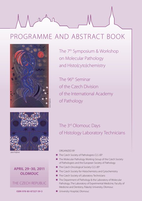
PROGRAMME AND ABSTRACT BOOK<br />
APRIL 29–30, 2011<br />
OLOMOUC<br />
THE CZECH REPUBLIC<br />
ISBN 978-80-87327-59-3<br />
The 7th Symposium & Workshop<br />
on Molecular Pathology<br />
<strong>and</strong> Histo(cyto)chemistry<br />
The 96th Seminar<br />
of the Czech Division<br />
of the International Academy<br />
of Pathology<br />
The 3rd Olomouc Days<br />
of Histology Laboratory Technicians<br />
ORGANIZED BY<br />
The Czech Society of Pathologists CLS JEP<br />
The Molecular Pathology Working Group of the Czech Society<br />
of Pathologists <strong>and</strong> the European Society of Pathology<br />
The Czech Oncological Society CLS JEP<br />
The Czech Society for Histochemistry <strong>and</strong> Cytochemistry<br />
The Czech Society of Laboratory Technicians<br />
The Department of Pathology & the Laboratory of Molecular<br />
Pathology; The Laboratory of Experimental Medicine, Faculty of<br />
Medicine <strong>and</strong> Dentistry, Palacký University, Olomouc<br />
University Hospital, Olomouc
The 7th Symposium & Workshop on Molecular Pathology <strong>and</strong> Histo(cyto)chemistry<br />
The 96th Seminar of the Czech Division of the International Academy of Pathology<br />
The 3rd Olomouc Days of Histology Laboratory Technicians<br />
April 29–30, 2011, Olomouc, Czech Republic<br />
Under the auspices of<br />
prof. M. Mašláň, Ph.D. / Rector of Palacký University, Olomouc<br />
prof. Z. Kolář, M.D., Ph.D. / Dean of the Faculty of Medicine <strong>and</strong> Dentistry, Palacký University, Olomouc<br />
R. Maráček, M.D. / Director of the University Hospital Olomouc<br />
Chairman of the Congress:<br />
Professor Zdeněk Kolář, M.D., Ph.D. / The Department of Pathology & the Laboratory of Molecular Pathology<br />
Faculty of Medicine <strong>and</strong> Dentistry, Palacký University, Olomouc, Hněvotínská 3, Olomouc, 775 15<br />
Tel: +420/585 632 451; Fax: +420/585 632 966; e-mail: kolarz@tunw.upol.cz<br />
Organizing & Programme Committee:<br />
Professor Jiří Ehrmann, M.D., Ph.D. / The Department of Pathology & the Laboratory of Molecular Pathology<br />
Faculty of Medicine <strong>and</strong> Dentistry, Palacký University, Olomouc, Hněvotínská 3, Olomouc, 775 15<br />
Tel: +420/585 632 466; Fax: +420/585 632 966; e-mail: info.lmp@seznam.cz<br />
Associate professor Martin Tichý, M.D., Ph.D.<br />
The Department of Pathology, Faculty of Medicine <strong>and</strong> Dentistry Palacký University, Olomouc,<br />
University Hospital, Hněvotínská 3, Olomouc, 775 15<br />
Tel: +420/585 632 453; Fax: +420/585 632 966; e-mail: info.lmp@seznam.cz<br />
Danuše Kvapilová<br />
The Department of Pathology, University Hospital, Hněvotínská 3, Olomouc, 775 15<br />
Tel: +420/585 632 465; Fax: +420/585 632 966; e-mail: d.kvapilova@post.cz<br />
Jan Bouchal, Marie Geierová, Marián Hajdúch, Jana Holinková, Monika Levková, Bohuslav Melichar, Eva Pimrová,<br />
Lenka Prokopová, Jana Steigerová, Jozef Škarda, Michaela Šváchová, Ivo Überall<br />
Venue:<br />
Friday, April 29 – Regional Centre Olomouc (Jeremenkova 40B, Olomouc, 772 00)<br />
Saturday, April 30 – Theoretical Institutes Building, Faculty of Medicine <strong>and</strong> Dentistry, Palacký University, Olomouc<br />
(Hněvotínská 3, Olomouc, 775 15)<br />
Conference Language:<br />
The 7 th Symposium & Workshop on Molecular Pathology <strong>and</strong> Histo(cyto)chemistry – English<br />
The 96 th Slide Seminar of the Czech Division of the International Academy of Pathology – Czech<br />
The 3 rd Congress of Histology Laboratory Technicians – Czech<br />
APRIL 29–30, 2011 | OLOMOUC | THE CZECH REPUBLIC<br />
3
4<br />
PROGRAMME<br />
The 7 th Symposium on Molecular Pathology <strong>and</strong> Histo(cyto)chemistry<br />
FRIDAY, APRIL 29, 2011<br />
Time Venue / language<br />
8:30–9:00 Registration<br />
9:00–9:15 Joint meeting ceremony Regional Centre<br />
Centaurus Hall / English, Czech<br />
Keynote lectures of invited speakers I<br />
Chairs: P. Dubový (Brno), E. Pikarski (Jerusalem)<br />
9:15–9:45 P. Dubový (Brno)<br />
Expression of cytokine proteins <strong>and</strong> mRNAs in the primary sensory neurons<br />
of neuropathic pain model<br />
9:45–10:15 E. Pikarski (Jerusalem)<br />
Advances in molecular diagnostics of hepatocellular carcinoma<br />
10:15–10:45 J. Moreira (Copenhagen)<br />
Development of novel early detection <strong>and</strong> stratifi cation markers for breast<br />
cancer: integrating pathology <strong>and</strong> proteomics<br />
10:45–11:15 L. Havel (Brno)<br />
Histochemistry in plant development <strong>and</strong> abiotic <strong>and</strong> biotic reactions<br />
11:15–11:45 Coff ee break<br />
Tissue Banking<br />
Chairs: P. De Blasio (Milano), L. Dušek (Brno)<br />
11:45–12:05 P. De Blasio (Milano)<br />
Tissue Banking <strong>and</strong> Tissue Microarrays<br />
12:05–12:25 L. Dušek (Brno)<br />
How to manage real time data support for registries <strong>and</strong> tissue banking in<br />
clinical practice? Cancer care case study<br />
12:25–12:45 G. Stanta (Trieste)<br />
Archive tissue biobanking as a basis for translational <strong>and</strong> reverse translational<br />
research<br />
PROGRAMME AND ABSTRACT BOOK<br />
Regional Centre<br />
Perseus Hall / English<br />
Regional Centre<br />
Perseus Hall / English<br />
Regional Centre<br />
Perseus Hall / English<br />
Regional Centre<br />
Perseus Hall / English<br />
Regional Centre<br />
Centaurus Hall / English<br />
Regional Centre<br />
Centaurus Hall / English<br />
Regional Centre<br />
Centaurus Hall / English<br />
12:45–13:00 Discussion Regional Centre<br />
Centaurus Hall / English<br />
13:00–14:00 Lunch break<br />
Poster Session<br />
Keynote lectures of invited speakers II<br />
Chairs: P. Murray (Birmingham), K. Smetana (Prague)<br />
14:00–14:30 P. Murray (Birmingham)<br />
Small lipids as therapeutic targets in cancer<br />
14:30–15:00 K. Smetana (Prague)<br />
Social life of cancer cell<br />
15:00–15:30 J. Sikora (Prague)<br />
Danon disease – lysosomal dyfunction disease – molecular pathology<br />
of LAMP2 protein - implications for clinical diagnostics<br />
15:30–15:45 R. C. Ecker (Wien)<br />
Applications of Slide-Based Single Cell Cytometry by TissueFAXS<br />
in Histopathology<br />
Regional Centre<br />
Perseus Hall / English<br />
Regional Centre<br />
Perseus Hall / English<br />
Regional Centre<br />
Perseus Hall / English<br />
Regional Centre<br />
Perseus Hall / English<br />
15:45–16:00 Discussion Regional Centre<br />
Perseus Hall / English
POSTERS<br />
PROGRAMME<br />
1 Increased susceptibility of MMP19 defi cient mice to DSS-induced colitis provides novel insights into pathogenesis<br />
of intestinal infl ammation<br />
Brauer R. 1 , Dziechciarkova M. 2 , Skarda J. 3 , Hajduch M. 2 , Sedlacek R. 1<br />
1Department of Transgenic Models of Diseases, Institute of Molecular Genetics AS CR, v.v.i, Prague, Czech Republic<br />
2Laboratory of Experimental Medicine, Department of Pediatrics <strong>and</strong> Oncology, Medical Faculty, Palacky University, Olomouc, Czech Republic<br />
3 Department of Pathology, Medical Faculty, Palacky University, Olomouc, Czech Republic<br />
2 Quadriplex model enhances urine-based detection of prostate cancer<br />
Jamaspishvili T. 1 , Kral M. 2 , Khomeriki I. 2 , Vyhnankova V. 2 , Mgebrishvili G. 1 , Student V. 2 , Kolar Z1 . <strong>and</strong> Bouchal J. 1<br />
1 Laboratory of Molecular Pathology <strong>and</strong> Department of Clinical <strong>and</strong> Molecular Pathology, Institute of Molecular <strong>and</strong> Translational Medicine,<br />
Faculty of Medicine <strong>and</strong> Dentistry, Palacky University <strong>and</strong> University Hospital, Olomouc, Czech Republic<br />
2Department of Urology, University Hospital, Olomouc, Czech Republic<br />
3 Comparison of proliferating activity in terminal villi of normal <strong>and</strong> diabetic placenta<br />
Jirkovská M. 1 , Kučera T. 1 , Jadrníček M. 1 , Niedobová V. 1 , Moravcová M. 2 , Krejčí V. 2 , Žižka Z. 2<br />
1Institute of Histology <strong>and</strong> Embryology, First Faculty of Medicine, Charles University in Prague, Prague, Czech Republic<br />
2Department of Obstetrics <strong>and</strong> Gynecology, First Faculty of Medicine, Charles University in Prague, Prague, Czech Republic<br />
4 A novel transgenic mouse model to study epidermal diff erentiation <strong>and</strong> wounding<br />
Kasparek P., Suchanova S., Krenek P., Sedlacek R.<br />
Department of Transgenic Models of Diseases, Institute of Molecular Genetics AS CR, Prague, Czech Republic<br />
5 Asporin modulates tissue microenvironment, invasive growth <strong>and</strong> bone metastasis of breast cancer<br />
Kharaishvili G. 1 , Cizkova M. 2 , Bouchalova K. 2 , Sedlakova E. 1 , Kolar Z. 1 , Bouchal J. 1<br />
1 Laboratory of Molecular Pathology of Institute of Pathology, Institute of Molecular <strong>and</strong> Translational Medicine, Faculty of Medicine <strong>and</strong> Dentistry,<br />
Palacky University, Olomouc, Czech Republic<br />
2Laboratory of Experimental Medicine, Pediatric Clinics of Medical Faculty of Palacky University <strong>and</strong> Faculty Hospital, Olomouc, Czech Republic<br />
6 Collagen triple helix repeat containing 1 protein in primary <strong>and</strong> metastatic breast cancer<br />
Kharaishvili G. 1 , Cizkova M. 2 , Bouchalova K. 2 , Sedlakova E. 1 , Kolar Z. 1 , Bouchal J. 1<br />
1 Laboratory of Molecular Pathology of Institute of Pathology, Institute of Molecular <strong>and</strong> Translational Medicine, Faculty of Medicine <strong>and</strong> Dentistry,<br />
Palacky University, Olomouc, Czech Republic<br />
2Laboratory of Experimental Medicine, Pediatric Clinics of Medical Faculty of Palacky University <strong>and</strong> Faculty Hospital, Olomouc, Czech Republic<br />
7 Apoptosis in placental vessels from pregnancies complicated by diabetes mellitus type I<br />
Kučera T. 1 , Jadrníček M. 1 , Niedobová V. 1 , Žižka Z. 2 , Krejčí V. 2 , Moravcová M. 2 , Jirkovská M. 1<br />
1Institute of Histology <strong>and</strong> Embryology, First Faculty of Medicine, Charles University in Prague, Czech Republic<br />
2Department of Obstetrics <strong>and</strong> Gynaecology, First Faculty of Medicine, Charles University <strong>and</strong> General University Hospital in Prague, Czech Republic<br />
8 Double immunohistochemical staining in routine diagnostic pathology<br />
Lodererova A., Honsova E., Voska L., Gabris V., Maluskova J.<br />
Department of Pathology, Institute for Clinical <strong>and</strong> Experimental Medicine, Prague, Czech Republic<br />
9 Micro RNA Assessment as a New Diagnostic <strong>and</strong> Prognostic Tool of Barrett´s Esophagus. Pilot Study<br />
Luzna P. 1 , Gregar J. 2 , Uberall I. 3 , Prochazka V. 2 , Ehrmann J. jr. 1,3<br />
1 Department of Histology <strong>and</strong> Embryology, Faculty of Medicine <strong>and</strong> Dentistry, Palacky University <strong>and</strong> University Hospital Olomouc, Czech Republic<br />
(pavla83@hotmail.com)<br />
2 nd II Internal Clinic, Faculty of Medicine <strong>and</strong> Dentistry, Palacky University <strong>and</strong> University Hospital Olomouc, Czech Republic<br />
3Laboratory of Molecular Pathology, Faculty of Medicine <strong>and</strong> Dentistry, Palacky University <strong>and</strong> University Hospital Olomouc, Czech Republic<br />
10 Prognostic factors in multiple myeloma<br />
Mareckova J. 1 ,Scudla V. 2 , Ehrmann J. 1 , Abrahamova P. 1<br />
1Department of Pathology, Laboratory of Molecular Pathology, Faculty of Medicine <strong>and</strong> Dentistry, Palacky University Olomouc, Czech Rebublic<br />
2 rd 3 Internal clinic, Faculty of Medicine <strong>and</strong> Dentistry, Palacky University Olomouc, University Hospital Olomouc, Czech Republic<br />
11 Determination of metallothioneins <strong>and</strong> alpha-methylacyl-CoA racemase in tumor prostate diseases<br />
Masarik M. 1 , Gumulec J. 1 , Sztalmachova M. 1,2 , Hlavna M. 1 , Kuchtickova S. 1 , Rovny A. 3 , Hrabec R. 3 , Eckschlager T. 4 , Krizkova S. 2 ,<br />
Adam V. 2 , Kizek R. 2<br />
1Department of Pathological Physiology, Faculty of Medicine Masaryk University, Brno, Czech Republic<br />
2Department of Chemistry <strong>and</strong> Biochemistry, Faculty of Agronomy, Mendel University, Brno, Czech Republic<br />
3Department of Urology, St. Anne´s University Hospital, Brno, Czech Republic<br />
4Department of Paediatric Haematology <strong>and</strong> Oncology, 2nd Faculty of Medicine Charles University in Prague, Czech Republic<br />
12 Identifi cation of grafted bone marrow cells in the recipient tissues<br />
Mokrý J. 1 , Filip S. 2 , Vávrová J. 3 , Čížková D. 1 , Šinkorová Z. 3 , Mičuda S. 4<br />
1Department of Histology <strong>and</strong> Embryology, Charles University in Prague, Faculty of Medicine, Hradec Králové, Czech Republic<br />
2Department of Oncology <strong>and</strong> Radiotherapy, Charles University in Prague, Faculty of Medicine <strong>and</strong> Teaching Hospital, Hradec Kralové, Czech Republic<br />
3 Department of Radiobiology, University of Defence Brno, Faculty of Military Health Sciences, Hradec Králové, Czech Republic<br />
4 Department of Pharmacology, Charles University in Prague, Faculty of Medicine, Hradec Králové, Czech Republic<br />
APRIL 29–30, 2011 | OLOMOUC | THE CZECH REPUBLIC<br />
5
6<br />
PROGRAMME<br />
13 Role of apoptosis associated genes in predicting clinical outcome of breast cancer<br />
Skálová H. 1 , Dundr P. 1 , Povýšil C. 1 , Velenská Z. 1 , Berková A. 1 , Staněk L. 1 , Dlouhá Z. 2 , Petruželka L. 2 , Tvrdík D. 1<br />
1Institute of Pathology, 1st Faculty of Medicine, Charles University <strong>and</strong> General University Hospital in Prague, Czech Republic<br />
2Department of Oncology, 1st Faculty of Medicine, Charles University <strong>and</strong> General University Hospital in Prague, Czech Republic<br />
14 Decalcifi cation options of bone material for consequential molecular biological <strong>and</strong> cytogenetic diagnosis –<br />
from the experimental model to trepanobiopsy<br />
Staněk L. 1,2,3 , Tvrdík D. 1 , Střítský J. 1 , Lísová S. 1 , Berková A. 1 , Ludvíková M. 1,3<br />
1Institute of Pathology, General Faculty Hospital <strong>and</strong> 1st Faculty of Medicine, Charles University in Prague, Prague, Czech Republic<br />
2 Department of Experimental virology, IHBT, Prague, Czech Republic<br />
3 Institute of Biology, Faculty of Medicine in Pilsen, Charles University, Pilsen, Czech Republic<br />
15 Role of PPARγ in colon cancer cells<br />
Straková N. 1,2,3 , Hofmanová J. 1,3 , Tylichová Z. 1,3 , Knopfová L. 4 , Šmarda J. 4 , Kozubík A. 1,3 , Ehrmann J. 2,3 , Kolář Z. 2,3<br />
1Laboratory of Cytokinetics, Institute of Biophysics, Acad Sci Czech Rep, v.v.i., Brno, Czech Republic<br />
2Laboratory of Molecular Pathology, Faculty of Medicine <strong>and</strong> Dentistry, Palacky University, Olomouc, Czech Republic<br />
3Biomedical Centre of Institute of Biophysics Acad Sci Czech Rep, v.v.i. <strong>and</strong> Faculty of Medicine <strong>and</strong> Dentistry, Palacky University, Olomouc, Czech Republic<br />
4 Laboratory of Cell Diff erentiation, Institute of Experimental Biology, Masaryk University, Brno, Czech Republic<br />
16 Molecular genetics changes in melanocystic lesions – case report<br />
Uvírová M. 1 , Dvořáčková J. 2 , Šimová J. 1 , Urbanovská I. 1 , Žiak D. 1 , Konvalinka D. 1 , Kubová B. 1<br />
1CGB laboratory, Ostrava, Czech Republic<br />
2Faculty of Medicine University of Ostrava, Ostrava, Czech Republic<br />
17 Role of matrix metalloproteinase-19 in liver fi brosis<br />
Zbodakova O. 1 , Jirouskova M. 1 , Sedlacek R. 1 , Ehrmann J. 2 , Hajduch M. 3 , Jirkovska M. 4<br />
1Laboratory of Transgenic Models of Diseases, Institute of Molecular Genetics of the ASCR, Prague, Czech Republic<br />
2Department of Pathology, Medical Faculty, Palacky University, Olomouc, Czech Republic<br />
3Laboratory of Experimental Medicine, Department of Pediatrics <strong>and</strong> Oncology, Palacky University <strong>and</strong> University Hospital, Olomouc, Czech Republic<br />
4 Institute of Histology <strong>and</strong> Embryology, First Faculty of Medicine, Charles University, Prague, Czech Republic<br />
SOCIAL PROGRAMME<br />
Time<br />
17:30–18:00 Welcome drink<br />
18:00–19:00 Concert in the Olomouc Archidiocesan Museum<br />
19:00–19:30 Hot wine time<br />
20:00–24:00 Social evening in Archa restaurant – Svatý Kopeček (bus transport guaranteed)<br />
Workshop on Molecular Pathology<br />
SATURDAY, APRIL 30, 2011<br />
Time Chairs: M. Mistrík, I. Überall (Olomouc) Venue/language<br />
9:00–9:30 P. Krist (Prague)<br />
3D Microscopy<br />
9:30–10:00 M. Mistrík (Olomouc)<br />
Applicated fl uorescence microscopy<br />
10:00–10:20 Coff ee break<br />
PROGRAMME AND ABSTRACT BOOK<br />
Theoretical Institutes Building<br />
Faculty of Medicine <strong>and</strong> Dentistry, Palacký<br />
University Olomouc / Czech<br />
Theoretical Institutes Building<br />
Faculty of Medicine <strong>and</strong> Dentistry, Palacký<br />
University Olomouc / Czech<br />
10:20–11:40 3D Microscopy – Practical demonstration by Zeiss (part I) Theoretical Institutes Building<br />
Faculty of Medicine <strong>and</strong> Dentistry, Palacký<br />
University Olomouc / Czech<br />
11:40–13:00 3D Microscopy – Practical demonstration by Zeiss (part II) Theoretical Institutes Building<br />
Faculty of Medicine <strong>and</strong> Dentistry, Palacký<br />
University Olomouc / Czech
PROGRAMME<br />
96. olomoucký meziregionální mezioborový diagnostický seminář<br />
Společnosti českých patologů a české sekce<br />
International Academy of Pathology<br />
PÁTEK, 29. DUBNA 2011<br />
Čas Místo konání/jazyk<br />
8:30–9:00 Registrace<br />
9:00–9:15 Slavnostní zahájení Regionální centrum<br />
sál Centaurus / anglicky, česky<br />
Diagnostický seminář Společnosti českých patologů a české sekce International Academy of Pathology<br />
Předsednictvo: M. Tichý, M. Geierová (Olomouc)<br />
9:15–10:45 Diagnostický seminář – část I Regionální centrum<br />
sál Andromeda / česky<br />
10:45–11:15 J. Berounský (České Budějovice)<br />
Vakuový systém pro přepravu a uchování biologického materiálu<br />
11:15–11:45 Přestávka, občerstvení<br />
Téma: Tissue Banking<br />
Předsednictvo: P. De Blasio (Milano), L. Dušek (Brno)<br />
11:45–12:05 P. De Blasio (Milano)<br />
Tissue Banking <strong>and</strong> Tissue Microarrays<br />
12:05–12:25 L. Dušek (Brno)<br />
How to manage real time data support for registries <strong>and</strong> tissue banking<br />
in clinical practice? Cancer care case study<br />
12:25–12:45 G. Stanta (Trieste)<br />
Archive tissue biobanking as a basis for translational <strong>and</strong> reverse translational<br />
research<br />
Regionální centrum<br />
sál Andromeda / česky<br />
Regionální centrum<br />
sál Centaurus / anglicky<br />
Regionální centrum<br />
sál Centaurus / anglicky<br />
Regionální centrum<br />
sál Centaurus / anglicky<br />
12:45–13:00 Diskuze Regionální centrum<br />
sál Centaurus / anglicky<br />
13:00–14:00 Oběd<br />
Sekce posterů<br />
14:00–16:00 Diagnostický seminář – část II Regionální centrum<br />
sál Andromeda / česky<br />
SPOLEČENSKÝ PROGRAM<br />
Čas<br />
17:30–18:00 Uvítací přípitek<br />
18:00–19:00 Koncert v Arcidiecézním muzeu v Olomouci<br />
19:00–19:30 Zahřátí u svařeného vína<br />
20:00–24:00 Společenský večer v restauraci Archa na Sv. Kopečku (odvoz autobusy zajištěn)<br />
Ve dnech 28. dubna – 1. května 2011 probíhá v Olomouci „Jarní Flora Olomouc“. Vřele doporučujeme!<br />
APRIL 29–30, 2011 | OLOMOUC | THE CZECH REPUBLIC<br />
7
8<br />
PROGRAMME<br />
Workshop <strong>molekulární</strong> patologie<br />
SOBOTA, 30. DUBNA 2011<br />
Čas Předsednictvo: M. Mistrík, I. Überall (Olomouc) Místo konání/jazyk<br />
9:00–9:30 P. Krist (Praha)<br />
Mikroskopie v 3D<br />
9:30–10:00 M. Mistrík (Olomouc)<br />
Aplikovaná fl uorescenční mikroskopie<br />
10:00–10:20 Přestávka, občerstvení<br />
10:20–11:40 Mikroskopie v 3D – praktická demonstrace I<br />
fi rmy Zeiss<br />
11:40–13:00 Mikroskopie v 3D – praktická demonstrace II<br />
fi rmy Zeiss<br />
PROGRAMME AND ABSTRACT BOOK<br />
Teoretické ústavy LF UP / česky<br />
Teoretické ústavy LF UP/ česky<br />
Teoretické ústavy LF UP / česky<br />
Teoretické ústavy LF UP / česky
3. olomoucké dny histologických laborantů<br />
PÁTEK, 29. DUBNA 2011<br />
PROGRAMME<br />
Čas Místo konání/jazyk<br />
8:30–9:00 Registrace<br />
9:00–9:15 Slavnostní zahájení Regionální centrum<br />
sál Centaurus / anglicky<br />
Konference histologických laborantů I<br />
Předsednictvo: J. Ehrmann, D. Kvapilová (Olomouc)<br />
9:15–9:35 L. Vodičková, A. Faltýnková., D. Novotná (Olomouc)<br />
Laboratorní zpracování neodvápněné kostní tkáně<br />
9:35–9:55 P. Skýpalová, M. Tichý, K. Žamboch (Olomouc)<br />
Využití histomorfometrie kostní dřeně k diagnostice renálních osteopatií<br />
9:55–10:15 J. Ehrmann (Olomouc)<br />
Koncept řešení histologických laboratoří – nové trendy<br />
10:15–10:35 J. Rázga (Brno)<br />
Produktivita práce v patologické laboratoři<br />
10:35–11:05 M. Richter (Praha)<br />
Automatizace imunohistochemie a in situ hybridizace systémy Ventana -<br />
nové metody a přístupy<br />
11:05–11:15 Diskuze<br />
11:15–11:45 Přestávka, občerstvení<br />
Téma: Tissue Banking<br />
Předsednictvo: P. De Blasio (Milano), L. Dušek (Brno)<br />
11:45–12:05 P. De Blasio (Milano)<br />
Tissue Banking <strong>and</strong> Tissue Microarrays<br />
12:05–12:25 L. Dušek (Brno)<br />
How to manage real time data support for registries <strong>and</strong> tissue banking in<br />
clinical practice? Cancer care case study<br />
12:25–12:45 G. Stanta (Trieste)<br />
Archive tissue biobanking as a basis for translational <strong>and</strong> reverse translational<br />
research<br />
Regionální centrum<br />
sál Centaurus / česky<br />
Regionální centrum<br />
sál Centaurus / česky<br />
Regionální centrum<br />
sál Centaurus / česky<br />
Regionální centrum<br />
sál Centaurus / česky<br />
Regionální centrum<br />
sál Centaurus / česky<br />
Regionální centrum<br />
sál Centaurus / anglicky<br />
Regionální centrum<br />
sál Centaurus / anglicky<br />
Regionální centrum<br />
sál Centaurus / anglicky<br />
12:45–13:00 Discussion Regionální centrum<br />
sál Centaurus / anglicky<br />
13:00–14:00 Oběd<br />
Sekce posterů<br />
Konference histologických laborantů II<br />
Předsednictvo: D. Kvapilová, J. Ehrmann (Olomouc)<br />
14:00–14:20 P. Hajzlerová, J. Mokrý (Hradec Králové)<br />
Využití tyramidů v imunohistochemii<br />
14:20–14:40 P. Lužná (Olomouc)<br />
Laserová mikrodisekce<br />
14:40–15:10 E. Knopek (Praha)<br />
BRADY – Laboratorní značení<br />
15:10–15:30 M. Janíková, J. Škarda, V. Žižková (Olomouc)<br />
Využití parafínových bloků k izolaci miRNA<br />
15:30–15:50 M. Ondráková (Brno)<br />
Informace ČAZL<br />
Regionální centrum<br />
sál Centaurus / česky<br />
Regionální centrum<br />
sál Centaurus / česky<br />
Regionální centrum<br />
sál Centaurus / česky<br />
Regionální centrum<br />
sál Centaurus / česky<br />
Regionální centrum<br />
sál Centaurus / česky<br />
APRIL 29–30, 2011 | OLOMOUC | THE CZECH REPUBLIC<br />
9
10<br />
15:50–16:00 Diskuze<br />
POSTERY<br />
PROGRAMME<br />
1 Tkáňový procesor TISSUE PROCESSOR STP 420D<br />
Tylečková M., Muchová A.<br />
CGB laboratoř, Ostrava – Vítkovice, Česká republika<br />
2 Porovnání hladin PSA s histologickým gradingem karcinomu prostaty<br />
Němečková L. 1 , Hafuda A. 2 , Martinková L. 1 , Vodičková J. 1 , Hladíková L. 1 , Straka V. 1 ,Hornych J. 3<br />
1Oddělení patologie, Oblastní nemocnice Náchod, Česká republika<br />
2Oddělení urologie, Oblastní nemocnice Náchod, Česká republika<br />
3Oddělení <strong>klinické</strong> biochemie a diagnostiky, Oblastní nemocnice Náchod, Česká republika<br />
SPOLEČENSKÝ PROGRAM<br />
Čas<br />
17:30–18:00 Uvítací přípitek<br />
18:00–19:00 Koncert v Arcidiecézním muzeu v Olomouci<br />
19:00–19:30 Zahřátí u svařeného vína<br />
20:00–24:00 Společenský večer v restauraci Archa na Sv. Kopečku (odvoz autobusy zajištěn)<br />
Ve dnech 28. dubna – 1. května 2011 probíhá v Olomouci „Jarní Flora Olomouc“. Vřele doporučujeme!<br />
Workshop <strong>molekulární</strong> patologie<br />
SOBOTA, 30. DUBNA 2011<br />
Čas Předsednictvo: M. Mistrík, I. Überall (Olomouc) Místo konání / jazyk<br />
9:00–9:30 P. Krist (Praha)<br />
Mikroskopie v 3D<br />
9:30–10:00 M. Mistrík (Olomouc)<br />
Aplikovaná fl uorescenční mikroskopie<br />
10:00–10:20 Přestávka, občerstvení<br />
10:20–11:40 Mikroskopie v 3D – praktická demonstrace I<br />
fi rmy Zeiss<br />
11:40–13:00 Mikroskopie v 3D – praktická demonstrace II<br />
fi rmy Zeiss<br />
PROGRAMME AND ABSTRACT BOOK<br />
Teoretické ústavy LF UP / česky<br />
Teoretické ústavy LF UP / česky<br />
Teoretické ústavy LF UP / česky<br />
Teoretické ústavy LF UP / česky
Honorary guests<br />
Juri Kopolovic, Professor, M.D., Ph.D.<br />
Juri Kopolovic is currently Professor of Pathology at the Hebrew University – Hadassah<br />
Medical Center in Jerusalem. He completed his medical studies in 1972 at the Hebrew<br />
University – Hadassah School of Medicine. During the residency at Hadassah Medical<br />
Center he became interested in nephropathology <strong>and</strong> gynecopathlogy. In 1986–1988 he<br />
continued his training with a clinical <strong>and</strong> research fellowship at the Pathology Department<br />
at the Beth Israel Hospital of the Harvard School of Medicine in Boston where he worked<br />
on morphological changes along the nephron in rat experimental model of hypoxia <strong>and</strong><br />
reperfusion. Upon returning to Israel he continued in research in the field of autoimmunity,<br />
atherosclerosis <strong>and</strong> extracellular matrix in malignancies of the female genital tract. It was<br />
shown that in experimental model of mice SLE treatment with intravenous gamma globulin<br />
lads to a complete clinical <strong>and</strong> morphological remission of SLE. He studied the role<br />
of extracellular matrix in local invasion, metastasis angiogenesis <strong>and</strong> prognosis in female<br />
genital tract malignancies. He is presently the chairman of the Department of Pathology<br />
at the Hebrew University School of Medicine, Jerusalem, Israel.<br />
Jaroslav Mokrý, Professor, M.D., Ph.D.<br />
Jaroslav Mokrý currently holds the position of Head of the Department of Histology <strong>and</strong><br />
Embryology at Charles University Medical Faculty in Hradec Kralove. He has been working<br />
in the Department of Histology <strong>and</strong> Embryology at Charles University Medical Faculty in<br />
Hradec Kralove since 1985. He completed his M.D. degree in general medicine from Charles<br />
University in Prague in 1990. In 1993–1994 he was on his research stay in the Department<br />
of Human Anatomy, Oxford University, UK, where he was engaged in cultivation of fetal<br />
dopaminergic neurons. He defended his Ph.D. thesis in 1995 <strong>and</strong> was appointed Professor<br />
of Histology <strong>and</strong> Embryology in 2006. He was awarded Charles University Rector’s Prizes in<br />
1987 <strong>and</strong> 1990 <strong>and</strong> Young Histochemist Award from International Federation of Societies<br />
of Histochemistry <strong>and</strong> Cytochemistry in Kyoto, Japan, in 1996. He is president of Czech<br />
Society for Histo- <strong>and</strong> Cytochemistry <strong>and</strong> vice-president of Czech Anatomical Society. He<br />
published 130 research articles <strong>and</strong> presented over 300 lectures or posters. Main focus<br />
of his scientific activity covers the areas of stem cell biology, regenerative medicine, cell<br />
cultivation, cell transplantation, cell therapy, cell differentiation, angiogenesis, detection<br />
of molecules in situ <strong>and</strong> histology.<br />
ABSTRACT BOOK<br />
CURRICULUM VITAE<br />
CURRICULUM VITAE<br />
11
12 ABSTRACT BOOK<br />
Invited guests<br />
Petr Dubový, Professor, D.Sc., Ph.D.<br />
Petr Dubový currently holds the position of Professor <strong>and</strong> Head of the Department of<br />
Anatomy <strong>and</strong> Senior Researcher in the Central European Institute of Technology (CEITEC),<br />
Masaryk University (MU) in Brno. He obtained his Master <strong>and</strong> Doctoral degree of Biology<br />
in the Faculty of Natural Science <strong>and</strong> was subsequently graduated <strong>and</strong> then taken degree<br />
of Professor of Human Anatomy in the Medical Faculty of MU. He was visiting scientist in<br />
the Department of Anatomy <strong>and</strong> Neuroscience Karolinska Institutet, Stockholm (1991–<br />
1995). In the period of 2001–2009, he was chairman of the Czech Society for Histo- <strong>and</strong><br />
Cytochemistry. He is interested predominantly in the cellular <strong>and</strong> molecular biology of<br />
neuron-glial cell interactions during degeneration <strong>and</strong> regeneration of the nervous system,<br />
especially using in situ proteomics (immunohistochemistry). His recent scientific work is<br />
directed to involvement of cytokines, chemokines <strong>and</strong> their receptors in neuropathic<br />
pain induction as simultaneous process of nerve regeneration. He has published over 100<br />
peer-reviewed papers as author <strong>and</strong> co-author.<br />
CURRICULUM VITAE<br />
Expression of cytokine proteins <strong>and</strong> mRNAs in the primary sensory neurons<br />
of neuropathic pain model<br />
Petr Dubový<br />
Department of Anatomy, Faculty of Medicine, <strong>and</strong> Central European Institute of Technology (CEITEC), Masaryk University, Brno,<br />
Czech Republic<br />
Peripheral neuropathic pain, manifested<br />
as spontaneous pain, hyperalgesia <strong>and</strong><br />
allodynia, arises as a result of many forms<br />
of nerve damage. Compelling evidence<br />
indicates that neuropathic pain induced<br />
by nerve injury is related to cellular <strong>and</strong><br />
molecular changes in the dorsal root<br />
ganglia (DRG) containing bodies of the<br />
primary afferent neurons. It is assumed<br />
that pro- <strong>and</strong> anti-inflammatory cytokines<br />
may contribute to both the induction <strong>and</strong><br />
maintenance of neuropathic pain. Chronic<br />
constriction injury (CCI) of the rat sciatic<br />
nerve is frequently used experimental<br />
model of neuropathic pain.<br />
Unilateral CCI of the rat sciatic nerve<br />
performed under aseptic conditions was<br />
used to study changes of TNFa, IL-6 <strong>and</strong><br />
IL-10 proteins <strong>and</strong> mRNAs in L4-L5 <strong>and</strong><br />
C7-C8 DRG removed from both sides of<br />
naïve (n=16), operated (n=64) <strong>and</strong> shamoperated<br />
(n=64) rats. Neuropathic pain<br />
induction was tested by withdrawal<br />
threshold of mechanoallodynia <strong>and</strong><br />
thermal hyperalgesia. Levels of TNFa, IL-<br />
6 <strong>and</strong> IL-10 proteins <strong>and</strong> their mRNA were<br />
analyzed by quantitative immunohistochemistry,<br />
ELISA, Western blot, in situ<br />
hybridization <strong>and</strong> RT-PCR in DRG samples<br />
from naïve, CCI- <strong>and</strong> sham-operated rats<br />
for 1, 3, 7 <strong>and</strong> 14 days.<br />
The naïve DRG displayed no or very<br />
weak immunofluorescence staining for<br />
the cytokine proteins in the neuronal<br />
bodies with a moderate intensity in small<br />
<strong>and</strong> medium-sized neurons. Unilateral<br />
CCI induced bilateral elevation of TNFa,<br />
IL-6 <strong>and</strong> IL-10 immunostaining usually in<br />
large-sized neuronal perikarya of both<br />
L4-L5 <strong>and</strong> C7-C8 DRG. ELISA <strong>and</strong> Western<br />
blot confirmed bilateral <strong>and</strong> temporal increase<br />
of TNFa, IL-6 <strong>and</strong> IL-10 proteins not<br />
only in the homonymous lumbar DRG<br />
associated with the sciatic nerve, but<br />
also in the heteronymous cervical ones<br />
non-associated with spinal segments of<br />
constricted nerve.<br />
TNFa protein was peaked in L4-L5<br />
DRG at 7 <strong>and</strong> C7-C8 DRG at 14 days after<br />
CCI, while IL-6 protein level was the highest<br />
in both lumbar <strong>and</strong> cervical DRG at 3<br />
days. In contrast, IL-10 protein was significantly<br />
increased in lumbar <strong>and</strong> cervical<br />
DRG of both sides at 1 day <strong>and</strong> 3 days.<br />
Elevated levels of TNFa <strong>and</strong> IL-6 mRNAs<br />
were proved in the neuronal bodies of all<br />
DRG from CCI rats by in situ hybridization<br />
<strong>and</strong> verified by RT-PCR.<br />
Surprisingly, sham-operated rats displayed<br />
bilateral increased staining for IL-6<br />
mRNA in satellite glial cells of both cervical<br />
<strong>and</strong> lumbar DRG, while staining for<br />
TNFa mRNA was enhanced in neuronal<br />
bodies. These increased levels of mRNAs<br />
were confirmed by RT-PCR<br />
Our results indicate that neuroinflammation<br />
expressed by increased production<br />
of cytokines is not limited to the primary<br />
sensory neurons of injured nerve,<br />
but is spread to the contralateral side<br />
as well as alongside neuroaxis to DRG<br />
neurons of remote segments. In addition,<br />
neuroinflammatory reaction of satellite<br />
glial cells in sham-operated rats suggests<br />
their sensitivity to tissue injury. Both<br />
neuroinflammatory reactions influence<br />
excitation of the primary sensory neurons<br />
<strong>and</strong> may participate in neuropathic<br />
pain induction. Moreover, changes of<br />
cytokine proteins in DRG non-associated<br />
with damaged nerve could be related<br />
with a general neuroinflammatory reaction<br />
of the nervous system to injury <strong>and</strong><br />
thereby promote potential of the DRG<br />
neurons for regeneration of their axons<br />
following a conditioning lesion.<br />
Supported by MSM0021622404.
Eli Pikarsky, M.D., Ph.D.<br />
Eli Pikarsky is at the Department of Pathology <strong>and</strong> the Lautenberg Center for Immunology.<br />
He received his M.D. <strong>and</strong> Ph.D. at the Hebrew University, Jerusalem, Israel in 1992 <strong>and</strong> 2000<br />
respectively. He actively participates in clinical duties at the department of Pathology <strong>and</strong><br />
heads the laboratory for molecular pathology which he established. Dr. Pikarsky’s research<br />
career is devoted to studying tissue interactions which are important for cancer initiation,<br />
promotion <strong>and</strong> progression. His research group includes 2 research associates holding<br />
a Ph.D. degree, 6 doctoral students <strong>and</strong> several technicians. His studies involve analysis<br />
of the interaction between inflammation <strong>and</strong> cancer cells <strong>and</strong> elucidating the roles of<br />
several transcription factors in cancer biology. He is a member of the steering committee<br />
of the Israel National Tissue Bank. He has received several awards at the Hebrew University<br />
including awards for excellence in research, in clinical practice <strong>and</strong> in teaching. He has also<br />
received the Daniel G. Miller Research Career Development Award from the Israel Cancer<br />
Research Fund, The Sir Zelman Cowen prize for discovery in medical research <strong>and</strong> the Teva<br />
Founders award for young scientists.<br />
ABSTRACT BOOK<br />
Advances in molecular diagnostics of hepatocellular carcinoma<br />
Eli Pikarski<br />
Department of Pathology <strong>and</strong> the Lautenberg Center for Immunology <strong>and</strong> Cancer Research, Jerusalem, Israel<br />
Hepatocellular carcinoma (HCC)<br />
afflicts more than 560,000 people<br />
worldwide each year <strong>and</strong> has one<br />
of the worst 1-year survival rates of<br />
any cancer type. Currently, there are<br />
no predictive markers for finetuning<br />
treatment of patients with HCC. We<br />
found that a chromosomal region<br />
encompassing VEGF-A is amplified in<br />
a subset of HCCs in humans <strong>and</strong> mice.<br />
This subset is histologically, molecularly<br />
<strong>and</strong> biologically distinct. Moreover,<br />
human HCCs that carry a synthenic<br />
VEGF-A-amplicon, also display distinctive<br />
histological features. Expression of<br />
VEGF-A in mice amplicon bearing<br />
tumors is associated with increased<br />
blood-vessel <strong>and</strong> macrophage density,<br />
<strong>and</strong> enhanced microenvironment<br />
derived HGF expression, which can<br />
support the proliferation of the<br />
malignant cells. Accordingly, we show<br />
that these amplicon-positive tumors<br />
are uniquely sensitive to treatment<br />
with soluble VEGF-A receptor or the<br />
multi-kinase inhibitor Sorafenib, which<br />
CURRICULUM VITAE<br />
is the first line treatment for advanced<br />
HCC. Our data indicates that cancer<br />
gene amplification may have cell-nonautonomous<br />
oncogenic effects via the<br />
tumor microenvironment. Moreover, the<br />
VEGF-A amplicon is a potential biomarker<br />
for tumor response to direct <strong>and</strong> indirect<br />
VEGF blockers. Our study underscores<br />
the potential for clinical translation of<br />
results obtained from mouse models<br />
of human cancer guided by cancer<br />
genome analysis.<br />
APRIL 29–30, 2011 | OLOMOUC | THE CZECH REPUBLIC<br />
13
14<br />
ABSTRACT BOOK<br />
José Moreira, M.Sc., Ph.D.<br />
José Moreira obtained his MSc. degree in Biochemistry from the Institute of Biomedical<br />
Sciences Abel Salazar in Porto, <strong>and</strong> a Ph.D. in Molecular Biology at the Dept. of Genetics,<br />
University of Copenhagen in 1998. Following a two-year period as Assistant Research<br />
Professor at the Dept. of Genetics, José Moreira took up a position as scientist at Active<br />
Biotech Research AB, Lund, Sweden. In 2002, he was appointed a senior scientist at the Dept.<br />
of Proteomics in Cancer, Danish Cancer Society. José Moreira is a member of the editorial<br />
board of Molecular <strong>and</strong> Cellular Proteomics <strong>and</strong> editorial manager for Molecular Oncology.<br />
His major achievements include contributions to some of the fundamental mechanisms<br />
underlying epigenetic regulation of genes, such as ATP-driven remodeling complexes being<br />
involved in activation as well as gene repression <strong>and</strong> the role that non-histone chromatin<br />
associated proteins play in the regulation of target genes. More recently, the identification<br />
of several biomarkers for bladder <strong>and</strong> breast cancer, as well as the establishment of<br />
a framework for biomarker discovery based on a new source, tissue interstitial fluid,<br />
<strong>and</strong> the identification a number of novel molecular markers that phenotypically define<br />
subpopulations of cells present in normal breast tissue. His current research interests<br />
focus on early detection <strong>and</strong> personalized treatment of cancer, in particular the concept<br />
of profiling of therapy-relevant protein markers in patients’ tumors <strong>and</strong> the development<br />
of a framework to identify biomarkers for blood-based diagnostic systems.<br />
PROGRAMME AND ABSTRACT BOOK<br />
CURRICULUM VITAE<br />
Development of novel early detection <strong>and</strong> stratifi cation markers for breast<br />
cancer: integrating pathology <strong>and</strong> proteomics<br />
José Moreira<br />
Department of Genetics, Institute of Molecular Biology, University of Copenhagen, Denmark<br />
Our limited underst<strong>and</strong>ing of the biological<br />
impact of the whole spectrum of<br />
early breast lesions together with a lack<br />
of accurate molecular-based risk criteria<br />
for the diagnosis <strong>and</strong> assignment of<br />
prognostic significance to biopsy findings<br />
presents an important problem<br />
in the clinical management of patients<br />
harboring precancerous breast lesions.<br />
As a result, there is a need to identify biomarkers<br />
that can better determine the<br />
outcome of early breast lesions by identifying<br />
subpopulations of cells in breast<br />
premalignant disease that are at high-risk<br />
of progression to invasive disease. A first<br />
step towards achieving this goal will be<br />
to define the molecular phenotypes of<br />
the various cell types <strong>and</strong> precursors -<br />
generated by the stem cell hierarchy -<br />
that are present in normal, benign, <strong>and</strong><br />
malignant conditions of the breast. For<br />
the past years our laboratory has carried<br />
out a systematic <strong>and</strong> comprehensive<br />
proteomic profiling of normal <strong>and</strong> malignant<br />
breast tissue in high-risk patients in<br />
a search for differentially expressed markers<br />
for early detection <strong>and</strong> stratification<br />
of patients, as well as novel targets for<br />
therapeutic intervention in breast cancer.<br />
This lecture will describe our efforts to<br />
address some of the issues that clinical<br />
proteomics is faced with, partly due to<br />
the formidable heterogeneity of tumors,<br />
but also due to limitations of the current<br />
proteomic technologies. The examples<br />
presented will include results from our<br />
work to identify proteins that characterize<br />
specific steps in the progression from<br />
early benign lesions to malignancy in<br />
breast apocrine carcinoma, the creation<br />
of a comprehensive database of proteins<br />
secreted/shedded by tumor cells <strong>and</strong><br />
present in tissue interstitial fluid <strong>and</strong> from<br />
a systematic proteomic study to characterize<br />
the phenotypes of the different<br />
cell subpopulations present in normal<br />
human mammary tissue that can help<br />
define the molecular phenotypes underlying<br />
mammary epithelial normalcy.<br />
Finally results from our recent work on<br />
therapy-relevant stratification of triple<br />
negative breast cancer patients will also<br />
be discussed.
Ladislav Havel, Professor, D.Sc., Ph.D.<br />
Ladislav Havel obtained his academic degree (RNDr.) from Masaryk University in Brno in 1977,<br />
<strong>and</strong> Ph.D. at the Institute of experimental Botany, Academy of Sciences of the Czech Republic,<br />
Prague in 1983. He then worked at the same Institute, working place in Olomouc. In 1988 he<br />
moved to Mendel University in Brno. He became associate professor in 1988 <strong>and</strong> full time<br />
professor in 1998. Ladislav Havel is the head of the Department of Plant Biology at the Faculty<br />
of Agronomy (since 2007). He is a member of several international <strong>and</strong> national scientific<br />
societies. He published over 100 scientific communications in international scientific journals<br />
<strong>and</strong> monographs. He has been focused on the regeneration processes in plant tissue cultures<br />
of several species, especially somatic embryogenesis, the role of <strong>programme</strong>d cell death in<br />
plant development <strong>and</strong> in responses of plant cells to environmental stimuli.<br />
ABSTRACT BOOK<br />
CURRICULUM VITAE<br />
Histochemistry in plant development <strong>and</strong> abiotic <strong>and</strong> biotic reactions<br />
Ladislav Havel<br />
Department of Plant Biology, Faculty of Agronomy, Mendel University of Agriculture <strong>and</strong> Forestry, Brno, Czech Republic<br />
Plant organisms have common characteristic<br />
of all eukaryotic organisms. In<br />
same features are different from other<br />
eukaryotes especially from animals <strong>and</strong><br />
human, e.g. plant cells have cell wall <strong>and</strong><br />
plastids, a special group of organelles. As<br />
in animals <strong>and</strong> human the histo- <strong>and</strong>/<br />
or cytochemical methods are powerful<br />
tool in research of plant growth <strong>and</strong> development<br />
<strong>and</strong> responses to biotic <strong>and</strong><br />
abiotic stimuli. Modern methods used<br />
in plant biology are usually optimized<br />
or derived from methods developed<br />
in animal <strong>and</strong> human research. A short<br />
survey of exploitation of these methods<br />
will be shown.<br />
15
16<br />
ABSTRACT BOOK<br />
Pasquale De Blasio, Professor, M.Eng.<br />
Pasquale De Blasio, is presently the CEO of BioRep (www.biorep.it), a global service provider<br />
biorepository based in Milan, Italy; President of ESBB (European, Middle-Eastern <strong>and</strong> African<br />
Society of Biopreservation <strong>and</strong> Biobanking, www.ESBB.org) <strong>and</strong> Adjunct Associate Professor<br />
at Temple University, Philadelphia-PA (USA) <strong>and</strong> founder of Integrated Systems Engineering<br />
S.r.l. (www.isenet.it) operating in development of automation instruments for molecular<br />
<strong>and</strong> cellular biology (e.g. Tissue. Microarrayers, Biorepositories automation, etc.). Pasquale De<br />
Blasio, has more than 35 years of industrial working experience in management positions.<br />
He worked for more than 14 years in Industrial <strong>and</strong> Power Plant design, <strong>and</strong> over 15 years in<br />
analytical instrumentation developments. He has also matured specific skills in multinational<br />
analytical instrumentation companies, as R&D Manager for the developments of HPLC<br />
Systems, UV/VIS Spectrophotometers, Ultra Centrifuges <strong>and</strong> Medical Anaesthesia Systems;<br />
<strong>and</strong> as Industrial Operations Management in the development <strong>and</strong> manufacturing of Blood<br />
Gas <strong>and</strong> Coagulation Analytical Instruments.. He has also experience with ISO 9001 Quality<br />
System, EEC Medical Directive <strong>and</strong> CE certification. Pasquale De Blasio, has a Bachelor of<br />
Science in Electrical Engineering from Villanova University, Villanova, Pennsylvania, USA<br />
(1976), <strong>and</strong> a Master Degree in Business Administration from Bocconi University, Milan,<br />
Italy (1986). He is co-author of several peer-reviewed scientific papers <strong>and</strong> co-participants<br />
of several industrial patents.<br />
Tissue Banking <strong>and</strong> Tissue Microarrays<br />
De Blasio P. 1,2 <strong>and</strong> Biunno I. 2,3<br />
1BioRep Via Fantoli 16/15, Milano, Italy<br />
2Integrated Systems Egineering (ISE) Via Fantoli 16/15, Milano, Italy<br />
3Institute for Biomedical Research (ITB-CNR) Via F.lli Cervi 93, Milano, Italy<br />
“Tissue Banking” is an emerging<br />
concept with solid basis in translational<br />
medicine <strong>and</strong> in genetic epidemiology<br />
since allows biomedical research to<br />
flourish in several aspects: a tool for (i)<br />
biomarker identification, (ii) biomarker<br />
validation <strong>and</strong> (iii) subsequent drug discovery.<br />
Tissue banking aims to collect<br />
large number of high quality of samples<br />
process <strong>and</strong> store them in a manner that<br />
is compatible with the “omics” analysis.<br />
“Tissue microarray” have become an<br />
essential pathology tool being able to<br />
validate novel biomarkers in epidemiology<br />
scale since hundreds of tissue fragments<br />
can be analysed simultaneously.<br />
PROGRAMME AND ABSTRACT BOOK<br />
The advancement in digital pathology<br />
with whole-slide imaging <strong>and</strong> new techniques<br />
in image analysis <strong>and</strong> image databases<br />
has driven improvements in tissue<br />
samples collection <strong>and</strong> Tissue Banking.<br />
The identification of biomarkers is essential<br />
not only for diagnostic purposes<br />
but also as predictors of disease response<br />
to new <strong>and</strong> old drugs. This necessitates<br />
the use of tissue samples collected <strong>and</strong><br />
stored in several centres, but in order to<br />
guarantee reproducible <strong>and</strong> comparable<br />
results, the multi-centre network must<br />
use st<strong>and</strong>ardized protocols (SOPs) <strong>and</strong><br />
common minimum dataset of the clinical<br />
data.<br />
CURRICULUM VITAE<br />
BioRep is a “Global Biorepository<br />
Service Provider” (www.biorep.it), which<br />
has implemented several St<strong>and</strong>ard<br />
Operating Procedures (SOPs) developed<br />
thanks to its participation in several national<br />
<strong>and</strong> international research projects.<br />
BioRep has available SOPs for: tissues<br />
procurements <strong>and</strong> management, tissue<br />
microarrays, tracking system (strong IT<br />
structure), biosafety requirements, storage<br />
<strong>and</strong> distribution.<br />
It has assessed a code of conduct<br />
for the social, ethical <strong>and</strong> legal considerations.
Ladislav Dušek, Associate Professor, D.Sc., Ph.D.<br />
Ladislav Dušek (born 1967) is a director of Institute of Biostatistics <strong>and</strong> Analyses of Masaryk<br />
University Brno, Czech Republic. He graduated as an experimental biologist in Masaryk<br />
University in 1991, his further academic degrees obtained in fysiology of animals <strong>and</strong> in<br />
environmental sciences. Since 1994, he has been teaching at the Faculty of Science <strong>and</strong><br />
Faculty of Medicine of Masaryk University. In 2004, he established a new research institute<br />
focused on biostatistics <strong>and</strong> health care informatics (IBA MU, www.iba.muni.cz) <strong>and</strong> since<br />
2004, he is a director of IBA MU. He is a chairman of coordination board of e-learning<br />
network of Czech <strong>and</strong> Slovak medical faculties MEFANET (www.mefanet.cz). He is a leader<br />
of more than 20 national cancer research <strong>programme</strong>s, focused namely on cancer screening<br />
(www.mamo.cz, www.kolorektum.cz, www.cervix.cz), epidemiology (www.svod.cz, www.<br />
uroweb.cz) <strong>and</strong> prospective registration of clinical data (www.registry.cz). He has published<br />
3 monographs, 5 authorized software tools in the field of cancer research <strong>and</strong> e-learning<br />
<strong>and</strong> more than 180 articles in international journals as an author or co-author. His research<br />
activities are focused on biostatistics, application of statistical methods in biology <strong>and</strong> in<br />
clinical sciences, health care informatics, cancer epidemiology <strong>and</strong> biology, human <strong>and</strong><br />
ecological risk assessment.<br />
ABSTRACT BOOK<br />
CURRICULUM VITAE<br />
How to manage real time data support for registries <strong>and</strong> tissue banking in clinical<br />
practice? Cancer care case study<br />
Ladislav Dušek<br />
Institute of Biostatistics <strong>and</strong> Analyses of Masaryk University Brno, Czech Republic<br />
In the era of personalized medicine,<br />
detailed description of the disease has<br />
become of the same importance as identification<br />
of a patient. It is particularly the<br />
case of chronic illnesses, which require<br />
multidimensional scoring of their stage<br />
<strong>and</strong> risk status. Personalized medicine<br />
strongly increases the value of tissue<br />
banking because we can turn back in<br />
time searching for useful prognostic<br />
factors or performing l<strong>and</strong> mark analyses<br />
of therapeutic response or survival.<br />
However, with the current dynamic arrival<br />
of many new biomarkers, corresponding<br />
research activity should address how<br />
to link the new dimensions with st<strong>and</strong>ard<br />
clinical data in order to utilize their incremental<br />
value to existing diagnostic<br />
methods. Moreover, targeted treatment<br />
of many chronic diseases (e.g. breast<br />
cancer, chronic leukaemia) brings a new<br />
challenge for attempts to measure the<br />
therapeutic results, as the patients can<br />
experience multiple disease-free periods<br />
during the course of the therapy. The<br />
added value of the therapeutic progress<br />
depends on whether we can link common<br />
clinical data with new molecular<br />
<strong>and</strong> genetic markers which are able to<br />
cope with multiple disease remissions<br />
or progressions in time.<br />
Any banking of clinical material<br />
should be coupled with complete diagnostic<br />
<strong>and</strong> therapeutic identification of<br />
recorded subjects otherwise the information<br />
stored is insufficient for future<br />
views. Comprehensive electronic health<br />
records (EHR) st<strong>and</strong> in the position of<br />
key factor limiting current progress in<br />
therapeutic optimization. There are two<br />
principal ways how to manage support<br />
for personalized medicine, focus on clinical<br />
registries or automated collection of<br />
data from hospital information systems<br />
(HIS). This presentation proposes a novel<br />
approach combining both these data<br />
sources in hospital–based data mining<br />
system supporting analyses of cancer<br />
data <strong>and</strong> tissue banking.<br />
Common HIS is composed of a number<br />
of modules <strong>and</strong> satellite systems<br />
producing large volumes of heterogeneous<br />
data. Most of the outcomes are<br />
produced to fulfill administrative duties,<br />
namely reporting for health care payers.<br />
In such administrative outcomes,<br />
the clinical information is inevitably insufficient<br />
<strong>and</strong> many clinically important<br />
views are not possible. It limits all fields<br />
of medicine, particularly the disciplines<br />
which require prognostic typology of patients,<br />
based not only on diagnosis itself.<br />
Cancer care can serve as a typical model<br />
of such information dem<strong>and</strong>ing field because<br />
in most of cancers, the therapeutic<br />
st<strong>and</strong>ards are related to diagnosis, clinical<br />
stage, grade <strong>and</strong> other prognostic<br />
markers. These attributes are however<br />
not well st<strong>and</strong>ardized in HIS data capture<br />
systems. Their retrospective availability<br />
can be managed only through<br />
external sources like population-based<br />
registries.<br />
This study documents functionality of<br />
a newly developed system which is able<br />
to merge records of the Czech National<br />
Cancer Registry (CNCR) <strong>and</strong> hospital administrative<br />
data related to cancer care. A<br />
new data mining tool provides solution<br />
for this highly required data merge. The<br />
proposed system is capable to generate<br />
synthesis of different data sources<br />
<strong>and</strong> to extract information relevant for<br />
the evaluation of cancer care. Database<br />
of CNCR contains of more than 1.6 million<br />
of recorded cancer cases collected<br />
in a st<strong>and</strong>ardized way in past 30 years.<br />
APRIL 29–30, 2011 | OLOMOUC | THE CZECH REPUBLIC<br />
17
18<br />
ABSTRACT BOOK<br />
In combination with HIS data from any<br />
given hospital, the CNCR is able to map<br />
epidemiologic load <strong>and</strong> to identify hospital<br />
case mix in this segment of medical<br />
care. The CNCR thus supports the weakest<br />
point in parametric data collection,<br />
i.e. retrospectively available, fully representative<br />
diagnostic typology of cases.<br />
Successful implementation of the<br />
proposed system will be demonstrated<br />
over data from five cancer centers in the<br />
Czech Republic. The final merged database<br />
consists of more than 500 thous<strong>and</strong>s<br />
of cancer care records containing<br />
all necessary diagnostic items <strong>and</strong> records<br />
on all therapeutic steps <strong>and</strong> corre-<br />
PROGRAMME AND ABSTRACT BOOK<br />
sponding outcomes. Available follow-up<br />
period exceeds 5 years in each participating<br />
hospital. The system can be easily<br />
linked to any tissue bank, registry or<br />
individually-based set of laboratory data.<br />
Comprehensive parametric structure<br />
forms a framework enabling all necessary<br />
views in the data, e.g. sorting according<br />
to demography, diagnoses, tumor morphology,<br />
or TNM classification of tumors.<br />
Routinely generated hospital records<br />
further contribute to the identification<br />
of therapeutic episodes, their content,<br />
cost <strong>and</strong> outcomes. Any examination<br />
of a biomarker is therefore associated<br />
with exactly measured disease status at<br />
a given time <strong>and</strong> its value is attributed to<br />
relevant therapeutic steps. The presentation<br />
introduces a new version of hospital<br />
reporting system which enables on-line<br />
analyses of the merged data<br />
Acknowledgements: Validation of<br />
the Czech National Cancer Registry<br />
<strong>and</strong> population–based monitoring of<br />
cancer disparities are supported by<br />
grant “Addressing Cancer Disparities<br />
in Central <strong>and</strong> Eastern Europe” Bristol-<br />
Myers Squibb Foundation, 2009 – 2011<br />
(Project: National Information System for<br />
the Assessment <strong>and</strong> Communication of<br />
Cancer Care Results <strong>and</strong> Quality in the<br />
Czech Republic).
Giorgio Stanta, Professor, M.D., Ph.D.<br />
Giorgio Stanta is Professor of Pathology at the Medicine, Surgery <strong>and</strong> Health Department at<br />
the University of Trieste. His main interest is molecular medicine <strong>and</strong> particular translational<br />
<strong>and</strong> diagnostic molecular pathology, mainly oncology. He is the head of the Molecular<br />
Histopathology Laboratory in the Cattinara Hospital in Trieste. He participates, as an<br />
expert, in the Biobanking <strong>and</strong> Biomolecular Resources Research Infrastructure (BBMRI).<br />
He is the coordinator of the European Group for molecular analysis in archive tissues of<br />
the European Society of Pathology <strong>and</strong> he is a member of the Management Board of<br />
“Molecular Pathology Study Group” of “European Society of Pathology” <strong>and</strong> a member of<br />
the executive committee of the Italian Reference Centre for BBMRI (European Infrastructure<br />
for Biobanking) as an expert in molecular analysis in human tissues <strong>and</strong> tissue biobanks.<br />
Giorgio Stanta is the coordinator of the European group IMPACTS “Archive tissues: improving<br />
molecular medicine research <strong>and</strong> clinical practice”, involving around twenty major university<br />
hospitals in 11 countries, with over 100 researchers (www.impactsnetwork.eu) <strong>and</strong> the<br />
coordinator of the biobanking network of pathological tissues at the Italian national level<br />
(NIPAB – Network of Italian Pathology Archives Biobank) <strong>and</strong> he is also the coordinator of<br />
the European biobanking network “Pan-European Archive Tissue Biobanking Network”,<br />
involving European pathologists in collaboration with the European Society of Pathology<br />
(ESP) <strong>and</strong> with the European Infrastructure for biobanks (BBMRI). He is the editor of the<br />
“Guidelines for Molecular Analysis in Archive Tissues”, those tissues which are fixed <strong>and</strong><br />
paraffin embedded (paraffin is the only clinical material used in hospital for any kind of<br />
diagnostics). This <strong>book</strong> will be soon published by Springer Verlag. Giorgio Stanta was<br />
appointed as an expert by the European Commission for FP7.<br />
Archive tissue biobanking as a basis for translational<br />
<strong>and</strong> reverse translational research<br />
ABSTRACT BOOK<br />
Giorgio Stanta<br />
Department of Medical, Surgical <strong>and</strong> Health Sciences, University of Trieste, Italy, stanta@impactsnetwork.eu<br />
Fixed <strong>and</strong> paraffin-embedded tissues<br />
(Archive Tissues – AT) are the only<br />
tissues usually available for any patient.<br />
For this reason any diagnostic molecular<br />
analysis should be performed in this<br />
type of tissues. Nowadays we are able<br />
to perform any kind of nucleic acid or<br />
proteomics analysis in AT <strong>and</strong> most of<br />
these methods have been validated.<br />
Preservation of tissues can be quite<br />
different depending on pre-analytical<br />
conditions <strong>and</strong> special effort should be<br />
done to st<strong>and</strong>ardize the treatments.<br />
AT is the only available collection of<br />
tissues that really represent the clinical<br />
heterogeneity of human diseases. In<br />
fact, we have in our pathology archives<br />
any kind of even rare lesions, with also<br />
well defined subtypes, clinical information<br />
<strong>and</strong> long follow-up periods.<br />
CURRICULUM VITAE<br />
Pathologists are the only experts able to<br />
correctly microdissect tissues to obtain<br />
reliable <strong>and</strong> reproducible analytical results.<br />
Moreover, they have a central position<br />
in the harmonization of the huge<br />
morphological-histological knowledge,<br />
reached in human pathology with the<br />
new molecular information.<br />
APRIL 29–30, 2011 | OLOMOUC | THE CZECH REPUBLIC<br />
19
20<br />
ABSTRACT BOOK<br />
Paul G. Murray, Professor, M.Sc., Ph.D.<br />
Paul G. Murray is currently Professor of Molecular Pathology at the School of Cancer<br />
Sciences of the College of Medical <strong>and</strong> Dental Studies, University of Birmingham. The major<br />
focus of his research group is to provide a better underst<strong>and</strong>ing of the molecular events<br />
leading to the development of Hodgkin´s lymphoma <strong>and</strong> especially to study the role of<br />
the Epstein-Barr virus in this process. He has succesfully finished 38 research grants <strong>and</strong><br />
published 78 original research papers <strong>and</strong> 14 review articles since 1989. He also published<br />
7 <strong>book</strong>s <strong>and</strong> 3 text<strong>book</strong>s. Paul G. Murray is a regular reviewer for over 20 peer-reviewed<br />
journals including Blood, American Journal of Pathology, Journal of Pathology, Oncogene<br />
<strong>and</strong> Cancer Research. Last but not least, he has succesfully supervised many Ph.D. students,<br />
presented multiple invited lecture at international conferences <strong>and</strong> is a member of several<br />
editorial boards <strong>and</strong> scientific societies.<br />
Small lipids as therapeutic targets in cancer<br />
PROGRAMME AND ABSTRACT BOOK<br />
CURRICULUM VITAE<br />
Murray P.G. 1,2 , Miscoria M. 1 , Abdullah M. 1 , Yap L.F. 1<br />
1School of Cancer Sciences, University of Birmingham,Birmingham, United Kingdom<br />
2 Laboratory of Molecular Pathology & Department of Pathology <strong>and</strong> Institute of Molecular <strong>and</strong> Translation Medicine,<br />
Faculty of Medicine <strong>and</strong> Dentistry, Palacky University, Olomouc, Czech Republic<br />
Two related oncogenic phospholipids,<br />
sphingosine-1-phosphate (S1P) <strong>and</strong><br />
lysophosphatidic acid (LPA) are implicated<br />
in the pathogenesis of variety of<br />
cancer types. We have identified that<br />
these lipids work at multiple levels in<br />
the pathogenesis <strong>and</strong> maintenance of<br />
the tumour phenotype of different cancer<br />
types. Work presented here demonstrates<br />
the therapeutic potential of<br />
targeting these lipids <strong>and</strong> the signaling<br />
pathways they regulate in the treatment<br />
of several malignancies with poor prognosis,<br />
including those of the head <strong>and</strong><br />
neck cancer <strong>and</strong> kidney.
Karel Smetana, Professor, M.D., Ph.D.<br />
Karel Smetana is Professor of Anatomy at the Institute of Anatomy, 1 st Faculty of Medicine,<br />
Charles University, from where he graduated in 1983. Dr. Smetana is a member of many<br />
professional commissions <strong>and</strong> scientific societies, including the Grant Agency of the Czech<br />
Republic. His work is focussed on cell <strong>and</strong> developmental biology, especially on squamous<br />
epithelia in physiological <strong>and</strong> pathological condition. He is an author of more than 170<br />
papers, patents <strong>and</strong> text<strong>book</strong> chapters, with articles cited more than 750 times <strong>and</strong> an<br />
H-index of 19. His work has been recognized by the National Scientific Award of the Czech<br />
Republic (2002), by award of Int. J. Oncol. (2005) <strong>and</strong> by the Annual Award of Minister of<br />
Education, Youth <strong>and</strong> Sport of the Czech Republic (2010).<br />
Social life of cancer cell<br />
ABSTRACT BOOK<br />
CURRICULUM VITAE<br />
Smetana K., Jr., Dvořánková B., Lacina L., Szabo P., Kolář M., Strnad H.<br />
Charles University, 1 st <strong>and</strong> 2 nd Faculty of Medicicine, Institute of Anatomy <strong>and</strong> Center of Cell Therapy <strong>and</strong> Tissue Repair,<br />
Institute of Molecular Genetics, Academy of Sciences of the Czech Republic, Prague, Czech Republic; karel.smetana@lf1.cuni.cz<br />
Increasing volume of data demonstrate<br />
important role cancer stem cells in<br />
tumor formation <strong>and</strong> spreading through<br />
organism. Because, the biology of normal<br />
tissue stem cells including the maintenance<br />
of their stemness is influenced<br />
by the surrounding microenvironment,<br />
the role of tumor microenvironment for<br />
its biology is also extensively studied.<br />
Based on results of many laboratories,<br />
the cancer stem cell niche has been<br />
placed to the tumor stroma. This tissue<br />
is rather heterogenic <strong>and</strong> it is composed<br />
from fibroblasts producing extraxcellular<br />
matrix, inflammatory cells <strong>and</strong> from the<br />
capillaries. The extensive crosstalk between<br />
stromal elements <strong>and</strong> cancer cells<br />
including stem cell pool is expectable.<br />
Among the stromal cells, the stromal<br />
fibroblasts seem to be of a great importance.<br />
Although, their origin is not clear,<br />
they are biologically active <strong>and</strong> they are<br />
able to influence the behavior of both<br />
the normal <strong>and</strong> cancer epithelial cells.<br />
The differences between both the normal<br />
<strong>and</strong> cancer stromal fibroblats were<br />
characterized using the cell biology <strong>and</strong><br />
molecular genetics microarray approach.<br />
The obtained data indicate the importance<br />
of epithelial-mesenchymal interaction<br />
in tumor biology with therapeutic<br />
perspective in future.<br />
APRIL 29–30, 2011 | OLOMOUC | THE CZECH REPUBLIC<br />
21
22<br />
Jakub Sikora, M.D., Ph.D.<br />
ABSTRACT BOOK<br />
Jakub Sikora graduated in 2000 <strong>and</strong> received Ph.D. in cell biology <strong>and</strong> pathology at the<br />
Charles University in Prague in 2007. Dr. Sikora is an assistant professor (lectures cell biology<br />
<strong>and</strong> cell pathology) at the Institute of Inherited Metabolic Disorders (1 st Faculty of Medicine,<br />
Charles University in Prague) where he heads the Laboratory of advanced microscopic<br />
techniques <strong>and</strong> tissue cultures, his professional interests include pathology of inborn errors<br />
of metabolism with special focus on lysosomal storage/dysfunction disorders. For his<br />
accomplishements, Dr. Sikora received several academic awards such as Prize of Professor<br />
Karel Weigner or Prize of Hlavka´s Foundation.<br />
PROGRAMME AND ABSTRACT BOOK<br />
CURRICULUM VITAE<br />
Danon disease – lysosomal dyfunction disease – molecular pathology<br />
of LAMP2 protein – implications for clinical diagnostics<br />
Sikora J. 1 , Majer F. 1 , Kubánek M. 2 , Vlášková H. 1 , Stolnaya L. 1 , Kró l. 3 , Kalina T. 3 , Dvořáková L. 1 <strong>and</strong> Elleder M. 1<br />
1 st Institute of Inherited Metabolic Disorders, Charles University in Prague – 1 Faculty of Medicine, Czech Republic<br />
2Cardiology Department, Institute for Clinical <strong>and</strong> Experimental Medicine, Prague, Czech Republic<br />
3 Department of Paediatric Haematology <strong>and</strong> Oncology, Childhood Leukaemia Investigation Prague,<br />
Charles University in Prague – 2nd Faculty of Medicine, Czech Republic<br />
Danon disease (DD) is a monogenic<br />
X-linked disorder, which is clinically characterized<br />
by combination of cardiomyopathy,<br />
skeletal myopathy <strong>and</strong> variable<br />
degree of mental retardation. DD develops<br />
due to mutations in the gene coding<br />
lysosomal associated membrane protein<br />
2 (LAMP2). The ultrastructural changes in<br />
DD reflect a defect in autophagosomal<br />
(AP) clearance due to inefficient AP/lysosomal<br />
membrane interaction <strong>and</strong> fusion<br />
impaired by defects in LAMP2.<br />
We present a DD family with a clinical<br />
phenotype of hypertrophic cardiomyopathy<br />
associated with pre-excitation,<br />
myopia <strong>and</strong> mild myopathy in affected<br />
males. The diagnosis of DD in the family<br />
members was based on the assessment<br />
of their clinical status <strong>and</strong> evaluation of<br />
the skeletal or cardiac muscle biopsies<br />
documenting typical histological <strong>and</strong><br />
ultrastructural DD changes including<br />
the absence of LAMP2 protein by immuno<br />
staining. Direct sequence analysis<br />
of LAMP2 gene <strong>and</strong> its mRNA revealed<br />
a novel protein truncation mutation in<br />
all affected family members.<br />
Our major aim is to present the results<br />
of the studies on the cellular pathology in<br />
two explanted DD hearts, comment on<br />
the utility of peripheral white blood cells<br />
(WBCs) for clinical DD diagnostics, <strong>and</strong><br />
discuss the functional studies focusing<br />
on the cellular processing of the novel<br />
pathologic LAMP2 protein variant.<br />
Histological findings corresponding<br />
to AP accumulation were present<br />
in all segments of the male DD<br />
hemizygote´s heart. By ultrastructural<br />
analysis, we have documented dilatations<br />
of T-tubular cardiomyocyte conductance<br />
system. Such changes, possibly<br />
affecting cardiomyocyte excitability<br />
<strong>and</strong> contractility, have never been, to<br />
our best knowledge, reported neither in<br />
endomyocardial DD biopsies or DD KO<br />
mice. Due to X chromosome inactivation,<br />
myocardium from a female DD heterozygote<br />
showed minute fraction of LAMP2<br />
immuno positive cardiomyocytes.<br />
To test the qualitative utility of WBCs<br />
in DD clinical diagnostics, we performed<br />
LAMP2 immuno detection in peripheral<br />
blood smears of all three patients.<br />
Samples from male hemizygotes (patient<br />
1 <strong>and</strong> 3) were devoid of LAMP2, whereas<br />
small fraction of myeloid lineage cells<br />
was LAMP2 positive in the female DD<br />
heterozygote. Electron microscopy of<br />
WBC pellets demonstrated scattered APs<br />
in neutrophilic granulocytes <strong>and</strong> monocytes,<br />
comparable to autophagic changes<br />
found in cardiomyocytes. LAMP2<br />
expression was further quantitatively<br />
evaluated in specific WBC sub-populations<br />
(granulocytes, CD4+ <strong>and</strong> CD8+<br />
T-lymphocytes, CD20+ B-lymphocytes,<br />
CD14+ monocytes <strong>and</strong> CD56+ natural<br />
killer cells) by polychromatic flow cytometry<br />
analysis. This approach delineated<br />
granulocytes <strong>and</strong> CD14+ monocytes<br />
as the dominant LAMP2 expressing<br />
WBC types in healthy human controls.<br />
Whereas male DD patients lacked LAMP2<br />
in all WBC subtypes, female DD heterozygote<br />
expressed LAMP2 in 14 % of her<br />
peripheral granulocytes <strong>and</strong> monocytes.<br />
Comparative plotting of ratio values of<br />
LAMP2 expression in CD14+ monocytes<br />
over LAMP2 expression in CD20+ B lymphocytes<br />
provided a mean to clearly
discriminate DD patients from healthy<br />
subjects. Summarized, we propose to<br />
consider peripheral WBCs as optimal<br />
“first choice” cellular population for<br />
LAMP2 expression testing when DD is<br />
a clinically relevant differential diagnostic<br />
option. Flow cytometry analysis, in contrast<br />
to peripheral blood smear LAMP2<br />
labeling, represents a quantitative way<br />
to disclose a skewed LAMP2 expression<br />
in DD female heterozygotes<br />
As the novel mutation results in<br />
a putative truncated 334 amino acids<br />
long LAMP2 protein, which lacks the<br />
transmembrane <strong>and</strong> C-terminal cytosolic<br />
domain, we hypothesized that this<br />
mutated LAMP2 variant may attain a relatively<br />
stable folding state <strong>and</strong> while not<br />
targeted to late endosomes/lysosomes,<br />
may become exocytosed. Therefore we<br />
performed LAMP2 Western blotting of<br />
cardiac muscle, WBCs <strong>and</strong> cultured skin<br />
fibroblasts as well as corresponding extra<br />
cellular media (plasma <strong>and</strong> tissue culture<br />
medium) but found no intra- or extra<br />
cellular truncated LAMP2. Additionally,<br />
we compared the expression <strong>and</strong> intracellular<br />
targeting of the three physiological<br />
LAMP2 splicing variants (A, B <strong>and</strong> C)<br />
C-terminally tagged by GFP with a mutated<br />
lAMP2 fusion protein in cultured skin<br />
fibroblasts. Contrary to normal LAMP2<br />
ABSTRACT BOOK<br />
variants, which became expressed <strong>and</strong><br />
were targeted to late endosomes/lysosomes<br />
as evaluated by GFP fluorescence,<br />
the mutated LAMP2 variant has not even<br />
been expressed (demonstrated by the<br />
lack of mature GFP fluorescence) suggesting<br />
folding instability <strong>and</strong> possibly<br />
its co- or early post-translational degradation.<br />
Acknowledgements: Malušková J.,<br />
Honsová E. (Clinical <strong>and</strong> Transplantation<br />
Pathology Department – Institute for<br />
Clinical <strong>and</strong> Experimental Medicine,<br />
Prague, Czech Republic).<br />
APRIL 29–30, 2011 | OLOMOUC | THE CZECH REPUBLIC<br />
23
24<br />
Rupert C. Ecker, Ph.D.<br />
ABSTRACT BOOK<br />
Rupert Ecker has been co-founder of TissueGnostics Austria <strong>and</strong> Chief Executive Officer. He<br />
is presently co-managing director in TissueGnostics-Romania <strong>and</strong> has held the position of<br />
a Chief Technology Officer. Before founding TissueGnostics he was a research scientist at the<br />
Competence Centre for Bio Molecular Therapeutics in Vienna, a joint venture between the<br />
University of Vienna <strong>and</strong> the Novartis Research Centre.As a co-inventor of the TissueFAXS<br />
technology Rupert Ecker has always been significantly involved in product development<br />
from system design to clinical testing <strong>and</strong> has successfully headed several joint R&D projects<br />
with academic partner institutions in the fields of advanced computer vision, cancer<br />
research, <strong>and</strong> stem cell biology. In March 2011 Rupert Ecker received the “Science2Business”<br />
award of the Austrian Chamber of Commerce for successfully managing joint industry &<br />
academia development collaborations.Rupert Ecker graduated in Cell Biology from the<br />
University of Vienna, doing his thesis at the research laboratories of the Depart. of Urology,<br />
General Hospital of Vienna. He has 17 years experience in microscopy <strong>and</strong> image analysis<br />
<strong>and</strong>, in addition, he has been trained in software development.<br />
Applications of Slide-Based Single Cell Cytometry by TissueFAXS<br />
in Histopathology<br />
Ecker R. C., Rogojanu R., Steiner G.<br />
TissueGnostics GmbH, Vienna, Austria<br />
Background: Automated identification<br />
<strong>and</strong> cytometric quantification of<br />
molecular markers (antibodies, histological<br />
stains, genetic probes) on the single<br />
cell level in tissue sections by means of<br />
slidebased cytometry has become an<br />
essential tool in biomedical research<br />
<strong>and</strong> routine analysis in histopathology.<br />
TissueFAXS is the microscopic equivalent<br />
to flow cytometry – applicable to tissue<br />
sections, cell culture monolayers <strong>and</strong><br />
cell smears/cytospin preparations <strong>and</strong><br />
allows observer independent analysis<br />
of all types of cells in all kinds of tissue<br />
sections in-situ.<br />
Technology & Method: The<br />
TissueFAXS system consists of a highend<br />
motorized fluorescence microscope<br />
<strong>and</strong> sophisticated software for image<br />
analysis. The samples can be stained by<br />
means of immunohistochemistry or immunofluorescence.<br />
By using the nuclear<br />
marker (e.g. hematoxilin or DAPI) the location<br />
of the individual cells is identified<br />
PROGRAMME AND ABSTRACT BOOK<br />
<strong>and</strong> the systém quantifies the staining<br />
intensity in each color channel for each<br />
<strong>and</strong> every cell. It allows even to distinguish<br />
between <strong>and</strong> compare between<br />
nuclear <strong>and</strong> cytoplasmatic marker expression<br />
in the same sample. For each<br />
cell or cellular compartment up to 14<br />
parameters are measured <strong>and</strong> stored in<br />
a database. Representation of cellular<br />
parameters may be obtained by dot<br />
plots <strong>and</strong>/or histograms in a FACS-like<br />
manner.<br />
Results: Results presented come from<br />
several studies <strong>and</strong> comprise different<br />
applications of slidebased cytometry.<br />
(i) Phenotypic characterization of tissue<br />
infiltrating leukocytes in tumor biology1,<br />
transplantation immunology2 <strong>and</strong> autoimmune<br />
diseases3 exemplifying the<br />
quantification of markers CD1a, CD3,<br />
CD4, CD8, CD11c, <strong>and</strong> CD68 in renal cell<br />
carcinoma, renal allograft rejection <strong>and</strong><br />
atopic dermatitis. (ii) Signal transduction<br />
research in prostate cancer4,5 <strong>and</strong> brain<br />
CURRICULUM VITAE<br />
neuropathology6 dealing with signal<br />
transducers <strong>and</strong> activators of transcription<br />
(STAT), <strong>and</strong> transforming growth<br />
factor beta. (iii) Quantification of proliferation<br />
marker Ki67 <strong>and</strong> Her2 in breast<br />
cancer.<br />
Discussion: The data above demonstrate<br />
novel approaches in research <strong>and</strong><br />
improved data transparency in diagnosis<br />
using slide-based cytometry. Observerbiased<br />
visual estimation in immunohistological<br />
analysis of tissue samples is<br />
replaced by observer independent measurements<br />
on the single-cell level, which<br />
is especially important in multi-center<br />
studies <strong>and</strong> set-up of clinical studies for<br />
the pharmaceutical industry.<br />
1. Steiner GE, et al. J Immunol Methods 2000; 237(1–<br />
2): 39–35.<br />
2. Kozakowski N, et al. Nephrology Dialysis Transplantation<br />
2009; 24(6): 1979–1986.<br />
3. Bangert C, et al. Dermatology, 2010; 222(1): 36–48.<br />
4. Neuwirt H, et al. Am J Pathol 2009; 174(5): 1921–1930.<br />
5. Puhr M, et al. Cancer Res 2009; 69(18): 7375–7384.<br />
6. Lohr J, et al. Clinical Cancer Research, in press, 2011.
Increased susceptibility of MMP19<br />
defi cient mice to DSS-induced<br />
colitis provides novel insights<br />
into pathogenesis of intestinal<br />
infl ammation<br />
Brauer R. 1 , Dziechciarkova M. 2 , Skarda J. 3 , Hajduch M. 2 ,<br />
Sedlacek R. 1<br />
1 Department of Transgenic Models of Diseases, Institute<br />
of Molecular Genetics AS CR, v.v.i, Prague, Czech Republic<br />
2 Laboratory of Experimental Medicine, Department<br />
of Pediatrics <strong>and</strong> Oncology, Medical Faculty, Palacky<br />
University, Olomouc, Czech Republic<br />
3 Department of Pathology, Medical Faculty, Palacky University,<br />
Olomouc, Czech Republic<br />
Introduction: Matrix metalloproteinase-19 (MMP19) belongs<br />
to a family of the zinc-dependent endopeptidases, which are<br />
known to remodel extracellular matrix (ECM) <strong>and</strong> process<br />
a number of cell surface receptors, their lig<strong>and</strong>s, <strong>and</strong> adhesion<br />
molecules. These proteolytical activities have direct impact on<br />
cell behaviour, proliferation, <strong>and</strong> survival. Beside their role in<br />
matrix processing MMPs are important in the development <strong>and</strong><br />
outcome of inflammatory responses by activating <strong>and</strong> releasing<br />
cytokines, establishing a chemokine gradient, controlling cell<br />
migration <strong>and</strong> survival, <strong>and</strong> contributing to epidermal barrier<br />
function. MMP19 was found to be constitutively secreted by<br />
various tissues <strong>and</strong> cell types <strong>and</strong> shown to be upregulated in<br />
skin under inflammatory conditions <strong>and</strong> during cancer progression.<br />
It was also shown that MMP19 controls delayed-type of<br />
hypersensitivity in the skin. Dependent on the cell <strong>and</strong> tissue<br />
type MMP19 can play either a suppressor role or can exacerbate<br />
the disease outcome.<br />
Aim: In this study, we aimed to analyse the functional role<br />
of MMP19 in intestinal inflammation by using MMP19-deficient<br />
mice in a mouse model of chemically-induced colitis.<br />
Material <strong>and</strong> methods: Dextran sodium sulphate (DSS) was<br />
administered to age-matched male wild-type (WT) <strong>and</strong> MMP-<br />
19 deficient mice (C57Bl/6 background) at a concentration of<br />
2 % (w/v) in drinking water for 6 days to induce acute colitis.<br />
For analysing the healing process mice were given 2 % DSS<br />
for 5 days followed by a 10 day recovery period using normal<br />
drinking water. The administration of two cycles of DSS (2 %<br />
DSS for 4 days, 10 days water) results in chronic colitis. Colitis<br />
severity was assessed by changes in body weight, stool consistency<br />
<strong>and</strong> occult bleeding. Furthermore, colon length was<br />
determined <strong>and</strong> histological analysis of its proximal, middle <strong>and</strong><br />
distal parts was performed. Cytokine concentrations (IL-6, IL-1β,<br />
KC, MCP-1) in plasma <strong>and</strong> supernatants of colon tissue explants<br />
were measured by Luminex.<br />
Results: During the course of DSS-induced acute colitis, the<br />
MMP19-deficient mice lost significantly more weight than the<br />
WT mice (22 % ± 5 % vs. 12 % ± 3 %, p < 0.08). Also, the disease<br />
activity index (DAI) that included also the severity of diarrhoea<br />
<strong>and</strong> the amount of blood in stool was remarkably higher in<br />
ABSTRACT BOOK<br />
MMP19-deficient mice. Histologically, the MMP19-deficient<br />
mice showed reduced infiltration of neutrophils into the colonic<br />
tissue during the course of colitis but increased lesion<br />
extent, ulceration <strong>and</strong> oedema. The differences between WT<br />
<strong>and</strong> knockout mice were even more striking in the recovery<br />
phase of the colitis as MMP19-deficient animals showed only<br />
65 % survival. During the chronic colitis just 50 % survived <strong>and</strong><br />
they did not regain their initial weight. MMP19-deficient animals<br />
showed elevated levels of pro-inflammatory cytokines in blood<br />
plasma <strong>and</strong> supernatants of ex vivo cultures of colonic tissues<br />
during all phases of colitis.<br />
Conclusion: The exacerbation of the colitis in the MMP19deficient<br />
mice together with elevated concentrations of proinflammatory<br />
cytokines derived from colonic tissue suggests<br />
that MMP19 plays an essential role in maintenance of intestinal<br />
homeostasis. This may have important implications for the two<br />
major inflammatory conditions of the intestine in humans,<br />
Crohn’s disease <strong>and</strong> ulcerative colitis.<br />
Quadriplex model enhances urinebased<br />
detection of prostate cancer<br />
Jamaspishvili T. 1 , Kral M. 2 , Khomeriki I. 2 ,<br />
Vyhnankova V. 2 , Mgebrishvili G. 1 , Student V. 2 ,<br />
Kolar Z1 . <strong>and</strong> Bouchal J. 1<br />
1 Laboratory of Molecular Pathology <strong>and</strong> Department<br />
of Clinical <strong>and</strong> Molecular Pathology, Institute of Molecular<br />
<strong>and</strong> Translational Medicine, Faculty of Medicine <strong>and</strong> Dentistry,<br />
Palacky University <strong>and</strong> University Hospital, Olomouc,<br />
Czech Republic<br />
2 Department of Urology, University Hospital, Olomouc,<br />
Czech Republic<br />
Background: The major advantages of multiplex urinebased<br />
assays are their noninvasive character, ability to monitor<br />
prostate cancer (CaP) with heterogeneous foci <strong>and</strong> detect<br />
cancer more accurately than do single marker tests. They might<br />
supplement serum PSA test which has low specificity resulting<br />
in a number of unnecessary biopsies especially in „grey zone“<br />
<strong>and</strong> thereby reducing complications associated with biopsy.<br />
While the test for the prostate cancer gene 3 (PCA3) is commercially<br />
available, the aim of our research was to test other<br />
putative urine markers in multiplex settings (AMACR, EZH2,<br />
GOLM1, MSMB, SPINK1 <strong>and</strong> TRPM8).<br />
Materials <strong>and</strong> methods: Expression of the c<strong>and</strong>idate biomarkers<br />
was studied in sedimented urine using quantitative<br />
RT-PCR on two sets of patients with PSA levels of 3–15 ng/ml<br />
(N=104) <strong>and</strong> 0.1–587 ng/ml (N=176). Total RNA isolation was<br />
performed by the Urine Exfoliated Cell RNA Purification Kit<br />
(Fisher Scientific, USA), quantified by Nanodrop, pretreated with<br />
Dnase I (Invitrogen) <strong>and</strong> reverse transcribed with SuperScript®<br />
III Reverse Transcriptase (Invitrogen), preamplified <strong>and</strong> tested on<br />
real-time PCR with Light Cycler® 480 (Roche). Clinicopathological<br />
characteristics were defined according to the WHO classifica-<br />
APRIL 29–30, 2011 | OLOMOUC | THE CZECH REPUBLIC<br />
25
26<br />
ABSTRACT BOOK<br />
tion. Statistical analysis was done in SPSS using Spearman,<br />
Mann-Whitney <strong>and</strong> ROC analysis. Univariate <strong>and</strong> multivariate<br />
regression analysis were used to determine predicted probability<br />
of the individual markers <strong>and</strong> their combinations.<br />
Results <strong>and</strong> discussion: We confirmed that PCA3 is an independent<br />
predictor of cancer in the patients without restriction<br />
of serum PSA values (0.1–587 ng/ml). However, AMACR was the<br />
only parameter that differentiated CaP from nonCaP patients<br />
in patients with serum PSA between 3 <strong>and</strong> 15 ng/ml. The area<br />
under curve (AUC) for this gene was 0.645 with both sensitivity<br />
<strong>and</strong> specificity at 65 %. Further improvement was achieved by<br />
multivariate logistic regression analysis which identified novel<br />
duplex (TRPM8 <strong>and</strong> MSMB), triplex (plus AMACR) <strong>and</strong> quadriplex<br />
(plus PCA3) models for the detection of early prostate<br />
cancers (AUC 0.665, 0.726 <strong>and</strong> 0.741, respectively).<br />
Conclusions: In the current study we demonstrated that<br />
novel qudriplex test could be implemented as an adjunct to<br />
serum PSA or urine PCA3 <strong>and</strong> this could improve decision making<br />
for diagnostics in the case of “PSA dilemma” patients.<br />
This work was supported by grants NS 9940–4 from the<br />
Czech Ministry of Health <strong>and</strong> MSM 6198959216 from the<br />
Czech Ministry of Education <strong>and</strong> EU infrastructure support<br />
CZ.1.05/2.1.00/01.0030. Tamar Jamaspishvili was also supported<br />
by GACR 303/09/H048 from the Grant Agency of the Czech<br />
Republic <strong>and</strong> LF_2010_006.<br />
Comparison of proliferating activity<br />
in terminal villi of normal <strong>and</strong> diabetic<br />
placenta<br />
Jirkovská M. 1 , Kučera T. 1 , Jadrníček M. 1 , Niedobová V. 1 ,<br />
Moravcová M. 2 , Krejčí V. 2 , Žižka Z. 2<br />
1 Institute of Histology <strong>and</strong> Embryology, First Faculty<br />
of Medicine, Charles University in Prague, Prague,<br />
Czech Republic<br />
2 Department of Obstetrics <strong>and</strong> Gynecology, First Faculty<br />
of Medicine, Charles University in Prague, Prague,<br />
Czech Republic<br />
Introduction: Placenta is a unique organ growing during<br />
its whole existence. In the last trimester it is expressed first<br />
of all by development <strong>and</strong> growth of terminal placental villi<br />
<strong>and</strong> their capillary bed. Comparing the normal <strong>and</strong> diabetic<br />
term placenta we have found out a higher degree of capillary<br />
branching in diabetic terminal villi.<br />
Aim: In this pilot study we tested the hypothesis that this<br />
phenomenon is connected with higher proliferating activity<br />
of cells constituting capillary wall.<br />
Material <strong>and</strong> methods: Specimens collected by systematic<br />
r<strong>and</strong>om sampling from five normal placentas <strong>and</strong> five placentas<br />
form pregnancies complicated by type I diabetes were fixed<br />
in formalin <strong>and</strong> embedded in paraffin. Histological sections<br />
were cut from five r<strong>and</strong>omly chosen blocks per placenta <strong>and</strong><br />
immunohistochemical detection of Ki-67 antigen as a marker<br />
PROGRAMME AND ABSTRACT BOOK<br />
of proliferation was performed. The number of Ki-67 positive<br />
nuclei was counted in trophoblast, stroma <strong>and</strong> capillary wall<br />
<strong>and</strong> the area of terminal villi was measured in 20 fields of view<br />
per section using Leica IM 500 program.<br />
Results: The Ki-67 labeled nuclei occurred in cytotrophoblast,<br />
villous stromal cells <strong>and</strong> in cells of capillary wall. The mean<br />
numbers of positive nuclei in cytotrophoblast, stromal cells as<br />
well as in cells of capillary wall per squared millimeter of section<br />
were found higher in diabetic placentas but the results were<br />
not significant due to great variability of data.<br />
Conclusions: Larger groups of placentas have to be assessed<br />
in order to compare the proliferating activity in norma <strong>and</strong><br />
diabetes, <strong>and</strong> to test the correlation of this parameter with<br />
the level of metabolic control, mode of delivery <strong>and</strong> sex of<br />
newborn.<br />
Supported by the GAČR, project number 304/09/0733.<br />
A novel transgenic mouse model to<br />
study epidermal differentiation <strong>and</strong><br />
wounding<br />
Kasparek P., Suchanova S., Krenek P., Sedlacek R.<br />
Department of Transgenic Models of Diseases, Institute<br />
of Molecular Genetics AS CR, Prague, Czech Republic<br />
Introduction: A number of skin diseases <strong>and</strong> pathological<br />
conditions such as dermatitis, psoriasis, or skin wounding are<br />
characterized by abnormal maturation <strong>and</strong> proliferation of<br />
keratinocytes. Thus, monitoring of keratinocyte differentiation<br />
in vivo appears to be a valuable tool to study processes of<br />
epidermal physiology <strong>and</strong> pathology. To address this issue, we<br />
generated a transgenic mouse model expressing a fluorescent<br />
marker tdTomato under the control of modified human involucrin<br />
promoter. Involucrin is well established differentiation<br />
marker being expressed in suprabasal layers of epidermis <strong>and</strong><br />
in hyperproliferative keratinocytes of healing wounds.<br />
Aim: To create a model to study patho-physiological processes<br />
in vitro <strong>and</strong> in vivo in the outmost layers of epidermis.<br />
Materials <strong>and</strong> methods: DNA construct containing truncated<br />
promoter <strong>and</strong> first intron of human involucrin, cDNA<br />
of tdTomato, <strong>and</strong> polyA signal was subcloned <strong>and</strong> used for<br />
generation of transgenic mice by pronuclear microinjection<br />
on C57Bl/6N background. Skin from mouse ears <strong>and</strong> paws was<br />
cryosectioned <strong>and</strong> the expression of the transgene compared<br />
to the expression of other differentiation <strong>and</strong> proliferation markers<br />
using involucrin, keratin 14, keratin 6 antibodies. Model of<br />
irritant dermatitis was induced by application of croton oil. Skin<br />
wound was induced on mouse ear by linear 3 mm-puncher. To<br />
study regulation of involucrin promoter in vivo, transgenic mice<br />
were depilated <strong>and</strong> analyzed by whole-body imaging.<br />
Results: We have generated a transgenic mouse expressing<br />
tdTomato, a fluorescent reporter, under the control of human<br />
involucrin promoter. To assess the specific targeting <strong>and</strong> regulation<br />
of transgene, multiple organs were isolated from positive
animals <strong>and</strong> analyzed for tdTomato signals using fluorescent<br />
microscopy. The truncated human involucrin promoter with<br />
its intron targeted the transgene efficiently <strong>and</strong> specifically<br />
into the uppermost epidermal layers as well as into epithelia of<br />
the tongue <strong>and</strong> bladder. Detail analyses of skin sections from<br />
transgenic animals revealed specific expression of tdTomato<br />
in differentiated keratinocytes. Induction of irritant dermatitis<br />
on mouse ear led to an increase of fluorescent signal in 24<br />
hours. In the model of wound healing the fluorescent reporter<br />
was clearly upregulated 24 hours post wounding <strong>and</strong> this<br />
augmented expression lasted for additional 96 hours with the<br />
peak of expression around 72 hours. These observations are in<br />
accordance with previously reported changes of epidermal differentiation<br />
during acute dermatitis <strong>and</strong> healing of skin wounds.<br />
Fluorescence signal of the reporter can be easily followed in<br />
vivo using whole-body imaging.<br />
Conclusions: The transgenic mouse expressing tdTomato reporter<br />
under the human involucrin promoter could be used as<br />
a valuable tool to study processes leading to the dysregulation<br />
of epidermal barrier. Keratinocytes undergoing rapid proliferation<br />
<strong>and</strong> differentiation are in transgenic animals characterized<br />
by increase of fluorescent signal, which can be easily detected<br />
in vivo. This novel mouse model can be further used to follow<br />
an effect of various therapeutic agents for treatment of skin<br />
diseases in vivo.<br />
Asporin modulates tissue<br />
microenvironment, invasive growth<br />
<strong>and</strong> bone metastasis of breast cancer<br />
Kharaishvili G. 1 , Cizkova M. 2 , Bouchalova K. 2 ,<br />
Sedlakova E. 1 , Kolar Z. 1 , Bouchal J. 1<br />
1 Laboratory of Molecular Pathology of Institute of Pathology,<br />
Institute of Molecular <strong>and</strong> Translational Medicine, Faculty<br />
of Medicine <strong>and</strong> Dentistry, Palacky University, Olomouc,<br />
Czech Republic<br />
2 Laboratory of Experimental Medicine, Pediatric Clinics<br />
of Medical Faculty of Palacky University <strong>and</strong> Faculty Hospital,<br />
Olomouc, Czech Republic<br />
Introduction: In an earlier publication we identified asporin<br />
as a novel cancer related protein (BMC Cancer 2007; 7: 55),<br />
also reported among bone metastasis specific genes (Cancer<br />
Letters 2009; 276: 212–220). Asporin is a member of the class<br />
I small leucine-rich repeat proteoglycan family <strong>and</strong> besides<br />
affinity to collagen, it binds calcium <strong>and</strong> initiates collagen<br />
mineralization. Asporin is abundantly expressed in tissues<br />
with high remodeling rate (e.g. periodontal ligament) whereas<br />
extremely high turnover of matrix proteins is also associated<br />
with invasive cancer.<br />
Material <strong>and</strong> methods: Formalin-fixed paraffin-embedded<br />
tissues [invasive ductal (101) <strong>and</strong> lobular (39) carcinomas<br />
classified according to hormone receptors <strong>and</strong> Her2; 5 bone<br />
metastases from autopsies] were immunohistochemically<br />
ABSTRACT BOOK<br />
stained with in-house polyclonal peptide antibody against<br />
asporin. The antibody was validated on uterus tissue as a positive<br />
control together with a commercial antibody (Abcam).<br />
Data were evaluated by non-parametric Mann-Whitney U test<br />
<strong>and</strong> Spearman correlation coefficient. Expression of asporin in<br />
both tissues <strong>and</strong> cell lines was also queried in databases Gene<br />
Expression Omnibus, ArrayExpress <strong>and</strong> Oncomine. Expression<br />
of asporin was further tested by RT-PCR in selected breast<br />
cancer cell lines. Migration <strong>and</strong> invasion of MDA-MB-231 cells<br />
were tested using real-time cell analysis Xcelligence system<br />
(Roche). Transwells were coated by solidifying collagen for 6<br />
hours (with or without recombinant asporin). Starved cells were<br />
seeded on the collagen in transwells while FBS was used as<br />
a chemoattractant in bottom wells.<br />
Results: Levels of asporin were significantly higher in invasive<br />
lobular carcinomas than ductal ones (p
28<br />
ABSTRACT BOOK<br />
pression profiling as breast cancer related protein. It has been<br />
reported to affect Wnt signaling, collagen deposition <strong>and</strong> bone<br />
formation <strong>and</strong> generally it belongs to extracellular matrix proteins<br />
which are abnormally expressed in tumor microenvironment.<br />
As CTHRC1 has not been studied by immunohistochemistry<br />
in breast cancer yet we decided to verify its expression in our<br />
patients set together with other relevant proteins.<br />
Material <strong>and</strong> methods: Formalin-fixed paraffin-embedded<br />
tissues of 173 invasive carcinomas (21 with metastases in bone,<br />
29 with other distant metastases), 27 lymph node metastases,<br />
36 benign <strong>and</strong> 31 normal cases were stained for proteins<br />
CTHRC1, periostin, versican, E-cadherin <strong>and</strong> beta-catenin using<br />
immunohistochemistry. Invasive carcinomas were classified<br />
according to hormone receptors <strong>and</strong> Her2 expression<br />
into luminal, triple negative <strong>and</strong> Her2 molecular subtypes.<br />
Specimens were assessed semiquantitatively by histoscore.<br />
Statistical analysis was performed with SPSS software using<br />
Spearman correlation, Wilcoxon <strong>and</strong> Mann-Whitney U test <strong>and</strong><br />
Cox proportional hazard function.<br />
Results: Unless otherwise specified below, CTHRC1 was<br />
predominantly localized in cancer cell cytoplasm <strong>and</strong> extracellular<br />
matrix, while versican <strong>and</strong> periostin were expressed mostly<br />
by peripherial areas of infiltrating carcinoma <strong>and</strong> stromal cells.<br />
Expressions of CTHRC1, versican <strong>and</strong> periostin were significantly<br />
higher in breast cancer tissue in comparison to normal<br />
(p
cells in vessels per mm2 of villous cross-sectional surface, while<br />
in those with poor compensation this value was 18,8±22,18/<br />
mm 2 . Also this difference was not statistically significant.<br />
Discussion: We found that number of apoptotic cells in<br />
placental vessels from DM type I pregnancies as well as in<br />
normal pregnancies is rather variable. For this reason a significant<br />
difference between these groups was not determined.<br />
Regarding vascular changes previously observed in DM type<br />
I placentas it could be concluded that these arise as a consequence<br />
of neovessel formation, but without significantly<br />
increased apoptosis of cells within the vessel wall.<br />
Double immunohistochemical staining<br />
in routine diagnostic pathology<br />
Lodererova A., Honsova E., Voska L., Gabris V.,<br />
Maluskova J.<br />
Department of Pathology, Institute for Clinical <strong>and</strong><br />
Experimental Medicine, Prague, Czech Republic<br />
Combining two different scientific disciplines – morphology<br />
<strong>and</strong> immunochemistry – immunohistochemistry has developed<br />
as an important instrument in research <strong>and</strong> clinical pathology.<br />
However, in some cases there is a need for knowledge<br />
about the relative localization of targets, which can only be<br />
obtained by vizualizing all relevant targets in one slide. Multiple<br />
staining can be defined as the detection of two or more targets<br />
on one slide. This technique enables the depiction of two or<br />
more targets/antigens in connection with other morphological<br />
features. We use the Benchmark ULTRA system in our lab, which<br />
is the next generation in IHC/ISH staining instrumentation. For<br />
the double staining method we use sequential staining. The<br />
most important step is to optimize the pretreatment step (time,<br />
buffer or protease) for both antigens. For the first detection<br />
step we selected ultraView Universal DAB system (brown) <strong>and</strong><br />
for the second one ultraView Universal Alkaline Phosphatase<br />
Detection Kit (red).<br />
The following combinations of antigens were determined<br />
as helpful: Ki 67 + CK 7; Ki 67 + CD 31; CD 34 + CK 7; CD 31 + p<br />
53; D2- 40 + AE 1/3; CD 3 + CK 7; κ + λ light chains.<br />
Conclusion: Detection of more targets/antigens in one<br />
slide has good results. Moreover, this method spares the tissue,<br />
which is very important in cases with small biopsy samples.<br />
ABSTRACT BOOK<br />
MicroRNA Assessment as a New<br />
Diagnostic <strong>and</strong> Prognostic Tool of<br />
Barrett´s Esophagus. Pilot Study<br />
Luzna P. 1 , Gregar J. 2 , Uberall I. 3 , Prochazka V. 2 ,<br />
Ehrmann J. jr. 1,3<br />
1 Department of Histology <strong>and</strong> Embryology, Faculty of Medicine<br />
<strong>and</strong> Dentistry, Palacky University <strong>and</strong> University Hospital<br />
Olomouc, Czech Republic (pavla83@hotmail.com)<br />
2 nd II Internal Clinic, Faculty of Medicine <strong>and</strong> Dentistry,<br />
Palacky University <strong>and</strong> University Hospital Olomouc,<br />
Czech Republic<br />
3 Laboratory of Molecular Pathology, Faculty of Medicine<br />
<strong>and</strong> Dentistry, Palacky University <strong>and</strong> University Hospital<br />
Olomouc, Czech Republic<br />
Backround: Barrett´s esophagus (BE) is the disease which<br />
can progress through several stages into the adenocarcinoma<br />
of the esophagus (EAC). Since 1993 the process of carcinogenesis<br />
has been investigated in connection with regulators of<br />
gene translation – microRNAs. It was found that among them<br />
miR-21, miR-192, miR-196a <strong>and</strong> miR-203 were often related to<br />
progression <strong>and</strong> prognosis of various tumors. Therefore, the<br />
aim of this pilot study was to assess the role of these miRNA<br />
in BE in various stages of progression.<br />
Design: 30 patients diagnosed with BE (15 cases without<br />
dysplasia, 10 cases with low grade dysplasia <strong>and</strong> 5 cases with<br />
high grade dysplasia/adenocarcinoma) were examined. 5 cases<br />
of normal esophagus were used as a control. Samples from<br />
paraffin blocks were cut into the slides, specific lesions were<br />
microdissected, total RNA was isolated <strong>and</strong> microRNA assays<br />
were carried out. Their levels were relatively quantified to two<br />
endogenous controls RNU44 <strong>and</strong> RNU48.<br />
Results: We found significant increase of miR-192 levels in<br />
all stages of BE compared to the control. The levels of miR-203<br />
were downregulated in all cases <strong>and</strong> moreover negatively<br />
correlated with the progression of disease. miR-21 expression<br />
was increased in all cases without dysplasia <strong>and</strong> in all cases<br />
with low grade dysplasia, however in cases with high grade<br />
dysplasia/adenocarcinoma it was not changed. Conversely,<br />
miR-196a was amplified in all adenocarcinoma cases <strong>and</strong> not<br />
in dysplastic lesions.<br />
Conclusion: Our results suggest that detection of miR-192<br />
may be important for primary diagnosis of Barrett´s esophagus.<br />
Moreover, we found out that detection of miR-203, miR-21<br />
<strong>and</strong> miR-196a may play role in assessment of progression of<br />
BE. Therefore, it seems that miRNA assays may serve as a new<br />
diagnostic <strong>and</strong> prognostic tool for BE.<br />
Acknowledgement: This work was supported by Grant of<br />
Ministry of Health of Czech Republic, No. NS 10279–3 <strong>and</strong> by<br />
Grant MSM 6198959216.<br />
APRIL 29–30, 2011 | OLOMOUC | THE CZECH REPUBLIC<br />
29
30<br />
ABSTRACT BOOK<br />
Prognostic factors in multiple myeloma<br />
Mareckova J.¹,Scudla V.², Ehrmann J.¹, Abrahamova P.¹<br />
¹ Department of Pathology, Laboratory of Molecular Pathology,<br />
Faculty of Medicine <strong>and</strong> Dentistry, Palacky University<br />
Olomouc, Czech Rebublic<br />
2 3 rd Internal clinic, Faculty of Medicine <strong>and</strong> Dentistry,<br />
Palacky University Olomouc, University Hospital Olomouc,<br />
Czech Republic<br />
Introduction: Multiple myeloma is plasma cells neoplasm<br />
characterized by serum monoclonal immunoglobulin called<br />
the M component <strong>and</strong> skeletal destruction with osteolytic<br />
lesions, pathologic fractures, bone pain, hypercalcemia <strong>and</strong><br />
anaemia. It is usually incurable with median of survival of 3<br />
years <strong>and</strong> 10 % at 10 %. Multiple myeloma (MM) is connected<br />
to a number of chromosomal abnormalities, in most cases<br />
IgH translocation with FGFR3, CCND1, CCND2, CCND3, c-MAF<br />
in early stages. Further chromosomal changes appear with<br />
the progression of the disease the most frequent of which<br />
are monoallelic deletion or monosomy of chromosome 13,<br />
trisomies of chromosome 8, 9, 15 <strong>and</strong> many others. It has also<br />
been revealed that some proteins controlling cell the cycle<br />
<strong>and</strong> apoptosis (p53, p16, FGFR3, cyclin D 1, 2, 3, Bcl-2, caspase 9)<br />
seem to play an important role during MM pathogenesis <strong>and</strong><br />
progression. However, there are no reliable data on their prognostic<br />
significance in the various stages of disease (monoclonal<br />
gammapathy of uncertain significance – MGUS, smouldering<br />
MM <strong>and</strong> advanced MM). Therefore the aim of this pilot study<br />
was analysis of expression of these proteins in various stages<br />
of MM <strong>and</strong> potentially extend the panel of prognostic markers<br />
which allows differentiation of the above-mentioned disease<br />
stages.<br />
Material <strong>and</strong> methods: Bone marrow from 35 patients<br />
treated by the same chemotherapy protocol (VAD) <strong>and</strong> autologous<br />
transplantation were used. St<strong>and</strong>ard indirect immunohistochemistry<br />
on formalin fixed, paraffin-embedded sections<br />
was used for the detection of p53, p16, FGFR3, cyclin D 1, 2, 3,<br />
Bcl-2, caspase 9 using a high temperature epitope retrieval<br />
technique. Immunohistochemical staining was evaluated by<br />
a semi-quantitative method using a histoscore which is the<br />
multiplication of positivity by intensity of staining. Intensity<br />
of staining was scored as weak (1), moderate (2) or strong<br />
(3) while positivity of staining was assessed as percentage of<br />
tumour cells.<br />
Results: Bone marrow samples of patients in advanced<br />
stages of MM showed high expression of Bcl-2 in tumor cells,<br />
in contrast to those in remission which showed weak or no<br />
positivity for Bcl-2. p53 <strong>and</strong> p16 were mostly completely negative.<br />
Only few patients with advanced disease showed strong<br />
positivity for p53. Caspase 9 was negative in biopsies taken<br />
before treatment but we detected several caspase 9 positive<br />
cells in patients after treatment.<br />
Conclusions: Based on decreased Bcl-2 expression in patients<br />
in remission, these preliminary data suggest that detection<br />
of low Bcl-2 expression in bioptic samples of MM might<br />
PROGRAMME AND ABSTRACT BOOK<br />
be used as positive prognostic factor. Negativity of p16 can<br />
be explained by hypermethylation which leads to p16 deactivation.<br />
Mutation of p53 is probably infrequent in multiple<br />
myeloma but the mechanism of its impaired function needs<br />
further study.<br />
This study was supported by IGA NR/9500–3 <strong>and</strong> UP<br />
LF_2011_009.<br />
Determination of metallothioneins <strong>and</strong><br />
alpha-methylacyl-CoA racemase in<br />
tumor prostate diseases<br />
Masarik M. 1 , Gumulec J. 1 , Sztalmachova M. 1,2 ,<br />
Hlavna M. 1 , Kuchtickova S. 1 , Rovny A. 3 , Hrabec R. 3 ,<br />
Eckschlager T. 4 , Krizkova S. 2 , Adam V. 2 , Kizek R. 2<br />
1 Department of Pathological Physiology, Faculty of Medicine<br />
Masaryk University, Brno, Czech Republic<br />
2 Department of Chemistry <strong>and</strong> Biochemistry, Faculty of<br />
Agronomy, Mendel University, Brno, Czech Republic<br />
3 Department of Urology, St. Anne´s University Hospital, Brno,<br />
Czech Republic<br />
4 Department of Paediatric Haematology <strong>and</strong> Oncology,<br />
2nd Faculty of Medicine Charles University in Prague,<br />
Czech Republic<br />
Introduction: Metallothioneins (MT) belong to the group<br />
of intracellular, low-molecular cysteine-rich proteins with molecular<br />
weight from 6 to10 kDa. Due to their high affinity to<br />
heavy metal ions (Zn, Cd, As, etc.), their main <strong>and</strong> crucial function<br />
is homeostasis maintenance <strong>and</strong> detoxification of heavy<br />
metals. The role of MT in tumour tissue remains still unclear,<br />
but MT can be considered as new promising tumour marker.<br />
Alpha-methylacyl-CoA racemase (AMACR) is a -oxidation of<br />
branchedβperoxisomal <strong>and</strong> mitochondrial enzyme involved<br />
in the fatty acids <strong>and</strong> was recently reported that AMACR has<br />
very high sensitivity <strong>and</strong> specificity to prostate adenocarcinoma.<br />
Therefore is very important to find new approaches in<br />
AMACR detection.<br />
Aim: The main aim of this paper is to study expression of<br />
MT <strong>and</strong> AMACR in tumor prostate cell lines <strong>and</strong> in patients<br />
serum on protein <strong>and</strong> mRNA level. Moreover, we compare<br />
the differences in MT expression between cells influnced with<br />
zinc ions.<br />
Material <strong>and</strong> methods: Biological samples. Prostatic cell lines<br />
derived from prostate adenocarcinoma (LNCaP-FGC, PC-3, <strong>and</strong><br />
22RVL) <strong>and</strong> prostatic cell line derived from normal epithelium<br />
(PNT1A – human immortalized prostatic cell line) were used.<br />
Protein detection. SDS-PAGE electrophoresis <strong>and</strong> Westernblot<br />
analysis with subsequent immunodetection were used.<br />
Immunohistochemical detection: Immunohistochemical detection<br />
of AMACR was performed by R.T.U Vectastain universal<br />
ABC kit (Vector Laboratories, Burlingame, CA, USA).<br />
Results: We investigated influence of zinc ions on MT expression<br />
in prostatic cell lines derived from prostate carcinoma
<strong>and</strong> MT level in patients serum samples. For the MT determination<br />
we used SDS-PAGE electrophoresis <strong>and</strong> western blot<br />
analysis with subsequent immunodetection. Moreover we<br />
used modern electrochemical methods (Brdicka reaction) for<br />
detection of MT. We demonstrated up-regulation of prostatic<br />
specific antigen expression in prostate tumour cells (LNCaP, PC-<br />
3, 22RVL) <strong>and</strong> contrariwise down-regulation of MT expression.<br />
This fact can be explain by decreased concentration of zinc<br />
ions in prostatic tumor cells <strong>and</strong> therefor lower concentration<br />
of MT in tumor cell lines in comparing with non-tumor cells.<br />
Besides immunodetection we measured MT by adsorptive<br />
transfer stripping technique coupled with differential pulsed<br />
voltammetry Brdicka reaction <strong>and</strong> we obtained similar results<br />
as in immunodetection. In case of AMACR, results show relatively<br />
big diferences between tumor <strong>and</strong> non-tumor cell lines<br />
in AMACR content in mRNA <strong>and</strong> protein level as well. In case<br />
of tumor cells the expression of AMACR was up-regulated in<br />
comparison with non tumour cells. Then we cultivated cells<br />
on glass slides for immunodetection of AMACR in situ <strong>and</strong> we<br />
obtained similar results.<br />
Conclusions: This study provides important new information<br />
about expresion of MT <strong>and</strong> AMACR in prostate tumor cell<br />
lines. Evidently MT expression is significantly down regulated<br />
in 22RVL, <strong>and</strong> PC-3 cells while AMACR was up regulated. We<br />
observed significant (α=0.05) difference in MT content between<br />
cell lines treated/nontreated with zinc ions by using<br />
immunohistochemical methods <strong>and</strong> electrochemical methods<br />
as well.<br />
Acknowledgements: This work was supported by the grants<br />
GACR 301/09/P436 <strong>and</strong> IGA MZ NS10200–3/2009.<br />
Identifi cation of grafted bone marrow<br />
cells in the recipient tissues<br />
Mokrý J. 1 , Filip S. 2 , Vávrová J. 3 , Čížková D. 1 ,<br />
Šinkorová Z. 3 , Mičuda S. 4<br />
1 Department of Histology <strong>and</strong> Embryology, Charles University<br />
in Prague, Faculty of Medicine, Hradec Králové, Czech Republic<br />
2 Department of Oncology <strong>and</strong> Radiotherapy, Charles University<br />
in Prague, Faculty of Medicine <strong>and</strong> Teaching Hospital, Hradec<br />
Kralové, Czech Republic<br />
3 Department of Radiobiology, University of Defence Brno,<br />
Faculty of Military Health Sciences, Hradec Králové,<br />
Czech Republic<br />
4 Department of Pharmacology, Charles University in Prague,<br />
Faculty of Medicine, Hradec Králové, Czech Republic<br />
Introduction: Identification of transplanted cells is facilitated<br />
when donor cells were tagged with endogenous vectors.<br />
Aim: Comparison of efficacy of two different models based<br />
on bone marrow donor cells derived from transgenic ROSA26<br />
<strong>and</strong> eGFP mice.<br />
Material <strong>and</strong> methods: Full bone marrow or lin-CD117+<br />
of B6;129S-Gt (ROSA) 26Sor mice or C57BL/6-Tg (CAG-EGFP)<br />
ABSTRACT BOOK<br />
C14Y01FM131Osb mice were transplanted into B6129SF2/J or<br />
C57BL/6J recipients subjected to 9Gy whole body lethal irradiation.<br />
At different time points post transplantation (30, 60 <strong>and</strong><br />
180 days), cell settlement in recipient tissues including spleen,<br />
thymus, small intestine <strong>and</strong> liver was assessed histologically<br />
<strong>and</strong> verified by qPCR analysis.<br />
Results: By day 30 transplanted cells infiltrated stromal components<br />
of examined organs as connective tissue w<strong>and</strong>ering<br />
cells. In the thymus <strong>and</strong> spleen, grafted bone marrow-derived<br />
cells participated in lymphatic infiltration of both organs including<br />
thymic cortex <strong>and</strong> splenic nodules, indicating generation<br />
of donor-derived T- <strong>and</strong> B-lymphocytes. Transplanted animals<br />
were allowed to survive up to 6 months, which demonstrated<br />
establishment of a stable cellular chimerism. Data from qPCR<br />
were in a correlation with histological analysis.<br />
Conclusion: eGFP mice represent a more reliable system<br />
over ROSA26 mice since GFP+ cells may be directly visualized<br />
in tissues (do not suffer from drawbacks associated with<br />
a poor penetration of X-Gal substrate <strong>and</strong> non-specifity of<br />
histochemical staining) <strong>and</strong> may be also directly analysed for<br />
flow cytometry. Histological examination of tissues obtained<br />
from transplanted mice is prior to PCR analysis because it allows<br />
not only to confirm identity, phenotype, status of transplanted<br />
cells <strong>and</strong> engraftment efficacy but also to specify their number,<br />
fate <strong>and</strong> distribution in recipient tissues.<br />
This work was supported by MSM 0021620820.<br />
Role of apoptosis associated genes<br />
in predicting clinical outcome<br />
of breast cancer<br />
Skálová H. 1 , Dundr P. 1 , Povýšil C. 1 , Velenská Z. 1 ,<br />
Berková A. 1 , Staněk L. 1 , Dlouhá Z. 2 , Petruželka L. 2 ,<br />
Tvrdík D. 1<br />
1 st Institute of Pathology, 1 Faculty of Medicine, Charles<br />
University <strong>and</strong> General University Hospital in Prague,<br />
Czech Republic<br />
2 st Department of Oncology, 1 Faculty of Medicine,<br />
Charles University <strong>and</strong> General University Hospital in Prague,<br />
Czech Republic<br />
Introduction: Neoadjuvant chemotherapy, also called preoperative<br />
chemotherapy, is now widely used in the treatment<br />
of breast carcinoma. It has several potential advantages<br />
compared with the traditional strategy of surgery followed<br />
by adjuvant chemotherapy. Neoadjuvant chemotherapy substantially<br />
reduces the size of the primary tumor <strong>and</strong> lymph<br />
node metastasis in grater then 80 % of cases, increasing the<br />
probability that breast-conserving surgery can be performed<br />
instead of mastectomy. Neoadjuvant chemotherapy is also<br />
a valuable research tool for discovery predictive markers of<br />
chemotherapy response.<br />
APRIL 29–30, 2011 | OLOMOUC | THE CZECH REPUBLIC<br />
31
32<br />
PROGRAMME AND ABSTRACT BOOK<br />
ABSTRACT BOOK<br />
Aim: The study is aimed at investigation of the biomarkers<br />
in predicting pathologic response to the therapy. We assume<br />
that regression of the tumors after neoadjuvant treatment is<br />
executed predominantly by apoptosis despite different chemotherapy<br />
agents.<br />
Material <strong>and</strong> methods: The role of apoptosis in regression of<br />
the tumors after neoadjuvant chemotherapy was determined<br />
by two independent method: TUNEL <strong>and</strong> anti-active caspase 3<br />
assay, respectively. The transcriptional profile of 84 key apoptosis<br />
genes was evaluated in both pre-therapeutically obtained<br />
tumor tissue by core needle biopsy <strong>and</strong> in speciemen removed<br />
by final surgery, using Real-Time PCR assay (SABiosciences).<br />
Results: This study included 16 patients with histologically<br />
proven invasive breast cancer, that are primarily indicated to<br />
breast conserving surgery or mastectomy but that are treated<br />
by neoadjuvant chemotherapy. In each case, analysis of transcription<br />
profile was performed before <strong>and</strong> after the treatment,<br />
respectively. On the basis of hierarchical cluster analysis of 13<br />
significantly changed genes, we divided patients into good <strong>and</strong><br />
bad prognosis groups which good correlate with progression<br />
free survival. In the good prognosis group we found statistically<br />
significant downregulation of expression of MCL1 <strong>and</strong> IGF1R<br />
genes after neoadjuvant treatment. We also found statistically<br />
significant overexpression of BCL2L10, BCL2AF1, CASP8, CASP10,<br />
CASP14, CIDEB, FADD, HRK, TNFRSF25, TNFSF8 <strong>and</strong> TNFSF7<br />
genes. In contrast, we found upregulation of IGF1R due to the<br />
treatment in the group of poor prognosis. The expression of<br />
remaining genes remained almost unchanged in this group<br />
of patients.<br />
Conclusion: Unfortunately, little progress has been made<br />
with regards to new molecular prognostic/predictive markers<br />
that can assist oncologists in treatment decision-making for<br />
breast cancer. In this study, we verify our presumption that<br />
tumor regression after chemotherapy is caused by apoptosis<br />
despite the diverse schema of the treatment. Moreover, we<br />
develop the 13 apoptosis associated genes expression assay<br />
which may be heplful for stratification of the patients into<br />
groups with different response to chemotherapy. As we shown,<br />
gene expression profiling after neoadjuvant chemotherapy is<br />
a valuable research tool for investigation of molecular markers<br />
which may better reflect tumor biology <strong>and</strong> treatment<br />
response than st<strong>and</strong>ard prognostic <strong>and</strong> predictive factors.<br />
This work was supported by Grant IGA MZ ČR No. NS10575–3.<br />
Decalcifi cation options of bone<br />
material for consequential molecular<br />
biological <strong>and</strong> cytogenetic diagnosis –<br />
from the experimental model to<br />
trepanobiopsy<br />
Staněk L. 1,2,3 , Tvrdík D. 1 , Střítský J. 1 , Lísová S. 1 ,<br />
Berková A. 1 , Ludvíková M. 1,3<br />
1 Institute of Pathology, General Faculty Hospital <strong>and</strong><br />
1st Faculty of Medicine, Charles University in Prague, Prague,<br />
Czech Republic<br />
2 Department of Experimental virology, IHBT, Prague,<br />
Czech Republic<br />
3 Institute of Biology, Faculty of Medicine in Pilsen,<br />
Charles University, Pilsen Czech Republic<br />
Molecular-biological <strong>and</strong> cytogenetic analysis of bone<br />
material has today a unique place in many areas of medicine. It<br />
is essential for the diagnosis of some lymphoproliferative disorders,<br />
<strong>and</strong> also for biomedical research. In biomedical research,<br />
the analysis concerns mainly the tissue engineering field, where<br />
mesenchymal stem cells differentiated into osteoblasts are<br />
used as a substitute in bioinplantology. Diagnostic in pathology<br />
includes especially the diagnosis of lymfoproliferative<br />
disease, mainly lymphomas, including both histological <strong>and</strong><br />
immunohistochemical examination. But subtyping on the basis<br />
of cytogenetic <strong>and</strong> molecular-biological methods, detection<br />
of faults, translocations or amplification, is necessary.<br />
For bioptical analysis <strong>and</strong> diagnostic ultra thin slices are<br />
required. Quality of these slices is dependent on superior material<br />
decalcification. However, most decalcifying techniques<br />
(e.g. formic acid) irreversibly damages the DNA <strong>and</strong> blocks<br />
subsequent molecular subtyping. Formic acid protons purine<br />
ring <strong>and</strong> thereby reduces the glycosidic bond to form covalent<br />
bonds. Furthermore, in the tissue degrades DNA to a string of<br />
less than 650 bp.<br />
For decalcification of trepanobioptic rollers – diagnosis<br />
<strong>and</strong> experimentally for a mouse femur (Mus musculus) EDTA/<br />
NaOH (10 mol/l – 40 %) protocol was used (EDTA – ethylenediaminetetraacetic<br />
acid CH2N (CH2CO2H) 2,2 – polyaminokarboxylic<br />
acid, which binds divalent cations). NaOH reacts with<br />
acid in form of ethylendiamine acid disodium salt (EDTA). This<br />
salt is more soluble in water than the acid itself. In reaction<br />
with metal ions (Mn+) such as Ca2+ complex ions of (MY) type<br />
are formed. We did the decalcification for three days on the<br />
shaker, changing EDTA every five hours <strong>and</strong> then rinse with<br />
water. Subsequently, the tissue was processed according to<br />
the protocol <strong>and</strong> paraffin blocks were prepared.<br />
The trepanobioptic rollers were processed by FISH using<br />
different Visis FISH probes (Abbott), to determine the translocations.<br />
DNA purification using QIAamp DNA Mini Kit (Qiagen)<br />
<strong>and</strong> subsequent DNA amplification using the IdentiClone IGH<br />
Gene Clonality Assay kit (InVivoScribe) for the detection of the<br />
Ig heavy chains genes remodeling was performed.
A mouse femur was cut up <strong>and</strong> classical histological H/E<br />
staining was done to verify bone structure preservation.<br />
Afterwards Visis probe LSI BCR/ABL Dual Color Translocation<br />
EC Probe (Abbott) was used for cytogenetic examination.<br />
All molecular methods were fully applicable <strong>and</strong> highly<br />
reproducible with very good visualization on specimens decalcified<br />
with this method. It can be concluded that this decalcification<br />
method is gentle, with tissue well preserved for routine<br />
histological staining <strong>and</strong> DNA remains in a condition suitable<br />
for further molecular biological <strong>and</strong> cytogenetic analysis.<br />
Role of PPARγ in colon cancer cells<br />
Straková N. 1,2,3 , Hofmanová J. 1,3 , Tylichová Z. 1,3 ,<br />
Knopfová L. 4 , Šmarda J. 4 , Kozubík A. 1,3 , Ehrmann J. 2,3 ,<br />
Kolář Z. 2,3<br />
1 Laboratory of Cytokinetics, Institute of Biophysics, Acad Sci<br />
Czech Rep, v.v.i., Brno, Czech Republic<br />
2 Laboratory of Molecular Pathology, Faculty of Medicine <strong>and</strong><br />
Dentistry, Palacky University, Olomouc, Czech Republic<br />
3 Biomedical Centre of Institute of Biophysics Acad Sci Czech<br />
Rep, v.v.i. <strong>and</strong> Faculty of Medicine <strong>and</strong> Dentistry, Palacky<br />
University, Olomouc, Czech Republic<br />
4 Laboratory of Cell Differentiation, Institute of Experimental<br />
Biology, Masaryk University, Brno, Czech Republic<br />
Colorectal cancer (CRC) is the third most commonly diagnosed<br />
cancer in Western countries. The treatment of CRC<br />
is focused on identifying gene networks involved in regulation<br />
of cell growth, differentiation <strong>and</strong> cell death. PPARs<br />
(Peroxisome Proliferator – Activated Receptors) are nuclear<br />
hormone receptors. After binding of lig<strong>and</strong> to receptor, PPAR<br />
forms a heterodimeric complex with retinoid X receptor which<br />
then binds to PPAR response element on DNA. This interaction<br />
is responsible for the regulation of genes involved in glucose<br />
<strong>and</strong> lipid metabolism, differentiation <strong>and</strong> apoptosis. There are<br />
three PPAR isoforms: PPARα, PPAR β/δ <strong>and</strong> PPARγ. The expression<br />
of PPARγ has been described in both normal <strong>and</strong> many<br />
different cancer cell types. In colon tumors, increased PPARγ<br />
level was detected. Thus, our interest was focused on the role<br />
of PPARγ in regulation of colon tumorigenesis.<br />
First, we compared the expression of PPARγ in 11 human<br />
colon cancer cell lines with different grade of malignancy by<br />
Western blotting. The highest expression was observed in<br />
adenocarcinoma HT-29 cells. Expression of PPARγ was confirmed<br />
by immunocytochemistry fluorescent staining. Next, we<br />
analyzed the effects of PPARγ synthetic lig<strong>and</strong>s – thiazolidinediones<br />
(rosiglitazone, ciglitazone) <strong>and</strong> compared them with<br />
the effects of short chain fatty acid – sodium butyrate (NaBt).<br />
Butyrate is produced by gastrointestinal tract by anaerobic<br />
fermentation of fiber <strong>and</strong> can modulate proliferation, differentiation,<br />
<strong>and</strong> apoptosis of colon cancer cells. Rosiglitazone<br />
<strong>and</strong> ciglitazone decreased proliferation of colon cancer cell<br />
lines in dose-dependent manner. Interestingly, real-time cell<br />
analysis using xCELLigence demonstrated that HCT-116 p21-<br />
ABSTRACT BOOK<br />
/- were more sensitive to rosiglitazone/ciglitazone treatment<br />
than wild type HCT-116. NaBt treatment dose-dependently<br />
reduced proliferation of colon cancer cell lines, too. And finally,<br />
the Luciferase assay was performed to analyze PPARγ activation<br />
<strong>and</strong> it detected that rosiglitazone, ciglitazone as well as<br />
NaBt activated this receptor. Moreover, NaBt increased PPARγ<br />
activity much more intensively than rosiglitazone or ciglitazone<br />
in colon cancer cell lines studied.<br />
In conclusion, colon cancer cell lines studied expressed<br />
PPARγ with different intensity. Rosiglitazone, ciglitazone as well<br />
as NaBt decreased cell proliferation <strong>and</strong> activated PPARγ. NaBt<br />
appeared as more potent activator of PPARγ than rosiglitazone/<br />
ciglitazone in colon cancer cell lines.<br />
This work was supported by grant IGA MZ ČR NT/11201–5<br />
<strong>and</strong> grant Czech Science Foundation No. 7301/11/1730.<br />
Molecular genetics changes in<br />
melanocystic lesions – case report<br />
Uvírová M. 1 , Dvořáčková J. 2 , Šimová J. 1 , Urbanovská I. 1 ,<br />
Žiak D. 1 , Konvalinka D. 1 , Kubová B. 1<br />
1CGB laboratory, Ostrava, Czech Republic<br />
2Faculty of Medicine University of Ostrava, Ostrava,<br />
Czech Republic<br />
Introduction: The worldwide incidence of malignant melanoma<br />
is increasing annually more than 4 %. Some forms of<br />
nevocellular lesions are a diagnostic problem in morphological<br />
terms; particularly in cases of pigmented lesions of benign<br />
nature, such as Spitz naevus. A correct determination of biological<br />
nature of the neuroectodermal tumour is one of the basic<br />
prerequisites for a proper treatment. Histological examination<br />
is the gold st<strong>and</strong>ard to establish the diagnosis. Although many<br />
cases may be reliably classified according to current histopathological<br />
criteria, there is a group of cases in which an agreement<br />
has not been reached even among experts. Molecular genetic<br />
studies show that the difference between melanomas <strong>and</strong><br />
benign nevocellular lesions consists of a multiplication or loss<br />
of specific chromosomal regions. In melanomas, using the CGH<br />
method, the most frequent deletions were found in regions<br />
6q, 8p, 9p <strong>and</strong> 10q, <strong>and</strong> the most frequent amplifications in<br />
regions 1q, 6p, 8q, 17q, 20q <strong>and</strong> over the entire chromosome<br />
7. Based on these studies a combined probe Vysis LSI RREB1/<br />
LSI MYB/LSI CCND1/CEP 6 (Abbott, USA) was developed, which<br />
can be used to detect numeric alterations associated with<br />
malignant melanoma as a diagnostic aid for formalin fixed,<br />
paraffin embedded sections.<br />
Aim: Case report<br />
Material <strong>and</strong> methods: Nevoid skin lesion of the left arm of<br />
seventy years old woman, 10 mm in diameter, was removed<br />
in 2006. Material was sent for histological examination with<br />
a diagnosis of benign naevus. Conclusion of the histological<br />
examination was dysplastic familiar naevus. In april 2010, reexcision<br />
of the scar recurrence was performed. Clinical diagnosis<br />
APRIL 29–30, 2011 | OLOMOUC | THE CZECH REPUBLIC<br />
33
34<br />
ABSTRACT BOOK<br />
defined as malignant melanoma. Conclusion of histological<br />
examination was supperficial spreading melanoma.<br />
For the identification of genetic changes associated with<br />
diagnosis of malignant melanoma in the sample from the year<br />
2010 Multi Colour FISH (Fluorescence In Situ Hybridisation), the<br />
combined probe Vysis LSI RREB1/LSI MYB/LSI CCND1/CEP 6<br />
(Abbott, USA) has been applied. The same assay was used in<br />
the sample from the year 2006.<br />
Results <strong>and</strong> conclusions: Using Interphase FISH method in<br />
the sample from the year 2010 (histology: supperficial spread<br />
melanoma) pathological finding was proven – amplification<br />
of gene CCND1 (lokus 11q13) in all examined tumour cells. We<br />
examined 30 nonoverlapping cells from 3 different areas of the<br />
tumour. In the material from the year 2006 we found isolated<br />
cells with the same aberration as in the first examined sample<br />
from the year 2010 – the amplification of gene CCND1.<br />
While, using the current histopathological criteria, the sample<br />
was diagnosed in 2006 as a rather dysplastic familiar naevus,<br />
genetic changes associated with the diagnosis of malignant<br />
melanoma in isolated cells were also found. Molecular testing<br />
seems to be a useful tool for diagnosing skin lesions.<br />
Role of matrix metalloproteinase-19<br />
in liver fi brosis<br />
Zbodakova O. 1 , Jirouskova M. 1 , Sedlacek R. 1 , Ehrmann J. 2 ,<br />
Hajduch M. 3 , Jirkovska M. 4<br />
1 Laboratory of Transgenic Models of Diseases, Institute of<br />
Molecular Genetics of the ASCR, Prague, Czech Republic<br />
2 Department of Pathology, Medical Faculty, Palacky University,<br />
Olomouc, Czech Republic<br />
3 Laboratory of Experimental Medicine, Department of<br />
Pediatrics <strong>and</strong> Oncology, Palacky University <strong>and</strong> University<br />
Hospital, Olomouc, Czech Republic<br />
4 Institute of Histology <strong>and</strong> Embryology, First Faculty of<br />
Medicine, Charles University, Prague, Czech Republic<br />
Introduction: Liver fibrosis is a complex process in which the<br />
interplay between diverse factors affects the outcome of the<br />
disease. Several matrix metalloproteinases (MMPs) have been<br />
shown to play a role in fibrosis development <strong>and</strong> its resolution.<br />
Their functions include degradation of basal membrane, release<br />
of growth factors <strong>and</strong> degradation of collagen deposits during<br />
the resolution of fibrosis. MMP-19 appears to be constitutively<br />
<strong>and</strong> highly expressed in the liver. However, nearly nothing is<br />
known about its function in this organ.<br />
PROGRAMME AND ABSTRACT BOOK<br />
Aim: To investigate role of MMP19 in the development <strong>and</strong><br />
resolution of liver fibrosis.<br />
Material <strong>and</strong> methods: Using MMP19-deficient mice, we<br />
studied involvement of MMP19 in the development <strong>and</strong><br />
resolution of liver fibrosis in an experimental model of CCL4<br />
administration. 8 weeks old WT <strong>and</strong> MMP19-deficient males<br />
were injected with CCl4 for 6 weeks. Samples were collected<br />
at peak of fibrosis (48h after last injection) or 10 <strong>and</strong> 15 days<br />
later to follow the resolution of fibrosis. Serum samples were<br />
analyzed to monitor liver function (ALT, AST, ALP, billirubin) <strong>and</strong><br />
chemokine levels (MCP1, KC, IL6). Samples of left lateral lobe<br />
were collected <strong>and</strong> further processed using αTNF staining<br />
with H&E, Sirius red, <strong>and</strong> several antibodies. Concurrently, liver<br />
samples were also snap-frozen for further analyses including<br />
mRNA analysis, HPLC analysis of hydroxyproline content, <strong>and</strong><br />
immunoblotting.<br />
Results: Both MMP19-deficient <strong>and</strong> wildtype (WT) mice<br />
developed pronounced liver fibrosis with deposition of fibrillar<br />
collagens after 6 weeks of chronic intoxication. Activity of aminotransferases<br />
in serum was highly elevated in all challenged<br />
animals compared to physiological conditions. However, the<br />
ALT <strong>and</strong> AST levels were significantly lower in MMP19-deficient<br />
mice compared to WT animals. Furthermore, the concentration<br />
of chemokines KC <strong>and</strong> MCP-1in sera of MMP19-deficient<br />
mice was lower than in control mice. Liver of both groups<br />
SMA staining, indicating activation of hepatic-exhibited strong<br />
positivity for stellate cells that are the main collagen-producing<br />
cell type in the liver. However, the localization was distinct<br />
in MMP19-deficient mice (mainly pericentral localization) in<br />
comparison to WT controls that exhibited staining along the<br />
central-portal connections. RNA chip analysis revealed 3.5<br />
times higher expression of MMP13 in MMP19-deficient mice<br />
compared to WT animals. This enzyme is typically expressed<br />
in first stages of fibrotic development with second peak later<br />
during the recovery process.<br />
In the recovery phase, serum levels of hepatocyte damage<br />
markers <strong>and</strong> chemokines were already in normal values in both<br />
mouse strains, however, fibrosis was still high in both WT <strong>and</strong><br />
MMP19-deficient mice after 10 days of recovery. Nevertheless,<br />
Sirius red staining revealed ongoing fibrosis in MMP19-deficient<br />
animals 5 days later while livers of wild-type mice showed healing<br />
process with minimum of collagen deposition.<br />
Conclusion: Altogether our results indicate that MMP19deficientmice<br />
display different kinetics in development <strong>and</strong><br />
resolution of liver fibrosis in CCL4 induced model. Our finding<br />
suggests that MMP19 deficiency leads to slower progression<br />
in development of fibrosis as well as slower resolution of liver<br />
fibrosis during recovery stage.
Lineq nabízí:<br />
• Dewarovy nádoby Taylor-Wharton HARSCO<br />
• dodávky tekutého dusíku<br />
• různé vestavby a příslušenství k Dewarovým nádobám<br />
• programovatelné zamrazovače Sy-Lab<br />
• projekci, realizaci a údržbu kryobank<br />
• transport vzorků v tekutém dusíku (LIN) nebo jeho parách<br />
• zapůjčení Dewarových nádob<br />
• skladování vzorků v LIN nebo jeho parách<br />
• zálohování vzorků při poruchách<br />
• monitorování Dewarových nádob a prostoru kryoskladu<br />
Lineq, s.r.o.<br />
V Horce 178<br />
252 28 Černošice<br />
tel./fax: +420 251 642 390<br />
mobil: +420 602 311 893<br />
e-mail: lineq@lineq.cz
36<br />
RCO PŘÍZEMÍ / FOYER<br />
PROGRAMME AND ABSTRACT BOOK<br />
KONGRESOVÝ SÁL<br />
GALERIE<br />
zázemí<br />
schody<br />
WC<br />
výtah výtah<br />
VCHOD<br />
schody<br />
atrium<br />
bufet<br />
šatna<br />
registrace
KONGRESOVÝ SÁL<br />
GALERIE<br />
PERSEUS<br />
Symposium<br />
& Workshop<br />
on Molecular<br />
Pathology<br />
RCO 1. POSCHODÍ / 1 ST STOREY<br />
schody<br />
WC<br />
výtah výtah<br />
schody<br />
atrium<br />
přístavba<br />
WC<br />
šatna<br />
ANDROMEDA<br />
Diagnostický<br />
seminář<br />
výtah<br />
ORION<br />
firmy<br />
LIBRA<br />
firmy<br />
BERENICÉ<br />
firmy<br />
CENTAURUS<br />
3. olomoucké dny<br />
histologických<br />
laborantů<br />
APRIL 29–30, 2011 | OLOMOUC | THE CZECH REPUBLIC<br />
37
38<br />
NOTES<br />
PROGRAMME AND ABSTRACT BOOK
HLAVNÍ SPONZOR<br />
SPONZOŘI<br />
edesa