Karyotype analysis of Lilium longiflorum and ... - Lilium Breeding
Karyotype analysis of Lilium longiflorum and ... - Lilium Breeding
Karyotype analysis of Lilium longiflorum and ... - Lilium Breeding
Create successful ePaper yourself
Turn your PDF publications into a flip-book with our unique Google optimized e-Paper software.
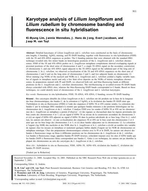
<strong>Karyotype</strong> <strong>analysis</strong> <strong>of</strong> <strong>Lilium</strong> <strong>longiflorum</strong> <strong>and</strong><br />
<strong>Lilium</strong> rubellum by chromosome b<strong>and</strong>ing <strong>and</strong><br />
fluorescence in situ hybridisation<br />
Ki-Byung Lim, Jannie Wennekes, J. Hans de Jong, Evert Jacobsen, <strong>and</strong><br />
Jaap M. van Tuyl<br />
Abstract: Detailed karyotypes <strong>of</strong> <strong>Lilium</strong> <strong>longiflorum</strong> <strong>and</strong> L. rubellum were constructed on the basis <strong>of</strong> chromosome<br />
arm lengths, C-b<strong>and</strong>ing, AgNO 3 staining, <strong>and</strong> PI–DAPI b<strong>and</strong>ing, together with fluorescence in situ hybridisation (FISH)<br />
with the 5S <strong>and</strong> 45S rDNA sequences as probes. The C-b<strong>and</strong>ing patterns that were obtained with the st<strong>and</strong>ard BSG<br />
technique revealed only few minor b<strong>and</strong>s on heterologous positions <strong>of</strong> the L. <strong>longiflorum</strong> <strong>and</strong> L. rubellum chromosomes.<br />
FISH <strong>of</strong> the 5S <strong>and</strong> 45S rDNA probes on L. <strong>longiflorum</strong> metaphase complements showed overlapping signals at<br />
proximal positions <strong>of</strong> the short arms <strong>of</strong> chromosomes 4 <strong>and</strong> 7, a single 5S rDNA signal on the secondary constriction<br />
<strong>of</strong> chromosome 3, <strong>and</strong> one 45S rDNA signal adjacent to the 5S rDNA signal on the subdistal part <strong>of</strong> the long arm <strong>of</strong><br />
chromosome 3. In L. rubellum, we observed co-localisation <strong>of</strong> the 5S <strong>and</strong> 45S rDNA sequences on the short arm <strong>of</strong><br />
chromosomes 2 <strong>and</strong> 4 <strong>and</strong> on the long arms <strong>of</strong> chromosomes 2 <strong>and</strong> 3, <strong>and</strong> two adjacent b<strong>and</strong>s on chromosome 12.<br />
Silver staining (Ag–NOR) <strong>of</strong> the nucleoli <strong>and</strong> NORs in L. <strong>longiflorum</strong> <strong>and</strong> L. rubellum yielded a highly variable number<br />
<strong>of</strong> signals in interphase nuclei <strong>and</strong> only a few faint silver deposits on the NORs <strong>of</strong> mitotic metaphase chromosomes.<br />
In preparations stained with PI <strong>and</strong> DAPI, we observed both red- <strong>and</strong> blue-fluorescing b<strong>and</strong>s at different<br />
positions on the L. <strong>longiflorum</strong> <strong>and</strong> L. rubellum chromosomes. The red-fluorescing or so-called reverse PI–DAPI b<strong>and</strong>s<br />
always coincided with rDNA sites, whereas the blue-fluorescing DAPI b<strong>and</strong>s corresponded to C-b<strong>and</strong>s. Based on these<br />
techniques, we could identify most <strong>of</strong> chromosomes <strong>of</strong> the L. <strong>longiflorum</strong> <strong>and</strong> L. rubellum karyotypes.<br />
Key words: fluorescence in situ hybridisation, FISH, 5S rDNA, 45S rDNA, C-b<strong>and</strong>ing, reverse PI–DAPI b<strong>and</strong>ing.<br />
Résumé : Des caryotypes détaillés du <strong>Lilium</strong> <strong>longiflorum</strong> et du L. rubellum ont été produits sur la base de la longueur<br />
des bras chromosomiques, des b<strong>and</strong>es C, de la coloration à l’AgNO 3 et la révélation des b<strong>and</strong>es PI–DAPI ainsi que<br />
l’hybridation in situ en fluorescence (FISH) à l’aide des séquences d’ADNr 5S et 45S comme sondes. La coloration des<br />
b<strong>and</strong>es C par la technique BSG st<strong>and</strong>ard n’a montré que quelques b<strong>and</strong>es mineures à des positions hétérologues sur les<br />
chromosomes du L. <strong>longiflorum</strong> et du L. rubellum. L’analyse FISH avec les sondes d’ADNr 5S et 45S sur les chromosomes<br />
du L. <strong>longiflorum</strong> en métaphase a permis d’observer : (i) des signaux se chevauchant dans les régions proximales des<br />
bras courts des chromosomes 4 et 7, (ii) un signal d’ADNr 5S au niveau de la constriction secondaire du chromosome 3<br />
et (iii) un signal d’ADNr 45S adjacent au signal d’ADNr 5S dans la portion sub-distale de ce bras long. Chez le L. rubellum,<br />
les auteurs ont observé : (i) une co-localisation des séquences 5S et 45S sur le bras court des chromosomes 2 et 4<br />
ainsi que sur les bras longs des chromosomes 2 et 3, et (ii) deux b<strong>and</strong>es adjacentes sur le chromosome 12. La coloration<br />
à l’argent (Ag–NOR) des nucléoles et des NOR chez le L. <strong>longiflorum</strong> et le L. rubellum a révélé un nombre très variable<br />
de signaux chez les noyaux en interphase et seulement quelques faibles dépôts d’argent sur les NOR de chromosomes en<br />
métaphase mitotique. Chez des préparations chromosomiques colorées avec le PI et le DAPI, les auteurs ont observé des<br />
b<strong>and</strong>es à fluorescence rouge ou bleue à différentes positions sur les chromosomes du L. <strong>longiflorum</strong> et du L. rubellum.<br />
Les b<strong>and</strong>es à fluorescence rouge, appelées b<strong>and</strong>es PI–DAPI inverses, coïncident toujours avec les sites d’ADNr t<strong>and</strong>is que<br />
les b<strong>and</strong>es DAPI à fluorescence bleue correspondent aux b<strong>and</strong>es C. Grâce à ces techniques, les auteurs ont pu identifier la<br />
plupart des chromosomes au sein des caryotypes du L. <strong>longiflorum</strong> et du L. rubellum.<br />
Mots clés : hybridation in situ en fluorescence, FISH, ADNr 5S, ADNr 45S, révélation des b<strong>and</strong>es C, révélation des<br />
b<strong>and</strong>es PI–DAPI inverses.<br />
[Traduit par la Rédaction] Lim et al. 918<br />
Received November 21, 2000. Accepted May 16, 2001. Published on the NRC Research Press Web site at http://genome.nrc.ca on<br />
September 7, 2001.<br />
Corresponding Editor: G. Jenkins.<br />
K.-B. Lim1 <strong>and</strong> J.M. van Tuyl. Plant Research International, Business Unit Genetics <strong>and</strong> <strong>Breeding</strong>, P.O. Box 16, 6700 AA<br />
Wageningen, The Netherl<strong>and</strong>s.<br />
J. Wennekes <strong>and</strong> J.H. de Jong. Laboratory <strong>of</strong> Genetics, Wageningen University, Wageningen, The Netherl<strong>and</strong>s.<br />
E. Jacobsen. Laboratory <strong>of</strong> Plant <strong>Breeding</strong>, Wageningen University, Wageningen, The Netherl<strong>and</strong>s.<br />
1 Corresponding author (e-mail: k.b.lim@plant.wag-ur.nl).<br />
Genome 44: 911–918 (2001) DOI: 10.1139/gen-44-5-911<br />
© 2001 NRC Canada<br />
911
912 Genome Vol. 44, 2001<br />
Introduction<br />
The huge genomes <strong>of</strong> species <strong>of</strong> the genus <strong>Lilium</strong> are<br />
among the largest in the plant kingdom (Bennett <strong>and</strong> Smith<br />
1976, 1991). The chromosomes <strong>of</strong> these species are also exceptionally<br />
large <strong>and</strong> have proved to be outst<strong>and</strong>ing material<br />
for cytogenetic research for more than a century. Since<br />
Strasburger’s paper on the chromosomes <strong>of</strong> <strong>Lilium</strong><br />
(Strasburger 1880), many studies have been published, especially<br />
on chromosome morphology in <strong>Lilium</strong> <strong>longiflorum</strong><br />
Thunb. (Stewart 1947), b<strong>and</strong>ing pattern (Holm 1976; Von<br />
Kalm <strong>and</strong> Smyth 1984), the detection <strong>of</strong> nucleolar organiser<br />
regions (NORs) (Von Kalm <strong>and</strong> Smyth 1980, 1984), <strong>and</strong> genome<br />
size (Van Tuyl <strong>and</strong> Boon 1997). Surprisingly, little attention<br />
was drawn to the chromosomes <strong>of</strong> <strong>Lilium</strong> rubellum<br />
Baker, which is an economically important species for<br />
interspecific hybridisation with L. <strong>longiflorum</strong>. Despite their<br />
phenotypic differences, most <strong>Lilium</strong> spp. have the same<br />
chromosome number (2n =2x = 24) <strong>and</strong> almost identical<br />
chromosome portraits (Stewart 1947). <strong>Lilium</strong> <strong>longiflorum</strong><br />
<strong>and</strong> L. rubellum (Noda 1991), which differ in nuclear DNA<br />
content by less than 2% (Van Tuyl <strong>and</strong> Boon 1997), were<br />
also found to have similar karyotypes. In addition, relatively<br />
high levels <strong>of</strong> meiotic recombination between the<br />
homoeologues <strong>of</strong> L. <strong>longiflorum</strong> × L. rubellum hybrids were<br />
observed (Lim et al. 2000), suggesting close synteny between<br />
the parental genomes (Fig. 3a).<br />
The increased use <strong>of</strong> L. <strong>longiflorum</strong> <strong>and</strong> L. rubellum in<br />
lily breeding requires detailed knowledge <strong>of</strong> the parental<br />
chromosome portraits for tracing chromosome behaviour in<br />
interspecific hybrids <strong>and</strong> backcross plants. The ability to<br />
identify individual chromosomes is essential in localizing<br />
the breakpoints in translocation chromosomes <strong>and</strong> the sites<br />
<strong>of</strong> recombination between homoeologues. Detailed karyotypes<br />
could also be instrumental in assigning linkage groups<br />
to chromosomes <strong>and</strong> mapping genes on the chromosome<br />
arms.<br />
Detailed karyotypes generally display chromosomes ordered<br />
in sequence <strong>of</strong> decreasing length, <strong>and</strong> include<br />
heterochromatin patterns based on C-, N-, <strong>and</strong> Q-b<strong>and</strong>ing<br />
<strong>and</strong> on other chromosome differentiation techniques. Satellite<br />
chromosomes can be recognised by their micro- or<br />
macro-satellites or by Ag–NOR staining, a technique that<br />
specifically visualises sites <strong>of</strong> metabolically active NORs<br />
(e.g., Linde-Laursen 1975). Fluorescence in situ hybridisation<br />
(FISH), which maps repetitive or single-copy sequences<br />
on the chromosomes, now complements b<strong>and</strong>ing technologies,<br />
along with the use <strong>of</strong> DNA-specific fluorochromes<br />
(Peterson et al. 1999), which elucidate local variation in<br />
DNA <strong>and</strong> (or) chromatin composition. The most common<br />
application <strong>of</strong> FISH is the localisation <strong>of</strong> rDNA families on<br />
the chromosomes. The 45S rDNA sequences were shown to<br />
be on the NOR <strong>of</strong> the satellite chromosomes <strong>and</strong> the 5S<br />
rDNA to be located, generally, on one <strong>of</strong> the other chromosomes<br />
(Gerlach <strong>and</strong> Dyer 1980; Leitch <strong>and</strong> Heslop-Harrison<br />
1992). Other repeats in FISH mapping studies include<br />
microsatellites (Cuadrado <strong>and</strong> Schwarzacher 1998), satellite<br />
DNAs (Pedersen et al. 1996), <strong>and</strong> retrotransposon repeat<br />
families (Heslop-Harrison et al. 1997; Miller et al. 1998;<br />
Presting et al. 1998).<br />
In <strong>Lilium</strong> spp., most chromosomes (4–6, 7–9, <strong>and</strong> 10–12)<br />
are morphologically too similar to be identified without<br />
additional diagnostic l<strong>and</strong>marks. C-b<strong>and</strong>s were seen in several<br />
studies (Holm 1976; Son 1977; Kongsuwan <strong>and</strong> Smyth<br />
1978; Smyth et al. 1989), revealing small b<strong>and</strong>s<br />
in L. <strong>longiflorum</strong> chromosomes 1, 3, 4, 7, 8, 9, <strong>and</strong> 12, <strong>and</strong><br />
NOR detection by isotopic in situ hybridisation <strong>and</strong> silver<br />
staining demonstrated nucleoli <strong>and</strong> active ribosomal sites on<br />
chromosomes 3, 4, <strong>and</strong> 7 (Von Kalm <strong>and</strong> Smyth 1980,<br />
1984). However, so far no reports using FISH to detect ribosomal<br />
DNA sequences in <strong>Lilium</strong> have appeared.<br />
Fluorescence microscopy <strong>of</strong> various plant chromosomes<br />
using a mixture <strong>of</strong> DAPI (4′,6-diamidino-2-phenylindole)<br />
<strong>and</strong> PI (propidium iodide) revealed small red-fluorescing<br />
b<strong>and</strong>s (Peterson et al. 1999; Andras et al. 2000). Such reverse<br />
PI–DAPI b<strong>and</strong>s were also detected in L. <strong>longiflorum</strong> ×<br />
L. rubellum hybrid at the NOR regions <strong>of</strong> a few satellite<br />
chromosomes (Lim et al. 2000).<br />
The aim <strong>of</strong> this study was a comparative <strong>analysis</strong> <strong>of</strong> the<br />
karyotypes <strong>of</strong> L. <strong>longiflorum</strong> <strong>and</strong> L. rubellum, using<br />
morphometric data, C-b<strong>and</strong>ing pr<strong>of</strong>iles, Ag–NOR staining,<br />
FISH detection <strong>of</strong> 45S rDNA <strong>and</strong> 5S rDNA sequences, <strong>and</strong><br />
reverse PI–DAPI b<strong>and</strong>ing, to identify all the chromosomes<br />
in each complement.<br />
Materials <strong>and</strong> methods<br />
Plant materials <strong>and</strong> chromosome preparation<br />
<strong>Lilium</strong> <strong>longiflorum</strong> ‘Snow Queen’ <strong>and</strong> L. rubellum (accession<br />
number 980085; originally from the mountainous area <strong>of</strong> the<br />
Fukushima prefecture, Japan) were obtained from the germplasm<br />
collection <strong>of</strong> Plant Research International, Wageningen, The Netherl<strong>and</strong>s.<br />
Root tips were collected in the early morning, pretreated<br />
in a saturated α-bromonaphthalin solution for 2hat20°C, <strong>and</strong> kept<br />
in this solution at 4°C until the next morning. The material was<br />
then rinsed three times in tap water before being fixed in acetic<br />
acid – ethanol (1:3) for 2 h. Root tips were stored at –20°C until<br />
used. The material was rinsed thoroughly before incubation in a<br />
pectolytic enzyme mixture (0.3% pectolyase Y23, 0.3% cellulase<br />
RS, <strong>and</strong> 0.3% cytohelicase) for about 1hat37°C. Squash preparations<br />
were made in a drop <strong>of</strong> 60% acetic acid. The microscope<br />
slides were frozen in liquid nitrogen <strong>and</strong> the cover slips removed<br />
with a razor blade. Slides were then finally dehydrated in absolute<br />
ethanol, air-dried, <strong>and</strong> stored at –20°C in a freezer until used.<br />
C-b<strong>and</strong>ing <strong>and</strong> silver staining<br />
Prior to the BaOH 2 – saline – Giemsa (BSG) b<strong>and</strong>ing procedure,<br />
the preparations were baked overnight at 37°C. Denaturation was<br />
performed in a 6% BaOH 2 solution at 20°C for 8 min, followed by<br />
a 30 min wash with tap water. Slides were then re-annealed in 2×<br />
SSC (0.3 M sodium chloride, 0.03 M sodium citrate, pH 7.0) at<br />
60°C for 50 min <strong>and</strong> stained in 4% Giemsa in 10 mM Sörensen<br />
buffer (pH 6.8) for 45 min. After a brief wash, preparations were<br />
air-dried <strong>and</strong> mounted in Euparol.<br />
For Ag–NOR staining, we used 200 mL <strong>of</strong> a freshly prepared<br />
50% AgNO 3 solution per slide, which was covered with a 24 ×<br />
36 mm piece <strong>of</strong> Nybolt 300 nylon cloth. The slides were left in a<br />
humidified petri dish for 45 min at 60°C until the nylon patch<br />
turned yellowish. The slides were then washed briefly in running<br />
tap water, air-dried, <strong>and</strong> mounted with Entellan-M (Merck) for microscopic<br />
observation.<br />
© 2001 NRC Canada
Lim et al. 913<br />
Table 1. Summary <strong>of</strong> the morphometric <strong>and</strong> karyotypic data for <strong>Lilium</strong> <strong>longiflorum</strong> <strong>and</strong> L. rubellum.<br />
Probe DNA<br />
Clone pTa71 contains a 9-kb EcoRI fragment <strong>of</strong> the 45S rDNA<br />
from wheat (Gerlach <strong>and</strong> Bedbrook 1979), <strong>and</strong> pScT7 contains a<br />
462-bp BamHI fragment <strong>of</strong> the 5S rDNA from rye (Lawrence <strong>and</strong><br />
Appels 1986). Isolated DNA <strong>of</strong> 45S <strong>and</strong> 5S rDNA sequences from<br />
pTa71 <strong>and</strong> pScT7 were labelled with biotin-16-dUTP or<br />
digoxigenin-11-dUTP by nick translation for in situ hybridisation,<br />
according to the manufacturer’s manual (Boehringer Mannheim,<br />
Germany).<br />
FISH<br />
Slides were left overnight at 37°C before treatment with 1 µg<br />
RNase A/mL <strong>of</strong> 2× SSC at 37°C for 60 min. The slides were<br />
washed three times in 2× SSC at 20°C for 5 min, hydrolysed with<br />
10 mM HCl at 37°C for 2 min, treated with 100 µL pepsin<br />
(5 µg/mL in 10 mM HCl) at 37°C for 10 min, washed two times in<br />
2× SSC for 5 min, treated with 4% paraformaldehyde for 10 min,<br />
washed another three times in 2× SSC, dehydrated through a<br />
graded ethanol series (70, 90, <strong>and</strong> 98% for 3 min each), <strong>and</strong> finally<br />
air-dried. Samples (40 µL) <strong>of</strong> the hybridisation mixture (100 ng <strong>of</strong><br />
the DNA isolated from pScT7 <strong>and</strong> pTa71, 2 mg <strong>of</strong> sheared herring<br />
sperm DNA (GibcoBRL), 50% deionised formamide, 10% (w/v)<br />
sodium dextran sulphate (Sigma), 2× SSC, <strong>and</strong> 0.25% (w/v) SDS)<br />
were denatured for 5 min at 70°C <strong>and</strong> then put directly on ice for<br />
at least 5 min. Each slide with 40 µL <strong>of</strong> the hybridisation mixture<br />
was covered with a slip <strong>of</strong> plastic sheet, denatured for 5 min at<br />
80°C, <strong>and</strong> left overnight at 37°C in a tightly closed humidified container.<br />
Slides were washed in 2× SSC for 15 min, transferred to<br />
0.1× SSC at 42°C for 30 min, <strong>and</strong> incubated for 60 min at 37°C in<br />
blocking buffer (0.1 M maleic acid, 0.15 M NaCl, 1% (w/v) blocking<br />
reagent from Boehringer Mannheim). Biotin- <strong>and</strong> digoxigeninlabelled<br />
probe DNA was detected by a Cy3–avidin–streptavidin<br />
detection system (Vector Laboratories) <strong>and</strong> a fluorescein<br />
isothiocyanate (FITC) – anti-digoxigenin detection system<br />
(Boehringer Mannheim, Germany). Slides were counterstained<br />
with 10 mg/mL DAPI, 5 mg/mL PI, or a mixed solution <strong>of</strong> DAPI<br />
<strong>and</strong> PI. Images were photographed with a Zeiss Axiophot<br />
photomicroscope equipped with epifluorescence illumination <strong>and</strong><br />
single-b<strong>and</strong> filters for DAPI, FITC, <strong>and</strong> Cy3–PI, using 400 ISO<br />
colour negative film. Films were scanned at 1200 dpi for digital<br />
processing with Adobe Photoshop ® (version 5.0; Adobe Inc.<br />
U.S.A.).<br />
L. <strong>longiflorum</strong> L. rubellum<br />
DNA content (pg/2C) 77.1±0.3 73.6±0.6<br />
Chromosome length (µm)<br />
Longest 34.4 (chr. 1) 33.8 (chr. 1)<br />
Shortest 18.1 (chr. 10) 16.3 (chr. 4)<br />
Total length ≈286.1 ≈269.9<br />
Ag–NOR staining<br />
Signals/interphase 6 6–10<br />
C-b<strong>and</strong>ed chromosomes 1, 3, 4, 7 (3x), 8(2x), 9, 11, 12 2 (2x),3(2x),4(2x),6(2x),8(2x), 12 (2x)<br />
FISH<br />
5S rDNA alone chr. 3 —<br />
45S rDNA alone chr. 3 chrs. 1, 6<br />
5S + 45S rDNA chrs. 4, 7 chrs. 2 (2x), 3, 4, 12<br />
DAPI <strong>and</strong> PI b<strong>and</strong>s<br />
DAPI alone chrs. 1, 3, 9 chrs. 2, 4, 12<br />
PI alone chrs. 4, 7 chrs. 1, 2, 12<br />
Reverse PI–DAPI b<strong>and</strong>s chrs. 4, 7 chrs. 2(2x), 3, 4, 12<br />
<strong>Karyotype</strong> <strong>analysis</strong> <strong>and</strong> flow-cytometric <strong>analysis</strong> <strong>of</strong><br />
nuclear DNA<br />
Chromosomes were measured with a ruler <strong>and</strong> arranged in order<br />
<strong>of</strong> decreasing short arm length, according to Stewart (1947). Total<br />
nuclear DNA content <strong>of</strong> DAPI-stained leaf nuclei was measured<br />
with a Partec CA-II cell analyser. Relative DNA content was calculated<br />
using Allium cepa nuclei as internal st<strong>and</strong>ard (DNA content =<br />
33.5 pg/2C).<br />
Results<br />
An overview <strong>of</strong> all the morphometric data, chromosome<br />
b<strong>and</strong>ing, <strong>and</strong> FISH results are given in Table 1. The positions<br />
<strong>of</strong> the b<strong>and</strong>s <strong>and</strong> FISH signals are depicted in the ideograms<br />
<strong>of</strong> Fig. 1. Flow-cytometric <strong>analysis</strong> <strong>of</strong> DAPI-stained<br />
nuclei gave values <strong>of</strong> 77.1 ± 0.3 pg/2C for L. <strong>longiflorum</strong><br />
<strong>and</strong> 73.6 ± 0.6 pg/2C for L. rubellum. In addition, the total<br />
length <strong>of</strong> the metaphase complement was ca. 286 µm<br />
for L. <strong>longiflorum</strong> <strong>and</strong> ca. 270 µm for L. rubellum. The<br />
(sub)metacentric chromosomes, 1 <strong>and</strong> 2, are far longer than<br />
all others, whereas chromosomes 3–12 are highly asymmetrical<br />
with centromere indexes ranging from 20 to 5%. Chromosomes<br />
4 <strong>and</strong> 7 <strong>of</strong> L. <strong>longiflorum</strong> <strong>and</strong> chromosomes 2 <strong>and</strong><br />
4<strong>of</strong>L. rubellum have satellites on their short arms. Further<br />
constrictions were observed on the long arms <strong>of</strong> chromosome<br />
3 (subdistal) <strong>of</strong> L. <strong>longiflorum</strong> <strong>and</strong> chromosomes 1, 2,<br />
<strong>and</strong> 12 (proximal) <strong>of</strong> L. rubellum.<br />
The C-b<strong>and</strong>ing technique revealed small b<strong>and</strong>s at 11 positions<br />
on different chromosomes <strong>of</strong> L. <strong>longiflorum</strong>. We<br />
observed single proximal heterochromatin b<strong>and</strong>s on chromosomes<br />
1, 11, <strong>and</strong> 12, a single subdistal b<strong>and</strong> on chromosome<br />
3 at the secondary constriction, <strong>and</strong> several intercalary b<strong>and</strong>s<br />
on chromosomes 7, 8, <strong>and</strong> 9. The L. rubellum karyotype<br />
showed a different pattern with a total <strong>of</strong> 12 small b<strong>and</strong>s on<br />
chromosomes 2 (2x),3(2x),4(2x),6(2x),8(2x), <strong>and</strong> 12<br />
(2x) (Figs. 2b <strong>and</strong> 2d). Ag–NOR staining <strong>of</strong> L. <strong>longiflorum</strong><br />
chromosomes showed weakly stained spots on chromosome<br />
4 only (Fig. 2e). Interphase nuclei, however, displayed six<br />
dark silver deposits or had completely stained nucleoli.<br />
In L. rubellum, the Ag–NOR dots on the mitotic metaphase<br />
© 2001 NRC Canada
914 Genome Vol. 44, 2001<br />
Fig. 1. Ideogram <strong>of</strong> <strong>Lilium</strong> <strong>longiflorum</strong> <strong>and</strong> L. rubellum indicating the positions <strong>of</strong> the C-b<strong>and</strong>s <strong>and</strong> DAPI, PI, 5S rDNA, <strong>and</strong> 45S<br />
rDNA sites. The reversed PI–DAPI b<strong>and</strong>s were obtained by staining the chromosome preparations with a mixture <strong>of</strong> DAPI <strong>and</strong> PI simultaneously.<br />
chromosome were even weaker than in L. <strong>longiflorum</strong> or not<br />
detectable, whereas interphase cells displayed 6–10 large<br />
spots.<br />
The red-fluorescing reverse PI–DAPI b<strong>and</strong>s were found<br />
on the secondary constrictions <strong>of</strong> chromosomes 4 <strong>and</strong> 7<br />
in L. <strong>longiflorum</strong> <strong>and</strong> chromosomes 1, 2, 4, <strong>and</strong> 12<br />
in L. rubellum (Fig. 3e). In L. <strong>longiflorum</strong>, DAPI b<strong>and</strong>s were<br />
detected in chromosomes 1, 3, <strong>and</strong> 9, <strong>and</strong> PI b<strong>and</strong>s were<br />
seen in chromosomes 3 <strong>and</strong> 4. In L. rubellum, DAPI b<strong>and</strong>s<br />
appeared in proximal positions on chromosomes 2, 4, <strong>and</strong><br />
12, <strong>and</strong> PI b<strong>and</strong>s appeared in a proximal position on chro-<br />
mosome 1, in a subdistal position on chromosome 2, <strong>and</strong> at<br />
two adjacent sites close to the centromere <strong>of</strong> chromosome<br />
12.<br />
FISH <strong>of</strong> 45S rDNA (pTa71 probe) with L. <strong>longiflorum</strong><br />
revealed signals on the secondary constrictions <strong>of</strong> chromosomes<br />
4 <strong>and</strong> 7 <strong>and</strong> near the secondary constriction on chromosome<br />
3. The 5S rDNA (pScT7 probe) hybridised to the<br />
long arm at the secondary constriction <strong>of</strong> chromosome 3<br />
proximal from the 45S rDNA site. The probe also hybridised<br />
to the secondary constrictions <strong>of</strong> chromosomes 4 <strong>and</strong> 7 <strong>and</strong><br />
co-localised with the 45S rDNA sites (Figs. 3b–3d).<br />
© 2001 NRC Canada
Lim et al. 915<br />
Fig. 2. (a <strong>and</strong> b) Giemsa C-b<strong>and</strong>ing pattern <strong>of</strong> <strong>Lilium</strong> <strong>longiflorum</strong> (a) <strong>and</strong> L. rubellum (b) chromosomes. (c <strong>and</strong> d) <strong>Karyotype</strong>s<br />
<strong>of</strong> L. <strong>longiflorum</strong> (c) <strong>and</strong> L. rubellum (d). (e) Ag–NOR staining <strong>of</strong> L. <strong>longiflorum</strong> chromosomes.<br />
In L. rubellum, co-localisation <strong>of</strong> 45S rDNA <strong>and</strong> 5S rDNA<br />
was observed in the secondary constrictions <strong>of</strong> chromosomes<br />
2, 3, 4, <strong>and</strong> 12, <strong>and</strong> a single spot <strong>of</strong> 45S rDNA was<br />
observed in the secondary constriction <strong>of</strong> chromosome 1 <strong>and</strong><br />
in the middle <strong>of</strong> the long arm <strong>of</strong> chromosome 6 (Fig. 3g).<br />
Discussion<br />
This study elucidated various dissimilarities between the<br />
chromosomes <strong>of</strong> L. <strong>longiflorum</strong> <strong>and</strong> L. rubellum that are not<br />
obvious from comparison <strong>of</strong> their unb<strong>and</strong>ed karyotypes.<br />
Firstly, the distribution <strong>of</strong> secondary constrictions differs between<br />
the species: three sites on chromosomes 3, 4, <strong>and</strong> 7<br />
in L. <strong>longiflorum</strong> <strong>and</strong> five sites on chromosomes 1, 2, 3, 4<br />
<strong>and</strong> 12 in L. rubellum. These differences between the two<br />
species are in agreement with the studies <strong>of</strong> Stewart (1947)<br />
<strong>and</strong> Ogihara (1968). Our observation made clear that secondary<br />
constrictions were associated with the nucleolus <strong>and</strong><br />
that they contain 45S rDNA repeats. Other characteristics,<br />
such as the C-b<strong>and</strong>s <strong>of</strong> L. <strong>longiflorum</strong>, were almost identical<br />
to those <strong>of</strong> <strong>Lilium</strong> formosanum (section Leucolirion) (cf.<br />
Stewart 1947; Smyth et al. 1989). The karyotype<br />
<strong>of</strong> L. rubellum, as far as the constrictions in chromosomes 1<br />
<strong>and</strong> 2 are concerned, is a combination <strong>of</strong> the chromosome<br />
morphology <strong>of</strong> its putative parental species, <strong>Lilium</strong><br />
japonicum <strong>and</strong> L. auratum. Most obvious is the constant<br />
number <strong>of</strong> single NORs on chromosomes 1, 2, 3, <strong>and</strong> 4<br />
found in many species <strong>of</strong> <strong>Lilium</strong> studied so far (Stewart<br />
1947). Sites <strong>of</strong> constitutive heterochromatin (C-b<strong>and</strong>s) <strong>of</strong>ten<br />
coincided with the secondary constrictions, but did not always<br />
do so. In L. <strong>longiflorum</strong>, only the C-b<strong>and</strong>s <strong>of</strong> chromosomes<br />
3, 4, <strong>and</strong> 7 co-localise with these constrictions <strong>and</strong><br />
contained 45S rDNA repeats (Fig. 1). These observations<br />
are in agreement with the previous studies <strong>of</strong> Stewart<br />
(1947), Von Kalm <strong>and</strong> Smyth (1984), Kongsuwan <strong>and</strong><br />
Smyth (1978), <strong>and</strong> Smyth et al. (1989), <strong>and</strong> are similar to<br />
observations <strong>of</strong> L. formosanum <strong>of</strong> the same section (Smyth<br />
et al. 1989). In L. rubellum, the C-b<strong>and</strong>ing pattern, which<br />
strongly resembles the patterns <strong>of</strong> L. auratum <strong>and</strong> other species<br />
<strong>of</strong> section Archelirion (cf. Smyth et al. 1989), corresponds<br />
to the sites <strong>of</strong> the secondary constrictions except for<br />
three interstitial b<strong>and</strong>s on the long arms <strong>of</strong> chromosomes 3,<br />
4, <strong>and</strong> 8 (Fig. 2d). Unfortunately, our Ag–NOR staining produced<br />
insufficient evidence to determine the number <strong>of</strong> active<br />
NORs on metaphase chromosomes. It is questionable<br />
whether our in vitro root tip material grown at 25°C is suitable<br />
for the detection <strong>of</strong> nucleolar activity.<br />
Further evidence for molecular differences in chromosomal<br />
organisation between L. <strong>longiflorum</strong> <strong>and</strong> L. rubellum<br />
came from the comparison <strong>of</strong> C-b<strong>and</strong>s <strong>and</strong> PI–DAPI b<strong>and</strong>s.<br />
In L. <strong>longiflorum</strong>, C-b<strong>and</strong>s <strong>and</strong> DAPI b<strong>and</strong>s appeared on<br />
chromosomes 1, 3, <strong>and</strong> 9, C-b<strong>and</strong>s <strong>and</strong> PI b<strong>and</strong>s appeared on<br />
chromosomes 4 <strong>and</strong> 7, <strong>and</strong> the remaining C-b<strong>and</strong>s appeared<br />
on chromosomes 7, 8, 11, <strong>and</strong> 12, without deviant PI–DAPI<br />
© 2001 NRC Canada
916 Genome Vol. 44, 2001<br />
© 2001 NRC Canada
Lim et al. 917<br />
Fig. 3. (a) Genomic in situ hybridization on the metaphase I complement <strong>of</strong> an interspecific hybrid between <strong>Lilium</strong> <strong>longiflorum</strong><br />
<strong>and</strong> L. rubellum shows a range <strong>of</strong> bivalent formation, indicating some homology in DNA sequence between the two species. (b) Simultaneous<br />
FISH detection <strong>of</strong> 5S <strong>and</strong> 45S rDNA probes (white arrowheads) in L. <strong>longiflorum</strong>. The red arrowheads represent the 5S rDNA<br />
probe, which has a stronger signal on chromosome 3 than the 45S rDNA probe. (c <strong>and</strong> d) <strong>Karyotype</strong>s <strong>of</strong> 5S rDNA (c) <strong>and</strong> 45S rDNA<br />
(d) onL. <strong>longiflorum</strong> chromosomes. (e) Simultaneous staining with PI <strong>and</strong> DAPI <strong>of</strong> L. rubellum chromosomes. Reverse PI–DAPI<br />
b<strong>and</strong>s are visible <strong>and</strong> are indicated by arrowheads. (f) Detailed karyotype <strong>of</strong> the reverse PI–DAPI b<strong>and</strong>s in e. (g) Detection <strong>of</strong> 45S<br />
rDNA on L. rubellum chromosomes. (h <strong>and</strong> i) Simultaneous detection <strong>of</strong> 5S <strong>and</strong> 45S rDNA. There are stronger 5S rDNA signals (h,<br />
red arrowheads) than 45S rDNA signals (i, green arrowheads) on an interphase nucleus <strong>of</strong> L. rubellum. (j) <strong>Karyotype</strong> <strong>of</strong> rDNA<br />
in L. rubellum. Scale bars = 10 µm.<br />
fluorescence (Fig. 1). A comparable situation—three Cheterochromatin<br />
classes—was found for L. rubellum chromosomes,<br />
although different chromosomes were involved.<br />
FISH <strong>of</strong> rDNA repeats allowed the following classes to be<br />
distinguished: (1) 45S + 5S rDNAs, as on L. <strong>longiflorum</strong><br />
chromosomes 4 <strong>and</strong> 7 <strong>and</strong> L. rubellum chromosomes 2, 3, 4,<br />
<strong>and</strong> 12; (2) only 45S rDNA, as on L. <strong>longiflorum</strong> chromosome<br />
3 <strong>and</strong> L. rubellum chromosomes 1 <strong>and</strong> 6; <strong>and</strong> (3) only<br />
5S rDNA, as on L. <strong>longiflorum</strong> chromosome 3<br />
<strong>and</strong> L. rubellum chromosome 12 (Figs. 3c, 3d, <strong>and</strong> 3j). The<br />
45S rDNA signals, which occurred mainly in the secondary<br />
constrictions, were mostly larger <strong>and</strong> brighter than the 5S rDNA<br />
signals. However, on chromosome 3 in both L. <strong>longiflorum</strong><br />
<strong>and</strong> L. rubellum, the 5S rDNA signal was stronger than the<br />
45S rDNA signal (Figs. 3b–3d <strong>and</strong> 3j). All 45S rDNA sites<br />
were found to co-localise with the simultaneously stained reverse<br />
PI–DAPI b<strong>and</strong>s, with the exception <strong>of</strong> the long-arm<br />
sites on chromosome 3 in L. <strong>longiflorum</strong> (Figs. 3c <strong>and</strong> 3d)<br />
<strong>and</strong> chromosomes 1 <strong>and</strong> 6 in L. rubellum (Fig. 3g). Lima-de-<br />
Faria (1976) <strong>and</strong> Schulz-Schaeffer (1980) observed that satellites<br />
are generally attached to the short arm <strong>of</strong> a NOR<br />
chromosome. The present results confirm that the satellite<br />
repeats (reverse PI–DAPI b<strong>and</strong>) appear in the secondary<br />
constriction <strong>of</strong> a short arm or, when in a long arm, appear<br />
very near the centromere (Fig. 1).<br />
Lima-De-Faria (1976) analysed the nucleolus-organising<br />
citrons in over 700 species <strong>and</strong> reported that, in 87% <strong>of</strong><br />
cases, the nucleolus was located on the short arm <strong>of</strong> the<br />
chromosome. Such striking conservatism in karyotype morphology<br />
suggests some molecular or physical constraint for<br />
chromosome arms to associate with the nucleolus. Surprisingly,<br />
the sites with ribosomal genes in <strong>Lilium</strong> spp. are far<br />
more variable than those observed for most other plant species,<br />
exhibiting various NORs on both long <strong>and</strong> short chromosome<br />
arms (Fig. 1). One <strong>of</strong> the explanations is that the<br />
large genome size allows greater variability in ribosomal<br />
gene distribution along the short arms <strong>and</strong> so permits chromosomal<br />
rearrangements that involve moving the NORs to<br />
proximal long arm positions (see Fig. 1). It is still not known<br />
whether such long-arm sites <strong>of</strong> 45S rDNA became silenced.<br />
DAPI is known to bind preferentially to A–T rich heterochromatic<br />
regions or to act as a dye that is specific for double-str<strong>and</strong>ed<br />
DNA (Schweizer <strong>and</strong> Nagl 1976; Trask 1999).<br />
PI intercalates between the bases <strong>of</strong> either single- or doublestr<strong>and</strong>ed<br />
nucleic acid molecules (Heslop-Harrison <strong>and</strong><br />
Schwarzacher 1996). NORs are composed <strong>of</strong> t<strong>and</strong>emly repeated<br />
G–C sequences (Macgregor <strong>and</strong> Kezer 1971;<br />
Yasmineh <strong>and</strong> Yunis 1971; Ingle et al. 1975). Therefore,<br />
staining simultaneously with PI <strong>and</strong> DAPI can give rise to a<br />
red b<strong>and</strong>, a so-called reverse PI–DAPI b<strong>and</strong>, at NOR posi-<br />
tions. The same type <strong>of</strong> reverse PI–DAPI b<strong>and</strong> has been<br />
demonstrated in species <strong>of</strong> the genera Lycopersicon <strong>and</strong><br />
Oryza (Peterson et al. 1999; Andras et al. 2000). A similar<br />
case <strong>of</strong> reverse PI–DAPI b<strong>and</strong>s was seen in t<strong>and</strong>emly<br />
repeated regions such as NORs in the chromosomes <strong>of</strong><br />
<strong>Lilium</strong> species (Lim et al. 2000). This b<strong>and</strong> position could<br />
represent DAPI negative b<strong>and</strong>s in G–C rich regions. We<br />
found that repetitive b<strong>and</strong>s such as reverse PI–DAPI b<strong>and</strong>s<br />
are located mostly at the same positions as rDNA sites, not<br />
only on the short arm but also on the long arm adjacent to<br />
the centromere (see Fig. 3f).<br />
Our study revealed a few major differences in the distribution<br />
<strong>of</strong> heterochromatin, rDNA sites, <strong>and</strong> several t<strong>and</strong>em repeats<br />
between L. <strong>longiflorum</strong> <strong>and</strong> L. rubellum. As genome<br />
painting <strong>of</strong> L. <strong>longiflorum</strong> × L. rubellum hybrids (Lim et al.<br />
2000) also demonstrates large-scale differences in total<br />
genomic DNA between parental species, it is likely that<br />
Ty/copia <strong>and</strong> (or) related dispersed repeat families diverged<br />
recently during the evolution <strong>of</strong> <strong>Lilium</strong> spp. For the breeder<br />
<strong>and</strong> geneticist, it is more important to know whether these<br />
changes in chromosome morphology, b<strong>and</strong>ing pattern, <strong>and</strong><br />
molecular organisation also reflect large-scale chromosomal<br />
rearrangements like translocations <strong>and</strong> inversions. Observation<br />
<strong>of</strong> regular bivalent formation in metaphase I microsporocytes<br />
<strong>of</strong> a L. <strong>longiflorum</strong> × L. rubellum hybrid seem to<br />
suggest that the two homoeologous genomes retain extensive<br />
correspondence (Fig. 3a).<br />
References<br />
Andras, S.C., Hartman, T.P.V., Alex<strong>and</strong>er, J., McBride, R., Marshall,<br />
J.A., Power, J.B., Cocking, E.C., <strong>and</strong> Davey, M.R. 2000.<br />
Combined PI–DAPI staining (CPD) reveals NOR asymmetry<br />
<strong>and</strong> facilitates karyotyping <strong>of</strong> plant chromosomes. Chromosome<br />
Res. 8: 387–391.<br />
Bennett, M.D., <strong>and</strong> Smith, J.B. 1976. Nuclear DNA amounts in angiosperms.<br />
Philos. Trans. R. Soc. Lond. 274: 227–274.<br />
Bennett, M.D., <strong>and</strong> Smith, J.B. 1991. Nuclear DNA amounts in angiosperms.<br />
Philos. Trans. R. Soc. Lond. B Biol. Sci. 334: 309–<br />
345.<br />
Cuadrado, A., <strong>and</strong> Schwarzacher, T. 1998. The chromosomal organization<br />
<strong>of</strong> simple sequence repeats in wheat <strong>and</strong> rye genomes.<br />
Chromosoma (Berlin), 107: 587–594.<br />
Gerlach, W.L., <strong>and</strong> Bedbrook, J.R. 1979. Cloning <strong>and</strong> characterization<br />
<strong>of</strong> ribosomal RNA genes from wheat <strong>and</strong> barley. Nucleic<br />
Acids Res. 7: 1869–1885.<br />
Gerlach, W.L., <strong>and</strong> Dyer, T.A. 1980. Sequence organization <strong>of</strong> the<br />
repeating units in the nucleus <strong>of</strong> wheat, which contain 5S rRNA<br />
genes. Nucleic Acids Res. 8: 4851–4865.<br />
Heslop-Harrison, J.S., <strong>and</strong> Schwarzacher, T. 1996. Flow cytometry<br />
<strong>and</strong> chromosome sorting. In Plant chromosomes laboratory<br />
© 2001 NRC Canada
918 Genome Vol. 44, 2001<br />
methods. Edited by K. Fukui <strong>and</strong> S. Nakayama. CRC Press,<br />
Boca Raton, Fla. pp. 85–106.<br />
Heslop-Harrison, J.S., Br<strong>and</strong>es, A., Taketa, S., Schmidt, T.,<br />
Vershinin, A.V., Alkhimova, E.G., Kamm, A., Doudrick, R.L.,<br />
Schwarzacher, T., Katsiotis, A., Kubis, S., Kumar, A., Pearce,<br />
S.R., Flavell, A.J., <strong>and</strong> Harrison, G.E. 1997. The chromosomal<br />
distributions <strong>of</strong> Ty1-copia group retrotransposable elements in<br />
higher plants <strong>and</strong> their implications for genome evolution.<br />
Genetica, 100: 197–204.<br />
Holm, P.B. 1976. The C <strong>and</strong> Q b<strong>and</strong>ing patterns <strong>of</strong> the chromosomes<br />
<strong>of</strong> <strong>Lilium</strong> <strong>longiflorum</strong> (Thunb.). Carlsberg Res. Commun.<br />
41: 217–224.<br />
Ingle, J., Timmis, J.N., <strong>and</strong> Sinclair, J. 1975. The relationship between<br />
satellite deoxyribonucleic acid, ribosomal ribonucleic<br />
acid gene redundancy, <strong>and</strong> genome size in plants. Plant Physiol.<br />
55: 496–501.<br />
Kongsuwan, K., <strong>and</strong> Smyth, D.R. 1978. DNA loss during Cb<strong>and</strong>ing<br />
<strong>of</strong> chromosomes <strong>of</strong> <strong>Lilium</strong> <strong>longiflorum</strong>. Chromosoma<br />
(Berlin), 68: 59–72.<br />
Lawrence, G.J., <strong>and</strong> Appels, R. 1986. Mapping the nucleolar organizer<br />
region, seed protein loci <strong>and</strong> isozyme loci on chromosome<br />
1R in rye. Theor. Appl. Genet. 71: 742–749.<br />
Leitch, I.J., <strong>and</strong> Heslop-Harrison, J.S. 1992. Physical mapping <strong>of</strong><br />
the 18S–5.8S–26S rRNA genes in barley by in situ hybridization.<br />
Genome, 35: 1013–1018.<br />
Lim, K.B., Chung, J.D., Van Kronenburg, B.C.E., Ramanna, M.S.,<br />
De Jong, J.H., <strong>and</strong> Van Tuyl, J.M. 2000. Introgression <strong>of</strong> <strong>Lilium</strong><br />
rubellum Baker chromosomes into L. <strong>longiflorum</strong> Thunb.: a genome<br />
painting study <strong>of</strong> the F 1 hybrid, BC 1 <strong>and</strong> BC 2 progenies.<br />
Chromosome Res. 8: 119–125.<br />
Lima-De-Faria, A. 1976. The chromosome field. I. Prediction <strong>of</strong><br />
the location <strong>of</strong> ribosomal cistrons. Hereditas, 83: 1–22.<br />
Linde-Laursen, I. 1975. Giemsa C-b<strong>and</strong>ing <strong>of</strong> the chromosomes <strong>of</strong><br />
‘Emir’ barley. Hereditas, 81: 285–289.<br />
Macgregor, H.C., <strong>and</strong> Kezer, J. 1971. The chromosomal localization<br />
<strong>of</strong> a heavy satellite DNA in the testis <strong>of</strong> Plethodon cinereus.<br />
Chromosoma (Berlin), 33: 167–182.<br />
Miller, J.T., Dong, F., Jackson, S.A., Song, J., <strong>and</strong> Jiang, J. 1998.<br />
Retrotransposon-related DNA sequences in the centromeres <strong>of</strong><br />
grass chromosomes. Genetics, 150: 1615–1623.<br />
Noda, S. 1991. Chromosomal variation <strong>and</strong> evolution in the genus<br />
<strong>Lilium</strong>. In Chromosome engineering in plants: genetics, breeding,<br />
evolution. Part B. Edited by T. Tsuchiya <strong>and</strong> P.K. Gupta.<br />
Elsevier, Amsterdam. pp. 507–524.<br />
Ogihara, R. 1968. <strong>Karyotype</strong>s <strong>of</strong> <strong>Lilium</strong> rubellum Baker. La<br />
Kromosomo, 74: 2415–2418.<br />
Pedersen, C., Rasmussen, S.K., <strong>and</strong> Linde-Laursen, I. 1996. Genome<br />
<strong>and</strong> chromosome identification in cultivated barley <strong>and</strong> related<br />
species <strong>of</strong> the Triticeae (Poaceae) by in situ hybridization<br />
with the GAA-satellite sequence. Genome, 39: 93–104.<br />
Peterson, D.G., Lapitan, N.L.V., <strong>and</strong> Stack, S.M. 1999. Localization<br />
<strong>of</strong> single <strong>and</strong> low-copy sequences on tomato synaptonemal<br />
complex spreads using fluorescence in situ hybridization<br />
(FISH). Genetics, 152: 427–439.<br />
Presting, G.G., Malysheva, L., Fuchs, J., <strong>and</strong> Schubert, I. 1998. A<br />
TY3/GYPSY retrotransposon-like squence localizes to the<br />
centromeric regions <strong>of</strong> cereal chromosomes. Plant J. 16: 721–<br />
728.<br />
Schulz-Schaeffer, J. 1980. The nucleolus organizer region. In<br />
Cytogenetics, plants, animals, human. Edited by J. Sybenga.<br />
Springer-Verlag. New York, U.S.A. pp. 36–38.<br />
Schweizer, D., <strong>and</strong> Nagl, W. 1976. Heterochromatin diversity in<br />
Cybidium, <strong>and</strong> its relationship to differential DNA replication.<br />
Exp. Cell Res. 98: 411–423.<br />
Smyth, D.R., Kongsuwan, K., <strong>and</strong> Wisdharomn, S. 1989. A survey<br />
<strong>of</strong> C-b<strong>and</strong> patterns in chromosomes <strong>of</strong> <strong>Lilium</strong> (Liliaceae). Plant<br />
Syst. Evol. 163: 53–69.<br />
Son, J.H. 1977. <strong>Karyotype</strong> <strong>analysis</strong> <strong>of</strong> <strong>Lilium</strong> lancifolium Thunb.<br />
by means <strong>of</strong> C-b<strong>and</strong>ing method. Jpn. J. Genet. 3: 217–221.<br />
Stewart, R.N. 1947. The morphology <strong>of</strong> somatic chromosomes in<br />
<strong>Lilium</strong>. Am. J. Bot. 34: 9–26.<br />
Strasburger, E. 1880. Zellbildung und Zelltheilung. 3rd ed. Fischer<br />
Verlag, Jena.<br />
Trask, B. 1999. Fluorescence in situ hybridization. In Genome<br />
<strong>analysis</strong>: a laboratory manual. Vol. 4. Mapping genomes. Edited<br />
by B. Birren, E.D. Green, P. Hieter, S. Klapholz, R.M. Myers,<br />
H. Riethman, <strong>and</strong> J. Roskams. Cold Spring Harbor Laboratory<br />
Press, Plainsview, N.Y. pp. 391–404.<br />
Van Tuyl, J.M., <strong>and</strong> Boon, E. 1997. Variation in DNA-content in<br />
the genus <strong>Lilium</strong>. Acta Hortic. 430: 829–835.<br />
Von Kalm, L., <strong>and</strong> Smyth, D.R. 1980. Silver staining test <strong>of</strong> nucleolar<br />
suppression in the <strong>Lilium</strong> hybrid ‘Black Beauty’. Exp. Cell<br />
Res. 129: 481–485.<br />
Von Kalm, L., <strong>and</strong> Smyth, D.R. 1984. Ribosomal RNA genes <strong>and</strong><br />
the substructure <strong>of</strong> nucleolar organizing genes in <strong>Lilium</strong>. Can. J.<br />
Genet. Cytol. 26: 158–166.<br />
Yasmineh, W.G., <strong>and</strong> Yunis, J.J. 1971. Satellite DNA in calf<br />
heterochromatin. Exp. Cell Res. 64: 41–48.<br />
© 2001 NRC Canada


