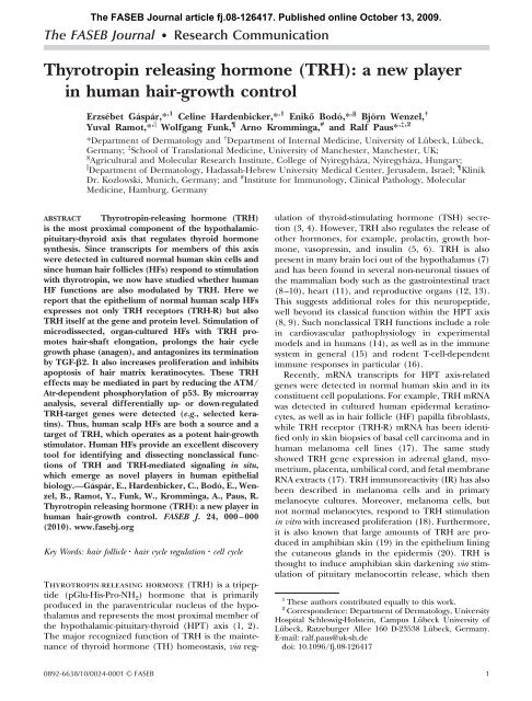
Thyrotropin releasing hormone (TRH) - The FASEB Journal
Thyrotropin releasing hormone (TRH) - The FASEB Journal
Thyrotropin releasing hormone (TRH) - The FASEB Journal

You also want an ePaper? Increase the reach of your titles
YUMPU automatically turns print PDFs into web optimized ePapers that Google loves.
<strong>The</strong> <strong>FASEB</strong> <strong>Journal</strong> article fj.08-126417. Published online October 13, 2009.<br />
<strong>The</strong> <strong>FASEB</strong> <strong>Journal</strong> Research Communication<br />
<strong>Thyrotropin</strong> <strong>releasing</strong> <strong>hormone</strong> (<strong>TRH</strong>): a new player<br />
in human hair-growth control<br />
Erzsébet Gáspár,* ,1 Celine Hardenbicker,* ,1 Enikő Bodó,* ,§ Björn Wenzel, †<br />
Yuval Ramot,* , Wolfgang Funk, Arno Kromminga, # and Ralf Paus* ,‡,2<br />
*Department of Dermatology and † Department of Internal Medicine, University of Lübeck, Lübeck,<br />
Germany; ‡ School of Translational Medicine, University of Manchester, Manchester, UK;<br />
§ Agricultural and Molecular Research Institute, College of Nyiregyháza, Nyiregyháza, Hungary;<br />
Department of Dermatology, Hadassah-Hebrew University Medical Center, Jerusalem, Israel; Klinik<br />
Dr. Kozlowski, Munich, Germany; and # Institute for Immunology, Clinical Pathology, Molecular<br />
Medicine, Hamburg, Germany<br />
ABSTRACT <strong>Thyrotropin</strong>-<strong>releasing</strong> <strong>hormone</strong> (<strong>TRH</strong>)<br />
is the most proximal component of the hypothalamicpituitary-thyroid<br />
axis that regulates thyroid <strong>hormone</strong><br />
synthesis. Since transcripts for members of this axis<br />
were detected in cultured normal human skin cells and<br />
since human hair follicles (HFs) respond to stimulation<br />
with thyrotropin, we now have studied whether human<br />
HF functions are also modulated by <strong>TRH</strong>. Here we<br />
report that the epithelium of normal human scalp HFs<br />
expresses not only <strong>TRH</strong> receptors (<strong>TRH</strong>-R) but also<br />
<strong>TRH</strong> itself at the gene and protein level. Stimulation of<br />
microdissected, organ-cultured HFs with <strong>TRH</strong> promotes<br />
hair-shaft elongation, prolongs the hair cycle<br />
growth phase (anagen), and antagonizes its termination<br />
by TGF-2. It also increases proliferation and inhibits<br />
apoptosis of hair matrix keratinocytes. <strong>The</strong>se <strong>TRH</strong><br />
effects may be mediated in part by reducing the ATM/<br />
Atr-dependent phosphorylation of p53. By microarray<br />
analysis, several differentially up- or down-regulated<br />
<strong>TRH</strong>-target genes were detected (e.g., selected keratins).<br />
Thus, human scalp HFs are both a source and a<br />
target of <strong>TRH</strong>, which operates as a potent hair-growth<br />
stimulator. Human HFs provide an excellent discovery<br />
tool for identifying and dissecting nonclassical functions<br />
of <strong>TRH</strong> and <strong>TRH</strong>-mediated signaling in situ,<br />
which emerge as novel players in human epithelial<br />
biology.—Gáspár, E., Hardenbicker, C., Bodó, E., Wenzel,<br />
B., Ramot, Y., Funk, W., Kromminga, A., Paus, R.<br />
<strong>Thyrotropin</strong> <strong>releasing</strong> <strong>hormone</strong> (<strong>TRH</strong>): a new player in<br />
human hair-growth control. <strong>FASEB</strong> J. 24, 000–000<br />
(2010). www.fasebj.org<br />
Key Words: hair follicle hair cycle regulation cell cycle<br />
<strong>Thyrotropin</strong>-<strong>releasing</strong> <strong>hormone</strong> (<strong>TRH</strong>) is a tripeptide<br />
(pGlu-His-Pro-NH 2) <strong>hormone</strong> that is primarily<br />
produced in the paraventricular nucleus of the hypothalamus<br />
and represents the most proximal member of<br />
the hypothalamic-pituitary-thyroid (HPT) axis (1, 2).<br />
<strong>The</strong> major recognized function of <strong>TRH</strong> is the maintenance<br />
of thyroid <strong>hormone</strong> (TH) homeostasis, via reg-<br />
0892-6638/10/0024-0001 © <strong>FASEB</strong><br />
ulation of thyroid-stimulating <strong>hormone</strong> (TSH) secretion<br />
(3, 4). However, <strong>TRH</strong> also regulates the release of<br />
other <strong>hormone</strong>s, for example, prolactin, growth <strong>hormone</strong>,<br />
vasopressin, and insulin (5, 6). <strong>TRH</strong> is also<br />
present in many brain loci out of the hypothalamus (7)<br />
and has been found in several non-neuronal tissues of<br />
the mammalian body such as the gastrointestinal tract<br />
(8–10), heart (11), and reproductive organs (12, 13).<br />
This suggests additional roles for this neuropeptide,<br />
well beyond its classical function within the HPT axis<br />
(8, 9). Such nonclassical <strong>TRH</strong> functions include a role<br />
in cardiovascular pathophysiology in experimental<br />
models and in humans (14), as well as in the immune<br />
system in general (15) and rodent T-cell-dependent<br />
immune responses in particular (16).<br />
Recently, mRNA transcripts for HPT axis-related<br />
genes were detected in normal human skin and in its<br />
constituent cell populations. For example, <strong>TRH</strong> mRNA<br />
was detected in cultured human epidermal keratinocytes,<br />
as well as in hair follicle (HF) papilla fibroblasts,<br />
while <strong>TRH</strong> receptor (<strong>TRH</strong>-R) mRNA has been identified<br />
only in skin biopsies of basal cell carcinoma and in<br />
human melanoma cell lines (17). <strong>The</strong> same study<br />
showed <strong>TRH</strong> gene expression in adrenal gland, myometrium,<br />
placenta, umbilical cord, and fetal membrane<br />
RNA extracts (17). <strong>TRH</strong> immunoreactivity (IR) has also<br />
been described in melanoma cells and in primary<br />
melanocyte cultures. Moreover, melanoma cells, but<br />
not normal melanocytes, respond to <strong>TRH</strong> stimulation<br />
in vitro with increased proliferation (18). Furthermore,<br />
it is also known that large amounts of <strong>TRH</strong> are produced<br />
in amphibian skin (19) in the epithelium lining<br />
the cutaneous glands in the epidermis (20). <strong>TRH</strong> is<br />
thought to induce amphibian skin darkening via stimulation<br />
of pituitary melanocortin release, which then<br />
1 <strong>The</strong>se authors contributed equally to this work.<br />
2 Correspondence: Department of Dermatology, University<br />
Hospital Schleswig-Holstein, Campus Lübeck University of<br />
Lübeck, Ratzeburger Allee 160 D-23538 Lübeck, Germany.<br />
E-mail: ralf.paus@uk-sh.de<br />
doi: 10.1096/fj.08-126417<br />
1
induces melanosome redistribution in cutaneous melanophores<br />
(21). However, whether normal human skin<br />
or its appendages also serve as targets of <strong>TRH</strong> remains<br />
unknown.<br />
In the current study, we explored this question by<br />
testing the response of normal human scalp HFs to<br />
<strong>TRH</strong>, given that it has been previously shown that they<br />
are both potent sources and important nonclassical<br />
targets of a wide range of neuroendocrine mediators<br />
(22). <strong>The</strong>se include corticotropin-<strong>releasing</strong> <strong>hormone</strong><br />
(CRH) (23–25), TSH (26), melatonin (27, 28), prolactin<br />
(29), endovanilloid receptors (30), endocannabioids<br />
(31), and THs (32).<br />
Utilizing organ-cultured, microdissected human<br />
scalp HFs (33), we have addressed the specific questions<br />
of whether normal human HFs express <strong>TRH</strong><br />
and/or <strong>TRH</strong>-R on the gene and protein level, and<br />
whether stimulation of human HFs with <strong>TRH</strong> alters<br />
hair growth, as measured by hair-shaft elongation, HF<br />
cycling, and hair matrix keratinocyte proliferation or<br />
apoptosis. Transforming growth factor-2 (TGF-2) is a<br />
key inducer of apoptosis-driven, normal HF regression<br />
(catagen) in humans (34–36). As TGF-2 induces<br />
apoptosis in the hair matrix keratinocytes (37), we also<br />
asked whether <strong>TRH</strong> can antagonize TGF-2-induced<br />
premature catagen transformation and/or inhibit hair<br />
matrix keratinocyte apoptosis in human HF. Since the<br />
ataxia telangiectasia-mutated (ATM) and ATM-Rad3<br />
related (Atr) kinase, and the ATM/Atr phosphorylation<br />
pathway of p53, are now recognized as key control<br />
elements in the modulation of apoptosis and/or cellcycle<br />
arrest (38–41), we furthermore asked whether<br />
the intrafollicular ATM/Atr pathway of p53 phosphorylation<br />
is altered by <strong>TRH</strong> administration. Finally, given<br />
that we have recently identified TSH as a negative<br />
regulator of keratin expression (26), we asked whether<br />
<strong>TRH</strong> exerts any effect on keratin expression.<br />
MATERIALS AND METHODS<br />
HF organ culture<br />
Anagen (VI) HFs were microdissected from normal temporofrontal<br />
human scalp skin obtained from 6 healthy adult females<br />
aged 51–61 yr (mean 54 yr) undergoing routine facelift surgery,<br />
adhering to the Helsinki guidelines and following approval by<br />
the Institutional Research Ethics Committee of the University of<br />
Lübeck. Isolated HFs from 3 of these patients were used for<br />
routine HF organ culture (26–33, 42), while the HFs from 2<br />
individuals were subjected to short-term organ culture for<br />
microarray analysis (see below). Additional HFs from 1 patient<br />
were used for 6 d organ culture, followed by protein<br />
extraction for Western blotting (see below). HFs were treated<br />
with <strong>TRH</strong> (3–30 nM) (18) or vehicle (culture medium only)<br />
with every change of culture medium (i.e., every 48 h). Fifteen<br />
to 18 HFs were studied in each experimental group, and 1<br />
experiment was performed with all HFs derived from a single<br />
individual. Since HF organ culture experiments were repeated<br />
twice, the total number of HFs studied per group was<br />
45–54, derived from 3 different individuals. <strong>The</strong> serum-free<br />
culture medium consisted of Williams’ E medium, supplemented<br />
with insulin and hydrocortisone (for details, see refs.<br />
26–33, 42). After6doforgan culture, all test and control HFs<br />
were embedded, snap-frozen, and processed for longitudinal<br />
cryosectioning. <strong>The</strong> frozen sections were stored at 80°C<br />
until used.<br />
Measurement of hair-growth parameters<br />
Hair-shaft length in organ culture was measured on d 0, 1, 3,<br />
5, and 6, using a Zeiss inverted binocular microscope with an<br />
eyepiece containing a graticule (Carl Zeiss, Oberkochen,<br />
Germany). (Note that hair-shaft elongation in serum-free<br />
human HF organ culture occurs at almost the rate of normal<br />
hair growth in vivo; ref. 34.)<br />
<strong>The</strong> hair cycle stage, which HFs had reached after 6dof<br />
organ culture (i.e., anagen VI vs. catagen), was assessed<br />
histologically and classified according to accepted morphological<br />
criteria (43).<br />
Analysis of HF matrix keratinocyte proliferation and<br />
apoptosis<br />
For simultaneous detection of apoptotic and proliferating<br />
cells in the HF, Ki-67/TUNEL (terminal dUTP nick endlabeling)<br />
double-immunofluorescence was used as described<br />
previously (28, 32, 33). Briefly, 6-m cryosections were fixed<br />
in formalin, ethanol, and acetic acid and labeled with a<br />
digoxigenin-deoxyUTP (ApopTag Fluorescein In Situ Apoptosis<br />
Detection Kit; Chemicon, Purchase, NY, USA) in the<br />
presence of terminal deoxynucleotidyl transferase (TdT),<br />
followed by incubation with a mouse anti-Ki-67 antiserum<br />
(Dako, Glostrup, Denmark). TUNEL-positive cells were visualized<br />
by an anti-digoxigenin FITC-conjugated antibody<br />
(ApopTag kit), whereas Ki-67 was detected by a rhodaminelabeled<br />
goat anti-mouse secondary antibody (Jackson Immuno-<br />
Research, West Grove, PA, USA). Cell nuclei were labeled<br />
with DAPI. Proliferating hair matrix and epidermal keratinocytes<br />
of normal human skin were used as positive control for<br />
Ki-67 immunofluorescence, while chemotherapy-treated human<br />
HFs were used as positive TUNEL control (33). Negative<br />
controls were run by omitting TdT or the primary Ki-67<br />
antibody and by observing negative immunoreactivity for<br />
these parameters in the expected human scalp skin compartments.<br />
For the quantitative evaluation and comparison of the<br />
double-staining, DAPI-, Ki-67-, or TUNEL-positive cells were<br />
counted in clearly defined reference regions (see Results).<br />
<strong>The</strong> number of DAPI-positive cells served as “total number of<br />
cells,” and the percentage of Ki-67-positive and/or TUNELpositive<br />
cells was calculated on this basis to enable comparison<br />
between vehicle and test groups.<br />
Detection of phosphorylated/nonphosphorylated p53 and<br />
Chk1/2 with Western blot analysis<br />
As a first attempt to further dissect the possible mechanisms<br />
by which <strong>TRH</strong> might exert its effects on human HF keratinocyte<br />
proliferation and/or apoptosis in situ, we explored the<br />
ATM/Atr-dependent phosphorylation of p53 and the checkpoint<br />
kinases Chk1/2 that may be affected by <strong>TRH</strong> like the<br />
“noncanonical pathway” for TGF--induced apopotosis (44,<br />
45). Protein from 10–12 microdissected HFs from 1 additional<br />
patient, which had been incubated with 3–15 nM <strong>TRH</strong><br />
in the presence or absence of 10 ng/ml TGF-2 for6din<br />
organ culture, was extracted with 100 l lysis buffer (10 mM<br />
Tris-HCl, pH 7.2; 2% SDS; 1% Triton-X 100; and 10%<br />
glycerol) containing protease inhibitor (Protease Complete;<br />
Roche, Mannheim, Germany). After centrifugation at 10,000<br />
rpm for 5 min, extracted protein samples (5 g from each)<br />
2 Vol. 24 February 2010 <strong>The</strong> <strong>FASEB</strong> <strong>Journal</strong> www.fasebj.org<br />
GÁSPÁR ET AL.
were separated on polyacrylamide gel (Gold Precast Gels;<br />
Lonza, Rockland, ME, USA). <strong>The</strong> gel was then electroblotted<br />
onto nitrocellulose membrane and blocked with 5% skimmed<br />
milk overnight at 4°C. Blots were incubated with primary<br />
antibodies against p53, phosphorylated p53-Ser-15, Chk1/2,<br />
and phosphorylated Chk1-Ser-345 and Chk2-Thy387 (Phospho-p53<br />
Antibody Sampler Kit and Phospho-ChK1/2 Antibody<br />
Sampler Kit; Cell Signaling Technology, Inc., Danvers,<br />
MA, USA; ref. 41). Bands were visualized with a luminescent<br />
detection system (Lumi GIO Reagent, Cell Signaling) according<br />
to the manufacturer’s instructions. Band intensity was<br />
measured with the Gel Documentation System (Bio-Rad,<br />
Munich, Germany).<br />
Quantitative immunohistochemistry: keratin 6<br />
Acetone-fixed cryosections of control and test HFs that had<br />
been treated for 6 d with <strong>TRH</strong> (3–30 nM) or vehicle only were<br />
preincubated with nonimmune goat serum (10% in TBS).<br />
Cryosections were first incubated with the primary antibodies<br />
against keratin 6 (mouse anti-human, 1:10, overnight, at 4°C;<br />
Progen, Heidelberg, Germany), followed by rhodamine-conjugated<br />
goat anti-mouse (1:200 in TBS for 45 min, room<br />
temperature, Jackson ImmunoResearch) secondary antibody.<br />
Counterstaining was performed with DAPI (Boehringer-<br />
Mannheim, Mannheim, Germany). Densitometric measurement<br />
of staining intensities in clearly defined reference areas<br />
(marked with rectangle, see Results) was performed using the<br />
ImageJ software (National Institutes of Health, Bethesda, MD,<br />
USA).<br />
Statistical analysis was assessed by unpaired t test, using<br />
Graph Pad Prizm 5.01 (Graph Pad Software, Inc., San Diego,<br />
CA, USA).<br />
Immunohistochemical detection of <strong>TRH</strong><br />
Cryosections fixed with 4% formaldehyde were incubated with<br />
the primary polyclonal rabbit anti-human <strong>TRH</strong> antibody (MP<br />
Biomedicals, Aurora, OH, USA) (18) overnight at 4°C, followed<br />
by incubation with FITC-labeled goat-anti-rabbit secondary antibody<br />
(AlexaFluor; Invitrogen GmbH, Karlsruhe, Germany) for<br />
90 min. Normal human hypothalamic tissue served as positive<br />
control, and negative control was performed by omission of the<br />
primary antibody and preabsorption of the primary antibody<br />
with <strong>TRH</strong> peptide. Cell nuclei were counterstained with DAPI.<br />
Slides were examined with a fluorescence microscope (Zeiss<br />
Axiovert 200M) and photodocumented using a Zeiss AxioCam-<br />
MRm digital camera with AxioVision 4.6 software. <strong>The</strong> staining<br />
intensity of the <strong>TRH</strong> IR was quantified with ImageJ software.<br />
Immunohistochemical detection of the <strong>TRH</strong>-R<br />
Fluorescence staining of the <strong>TRH</strong>-R was performed by using<br />
the tyramide signal amplification (TSA) system (29). After<br />
blocking the endogenous peroxidase of acetone-fixed fullthickness<br />
skin cryosections with 3% H 2O 2, cryosections were<br />
preincubated with avidin/biotin and normal goat serum<br />
(Dako; 5% in TNT). <strong>The</strong>n sections were incubated overnight<br />
at 4°C with a polyclonal rabbit anti-<strong>TRH</strong>-R antibody (Acris,<br />
Hiddenhausen, Germany; 1:500, 1:1000, and 1:2000 in<br />
TNT2% normal goat serum). Next, a biotinylated goat<br />
anti-rabbit antibody (Jackson ImmunoResearch; 1:200 in<br />
TNT2% normal goat serum) was added for 45 min, followed<br />
by incubation with streptavidin-conjugated horseradish<br />
peroxidase (TSA kit; Perkin-Elmer, Boston, MA, USA) and<br />
amplification by FITC-tyramide (TSA kit; 1:50 in amplification<br />
dilutent). Cryosections were counterstained with DAPI.<br />
Normal human pituitary gland sections were used as positive<br />
<strong>TRH</strong> STIMULATES HUMAN HAIR GROWTH<br />
controls and revealed the expected positive IR, and negative<br />
controls were performed by omission of the primary antibody<br />
and preabsorption of the primary antibody with <strong>TRH</strong>-R<br />
peptide (Acris).<br />
Detection of <strong>TRH</strong> and <strong>TRH</strong>-R gene expression by RT-PCR<br />
PCR analysis was performed on cDNA generated from total<br />
RNA isolated from freshly microdissected HFs (20 HFs/<br />
group) using the RNAeasy kit (Qiagen, Hilden, Germany).<br />
Total RNA (0.5 g) was reverse transcribed with the Super-<br />
Script First-Strand Synthesis System (Applied Biosystems, Foster,<br />
CA, USA). PCR analysis of <strong>TRH</strong> and <strong>TRH</strong>-R standardization<br />
of the cDNA samples was performed by amplification of<br />
the housekeeping gene G6-PDH using undisclosed primer<br />
pairs from Roche (Mannheim, Germany). PCR amplification<br />
[94°C for 2 min; 40 cycles of 94°C for 30 s, 58°C (G6-PDH),<br />
58°C (for <strong>TRH</strong> and <strong>TRH</strong>R) for 30 s, 68°C for 30 s] was<br />
performed with the following primers (MWG Biotech, Ebersberg,<br />
Germany): <strong>TRH</strong> forward ATGCCCGGCCCTTGGTT-<br />
GCTGCT and reverse AGCTCCTTTGAGCCTTGGGATCT;<br />
<strong>TRH</strong>-R forward ATGGAAAACGAGACAGTCAGTGAA and reverse<br />
TTAGGTAAATAGGTGAGTAGTAAT. Melting-curve analyses<br />
were performed to confirm the specificity of the PCR<br />
products. In addition, 501-bp <strong>TRH</strong> and 582-bp <strong>TRH</strong>-R gene<br />
products were visualized on a 0.7% agarose gel stained with<br />
ethidium bromide. cDNA from normal human hypothalamus<br />
and normal human pituitary served as positive controls for<br />
<strong>TRH</strong> and <strong>TRH</strong>-R, respectively.<br />
Microarray analysis<br />
Gene-expression profiling of HFs from 2 different individuals,<br />
in addition to the 3 individuals whose HFs had been subjected<br />
to the organ cultures summarized above, was analyzed by<br />
Human Whole Genome Oligo Microarray (44 kb), commercially<br />
performed by Miltenyi Biotech GmbH (Bergisch-Gladbach,<br />
Germany), using appropriate controls, according to the<br />
company’s standard operating procedures. Microdissected<br />
frontotemporal scalp HFs (20 HFs/group), all derived from a<br />
single female donor, were treated with vehicle or <strong>TRH</strong> (30<br />
nM) for 24 h, and total RNA was isolated by RNeasy Mini Kit<br />
(Qiagen). Candidate genes were selected according to the<br />
following criteria: equidirectional expression changes in both<br />
examined individuals, value of P 0.0001, and changes<br />
2-fold.<br />
RESULTS<br />
Normal human HFs express <strong>TRH</strong> and <strong>TRH</strong>-R on<br />
both the mRNA and protein levels<br />
As analyzed by RT-PCR, both <strong>TRH</strong> mRNA, as well as <strong>TRH</strong>-R<br />
transcripts, were detected in microdissected human HFs at<br />
the expected basepair lengths (Figs. 1A and 2A). <strong>The</strong>se<br />
gene transcripts appeared to be translated into proteins,<br />
since specific, <strong>TRH</strong>-like IR was found in the HF<br />
epithelium, with maximal IR intensity seen in the basal<br />
outer root sheath (ORS; Fig. 1B). IR for <strong>TRH</strong>-R was<br />
much more restricted, being most prominent in the<br />
inner root sheath (IRS) of the HF (Fig. 2B).<br />
3
marker<br />
Figure 1. <strong>TRH</strong> mRNA and protein expression in the human<br />
HF. <strong>The</strong> 501-bp <strong>TRH</strong> mRNA transcript (A) and <strong>TRH</strong> IR (B)<br />
were detectable in microdissected normal, human HFs.<br />
Green fluorescence staining represents <strong>TRH</strong> IR (arrows) in<br />
the ORS of the HF (B) and in the normal human hypothalamic<br />
tissue (C) used as positive control. Inset: negative<br />
control. Scale bars 50 m. ORS, outer root sheath; DP,<br />
dermal papilla.<br />
<strong>TRH</strong> stimulates hair-shaft elongation and prolongs<br />
anagen<br />
Microdissected, organ-cultured normal human scalp<br />
skin HFs responded to stimulation with 15 or 30 nM<br />
<strong>TRH</strong> by significantly increased hair-shaft growth<br />
(P0.001; Fig. 3A). Potentially more clinically significant<br />
was the observation that significantly increased<br />
percentage of <strong>TRH</strong>-treated HFs remained in anagen<br />
after 6dofculture, as measured by quantitative hair<br />
cycle histomorphometry, when compared with vehicletreated<br />
controls. While only 25 and 28% of the HFs that<br />
were cultured in the presence of <strong>TRH</strong> (15 and 30 nM,<br />
respectively) entered catagen by the end of the incubation<br />
time, 42% of the control HFs had done so (Fig.<br />
3B). While the most effective <strong>TRH</strong> dose for exerting<br />
these effects (15 or 30 nM) varied between individual<br />
HF donors, these findings suggest that <strong>TRH</strong> is indeed a<br />
novel hair-growth-promoting agent, a functional role<br />
not previously described for <strong>TRH</strong>.<br />
<strong>TRH</strong> induces proliferation of matrix keratinocytes<br />
and inhibits apoptosis<br />
Enhanced hair-shaft elongation and prolonged anagen<br />
duration by a test agent in human HF organ culture is<br />
commonly accompanied by corresponding changes of<br />
keratinocyte proliferation and apoptosis in the hair<br />
matrix of anagen VI HFs, i.e., the actual “hair-shaft<br />
factory.” This was confirmed by quantitative immunohistomorphometry<br />
of the hair matrix, using Ki-67 and<br />
TUNEL as markers for proliferation and apoptosis,<br />
comparing anagen VI HFs from test and control<br />
groups. Hair matrix keratinocytes of HFs that had been<br />
incubated with <strong>TRH</strong> for 6dinorgan culture showed a<br />
significantly higher proliferation rate compared with<br />
vehicle-treated controls (Fig. 4A, B). As expected, anagen<br />
VI HFs, in all experimental groups, showed a small<br />
number of TUNEL-positive cells. Yet, <strong>TRH</strong> treatment<br />
further reduced that small number of apoptotic cells<br />
(Fig. 4C, D).<br />
<strong>TRH</strong> antagonizes TGF-2-induced premature catagen<br />
and intrafollicular apoptosis<br />
As expected, 10 ng/ml TGF-2 induced premature<br />
catagen development, with almost 60% of TGF-2treated<br />
HFs having already entered into catagen by<br />
the end of organ culture. In stark contrast, 60–70%<br />
of the <strong>TRH</strong>-treated HFs remained in anagen. This<br />
corresponded well to the effects exerted by both<br />
agents on hair matrix keratinocyte apoptosis, as <strong>TRH</strong><br />
marker<br />
marker<br />
Figure 2. <strong>TRH</strong>-R mRNA transcript and protein expression<br />
in the human HF. Normal, human scalp skin HFs express<br />
<strong>TRH</strong>-R on the mRNA (582 bp) (A) and on the protein (B)<br />
level. Green fluorescence staining represents <strong>TRH</strong>-R IR<br />
(arrows) in the IRS of the normal, human scalp skin HFs<br />
(B) and in the normal, human anteriomedial pituitary<br />
tissue used as positive control (C). Inset: negative control.<br />
Scale bars 50 m. IRS, inner root sheath; DP, dermal<br />
papilla.<br />
4 Vol. 24 February 2010 <strong>The</strong> <strong>FASEB</strong> <strong>Journal</strong> www.fasebj.org<br />
GÁSPÁR ET AL.
Figure 3. Effects of <strong>TRH</strong> on hair-shaft elongation and hair cycle;<br />
15 and 30 nM <strong>TRH</strong> significantly stimulated hair-shaft elongation<br />
(A) and prolonged the growth phase (anagen) of microdissected,<br />
organ-cultured human scalp skin HFs (B). Elongation<br />
was measured every second day for 6 d; n 15–18 HFs/group.<br />
Columns represent means se; cumulative results of 3 different<br />
experiments. *P 0.05, **P 0.01, ***P 0.001 vs. control;<br />
Mann-Whitney test.<br />
effectively and significantly antagonized not only<br />
TGF-2-induced premature catagen transformation<br />
of human anagen VI scalp HFs in organ culture but<br />
also the up-regulation of hair matrix keratinocyte<br />
apoptosis by TGF-2 (Fig. 5).<br />
<strong>TRH</strong> inhibits cell-cycle arrest and apoptosis<br />
trigger p53<br />
As a first attempt to dissect important molecular<br />
pathways that may underlie the observed effects of<br />
<strong>TRH</strong> on hair matrix keratinocyte proliferation and<br />
apoptosis, protein extracts from homogenates of<br />
<strong>TRH</strong> STIMULATES HUMAN HAIR GROWTH<br />
<strong>TRH</strong>- or vehicle-treated human scalp HFs were compared<br />
by Western blot with respect to p53 phosphorylation.<br />
As shown in Fig. 6, <strong>TRH</strong> significantly reduced the<br />
phosphorylation of p53 at Ser-15 in a dose-dependent<br />
manner, compared with vehicle-treated HFs. In contrast,<br />
TGF-2 significantly increased phosphorylation of<br />
p53. Coincubation of HFs with <strong>TRH</strong> and TGF-2 reduced<br />
p53 phosphorylation in a dose-dependent manner<br />
when compared with HFs incubated with TGF-2<br />
only. Moreover, in <strong>TRH</strong>-treated HFs, phosphorylation<br />
of the cell-cycle checkpoint kinase Chk1 at Ser-245 was<br />
also significantly and dose dependently reduced, compared<br />
with control HFs (Fig. 7A, B). Likewise, P-Thr-<br />
387 phosphorylation of Chk2 was also reduced significantly<br />
(15 nM) in <strong>TRH</strong>-treated HFs (Fig. 7C, D).<br />
Thus, <strong>TRH</strong> may directly or indirectly inhibit hair<br />
matrix apoptosis via a reduction of p53 phosphorylation,<br />
in direct contrast to the effect of TGF-2.<br />
Moreover, <strong>TRH</strong> is able to compensate the TGF-2induced<br />
p53 activation, and it dephosphorylates important<br />
cell-cycle checkpoint kinases, such as Chk1<br />
and Chk2.<br />
Microarray analysis reveals novel candidate target<br />
genes of <strong>TRH</strong><br />
Finally, we used genome-wide microarray analysis to<br />
identify novel human HFs candidate target genes that<br />
are regulated by <strong>TRH</strong> and that could be systemically<br />
examined in further studies. Adopting rigid selection<br />
criteria to identify these genes (see Materials and<br />
Methods), 1 up-regulated and 6 down-regulated genes<br />
were identified (Table 1). <strong>TRH</strong> and <strong>TRH</strong>-R mRNA<br />
were both detected in the microarray, but their expression<br />
level did not change (see Supplemental Table 1).<br />
Neurofilament 3 was up-regulated, while differentially<br />
down-regulated candidate genes included several keratins.<br />
As keratin 6 is prominently expressed in the ORS of<br />
normal human HFs, we selected keratin 6 for investigation<br />
at the protein level. As indicated in Fig. 8, quantitative<br />
immunohistomorphometry revealed a significant<br />
down-regulation of keratin 6-associated IR in the ORS<br />
of <strong>TRH</strong>-treated human HFs, compared with vehicle<br />
controls.<br />
We were unable to investigate the remaining protein<br />
products of the other candidate <strong>TRH</strong>-regulated genes,<br />
as all available HF sections had all been consumed in<br />
the extensive analyses presented in Figs. 1B,2B,3B,4,5,<br />
and 7. Interestingly, none of the conventionally discussed<br />
target genes of <strong>TRH</strong>, including TSH and prolactin,<br />
were identified as differentially regulated candidate<br />
genes in our microarray analysis.<br />
DISCUSSION<br />
Here we show for the first time that normal human<br />
scalp HFs express <strong>TRH</strong> transcript and protein in situ.<br />
5
Figure 4. Effects of <strong>TRH</strong> on proliferation and apoptosis. <strong>TRH</strong> (3–30 nM) significantly stimulated hair matrix keratinocyte<br />
proliferation (A, B) and inhibited apoptosis (C, D) in the microdissected, organ-cultured human scalp skin HF; n 15–18<br />
HFs/group. Columns represent means se; cumulative results of 3 different experiments. *P 0.05, **P 0.01, ***P 0,001<br />
vs. control; Mann-Whitney test. Red fluorescence staining labels Ki-67-positive (proliferating) (B), and green fluorescence labels<br />
the TUNEL-positive apoptotic cells (D). Ki-67/TUNEL positive cells were counted below a line marking the end of the dermal<br />
papilla (indicated with scattered line). Scale bars 50 m. HM, hair matrix; DP, dermal papilla.<br />
Our data also suggest that HFs, the most prominent<br />
appendages of human skin, respond to <strong>TRH</strong> via a<br />
functionally active cognate receptor, <strong>TRH</strong>-R. This is<br />
entirely consistent with previous reports documenting<br />
the transcription of HP-axis elements in the<br />
various human skin-cell populations (17) and the<br />
massive production of <strong>TRH</strong> in amphibian skin (19).<br />
Moreover, it fits well with recent evidence that human<br />
HFs prominently produce an extensive range of<br />
neuropeptide <strong>hormone</strong>s, including the classical “hypothalamic”<br />
peptide <strong>hormone</strong> CRH, expressed in<br />
human scalp HFs on the gene and protein level<br />
(23–25, 46, 47).<br />
Furthermore, we have shown that <strong>TRH</strong> is a potent<br />
hair-growth stimulator. In addition, our observations that<br />
<strong>TRH</strong> stimulates hair-shaft formation, prolongs anagen,<br />
stimulates hair matrix proliferation, inhibits HF keratinocyte<br />
apoptosis, and down-regulates ORS keratinocyte<br />
expression of keratin 6 in situ provide further<br />
evidence that this tripeptide <strong>hormone</strong> is not only a<br />
novel player in HF biology but also in human epithe-<br />
lial cell biology. <strong>The</strong> hair-growth-promoting effects<br />
of exogenous <strong>TRH</strong> application certainly suggest that<br />
<strong>TRH</strong>-R stimulation could represent an important<br />
clinical strategy for the promotion of human hair<br />
growth. Of course, topical application would be<br />
necessary to minimize the risk of disturbance to the<br />
systemic HPT axis.<br />
<strong>The</strong> presence of <strong>TRH</strong> at the gene and protein<br />
levels in defined tissue compartments also raises the<br />
possibility that autocrine, or even paracrine, hairgrowth<br />
regulation occurs via intrafollicularly produced<br />
<strong>TRH</strong>. <strong>The</strong> development of highly selective<br />
<strong>TRH</strong>-R antagonists, lacking any intrinsic agonist activity,<br />
will help further delineate whether the proposed<br />
autocrine and/or paracrine <strong>TRH</strong>/<strong>TRH</strong>-R-mediated<br />
signaling events are important for normal<br />
human hair-growth regulation.<br />
Our currently available immunohistological data<br />
suggest that functional <strong>TRH</strong>-Rs are predominantly<br />
expressed in the HF IRS, a rigid, terminally differentiated,<br />
nonproliferating compartment of the HF ep-<br />
6 Vol. 24 February 2010 <strong>The</strong> <strong>FASEB</strong> <strong>Journal</strong> www.fasebj.org<br />
GÁSPÁR ET AL.
Figure 5. <strong>TRH</strong> preincubation compensates the catagen induction<br />
of TGF-2. Combined administration of TGF-2 (10<br />
ng/ml) and <strong>TRH</strong> (3–30 nM) increased the number of the<br />
anagen HFs in organ culture after 6 d administration (A), and<br />
30 nM <strong>TRH</strong> significantly inhibited apoptosis (B). Columns<br />
represent means se; cumulative results of 3 different<br />
experiments. *P 0.05 vs. TGF-2; Mann-Whitney test.<br />
ithelium. <strong>The</strong> IRS has not been previously considered<br />
to play an important role in hair-growth control<br />
(48–50). If in situ hybridization studies of the IRS,<br />
which we were unable to perform due to the shortage<br />
of available tissue sections, support a restricted expression<br />
of <strong>TRH</strong>-R in the IRS, and if subsequent laser<br />
capture microdissection-based arrays show that <strong>TRH</strong><br />
treatment modulates IRS gene expression, intriguing<br />
new questions about the communication of the IRS<br />
with the hair matrix will be raised. <strong>The</strong>se would<br />
center on the speculation that the IRS may influence<br />
proliferation, differentiation, and/or apoptosis in<br />
the hair matrix to a larger extent than hitherto<br />
suspected.<br />
<strong>TRH</strong> STIMULATES HUMAN HAIR GROWTH<br />
We were particularly intrigued by the strong antagonistic<br />
effects of <strong>TRH</strong> on TGF-2, with respect to<br />
catagen and apoptosis induction. It is widely recognized<br />
that TGF-2 induces apoptosis in epithelial<br />
cells via the Smad signaling pathway (41, 51). However,<br />
TGF-2 can also activate noncanonical pathways,<br />
such as mitogen-activated protein kinases or<br />
phosphatidylinositol-3-kinase/AKT pathways (44). As<br />
TGF-2 levels increase immediately before the HF<br />
enters catagen (35, 37, 52) and TGF-2 administration<br />
induces premature catagen development (34,<br />
35), we investigated the influence of <strong>TRH</strong> on key<br />
checkpoints of apoptosis and cell-cycle arrest, along<br />
the ATM/Atr pathway, specifically on phosphorylation<br />
of p53 and selected checkpoint kinases (Chk1/2;<br />
refs. 38–40, 45, 53–55). However, it is exceptionally<br />
difficult to dissect the direct cell cycle and apoptosis<br />
machinery effects of any test agent on a multitissue<br />
miniorgan interaction system like the human HF. Indeed,<br />
if there is both exogenous application and endogenous<br />
production of this agent within the system,<br />
there may be additional effects, for example, in hair<br />
matrix keratinocyte proliferation or apoptosis, the<br />
Figure 6. Effects of TGF-2 and <strong>TRH</strong> on p53 phosphorylation;<br />
10 ng/ml TGF-2 increased but <strong>TRH</strong> dose dependently<br />
reduced the phosphorylation of p53 in microdissected, organ-cultured<br />
HFs after 6 d. A) Representative immunoblot of<br />
p53 phosphorylation at Ser-15. Lane 1, control; lane 2, 10<br />
ng/ml TGF-; lane 3, 15 nM <strong>TRH</strong>; lane 4, 30 nM <strong>TRH</strong>; lane<br />
5, TGF-2 15 nM <strong>TRH</strong>; lane 6, TGF-2 30 nM <strong>TRH</strong>; lane<br />
M, 60-kDa protein marker. -Actin (43 kDa) is shown as<br />
protein loading control. B) Densitometric analysis of 56-kDa<br />
p53-Ser-15P showed increased phosphorylation after TGF-2<br />
treatment, while <strong>TRH</strong> reduced p53 phosphorylation, and<br />
compensated the TGF-2 effect. Columns represent means <br />
se, cumulative results of 3 consecutive experiments. **P <br />
0.01, ***P 0.001; Mann-Whitney test.<br />
7
Figure 7. Effects of <strong>TRH</strong> on Chk1/Chk2 phosphorylation. <strong>TRH</strong> dose dependently reduced the phosphorylation of Chk1/2 in<br />
microdissected, organ-cultured HFs. A, C). Representative immunoblots of Chk1 phosphorylation at Ser-345 (A) and Chk2<br />
phosphorylation at Thr-387 (C). Lane 1, control; lane 2, 3 nM <strong>TRH</strong>; lane 3, 15 nM <strong>TRH</strong>; lane M, 60-kDa protein marker. -Actin<br />
(43 kDa) is shown as protein loading control. B, D) Densitometric analysis of 55-kDa Chk1-Ser-345P (B) and 60-kDa<br />
Chk2-Thr387P (D) showed reduced phosphorylation after <strong>TRH</strong> administration. Columns represent means se; cumulative<br />
results of 3 consecutive experiments. *P 0.05, ***P 0.001 vs. control; Mann-Whitney test.<br />
cause of which may be difficult to directly attribute to<br />
external application of internal production.<br />
However, we sought to obtain at least first molecular<br />
pointers to potentially important signaling pathways,<br />
which may be targeted by <strong>TRH</strong> in human HF cells in<br />
situ. <strong>The</strong>se would serve as a point of reference for<br />
future <strong>TRH</strong> related research with isolated, defined HF<br />
cell populations. <strong>The</strong>refore, we examined the posttranscriptional<br />
effects of <strong>TRH</strong> on the ATM/Atr cascade,<br />
which regulates apoptosis and cell cycling (38–<br />
TABLE 1. Intrafollicular candidate genes for regulation by <strong>TRH</strong><br />
Primary<br />
sequence name Sequence description<br />
40). We selected 3 crucial checkpoints, namely, p53<br />
phosphorylation, which regulates apoptosis, and the<br />
phosphorylation of the Chk1/2 kinases, which regulate<br />
cell-cycle arrest (39, 40, 53–55). Of course, our organ<br />
culture system did not permit a more exact examination<br />
of <strong>TRH</strong> or the activated THR-R action on the<br />
ATM/Atr cascade. As the cell homogenates used for<br />
Western blots comprised all cell components of 10<br />
HFs, the phosphorylation signals obtained derived<br />
from both cell components affected by the incuba-<br />
Fold<br />
change P value<br />
Fold<br />
change P value<br />
KRTHA1 Homo sapiens keratin, hair, acidic, 1 (KRTHA1), mRNA<br />
NM_002277<br />
3.842 3.66E-19 3.422 2.11E-17<br />
KRTHB6 H. sapiens keratin, hair, basic, 6 (monilethrix)<br />
(KRTHB6), mRNA NM_002284<br />
4.432 3.68E-21 3.382 3.25E-17<br />
KRT6B H. sapiens keratin 6B (KRT6B), mRNA NM_005555 2.542 1.67E-12 2.772 6.05E-14<br />
MGC10814 H. sapiens hypothetical protein MGC10814, mRNA<br />
(cDNA clone MGC:10814 IMAGE:3613095), complete<br />
cds. BC004943<br />
2.432 3.77E-11 2.12 5.25E-09<br />
UBCE7IP5 H. sapiens likely ortholog of mouse ubiquitin-conjugating<br />
enzyme 7 interacting protein 5 (UBCE7IP5), transcript<br />
variant 1, mRNA NM_014948<br />
2.112 0 2.032 8.56E-06<br />
KRTHA5 H. sapiens keratin, hair, acidic, 5 (KRTHA5), mRNA<br />
NM_002280<br />
2.052 8.76E-09 1.732 4.77E-06<br />
NEF3 H. sapiens neurofilament 3 (150 kDa medium) (NEF3),<br />
mRNA NM_005382<br />
2.331 8.44E-08 2.181 1.57E-07<br />
List of the genes whose transcription was modulated equidirectionally by <strong>TRH</strong> treatment in the pooled, individually analyzed HF RNA<br />
extracts of both patients (2-fold changes, P0.0001). Microarray analysis (human whole-genome microarray) was performed by a commercial<br />
service (Miltenyi Biotech GmbH). 2, down-regulation; 1, up-regulation.<br />
8 Vol. 24 February 2010 <strong>The</strong> <strong>FASEB</strong> <strong>Journal</strong> www.fasebj.org<br />
GÁSPÁR ET AL.
A<br />
B<br />
Relative staining<br />
intensity<br />
Control <strong>TRH</strong> 3nM <strong>TRH</strong> 30nM<br />
50<br />
45<br />
40<br />
40<br />
0<br />
Control<br />
<strong>TRH</strong>3nM<br />
* *<br />
<strong>TRH</strong>30nM<br />
tion with <strong>TRH</strong> and/or TGF-2 and those unaffected.<br />
<strong>The</strong>refore, we always observed a threshold of phosphorylation,<br />
although the total phosphorylation, for<br />
example, p53-Ser-15, was increased by TGF-2 and<br />
drastically antagonized by <strong>TRH</strong> (Fig. 6), which correlates<br />
with the apoptosis induction of TGF- 2 and<br />
the apoptosis reduction of <strong>TRH</strong> (Fig. 5B). A complementary<br />
effect of <strong>TRH</strong> on phosphorylated checkpoint<br />
kinases Chk1/2 was observed, which subsequently<br />
would activate cdc25and cdc2/cyclin B (55).<br />
Taken together, the effects of <strong>TRH</strong> on the cellsignaling<br />
segment we investigated may be characterized<br />
as a release of two cell-cycle brakes, caused by p53 and<br />
Chk1/2. While these results only provide a first glimpse<br />
into the mechanisms by which <strong>TRH</strong> modulates epithelial<br />
cell cycling, they already allow the formulation of a<br />
reasonable and testable working hypothesis, namely<br />
that <strong>TRH</strong> may stimulate human hair growth by inhibiting<br />
ATM/Atr-mediated phosphorylation at essential<br />
cell-cycle checkpoints: 1) dephosphorylation of Serin-<br />
P15-p53, which stabilizes the mdm2/p53 complexes<br />
and inactivates p53, leading to decreased apoptosis in<br />
the HF; 2) dephosphorylation of Chk1, which activates<br />
cdc25 and cdc2/cyclin B, leading to G 2/M cell-cycle<br />
progression and increased cell proliferation in HFs;<br />
and 3) dephosphorylation of Chk2, which also activates<br />
cdc25/cyclin B, reduces the phosphorylation of Ser-<br />
P20-p53, stabilizes, and deactivates p53, which is accompanied<br />
by increased proliferation and decreased apoptosis<br />
of the cells in the HF.<br />
We performed our microarray analysis of <strong>TRH</strong>-treated<br />
HFs to further test whether the detected <strong>TRH</strong>-R tran-<br />
<strong>TRH</strong> STIMULATES HUMAN HAIR GROWTH<br />
Figure 8. Effect of <strong>TRH</strong> on keratin 6 production. <strong>TRH</strong> (3 and 30<br />
nM) caused significant down-regulation of keratin 6 protein<br />
expression in the ORS of microdissected, organ-cultured normal,<br />
human scalp skin HFs after 6dofadministration. A) Red<br />
fluorescence staining represents keratin 6 IR in the ORS of the<br />
HFs. Staining intensity was measured in the reference areas<br />
marked with rectangles. B) Relative staining analysis. Columns<br />
represent means se; n 7–10 HFs/group; cumulative results<br />
of 2 different experiments. *P 0.05 vs. control; unpaired<br />
2-tailed t test. Scale bars 50 m.<br />
scripts and proteins corresponded to functionally active<br />
receptors and to obtain new indications as to potential<br />
intrafollicular target genes of <strong>TRH</strong>/<strong>TRH</strong>-R-mediated signaling.<br />
<strong>The</strong> fact that multiple differentially regulated<br />
candidate genes were detected supports the premise that<br />
the <strong>TRH</strong>-Rs are indeed functional. In addition, several<br />
nonclassical, extrapituitary <strong>TRH</strong> target genes were identified;<br />
namely intermediate filaments. That keratin 6 IR in<br />
situ was down-regulated by <strong>TRH</strong>, as suggested by our<br />
microarray data, lends further support to the concept that<br />
<strong>TRH</strong> is an important, hitherto unknown, player in keratinocyte<br />
biology. Interestingly, well-recognized <strong>TRH</strong> target<br />
genes such as TSH and prolactin (4, 5) were not<br />
identified as differentially regulated candidate <strong>TRH</strong> target<br />
genes by our microarray analyses.<br />
Of course, all the differentially regulated candidate<br />
genes remain to be confirmed by quantitative<br />
PCR. Furthermore, this should be followed up on the<br />
protein expression level, and the exact target cells<br />
need to be identified. However, the available limited<br />
data suggest that <strong>TRH</strong> effects many more direct<br />
and/or indirect target genes in the human HF, than<br />
would have been suspected from the classical <strong>TRH</strong><br />
literature. <strong>The</strong>refore, mainstream <strong>TRH</strong> research may<br />
wish to consider exploiting the abundantly available,<br />
and easily accessible, microdissected, and cultured<br />
human scalp HFs, as an instructive, and clinically<br />
relevant, model system for exploring human <strong>TRH</strong><br />
functions beyond the classical HPT axis.<br />
We thank Dr. Burkhard Poeggeler for help throughout this<br />
study and gratefully acknowledge his expert advice. We also<br />
9
thank S. Grammerstorf and G. Scheel for excellent technical<br />
assistance and Dr. Ewan Langan for editing the manuscript.<br />
This study was supported in part by a grant from Deutsche<br />
Forschungsgemeinschaft (DFG) to R. P., as well as by a<br />
Minerva Research Followship to Y. R.<br />
REFERENCES<br />
1. Lechan, R. M., and Hollenberg, A. N. (2003) <strong>Thyrotropin</strong><strong>releasing</strong><br />
<strong>hormone</strong> (<strong>TRH</strong>) In: Encyclopedia of Hormones (Henry,<br />
H. L., and Norman, A. W., eds) pp. 510–524, Elsevier Science,<br />
New York<br />
2. Nikrodhanond, A. A., Ortiga-Carvalho, T. M., Shibusawa, N.,<br />
Hashimoto, K., Liao, X. H., Refetoff, S., Yamada, M., Mori, M.,<br />
and Wondisford, F. E. (2006) Dominant role of thyrotropin<strong>releasing</strong><br />
<strong>hormone</strong> in the hypothalamic-pituitary-thyroid axis.<br />
J. Biol. Chem. 281, 5000–5007<br />
3. Jackson, I. M. D. (1994) Regulation of <strong>Thyrotropin</strong> Secretion, Raven<br />
Press, New York<br />
4. Chiamolera, M. I., and Wondisford, F. E. (2009) <strong>TRH</strong> and the<br />
thyroid <strong>hormone</strong> feedback mechanism. Endocrinology 150, 1091–<br />
1096<br />
5. Bowers, C. Y., Fiesen, H. G., Hwang, P., Guyda, H. J., and<br />
Folkers, K. (1971) Prolactin and thyrotropin release in man by<br />
synthetic pyroglutamyl-histidyl-prolinamide. Biochem. Biophys.<br />
Res. Commun. 45, 1033–1041<br />
6. Wiber, J. F., and Utiger, R. D. (1968) In vitro studies on<br />
mechanism of action of thyrotropin <strong>releasing</strong> factor. Proc. Soc.<br />
Exp. Biol. Med. 127, 488–490<br />
7. Jackson, I. M., and Reichlin, S. (1977) Brain thyrotrophin<strong>releasing</strong><br />
<strong>hormone</strong> is independent of the hypothalamus. Nature<br />
267, 853–854<br />
8. Wilber, J. F., and Xu, A. H. (1998) <strong>The</strong> thyrotropin-<strong>releasing</strong><br />
<strong>hormone</strong> gene: cloning, characterization, and transcriptional<br />
regulation in the central nervous system, heart, and testis.<br />
Thyroid 8, 897–901<br />
9. Luo, L. G., and Jackson, I. M. (2007) <strong>Thyrotropin</strong> <strong>releasing</strong><br />
<strong>hormone</strong> (<strong>TRH</strong>) may preserve pancreatic islet cell function:<br />
potential role in the treatment of diabetes mellitus. Acta Biomed.<br />
Suppl. 1, 216–221<br />
10. Luo, L., Luo, J. Z., and Jackson, I. M. (2008) <strong>Thyrotropin</strong><strong>releasing</strong><br />
<strong>hormone</strong> (<strong>TRH</strong>) reverses hyperglycemia in rat. Biochem.<br />
Biophys. Res. Commun. 374, 69–73<br />
11. Socci, R., Kolbeck, R. C., and Mészáros, L. G. (1996) Positive<br />
ionotropic effect of thyrotropin-<strong>releasing</strong> <strong>hormone</strong> on isolated<br />
rat hearts. Gen. Physiol. Biophys. 15, 309–316<br />
12. Shambaugh, G., Kubek, M., and Wilber, J. F. (1979) <strong>Thyrotropin</strong>-<strong>releasing</strong><br />
<strong>hormone</strong> activity in the human placenta. J. Clin.<br />
Endocrinol. Metab. 48, 483–486<br />
13. Bílek, R. (2000) <strong>TRH</strong>-like peptides in prostate gland and other<br />
tissues. Physiol. Res. Suppl. 49, 19–26<br />
14. García, S. I., and Pirola, C. J. (2005) <strong>Thyrotropin</strong>-<strong>releasing</strong><br />
<strong>hormone</strong> in cardiovascular pathophysiology. Regul. Pep. 128,<br />
239–246<br />
15. Kamath, J., Yarbrough, G. G., Prange, A. J. Jr., and Winokur, A.<br />
(2009) <strong>The</strong> thyrotropin-<strong>releasing</strong> <strong>hormone</strong> (<strong>TRH</strong>)-immune system<br />
homeostatic hypothesis. Pharmacol. <strong>The</strong>r. 121, 20–28<br />
16. Perez Castro, C., Peñalva, R., Páez Pereda, M., Renner, U., Reul,<br />
J. M., Stalla, G. K., Holsboer, F., and Arzt, E. (1999) Early<br />
activation of thyrotropin-<strong>releasing</strong> <strong>hormone</strong> and prolactin plays<br />
a critical role during a T cell-dependent immune response.<br />
Endocrinology 140, 690–697<br />
17. Slominski, A., Wortsman, J., Kohn, L., Ain, K. B., Venkataraman,<br />
G. M., Pisarchik, A., Chung, J. H., Giuliani, C., Thornton, M.,<br />
Slugocki, G., and Tobin, D. J. (2002) Expression of hypothalamic-pituitary-thyroid<br />
axis related genes in the human skin. J. Invest.<br />
Dermatol. 119, 1449–1455<br />
18. Ellerhorst, J. A., Naderi, A. A., Johnson, M. K., Pelletier, P.,<br />
Prieto, V. G., Diwan, A. H., Johnson, M. M., Gunn, D. C., Yekell,<br />
S., and Grimm, E. A. (2004) Expression of thyrotropin-<strong>releasing</strong><br />
<strong>hormone</strong> by human melanoma and nevi. Clin. Cancer Res. 10,<br />
5531–5536<br />
19. Jackson, I. M., and Reichlin, S. (1977) <strong>Thyrotropin</strong>-<strong>releasing</strong><br />
<strong>hormone</strong>: abundance in the skin of the frog, Rana pipiens.<br />
Science 198, 414–415<br />
20. Bolaffi, J. L., and Jackson, I. M. (1979) Immunohistochemical<br />
localization of thyrotropin-<strong>releasing</strong> <strong>hormone</strong> (<strong>TRH</strong>) in skin of<br />
Rana pipiens. Cell Tissue Res. 202, 505–508<br />
21. Vaudry, H., Chartrel, N., Desrues, L., Galas, L., Kikuyama, S.,<br />
Mor, A., Nicolas, P., and Tonon, M. C. (1999) <strong>The</strong> pituitary-skin<br />
connection in amphibians. Reciprocal regulation of melanotrope<br />
cells and dermal melanocytes. Ann. N. Y. Acad. Sci. 885,<br />
41–56<br />
22. Slominski, A., and Wortsman, J. (2000) Neuroendocrinology of<br />
the skin. Endocr. Rev. 21, 457–487<br />
23. Slominski, A. T., Botchkarev, V., Choudhry, M., Fazal, N.,<br />
Fechner, K., Furkert, J., Krause, E., Roloff, B., Sayeed, M., Wei,<br />
E., Zbytek, B., Zipper, J., Wortsman, J., and Paus, R. (1999)<br />
Cutaneous expression of CRH and CRH-R. Is there a “skin stress<br />
response system?” Ann. N. Y. Acad. Sci. 885, 287–311<br />
24. Slominski, A., Wortsman, J., Pisarchik, A., Zbytek, B., Linton,<br />
E. A., Mazurkiewicz, J. E., and Wei, E. T. (2001) Cutaneous<br />
expression of corticotropin-<strong>releasing</strong> <strong>hormone</strong> (CRH), urocortin,<br />
and CRH receptors. <strong>FASEB</strong> J. 15, 1678–1693<br />
25. Ito, N., Ito, T., Kromminga, A., Bettermann, A., Takigawa, M.,<br />
Kees, F., Straub, R. H., and Paus, R. (2005) Human hair<br />
follicles display a functional equivalent of the hypothalamicpituitary-adrenal<br />
axis and synthesize cortisol. <strong>FASEB</strong> J. 19,<br />
1332–1334<br />
26. Bodó, E., Kromminga, A., Bíró, T., Borbíró, I., Gáspár, E.,<br />
Zmijewski, M. A., van Beek, N., Langbein, L., Slominski, A., and<br />
Paus, R. (2008) Human female hair follicles are a direct,<br />
nonclassical target for thyroid-stimulating <strong>hormone</strong>. J. Invest.<br />
Dermatol. 89, 131–141<br />
27. Kobayashi, H., Kromminga, A., Dunlop, T. W., Tychsen, B.,<br />
Conrad, F., Suzuki, N., Memezawa, A., Bettermann, A., Aiba, S.,<br />
Carlberg, C., and Paus, R. (2005) A role of melatonin in<br />
neuroectodermal-mesodermal interactions: the hair follicle synthesizes<br />
melatonin and expresses functional melatonin receptors.<br />
<strong>FASEB</strong> J. 19, 1710–1712<br />
28. Slominski, A., Wortsman, J., and Tobin, D. J. (2005) <strong>The</strong><br />
cutaneous serotoninergic/melatoninergic system: securing a<br />
place under the sun. <strong>FASEB</strong> J. 19, 176–194<br />
29. Foitzik, K., Krause, K., Conrad, F., Nakamura, M., Funk, W., and<br />
Paus, R. (2006) Human scalp hair follicles are both a target and<br />
a source of prolactin, which serves as an autocrine and/or<br />
paracrine promoter of apoptosis-driven hair follicle regression.<br />
Am. J. Pathol. 168, 748–756<br />
30. Bodó, E., Bíró, T., Telek, A., Czifra, G., Griger, Z., Tóth, B. I.,<br />
Mescalchin, A., Ito, T., Bettermann, A., Kovács, L., and Paus, R.<br />
2005 A hot new twist to hair biology: involvement of vanilloid<br />
receptor-1 (VR1/TRPV1) signaling in human hair growth control.<br />
Am. J. Pathol. 166, 985–998<br />
31. Telek, A., Bíró, T., Bodó, E., Tóth, B. I., Borbíró, I., Kunos, G.,<br />
and Paus, R. (2007) Inhibition of human hair follicle growth by<br />
endo- and exocannabinoids. <strong>FASEB</strong> J. 21, 3534–3541<br />
32. Van Beek, N., Bodó, E., Kromminga, A., Gáspár, E., Meyer, K.,<br />
Zmijewski, M. A., Slominski, A., Wenzel, B. E., and Paus, R.<br />
(2008) Thyroid <strong>hormone</strong>s directly alter human hair follicle<br />
functions: anagen prolongation and stimulation of both hair<br />
matrix keratinocyte proliferation and hair pigmentation. J. Clin.<br />
Endocrinol. Metab. 93, 4381–4388<br />
33. Bodó, E., Tobin, D. J., Kamenisch, Y., Bíró, T., Berneburg, M.,<br />
Funk, W., and Paus, R. (2007) Dissecting the impact of chemotherapy<br />
on the human hair follicle: a pragmatic in vitro assay for<br />
studying the pathogenesis and potential management of hair<br />
follicle dystrophy. Am. J. Pathol. 171, 1153–1167<br />
34. Philpott, M. P., Sanders, D., Westgate, G. E., and Kealey, T.<br />
(1994) Human hair growth in vitro: a model for the study of<br />
hair follicle biology. J. Dermatol. Sci. Suppl. 7, S55–S72<br />
35. Seiberg, M., Marthinuss, J., and Stenn, K. S. (1995) Changes in<br />
expression of apoptosis-associated genes in skin mark early<br />
catagen. J. Invest. Dermatol. 104, 78–82<br />
36. Soma, T., Tsuji, Y., and Hibino, T. (2002) Involvement of<br />
transforming growth factor-beta2 in catagen induction during<br />
the human hair cycle. J. Invest. Dermatol. 118, 993–997<br />
37. Hibino, T., and Nishiyama, T. (2004) Role of TGF-beta2 in the<br />
human hair cycle. J. Dermatol. Sci. 35, 9–18<br />
38. Hirao, A., Kong, Y. Y., Matsuoka, S., Wakeham, A., Ruland, J.,<br />
Yoshida, H., Liu, D., Elledge, S. J., and Mak, T. W. (2000) DNA<br />
damage-induced activation of p53 by the checkpoint kinase<br />
Chk2. Science 287, 1824–1827<br />
10 Vol. 24 February 2010 <strong>The</strong> <strong>FASEB</strong> <strong>Journal</strong> www.fasebj.org<br />
GÁSPÁR ET AL.
39. Matsuoka, S., Rotman, G., Ogawa, A., Shiloh, Y., Tamai, K., and<br />
Elledge, S. J. (2000) Ataxia telangiectasia-mutated phosphorylates<br />
Chk2 in vivo and in vitro. Proc. Natl. Acad. Sci. U. S. A. 97, 10389–10394<br />
40. Lavin, M. F., and Gueven, N. (2006) <strong>The</strong> complexity of p53<br />
stabilization and activation. Cell Death Differ. 13, 941–950<br />
41. Zhang, S., Ekman, M., Thakur, N., Bu, S., Davoodpour, P.,<br />
Grimsby, S., Tagami, S., Heldin, C. H., and Landström, M. (2006)<br />
TGFbeta1-induced activation of ATM and p53 mediates apoptosis<br />
in a Smad7-dependent manner. Cell Cycle 5, 2787–2795<br />
42. Philpott, M. P., Green, M. R., and Kealey, T. (1990) Human hair<br />
growth in vitro. J. Cell Sci. 97, 463–471<br />
43. Kloepper, E. J., Sugawara, K., Al-Nuaimi, Y., Gáspár, E., van<br />
Beek, N., and Paus, R. (2009) Methods in hair research: how to<br />
objectively distinguish between anagen and catagen in human<br />
hair follicle organ culture. [E-pub ahead of print] J. Exp.<br />
Dermatol. doi:10.1111/j.1600–0625.2009.00939.<br />
44. Zhang, Y. E. (2009) Non-Smad pathways in TGF-beta signaling.<br />
Cell Res. 19, 128–139<br />
45. Castedo, M., Perfettini, J. L., Roumier, T., Yakushijin, K., Horne,<br />
D., Medema, R., and Kroemer, G. (2004) <strong>The</strong> cell cycle checkpoint<br />
kinase Chk2 is a negative regulator of mitotic catastrophe.<br />
Oncogene 23, 4353–4361<br />
46. Ito, N., Ito, T., Betterman, A., and Paus, R. (2004) <strong>The</strong> human<br />
hair bulb is a source and target of CRH. J. Invest. Dermatol. 122,<br />
235–237<br />
47. Slominski, A., Wortsman, J., Tuckey, R. C., and Paus, R. (2007)<br />
Differential expression of HPA axis homolog in the skin. Mol.<br />
Cell. Endocrinol. 265–266, 143–149<br />
<strong>TRH</strong> STIMULATES HUMAN HAIR GROWTH<br />
48. Stenn, K. S., and Paus, R. (2001) Controls of hair follicle cycling.<br />
Physiol. Rev. 81, 449–494<br />
49. Paus, R., and Foitzik, K. (2000) In search of the “hair cycle<br />
clock”: a guided tour. Differentiation 72, 489–511<br />
50. Schneider, M. R., Schmidt-Ullrich, R., and Paus, R. (2009)<br />
<strong>The</strong> hair follicle as a dynamic miniorgan. Curr. Biol. 19,<br />
R132–142<br />
51. Botchkarev, V. A. (2003) Bone morphogenetic proteins and<br />
their antagonists in skin and hair follicle biology. J. Invest.<br />
Dermatol. 120, 36–47<br />
52. Foitzik, K., Lindner, G., Mueller-Roever, S., Maurer, M., Botchkareva,<br />
N., Botchkarev, V., Handjiski, B., Metz, M., Hibino, T.,<br />
Soma, T., Dotto, G. P., and Paus, R. (2000) Control of murine<br />
hair follicle regression (catagen) by TGF-beta1 in vivo. <strong>FASEB</strong> J.<br />
14, 752–760<br />
53. Pommier, Y., Sordet, O., Rao, V. A., Zhang, H., and Kohn, K. W.<br />
(2005) Targeting chk2 kinase: molecular interaction maps and<br />
therapeutic rationale. Curr. Pharm. Des. 11, 2855–2872<br />
54. Lavin, M. F. (2008) Ataxia-telangiectasia: from a rare disorder<br />
to a paradigm for cell signalling and cancer. Nat. Rev. Mol.<br />
Cell Biol. 9, 759–769 [Erratum in: Nat. Rev. Mol. Cell Biol. 2008<br />
9(12)]<br />
55. Karlsson-Rosenthal, C., and Millar, J. B. (2006) Cdc25: mechanisms<br />
of checkpoint inhibition and recovery. Trends Cell Biol. 16,<br />
285–292<br />
Received for publication April 30, 2009.<br />
Accepted for publication September 10, 2009.<br />
11