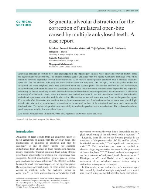
Segmental alveolar distraction for the correction of unilateral open ...
Segmental alveolar distraction for the correction of unilateral open ...
Segmental alveolar distraction for the correction of unilateral open ...

You also want an ePaper? Increase the reach of your titles
YUMPU automatically turns print PDFs into web optimized ePapers that Google loves.
CLINICAL<br />
SECTION<br />
<strong>Segmental</strong> <strong>alveolar</strong> <strong>distraction</strong> <strong>for</strong> <strong>the</strong><br />
<strong>correction</strong> <strong>of</strong> <strong>unilateral</strong> <strong>open</strong>-bite<br />
caused by multiple ankylosed teeth: A<br />
case report<br />
Takafumi Susami, Masako Matsuzaki, Yuji Ogihara, Miyuki Sakiyama,<br />
Tsuyoshi Takato<br />
University <strong>of</strong> Tokyo Hospital, Tokyo, Japan<br />
Yasushi Sugawara<br />
Jichi Medical School, Tochigi, Japan<br />
Shigeyuki Matsumoto<br />
Matsumoto Dental Clinic, Tokyo, Japan<br />
Ankylosed teeth fail to erupt to meet <strong>the</strong>ir counterparts in <strong>the</strong> opposite jaw. In cases where ankylosis occurs in multiple teeth,<br />
<strong>the</strong> occlusion shows an <strong>open</strong> bite. This article describes a case <strong>of</strong> <strong>unilateral</strong> <strong>open</strong> bite caused by multiple ankylosed teeth, where<br />
treatment involved segmental <strong>alveolar</strong> bone <strong>distraction</strong>. A 25-year-old female patient presented with a left-sided <strong>unilateral</strong><br />
<strong>open</strong> bite. On <strong>the</strong> left-hand side, only <strong>the</strong> lower incisors were not ankylosed. On <strong>the</strong> right, <strong>the</strong> maxillary first molar was<br />
ankylosed. All <strong>the</strong>se ankylosed teeth were positioned below <strong>the</strong> occlusal plane. Her mo<strong>the</strong>r and bro<strong>the</strong>r also had multiple<br />
ankylosed teeth, and a familial cause was considered. Orthodontic tooth movement was considered impossible and segmental<br />
osteotomy on <strong>the</strong> left maxillary <strong>alveolar</strong> bone and downward bone <strong>distraction</strong> were per<strong>for</strong>med as an alternative. A distractor<br />
consisting <strong>of</strong> orthodontic bands, wires and screws was devised and worn in <strong>the</strong> left mandibular dentition. Multi-bracket<br />
orthodontic appliances were also used <strong>for</strong> <strong>distraction</strong>. The amount <strong>of</strong> vertical movement was 7 mm at <strong>the</strong> premolar region.<br />
Five months after <strong>distraction</strong>, <strong>the</strong> multibracket appliance was removed, and fixed and removable retainers were placed. Eight<br />
months after <strong>distraction</strong>, prosthodontic restorations on <strong>the</strong> occlusal surfaces <strong>of</strong> <strong>the</strong> ankylosed teeth were made to obtain <strong>the</strong><br />
final occlusion. The <strong>unilateral</strong> <strong>open</strong> bite was successfully treated and a good occlusion was obtained. The occlusion has shown<br />
good long-term stability <strong>for</strong> more than 3 years.<br />
Key words: Alveolar bone <strong>distraction</strong>, <strong>open</strong> bite, segmental osteotomy, tooth ankylosis<br />
Received 18th July 2005; accepted 28th March 2006<br />
Introduction<br />
Ankylosis <strong>of</strong> teeth occurs from an anatomic fusion <strong>of</strong><br />
tooth cementum or dentin with <strong>the</strong> <strong>alveolar</strong> bone. The<br />
pathogenesis <strong>of</strong> ankylosis is unknown and may be<br />
secondary to one <strong>of</strong> many factors. For example,<br />
disturbances from changes in local metabolism, trauma,<br />
injury, chemical or <strong>the</strong>rmal irritation, local failure <strong>of</strong> bone<br />
growth and abnormal pressure from <strong>the</strong> tongue have been<br />
suggested. Several investigators believe genetic predisposition<br />
has a significant influence. 1 The affected teeth fail<br />
to erupt to meet <strong>the</strong>ir counterparts in <strong>the</strong> opposite jaw at<br />
<strong>the</strong> appropriate occlusal plane. In cases where ankylosis<br />
occurs in multiple teeth, <strong>the</strong> occlusion shows an<br />
<strong>open</strong> bite. 2,3 In <strong>the</strong>se circumstances, orthodontic tooth<br />
Journal <strong>of</strong> Orthodontics, Vol. 33, 2006, 153–159<br />
movement to correct <strong>the</strong> <strong>open</strong> bite is impossible and surgical<br />
repositioning <strong>of</strong> <strong>the</strong> ankylosed teeth is required. 2,4,5<br />
Recently, bone <strong>distraction</strong> has become popular in <strong>the</strong><br />
treatment <strong>of</strong> <strong>the</strong> patients with cleft lip and/or palate, 6,7<br />
hemifacial microsomia, 8–10 and syndromic craniosynostosis.<br />
11–13 This technique can also be applied to<br />
<strong>alveolar</strong> ridge augmentation to assist in <strong>the</strong> placement<br />
<strong>of</strong> dental implants. 11,14–18 Several authors have reported<br />
on <strong>the</strong> treatment <strong>of</strong> ankylosed teeth: Isaacson et al., 19<br />
Kinzinger et al. 20 and K<strong>of</strong>od et al. 21 reported <strong>the</strong><br />
movement <strong>of</strong> an ankylosed central incisor using a<br />
<strong>distraction</strong> osteogenesis technique.<br />
In this article, a case is described <strong>of</strong> a <strong>unilateral</strong> <strong>open</strong><br />
bite caused by familial multiple ankylosed teeth which<br />
was treated using segmental <strong>alveolar</strong> bone <strong>distraction</strong>.<br />
Address <strong>for</strong> correspondence: Takafumi Susami, Department <strong>of</strong><br />
Oral-Maxill<strong>of</strong>acial Surgery, Dentistry and Orthodontics, University<br />
<strong>of</strong> Tokyo Hospital, 7-3-1, Hongo, Bunkyo, Tokyo 113-8655, Japan.<br />
Email: susami-ora@h.u-tokyo.ac.jp<br />
# 2006 British Orthodontic Society DOI 10.1179/146531205225021572
154 T. Susami et al. Clinical Section JO September 2006<br />
(a) (b) (c)<br />
Figure 1 (a–c) Occlusion be<strong>for</strong>e treatment. Occlusal contact only on UR7 and UR5 to UR1<br />
Patient<br />
The patient was a 25-year-old female whose chief<br />
complaint was a <strong>unilateral</strong> <strong>open</strong> bite in her left occlusion<br />
(Figure 1). The only teeth in occlusion were <strong>the</strong> righthand<br />
labial segments and <strong>the</strong> right-hand buccal segments<br />
except <strong>for</strong> UR6. This tooth and all left-hand teeth<br />
were positioned below <strong>the</strong> occlusal plane. These infraoccluded<br />
teeth showed no mobility and had a dull sound<br />
on percussion. The panoramic radiograph showed an<br />
unclear delineation <strong>of</strong> <strong>the</strong> periodontal membrane <strong>of</strong><br />
<strong>the</strong>se teeth and root resorption was found in several<br />
teeth (Figure 2). Dental caries was found in <strong>the</strong> left<br />
mandibular molars. The lateral cephalogram showed a<br />
long face with an increased Frank<strong>for</strong>t-mandibular<br />
planes angle. The anterior-posterior maxillo-mandibular<br />
relationship was skeletal Class II (Figure 3 and<br />
Table 1). The postero-anterior cephalogram showed a<br />
symmetrical facial contour (Figure 3). These findings<br />
suggested a diagnosis <strong>of</strong> multiple ankylosed teeth and<br />
reduced <strong>alveolar</strong> bone height. The patient’s mo<strong>the</strong>r and<br />
bro<strong>the</strong>r also had multiple ankylosed teeth, and had been<br />
Figure 2 OPT taken be<strong>for</strong>e treatment. The periodontal<br />
membrane <strong>of</strong> <strong>the</strong> ankylosed teeth showed unclear delineation, and<br />
root resorption was found in several teeth<br />
treated by overlay pros<strong>the</strong>tics. A familial cause was<br />
considered. The patient had a history <strong>of</strong> orthodontic<br />
treatment to correct <strong>the</strong> <strong>open</strong> bite in ano<strong>the</strong>r hospital,<br />
but this had failed, resulting in <strong>the</strong> intrusion <strong>of</strong> adjacent<br />
normal teeth.<br />
Treatment<br />
Orthodontic tooth movement was considered impossible,<br />
and a segmental osteotomy and gradual inferior<br />
movement <strong>of</strong> <strong>the</strong> left maxillary dentition and supporting<br />
<strong>alveolar</strong> bone was planned. Multi-bracket appliances<br />
were placed prior to surgery and segmental rectangular<br />
wires (0.016 6 0.022-inch) were passively fitted to <strong>the</strong><br />
brackets on <strong>the</strong> left and right maxillary teeth. A<br />
rectangular wire (0.016 6 0.022-inch) was also placed<br />
in <strong>the</strong> lower dentition. A <strong>distraction</strong> device consisting<br />
<strong>of</strong> orthodontic bands, wires and screws (Figure 4) was<br />
placed on <strong>the</strong> left lower dentition.<br />
A segmental <strong>alveolar</strong> osteotomy was carried out under<br />
general anaes<strong>the</strong>sia to <strong>the</strong> upper left maxillary segment.<br />
Horizontal osteotomies on <strong>the</strong> maxillary buttress and<br />
<strong>the</strong> palatal <strong>alveolar</strong> bone were per<strong>for</strong>med in <strong>the</strong> maxilla,<br />
as well as a vertical midline osteotomy, which continued<br />
Table 1 Cephalometric values<br />
Patient Mean SD Japanese norm<br />
SNA (u) 91.4 3.5 82.3<br />
SNB (u) 83.8 3.5 78.9<br />
ANB (u) 7.6 1.8 3.4<br />
FMA (u) 39.2 5.2 28.8<br />
U1–SN (u) 104.4 5.6 104.5<br />
L1–MP(u) 88.1 5.8 96.3<br />
FMA: Frank<strong>for</strong>t-mandibular planes angle; U1–SN: Maxillary incisor-SN<br />
plane angle; L1–MP: Lower incisor-mandibular plane angle.<br />
SD: standard deviation.<br />
Japanese norm is from Iizuka and Ishikawa. 26
JO September 2006 Clinical Section <strong>Segmental</strong> <strong>alveolar</strong> <strong>distraction</strong> 155<br />
Figure 3 Cephalograms taken be<strong>for</strong>e treatment. The lateral cephalogram showed a long face with a steep Frank<strong>for</strong>t-mandibular planes<br />
angle and Skeletal II pattern. The postero-anterior cephalogram presented a symmetric facial contour<br />
to <strong>the</strong> cut end <strong>of</strong> <strong>the</strong> palatal osteotomy (Figure 5). After<br />
completing <strong>the</strong> osteotomies, <strong>the</strong> segment was mobilized.<br />
The left maxillary arch wire was ligated to <strong>the</strong> distractor<br />
on <strong>the</strong> lower left dentition. Intermaxillary fixation was<br />
<strong>the</strong>n placed on <strong>the</strong> right dentition to prevent mandibular<br />
movement and to stabilize <strong>the</strong> occlusion during <strong>distraction</strong><br />
(Figure 6a,b).<br />
The screws <strong>of</strong> <strong>the</strong> distractor were placed parallel to <strong>the</strong><br />
occlusal plane so that <strong>the</strong>y moved in a posterior<br />
direction as <strong>the</strong>y were turned. The wires ligated to <strong>the</strong><br />
Figure 4 Distraction device consisted <strong>of</strong> orthodontic bands, wires<br />
and screws<br />
screws were pulled in a posterior direction and changed<br />
<strong>the</strong> direction vertically at hooks soldered to <strong>the</strong><br />
orthodontic bands. Thus, <strong>the</strong> maxillary segment was<br />
pulled downwards. The screws were turned twice a day,<br />
creating 0.7 mm downward <strong>distraction</strong> per day <strong>of</strong> <strong>the</strong><br />
left maxillary dentition. The <strong>distraction</strong> continued <strong>for</strong><br />
17 days. The measurement <strong>of</strong> dental models taken<br />
be<strong>for</strong>e and after <strong>distraction</strong> revealed that overbite at<br />
<strong>the</strong> maxillary first premolar increased from 29.5 mm to<br />
22.5 mm. Thus, <strong>the</strong> total amount <strong>of</strong> downward movement<br />
was estimated as 7 mm at <strong>the</strong> premolar region.<br />
After a 1 month consolidation period, <strong>the</strong> intermaxillary<br />
fixation was released and <strong>the</strong> distractor was<br />
replaced with orthodontic brackets. Continuous rectangular<br />
wires were worn in both maxillary and mandibular<br />
dentitions and inter-maxillary elastics were applied to<br />
prevent relapse (Figure 7a,b).<br />
Five months after <strong>the</strong> completion <strong>of</strong> <strong>distraction</strong>, <strong>the</strong><br />
orthodontic brackets were removed and a fixed intercanine<br />
palatal wire was placed on <strong>the</strong> maxillary<br />
dentition (Figure 8a,b). Removable retainers were used<br />
simultaneously and dental caries in <strong>the</strong> left mandibular<br />
molars was treated. Eight months after completion <strong>of</strong><br />
<strong>the</strong> <strong>distraction</strong>, pros<strong>the</strong>tic attachments were fitted to <strong>the</strong><br />
occlusal surface <strong>of</strong> <strong>the</strong> teeth below <strong>the</strong> occlusal plane<br />
(left maxillary premolars and molars, left mandibular<br />
first molar and right maxillary first molar). At 3-year<br />
follow-up, <strong>the</strong> occlusion was stable and <strong>the</strong> patient was
156 T. Susami et al. Clinical Section JO September 2006<br />
Figure 5 <strong>Segmental</strong> maxillary osteotomy. Horizontal osteotomies on <strong>the</strong> maxillary buttress and <strong>the</strong> palatal <strong>alveolar</strong> bone were per<strong>for</strong>med<br />
in <strong>the</strong> maxilla, as well as a vertical midline osteotomy, which continued to <strong>the</strong> cut end <strong>of</strong> palatal osteotomy<br />
(a) (b)<br />
Figure 6 (a,b) Distraction procedure. The distractor was placed on <strong>the</strong> left mandibular dentition. Screws were parallel to <strong>the</strong> occlusal<br />
plane. Intermaxillary fixation was placed on <strong>the</strong> right dentition<br />
(a) (b)<br />
Figure 7 (a,b) At <strong>the</strong> end <strong>of</strong> consolidation period. The distractor was replaced with orthodontic brackets and continuous rectangular<br />
wires were worn in both maxillary and mandibular dentitions
JO September 2006 Clinical Section <strong>Segmental</strong> <strong>alveolar</strong> <strong>distraction</strong> 157<br />
satisfied with <strong>the</strong> treatment outcome (Figure 9a-c). Bone<br />
<strong>for</strong>mation in <strong>the</strong> area <strong>of</strong> <strong>distraction</strong> was satisfactory and<br />
<strong>the</strong> height <strong>of</strong> <strong>the</strong> interdental bone between central<br />
incisors was good (Figure 10).<br />
Discussion<br />
(a) (b)<br />
Figure 8 (a,b) Five months after <strong>the</strong> completion <strong>of</strong> <strong>distraction</strong> when <strong>the</strong> orthodontic brackets were removed<br />
(a) (b) (c)<br />
Figure 9 (a–c) Occlusion 3 years after treatment. Eight months after completion <strong>of</strong> <strong>the</strong> <strong>distraction</strong>, pros<strong>the</strong>tic attachments were fitted to<br />
<strong>the</strong> occlusal surface <strong>of</strong> <strong>the</strong> teeth<br />
Figure 10 Maxillary occlusal radiographs. (A) Immediately after <strong>distraction</strong>. Radiolucent area was observed at <strong>the</strong> midline <strong>of</strong> <strong>the</strong> palate.<br />
(B) Three years after treatment.<br />
Partial or complete failure <strong>of</strong> posterior teeth to erupt<br />
produces a lateral <strong>open</strong>-bite. Pr<strong>of</strong>fit and Vig suggested<br />
two possible causes <strong>of</strong> <strong>the</strong> failure <strong>of</strong> tooth eruption:<br />
N mechanical interference with eruption;<br />
N failure <strong>of</strong> <strong>the</strong> eruptive mechanism <strong>of</strong> <strong>the</strong> tooth. 2<br />
The first possible cause, mechanical interference with<br />
eruption, can cause ankylosis <strong>of</strong> <strong>the</strong> tooth to <strong>the</strong> <strong>alveolar</strong><br />
bone as a result <strong>of</strong> trauma or by obstacles in <strong>the</strong> path <strong>of</strong><br />
<strong>the</strong> erupting tooth, such as supernumerary teeth or nonresorbing<br />
deciduous tooth roots. Pressures from <strong>the</strong> s<strong>of</strong>t<br />
tissues interposed between <strong>the</strong> teeth (cheek, tongue and<br />
finger) can also prevent eruption.
158 T. Susami et al. Clinical Section JO September 2006<br />
The second possible cause <strong>of</strong> eruption failure is a<br />
disturbance <strong>of</strong> <strong>the</strong> eruption mechanism termed ‘primary<br />
failure <strong>of</strong> eruption’. 2,22 In this condition, non-ankylosed<br />
teeth fail to erupt fully or partially because <strong>of</strong><br />
malfunction <strong>of</strong> <strong>the</strong> eruption mechanism. Patients have<br />
no o<strong>the</strong>r recognizable disorder and no mechanical<br />
interferences with eruption seem to exist. Posterior teeth<br />
are involved more than anterior teeth and this may<br />
result in a posterior <strong>open</strong>-bite. Involvement may be<br />
<strong>unilateral</strong> or bilateral, but <strong>the</strong> condition is rarely<br />
symmetrical and frequently <strong>unilateral</strong>. The age at onset<br />
<strong>of</strong> <strong>the</strong> condition is not clear and some teeth may erupt<br />
normally at first and <strong>the</strong>n stop erupting. Involved teeth<br />
tend to become ankylosed, but failure <strong>of</strong> eruption is<br />
usually apparent be<strong>for</strong>e definite ankylosis occurs.<br />
Application <strong>of</strong> orthodontic <strong>for</strong>ce leads to ankylosis,<br />
ra<strong>the</strong>r than normal tooth movement.<br />
Pelias and Kinnebrew 3 reported <strong>the</strong> familial occurrence<br />
<strong>of</strong> <strong>the</strong> multiple ankylosed teeth. The proband<br />
(defined as <strong>the</strong> first individual chosen from a family who<br />
has come to <strong>the</strong> notice <strong>of</strong> a researcher, and through<br />
whom investigation <strong>of</strong> a pedigree begins, due to <strong>the</strong><br />
presence <strong>of</strong> some trait whose inheritance is to be studied)<br />
<strong>of</strong> <strong>the</strong>ir report was a 32-year-old female who had a<br />
bilateral lateral <strong>open</strong>-bite caused by multiple ankylosed<br />
teeth. Systematic evaluation revealed simultaneously<br />
occurring mild bilateral clinodactyly <strong>of</strong> <strong>the</strong> fifth fingers.<br />
The family history was positive and 12 affected persons<br />
were found in four generations. Dental abnormality and<br />
mild mandibular prognathism are found in some<br />
affected patients. Pelias and Kinnebrew 3 considered<br />
<strong>the</strong>se abnormalities were transmitted in an autosomal<br />
dominant manner from pedigree analysis.<br />
The occlusion <strong>of</strong> our patient was very similar to that in<br />
Pelias and Kinnebrew’s 3 article. The lateral <strong>open</strong> bite<br />
was caused by multiple ankylosed teeth and maxillary<br />
posterior teeth showed a reverse curve <strong>of</strong> Spee. Open<br />
bite was, however, found <strong>unilateral</strong>ly in our case. Our<br />
patient had no history <strong>of</strong> facial trauma and no apparent<br />
mechanical interference with eruption was found. The<br />
mo<strong>the</strong>r and bro<strong>the</strong>r <strong>of</strong> our patient also had multiple<br />
ankylosed teeth, and had been treated by overlay<br />
pros<strong>the</strong>tics. Thus, we considered <strong>the</strong> <strong>open</strong> bite <strong>of</strong> our<br />
patient was caused by hereditary primary failure <strong>of</strong><br />
eruption. The ankylosis might have occurred spontaneously<br />
or as a result <strong>of</strong> previous orthodontic treatment<br />
in ano<strong>the</strong>r hospital.<br />
In <strong>the</strong> treatment <strong>of</strong> ankylosis, a dento-<strong>alveolar</strong><br />
osteotomy has <strong>of</strong>ten been per<strong>for</strong>med to correct <strong>the</strong><br />
inferiorly positioned teeth. 4,5 The alternative is pros<strong>the</strong>tic<br />
build-up. 23,24 When ankylosis occurs in multiple<br />
teeth, a segmental <strong>alveolar</strong> bone osteotomy is essential<br />
and a bone graft can also be interposed between <strong>the</strong><br />
segment and <strong>the</strong> basal <strong>alveolar</strong> bone. 3 Prosthodontic<br />
restorations are sometimes required to establish a<br />
satisfactory final occlusion. 2 Recently, bone <strong>distraction</strong><br />
has became popular in <strong>the</strong> treatment <strong>of</strong> crani<strong>of</strong>acial<br />
anomalies 6–13 and <strong>alveolar</strong> ridge augmentation. 11,14–18<br />
For <strong>the</strong> treatment <strong>of</strong> an ankylosed central incisor, a<br />
single-tooth osteotomy and subsequent vertical <strong>distraction</strong><br />
to avoid bone grafting and to stretch s<strong>of</strong>t tissue has<br />
recently been reported. 19–21<br />
In this case, orthodontic tooth movement was<br />
considered to be impossible from <strong>the</strong> clinical examination<br />
and treatment history, and a segmental <strong>alveolar</strong><br />
bone osteotomy was undertaken instead. As a segmental<br />
osteotomy in <strong>the</strong> mandibular <strong>alveolar</strong> bone carries a<br />
high risk to <strong>the</strong> blood supply and injury to <strong>the</strong> inferior<strong>alveolar</strong><br />
nerve, 25 we per<strong>for</strong>med a segmental osteotomy<br />
only in <strong>the</strong> left maxillary bone. The reverse curve <strong>of</strong> Spee<br />
in <strong>the</strong> left maxillary dentition and <strong>the</strong> acceptable curve<br />
in <strong>the</strong> left mandibular dentition supported this treatment<br />
plan. However, mandibular segmental osteotomy might<br />
have produced a more satisfactory outcome to reduce<br />
<strong>the</strong> need <strong>for</strong> fixed onlay prosthodontics.<br />
A gradual <strong>distraction</strong> technique was selected to avoid<br />
bone grafting and to stretch <strong>the</strong> s<strong>of</strong>t tissue, which<br />
o<strong>the</strong>rwise is <strong>of</strong>ten a limiting factor. As ankylosed teeth<br />
act as perfect orthodontic anchorage, <strong>the</strong> distractor was<br />
placed in <strong>the</strong> ankylosed left lower dentition and <strong>the</strong><br />
upper dentition was distracted using screws and wires.<br />
There are two alternatives to consider <strong>for</strong> segmental<br />
<strong>distraction</strong>: <strong>the</strong> use <strong>of</strong> vertical elastics or <strong>the</strong> fixed screw<br />
type distractor across <strong>the</strong> osteotomy cuts. With <strong>the</strong> use<br />
<strong>of</strong> vertical elastics however, control <strong>of</strong> <strong>the</strong> <strong>distraction</strong><br />
rate is difficult. The fixed screw type distractor may<br />
avoid inter-maxillary fixation. However, it needs two<br />
surgical interventions—at placement and at removal—<br />
fur<strong>the</strong>rmore, <strong>the</strong> <strong>distraction</strong> direction is not flexible. For<br />
<strong>the</strong>se reasons, we chose screws and inter-maxillary wires<br />
to avoid two surgical episodes, to keep <strong>distraction</strong> rate<br />
constant and to control <strong>the</strong> <strong>distraction</strong> direction.<br />
The outcome <strong>of</strong> <strong>the</strong> <strong>distraction</strong> treatment was<br />
excellent and <strong>the</strong> distracted segment was stable more<br />
than 3 years post-surgery. The possible complications <strong>of</strong><br />
<strong>the</strong> segmental osteotomy are significant periodontal<br />
defects at <strong>the</strong> vertical osteotomy sites and <strong>the</strong> loss <strong>of</strong><br />
vitality <strong>of</strong> teeth adjacent to osteotomy cut. Loss <strong>of</strong> blood<br />
supply to both teeth and <strong>alveolar</strong> bone in <strong>the</strong> mobilized<br />
segments is rare but teeth and segments <strong>of</strong> bone may<br />
be lost in this circumstance. 25 In our case, a vertical<br />
osteotomy was made at <strong>the</strong> maxillary midline between<br />
<strong>the</strong> central incisors. The interdental bone at this site at 3<br />
years after osteotomy showed good height and no<br />
complications were found in <strong>the</strong> teeth adjacent to <strong>the</strong><br />
osteotomy cut. Careful surgical procedure seemed to be
JO September 2006 Clinical Section <strong>Segmental</strong> <strong>alveolar</strong> <strong>distraction</strong> 159<br />
able to avoid <strong>the</strong> complications in <strong>the</strong> adjacent teeth.<br />
Gradual <strong>distraction</strong> <strong>of</strong> <strong>the</strong> segment might be advantageous<br />
to <strong>the</strong> blood supply <strong>of</strong> <strong>the</strong> mobilized segment.<br />
Conclusion<br />
A case with <strong>unilateral</strong> <strong>open</strong> bite caused by familial<br />
multiple ankylosed teeth was successfully treated using<br />
segmental <strong>alveolar</strong> bone <strong>distraction</strong>. A good occlusion<br />
was obtained with final prosthodontic restorations on<br />
<strong>the</strong> occlusal surfaces <strong>of</strong> <strong>the</strong> ankylosed teeth. The<br />
treatment result was stable after more than 3 years<br />
without any complications.<br />
References<br />
1. Neville BW, Damm DD, Allen CM, Bouquot JE. Oral and<br />
Maxill<strong>of</strong>acial Pathology, 2nd edn. Philadelphia: Saunders,<br />
2002; 67–68.<br />
2. Pr<strong>of</strong>fit WR, Vig KWL. Primary failure <strong>of</strong> eruption: A<br />
possible cause <strong>of</strong> posterior <strong>open</strong>-bite. Am J Orthod 1981; 80:<br />
173–90.<br />
3. Pelias MZ, Kinnebrew MC. Autosomal dominant transmission<br />
<strong>of</strong> ankylosed teeth, abnormalities <strong>of</strong> <strong>the</strong> jaws, and<br />
clinodactyly. A four-generation study. Clin Genet 1985; 27:<br />
496–500.<br />
4. Epker BN, Paulus PJ. Surgical-orthodontic <strong>correction</strong> <strong>of</strong><br />
adult malocclusions: single-tooth dento-osseous osteotomies.<br />
Am J Orthod 1978; 74: 551–63.<br />
5. Medeiros PJ, Bezerra AR. Treatment <strong>of</strong> an ankylosed<br />
central incisor by single-tooth dento-osseus osteotomy. Am J<br />
Orthod Dentfacial Orthop 1997; 112: 496–501.<br />
6. Polley JW, Figueroa AA. Rigid external <strong>distraction</strong>: Its<br />
application in cleft maxillary de<strong>for</strong>mities. Plast Reconstr<br />
Surg 1998; 102: 1360–72.<br />
7. Liou EJW, Chen PKT, Huang CS, Chen YR. Interdental<br />
<strong>distraction</strong> osteogenesis and rapid orthodontic tooth<br />
movement: A novel approach to approximate a wide<br />
<strong>alveolar</strong> cleft or bony defect. Plast Reconstr Surg 2000;<br />
105: 1262–72.<br />
8. McCarthy JG, Schreiber J, Karp N, Thorne CH,<br />
Grayson BH. Leng<strong>the</strong>ning <strong>the</strong> human mandible by gradual<br />
<strong>distraction</strong>. Plast Reconstr Surg 1992; 89: 1–8.<br />
9. Molina F, Monasterio FO. Mandibular elongation and<br />
remodeling by <strong>distraction</strong>: A farewell to major osteotomies.<br />
Plast Reconstr Surg 1995; 96: 825–40.<br />
10. Huisinga-Fischer CE, Vaandrager JM, Prahl-Anderson B.<br />
Longitudinal results <strong>of</strong> mandibular <strong>distraction</strong> osteogenesis in<br />
hemifacial microsomia. J Crani<strong>of</strong>acial Surg 2003; 14: 924–33.<br />
11. Chin M, Toth BA. Distraction osteogenesis in maxill<strong>of</strong>acial<br />
surgery using internal devices: Review <strong>of</strong> five cases. J Oral<br />
Maxill<strong>of</strong>ac Surg 1996; 54: 45–53.<br />
12. Cohen SR. Crani<strong>of</strong>acial <strong>distraction</strong> with a modular internal<br />
<strong>distraction</strong> system: Evolution <strong>of</strong> design and surgical<br />
techniques. Plast Reconstr Surg 1999; 103: 1592–607.<br />
13. Gosain AK, Santoro TD, Havlik RJ, Cohen SR,<br />
Holmes RE. Midface <strong>distraction</strong> following Le Fort III<br />
and monobloc osteotomies: Problems and solutions. Plast<br />
Reconstr Surg 2002; 109: 1797–808.<br />
14. Gaggl A, Schultes G, Kärcher H. Distraction implants: a<br />
new operative technique <strong>for</strong> <strong>alveolar</strong> ridge augmentation.<br />
J Craniomaxill<strong>of</strong>ac Surg 1999; 27: 214–21.<br />
15. Rachmiel A, Srouji S, Peled M. Alveolar ridge augmentation<br />
by <strong>distraction</strong> osteogenesis. Int J Oral Maxill<strong>of</strong>ac Surg<br />
2001; 30: 510–17.<br />
16. Klesper B, Lazar F, Sießegger M, Hidding J, Zöller JE.<br />
Vertical <strong>distraction</strong> osteogenesis <strong>of</strong> fibula transplants<br />
<strong>for</strong> mandibular reconstruction — a preliminary study.<br />
J Craniomaxill<strong>of</strong>ac Surg 2002; 30: 280–85.<br />
17. Robiony M, Toro C, Stucki-McCormick SU, Zerman N,<br />
Costa F, Politi M. The ‘FAD’ (Floating Alveolar Device):<br />
A bidirectional <strong>distraction</strong> system <strong>for</strong> <strong>distraction</strong> osteogenesis<br />
<strong>of</strong> <strong>the</strong> <strong>alveolar</strong> process. J Oral Maxill<strong>of</strong>ac Surg 2004; 62,<br />
Suppl 2: 136–42.<br />
18. Hwang SJ, Jung JG, Jung JU, Kyung SH. Vertical <strong>alveolar</strong><br />
bone <strong>distraction</strong> at molar region using lag screw principle.<br />
J Oral Maxill<strong>of</strong>ac Surg 2004; 62: 787–94.<br />
19. Isaacson RJ, Strauss RA, Bridges-Poquis A, Peluso AR,<br />
Lindauer SJ. Moving an ankylosed central incisor using<br />
orthodontics, surgery and <strong>distraction</strong> osteogenesis. Angle<br />
Orthod 2001; 71: 411–18.<br />
20. Kinzinger GSM, Jänicke S, Riediger D, Diedrich PR.<br />
Orthodontic fine adjustment after vertical callus <strong>distraction</strong><br />
<strong>of</strong> an ankylosed incisor using <strong>the</strong> floating bone concept. Am<br />
J Orthod Dent<strong>of</strong>acial Orthop 2003; 124: 582–90.<br />
21. K<strong>of</strong>od T, Würtz V, Melsen B. Treatment <strong>of</strong> an ankylosed<br />
central incisor by single tooth dento-osseus osteotomy and a<br />
simple <strong>distraction</strong> device. Am J Orthod Dent<strong>of</strong>acial Orthop<br />
2005; 127: 72–80.<br />
22. O’Connell AC, Torske KR. Primary failure <strong>of</strong> tooth<br />
eruption. A unique case. Oral Surg Oral Med Oral Pathol<br />
Oral Radiol Endod 1999; 87: 714–20.<br />
23. Raghoebar GM, Boering G, Booy K, Vissink A. Treatment<br />
<strong>of</strong> retained permanent molar. J Oral Maxill<strong>of</strong>ac Surg 1990;<br />
48: 1033–38.<br />
24. Mullally BH, Blakely D, Burden DJ. Ankylosis: an<br />
orthodontic problem with a restorative solution. Br Dent<br />
J 1995; 179: 426–30.<br />
25. White RP, Terry BC. <strong>Segmental</strong> jaw surgery. In: Pr<strong>of</strong>fit<br />
WR, White RP (Eds) Surgical-orthodontic Treatment. St<br />
Louis: Mosby, 1991; 283–319.<br />
26. Iizuka T, Ishikawa F. Normal standards <strong>for</strong> various<br />
cephalometric analysis in Japanese adults. J Jpn Orthod<br />
Soc 1957; 16: 4–12.