

ATOMIC AND NUCLEAR PHYSICS - Hiperlab
ATOMIC AND NUCLEAR PHYSICS - Hiperlab
ATOMIC AND NUCLEAR PHYSICS - Hiperlab

Create successful ePaper yourself
Turn your PDF publications into a flip-book with our unique Google optimized e-Paper software.
<strong>ATOMIC</strong> <strong>AND</strong> <strong>NUCLEAR</strong> <strong>PHYSICS</strong><br />
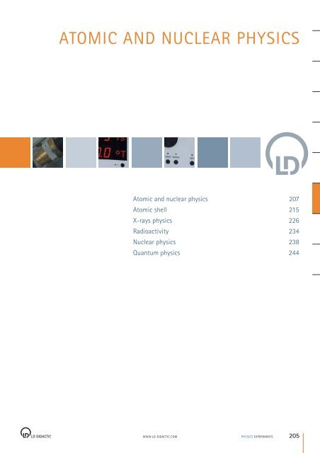
Atomic and nuclear physics 207<br />
Atomic shell 215<br />
X-rays physics 226<br />
Radioactivity 234<br />
Nuclear physics 238<br />
Quantum physics 244<br />
WWW.LD-DIDACTIC.COM <strong>PHYSICS</strong> EXPERIMENTS<br />
205
P6 <strong>ATOMIC</strong> <strong>AND</strong> <strong>NUCLEAR</strong> <strong>PHYSICS</strong><br />
P6.1 Introductory experiments 207<br />
P6.1.1 Oil-spot experiment 207<br />
P6.1.2 Millikan experiment 208<br />
P6.1.3 Specific electron charge 209<br />
P6.1.4 Planck’s constant 210-212<br />
P6.1.5 Dual nature of wave and particle 213<br />
P6.1.6 Paul trap 214<br />
P6.2 Atomic shell 215<br />
P6.2.1 Balmer series of hydrogen 215-216<br />
P6.2.2 Emission and absorption spectra 217-219<br />
P6.2.3 Inelastic collisions of electrons 220<br />
P6.2.4 Franck-Hertz experiment 221-222<br />
P6.2.6 Electron spin resonance 223<br />
P6.2.7 Normal Zeeman effect 224<br />
P6.2.8 Optical pumping<br />
(anomalous Zeeman effect) 225<br />
P6.3 X-rays physics 226<br />
P6.3.1 Detection of X-rays 226-227<br />
P6.3.2 Attenuation of X-rays 228<br />
P6.3.3 Physics of the atomic shell 229<br />
P6.3.5 X-ray energy spectroscopy 230<br />
P6.3.6 Structure of X-ray spectrums 231<br />
P6.3.7 Compton effect at X-rays 232<br />
P6.3.8 X-ray tomography 233<br />
P6.4 Radioactivity 234<br />
P6.4.1 Detecting radioactivity 234<br />
P6.4.2 Poisson distribution 235<br />
P6.4.3 Radioactive decay and half-life 236<br />
P6.4.4 Passage of α, β and γ radiation 237<br />
206 <strong>PHYSICS</strong> EXPERIMENTS<br />
WWW.LD-DIDACTIC.COM<br />
P6.5 Nuclear physics 238<br />
P6.5.1 Demonstrating paths of particles 238<br />
P6.5.2 Rutherford scattering 239<br />
P6.5.3 Nuclear magnetic resonance 240<br />
P6.5.4 α-spectroscopy 241<br />
P6.5.5 γ-spectroscopy 242<br />
P6.5.6 Compton effect 243<br />
P6.6 Quantum physics 244<br />
P6.6.1 Quantum optics 244
<strong>ATOMIC</strong> <strong>AND</strong> <strong>NUCLEAR</strong> <strong>PHYSICS</strong> INTRODUCTORY EXPERIMENTS<br />
Estimating the size of oil molecules (P6.1.1.1)<br />
Cat. No. Description<br />
664 179 Crystallization dish, 230 mm Ø 1<br />
665 844 Burette, amber glass, 10 ml 1<br />
664 110 Beaker, 50 ml, tall form 1<br />
665 751 Graduated cylinder with plastic base, 10 ml 1<br />
665 754 Graduated cylinder with plastic base, 100 ml 1<br />
300 02 Stand base, V-shape, 20 cm 1<br />
300 43 Stand rod 75 cm, 12 mm Ø 1<br />
301 01 Leybold multiclamp 1<br />
666 555 Universal clamp, 0 ... 80 mm 1<br />
675 3410 Water, pure, 5 l 1<br />
672 1240 Glycerinetrioleate, 100 ml 1<br />
674 2220 Benzine, 40 ... 70 °C, 1 l 1<br />
670 6920 Lycopodium spores, 25 g 1<br />
Determining the area A of the oil spot<br />
P6.1.1.1<br />
WWW.LD-DIDACTIC.COM <strong>PHYSICS</strong> EXPERIMENTS<br />
P6.1.1<br />
One important issue in atomic physics is the size of the atom. An<br />
investigation of the size of molecules makes it easier to come to a<br />
usable order of magnitude by experimental means. This is estimated<br />
from the size of an oil spot on the surface of water using simple<br />
means.<br />
In the experiment P6.1.1.1, a drop of glycerin nitrioleate as added to a<br />
grease-free water surface dusted with Lycopodium spores. Assuming<br />
that the resulting oil spot has a thickness of one molecule, we can<br />
calculate the size d of the molecule according to<br />
d V<br />
<br />
A<br />
Oil-spot experiment<br />
P6.1.1.1<br />
Estimating the size of oil molecules<br />
from the volume V of the oil droplet and the area A of the oil spot.<br />
The volume of the oil spot is determined from the number of drops<br />
needed to fill a volume of 1 cm 3 . The area of the oil spot is determined<br />
using graph paper.<br />
207
INTRODUCTORY EXPERIMENTS<br />
P6.1.2<br />
Determining the electric unit charge after Millikan and verifying the charge quantification<br />
- Measuring the suspension voltage and the falling speed (P6.1.2.1)<br />
Cat. No. Description<br />
559 411 Millikan apparatus 1 1 1 1<br />
559 421 Millikan supply unit 1 1 1 1<br />
313 033 Electronic stopclock 1 2<br />
501 46 Cable, 100 cm, red/blue, pair 3 4 3 3<br />
524 013 Sensor-CASSY 2 1 1<br />
524 220 CASSY Lab 2 1 1<br />
524 034 Timer box 1 1<br />
501 461 Cable, 100 cm, black, pair 1 1<br />
500 421 Connecting lead, 50 cm, red 1<br />
additionally required:<br />
PC with Windows XP/Vista/7<br />
The histogram reveals the qantum nature of the change<br />
208 <strong>PHYSICS</strong> EXPERIMENTS<br />
WWW.LD-DIDACTIC.COM<br />
P6.1.2.1<br />
P6.1.2.2<br />
P6.1.2.3<br />
P6.1.2.4<br />
1 1<br />
<strong>ATOMIC</strong> <strong>AND</strong> <strong>NUCLEAR</strong> <strong>PHYSICS</strong><br />
With his famous oil-drop method, R. A. Millikan succeeded in demonstrating<br />
the quantum nature of minute amounts of electricity in<br />
1910. He caused charged oil droplets to be suspended in the vertical<br />
electric field of a plate capacitor and, on the basis of the radiusr and<br />
the electric field E, determined the charge q of a suspended droplet:<br />
4<br />
3 g<br />
q r <br />
3 E<br />
: density of oil<br />
g:<br />
gravitanional acceleration<br />
He discovered that q only occurs as a whole multiple of an electron<br />
charge e. His experiments are produced here in two variations.<br />
In the variation P6.1.2.1 and P6.1.2.3, the electric field<br />
E U<br />
<br />
d<br />
d:<br />
plate spacing<br />
is calculated from the voltage U at the plate capacitor at which the observed<br />
oil droplet just begins to hover. The constant falling velocityv 1<br />
of the droplet when the electric field is switched off is subsequently<br />
measured to determine the radius. From the equilibrium between the<br />
force of gravity and Stokes friction, we derive the equation<br />
4<br />
3<br />
r g 6<br />
r v<br />
3<br />
:<br />
viscosity<br />
Millikan experiment<br />
P6.1.2.1<br />
Determining the electric unit charge after<br />
Millikan and verifying the charge quantification<br />
- Measuring the suspension voltage<br />
and the falling speed<br />
P6.1.2.2<br />
Determining the electric unit charge after<br />
Millikan and verifying the charge quantification<br />
- Measuring the rising and falling<br />
speed<br />
P6.1.2.3<br />
Determining the electric unit charge after<br />
Millikan and verifying the charge quantification<br />
- Measuring the suspension voltage<br />
and the falling speed with CASSY<br />
P6.1.2.4<br />
Determining the electric unit charge after<br />
Millikan and verifying the charge quantification<br />
- Measuring the rising and falling<br />
speed with CASSY<br />
1<br />
In the variant P6.1.2.2 and P6.1.2.4, the oil droplets are observed<br />
which are not precisely suspended, but which rise with a low velocityv<br />
2. The following applies for these droplets:<br />
q U 4<br />
3<br />
r g 6<br />
r v<br />
d 3<br />
Additionally, the falling speed v 1 is measured, as in the variant<br />
P6.1.2.1 and P6.1.2.3. The measuring accuracy for the charge q can<br />
be increased by causing the oil droplet under study to rise and fall<br />
over a given distance several times in succession and measuring the<br />
total rise and fall times.<br />
2
<strong>ATOMIC</strong> <strong>AND</strong> <strong>NUCLEAR</strong> <strong>PHYSICS</strong> INTRODUCTORY EXPERIMENTS<br />
Determining the specific charge of the electron (P6.1.3.1)<br />
Cat. No. Description<br />
555 571 Fine beam tube 1<br />
555 581 Helmholtz coils with stand 1<br />
531 120 Multimeter LDanalog 20 2<br />
521 65 Tube power supply 0...500 V 1<br />
521 545 DC power supply, 0 ... 16 V, 0 ... 5 A 1<br />
311 77 Steel tape measure, l = 2 m/78“ 1<br />
500 614 Safety connection lead 25 cm, black 3<br />
500 624 Safety connection lead, 50 cm, black 3<br />
500 644 Safety connection lead, 100 cm, black 7<br />
531 835 Universal Measuring Instrument Physics 1*<br />
524 0382 Axial B Sensor S, ±1000 mT 1*<br />
501 11 Extension cable, 15-pole 1*<br />
*additionally recommended<br />
Circular electron path in fine beam tube<br />
P6.1.3.1<br />
WWW.LD-DIDACTIC.COM <strong>PHYSICS</strong> EXPERIMENTS<br />
P6.1.3<br />
The mass m e of the electron is extremely difficult to determine in an<br />
experiment. It is much easier to determine the specific charge of the<br />
electron<br />
e<br />
m e<br />
from which we can calculate the mass m e for a given electron<br />
chargee.<br />
In the experiment P6.1.3.1, a tightly bundled electron beam is diverted<br />
into a closed circular path using a homogeneous magnetic field in<br />
order to determine the specific electron charge. The magnetic field<br />
B which diverts the electrons into the path with the given radius r is<br />
determined as a function of the acceleration voltage U. The Lorentz<br />
force caused by the magnetic field acts as a centripetal force. It depends<br />
on the velocity of the electrons, which in turn is determined<br />
by the acceleration voltage. The specific electron charge can thus be<br />
determined from the measurement quantities U, B and r according<br />
to the formula<br />
e U<br />
<br />
m B r<br />
e<br />
2 2 2<br />
Specific electron charge<br />
P6.1.3.1<br />
Determining the specific charge of the<br />
electron<br />
209
INTRODUCTORY EXPERIMENTS<br />
P6.1.4<br />
Determining Planck’s constant - Measuring in a compact assembly (P6.1.4.1)<br />
Cat. No. Description<br />
558 77 Photocell for determining h 1 1<br />
558 79 Compact arrangement for determining Planck‘s constant 1 1<br />
451 15 High pressure mercury lamp 1 1<br />
451 195 Power supply unit for mercury lamp 1 1<br />
532 14 Electrometer amplifier 1<br />
562 791 Plug-in power supply, 12 V AC 1<br />
578 22 Capacitor 100 pF, STE 2/19 1<br />
579 10 Key switch (NO), singel-pole, STE 2/19 1<br />
590 011 Clamping plug 2<br />
531 120 Multimeter LDanalog 20 1 2<br />
575 24 Screened cable BNC/4 mm plug 1<br />
502 04 Distribution box 1<br />
500 414 Connecting lead, 25 cm, black 1<br />
501 45 Cable, 50 cm, red/blue, pair 1 2<br />
501 461 Cable, 100 cm, black, pair 1 1<br />
500 440 Connecting lead, 100 cm, yellow/green 1<br />
532 00 I Measuring amplifier D 1<br />
576 74 Plug-in board DIN A4 1<br />
576 86 Monocell holder 3<br />
685 48ET5 Mono cells 1.5 V (IEC R20), set of 5 1<br />
577 93 Potentiometer, 10-turn, 1 kOhm, STE 4/50 1<br />
579 13 Toggle switch, single-pole, STE 2/19 1<br />
501 48 Bridging plugs, set of 10 1<br />
501 02 BNC cable, 1 m 1<br />
500 444 Connecting lead, 100 cm, black 1<br />
210 <strong>PHYSICS</strong> EXPERIMENTS<br />
WWW.LD-DIDACTIC.COM<br />
P6.1.4.1<br />
P6.1.4.5<br />
<strong>ATOMIC</strong> <strong>AND</strong> <strong>NUCLEAR</strong> <strong>PHYSICS</strong><br />
Planck’s constant<br />
P6.1.4.1<br />
Determining Planck’s constant - Measuring<br />
in a compact assembly<br />
P6.1.4.5<br />
Determining Planck’s constant - Recording<br />
the current-voltage characteristics,<br />
measuring in a compact assembly<br />
When light with the frequency ν falls on the cathode of a photocell,<br />
electrons are released. Some of the electrons reach the anode where<br />
they generate a current in the external circuit, which is compensated<br />
to zero by applying a voltage with opposite sign U = –U 0 . The applicable<br />
relationship<br />
e U0 h W<br />
W:<br />
electronic work function<br />
was first used by R. A. Millikan to determine Planck’s constant h.<br />
In the experiment P6.1.4.1, a compact arrangement is used to determine<br />
h, in which the light from a high-pressure mercury vapour<br />
lamp is spectrally dispersed in a direct-vision prism. The light of precisely<br />
one spectral line at a time falls on the cathode. A capacitor<br />
is connected between the cathode and the anode of the photocell<br />
which is charged by the anode current, thus generating the opposing<br />
voltageU. As soon as the opposing voltage reaches the value<br />
U 0, the anode current is zero and the charging of the capacitor is<br />
finished. U 0 is measured without applying a current by means of an<br />
electrometer amplifier.<br />
In the experiment P6.1.4.5 light from a mercury gas discharge lamp<br />
is deflected by a direct view prism, one wavelength selected and<br />
focused onto the photocathode. The countervoltage of the anode is<br />
varied and the resulting current is measured with high sensitivity. The<br />
variation of the characteristic curves under irradiation with different<br />
wavelengths leads to the determination of Plancks constant h.
<strong>ATOMIC</strong> <strong>AND</strong> <strong>NUCLEAR</strong> <strong>PHYSICS</strong> INTRODUCTORY EXPERIMENTS<br />
Determining Planck’s constant - Separation of wavelengths with a straight-view prism on the optical bench<br />
(P6.1.4.2)<br />
Cat. No. Description<br />
558 77 Photocell for determining h 1<br />
558 791 Holder for photocell 1<br />
460 32 Optical bench, standard cross section, 1 m 1<br />
460 335 Optical bench, standard cross section, 0.5 m 1<br />
460 341 Swivel joint with circular scale 1<br />
460 373 Optics rider 60/50 2<br />
460 374 Optics rider 90/50 4<br />
460 382 Tilting rider 90/50 1<br />
460 02 Lens in frame f = +50 mm 1<br />
460 08 Lens in frame f = +150 mm 1<br />
461 62 Slit diaphragms, set of 2 1<br />
460 22 Holder with spring clips 1<br />
460 14 Adjustable slit 1<br />
460 13 Projection objective 1<br />
466 05 Direct vision prism 1<br />
466 04 Holder for direct vision prism 1<br />
451 15 High pressure mercury lamp 1<br />
451 195 Power supply unit for mercury lamp 1<br />
532 00 I Measuring amplifier D 1<br />
531 120 Multimeter LDanalog 20 2<br />
576 74 Plug-in board DIN A4 1<br />
576 86 Monocell holder 3<br />
685 48ET5 Mono cells 1.5 V (IEC R20), set of 5 1<br />
577 93 Potentiometer, 10-turn, 1 kOhm, STE 4/50 1<br />
P6.1.4.2<br />
WWW.LD-DIDACTIC.COM <strong>PHYSICS</strong> EXPERIMENTS<br />
P6.1.4<br />
The experiment P6.1.4.2 uses an open arrangement on the optical<br />
bench. Here as well, the wavelengths of the light are dispersed using<br />
a direct-vision prism. The opposing voltage U is tapped from a DC<br />
voltage source via a voltage divider, and varied until the anode current<br />
is compensated precisely to zero. The I-measuring amplifier D is<br />
used for conducting sensitive measurements of the anode current.<br />
Cat. No. Description<br />
Planck’s constant<br />
P6.1.4.2<br />
Determining Planck’s constant -<br />
Separation of wavelengths with a straightview<br />
prism on the optical bench<br />
579 13 Toggle switch, single-pole, STE 2/19 1<br />
501 48 Bridging plugs, set of 10 1<br />
501 45 Cable, 50 cm, red/blue, pair 2<br />
500 444 Connecting lead, 100 cm, black 1<br />
P6.1.4.2<br />
211
INTRODUCTORY EXPERIMENTS<br />
P6.1.4<br />
Determining Planck’s constant - Selection of wavelengths using interference filters on the optical bench (P6.1.4.3_a)<br />
Cat. No. Description<br />
558 77 Photocell for determining h 1 1<br />
558 791 Holder for photocell 1 1<br />
460 335 Optical bench, standard cross section, 0.5 m 1 1<br />
460 374 Optics rider 90/50 2 2<br />
460 375 Optics rider 120/50 3 3<br />
558 792 Filter wheel with iris diaphragm 1 1<br />
468 401 Interference filter, 578 nm 1 1<br />
468 402 Interference filter, 546 nm 1 1<br />
468 403 Interference filter, 436 nm 1 1<br />
468 404 Interference filter, 405 nm 1 1<br />
460 03 Lens in frame f = +100 mm 1 1<br />
460 26 Iris diaphragm 1 1<br />
451 15 High pressure mercury lamp 1 1<br />
451 195 Power supply unit for mercury lamp 1 1<br />
532 14 Electrometer amplifier 1<br />
562 791 Plug-in power supply, 12 V AC 1<br />
578 22 Capacitor 100 pF, STE 2/19 1<br />
579 10 Key switch (NO), singel-pole, STE 2/19 1<br />
590 011 Clamping plug 2<br />
531 120 Multimeter LDanalog 20 1 2<br />
501 10 BNC straight 1<br />
501 09 Adapter BNC/4 mm, single pole 1<br />
340 89ET5 Coupling plug, 4 mm, set of 5 1<br />
502 04 Distribution box 1<br />
501 45 Cable, 50 cm, red/blue, pair 1 2<br />
500 440 Connecting lead, 100 cm, yellow/green 2<br />
468 406 Interference filter, 365 nm 1<br />
532 00 I Measuring amplifier D 1<br />
212 <strong>PHYSICS</strong> EXPERIMENTS<br />
WWW.LD-DIDACTIC.COM<br />
P6.1.4.3 (a)<br />
P6.1.4.4 (a)<br />
<strong>ATOMIC</strong> <strong>AND</strong> <strong>NUCLEAR</strong> <strong>PHYSICS</strong><br />
Planck’s constant<br />
P6.1.4.3<br />
Determining Planck’s constant - Selection<br />
of wavelengths using interference filters on<br />
the optical bench<br />
P6.1.4.4<br />
Determining Planck’s constant - Recording<br />
the current-voltage characteristics,<br />
selection of wavelengths using interference<br />
filters on the optical bench<br />
In determining Planck’s constant using the photoelectric effect, it<br />
must be ensured that only the light of a single spectral line of the<br />
high-pressure mercury vapour lamp falls on the cathode of the photocell<br />
at any one time. As an alternative to a prism, it is also possible<br />
to use narrow-band interference filters to select the wavelength. This<br />
simplifies the subsequent optical arrangement, and it is no longer<br />
necessary to darken the experiment room. Also, the intensity of the<br />
light incident on the cathode can be easily varied using an iris diaphragm.<br />
In the experiment P6.1.4.3, the capacitor method described previously<br />
(see P6.1.4.1) is used to generate the opposing voltage U between<br />
the cathode and the anode of the photocell. The voltage at the capacitor<br />
is measured without current using the electrometer amplifier.<br />
Note: The opposing voltage U can alternatively be tapped from a<br />
DC voltage source. The I-measuring amplifier D is recommended for<br />
sensitive measurements of the anode current (see P 6.1.4.2).<br />
In the experiment P6.1.4.4 one of the emission lines from a mercury<br />
gas discharge lamp is selected by interference filters and focused<br />
onto the photocathode. The countervoltage of the anode is varied<br />
and the resulting current is measured with high sensitivity. The variation<br />
of the characteristic curves under irradiation with different wavelengths<br />
leads to the determination of Planck’s constant h.<br />
Cat. No. Description<br />
576 74 Plug-in board DIN A4 1<br />
576 86 Monocell holder 3<br />
685 48ET5 Mono cells 1.5 V (IEC R20), set of 5 1<br />
577 93 Potentiometer, 10-turn, 1 kOhm, STE 4/50 1<br />
579 13 Toggle switch, single-pole, STE 2/19 1<br />
501 48 Bridging plugs, set of 10 1<br />
500 444 Connecting lead, 100 cm, black 1<br />
P6.1.4.3 (a)<br />
P6.1.4.4 (a)
<strong>ATOMIC</strong> <strong>AND</strong> <strong>NUCLEAR</strong> <strong>PHYSICS</strong> INTRODUCTORY EXPERIMENTS<br />
Difflection of electrons at a polycrystalline lattice (Debye-Scherrer diffraction) (P6.1.5.1)<br />
Cat. No. Description<br />
555 626 Electron diffraction tube 1<br />
555 600 Tube stand 1<br />
521 70 High voltage power supply, 10 kV 1<br />
311 54 Precision vernier callipers 1<br />
500 611 Safety connection lead, 25 cm, red 1<br />
500 621 Safety connection lead, 50 cm, red 1<br />
500 641 Safety connection lead, 100 cm, red 1<br />
500 642 Safety connection lead, 100 cm, blue 1<br />
500 644 Safety connection lead, 100 cm, black 2<br />
555 629 Cross grating, rotatable 1<br />
450 63 Halogen lamp, 12 V / 90 W 1<br />
450 64 Halogen lamp housing, 12 V, 50 / 90 W 1<br />
450 66 Picture slider 1<br />
521 25 Transformer, 2 ... 12 V, 120 W 1<br />
460 03 Lens in frame f = +100 mm 1<br />
460 22 Holder with spring clips 1<br />
441 53 Translucent screen 1<br />
311 77 Steel tape measure, l = 2 m/78“ 1<br />
460 43 Small optical bench 1<br />
301 01 Leybold multiclamp 5<br />
300 01 Stand base, V-shape, 28 cm 1<br />
501 46 Cable, 100 cm, red/blue, pair 1<br />
Optical analogon of Debye-Scherrer diffraction (P6.1.5.2)<br />
P6.1.5.1<br />
P6.1.5.2<br />
WWW.LD-DIDACTIC.COM <strong>PHYSICS</strong> EXPERIMENTS<br />
P6.1.5<br />
In 1924, L. de Broglie first hypothesized that particles could have<br />
wave properties in addition to their familiar particle properties, and<br />
that their wavelength depends on the linear momentum p<br />
h<br />
p h:<br />
Planck's constant<br />
His conjecture was confirmed in 1927 by the experiments of C. Davisson<br />
and L. Germer on the diffraction of electrons at crystalline<br />
structures.<br />
The experiment P6.1.5.1 demonstrates diffraction of electrons at<br />
polycrystalline graphite. As in the Debye-Scherrer method with xrays,<br />
we observe diffraction rings in the direction of radiation which<br />
surround a central spot on a screen. These are caused by the diffraction<br />
of electrons at the lattice planes of microcrystals which fulfill<br />
the Bragg condition<br />
2 d sin n <br />
: angular aperture of diffraction ring<br />
d: spacing of lattice planes<br />
As the graphite structure contains two lattice-plane spacings, two<br />
diffraction rings in the first order are observed. The electron wavelength<br />
<br />
h<br />
2 m e U<br />
e<br />
m : mass of electron, e:<br />
elementary charge<br />
e<br />
is determined by the acceleration voltage U, so that for the angular<br />
aperture of the diffraction rings we can say<br />
1<br />
sin<br />
<br />
U<br />
Dual nature of wave and particle<br />
P6.1.5.1<br />
Diffraction of electrons at a polycrystalline<br />
lattice (Debye-Scherrer diffraction)<br />
P6.1.5.2<br />
Optical analogy to electron diffraction at a<br />
polycrystalline lattice<br />
The experiment P6.1.5.2 illustrates the Debye-Scherrer method used<br />
in the electron diffraction tube by means of visible light. Here, parallel,<br />
monochromatic light passes through a rotating cross grating. The<br />
diffraction pattern of the cross grating at rest, consisting of spots of<br />
light arranged around the central beam in a network-like pattern, is<br />
deformed by rotation into rings arranged concentrically around the<br />
central spot.<br />
213
INTRODUCTORY EXPERIMENTS<br />
P6.1.6<br />
Observing individual lycopod spores in a Paul trap (P6.1.6.1_a)<br />
Cat. No. Description<br />
558 80 Paul trap 1<br />
471 830 He-Ne-Laser, linear polarized 1<br />
460 01 Lens in frame f = +5 mm 1<br />
460 335 Optical bench, standard cross section, 0.5 m 1<br />
460 373 Optics rider 60/50 3<br />
522 27 Power supply, 450 V 1<br />
521 35 Variable extra-low voltage transformer S 1<br />
562 11 U-core with yoke 1<br />
562 121 Clamping device with spring clip 1<br />
562 18 Coil with 50 turns 1<br />
562 16 Coil with 10,000 turns 1<br />
531 120 Multimeter LDanalog 20 1<br />
536 211 Measuring resistor 10 MOhm 1<br />
502 04 Distribution box 1<br />
500 624 Safety connection lead, 50 cm, black 2<br />
500 641 Safety connection lead, 100 cm, red 1<br />
500 642 Safety connection lead, 100 cm, blue 1<br />
500 644 Safety connection lead, 100 cm, black 1<br />
500 98 Safety adapter sockets, black, set of 6 1<br />
501 45 Cable, 50 cm, red/blue, pair 2<br />
500 440 Connecting lead, 100 cm, yellow/green 1<br />
214 <strong>PHYSICS</strong> EXPERIMENTS<br />
WWW.LD-DIDACTIC.COM<br />
P6.1.6.1 (a)<br />
<strong>ATOMIC</strong> <strong>AND</strong> <strong>NUCLEAR</strong> <strong>PHYSICS</strong><br />
Spectroscopic measurements of atomic energy levels are normally<br />
impaired by the motion of the atoms under study with respect to<br />
the radiation source. This motion shifts and broadens the spectral<br />
lines due to the Doppler effect, which becomes strongly apparent in<br />
high-resolution spectroscopy. The influence of the Doppler effect is<br />
reduced when individual atoms are enclosed in a small volume for<br />
spectroscopic measurements. For charged particles (ions), this can<br />
be achieved using the ion trap developed by W. Paul in the 1950‘s.<br />
This consists of two rotationally symmetrical cover electrodes and<br />
one ring electrode. The application of an AC voltage generates a<br />
time-dependent, parabolic potential with the form<br />
r 2z<br />
U r, zt , U 0 cost<br />
2<br />
2 r<br />
2 2<br />
z:<br />
coordinate on the axis of symmetry<br />
r:<br />
coordinate perpendicular to axis of symmetry<br />
r 0 : inside radius of ring electrode<br />
0<br />
An ion with the charge q and the mass m remains trapped in this<br />
potential when the conditions<br />
are fulfilled.<br />
Paul trap<br />
P6.1.6.1<br />
Observing individual lycopod spores in a<br />
Paul trap<br />
<br />
0. 4 < < 1.2 where = r 2<br />
q<br />
0<br />
m U<br />
The experiment P6.1.6.1 demonstrates how a Paul trap works using<br />
a model which can be operated with no special requirements at<br />
standard air pressure and with 50 Hz AC. When a suitable voltage<br />
amplitude U 0 is set, it is possible to trap lycopod spores for several<br />
hours and observe them under laser light. Tilting of the entire ion trap<br />
causes the trapped particles to move radially within the ring electrode.<br />
When a voltage is applied between the cover electrodes, it is<br />
possible to shift the potential along the z-axis.<br />
0<br />
2
<strong>ATOMIC</strong> <strong>AND</strong> <strong>NUCLEAR</strong> <strong>PHYSICS</strong> <strong>ATOMIC</strong> SHELL<br />
Determining the wavelengths H α, H β and H γ from the Balmer series of hydrogen (P6.2.1.1)<br />
Cat. No. Description<br />
451 13 Balmer lamp 1 1<br />
451 141 Power supply unit for the Balmer lamps 1 1<br />
471 23 Ruled grating 6000/cm (Rowland) 1<br />
311 77 Steel tape measure, l = 2 m/78“ 1<br />
460 02 Lens in frame f = +50 mm 1<br />
460 03 Lens in frame f = +100 mm 1<br />
460 14 Adjustable slit 1<br />
460 22 Holder with spring clips 1<br />
441 53 Translucent screen 1<br />
460 43 Small optical bench 1<br />
300 01 Stand base, V-shape, 28 cm 1<br />
301 01 Leybold multiclamp 6<br />
467 112 School spectroscope 1<br />
Emission spectrum of atomic hydrogen<br />
P6.2.1.1<br />
P6.2.1.2<br />
WWW.LD-DIDACTIC.COM <strong>PHYSICS</strong> EXPERIMENTS<br />
P6.2.1<br />
In the visible range, the emission spectrum of atomic hydrogen has<br />
four lines, H α, H β, H γ and H δ; this sequence continues into the ultraviolet<br />
range to form a complete series. In 1885, Balmer empirically<br />
worked out a formula for the frequencies of this series<br />
1 1 <br />
R <br />
2 m <br />
15 -1<br />
R : 3.2899 10<br />
s : Rydberg<br />
constant<br />
<br />
2 2 ,<br />
m:<br />
3, 4, 5, …<br />
which could later be explained using Bohrs model of the atom.<br />
In the experiment P6.2.1.1, the emission spectrum is excited using a<br />
Balmer lamp filled with water vapor, in which an electric discharge<br />
splits the water molecules into an excited hydrogen atom and a hydroxyl<br />
group. The wavelengths of the lines H α, H β and H γ are determined<br />
using a high-resolution grating. In the first diffraction order<br />
of the grating, we can find the following relationship between the<br />
wavelength λ and the angle of observation ϑ:<br />
d sin<br />
d:<br />
grating constant<br />
Balmer series of hydrogen<br />
P6.2.1.1<br />
Determining the wavelengths H α, H β and H γ<br />
from the Balmer series of hydrogen<br />
P6.2.1.2<br />
Observing the Balmer series of hydrogen<br />
using a prism spectrometer<br />
The measured values are compared with the values calculated according<br />
to the Balmer formula.<br />
In the experiment P6.2.1.2 the Balmer series is studied by means of<br />
a prism spectroscope (complete device).<br />
Observing the Balmer series of hydrogen using a prism spectrometer (6.2.1.2)<br />
215
<strong>ATOMIC</strong> SHELL<br />
P6.2.1<br />
Observing the splitting of the Balmer series on deuterated hydrogen (isotope splitting) (P6.2.1.3_b)<br />
Cat. No. Description<br />
451 41 Balmer lamp, deuterated 1<br />
451 141 Power supply unit for the Balmer lamps 1<br />
460 02 Lens in frame f = +50 mm 1<br />
460 14 Adjustable slit 1<br />
460 13 Projection objective 1<br />
471 27 Holographic grating in frame 1<br />
460 09 Lens in frame f = +300 mm 1<br />
337 47USB VideoCom USB 1<br />
460 32 Optical bench, standard cross section, 1 m 1<br />
460 335 Optical bench, standard cross section, 0.5 m 1<br />
460 341 Swivel joint with circular scale 1<br />
460 374 Optics rider 90/50 6<br />
additionally required:<br />
PC with Windows 2000/XP/Vista<br />
216 <strong>PHYSICS</strong> EXPERIMENTS<br />
WWW.LD-DIDACTIC.COM<br />
P6.2.1.3 (b)<br />
1<br />
<strong>ATOMIC</strong> <strong>AND</strong> <strong>NUCLEAR</strong> <strong>PHYSICS</strong><br />
The Balmer series of the hydrogen atom is given by the electron transitions<br />
to the second energy level (principal quantum number n =2)<br />
from higher states (m: 3, 4, 5,...). The wavelength of the emitted photons<br />
is given by<br />
c 1 1 <br />
R 2 2 <br />
n<br />
m <br />
R<br />
Rydberg constant<br />
Here, one assumes that the mass of the nucleus is much bigger than<br />
the mass of the electron. For a more exact calculation, the Rydberg<br />
constant has to be corrected employing the reduced mass. Therefore,<br />
the Rydberg constants R H for hydrogen and R D for the isotope<br />
deuterium whose nucleus consists of a proton and a neutron differ.<br />
The spectral lines of the Balmer series of deuterium are shifted to<br />
somewhat smaller wavelengths compared to the spectral lines of hydrogen.<br />
This effect is called isotopic shift.<br />
In the experiment P6.2.1.3 the Balmer series is studied by means of<br />
a high resolution spectrometer setup. A holographic grating with the<br />
grating constant g is used. The wavelength splitting is calculated<br />
from the angle β of the 1. order maximum and the angle splitting ∆β:<br />
g cos <br />
Balmer series of hydrogen<br />
P6.2.1.3<br />
Observing the splitting of the Balmer series<br />
on deuterated hydrogen (isotope splitting)
<strong>ATOMIC</strong> <strong>AND</strong> <strong>NUCLEAR</strong> <strong>PHYSICS</strong> <strong>ATOMIC</strong> SHELL<br />
Displaying the spectral lines of inert gases and metal vapors (P6.2.2.1)<br />
Cat. No. Description<br />
451 011 Spectrum lamp Ne 1<br />
451 041 Spectrum lamp Cd 1<br />
451 062 Spectrum lamp Hg 100 1<br />
451 111 Spectrum lamp Na 1 1<br />
451 16 Housing for spectrum lamps 1 1<br />
451 30 Universal choke 1 1<br />
471 23 Ruled grating 6000/cm (Rowland) 1<br />
311 77 Steel tape measure, l = 2 m/78“ 1<br />
460 02 Lens in frame f = +50 mm 1<br />
460 03 Lens in frame f = +100 mm 1<br />
460 14 Adjustable slit 1<br />
460 22 Holder with spring clips 1<br />
441 53 Translucent screen 1 1<br />
460 43 Small optical bench 1<br />
300 01 Stand base, V-shape, 28 cm 1<br />
301 01 Leybold multiclamp 6 2<br />
450 60 Lamp housing with cable 1<br />
450 511 Incandescent lamps 6 V, 30 W, E14, set of 2 1<br />
521 210 Transformer, 6/12 V 1<br />
300 02 Stand base, V-shape, 20 cm 2<br />
300 11 Saddle base 1<br />
300 42 Stand rod 47 cm, 12 mm Ø 2<br />
666 711 Butane gas burner 1<br />
666 712ET3 Butane cartridge, 190 g, 3 pieces 1<br />
666 962 Spatula, double ended, 150 x 9 mm 1<br />
673 0840 Magnesia rods, set of 25 1<br />
673 5700 Sodium chloride, 250 g 1<br />
P6.2.2.1<br />
P6.2.2.2<br />
Emission spectra<br />
WWW.LD-DIDACTIC.COM <strong>PHYSICS</strong> EXPERIMENTS<br />
P6.2.2<br />
When an electron in the shell of an atom or atomic ion drops from an<br />
excited state with the energy E 2 to a state of lower energy E 1, it can<br />
emit a photon with the frequency<br />
E2 E<br />
1 <br />
h<br />
h:<br />
Planck's constant<br />
Emission and absorption<br />
spectra<br />
P6.2.2.1<br />
Displaying the spectral lines of inert gases<br />
and metal vapors<br />
P6.2.2.2<br />
Qualitative investitation of the absorption<br />
spectrum of sodium<br />
In the opposite case, a photon with the same frequency is absorbed.<br />
As the energies E 1 and E 2 can only assume discrete values, the photons<br />
are only emitted and absorbed at discrete frequencies. The totality<br />
of the frequencies which occur is referred to as the spectrum of<br />
the atom. The positions of the spectral lines are characteristic of the<br />
corresponding element.<br />
The experiment P6.2.2.1 disperses the emission spectra of metal<br />
vapors and inert gases (mercury, sodium, cadmium and neon) using<br />
a high-resolution grating and projects these on the screen for<br />
comparison purposes.<br />
In the experiment P6.2.2.2, the flame of a Bunsen burner is alternately<br />
illuminated with white light and sodium light and observed on<br />
a screen. When sodium is burned in the flame, a dark shadow appears<br />
on the screen when illuminating with sodium light. From this it<br />
is possible to conclude that the light emitted by a sodium lamp is absorbed<br />
by the sodium vapor, and that the same atomic components<br />
are involved in both absorption and emission.<br />
217
<strong>ATOMIC</strong> SHELL<br />
P6.2.2<br />
Investigating the spectrum of a high pressure mercury lamp (P6.2.2.3_c)<br />
Cat. No. Description<br />
451 15 High pressure mercury lamp 1<br />
451 195 Power supply unit for mercury lamp 1<br />
460 02 Lens in frame f = +50 mm 1<br />
460 09 Lens in frame f = +300 mm 1<br />
460 13 Projection objective 1<br />
460 14 Adjustable slit 1<br />
471 27 Holographic grating in frame 1<br />
441 531 Screen 1<br />
337 47USB VideoCom USB 1<br />
460 335 Optical bench, standard cross section, 0.5 m 1<br />
460 32 Optical bench, standard cross section, 1 m 1<br />
460 341 Swivel joint with circular scale 1<br />
460 373 Optics rider 60/50 1<br />
460 374 Optics rider 90/50 4<br />
460 382 Tilting rider 90/50 1<br />
additionally required:<br />
PC with Windows 2000/XP/Vista<br />
218 <strong>PHYSICS</strong> EXPERIMENTS<br />
WWW.LD-DIDACTIC.COM<br />
P6.2.2.3 (c)<br />
1<br />
<strong>ATOMIC</strong> <strong>AND</strong> <strong>NUCLEAR</strong> <strong>PHYSICS</strong><br />
Spectral lines arise by the transistion of electrons from higher to<br />
lower energy states in the shell excited atoms. The wavelength of the<br />
emitted light depends on this energy difference:<br />
h c<br />
E h <br />
<br />
<br />
Emission and absorption<br />
spectra<br />
P6.2.2.3<br />
Investigating the spectrum of a high<br />
pressure mercury lamp<br />
The multiple energy states in the term scheme of mercury results in<br />
a large number of lines with different intensities (transition probabilities).<br />
These lines can be observed in the visible range resp. measured<br />
in the near UV range.<br />
In the experiment P6.2.2.3 the spectral lines of a high pressure mercury<br />
lamp are investigated with a high-resolution spectrometer assembly<br />
using a holographic grating. The grating works in reflection,<br />
leading to a high intensity of the lines.<br />
Different lines are observed and their wavelengths determined, especially<br />
the yellow, green, blue, violet and also the ultraviolet line.<br />
Some lines are investigated closely, e.g. the yellow double line, and<br />
the splitting of the wavelengths is determined.
<strong>ATOMIC</strong> <strong>AND</strong> <strong>NUCLEAR</strong> <strong>PHYSICS</strong> <strong>ATOMIC</strong> SHELL<br />
Recording the emission spectra of flame colouration (P6.2.2.4)<br />
Cat. No. Description<br />
467 251 Spectrometer (compact) USB, physics 1 1 1<br />
460 251 Fibre holder 1 1* 1<br />
300 11 Saddle base 1 1* 1<br />
666 711 Butane gas burner 1<br />
666 712ET3 Butane cartridge, 190 g, 3 pieces 1<br />
666 731 Gas igniter, mechanical 1<br />
673 0840 Magnesia rods, set of 25 1<br />
604 5681 Powder spatula, 150mm 1<br />
667 089 Spotting tile 1<br />
661 088 Salts for flame tests 1<br />
674 6940 Hydrochloric acid, 0.1 mol/l, 50 ml 1<br />
467 63 Spectral tube Hg (with Ar) 1<br />
467 67 Spectral tube He 1<br />
467 68 Spectral tube Ar 1<br />
467 69 Spectral tube Ne 1<br />
467 81 Holder for spectral tubes 1<br />
521 70 High voltage power supply, 10 kV 1<br />
536 251 Measuring resistor 100 kOhm 1<br />
300 02 Stand base, V-shape, 20 cm 1<br />
300 40 Stand rod 10 cm, 12 mm Ø 1<br />
301 01 Leybold multiclamp 1<br />
500 621 Safety connection lead, 50 cm, red 1<br />
500 622 Safety connection lead, 50 cm, blue 1<br />
500 611 Safety connection lead, 25 cm, red 1<br />
500 610 Safety connecting lead, 25 cm,yellow/green 1<br />
additionally required:<br />
PC with Windows XP or Vista<br />
*additionally recommended<br />
P6.2.2.4<br />
P6.2.2.5<br />
P6.2.2.6<br />
1 1 1<br />
Spectra of gas discharge lamps (P6.2.2.6)<br />
Emission and absorption<br />
spectra<br />
WWW.LD-DIDACTIC.COM <strong>PHYSICS</strong> EXPERIMENTS<br />
P6.2.2<br />
P6.2.2.4<br />
Recording the emission spectra of flame<br />
colouration<br />
P6.2.2.5<br />
Recording Fraunhofer lines with a small<br />
spectrometer<br />
P6.2.2.6<br />
Recording the spectra of gas discharge<br />
lamps with a compact spectrometer<br />
In the experiment P6.2.2.4 flame tests with metal salts are performed.<br />
A compact spectrometer at the USB port of a PC enables<br />
the easy recording of such transient processes and analyses the different<br />
emission lines. In contrast to classical observation with the<br />
eye, the spectrometer records also lines in the IR region, identifying<br />
potassium for example.<br />
In the experiment P6.2.2.5 Fraunhofer absorption lines in the solar<br />
spectrum are recorded with a compact spectrometer. The presence<br />
of several elements in the solar photosphere is shown.<br />
Experiment P6.2.2.6 records the spectra of gas discharge lamps using<br />
a compact and easy to use spectrometer.<br />
219
<strong>ATOMIC</strong> SHELL<br />
P6.2.3<br />
Discontinuous energy emission of electrons in a gas-filled triode (P6.2.3.1)<br />
Cat. No. Description<br />
555 614 Gas triode 1<br />
555 600 Tube stand 1<br />
521 65 Tube power supply 0...500 V 1<br />
531 120 Multimeter LDanalog 20 3<br />
500 621 Safety connection lead, 50 cm, red 1<br />
500 641 Safety connection lead, 100 cm, red 4<br />
500 642 Safety connection lead, 100 cm, blue 6<br />
Anode current I as a function of the acceleration voltage U for He<br />
220 <strong>PHYSICS</strong> EXPERIMENTS<br />
WWW.LD-DIDACTIC.COM<br />
P6.2.3.1<br />
<strong>ATOMIC</strong> <strong>AND</strong> <strong>NUCLEAR</strong> <strong>PHYSICS</strong><br />
Inelastic collisions of electrons<br />
P6.2.3.1<br />
Discontinuous energy emission of<br />
electrons in a gas-filled triode<br />
In inelastic collision of an electron with an atom, the kinetic energy<br />
of the electron is transformed into excitation or ionization energy of<br />
the atom. Such collisions are most probable when the kinetic energy<br />
is exactly equivalent to the excitation or ionization energy. As the<br />
excitation levels of the atoms can only assume discrete values, the<br />
energy emission in the event of inelastic electron collision is discontinuous.<br />
The experiment P6.2.3.1 uses a tube triode filled with helium to demonstrate<br />
this discontinuous emission of energy. After acceleration<br />
in the electric field between the cathode and the grid, the electrons<br />
enter an opposing field which exists between the grid and the anode.<br />
Only those electrons with sufficient kinetic energy reach the<br />
anode and contribute to the current I flowing between the anode and<br />
ground. Once the electrons in front of the grid have reached a certain<br />
minimum energy, they can excite the gas atoms through inelastic<br />
collision. When the acceleration voltage U is continuously increased,<br />
the inelastic collisions initially occur directly in front of the grid, as<br />
the kinetic energy of the electrons reaches its maximum value here.<br />
After collision, the electrons can no longer travel against the opposing<br />
field. The anode current I is thus greatly decreased. When the acceleration<br />
voltage U is increased further, the excitation zone moves<br />
toward the cathode, the electrons can again accumulate energy on<br />
their way to the grid and the current I again increases. Finally, the<br />
electrons can excite gas atoms a second time, and the anode current<br />
drops once more.
<strong>ATOMIC</strong> <strong>AND</strong> <strong>NUCLEAR</strong> <strong>PHYSICS</strong> <strong>ATOMIC</strong> SHELL<br />
Franck-Hertz experiment with mercury - Recording with the oscilloscope (P6.2.4.1_b)<br />
Cat. No. Description<br />
555 854 Hg Franck-Hertz tube 1 1 1 1<br />
555 864 Socket for Hg-FH tube, with DIN connector 1 1 1 1<br />
555 81 Electric oven, 230 V 1 1 1 1<br />
555 880 Franck-Hertz operating device 1 1 1 1<br />
666 193 Temperature sensor, NiCr-Ni 1 1 1 1<br />
575 212 Two-channel oscilloscope 400 1<br />
575 24 Screened cable BNC/4 mm plug 2<br />
575 664 XY-YT recorder, size A4 1<br />
501 46 Cable, 100 cm, red/blue, pair 2 2<br />
524 013 Sensor-CASSY 2 1<br />
524 220 CASSY Lab 2 1<br />
additionally required:<br />
PC with Windows XP/Vista/7<br />
Franck-Hertz curve for mercury<br />
P6.2.4.1 (a)<br />
P6.2.4.1 (b)<br />
P6.2.4.1 (c)<br />
P6.2.4.2<br />
1<br />
Franck-Hertz experiment<br />
WWW.LD-DIDACTIC.COM <strong>PHYSICS</strong> EXPERIMENTS<br />
P6.2.4<br />
P6.2.4.1<br />
Franck-Hertz experiment with mercury<br />
- Recording with the oscilloscope, the XYrecorder<br />
and point by point<br />
P6.2.4.2<br />
Franck-Hertz experiment with mercury<br />
- Recording and evaluation with CASSY<br />
In 1914, J. Franck and G. Hertz reported observing discontinuous<br />
energy emission when electrons passed through mercury vapor, and<br />
the resulting emission of the ultraviolet spectral line (λ = 254 nm) of<br />
mercury. A few months later, Niels Bohr recognized that their experiment<br />
supported his model of the atom.<br />
This experiment is offered in two variations, experiments P6.2.4.1<br />
and P6.2.4.2, which differ only in the means of recording and evaluating<br />
the measurement data. The mercury atoms are enclosed in a<br />
tetrode with cathode, grid-type control electrode, acceleration grid<br />
and target electrode. The control grid ensures a virtually constant<br />
emission current of the cathode. An opposing voltage is applied<br />
between the acceleration grid and the target electrode. When the<br />
acceleration voltage U between the cathode and the acceleration<br />
grid is increased, the target current I corresponds closely to the tube<br />
characteristic once it rises above the opposing voltage. As soon as<br />
the electrons acquire sufficient kinetic energy to excite the mercury<br />
atoms through inelastic collisions, the electrons can no longer reach<br />
the target, and the target current drops. At this acceleration voltage,<br />
the excitation zone is directly in front of the excitation grid. When the<br />
acceleration voltage is increased further, the excitation zone moves<br />
toward the cathode, the electrons can again accumulate energy on<br />
their way to the grid and the target current again increases. Finally,<br />
the electrons can excite the mercury atoms once more, the target<br />
current drops again, and so forth. The I(U) characteristic thus demonstrates<br />
periodic variations, whereby the distance between the<br />
minima ∆U = 4.9 V corresponds to the excitation energy of the mercury<br />
atoms from the ground state 1 S 0 to the first 3 P 1 state.<br />
221
<strong>ATOMIC</strong> SHELL<br />
P6.2.4<br />
Franck-Hertz experiment with neon - Recording and evaluation with CASSY (P6.2.4.4)<br />
Cat. No. Description<br />
555 870 Ne Franck-Hertz tube 1 1 1 1<br />
555 871 Socket for Ne-FH tube 1 1 1 1<br />
555 872 Connecting cable for Ne-FH, 6-pole 1 1 1 1<br />
555 880 Franck-Hertz operating device 1 1 1 1<br />
575 212 Two-channel oscilloscope 400 1<br />
575 24 Screened cable BNC/4 mm plug 2<br />
575 664 XY-YT recorder, size A4 1<br />
501 46 Cable, 100 cm, red/blue, pair 2 2<br />
524 013 Sensor-CASSY 2 1<br />
524 220 CASSY Lab 2 1<br />
additionally required:<br />
PC with Windows XP/Vista/7<br />
Luminous layers between control electrode and acceleration grid<br />
222 <strong>PHYSICS</strong> EXPERIMENTS<br />
WWW.LD-DIDACTIC.COM<br />
P6.2.4.3 (a)<br />
P6.2.4.3 (b)<br />
P6.2.4.3 (c)<br />
P6.2.4.4<br />
1<br />
<strong>ATOMIC</strong> <strong>AND</strong> <strong>NUCLEAR</strong> <strong>PHYSICS</strong><br />
Franck-Hertz experiment<br />
P6.2.4.3<br />
Franck-Hertz experiment with neon<br />
- Recording with the oscilloscope, the XYrecorder<br />
and point by point<br />
P6.2.4.4<br />
Franck-Hertz experiment with neon -<br />
Recording and evaluation with CASSY<br />
When neon atoms are excited by means of inelastic electron collision<br />
at a gas pressure of approx. 10 hPa, excitation is most likely to occur<br />
to states which are 18.7 eV above the ground state. The de-excitation<br />
of these states can occur indirectly via intermediate states, with the<br />
emission of photons. In this process, the photons have a wavelength<br />
in the visible range between red and green. The emitted light can<br />
thus be observed with the naked eye and e.g. measured using the<br />
school spectroscope Kirchhoff/Bunsen (467 112).<br />
The Franck-Hertz experiment with neon is offered in two variations,<br />
experiments P6.2.4.3 and P6.2.4.4, which differ only in the means of<br />
recording and evaluating the measurement data. In both variations,<br />
the neon atoms are enclosed in a glass tube with four electrodes:<br />
the cathode K, the grid-type control electrode G 1, the acceleration<br />
grid G 2 and the target electrode A. Like the Franck-Hertz experiment<br />
with mercury, the acceleration voltage U is continuously increased<br />
and the current I of the electrons which are able to overcome the opposing<br />
voltage between G 2 and A and reach the target is measured.<br />
The target current is always lowest when the kinetic energy directly<br />
in front of grid G 2 is just sufficient for collision excitation of the neon<br />
atoms, and increases again with the acceleration voltage. We can<br />
observe clearly separated luminous red layers between grids G 1 and<br />
G 2; their number increases with the voltage. These are zones of high<br />
excitation density, in which the excited atoms emit light in the visible<br />
spectrum.
<strong>ATOMIC</strong> <strong>AND</strong> <strong>NUCLEAR</strong> <strong>PHYSICS</strong> <strong>ATOMIC</strong> SHELL<br />
Electron spin resonance at DPPH - determinig the magnetic field as a function of the resonance frequency (P6.2.6.2)<br />
Cat. No. Description<br />
514 55 ESR basic unit 1 1<br />
514 571 ESR operating unit 1 1<br />
555 604 Helmholtz coils, pair 1<br />
575 212 Two-channel oscilloscope 400 1 1<br />
501 02 BNC cable, 1 m 2<br />
300 11 Saddle base 3 2<br />
501 23 Connecting lead, 25 cm, black 1<br />
501 25 Connecting lead, 50 cm, red 1<br />
501 26 Connecting lead, 50 cm, blue 1<br />
531 120 Multimeter LDanalog 20 1<br />
575 24 Screened cable BNC/4 mm plug 1<br />
501 644 Two-way adapters, black, set of 6 1<br />
590 13 Insulated stand rod, 25 cm 1<br />
Diagram of resonance condition of free electrons<br />
P6.2.6.2<br />
P6.2.6.3<br />
WWW.LD-DIDACTIC.COM <strong>PHYSICS</strong> EXPERIMENTS<br />
P6.2.6<br />
The magnetic moment of the unpaired electron with the total angular<br />
momentum j in a magnetic field assumes the discrete energy states<br />
E g m B where m j, j 1,<br />
…,<br />
j<br />
m j B<br />
J<br />
B<br />
T Boh<br />
<br />
9. 274 10<br />
:<br />
24<br />
r's magneton<br />
g j:<br />
g factor<br />
When a high-frequency magnetic field with the frequency ν is applied<br />
perpendicularly to the first magnetic field, it excites transitions<br />
between the adjacent energy states when these fulfill the resonance<br />
condition<br />
h E E<br />
m+1 m<br />
h:<br />
Planck's constant<br />
Electron spin resonance<br />
P6.2.6.2<br />
Electron spin resonance at DPPH -<br />
determinig the magnetic field as a function<br />
of the resonance frequency<br />
P6.2.6.3<br />
Resonance absorption of a passive RF<br />
oscillator circuit<br />
This fact is the basis for electron spin resonance, in which the resonance<br />
signal is detected using radio-frequency technology. The electrons<br />
can often be regarded as free electrons. The g-factor then deviates<br />
only slightly from that of the free electron (g = 2.0023), and the<br />
resonance frequency ν in a magnetic field of 1mT is about 27.8MHz.<br />
The actual aim of electron spin resonance is to investigate the internal<br />
magnetic fields of the sample substance, which are generated by<br />
the magnetic moments of the adjacent electrons and nuclei.<br />
The experiment P6.2.6.2 verifies electron spin resonance in diphenylpicryl-<br />
hydrazyl (DPPH). DPPH is a radical, in which a free electron<br />
is present in a nitrogen atom. In the experiment, the magnetic field<br />
B which fulfills the resonance condition the resonance frequencies ν<br />
can be set in a continuous range from 13 to 130MHz. The aim of the<br />
evaluation is to determine the g factor.<br />
The object of the experiment P6.2.6.3 is to verify resonance absorption<br />
using a passive oscillator circuit.<br />
223
<strong>ATOMIC</strong> SHELL<br />
P6.2.7<br />
Measuring the Zeeman splitting of the red cadmium line as a function of the magnetic field - spectroscopy using a<br />
Fabry-Perot etalon (P6.2.7.4_b)<br />
Cat. No. Description<br />
451 12 Cadmium lamp 1 1<br />
451 30 Universal choke 1 1<br />
562 11 U-core with yoke 1 1<br />
562 131 Coil with 480 turns, 10 A, 2 2<br />
560 315 Pole pieces with great bore, pair 1 1<br />
521 55 High current power supply 1 1<br />
471 221 Fabry-Perot-Etalon 1 1<br />
460 08 Lens in frame f = +150 mm 2 2<br />
472 601 Quarter-wavelength plate, 140 nm 1<br />
472 401 Polarization filter 1<br />
468 41 Holder for interference filters 1 1<br />
468 400 Interference filter, 644 nm 1 1<br />
460 135 Ocular with scale 1<br />
460 32 Optical bench, standard cross section, 1 m 1 1<br />
460 381 Rider base with threads 1 1<br />
460 373 Optics rider 60/50 7 5<br />
501 33 Connecting lead, 100 cm, black 3 3<br />
337 47USB VideoCom USB 1<br />
524 009 Mobile-CASSY 1<br />
524 0381 Combi B Sensor S 1<br />
501 11 Extension cable, 15-pole 1<br />
300 02 Stand base, V-shape, 20 cm 1<br />
300 42 Stand rod 47 cm, 12 mm Ø 1<br />
301 01 Leybold multiclamp 1<br />
additionally required:<br />
PC with Windows 2000/XP/Vista<br />
224 <strong>PHYSICS</strong> EXPERIMENTS<br />
WWW.LD-DIDACTIC.COM<br />
P6.2.7.3 (b)<br />
P6.2.7.4 (b)<br />
1<br />
<strong>ATOMIC</strong> <strong>AND</strong> <strong>NUCLEAR</strong> <strong>PHYSICS</strong><br />
The Zeeman effect is the name for the splitting of atomic energy levels<br />
in an external magnetic field and, as a consequence, the splitting<br />
of the transitions between the levels. The effect was predicted by H.<br />
A. Lorentz in 1895 and experimentally confirmed by P. Zeeman one<br />
year later. In the red spectral line of cadmium (λ=643.8nm), Zeeman<br />
observed a line triplet perpendicular to the magnetic field and<br />
a line doublet parallel to the magnetic field, instead of just a single<br />
line. Later, even more complicated splits were discovered for other<br />
elements, and were collectively designated the anomalous Zeeman<br />
effect. It eventually became apparent that the normal Zeeman effect<br />
is the exception, as it only occurs at transitions between atomic levels<br />
with the total spin S = 0.<br />
In the experiment P6.2.7.3, the Zeeman effect is observed at the red<br />
cadmium line perpendicular and parallel to the magnetic field, and<br />
the polarization state of the individual Zeeman components is determined.<br />
The observations are explained on the basis of the radiating<br />
characteristic of dipole radiation. The so-called π component corresponds<br />
to a Hertzian dipole oscillating parallel to the magnetic field,<br />
i.e. it cannot be observed parallel to the magnetic field and radiates<br />
linearly polarized light perpendicular to the magnetic field. Each of<br />
the two σ components corresponds to two dipoles oscillating perpendicular<br />
to each other with a phase differential of 90°. They radiate<br />
circularly polarized light in the direction of the magnetic field and<br />
linearly polarized light parallel to it.<br />
In the experiment P6.2.7.4, the Zeeman splitting of the red cadmium<br />
line is measured as a function of the magnetic field B. The energy<br />
interval of the triplet components<br />
h e<br />
E<br />
m B<br />
<br />
4 e<br />
m : mass of electron, e:<br />
electron charge<br />
e<br />
h:<br />
Planck's constant<br />
B:<br />
magnetic induction<br />
Normal Zeeman effect<br />
P6.2.7.3<br />
Observing the normal Zeeman effect in<br />
transverse and longitudinal configuration<br />
- spectroscopy using a Fabry-Perot etalon<br />
P6.2.7.4<br />
Measuring the Zeeman splitting of the red<br />
cadmium line as a function of the magnetic<br />
field - spectroscopy using a Fabry-Perot<br />
etalon<br />
is used to calculate the specific electron charge.
<strong>ATOMIC</strong> <strong>AND</strong> <strong>NUCLEAR</strong> <strong>PHYSICS</strong> <strong>ATOMIC</strong> SHELL<br />
Optical pumping: observing the pump signal (P6.2.8.2)<br />
Cat. No. Description<br />
558 823 Rubidium high-frequency lamp 1 1 1 1<br />
558 826 Helmholtz coils on rider 1 1 1 1<br />
558 833 Absorption chamber with Rb cell 1 1 1 1<br />
558 835 Silicon photodetector 1 1 1 1<br />
558 836 I/U converter for silicon photodetector 1 1 1 1<br />
530 88 Plug-in power unit, 230 V/9,2 V DC 1 1 1 1<br />
558 814 Operating device for optical pumping 1 1 1 1<br />
521 45 DC power supply, 0 ... ±15 V 1 1 1 1<br />
501 02 BNC cable, 1 m 2 3 3 3<br />
575 294 Digital storage oscilloscope 507 1 1 1 1<br />
531 282 Multimeter Metrahit Pro 1 1 1 1<br />
504 48 Two-way switch 1 1 1 1<br />
468 000 Line filter, 795 nm 1 1 1 1<br />
472 410 Polarization filter for red radiation 1 1 1 1<br />
472 611 Quarter-wavelength plate 200 nm 1 1 1 1<br />
460 021 Lens in frame f = +50 mm, on brass rod 1 1 1 1<br />
460 031 Lens in frame f = +100 mm, on brass rod 1 1 1 1<br />
460 32 Optical bench, standard cross section, 1 m 1 1 1 1<br />
460 370 Optics rider 60/34 6 6 6 6<br />
460 374 Optics rider 90/50 1 1 1 1<br />
666 7681 Circulation thermostat SC 100-S5P 1 1 1 1<br />
688 115 Silicone tubing 6 x 2 mm, 5.0 m 1 1 1 1<br />
501 28 Connecting lead, 50 cm, black 4 4 4 4<br />
501 38 Connecting lead, 200 cm, black 2 2 2 2<br />
675 3410 Water, pure, 5 l 2 2 2 2<br />
522 551 Function generator, 12 MHz 1 1 1<br />
501 022 BNC cable, 2 m 1 1 1<br />
P6.2.8.1<br />
P6.2.8.2-3<br />
P6.2.8.4<br />
P6.2.8.5-6<br />
WWW.LD-DIDACTIC.COM <strong>PHYSICS</strong> EXPERIMENTS<br />
P6.2.8<br />
The two hyperfine structures of the ground state of an alkali atom<br />
with the total angular momentums<br />
1 1<br />
F I , F I <br />
2 2<br />
split in a magnetic field B into 2F ± + 1 Zeeman levels having an energy<br />
which can be described using the Breit-Rabi formula<br />
E<br />
E<br />
4mF<br />
E Kgm I F 1<br />
<br />
2 2I 1 2 2I 1<br />
<br />
Optical pumping (anomalous<br />
Zeeman effect)<br />
P6.2.8.1<br />
Optical pumping: observing the pump<br />
signal<br />
P6.2.8.2<br />
Optical pumping: measuring and observing<br />
the Zeeman transitions in the ground<br />
states of Rb-87 with σ + - and σ - -pumped<br />
light<br />
P6.2.8.3<br />
Optical pumping: measuring and observing<br />
the Zeeman transitions in the ground<br />
states of Rb-85 with σ + - and σ - -pumped<br />
light<br />
P6.2.8.4<br />
Optical pumping: measuring and observing<br />
the Zeeman transitions in the ground<br />
states of Rb-87 as a function of the<br />
magnetic flux density B<br />
P6.2.8.5<br />
Optical pumping: measuring and observing<br />
the Zeeman transitions in the ground<br />
states of Rb-85 as a function of the<br />
magnetic flux density B<br />
P6.2.8.6<br />
Optical pumping: measuring and observing<br />
two-quantum transitions<br />
gJB g<br />
IK where <br />
B<br />
E<br />
E:<br />
hyperfine structure interval<br />
I: nuclear spin, m : magnetic<br />
quantum number<br />
F<br />
B: Bohr's magneton, K: nuclear magneton<br />
gJ: shell g factor, gI:<br />
nuclear g factor<br />
Transitions between the Zeeman levels can be observed using a<br />
method developed by A. Kastler. When right-handed or left-handed<br />
circularly polarized light is directed parallel to the magnetic field, the<br />
population of the Zeeman level differs from the thermal equilibrium<br />
population, i.e. optical pumping occurs, and RF radiation forces<br />
transitions between the Zeeman levels.<br />
The change in the equilibrium population when switching from righthanded<br />
to left-handed circular pumped light is verified in the experiment<br />
P6.2.8.1.<br />
The experiments P6.2.8.2 and P6.2.8.3 measure the Zeeman transitions<br />
in the ground state of the isotopes Rb-87 and Rb-85 and determine<br />
the nuclear spin I from the number of transitions observed.<br />
The observed transitions are classified through comparison with the<br />
Breit-Rabi formula.<br />
In the experiments P6.2.8.4 and P6.2.8.5, the measured transition<br />
frequencies are used for precise determination of the magnetic field<br />
B as a function of the magnet current I. The nuclear g factors g I are<br />
derived using the measurement data.<br />
In the experiment P6.2.8.6, two-quantum transitions are induced and<br />
observed for a high field strength of the irradiating RF field.<br />
2<br />
225
X-RAY <strong>PHYSICS</strong><br />
P6.3.1<br />
X-ray photography: Exposure of film stock due to X-rays (P6.3.1.2)<br />
Cat. No. Description<br />
554 800 X-ray apparatus, basic device 1 1 1 1<br />
554 861 X-ray tube Mo 1 1 1 1<br />
554 838 Film holder X-ray 1 1<br />
554 896 X-ray film Agfa Dentus M2 1<br />
554 8971 Developer and fixer for X-ray film 1<br />
554 8931 Changing bag with developer tank 1*<br />
554 8391 Implant model 1<br />
554 839 Blood vessel model for contrast medium 1<br />
602 023 Beaker, 150 ml, low form 1<br />
602 295 Bottle brown glass wide treath with cap, 250 ml 1<br />
602 783 Glass rod, 200 mm, Ø 6 mm 1<br />
672 6610 Potassium iodide, 100 g 1<br />
*additionally recommended<br />
Screen of the implant model Screen of the blood vessel model<br />
226 <strong>PHYSICS</strong> EXPERIMENTS<br />
WWW.LD-DIDACTIC.COM<br />
P6.3.1.1<br />
P6.3.1.2<br />
P6.3.1.5<br />
P6.3.1.6<br />
<strong>ATOMIC</strong> <strong>AND</strong> <strong>NUCLEAR</strong> <strong>PHYSICS</strong><br />
Detection of X-rays<br />
P6.3.1.1<br />
Fluorescence of a luminescent screen due<br />
to X-rays<br />
P6.3.1.2<br />
X-ray photography: Exposure of film stock<br />
due to X-rays<br />
P6.3.1.5<br />
Investigation of an implant model<br />
P6.3.1.6<br />
Influence of a contrast medium on the<br />
absorption of X-rays<br />
Soon after the discovery of X-rays by W. C. Röntgen, physicians<br />
began to exploit the ability of this radiation to pass through matter<br />
which is opaque to ordinary light for medical purposes. The technique<br />
of causing a luminescent screen to fluoresce with X-ray radiation<br />
is still used today for screen examinations, although image<br />
amplifiers are used additionally. The exposure of a film due to X-ray<br />
radiation is used both for medical diagnosis and materials testing,<br />
and is the basis for dosimetry with films.<br />
The experiment P6.3.1.1 demonstrates the transillumination with Xrays<br />
using simple objects made of materials with different absorption<br />
characteristics. A luminescent screen of zinc-cadmium sulfate is<br />
used to detect X-rays; the atoms in this compound are excited by the<br />
absorption of X-rays and emit light quanta in the visible light range.<br />
This experiment investigates the effect of the emission current I of<br />
the X-ray tube on the brightness and the effect of the high voltage U<br />
on the contrast of the luminescent screen.<br />
The experiment P6.3.1.2 records the transillumination of objects using<br />
X-ray film. Measuring the exposure time required to produce a<br />
certain degree of exposure permits quantitative conclusions regarding<br />
the intensity of the x-rays.<br />
The experiment P6.3.1.5 demonstrates the use of radioscopy to detect<br />
hidden objects. A metal rod inside a block of wood is visually<br />
invisible, but can be seen by X-ray fluorescence and its dimensions<br />
measured.<br />
The experiment P6.3.1.6 demonstrates the use of contrast medium.<br />
The radiopaque iodine solution is flowing through channels inside a<br />
plate and is clearly visible in the X-ray fluorescence image, but pure<br />
water is not.
<strong>ATOMIC</strong> <strong>AND</strong> <strong>NUCLEAR</strong> <strong>PHYSICS</strong> X-RAY <strong>PHYSICS</strong><br />
Detecting X-rays using an ionization chamber (P6.3.1.3)<br />
Cat. No. Description<br />
554 800 X-ray apparatus, basic device 1<br />
554 861 X-ray tube Mo 1<br />
554 840 Plate capacitor X-ray 1<br />
522 27 Power supply, 450 V 1<br />
532 14 Electrometer amplifier 1<br />
577 02 STE Resistor 1 GOhm, 0.5 W 1<br />
531 120 Multimeter LDanalog 20 2<br />
575 24 Screened cable BNC/4 mm plug 1<br />
501 451 Cable, 50 cm, black, pair 1<br />
501 46 Cable, 100 cm, red/blue, pair 1<br />
501 45 Cable, 50 cm, red/blue, pair 2<br />
Mean ion dose rate < j > as a function of the tube high voltage U, I = 1.0 mA<br />
P6.3.1.3-4<br />
WWW.LD-DIDACTIC.COM <strong>PHYSICS</strong> EXPERIMENTS<br />
P6.3.1<br />
As X-rays ionize gases, they can also be measured via the ionization<br />
current of an ionization chamber.<br />
The aim of the experiments P6.3.1.3 and P6.3.1.4 is to detect X-rays<br />
using an ionization chamber. First, the ionization current is recorded<br />
as a function of the voltage at the capacitor plates of the chamber<br />
and the saturation range of the characteristic curves is identified.<br />
Next, the mean ion dose rate<br />
J<br />
I<br />
<br />
m<br />
ion<br />
Detection of X-rays<br />
P6.3.1.3<br />
Detecting X-rays using an ionization<br />
chamber<br />
P6.3.1.4<br />
Determining the ion dose rate of the X-ray<br />
tube with molydenum anode<br />
is calculated from the ionization current I ion which the X-radiation<br />
generates in the irradiated volume of air V, and the mass m of the irradiated<br />
air. The measurements are conducted for various emission<br />
currents I and high voltages U of the X-ray tube.<br />
227
X-RAY <strong>PHYSICS</strong><br />
P6.3.2<br />
Investigating the attenuation of X-rays as a function of the absorber material and absorber thickness (P6.3.2.1)<br />
Cat. No. Description<br />
554 800 X-ray apparatus, basic device 1 1 1<br />
554 861 X-ray tube Mo 1 1 1<br />
554 831 Goniometer 1 1 1<br />
559 01 End-window counter for α-, β−, γ- and X-rays 1 1 1<br />
554 834 Absorption accessory X-ray 1<br />
554 78 NaCl crystal for Bragg reflection 1 1<br />
554 832 Absorber foils, set 1 1<br />
additionally required:<br />
PC with Windows 2000/XP/Vista<br />
228 <strong>PHYSICS</strong> EXPERIMENTS<br />
WWW.LD-DIDACTIC.COM<br />
P6.3.2.1<br />
P6.3.2.2<br />
1<br />
P6.3.2.3<br />
<strong>ATOMIC</strong> <strong>AND</strong> <strong>NUCLEAR</strong> <strong>PHYSICS</strong><br />
The attenuation of X-rays on passing through an absorber with the<br />
thickness d is described by Lambert‘s law for attenuation:<br />
I I e<br />
0<br />
d<br />
I : intensity of primary beam<br />
0<br />
I:<br />
transmitted intensity<br />
Attenuation of X-rays<br />
P6.3.2.1<br />
Investigating the attenuation of X-rays as<br />
a function of the absorber material and<br />
absorber thickness<br />
P6.3.2.2<br />
Investigating the wavelength dependency<br />
of the attenuation coefficient<br />
P6.3.2.3<br />
Investigating the relationship between<br />
the attenuation coefficient and the atomic<br />
number Z<br />
Here, the attenuation is due to both absorption and scattering of<br />
the X-rays in the absorber. The linear attenuation coefficient µ depends<br />
on the material of the absorber and the wavelength λ of the<br />
X-rays. An absorption edge, i.e. an abrupt transition from an area of<br />
low absorption to one of high absorption, may be observed when the<br />
energy h · ν of the X-ray quantum just exceeds the energy required<br />
to move an electron out of one of the inner electron shells of the<br />
absorber atoms.<br />
The object of the experiment P6.3.2.1 is to confirm Lambert‘s law<br />
using aluminium and to determine the attenuation coefficients µ for<br />
six different absorber materials averaged over the entire spectrum of<br />
the X-ray apparatus.<br />
The experiment P6.3.2.2 records the transmission curves<br />
I T <br />
I0 for various absorber materials. The aim of the evaluation is to confirm<br />
the λ 3 relationship of the attenuation coefficients for wavelengths<br />
outside of the absorption edges.<br />
In the experiment P6.3.2.3, the attenuation coefficient µ(λ) of different<br />
absorber materials is determined for a wavelength λ which lies<br />
outside of the absorption edge. This experiment reveals that the attenuation<br />
coefficient is closely proportional to the fourth power of the<br />
atomic number Z of the absorbers.
<strong>ATOMIC</strong> <strong>AND</strong> <strong>NUCLEAR</strong> <strong>PHYSICS</strong> X-RAY <strong>PHYSICS</strong><br />
Investigating the energy spectrum of an X-ray tube as a function of the high voltage and the emission current<br />
(P6.3.3.2)<br />
Cat. No. Description<br />
554 801 X-ray apparatus Mo, complete 1 1 1<br />
559 01 End-window counter for α-, β−, γ- and X-rays 1 1 1<br />
554 832 Absorber foils, set 1<br />
additionally required:<br />
PC with Windows 2000/XP/Vista<br />
P6.3.3.1-3<br />
P6.3.3.5<br />
P6.3.3.6<br />
1 1 1<br />
WWW.LD-DIDACTIC.COM <strong>PHYSICS</strong> EXPERIMENTS<br />
P6.3.3<br />
The radiation of an X-ray tube consists of two components: continuous<br />
bremsstrahlung radiation is generated when fast electrons<br />
are decelerated in the anode. Characteristic radiation consisting of<br />
discrete lines is formed by electrons dropping to the inner shells of<br />
the atoms of the anode material from which electrons were liberated<br />
by collision.<br />
To confirm the wave nature of X-rays, the experiment P6.3.3.1 investigates<br />
the diffraction of the characteristic K α and K ß lines of the molybdenum<br />
anode at an NaCl monocrystal and explains these using<br />
Bragg‘s law of reflection.<br />
The experiment P6.3.3.2 records the energy spectrum of the X-ray<br />
apparatus as a function of the high voltage and the emission current<br />
using a goniometer in the Bragg configuration. The aim is to investigate<br />
the spectral distribution of the continuum of bremsstrahlung<br />
radiation and the intensity of the characteristic lines.<br />
The experiment P6.3.3.3 measures how the limit wavelength λ min of<br />
the continuum of bremsstrahlung radiation depends on the high voltage<br />
U of the X-ray tube. When we apply the Duane-Hunt relationship<br />
c<br />
e U h <br />
min<br />
e:<br />
electron charge<br />
c:<br />
velocity of light<br />
to the measurement data, we can derive Planck‘s constant h.<br />
The object of the experiment P6.3.3.5 is to filter X-rays using the absorption<br />
edge of an absorber, i. e. the abrupt transition from an area<br />
of low absorption to one of high absorption.<br />
The experiment P6.3.3.6 determines the wavelengths λ K of the absorption<br />
edges as as function of the atomic number Z. When we apply<br />
Moseley‘s law<br />
<br />
1 2<br />
K<br />
<br />
R Z <br />
Physics of the atomic shell<br />
P6.3.3.1<br />
Bragg reflection: diffraction of X-rays at a<br />
monocrystal<br />
P6.3.3.2<br />
Investigating the energy spectrum of an<br />
X-ray tube as a function of the high voltage<br />
and the emission current<br />
P6.3.3.3<br />
Duane-Hunt relation and determination of<br />
Planck‘s constant<br />
P6.3.3.5<br />
Edge absorption: filtering X-rays<br />
P6.3.3.6<br />
Moseley‘s law and determination of the<br />
Rydberg constant<br />
to the measurement data we obtain the Rydberg constant R and the<br />
mean screening σ.<br />
229
X-RAY <strong>PHYSICS</strong><br />
P6.3.5<br />
Recording and calibrating an X-ray energy spectrum (P6.3.5.1)<br />
Cat. No. Description<br />
554 800 X-ray apparatus, basic device 1 1 1 1 1<br />
554 861 X-ray tube Mo 1 1 1<br />
554 831 Goniometer 1 1 1 1 1<br />
559 938 X-ray energy detector 1 1 1 1 1<br />
524 013 Sensor-CASSY 2 1 1 1 1 1<br />
524 058 MCA box 1 1 1 1 1<br />
524 220 CASSY Lab 2 1 1 1 1 1<br />
501 02 BNC cable, 1 m 1 1 1 1 1<br />
554 862 X-ray tube Cu 1 1<br />
554 844 Targets K-line fluorescence, set 1<br />
554 846 Targets L-line fluorescence, set 1<br />
554 78 NaCl crystal for Bragg reflection 1<br />
additionally required:<br />
PC with Windows XP/Vista/7<br />
X-ray flourescence of different elements (P6.3.5.4/5)<br />
230 <strong>PHYSICS</strong> EXPERIMENTS<br />
WWW.LD-DIDACTIC.COM<br />
P6.3.5.1-2<br />
P6.3.5.3<br />
P6.3.5.4<br />
P6.3.5.5<br />
P6.3.5.6<br />
1 1 1 1 1<br />
<strong>ATOMIC</strong> <strong>AND</strong> <strong>NUCLEAR</strong> <strong>PHYSICS</strong><br />
X-ray energy spectroscopy<br />
P6.3.5.1<br />
Recording and calibrating an X-ray energy<br />
spectrum<br />
P6.3.5.2<br />
Recording the energy spectrum of a<br />
molybdenum anode<br />
P6.3.5.3<br />
Recording the energy spectrum of a<br />
copper anode<br />
P6.3.5.4<br />
Investigation of the characteristic spectra<br />
as a function of the element‘s atomic<br />
number: K-lines<br />
P6.3.5.5<br />
Investigation of the characteristic spectra<br />
as a function of the element‘s atomic<br />
number: L-lines<br />
P6.3.5.6<br />
Energy-resolved Bragg reflection in<br />
different orders of diffraction<br />
The X-ray energy detector enables recording of the energy spectrum<br />
of X-rays. The detector is a Peltier-cooled photodiode where<br />
in the incoming X-rays produce electron-hole pairs. The number of<br />
electron-hole pairs and thus the voltage pulse height after amplification<br />
is proportional to the X-ray energy. The pulse height analysis is<br />
carried out with CASSY used as a multichannel analyzer (MCA-Box),<br />
which is connected to a computer (PC).<br />
The object of the experiment P6.3.5.1 is to record the X-ray fluorescence<br />
spectrum of a target and to use the known energies for calibration<br />
of the energy axis. The target is made of a zincplated steel<br />
and emits several fluorescent lines.<br />
The experiments P6.3.5.2 and P6.5.3.3 use the calibrated detector to<br />
record emission spectra of either a molybdenum anode or a copper<br />
anode. The resulting spectrum shows the characteristic lines of the<br />
anode material and the bremsstrahlung continuum.<br />
The experiment P6.3.5.4 demonstrates differences in the characteristic<br />
fluorescent K-lines (transitions to K-shell) within the X-ray spectra<br />
of different elements. These are used to confirm Moseleys law<br />
and show aspects of material analysis.<br />
The experiment P6.3.5.5 shows similar characteristic fluorescent Llines<br />
for heavier elements, demonstrating the X-ray emission from<br />
transitions to the L-shell.<br />
In the experiment P6.3.5.6 using the X-ray energy detector in Bragg<br />
geometry it is possible to observe different X-ray energies simultaneously,<br />
because Bragg condition is fulfilled for different orders.
<strong>ATOMIC</strong> <strong>AND</strong> <strong>NUCLEAR</strong> <strong>PHYSICS</strong> X-RAY <strong>PHYSICS</strong><br />
Fine structure of the characteristic X-ray radiation of a tungsten anode (P6.3.6.5)<br />
Cat. No. Description<br />
554 800 X-ray apparatus, basic device 1 1 1 1<br />
554 861 X-ray tube Mo 1<br />
554 831 Goniometer 1 1 1 1<br />
554 78 NaCl crystal for Bragg reflection 1<br />
559 01 End-window counter for α-, β−, γ- and X-rays 1 1 1 1<br />
554 862 X-ray tube Cu 1<br />
554 791 KBr crystal for Bragg reflection 1<br />
554 863 X-ray tube Fe 1<br />
554 77 LiF crystal for Bragg reflection 1 1<br />
554 864 X-ray tube W 1<br />
additionally required:<br />
PC with Windows 2000/XP/Vista<br />
Splitting of the K α and K β line in 3rd to 5th order<br />
P6.3.6.1<br />
P6.3.6.2<br />
P6.3.6.3<br />
P6.3.6.5-6<br />
1 1 1 1<br />
WWW.LD-DIDACTIC.COM <strong>PHYSICS</strong> EXPERIMENTS<br />
P6.3.6<br />
Structure of X-ray spectrums<br />
P6.3.6.1<br />
Fine structure of the characteristic X-ray<br />
radiation of a molybdenum anode<br />
P6.3.6.2<br />
Fine structure of the characteristic X-ray<br />
radiation of a copper anode<br />
P6.3.6.3<br />
Fine structure of the characteristic X-ray<br />
radiation of an iron anode<br />
P6.3.6.5<br />
Fine structure of the characteristic X-ray<br />
radiation of a tungsten anode<br />
P6.3.6.6<br />
Determining the binding energy of<br />
individual subshells by selective excitation<br />
The structure and fine-structure of X-ray spectra gives valuable Information<br />
on the position of the atomic energy levels. The systematics<br />
of X-ray transitions are presented. Starting with molybdenum<br />
and completed with other anode materials like copper and iron the<br />
K-shell transitions of light and medium elements are investigated.<br />
In contrast to these materials the heavy elements like tungsten show<br />
characteristic emission from the L-shell with a lot of details, because<br />
the lower level of the transition consits of several sublevels which can<br />
also be selectively excited.<br />
The experiment P6.3.6.1 investigates the X-ray spectrum of a molybdenum<br />
anode and the fine structure of the K α line.<br />
The experiments P6.3.6.2 and P6.3.6.3 observe the low-energy characteristic<br />
radiation from a copper or iron anode and the fine structure<br />
of the K α line.<br />
The experiment P6.3.6.5 demonstrates the fine structure of the<br />
tungsten L-lines. Due to the splitting of the energy levels there are<br />
approximately 10 transitions visible (L α1-2, L β1-5, L γ1-3), which can be<br />
used to evaluate the position of the energy levels and to demonstrate<br />
allowed and forbidden transitions.<br />
In addition to experiment P6.3.6.5, the experiment P6.3.6.6 measures<br />
directly the splitting of the L-shell. At a low acceleration voltage<br />
only the L3 level can be exited, with raising voltages transitions to L2<br />
and later L1 become observable. The absolute binding energies of<br />
the L-sublevels can be measured directly.<br />
231
X-RAY <strong>PHYSICS</strong><br />
P6.3.7<br />
Compton effect: Measurement the energy of the scattered photons as a function of the scattering angle (P6.3.7.2)<br />
Cat. No. Description<br />
554 800 X-ray apparatus, basic device 1 1<br />
554 861 X-ray tube Mo 1 1<br />
554 831 Goniometer 1 1<br />
559 01 End-window counter for α-, β−, γ- and X-rays 1<br />
554 836 Compton accessory X-ray 1<br />
554 8371 Compton accessory X-ray II 1<br />
559 938 X-ray energy detector 1<br />
524 013 Sensor-CASSY 2 1<br />
524 058 MCA box 1<br />
524 220 CASSY Lab 2 1<br />
501 02 BNC cable, 1 m 1<br />
additionally required:<br />
PC with Windows XP/Vista/7<br />
Energy shift of the scattered X-rays at different angeles (P6.3.7.2)<br />
232 <strong>PHYSICS</strong> EXPERIMENTS<br />
WWW.LD-DIDACTIC.COM<br />
P6.3.7.1<br />
P6.3.7.2<br />
1<br />
<strong>ATOMIC</strong> <strong>AND</strong> <strong>NUCLEAR</strong> <strong>PHYSICS</strong><br />
At a time (early 1920‘s) when the particle nature of light (photons)<br />
suggested by the photoelectric effect was still being debated, the<br />
Compton experiment, the scattering of Xrays on weakly bound electrons,<br />
in 1923 gave another evidence of particle-like behaviour of<br />
X-rays in this process.<br />
Compton investigated the scattering of X-rays passing through matter.<br />
According to classical physics the frequency of the radiation<br />
should not be changed by the scattering process. However, A. H.<br />
Compton observed a frequency change for scattered X-rays. He interpreted<br />
this in the particle model as a collision of the X-ray photon<br />
and an electron of the scattering material. Assuming total energy and<br />
momentum to be conserved, energy is transferred from the photon<br />
to the electron, so the energy of the scattered photon depends on<br />
the scattering angle ϑ.<br />
The experiment P6.3.7.1 verifies the Compton shift using the endwindow<br />
counter. The change of frequency or wavelength due to the<br />
scattering process is apparent as a change of the attenuation of an<br />
absorber, which is placed either in front of or behind the scattering<br />
body.<br />
The object of the experiment P6.3.7.2 is to record directly the energy<br />
spectra of the scattered X-rays with the X-ray energy detector as a<br />
function of the scattering angle ϑ. The energy E(ϑ) of the scattered<br />
photons at different angles is determined and compared with the calculated<br />
energy obtained from conservation of energy and momentum<br />
by using the relativistic expression for the energy:<br />
E0<br />
E <br />
E0<br />
1 1 cos<br />
2 <br />
m c<br />
E : energy of the photon before the collision<br />
0<br />
m:<br />
mass of electron at rest<br />
c: velocity of light<br />
Compton effect at X-rays<br />
P6.3.7.1<br />
Compton effect: verifying the energy loss<br />
of the scattered X-ray quantum<br />
P6.3.7.2<br />
Compton effect: Measurement the energy<br />
of the scattered photons as a function of<br />
the scattering angle
<strong>ATOMIC</strong> <strong>AND</strong> <strong>NUCLEAR</strong> <strong>PHYSICS</strong> X-RAY <strong>PHYSICS</strong><br />
Measurement and presentation of a computed tomogram (P6.3.8.1)<br />
Cat. No. Description<br />
554 800 X-ray apparatus, basic device 1 1 1<br />
554 831 Goniometer 1 1 1<br />
554 864 X-ray tube W 1 1 1<br />
554 821 Computed tomography module 1 1 1<br />
554 825 LEGO ® Adapter 1<br />
additionally required:<br />
PC with Windows XP or Vista<br />
Computed tomography of a Lego figure (P6.3.8.2)<br />
P6.3.8.1<br />
P6.3.8.2<br />
P6.3.8.4-5<br />
1 1 1<br />
X-ray Tomography<br />
P6.3.8.1<br />
Measurement and presentation of a<br />
computed tomogram<br />
P6.3.8.2<br />
Computed tomography of simple<br />
geometrical objects<br />
P6.3.8.4<br />
Measuring absorption coefficients<br />
in structured media with computed<br />
tomography<br />
P6.3.8.5<br />
Computed tomography of biological<br />
samples<br />
WWW.LD-DIDACTIC.COM <strong>PHYSICS</strong> EXPERIMENTS<br />
P6.3.8<br />
In 1972 the first computed tomographic scanner was built by<br />
Godfrey Hounsfield who, together with Allan Cormack, was<br />
awarded the Nobel Prize in Physiology or Medicine in 1979.<br />
The basic idea of computed tomography (CT) is the illumination<br />
of an object by X-rays from numerous different angles.<br />
Our educational X-ray apparatus allows the illumination<br />
of objects by X-rays. The resulting 2D-projections<br />
are visualised at the fluorescence screen.<br />
By turning an object using the built-in goniometer of the X-ray apparatus,<br />
and recording the 2D-projections from each angular step,<br />
the computer can reconstruct the object illuminated by X-rays. Our<br />
e-learning software visualises the back projection (necessary for reconstructing<br />
the computed tomography) concurrently with the scanning<br />
process. The 3D-model is then displayed on the PC screen.<br />
Experiment P6.3.8.1 discusses the basics of computed tomography.<br />
The computed tomographies of simple geometrical objects are recorded<br />
and displayed.<br />
Experiment P6.3.8.2 shows the CT of simple geometrical objects to<br />
demonstrate the basic properties of tomography.<br />
Experiment P6.3.8.4 analyses the absorption coefficient of water<br />
inside a plastic body to demonstrate the capabilities of CT in distinguishing<br />
different kinds of tissues and discusses hardening effects<br />
of the X-rays.<br />
Experiment P6.3.8.5 analyses the CT of real biological specimens<br />
and applies to the results of the previous experiments.<br />
233
RADIOACTIVITY<br />
P6.4.1<br />
Ionization of air through radioactivity (P6.4.1.1)<br />
Cat. No. Description<br />
559 821 Am-241 preparation 1<br />
546 311 Zinc and grid electrodes 1<br />
532 14 Electrometer amplifier 1<br />
532 16 Connecting rod 1 1<br />
577 03 Resistor 10 GOhm, 0.5 W, STE 2/19 1<br />
531 120 Multimeter LDanalog 20 1<br />
522 27 Power supply, 450 V 1<br />
500 412 Connecting lead, 25 cm, blue 1<br />
501 45 Cable, 50 cm, red/blue, pair 2 1<br />
501 451 Cable, 50 cm, black, pair 1<br />
546 282 Geiger counter with adapter 1<br />
559 435 Ra 226 preparation, 5 kBq 1 1<br />
521 70 High voltage power supply, 10 kV 1<br />
575 212 Two-channel oscilloscope 400 1<br />
575 24 Screened cable BNC/4 mm plug 1<br />
666 555 Universal clamp, 0 ... 80 mm 1<br />
301 01 Leybold multiclamp 1<br />
300 41 Stand rod 25 cm, 12 mm Ø 1<br />
300 11 Saddle base 1 2<br />
500 610 Safety connecting lead, 25 cm,yellow/green 1<br />
559 01 End-window counter for α-, β−, γ- and X-rays 1<br />
575 48 Digital counter 1<br />
590 13 Insulated stand rod, 25 cm 1<br />
591 21 Clip plug, large 1<br />
234 <strong>PHYSICS</strong> EXPERIMENTS<br />
WWW.LD-DIDACTIC.COM<br />
P6.4.1.1<br />
P6.4.1.3<br />
P6.4.1.4<br />
<strong>ATOMIC</strong> <strong>AND</strong> <strong>NUCLEAR</strong> <strong>PHYSICS</strong><br />
Detecting radioactivity<br />
P6.4.1.1<br />
Ionization of air through radioactivity<br />
P6.4.1.3<br />
Demonstrating radioactive radiation with a<br />
Geiger counter<br />
P6.4.1.4<br />
Recording the characteristic of a Geiger-<br />
Müller (end-window) counter tube<br />
In 1895, H. Becquerel discovered radioactivity while investigating<br />
uranium salts. He found that these emitted a radiation which was<br />
capable of fogging light-sensitive photographic plates even through<br />
black paper. He also discovered that this radiation ionizes air and<br />
that it can be identified by this ionizing effect.<br />
In the experiment P6.4.1.1, a voltage is applied between two electrodes,<br />
and the air between the two electrodes is ionized by radioactivity.<br />
The ions created in this way cause a charge transport which<br />
can be detected using an electrometer as a highly sensitive ammeter.<br />
The experiment P6.4.1.3 uses a Geiger counter to detect radioactivity.<br />
A potential is applied between a cover with hole which serves<br />
as the cathode and a fine needle as the anode; this potential is just<br />
below the threshold of the disruptive field strength of the air. As a<br />
result, each ionizing particle which travels within this field initiates a<br />
discharge collision.<br />
The experiment P6.4.1.4 records the current-voltage characteristic<br />
of a Geiger-Müller counter tube. Here too, the current increases proportionally<br />
to the voltage for low voltage values, before reaching a<br />
saturation value which depends on the intensity or distance of the<br />
preparation.
<strong>ATOMIC</strong> <strong>AND</strong> <strong>NUCLEAR</strong> <strong>PHYSICS</strong> RADIOACTIVITY<br />
Statistical variations in determining counting rates (P6.4.2.1)<br />
Cat. No. Description<br />
524 013 Sensor-CASSY 2 1<br />
524 220 CASSY Lab 2 1<br />
524 0331 Geiger-Müller counter tube S 1<br />
559 835 Radioactive preparations, set of 3 1<br />
591 21 Clip plug, large 1<br />
590 02ET2 Clip plug, small, set of 2 1<br />
532 16 Connecting rod 2<br />
300 11 Saddle base 2<br />
additionally required:<br />
PC with Windows XP/Vista/7<br />
Measured and calculated Poisson distribution Histogram: h(n), curve: N · wB (n)<br />
P6.4.2.1<br />
1<br />
WWW.LD-DIDACTIC.COM <strong>PHYSICS</strong> EXPERIMENTS<br />
P6.4.2<br />
For each individual particle in a radioactive preparation, it is a matter<br />
of coincidence whether it will decay over a given time period ∆t. The<br />
probability that any particular particle will decay in this time period<br />
is extremely low. The number of particles n which will decay over<br />
time ∆t thus shows a Poisson distribution around the mean value .<br />
In other words, the probability that n decays will occur over a given<br />
time period ∆t is<br />
W n<br />
<br />
<br />
n e<br />
n<br />
<br />
!<br />
<br />
is proportional to the size of the preparation and the time ∆t, and<br />
inversely proportional to the half-life T 1/2 of the radioactive decay.<br />
Using a computer-assisted measuring system, the experiment<br />
P6.4.2.1 determines multiple pulse counts n triggered in a Geiger-<br />
Müller counter tube by radioactive radiation over a selectable gate<br />
time ∆t. After a total of N counting runs, the frequencies h(n) are determined<br />
at which precisely n pulses were counted, and displayed as<br />
histograms. For comparision, the evaluation program calculates the<br />
mean value and the standard deviation<br />
<br />
Poisson distribution<br />
P6.4.2.1<br />
Statistical variations in determining<br />
counting rates<br />
of the measured intensity distribution h(n) as well as the Poisson distribution<br />
w (N).<br />
235
RADIOACTIVITY<br />
P6.4.3<br />
Determining the half-life of Cs-137 - Recording and evaluating the decay curve with CASSY (P6.4.3.4)<br />
Cat. No. Description<br />
559 815 Cs/Ba-137m isotope generator 1 1<br />
524 0331 Geiger-Müller counter tube S 1 1<br />
524 009 Mobile-CASSY 1<br />
300 02 Stand base, V-shape, 20 cm 1 1<br />
300 42 Stand rod 47 cm, 12 mm Ø 1 1<br />
301 01 Leybold multiclamp 2 2<br />
666 555 Universal clamp, 0 ... 80 mm 2 2<br />
664 043 Test tubes, 160 x 16 mm Ø (10) 1 1<br />
664 103 Beaker, 250 ml, squat 1 1<br />
524 013 Sensor-CASSY 2 1<br />
524 220 CASSY Lab 2 1<br />
additionally required:<br />
PC with Windows XP/Vista/7<br />
236 <strong>PHYSICS</strong> EXPERIMENTS<br />
WWW.LD-DIDACTIC.COM<br />
P6.4.3.3 (b)<br />
P6.4.3.4<br />
1<br />
<strong>ATOMIC</strong> <strong>AND</strong> <strong>NUCLEAR</strong> <strong>PHYSICS</strong><br />
For the activity of a radioactive sample, we can say:<br />
A t<br />
<br />
dN<br />
dt<br />
Here, N is the number of radioactive nuclei at time t. It is not possible<br />
to predict when an individual atomic nucleus will decay. However,<br />
from the fact that all nuclei decay with the same probability, it follows<br />
that over the time interval dt, the number of radioactive nuclei will<br />
decrease by<br />
dN N dt<br />
: decay constant<br />
Thus, for the number N, the law of radioactive decay applies:<br />
<br />
N t N e<br />
0<br />
t N : number of radioactive nuclei at time t 0<br />
0<br />
Among other things, this law states that after the half-life<br />
t1/ 2<br />
ln2<br />
<br />
<br />
Radioactive decay and half-life<br />
P6.4.3.3<br />
Determining the half-life of Cs-137 - Pointby-point<br />
recording of a decay curve<br />
P6.4.3.4<br />
Determining the half-life of Cs-137 -<br />
Recording and evaluating the decay curve<br />
with CASSY<br />
the number of radioactive nuclei will be reduced by half.<br />
To determine the half-life of Ba-137m in the experiments P6.4.3.3<br />
and P6.4.3.4, a plastic bottle with Cs-137 stored at salt is used. The<br />
metastable isotop Ba-137m arising from the β-decay is released by<br />
an eluation solution. The half-time amounts to 2.6 minutes approx.
<strong>ATOMIC</strong> <strong>AND</strong> <strong>NUCLEAR</strong> <strong>PHYSICS</strong> RADIOACTIVITY<br />
Attenuation of β radiation when passing through matter (P6.4.4.2)<br />
Cat. No. Description<br />
559 835 Radioactive preparations, set of 3 1 1 1<br />
559 01 End-window counter for α-, β−, γ- and X-rays 1 1<br />
575 471 Counter S 1 1<br />
559 18 Holder with absorber foils 1<br />
590 02ET2 Clip plug, small, set of 2 1 1 1<br />
591 21 Clip plug, large 1 1<br />
532 16 Connecting rod 2 2 1<br />
300 11 Saddle base 2 2<br />
460 97 Scaled metal rail, 0,5 m 1<br />
667 9182 Geiger counter 1<br />
559 94 Absorbers and targets, set 1<br />
666 555 Universal clamp, 0 ... 80 mm 1<br />
666 572 Stand ring with stem, 7 cm Ø 1<br />
300 02 Stand base, V-shape, 20 cm 1<br />
300 42 Stand rod 47 cm, 12 mm Ø 1<br />
301 01 Leybold multiclamp 3<br />
559 855 Co-60 preparation 1*<br />
*additionally recommended<br />
P6.4.4.2<br />
P6.4.4.3<br />
P6.4.4.4<br />
WWW.LD-DIDACTIC.COM <strong>PHYSICS</strong> EXPERIMENTS<br />
P6.4.4<br />
High-energy α and β particles release only a part of their energy<br />
when they collide with an absorber atom. For this reason, many collisions<br />
are required to brake a particle completely. Its range R<br />
2<br />
E0<br />
R <br />
n Z<br />
depends on the initial energy E 0, the number density n and the atomic<br />
number Z of the absorber atoms.<br />
Low-energy and β particles or γ radiation are braked to a certain<br />
fraction when passing through a specific absorber density dx, or<br />
are absorbed or scattered and thus disappear from the beam. As<br />
a result, the radiation intensity I decreases exponentially with the<br />
absorption distance x<br />
I I e x <br />
0<br />
:<br />
attenuation coefficient<br />
The experiment P6.4.4.2 examines the attenuation of β radiation<br />
from Sr-90 in aluminum as a function of the absorber thickness d.<br />
This experiment shows an exponential decrease in the intensity.<br />
As a comparison, the absorber is removed in the experiment P6.4.4.3<br />
and the distance between the β preparation and the counter tube is<br />
varied. As one might expect for a point-shaped radiation source, the<br />
following is a good approximation for the intensity:<br />
I d <br />
d<br />
1 2<br />
Attenuation of α-, β- and γ<br />
radiation<br />
P6.4.4.2<br />
Attenuation of β radiation when passing<br />
through matter<br />
P6.4.4.3<br />
Confirming the inverse-square law of<br />
distance for β radiation<br />
P6.4.4.4<br />
Absorption of γ radiation through matter<br />
The experiment P6.4.4.4 examines the attenuation of γ radiation in<br />
matter. Here too, the decrease in intensity is a close approximation<br />
of an exponential function. The attenuation coefficient depends on<br />
the absorber material and the γ energy.<br />
237
<strong>NUCLEAR</strong> <strong>PHYSICS</strong><br />
P6.5.1<br />
Demonstrating the tracks of α particles in a Wilson cloud chamber (P6.5.1.1)<br />
Cat. No. Description<br />
559 57 Wilson cloud chamber 1<br />
559 59 Radium source for Wilson chamber 1<br />
450 60 Lamp housing with cable 1<br />
450 511 Incandescent lamps 6 V, 30 W, E14, set of 2 1<br />
460 20 Aspherical condenser with diaphragm holder 1<br />
522 27 Power supply, 450 V 1<br />
521 210 Transformer, 6/12 V 1<br />
301 06 Bench clamp 1<br />
300 11 Saddle base 1<br />
501 46 Cable, 100 cm, red/blue, pair 1<br />
671 9720 Ethanol, denaturated, 1 l 1<br />
Droplet traces in the Wilson cloud chamber<br />
238 <strong>PHYSICS</strong> EXPERIMENTS<br />
WWW.LD-DIDACTIC.COM<br />
P6.5.1.1<br />
<strong>ATOMIC</strong> <strong>AND</strong> <strong>NUCLEAR</strong> <strong>PHYSICS</strong><br />
Demonstrating paths of particles<br />
P6.5.1.1<br />
Demonstrating the tracks of α particles in a<br />
Wilson cloud chamber<br />
In a Wilson cloud chamber, a saturated mixture of air, water and alcohol<br />
vapor is briefly caused to assume a supersaturated state due<br />
to adiabatic expansion. The supersaturated vapor condenses rapidly<br />
around condensation seeds to form tiny mist droplets. Ions, which<br />
are formed e.g. through collisions of α particles and gas molecules<br />
in the cloud chamber, make particularly efficient condensations<br />
seeds.<br />
In the experiment P6.5.1.1, the tracks of α particles are observed in<br />
a Wilson cloud chamber. Each time the pump is vigorously pressed,<br />
these tracks are visible as traces of droplets in oblique light for one<br />
to two seconds. An electric field in the chamber clears the space of<br />
residual ions.
<strong>ATOMIC</strong> <strong>AND</strong> <strong>NUCLEAR</strong> <strong>PHYSICS</strong> <strong>NUCLEAR</strong> <strong>PHYSICS</strong><br />
Rutherford scattering: measuring the scattering rate as a function of the scattering angle and the atomic number<br />
(P6.5.2.1)<br />
Cat. No. Description<br />
559 82OZ Am-241 preparation 1<br />
559 56 Rutherford scattering chamber 1<br />
559 52 Aluminium foil in frame 1<br />
559 931 Discriminator preamplifier 1<br />
562 791 Plug-in power supply, 12 V AC 1<br />
575 471 Counter S 1<br />
378 73 Vacuum pump S 1.5 1<br />
378 005 T-Piece DN 16 KF 1<br />
378 040ET2 Centering ring (adapter) DN 10/16 KF, 2 pieces 1<br />
378 045ET2 Centering ring DN 16 KF, set of 2 1<br />
378 050 Clamping ring DN 10/16 KF 2<br />
378 771 Air inlet valve with DN 10 KF 1<br />
378 031 Small flange DN 16 with hose nozzle 1<br />
667 186 Rubber tubing (vacuum), 8 x 5 mm, 1m 1<br />
501 01 BNC cable, 0.25 m 1<br />
575 24 Screened cable BNC/4 mm plug 1<br />
P6.5.2.1<br />
Scattering rate N as a function of the scattering angle ϑ<br />
WWW.LD-DIDACTIC.COM <strong>PHYSICS</strong> EXPERIMENTS<br />
P6.5.2<br />
The fact that an atom is “mostly empty space” was confirmed by<br />
Rutherford, Geiger and Marsden in one of the most significant experiments<br />
in the history of physics. They caused a parallel beam of<br />
particles to fall on an extremely thin sheet of gold leaf. They discovered<br />
that most of the α particles passed through the gold leaf<br />
virtually without deflection, and that only a few were deflected to<br />
a greater degree. From this, they concluded that atoms consist of<br />
a virtually massless extended shell, and a practically point-shaped<br />
massive nucleus.<br />
The experiment P6.5.2.1 reproduces these observations using an<br />
Am-241 preparation in a vacuum chamber. The scattering rate N()<br />
is measured as a function of the scattering angle using a Geiger-<br />
Müller counter tube. As scattering materials, a sheet of gold leaf<br />
(Z=80) and aluminum foil (Z=13) are provided. The scattering rate<br />
confirms the relationship<br />
1<br />
N and N Z<br />
4 <br />
sin<br />
2<br />
Rutherford scattering<br />
P6.5.2.1<br />
Rutherford scattering: measuring the<br />
scattering rate as a function of the<br />
scattering angle and the atomic number<br />
2<br />
239
<strong>NUCLEAR</strong> <strong>PHYSICS</strong><br />
P6.5.3<br />
Nuclear magnetic resonance in polystyrene, glycerin and Teflon (P6.5.3.1_a)<br />
Cat. No. Description<br />
514 602 NMR supply unit 1 1<br />
514 606 NMR probe 1 1<br />
562 11 U-core with yoke 1 1<br />
562 131 Coil with 480 turns, 10 A, 2 2<br />
521 545 DC power supply, 0 ... 16 V, 0 ... 5 A 1 1<br />
575 294 Digital storage oscilloscope 507 1<br />
501 02 BNC cable, 1 m 2<br />
500 621 Safety connection lead, 50 cm, red 1 1<br />
500 641 Safety connection lead, 100 cm, red 1 1<br />
500 642 Safety connection lead, 100 cm, blue 1 1<br />
531 835 Universal Measuring Instrument Physics 1*<br />
524 0381 Combi B Sensor S 1*<br />
501 11 Extension cable, 15-pole 1*<br />
524 013 Sensor-CASSY 2 1<br />
524 220 CASSY Lab 2 1<br />
575 24 Screened cable BNC/4 mm plug 2<br />
additionally required:<br />
PC with Windows XP/Vista/7<br />
*additionally recommended<br />
240 <strong>PHYSICS</strong> EXPERIMENTS<br />
WWW.LD-DIDACTIC.COM<br />
P6.5.3.1 (a)<br />
P6.5.3.1 (b)<br />
1<br />
<strong>ATOMIC</strong> <strong>AND</strong> <strong>NUCLEAR</strong> <strong>PHYSICS</strong><br />
The magnetic moment of the nucleus entailed by the nuclear spin I<br />
assumes the energy states<br />
E g m B with m I, I 1,<br />
…,<br />
I<br />
m I K<br />
J<br />
K<br />
T n<br />
<br />
5. 05110 :<br />
27<br />
uclear magneton<br />
gI:<br />
g factor of nucleus<br />
in a magnetic field B. When a high-frequency magnetic field with the<br />
frequency ν is applied perpendicularly to the first magnetic field, it<br />
excites transitions between the adjacent energy states when these<br />
fulfill the resonance condition<br />
h Em 1<br />
Em<br />
h:<br />
Planck's constant<br />
This fact is the basis for nuclear magnetic resonance, in which the<br />
resonance signal is detected using radio-frequency technology. For<br />
example, in a hydrogen nucleus the resonance frequency in a magnetic<br />
field of 1 T is about 42.5 MHz. The precise value depends on<br />
the chemical environment of the hydrogen atom, as in addition to the<br />
external magnetic field B the local internal field generated by atoms<br />
and nuclei in the near vicinity also acts on the hydrogen nucleus. The<br />
width of the resonance signal also depends on the structure of the<br />
substance under study.<br />
The experiment P6.5.3.1 verifies nuclear magnetic resonance in polystyrene,<br />
glycerine and Teflon. The evaluation focuses on the position,<br />
width and intensity of the resonance lines.<br />
Diagram of resonance condition of hydrogen<br />
Nuclear magnetic resonance<br />
P6.5.3.1<br />
Nuclear magnetic resonance in<br />
polystyrene, glycerin and Teflon
<strong>ATOMIC</strong> <strong>AND</strong> <strong>NUCLEAR</strong> <strong>PHYSICS</strong> <strong>NUCLEAR</strong> <strong>PHYSICS</strong><br />
α spectroscopy of radioactive samples (P6.5.4.1)<br />
Cat. No. Description<br />
559 565 Alpha spectroscopy chamber 1 1 1 1<br />
559 921 Semiconductor detector 1 1 1 1<br />
559 825 Am-241 preparation, open 3.7 kBq 1 1 1<br />
559 435 Ra 226 preparation, 5 kBq 1 1 1<br />
524 013 Sensor-CASSY 2 1 1 1 1<br />
524 058 MCA box 1 1 1 1<br />
524 220 CASSY Lab 2 1 1 1 1<br />
559 931 Discriminator preamplifier 1 1 1 1<br />
501 16 Multi-core cable 6-pole, 1.5 m 1 1 1 1<br />
501 02 BNC cable, 1 m 1 1 1 1<br />
501 01 BNC cable, 0.25 m 1 1 1 1<br />
378 73 Vacuum pump S 1.5 1 1 1 1<br />
378 005 T-Piece DN 16 KF 1 1 1<br />
378 040ET2 Centering ring (adapter) DN 10/16 KF, 2 pieces 1 1 1<br />
378 771 Air inlet valve with DN 10 KF 1 1 1<br />
378 045ET2 Centering ring DN 16 KF, set of 2 1 2 1 1<br />
378 050 Clamping ring DN 10/16 KF 2 3 2 2<br />
378 031 Small flange DN 16 with hose nozzle 1 1 1 1<br />
667 186 Rubber tubing (vacuum), 8 x 5 mm, 1m 1 1 1 1<br />
575 212 Two-channel oscilloscope 400 1*<br />
378 015 Cross DN 16 KF 1<br />
378 776 Variable leak valve DN 16 KF 1<br />
378 510 Pointer manometer 1<br />
311 77 Steel tape measure, l = 2 m/78“ 1<br />
559 521 Gold and aluminium foil in holder 1<br />
additionally required:<br />
PC with Windows XP/Vista/7<br />
*additionally recommended<br />
P6.5.4.1<br />
P6.5.4.2<br />
P6.5.4.3<br />
P6.5.4.4<br />
1 1 1 1<br />
WWW.LD-DIDACTIC.COM <strong>PHYSICS</strong> EXPERIMENTS<br />
P6.5.4<br />
Up until about 1930, the energy of α rays was characterized in terms<br />
of their range in air. For example, a particle of 5.3 MeV (Po-210) has a<br />
range of 3.84 cm. Today, α energy spectra can be studied more precisely<br />
using semiconductor detectors. These detect discrete lines<br />
which correspond to the discrete excitation levels of the emitting<br />
nuclei.<br />
The aim of the experiment P6.5.4.1 is to record and compare the<br />
αenergy spectra of the two standard preparations Am241 and<br />
Ra226. To improve the measuring accuracy, the measurement is<br />
conducted in a vacuum chamber.<br />
In the experiment P6.5.4.2, the energy E of α particles is measured<br />
as a function of the air pressure p in the vacuum chamber. The measurement<br />
data is used to determine the energy per unit of distance<br />
dE /dx which the α particles lose in the air. Here,<br />
p<br />
x x<br />
p<br />
0<br />
0<br />
x : actual distance<br />
0<br />
p : standard pressure<br />
0<br />
α spectroscopy<br />
P6.5.4.1<br />
α spectroscopy of radioactive samples<br />
P6.5.4.2<br />
Determining the energy loss of α radiation<br />
in air<br />
P6.5.4.3<br />
Determining the energy loss of α radiation<br />
in aluminum and in gold<br />
P6.5.4.4<br />
Determining age using a Ra-226 sample<br />
is the apparent distance between the preparation and the detector.<br />
The experiment P6.5.4.3 determines the amount of energy of α particles<br />
lost per unit of distance in gold and aluminum as the quotient of<br />
the change in the energy ∆E and the thickness ∆x of the metal foils.<br />
In the experiment P6.5.4.4, the individual values of the decay chain<br />
of Ra-226 leading to the α energy spectrum are analyzed to determine<br />
the age of the Ra-226 preparation used here. The activities A1 and A2 of the decay chain preceding and following the longer-life<br />
isotope Pb-210 are used to determine the age of the sample from the<br />
relationship<br />
T<br />
A2 A1 e 1<br />
<br />
<br />
<br />
<br />
<br />
32.2<br />
a: liftime of Pb-210<br />
241
<strong>NUCLEAR</strong> <strong>PHYSICS</strong><br />
P6.5.5<br />
Absorption of γ radiation (P6.5.5.3)<br />
Cat. No. Description<br />
559 845 Mixed preparation α, β, γ 1 1 1<br />
559 901 Scintillation counter 1 1 1 1 1 2 2<br />
559 891 Socket for scintillator screening 1 1 1 1 1 1 1<br />
559 912 Detector output stage 1 1 1 1 1 2 2<br />
521 68 High voltage power supply, 1.5 kV 1 1 1 1 1 2 2<br />
524 013 Sensor-CASSY 2 1 1 1 1 1 1 1<br />
524 058 MCA box 1 1 1 1 1 2 2<br />
524 220 CASSY Lab 2 1 1 1 1 1 1 1<br />
300 42 Stand rod 47 cm, 12 mm Ø 1 1 1 1 1 1<br />
301 01 Leybold multiclamp 1 1 1 1 1 1<br />
666 555 Universal clamp, 0 ... 80 mm 1 1 1 1 1 1<br />
242 <strong>PHYSICS</strong> EXPERIMENTS<br />
WWW.LD-DIDACTIC.COM<br />
P6.5.5.1<br />
575 212 Two-channel oscilloscope 400 1*<br />
501 02 BNC cable, 1 m 1*<br />
559 835 Radioactive preparations, set of 3 1 1 1<br />
559 855 Co-60 preparation 1* 1* 1<br />
559 94 Absorbers and targets, set 1 1<br />
559 89 Scintillator screening 1 1<br />
559 88 Marinelli beaker 2<br />
559 885<br />
Calibrating preparation Cs-137,<br />
5 kBq<br />
672 5210 Potassium chloride, 250 g 4<br />
559 865 Na-22 preparation 1<br />
additionally required:<br />
PC with Windows XP/Vista/7<br />
*additionally recommended<br />
P6.5.5.2<br />
P6.5.5.3<br />
P6.5.5.4<br />
1<br />
P6.5.5.5<br />
P6.5.5.6<br />
P6.5.5.7<br />
1 1 1 1 1 1 1<br />
<strong>ATOMIC</strong> <strong>AND</strong> <strong>NUCLEAR</strong> <strong>PHYSICS</strong><br />
γ spectroscopy<br />
P6.5.5.1<br />
Detecting γ radiation with a scintillation<br />
counter<br />
P6.5.5.2<br />
Recording and calibrating a γ spectrum<br />
P6.5.5.3<br />
Absorption of γ radiation<br />
P6.5.5.4<br />
Identifying and determining the activity of<br />
radioactive samples<br />
P6.5.5.5<br />
Recording a β spectrum with a scintillation<br />
counter<br />
P6.5.5.6<br />
Coincidence and γ-γ angular correlation in<br />
positron decay<br />
P6.5.5.7<br />
Coincidence at γ declay of cobalt<br />
γ-spectra recorded with the scintillation counter allow to identify different<br />
nuclei and give insight into fundamental aspects of nuclear<br />
physics and the interaction of radiation with matter, like compton<br />
scattering or photoeffect.<br />
In the experiment P6.5.5.1, the output pulses of the scintillation<br />
counter are investigated using the oscilloscope and the multichannel<br />
analyzer MCA-CASSY. The total absorption peak and the Compton<br />
distribution are identified in the pulse-amplitude distribution generated<br />
with monoenergetic γ radiation.<br />
The aim of the experiment P6.5.5.2 is to record and compare theγ<br />
energy spectra of standard preparations. The total absorption peaks<br />
are used to calibrate the energy of the scintillation counter and to<br />
identify the preparations.<br />
The experiment P6.5.5.3 examines the attenuation of γ radiation in<br />
various absorbers. The aim here is to show how the attenuation coefficient<br />
µ depends on the absorber material and the γ energy.<br />
A Marinelli beaker is used in the experiment P6.5.5.4 for quantitative<br />
measurements of weakly radioactive samples. This apparatus encloses<br />
the scintillator crystal virtually completely, ensuring a defined<br />
measurement geometry. Lead shielding considerably reduces the<br />
interfering background from the laboratory environment.<br />
The experiment P6.5.5.5 records the continuous spectrum of a pureβ<br />
radiator (Sr-90/Y-90) using the scintillation counter. To determine the<br />
energy loss dE/dx of the β particles in aluminum, aluminium absorbers<br />
of various thicknesses x are placed in the beam path between the<br />
preparation and the detector.<br />
In the experiment P6.5.5.6, the spatial correlation of the two γ quanta<br />
in electron-positron pair annihilation is demonstrated. The conservation<br />
of momentum requires emission of the two quanta at an angle of<br />
180°. Selective measurement of a coincidence spectrum leads to the<br />
suppression of non-correlated lines.<br />
The experiment P6.5.5.7 shows the decay of Cobalt-60 in detail and<br />
proves the existence of a decay chain by coincidence measurements.
<strong>ATOMIC</strong> <strong>AND</strong> <strong>NUCLEAR</strong> <strong>PHYSICS</strong> <strong>NUCLEAR</strong> <strong>PHYSICS</strong><br />
Quantitative observation of the Compton effect (P6.5.6.1)<br />
Cat. No. Description<br />
559 800 Equipment set Compton scattering 1<br />
559 809 Cs-137 preparation, 3.7 MBq 1<br />
559 845 Mixed preparation α, β, γ 1<br />
559 901 Scintillation counter 1<br />
559 912 Detector output stage 1<br />
521 68 High voltage power supply, 1.5 kV 1<br />
524 013 Sensor-CASSY 2 1<br />
524 058 MCA box 1<br />
524 220 CASSY Lab 2 1<br />
Measuring arrangement<br />
additionally required:<br />
PC with Windows XP/Vista/7<br />
P6.5.6.1<br />
1<br />
WWW.LD-DIDACTIC.COM <strong>PHYSICS</strong> EXPERIMENTS<br />
P6.5.6<br />
In the Compton effect, a photon transfers a part of its energy E 0 and<br />
its linear momentum<br />
E0<br />
p0<br />
<br />
c<br />
c:<br />
speed of light in a vacuum<br />
to a free electron by means of elastic collision. Here, the laws of conservation<br />
of energy and momentum apply just as for the collision of<br />
two bodies in mechanics. The energy<br />
E0<br />
E <br />
E0<br />
1 1 cos<br />
2<br />
m c<br />
m:<br />
mass of electron at rest<br />
and the linear momentum<br />
p E<br />
<br />
c<br />
<br />
of the scattered photon depend on the scattering angle ϑ. The effective<br />
cross-section depends on the scattering angle and is described<br />
by the Klein-Nishina formula:<br />
d<br />
1 p p<br />
p <br />
r <br />
d<br />
2 p p p <br />
0<br />
2<br />
2<br />
0<br />
2<br />
sin<br />
2<br />
0<br />
0<br />
-15<br />
r : 2.5 10<br />
m: classic<br />
electron radius<br />
0<br />
Compton effect<br />
P6.5.6.1<br />
Quantitative observation of the Compton<br />
effect<br />
In the experiment P6.5.6.1, the Compton scattering of γ quanta with<br />
the energy E 0 = 667 keV at the quasi-free electrons of an aluminium<br />
scattering body is investigated. For each scattering angle ϑ, a calibrated<br />
scintillation counter records one γ spectrum with and one<br />
without aluminum scatterer as a function of the respective scattering<br />
angle. The further evaluation utilizes the total absorption peak of the<br />
differential spectrum. The position of this peak gives us the energy<br />
E(ϑ). Its integral counting rate N(ϑ) is compared with the calculated<br />
effective cross-section.<br />
243
QUANTUM <strong>PHYSICS</strong><br />
P6.6.1<br />
Quantum eraser (P6.6.1.1)<br />
Cat. No. Description<br />
473 40 Base plate for laser optics 1<br />
471 830 He-Ne-Laser, linear polarized 1<br />
473 411 Laser mount 1<br />
473 421 Optics base 9<br />
473 431 Holder for beam divider 2<br />
473 432 Beam divider 50 % 2<br />
473 461 Planar mirror with fine adjustment 2<br />
473 471 Spherical lens f = 2.7 mm 2<br />
473 49 Polarizing filter for base plate for laser optics 3<br />
441 53 Translucent screen 2<br />
300 11 Saddle base 2<br />
311 02 Metal rule, l = 1 m 1<br />
244 <strong>PHYSICS</strong> EXPERIMENTS<br />
WWW.LD-DIDACTIC.COM<br />
P6.6.1.1<br />
<strong>ATOMIC</strong> <strong>AND</strong> <strong>NUCLEAR</strong> <strong>PHYSICS</strong><br />
Quantum optics<br />
P6.6.1.1<br />
Quantum eraser<br />
Quantum optics is a field of research in physics, dealing with the application<br />
of quantum mechanics to phenomena involving light and its<br />
interactions with matter.<br />
A basic principle of quantum mechanics is complementarity: each<br />
quantummechanical object has both wave-like and particle-like<br />
properties. In the experiment P6.6.1.1 an analogue experiment to a<br />
quantum eraser is built up. It shows the complementarity of whichway<br />
information and interference.