Image Management at FMI - Imagic Bildverarbeitung AG
Image Management at FMI - Imagic Bildverarbeitung AG
Image Management at FMI - Imagic Bildverarbeitung AG

Create successful ePaper yourself
Turn your PDF publications into a flip-book with our unique Google optimized e-Paper software.
Multimodal Imaging in Life Science Requires<br />
Intelligent Digital D<strong>at</strong>a <strong>Management</strong><br />
Abstract<br />
Within the last decade, digital imaging<br />
techniques replaced the classical neg<strong>at</strong>ives<br />
and dark rooms. Thus, in many<br />
microscopic facilities a tremendous<br />
amount of digital d<strong>at</strong>a piles up in various<br />
d<strong>at</strong>a form<strong>at</strong>s and has to be stored, administered<br />
and documented. Moreover, due to<br />
technical improvements, i.e. the increased<br />
resolution of images, autom<strong>at</strong>ed image<br />
acquisition tools and the growing number<br />
of 3D and 4D applic<strong>at</strong>ions, the d<strong>at</strong>a is<br />
overwhelming the storage and administr<strong>at</strong>ion<br />
capacities. Only by using dedic<strong>at</strong>ed<br />
image management systems the whole<br />
process of image acquisition, document<strong>at</strong>ion<br />
and storage can be autom<strong>at</strong>ed, standardised<br />
and, most important, will keep<br />
the value of your image over a long period<br />
of time. In addition to loss-free and effective<br />
document<strong>at</strong>ion, the stored knowledge<br />
can be further reinvestig<strong>at</strong>ed under different<br />
new aspects and kept in use even<br />
after the oper<strong>at</strong>or left the labor<strong>at</strong>ory.<br />
However, the introduction of image<br />
d<strong>at</strong>abases is the first step towards a final<br />
solution for d<strong>at</strong>a handling, since there is<br />
a lot more inform<strong>at</strong>ion th<strong>at</strong> has to be<br />
handled, e.g. sample specific<strong>at</strong>ions,<br />
prepar<strong>at</strong>ion protocols, analytical d<strong>at</strong>a<br />
and measurements. Future labor<strong>at</strong>ory<br />
d<strong>at</strong>a management systems must provide<br />
a convincing solution for storing all essential<br />
d<strong>at</strong>a th<strong>at</strong> arise during the process<br />
flow of labor<strong>at</strong>ory investig<strong>at</strong>ion. These<br />
systems might even replace the old-fashioned<br />
hand-written labor<strong>at</strong>ory journal.<br />
From Neg<strong>at</strong>ive to Digital Imaging: A<br />
Change of Paradigm<br />
The most prominent change th<strong>at</strong> microscopy<br />
facilities have undergone in the<br />
last 10 years is the replacement of<br />
darkroom technology by digital imaging.<br />
This change does not only affect the<br />
photographers, who had to switch from<br />
classical handcraft to computer work,<br />
but also the microscopist.<br />
Keywords :<br />
image d<strong>at</strong>abases, image processing, image<br />
server, multimodal imaging, d<strong>at</strong>a management,<br />
digital imaging<br />
Reprint from<br />
Today, the number of images th<strong>at</strong> can be<br />
recorded is virtually unlimited. Moreover,<br />
the broad range of image processing tools<br />
leads to even more images with modified<br />
inform<strong>at</strong>ion content. This flood of digital<br />
d<strong>at</strong>a, however, poses challenges for the organis<strong>at</strong>ion<br />
and document<strong>at</strong>ion of the image<br />
pool. Requirements for a d<strong>at</strong>a management<br />
system following the work-flow in an<br />
imaging facility are…<br />
� Each respective image file has to be<br />
uniquely named<br />
� Experimental inform<strong>at</strong>ion as well as a<br />
brief description of the motif have to<br />
be documented together with the image<br />
files and loc<strong>at</strong>ion on d<strong>at</strong>a carriers<br />
� The image files must be linked to<br />
other analogue and/or digital experimental<br />
d<strong>at</strong>a from the same project<br />
� Raw image d<strong>at</strong>a must be clearly<br />
distinguishable from modified or<br />
processed images<br />
� The storage of digital d<strong>at</strong>a on local<br />
drives with personalised folder<br />
architecture has to be replaced by a<br />
standardized storage process on a<br />
central file server.<br />
� <strong>Image</strong> d<strong>at</strong>a must be stored on a<br />
common server, accessible from all<br />
workst<strong>at</strong>ions<br />
� To facilit<strong>at</strong>e viewing and sharing of<br />
images, the image file form<strong>at</strong> should<br />
be standardised and independent<br />
from the image source<br />
� A backup routine must guarantee<br />
long-term availability of all important<br />
image d<strong>at</strong>a independent from file<br />
form<strong>at</strong> or d<strong>at</strong>a carrier technology<br />
� The transfer of complete inform<strong>at</strong>ion<br />
has to be assured when personnel<br />
changes<br />
In general, there are two distinct levels<br />
of inform<strong>at</strong>ion flow: the classical labor<strong>at</strong>ory<br />
workflow, usually documented in<br />
the labor<strong>at</strong>ory journal, and the d<strong>at</strong>a<br />
backbone, necessary for storage and<br />
retrieval. These levels have to be rel<strong>at</strong>ionally<br />
linked (see also Fig. 2).<br />
<strong>Image</strong> D<strong>at</strong>abases and Their Surplus Value<br />
for Microscopy Units: A Field Report<br />
The gener<strong>at</strong>ion of digital image d<strong>at</strong>a today<br />
ranges from simple digital photography<br />
to the highly sophistic<strong>at</strong>ed recording<br />
of light and electron microscopic d<strong>at</strong>a in<br />
3D, 4D and 5D. Many of the digital<br />
recording devices in high-end microscopy<br />
are using non-standardised file<br />
form<strong>at</strong>s, depending on the manufacturer<br />
and the complexity of the image d<strong>at</strong>a<br />
(e.g. *.lsm, *.zvi, *.lei, *.ics, *.pic, *.ome).<br />
For each of these systems, recording inform<strong>at</strong>ion<br />
should be digitally linked to<br />
the image files, either within the image<br />
file or within an additional text file (e.g.<br />
*.lei, *.txt, *.xml), in order to facilit<strong>at</strong>e<br />
the restor<strong>at</strong>ion of the imaging conditions<br />
l<strong>at</strong>er on. Such image/info packages are<br />
transferred to a central image server, secured<br />
by regular backups. This usually<br />
leads to a complex process-architecture<br />
as shown in Fig. 1.<br />
Although <strong>at</strong> first glance this architecture<br />
seems to solve most problems in handling<br />
digital image d<strong>at</strong>a, <strong>at</strong> closer inspection<br />
it does not. Even if d<strong>at</strong>a storage and<br />
backup might assure d<strong>at</strong>a safety, this<br />
system is hardly to search through for a<br />
specific inform<strong>at</strong>ion. Just imagine how<br />
long it takes to find this inform<strong>at</strong>ion,<br />
from screening the labor<strong>at</strong>ory files for<br />
the name and searching the file of interest<br />
on the image server or within the<br />
backup files? How is the retrieval of relevant<br />
inform<strong>at</strong>ion from the labor<strong>at</strong>ory<br />
files guaranteed? Who other than the author<br />
is able to find inform<strong>at</strong>ion within his<br />
labor<strong>at</strong>ory journal? Even worse: wh<strong>at</strong>, if<br />
one searches not for inform<strong>at</strong>ion from<br />
one defined experiment but looking for<br />
general image d<strong>at</strong>a, e.g. on the distribution<br />
of cell organelles within a certain tissue<br />
type? Is it possible to search for relevant<br />
image files in running and finished<br />
projects by screening the lab files? At this<br />
point, <strong>at</strong> l<strong>at</strong>est, image d<strong>at</strong>abases really<br />
become a mand<strong>at</strong>ory tool for d<strong>at</strong>a management<br />
within a microscopic facility.<br />
<strong>Image</strong> management systems, e.g.<br />
<strong>Image</strong>Access provide complete solutions<br />
for handling images and the corresponding<br />
inform<strong>at</strong>ion, from image acquisition<br />
and storage through image evalu<strong>at</strong>ion<br />
Reprint from Imaging & Microscopy 04/2005, pp 45-47 GIT VERL<strong>AG</strong> GmbH & Co. KG, Darmstadt, Germany, www.gitverlag.com
Fig. 1: Digital imaging in life sciences<br />
Each digital imaging device is usually linked to a computer-based image management software (based on e.g.<br />
Windows, Mac OS or Linux), especially adapted to the recording device. For a proper info management, i.e. document<strong>at</strong>ion<br />
of the raw image d<strong>at</strong>a, the image files must be linked to the (handwritten) labor<strong>at</strong>ory journal, and this link has<br />
also to be established on the image server. Finally, for long term storage a regular server backup must be established<br />
to guarantee full d<strong>at</strong>a recovery to the image server after system crashes; this backup regime should also cover the inform<strong>at</strong>ion<br />
from the labor<strong>at</strong>ory journals.<br />
Fig. 2: Process and inform<strong>at</strong>ion flow in a microscopy unit<br />
For optimal process control, every experimental d<strong>at</strong>a produced for a certain sample has to be stored in the image d<strong>at</strong>abase.<br />
Standardised sample inform<strong>at</strong>ion must be linked to every image of the respective sample within the d<strong>at</strong>abase by<br />
the oper<strong>at</strong>or. For each image, microscope and camera metad<strong>at</strong>a (e.g. instrument settings, calibr<strong>at</strong>ion inform<strong>at</strong>ion)<br />
should be autom<strong>at</strong>ically stored to the d<strong>at</strong>abase. All stored inform<strong>at</strong>ion is afterwards used for further processing of the<br />
images, and the newly gener<strong>at</strong>ed inform<strong>at</strong>ion, e.g. measurements, is again autom<strong>at</strong>ically stored to the d<strong>at</strong>abase.<br />
Finally, all inform<strong>at</strong>ion can be directly used for the semi-autom<strong>at</strong>ed gener<strong>at</strong>ion of reports using standardised report<br />
forms based on customisable templ<strong>at</strong>es.<br />
and analysis up to the semi-autom<strong>at</strong>ed<br />
gener<strong>at</strong>ion of reports and present<strong>at</strong>ions<br />
(Fig. 2). All d<strong>at</strong>a gener<strong>at</strong>ors in the lab are<br />
ideally linked to the d<strong>at</strong>abase via a grabbing<br />
interface. The image files are stored<br />
in a standardised, uncompressed file form<strong>at</strong>,<br />
and all acquisition inform<strong>at</strong>ion<br />
stored in the header of the image file or<br />
in a separ<strong>at</strong>e file is now available as<br />
search fields within the image d<strong>at</strong>abase<br />
(e.g. magnific<strong>at</strong>ion, objective, calibr<strong>at</strong>ion).<br />
Moreover, unambiguous naming of<br />
folders and image files is guaranteed and<br />
administered by the d<strong>at</strong>abase.<br />
On top of these autom<strong>at</strong>ically stored image<br />
d<strong>at</strong>a, the d<strong>at</strong>abase can handle additional<br />
inform<strong>at</strong>ion from the labor<strong>at</strong>ory<br />
files such as sample d<strong>at</strong>a, prepar<strong>at</strong>ion<br />
protocols or specific comments on sample<br />
and/or images. For essential inform<strong>at</strong>ion,<br />
the input should be made mand<strong>at</strong>ory<br />
– i.e. image recording is not possible<br />
without entering this inform<strong>at</strong>ion into<br />
the d<strong>at</strong>abase. Customisable d<strong>at</strong>abase<br />
layouts allow a tailor-made organis<strong>at</strong>ion<br />
of the images together with all accompanying<br />
inform<strong>at</strong>ion in order to cover the<br />
labor<strong>at</strong>ory needs in both, university or<br />
industry. In the end, even the semi-autom<strong>at</strong>ed<br />
compil<strong>at</strong>ion of experimental reports<br />
is possible directly from the<br />
d<strong>at</strong>abase, using customised templ<strong>at</strong>es.<br />
Benefits of Using <strong>Image</strong> D<strong>at</strong>a<br />
<strong>Management</strong> Tools<br />
Besides the evident advantages, i.e. (1)<br />
the autom<strong>at</strong>ion and standardis<strong>at</strong>ion of<br />
image acquisition and inform<strong>at</strong>ion storage<br />
and (2) the reduction of complexity<br />
regarding the number of necessary software<br />
packages for image recording, evalu<strong>at</strong>ion<br />
and measurement, the most important<br />
gain is the searching capability of<br />
such d<strong>at</strong>abases. This leads to reliable and<br />
fast results. Moreover, the search result is<br />
independent from individual bias, as long<br />
as common rules for d<strong>at</strong>a input are defined<br />
and obeyed by the staff. Although<br />
the resources needed for implementing<br />
such a system in our labs were formidable<br />
in both aspects, finances as well as<br />
manpower, we learned th<strong>at</strong> the advantages<br />
we now see are tremendous:<br />
� All image d<strong>at</strong>a accessible through the<br />
intranet or even internet<br />
� Independent and safe access through<br />
flexible user management<br />
� <strong>Image</strong> archiving directly <strong>at</strong> acquisition<br />
or processing workst<strong>at</strong>ion<br />
� One point of entry for all different file<br />
form<strong>at</strong>s<br />
� No loss of files anymore<br />
� D<strong>at</strong>a mining across all different project<br />
and experiments possible
Fig. 3: Integr<strong>at</strong>ed labor<strong>at</strong>ory d<strong>at</strong>a management<br />
Today, the digitalis<strong>at</strong>ion r<strong>at</strong>e of experimental d<strong>at</strong>a has already reached almost 100%. This is true for the gener<strong>at</strong>ion of<br />
d<strong>at</strong>a (e.g. images, spectra) as well as for d<strong>at</strong>a processing (e.g. measurements, st<strong>at</strong>istics) and the final public<strong>at</strong>ion in internal<br />
reports or scientific journals.<br />
Thus, the traditionally handwritten, i.e. non-digital labor<strong>at</strong>ory journal is no longer the adequ<strong>at</strong>e tool to overlook this<br />
huge amount of inform<strong>at</strong>ion, and it might soon be replaced by software-based solutions providing an Integr<strong>at</strong>ed Labor<strong>at</strong>ory<br />
<strong>Management</strong> System th<strong>at</strong> will go along with the complete experimental project flow and integr<strong>at</strong>es all digital<br />
d<strong>at</strong>a gener<strong>at</strong>ors and processors, e.g. image d<strong>at</strong>abases (red frame).<br />
� Massively reduced resources for d<strong>at</strong>a<br />
maintenance and retrieval<br />
We feel th<strong>at</strong> image d<strong>at</strong>abases will evolve<br />
from “nice-to-have but expensive addons”<br />
to absolutely essential tools for<br />
inform<strong>at</strong>ion management and process<br />
control in integr<strong>at</strong>ed microscopy facilities.<br />
Future of <strong>Image</strong> D<strong>at</strong>abases: A New Era<br />
of Viewing and Retrieving <strong>Image</strong>s and<br />
Metad<strong>at</strong>a<br />
Although the standardis<strong>at</strong>ion of the whole<br />
imaging work flow as shown in Fig. 2 is an<br />
important step towards an optimal inform<strong>at</strong>ion<br />
handling, this is r<strong>at</strong>her the beginning<br />
than the endpoint of an evolution. A<br />
lot of additional digital inform<strong>at</strong>ion has to<br />
be stored, e.g. Excel sheets with sample<br />
lists, Word documents with prepar<strong>at</strong>ion<br />
protocols or Adobe Acrob<strong>at</strong> files with rel<strong>at</strong>ed<br />
liter<strong>at</strong>ure. Moreover, images are just<br />
one class of digital inform<strong>at</strong>ion th<strong>at</strong> is gener<strong>at</strong>ed<br />
during experiments, and only the<br />
combin<strong>at</strong>ion of microscopic inform<strong>at</strong>ion<br />
with results from e.g. molecular biology<br />
approaches or spectroscopic examin<strong>at</strong>ions<br />
might finally lead to s<strong>at</strong>isfying results.<br />
The most prominent link between all experimental<br />
d<strong>at</strong>a th<strong>at</strong> may contribute to<br />
the final report still is the labor<strong>at</strong>ory<br />
journal. This leads to the idea th<strong>at</strong> a<br />
management system for all available labor<strong>at</strong>ory<br />
d<strong>at</strong>a should simply be replaced<br />
by a digital labor<strong>at</strong>ory journal and avoid<br />
multiple input procedures of the same<br />
d<strong>at</strong>a <strong>at</strong> different working st<strong>at</strong>ions (Fig.<br />
3). Similar to the carry-around book,<br />
such a software package has to be present<br />
<strong>at</strong> every d<strong>at</strong>a source in the labor<strong>at</strong>ory<br />
as well as on every desktop, and it<br />
should autom<strong>at</strong>ically provide rel<strong>at</strong>ional<br />
links to the respective d<strong>at</strong>a gener<strong>at</strong>or.<br />
For future applic<strong>at</strong>ions, also the distinction<br />
between available d<strong>at</strong>a and essential<br />
d<strong>at</strong>a might become more and more important.<br />
The declining costs of disk space<br />
together with the increasing computer<br />
performance enable the storage of<br />
literally all inform<strong>at</strong>ion available, while<br />
eventually only a small portion of this<br />
inform<strong>at</strong>ion is necessary for a future eval-<br />
u<strong>at</strong>ion. So the final question might be: do<br />
we really need all the inform<strong>at</strong>ion we can<br />
store and, if not, how do we know in the<br />
beginning wh<strong>at</strong> inform<strong>at</strong>ion we need in<br />
the future? Until we find an answer to this<br />
question, semi-autom<strong>at</strong>ed storage of all<br />
available d<strong>at</strong>a in qualified d<strong>at</strong>abase systems<br />
seems to be the only solution to<br />
avoid loss of inform<strong>at</strong>ion and d<strong>at</strong>a. This<br />
turns d<strong>at</strong>a mining and realloc<strong>at</strong>ion of old<br />
d<strong>at</strong>a into a real scientific work field in order<br />
to provide a knowledge gener<strong>at</strong>ion<br />
environment for future challenges in various<br />
applic<strong>at</strong>ion fields.<br />
Roger Wepf, Stefan Biel, P<strong>at</strong>rick Schwarb<br />
Stefan Biel<br />
Advanced Development Deo/AP<br />
Tel.: +49 40 4909 6671<br />
stefan.biel@beiersdorf.com<br />
Roger Wepf<br />
GM Research Microscopy<br />
Tel: +49 40 4909 2588<br />
roger.wepf@beiersdorf.com<br />
Beiersdorf <strong>AG</strong><br />
Unnastraße 48<br />
20245 Hamburg, Germany<br />
P<strong>at</strong>rick Schwarb<br />
Friedrich Miescher Institute for Biomedical<br />
Research<br />
Microscopy and Imaging Facility<br />
Maulbeerstr. 66<br />
4058 Basel, Switzerland<br />
Tel : +41 79 353 49 65<br />
p<strong>at</strong>rick.schwarb@fmi.ch
<strong>Image</strong> <strong>Management</strong> <strong>at</strong> <strong>FMI</strong><br />
(Friedrich Miescher Institut, Part of the Novartis Research Found<strong>at</strong>ion, Basel)<br />
Founded in 1970, the Friedrich Miescher<br />
Institute is devoted to fundamental<br />
biomedical research. It employs new<br />
technologies to explore basic molecular<br />
mechanisms of cells and organisms in<br />
health and disease. The current research<br />
focuses on the fields of epigenetic,<br />
growth control and neurobiology. The<br />
<strong>FMI</strong> is an intern<strong>at</strong>ionally recognized research<br />
center th<strong>at</strong> has initi<strong>at</strong>ed key developments<br />
in molecular biology over the<br />
years. It also provides young scientists<br />
from all over the world with an opportunity<br />
to particip<strong>at</strong>e in scientific research.<br />
<strong>FMI</strong> employs 22 research groups and a<br />
core of technicians and support staff, and<br />
is home to 90 PhD students and 75 postdoctoral<br />
fellows from almost 40 different<br />
countries.<br />
In May 2005 the <strong>Image</strong> <strong>Management</strong> System<br />
<strong>Image</strong>Access by <strong>Imagic</strong> <strong>Bildverarbeitung</strong><br />
<strong>AG</strong>, Switzerland, has been introduced<br />
<strong>at</strong> the <strong>FMI</strong>.<br />
The goal was a complete solution to handle<br />
the fast increasing amount of image<br />
rel<strong>at</strong>ed d<strong>at</strong>a <strong>at</strong> the institute. In order to<br />
cope with the wide variety of image ac-<br />
quisition systems (wide field, confocal<br />
and two-photon microscopes) and to<br />
meet the requirements of other imaging<br />
systems (Blot, Gel and Scanner) and<br />
desktop users, the system was designed<br />
to provide several different d<strong>at</strong>a views<br />
and keyword lists.<br />
<strong>Image</strong>Access, as a well established <strong>Image</strong><br />
<strong>Management</strong> System in general microscopy<br />
and one of the leaders in handling<br />
microscope oper<strong>at</strong>ing parameters<br />
and metad<strong>at</strong>a, was extended with experiment<br />
and project inform<strong>at</strong>ion in order to<br />
achieve a long term value of each picture.<br />
All meta d<strong>at</strong>a are autom<strong>at</strong>ically stored<br />
within the d<strong>at</strong>abase, and the linked image<br />
and document files are kept on a<br />
dedic<strong>at</strong>ed file server. This allows instant<br />
access from any work place to images,<br />
documents and meta d<strong>at</strong>a throughout<br />
the whole <strong>FMI</strong> intranet for further processing<br />
and present<strong>at</strong>ion. Due to the variety<br />
of image types (2D, 3D, 4D, movies),<br />
they are linked to specific external viewers<br />
(e.g. IMARIS) or can be dropped to<br />
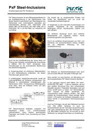
Figure 1: <strong>Image</strong>Acess user interface showing thumbnails, project and experimental d<strong>at</strong>a<br />
any other imaging software (e.g. <strong>Image</strong>-<br />
Pro, Metamoroph, Photoshop) for visualiz<strong>at</strong>ion.<br />
After a year of experience using the system,<br />
some important facts are to be considered:<br />
� Advanced autom<strong>at</strong>ed microscopy requires<br />
individual setups to maintain<br />
speed and resolution. This customis<strong>at</strong>ion<br />
process may take quite some time<br />
and involves local IT<br />
� Dedic<strong>at</strong>ed user trainings for the different<br />
tasks like image acquisition,<br />
processing and present<strong>at</strong>ion are key<br />
requirements of a successful system<br />
implement<strong>at</strong>ion<br />
� Due to the typically large fluctu<strong>at</strong>ions<br />
of students and post docs, it is essential<br />
to maintain and build up system<br />
know how by assigning in-house super<br />
users within the various groups<br />
<strong>Image</strong>Access by <strong>Imagic</strong> <strong>Bildverarbeitung</strong><br />
<strong>AG</strong> is one of the only Imaging<br />
Systems on the market not only to manage<br />
2D images, but also to cope with the<br />
fast growing amount of multidimensional<br />
d<strong>at</strong>asets and metad<strong>at</strong>a. The user interface<br />
reflects nicely the scientist’s work<br />
flow, increases productivity and is a<br />
gre<strong>at</strong> help to achieve the real scientific<br />
goals.<br />
<strong>Imagic</strong> <strong>Bildverarbeitung</strong> <strong>AG</strong><br />
Europastrasse 27<br />
CH-8152 Gl<strong>at</strong>tbrugg<br />
Tel. +41 (0)44 809 40 60<br />
Fax +41 (0)44 809 40 61<br />
www.imagic-imaging.com