Interdental Papilla Reconstruction Combining Periodontal and ...
Interdental Papilla Reconstruction Combining Periodontal and ...
Interdental Papilla Reconstruction Combining Periodontal and ...

You also want an ePaper? Increase the reach of your titles
YUMPU automatically turns print PDFs into web optimized ePapers that Google loves.
Pratique<br />
C l i n i q u e<br />
<strong>Interdental</strong> <strong>Papilla</strong> <strong>Reconstruction</strong> <strong>Combining</strong><br />
<strong>Periodontal</strong> <strong>and</strong> Orthodontic Therapy in Adult<br />
<strong>Periodontal</strong> Patients: A Case Report<br />
Fern<strong>and</strong>o Inocencio, DDS, Dip Ortho; Harinder S. S<strong>and</strong>hu, DDS, PhD, Dip Perio<br />
SOMMAIRE<br />
La migration des dents antérieures supérieures provoquée par la perte de soutien<br />
parodontal peut modifier l’apparence esthétique. La perte de contacts entre les dents<br />
adjacentes provoque la récession des papilles interdentaires. Afin de rétablir une<br />
santé parodontale durable et l’apparence esthétique normale d’une femme de 37 ans<br />
en bonne santé, mais atteinte de parodontite chronique avancée généralisée dans<br />
l’arcade supérieur, on a eu recours à une technique alliant des soins parodontaux<br />
et orthodontiques qui a permis d’obtenir un parodonte stable et une esthétique<br />
agréable au niveau antérieur supérieur.<br />
Pour les citations, la version définitive de cet article est la version électronique : www.cda-adc.ca/jcda/vol-74/issue-6/531.html<br />
Pathologic migration of maxillary anterior<br />
teeth because of a loss of periodontal support<br />
is very common. 1 This migration can<br />
result in the extrusion of teeth, 2–4 loss of contact<br />
points, missing papillae <strong>and</strong> poor appearance of<br />
the esthetic zone. 5–7 In the absence of contacts<br />
between the adjacent teeth, papillae recede. It<br />
has been proposed that the distance from the<br />
contact point to the alveolar crest is at least partially<br />
indicative of the presence of interdental<br />
papillae. 8<br />
Several periodontal surgical techniques have<br />
been proposed to recreate the missing papillae<br />
5,7,9–17 ; however, predictable results have not<br />
yet been achieved. 18 In cases of loss of periodontal<br />
support <strong>and</strong> shifting teeth, a multidisciplinary<br />
treatment approach with esthetic periodontal<br />
surgery is required to eliminate periodontal<br />
inflammation without loss of soft tissue. 4,5,7,18,19<br />
This surgery should be followed immediately<br />
Auteur-ressource<br />
Dr S<strong>and</strong>hu<br />
Courriel :<br />
harinder.s<strong>and</strong>hu@<br />
schulich.uwo.ca<br />
by orthodontic treatment to reduce the infrabony<br />
defects caused by the intrusion <strong>and</strong> lateral<br />
movement of teeth into the defect. 1–3,20–23 These<br />
clinical techniques may also help to re-establish<br />
the contact points, reduce the distance between<br />
the contact point <strong>and</strong> the alveolar crest <strong>and</strong><br />
re-form the interdental papillae. 5–8,18,22,24–26<br />
This paper is the initial report of an interdisciplinary<br />
investigation into the outcome of<br />
treatment designed to enhance the prognosis of<br />
periodontally involved teeth <strong>and</strong> to improve the<br />
appearance of the esthetic zone.<br />
Case Report<br />
A healthy 37-year-old woman who was a<br />
smoker sought treatment of her shifting maxillary<br />
teeth <strong>and</strong> closure of diastemas (Fig. 1).<br />
A diagnosis of generalized advanced chronic<br />
periodontitis in the maxillary arch was made.<br />
Undergoing smoking cessation counselling<br />
JADC • www.cda-adc.ca/jadc • Juillet/Août 2008, Vol. 74, N o 6 • 531
––– S<strong>and</strong>hu –––<br />
Figure 1: Initial intraoral photographs showing generalized spacing <strong>and</strong> absence of papillae in the upper anterior segment.<br />
Figure 2: Initial radiographs revealed the presence of bony defects between the central incisors<br />
<strong>and</strong> the lateral incisors <strong>and</strong> canines.<br />
helped the patient quit smoking; she has been tobacco-free<br />
since the surgical treatment.<br />
After the initial sanative phase, osseous surgery for<br />
pocket reduction was completed in sextant 1 <strong>and</strong> sextant<br />
3. To avoid maxillary anterior soft-tissue loss, papilla<br />
preservation flap surgery was done. 12,13 This procedure<br />
allowed access to the infrabony defects (Fig. 2) for debridement<br />
without loss of papillae. No adjunct guided tissue<br />
regeneration was attempted.<br />
The patient was followed 6 weeks for postoperative care<br />
<strong>and</strong> was referred to an orthodontist for further orthodontic<br />
management of the diastemas <strong>and</strong> missing papillae. The<br />
patient was seen every 3 months for periodontal maintenance<br />
during her orthodontic treatment. <strong>Periodontal</strong> pocket<br />
depths <strong>and</strong> clinical attachment levels before <strong>and</strong> after<br />
periodontal treatment, <strong>and</strong> after orthodontic treatment are<br />
shown in Table 1.<br />
Although some proinclination of the incisors was<br />
present, the relatively shallow overjet <strong>and</strong> overbite made<br />
room for the intrusion <strong>and</strong> retrusion of the incisor segment<br />
very limited.<br />
Upper brackets on the incisors <strong>and</strong> canines, as well<br />
as direct bonding tubes on the first molars, were bonded,<br />
starting with a nickel-titanium 0.016 archwire for initial<br />
alignment, followed by nickel-titanium 0.018, nickel-<br />
titanium 0.018 × 0.025 <strong>and</strong> stainless steel 0.018 × 0.025 archwires.<br />
Closing the space by moving the central <strong>and</strong> lateral<br />
incisors into the bony defects was started with an elastomeric<br />
power chain.<br />
Because of the reduction in the overjet, lower brackets<br />
that mirrored the upper ones were bonded to anchor the<br />
closure of the space by means of the mesialization of the<br />
upper posterior segments with Class III elastics. Some intrusion<br />
of the upper incisors was intended, but it was limited<br />
because of the shallow overbite. During the orthodontic<br />
procedures, oral hygiene maintenance was done every<br />
3 months.<br />
After the space was closed, the esthetic appearance<br />
of the papillae was significantly improved, resulting in<br />
a normal appearance of the whole anterior segment<br />
(Fig. 3). Periapical radiographs showed improvement at<br />
the level of the bone crest between the central incisors <strong>and</strong><br />
between the lateral incisors <strong>and</strong> canines (Fig. 4). Resinbonded<br />
fixed retention was established after the appliances<br />
were debonded.<br />
532 JADC • www.cda-adc.ca/jadc • Juillet/Août 2008, Vol. 74, N o 6 •
––– <strong>Papilla</strong> <strong>Reconstruction</strong> –––<br />
Figure 3: Post-treatment photographs showing proper proximal contacts <strong>and</strong> improved appearance of the papillae.<br />
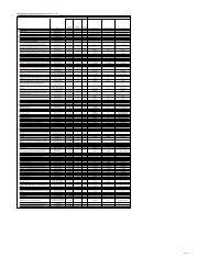
Table 1 Clinical measurements (mm) of lateral incisors before treatment, after periodontal treatment <strong>and</strong> after orthodontic<br />
treatment<br />
Incisors<br />
Right lateral incisor<br />
Before<br />
treatment<br />
Probing pocket depth (mm) Clinical attachment level (mm)<br />
After<br />
periodontal<br />
treatment<br />
After<br />
orthodontic<br />
treatment<br />
Before<br />
treatment<br />
After<br />
periodontal<br />
treatment<br />
After<br />
orthodontic<br />
treatment<br />
Mesiobuccal 3 3 2 3 3 2<br />
Buccal 2 2 2 2 2 2<br />
Distobuccal 7 4 2 7 4 2<br />
Distolingual 7 4 3 4 3 3<br />
Lingual 5 2 2 5 2 2<br />
Mesiolingual 3 2 2 3 2 2<br />
Left lateral incisor<br />
Figure 4: Post-treatment radiographs showing a significant reduction of the bony defects after<br />
space closure.<br />
Mesiobuccal 3 2 2 3 2 2<br />
Buccal 2 2 2 2 2 2<br />
Distobuccal 4 4 2 4 4 2<br />
Distolingual 6 2 2 6 2 2<br />
Lingual 2 2 2 2 2 2<br />
Mesiolingual 3 2 2 3 2 2<br />
JADC • www.cda-adc.ca/jadc • Juillet/Août 2008, Vol. 74, N o 6 • 533
Discussion<br />
Periodontic plastic surgical procedures have tremendously<br />
improved with the use of microscopes <strong>and</strong> microsurgical<br />
instruments. Although many investigations 9–11,14,16,17,26,27<br />
have proposed procedures for reconstructing the papillae,<br />
predictable results have not yet been achieved because of a<br />
variety of factors. 18 Infrabony defects can be treated by extrusion<br />
20 <strong>and</strong> intrusion 5,7,19 of teeth, bodily movement of the<br />
teeth into the bony defects 21,22 <strong>and</strong> guided tissue regeneration.<br />
7 For our patient, we used a modified papilla preservation<br />
technique, followed by orthodontic mesialization of the<br />
posterior segments <strong>and</strong> minor intrusion of the maxillary<br />
anterior segment. Our patient’s occlusal conditions limited<br />
the amount of intrusion feasible <strong>and</strong> may have had an impact<br />
on the final results.<br />
With this combined periodontal <strong>and</strong> orthodontic treatment,<br />
stable periodontal health was achieved. Pocket depth<br />
was reduced <strong>and</strong> the level of clinical attachment improved.<br />
Results of a radiographic survey showed that the infrabony<br />
defects were at least partially healed <strong>and</strong> the height of the<br />
alveolar crest around the lateral incisors increased. These<br />
results were obtained without the addition of any regenerative<br />
materials. With the re-establishment of contact points<br />
between the central <strong>and</strong> lateral incisors <strong>and</strong> the lateral incisors<br />
<strong>and</strong> canines, the interdental papillae returned to their<br />
proper contours <strong>and</strong> almost filled the interdental spaces.<br />
These clinical improvements created an esthetic appearance<br />
free from receded papillae <strong>and</strong> diastemas. For our patient,<br />
the reduction in pocket depth, gain in clinical attachment<br />
level <strong>and</strong> reduction in mobility were consistent with the<br />
elimination of inflammation <strong>and</strong> improved home care. 28<br />
Radiographic reduction of our patient’s infrabony defects,<br />
especially around the lateral incisors, was consistent<br />
with results obtained with the intrusion 5,7,19,21 <strong>and</strong> lateral<br />
movement 1–3,20–23 of teeth. Although interpreting nonst<strong>and</strong>ardized<br />
radiographs has limitations, our patient’s combined<br />
radiographic improvement in bone level, reduction in<br />
pocket depth, gain in the clinical attachment level <strong>and</strong> absence<br />
of gingival recession indicate definitive improvement<br />
in her periodontal status. Our patient has now maintained<br />
this stable stage for 3 years.<br />
The re-establishment of contact points through mesial<br />
movement <strong>and</strong> intrusion also allowed proper re-formation<br />
of interdental papillae. This is consistent with the concept<br />
that the distance between the contact point <strong>and</strong> the alveolar<br />
crest is at least partially indicative of the presence of interdental<br />
papillae. 8 The intrusion of teeth also allows the wider<br />
mesiodistal dimension of the crowns to close the embrasure<br />
space.<br />
In conclusion, within the limitations of this investigation,<br />
combined periodontal <strong>and</strong> orthodontic therapy<br />
that involved mesial movement of teeth to the point of<br />
minor intrusion achieved a healthy stable periodontium<br />
<strong>and</strong> significantly improved the appearance of the esthetic<br />
zone. a<br />
––– S<strong>and</strong>hu –––<br />
THE AUTHORS<br />
534 JADC • www.cda-adc.ca/jadc • Juillet/Août 2008, Vol. 74, N o 6 •<br />
Dr. Inocencio is an assistant professor in the division of<br />
orthodontics <strong>and</strong> pediatric dentistry, Schulich School of<br />
Medicine <strong>and</strong> Dentistry, University of Western Ontario,<br />
London, Ontario.<br />
Dr. S<strong>and</strong>hu is a professor in the division of periodontics,<br />
Schulich School of Medicine <strong>and</strong> Dentistry, University of<br />
Western Ontario, London, Ontario.<br />
Correspondence to: Dr. Harinder S. S<strong>and</strong>hu, Schulich School of Medicine<br />
<strong>and</strong> Dentistry, University of Western Ontario, Dental Sciences Building,<br />
Room 1003, London, ON N6A 5C1.<br />
Acknowledgments: We would like to thank Meghan Perinpanayagam for<br />
her assistance in the preparation of this manuscript.<br />
The authors have no declared financial interests.<br />
This article has been peer reviewed.<br />
References<br />
1. Steffensen B, Storey AT. Orthodontic intrusive forces in the treatment of<br />
periodontally compromised incisors: a case report. Int J Periodontics Restorative<br />
Dent 1993; 13(5):433–41.<br />
2. Melsen B, Agerbaek N, Eriksen J, Terp S. New attachment through<br />
periodontal treatment <strong>and</strong> orthodontic intrusion. Am J Orthod Dentofacial<br />
Orthop 1988; 94(2):104–16.<br />
3. Melsen B, Agerbaek N, Markenstam G. Intrusion of incisors in adult<br />
patients with marginal bone loss. Am J Orthod Dentofacial Orthop 1989;<br />
96(3):232–41.<br />
4. Melsen B, Agerbaek N. Orthodontics as an adjunct to rehabilitation.<br />
Periodontol 2000 1994; 4:148–59.<br />
5. Cardaropoli D, Re S, Corrente G, Abundo R. <strong>Reconstruction</strong> of the maxillary<br />
midline papilla following a combined orthodontic-periodontic treatment<br />
in adult periodontal patients. J Clin Periodontol 2004; 31(2):79–84.<br />
6. Cardaropoli D, Re S, Corrente G. The <strong>Papilla</strong> Presence Index (PPI): a new<br />
system to assess interproximal papillary levels. Int J Periodontics Restorative<br />
Dent 2004; 24(5):488–92.<br />
7. Cardaropoli D, Re S. <strong>Interdental</strong> papilla augmentation procedure following<br />
orthodontic treatment in a periodontal patient. J Periodontol 2005;<br />
76(4):655–61.<br />
8. Tarnow DP, Magner AW, Fletcher, P. The effect of the distance from the<br />
contact point to the crest of bone on the presence or absence of the interproximal<br />
dental papilla. J Periodontol 1992; 63(12):995–6.<br />
9. Azzi R, Etienne D, Carranza F. Surgical reconstruction of the interdental<br />
papilla. Int J Periodontics Restorative Dent 1998; 18(5):466–73.<br />
10. Azzi R, Takei HH, Etienne D, Carranza FA. Root coverage <strong>and</strong> papilla<br />
reconstruction using autogenous osseous <strong>and</strong> connective tissue grafts. Int J<br />
Periodontics Restorative Dent 2001; 21(2):141–7.<br />
11. Carnio J. Surgical reconstruction of interdental papilla using an interposed<br />
subepithelial connective tissue graft: a case report. Int J Periodontics<br />
Restorative Dent 2004; 24(1):31–7.<br />
12. Cortellini P, Pini Prato G, Tonetti MS. The modified papilla preservation<br />
technique with bioresorbable barrier membranes in the treatment of<br />
intrabony defects. Case reports. Int J Periodontics Restorative Dent 1996;<br />
16(6):546–59.<br />
13. Cortellini P, Pini Prato GP, Tonetti MS. The simplified papilla preservation<br />
flap. A novel surgical approach for the management of soft tissues<br />
in regenerative procedures. Int J Periodontics Restorative Dent 1999;<br />
19(6):589–99.<br />
14. Franceti L, Del Fabbro M, Testori Tl, Weinstein R. <strong>Periodontal</strong> microsurgery:<br />
report of 16 cases consecutively treated by the free rotated<br />
papilla autograft technique combined with coronally advanced flap.<br />
Int J Periodontics Restorative Dent 2004; 24(3):272–9.<br />
15. Han TJ, Takei HH. Progress in gingival papilla reconstruction. Periodontol<br />
2000 1996; 11:65–8.<br />
16. Nemcovsky CE. Interproximal papilla augmentation procedure: a novel<br />
surgical approach <strong>and</strong> clinical evaluation of 10 consecutive procedures.<br />
Int J Periodontics Restorative Dent 2001; 21(6):553–9.
––– <strong>Papilla</strong> <strong>Reconstruction</strong> –––<br />
17. Cardaropoli D, Re S, Manuzzi W, Gaveglio L, Cardaropoli G. Bio-Oss collagen<br />
<strong>and</strong> orthodontic movement for the treatment of infrabony defects in<br />
the esthetic zone. Int J Periodontics Restorative Dent 2006; 26(6):553–9.<br />
18. Prato GP, Rotundo R, Cortellini P, Tinti C, Azzi R. <strong>Interdental</strong> papilla<br />
management: a review <strong>and</strong> classification of the therapeutic approaches. Int<br />
J Periodontics Restorative Dent 2004; 24(3):246–55.<br />
19. Cirelli JA, Cirelli CC, Holzhausen M, Martins LP, Br<strong>and</strong>ao CH. Combined<br />
periodontal, orthodontic, <strong>and</strong> restorative treatment of pathologic migration<br />
of anterior teeth: a case report. Int J Periodontics Restorative Dent 2006;<br />
26(5):501–6.<br />
20. Ingber JS. Forced eruption. I. A method of treating isolated one <strong>and</strong> two<br />
wall infrabony osseous defects — rationale <strong>and</strong> case report. J Periodontol<br />
1974; 45(4):199–206.<br />
21. Nevins M, Wise RJ. Use of orthodontic therapy to alter infrabony pockets.<br />
2. Int J Periodontics Restorative Dent 1990; 10(3):198–207.<br />
22. Polson A, Caton J, Polson AP, Nyman S, Novak J, Reed B. <strong>Periodontal</strong><br />
response after tooth movement into intrabony defects. J Periodontol 1984;<br />
55(4):197–202.<br />
23. Re S, Corrente G, Abundo R, Cardaropoli D. The use of orthodontic intrusive<br />
movement to reduce infrabony pockets in adult periodontal patients:<br />
a case report. Int J Periodontics Restorative Dent 2002; 22(4):365–71.<br />
24. Kokich VG. Esthetics: the orthodontic-periodontic restorative connection.<br />
Semin Orthod 1996; 2(1):21–30.<br />
25. Nordl<strong>and</strong> WP, Tarnow DP. A classification system for loss of papillary<br />
height. J Periodontol 1998; 69(10):1124–6.<br />
26. Ong, MA, Wang HL, Smith FN. Interrelationship between periodontics<br />
<strong>and</strong> adult orthodontics. J Clin Periodontol 1998; 25(4):271–7.<br />
27. Allen A. Use of the gingival unit transfer in soft tissue grafting: report of<br />
three cases. Int J Periodontics Restorative Dent 24(2):165–75.<br />
28. Listgarten MA, Levin S. Positive correlation between the proportions of<br />
subgingival spirochetes <strong>and</strong> motile bacteria <strong>and</strong> susceptibility of human subjects<br />
to periodontal deterioration. J Clin Periodontol 1981; 8(2):122–38.<br />
f02 7149F RegimenAd_JADC.qxd:Layout 1<br />
JADC • www.cda-adc.ca/jadc • Juillet/Août 2008, Vol. 74, N o 6 • 535