Download
Download
Download

You also want an ePaper? Increase the reach of your titles
YUMPU automatically turns print PDFs into web optimized ePapers that Google loves.
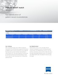
Safety, stability,<br />
predictability and visual<br />
acuity of ReLEx smile<br />
Clinical results by<br />
Ekktet Chansue 1<br />
Dr. Ekktet Chansue is founder and Medical Director of the<br />
TRSC International LASIK Center in Bangkok, Thailand.<br />
He was recognized as “The fi rst surgeon to perform LASIK<br />
in Thailand” and is performing ReLEx smile since early 2011.<br />
He presents his results with ReLEx smile from a study<br />
including 326 eyes, with an average patient age of 31 years<br />
(range 18 to 56) and a mean pre-op SEQ of -4.95 D ± 1.89 D<br />
(range -10.50 D to -1.00 D).<br />
%<br />
100<br />
90<br />
80<br />
70<br />
60<br />
50<br />
40<br />
30<br />
20<br />
10<br />
0<br />
month<br />
(eyes)<br />
-5 to -2<br />
1 m (280)<br />
3 m (239)<br />
6 m (118)<br />
-1.99 to -1<br />
1 Reference see page 11<br />
»<br />
The procedure is highly accurate and very neutral<br />
in terms of spherical aberration and independent<br />
of the amount of correction.<br />
Dr. Ekktet Chansue, TRSC International LASIK Center,<br />
Thailand, June 2012<br />
Refractive outcome: percentage within attempted Effectiveness: pre-op CDVA vs. post-op UDVA<br />
%<br />
100<br />
90<br />
80<br />
70<br />
60<br />
50<br />
40<br />
30<br />
20<br />
10<br />
0<br />
month<br />
(eyes)<br />
lost > 2<br />
1 m (280)<br />
3 m (237)<br />
6 m (118)<br />
lost 2<br />
Safety: change in CDVA<br />
0 % 6 %<br />
-0.99 to -0.5<br />
lost 1<br />
-0.49 to +0.50<br />
59 %<br />
unchanged<br />
gained 1<br />
89 %<br />
36 %<br />
+0.51 to +1<br />
5 %<br />
gained 2<br />
0 %<br />
+1.01 to +5<br />
gained > 2<br />
diopters<br />
lines<br />
• Convincing visual outcomes: 92 % of the patients<br />
have UDVA 20/20 or better after already one month<br />
(pre-op 98 % of patients had CDVA of 20/20 or better)<br />
• Refractive outcome of 100 % of all eyes is within ± 1 D<br />
after 3 months<br />
• Highly predictable results<br />
• Very stable results with almost no regression<br />
• BCVA at 6 months: 95 % of eyes have gained one line<br />
or stayed unchanged, no eye lost 2 or more lines<br />
%<br />
100<br />
90<br />
80<br />
70<br />
60<br />
50<br />
40<br />
30<br />
20<br />
10<br />
0<br />
1.00<br />
0.00<br />
pre-op<br />
-1.00<br />
-2.00<br />
-3.00<br />
-4.00<br />
-5.00<br />
-6.00<br />
Stability<br />
month<br />
(eyes)<br />
1m (280)<br />
3m (239)<br />
6m (118)<br />
pre-op CDVA<br />
‘20/10’<br />
20/12.5<br />
or better<br />
-4.95<br />
20/16 or better<br />
20/20 or better<br />
20/25 or better<br />
»<br />
92 %<br />
92 %<br />
92 %<br />
98 %<br />
20/32 or better<br />
20/40 or better<br />
20/50 or better<br />
20/63 or worse<br />
0.09 0.05 0.08<br />
1 m 3 m 6 m<br />
-7.00<br />
n=326 n=280 n=239 n=118<br />
-8.00<br />
diopters<br />
month<br />
3
ReLEx smile and<br />
Femto-LASIK<br />
A comparison by<br />
Eui-Sang Chung 4<br />
Prof. Eui-Sang Chung is the Chief of the cornea division<br />
in Samsung Medical Center in Seoul, Koreas largest ophthalmological<br />
clinic serving over 100,000 outpatients and<br />
performing 6,000 operations and is Associate Professor<br />
of Ophthalmology, Sungkyunkwan University School of<br />
Medicine. He was the first surgeon to start ReLEx flex in<br />
Korea and has been doing ReLEx smile since June 2011.<br />
In his study he compares the results of ReLEx smile and<br />
Femto-LASIK and concludes:<br />
4 Reference see page 11<br />
4<br />
Results: Refractive outcome (MR SEQ percent within attempted)<br />
%<br />
100<br />
90<br />
80<br />
70<br />
60<br />
50<br />
40<br />
30<br />
20<br />
10<br />
0<br />
month<br />
(eyes)<br />
1w<br />
1m<br />
3m<br />
6m<br />
≤ -1.0<br />
Results: Safety (change in CDVA)<br />
%<br />
100<br />
90<br />
80<br />
70<br />
60<br />
50<br />
40<br />
30<br />
20<br />
10<br />
0<br />
month<br />
(eyes)<br />
lost > 2<br />
1w<br />
1m<br />
3m<br />
6m<br />
1 eye<br />
lost 2<br />
ReLEx smile<br />
-1.0 < ≤ -0.5 -0.5 < < 0.5 0.5 ≤ < 1.0 1.0 ≤<br />
3 eyes 1 eye<br />
lost 1<br />
Diopters<br />
ReLEx smile<br />
no change<br />
Diopters<br />
gain 1<br />
gain 2<br />
gain > 2<br />
»<br />
ReLEx is the right direction of Laser<br />
Vision Correction.<br />
Prof. Eui-Sang Chung, Samsung Medical Center,<br />
Seoul, Korea, May 2012<br />
%<br />
100<br />
90<br />
80<br />
70<br />
60<br />
50<br />
40<br />
30<br />
20<br />
10<br />
0<br />
»<br />
• ReLEx smile is a safe, predictable and effective<br />
procedure for treating myopia and myopic astigmatism<br />
• Results for safety and refractive outcome are<br />
comparable to Femto-LASIK<br />
ReLEx smile LASIK<br />
Age (years) 29.55 ± 5.59 (19 ~ 41) 28.67 ± 6.30 (18 ~ 37)<br />
MRSE (diopters) -5.01 ± 2.55<br />
-5.71 ± 2.26<br />
(1.40 ~ 11.625) (1.925 ~ -10.125)<br />
Eyes 81 eyes of 41 patients 38 eyes of 28 patients<br />
%<br />
100<br />
90<br />
80<br />
70<br />
60<br />
50<br />
40<br />
30<br />
20<br />
10<br />
0<br />
month<br />
(eyes)<br />
lost > 2<br />
1w<br />
1m<br />
3m<br />
6m<br />
LASIK<br />
≤ -1.0 -1.0 < ≤ - 0.5 -0.5 < < 0.5 0.5 ≤ < 1.0 1.0 ≤<br />
month<br />
(eyes)<br />
1w<br />
1m<br />
3m<br />
6m<br />
lost 2<br />
lost 1<br />
Diopters<br />
LASIK<br />
no change<br />
Diopters<br />
gain 1<br />
gain 2<br />
gain > 2
Postoperative dry eye<br />
A comparison between<br />
ReLEx smile and LASIK 5<br />
Prof. Dan Z. Reinstein started performing ReLEx smile in 2010<br />
and describes describes one of the biggest advantages of<br />
the fl apless ReLEx smile procedure to be the reduction of<br />
postoperative dry eye compared with that observed after<br />
PRK and LASIK. In ReLEx smile the anterior corneal anatomy<br />
is preserved and the anterior stromal nerve plexus is disrupted<br />
signifi cantly less since there are no sidecuts created – no fl ap<br />
is created; this should result in fewer dry eye symptoms and a<br />
faster recovery of postoperative patient comfort as has been<br />
found in preliminary studies where corneal sensation recovered<br />
to baseline levels after 3 months.<br />
The cornea is one of the most densely innervated peripheral<br />
tissues in humans with the majority of nerves located in the<br />
anterior stroma, Bowman’s layer and epithelium. In LASIK, the<br />
anterior stromal nerve plexus is disrupted by the creation of<br />
a fl ap with further nerves being severed by the excimer laser<br />
ablation (similarly in PRK). Postoperatively, this means that the<br />
patient may have dry eye symptoms and decreased corneal<br />
sensitivity while the nerves regenerate. A number of studies<br />
have reported that corneal sensation takes at least 6 months<br />
6 –14<br />
to recover to normal levels after LASIK.<br />
Diagrams demonstrating the difference between ReLEx smile (top) and LASIK<br />
(bottom) in how the two procedures affect the anterior corneal nerve plexus.<br />
5 –14 Reference see page 11<br />
»<br />
Leaving the anterior stromal nerve plexus of the cornea<br />
intact makes ReLEx smile into the least traumatic<br />
corneal refractive procedure ever – our studies appear<br />
to confi rm drastically reduced dry eye side effects<br />
compared to LASIK.<br />
Prof. Dan Z. Reinstein, London Vision Clinic,<br />
United Kingdom, June 2012<br />
»<br />
• ReLEx smile reduced postoperative dry eye compared<br />
to that observed after LASIK and PRK<br />
• Faster recovery of corneal sensation to baseline level<br />
observed after ReLEx smile<br />
• Significantly less disruption of anterior stromal nerve<br />
plexus with ReLEx smile compared to LASIK<br />
Mean corneal sensation for 39 eyes after ReLEx smile compared with the corneal<br />
sensation after LASIK averaged over nine published studies.<br />
5
Biomechanical stability<br />
Advantages of ReLEx smile<br />
as a refractive procedure 15<br />
Dr. Cynthia Roberts is Professor of Ophthalmology and<br />
Biomedical Engineering at the Ohio State University.<br />
To compare the biomechanical consequences of ReLEx smile<br />
to a standard LASIK procedure, she and her colleagues<br />
(Abhijit Sinha Roy, PhD and William Joseph Dupps, Jr.,<br />
MD, PhD of the Cleveland Clinic Foundation) generated<br />
a non-linear, anisotropic, fi ber-dependent material model.<br />
Biomechanical properties were taken from the literature,<br />
including reduction in elastic modulus within the LASIK fl ap<br />
and at the interface. ReLEx smile was assumed to have less<br />
reduction in modulus as a function of the ratio of side cut<br />
arc length between LASIK and ReLEx smile. Stress distribution<br />
was calculated within the fl ap (LASIK) / cap area (ReLEx smile)<br />
and within the stromal bed and compared between both<br />
methods with the following results:<br />
15 Reference see page 11<br />
6<br />
»<br />
The biomechanical aspects of ReLEx smile are very<br />
exciting. Our model confi rms that the biomechanical<br />
stability of the anterior corneal layer is much less affected<br />
with ReLEx smile compared to LASIK due to the innovative<br />
approach of minimizing the number of anterior lamellae<br />
that are cut.<br />
»<br />
Prof. Cynthia Roberts, Ohio State University,<br />
USA, June 2012<br />
• ReLEx smile has stress distribution in the cap and the<br />
stromal bed that is much closer to the unoperated<br />
state (of equivalent thickness) than LASIK<br />
• LASIK has greatly reduced peak stress within the flap<br />
compared to the preoperative state due to cutting<br />
of many tension-bearing anterior lamellae<br />
(Middle upper row)<br />
• LASIK has greatly increased peak stress at the level<br />
of residual stromal bed due to inability of the flap<br />
to carry the stress which is then transmitted into the<br />
stromal bed (Middle lower row)<br />
Pre-op LASIK SMILE<br />
The top row shows the stress maps in the anterior corneal layers near the surface<br />
in an unoperated state (left), after making a LASIK fl ap (middle), and after a<br />
ReLEx smile cap (right). The bottom row shows the corresponding stress maps<br />
at the level of the residual stromal bed (RSB). Note that ReLEx smile is closer to<br />
pre-op than LASIK.<br />
Surface RSB
Biomechanical stability<br />
Superior differences of<br />
ReLEx smile over LASIK 16<br />
Prof. Dan Z. Reinstein is convinced that the extra<br />
biomechani cal stability provided by this fl apless minimally<br />
invasive procedure will bring a number of benefi ts.<br />
Figure 1 shows diagrams of intact stromal lamellae after<br />
LASIK and ReLEx smile highlighting the anterior lamellae<br />
that remain intact after ReLEx smile. Residual stromal<br />
thickness (RST) calculations are shown for a 500 μm cornea<br />
with a 100 μm ablation/lenticule and 120 μm flap/cap<br />
thickness. The LASIK RST of 280 μm consists only of<br />
posterior stroma, whereas the ReLEx smile RST has the<br />
same 280 μm of posterior stroma, but also has 70 μm of<br />
anterior stroma, which makes a total of 350 μm of stroma.<br />
However, since anterior stroma is 50 % stronger than<br />
posterior stroma, a further 35 μm (50 % of the 70 μm<br />
of anterior stroma) can be added to make an effective<br />
total of 385 μm.<br />
Figure 1<br />
16 –18 Reference see page 11<br />
»<br />
ReLEx smile represents the ultimate dream<br />
of Prof Jose Ignacio Barraquer Moner:<br />
minimally invasive keratomileusis.<br />
Prof. Dan Z. Reinstein, London Vision Clinic,<br />
United Kingdome, June 2012<br />
The absence of a flap will result in increased<br />
biomechanical integrity for two reasons:<br />
• Anterior stromal lamellae are stronger than<br />
posterior stromal lamellae 17 , therefore the postoperative<br />
cornea will be stronger after ReLEx smile as the<br />
anterior stromal lamellae remain intact. The opposite is<br />
true in LASIK where the biomechanical stability of the<br />
cornea effectively relies only on the residual posterior<br />
stromal lamellae.<br />
• Vertical cuts (e.g. flap sidecut) have more biomechanical<br />
impact than horizontal cuts 18 (Figure 2), meaning<br />
that the ReLEx smile procedure minimizes the biomechanical<br />
change to the cornea. This also allows the<br />
lenticule to be removed from deeper in the cornea<br />
to take further advantage of the stronger anterior<br />
stromal lamellae.<br />
»<br />
90 μm 160 μm<br />
LASIK Flap 9 % 32 %<br />
Sidecut Only 9 % 33 %<br />
Delamination Only 5 % 5 %<br />
Figure 2: Percentage increase in central corneal strain on human cadaver eyes<br />
after the creation of a LASIK fl ap, a sidecut only or delamination only at both<br />
90 μm and 160 μm. 18<br />
Sidecut and whole fl ap resulted in a similar increase in strain with signifi cantly<br />
greater increase for the 160 μm depth. Increase in strain was the same at both<br />
depths when the delamination cut only was performed. Applying this fi nding to<br />
ReLEx smile, since no anterior corneal sidecut is created, there will be slightly<br />
less increase in corneal strain in ReLEx smile compared to thin fl ap LASIK and a<br />
signifi cant difference in corneal strain compared to LASIK with a thicker fl ap.<br />
7
A ReLEx smile Case<br />
By Dan Z. Reinstein 19<br />
Patient: Right eye of a 40 year old (Caucasian) male with<br />
high myopia.<br />
Treatment planning: Central corneal thickness was 529 μm,<br />
lenticule thickness was 150 μm (intended correction was<br />
plano, 6-mm optical zone), cap thickness was 120 μm, to<br />
leave 259 μm of residual stroma. As no flap was created,<br />
there was also 65 μm of untouched anterior stroma, so the<br />
total stroma was 324 μm. A 3-mm supero-temporal incision<br />
was used to remove the lenticule.<br />
Fluorescein slit lamp photo at the one day post-op in which the boundary<br />
of the lenticule can be seen to be well centered on the corneal vertex.<br />
The supero-temporal 3-mm incision can be seen.<br />
8<br />
»<br />
This patient trains the military in high security lock<br />
mechanisms; he was told that laser couldn’t help him.<br />
Thanks to the enhanced biomechanics of ReLEX smile<br />
he was made 20/20 from -10D in one shot.<br />
Prof. Dan Z. Reinstein, London Vision Clinic,<br />
United Kingdom, August 2012<br />
Treatment summary:<br />
• Superbly accurate refractive correction<br />
• CDVA same as pre-op within fi rst week<br />
• UDVA same as pre-op CDVA<br />
• Contrast sensitivity slightly improved<br />
• Corneal sensation only slightly reduced at 1 day<br />
(compared with zero at 1 day after LASIK)<br />
and fully recovered by 1 month (compared with<br />
6 months after LASIK)<br />
• Large, well-centered optical zone on topography<br />
Contrast sensitivity before and 3 months after ReLEx smile.<br />
»
Pre-op 1 day 1 week 1 month 3 months<br />
Manifest refraction -10.25* -0.50 x 118<br />
(target plano)<br />
+1.25 -0.50 x 15 +0.50 -0.25 x 30 +0.50 -0.75 x 75 +0.50 -0.50 x 74<br />
CDVA 20/16 20/20 20/12.5 20/16 20/16<br />
UDVA – 20/20 20/16 20/16 20/16<br />
Contrast sensitivity Low normal range Slightly better Slightly better – Slightly better<br />
than pre-op than pre-op<br />
than pre-op<br />
Corneal sensation 60 40 50 60 60<br />
Atlas tangential curvature topography maps before (top left) and 3 months after (bottom left). The difference map is shown on the right demonstrating the<br />
well-centered 6-mm optical zone.<br />
*outside approved treatment range, clinical study software was used<br />
19 Reference see page 11<br />
9
Surface quality of extracted ReLEx smile lenticule<br />
using environmental SEM technique 20<br />
The lenticule extracted from patients were preserved and<br />
prepared for imaging. Environmental or “wet” scanning<br />
electron microscopy was performed on lenticule anterior,<br />
posterior and edge surfaces.<br />
Lenticule<br />
edge<br />
Anterior lenticule surface – low magnifi cation<br />
Anterior lenticule surface – high magnifi cation<br />
10<br />
anterior<br />
20 Reference see page 11<br />
posterior<br />
• Very smooth cutting surface<br />
• Lenticule removal without residual pieces<br />
• High quality of surface and edges, appropriate<br />
for quality of vision<br />
• Anterior and posterior lenticule cut refer to each<br />
other, appropriate for refractive correction<br />
Posterior lenticule surface – low magnifi cation<br />
Edge and anterior surface of lenticule – high magnifi cation