Download - Carl Zeiss Meditec AG
Download - Carl Zeiss Meditec AG
Download - Carl Zeiss Meditec AG

Create successful ePaper yourself
Turn your PDF publications into a flip-book with our unique Google optimized e-Paper software.
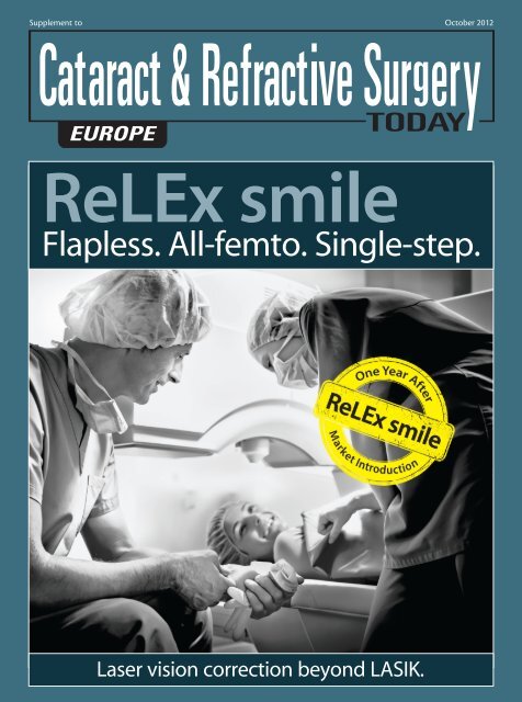
Supplement to October 2012<br />
ReLEx smile<br />
Flapless. All-femto. Single-step.<br />
Laser vision correction beyond LASIK.
3 Biomechanical Advantages of ReLEx smile<br />
By Cynthia J. Roberts, PhD; Abhijit Sinha Roy, PhD;<br />
William J. Dupps, MD, PhD; and Jesper Hjortdal, MD, PhD<br />
ReLEx smile: Flapless. All-femto. Single-step.<br />
ReLEx smile<br />
Flapless. All-femto. Single-step.<br />
CONTENTS<br />
5 The Effects of Refractive Surgery on Dry Eye Syndrome<br />
and Corneal Sensation<br />
By Kimiya Shimizu, MD<br />
7 Efficacy and Safety With ReLEx smile<br />
By Jodhbir S. Mehta, MD<br />
9 What We Have Achieved With ReLEx<br />
By Rainer Wiltfang, MD<br />
11 ReLEx smile: A Technique for All Practices<br />
By Cati Albou-Ganem, MD<br />
OCTOBER 2012 SUPPLEMENT TO CATARACT & REFRACTIVE SURGERY TODAY EUROPE 3
ReLEx smile: Flapless. All-femto. Single-step.<br />
Biomechanical Advantages<br />
of ReLEx smile<br />
Compared to LASIK, this flapless refractive surgery technique maintains a stress pattern<br />
that more closely resembles the unoperated cornea.<br />
BY CYNTHIA J. ROBERTS, PhD; ABHIJIT SINHA ROY, PhD; WILLIAM J. DUPPS, MD, PhD;<br />
AND JESPER HJORTDAL, MD, PhD<br />
For more than 1 year now, surgeons have been using<br />
ReLEx smile to provide patients with refractive correction<br />
via lenticule extraction. Although there are<br />
numerous potential benefits compared with LASIK, this<br />
article will focus on the biomechanical advantages of the<br />
ReLEx smile cap as compared with the LASIK flap.<br />
ReLEx is exclusively performed with the VisuMax<br />
femtosecond laser (<strong>Carl</strong> <strong>Zeiss</strong> <strong>Meditec</strong>). This minimally<br />
invasive procedure uses tissue removal with two femtosecond<br />
incisions that intersect to create a lenticule for<br />
extraction, in place of tissue ablation, which is the mechanism<br />
of action for LASIK and PRK corrections. From<br />
results thus far, several surgeons have shown that ReLEx<br />
smile introduces very little spherical aberration across<br />
the optical zone and increases treatment efficiency, especially<br />
for higher refractive errors, because only two incisions<br />
are needed to create a lenticule independent of the<br />
level of correction and only one laser is used.<br />
Dr. Hjortdal has been using ReLEx smile for 18 months.<br />
He has also seen similar trends, and his patients have<br />
been happy with their postoperative outcomes.<br />
However, the scientist side of him wanted to know<br />
Figure 1. Meshing was performed in Truegrid with 8-noded, linear, hexahedral<br />
elements.<br />
4 SUPPLEMENT TO CATARACT & REFRACTIVE SURGERY TODAY EUROPE OCTOBER 2012<br />
The ReLEx smile cap created<br />
less of a defect in the anterior<br />
surface [compared with LASIK],<br />
and thus had a central surface<br />
stress that was closer to the<br />
preoperative state.<br />
more, which led to a collaboration with Drs. Roberts,<br />
Dupps, and Sinha Roy for biomechanical modeling.<br />
Specifically, could this procedure provide higher biomechanical<br />
stability compared with traditional LASIK<br />
as well as femtosecond LASIK?<br />
THE ReLEx smile CAP<br />
With ReLEx smile, there is no longer a need to create a<br />
corneal flap. Instead, a small side cut incision is created at<br />
an approximate depth of 100 to 120 µm in the cornea for<br />
lenticule extraction, creating a smile cap. The obvious benefits<br />
of cap creation are that (1) complications associated<br />
with the LASIK flap cut, including<br />
incomplete and irregular flap cuts,<br />
thin flaps, buttonholes, and free<br />
caps, are eliminated and (2) reduced<br />
risk of induced astigmatism related<br />
to flap complications.<br />
We hypothesize, however, that<br />
there are additional biomechanical<br />
advantages to ReLEx cap creation<br />
over a flap. Therefore, we recently<br />
designed a nonlinear, anisotropic,<br />
fiber-dependent material model<br />
to compare the biomechanical<br />
consequences of ReLEx smile to a<br />
standard LASIK procedure.<br />
To create this model, we used
Figure 2. Results of the simulations. Row 1: three Von Mises stress maps at the<br />
surface of the cornea; row 2: stress maps at the level of the residual stromal bed.<br />
(red = greater stress; blue = lower stress)<br />
topography from a normal patient to construct a 3-D<br />
finite element model, including a 53-µm epithelium of<br />
constant thickness and using the Munnerlyn algorithm<br />
for an equivalent spherical treatment in both ReLEx<br />
smile and LASIK models.<br />
COMPARISON OF TENSION-BEARING LAMELLAE<br />
In the finite element model, the LASIK flap and ReLEx<br />
smile cap were simulated with equivalent dimensions of<br />
width and thickness, thus allowing us to focus the comparison<br />
on the differences in the pattern of disrupting<br />
the continuity of tension-bearing lamellae in each procedure.<br />
The wound interface between either the LASIK<br />
flap or the ReLEx smile cap and the residual stromal bed,<br />
as well as the sidecuts, were simulated as a thin layer of<br />
20-µm thickness. Meshing was performed in Truegrid<br />
with 8-noded, linear, hexahedral elements (Figure 1).<br />
The results of the simulations are shown in Figure 2.<br />
In row 1, three Von Mises stress maps at the surface of<br />
the cornea are depicted. Von Mises stress is the equivalent<br />
stress of the three principal stresses at a point. Row 2<br />
shows these stress maps at the level of the residual stromal<br />
bed. In both rows, the color red correlates with greater<br />
stress, whereas the color blue correlates with lower stress.<br />
Still referring to Figure 2, the first column represents<br />
a cornea of similar postoperative thickness but without<br />
flap/cap creation or tissue removal; the second column<br />
is after a LASIK procedure, and the third is after a ReLEx<br />
smile procedure. Both procedures were simulated with<br />
either a LASIK flap or ReLEx smile cap thickness and<br />
diameter of 100 µm and 9 mm, respectively. Although<br />
Figure 2 illustrates a 9.00 D treatment, the stress pattern<br />
was similar for all simulated corrections.<br />
RESULTS<br />
This study showed that the LASIK flap did not bear the<br />
ReLEx smile: Flapless. All-femto. Single-step.<br />
same stress as it had preoperatively. This<br />
was due to the severing of a substantial<br />
number of tension-bearing lamellae,<br />
resulting in reduced central stress at the<br />
corneal surface compared with the preoperative<br />
state. Because the stress was<br />
transferred to the residual stromal bed,<br />
the central stress increased within the<br />
posterior cornea to a greater magnitude<br />
than the preoperative state.<br />
The ReLEx smile cap, however, created<br />
less of a mechanical insult to the<br />
anterior cornea, and thus had a central<br />
surface stress that was closer to the<br />
unoperated state. Additionally, the<br />
central stress at the level of the residual<br />
stromal bed was also closer to the<br />
unoperated stress pattern. The posterior<br />
corneal stress was much lower<br />
than with LASIK.<br />
CONCLUSION<br />
Our study showed theoretical clear biomechanical<br />
advantages of ReLEx smile over flap-based corneal<br />
refractive surgery. Compared with LASIK, ReLEx smile<br />
treatments maintained a stress pattern that was much<br />
closer to the unoperated cornea in both magnitude and<br />
distribution.<br />
William J. Dupps, MD, PhD, is a refractive and<br />
corneal surgeon at the Cleveland Clinic Cole Eye<br />
Institute. Dr. Dupps states that he has received<br />
research funding from <strong>Carl</strong> <strong>Zeiss</strong> <strong>Meditec</strong>. He<br />
may be reached at tel: +1 216 444 8396.<br />
Jesper Hjortdal, MD, PhD, is a Clinical<br />
Professor of Ophthalmology and a refractive<br />
and corneal surgeon in the Department of<br />
Ophthalmology, Aarhus University Hospital,<br />
Denmark. Dr. Hjortdal states that he has no<br />
financial interest in the products or companies<br />
mentioned. He may be reached at tel: +45 7846 3221;<br />
e-mail: jesper.hjortdal@dadlnet.dk.<br />
Cynthia J. Roberts, PhD, is Professor of<br />
Ophthalmology and Biomedical Engineering<br />
at The Ohio State University in Columbus. Dr.<br />
Roberts states that she has received research<br />
funding from <strong>Carl</strong> <strong>Zeiss</strong> <strong>Meditec</strong>. She may be<br />
reached at tel: +1 614 292 1831; e-mail: roberts.8@osu.edu.<br />
Abhijit Sinha Roy, PhD, is a Research Associate<br />
at the Cleveland Clinic Cole Eye Institute. Dr.<br />
Sinha Roy states that he has received research<br />
funding from <strong>Carl</strong> <strong>Zeiss</strong> <strong>Meditec</strong>. He may<br />
be reached at tel: +1 513 255 0561; e-mail:<br />
asroy27@yahoo.com.<br />
OCTOBER 2012 SUPPLEMENT TO CATARACT & REFRACTIVE SURGERY TODAY EUROPE 5
ReLEx smile: Flapless. All-femto. Single-step.<br />
The Effects of Refractive<br />
Surgery on Dry Eye Syndrome<br />
and Corneal Sensation<br />
As a flapless and minimally invasive procedure, ReLEx smile has the potential to cause fewer<br />
dry eye symptoms compared with femtosecond LASIK.<br />
BY KIMIYA SHIMIZU, MD<br />
It is not uncommon for a<br />
patient to suffer from dry<br />
eye syndrome after refractive<br />
surgery. In many of<br />
these cases, however, the dry<br />
and itchy side effects result<br />
from the LASIK flap and not<br />
from the actual refractive<br />
treatment. In procedures<br />
that do not require a flap,<br />
such as <strong>Carl</strong> <strong>Zeiss</strong> <strong>Meditec</strong>’s<br />
ReLEx smile treatment, these<br />
symptoms are much less<br />
common. For this and many<br />
other reasons, ReLEx smile is<br />
now my procedure of choice<br />
for patients who desire<br />
refractive correction.<br />
SHIFTING OF MY<br />
SURGICAL PREFERENCES<br />
I started performing refractive surgery in Japan in the<br />
1990s. In the early part of the decade, I started performing<br />
PRK and radial keratotomy (RK) and, in 1997, I began<br />
to perform LASIK. I transitioned to LASIK because of the<br />
promise for better visual recovery and less pain, and I<br />
believed that it was a better procedure compared with<br />
surface ablation or RK. For the next 11 years, I performed<br />
LASIK in the majority of my patients and enjoyed excellent<br />
results and successful surgeries.<br />
In 2008, I stopped performing LASIK in favor of other<br />
procedures. The biggest reason for my decision is that I<br />
began to worry about the long-lasting dry eye symptoms<br />
(Figure 1A) that became increasingly more common in<br />
my LASIK patients. Because dry eye syndrome becomes<br />
A<br />
Figure 1. (A) Dry eye, (B) infection, (C) epithelial ingrowth, (D) IOP-induced interlamellar<br />
stromal keratitis, (E) diffuse lamellar keratitis, and (F) corneal perforation can all occur<br />
after LASIK, as a result of flap creation.<br />
6 SUPPLEMENT TO CATARACT & REFRACTIVE SURGERY TODAY EUROPE OCTOBER 2012<br />
B C<br />
D E F<br />
more prominent with age, some of my older patients<br />
began to return to the clinic complaining of dry eyes as<br />
well as other corneal sensations.<br />
Another reason that I decided to abandon LASIK is<br />
that patients who have undergone this treatment can<br />
experience decreased visual performance, including<br />
increased higher-order aberrations and onset of presbyopia.<br />
Infections (Figure 1B), epithelial ingrowth (Figure<br />
1C), intraocular pressure (IOP)-induced interlamellar<br />
stromal keratitis (Figure 1D), diffuse lamellar keratitis<br />
(Figure 1E), and corneal perforations (Figure 1F) can also<br />
occur after LASIK. These are mainly the result of flap creation,<br />
as it involves cutting the trigeminal nerve affects<br />
corneal sensation.
A B<br />
A B<br />
Figure 2. (A) In vivo confocal microscopic images of corneal<br />
subbasal nerve plexuses 1 month after ReLEx smile. (B) In vivo<br />
confocal microscopic images of corneal subbasal nerve<br />
plexuses 1 month after LASIK.<br />
TRIGEMINAL NERVE DENSITY TESTING<br />
We recently examined the changes in trigeminal<br />
nerve density by studying the nerve structure under a<br />
confocal specular microscope before and after LASIK<br />
flap creation in 44 eyes (22 patients). Compared with<br />
preoperative levels, trigeminal nerve density decreased<br />
to 10% and 15% at 1 and 3 months, respectively<br />
(Figure 2). This confirmed our decision to forego LASIK<br />
for other, in my opinion, safer refractive surgery procedures.<br />
In order to prevent this decrease in trigeminal nerve<br />
density, we transitioned to flapless refractive surgery<br />
procedures. The newest refractive correction that does<br />
not require a flap is ReLEx smile. Using the VisuMax<br />
femtosecond laser (<strong>Carl</strong> <strong>Zeiss</strong> <strong>Meditec</strong>), a refractive<br />
lenticule is created in the intact cornea and removed<br />
through a small incision of less than 4 mm, compared<br />
with the 20- to 25-mm incision required for the LASIK<br />
flap sidecut (Figure 3).<br />
After we gained experience with ReLEx smile, we<br />
returned to our initial study design to test the trigeminal<br />
nerve density before and after surgery in 44 eyes. With<br />
ReLEx smile, trigeminal nerve density decreased to 50%<br />
and 60% at 1 and 3 months after ReLEx smile surgery,<br />
respectively. Comparing the numbers from these two<br />
studies, we were able to show that, although both surgical<br />
techniques cause density decrease, ReLEx smile has<br />
the potential to preserve trigeminal nerve density far<br />
better than LASIK.<br />
CONCLUSION<br />
In addition to the preservation of trigeminal nerve<br />
density, ReLEx smile uses less energy and a smaller<br />
spot size than LASIK and therefore creates a smooth<br />
surface. Additionally, the short treatment time, which<br />
is independent from the degree of myopia, translates<br />
ReLEx smile: Flapless. All-femto. Single-step.<br />
Figure 3. (Left) LASIK flap and the (right) ReLEx smile cap.<br />
Figure 4. ReLEx smile: postoperative day 1.<br />
to a short suction time and a short and low rise in<br />
IOP. These things increase the safety of the procedure.<br />
Additionally, not only is ReLEx smile minimally invasive,<br />
but it also induces minimal corneal nerve damage<br />
because the intrastromal lenticule and access cut are<br />
completed in one step and manual tissue separation<br />
and extraction of the refractive lenticule is possible<br />
through a small (4-mm) incision. Figure 4 shows an<br />
operated eye 1 day after ReLEx smile.<br />
Patients typically experience less pain after ReLEx<br />
smile versus LASIK, although pain is not common with<br />
either procedure, and we have not experienced any<br />
complications associated with ReLEx smile. Because<br />
ReLEx smile has good predictability, safety, and efficacy,<br />
we are more confident when offering this procedure to<br />
patients.<br />
For the reasons outlined above, we replaced LASIK<br />
with ReLEx smile in the majority of our patients,<br />
especially for myopic corrections up to -6.00 D.<br />
Kimiya Shimizu, MD, is a Professor of<br />
Ophthalmology and Chair of the Department of<br />
Ophthalmology, Kitasato University, Japan. Dr.<br />
Shimizu states that has no financial interest in<br />
the products or companies mentioned. He may<br />
be reached at e-mail: kimiyas@med.kitasato-u.ac.jp.<br />
OCTOBER 2012 SUPPLEMENT TO CATARACT & REFRACTIVE SURGERY TODAY EUROPE 7
ReLEx smile: Flapless. All-femto. Single-step.<br />
Efficacy and Safety With<br />
ReLEx smile<br />
An overview of the clinical advantages of this procedure.<br />
BY JODHBIR S. MEHTA, MD<br />
ReLEx smile may just be the paradigm shift in laser vision<br />
correction that refractive surgeons have been waiting<br />
for. Instead of weakening the biomechanical stability<br />
with ablation procedures such as LASIK, surgeons<br />
now have the ability to perform refractive correction by<br />
creating a lenticule inside the intact cornea and subsequently<br />
removing it through a small (less than 4 mm)<br />
incision. This technique does not completely sever nerve<br />
pathways and avoids the need to create a flap, which<br />
provides surgeons with the opportunity to perform<br />
minimally invasive refractive corneal surgery.<br />
This article overviews the clinical advantages of lenticule<br />
extraction compared with the most commonly<br />
performed refractive surgery technique in the world:<br />
LASIK. If the excellent results with ReLEx smile continue,<br />
we believe that it has the potential to overtake LASIK in<br />
terms of popularity. Because ReLEx smile is flapless and<br />
requires an 80% smaller sidecut incision in the anterior<br />
cornea and a 30% smaller lamellar incision (cap cut)<br />
than the LASIK flap, biomechanical stability is minimally<br />
reduced and dry eye syndrome avoided.<br />
EARLY STUDIES<br />
In our first preclinical study, 1 my colleagues and I performed<br />
a paired eye study in 36 rabbits to compare ReLEx<br />
flex with standard LASIK. Although ReLEx flex requires flap<br />
creation, it is typically just 0.5 to 1.0 mm larger in diameter<br />
than the optical zone. In the first study, 18 rabbit eyes<br />
underwent ReLEx flex and 18 underwent LASIK. In each<br />
group, we looked at three refractive correction subgroups,<br />
-3.00, -6.00, and -9.00 D, to determine the differences of<br />
the two procedures in early wound-healing responses.<br />
This study showed that, compared with LASIK, ReLEx had<br />
a better postoperative wound-healing response, resulting<br />
in less topographic changes, less inflammation, and less<br />
extracelluar matrix deposition. These findings were statistically<br />
significant at high refractive corrections (-6.00 D and<br />
above). There was, however, no difference between the<br />
groups in cell death and proliferation after surgery.<br />
Our take-home message from this study was that,<br />
with ReLEx, approximately the same amount of energy<br />
8 SUPPLEMENT TO CATARACT & REFRACTIVE SURGERY TODAY EUROPE OCTOBER 2012<br />
is applied to the eye regardless of the level of correction.<br />
The energy is equivalent to approximately 0.58 J. With<br />
LASIK, however, the energy required to complete the<br />
ablation increases as the level of correction increases.<br />
Therefore, the energy can range from 4 J for a low correction<br />
to 12 J, for instance, for a -9.00 D correction.<br />
After we determined that ReLEx resulted in good postoperative<br />
wound healing in rabbit eyes, we began to wonder<br />
if the laser cuts required for the procedure would affect the<br />
stromal bed quality with different ablations. Did these cuts<br />
in the cornea make the stromal bed rougher than preoperatively,<br />
and would that result in slower visual recovery?<br />
Would the stromal bed be roughest in higher myopic eyes<br />
because deeper cuts were required? To answer these questions,<br />
we performed a prospective clinical case series in 33<br />
patients who underwent ReLEx flex in both eyes 2 and a<br />
human eye-bank study on donor cadaver eyes to look at<br />
stromal bed smoothness on scanning electron microscopy.<br />
During this human eye-bank study, we were able to<br />
show that the degree of roughness of the stromal bed<br />
was independent of the level of myopic correction. The<br />
laboratory results were supported by the clinical visual acuity<br />
results. The mean preoperative spherical equivalent (SE)<br />
was -5.77 ±2.04 D, improving to 0.14 ±0.53 D at 3 months<br />
postoperatively. Additionally, UCVA in 94% of eyes was<br />
20/25 or better at 3 months, and all eyes achieved refractive<br />
stability within 1 month after surgery (P
surgeons assume that introducing a new technology may<br />
compromise the safety and efficacy of a well-established<br />
procedure. However, we experienced the opposite. In fact,<br />
our previous results with the microkeratome—even in our<br />
best year—are inferior to our results with the femtosecond<br />
laser—even in the early years of its inception in 2008.<br />
We expected there may be a dip in postoperative results<br />
when we started performing ReLEx. Because we have such<br />
an extensive database of surgical results, it has been very<br />
easy for us to compare results with ReLEx and LASIK. Not<br />
only have our ReLEx flex results matched our femtosecond<br />
LASIK results, but this has been achieved without any specific<br />
nomogram adjustment to optimize our outcomes.<br />
We have also performed a study in 1,500 eyes that<br />
underwent myopic LASIK and 100 eyes that underwent<br />
ReLEx for myopia correction. We matched the eyes for<br />
their preoperative level of myopic correction and found<br />
that the visual outcomes in both groups were similar. The<br />
other thing that we were able to show in this study is that<br />
not only was the safety and efficacy equivalent in these<br />
two groups but when we looked at the eyes that had<br />
between -5.00 and -10.00 D of myopia preoperatively, the<br />
ReLEx eyes had better predictability than the LASIK eyes.<br />
TRANSITIONING TO ReLEx smile<br />
Today, we have transitioned from ReLEx flex to ReLEx<br />
smile. This flapless version of the ReLEx technique is<br />
even less invasive than ReLEx flex. Although we have a<br />
much smaller cohort of results to draw from, we have<br />
compared our newest ReLEx smile results to our results<br />
with not only femtosecond LASIK but also with ReLEx<br />
flex. What we are seeing is that ReLEx smile increases<br />
upon the safety and efficacy that we were able to<br />
achieve with ReLEx flex. Additionally, a much higher<br />
number of patients achieve 20/16 vision than compared<br />
with our femtosecond LASIK patients.<br />
We believe that the reason for the improvement in<br />
results between ReLEx flex and ReLEx smile has to do with<br />
the elimination of flap creation. We have observed that for<br />
higher corrections (between -5.00 and -10.00 D) the flap<br />
induces a significant amount of aberrations into the optical<br />
system. I like to use the following analogy to explain<br />
this phenomenon: The anterior cornea is like a suspension<br />
bridge. Therefore, as you cut vertically across the suspension<br />
bridge (ie, flap), you initiate a collapse in the structure.<br />
With ReLEx smile, however, there is only a small vertical<br />
cut and therefore minimal collapse or stromal damage.<br />
In our studies, there were less induced aberrations<br />
after ReLEx flex than after femtosecond LASIK. This year,<br />
we are conducting a follow-up study with ReLEx smile<br />
that will assess induced aberrations.<br />
TAKE A CHANCE ON A NEW TREATMENT<br />
LASIK is perhaps the most well-known, well-documented<br />
elective procedure in the world. It has helped<br />
ReLEx smile: Flapless. All-femto. Single-step.<br />
millions of people worldwide achieve excellent visual<br />
quality without the need for spectacles. However, as<br />
with any procedure, there are disadvantages. In 2009,<br />
Solomon et al 3 published a study showing that approximately<br />
4% to 5% of patients were unhappy after LASIK.<br />
This is a very small percentage; however, if given the<br />
sheer volume of patients who have undergone LASIK,<br />
this equates to a significant number of unhappy<br />
patients. The most common complaints are dry eye<br />
syndrome, problems with night vision or glare, regression,<br />
and complications during flap formation or traumatic<br />
dislocation postoperatively.<br />
Therefore, when we heard about the flapless, minimally<br />
invasive ReLEx smile procedure about 3 or 4 years<br />
ago, we considered switching to this treatment. Because<br />
the cornea is not cut all the way around, as it is for LASIK<br />
flap creation, biomechanical stability should improve,<br />
which should translate into a reduction of the side<br />
effects of LASIK, including post-LASIK ectasia.<br />
Taking a chance on a new treatment is worthwhile if<br />
it can provide our patients with a safer surgery that is<br />
just as effective, if not more effective, than other wellestablished<br />
treatments. I am pleased with the results we<br />
have had with ReLEx smile thus far, and I envision results<br />
continuing to improve as the treatment matures.<br />
At this time, however, an enhancement after ReLEx<br />
smile is challenging. <strong>Carl</strong> <strong>Zeiss</strong> <strong>Meditec</strong> is working on<br />
solving this issue. If regression occurs, one option is to<br />
perform surface ablation. But because ReLEx smile uses a<br />
lot less energy and there is maintenance of corneal integrity,<br />
regression is not as common as it is after LASIK.<br />
CONCLUSION<br />
ReLEx smile provides surgeons the advantages of LASIK<br />
while avoiding the potential side effects related to flap<br />
creation. Biomechanical stability is relatively stronger<br />
after ReLEx smile compared with LASIK, making wound<br />
healing faster after ReLEx smile. Additionally, there is no<br />
surface dryness on the tear break-up time after ReLEx.<br />
This procedure certainly has the potential to overtake<br />
LASIK as the procedure of choice for most patients.<br />
Jodhbir S. Mehta, MD, is Associate Professor<br />
and Co-Head of the Corneal and External<br />
Disease Service, is a Consultant at the Refractive<br />
Service Singapore National Eye Centre, and is<br />
Associate Professor at DUKE-NUS and YLL,<br />
School of Medicine NUS. Dr. Mehta did not provide financial<br />
disclosure information. He may be reached at e-mail: jodmehta@gmail.com.<br />
1. Riau AK, Angunawela RI, Chaurasia SS, et al. Early corneal wound healing and inflammatory<br />
responses after refractive lenticule extraction (ReLEx). IOVS. 2011;52(9):6213-6221.<br />
2. Ang M, Chaurasia SS, Angunawela RI, et al. Femtosecond lenticule extraction (FLEx):<br />
Clinical results, interface evaluation, and intraocular pressure variation. IOVS. 2012;53(4).<br />
3. Solomon KD, de Castro F, Sandoval H, et al. LASIK world literature review; Quality of life<br />
and patient satisfaction. Ophthalmology. 2009;116(4):691-701.<br />
OCTOBER 2012 SUPPLEMENT TO CATARACT & REFRACTIVE SURGERY TODAY EUROPE 9
ReLEx smile: Flapless. All-femto. Single-step.<br />
What We Have<br />
Achieved With ReLEx<br />
Continuous improvement of settings helped to improve cut quality and tissue separation<br />
and reduce treatment time and improve visual outcomes on postoperative day 1.<br />
BY RAINER WILTFANG, MD<br />
Every femtosecond laser system has optimal settings<br />
that, when used properly, allow a smooth treatment<br />
(workflow) and achieve the best possible outcomes<br />
postoperatively. My colleagues and I were wondering if<br />
there was room for improvement for the settings currently<br />
used for performing ReLEx.<br />
During femtosecond laser treatments, the laser’s<br />
pulses create gas bubbles that bind together in the<br />
intrastromal surface. In some instances, because of the<br />
high level of energy being applied to the eye, a transient<br />
opaque bubble layer can form temporarily, especially<br />
when using thin flaps. Another concern with femtosecond<br />
laser use is Christmas tree formation. In Christmas<br />
tree formation, the gas bubbles move into neighboring<br />
tissue layers and form white patterns that resemble a<br />
Christmas tree. Both issues result in compromised cut<br />
quality and can lead to difficult tissue separation.<br />
My colleagues and I began studying the possible<br />
parameters of influence, which included spot spacing,<br />
track spacing, and energy level, as well as the relationship<br />
between those parameters (ie, energy/area). We<br />
presumed that both the amount of energy placed in<br />
the eye as well as the area with which the energy was<br />
applied influences the occurrence of the opaque bubble<br />
layer and Christmas tree formation. Furthermore,<br />
we hypothesized that use of lower energy in a tighter<br />
area would reduce the incidence of both side effects.<br />
We decided to test the following two parameters:<br />
(1) keep the spot and track spacing at their original<br />
settings while reducing the energy and (2) enlarge the<br />
track and spot spacing while keeping the energy per<br />
area at approximately the same levels. These tests were<br />
to be performed initially in pig eyes.<br />
STUDY APPROACH<br />
The first parameter we tested was energy density, where<br />
we reduced the energy while using the same spot spacing.<br />
This test helped us determine the smallest energy density<br />
possible that still achieved adequate tissue separation.<br />
10 SUPPLEMENT TO CATARACT & REFRACTIVE SURGERY TODAY EUROPE OCTOBER 2012<br />
Our next step was to find a way to reduce the<br />
amount of energy density being applied to the eye<br />
without reducing the effectiveness of the treatment by<br />
increasing the spot spacing. We again tested parameters<br />
in pig eyes and were able to reduce energy levels<br />
per surface area of the treatment. From these studies,<br />
we were much closer to determining the best combination<br />
of spot spacing and energy level that would<br />
achieve maximal cut quality and tissue separation.<br />
FINDINGS<br />
When we started to use these settings in human<br />
eyes, we saw significantly better distance UCVA on<br />
postoperative day 1 and various time points. We also<br />
experienced reduced treatment times, resulting in a<br />
reduced risk of suction loss. The same parameters were<br />
applicable for all treatments, regardless if the patient<br />
required a correction of -3.00 or -8.00 D.<br />
We now know that energy/area greatly affect the<br />
predictability of Christmas tree and opaque bubble<br />
layer formation. With the optimized parameters for<br />
ReLEx, I have not seen any Christmas tree formation<br />
and always experience very easy tissue separation.<br />
Additionally, the opaque bubble layer is not visible<br />
after 2 minutes of treatment, resulting in a clear cornea.<br />
These results are superior when comparing the <strong>Zeiss</strong><br />
system to the IntraLase (Abbott Medical Optics Inc.),<br />
because the IntraLase has a higher energy level.<br />
As mentioned, the enlarged spot and track spacing<br />
also reduced the already short treatment time by up to<br />
30%, or from 30 to 20 seconds. This further reduction<br />
in treatment time leads to a reduced risk of suction<br />
loss, which surgeons find highly beneficial.<br />
DISCUSSION<br />
After optimizing our settings, I am now comfortable<br />
using ReLEx smile to treat patients with -3.00 to -10.00 D<br />
of myopia. I am also able to treat up to 4.00 D of astigmatism<br />
with this technique. In the first 10 eyes I have
Figure 1. A total of 10% of eyes gained 1 line of distance BCVA.<br />
Figure 2. All patients were 0.8 or better on postoperative day 1.<br />
treated with ReLEx smile (-3.00 to -7.00 D of sphere; -0.25<br />
to 2.00 D of cylinder), CDVA remained unchanged in<br />
90%, and 10% gained 1 line (Figure 1). On day 1 postoperative,<br />
all eyes were 0.8 or better, while 90% were<br />
1.0 or better (Figure 2).<br />
What I like about ReLEx smile is its high predictability<br />
rate, as less than 2% of my patients have needed any sort<br />
ReLEx smile: Flapless. All-femto. Single-step.<br />
of enhancement. When<br />
enhancement is needed, I<br />
follow a similar protocol to<br />
enhancements after femtosecond<br />
LASIK. The only difference<br />
is that I create a 60º<br />
sidecut instead of lifting the<br />
270º LASIK flap. I do caution<br />
my patients that enhancement<br />
after ReLEx smile<br />
might result in increased<br />
pain for 2 days.<br />
CONCLUSION<br />
I believe that ReLEx smile<br />
is dramatically changing the<br />
way we practice refractive<br />
surgery. Figuring out the<br />
ideal spot spacing has helped<br />
us maximize the benefits of<br />
ReLEx smile as a treatment<br />
for refractive errors. It seems<br />
to be a stable, predictable,<br />
and safe procedure with<br />
excellent visual outcomes<br />
even on postoperative day<br />
1. Because of a 30% shorter<br />
treatment time, there is less<br />
risk for suction loss. I also<br />
notice that tissue separation<br />
is easier, with more uniformity<br />
in the appearance of<br />
Christmas tree formation<br />
and the opaque bubble layer.<br />
Therefore, it is my feeling<br />
that ReLEx smile is the<br />
future of refractive surgery.<br />
In the next 5 years, we may<br />
begin to see more ReLEx<br />
smile procedures being performed<br />
than femtosecond<br />
LASIK procedures.<br />
Rainer Wiltfang, MD, is Head of the<br />
Department at Smileyes, Munchen, Germany.<br />
Dr. Wiltfang states that he has no financial<br />
interest in the products or companies mentioned.<br />
He may be reached at e-mail: wiltfang@<br />
smileeyes.de.<br />
OCTOBER 2012 SUPPLEMENT TO CATARACT & REFRACTIVE SURGERY TODAY EUROPE 11
ReLEx smile: Flapless. All-femto. Single-step.<br />
ReLEx smile: A Technique<br />
for All Practices<br />
There are plentiful advantages for a modern, high-volume, multi-user clinic like the Clinique<br />
de la Vision in Paris.<br />
BY CATI ALBOU-GANEM, MD<br />
Even in today’s tough economic times, a good number<br />
of patients continue to present for refractive surgery.<br />
We strive to offer patients a cost-effective, efficient<br />
method for refractive correction that will render them<br />
spectacle independent after surgery. When the ReLEx<br />
smile procedure was introduced at the Clinique de la<br />
Vision in Paris, it was like a dream that turned into reality.<br />
Our surgeons wanted to perform the procedure immediately,<br />
as we all thought that flapless, small-incision, minimally<br />
invasive refractive surgery would offer many benefits<br />
to our patients as well as to our practice. Although we<br />
were all excited about the opportunity to perform ReLEx<br />
smile (Figure 1), we decided to roll out the procedure<br />
slowly, first training a handful of surgeons who could then<br />
help to train the rest of our staff.<br />
The Clinique de la Vision employs approximately 50<br />
surgeons, the majority of whom only practice at the clinic<br />
a few times per month. All surgeons use their own individualized<br />
technique, on the laser platform that is most<br />
comfortable for them. We have one WaveLight Allegretto<br />
(Alcon Laboratories, Inc.), two Technolas Excimer<br />
Workstations (Technolas Perfect Vision GmbH), one MEL<br />
80 (<strong>Carl</strong> <strong>Zeiss</strong> <strong>Meditec</strong>), two IntraLase femtosecond lasers<br />
(Abbott <strong>Meditec</strong>al Optics Inc.), one 230 F Femtosecond<br />
Laser (Technolas Perfect Vision GmbH) to perfom<br />
Intracor, and one VisuMax femtosecond laser (<strong>Carl</strong> <strong>Zeiss</strong><br />
<strong>Meditec</strong>). We purchased the VisuMax in October 2011,<br />
and that following month I became one of the first surgeon<br />
to perform ReLEx smile. Now, five surgeons are<br />
performing the procedure at our clinic. We hope to begin<br />
teaching other surgeons the techniques necessary to perform<br />
ReLEx smile over the next few months.<br />
Prior to learning ReLEx smile, I typically performed<br />
LASIK with the IntraLase femtosecond laser and the<br />
Technolas or WaveLight excimer laser. These systems are<br />
very precise, and because of the accuracy of the laser I was<br />
hesitant to switch to using the VisuMax to perform ReLEx<br />
smile. Would my results with ReLEx smile be as impressive<br />
as my results with LASIK? Would the learning curve be<br />
long? Would my patients experience comparable visual<br />
12 SUPPLEMENT TO CATARACT & REFRACTIVE SURGERY TODAY EUROPE OCTOBER 2012<br />
Figure 1. Femtoseconde refractive lenticule. The small circle<br />
is the posterior plan; the large circle is the anterior plan.<br />
quality with this new technique?<br />
Any concerns I had about the procedure quickly disappeared,<br />
because even after the first case I was impressed<br />
with the ease of the technique and the excellent postoperative<br />
results.<br />
LEARNING CURVE<br />
There is a slight learning curve associated with ReLEx<br />
smile, especially with programming the laser, but ReLEx<br />
flex, the version of lenticule extraction requiring a flap,<br />
is a good introduction to the basic surgical technique.<br />
I recommend starting with ReLEx flex for the first few<br />
procedures and transitioning slowly to ReLEx smile after<br />
perfecting the steps of ReLEx flex.<br />
Some high-volume centers shy away from incorporating<br />
new surgical techniques into practice until there is a published<br />
body of long-term results in the literature. However,<br />
the Clinique de la Vision decided to adopt an innovative<br />
technique like ReLEx smile relatively early because, after<br />
a small number of surgeons gained valuable experience,<br />
we deemed it easier to perform on patients with myopia<br />
than LASIK. This accounts for a significant amount of our<br />
patients. LASIK has a good reputation and remains an
Figure 2. The incision is opened.<br />
excellent option for refractive surgery, but because the<br />
ReLEx smile procedure has faster results we believe it is the<br />
better choice for our patients.<br />
When training new ophthalmic specialists, two technicians<br />
or surgeons first observe the procedure and then<br />
assist with subsequent procedures before attempting the<br />
technique themselves. At the Clinique de la Vision, we are<br />
lucky to have skilled technicians and surgeons who learn<br />
very quickly, so they progress to assisting the surgeon<br />
within the first few surgeries.<br />
RESULTS AND ENHANCEMENTS<br />
As I previously mentioned, I was skeptical about the<br />
transition from LASIK to ReLEx smile, but my initial experience<br />
convinced me that it is the superior treatment. At<br />
this time, I prefer to perform ReLEx smile in patients with<br />
-4.00 D or more of myopia. However, I will default to<br />
LASIK if there is a high amount of astigmatism (-2.00 D or<br />
more).<br />
I especially appreciate the small incision size (3 to 4<br />
mm) required with ReLEx smile, and I quickly noticed that<br />
refractive results have been more precise than with other<br />
techniques. I have already performed more than 35 ReLEx<br />
smile procedures and have found that patients are able to<br />
go back to work sooner, and they can watch television or<br />
use their computers only a few hours after surgery. Visual<br />
side effects may linger for a couple of weeks after surgery,<br />
but the procedure is very efficient and patients have been<br />
happy with their results. Additionally, pain is minimal during<br />
and especially after surgery.<br />
Figure 3. Dissection of the anterior plane<br />
of the lenticule.<br />
Figure 5. Lenticule extraction. Figure 6. Final outcome of surgery.<br />
ReLEx smile: Flapless. All-femto. Single-step.<br />
Figure 4. Posterior plane.<br />
One thing to note is that enhancements<br />
after ReLEx can be challenging,<br />
but I have found that the best<br />
strategy is to create a flap and subsequently<br />
perform LASIK. If necessary,<br />
a sidecut incision can be made<br />
to locate the plane of the lenticule.<br />
Once that is found, the surgeon<br />
would cross the cut of the lenticule<br />
and perform the enhancement by<br />
lifting the flap. However, thus far<br />
I have only had one patient who<br />
required an enhancement.<br />
CONCLUSION<br />
I am honored to be one of the first surgeon at Clinique<br />
de la Vision to perform ReLEx smile, and I am extremely<br />
happy with the results so far. I continue to discuss the<br />
benefits of ReLEx smile with my fellow surgeons, and I<br />
have successfully convinced several to try this technique<br />
of refractive correction. Slowly but surely more surgeons<br />
at my clinic are interested in learning ReLEx smile, and I<br />
believe its popularity will continue to grow worldwide.<br />
Currently, patients with high myopia benefit the most<br />
from the ReLEx smile procedure. I also believe that ReLEx<br />
(flex and smile) will eventually replace conventional<br />
femtosecond LASIK, because it is easier to perform, it is<br />
a gentle treatment with a small incision, and patients are<br />
happy after surgery.<br />
Right now, we have a plethora of surgical options to<br />
treat refractive errors, but I feel that ReLEx smile is the<br />
most appropriate choice when treating high myopia.<br />
This has become my go-to surgery in this population,<br />
and I look forward to increasing my indications for ReLEx<br />
smile, as the future potential of ReLEx is promising. I<br />
could imagine that, in the future, we will also be able to<br />
treat hyperopic and possibly even presbyopic patients.<br />
Cati Albou-Ganem, MD, is a founding member<br />
and surgeon at Clinique de la Vision, Paris. Dr.<br />
Albou-Ganem states that she is a paid consultant<br />
to <strong>Carl</strong> <strong>Zeiss</strong> <strong>Meditec</strong> and PhysIOL. She may be<br />
reached at e-mail: cati.ganem@wanadoo.fr.<br />
OCTOBER 2012 SUPPLEMENT TO CATARACT & REFRACTIVE SURGERY TODAY EUROPE 13
NOTES
The moment flapless surgery<br />
becomes clearly visible: in a smile.<br />
This is the moment we work for.<br />
// ReLEx<br />
MadE By CaRL ZEiss<br />
Flapless. All-femto. Single-step. For the first time ever, advanced femtosecond technology and<br />
highly precise lenticule extraction are combined to perform minimally invasive corrections –<br />
with a single system: the VisuMax ® from <strong>Carl</strong> <strong>Zeiss</strong>. Thereby, a refractive lenticule is created<br />
within the intact cornea and extracted through a small incision. The new flapless procedure<br />
offers clear advantages: minimal surgical impact on the corneal stability and excellent predictability<br />
of the refractive outcomes.<br />
www.meditec.zeiss.com/ReLEx<br />
000000-2021-996