Download - Carl Zeiss Meditec AG
Download - Carl Zeiss Meditec AG
Download - Carl Zeiss Meditec AG

You also want an ePaper? Increase the reach of your titles
YUMPU automatically turns print PDFs into web optimized ePapers that Google loves.
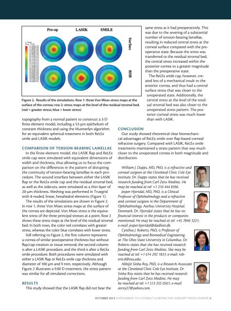
Figure 2. Results of the simulations. Row 1: three Von Mises stress maps at the<br />
surface of the cornea; row 2: stress maps at the level of the residual stromal bed.<br />
(red = greater stress; blue = lower stress)<br />
topography from a normal patient to construct a 3-D<br />
finite element model, including a 53-µm epithelium of<br />
constant thickness and using the Munnerlyn algorithm<br />
for an equivalent spherical treatment in both ReLEx<br />
smile and LASIK models.<br />
COMPARISON OF TENSION-BEARING LAMELLAE<br />
In the finite element model, the LASIK flap and ReLEx<br />
smile cap were simulated with equivalent dimensions of<br />
width and thickness, thus allowing us to focus the comparison<br />
on the differences in the pattern of disrupting<br />
the continuity of tension-bearing lamellae in each procedure.<br />
The wound interface between either the LASIK<br />
flap or the ReLEx smile cap and the residual stromal bed,<br />
as well as the sidecuts, were simulated as a thin layer of<br />
20-µm thickness. Meshing was performed in Truegrid<br />
with 8-noded, linear, hexahedral elements (Figure 1).<br />
The results of the simulations are shown in Figure 2.<br />
In row 1, three Von Mises stress maps at the surface of<br />
the cornea are depicted. Von Mises stress is the equivalent<br />
stress of the three principal stresses at a point. Row 2<br />
shows these stress maps at the level of the residual stromal<br />
bed. In both rows, the color red correlates with greater<br />
stress, whereas the color blue correlates with lower stress.<br />
Still referring to Figure 2, the first column represents<br />
a cornea of similar postoperative thickness but without<br />
flap/cap creation or tissue removal; the second column<br />
is after a LASIK procedure, and the third is after a ReLEx<br />
smile procedure. Both procedures were simulated with<br />
either a LASIK flap or ReLEx smile cap thickness and<br />
diameter of 100 µm and 9 mm, respectively. Although<br />
Figure 2 illustrates a 9.00 D treatment, the stress pattern<br />
was similar for all simulated corrections.<br />
RESULTS<br />
This study showed that the LASIK flap did not bear the<br />
ReLEx smile: Flapless. All-femto. Single-step.<br />
same stress as it had preoperatively. This<br />
was due to the severing of a substantial<br />
number of tension-bearing lamellae,<br />
resulting in reduced central stress at the<br />
corneal surface compared with the preoperative<br />
state. Because the stress was<br />
transferred to the residual stromal bed,<br />
the central stress increased within the<br />
posterior cornea to a greater magnitude<br />
than the preoperative state.<br />
The ReLEx smile cap, however, created<br />
less of a mechanical insult to the<br />
anterior cornea, and thus had a central<br />
surface stress that was closer to the<br />
unoperated state. Additionally, the<br />
central stress at the level of the residual<br />
stromal bed was also closer to the<br />
unoperated stress pattern. The posterior<br />
corneal stress was much lower<br />
than with LASIK.<br />
CONCLUSION<br />
Our study showed theoretical clear biomechanical<br />
advantages of ReLEx smile over flap-based corneal<br />
refractive surgery. Compared with LASIK, ReLEx smile<br />
treatments maintained a stress pattern that was much<br />
closer to the unoperated cornea in both magnitude and<br />
distribution.<br />
William J. Dupps, MD, PhD, is a refractive and<br />
corneal surgeon at the Cleveland Clinic Cole Eye<br />
Institute. Dr. Dupps states that he has received<br />
research funding from <strong>Carl</strong> <strong>Zeiss</strong> <strong>Meditec</strong>. He<br />
may be reached at tel: +1 216 444 8396.<br />
Jesper Hjortdal, MD, PhD, is a Clinical<br />
Professor of Ophthalmology and a refractive<br />
and corneal surgeon in the Department of<br />
Ophthalmology, Aarhus University Hospital,<br />
Denmark. Dr. Hjortdal states that he has no<br />
financial interest in the products or companies<br />
mentioned. He may be reached at tel: +45 7846 3221;<br />
e-mail: jesper.hjortdal@dadlnet.dk.<br />
Cynthia J. Roberts, PhD, is Professor of<br />
Ophthalmology and Biomedical Engineering<br />
at The Ohio State University in Columbus. Dr.<br />
Roberts states that she has received research<br />
funding from <strong>Carl</strong> <strong>Zeiss</strong> <strong>Meditec</strong>. She may be<br />
reached at tel: +1 614 292 1831; e-mail: roberts.8@osu.edu.<br />
Abhijit Sinha Roy, PhD, is a Research Associate<br />
at the Cleveland Clinic Cole Eye Institute. Dr.<br />
Sinha Roy states that he has received research<br />
funding from <strong>Carl</strong> <strong>Zeiss</strong> <strong>Meditec</strong>. He may<br />
be reached at tel: +1 513 255 0561; e-mail:<br />
asroy27@yahoo.com.<br />
OCTOBER 2012 SUPPLEMENT TO CATARACT & REFRACTIVE SURGERY TODAY EUROPE 5