Oscillochloris trichoides strain DG-6 - Microbiology
Oscillochloris trichoides strain DG-6 - Microbiology
Oscillochloris trichoides strain DG-6 - Microbiology

Create successful ePaper yourself
Turn your PDF publications into a flip-book with our unique Google optimized e-Paper software.
<strong>Microbiology</strong> (1 999), 145, 1743-1 748 Printed in Great Britain<br />
Department of<br />
Micro biology, Moscow<br />
State University, Moscow,<br />
1 19899, Russia<br />
2 Institute of Biochemistry<br />
and Physiology of<br />
Microorganisms, Russian<br />
Academy of Sciences,<br />
Pushchino, 142292, Russia<br />
INTRODUCTION<br />
<strong>Oscillochloris</strong> <strong>trichoides</strong> <strong>DG</strong>-6 belongs to the filamen-<br />
tous anoxygenic photosynthetic bacteria (Pierson &<br />
Castenholz, 1995). As far as we know, this <strong>strain</strong> of<br />
<strong>Oscillochloris</strong> is the only one that has been isolated in<br />
pure culture (Keppen et al., 1993, 1994). Morpho-<br />
logically it is similar to Chloroflexus aurantiacus. 0.<br />
<strong>trichoides</strong> <strong>DG</strong>-6, however, unlike Chloroflexus uurun-<br />
tiacus, produces gas vesicles, is rnesophilic and grows<br />
only in the light under anaerobic conditions. The results<br />
of DNA-DNA hybridization and nucleotide sequencing<br />
of 5s rRNA indicate the absence of a close phylogenetic<br />
relationship between this species, Chloroflexus auran-<br />
tiacus and the green sulfur bacterium Chloro bium<br />
vibrioforme (Keppen et al., 1994). 0. <strong>trichoides</strong> is a<br />
photolithoautotroph that uses hydrogen or sulfide for<br />
............................. ** .................................................................................................. ......*..l*..l*..*<br />
Abbreviations : PEP, phosphoenolpyruvate; 3-PGA, 3-phosphoglycerate.<br />
Evidence for the presence of the reductive<br />
pentose phosphate cycle in a filamentous<br />
anoxygenic photosynthetic bacterium,<br />
<strong>Oscillochloris</strong> <strong>trichoides</strong> <strong>strain</strong> <strong>DG</strong>-6<br />
Ruslan N. Ivanovsky,’ Yuri I. Fal,’ Ivan A. Berg,’ Natalya V. Ugolkova,’<br />
Elena N. Krasilnikova,’ Olga I. Keppen,’ Leonid M. Zakharchuc’<br />
and Anatolii M. Zyakun’<br />
Author for correspondence: Ruslan N. Ivanovsky. Tel: +7 095 939 4203. Fax: +7 095 939 4658.<br />
e-mail : ruslan@protein.bio.msu.su<br />
Studies on autotrophic CO, fixation by the filamentous anoxygenic<br />
photosynthetic bacteri um <strong>Oscillochloris</strong> frichoides <strong>strain</strong> <strong>DG</strong>- 6 demonstrated<br />
that, unlike other green bacteria, this organism metabolized CO, via the<br />
reductive pentose phosphate cycle. Both key enzymes of this cycle - ribulose-<br />
1,5-bisphosphate carboxylase/oxygenase and phosphoribulokinase - were<br />
detected in cell extracts. The main product of ribulose 1,5-bisphosphate-<br />
dependent CO, fixation was 3-phosphoglyceric acid. KCN, which is known to be<br />
a competitive inhibitor of ribulose-1,5-bisphosphate carboxylase/oxygenase,<br />
completely inhibited the CO, assimilation by whole cells as well as by cell<br />
extracts of 0. frichoides. The I3Ul2C carbon isotope fractionation during<br />
photoautotrophic growth of 0. <strong>trichoides</strong> was -19*7O/io, which is close to that<br />
obtained for autotrophic organisms that use ribulose-1,5-bisphosphate<br />
carboxylase as the primary carboxylation enzyme. Cell extracts of 0. <strong>trichoides</strong><br />
contained all the enzymes of the tricarboxylic acid cycle except 2-oxoglutarate<br />
dehydrogenase. No activity of isocitrate lyase, a key enzyme of the glyoxylate<br />
shunt, was found in cell extracts of 0. frichoides <strong>DG</strong>-6.<br />
Keywords : green bacteria, <strong>Oscillochloris</strong> <strong>trichoides</strong>, CO, fixation pathway, ribulose-<br />
1,5-bisphosphate carboxylase/oxygenase, tricarboxylic acid cycle<br />
CO, fixation. The pathway of CO, fixation is not<br />
known. The ability to use organic substrates is very<br />
restricted. Acetate and pyruvate were the only substrates<br />
that stimulated the growth of 0. <strong>trichoides</strong> <strong>DG</strong>-6 on<br />
medium that contained sulfide and bicarbonate (Keppen<br />
et ul., 1994). In this communication, we present evidence<br />
for operation of the reductive pentose phosphate cycle in<br />
0. <strong>trichoides</strong> <strong>strain</strong> <strong>DG</strong>-6.<br />
METHODS<br />
Bacterial <strong>strain</strong>s and growth conditions. 0. <strong>trichoides</strong> <strong>strain</strong><br />
<strong>DG</strong>-6 (DM MSU 327) was grown in the medium described<br />
earlier (Keppen st al., 1994). The medium contained<br />
Na,S.SH,O (0.05 */o, w/v), NaHCO, (0-1 YO, w/v) and sodium<br />
acetate (0.1 ‘/o, w/v) as growth substrates. Carbon isotope<br />
fractionation was determined in cells grown photoautotrophi-<br />
cally in medium without acetate. The concentration of<br />
bicarbonate was 0-5 O/o (w/v). The cultures were grown under<br />
anaerobic conditions (28 “C, 2000 lx) in completely filled<br />
500 ml bottles with magnetic stirring.<br />
0002-32 16 0 1999 SGM 1743
R. N. IVANOVSKY and OTHERS<br />
Chlorobium vibrioforme <strong>strain</strong> 8327 was grown photoautotrophically<br />
in completely filled bottles (28 OC, 2000 lx) on the<br />
medium described by Larsen (1952) with NaHCO, (0-5 YO),<br />
Na,S .9H,O (0.1 '/o) and sodium thiosulfate (02 */o, w/v).<br />
Thiocapsa roseopersicina <strong>strain</strong> BBS (DM MSU 317) was<br />
grown photoautotrophically in Completely filled bottles<br />
(28 "C, 2000 lx) on modified Pfennig medium (Bogorov, 1974)<br />
with NaHCO, (0-5 %), Na,S. 9H20 (0.1 '/o), sodium thiosulfate<br />
(02%) and NaCl (2%, w/v).<br />
ChlorofZexus aurantiacus <strong>strain</strong> B-3 (DM MSU 322) was<br />
grown photoautotrophically on modified <strong>DG</strong>N medium<br />
(Keppen at al., 1994) in which sulfide was replaced by<br />
hydrogen as the electron source and the final concentration of<br />
NaHCO, was 0.5% (w/v). The cells were grown at 55 "C<br />
(2000 lx) in screw-capped bottles approximately one-third full<br />
of medium and the remaining volume filled with H, gas.<br />
Whole-cell la belling studies. Cultures from the late exponential-growth<br />
phase were harvested, washed twice with mineral<br />
growth medium lacking Na,S and NaHCO, and resuspended<br />
in the same medium at a density of 0.143 mg protein ml-'.<br />
Experiments for the assimilation of 14C0, by cells of phototrophic<br />
bacteria were carried out in medical syringes at a light<br />
intensity of 3000 lx. After 30 min preincubation with NaHCO,<br />
(5 mM), Na2S (500 mg 1-l) (or H,) and inhibitors (if necessary),<br />
the reaction was started by the addition of NaH14C0,<br />
(0-04 MBq), and stopped at fixed time intervals by the filtration<br />
of 1 ml cell suspension through 045 pm nitrocellulose filters.<br />
Filters were counted in an LKB RacBeta model 1127 liquid<br />
scintillation counter.<br />
Preparation of cell extracts and enzyme assays. Cells were<br />
collected and washed as described above and finally resuspended<br />
in TMD buffer (50 mM Tris/HCl, 5 mM MgCI,,<br />
5 mM DTT, pH 7-8) at a density of 5-10 mg protein m1-I.<br />
Unless otherwise indicated, cell extracts were prepared by<br />
sonication at 22 kHz, 2 min, 4 "C. Debris was removed by<br />
centrifugation of the extract at 45 000 g for 30 min (4 "C) and<br />
the supernatant (cell extract) was used for enzymic studies,<br />
Enzyme activities were assayed immediately after preparation<br />
of cell extracts. All enzyme assays were carried out at room<br />
temperature under aerobic conditions and were repeated<br />
using at least two independent cultures.<br />
Ribulose-1,5-bisphosphate carboxylase (EC 4.1.1.39) was<br />
determined as the ribulose 1,5-bisphosphate-dependent fixation<br />
of NaH14C0, into acid-stable products using a modification<br />
of the procedure described by Schauder et al. (1987).<br />
The assay was performed in Eppendorf tubes. The reaction<br />
mixture (05 ml) contained: TMD buffer, NaHI4CO, (15 rnM,<br />
0.04 MBq) and cell extract (0.5-1.0 mg protein). When needed,<br />
KCN (1 mM) was added. After preincubation for 5 min the<br />
reaction was started by the addition of ribulose 1,5-bisphosphate<br />
(1.0 mM). The reaction was stopped after 5, 10 or<br />
15 min by the addition of 100 p1 12 M formic acid. 14C fixed<br />
during the incubation period was determined from samples<br />
dried at 80 "C and counted by liquid scintillation.<br />
Phosphoribulokinase (EC 2.7.1.19) was assayed by two<br />
methods. The first involved coupling ribulose 5-phosphate<br />
conversion to ribulose 1,S-bisphosphate with the ribulose-1,sbisphosphate<br />
carboxylase reaction. The reaction conditions<br />
were as with ribulose 1,5-bisphosphate carboxylase, except<br />
that ribulose 1,5-bisphosphate was replaced by ribulose 5-<br />
phosphate (1.5 mM). The reaction was started by the addition<br />
of ribulose 5-phosphate. The second method followed the<br />
appearance of ADP (Hart & Gibson, 1975). The reaction<br />
mixture contained 50 mM Tris/HC1 pH 7.5, 5 mM ATP,<br />
15 mM MgCl,, 1 mM ribulose 5-phosphate, 0.4 mM NADH,<br />
1744<br />
2.5 mM phosphoenolpyruvate (PEP), 5 units pyruvate kinase<br />
(type 11, from rabbit muscle), 5 units lactate dehydrogenase<br />
(type XVIl, from bovine heart), 5 mM DTT and cell extract<br />
(003+05 mg protein ml-').<br />
PEP carboxylase (EC 4,1.1,31), PEP carboxykinase (EC<br />
4.1.1.32) and PEP carboxytransphosphorylase (EC<br />
4.1.1.38) were assayed radiochemically by determining PEPdependent<br />
CO, fixation. The assay mixture contained : 50 mM<br />
Tris/HCl pH 7*5,6 mM PEP, 15 mM (0-04 MBq) NaH14C0,,<br />
0.6 mM NADH, 4 mM MnCI,, 5 mM DTT and cell extract<br />
(0-5-1.5 rng protein ml-l). For PEP carboxykinase and PEP<br />
carboxytransphosphorylase determination, the above mixture<br />
was supplemented with 5mM ADP and 2mM K,HPO,,<br />
respectively.<br />
Pyruvate carboxylase (EC 4.4.1 .1) was assayed radiochemically<br />
by determining pyruvate-dependent CO, fixation. The<br />
reaction mixture contained: TMD buffer, 80 mM pyruvate,<br />
15 mM (0.04 MBq) NaH14C0,, 5 mM ATP and cell extract<br />
(25-3-0 mg protein m1-l).<br />
Acetyl-CoA synthecase (EC 6.2.1.1) was measured according<br />
to Berg (1956). Citrate synthase (EC 4.1.3.7) was assayed<br />
using 5,5'-dithiobis-2-nitrobenzoate as described by Srere<br />
(1969). Isocitrate dehydrogenase (EC 1 .la 1-42) was determined<br />
by following the reduction of NADP with isocitrate<br />
according to Cleland et al. (1969). Aconitase (EC 4.2.1.3)<br />
was assayed in a manner similar to the assay for isocitrate<br />
dehydrogenase, but isocitrate was substituted with citrate. 2-<br />
Oxoglutarate dehydrogenase (EC 1.2.4.2) was determined<br />
by following the reduction of NAD with 2-oxoglutarate as<br />
described by Reed & Mukherjee (1969). Succinate dehydrogenase<br />
(EC 1 .3.99.1) was assayed by following the reduction<br />
of triphenyltetrazolium chloride at 485 nm as described by<br />
Holo & Sirevag (1986). Fumarate hydratase (EC 4.2.1 .2) was<br />
determined photometrically by following fumarate disappearance<br />
at 240 nm as described by Brandis-Heep et ~ l (1983). .<br />
Malate dehydrogenase (EC 1.1.1.37) was assayed by following<br />
NADH oxidation by oxaloacetate according to<br />
Yoshida (1969). Isocitrate lyase (EC 4.1.3.1) and malate<br />
synthase (EC 4.1.3.2) were measured as described Dixon &<br />
Kornberg (1959).<br />
For the measurement of pyruvate- and 2-oxoglutarate synthase<br />
reactions, cells were resuspended in TMD buffer with<br />
1 mM EDTA. Frozen cell suspensions were broken by passing<br />
through a cooled X-press. Debris was removed by centrifugation<br />
of the extract at 45000g for 15 min (4 "C). The<br />
supernatant was used for enzymic studies immediately.<br />
Pyruvate synthase (EC 1.2.7.1) was demonstrated as pyruvate<br />
synthesis and as pyruvate-l4C0, exchange. The pyruvate<br />
synthesis reaction mixture (in a final volume of 1 ml) consisted<br />
of: TMD buffer, 0-5 mM EDTA, 2 mM methyl viologen,<br />
1 mM acetyl-CoA, 2 mM serine, 10 mM NaHl'CO,<br />
(0.04 MBq) and cell extract (0.5-1.0 mg protein). Acetyl-CoA<br />
was omitted in the control. The exchange reaction mixture (in<br />
a final volume of 1 ml) consisted of: TMD buffer, 0.5 mM<br />
EDTA, 0.1 mM acetyl-CoA, 10 mM sodium pyruvate, 10 rnM<br />
(0-04 MBq) NaH1*CO, and cell extract (05-1-0 mg protein).<br />
Both assays were conducted anaerobically under hydrogen.<br />
2-Oxoglutarate synthase (EC 1.2.7.3) was assayed in a 2-<br />
o~oglutarate-~'CO, exchange reaction in similar manner to<br />
the assay for pyruvate synthase, except that pyruvate was<br />
substituted with 2-oxoglutarate and acetyl-CoA was substituted<br />
with succinyl-CoA.<br />
Paper chromatography. After termination of the ribulose-1,5bisphosphate<br />
carboxylase reaction by the addition of ethanol<br />
(1 ml) and removal of denatured protein by centrifugation, the<br />
products of the reaction were examined by ascending two-
dimensional chruniatographic procedures using Whatman no.<br />
1 filter paper (12 x 12 cm) according to established procedures<br />
(Bandurski ts: Aselrod, 1951; Wood, 19611). Following ad-<br />
dition of about 3500c.p.m. of the reaction mixture to the<br />
paper, it was developed first in an acid solvent (rnethmol/88 ?O<br />
formic acid/water; 80:15:5, by vol.) and then in the basic<br />
solvent (methahol/ammonium hydroxide/water; 60: 10: 30,<br />
by vo!.). Another method used for the determination of the<br />
reaction products was the method described by Bassham &<br />
Calvin (1957). The solvent used in the first dimension was a<br />
neutral solvent (phcnol/water; 72:28, w/w) and the second<br />
solvent was an acid one (n-butanol/propionic acid/water ;<br />
100: 50 :70, by vol.). 3-Phosphoglycerate (3-PGA) was visual-<br />
ized by using phosphomolyhdate as the indicator and labelled<br />
compounds were located by auroradiography. The verification<br />
of .3-PGA on chromatograms was cstablished by co-chromato-<br />
graphic procedures.<br />
Carbon isotope measurements. Dry biomass samples were<br />
combusted to CO, in scaled Pyrex tubes. Approximately 5 mg<br />
dried sample and 0-5 g coppcr oxide wire were placed into a<br />
preheated Pyrex tube (length 20cn-1, outer diameter 8 mm)<br />
sealed at one end. Sample rubes were then attached to a<br />
vacuum line, evacuated to 2067 Pa and sealed with a torch. The<br />
sealed tubes were placed in a muffle furnace at 5.50 "C for 48 h.<br />
Gases from the combusted cubes were released into a sampling<br />
vacuum linc. The CO, was cryogenically purified and trans-<br />
ferred'jnto tubcs.(2 ml volume). The tubes were broken in :I<br />
device Connected with an inlet system to the mass spec-<br />
t rometer.<br />
The CO, was precipitated as BaCO, following addition of<br />
the saturated solution of Ba(C)k I)2 to the culture medium.<br />
Organic admixtures were removed by ignition in the oxygen<br />
atmosphere at 550 "C. Carbonate was converted to CO, by<br />
the addition of H3POd after the evacuation ot the atmospheric<br />
gases from the reaction vessel.<br />
Isotopic abundance was determined using a dual-inlet, dual-<br />
collector mass spectrometer CH-7 (Varian). The S'"C value is<br />
presented a3 per milk Jeviat ioii Iroin the PDB standard<br />
(Craig, 1957) :<br />
Protein was measured according to the Lowry method, using<br />
bovine scrum albumin as standard.<br />
Materials. Enzymes and biochemicals were purchased from<br />
Sigma. NaH'"C0, was obtained from Amersham. Other<br />
macerials were from standard commercial sources.<br />
RESULTS AND DISCUSSION<br />
0. trichoid~s <strong>DG</strong>-6 is capable of photoautotrophic<br />
:issirnilation of hicarbonate using sulfide as ;1n electron<br />
donor. The addition of cyanide, a comprtitive inhibitor<br />
of ribulose-1,s-bisphosphate carboxyhse (Takabe &<br />
Akazawa, 1977!, leads to complete inhibition of "CO,<br />
assimilation in 0. trichoiiies <strong>DG</strong>-ti cclls (Table 1).<br />
Analogous data were obtained for '1'. roseopersicitza,<br />
which uses the Calvin cycle for CO, assimilation. By<br />
contrast, CO, fixation by whole cells of Cl~loroC?ium<br />
and Cbloroflexus, which do not use the reductive<br />
pentose phosphate cycle for autotrophic CO, fixation,<br />
was not affected by KCK (or cyanide had a subtle effect<br />
Calvin cycle in <strong>Oscillochloris</strong> trichoitles<br />
Table 1. Effect of KCN on bicarbonate assimilation by<br />
whole cells of phototrophic bacteria<br />
The rate of hicarbonate fixation in the absence of electron<br />
donor (sulfide or hydrogen) was negligible ( < 10 0;b 1. Mcasurc-<br />
rnents wcre done in triplicate and values were within 22.596<br />
of each other.<br />
Organism Rate of bicarbonate fixation<br />
lnmol min-l (mg protein)-']<br />
Sulfide Sulfide + KCN*<br />
Osiillochloris trichoirfcs 17.3 07<br />
Thio~~rps~ rojeopersicina? 76.0 3*6<br />
Chlorof7exus aurantincust 7.1 4-3<br />
C/dOYObtU# uihrioforme+ 246.2 229.0<br />
"The conccntration of KCN was 1 mhl.<br />
tSul6dt. was replaced by hydrogen as the electron sourcc.<br />
+The concentration of Na,S was 1 g I-'.<br />
Table 2. Activity of enzymes involved in CO, assimilation<br />
in <strong>Oscillochloris</strong> <strong>trichoides</strong><br />
Enzyme Specific activity<br />
[nmol min-'<br />
(mg protein)-']<br />
.- . .. .<br />
Kibulose- 1 ,5-bisp hosphatc carbox ylase<br />
Phosphuri bulukinase"<br />
PEP carboxylase<br />
PEP carbosykinase<br />
PEP carbuxytr3nsphosphorvlase<br />
Pyruvatc carboxylasc<br />
P y ruv a tt: s y ri t haw<br />
Pyriivate synthesis<br />
P y ru va re-' TO ex c h a nge<br />
2-Oxoglutaratr synthaset<br />
*This activity was obtained by following the appearance of ADP.<br />
When measured by the method which involved coupling ribulosc<br />
S-phosphate ccmversioti to ribulosc 1,5-bisphosphatc with the<br />
r i hi I use- 1 ,i - his p h 0s p h ate car bo x y 1 a sc r c a c t io n , 1 ow c r a c t i v it y<br />
was obtained [ 11 nmol min-' (mg protein)-':.<br />
i This en7.yme was measured as 2-oxoglurarate-"'CO, cxchange.<br />
on C02 fixation) (Table 1). These data indicate that a<br />
functional reductive pentose phosphate cycle is present<br />
in 0. <strong>trichoides</strong> <strong>DG</strong>-6. Activity of ribulose- 1,5-bisphosphatt.<br />
carboxylase and phosphoribulokinase, the key<br />
enzymes of this cyclc, was dctcctcd in cell extracts of 0.<br />
<strong>trichoides</strong> <strong>DG</strong>-4 (Table 2) and the observed activity of<br />
these enzymes is sufficient to support the observed<br />
growth rate of 0. <strong>trichoides</strong> <strong>DG</strong>-6 under autocrophic<br />
conditions (I[ = 0.045 h * l).<br />
The main product of "C-bicarbonate assimilation in the<br />
presence of ribulose 1,S-hisphosphate by cell exmcts of<br />
1745
R. N. IVANOVSKY and OTHERS<br />
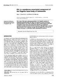
Fig. lm Radioactive products formed from NaH14C0, in the<br />
presence of ribulose 1,5-bisphosphate and cell extracts of<br />
0. <strong>trichoides</strong>. The basic solvent (methanol/ammonium<br />
hydroxide/water) was used in the second dimension (arrow 1)<br />
and the acid solvent (methanol/formic acid/water) was used in<br />
the first dimension (arrow 2). The assay and chromatography<br />
analysis were performed as described in Methods.<br />
0. <strong>trichoides</strong> <strong>DG</strong>-6 was identified by paper chromato-<br />
graphy : in the systems used [methanol/formic acid/<br />
water and rnethanol/ammonium hydroxide/water (see<br />
Fig. l), and phenol/water and n-butanol/propionic<br />
acid/water (data not shown)], a labelled product co-<br />
migrated with authentic 3-PGA. Like 14C-bicarbonate<br />
fixation by whole cells, carboxylation of ribulose 1,5-<br />
1746<br />
-<br />
- 0<br />
E<br />
2000<br />
1500<br />
v<br />
m x 1000<br />
.- Lc<br />
$ f<br />
500<br />
/<br />
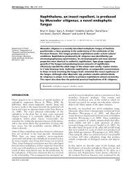
bisphosphate in cell extracts of 0. <strong>trichoides</strong> <strong>DG</strong>-6, as<br />
well as in cell extracts of T. roseopersicina, was<br />
repressed in the presence of cyanide (Fig. 2). These data<br />
support the conclusion that CO, fixation in 0. <strong>trichoides</strong><br />
<strong>DG</strong>-6 under photoautotrophic conditions is carried out<br />
via the reductive pentose phosphate cycle. This was<br />
confirmed by the analysis of the results of 13C/12C stable<br />
isotope fractionation. It was shown earlier (Sirevag<br />
et nl., 1977; Quandt et af., 1977) that photosynthetic<br />
bacteria which incorporate CO, by ribulose-1,S-bis-<br />
phosphate carboxylase fractionate 13C isotope to a much<br />
larger degree than bacteria that use other carboxylation<br />
reactions. The results of our isotope studies (Table 3)<br />
are consistent with the conclusion that the photosyn-<br />
thetic bacterium 0. <strong>trichoides</strong> <strong>DG</strong>-6, like T. roseoper-<br />
sicina, fixes CO, via the reductive pentose phosphate<br />
cycle. A higher level of 13C isotope fractionation (Ai%)<br />
was observed in 0. <strong>trichoides</strong> <strong>DG</strong>-6 than in Chloroflexus<br />
aurantiacus and Chlorobium uibrioforme, which use<br />
different pathways for CO, utilization. Thus, taken<br />
together, the biochemical data and fractionation studies<br />
indicate that 0. <strong>trichoides</strong> <strong>DG</strong>-6 fixes CO, via the<br />
reductive pentose phosphate cycle.<br />
A number of enzymes catalysing carboxylation of<br />
pyruvate and PEP were also determined (Table 2). The<br />
function of these enzymes in 0. <strong>trichoides</strong> <strong>DG</strong>-4 may be<br />
the utilization of PEP synthesized in the course of<br />
bicarbonate fixation via the reductive pentose phosphate<br />
cycle. Oxaloacetate and malate, formed as a result of<br />
these carboxylation reactions, are metabolized via a<br />
truncated tricarboxylic acid cycle. All enzymes of this<br />
cycle except 2-oxoglutarate dehydrogenase were de-<br />
5 10 15 5 10 15<br />
Time (min)<br />
Fig. 2. Activity of ribulose-l,5-bisphosphate carboxylase in cell extracts of 0. <strong>trichoides</strong> (4rng protein) (a) or T.<br />
roseopersicina (2.7 mg protein) (b) in the presence of KCN. W, Complete assay mixture; A, complete assay mixture+KCN;<br />
+, complete assay mixture without ribulose 1,5-bisphosphate.<br />
*...***..***.
Table 3, 13U12C-ca~bon isotope fractionation by different phototrophic bacteria<br />
<strong>Oscillochloris</strong> <strong>trichoides</strong><br />
Thiocapsa roseopersicina<br />
Chloroflexus aurantiacus<br />
Chlorobium vibrioforme<br />
- 28.2<br />
- 29.8<br />
- 22.5<br />
- 20.5<br />
:C 6 13 Ccells, carbon isotope contents of CO, in the cell biomass.<br />
t P3Cmedium, carbon isotope contents of the culture medium.<br />
$ A13C = B13Cc,lls - S13Cmedium.<br />
Table 4. Activity of enzymes of the tricarboxylic acid<br />
cycle in <strong>Oscillochloris</strong> <strong>trichoides</strong><br />
Enzyme Specific activity<br />
[nmol min-'<br />
(mg protein)-']<br />
Acetyl-CoA synthetase<br />
Citrate synthase<br />
Aconitase<br />
Isocitrate dehydrogenase<br />
2-Oxoglutarate dehydrogenase<br />
Succinate dehydrogenase<br />
Fumarate hydratase<br />
Malate dehydrogenase<br />
lsocitrate lyase<br />
Malate synthase<br />
78.2<br />
11-4<br />
63.8<br />
34.3<br />
< 0.1<br />
11.2<br />
200.9<br />
173.3<br />
R. N. IVANOVSKY and OTHERS<br />
Holo, H. (1 989). Chloroflexus aurantiacus secretes 3-hydroxypro-<br />
pionate, a possible intermediate in the assimilation of CO, and<br />
acetate. Arch Microbiol 151, 252-256.<br />
Holo, H. & Sirevag, R. (1986). Autotrophic growth and CO,<br />
fixation of Chloroflexus aurantiacus. Arch Microbiol 145, 173-<br />
180.<br />
Ivanovsky, R. N., Krasilnikova, E. N. & Fal, Y. 1. (1993). A pathway<br />
of the autotrophic CO, fixation in Chloroflexus aurantiacus.<br />
Arch Microbiol 159, 257-264.<br />
Keppen, 0. I., Baulina, 0. I., Lysenko, A. M. & Kondratieva, E. N.<br />
(1993). A new green bacterium of the Chloroflexaceae family.<br />
Mikrobiologiya 62, 267-275.<br />
Keppen, 0. I., Baulina, 0. I. & Kondratieva, E. N. (1994). <strong>Oscillochloris</strong><br />
<strong>trichoides</strong> neotype <strong>strain</strong> <strong>DG</strong>-6. Photosynth Res 41,<br />
29-33.<br />
Kondratieva, E. N., Ivanovsky, R. N. & Krasilnikova, E. N. (1981).<br />
Light and dark metabolism in purple sulfur bacteria. In Soviet<br />
Science Review, vol. 2, pp. 325-364. Edited by V. P. Skulachev.<br />
Guildford, NY: IPC Science and Technology Press.<br />
Larsen, H. (1952). On the culture and general physiology of the<br />
green sulfur bacteria. J Bacteriol64, 187-196.<br />
Pierson, B. K. & Castenholz, R. W. (1995). Taxonomy and<br />
physiology of filamentous anoxygenic phototrophs. In Anoxy-<br />
genic Photosynthetic Bacteria, pp. 3147. Edited by R. E.<br />
Blankenship, M. T. Madigan & C. E. Bauer. Dordrecht : Kluwer.<br />
Quandt, L., Gottschalk, G., Ziegler, H. & Stichler, W. (1977).<br />
Isotope discrimination by photosynthetic bacteria. FEMS Micro-<br />
biol Lett 1, 125-128.<br />
1748<br />
Reed, L. J. & Mukherjee, B. B. (1969). a-Ketoglutarate dehydro-<br />
genase complex from Escherichia coli. Methods Enzymol 13,<br />
55-6 1.<br />
Schauder, R., Widdel, F. & Fuchs, G. (1987). Carbon assimilation<br />
pathways in sulfate-reducing bacteria. 11. Enzymes of a reductive<br />
citric acid cycle in the autotrophic Desulfobacter hydrogeno-<br />
philus. Arch Microbiol 148, 218-225.<br />
Sirevag, R., Buchanan, B. B., Berry, J. A. & Troughton, J. H. (1977).<br />
Mechanisms of CO, fixation in bacterial photosynthesis studied<br />
by the carbon isotope fractionation technique. Arch Microbiol<br />
112,3538.<br />
Srere, P. A. (1969). Citrate synthase. Methods Enzymol 13,3-11.<br />
Strauss, G. & Fuchs, G. (1993). Enzymes of a novel autotrophic<br />
CO, fixation pathway in the phototrophic bacterium Chloroflexus<br />
azjrantiactrs, the 3-hydroxypropionate cycle. Eur Biochem<br />
215,633-643.<br />
Takabe, T. & Akazawa, T. (1977). A comparative study of the effect<br />
0, on photosynthetic carbon metabolism by Chlorobium thiosulfutophilum<br />
and Chrornatium vinosum. Plant Cell Physiol 18,<br />
753-765.<br />
Wood, T. (1 968). The detection and identification of intermediates<br />
of the pentose phosphate cycle and related compounds. J<br />
Chromatogr 35,352-361.<br />
Yoshida, A. (1969). L-Malate dehydrogenase from Bacillus subtilis.<br />
Methods Enzyrnol 13, 141-145.<br />
Received 13 January 1999; revised 22 March 1999; accepted<br />
30 March 1999.