Synthesis of a New Substrate for Exo-galactofuranosidases, 4 ...
Synthesis of a New Substrate for Exo-galactofuranosidases, 4 ...
Synthesis of a New Substrate for Exo-galactofuranosidases, 4 ...

Create successful ePaper yourself
Turn your PDF publications into a flip-book with our unique Google optimized e-Paper software.
Romanian Biotechnological Letters Vol. 15, No.2, 2010<br />
Copyright © 2010 University <strong>of</strong> Bucharest Printed in Romania. All rights reserved<br />
ORIGINAL PAPER<br />
<strong>Synthesis</strong> <strong>of</strong> a <strong>New</strong> <strong>Substrate</strong> <strong>for</strong> <strong>Exo</strong>-galact<strong>of</strong>uranosidases, 4-Nitrocatechol<br />
1-yl α-D-Galact<strong>of</strong>uranoside<br />
Abstract<br />
5096<br />
Received <strong>for</strong> publication, October 19, 2009<br />
Accepted, March 15, 2010<br />
DUMITRU PETRU IGA A , SILVIA IGA A , NICULAE VELICU B , TANASE PETRUT B ,<br />
NICOLAE-DANIEL CONDUR B<br />
A Faculty <strong>for</strong> Biology, Splaiul Independentei 95, Bucuresti-5, Romania, R-05090<br />
B "Spiru Haret" University, Faculty <strong>of</strong> Veterinary Medicine, Dept. <strong>of</strong> Clinical Sciences, Str.<br />
Masina de paine 47, Bucharest-2, Romania, R-02070<br />
e-mail: pdiga49@yahoo.com<br />
Glycosylation <strong>of</strong> 4-nitrocatechol with tetra-O-acetyl α-D-galact<strong>of</strong>uranosyl bromide, by<br />
different methods (Koenigs-Knorr, Helferich, reaction with phenoxide) led to 4’-nitrocatechol 1’-yl<br />
β-D-(2,3,5,6-tetra-O-acetyl)galact<strong>of</strong>uranoside. Glycosylation donor, tetra-O-acetyl<br />
α-D-galact<strong>of</strong>uranosyl bromide, was obtained by bromination <strong>of</strong> penta-O-acetyl β-D-galact<strong>of</strong>uranose<br />
with hydrogen bromide in glacial acetic acid. β-D-Galact<strong>of</strong>uranose pentaacetate was synthesized by<br />
peracetylation <strong>of</strong> D-galactose in boiling pyridine followed by separation <strong>of</strong> the aimed product by<br />
fractional crystallization. The crystalline mass obtained was recrystallized from ethanol. Due to the<br />
fact that penta-O-acetyl β-D-galact<strong>of</strong>uranose has levorotary action, optical rotation was a constant<br />
guide along the preparation <strong>of</strong> the furanosic precursor. The aglycon, 4-nitrocatechol, was prepared<br />
by acidic hydrolysis <strong>of</strong> 2-hydroxy 5-nitrophenyl sulfate, synthesized at its turn by reacting<br />
4-nitrophenol and potassium persulfate in a strongly alkaline environment conferred by potassium<br />
hydroxide. All intermediates and products were characterized chemically and by physical methods.<br />
Keywords: exo-galact<strong>of</strong>uranosidase, 4-nitrocatechol 1-yl α-D-galact<strong>of</strong>uranoside, penta-O-acetyl β-Dgalact<strong>of</strong>uranose,<br />
nitrocatechol, 1 H and 13 C NMR spectra.<br />
Introduction<br />
In spite <strong>of</strong> the fact that the number <strong>of</strong> papers dealing with D-galactopyranosides is by<br />
two or three order <strong>of</strong> magnitude bigger than the similar number concerning Dgalact<strong>of</strong>uranosides,<br />
molecular diversity <strong>of</strong> the latter ones is practically identical with<br />
molecular diversity <strong>of</strong> D-galactopyranosides. Numerous polysaccharides containing Dgalact<strong>of</strong>uranose<br />
have been found both in pathogenic microorganisms [1,2,3,4], as well as in<br />
beneficial ones [5,6]. A special natural reagent <strong>for</strong> galact<strong>of</strong>uranosylation, UDP-α-Dgalact<strong>of</strong>uranose,<br />
has been found in microorganisms producing furanosides [7,8].<br />
Subsequently, it was revealed that UDP-α-D-galact<strong>of</strong>uranose is produced by isomerisation <strong>of</strong><br />
UDP-α-D-galactopyranose by UDP-α-D-galactopyranose mutase [8,9]. Neutral<br />
glycosphingolipids are represented by three compounds: longiside (β-D-Galf-3-α-D-Galp-<br />
1’Cer) [10], agelagalastatin (α-D-Galf-2-β-D-Galf-3-α-D-Galp-1’Cer) [11], ectyoceramide (β-<br />
D-Galf-1’Cer) [12]. Gangliosides having relatively complicated structures were isolated from<br />
marine organisms: Acanthaster planci (two) [13] and Asterina pectinifera [14].<br />
Glycoglycerolipids are exemplified by both α configuration – sn 1,2-di-O-acyl 3-α-Dgalact<strong>of</strong>uranoside<br />
glycerol, in Acholeplasma axanthum [15] and ß configuration, sn 1,2-di-Oacyl<br />
3-β-D-galact<strong>of</strong>uranoside glycerol in: Mycoplasma mycoides [16], Bacteroides<br />
symbioticus [17], Bifidobacterium bifidum [18], Arthrobacter globi<strong>for</strong>mis [19]. A unique<br />
galact<strong>of</strong>uranosylated poly(glycerophosphate) lipoteichoic acid has been isolated from a<br />
Streptococcus sp. closely related to Streptococcus pneumoniae [20]. An interesting feature <strong>of</strong>
DUMITRU PETRU IGA, SILVIA IGA, NICULAE VELICU, TANASE PETRUT, NICOLAE-DANIEL CONDUR<br />
furanosic ring is its existence in glycoglycerolipids with the structure sn 1-β-Dgalact<strong>of</strong>uranoside<br />
2,3-di-O-acyl glycerol, that have been isolated till now exclusively from<br />
Archaebacteria [21]. Furanosylated sterylglycolipids are illustrated by two crossasterosides<br />
from the starfish Crossaster papposus, (24R)-24-ethyl-5α-cholestane-3ß, 6ß, 8, 15α,16ß, 29hexaol<br />
29-O-[2-O-methyl-ß-D-xylopyranosyl-(1 2)-ß-D-galact<strong>of</strong>uranoside] and its 4ßhydroxy<br />
analogue [22] and by indicoside A, (24R)-24-methyl-5α-cholestane-3ß, 6α, 7α, 8,<br />
15β, 24, 28-heptol-ß-D-(5-O-methyl)galact<strong>of</strong>uranoside, from the starfish Astropecten indicus<br />
[23]. Helminthosporoside toxin C from Helminthosporium sacchari possesses a variable<br />
number <strong>of</strong> β-D-galact<strong>of</strong>uranosic residues and these structures represent the group <strong>of</strong><br />
sesquiterpenglycolipids (polyprenylglycolipids) [24]. Lecythophorin, a representative <strong>of</strong><br />
resorcinolglycolipids and an interspecific mediator has also D-galact<strong>of</strong>uranose in its<br />
molecule [25]. Glycoproteins based on D-galact<strong>of</strong>uranose are represented by α-Dgalactosidase<br />
from Aspergillus niger [26,27] and by phospholipase C and phytase <strong>of</strong> A.<br />
fumigatus [28].<br />
Development <strong>of</strong> enzymology <strong>of</strong> furanosidases, either <strong>for</strong> metabolic studies or <strong>for</strong> the<br />
use <strong>of</strong> these enzymes as analytical or synthetic tools, is strongly dependent on the elaboration<br />
<strong>of</strong> suitable substrates. In this paper, a new substrate <strong>for</strong> exo-galact<strong>of</strong>uranosidases has been<br />
synthesized and characterized, 4-nitrocatechol 1-yl α-D-galact<strong>of</strong>uranoside.<br />
Materials and methods<br />
Materials. CDCl3 containing TMS, D-galactose, 4-nitrophenyl ß-Dgalactopyranoside,<br />
1,2-dichloroethane, acetic acid, acetic anhydride, acetone, anthrone,<br />
chlor<strong>of</strong>orm, diethyl ether, ethanol, methanol, 4-nitrophenol, p-toluenesulfonic acid, pyridine,<br />
toluene, active carbon, ammonium molibdate, cadmium carbonate, calcium sulfate, Ce(SO4)2,<br />
concd. sulfuric acid, dry zinc chloride, HBr in glacial acetic acid (33 %), molecular sieve (4<br />
Å), potassium hydroxide, potassium persulfate, sodium carbonate, sodium bicarbonate,<br />
sodium hydroxide, sodium metal, ferric chloride, ready-to-use thin layer plates, silicagel <strong>for</strong><br />
column chromatography, Celite, Dowex 50 WX2, were either from Merck or Fluka.<br />
Methods used<br />
NMR Spectra registration. 1 H and 13 C NMR spectra <strong>of</strong> initial precursors,<br />
intermediates, and final products were registered in peracetylated <strong>for</strong>m in CDCl3 containing<br />
TMS. The spectra <strong>of</strong> intermediates and final products have been referred to the spectra <strong>of</strong><br />
peracetylated initial precursors.<br />
One-dimensional NMR studies. NMR experiments were per<strong>for</strong>med on a Bruker<br />
Avance DRX 400 spectrometer using 400 and 100 MHz <strong>for</strong> 1 H and 13 C frequencies,<br />
respectively.<br />
Two-dimensional NMR experiments. The 1 H- 1 H correlation spectroscopy (COSY)<br />
and 1 H- 13 C heteronuclear multiple quantum coherence (HMQC) experiments were carried out<br />
with an inverse probe.<br />
IR spectra were recorded as KBr pellets or as a solution in Nujol on a Bruker<br />
Equinox 55 FT-IR spectrometer.<br />
Acetylation. Compounds were solved in pyridine and stirred overnight with an excess<br />
<strong>of</strong> acetic anhydride, the ratio between acetic anhydride and pyridine was 2/1 (v/v).<br />
Benzoylation. Compounds were solved in pyridine, the solution was cooled on ice<br />
and benzoyl chloride was added in small portions, the ratio between solvent and benzoylatig<br />
agent was 10:1. Then pyridine was removed by coevaporating with toluene. Excess benzoyl<br />
chloride was decomposed by adding ice in the reaction mixture and the acidic products<br />
Romanian Biotechnological Letters, Vol. 15, No. 2, 2010 5097
<strong>Synthesis</strong> <strong>of</strong> a <strong>New</strong> <strong>Substrate</strong> <strong>for</strong> <strong>Exo</strong>-galact<strong>of</strong>uranosidases, 4-Nitrocatechol 1-yl α-D-Galact<strong>of</strong>uranoside<br />
<strong>for</strong>med (HCl, benzoic acid) were removed by partition between chlor<strong>of</strong>orm and a saturated<br />
solution <strong>of</strong> NaHCO3.<br />
<strong>Synthesis</strong> <strong>of</strong> 1,2-dihydroxy 4-nitrobenzene (4-nitrocatechol). Reaction <strong>of</strong> 4nitrophenol<br />
with potassium persulfate in a strongly alkaline environment conferred by<br />
potassium hydroxide gave 2-hydroxy 5-nitrophenyl sulfate (4-nitrocatechol sulfate); the<br />
acidic hydrolysis <strong>of</strong> this compound and partition between water and diethyl ether led to 4nitrocatechol<br />
[29].<br />
Glycosylation agent and Koenigs-Knorr glycosylation. Penta-O-acetyl-ß-Dgalact<strong>of</strong>uranoside<br />
was prepared as indicated Schlubach and Prochovnick [30]. By<br />
bromination <strong>of</strong> peracetylated sugar with hydrobromic acid in glacial acetic acid, 1-bromo-1deoxy-α-D-2,3,5,6-tetra-O-acetyl-galact<strong>of</strong>uranoside<br />
was obtained [31]. Glycosylation <strong>of</strong> 4nitrocatechol<br />
was per<strong>for</strong>med in boiling dry toluene, by using cadmium carbonate as chemical<br />
condensing agent [32,33]. Freshly dried molecular sieves (4 Å) and calcium sulfate were used<br />
as water scavengers.<br />
Helferich glycosylation was made by boiling the protected sugar with ptoluenesulfonic<br />
acid or dry zinc chloride in toluene [34].<br />
Glycosylation <strong>of</strong> 4-nitrocatechol as potassium phenoxide with 1-bromo-1-deoxy-α-<br />
D-2,3,5,6-tetra-O-acetyl-galact<strong>of</strong>uranoside was accomplished in acetone [35].<br />
Thin layer chromatography (TLC) was achieved on ready-to-use plates in the<br />
following mobile phases: solvent system (SS) I (chlor<strong>of</strong>orm-methanol, 19/1, v/v), SS II<br />
(toluene-ethanol, 3/1, v/v), SS III (toluene-methanol, 7/1), SS IV (chlor<strong>of</strong>orm-methanolwater,<br />
60/25/4, v/v). Visualization <strong>of</strong> spots was made in three ways: (a) by dipping the plates<br />
in a solution called mostain consisting <strong>of</strong> water, sulfuric acid, ammonium molybdate and<br />
Ce(SO4)2, followed by heating on a hot plate; (b) under UV light in comparison with 4nitrophenyl<br />
ß-D-galactopyranoside; (c) by dipping the plates in a diluted solution <strong>of</strong> sodium<br />
hydroxide.<br />
Reaction mixture after glycosylation in case <strong>of</strong> Koenigs-Knorr was diluted with<br />
chlor<strong>of</strong>orm, mixed with Celite, filtered and evaporated to dryness. In case <strong>of</strong> Helferich<br />
method, reaction mixture was neutralized with sodium carbonate, filtered and the solvent<br />
removed. The mixture from glycosylation <strong>of</strong> phenoxide was neutralized with acetic acid,<br />
repeatedly mixed with toluene and evaporated by rotavapor.<br />
Either residue was partitioned between water and chlor<strong>of</strong>orm, aqueous phase being<br />
discarded. Chlor<strong>of</strong>ormic phase was repeatedly chromatographed on silica gel column. The<br />
purified product was analyzed <strong>for</strong> sugar content by anthrone reaction [36] and <strong>for</strong> 4nitrocatechol<br />
by using molar coefficient <strong>of</strong> 12.500 cm 2 ·mol −1 [37]. A small portion was<br />
acetylated <strong>for</strong> NMR analysis.<br />
Protecting acetate groups were removed by Zemplen deacylation <strong>of</strong> the product with<br />
0.2 M sodium methoxide followed by neutralization with Dowex 50 WX2 (H + ), filtration and<br />
concentration. In this <strong>for</strong>m, the product is ready <strong>for</strong> enzymatic test.<br />
Results and discussion<br />
2-Hydroxy 5-nitrophenylsulfate (4-nitrocatechol sulfate) prepared in this paper had m.<br />
p. 245-250 o C (decomp.) and gave a red color with a neutral solution <strong>of</strong> ferric chloride. Its IR<br />
spectra presented a band <strong>of</strong> absorption at 1254 cm −1 (sulfate ester); this band dissapeared after<br />
its conversion to 4-nitrocatechol by acidic hydrolysis. It was also tested as substrate <strong>for</strong><br />
arylsulphatase from Helix pomatia [37]. 1,2-Dihydroxy 4-nitrobenzene (4-nitrocatechol)<br />
obtained by acidic hydrolysis had m. p. 175 o C and its dibenzoate had m. p. 156 o C, after two<br />
recrystallizations from aqueous ethanol.<br />
5098<br />
Romanian Biotechnological Letters, Vol. 15, No. 2, 2010
DUMITRU PETRU IGA, SILVIA IGA, NICULAE VELICU, TANASE PETRUT, NICOLAE-DANIEL CONDUR<br />
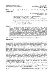
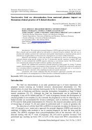
TLC analysis <strong>of</strong> total glycosylation mixture and its visualization with mostain<br />
presented numerous chromatographical bands (Fig. 1a). Visualization <strong>of</strong> an identical plate<br />
a b c d<br />
Fig. 1. TLC analysis <strong>of</strong> purification steps <strong>of</strong> 4-nitrocatechol 1-yl α-D-galact<strong>of</strong>uranoside. 1a, both starts: crude<br />
mixture <strong>of</strong> glycosylation; migration, SS I; visualization, mostain. 1b, ibid.; visualization, sodium<br />
hydroxide. 1c and 1d, the glycoside after purification; migration, SS III; visualization with mostain (1c);<br />
visualization with sodium hydroxide (1d).<br />
with sodium hydroxide solution indicated two or even three red bands and one or two yellow<br />
bands (Fig. 1b). The number <strong>of</strong> spots and their <strong>for</strong>m and position, under UV light, was similar<br />
to alkaline visualization. A well known fact, concerning 4-nitrocatechol and its derivatives is<br />
that the free phenol assumes red color in alkaline environment while the blocking <strong>of</strong> at least<br />
one phenolic group prevents the compound to turn to red, the color assumed at strongly<br />
alkaline pH being yellow. Such behaviour has been attributed to 2-hydroxy 5nitrophenylsulfate<br />
and constitutes the principle <strong>of</strong> its use as substrate <strong>for</strong> (aryl)sulfatases, this<br />
compound being extremely resistant to alkaline solution [37]. Glycosidic linkage with<br />
phenolic aglycons is also relatively stable in alkaline solution and on this property is based the<br />
use <strong>of</strong> 4-nitrophenol-glycosides as substrates <strong>for</strong> a series <strong>of</strong> enzymes: glucosidases,<br />
galactosidases, hexosaminidases, arabinosidases, etc., [38]. Contrary to inorganic ester or<br />
glycosidic linkages, organic ester bonds are extremely susceptible to alkaline treatment and<br />
this property is largely used to remove ester protecting groups by Zemplen deacylation or to<br />
simplify the structure <strong>of</strong> glycolipids or phospholipids by the so-called mild alkaline treatment<br />
[33,36,38]. The compounds <strong>for</strong>ming red bands were di-O-acetyl 4-nitrocatechol (the fastest<br />
one), O-acetyl 4-nitrocatechol and 4-nitrocatechol (the slowest one). In fact, some <strong>of</strong> them<br />
become red instantly while others could be contemplated while grew red in time <strong>of</strong> seconds<br />
after alkalinization, as a direct pro<strong>of</strong> <strong>of</strong> alkaline hydrolysis <strong>of</strong> the acetate esters in situ.<br />
Acylation products <strong>of</strong> aglycon – a reaction similar to in vivo acylation by transacylases – are<br />
consecrated <strong>for</strong> glycosylation reactions when protecting groups are esters [32].<br />
A relatively abundant product was obtained having the following properties: in native<br />
state it migrated in the first third <strong>of</strong> the plate with SS I and SS IV. It presented no color with<br />
sodium hydroxide and no absorption in UV light, however, it colored intensely with mostain.<br />
After Zemplen deacylation and neutralization with Dowex 50 WX2 (H + ) it could not be<br />
moved from the start by TLC, with all solvent systems used in this paper. According to these<br />
properties, it could be either unreacted sugar or ß-D-galact<strong>of</strong>uranosyl-ß’-D-galact<strong>of</strong>uranosyl<br />
(a trehalose type furanosic disaccharide), but we did not undertake further studies on it.<br />
After repeatedly achieved chromatography, a product was isolated giving one spot by<br />
TLC in UV light, by mostain visualization and by visualization with sodium hydroxide (Fig.<br />
1c, 1d) in all solvent systems used. It strongly absorbed UV light in a manner <strong>of</strong> remarcable<br />
resemblance with 4-nitrophenyl ß-D-galactopyranoside, and differed from the absorption <strong>of</strong><br />
free phenol. In alkaline solution it colored in yellow, and this constituted a pro<strong>of</strong> that at least<br />
one hydroxyl group <strong>of</strong> 4-nitrocatechol was blocked by a linkage resistant at alkaline pH. Its<br />
migration as a monosaccharide by TLC, in comparison with 4-nitrophenyl ß-Dgalactopyranoside,<br />
was the first token that at least one hydroxyl group <strong>of</strong> 4-nitrocatechol was<br />
free. After acidic hydrolysis <strong>of</strong> the glycoside, a red color appeared, when the pH was brought<br />
to alkaline value. This product contained D-galactose (anthrone) and 4-nitrocatechol<br />
Romanian Biotechnological Letters, Vol. 15, No. 2, 2010 5099
<strong>Synthesis</strong> <strong>of</strong> a <strong>New</strong> <strong>Substrate</strong> <strong>for</strong> <strong>Exo</strong>-galact<strong>of</strong>uranosidases, 4-Nitrocatechol 1-yl α-D-Galact<strong>of</strong>uranoside<br />
(spectrophotometric determination, 510 nm) in the molar ratio 1:1. There was an excellent<br />
agreement between chemical data and NMR data.<br />
1,2-Di-O-acetyl 4-nitrobenzene. NMR spectra <strong>of</strong> peracetylated 4-nitrocatechol<br />
disclosed the presence <strong>of</strong> constituent chemical groups:<br />
1 H-NMR (CDCl3; δ, ppm; J, Hz): 8.12 (s, H-3), 8.15 (d, 8.8 Hz, H-5), 7.39 (d, 8.8 Hz,<br />
H-6), 2.33 and 2.34 (s, 6H, CH3 groups <strong>of</strong> acetate).<br />
13 C-NMR (CDCl3): 147.43 (C-1), 145.46 (C-2), 119.66 (C-3), 142.35 (C-4), 121.89<br />
(C-5), 124.07 (C-6), 20.50 and 20.62 (CH3 groups <strong>of</strong> acetate), 167.28 and 167.56 (CO groups<br />
<strong>of</strong> acetate).<br />
1,2,3,5,6-Penta-O-acetyl β-D-galact<strong>of</strong>uranose. 1 H-NMR (CDCl3; δ, ppm; J, Hz):<br />
6.12 (s, H-1β), 5.11 (d, 1.6 Hz, H-2), 5.02 (dd, 2.0 Hz, 1.6 Hz, H-3), 4.28 (d, 1.2 Hz, H-4),<br />
5.28 (H-5), 4.25 (d, 4.0 Hz, H-6a), 4.13 (dd, 6.8 Hz, 4.4 Hz, H-6b), 1.993, 2.056, 2.061, 2.072<br />
(s, 15H, CH3 groups <strong>of</strong> acetate). It presented a levorotary action, [α]D 23 –41 0 in CHCl3.<br />
13 C-NMR (CDCl3): 98.96 (C-1), 80.44 (C-2), 76.19 (C-3), 82.04 (C-4), 69.11 (C-5),<br />
62.39 (C-6), 20.47, 20.50, 20.52, 20.65 and 20.83 (CH3 groups <strong>of</strong> acetate), 168.89, 169.26,<br />
169.62, 169.85 and 170.36 (CO groups <strong>of</strong> acetate).<br />
2’-O-Acetyl-4’-nitrobenzene 1’-yl α-D-(2,3,5,6-tetra-O-acetyl)galact<strong>of</strong>uranoside.<br />
1 H-NMR (CDCl3; δ, ppm; J, Hz): 5.14 (d, 3.6 Hz, H-1α), 5.15 (d, 8.0 Hz, H-2), 5.53 (d, 2.4<br />
Hz, H-3), 4.18 (dd, 7.6 Hz, 3.6 Hz, H-4), 5.49 (d, 3.2 Hz), 4.29 (dd, 5.2 Hz, 6.8 Hz, H-6a),<br />
4.13 (dd, 2.0 Hz, 6.8 Hz, H-6b), 7.98 (H-3’), 8.12 (d, 2.8 Hz, H-5’), 7.14 (d, 9.2 Hz, H-6’),<br />
2.02, 2.08, 2.09, 2.19, 2.33 (s, 15H, CH3 groups <strong>of</strong> acetate).<br />
13 C-NMR (CDCl3): 98.70 (C-1), 70.47 (C-2), 66.56 (C-3), 71.56 (C-4), 67.66 (C-5),<br />
61.35 (C-6), 153.49 (C-1’), 139.79 (C-2’), 119.69 (C-3’), 142.73 (C-4’), 123.81 (C-5’),<br />
114.31 (C-6’). 168.37, 169.70, 170.01, 170.12, 170.30 (CO groups <strong>of</strong> acetate).<br />
Aromatic character <strong>of</strong> the prepared 4-nitrocatechol was evident from the shift values<br />
<strong>of</strong> protons in 1 H NMR spectra as well as from the shift values <strong>of</strong> carbon atoms in 13 C NMR<br />
spectra. On the other hand, excellent spectral data, as a consequence <strong>of</strong> the purity degree <strong>of</strong><br />
the envisaged compound, have been obtained <strong>for</strong> 1,2,3,5,6-Penta-O-acetyl β-Dgalact<strong>of</strong>uranose,<br />
the quality <strong>of</strong> the spectra being evident by comparison with similar data from<br />
literature [24,39,40].<br />
A compound has been synthesized containing D-galactose and 4-nitrocatechol in the<br />
molar ratio 1:1. The linkage between carbohydrate and 4-nitrocatechol is a glycosidic one: it<br />
is cleaved by acidic hydrolysis. Were it a direct carbon-carbon bond between sugar and<br />
phenol – a type <strong>of</strong> Friedel-Crafts derivatives, <strong>for</strong>med as secondary products in glycosylation<br />
<strong>of</strong> phenols – such linkages are not cleaved by acidic hydrolysis; moreover, NMR data were<br />
completely different <strong>for</strong> Friedel-Crafts compounds. 1 H NMR spectra <strong>of</strong> the glycoside<br />
synthesized in this paper indicate an α-galact<strong>of</strong>uranoside <strong>of</strong> 4-nitrocatechol. Coupling<br />
constant value <strong>of</strong> 3.6 Hz at H-1, as well as the other coupling constants, clearly indicated the<br />
furanosic ring and α-configuration. Another argument to this assertion is the value <strong>of</strong><br />
chemical shift <strong>of</strong> C-1, 98.7 ppm. The molar ratio <strong>of</strong> 1:1 between D-galact<strong>of</strong>uranose and 4nitrocatechol<br />
has been confirmed by NMR spectra. By comparing a good deal <strong>of</strong> NMR data<br />
from our laboratory [33,41,42,43] and from others [12,39,40], as well as the data from this<br />
paper we have concluded that the linkage <strong>of</strong> acetate to a phenol resulted in a downfield shift<br />
<strong>of</strong> acetate methyl group from 1.9-2.0 ppm to about 2.3 ppm: estrone, 2.3 ppm; D,L-αtocopherol,<br />
2.3 ppm; 4-nitrocatechol, 2.3 ppm. A similar, although contrary, effect has been<br />
noticed in case <strong>of</strong> acetate carbonyl group: in case this group is linked to sugars, chemical shift<br />
<strong>for</strong> this group is around 170 ppm. On the other hand, when this group is found in phenol type<br />
esters, the following values have been registered: estrone, 169.6 ppm; D,L-α-tocopherol,<br />
169.7 ppm; 4-nitrocatechol, 167.3, 167.6 ppm; in case <strong>of</strong> the glycoside synthesized in this<br />
paper, a distinct value <strong>of</strong> 168.4 ppm was noticed. In 1 H NMR spectra <strong>of</strong> glycoside, a similar<br />
5100<br />
Romanian Biotechnological Letters, Vol. 15, No. 2, 2010
DUMITRU PETRU IGA, SILVIA IGA, NICULAE VELICU, TANASE PETRUT, NICOLAE-DANIEL CONDUR<br />
value, 2.3 ppm, was registered. These values unequivocally demonstrated that only the group<br />
1-hydroxy <strong>of</strong> 4-nitrocatechol was galact<strong>of</strong>uranosylated. As is evident from the above detailed<br />
spectra <strong>of</strong> the compounds, the glycoside synthesized by us contained simultaneously the<br />
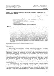
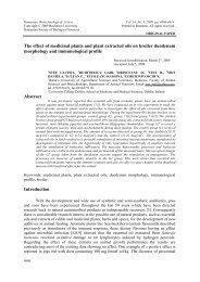
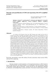
NMR signals <strong>for</strong> sugar and <strong>for</strong> 4-nitrocatechol. As a final conclusion, chemical and physical<br />
data converged to the following structure <strong>of</strong> glycoside: 2’-O-acetyl-4’-nitrobenzene 1’-yl α-D-<br />
(2,3,5,6-tetra-O-acetyl)galact<strong>of</strong>uranoside (Fig. 2).<br />
AcO<br />
AcO<br />
O<br />
OAc<br />
OAc<br />
O<br />
OAc<br />
KO<br />
OK<br />
NO 2<br />
OAc AcO<br />
Br<br />
OAc<br />
AcO<br />
O<br />
OAc<br />
AcO<br />
AcO<br />
Romanian Biotechnological Letters, Vol. 15, No. 2, 2010 5101<br />
O<br />
OAc<br />
OH<br />
NO 2<br />
4<br />
O<br />
OAc 1<br />
AcO<br />
AcO<br />
5<br />
6<br />
2 O<br />
OAc<br />
Fig. 2. 4-Nitrocatechol 1’-yl-α-D-galact<strong>of</strong>uranoside, a new possible substrate <strong>for</strong> exo-galact<strong>of</strong>uranosidases and<br />
glycosyl transferases.<br />
The first enzymatic substrates <strong>for</strong> exo-ß-D-galact<strong>of</strong>uranosidase were methyl β-Dgalact<strong>of</strong>uranoside,<br />
ethyl β-D-galact<strong>of</strong>uranoside, 5-O-β-D-galact<strong>of</strong>uranosyl- and 6-O-β-Dgalact<strong>of</strong>uranosyl-D-galactose-containing<br />
substances as well as a heteropolysaccharide with a<br />
complicated structure, peptidophosphogalactomannan [44,45]. The first two compounds were<br />
synthesized by Fischer glycosidation [46], a method <strong>of</strong> historical value that led to the<br />
knowledge <strong>of</strong> sugar isomerism concerning the two configurations, α and ß, and the two types<br />
<strong>of</strong> rings, pyranosic and furanosic. An extremely efficient method, that allowed the separation<br />
<strong>of</strong> the four methyl-galactosides, was used <strong>for</strong> purification i. e., column chromatography on<br />
cellulose [47]. Subsequently, both methyl-furanosides have been labelled with 3 H on C-6, by<br />
reduction <strong>of</strong> the respective methyl ester-methyl galacturonide with NaB 3 H4 [48].<br />
4-Nitrophenyl ß-D-galact<strong>of</strong>uranoside, one <strong>of</strong> the most used substrates <strong>for</strong> exo-furanosidases,<br />
has been synthesized by two routes: (a) from penta-O-benzoyl D-galact<strong>of</strong>uranose (β-isomer or<br />
αβ-mixture <strong>of</strong> isomers) and 4-nitrophenol in the presence <strong>of</strong> catalytic amounts <strong>of</strong> ptoluenesulfonic<br />
acid [49]; (b) from penta-O-acetyl-β-D-galact<strong>of</strong>uranose and 4-nitrophenol by<br />
a tin chloride catalyzed reaction followed by O-deacetylation [50]. The synthesis <strong>of</strong> 4nitrophenyl<br />
ß-D-galact<strong>of</strong>uranoside was resumed and improved and a 3-deoxy-derivative was<br />
also prepared in order to determine the influence <strong>of</strong> hydroxy group on C-3 <strong>of</strong> carbohydrate on<br />
enzymatic activity [51]. For the studies concerning the catalytic requirements <strong>of</strong> β-Dgalact<strong>of</strong>uranosidase,<br />
two other substrates have been synthesized, 4-nitrophenyl β-Dfuc<strong>of</strong>uranoside<br />
and β-D-fuc<strong>of</strong>uranosyl-(1-3)-D-mannopyranose [52]. A fluorogenic substrate,<br />
4-methylcoumarin-7-yl β-D-galact<strong>of</strong>uranoside was synthesized from penta-O-benzoyl αβ-Dgalact<strong>of</strong>uranose<br />
and tetrabutylammonium salt <strong>of</strong> 4-methylcoumarin in a tin(IV) chloride<br />
promoted glycosylation [53].<br />
Conclusions<br />
1. Radical sulfation <strong>of</strong> 4-nitrophenol led to 4-nitrocatechol sulfate while its hydrolysis may<br />
be an efficient route <strong>for</strong> 4-nitrocatechol preparation.<br />
2. Glycosylation <strong>of</strong> 4-nitrocatechol as a double phenoxide or by Koenigs-Knorr or by<br />
Helferich method led to 2-hydroxy 4’-nitrobenzene 1’-yl α-D-(2,3,5,6-tetra-Oacetyl)galact<strong>of</strong>uranoside,<br />
providing that a suitable ratio between reagents was adopted.<br />
3. Chemical means combined with spectral data unequivocally characterized the envisaged<br />
product. <strong>New</strong> NMR spectral data were obtained <strong>for</strong> 2’-O-acetyl-4’-nitrobenzene 1’-yl α-<br />
D-(2,3,5,6-tetra-O-acetyl)galact<strong>of</strong>uranoside, a new compound <strong>for</strong> chemical literature.<br />
6'<br />
1'<br />
5'<br />
OAc<br />
3'<br />
NO 2
<strong>Synthesis</strong> <strong>of</strong> a <strong>New</strong> <strong>Substrate</strong> <strong>for</strong> <strong>Exo</strong>-galact<strong>of</strong>uranosidases, 4-Nitrocatechol 1-yl α-D-Galact<strong>of</strong>uranoside<br />
Aknowledgements<br />
The accomplishment <strong>of</strong> this work was essentially conditioned by the equipment<br />
donated by the University <strong>of</strong> Konstanz and the University <strong>of</strong> Mainz, both from Germany.<br />
Thanks are due to Pr<strong>of</strong>. R. R. Schmidt and Pr<strong>of</strong>. H. Kunz <strong>for</strong> their generosity.<br />
References<br />
1. P. W. CLUTTERBUCK, W. N. HAWORTH, H. RAISTRICK, G. SMITH, M. STACEY, Biochem. J. 28,<br />
94-110 (1934).<br />
2. P. A. J. GORIN, J. F. T. SPENCER, Can. J. Chem. 37, 499-502 (1959).<br />
3. B. A. TUEKAM, Y. I. PARK, C. J. UNKEFER, J. E. GANDER, Appl. Environ. Microbiol. 67, 4648-4656 (2001).<br />
4. C. ABEYGUNAWARDANA, C. A. BUSH, J. O. CISAR, Biochemistry, 29, 234-248 (1990).<br />
5. M. NAGAOKA, S. HASHIMOTO , T. WATANABE , T. YOKOKURA , Y. MORI, Biol. Pharm. Bull. 17,<br />
1012-1017 (1994).<br />
6. M. NAGAOKA, S. HASHIMOTO, H. SHIBATA, I. KIMURA, K. KIMURA, H. SAWADA, T.<br />
YOKOKURA, Carbohydr. Res. 281, 285-291 (1996).<br />
7. A. GARCIA TREJO, G. J. F. CHITTENDEN, J. G. BUCHANAN, J. BADDILEY, Biochem. J. 117, 637-639 (1970).<br />
8. A. GARCIA TREJO, J. W. HADDOCK, G. J. F. CHITTENDEN, J. BADDILEY, Biochem. J. 122, 49-57 (1971).<br />
9. P. M. NASSAU, S. L. MARTIN, R. E. BROWN, A. WESTON, D. MONSEY, M. R. McNEIL, K.<br />
DUNCAN, J. Bacteriol. 178, 1047-1052 (1996).<br />
10. F. CAFIERI, E. FATTORUSSO, Y. MAHAJNAH A. MANGONI, Liebigs Ann. Chem. 1187-1189 (1994).<br />
11. G. R. PETTIT, J.-P. XU, United States Patent, 6,281,196 (2000).<br />
12. V. COSTANTINO, E. FATTORUSSO, C. IMPERATORE, A. MANGONI, Eur. J. Org. Chem. 1433-<br />
1437(2003).<br />
13. Y. KAWANO, R. HIGUCHI, T. KOMORI, Liebigs Ann. Chem. 43-50 (1990).<br />
14. R. HIGUCHI, S. INOUE, K. INAGAKI, M. SAKAI, T. MIYAMOTO, T. KOMORI, M. INAGAKI, R.<br />
ISOBE, Chem. Pharm. Bull. 54, 287-291 (2006).<br />
15. W. R. MAYBERRY, P. F. SMITH, Biochim. Biophys. Acta, 752, 434-443 (1983).<br />
16. P. PLACKETT, Biochemistry, 6, 2746-2754 (1967).<br />
17. R. E. REEVES, N. G. LATOUR, R. J. LOUSTEAU, Biochemistry, 3, 1248-1249 (1964).<br />
18. J. H. VEERKAMP, Biochim. Biophys. Acta, 273, 359-367 (1972).<br />
19. R. W. WALKER, C. P. BASTL, Carbohydr. Res. 4, 49-54 (1967).<br />
20. P. ROETHLISBERGER, N. IIDA-TANAKA, K. HOLLEMEYER, E. HEINZLE, I. ISHIZUKA, W.<br />
FISCHER, Europ. J. Biochem. 267, 5520-5530 (2000).<br />
21. Y. KOGA, M. NISHIHARA, H. MORII, M. AKAGAWA-MATSUSHITA, Microbiol. Rev. 57, 164-182 (1993).<br />
22. A. A. KICHA, A. I. KALINOVSKII, V. A. STONIK, Chem. Nat. Compd. 25, 569-572 (1990).<br />
23. R. RICCIO, L. MINALE, S. BANO, V. U. AHMAD, Tetrahedron Lett. 28, 2291-2294 (1987).<br />
24. F. SUGAWARA, H. NAKAYAMA, T. OGAWA, Agric. Biol. Chem. 50, 1557-1561 (1986).<br />
25. W. A. AYER, N. KAWAHARA, Tetrahedron Lett. 36, 7953-7956 (1995).<br />
26. G. L. F. WALLIS, R. L. EASTON, K. JOLLY, F. W. HEMMING, J. F. PEBERDY, Eur. J. Biochem. 268,<br />
4134-4143 (2001).<br />
27. T. TAKAYANAGI, K. KUSHIDA, K. IDONUMA, K. AJISAKA, Glycoconjugate J. 9, 229-234 (1992).<br />
28. W. MORELLE, M. BERNARD, J.-P. DEBEAUPUIS, M. BUITRAGO, M. TABOURET, J.-P. LATGE,<br />
Eukar. Cell, 4, 1308-1316 (2005).<br />
29. J. N. SMITH, J. Chem. Soc., 2861-2863 (1951) .<br />
30. H. H. SCHLUBACH, V. PROCHOWNICK, Ber. 63, 2298-2301 (1930).<br />
31. J. CONCHIE, G. A. LEVVY, Methods Carbohydr. Chem. 2, 335-337 (1963).<br />
32. T. IIDA, S. NISHIDA, Y. YAMAGUCHI, M. KODAKE, F. C. CHANG, T. NIWA, J. GOTO, T.<br />
NAMBARA, J. Lipid Res. 36, 628-638 (1995).<br />
33. N. F. PREDESCU, S. IGA, A. NICOLESCU, D. P. IGA, Rev. Chim. 59, 52-55 (2008).<br />
34. B. HELFERICH, W. PIEL, F. ECKSTEIN, Chem. Ber. 94, 491-495 (1961).<br />
35. M. SEIDMAN, K. P. LINK, J. Am. Chem. Soc. 72, 4324 (1950).<br />
36. S. IGA, M. SERBAN, S. LOTJONEN, D. P. IGA, Rev. Roum. Biochim. 30, 97-101 (1993).<br />
37. D. P. IGA, P. PORTOKALAKIS, Rev. Roum. Biochim. 21, p. 119-124 (1984).<br />
38. S. IGA, Gangliosides from extraneural tissues, PhD thesis. Institute <strong>for</strong> Biochemistry <strong>of</strong> Rumanian<br />
Academy, Bucharest, 1996.<br />
39. G. J. F. CHITTENDEN, Carbohydr. Res. 25, 35-41 (1972).<br />
5102<br />
Romanian Biotechnological Letters, Vol. 15, No. 2, 2010
DUMITRU PETRU IGA, SILVIA IGA, NICULAE VELICU, TANASE PETRUT, NICOLAE-DANIEL CONDUR<br />
40. V. FERRIERES, M. GELIN, R. BOULCH, L. TOUPET, D. PLUSQUELLEC, Carbohydr. Res. 314, 79–83(1998).<br />
41. D. P. IGA, S. IGA, R. R. SCHMIDT, M. C. BUZAS, Carbohydr. Res. 340, 2052-2054 (2005).<br />
42. D. BERECHET, A. BAKUMUSU NZANGA, S. IGA, G. CÎMPEANU, D. P. IGA, Roum. Biotechnol. Lett.,<br />
13, 3807-3814 (2008).<br />
43. D. P. IGA, S. IGA, Open Org. Chem. J. (USA) 2, 46-51 (2008).<br />
44. M. RIETSCHEL-BERST, N. H. JENTOFT, P. D. RICK, C. PLETCHER, F. FANG, J. E. GANDER, J.<br />
Biol. Chem. 252, 3219-3226 (1977).<br />
45. C. J. UNKEFER, J. E. GANDER, J. Biol. Chem. 254, 12131–12135 (1979).<br />
46. E. FISCHER, Ber., 28, 1145-1167 (1895).<br />
47. I. AUGESTAD, E. BERNER, Acta Chem. Scand. 8, 251-256 (1954).<br />
48. A. BORDONI, C. LIMA, K. MARINO, R. M. de LEDERKREMER, Carbohydr. Res. 343, 1863-1869 (2008).<br />
49. O. VARELA, C. MARINO, R. M. de LEDERKREMER, Carbohydr. Res. 155, 247-251 (1986).<br />
50. M. A. COUSIN, S. NOTERMANS, P. HOOGERHOUT, J. H. VAN-BOOM, J. Appl. Bacteriol. 66, 311-<br />
318 (1989).<br />
51. C. MARINO, A. CHIOCCONI, O. VARELA, R. M. DE LEDERKREMER Carbohydr. Res. 311, 183-189<br />
(1998).<br />
52. A. CHIOCCONI, C. MARINO, R. M. de LEDERKREMER, Carbohydr Res. 323, 7-13 (2000).<br />
53. C. MARINO, M. J. CANCIO, O. VARELA, R. M. de LEDERKREMER, Carbohydr. Res. 276, 209-213<br />
(1995).<br />
Romanian Biotechnological Letters, Vol. 15, No. 2, 2010 5103