

Twist_technique_in_press
Twist_technique_in_press
Twist_technique_in_press

Create successful ePaper yourself
Turn your PDF publications into a flip-book with our unique Google optimized e-Paper software.
CRANIOMAXILLOFACIAL DEFORMITIES/COSMETIC SURGERY<br />
J Oral Maxillofac Surg<br />
xx:xxx, 2012<br />
“<strong>Twist</strong> Technique” for Pterygomaxillary<br />
Dysjunction <strong>in</strong> M<strong>in</strong>imally Invasive<br />
Le Fort I Osteotomy<br />
Federico Hernández-Alfaro, MD, DDS, PhD,* and<br />
Raquel Guijarro-Martínez, MD†<br />
Purpose: To present a new <strong>technique</strong> for effective, rapid, and safe pterygomaxillary dysjunction <strong>in</strong> the<br />
context of a m<strong>in</strong>imally <strong>in</strong>vasive Le Fort I protocol and to provide the authors’ prelim<strong>in</strong>ary experience.<br />
Materials and Methods: In total, 1,297 consecutive patients underwent Le Fort I osteotomy as an<br />
isolated procedure or <strong>in</strong> comb<strong>in</strong>ation with mandibular surgery. In all cases, the “twist <strong>technique</strong>” was<br />
used to downfracture the maxilla. This method achieves pterygomaxillary dysjunction us<strong>in</strong>g a frontal<br />
approach and a straight osteotome that is driven along the standard Le Fort I horizontal osteotomy toward<br />
the pterygomaxillary junction. Downfracture is achieved by <strong>in</strong>wardly rotat<strong>in</strong>g the osteotome fixed at the<br />
zygomatic buttress.<br />
Results: The studied sample consisted of 820 women and 477 men (mean age, 28.4 years). Mean<br />
surgical time of the maxillary procedure was 44 m<strong>in</strong>utes. Mean <strong>in</strong>cision length was 2.8 cm. No significant<br />
neurovascular complications or cl<strong>in</strong>ically evident iatrogenic fractures occurred. Mean maxillary advancement<br />
was 5.5 mm (range, 2.0 to 14.0 mm).<br />
Conclusions: Compared with classic pterygomaxillary dysjunction, the twist <strong>technique</strong> uses a frontal<br />
approach and a straight osteotome. This technical modification requires a substantially smaller <strong>in</strong>cision,<br />
achieves an immediate effective separation of the maxilla, and enables adequate visualization of the<br />
palat<strong>in</strong>e neurovascular bundle. The authors’ prelim<strong>in</strong>ary experience <strong>in</strong> 1,297 patients shows the <strong>technique</strong>’s<br />
safety and efficacy.<br />
© 2012 American Association of Oral and Maxillofacial Surgeons<br />
J Oral Maxillofac Surg xx:xxx, 2012<br />
In experienced hands, Le Fort I maxillary osteotomy<br />
currently is a safe, reliable, and predictable procedure.<br />
1 The development of specific surgical <strong>in</strong>struments,<br />
an <strong>in</strong>creased knowledge of the biology of this<br />
particular osteotomy, and optimal anesthesiology conditions<br />
have significantly decreased its former morbidity<br />
and duration. 2-6<br />
Received from the Institute of Maxillofacial Surgery, Teknon Med-<br />
ical Center, Barcelona, Spa<strong>in</strong>.<br />
*Director; Cl<strong>in</strong>ical Professor, Department of Oral and Maxillofa-<br />
cial Surgery, Universitat Internacional de Catalunya, Barcelona,<br />
Spa<strong>in</strong>.<br />
†Fellow.<br />
Address correspondence and repr<strong>in</strong>t requests to Dr Hernández-<br />
Alfaro: Institute of Maxillofacial Surgery, Teknon Medical Center<br />
Barcelona, Vilana, 12, D-185, 08022 Barcelona, Spa<strong>in</strong>; e-mail:<br />
director@<strong>in</strong>stitutomaxilofacial.com<br />
© 2012 American Association of Oral and Maxillofacial Surgeons<br />
0278-2391/12/xx0x-0$36.00/0<br />
http://dx.doi.org/10.1016/j.joms.2012.04.032<br />
1<br />
Successful mobilization of the maxilla dur<strong>in</strong>g Le<br />
Fort I osteotomy requires an effective separation of<br />
the maxilla from the pterygoid process of the sphenoid<br />
bone. This dysjunction must be clean and precise<br />
to avoid neurovascular complications and potential<br />
skull base structures. 4,7-9 The aim of this report is<br />
to present a new <strong>technique</strong> for effective, rapid, and<br />
safe pterygomaxillary dysjunction <strong>in</strong> the context of a<br />
m<strong>in</strong>imally <strong>in</strong>vasive Le Fort I protocol and to describe<br />
the authors’ prelim<strong>in</strong>ary experience with this procedure.<br />
Materials and Methods<br />
From January 2000 to January 2012, 1,297 consecutive<br />
nonsyndromic patients underwent Le Fort I osteotomy<br />
as an isolated procedure or <strong>in</strong> comb<strong>in</strong>ation<br />
with mandibular surgery at the authors’ center. A<br />
m<strong>in</strong>imally <strong>in</strong>vasive Le Fort I protocol was followed.<br />
This protocol is described <strong>in</strong> detail <strong>in</strong> the next section.<br />
In particular, the “twist <strong>technique</strong>” was used to
2 TWIST TECHNIQUE IN LE FORT I OSTEOTOMY<br />
downfracture the maxilla <strong>in</strong> all cases. Patients <strong>in</strong><br />
whom significant scar tissue or abnormal anatomy<br />
was anticipated, such as cleft patients or syndromic<br />
cases, received a modified <strong>in</strong>cision and were not <strong>in</strong>cluded<br />
<strong>in</strong> this study. Guidel<strong>in</strong>es from the Declaration<br />
of Hels<strong>in</strong>ki were followed at all treatment phases.<br />
After a 12-year period, a retrospective evaluation of<br />
patients who underwent this surgical protocol was<br />
performed. Be<strong>in</strong>g a retrospective analysis, the study<br />
was exempt from <strong>in</strong>stitutional review board approval.<br />
SURGICAL TECHNIQUE<br />
The procedure was performed under general anesthesia<br />
and controlled hypotension. Through a m<strong>in</strong>imally<br />
<strong>in</strong>vasive <strong>in</strong>cision from lateral <strong>in</strong>cisor to lateral<br />
<strong>in</strong>cisor, the nasal sp<strong>in</strong>e was osteotomized from the<br />
maxilla with a sharp 0.5-cm osteotome. After this<br />
subsp<strong>in</strong>al osteotomy, the nasal mucosa was detached<br />
from the nasal floor with a periosteal elevator. Us<strong>in</strong>g<br />
the latter, the nasal septum was luxated laterally to<br />
separate it from the nasal crest of the maxilla. Subsequently,<br />
standard Le Fort I horizontal osteotomies<br />
were performed with a reciprocat<strong>in</strong>g saw with a 4-cm<br />
blade. Posteriorly, the cut was slanted slightly downward<br />
toward the maxillary tuberosity. The medial<br />
walls of the maxillary s<strong>in</strong>uses were cut as the reciprocat<strong>in</strong>g<br />
saw proceeded medially. Lateral osteotomies<br />
were completed by driv<strong>in</strong>g a sharp, straight, 2-cm<br />
osteotome from the nasal crest of the maxilla to the<br />
pterygomaxillary junction (Fig 1). A classic pterygomaxillary<br />
dysjunction from a lateral approach (ie, driv<strong>in</strong>g<br />
a curved osteotome at the pterygomaxillary fissure)<br />
was not performed. Instead, a straight<br />
osteotome was driven through the horizontal osteotomy<br />
from the pyriform buttress back to the junction<br />
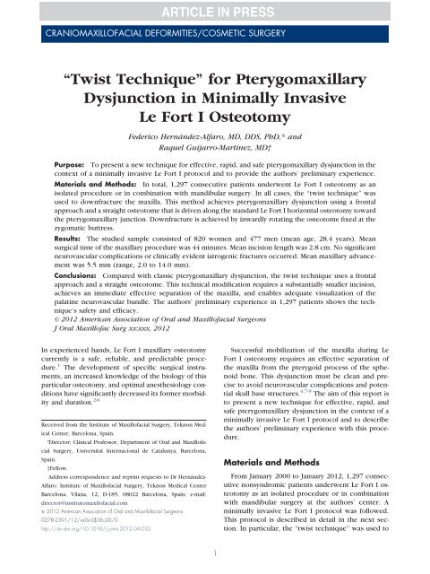
FIGURE 1. The osteotome is driven from the nasal crest of the<br />
maxilla toward the pterygomaxillary junction. A narrow periosteal<br />
elevator (left) is used to protect the nasal mucosa.<br />
Hernández-Alfaro and Guijarro-Martínez. <strong>Twist</strong> Technique <strong>in</strong> Le<br />
Fort I Osteotomy. J Oral Maxillofac Surg 2012.<br />
FIGURE 2. Skull base model. The osteotome progresses along the<br />
horizontal osteotomy from the pyriform buttress back to the pterygomaxillary<br />
junction.<br />
Hernández-Alfaro and Guijarro-Martínez. <strong>Twist</strong> Technique <strong>in</strong> Le<br />
Fort I Osteotomy. J Oral Maxillofac Surg 2012.<br />
of the posterior wall of the maxillary s<strong>in</strong>us to the<br />
pterygoid plates (Fig 2). Subsequently, once the osteotome<br />
was fixed at the pterygomaxillary junction<br />
and underneath the zygomatic buttress, it was rotated<br />
<strong>in</strong>wardly, thus provok<strong>in</strong>g downfracture of the maxilla<br />
(Fig 3). No mallet <strong>press</strong>ure was used dur<strong>in</strong>g this maneuver.<br />
Rather, a swift twist of the chisel under controlled<br />
manual force led to an immediate vertical<br />
separation of the maxilla from the cranial base. Once<br />
the pterygomaxillary dysjunction was completed at<br />
one side, the twist <strong>technique</strong> was repeated at the<br />
contralateral side. For complete mobilization of the<br />
maxilla, the palat<strong>in</strong>e neurovascular bundles were liberated<br />
with the aid of a piezoelectric saw. Maxillary<br />
reposition<strong>in</strong>g and fixation proceeded as usual (Fig 4).<br />
The <strong>technique</strong> is summarized <strong>in</strong> the supplementary<br />
video file onl<strong>in</strong>e.<br />
FIGURE 3. Immediate downfracture of the maxilla is achieved by<br />
<strong>in</strong>wardly rotat<strong>in</strong>g the osteotome (arrow).<br />
Hernández-Alfaro and Guijarro-Martínez. <strong>Twist</strong> Technique <strong>in</strong> Le<br />
Fort I Osteotomy. J Oral Maxillofac Surg 2012.
HERNÁNDEZ-ALFARO AND GUIJARRO-MARTÍNEZ 3<br />
FIGURE 4. The procedure is successfully completed through a<br />
m<strong>in</strong>imally <strong>in</strong>vasive <strong>in</strong>cision.<br />
Hernández-Alfaro and Guijarro-Martínez. <strong>Twist</strong> Technique <strong>in</strong> Le<br />
Fort I Osteotomy. J Oral Maxillofac Surg 2012.<br />
Results<br />
The studied sample consisted of 820 women and<br />
477 men. Mean age at the time of surgery was 28.4<br />
years (range, 12 to 67 years).<br />
In 985 cases, a bimaxillary surgery was performed;<br />
the rema<strong>in</strong><strong>in</strong>g 312 cases underwent an isolated Le<br />
Fort I maxillary osteotomy. In all cases, an effective<br />
downfracture of the maxilla was achieved with the<br />
twist <strong>technique</strong>; there was no need for conversion to<br />
the classic pterygomaxillary dysjunction. In total, 733<br />
patients required further maxillary segmentation <strong>in</strong> 3<br />
to 4 pieces, which was successfully achieved us<strong>in</strong>g<br />
the same approach as <strong>in</strong> nonsegmented cases. Mean<br />
surgical time of the maxillary procedure (from <strong>in</strong>cision<br />
to last suture) was 44 m<strong>in</strong>utes (range, 31 to 72<br />
m<strong>in</strong>). Mean <strong>in</strong>cision length was 2.8 cm (range, 2.2 to<br />
3.9 cm). Mean maxillary advancement was 5.5 mm<br />
(range, 2.0 to 14.0 mm). In total, 485 patients required<br />
third molar extraction at the time of orthognathic<br />
surgery. In these cases, the third molars were<br />
extracted us<strong>in</strong>g a standard occlusal approach before<br />
<strong>in</strong>itiat<strong>in</strong>g the Le Fort I osteotomy procedure.<br />
Patients were discharged from the hospital with<strong>in</strong><br />
an average period of 18 hours (range, 8 to 24 hr).<br />
There was no need for blood transfusion. No postoperative<br />
<strong>in</strong>fectious complications occurred. Similarly,<br />
no cl<strong>in</strong>ically evident iatrogenic fractures or significant<br />
neurovascular complications were noted. However,<br />
488 patients reported temporary numbness of the<br />
<strong>in</strong>fraorbital nerve, which resolved with<strong>in</strong> an average<br />
period of 6 days (range, 3 to 15 days).<br />
Discussion<br />
Unlike classic pterygomaxillary dysjunction, which<br />
entails a lateral approach to the pterygomaxillary fis-<br />
sure with a curved osteotome, the twist <strong>technique</strong><br />
seeks to achieve pterygomaxillary dysjunction from a<br />
frontal approach with a straight osteotome. Downfracture<br />
is achieved by <strong>in</strong>wardly rotat<strong>in</strong>g the osteotome<br />
that has been previously fixed at the zygomatic<br />
buttress by slid<strong>in</strong>g the osteotome backward<br />
along the lateral osteotomies. Separation of the maxilla<br />
is completed <strong>in</strong>stantly. Successful maxillary separation<br />
from the cranial base can be verified under<br />
excellent direct vision and the greater palat<strong>in</strong>e neurovascular<br />
bundle may be dissected easily. Lateral<br />
vision is adequate to enable an equilibrated elim<strong>in</strong>ation<br />
of bony <strong>in</strong>terferences and assure good bone-tobone<br />
contact.<br />
This modified approach enables a substantially<br />
smaller soft tissue <strong>in</strong>cision (2.8 cm on average) than<br />
the classic “molar-to-molar” exposure. The risk of<br />
ischemic events is m<strong>in</strong>imized by the preservation of<br />
most of the vascular supply to the bone through the<br />
buccal corridors. In addition, the f<strong>in</strong>al visible scar on<br />
the buccal mucosa is significantly smaller. Despite<br />
this m<strong>in</strong>imally <strong>in</strong>vasive approach, the present results<br />
<strong>in</strong>dicated that the procedure is perfectly feasible under<br />
the required conditions of patient safety and technical<br />
accuracy, <strong>in</strong>clud<strong>in</strong>g cases <strong>in</strong> which maxillary<br />
segmentation is required. It must be noted, however,<br />
that decreas<strong>in</strong>g the <strong>in</strong>cision length should be considered<br />
a technical progression from the classic approach<br />
and not a primary goal for the <strong>in</strong>experienced<br />
orthognathic surgeon. That said, the twist <strong>technique</strong><br />
is technically undemand<strong>in</strong>g and is taught at the authors’<br />
center as a standard method for pterygomaxillary<br />
dysjunction. Similarly, <strong>in</strong> cases <strong>in</strong> which significant<br />
scar tissue or abnormal anatomy is anticipated,<br />
such as patients with cleft or syndromic cases, a<br />
wider <strong>in</strong>cision is recommended, although maxillary<br />
mobilization can still be achieved safely and efficiently<br />
with the twist <strong>technique</strong>.<br />
Potentially severe complications after pterygomaxillary<br />
dysjunction have been reported <strong>in</strong> the scientific<br />
literature. 2,4,7-10 Many of these complications have<br />
been caused by malposition<strong>in</strong>g the osteotome or by<br />
accidental fractures dur<strong>in</strong>g maxillary downfracture. 4<br />
Although several technical modifications have been<br />
proposed to m<strong>in</strong>imize the risk of pterygoid process<br />
fracture, 7,11-18 studies of stra<strong>in</strong> distribution with different<br />
osteotome designs have <strong>in</strong>dicated that pterygoid<br />
plate fractures are likely to occur regardless of<br />
the type of osteotome used. 19 Similarly, they occur<br />
irrespective of the use or nonuse of a pterygoid<br />
chisel. 10 At any rate, a pterygoid plate fracture cannot<br />
be considered a complication because it is not necessarily<br />
the cause of hemorrhage or nerve <strong>in</strong>jury. 4,10 In<br />
fact, <strong>in</strong>tentional fractur<strong>in</strong>g of the pterygoid process is<br />
occasionally necessary when maxillary reposition<strong>in</strong>g<br />
is h<strong>in</strong>dered by <strong>in</strong>terference with the pterygoid pro-
4 TWIST TECHNIQUE IN LE FORT I OSTEOTOMY<br />
cess. 4 Despite the authors’ cl<strong>in</strong>ically favorable results<br />
with no significant complications <strong>in</strong> a long<br />
series of patients, an ongo<strong>in</strong>g study will try to<br />
specify the particular radiologic characteristics—if<br />
any— of pterygomaxillary dysjunction as achieved<br />
by the twist <strong>technique</strong>.<br />
Regard<strong>in</strong>g the limitations of the m<strong>in</strong>imally <strong>in</strong>vasive<br />
Le Fort I procedure described <strong>in</strong> this report, the<br />
authors differentiate two aspects: <strong>in</strong>cision length and<br />
twist <strong>technique</strong> maneuver. In cases <strong>in</strong> which significant<br />
scar tissue or an abnormal anatomy is expected,<br />
such as patients with cleft or syndromic cases, a<br />
wider <strong>in</strong>cision is preferred for safety reasons. In addition,<br />
although the authors’ m<strong>in</strong>imally <strong>in</strong>vasive <strong>in</strong>cision<br />
poses no limitations to the magnitude of maxillary<br />
advancement or clockwise rotation, significant anticlockwise<br />
maxillary rotation is managed with posterior<br />
plat<strong>in</strong>g and, hence, requires 1 to 2 cm broaden<strong>in</strong>g<br />
of the <strong>in</strong>cision to enable proper access to the zygomaticomaxillary<br />
buttress. It must be noted that, when<br />
<strong>in</strong>dicated, third molar extraction is always performed<br />
from an occlusal approach before the Le Fort I procedure.<br />
The twist <strong>technique</strong> of pterygomaxillary dysjunction<br />
is a safe, efficient, technical modification for<br />
maxillary downfracture. In the authors’ experience,<br />
no particular limitations or contra<strong>in</strong>dications must be<br />
acknowledged.<br />
Compared with classic pterygomaxillary dysjunction,<br />
the twist <strong>technique</strong> uses a frontal approach and<br />
a straight osteotome. Downfracture is achieved by<br />
<strong>in</strong>wardly rotat<strong>in</strong>g the osteotome that has been fixed at<br />
the zygomatic buttress. This modified approach<br />
enables a substantially smaller soft tissue <strong>in</strong>cision,<br />
achieves an immediate effective separation of the<br />
maxilla, and enables adequate visualization of the<br />
greater palat<strong>in</strong>e neurovascular bundle. Prelim<strong>in</strong>ary experience<br />
<strong>in</strong> more than 1,200 patients <strong>in</strong>dicates the<br />
procedure meets the necessary requirements of safety<br />
and technical accuracy.<br />
References<br />
1. Hoffman GR, Islam S: The difficult Le Fort I osteotomy and<br />
downfracture: A review with consideration given to an atypical<br />
maxillary morphology. J Plast Reconstr Aesthet Surg 61:1029,<br />
2008<br />
2. Lanigan DT, Hey JH, West RA: Major vascular complications of<br />
orthognathic surgery: Hemorrhage associated with Le Fort I<br />
osteotomies. J Oral Maxillofac Surg 48:561, 1990<br />
3. Ueki K, Hashiba Y, Marukawa K, et al: Assessment of pterygomaxillary<br />
separation <strong>in</strong> Le Fort I osteotomy <strong>in</strong> Class III patients.<br />
J Oral Maxillofac Surg 67:833, 2009<br />
4. Ueki K, Nakagawa K, Marukawa K, et al: Le Fort I osteotomy<br />
us<strong>in</strong>g an ultrasonic bone curette to fracture the pterygoid<br />
plates. J Craniomaxillofac Surg 32:381, 2004<br />
5. Bell WH, Fonseca RJ, Kenneky JW, et al: Bone heal<strong>in</strong>g and<br />
revascularization after total maxillary osteotomy. J Oral Surg<br />
33:253, 1975<br />
6. Epker BN: Vascular considerations <strong>in</strong> orthognathic surgery. II.<br />
Maxillary osteotomies. Oral Surg Oral Med Oral Pathol 57:473,<br />
1984<br />
7. Precious DS, Morrison A, Ricard D: Pterygomaxillary separation<br />
without the use of an osteotome. J Oral Maxillofac Surg 49:98,<br />
1991<br />
8. Rob<strong>in</strong>son PP, Hendy CW: Pterygoid plate fractures caused by<br />
the Le Fort I osteotomy. Br J Oral Maxillofac Surg 24:198, 1986<br />
9. Cruz AA, dos Santos AC: Bl<strong>in</strong>dness after Le Fort I osteotomy: A<br />
possible complication associated with pterygomaxillary separation.<br />
J Craniomaxillofac Surg 34:210, 2006<br />
10. Precious DS, Goodday RH, Bourget L, et al: Pterygoid plate<br />
fracture <strong>in</strong> Le Fort I osteotomy with and without pterygoid<br />
chisel: A computed tomography scan evaluation of 58 patients.<br />
J Oral Maxillofac Surg 51:151, 1993<br />
11. Dupont C, Ciaburro TH, Prévost Y: Simplify<strong>in</strong>g the Le Fort I<br />
type of maxillary osteotomy. Plast Reconstr Surg 54:142, 1974<br />
12. Trimble LD, Tideman H, Stoel<strong>in</strong>ga PJ: A modification of the<br />
pterygoid plate separation <strong>in</strong> low-level maxillary osteotomies.<br />
J Oral Maxillofac Surg 41:544, 1983<br />
13. Wikkel<strong>in</strong>g OM, Tacoma J: Osteotomy of the pterygomaxillary<br />
junction. Int J Oral Surg 4:99, 1975<br />
14. Cheng LH, Rob<strong>in</strong>son PP: Evaluation of a swan’s neck osteotome<br />
for pterygomaxillary dysjunction <strong>in</strong> the Le Fort I osteotomy.<br />
Br J Oral Maxillofac Surg 31:52, 1993<br />
15. Juniper RP, Stajcić Z: Pterygoid plate separation us<strong>in</strong>g an oscillat<strong>in</strong>g<br />
saw <strong>in</strong> Le Fort I osteotomy. Technical note. J Craniomaxillofac<br />
Surg 19:153, 1991<br />
16. Laster Z, Ardekian L, Rachmiel A, et al: Use of the “shark-f<strong>in</strong>”<br />
osteotome <strong>in</strong> separation of the pterygomaxillary junction <strong>in</strong> Le<br />
Fort I osteotomy: A cl<strong>in</strong>ical and computerized tomography<br />
study. Int J Oral Maxillofac Surg 31:100, 2002<br />
17. Stajcić Z: Alter<strong>in</strong>g the angulation of a curved osteotome—Does<br />
it have effects on the type of pterygomaxillary disjunction <strong>in</strong> Le<br />
Fort I osteotomy? An experimental study. Int J Oral Maxillofac<br />
Surg 20:301, 1991<br />
18. Lanigan DT, Loewy J: Postoperative computed tomography<br />
scan study of the pterygomaxillary separation dur<strong>in</strong>g the Le<br />
Fort I osteotomy us<strong>in</strong>g a micro-oscillat<strong>in</strong>g saw. J Oral Maxillofac<br />
Surg 53:1161, 1995<br />
19. Hiranuma Y, Yamamoto Y, Iizuka T: Stra<strong>in</strong> distribution dur<strong>in</strong>g<br />
separation of the pterygomaxillary suture by osteotomes. Comparison<br />
between Obwegeser’s osteotome and swan’s neck<br />
osteotome. J Craniomaxillofac Surg 16:13, 1988<br />
Appendix<br />
Supplementary Data<br />
Supplementary data associated with this article can<br />
be found, <strong>in</strong> the onl<strong>in</strong>e version, at http://dx.doi.org/<br />
10.1016/j.joms.2012.04.032.