mammalian remains in the chinji type locality of - Journal of Animal ...
mammalian remains in the chinji type locality of - Journal of Animal ...
mammalian remains in the chinji type locality of - Journal of Animal ...

You also want an ePaper? Increase the reach of your titles
YUMPU automatically turns print PDFs into web optimized ePapers that Google loves.
The <strong>Journal</strong> <strong>of</strong> <strong>Animal</strong> & Plant Sciences 19(4): 2009, Pages: 224-229<br />
ISSN: 1018-7081<br />
MAMMALIAN REMAINS IN THE CHINJI TYPE LOCALITY OF THE CHINJI<br />
FORMATION: A NEW COLLECTION<br />
M. A. Khan, M. Malik, A. M. Khan * M. Iqbal ** , and M. Akhtar *<br />
Zoology Department, GC University, Faisalabad, Pakistan<br />
* Zoology Department, Punjab University, Lahore, Pakistan<br />
** Zoology Department, Govt. College <strong>of</strong> Science, Wahdat road Lahore, Pakistan<br />
Correspondence author email: akbaar111@yahoo.ca<br />
ABSTRACT<br />
Seven <strong>mammalian</strong> fossils specimens <strong>of</strong> vary<strong>in</strong>g preservation states are described and discussed. The recovered <strong>rema<strong>in</strong>s</strong><br />
<strong>in</strong>clude <strong>the</strong> upper and <strong>the</strong> lower dentition, and derive mostly from <strong>the</strong> flood pla<strong>in</strong> channel deposits. The systematics <strong>of</strong><br />
<strong>the</strong> rare f<strong>in</strong>d<strong>in</strong>gs Elachistocerus sp., Gazella sp. and Miotragocerus sp. are also discussed <strong>in</strong> here. Listriodon cf.<br />
pentapotamiae is also recovered from <strong>the</strong> middle Miocene <strong>of</strong> <strong>the</strong> Ch<strong>in</strong>ji <strong>type</strong> <strong>locality</strong> and consequently discussed <strong>in</strong> this<br />
paper. The aim <strong>of</strong> <strong>the</strong> paper is to describe poorly documented Middle Miocene <strong>mammalian</strong> species.<br />
Key words: Bovids, Elachistocerus, Gazella, Listriodon, Miotragocerus, Mammalian fossils, Ch<strong>in</strong>ji Formation.<br />
INTRODUCTION<br />
The identified taxa are recovered <strong>in</strong> <strong>the</strong> <strong>type</strong><br />
<strong>locality</strong> outcrops <strong>of</strong> <strong>the</strong> Ch<strong>in</strong>ji village (Lower Siwaliks),<br />
district Chakwal, Punjab, Pakistan. The new material is<br />
housed <strong>in</strong> <strong>the</strong> Palaeontology Laboratory <strong>of</strong> <strong>the</strong> Zoology<br />
Department, GC University Faisalabad (<strong>in</strong>stitutional<br />
abbreviation PC-GCUF) and some specimens are housed<br />
<strong>in</strong> <strong>the</strong> Palaeontology Laboratory <strong>of</strong> <strong>the</strong> Punjab University<br />
(<strong>in</strong>stitutional abbreviation PUPC) are also <strong>in</strong>cluded <strong>in</strong> this<br />
study. The catalogue number <strong>of</strong> <strong>the</strong> specimens consists <strong>of</strong><br />
series i.e. yearly catalogue number and serially catalogue<br />
number, so figures <strong>of</strong> <strong>the</strong> specimen represent <strong>the</strong><br />
collection year (numerator) and <strong>the</strong> serial number<br />
(denom<strong>in</strong>ator) <strong>of</strong> that year (e.g., 08/21).<br />
Measurements are <strong>in</strong> millimeters with length<br />
be<strong>in</strong>g <strong>the</strong> maximum dimension parallel to <strong>the</strong> mesiodistal<br />
axis <strong>of</strong> <strong>the</strong> tooth, trigonid width <strong>the</strong> maximum through<br />
<strong>the</strong> protoconid and <strong>the</strong> metaconid perpendicular to <strong>the</strong><br />
mesiodistal axis, and talonid width <strong>the</strong> maximum through<br />
<strong>the</strong> hypoconid and <strong>the</strong> entoconid perpendicular to <strong>the</strong><br />
mesiodistal axis. Capital letters <strong>in</strong>dicate dentition with<br />
superscript numbers for <strong>the</strong> upper dentition and subscript<br />
for <strong>the</strong> lower dentition. The term<strong>in</strong>ology <strong>of</strong> <strong>the</strong> tooth<br />
crown elements and manners <strong>of</strong> measurements follow<br />
Gentry (1994) and Pickford (1988).<br />
Systematic Palaeontology<br />
Order Artiodactyla Owen, 1848<br />
Family Bovidae Gray, 1821<br />
Subfamily Bov<strong>in</strong>ae Gray, 1821<br />
Tribe Boselaph<strong>in</strong>i Knottnerus-Meyer, 1907<br />
Genus Elachistoceras Thomas, 1977<br />
cf. Elachistoceras khauristanensis Thomas, 1977:<br />
224<br />
Abbreviated Diagnosis: Bovid <strong>of</strong> very small size, close<br />
to that <strong>of</strong> Steenbok (Raphicerus campestris). Rectil<strong>in</strong>ear<br />
horn cores <strong>of</strong> very small dimensions, has oval section.<br />
These teeth are typical among that <strong>of</strong> Boselaph<strong>in</strong>i.<br />
Compared with <strong>the</strong> lower teeth <strong>of</strong> Tetracerus<br />
quadricornis, it is noted that <strong>the</strong> morphological<br />
characteristics are: Molars ra<strong>the</strong>r brachydonts, def<strong>in</strong>itely<br />
convex l<strong>in</strong>gual wall; Non smooth l<strong>in</strong>gual wall (be<strong>in</strong>g<br />
dist<strong>in</strong>guished by from Neotrag<strong>in</strong>i); Enamel rugosed;<br />
Presence <strong>of</strong> a weak capr<strong>in</strong>e fold (or antero-stylid), less<br />
developed l<strong>in</strong>gual furrows dist<strong>in</strong>guished from<br />
Cephaloph<strong>in</strong>ae (Thomas, 1977).<br />
Referred Material: PUPC 97/18 – left mendibular<br />
fragment with M3; PUPC 97/19 – right mandibular<br />
fragment with M1-M3.<br />
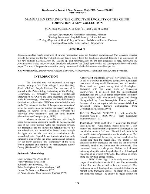
Description: PUPC 97/18 (Fig. 1) comprises <strong>the</strong> lower<br />
third molar (M3) with broken second molar (M2). The<br />
roots <strong>of</strong> <strong>the</strong> second molar are preserved. The length <strong>of</strong> <strong>the</strong><br />
mandibular ramus is 28.2 mm. The third left molar is <strong>in</strong><br />
an excellent state <strong>of</strong> preservation and <strong>in</strong> middle wear. The<br />
enamel is rugose and <strong>the</strong> rugosity is more evident on <strong>the</strong><br />
l<strong>in</strong>gual side than <strong>the</strong> buccal side. The apices <strong>of</strong> <strong>the</strong><br />
paraconid and <strong>the</strong> metaconid are sharp and <strong>the</strong> metaconid<br />
noticeably smaller and lower than <strong>the</strong> protoconid. The<br />
paraconid bears very sharp and dist<strong>in</strong>ct vertical crest<br />
extend<strong>in</strong>g along <strong>the</strong> anterol<strong>in</strong>gual edge. A well developed<br />
hypoconulid is present posteriorly. The talonid is sharp,<br />
sp<strong>in</strong>e like hav<strong>in</strong>g a closed fossette.<br />
PUPC 97/19 (Fig. 2) is <strong>in</strong> early wear and <strong>the</strong><br />
length <strong>of</strong> mandibular ramus is 35.6 mm. The metaconids<br />
<strong>of</strong> <strong>the</strong> first and <strong>the</strong> second molars are damaged. The<br />
protoconid is well developed and projected. Ectostylid is<br />
present <strong>in</strong> <strong>the</strong> transverse valley. The apices <strong>of</strong> <strong>the</strong> conids<br />
are somewhat conical. The enamel is rugose equally on
The <strong>Journal</strong> <strong>of</strong> <strong>Animal</strong> & Plant Sciences 19(4): 2009, Pages: 224-229<br />
ISSN: 1018-7081<br />
<strong>the</strong> both sides. The labially and l<strong>in</strong>gually flattened<br />
metaconid and entoconid are slightly oblique to <strong>the</strong> long<br />
axis <strong>of</strong> <strong>the</strong> tooth and separated by a l<strong>in</strong>gual embayment.<br />
The conids have non smooth convex l<strong>in</strong>gual wall. The<br />
anterior c<strong>in</strong>gulum is strong, form<strong>in</strong>g a “goat-fold”<br />
anteriorly. There are deep medial valleys <strong>of</strong> <strong>the</strong> trigonid<br />
and <strong>the</strong> talonid. A well developed hypoconulid is present<br />
<strong>in</strong> <strong>the</strong> last molar.<br />
Table 1. Comparative measurements <strong>of</strong> <strong>the</strong> studied<br />
material (Elachistoceras khauristanensis) <strong>in</strong><br />
mm (millimeters).<br />
Number Nature Length Width<br />
PUPC 97/18 Left mandibular<br />
ramus hav<strong>in</strong>g M3<br />
M3=13.4 M3=7.6<br />
PUPC 97/19 Right Mandibular M1 = 9.5 M1 =<br />
ramus hav<strong>in</strong>g M1-<br />
3<br />
M2 = 9<br />
M3 =<br />
6.2<br />
M2 = 7<br />
12.5 M3 = 6<br />
GSP 4278<br />
(Thomas,<br />
1977)<br />
GSP 5116<br />
(Thomas,<br />
1977)<br />
Right mandible<br />
hav<strong>in</strong>g M1- M2<br />
and a former lobe<br />
<strong>of</strong> M3.<br />
Right M3<br />
M1 = 8.2<br />
M2 = 8.9<br />
M3 =<br />
11.6<br />
M1 =<br />
5.1<br />
M2 =<br />
5.4<br />
M3 =<br />
5.4<br />
Brachydont molars, def<strong>in</strong>itely convex non<br />
smooth l<strong>in</strong>gual wall; rugose enamel and presence <strong>of</strong> weak<br />
capr<strong>in</strong>e fold are dist<strong>in</strong>ct features <strong>of</strong> <strong>the</strong> studied<br />
specimens. These features be<strong>in</strong>g <strong>the</strong> characteristics <strong>of</strong><br />
Elachistoceras, discrim<strong>in</strong>ate <strong>the</strong>m from <strong>the</strong> genus<br />
Gazella. The teeth <strong>of</strong> Gazella can be dist<strong>in</strong>guish from <strong>the</strong><br />
teeth <strong>of</strong> Elachistoceras, for <strong>the</strong> presence <strong>of</strong> deep median<br />
valley <strong>of</strong> trigonid and talonid and well developed<br />
hypoconulid.<br />
The apices <strong>of</strong> <strong>the</strong> paraconid and <strong>the</strong> metaconid<br />
are sharp and V-shaped. A deep median valley <strong>of</strong> trigonid<br />
and talonid and well developed hypoconulid development<br />
are <strong>the</strong> characteristic features <strong>of</strong> <strong>the</strong> studied specimens.<br />
These both teeth show typical features <strong>of</strong> Elachistoceras<br />
(Thomas, 1977). As <strong>the</strong> whole <strong>the</strong> teeth are compared<br />
very favorably with <strong>the</strong> <strong>type</strong> specimen. It has <strong>the</strong> almost<br />
same relative proportions <strong>of</strong> <strong>the</strong> length as <strong>the</strong> teeth <strong>of</strong> <strong>the</strong><br />
holo<strong>type</strong> but <strong>the</strong> length and width are slightly greater than<br />
<strong>the</strong> holo<strong>type</strong> (Table 1). This difference is with<strong>in</strong> <strong>the</strong> range<br />
<strong>of</strong> <strong>in</strong>dividual variation.<br />
Genus Miotragocerus Stromer, 1928<br />
Miotragocerus sp.<br />
Abbreviated Diagnosis: Molars quadrate, subhypsodont;<br />
presence <strong>of</strong> median basal pillar and anterior transverse<br />
flange; <strong>the</strong> parastyle is strongly developed.<br />
225<br />
Referred Material: PUPC 97/21 - left maxillary ramus<br />
with M 1-2 ; PC-GCUF 08/20 - broken left molar probably<br />
M3.<br />
Description: The length <strong>of</strong> <strong>the</strong> studied maxillary ramus<br />
(PUPC 97/21) is 28.2 mm. The first molar (M 1 ) is <strong>in</strong> <strong>the</strong><br />
middle stage <strong>of</strong> wear and <strong>the</strong> dent<strong>in</strong>e is clearly exposed<br />
(Fig. 3). The enamel on <strong>the</strong> l<strong>in</strong>gual side is moderately<br />
thick and f<strong>in</strong>ely rugose. The metacone and <strong>the</strong> paracone<br />
are less broad than <strong>the</strong> hypocone and <strong>the</strong> protocone. The<br />
protocone is somewhat U-shaped <strong>in</strong> its general<br />
appearance. The metastyle is well-developed and stronger<br />
than <strong>the</strong> mesostyle and <strong>the</strong> parastyle. A rudimentary goatfold<br />
is present anteriorly which is not prom<strong>in</strong>ent. The first<br />
upper molar (M 1 ) is remarkable for its subhypsodonty,<br />
surpass<strong>in</strong>g <strong>in</strong> this respect <strong>the</strong> first molar <strong>of</strong> any o<strong>the</strong>r<br />
member <strong>of</strong> <strong>the</strong> family. The tooth itself is columnar and<br />
strongly curved, <strong>the</strong> convexity be<strong>in</strong>g outwards; it narrows<br />
greatly towards <strong>the</strong> base. On <strong>the</strong> outer face <strong>of</strong> <strong>the</strong> first<br />
molar <strong>the</strong> mesostyle is strongly developed, form<strong>in</strong>g a<br />
sharp ridge extend<strong>in</strong>g up nearly to <strong>the</strong> root: <strong>the</strong> parastyle<br />
forms a sharp ridge but does not extend so far up. At <strong>the</strong><br />
posterior end <strong>of</strong> <strong>the</strong> crown <strong>the</strong> metacone forms a sharp<br />
angle project<strong>in</strong>g backward. The paracone forms a slight<br />
convexity while <strong>the</strong> metacone is nearly flat. The valley<br />
between <strong>the</strong>m extends up nearly to <strong>the</strong> neck <strong>of</strong> <strong>the</strong> tooth.<br />
The general form <strong>of</strong> <strong>the</strong> second upper (M 2 ) molar is very<br />
similar to that <strong>of</strong> <strong>the</strong> first molar but it is larger size than<br />
<strong>the</strong> first one with subhypsodont.<br />
PUPC 08/20 (Fig. 4) is <strong>in</strong> an early wear so that<br />
all <strong>the</strong> pr<strong>in</strong>cipal cones are well preserved. The talonid <strong>of</strong><br />
<strong>the</strong> last molar is damaged. The enamel is moderately<br />
thick, rugose and equally dispersed. A weak goatfold is<br />
present anteriorly. The praeprotocristid and <strong>the</strong><br />
postprotocristid are prom<strong>in</strong>ent on <strong>the</strong> buccal side <strong>of</strong> <strong>the</strong><br />
molar while <strong>the</strong> praemetacristid and <strong>the</strong> postmetacristid<br />
are united with <strong>the</strong> divergent styles. The mesostylid is<br />
broken whereas <strong>the</strong> metatylid and <strong>the</strong> entostylid are<br />
present. The ectostylids are strong and prom<strong>in</strong>ent.<br />
Table 2: Measurements (mm) <strong>of</strong> <strong>the</strong> cheek teeth <strong>of</strong><br />
Miotragocerus sp.<br />
Specimens Nature Length Width<br />
PUPC<br />
97/21<br />
PUPC<br />
08/20<br />
Left maxillary<br />
fragment hav<strong>in</strong>g<br />
M 1-2<br />
Left broken molar<br />
M3<br />
M 1 =<br />
12.6<br />
M 2 = 9.8<br />
M3 = 21<br />
M 1 =<br />
13.8<br />
M 2 =<br />
12.3<br />
M3= 9.2<br />
Discussion: Subhypsodont molars, columnar and<br />
strongly curved tooth, wide crown ra<strong>the</strong>r than long <strong>in</strong><br />
upper dentition while longer than wide <strong>in</strong> lower dentition<br />
and <strong>of</strong> <strong>the</strong> strong mesostyle on <strong>the</strong> buccal side are <strong>the</strong><br />
dist<strong>in</strong>ct features that have been discussed (Table, 2). The<br />
studied specimens have subhypsodont molars which are
The <strong>Journal</strong> <strong>of</strong> <strong>Animal</strong> & Plant Sciences 19(4): 2009, Pages: 224-229<br />
ISSN: 1018-7081<br />
very dist<strong>in</strong>ct. The enamel is moderately thick and f<strong>in</strong>ely<br />
rugose. Goat fold is not well developed <strong>in</strong> both upper and<br />
lower molars. Morpho-metrically <strong>the</strong> teeth show <strong>the</strong><br />
typical features <strong>of</strong> Miotragocerus (Stromer, 1928).<br />
However, <strong>the</strong> material is too complete to identify it up to<br />
species level.<br />
Subfamily Antilop<strong>in</strong>ae Gray, 1821<br />
Tribe Antilop<strong>in</strong>i Gray, 1821<br />
Genus Gazella Bla<strong>in</strong>ville, 1816<br />
Gazella sp.<br />
Referred Material: PC-GCUF 98/101 – right maxillary<br />
fragment with P 4 –M 3 .<br />
Description: PUPC 98/101 (Fig. 5) is well preserved<br />
right maxillary fragment with a small piece <strong>of</strong> palate<br />
hav<strong>in</strong>g P 4 -M 3 . The length <strong>of</strong> <strong>the</strong> maxillary ramus is 61.1<br />
mm. The P 4 is <strong>in</strong> its early wear, horse-shoe shaped and<br />
shows all <strong>the</strong> morphological characteristics. The enamel<br />
is somewhat rugose. A prom<strong>in</strong>ent central cavity is<br />
present. A small, very th<strong>in</strong>, transverse enamel layer<br />
connects <strong>the</strong> posterior end <strong>of</strong> protocone with hypocone.<br />
The paracone is comparatively higher than <strong>the</strong> protocone.<br />
The median rib is very prom<strong>in</strong>ent and closer to <strong>the</strong><br />
parastyle. The parastyle is damaged but <strong>the</strong> mesostyle<br />
and <strong>the</strong> metastyle are well preserved. The mesostyle is<br />
stronger than <strong>the</strong> metastyle. The anterior median rib is<br />
stronger than <strong>the</strong> posterior one and <strong>the</strong> both are dist<strong>in</strong>ct<br />
up to <strong>the</strong> base <strong>of</strong> <strong>the</strong> crown. The hypocone appears to be<br />
less crescentic because <strong>of</strong> <strong>the</strong> wear. The posthypocrista is<br />
thicker than <strong>the</strong> prehypocrista.<br />
Table 3. Comparative measurements (mm) <strong>of</strong> <strong>the</strong><br />
Gazella sp. studied material.<br />
Number Nature Length Width<br />
PUPC<br />
98/101<br />
PUPC 83/67<br />
(Akhtar,<br />
1992)<br />
PUPC 86/76<br />
(Akhtar,<br />
1992)<br />
Right maxillary<br />
fragment hav<strong>in</strong>g<br />
P 4 -M 3<br />
Right maxillary<br />
fragment hav<strong>in</strong>g<br />
P 4 -M 3<br />
A left maxilla<br />
bear<strong>in</strong>g M 1-3<br />
P 4 = 9.6<br />
M 1 = 14<br />
M 2 = 14<br />
M 3 =<br />
14.2<br />
P 4 = 9<br />
M 1 =<br />
13.2<br />
M 2 = 14<br />
M 3 =<br />
15.5<br />
M 1 =<br />
13.4<br />
M 2 =<br />
15.4<br />
M 3 =<br />
16.3<br />
P 4 = 9.7<br />
M 1 = 13<br />
M 2 = 12<br />
M 3 = 12.5<br />
P 4 = 9<br />
M 1 = 12<br />
M 2 = 11.5<br />
M 3 = 11<br />
M 1 = 13<br />
M 2 = 12.5<br />
M 3 = 11<br />
226<br />
The M 2 is <strong>in</strong> early wear and subhypsodont. All <strong>the</strong><br />
pr<strong>in</strong>cipal cones are well developed. The anterior cones<br />
are wider than <strong>the</strong> posterior ones. The enamel is<br />
moderately thick. The parastyle is well developed and<br />
stronger than <strong>the</strong> mesostyle while <strong>the</strong> metastyle is weakly<br />
developed. The height <strong>of</strong> <strong>the</strong> parastyle is more as<br />
compare to <strong>the</strong> mesostyle. The posterior end <strong>of</strong> <strong>the</strong><br />
protcone is moderately expanded. The central cavities are<br />
moderately wide and deep. No spurs project <strong>in</strong>to <strong>the</strong>se<br />
cavities. The entostyle is absent <strong>in</strong> <strong>the</strong> cheek teeth which<br />
is <strong>the</strong> characteristic <strong>of</strong> Gazella upper dentition.<br />
Discussion: The transverse diameter <strong>of</strong> P 4 <strong>in</strong> G. lydekkeri<br />
exceeds antero-posterior diameter (Pilgrim, 1937). The<br />
upper molars <strong>of</strong> Gazella have greater antero-posterior<br />
diameter than transverse diameter (Akhtar, 1992; Pilgrim,<br />
1937, 1939). The studied molars are slightly broad. The<br />
l<strong>in</strong>gual side <strong>of</strong> <strong>the</strong> studied specimen is somewhat rugose<br />
and convex. Buccally, it shows a strong, narrow<br />
parastyle, and equally strong and broader median rib and<br />
a much weaker metastyle. All <strong>the</strong>se teeth show typical<br />
morphometrical features <strong>of</strong> Gazella (Table, 3). The<br />
molars have anterior and median folds <strong>of</strong> about equal<br />
strength and ra<strong>the</strong>r prom<strong>in</strong>ent; <strong>the</strong> posterior fold is weak.<br />
The median rib <strong>of</strong> <strong>the</strong> anterior lobe is strongly developed;<br />
that <strong>of</strong> <strong>the</strong> posterior lobe is weaker. The molar structure<br />
represents PUPC 98/101 is typically <strong>of</strong> <strong>the</strong> genus Gazella<br />
(Pilgrim, 1937). The prom<strong>in</strong>ent median ribs, narrow<br />
styles, <strong>the</strong> absence <strong>of</strong> median basal pillar show clearly<br />
<strong>the</strong>ir <strong>in</strong>clusion to Gazella. However, more material is<br />
required to identify it up to species level and due to<br />
scarcity <strong>of</strong> <strong>the</strong> material, Gazella sp. is attributed for <strong>the</strong><br />
material.<br />
Family Suidae Gray, 1821<br />
Subfamily Listriodont<strong>in</strong>ae Simpson, 1945<br />
Genus Listriodon von Meyer, 1846<br />
Listriodon pentapotamiae<br />
Diagnosis: The listriodonts are middle Miocene suids<br />
possess<strong>in</strong>g several archaic features such as primitive<br />
basicranium, unflared zygoma, parietal l<strong>in</strong>es not widely<br />
separated, no can<strong>in</strong>e flanges, rounded snout and low<br />
glenoids. The listriodonts possess a very elongated<br />
mandible, achieved both by elongation <strong>of</strong> <strong>the</strong> symphysis<br />
as well as by retir<strong>in</strong>g <strong>the</strong> ascend<strong>in</strong>g ramus. In side view,<br />
<strong>the</strong> whole <strong>of</strong> M3 is visible as well as gap beh<strong>in</strong>d M3. The<br />
symphysis is splayed outwards, so that <strong>the</strong> lower can<strong>in</strong>es<br />
emerge almost horizontally. The <strong>in</strong>cisive marg<strong>in</strong> is<br />
evenly curved and projects substantially <strong>in</strong> front <strong>of</strong> <strong>the</strong><br />
can<strong>in</strong>es. Between <strong>the</strong> can<strong>in</strong>es and anterior premolar (P2),<br />
<strong>the</strong>re is long diastema, <strong>the</strong> borders <strong>of</strong> which lie well<br />
below <strong>the</strong> occlusal surface <strong>of</strong> <strong>the</strong> cheek teeth. P1 is<br />
reduced or lost <strong>in</strong> most species. In Lisriodont<strong>in</strong>ae <strong>the</strong> I 1 is<br />
spatulate and occludes with I1-2. In Listriodon females,<br />
upper can<strong>in</strong>es are usually two rooted if <strong>the</strong>y are not<br />
hypsodont although <strong>the</strong> lower can<strong>in</strong>es seem to be more
The <strong>Journal</strong> <strong>of</strong> <strong>Animal</strong> & Plant Sciences 19(4): 2009, Pages: 224-229<br />
ISSN: 1018-7081<br />
nearly s<strong>in</strong>gle rooted. I 2 is a robust triangular tooth set<br />
vertically <strong>in</strong> <strong>the</strong> premaxillae. The tip is triangular <strong>in</strong><br />
l<strong>in</strong>gual view, with a l<strong>in</strong>gual c<strong>in</strong>gulum and a central rib.<br />
The crown is slightly <strong>of</strong>fset from <strong>the</strong> root. There are two<br />
wear facets along <strong>the</strong> occlusal edge <strong>of</strong> <strong>the</strong> tooth; <strong>the</strong><br />
mesial one corresponds to <strong>the</strong> outer portion <strong>of</strong> <strong>the</strong> scoopshaped<br />
distal edge <strong>of</strong> I2, while <strong>the</strong> distal one is caused by<br />
wear with <strong>the</strong> root ward half <strong>of</strong> <strong>the</strong> scoop <strong>in</strong> I2. In I2 <strong>the</strong><br />
facet caused by I 2 is very prom<strong>in</strong>ent along <strong>the</strong> distal edge<br />
and <strong>in</strong> <strong>the</strong> body <strong>of</strong> <strong>the</strong> scoop, while I 1 occludes only at <strong>the</strong><br />
tip. I 2 has bifurcate tip when unworn. M 1 is a square tooth<br />
with four ma<strong>in</strong> cusps disposed <strong>in</strong> two lophs, with anterior<br />
and posterior c<strong>in</strong>gula. The anterior, median and posterior<br />
accessory cusps, present <strong>in</strong> all suids are very small <strong>in</strong><br />
Listriodon, and soon disappear with wear (Pickford,<br />
1988).<br />
Referred Material: PC-GCUF 08/21 – broken lower<br />
molar (M); PC – GCUF 08/22 upper <strong>in</strong>cisor (I).<br />
Description: The molar is damaged and <strong>in</strong> late wear<br />
stage (Fig. 6). Due to late wear its crown morphology is<br />
not very dist<strong>in</strong>ct and clear. The squared molar with four<br />
ma<strong>in</strong> cusps disposed <strong>in</strong> two lophs, with anterior and<br />
posterior c<strong>in</strong>gula. Its posterior part is higher while <strong>the</strong><br />
anterior one is lower. Molar enamel is th<strong>in</strong> and equally<br />
dispersed. The molar have less l<strong>in</strong>gual and buccal flare.<br />
The posterior accessory cusps are prom<strong>in</strong>ent and<br />
centrally placed.<br />
Table 4: Comparative measurements (mm) <strong>of</strong> <strong>the</strong><br />
studied material (Listriodon pentapotamiae).<br />
Name Nature Length Width<br />
PC-GCUF 08/21 broken molar (M) M = 17.2 M = 14.5<br />
PC-GCUF 08/22 <strong>in</strong>cisor (I) I = 21.7 I = 9.9<br />
GSP 1424<br />
(Pickford, 1988)<br />
Left <strong>in</strong>cisor I 1 I 1 = 22.7 I 1 = 11.3<br />
PC-GCUF 08/22 (Fig. 7) is <strong>in</strong> good state <strong>of</strong><br />
preservation. The enamel is rugose and <strong>the</strong> rugosity is<br />
more on <strong>the</strong> outer side than <strong>the</strong> <strong>in</strong>ner one. It is a robust<br />
triangular tooth set vertically <strong>in</strong> <strong>the</strong> premaxilla with step<br />
cutt<strong>in</strong>g edge. The tip is triangular <strong>in</strong> view, with a l<strong>in</strong>gual<br />
227<br />
c<strong>in</strong>gulum and a central rib. There are two wear facets<br />
along <strong>the</strong> occlusal edge <strong>of</strong> <strong>the</strong> tooth. It is a bifurcate<br />
<strong>in</strong>cisor which tip occludes with <strong>the</strong> opposite one.<br />
Discussion: Lophodont molar; po<strong>in</strong>ted cusps; anterior,<br />
median and posterior cusps are reduced; moderately thick<br />
enamel; <strong>in</strong>cisors with simple pegs <strong>of</strong>ten with a divided<br />
tip; a central l<strong>in</strong>gual rib mesial and distal borders raised<br />
ligually and l<strong>in</strong>gual c<strong>in</strong>gulum are dist<strong>in</strong>ct features that<br />
have been discussed. The I 1 <strong>of</strong> Listriodon retmaensis has<br />
two ma<strong>in</strong> grooves while Listriodon pentapotamiae<br />
possess only s<strong>in</strong>gle groove <strong>in</strong> <strong>the</strong> labial surface <strong>of</strong> <strong>the</strong><br />
crown. The morphometric features <strong>of</strong> <strong>the</strong> studied <strong>in</strong>cisor<br />
suggest aff<strong>in</strong>ities with Listriodon pentapotamiae (Table,<br />
4). It has long been suggested that listriodonts occur <strong>in</strong><br />
early Miocene deposits at Bugti and S<strong>in</strong>d. Unfortunately,<br />
<strong>the</strong> Bugti specimens are extremely fragmentary and <strong>the</strong>re<br />
is debate about <strong>the</strong>ir subfamilial status (Pickford, 1988;<br />
Van der Made, 1996).<br />
The leng<strong>the</strong>n<strong>in</strong>g <strong>of</strong> <strong>the</strong> crown is reflected to a<br />
great degree <strong>in</strong> <strong>the</strong> root morphology which becomes<br />
strongly triangular <strong>in</strong> l<strong>in</strong>gual or labial view, taper<strong>in</strong>g<br />
sharply towards <strong>the</strong> apex. Study <strong>of</strong> <strong>the</strong> number <strong>of</strong> grooves<br />
<strong>in</strong> listriodont upper <strong>in</strong>cisors lead to <strong>the</strong> suggestion that<br />
several l<strong>in</strong>eages <strong>of</strong> listriodonts can be recognized. The I 1<br />
<strong>of</strong> Listriodon retamaensis has two ma<strong>in</strong> grooves whereas<br />
its contemporaries L. lockharti and Eurolistriodon adelli<br />
had one and none respectively. The two species L.<br />
latidens and L. meidamon have two ma<strong>in</strong> grooves and <strong>the</strong><br />
cutt<strong>in</strong>g edge <strong>of</strong> <strong>the</strong> crown is <strong>of</strong>ten heavily beaded,<br />
suggest<strong>in</strong>g that <strong>the</strong>y may well be descendents <strong>of</strong> L.<br />
retamaensis, L. splendens, L. pentapotamiae, and L.<br />
<strong>in</strong>termedius represent a second l<strong>in</strong>eage <strong>in</strong> which <strong>the</strong>re<br />
was one ma<strong>in</strong> groove <strong>in</strong> <strong>the</strong> labial surface <strong>of</strong> <strong>the</strong> crown.<br />
The condition <strong>in</strong> African L. akatikubas is not clear<br />
because <strong>the</strong> only known upper central <strong>in</strong>cisor is heavily<br />
worn but its length/breadth <strong>in</strong>dex suggests aff<strong>in</strong>ities with<br />
L. retamaensis (Wilk<strong>in</strong>son, 1976). Species <strong>of</strong><br />
Lopholistriodon have two ma<strong>in</strong> grooves <strong>in</strong> <strong>the</strong>ir I 1 but<br />
<strong>the</strong>y differ from those <strong>of</strong> o<strong>the</strong>r Listriodon species by<br />
hav<strong>in</strong>g more cyl<strong>in</strong>drical roots that do not taper so<br />
markedly towards <strong>the</strong>ir apices, and <strong>the</strong> crown is less<br />
elongated mesio-distally (Pickford and Morales, 2003).
The <strong>Journal</strong> <strong>of</strong> <strong>Animal</strong> & Plant Sciences 19(4): 2009, Pages: 224-229<br />
ISSN: 1018-7081<br />
Plate 1. Elachistoceras: 1. PUPC 97/18 – M3; 2. PUPC 97/19 – mandibular ramus hav<strong>in</strong>g M1-M3. Miotragocerus: 3.<br />
PUPC 97/21 – left maxillary fragment with M 1 -M2; 4. PUPC 08/20 – broken M3. Gazella: 5. PUPC 98/101 – right<br />
maxillary fragment hav<strong>in</strong>g P 4 -M 3 . Listriodon: 6. PC-GCUF 08/21 – broken molar (M); 7. PC-GCUF 08/22 – <strong>in</strong>cisor (I).<br />
Scale bar 10 mm and all photos <strong>in</strong> occlusal view.<br />
Conclusion: The new collection from <strong>the</strong> Ch<strong>in</strong>ji<br />
Formation <strong>of</strong> <strong>the</strong> middle Miocene <strong>in</strong>cludes small sized<br />
bovids Elachistocerus sp., Gazella sp., Miotragocerus sp.<br />
and commonly found middle Miocene suid Listriodon cf.<br />
pentapotamiae. The Elachistocerus is a four horned<br />
tetracere like boselaph<strong>in</strong>e found <strong>in</strong> <strong>the</strong> Lower and Middle<br />
Siwaliks (Thomas, 1977). Tetracerus quadricornis live <strong>in</strong><br />
<strong>the</strong> open areas <strong>of</strong> Indian pen<strong>in</strong>sula, testify <strong>the</strong><br />
palaeontological history <strong>of</strong> <strong>the</strong> Elachistocerus (Thomas,<br />
1977). The Tragoportax and Miotragocerus are large<br />
boselaph<strong>in</strong>es from <strong>the</strong> late Miocene assemblages. The<br />
differences between Tragoportax and Miotragocerus<br />
have not always been clear and researchers have differed<br />
over how best to diagnose <strong>the</strong>m (Bibi and Gulec, 2008).<br />
225<br />
Gazella is recorded from <strong>the</strong> Lower and <strong>the</strong><br />
Middle Siwaliks (Khan, 2008, 2007; Akhtar, 1992).<br />
Pilgrim and Hopwood (1928) simply summarized all <strong>the</strong><br />
nomenclature <strong>of</strong> fossil gazelles propagat<strong>in</strong>g <strong>the</strong> existence<br />
<strong>of</strong> a large number <strong>of</strong> species. Gazella lydekkeri recovered<br />
from <strong>the</strong> late Miocene and <strong>the</strong> middle Miocene<br />
assemblages <strong>of</strong> <strong>the</strong> Siwaliks respectively (Pilgrim, 1937,<br />
1939). Gazella capricornis, <strong>the</strong> best known <strong>of</strong> <strong>the</strong><br />
European Ponta<strong>in</strong> species is represented by more material<br />
than Gazella deperdita (Gentry, 1966). It differs from<br />
Gazella lydekkeri <strong>in</strong> hav<strong>in</strong>g large size <strong>of</strong> skull, and less<br />
hypsodonty. In this respect, Gazella lydekkeri is more<br />
primitive than Gazella capricornis. The most progressive<br />
feature <strong>of</strong> Gazella lydekkeri is hypsodonty. The Ch<strong>in</strong>ese<br />
Ponta<strong>in</strong> gazelles are more progressive especially <strong>the</strong>
The <strong>Journal</strong> <strong>of</strong> <strong>Animal</strong> & Plant Sciences 19(4): 2009, Pages: 224-229<br />
ISSN: 1018-7081<br />
species Gazella lydekkeri although <strong>the</strong> width at <strong>the</strong> orbits<br />
is greater. The horn-cores seem to be identical <strong>in</strong> <strong>the</strong>ir<br />
morphology with Gazella lydekkeri and <strong>the</strong> nasal are<br />
somewhat shorter. In Gazella dorcadoides and Gazella<br />
altidens <strong>the</strong> teeth are still more hypsodont than Gazella<br />
lydekkeri.<br />
Pickford (1988) described that <strong>the</strong> Siwalik<br />
Listriodont pentapotamiae is very similar to L. splendens<br />
<strong>of</strong> Europe, both <strong>in</strong> size and morphology. The only<br />
consistent major differences between <strong>the</strong> two are <strong>the</strong><br />
smaller central <strong>in</strong>cisors and upper can<strong>in</strong>es. Whe<strong>the</strong>r <strong>the</strong>se<br />
differences are great enough to warrant separate specific<br />
identity or whe<strong>the</strong>r <strong>the</strong>y reflect geographic variation <strong>in</strong> a<br />
s<strong>in</strong>gle species is difficult to tell without better record<br />
from geographically <strong>in</strong>termediate areas.<br />
The collection leads us to admit existence <strong>of</strong><br />
Elachistocerus sp., Gazella sp., Miotragocerus and<br />
Listriodon cf. pentapotamiae <strong>in</strong> <strong>the</strong> Ch<strong>in</strong>ji Formation <strong>of</strong><br />
<strong>the</strong> Siwaliks. Highly lophodont Listriodon<br />
pentapotamiae, and its synonym L. <strong>the</strong>obaldi, is abundant<br />
<strong>in</strong> <strong>the</strong> Ch<strong>in</strong>ji succession and deposits <strong>of</strong> similar age<br />
elsewhere <strong>in</strong> <strong>the</strong> Potwar Plateau. Biostratigraphy and<br />
palaeomagnetic stratigraphy <strong>in</strong>dicate that <strong>the</strong> Ch<strong>in</strong>ji<br />
levels correspond to MN6 and MN7/8 (Barry et al., 2002;<br />
Badgely and Tauxe, 1990). The Boselaph<strong>in</strong>i small<br />
species Elachistocerus sp. <strong>in</strong>dicates that <strong>the</strong>re was<br />
probably a variety <strong>of</strong> boselaph<strong>in</strong>e bovids <strong>in</strong> <strong>the</strong> Lower<br />
Siwaliks. The fossil range <strong>in</strong> age between 14.2 and 11.2<br />
million years old and provide evidence for <strong>the</strong> existence<br />
<strong>of</strong> small bovid faunas <strong>in</strong> <strong>the</strong> Ch<strong>in</strong>ji Formation <strong>of</strong> <strong>the</strong><br />
Siwaliks.<br />
REFERENCES<br />
Akhtar, M. (1992). Taxonomy and Distribution <strong>of</strong> <strong>the</strong><br />
Siwalik Bovids. Ph. D. diss., University <strong>of</strong> <strong>the</strong><br />
Punjab, Lahore, Pakistan, 475 pp.<br />
Badgley, C.E. and L. Tauxe. (1990). Paleomagnetic<br />
stratigraphy and time <strong>in</strong> sediments: studies <strong>in</strong><br />
alluvial Siwalik rocks <strong>of</strong> Pakistan. <strong>Journal</strong> <strong>of</strong><br />
Geology, 98: 457-477.<br />
Barry, J., M. Morgan, L. Flynn, D. Pilbeam, A.K.<br />
Behrensmeyer, S. Raza, I. Khan, C. Badgely, J.<br />
Hicks, and J. Kelley (2002). Faunal and<br />
Environmental change <strong>in</strong> <strong>the</strong> Late Miocene<br />
Siwaliks <strong>of</strong> Nor<strong>the</strong>rn Pakistan. Paleobiology, 28:<br />
1-72.<br />
Bibi, F. and E.S. Guluc (2008). Bovidae (Mammalia:<br />
Artiodactyla) from <strong>the</strong> Late Miocene <strong>of</strong> Sivas,<br />
Turkey. <strong>Journal</strong> <strong>of</strong> Vertebrate Palaeontology,<br />
28(2): 501-519.<br />
Bla<strong>in</strong>ville, H.M.D. (1816). Sur plusieurs espèces d’animaux<br />
mammifères de l’ordre des Rum<strong>in</strong>ans. Bullet<strong>in</strong> des<br />
Sciences de la Société Philomatique, Paris: 73–82.<br />
225<br />
Gentry, A.W. (1966). Fossil Antilop<strong>in</strong>i <strong>of</strong> East Africa. Bull.<br />
Brit. Mus. Nat. Hist. (Geol.), 12: 45-106.<br />
Gentry, A.W. (1994). The Miocene differentiation <strong>of</strong> Old<br />
World Pecora (Mammalia). Historical Biology, 7:<br />
115-158.<br />
Gray, J.E. (1821). On <strong>the</strong> natural arrangement <strong>of</strong> vertebrose<br />
animals. London Medical Repository, 15: 296-310.<br />
Khan, M.A. (2007). Taxonomic Studies on Fossil Rema<strong>in</strong>s<br />
<strong>of</strong> Rum<strong>in</strong>ants from Tertiary Hills <strong>of</strong> Hasnot,<br />
Pakistan. Ph. D <strong>the</strong>sis (unpublished), Punjab<br />
University, Lahore, Pakistan, 269 pp.<br />
Khan, M.A. (2008). Fossil bovids from <strong>the</strong> late Miocene <strong>of</strong><br />
Padri, Jhelum, Pakistan. Pakistan J. Zoology,<br />
40(1): 25-29.<br />
Knottnerus-Meyer (1907). Über das Tränenbe<strong>in</strong> der<br />
Huftiere. Vergleichendanatomischer Beitrag zur<br />
Systematik der rezenten Ungulata. Archiv für<br />
Naturgeschichte 73:1 –152.<br />
Owen, R. (1848). Report on <strong>the</strong> arche<strong>type</strong> and homologies<br />
<strong>of</strong> <strong>the</strong> vertebrate skeleton. Rep. 16 th Meet<strong>in</strong>g<br />
British Association <strong>of</strong> Advanced Society, 169-340.<br />
Pickford, M. (1988). Revision <strong>of</strong> <strong>the</strong> Miocene Suidae <strong>of</strong> <strong>the</strong><br />
Indian Subcont<strong>in</strong>ent. Münchner<br />
Geowissenschaften Abhandlungen, 12: 1-91.<br />
Pickford, M. and J. Morales (2003). New Listriodont<strong>in</strong>ae<br />
(Mammalia, Suidae) from Europe and a review <strong>of</strong><br />
listriodont evolution, biostratigraphy and<br />
biogeography. Geodiversitas, 25(2): 347-404.<br />
Pilgrim, G.E. (1939). The fossil Bovidae <strong>of</strong> India. Pal. Ind.,<br />
N.S., 26(1): 1-356.<br />
Pilgrim, G.E. (1937). Siwalik antelopes and oxen <strong>in</strong> <strong>the</strong><br />
American Museum <strong>of</strong> Natural History. Bull. Amer.<br />
Mus. Nat. Hist., 72: 729-874.<br />
Pilgrim, G.E. and A.T. Hopwood (1928). Catalogue <strong>of</strong> <strong>the</strong><br />
Ponta<strong>in</strong> Bovidae <strong>of</strong> Europe. British Museum<br />
Natural History, London, 8: 1-106.<br />
Simpson G.G. (1945). The pr<strong>in</strong>cipals <strong>of</strong> classification and a<br />
classification <strong>of</strong> Mammals. Bullet<strong>in</strong> <strong>of</strong> <strong>the</strong><br />
American Museum <strong>of</strong> Natural History 85: 1-350.<br />
Stromer, E. (1928). Wirbeltiere im obermiozanen Fl<strong>in</strong>z<br />
Mi<strong>in</strong>chens. Abhandl. Bayer. Akad. Wiss. math.natur-wiss.,<br />
32(1): 1-71.<br />
Thomas, H. (1977). New Bovid <strong>in</strong> <strong>the</strong> layers <strong>of</strong> Nagri, Plate<br />
<strong>of</strong> Potwar, Pakistan: Elachistoceros khurastenensis<br />
gen. and sp. Nov. Bull. Palaeonto. Geol. France,<br />
19(7): 375-383.<br />
Van Der Made, J. (1999). Intercont<strong>in</strong>ental relationship<br />
Europe-Africa and <strong>the</strong> Indian Subcont<strong>in</strong>ent, <strong>in</strong><br />
Rössner G. and Heissig K. (eds). The Miocene<br />
Land Mammals <strong>of</strong> Europe. Friedrich Pfeil,<br />
Munich: 457-472.<br />
Von Meyer, H. (1846). Über die fossilen vonWirbeltieren<br />
welche die Herren von Schlag<strong>in</strong>tweit von ihren<br />
Reisen Indien und Hochasien mittgebracht haben.<br />
Palaeontographica 15, 1–40.<br />
Wilk<strong>in</strong>son, A. (1976). The lower Miocene Suidae <strong>of</strong> Africa.<br />
Foss. Vertebr. Africa, 4: 173-282.