Marine Biology Lab: Fish Dissection
Marine Biology Lab: Fish Dissection
Marine Biology Lab: Fish Dissection

You also want an ePaper? Increase the reach of your titles
YUMPU automatically turns print PDFs into web optimized ePapers that Google loves.
Name ___________________________<br />
Date _________ Period 1 2 3 4 5 6<br />
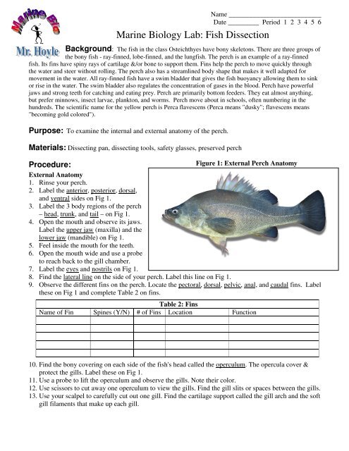
<strong>Marine</strong> <strong>Biology</strong> <strong>Lab</strong>: <strong>Fish</strong> <strong>Dissection</strong><br />
Background: The fish in the class Osteichthyes have bony skeletons. There are three groups of<br />
the bony fish - ray-finned, lobe-finned, and the lungfish. The perch is an example of a ray-finned<br />
fish. Its fins have spiny rays of cartilage &/or bone to support them. Fins help the perch to move quickly through<br />
the water and steer without rolling. The perch also has a streamlined body shape that makes it well adapted for<br />
movement in the water. All ray-finned fish have a swim bladder that gives the fish buoyancy allowing them to sink<br />
or rise in the water. The swim bladder also regulates the concentration of gases in the blood. Perch have powerful<br />
jaws and strong teeth for catching and eating prey. Perch are primarily bottom feeders. They eat almost anything,<br />
but prefer minnows, insect larvae, plankton, and worms. Perch move about in schools, often numbering in the<br />
hundreds. The scientific name for the yellow perch is Perca flavescens (Perca means "dusky"; flavescens means<br />
"becoming gold colored").<br />
Purpose: To examine the internal and external anatomy of the perch.<br />
Materials: Dissecting pan, dissecting tools, safety glasses, preserved perch<br />
Procedure:<br />
Figure 1: External Perch Anatomy<br />
External Anatomy<br />
1. Rinse your perch.<br />
2. <strong>Lab</strong>el the anterior, posterior, dorsal,<br />
and ventral sides on Fig 1.<br />
3. <strong>Lab</strong>el the 3 body regions of the perch<br />
– head, trunk, and tail – on Fig 1.<br />
4. Open the mouth and observe its jaws.<br />
<strong>Lab</strong>el the upper jaw (maxilla) and the<br />
lower jaw (mandible) on Fig 1.<br />
5. Feel inside the mouth for the teeth.<br />
6. Open the mouth wide and use a probe<br />
to reach back to the gill chamber.<br />
7. <strong>Lab</strong>el the eyes and nostrils on Fig 1.<br />
8. Find the lateral line on the side of your perch. <strong>Lab</strong>el this line on Fig 1.<br />
9. Observe the different fins on the perch. Locate the pectoral, dorsal, pelvic, anal, and caudal fins. <strong>Lab</strong>el<br />
these on Fig 1 and complete Table 2 on fins.<br />
Table 2: Fins<br />
Name of Fin Spines (Y/N) # of Fins Location Function<br />
10. Find the bony covering on each side of the fish's head called the operculum. The opercula cover &<br />
protect the gills. <strong>Lab</strong>el these on Fig 1.<br />
11. Use a probe to lift the operculum and observe the gills. Note their color.<br />
12. Use scissors to cut away one operculum to view the gills. Find the gill slits or spaces between the gills.<br />
13. Use your scalpel to carefully cut out one gill. Find the cartilage support called the gill arch and the soft<br />
gill filaments that make up each gill.
Name ___________________________<br />
Date _________ Period 1 2 3 4 5 6<br />
14. Use forceps to remove a few scales from your fish. Observe the scales under the<br />
Fig 3<br />
magnifying glass or microscope. Sketch a scale on Fig 3.<br />
Internal Anatomy<br />
15. Insert the tip of the scissors in the anus and cut toward the head, between the pelvic fins<br />
and just past the pectoral fins. Cut only through the skin, careful not cut any organs.<br />
Continue cutting through skin and muscle as in Fig 4.<br />
16. Carefully lift off the flap of skin and muscle to expose the internal organs in the body cavity.<br />
17. Locate the cream colored liver in the front of the body cavity. Also locate the gall bladder between the<br />
lobes of the liver. <strong>Lab</strong>el these on Fig 5.<br />
18. Remove the gall bladder & liver to observe the short esophagus attached to the stomach. Fig 4: Cut lines<br />
<strong>Lab</strong>el the stomach on Fig 5.<br />
19. At the posterior end of the stomach are the coiled intestines. <strong>Lab</strong>el these on Fig 5.<br />
20. Find the small reddish brown spleen near the stomach and label this on Fig 5.<br />
21. Below the operculum, are the bony gill rakers. Locate these and them label them on Fig 5.<br />
22. In front of the liver & behind the gill rakers is the pericardial cavity containing the heart. The heart of a<br />
fish only has 2 chambers --- an atrium & and a ventricle. Locate the heart & label it on Fig 5.<br />
23. In the upper part of the body cavity, below the lateral line is the swim bladder. This sac has a thin wall.<br />
<strong>Lab</strong>el the swim bladder on Fig 5.<br />
Figure 5: Internal Perch Anatomy<br />
24. Below the swim bladder are the gonads<br />
(testes or ovaries). <strong>Lab</strong>el them on Fig 5.<br />
25. Find the two long, dark kidneys in the<br />
posterior end. <strong>Lab</strong>el the kidneys in Fig 5<br />
26. Wastes exit the body through the anus<br />
located on the ventral side of the perch.<br />
<strong>Lab</strong>el the anus on Fig 5.<br />
Questions:<br />
1. Are both jaws of the fish equally<br />
movable? Explain your answer.<br />
2. Does the perch have eyelids?<br />
3. How many gills are located on each side of the perch? What covering protects them?<br />
4. The tail end has no organs; it’s all muscle. Why do you think that is?<br />
5. Explain how gas exchange occurs at the gills.<br />
6. Which fin was the largest? What other difference do you notice in this fin compared to the others?<br />
7. What is the function of the lateral line?<br />
8. Describe how the scales are arranged on the trunk & tail of your fish.<br />
9. What is the function of the swim bladder? Explain how it works.<br />
To learn more, check out:<br />
http://bioweb.uwlax.edu/zoolab/Table_of_Contents/<strong>Lab</strong>-9b/Perch_Mount_1/perch_mount_1.htm