Hemorrhagic Shock - SOGC
Hemorrhagic Shock - SOGC
Hemorrhagic Shock - SOGC

You also want an ePaper? Increase the reach of your titles
YUMPU automatically turns print PDFs into web optimized ePapers that Google loves.
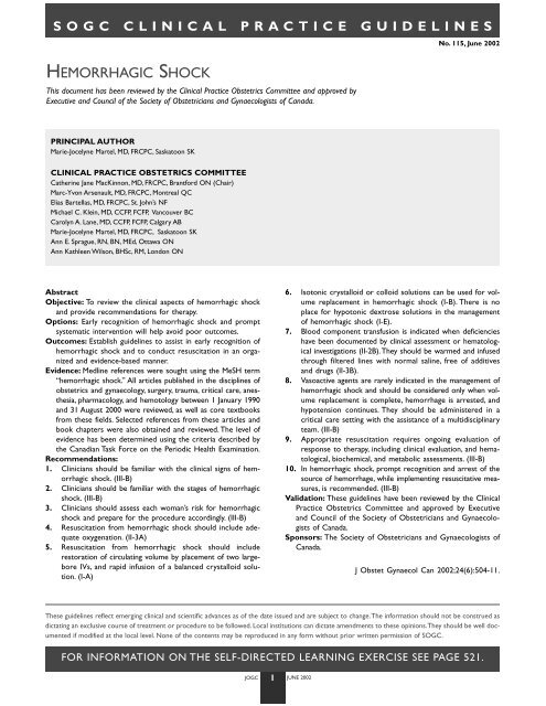
<strong>SOGC</strong> CLINICAL PRACTICE GUIDELINES<br />
HEMORRHAGIC SHOCK<br />
This document has been reviewed by the Clinical Practice Obstetrics Committee and approved by<br />
Executive and Council of the Society of Obstetricians and Gynaecologists of Canada.<br />
PRINCIPAL AUTHOR<br />
Marie-Jocelyne Martel, MD, FRCPC, Saskatoon SK<br />
CLINICAL PRACTICE OBSTETRICS COMMITTEE<br />
Catherine Jane MacKinnon, MD, FRCPC, Brantford ON (Chair)<br />
Marc-Yvon Arsenault, MD, FRCPC, Montreal QC<br />
Elias Bartellas, MD, FRCPC, St. John’s NF<br />
Michael C. Klein, MD, CCFP, FCFP, Vancouver BC<br />
Carolyn A. Lane, MD, CCFP, FCFP, Calgary AB<br />
Marie-Jocelyne Martel, MD, FRCPC, Saskatoon SK<br />
Ann E. Sprague, RN, BN, MEd, Ottawa ON<br />
Ann Kathleen Wilson, BHSc, RM, London ON<br />
Abstract<br />
Objective: To review the clinical aspects of hemorrhagic shock<br />
and provide recommendations for therapy.<br />
Options: Early recognition of hemorrhagic shock and prompt<br />
systematic intervention will help avoid poor outcomes.<br />
Outcomes: Establish guidelines to assist in early recognition of<br />
hemorrhagic shock and to conduct resuscitation in an organized<br />
and evidence-based manner.<br />
Evidence: Medline references were sought using the MeSH term<br />
“hemorrhagic shock.” All articles published in the disciplines of<br />
obstetrics and gynaecology, surgery, trauma, critical care, anesthesia,<br />
pharmacology, and hemotology between 1 January 1990<br />
and 31 August 2000 were reviewed, as well as core textbooks<br />
from these fields. Selected references from these articles and<br />
book chapters were also obtained and reviewed. The level of<br />
evidence has been determined using the criteria described by<br />
the Canadian Task Force on the Periodic Health Examination.<br />
Recommendations:<br />
1. Clinicians should be familiar with the clinical signs of hemorrhagic<br />
shock. (III-B)<br />
2. Clinicians should be familiar with the stages of hemorrhagic<br />
shock. (III-B)<br />
3. Clinicians should assess each woman’s risk for hemorrhagic<br />
shock and prepare for the procedure accordingly. (III-B)<br />
4. Resuscitation from hemorrhagic shock should include adequate<br />
oxygenation. (II-3A)<br />
5. Resuscitation from hemorrhagic shock should include<br />
restoration of circulating volume by placement of two largebore<br />
IVs, and rapid infusion of a balanced crystalloid solution.<br />
(I-A)<br />
These guidelines reflect emerging clinical and scientific advances as of the date issued and are subject to change.The information should not be construed as<br />
dictating an exclusive course of treatment or procedure to be followed. Local institutions can dictate amendments to these opinions.They should be well documented<br />
if modified at the local level. None of the contents may be reproduced in any form without prior written permission of <strong>SOGC</strong>.<br />
FOR INFORMATION ON THE SELF-DIRECTED LEARNING EXERCISE SEE PAGE 521.<br />
JOGC 1 JUNE 2002<br />
No. 115, June 2002<br />
6. Isotonic crystalloid or colloid solutions can be used for volume<br />
replacement in hemorrhagic shock (I-B). There is no<br />
place for hypotonic dextrose solutions in the management<br />
of hemorrhagic shock (I-E).<br />
7. Blood component transfusion is indicated when deficiencies<br />
have been documented by clinical assessment or hematological<br />
investigations (II-2B).They should be warmed and infused<br />
through filtered lines with normal saline, free of additives<br />
and drugs (II-3B).<br />
8. Vasoactive agents are rarely indicated in the management of<br />
hemorrhagic shock and should be considered only when volume<br />
replacement is complete, hemorrhage is arrested, and<br />
hypotension continues. They should be administered in a<br />
critical care setting with the assistance of a multidisciplinary<br />
team. (III-B)<br />
9. Appropriate resuscitation requires ongoing evaluation of<br />
response to therapy, including clinical evaluation, and hematological,<br />
biochemical, and metabolic assessments. (III-B)<br />
10. In hemorrhagic shock, prompt recognition and arrest of the<br />
source of hemorrhage, while implementing resuscitative measures,<br />
is recommended. (III-B)<br />
Validation: These guidelines have been reviewed by the Clinical<br />
Practice Obstetrics Committee and approved by Executive<br />
and Council of the Society of Obstetricians and Gynaecologists<br />
of Canada.<br />
Sponsors: The Society of Obstetricians and Gynaecologists of<br />
Canada.<br />
J Obstet Gynaecol Can 2002;24(6):504-11.
INTRODUCTION<br />
<strong>Hemorrhagic</strong> shock is a rare but serious complication, which<br />
may occur in many obstetrical or gynaecological situations.<br />
Hemorrhage is a leading cause of maternal death in the developing<br />
world. 1 Death and morbidity secondary to hemorrhage<br />
are becoming less common due to early recognition and intervention<br />
and improved availability of medical resources. 1 Ten recommendations<br />
for the management of hemorrhagic shock are<br />
listed in the following text and have been graded according to<br />
their level of evidence as determined by the criteria of the Canadian<br />
Task Force on the Periodic Health Examination (Table 1). 2<br />
HEMORRHAGIC SHOCK IN OBSTETRICS<br />
Obstetrical hemorrhage is often acute, dramatic, and underestimated.<br />
3 Postpartum hemorrhage is a significant cause of maternal<br />
death. 3 Management of postpartum hemorrhage has been<br />
reviewed in detail in <strong>SOGC</strong> Clinical Practice Guidelines for the<br />
Prevention and Management of Postpartum Hemorrhage. 3<br />
HEMORRHAGIC SHOCK IN GYNAECOLOGY<br />
A surgical procedure is the most common antecedent of acute<br />
gynaecological hemorrhage, although patients will occasionally<br />
present with acute hemorrhage from a ruptured ectopic pregnancy<br />
or from a neoplasm. 1 Risk identification is important in<br />
counselling patients prior to surgery and in preparation of the<br />
surgical team. Any process that distorts pelvic anatomy, such as<br />
endometriosis, neoplasm, or adhesions, or that leads to an inflam-<br />
TABLE 1<br />
QUALITY OF EVIDENCE ASSESSMENT 2<br />
The quality of evidence reported in these guidelines has been<br />
described using the Evaluation of Evidence criteria outlined in<br />
the Report of the Canadian Task Force on the Periodic<br />
Health Exam.<br />
I: Evidence obtained from at least one properly randomized<br />
controlled trial.<br />
II-1: Evidence from well-designed controlled trials without<br />
randomization.<br />
II-2: Evidence from well-designed cohort (prospective or<br />
retrospective) or case-control studies, preferably from<br />
more than one centre or research group.<br />
II-3: Evidence obtained from comparisons between times or<br />
places with or without the intervention. Dramatic<br />
results in uncontrolled experiments (such as the results<br />
of treatment with penicillin in the 1940s) could also be<br />
included in this category.<br />
III: Opinions of respected authorities, based on clinical<br />
experience, descriptive studies, or reports of expert<br />
committees.<br />
1<br />
JOGC 2 JUNE 2002<br />
matory response may be associated with increased intraoperative<br />
blood loss. Identification, isolation, and rapid control of bleeding<br />
encountered during the procedure will limit the total loss.<br />
The anatomy of the pelvis and landmarks of the vascular tree<br />
must be familiar to every pelvic surgeon.<br />
Patients with delayed postoperative hemorrhage may present<br />
with bleeding from the wound or vagina or with evidence<br />
of a hemoperitoneum. Careful examination and resuscitation<br />
with definitive and prompt control of blood loss is required,<br />
which may require a return to the operating theatre.<br />
CLINICAL PRESENTATION AND<br />
COMPLICATIONS OF HEMORRHAGIC SHOCK<br />
Hemorrhage occurs when there is excessive external or internal<br />
blood loss. 4 A defined volume is difficult to measure in most<br />
situations, and the loss evaluated visually is often underestimated.<br />
4 <strong>Shock</strong> occurs when there is hypoperfusion of vital<br />
organs. Hypoperfusion may be due to malfunction of the<br />
myocardium (cardiogenic shock), overwhelming infection leading<br />
to redistribution of circulating volume into the extravascular<br />
space (septic shock), or hypovolemia due to severe<br />
dehydration or hemorrhage (hypovolemic shock). 1 Signs and<br />
symptoms of hemorrhagic shock will vary depending on the<br />
volume and rate of blood loss (Table 2). 1<br />
The key systems affected by hemorrhagic shock are the central<br />
nervous, cardiac, and renal systems. 5 The central nervous<br />
system is able to function despite hypoperfusion, until the mean<br />
CLASSIFICATION OF RECOMMENDATIONS<br />
Recommendations included in these guidelines have been<br />
adapted from the ranking method described in the Classification<br />
of Recommendations found in the Report of the Canadian Task<br />
Force on the Periodic Health Exam.<br />
A. There is good evidence to support the recommendation<br />
that the condition be specifically considered in a periodic<br />
health examination.<br />
B. There is fair evidence to support the recommendation<br />
that the condition be specifically considered in a periodic<br />
health examination.<br />
C. There is poor evidence regarding the inclusion or exclusion<br />
of the condition in a periodic health examination,<br />
but recommendations may be made on other grounds.<br />
D. There is fair evidence to support the recommendation<br />
that the condition not be considered in a periodic health<br />
examination.<br />
E. There is good evidence to support the recommendation<br />
that the condition be excluded from consideration in a<br />
periodic health examination.
TABLE 2<br />
CLINICAL FEATURES OF HEMORRHAGIC SHOCK1 System Early shock Late shock<br />
CNS Altered mental status Obtunded<br />
Cardiac Tachycardia Cardiac failure<br />
Orthostatic hypotension Arrhythmias<br />
Hypotension<br />
Renal Oliguria Anuria<br />
Respiratory Tachypnea Tachypnea<br />
Respiratory failure<br />
Hepatic No change Liver failure<br />
Gastrointestinal No change Mucosal bleeding<br />
Hematological Anemia Coagulopathy<br />
Metabolic None Acidosis<br />
Hypocalcemia<br />
Hypomagnesemia<br />
arterial pressure falls below 60–70 mmHg. 1 With increasing severity<br />
of hypovolemia, mild agitation and confusion progress to<br />
lethargy and obtundation. 1 The heart plays an important role in<br />
the compensation for losses in early shock. 3 Early hypovolemia is<br />
associated with reflex tachycardia and increased stroke volume. 4,6<br />
With continued loss, hypoperfusion of the coronary arteries and<br />
myocardium leads to cardiac dysfunction, ischemia, and failure: 1<br />
symptoms of chest pain and dyspnea with signs of rales, tachypnea,<br />
and murmurs or arrhythmias are indicative of this process.<br />
The kidney will compensate for losses by activation of the reninangiotensin-aldosterone<br />
system. 4 Early, reversible renal injury is<br />
associated with low urine sodium concentration and high urine<br />
osmolality (>500 mOsm). 1 Oliguria is a reliable sign that these<br />
compensatory mechanisms have been overwhelmed. 1<br />
All organ systems are ultimately affected in shock. Respiratory,<br />
hepatic, and gastrointestinal systems can be affected early<br />
in the process since cardiac output is redirected to the most<br />
important organs: the heart, brain, and kidneys. 6,7 Manifestations<br />
of lung injury include: dyspnea, tachypnea, pulmonary<br />
infiltrates, and edema leading to decreased tissue compliance<br />
and hypoxia. Symptoms of adult respiratory distress syndrome<br />
(ARDS) include: intrapulmonary shunting, reduced pulmonary<br />
compliance, and low arterial pO 2 that often requires assisted<br />
mechanical ventilation. 6,8 Moderate elevations of bilirubin and<br />
alkaline phosphatase can be seen with ischemic hepatic injury. 1<br />
Gastrointestinal ischemia manifests as bleeding of either frank<br />
blood or coffee ground hematemesis or hematochezia or with<br />
delayed abdominal pain secondary to gut ischemia. 9 Erosion of<br />
the intestinal mucosa allows bacteremia and subsequent sep-<br />
JOGC 3 JUNE 2002<br />
sis. 10 Multi-system injury can lead to coagulopathy, and metabolic<br />
disturbances such as acidosis. 1<br />
RECOMMENDATION<br />
1. Clinicians should be familiar with the clinical signs of<br />
hemorrhagic shock. (III-B)<br />
PATHOPHYSIOLOGY OF HEMORRHAGIC SHOCK<br />
In hemorrhagic shock, an acute reduction in blood volume leads<br />
to sympathetic compensation by peripheral vasoconstriction,<br />
tachycardia, and increased myocardial contractility, which in<br />
turn increases the myocardial demand for oxygen, to a level that<br />
cannot be maintained. 1 Simultaneously, tissue hypoperfusion<br />
from precapillary vasoconstriction leads to anaerobic metabolism<br />
and acidosis. 11 Tissue hypoxia, acidosis, and the release of<br />
various mediators lead to a systemic inflammatory response. 5,11<br />
Reperfusion injury occurs when oxygen radicals released<br />
during the acute phase are systemically circulated as whole<br />
body perfusion is restored. 4,11,12 Humoral and cellular inflammatory<br />
systems are also activated, and contribute to vascular<br />
and cellular injury. 12,13 Transmigration of microorganisms and<br />
endotoxins across weakened mucosal barriers contributes to<br />
systemic inflammatory response syndrome (SIRS) and multiple<br />
organ failure. 4,10,11<br />
CLASSIFICATION OF HEMORRHAGIC SHOCK<br />
A classification of hemorrhagic shock is outlined in Table 3. This<br />
type of classification may aid in determining the volume required<br />
for initial replacement, and the listed signs of shock in determining<br />
the severity of occult losses. The symptoms and sequelae<br />
of hemorrhage are ultimately related to perfusion of tissues.<br />
Loss of less than, or equal to, 15% of blood volume (compensated<br />
shock) may not be associated with any change in blood<br />
pressure (BP), pulse, or capillary refill. Mild shock is usually easily<br />
compensated, especially in the younger, healthy woman of<br />
reproductive age. 14 Further losses lead to tachycardia, a catecholamine<br />
response characterized by increased sympathetic tone.<br />
Resting BP is usually normal, but orthostatic changes in BP and<br />
pulse may be evident. Simple resuscitative measures will successfully<br />
reverse these changes. 1 Ongoing losses of blood volume<br />
may overtake the heart’s ability to compensate, and marked<br />
tachycardia is associated with a fall in BP, classified as moderate<br />
shock. With continued bleeding, hypoperfusion of tissues occurs,<br />
leading to anaerobic metabolism and acidosis, classified as severe<br />
shock. The patient demonstrates marked tachycardia and tachypnea<br />
with respiratory failure, becomes oliguric, and then anuric.<br />
Obtundation and loss of consciousness may also occur. 1 Cellular<br />
dysfunction, followed by cell death, leads to multiple organ<br />
failure, resulting in irreversible shock. 1,15 The mortality rate at<br />
this stage is in excess of 30%. 1
TABLE 3<br />
RECOMMENDATION<br />
2. Clinicians should be familiar with the stages of hemorrhagic<br />
shock. (III-B)<br />
RISK FACTORS<br />
Evaluation of all patients presenting for obstetrical care or<br />
surgery should include a complete medical history. A personal<br />
or family history of coagulopathy, or personal use of anticoagulants,<br />
should be documented. A complete physical examination<br />
may reveal extensive bruising or petechiae. Investigations<br />
to assess coagulation status should be obtained in these situations<br />
and consultation from other disciplines considered.<br />
All proposed procedures should be reviewed with the<br />
patient. The risk of complications including hemorrhage<br />
should be outlined and the discussion documented in the<br />
chart. 17 Certain clinical conditions and their surgical management<br />
are associated with an increased risk of hemorrhage, such<br />
as ectopic pregnancy, myomectomy, abruptio placenta, placenta<br />
previa, and malignant disease. 17 In some situations, it<br />
may be appropriate to counsel women about autologous blood<br />
transfusion or hemodilution techniques. 17,18 Jehovah’s Witnesses<br />
may require special consideration. 19<br />
RECOMMENDATION<br />
3. Clinicians should assess each woman’s risk for hemorrhagic<br />
shock and prepare for the procedure accordingly.<br />
(III-B)<br />
MANAGEMENT<br />
Early resuscitation includes control of bleeding and restoration<br />
of circulating blood volume for oxygenation of tissues. 16 Techniques<br />
to minimize blood loss should be applied whenever possible.<br />
Exposure of the bleeding site, experienced assistance, and<br />
sound knowledge of pelvic anatomy, as well as a calm, system-<br />
CLASSIFICATION OF HEMORRHAGIC SHOCK 1,5,16<br />
Compensated Mild Moderate Severe<br />
Blood Loss (mL) ≤1000 1000–1500 1500–2000 >2000<br />
Heart rate (bpm) 100 >120 >140<br />
Blood pressure Normal Orthostatic change Marked fall Profound fall<br />
Capillary refill Normal May be delayed Usually delayed Always delayed<br />
Respiration Normal Mild increase Moderate tachypnea Marked tachypnea:<br />
respiratory collapse<br />
Urinary output (mL/h) >30 20–30 5–20 Anuria<br />
Mental status Normal or agitated Agitated Confused Lethargic, obtunded<br />
JOGC 4 JUNE 2002<br />
atic approach to vascular areas, will be useful in the prompt control<br />
of hemorrhage. As soon as the first signs of excessive blood<br />
loss and shock are evident, assistance from other members of the<br />
health care team, which may include an anesthetist, a second<br />
gynaecologist, a general surgeon, a vascular surgeon, a critical<br />
care specialist, a hematologist, and experienced nursing staff,<br />
should be considered when appropriate and if available. Laboratory<br />
and blood bank services should be informed and available<br />
for support. Since cell death due to hypoxic injury is the final<br />
common pathway in shock, all efforts should be directed at<br />
restoring tissue oxygenation as soon as possible. A useful<br />
mnemonic to achieve this goal is ORDER: Oxygenate, Restore<br />
circulating volume, Drug therapy, Evaluate response to therapy,<br />
Remedy underlying cause. 1,14 Outcome is dependent on early<br />
recognition and on immediate aggressive therapy, which relies<br />
on two basic principles: replace losses and arrest bleeding.<br />
OXYGENATION<br />
The initial step in any patient resuscitation is to secure an airway<br />
and provide adequate oxygenation. 16 In most surgical situations,<br />
an airway will already be in place, managed by the anesthetist. If<br />
regional anesthesia has been used, supplemental oxygen should<br />
be applied. 4 Consideration should be given to endotracheal intubation,<br />
if the patient is becoming disoriented or is tiring, and in<br />
an obtunded patient should be instituted immediately. 1 After<br />
extensive fluid resuscitation, edema of the trachea may make intubation<br />
difficult. Positive ventilatory pressures may be required in<br />
those patients with decreased pulmonary compliance.<br />
RECOMMENDATION<br />
4. Resuscitation from hemorrhagic shock should include<br />
adequate oxygenation. (II-3A)<br />
RESTORE CIRCULATING VOLUME<br />
Intravascular replacement of blood volume lost may be accomplished<br />
using crystalloid, colloid, or blood products. Initial
therapy should consist of 1–2L of intravenous (IV) crystalloid.<br />
1,16 Intravenous access should be of large calibre (14–16<br />
gauge) and in multiple sites to facilitate rapid volume infusion.<br />
5,16 A central line may be considered, but it does not<br />
appear to provide any advantage over peripheral lines for rapid<br />
infusion of volume. 5,16 The time and skill required to establish<br />
a central line, and the risk of complications such as pneumothorax,<br />
should also be considered. 1,5 Central venous pressure<br />
measurements may be helpful for safe resuscitation, if there has<br />
been injury to the cardiovascular system or vascular injury in<br />
the lung, as the amount of fluid required to restore tissue perfusion<br />
may be difficult to estimate. These patients are at risk<br />
for pulmonary edema and lung injury, if excessive fluid replacement<br />
is given. 6,7 In monitoring patients with multiple organ<br />
failure, central venous pressure measurements may also be useful<br />
in resuscitation and monitoring. 16 Normal central venous<br />
pressure is 5 mmHg (range 0–8 mmHg). 14 Elevated pressures<br />
are seen in fluid overload, right ventricular failure, pulmonary<br />
embolus, cardiac tamponade, and severe tricuspid regurgitation.<br />
Low values are seen with shock from hypovolemia, sepsis,<br />
and anaphylaxis. 14<br />
RECOMMENDATION<br />
5. Resuscitation from hemorrhagic shock should include<br />
restoration of circulating volume by placement of two<br />
large-bore IVs, and rapid infusion of a balanced crystalloid<br />
solution. (I-A)<br />
CRYSTALLOID SOLUTIONS<br />
Crystalloid solutions are electrolyte solutions administered intravenously.<br />
Advantages of crystalloid solutions include availability,<br />
safety, and low cost. 5,6 The main disadvantage of using<br />
crystalloid solutions is their rapid movement from the intravascular<br />
to the extravascular space, leading to three or more times<br />
requirement for replacement, 1,4,20,21 and resulting in tissue<br />
edema. 22 Ringer’s lactate is preferred over normal saline to avoid<br />
hyperchloremic acidosis associated with prolonged use of sodium<br />
solutions. 1,23,24 Hypertonic salt solutions are not generally<br />
recommended because of the risk of electrolyte disturbances.<br />
1,6,20,25 There is no role for hypotonic dextrose solutions<br />
in the management of hypovolemic shock. 21<br />
TABLE 4<br />
JOGC 5 JUNE 2002<br />
COLLOID SOLUTIONS<br />
Colloid solutions contain molecules designed to stay within the<br />
intravascular compartment. 1,6,11,20-23 These products include<br />
albumin, hydroxyethyl starch, dextrans, and gelatins. They are<br />
more expensive and less available than crystalloids. 22,24 Each of<br />
these oncotic replacement products carries the risk of reaction.<br />
1,20,23 Some will bind calcium or affect circulating<br />
immunoglobulins. 6,7,20,26 There is no convincing evidence that<br />
colloid solutions offer any advantage over crystalloid solutions<br />
in the replacement of volume in hemorrhagic shock. 1,4,5-7,<br />
20-22,24,27,28 A recent review of the use of albumin in the treatment<br />
of hypovolemia suggests that its use may increase the risk<br />
of death. 29 Crystalloids and colloids may be used together. 22<br />
RECOMMENDATION<br />
6. Isotonic crystalloid or colloid solutions can be used for<br />
volume replacement in hemorrhagic shock (I-B). There<br />
is no place for hypotonic dextrose solutions in the management<br />
of hemorrhagic shock (I-E).<br />
TRANSFUSION<br />
Many blood products are available to restore circulating volume,<br />
and replace coagulation factors and oxygen-carrying<br />
capacity (Table 4). Component therapy allows specific replacement<br />
to address specific needs. Hypovolemia is best corrected<br />
with crystalloid solution. In hemorrhagic shock, packed red<br />
blood cells (PRBC) are most commonly used to restore intravascular<br />
volume and oxygen-carrying capacity. The oxygencarrying<br />
capacity of most healthy individuals will not be compromised<br />
until the hemoglobin concentration falls below<br />
60–70 g/L. 1,7,20,30,31 There is no recommended “threshold<br />
hemoglobin.” 7,30 Blood losses greater than 20–25% or cases of<br />
documented or suspected coagulopathy may require replacement<br />
of coagulation factors; coagulation studies are recommended<br />
after transfusion of 5 to 10 units of PRBCs. 14<br />
Platelet transfusions are indicated in situations of significant<br />
thrombocytopenia (platelet count less than 20,000 to<br />
50,000 per mm 3 ) and continued hemorrhage. 31 Coagulation<br />
factor concentrates are available for identified deficiencies, and<br />
fresh frozen plasma can be administered in acute situations<br />
where the partial thromboplastin time and prothrombin time<br />
INDICATIONS FOR BLOOD COMPONENT THERAPY 14,20,30-35<br />
Component Indication Usual starting dose<br />
Packed RBC Replacement of oxygen-carrying capacity 2– 4 Units IV<br />
Platelets Thrombocytopenia or thrombasthenia with bleeding 6–10 Units IV<br />
Fresh frozen plasma Documented coagulopathy 2–6 Units IV<br />
Cryoprecipitate Coagulopathy with low fibrinogen 10–20 Units IV
are elevated. There is no indication for prophylactic administration<br />
of platelets, plasma, or specific components in resuscitation<br />
of hemorrhagic shock. 20,30-33 Immediate complications<br />
of blood transfusions are increased when large volumes of<br />
blood product are infused. 30 All blood products should be<br />
cross-matched and administered through filtered lines with<br />
normal saline, free of additives or drugs. 16 Acid-base and electrolyte<br />
disturbances can be evident with large volume transfusions.<br />
16 Blood products should be warmed to prevent<br />
hypothermia. 1,30,32<br />
RECOMMENDATION<br />
7. Blood component transfusion is indicated when deficiencies<br />
have been documented by clinical assessment or<br />
hematological investigations (II-2B). They should be<br />
warmed and infused through filtered lines with normal<br />
saline, free of additives and drugs (II-3B).<br />
DRUG THERAPY<br />
VASOACTIVE AGENTS<br />
After adequate volume replacement has been achieved, vasoactive<br />
agents, which include inotropes and vasopressors, may<br />
be considered but are not often required in hemorrhagic shock. 1<br />
When required, inotropic agents are administered first, followed<br />
by vasopressors in refractory cases. There is a risk that these<br />
agents may cause a further limitation of perfusion and oxygenation<br />
of distal organs. 7,14 Ideally, these drugs should be<br />
administered in a critical care setting with the assistance of a<br />
multidisciplinary team.<br />
Dopamine can stimulate the function of alpha- and beta1- adrenergic receptors. 7 At low doses of 1–3 µg/kg/min dopa-<br />
JOGC 6 JUNE 2002<br />
mine, the dopamine receptors in the cerebral, renal, and<br />
mesenteric circulation are stimulated, resulting in vasodilatation<br />
and increased urinary output. At moderate doses<br />
(2–10 µg/kg/min), alpha- and beta 1 -adrenergic receptors are<br />
also stimulated, increasing myocardial contractility and cardiac<br />
output, resulting in an increase in myocardial oxygen consumption.<br />
At higher doses (>10 µg/kg/min), the alpha-adrenergic<br />
receptors are stimulated, leading to vasoconstriction<br />
and increases in blood pressure. 1,7<br />
Dobutamine is a beta 1 - and beta 2 -adrenergic stimulator.<br />
Beta 2 stimulation leads to systemic vasodilatation and reduced<br />
afterload. 7 Dobutamine is associated with less pulmonary congestion<br />
and less tachycardia than dopamine. 7<br />
Phenylephrine, norepinephrine, and epinephrine are used<br />
in situations of refractory shock. Their principal effect is to<br />
increase blood pressure by increasing systemic vascular resistance.<br />
They also produce some degree of coronary vasoconstriction,<br />
which may exacerbate myocardial ischemia (Table 5). 7<br />
RECOMMENDATION<br />
8. Vasoactive agents are rarely indicated in the management<br />
of hemorrhagic shock and should be considered only<br />
when volume replacement is complete, hemorrhage is<br />
arrested, and hypotension continues. They should be<br />
administered in a critical care setting with the assistance<br />
of a multidisciplinary team. (III-B)<br />
OTHER DRUG THERAPY<br />
Broad coverage antibiotic therapy should be instituted in cases<br />
of hemorrhagic shock, as there is a significant risk of sepsis. 1<br />
Ischemic injury to the gut makes it vulnerable to transmucosal<br />
TABLE 5<br />
PHARMACOLOGICAL SUPPORT OF THE CARDIOVASCULAR SYSTEM1,7 Agent Usual dose range Effect<br />
Inotropic agents<br />
Dopamine 1–3 µg/kg/min Increased renal output<br />
Vasodilation<br />
2–10 µg/kg/min Increased heart rate<br />
Increased cardiac output<br />
>10 µg/kg/min Peripheral vasoconstriction<br />
Increased heart rate and contractility<br />
Dobutamine 2–10 µg/kg/min Increased heart rate and contractility<br />
Decreased afterload<br />
Vasopressor agents<br />
Phenylephrine 1–5 µg/kg/min Peripheral vasoconstriction<br />
Norepinephrine 1–4 µg/min Peripheral vasoconstriction<br />
Epinephrine 1– 8 µg/min Peripheral vasoconstriction
erosions and may lead to bacteremia. 10 The gastric mucosa is<br />
susceptible to stress ulceration, which can be reduced by the use<br />
of antacids or H 2 blockers. 1<br />
Stroma-free hemoglobin (diaspirin cross-linked hemoglobin)<br />
is a new product currently under evaluation, which can<br />
replace the oxygen-carrying capacity of PRBCs. Its affinity for<br />
nitric oxide contributes to peripheral vasoconstriction. Potential<br />
advantages include a longer shelf life and universal compatibility.<br />
35 There is no clear clinical advantage over PRBCs.<br />
Risks include extravasation, possible coagulopathy, and the risks<br />
associated with human blood components, as well as documented<br />
toxicities. 15,20,35-37<br />
EVALUATION OF RESPONSE TO THERAPY<br />
Once oxygenation and circulating volume have been restored,<br />
re-evaluation of the clinical situation is in order. Vital signs,<br />
mental status, urinary output, and capillary refill should be<br />
assessed regularly throughout resuscitation. 14,16 Initiation of<br />
central monitoring may be indicated at this time, if the response<br />
to initial resuscitation has been less than expected or if blood<br />
loss is ongoing. 7 Blood should be drawn to assess hematological,<br />
coagulation, electrolyte, and metabolic status. Electrolyte<br />
and metabolic disorders as well as coagulation deficiencies<br />
should be corrected. Arterial blood gases should be obtained to<br />
determine adequacy of oxygenation. Management of alterations<br />
to oxygenation, ventilation, pH, fluid, and electrolyte balance<br />
should now be made based on laboratory measurements. 16 Blood<br />
components may also be used to replace identified deficiencies.<br />
RECOMMENDATION<br />
9. Appropriate resuscitation requires ongoing evaluation of<br />
response to therapy, including clinical evaluation, and<br />
hematological, biochemical, and metabolic assessments.<br />
(III-B)<br />
REMEDY THE UNDERLYING CAUSE<br />
Most cases of unresponsive shock in gynaecologic patients are due<br />
to ongoing losses of blood volume. While initial stabilization is<br />
taking place, attention should be directed to the prompt arrest of<br />
bleeding. Aggressive restoration of normal blood pressure without<br />
arrest of hemorrhage will enhance further losses of blood volume<br />
by increasing flow and impeding coagulation at the site of<br />
injury. 11,15,25,27,38-40 Mild to moderate hypotension allows clot formation<br />
and slows bleeding from the injured vessel. 39 The source<br />
of blood loss should be identified and arrested as soon as possible.<br />
14 Occult retroperitoneal hemorrhage should be suspected in<br />
patients who have a deteriorating hemodynamic status more than<br />
12 hours post surgery. 17 In these cases, identification of the exact<br />
site of bleeding can be very difficult. Hemorrhage during or following<br />
an operative procedure should be addressed surgically, with<br />
immediate exploration, isolation, and ligation of bleeding vessels.<br />
In most cases, hemostasis can be achieved in this manner. Isola-<br />
JOGC 7 JUNE 2002<br />
tion and ligation of the uterine or internal iliac arteries are useful<br />
techniques to control hemorrhage in the pelvis. 17 The ureter is<br />
especially vulnerable during the placement of hemostatic sutures<br />
in the pelvis. If hemorrhage continues, coagulation should be evaluated<br />
and any identified deficiencies should be corrected. Adjunctive<br />
measures such as radiologic embolization and packing of the<br />
pelvis should be considered in refractory cases. 17<br />
Careful documentation of events and the interventions performed<br />
should be completed in a timely fashion. Communication<br />
with the patient, her partner, and her family should be<br />
prompt, frequent, and clear.<br />
RECOMMENDATION<br />
10. In hemorrhagic shock, prompt recognition and arrest<br />
of the source of hemorrhage, while implementing resuscitative<br />
measures, is recommended. (III-B)<br />
CONCLUSION<br />
<strong>Hemorrhagic</strong> shock may occur in many obstetrical or gynaecological<br />
conditions. Prompt recognition of blood losses and implementation<br />
of resuscitative measures in a calm and systematic<br />
fashion are imperative. A multidisciplinary approach to the multisystem<br />
effects of shock is essential to optimize the outcome.<br />
REFERENCES<br />
1. Smith HO. <strong>Shock</strong> in the gynecologic patient. In: Rock JA,Thomson JD,<br />
editors.Te Linde’s operative gynecology. 8th ed. Lippincott-Raven; 1997.<br />
p. 245-61.<br />
2. Woolf SH, Battista RN,Angerson GM, Logan AG, Eel W. Canadian Task<br />
Force on the Periodic Health Exam. Ottawa: Canada Communication<br />
Group; 1994. p. xxxvii.<br />
3. <strong>SOGC</strong> Clinical Practice Guidelines. Prevention and management of postpartum<br />
hemorrhage. J Soc Obstet Gynaecol Can 2000;22(4):271-81.<br />
4. Barber A, Shires GT. <strong>Shock</strong>. In: Schwartz SI, editor. Principles of surgery.<br />
7th ed. McGraw-Hill. 1999. p.101-22.<br />
5. Falk JL, O’Brien JF, Kerr R. Fluid resuscitation in traumatic hemorrhagic<br />
shock. Crit Care Clin 1992;8(2):323-40.<br />
6. Shires GT, Barber AE, Illner HP. Current status of resuscitation:<br />
solutions including hypertonic saline. Adv Surg 1995;28:133-70.<br />
7. Domsky MF,Wilson RF. Hemodynamic resuscitation. Crit Care Clin<br />
1993;10(4):715-26.<br />
8. Croce MA, Fabian TC, Davis KA, Gavin TJ. Early and late acute respiratory<br />
distress syndrome: two distinct clinical entities. J Trauma<br />
1999;46(3):361-8.<br />
9. Ludwig KA, Quebbeman EJ, Bergstein JM,Wallace JR,Wittmann DH,<br />
Aprahamian C. <strong>Shock</strong>-associated right colon ischemia and necrosis.<br />
J Trauma 1995;39(6):1171-4.<br />
10. Kale IT, Kuzu MA, Berkem H, Berkem R, Acar N.The presence of<br />
hemorrhagic shock increases the rate of bacterial translocation in<br />
blunt abdominal trauma. J Trauma 1998;44(1):171-4.<br />
11. Marzi I. <strong>Hemorrhagic</strong> shock: update in pathophysiology and therapy.<br />
Acta Anaesthesiol Scand Suppl 1997;111:42-4.<br />
12. Heckbert SR,Vedder NB, Hoffman W,Winn RK, Hudson LD,<br />
Jurkovich GJ, et al. Outcome after hemorrhagic shock in trauma<br />
patients. J Trauma 1998;45(3):545-9.<br />
13. Seekamp A, Jochum M, Ziegler M, van Griensven M, Martin M, Regel G.<br />
Cytokines and adhesion molecules in elective and accidental traumarelated<br />
ischemia/reperfusion. J Trauma 1998;44(5):874-82.
14. ACOG Educational Bulletin. <strong>Hemorrhagic</strong> shock. Number 235. April<br />
1997 (replaces no. 82, December 1984). American College of Obstetricians<br />
and Gynecologists. Int J Gynaecol Obstet 1997;57(2):219-26.<br />
15. Shoemaker WC, Peitzman AB, Bellamy R, Bellomo R, Bruttig SP,<br />
Capone A, et al. Resuscitation from severe hemorrhage. Crit Care<br />
Med 1996;24(2 Suppl):S12-23.<br />
16. The American College of Surgeons. <strong>Shock</strong>. In:The American College<br />
of Surgeons, editor. Advanced trauma life support. 1990. p. 59-73.<br />
17. Thompson JD, Rock WA. Control of pelvic hemorrhage. In: Rock JA,<br />
Thomson JD, editors.Te Linde’s operative gynecology. 8th ed.<br />
Lippincott-Raven Publishers; 1997. p.197-232.<br />
18. Rebarber A, Lonser R, Jackson S, Copel JA, Sipes S.The safety of intraoperative<br />
autologous blood collection and autotransfusion during<br />
Cesarean section. Am J Obstet Gynecol 1998;179:715-20.<br />
19. Culkin Mann M,Votto J, Kambe J, McNamee J. Management of the<br />
severely anemic patient who refuses transfusion: lessons learned during<br />
the care of a Jehovah’s Witness. Ann Intern Med 1992;117:1042-8.<br />
20. Gould SA, Sehgal LR, Sehgal HL, Moss GS. Hypovolemic shock. Crit<br />
Care Clin 1993;9(2):239-59.<br />
21. Lucas CE. Update on trauma care in Canada. 4. Resuscitation through<br />
the three phases of hemorrhagic shock after trauma. Can J Surg<br />
1990;33(6):451-6.<br />
22. Davies MJ. Crystalloid or colloid: does it matter? J Clin Anesth<br />
1989;1(6):464-71.<br />
23. Dubick MA,Wade CE. A review of the efficacy and safety of 7.5%<br />
NaCl/6% dextran 70 in experimental animals and in humans. J Trauma<br />
1994;36(3):323-30.<br />
24. Vassar MJ, Perry CA, Holcroft JW. Prehospital resuscitation of hypotensive<br />
trauma patients with 7.5% NaCl versus 7.5% NaCl with added<br />
dextran: a controlled trial. J Trauma 1993;34(5):622-32.<br />
25. Deakin CD. Early fluid resuscitation in haemorrhagic shock. Eur J Emerg<br />
Med 1994;1(2):83-5.<br />
26. Sillett HK,Whicher JT,Trejdosiewicz LK. Effects of resuscitation fluids<br />
on nonadaptive immune responses.Transfusion 1997;37(9):953-9.<br />
27. Napolitano LM. Resuscitation following trauma and hemorrhagic shock:<br />
is hydroxyethyl starch safe? [editorial; comment]. Crit Care Med<br />
1995;23(5):795-7.<br />
28. Poole GV, Meredith JW, Pennell T, Mills SA. Comparison of colloids<br />
and crystalloids in resuscitation from hemorrhagic shock. Surg Gynecol<br />
Obstet 1982;154:577-86.<br />
29. Bunn F, Lefebvre C, Li-Wan-Po A, Li L, Roberts I, Schierhout G. Human<br />
albumin solution for resuscitation and volume expansion in critically ill<br />
patients. Cochrane Database Syst Rev 2000;2:CD001208.<br />
30. Guidelines for red blood cell and plasma transfusion for adults and<br />
children. Can Med Assoc J 1997;156(11):S1-S54.<br />
31. Schwartz SI. Hemostasis, surgical bleeding and transfusion. In: Schwartz<br />
SI, editor. Principles of surgery. 7th ed. McGraw-Hill; 1999. p. 77-100.<br />
32. Hocker P, Hartmann T. Management of massive transfusion.<br />
Acta Anaesthesiol Scand Suppl 1997;111:205-7.<br />
33. Harrigan C, Lucas CE, Ledgerwood AM.The effect of hemorrhagic<br />
shock on the clotting cascade in injured patients. J Trauma<br />
1989;29(10):1416-21.<br />
34. Martin DJ, Lucas CE, Ledgerwood AM, Hoschner J, McGonigal MD,<br />
Grabow D. Fresh frozen plasma supplement to massive red blood cell<br />
transfusion. Ann Surg 1985;202(4):505-11.<br />
35. ACOG technical bulletin. Blood component therapy. Number 199 –<br />
November 1994 (replaces no. 78, July 1984). Committee on Technical<br />
Bulletins of the American College of Obstetricians and Gynecologists.<br />
Int J Gynaecol Obstet 1995;48(2):233-8.<br />
36. Ogden JE, Parry ES.The development of hemoglobin solutions as red<br />
cell substitutes. Int Anesthesiol Clin 1995;33(1):115-29.<br />
37. Rabinovici R, Neville LF, Rudolph AS, Feuerstein G. Hemoglobin-based<br />
oxygen-carrying resuscitation fluids [editorial; comment]. Crit Care<br />
Med 1995;23(5):801-4.<br />
38. Assalia A, Schein M. Resuscitation for haemorrhagic shock. Br J Surg<br />
1993;80(2):213.<br />
JOGC 8 JUNE 2002<br />
39. Bickell WH. Are victims of injury sometimes victimized by attempts at<br />
fluid resuscitation? [editorial; comment]. Ann Emerg Med<br />
1993;22(2):225-6.<br />
40. Dries DJ. Hypotensive resuscitation. <strong>Shock</strong> 1996;6(5):311-6.