functional anatomy of the spine by avicenna
functional anatomy of the spine by avicenna
functional anatomy of the spine by avicenna

Create successful ePaper yourself
Turn your PDF publications into a flip-book with our unique Google optimized e-Paper software.
NADERI ET AL.<br />
This is a very welcome brief historical note indicating that<br />
Avicenna devoted a significant degree <strong>of</strong> time and effort to<br />
problems <strong>of</strong> <strong>the</strong> <strong>spine</strong>. It is a delight to read this historical<br />
vignette.<br />
Edward R. Laws, Jr.<br />
Charlottesville, Virginia<br />
The Eastern physician Ibn Sina, known as Avicenna, was a<br />
very important scientist who lived between <strong>the</strong> years AD<br />
980 and 1037. His anatomic works, especially on <strong>spine</strong>, were<br />
written with a surgeon’s consciousness, because he was a<br />
surgeon. The origin <strong>of</strong> his anatomic knowledge is not known,<br />
because anatomic dissection on human bodies was prohibited<br />
during Avicenna’s era. However, his orientation and descriptions<br />
<strong>of</strong> <strong>the</strong> spinal <strong>anatomy</strong> at that period are exceptional.<br />
Yücel Kanpolat<br />
Ankara, Turkey<br />
In <strong>the</strong> present-day literature, <strong>the</strong>re is very little in <strong>the</strong> English<br />
language on <strong>the</strong> original writings <strong>of</strong> Avicenna and his classic<br />
work titled The Canons <strong>of</strong> Medicine. The authors <strong>of</strong> this<br />
article have provided an English translation from <strong>the</strong> original<br />
Arabic <strong>of</strong> <strong>the</strong> various sections dealing with <strong>the</strong> <strong>anatomy</strong> and<br />
function <strong>of</strong> <strong>the</strong> human <strong>spine</strong>. After one reads <strong>the</strong>se various<br />
sections, it becomes quite clear that Avicenna had a most<br />
remarkable grasp <strong>of</strong> <strong>the</strong> <strong>anatomy</strong> <strong>of</strong> <strong>the</strong> <strong>spine</strong> and some <strong>of</strong> its<br />
biomechanical aspects. An intriguing question that arises, and<br />
one not easily answered, is how many <strong>of</strong> <strong>the</strong> original dissections<br />
were performed <strong>by</strong> Avicenna himself, versus what he<br />
adapted from <strong>the</strong> earlier Greco-Roman writers. The authors do<br />
allude to this in <strong>the</strong> text, but it would be wonderful if more<br />
information might be provided with some fur<strong>the</strong>r research.<br />
There has been much discussion in <strong>the</strong> historical literature<br />
about how <strong>the</strong> Koran forbids human dissection, yet I remember<br />
from years <strong>of</strong> reading various sources that <strong>the</strong>re were<br />
exceptions to this edict <strong>by</strong> some <strong>of</strong> <strong>the</strong> early anatomists. This<br />
last thought leads naturally to Avicenna and <strong>the</strong> question <strong>of</strong><br />
whe<strong>the</strong>r he actually performed human dissections. It would<br />
appear that he did.<br />
James T. Goodrich<br />
Bronx, New York<br />
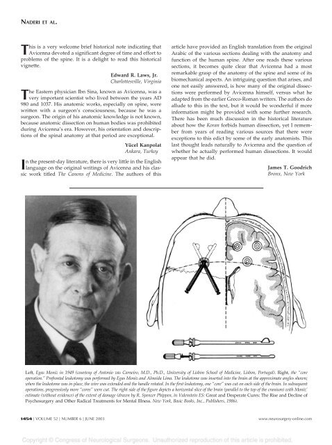
Left, Egas Moniz in 1949 (courtesy <strong>of</strong> Antonio vas Carneiro, M.D., Ph.D., University <strong>of</strong> Lisbon School <strong>of</strong> Medicine, Lisbon, Portugal). Right, <strong>the</strong> “core<br />
operation.” Prefrontal leukotomy was performed <strong>by</strong> Egas Moniz and Almeida Lima. The leukotome was inserted into <strong>the</strong> brain at <strong>the</strong> approximate angles shown;<br />
when <strong>the</strong> leukotome was in place, <strong>the</strong> wire was extended and <strong>the</strong> handle rotated. In <strong>the</strong> first leukotomy, one “core” was cut on each side <strong>of</strong> <strong>the</strong> brain. In subsequent<br />
operations, progressively more “cores” were cut. The right side <strong>of</strong> <strong>the</strong> figure depicts a horizontal slice <strong>of</strong> <strong>the</strong> brain (parallel to <strong>the</strong> top <strong>of</strong> <strong>the</strong> cranium) with Moniz’<br />
estimate (without evidence) <strong>of</strong> <strong>the</strong> extent <strong>of</strong> damage (drawn <strong>by</strong> R. Spencer Phippen, in Valenstein ES: Great and Desperate Cures: The Rise and Decline <strong>of</strong><br />
Psychosurgery and O<strong>the</strong>r Radical Treatments for Mental Illness. New York, Basic Books, Inc., Publishers, 1986).<br />
1454 | VOLUME 52 | NUMBER 6 | JUNE 2003 www.neurosurgery-online.com