Spider Silk: From Soluble Protein to Extraordinary Fiber - Physics
Spider Silk: From Soluble Protein to Extraordinary Fiber - Physics
Spider Silk: From Soluble Protein to Extraordinary Fiber - Physics

You also want an ePaper? Increase the reach of your titles
YUMPU automatically turns print PDFs into web optimized ePapers that Google loves.
Reviews<br />
Biomimetic Polymers<br />
DOI: 10.1002/anie.200803341<br />
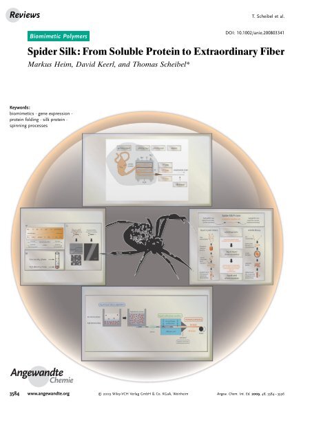
<strong>Spider</strong> <strong>Silk</strong>: <strong>From</strong> <strong>Soluble</strong> <strong>Protein</strong> <strong>to</strong> <strong>Extraordinary</strong> <strong>Fiber</strong><br />
Markus Heim, David Keerl, and Thomas Scheibel*<br />
Keywords:<br />
biomimetics · gene expression ·<br />
protein folding · silk protein ·<br />
spinning processes<br />
Angewandte<br />
Chemie<br />
T. Scheibel et al.<br />
3584 www.angewandte.org 2009 Wiley-VCH Verlag GmbH & Co. KGaA, Weinheim Angew. Chem. Int. Ed. 2009, 48, 3584 – 3596
<strong>Spider</strong> <strong>Silk</strong><br />
<strong>Spider</strong> silks outrival natural and many synthetic fibers in terms of<br />
their material characteristics. In nature, the formation of a solid fiber<br />
from soluble spider silk proteins is the result of complex biochemical<br />
and physical processes that take place within specialized spinning<br />
organs. Herein, we present natural and artificial silk production processes,<br />
from gene transcription <strong>to</strong> silk protein processing and finally<br />
fiber assembly. In-vivo and in-vitro findings in the field of spider silk<br />
research are the basis for the design of new proteins and processing<br />
strategies, which will enable applications of these fascinating proteinbased<br />
materials in technical and medical sciences.<br />
1. Introduction<br />
Mankind has used spider silk as a material long before it<br />
appeared in the focus of research. In ancient Greece, natural<br />
cobwebs were used <strong>to</strong> seal bleeding wounds, and in Australasia,<br />
spider silk threads or whole spider webs were used for<br />
fishing. Later, spider silks were also utilized for military<br />
purposes, and in particular for the construction of crosshairs. [1]<br />
The variety of applications for spider silk is due in part <strong>to</strong> its<br />
extremely high mechanical stability, biocompatibility,<br />
smoothness, and thinness in comparison <strong>to</strong> other available<br />
materials.<br />
Unlike other arthropods, spiders produce a variety of<br />
different silks with diverse properties. Female orb-weaving<br />
spiders (ecribellate spiders) utilize up <strong>to</strong> six different silks and<br />
a silk-like glue, each produced in a specialized gland, and each<br />
[2, 3]<br />
tailored <strong>to</strong> fulfill a certain task (Figure 1 and Table 1).<br />
Figure 1. Scanning electron microscopy image of spider silk taken<br />
from a web of the garden spider Araneus diadematus.<br />
The frame and radii of an orb web are constructed by the<br />
so-called dragline silk, the main constituents of which are<br />
typically two major ampullate spidroins (MAS). Among all<br />
types of silk, draglines have the greatest <strong>to</strong>ughness, therefore<br />
providing shape and stability for the web and serving as the<br />
spider s lifeline. The capture spiral, which is designed for<br />
dissipating the kinetic energy of impacting prey, is built of a<br />
single flagelliform silk protein. [3] Because flagelliform silk<br />
itself is not sticky, the capture spiral of ecribellate spiders<br />
receives an additional adhesive coating secreted by the<br />
aggregate silk gland <strong>to</strong> tether the captured prey <strong>to</strong> the<br />
Angew. Chem. Int. Ed. 2009, 48, 3584 – 3596 2009 Wiley-VCH Verlag GmbH & Co. KGaA, Weinheim<br />
<strong>From</strong> the Contents<br />
Angewandte<br />
Chemie<br />
1. Introduction 3585<br />
2. <strong>Silk</strong> Production: <strong>From</strong> Gene <strong>to</strong><br />
<strong>Protein</strong> 3587<br />
3. <strong>Silk</strong> <strong>Protein</strong> Assembly:<br />
Conformational Changes and<br />
Phase Separation 3588<br />
4. <strong>Fiber</strong> Formation: Liquid–Solid<br />
Phase Transition 3592<br />
5. Summary and Outlook 3594<br />
net. [3–5] When constructing a web, an orb-weaving spider first<br />
uses silk proteins produced in the minor ampullate gland<br />
(minor ampullate spidroins, MIS) <strong>to</strong> form an auxiliary spiral<br />
that serves as a scaffold for the emerging web and as a<br />
template for the capture spiral. [6] To interconnect the different<br />
silk types and <strong>to</strong> attach the web <strong>to</strong> the environment, spiders<br />
use “attachment cement”, silk proteins originating from the<br />
piriform gland. [7] Other silks are used <strong>to</strong> protect the offspring;<br />
the silken egg case is built from two different types of silk.<br />
<strong>Silk</strong>s from the tubulliform (cylindrical) gland form a <strong>to</strong>ugh<br />
shell that provides structure and stability <strong>to</strong> the egg case,<br />
protecting a spider s offspring from mechanical injury. Aciniform<br />
silk is used as a soft inner egg case layer, thus providing<br />
[3, 8]<br />
additional protection, or <strong>to</strong> wrap captured prey.<br />
In the 1950s, spider silk, and in particular dragline silk,<br />
entered the focus of material sciences owing <strong>to</strong> its outstanding<br />
mechanical properties, which outperform most other natural<br />
and man-made fibers. [9] Available data are summarized in<br />
Table 2 for Araneus diadematus dragline silk in comparison <strong>to</strong><br />
other fibrous materials, and <strong>to</strong> steel and copper.<br />
Dragline silk is five times <strong>to</strong>ugher than steel by weight and<br />
even three times <strong>to</strong>ugher than man-made synthetic fibers,<br />
such as Kevlar 49. [10–12] Apart from its classical mechanical<br />
properties, dragline silk has the ability <strong>to</strong> undergo supercontraction.<br />
When a native dragline thread comes in contact<br />
with water, or a relative humidity greater than 60%, the<br />
thread starts <strong>to</strong> swell radially, leading <strong>to</strong> an increase in<br />
diameter and a shrinking in length of about 50 %. [13–15] In<br />
nature, this characteristic property allows reorientation of<br />
hydrogen bonds between the spider silk protein molecules<br />
[*] M. Heim, [+] Dipl.-Ing. D. Keerl, [+] Prof. Dr. T. Scheibel<br />
Lehrstuhl für Biomaterialien, Fakultät für Angewandte Naturwissenschaften,<br />
Universität Bayreuth<br />
95440 Bayreuth (Germany)<br />
Fax: (+ 49)921-55-7346<br />
E-mail: thomas.scheibel@uni-bayreuth.de<br />
Homepage: http://www.fiberlab.de<br />
[ + ] These authors contributed equally <strong>to</strong> this Review.<br />
3585
Reviews<br />
Table 1: The seven different silks produced by the female orb-weaving spider Araneus diadematus.<br />
<strong>Silk</strong> Origin Mechanical data Sequence data<br />
structural and dragline silk major ampullate silk gland strength: 1.1 GPa [3]<br />
extensibility: 27% [3]<br />
<strong>to</strong>ughness: 180 MJm 3[3]<br />
partial sequence data for<br />
Araneus diadematus and<br />
Nephila clavipes,<br />
complete sequence data for<br />
Latrodectus hesperus [22]<br />
auxiliary spiral thread minor ampullate silk gland n/a partial sequence data from<br />
Nephila clavipes<br />
capture spiral (flagelliform) thread flagelliform silk gland extensibility: 300% [3]<br />
<strong>to</strong>ughness: 150 MJm 3[3]<br />
<strong>to</strong>ugh outer egg case tubulliform and<br />
cylindrical silk gland<br />
soft inner<br />
egg case layer<br />
and wrapping<br />
aciniform silk gland strength: ca. 0.7 GPa [a]<br />
Markus Heim studied biochemistry at the<br />
Technische Universität München, where he<br />
received his M.Sc. in 2006. He is a fellow of<br />
the Graduiertenförderung Universität Bayern<br />
e.V. and is currently working on his PhD<br />
thesis under supervision of Thomas Scheibel<br />
at the University of Bayreuth. His research<br />
focuses on the structure–function relationships<br />
of spider silks and silk-like proteins.<br />
partial sequence data from<br />
Nephila clavipes<br />
n/a partial sequence data from<br />
Latrodectus hesperus<br />
extensibility: 86% [a]<br />
<strong>to</strong>ughness: 250 MJm 3[a]<br />
attachment cement piriform silk gland n/a n/a<br />
sticky aqueous<br />
coating<br />
[a] Data is from Argiope trifasciata.<br />
partial sequence data from<br />
Araneus diadematus and<br />
Argiope trifasciata<br />
aggregate silk gland n/a composition of lowmolecular-mass<br />
compounds<br />
from araneoid spiders, [5]<br />
isolation of two cDNAs for<br />
Latrodectus hesperus [100]<br />
Table 2: Comparison of mechanical properties of Araneus diadematus dragline silk and other well-known natural and synthetic fibers. [a]<br />
Material Density<br />
[gcm 3 ]<br />
Strength<br />
[GPa]<br />
Stiffness<br />
[GPa]<br />
Extensibility<br />
[%]<br />
Toughness<br />
[MJm 3 ]<br />
Specific Properties<br />
T. Scheibel et al.<br />
Araneus diadematus 1.3 1.1 10 27 180 <strong>to</strong>rsional shape memory without external stimulus,<br />
silk (dragline)<br />
[20] reversible<br />
supercontraction (<strong>to</strong> 50% of original length)<br />
Bombyx mori silk<br />
(cocoon)<br />
1.3 0.6 7 18 70 availability (silkworm farming)<br />
elastin 1.3 0.002 0.001 15 2 shape memory when poked or pinched<br />
nylon 6.6 1.1 0.95 5 18 80 high resistance <strong>to</strong> heat and friction [102]<br />
kevlar 49 1.4 3.6 130 2.7 50 high strength-<strong>to</strong>-weight ratio [103]<br />
steel 7.8 1.5 200 0.8 6 versatility (alloying, tempering, swaging)<br />
copper (soft) 8.9 0.2 120 40 – exceptional electrical conductivity<br />
wool (at 100% RH) [b] 1.3 0.2 0.5 5 60 circa 40% water uptake before wet <strong>to</strong> <strong>to</strong>uch,<br />
high ignition temperature<br />
carbon fiber 1.8 4 300 1.3 25 high strength-<strong>to</strong>-weight ratio<br />
[a] If not otherwise mentioned, the data shown are taken from Ref. [101]. [b] RH: relative humidity.<br />
David Keerl studied chemical engineering at<br />
the Technische Universität München, where<br />
he received his Diplom in 2006. He joined<br />
the group of Thomas Scheibel as a PhD<br />
student in August 2006. He is currently<br />
investigating the biomimetic spider silk spinning<br />
process and the mechanical properties<br />
of artificial spider silk materials.<br />
3586 www.angewandte.org 2009 Wiley-VCH Verlag GmbH & Co. KGaA, Weinheim Angew. Chem. Int. Ed. 2009, 48, 3584 – 3596
<strong>Spider</strong> <strong>Silk</strong><br />
during the uptake of water, [3, 15–17] thereby plasticizing the<br />
thread and changing its mechanical properties. [18] By this<br />
process, “worn-out” silk threads within a spider s net are<br />
renewed in the morning dew, and the web regains its<br />
rigidity. [1, 17, 19] Interestingly, supercontraction of spider silk<br />
takes place at ambient temperatures, whereas induction of the<br />
same process in man-made fibers generally requires elevated<br />
temperatures or harsh solvent conditions (e.g. hexafluoroisopropanol,<br />
or other alcohols). [18] Furthermore, spider silk also<br />
has a <strong>to</strong>rsional shape memory, which allows the spider<br />
dragline thread, after being twisted, <strong>to</strong> oscillate only slightly,<br />
and by this means <strong>to</strong> <strong>to</strong>tally recover its initial form. [20,21] This<br />
unique property allows spiders <strong>to</strong> rapidly descend using<br />
dragline silk as a lifeline in case of danger.<br />
The intriguing characteristics of spider silk have attracted<br />
the interest of scientists <strong>to</strong> investigate the molecular building<br />
blocks of spider silk (mainly the proteins), the self-assembly<br />
properties of the spider silk proteins, and the fiber spinning<br />
process, all with the aim of employing spider silk for<br />
technological applications.<br />
In this Review, we will shed light on the very complex<br />
processes involved in going from genetic information <strong>to</strong> a<br />
solid silk thread. In each section, in-vivo processes are<br />
compared <strong>to</strong> in-vitro findings, thus providing a basis for the<br />
production of artificial spider silk fibers for technical applications<br />
in the near future.<br />
2. <strong>Silk</strong> Production: <strong>From</strong> Gene <strong>to</strong> <strong>Protein</strong><br />
2.1. <strong>Protein</strong> Secretion from <strong>Spider</strong> Glands<br />
<strong>Spider</strong> silk proteins are encoded by a diverse set of genes,<br />
almost all of which belong <strong>to</strong> a single gene family. [22–24]<br />
Members of this gene superfamily have many similar<br />
molecular characteristics, such as a highly repetitive core<br />
sequence composed of tandemly arrayed consensus motifs<br />
flanked by two nonrepetitive terminal regions. However, the<br />
organization of the respective loci can differ markedly, as seen<br />
in the sequences of different spidroin types. Whereas the<br />
completely sequenced major ampullate spidroins 1 and 2<br />
(MaSp1 and MaSp2) of black widow spider (Latrodectus<br />
Thomas Scheibel holds the chair of biomaterials<br />
at the University of Bayreuth in<br />
Germany. He studied and received his doc<strong>to</strong>rate<br />
from the University of Regensburg in<br />
Germany, and his habilitation from the<br />
Technische Universität München. He was a<br />
Kemper Foundation postdoc<strong>to</strong>ral fellow and<br />
a DFG postdoc<strong>to</strong>ral fellow at the University<br />
of Chicago (USA). He received the junior<br />
scientist award from the Center of Competence<br />
for New Materials in 2004. Together<br />
with a journalist he won the Promega award<br />
“Main Thing Biology” in 2005. He received<br />
the Biomimetics award of the German Ministry for Education and<br />
Research (BMBF) in 2006, and their “Innovation by Nature” award in<br />
2007. He received the Heinz Maier Leibnitz Medal in 2007, and the<br />
Karl-Heinz Beckurts award in 2008.<br />
Angewandte<br />
Chemie<br />
hesperus) dragline silk are each encoded by single exons<br />
comprising 9390 and 11340 bp, respectively, [22] the genetic<br />
information of the flagelliform spider silk protein of the<br />
golden orb weaver Nephila clavipes, which is not completely<br />
but substantially sequenced, is estimated <strong>to</strong> split over 13<br />
exons divided by highly conserved introns. [25]<br />
Transcription of certain spider silk genes may lead <strong>to</strong><br />
different versions of the same spidroin, called isoforms, as<br />
alternative start codons exist in the 5’ region. [26] Furthermore,<br />
for spider silk genes displaying an intron–exon structure,<br />
premature mRNA, which still contains the transcribed<br />
introns, has <strong>to</strong> be processed before translation. Translation<br />
of the genetic information in<strong>to</strong> the amino acid sequence of<br />
spider silk proteins takes place within tall columnar endothelial<br />
cells lying in the uppermost part of a spider s silk gland in<br />
an elongated, convoluted diverging region. [27] These cells<br />
harbor an extensive endoplasmic reticulum (ER) and a large<br />
number of secre<strong>to</strong>ry vesicles. [28,29] In the case of dragline silk,<br />
the expression of the respective genes within the epithelial<br />
cells of the major ampullate gland is followed by the secretion<br />
of the major ampullate spidroins MaSp1 and MaSp2. These<br />
spider silk proteins generally have a highly repetitive core<br />
sequence consisting of iterated tandem repeats of certain<br />
consensus motifs. Alanine-rich stretches (A n or (GA) n;<br />
A alanine, G glycine), GPGXX (P proline, X often representing<br />
glutamine), and GGX (X represents alanine, leucine,<br />
glutamine, or tyrosine) are the consensus motifs of the core<br />
region of major ampullate silk proteins, [10] which have been<br />
highly conserved between the major ampullate spidroins<br />
(MAS) of different orb-weaving spiders for the last 125<br />
million years. [30] Owing <strong>to</strong> the extensive repetition of relatively<br />
short consensus motifs, spider silk proteins contain an<br />
unusually high content of the five amino acids glycine,<br />
glutamine, alanine, proline, and serine relative <strong>to</strong> many<br />
other proteins (Figure 2 a). The core region is flanked by<br />
non-repetitive carboxy- [31, 32] and amino-terminal [26] sequences,<br />
which are also conserved. Molecular weights of dragline<br />
silk proteins are estimated <strong>to</strong> range from 250–350 kDa. [10,33]<br />
The strong conservation of the consensus motifs and, <strong>to</strong> a<br />
lesser degree, the termini, and the unusually high content of<br />
non-polar and of polar amino acids, coupled with a very low<br />
content of charged acidic and basic amino acids (Figure 2 b),<br />
leads <strong>to</strong> the conclusion that the primary structure of the silk is<br />
extremely important for both the fiber assembly process and<br />
the characteristic features of the solid silk fiber. The<br />
extremely low content of charged amino acids and the<br />
extremely high abundance of glutamine differentiates spider<br />
silk proteins further from other extracellular and structural<br />
proteins, such as collagen.<br />
The secondary structure of secreted MAS reflects that of<br />
natively unfolded proteins, mainly consisting of random-coil<br />
and polyproline-II helix-like structures. [34] The extended<br />
polyproline-II helix-like regions in particular are thought <strong>to</strong><br />
maintain the solubility of MAS in the spinning dope (feeds<strong>to</strong>ck<br />
solution) with protein concentrations of up <strong>to</strong> 50 % w/v<br />
by preventing the formation of intramolecular hydrogen<br />
bonds, favoring instead hydrogen bonding between side<br />
chains and the solvent. [35] Interestingly, apart from maintaining<br />
solubility, the polyproline-II helices in MAS can be readily<br />
Angew. Chem. Int. Ed. 2009, 48, 3584 – 3596 2009 Wiley-VCH Verlag GmbH & Co. KGaA, Weinheim www.angewandte.org<br />
3587
Reviews<br />
Figure 2. a) Content of the five most abundant amino acids glycine<br />
(Gly), glutamine (Gln), alanine (Ala), proline (Pro), and serine (Ser) in<br />
the known fragment of major ampullate spidroin 3 (ADF-3) of Araneus<br />
diadematus compared <strong>to</strong> the intracellular protein b-actin, the extracellular<br />
globular proteins bovine serum albumin (BSA) and the hemoglobin<br />
a subunit, and the fibrous extracellular protein collagen type I (a1<br />
subunit). b) Comparison of these five proteins with respect <strong>to</strong> <strong>to</strong>tal<br />
amino acid composition, grouped by their chemical characteristics,<br />
based on published sequences: U47855 (ADF-3), NP 001092 (b-actin),<br />
NP 851335 (BSA), P69905 (hemoglobin a subunit), and NM 000088<br />
(collagen type I, a1).<br />
transformed thermodynamically in<strong>to</strong> a b-sheet structure<br />
owing <strong>to</strong> their characteristic dihedral angles. This transformation<br />
is important during the spinning process discussed<br />
below. [34]<br />
After secretion, MAS apparently form droplet-like structures<br />
made of tightly hexacolumnar-packed spider silk protein<br />
molecules in the glandular ampulla. [11, 27] The highly concentrated<br />
and dissolved spider silk protein undergoes further<br />
rather complex processes, finally yielding a solid silk fiber.<br />
2.2. Biotechnological <strong>Spider</strong> <strong>Silk</strong> Production<br />
The ability <strong>to</strong> produce spider silk proteins in sufficient<br />
amounts and in a cost-effective way is essential for the<br />
application of spider silks as high-performance materials. As<br />
the farming of spiders is hampered by their terri<strong>to</strong>rial and<br />
cannibalistic behavior, [36] biotechnological production of<br />
T. Scheibel et al.<br />
spider silk proteins is a promising alternative. Therefore,<br />
scientists have put remarkable efforts in<strong>to</strong> developing various<br />
cloning and production strategies. The main obstacle <strong>to</strong> a<br />
successful biotechnological production of spider silk proteins<br />
was a limitation of the polymerase chain reaction (PCR),<br />
which is unable <strong>to</strong> reliably amplify repetitive sequences as<br />
found in spider silk genes. The repetitive structure of silk<br />
genes is also a challenge <strong>to</strong> finding a suitable expression host.<br />
Therefore, modern biotechnology is needed <strong>to</strong> specifically<br />
design spider-silk-like genes and distinct host organisms for<br />
protein production. [37] A host must provide the genetic<br />
stability of the transgenic sequence, and its translational<br />
machinery must cope with repetitive mRNAs, which often<br />
tend <strong>to</strong> form large secondary structures. Furthermore, upon<br />
induction, tRNA and amino acid s<strong>to</strong>cks are often rapidly<br />
depleted owing <strong>to</strong> the disproportionately high incorporation<br />
of the five amino acids glycine, glutamine, alanine, proline,<br />
and serine (Figure 2a). To overcome the mentioned hurdles,<br />
spider silk proteins have been produced in genetically<br />
modified bacteria, [38–42] yeasts, [40, 43] plants, [44–46] insect [47] and<br />
mammalian cells, [48] and also in transgenic animals. [49] Each<br />
host offers certain advantages, but also presents certain<br />
obstacles (Table 3).<br />
Several approaches have been employed regarding gene<br />
design. [10, 38,40, 46, 48,50–52] Our group, among others, has developed<br />
a cloning system that allows the creation of artificial<br />
spider silk genes by seamlessly joining solid-phase synthesized<br />
oligonucleotides. [53] This method not only enables the mimicking<br />
of the modular arrangement of spider silk consensus<br />
motifs, but also allows the codon usage <strong>to</strong> be adjusted<br />
according <strong>to</strong> the needs of the designated expression host.<br />
Using this system, we were able <strong>to</strong> recombinantly produce a<br />
variety of spider-silk-like proteins that are based on sequences<br />
of dragline silk proteins of Araneus diadematus and of<br />
flagelliform silk proteins of Nephila clavipes in bacteria and in<br />
[47, 53]<br />
insect cells.<br />
3. <strong>Silk</strong> <strong>Protein</strong> Assembly: Conformational Changes<br />
and Phase Separation<br />
<strong>Spider</strong> dragline silk proteins are s<strong>to</strong>red in the ampulla of<br />
the major ampullate gland until they are processed in<strong>to</strong> fibers.<br />
During the natural spinning process, the proteins move<br />
distally through the gland (Figure 3), where they encounter<br />
changes in their biochemical environment and elongational<br />
and shear forces.<br />
The biochemical and physical changes are accompanied<br />
by a liquid–liquid phase separation followed by a liquid–solid<br />
phase transition that results in a preliminary silk fiber. The<br />
final structure of the fiber is reached after a drawdown<br />
process in the last limb of the duct and evaporation of some of<br />
the solvent water in air.<br />
The assembly pathways of natural spider silk proteins<br />
have been explained by two different theories, which were<br />
obtained from in-vivo (Section 3.1) and in-vitro results<br />
(Section 3.2) that concern the molecular orientation during<br />
s<strong>to</strong>rage, phase separation process, and conformational<br />
changes of the proteins (Figure 4).<br />
3588 www.angewandte.org 2009 Wiley-VCH Verlag GmbH & Co. KGaA, Weinheim Angew. Chem. Int. Ed. 2009, 48, 3584 – 3596
<strong>Spider</strong> <strong>Silk</strong><br />
Angewandte<br />
Chemie<br />
Table 3: Summary of organisms used for recombinant production of spider silk proteins.<br />
Expression Host <strong>Silk</strong> <strong>Spider</strong> [a] bacteria:<br />
Advantages Disadvantages<br />
Escherichia coli various engineered N.c. easy <strong>to</strong> handle expression system; nucleotide sequences must be adapted <strong>to</strong> pro-<br />
(B and K12 deriva- spider silk proteins A.d. easy <strong>to</strong> manipulate;<br />
karyotic codon usage;<br />
[38–42, 53]<br />
tives) rapid growth;<br />
poor production of larger spidroins;<br />
easy <strong>to</strong> upscale;<br />
genetic instability of repetitive nucleotide<br />
yeast:<br />
cost-efficient fermentation<br />
sequences (deletions, insertions);<br />
premature translation termination!product<br />
inhomogenity<br />
[40, 43]<br />
Pichia pas<strong>to</strong>ris engineered MAS n.m. easy <strong>to</strong> upscale;<br />
multiple gene insertions may occur!product<br />
cost-efficient fermentation;<br />
inhomogeneity;<br />
production of larger silk proteins possi- expression efficiency decreases with increasing<br />
plants:<br />
ble in eukaryotes;<br />
no premature translation termination;<br />
post-translational modifications possible;<br />
secreted production possible, enabling<br />
higher protein yields<br />
gene size<br />
Arabidopsis thaliana<br />
[44]<br />
MAS and derived pro- N.c. only 10–50% of the cost of bacterial genetic manipulation more complicated than for<br />
teins<br />
fermentation;<br />
bacteria;<br />
Solanum tubero-<br />
easy <strong>to</strong> upscale;<br />
longer generation intervals;<br />
sum<br />
stable production of larger spidroins; large-scale field cultivation may raise legal issues<br />
[45, 46]<br />
(pota<strong>to</strong>) post-translational modifications possi-<br />
Nicotiana<br />
[45, 46]<br />
(<strong>to</strong>bacco)<br />
insect cells:<br />
ble<br />
Bombyx mori<br />
cells [51]<br />
Spodoptera frugiperda<br />
cells (sf9,<br />
sf21) [47]<br />
flagelliform silk, N.c. among all used expression systems, time-consuming owing <strong>to</strong> longer generation<br />
MAS (originating from A.d. insects are phylogenetically closest intervals compared <strong>to</strong> bacteria and <strong>to</strong> more<br />
cDNA) and mutated<br />
related <strong>to</strong> spiders;<br />
complicated cloning procedures;<br />
fragments thereof<br />
production of larger silk proteins possi- cy<strong>to</strong>solic production of certain spider silk proteins<br />
ble in eukaryotes;<br />
resulted in protein aggregation!subsequent<br />
animal cells:<br />
availability of convenient commercial<br />
cell-culture systems;<br />
no translational pausing!higher<br />
product homogeneity;<br />
secreted production possible, enabling<br />
higher protein yields;<br />
post-translational modifications possible;<br />
fermentable cell cultures!large-scale<br />
biomass production<br />
renaturation reduces protein yields<br />
baby hamster<br />
kidney (BHK)<br />
cells [48]<br />
bovine mammary<br />
epithelial alveolar<br />
(MAC) cells [48]<br />
MAS cDNA sequences N.c. production of larger silk proteins possi- fast depletion of tRNA pools owing <strong>to</strong> the unique<br />
and variations thereof A.d. ble in eukaryotes;<br />
amino acid composition of spider silk proteins;<br />
secreted production possible, enabling translational pausing resulting in heterogenous<br />
higher protein yields<br />
protein expression;<br />
time-consuming owing <strong>to</strong> longer generation<br />
transgenic animals:<br />
intervals compared <strong>to</strong> bacteria and <strong>to</strong> more<br />
complicated cloning procedures<br />
BELE goats [b]<br />
Mus musculus [52]<br />
subunits of silk mole- n.m. production of larger silk proteins possi- creation of transgenic mammals is very timeculesble<br />
in eukaryotes;<br />
consuming;<br />
engineered MAS<br />
post-translational modifications possi- separation of spider silk proteins and milk caseins<br />
ble;<br />
during purification is challenging;<br />
protein is secreted <strong>to</strong> milk or urine, creation of transgenic animals may raise ethical<br />
enabling high protein yields;<br />
and/or legal issues;<br />
constitutive production of silk proteins; mice produce only low amounts of milk, milking<br />
production and secretion last for duration<br />
of lactation (milk) or lifetime<br />
(urine) of the transgenic animals<br />
may be challenging<br />
[a] N.c.: Nephila clavipes (golden orb weaver); A.d.: Araneus diadematus (garden cross spider); n.m. not mentioned in the cited publication(s). [b] The<br />
method is patented for mammals in general. [49]<br />
Angew. Chem. Int. Ed. 2009, 48, 3584 – 3596 2009 Wiley-VCH Verlag GmbH & Co. KGaA, Weinheim www.angewandte.org<br />
3589
Reviews<br />
Figure 3. <strong>Spider</strong> silk processing. Major ampullate spidroins (dragline silk proteins) are<br />
secreted by epithelial cells lining the gland. The secreted protein is s<strong>to</strong>red as highly<br />
concentrated spinning dope. Towards the spinneret, the silk proteins pass three limbs<br />
of the tapering spinning duct, accompanied by changes in their biochemical environment,<br />
extensional flow, and shear forces. The preliminary dragline silk fiber finally exits<br />
the gland through the spinneret and is finished by post-spin drawing and evaporation<br />
of some of the remaining solvent in air.<br />
3.1. The <strong>Silk</strong> Assembly Process within a <strong>Spider</strong>’s Spinning Duct<br />
As determined from research by Vollrath and Knight, the<br />
freshly synthesized, rod-shaped spider silk proteins first adopt<br />
a nematic liquid-crystalline phase within the dope, with the<br />
long axis of the molecules oriented parallel <strong>to</strong> each other and<br />
perpendicular <strong>to</strong> the secreting epithelium. [11] Upon movement<br />
through the ampulla, the orientation of the long axes turns<br />
until they lie parallel <strong>to</strong> the epithelial walls. The spider silk<br />
[11, 62]<br />
Figure 4. Two established models that describe spider silk thread formation.<br />
proteins retain this nematic orientation until they enter<br />
the second limb (Figure 3) of the spinning duct, where<br />
they are organized in bilayered disks with their long axes<br />
perpendicularly arranged <strong>to</strong> the plane of the disk. This<br />
arrangement is commonly known as cellular optical<br />
texture, [54] and is achieved under relatively low stress<br />
forces. Accelerating elongational flow and shear forces<br />
in the third ductal limb act on the preorientation of the<br />
spider silk protein, leading <strong>to</strong> an elongation and alignment<br />
of the disk-like structures (Figure 4). [27] In this step,<br />
the conformational transition of the silk proteins from<br />
random-coil and polyproline-II helix-like conformations<br />
<strong>to</strong> mainly b-sheet-rich structures is promoted. [11]<br />
The conformational change is further supported by a<br />
slight acidification of the spinning dope. [27, 55–58] Acidification<br />
of silk proteins results in neutralization of<br />
glutamate residues, which are typically negatively<br />
charged under physiological conditions, thereby promoting<br />
hydrophobic interactions. As a consequence, the<br />
spinning dope undergoes gelation in the distal part of the<br />
duct, resulting in an increased viscosity, which in<br />
combination with rapid extensional flow supports the<br />
[56, 57,59]<br />
internal drawdown process.<br />
Finally, in the third limb, epithelial cells with apical<br />
microvilli provide a large surface area for resorbing water,<br />
which is additionally facilitated by the thin cuticle lining the<br />
duct in this region. [11,58, 60] Assuming that the convective<br />
removal of water by the epithelial lining is fast, the process fits<br />
well <strong>to</strong> a numerical model proposing that further water<br />
removal is solely governed by internal diffusion. Diffusion of<br />
residual water, which is dependent on its diffusion<br />
coefficient, out of the silk assembly is the ratelimiting<br />
step. [61] Slow diffusion of water leads <strong>to</strong><br />
increased fiber plasticity, as intra- and intermolecular<br />
hydrogen bonds have more time <strong>to</strong><br />
reorient. Shortly before the fiber exits the spider s<br />
abdomen, the lips of the spigot, which fit tightly<br />
around the silk fiber as it forms, remove most of<br />
the remaining residual water. [11]<br />
3.2. Analyzing <strong>Silk</strong> Assembly In Vitro<br />
T. Scheibel et al.<br />
In general, two different approaches have<br />
been employed <strong>to</strong> investigate silk assembly in<br />
vitro: 1) dissolving native silk fibers in harsh<br />
solvents (e.g. highly concentrated LiBr or LiSCN<br />
solutions, hexafluoroisopropanol, hexafluoroace<strong>to</strong>ne<br />
hydrate) <strong>to</strong> obtain reconstituted/regenerated<br />
silk dope solutions; and 2) producing recombinant<br />
silk proteins based on sequences derived<br />
from the native sequence (see Section 2.2), which<br />
are then dissolved in aqueous solutions (nativelike<br />
conditions).<br />
Using regenerated B. mori fibroins, Kaplan<br />
and Jin attempted <strong>to</strong> clarify the process of silk<br />
self-assembly during natural spinning. [62] In contrast<br />
<strong>to</strong> the theory of Vollrath (Section 3.1),<br />
fracture surfaces of native silks often show<br />
3590 www.angewandte.org 2009 Wiley-VCH Verlag GmbH & Co. KGaA, Weinheim Angew. Chem. Int. Ed. 2009, 48, 3584 – 3596
<strong>Spider</strong> <strong>Silk</strong><br />
globular structures in their internal core region. Furthermore,<br />
elongated fibrillar structures have been found in coat regions<br />
of dragline silks. [63] B. mori fibroins and spider silk proteins<br />
usually show an amphiphilic sequence, implying short alternating<br />
hydrophilic and hydrophobic amino acid stretches<br />
flanked by larger hydrophilic terminal regions, which renders<br />
these molecules surfactant-like with the ability <strong>to</strong> form<br />
micelles. [3, 10,53, 64] In a protein concentration-driven process,<br />
it could be shown that these micelles coalesce <strong>to</strong> form larger<br />
globular structures. The forcefield created by elongational<br />
flow and ductal wall boundaries elongates the globular<br />
structures, shaping them in<strong>to</strong> fibrillar morphologies. These<br />
fibrillar structures are thought <strong>to</strong> be the precursors of the<br />
[62, 63]<br />
subsequent spider silk fiber.<br />
Although the same aspects of silk protein preorientation<br />
have been highlighted in both in-vitro and in-vivo studies,<br />
several important effects influencing secondary, tertiary, and<br />
quaternary structures of the proteins have not been considered.<br />
Upon passage through the spinning duct, the proteins<br />
encounter remarkable changes in their solvent environment,<br />
leading <strong>to</strong> salting-out effects accompanied by structure<br />
formation. The changes include an increase in potassium<br />
and phosphate concentration, a decrease in sodium and<br />
chloride concentration, removal of water, and slight acidification.<br />
[27, 55, 58,65] The stability of proteins in aqueous solution in<br />
general is affected by its surrounding ions: according <strong>to</strong><br />
studies by Hofmeister in the early 20th century, chaotropic<br />
(“salting-in”) ions stabilize soluble proteins, whereas kosmotropic<br />
(“salting-out”) ions promote structure formation and<br />
protein aggregation (Figure 5a). [66, 67] To unravel Hofmeister<br />
effects on silk proteins, we determined the solubility of<br />
recombinant, engineered silk proteins based on sequences of<br />
MAS from Araneus diadematus. The solubility is determined<br />
by the hydrophobic/hydrophilic properties of the repetitive<br />
sequences in the individual protein: the more hydrophilic<br />
eADF3 protein is water soluble up <strong>to</strong> 30 % w/v, whereas the<br />
more hydrophobic eADF4 protein gelates at concentrations<br />
of around 10 % w/v. [53, 68] These findings are consistent with<br />
those from other groups, who achieved solubility of recombinant<br />
silk proteins (MA spidroin 1 and 2 analogues) from<br />
Nephila clavipes in the range of 20% w/v in aqueous<br />
solution. [69] We observed that in the absence of chaotropic<br />
ions (for example, using deionized water) and at subcritical<br />
protein concentrations (the proteins are completely in<br />
solution), a liquid–liquid phase separation takes place,<br />
resulting in an increased protein concentration in a high<br />
density phase; that is, having large colloidal assemblies<br />
without detectable secondary structure (Figure 5b). [64]<br />
In contrast, the presence of chaotropic salts, such as<br />
sodium chloride, as found in the spider s s<strong>to</strong>rage dope,<br />
inhibited aggregation and assembly of the silk proteins and<br />
even prevented liquid–liquid phase separation. Moreover, as<br />
soon as sodium chloride was exchanged with “salting-out”<br />
ions, structure formation began. [64] The “salting-out” effect<br />
depended not only on the ions employed, but also on the<br />
sequence of the repetitive core and on the flanking nonrepetitive<br />
(NR) domains, which amplify the response of the<br />
repetitive (rep) domain <strong>to</strong> fac<strong>to</strong>rs promoting “saltingout”.<br />
[53,68]<br />
Figure 5. Prerequisites for in-vitro silk fiber assembly. a) Effect of salts<br />
on proteins (Hofmeister series); [66] b) liquid–liquid phase separation;<br />
c) importance of elongational flow and mechanical drawing on fiber<br />
formation. Without elongational flow or mechanical drawing, only<br />
spherical aggregates are formed (left).<br />
Angewandte<br />
Chemie<br />
Similar <strong>to</strong> conditions found in-vivo, in-vitro processing<br />
and assembly of silk proteins was influenced by the pH value<br />
and by physical stress. [27, 65,70–72] At pH > 8.5, phase separation<br />
is inhibited owing <strong>to</strong> depro<strong>to</strong>nation of tyrosine residues.<br />
Anionic tyrosylates within the hydrophobic sequences of the<br />
proteins increase the hydrophilicity and thus reduce interchain<br />
hydrophobic interactions. [64] To more closely analyze<br />
the influence of acidification during silk assembly, a microfluidic<br />
device was employed in which the ion concentrations<br />
and pH value could be controlled, and simultaneously,<br />
physical stress could be applied by channel design. [65]<br />
In the natural process, the linear velocity of the spinning<br />
dope during passage through the duct increases exponentially<br />
Angew. Chem. Int. Ed. 2009, 48, 3584 – 3596 2009 Wiley-VCH Verlag GmbH & Co. KGaA, Weinheim www.angewandte.org<br />
3591
Reviews<br />
before the drawdown taper, suggesting that wall shear may<br />
play a role in the transition from liquid dope <strong>to</strong> solid silk;<br />
moreover, controlled flow elongation and water removal<br />
provide an increase in b-sheet structures. [57, 73, 74] In the absence<br />
of salting-out conditions and acidification, elongational flow<br />
did not affect the structural state of the employed silk<br />
proteins, whereas salting-out in the absence of elongational<br />
flow led <strong>to</strong> the formation of spherical aggregates. However, in<br />
the microfluidic device, silk fibers formed only after addition<br />
of phosphate, application of a simultaneous elongational flow,<br />
and a pH change from pH 8 <strong>to</strong> pH 6. [65] It could be shown that<br />
fibers resulted from preformed spherical aggregates that were<br />
forced in<strong>to</strong> contact by the elongational flow in the microfluidic<br />
channel. [65] The resulting fibers were highly flexible,<br />
having structurally highly-ordered regions (mainly b-sheetrich)<br />
along the thread. The surface of the fibers obtained<br />
grainy structures, leading <strong>to</strong> the assumption that the resulting<br />
fibers are still not mature, and in fact most likely represented<br />
an early or intermediate stage of fiber formation. [65]<br />
3.3. A Combined Model for <strong>Spider</strong> <strong>Silk</strong> Assembly<br />
The two models shown in Figure 4 are not mutually<br />
exclusive. The characteristics of the spinning dope as depicted<br />
by these models reflect the physics of liquid crystals, implying<br />
that the micelle formation observed by Jin and Kaplan does<br />
not exclude liquid crystallinity. Lyotropic liquid crystals (i.e.,<br />
liquid crystals that are able <strong>to</strong> undergo phase transitions<br />
dependent on the concentration of its main component) with<br />
amphiphilic character show concentration-dependent selfassembly<br />
behavior in solution: at low concentrations, they<br />
spontaneously assemble in<strong>to</strong> micelles, whereas at higher<br />
concentrations, they are ordered in<strong>to</strong> hexagonal columns. [75]<br />
We propose that this might explain why in-vivo investigations<br />
usually lead <strong>to</strong> the conclusion that the spinning dope displays<br />
liquid-crystalline behavior, whereas in-vitro studies (using<br />
either reconstituted or recombinant silks) give rise <strong>to</strong> a<br />
micellar-like preoriented spinning dope. It should be noted<br />
that native and reconstituted silk dope differ significantly in<br />
their rheological characteristics: native silk dope behaves like<br />
a molten polymer, whereas reconstituted silk dope does<br />
not. [76] Importantly, a higher protein concentration will lead <strong>to</strong><br />
dramatically increased viscosities, enabling fiber formation at<br />
much lower elongational flow rates. [65] These findings indicate<br />
that liquid crystalline behavior of the spinning dope could be<br />
beneficial, but it is definitely not necessary for fiber assembly.<br />
4. <strong>Fiber</strong> Formation: Liquid–Solid Phase Transition<br />
4.1. Phase Transition in the Distal Part of the Spinning Duct<br />
The final step of the spinning process is the transition from<br />
a high-density liquid <strong>to</strong> a solid phase that starts in the distal<br />
part of the duct. [11, 58, 59, 74] As the spinning dope flows through<br />
the spinning duct, a liquid–solid phase transition is initiated<br />
by water removal in a rapid convective process, as described<br />
above, [11, 61] which is contrary <strong>to</strong> the previous postulation that<br />
T. Scheibel et al.<br />
solidification occurs solely upon contact with air. [11, 27, 77–79] A<br />
semi-solid intermediate or premature fiber is moved through<br />
the duct by a pumping mechanism involving the cooperative<br />
work of two muscles, and finally exits through a spigot (often<br />
referred <strong>to</strong> as a valve). [27]<br />
Mechanistic details of the process of moving a semi-solid<br />
spinning dope through a convergent die-like spigot could be<br />
explained by carrying out rheological studies. [80] It was shown<br />
that the force required <strong>to</strong> push the silk dope through the<br />
spigot is 500 times lower than that associated with corresponding<br />
viscous New<strong>to</strong>nian fluids, owing <strong>to</strong> the non-New<strong>to</strong>nian<br />
fluid behavior of the silk dope. [56, 81] Viscous non-<br />
New<strong>to</strong>nian fluids usually show shear-thinning behavior; that<br />
is, with increasing shear force, the viscosity (i.e., the resistance<br />
of the fluid <strong>to</strong> shear forces) of the fluid decreases.<br />
Moreover, the silk dope displays increasing resistance <strong>to</strong><br />
stretching with time and strain imposed during elongational<br />
flow, leading <strong>to</strong> a viscoelastic fluid filament, [80,82] which is not<br />
contradic<strong>to</strong>ry <strong>to</strong> the shear-thinning behavior. The thinning of<br />
the viscoelastic fluid filament (often referred <strong>to</strong> as “necking”)<br />
driven by capillary pressure and resisted by the viscoelastic<br />
stress in the elongating filament, can be best described with<br />
the time-evolutionary necking model. [80,83] In this model, the<br />
thinning/drying process of a viscoelastic filament is based on<br />
the ratio of capillary thinning of the filament and the internal<br />
diffusion of water over time. The resulting necking rate can be<br />
further modulated by accessory evaporation of solvent from<br />
the thread, as the evaporation rate increases with time owing<br />
<strong>to</strong> the increasing ratio of surface area <strong>to</strong> volume. Thus, further<br />
loss of water leads <strong>to</strong> an increase in fluid viscosity and an<br />
additional slowdown in the necking rate. [83] The resistance of a<br />
fluid filament <strong>to</strong> further stretching is characterized by its<br />
extensional viscosity properties, which increase a hundred fold<br />
during capillary thinning. At large strains, the filament undergoes<br />
strain hardening, which inhibits capillary breakup and<br />
finally stabilizes the filament owing <strong>to</strong> the combined action of<br />
molecular elongation and solvent evaporation. Ultimately, a<br />
solid, uniform fiber is formed with constant diameter. [80]<br />
4.2. Final <strong>Fiber</strong> Formation and Control over Mechanical<br />
Properties<br />
The liquid–solid transition is initiated by environmental<br />
conditions, such as partial water removal, elongational flow,<br />
and shear forces (see Section 3.1). The liquid–solid transition<br />
is completed after exiting the spigot, and is caused by the<br />
combination of drawing and loss of water arising from<br />
evaporation in air. Fac<strong>to</strong>rs influencing the evaporation<br />
process include the fiber radius, time of exposure <strong>to</strong> air,<br />
atmospheric humidity and temperature, and the speed of air<br />
flow. [84] However, solvent evaporation is not essential for fiber<br />
formation, as some natural silks are successfully spun in<br />
aqueous environments. [11, 27,77, 78] The drawing and/or stretching<br />
of the fiber from the spigot by the spider leads <strong>to</strong> a<br />
reduction in its diameter (supported by the fact that silks tend<br />
<strong>to</strong> display a moderate, positive Poisson ratio, with a linear<br />
relationship between diameter and extension) resulting in<br />
improved mechanical properties of the fiber. [85]<br />
3592 www.angewandte.org 2009 Wiley-VCH Verlag GmbH & Co. KGaA, Weinheim Angew. Chem. Int. Ed. 2009, 48, 3584 – 3596
<strong>Spider</strong> <strong>Silk</strong><br />
The freshly drawn dragline silk fiber, which is usually a<br />
two filament fiber known as a bave, is in most cases fixed <strong>to</strong> a<br />
substrate using a silk from the spider s piriform gland prior <strong>to</strong><br />
further drawing by moving or descending using the spider s<br />
body weight and/or force of gravity. Alternatively, dragline<br />
[3,10, 11, 62, 86,87]<br />
silks are drawn out by a spider with its hind legs.<br />
These three different methods that are actively applied by<br />
a spider give rise <strong>to</strong> the broad range and variability of a<br />
dragline fiber s mechanical properties: 1) the vertical descent<br />
method, in which a spider exerts friction forces up <strong>to</strong> more<br />
than twice its body weight, results in strong fibers; [87] 2) a<br />
spider in free fall spins silk at low forces of approximately<br />
10% of its body weight without applying any additional<br />
frictional force; [86,87] 3) fibers spun during undisturbed climbing<br />
of a spider represent the lowest limit in stress–strain<br />
curves. [15,87]<br />
The effects of drawing speed on mechanical properties<br />
show a strong linear relationship, indicating that protein<br />
folding and molecular interactions between individual proteins<br />
are strongly affected by this process. The rate of drawing<br />
affects the time required for protein alignment, with higher<br />
draw rates reducing this time because of increased shear<br />
forces and forces of elongational flow. [85]<br />
Apart from the active controls employed by a spider, silk<br />
fiber properties are affected by environmental influences,<br />
such as diet, temperature, and humidity, and body<br />
weight. [3, 74,79, 85, 88–91] The resulting variability allows the production<br />
of a tailored material ideally suited for either an<br />
inhabited environment or immediate needs. [92]<br />
4.3. In-Vitro <strong>Silk</strong> Spinning<br />
Several studies have investigated artificial spinning of<br />
spider silk, but so far no process has resulted in silk fibers that<br />
perfectly mimic the mechanical properties of natural silks.<br />
Most of the techniques that have been applied <strong>to</strong> form fibers<br />
from a silk solution have been based on solvent extrusion,<br />
Angewandte<br />
Chemie<br />
wet-spinning through a coagulation bath, electrospinning, and<br />
microfluidic approaches, sometimes using organic sol-<br />
[10, 93]<br />
vents.<br />
The natural spinning process is a complex combination of<br />
an extrusion and drawing process. [27,58] Such a combination<br />
distinguishes the natural spinning process from any known<br />
method of producing synthetic polymer fibers, and makes the<br />
task of mimicking the natural spinning process very challenging.<br />
In contrast <strong>to</strong> a technical spinning procedure, in which<br />
physical transformation, spinning, and drawing are sequen-<br />
[11, 48, 73,94]<br />
tial, the process in a spider is rapid and concerted.<br />
Microfluidic devices are a promising <strong>to</strong>ol <strong>to</strong> further investigate<br />
and thus understand the sequences and kinetics of silk<br />
assembly. However, the goal should be <strong>to</strong> integrate such<br />
findings in<strong>to</strong> the development of a biomimetic spinning<br />
process, which is currently under investigation by several<br />
independent research groups. One example is given in<br />
[65,95, 96]<br />
Figure 6.<br />
As several fac<strong>to</strong>rs, such as the internal water removal<br />
process, spin-dope fluid behavior, and environmental influences<br />
affect drawing of the silk thread and thereby its<br />
properties, all have <strong>to</strong> be considered when mimicking the<br />
natural spinning process. Moreover, it might be necessary <strong>to</strong><br />
apply additional post-spinning procedures adapted from<br />
common technical spinning processes <strong>to</strong> yield high-performance<br />
silk fibers.<br />
Post-spin drawing can influence the mechanical properties<br />
of silk fibers in a similar manner as the drawing rate in nature<br />
(see Section 4.2). Post-spin drawing leads <strong>to</strong> longer and<br />
thinner fibers as a consequence of a constant volume,<br />
resulting in improved mechanical properties. 13 C NMR spectroscopy<br />
studies indicate a linear increase in the fraction of<br />
alanine residues in the b-sheet conformation (otherwise<br />
present as random coil or helical structures) with the draw<br />
ratio (the ratio of drawn fiber length <strong>to</strong> original fiber<br />
length). [97]<br />
As a spider web maintains its flexibility in nature by<br />
rehydration, giving rise <strong>to</strong> supercontraction, it may prove<br />
Figure 6. The biomimetic spinning process. Liquid–liquid phase separation results in the formation of a high-density phase, which is separated<br />
from the low-density phase for further processing. The high-density phase is pumped through a diffusion unit in which ion exchange and<br />
acidification lead <strong>to</strong> a liquid–solid phase transition. The semisolid fiber is drawn out at constant speed from the spinneret, in which the remaining<br />
residual water is removed, resulting in a solid silk fiber.<br />
Angew. Chem. Int. Ed. 2009, 48, 3584 – 3596 2009 Wiley-VCH Verlag GmbH & Co. KGaA, Weinheim www.angewandte.org<br />
3593
Reviews<br />
possible <strong>to</strong> use environmental conditions, namely humidity, <strong>to</strong><br />
control the properties of technically spun fibers. Post-spin<br />
drawing of an artificially produced fiber through an aqueous<br />
bath results in stronger (manifested as an increase of yield<br />
stress, breaking stress, and initial modulus) but less extensible<br />
fibers (shown by the decrease of breaking strain, although the<br />
strain energy itself increased). [79] This improvement of the<br />
fiber s mechanical properties is related <strong>to</strong> the fact that<br />
immersing the silk fiber in water in combination with<br />
simultaneous drawing gives rise <strong>to</strong> improved orientation<br />
and thus improved mechanical properties.<br />
Important aspects in mimicking the natural spinning<br />
process are the design of the artificial spinning duct and the<br />
post-spin processing. Moreover, it is known that the artificial<br />
duct geometry influences the elongational flow characteristics:<br />
1) the slow decrease in diameter prevents premature<br />
crystallization owing <strong>to</strong> slow elongational flow rates; and<br />
2) the hyperbolic geometry of the spinning duct accounts for<br />
constant elongational flow, reducing disorientation. [59]<br />
5. Summary and Outlook<br />
There is a great deal of knowledge about how certain<br />
fac<strong>to</strong>rs, such as protein composition, biochemical environment,<br />
elongational flow, and shear forces, influence distinct<br />
processes during silk spinning. However, the control, the<br />
sequence, and complex interplay of these processes are still<br />
poorly unders<strong>to</strong>od. The combination of an extrusion and<br />
drawing process and numerous modification opportunities,<br />
such as drawing speed during postspinning, is one of the<br />
biggest challenges for <strong>to</strong>day s research in the field of silk<br />
biomimetics.<br />
Four prerequisites appear <strong>to</strong> be crucial <strong>to</strong> successfully<br />
mimicking spider silks:<br />
1) gene design for efficient and accurate recombinant protein<br />
production and protein structure formation, including<br />
control of size and amount of b-crystals, which are<br />
essential for fiber strength, and the liquid crystal orienta-<br />
[57, 59, 98, 99]<br />
tion, which affects the flow properties<br />
2) optimal chemical and physical conditions during silk<br />
protein processing following a distinct order of events<br />
[57, 65]<br />
without premature protein aggregation<br />
3) “water management” during the spinning process<br />
4) controlled external parameters such as drawing speed and<br />
wetting<br />
Although progress has been made in protein design and<br />
protein production and in understanding certain biochemical<br />
parameters, additional efforts are necessary <strong>to</strong> optimize the<br />
liquid–liquid phase separation behavior of the proteins<br />
involved. Moreover, the important point of water management<br />
has been neglected in in-vitro studies <strong>to</strong> date, although<br />
water removal plays a crucial role in the natural spinning<br />
process.<br />
Once a biomimetic spinning process is established,<br />
producing engineered silk fibers will allow multiple technical<br />
applications. One day, biomimetic silk fibers can be envi-<br />
sioned as a substitute for many natural and man-made fibers<br />
in materials and medical sciences.<br />
We would like <strong>to</strong> thank Dr. John Hardy and Eileen Lintz for<br />
critical comments and fruitful discussions regarding our<br />
manuscript, Dr. Lin Römer for inspirational comments on<br />
our Figures, and Claudia Blüm and Ute Slotta for proofreading<br />
the manuscript. M.H. gratefully acknowledges a fellowship<br />
from the Eliteförderung nach dem Bayerischen Eliteförderungsgesetz,<br />
Universität Bayern e.V. The work is financially<br />
supported by the Bundesministerium für Bildung und Forschung<br />
(BMBF) grant number 13N9736.<br />
Received: July 9, 2008<br />
Published online: February 11, 2009<br />
T. Scheibel et al.<br />
[1] V. B. Gerritsen, <strong>Protein</strong> Spotlight 2002, 24, 1–2.<br />
[2] F. Vollrath, J. Biotechnol. 2000, 74, 67 – 83.<br />
[3] L. Roemer, T. Scheibel in Fibrous <strong>Protein</strong>s (Ed.: T. Scheibel),<br />
Landes Bioscience, Austin, 2008, pp. 137 – 151.<br />
[4] F. Vollrath, E. K. Tillinghast, Naturwissenschaften 1991, 78,<br />
557 – 559.<br />
[5] M. A. Townley, E. K. Tillinghast, C. D. Neefus, J. Exp. Biol.<br />
2006, 209, 1463 – 1486.<br />
[6] S. Zschokke, Bull. Br. Arachnol. Soc. 1993, 9, 167 – 173.<br />
[7] J. M. Gosline, M. E. Demont, M. W. Denny, Endeavour 1986,<br />
10, 37 – 43.<br />
[8] K. Vasanthavada, X. Hu, A. M. Falick, C. La Mattina, A. M. F.<br />
Moore, P. R. Jones, R. Yee, R. Reza, T. Tu<strong>to</strong>n, C. Vierra, J. Biol.<br />
Chem. 2007, 282, 35088 – 35097.<br />
[9] F. K. Ko, J. Jovicic, Biomacromolecules 2004, 5, 780 – 785.<br />
[10] T. Scheibel, Microb. Cell Fact. 2004, 3, 14.<br />
[11] F. Vollrath, D. P. Knight, Nature 2001, 410, 541 – 548.<br />
[12] R. W. Work, Text. Res. J. 1976, 46, 485 – 492.<br />
[13] Z. Z. Shao, F. Vollrath, Polymer 1999, 40, 1799 – 1806.<br />
[14] Z. Shao, F. Vollrath, J. Sirichaisit, R. J. Young, Polymer 1999,<br />
40, 2493 – 2500.<br />
[15] J. Perez-Rigueiro, M. Elices, G. V. Guinea, Polymer 2003, 44,<br />
3733 – 3736.<br />
[16] L. W. Jelinski, A. Blye, O. Liivak, C. Michal, G. LaVerde, A.<br />
Seidel, N. Shah, Z. T. Yang, Int. J. Biol. Macromol. 1999, 24,<br />
197 – 201.<br />
[17] G. V. Guinea, M. Elices, J. Perez-Rigueiro, G. Plaza, Polymer<br />
2003, 44, 5785 – 5788.<br />
[18] Y. Liu, Z. Z. Shao, F. Vollrath, Nat. Mater. 2005, 4, 901 – 905.<br />
[19] F. I. Bell, I. J. McEwen, C. Viney, Nature 2002, 416, 37.<br />
[20] O. Emile, A. Le Floch, F. Vollrath, Nature 2006, 440, 621.<br />
[21] O. Emile, A. Le Floch, F. Vollrath, Phys. Rev. Lett. 2007, 98,<br />
167402.<br />
[22] N. A. Ayoub, J. E. Garb, R. M. Tinghitella, M. A. Collin, C. Y.<br />
Hayashi, PLoS ONE 2007, 2, e514.<br />
[23] P. A. Guerette, D. G. Ginzinger, B. H. Weber, J. M. Gosline,<br />
Science 1996, 272, 112 – 115.<br />
[24] J. E. Garb, T. Dimauro, V. Vo, C. Y. Hayashi, Science 2006, 312,<br />
1762.<br />
[25] C. Y. Hayashi, R. V. Lewis, Bioessays 2001, 23, 750 – 756.<br />
[26] A. Rising, G. Hjalm, W. Engstrom, J. Johansson, Biomacromolecules<br />
2006, 7, 3120 – 3124.<br />
[27] F. Vollrath, D. P. Knight, Int. J. Biol. Macromol. 1999, 24, 243 –<br />
249.<br />
[28] A. L. Bell, D. B. Peakall, J. Cell Biol. 1969, 42, 284 – 295.<br />
[29] A. Plazaola, G. C. Candelas, Tissue Cell 1991, 23, 277 – 284.<br />
[30] P. A. Selden, Palaeon<strong>to</strong>logy 1990, 33, 257 – 285.<br />
3594 www.angewandte.org 2009 Wiley-VCH Verlag GmbH & Co. KGaA, Weinheim Angew. Chem. Int. Ed. 2009, 48, 3584 – 3596
<strong>Spider</strong> <strong>Silk</strong><br />
[31] R. Beckwitt, S. Arcidiacono, J. Biol. Chem. 1994, 269, 6661 –<br />
6663.<br />
[32] A. Sponner, E. Unger, F. Grosse, K. Weisshart, Biomacromolecules<br />
2004, 5, 840 – 845.<br />
[33] W. A. Gaines IV, W. R. Marcotte Jr, Insect Mol. Biol. 2008, 17,<br />
465 – 474.<br />
[34] T. Lefevre, J. Leclerc, J. F. Rioux-Dube, T. Buffeteau, M. C.<br />
Paquin, M. E. Rousseau, I. Cloutier, M. Auger, S. M. Gagne, S.<br />
Boudreault, C. Cloutier, M. Pezolet, Biomacromolecules 2007,<br />
8, 2342 – 2344.<br />
[35] D. H. Hijirida, K. G. Do, C. Michal, S. Wong, D. Zax, L. W.<br />
Jelinski, Biophys. J. 1996, 71, 3442 – 3447.<br />
[36] L. R. Fox, Annu. Rev. Ecol. Syst. 1975, 6, 87 – 106.<br />
[37] C. Vendrely, T. Scheibel, Macromol. Biosci. 2007, 7, 401 – 409.<br />
[38] J. T. Prince, K. P. Mcgrath, C. M. Digirolamo, D. L. Kaplan,<br />
Biochemistry 1995, 34, 10879 – 10885.<br />
[39] S. R. Fahnes<strong>to</strong>ck, S. L. Irwin, Appl. Microbiol. Biotechnol.<br />
1997, 47, 23 – 32.<br />
[40] S. R. Fahnes<strong>to</strong>ck, Z. Yao, L. A. Bedzyk, J. Biotechnol. 2000, 74,<br />
105 – 119.<br />
[41] Y. Fukushima, Biopolymers 1998, 45, 269 – 279.<br />
[42] Y. T. Zhou, S. X. Wu, V. P. Conticello, Biomacromolecules 2001,<br />
2, 111 – 125.<br />
[43] S. R. Fahnes<strong>to</strong>ck, L. A. Bedzyk, Appl. Microbiol. Biotechnol.<br />
1997, 47, 33 – 39.<br />
[44] L. A. Barr, S. R. Fahnes<strong>to</strong>ck, J. J. Yang, Mol. Breed. 2004, 13,<br />
345 – 356.<br />
[45] J. Scheller, U. Conrad, Curr. Opin. Plant Biol. 2005, 8, 188 – 196.<br />
[46] J. Scheller, K. H. Guhrs, F. Grosse, U. Conrad, Nat. Biotechnol.<br />
2001, 19, 573 – 577.<br />
[47] D. Huemmerich, T. Scheibel, F. Vollrath, S. Cohen, U. Gat, S.<br />
Ittah, Curr. Biol. 2004, 14, 2070 – 2074.<br />
[48] A. Lazaris, S. Arcidiacono, Y. Huang, J. F. Zhou, F. Duguay, N.<br />
Chretien, E. A. Welsh, J. W. Soares, C. N. Karatzas, Science<br />
2002, 295, 472 – 476.<br />
[49] C. N. Karatzas, J. D. Turner, A.-L. Karatzas (Nexia Biotechnologies,<br />
Inc.), WO 99/47661, 1999.<br />
[50] C. Wong Po Foo, D. L. Kaplan, Adv. Drug Delivery Rev. 2002,<br />
54, 1131 – 1143.<br />
[51] Y. G. Miao, Y. S. Zhang, K. Nakagaki, T. F. Zhao, A. C. Zhao,<br />
Y. Meng, M. Nakagaki, E. Y. Park, K. Maenaka, Appl. Microbiol.<br />
Biotechnol. 2006, 71, 192 – 199.<br />
[52] H. T. Xu, B. L. Fan, S. Y. Yu, Y. H. Huang, Z. H. Zhao, Z. X.<br />
Lian, Y. P. Dai, L. L. Wang, Z. L. Liu, J. Fei, N. Li, Anim.<br />
Biotechnol. 2007, 18, 1 – 12.<br />
[53] D. Huemmerich, C. W. Helsen, S. Quedzuweit, J. Oschmann, R.<br />
Rudolph, T. Scheibel, Biochemistry 2004, 43, 13604 – 13612.<br />
[54] J. D. Bunning, J. E. Lydon, Liq. Cryst. 1996, 20, 381 – 385.<br />
[55] D. P. Knight, F. Vollrath, Naturwissenschaften 2001, 88, 179 –<br />
182.<br />
[56] A. E. Terry, D. P. Knight, D. Porter, F. Vollrath, Biomacromolecules<br />
2004, 5, 768 – 772.<br />
[57] C. Dicko, J. M. Kenney, F. Vollrath, Fibrous <strong>Protein</strong>s: Amyloids,<br />
Prions and Beta-<strong>Protein</strong>s 2006, 73, 17 – 53.<br />
[58] F. Vollrath, D. P. Knight, X. W. Hu, Proc. R. Soc. London Ser. B<br />
1998, 265, 817 – 820.<br />
[59] D. P. Knight, F. Vollrath, Proc. R. Soc. London Ser. B 1999, 266,<br />
519 – 523.<br />
[60] J. Palmer, F. Coyle, F. Harrison, J. Morphol. 1982, 174, 269 –<br />
274.<br />
[61] N. Kojic, M. Kojic, S. Gudlavalleti, G. McKinley, Biomacromolecules<br />
2004, 5, 1698 – 1707.<br />
[62] H. J. Jin, D. L. Kaplan, Nature 2003, 424, 1057 – 1061.<br />
[63] J. D. van Beek, S. Hess, F. Vollrath, B. H. Meier, Proc. Natl.<br />
Acad. Sci. USA 2002, 99, 10266 – 10271.<br />
[64] J. H. Exler, D. Hummerich, T. Scheibel, Angew. Chem. 2007,<br />
119, 3629 – 3632; Angew. Chem. Int. Ed. 2007, 46, 3559 – 3562.<br />
Angewandte<br />
Chemie<br />
[65] S. Rammensee, U. Slotta, T. Scheibel, A. R. Bausch, Proc. Natl.<br />
Acad. Sci. USA 2008, 105, 6590 – 6595.<br />
[66] M. G. Cacace, E. M. Landau, J. J. Ramsden, Q. Rev. Biophys.<br />
1997, 30, 241 – 277.<br />
[67] U. Slotta, S. Rammensee, S. Gorb, T. Scheibel, Angew. Chem.<br />
2008, 120, 4668 – 4670; Angew. Chem. Int. Ed. 2008, 47, 4592 –<br />
4594.<br />
[68] J. P. Zbilut, T. Scheibel, D. Huemmerich, C. L. Webber, M.<br />
Colafranceschi, A. Giuliani, ApPhA 2006, 82, 243 – 251.<br />
[69] S. Islam, C. Karatzas, A. Rodenhiser, A. Alwattari, Y. Huang,<br />
C. Turcotte (Nexia Biotechnologies Inc.), US2004/0102614A1,<br />
2004.<br />
[70] J. A. Kluge, U. Rabotyagova, G. G. Leisk, D. L. Kaplan, Trends<br />
Biotechnol. 2008, 26, 244 – 251.<br />
[71] Q. X. Ruan, P. Zhou, B. W. Hu, D. Ji, FEBS J. 2008, 275, 219 –<br />
232.<br />
[72] C. W. P. Foo, E. Bini, J. Huang, S. Y. Lee, D. L. Kaplan, ApPhA<br />
2006, 82, 193 – 203.<br />
[73] C. Viney, A. E. Huber, D. L. Dunaway, K. Kerkam, S. T. Case in<br />
<strong>Silk</strong> Polymers. Materials Science and Biotechnology (Eds.: D. L.<br />
Kaplan, W. W. Adams, B. Farmer, C. Viney), American<br />
Chemical Society, Washing<strong>to</strong>n, DC, 1994, pp. 120 – 136.<br />
[74] D. P. Knight, M. M. Knight, F. Vollrath, Int. J. Biol. Macromol.<br />
2000, 27, 205 – 210.<br />
[75] M. Barón, Pure Appl. Chem. 2001, 73, 845 – 895.<br />
[76] C. Holland, A. E. Terry, D. Porter, F. Vollrath, Polymer 2007,<br />
48, 3388 – 3392.<br />
[77] P. J. Willcox, S. P. Gido, W. Muller, D. L. Kaplan, Macromolecules<br />
1996, 29, 5106 – 5110.<br />
[78] C. Riekel, F. Vollrath, Int. J. Biol. Macromol. 2001, 29, 203 –<br />
210.<br />
[79] Y. Liu, Z. Z. Shao, F. Vollrath, Chem. Commun. 2005, 2489 –<br />
2491.<br />
[80] N. Kojic, J. Bico, C. Clasen, G. H. McKinley, J. Exp. Biol. 2006,<br />
209, 4355 – 4362.<br />
[81] X. Chen, D. P. Knight, F. Vollrath, Biomacromolecules 2002, 3,<br />
644 – 648.<br />
[82] D. O. Olagunju, J. Non-New<strong>to</strong>nian Fluid Mech. 1999, 87, 27–<br />
46.<br />
[83] A. Tripathi, K. C. Tam, G. H. McKinley, Macromolecules 2006,<br />
39, 1981 – 1999.<br />
[84] R. W. Work, J. Exp. Biol. 1985, 118, 379 – 404.<br />
[85] F. Vollrath, B. Madsen, Z. Z. Shao, Proc. R. Soc. London Ser. B<br />
2001, 268, 2339 – 2346.<br />
[86] C. S. Ortlepp, J. M. Gosline, Biomacromolecules 2004, 5, 727 –<br />
731.<br />
[87] J. Perez-Rigueiro, M. Elices, G. Plaza, J. I. Real, G. V. Guinea, J.<br />
Exp. Biol. 2005, 208, 2633 – 2639.<br />
[88] F. Vollrath, Int. J. Biol. Macromol. 1999, 24, 81 – 88.<br />
[89] F. Vollrath, D. Porter, ApPhA 2006, 82, 205 – 212.<br />
[90] B. Madsen, Z. Z. Shao, F. Vollrath, Int. J. Biol. Macromol. 1999,<br />
24, 301 – 306.<br />
[91] D. B. Zax, D. E. Armanios, S. Horak, C. Malowniak, Z. T. Yang,<br />
Biomacromolecules 2004, 5, 732 – 738.<br />
[92] M. A. Garrido, M. Elices, C. Viney, J. Perez-Rigueiro, Polymer<br />
2002, 43, 4495 – 4502.<br />
[93] J. G. Hardy, L. Roemer, T. Scheibel, Polymer 2008, 49, 4309 –<br />
4327.<br />
[94] C. Viney in Structural Biological Materials: Design and<br />
Structure-Property Relationships, Vol. 10 (Ed.: M. Elices),<br />
American Chemical Society, Washing<strong>to</strong>n, DC, 2000, pp. 295 –<br />
333.<br />
[95] F. Vollrath, D. P. Knight, WO 01/38614A1, 1999.<br />
[96] T. Scheibel, D. Hummerich (Technische Universitaet Muenchen),<br />
WO 2007/031301A3, 2007.<br />
Angew. Chem. Int. Ed. 2009, 48, 3584 – 3596 2009 Wiley-VCH Verlag GmbH & Co. KGaA, Weinheim www.angewandte.org<br />
3595
Reviews<br />
[97] A. Seidel, O. Liivak, S. Calve, J. Adaska, G. D. Ji, Z. T. Yang, D.<br />
Grubb, D. B. Zax, L. W. Jelinski, Macromolecules 2000, 33,<br />
775 – 780.<br />
[98] F. N. Braun, C. Viney, Int. J. Biol. Macromol. 2003, 32, 59 – 65.<br />
[99] E. Bini, D. P. Knight, D. L. Kaplan, J. Mol. Biol. 2004, 335, 27–<br />
40.<br />
T. Scheibel et al.<br />
[100] X. Y. Hu, J. Yuan, X. D. Wang, K. Vasanthavada, A. M. Falick,<br />
P. R. Jones, C. La Mattina, C. A. Vierra, Biochemistry 2007, 46,<br />
3294-3303.<br />
[101] J. M. Gosline, J. Exp. Biol. 1999, 202, 3295 – 3303.<br />
[102] http://www.polymerprocessing.com/polymers/PA66.html.<br />
[103] http://www2.dupont.com/Kevlar/en_US/index.html.<br />
3596 www.angewandte.org 2009 Wiley-VCH Verlag GmbH & Co. KGaA, Weinheim Angew. Chem. Int. Ed. 2009, 48, 3584 – 3596