Spider Silk: From Soluble Protein to Extraordinary Fiber - Physics
Spider Silk: From Soluble Protein to Extraordinary Fiber - Physics
Spider Silk: From Soluble Protein to Extraordinary Fiber - Physics

You also want an ePaper? Increase the reach of your titles
YUMPU automatically turns print PDFs into web optimized ePapers that Google loves.
Reviews<br />
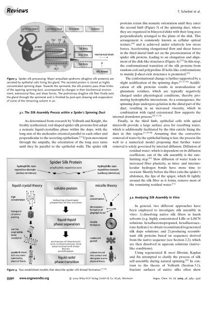
Figure 3. <strong>Spider</strong> silk processing. Major ampullate spidroins (dragline silk proteins) are<br />
secreted by epithelial cells lining the gland. The secreted protein is s<strong>to</strong>red as highly<br />
concentrated spinning dope. Towards the spinneret, the silk proteins pass three limbs<br />
of the tapering spinning duct, accompanied by changes in their biochemical environment,<br />
extensional flow, and shear forces. The preliminary dragline silk fiber finally exits<br />
the gland through the spinneret and is finished by post-spin drawing and evaporation<br />
of some of the remaining solvent in air.<br />
3.1. The <strong>Silk</strong> Assembly Process within a <strong>Spider</strong>’s Spinning Duct<br />
As determined from research by Vollrath and Knight, the<br />
freshly synthesized, rod-shaped spider silk proteins first adopt<br />
a nematic liquid-crystalline phase within the dope, with the<br />
long axis of the molecules oriented parallel <strong>to</strong> each other and<br />
perpendicular <strong>to</strong> the secreting epithelium. [11] Upon movement<br />
through the ampulla, the orientation of the long axes turns<br />
until they lie parallel <strong>to</strong> the epithelial walls. The spider silk<br />
[11, 62]<br />
Figure 4. Two established models that describe spider silk thread formation.<br />
proteins retain this nematic orientation until they enter<br />
the second limb (Figure 3) of the spinning duct, where<br />
they are organized in bilayered disks with their long axes<br />
perpendicularly arranged <strong>to</strong> the plane of the disk. This<br />
arrangement is commonly known as cellular optical<br />
texture, [54] and is achieved under relatively low stress<br />
forces. Accelerating elongational flow and shear forces<br />
in the third ductal limb act on the preorientation of the<br />
spider silk protein, leading <strong>to</strong> an elongation and alignment<br />
of the disk-like structures (Figure 4). [27] In this step,<br />
the conformational transition of the silk proteins from<br />
random-coil and polyproline-II helix-like conformations<br />
<strong>to</strong> mainly b-sheet-rich structures is promoted. [11]<br />
The conformational change is further supported by a<br />
slight acidification of the spinning dope. [27, 55–58] Acidification<br />
of silk proteins results in neutralization of<br />
glutamate residues, which are typically negatively<br />
charged under physiological conditions, thereby promoting<br />
hydrophobic interactions. As a consequence, the<br />
spinning dope undergoes gelation in the distal part of the<br />
duct, resulting in an increased viscosity, which in<br />
combination with rapid extensional flow supports the<br />
[56, 57,59]<br />
internal drawdown process.<br />
Finally, in the third limb, epithelial cells with apical<br />
microvilli provide a large surface area for resorbing water,<br />
which is additionally facilitated by the thin cuticle lining the<br />
duct in this region. [11,58, 60] Assuming that the convective<br />
removal of water by the epithelial lining is fast, the process fits<br />
well <strong>to</strong> a numerical model proposing that further water<br />
removal is solely governed by internal diffusion. Diffusion of<br />
residual water, which is dependent on its diffusion<br />
coefficient, out of the silk assembly is the ratelimiting<br />
step. [61] Slow diffusion of water leads <strong>to</strong><br />
increased fiber plasticity, as intra- and intermolecular<br />
hydrogen bonds have more time <strong>to</strong><br />
reorient. Shortly before the fiber exits the spider s<br />
abdomen, the lips of the spigot, which fit tightly<br />
around the silk fiber as it forms, remove most of<br />
the remaining residual water. [11]<br />
3.2. Analyzing <strong>Silk</strong> Assembly In Vitro<br />
T. Scheibel et al.<br />
In general, two different approaches have<br />
been employed <strong>to</strong> investigate silk assembly in<br />
vitro: 1) dissolving native silk fibers in harsh<br />
solvents (e.g. highly concentrated LiBr or LiSCN<br />
solutions, hexafluoroisopropanol, hexafluoroace<strong>to</strong>ne<br />
hydrate) <strong>to</strong> obtain reconstituted/regenerated<br />
silk dope solutions; and 2) producing recombinant<br />
silk proteins based on sequences derived<br />
from the native sequence (see Section 2.2), which<br />
are then dissolved in aqueous solutions (nativelike<br />
conditions).<br />
Using regenerated B. mori fibroins, Kaplan<br />
and Jin attempted <strong>to</strong> clarify the process of silk<br />
self-assembly during natural spinning. [62] In contrast<br />
<strong>to</strong> the theory of Vollrath (Section 3.1),<br />
fracture surfaces of native silks often show<br />
3590 www.angewandte.org 2009 Wiley-VCH Verlag GmbH & Co. KGaA, Weinheim Angew. Chem. Int. Ed. 2009, 48, 3584 – 3596