silkworm grasserie and pebrine for open elective
silkworm grasserie and pebrine for open elective
silkworm grasserie and pebrine for open elective

Create successful ePaper yourself
Turn your PDF publications into a flip-book with our unique Google optimized e-Paper software.
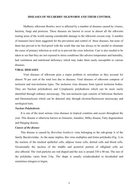
DISEASES OF MULBERRY SILKWORM AND THEIR CONTROL<br />
Mulberry <strong>silkworm</strong> Bombyx mori is affected by a number of diseases caused by viruses,<br />
bacteria, fungi <strong>and</strong> protozoa. These diseases are known to occur in almost all the <strong>silkworm</strong><br />
rearing areas of the world causing considerable damage to the <strong>silkworm</strong> cocoon crop. A number<br />
of measures have been suggested <strong>for</strong> the prevention <strong>and</strong> control of these diseases, but none of<br />
them has proved to be fool-proof with the result that one has always to be careful to eliminate<br />
the cause of primary infection as well as to prevent the cross infection. Care is also needed to be<br />
taken to see that they are not exposed to stress conditions like adverse temperature <strong>and</strong> humidity,<br />
bad ventilation <strong>and</strong> nutritional deficiency which may make them easily susceptible to various<br />
diseases.<br />
VIRAL DISEASES<br />
Viral diseases of <strong>silkworm</strong> pose a major problem to sericulture as they account <strong>for</strong><br />
almost 70 per cent of the total loss due to diseases. Viral diseases of <strong>silkworm</strong> comprise of<br />
inclusion <strong>and</strong> non-inclusion types. The inclusion virus diseases <strong>for</strong>m typical inclusion bodies.<br />
They are Nuclear polyhedrosis <strong>and</strong> Cytoplasmic polyhedrosis which can be more easily<br />
identified through ordinary microscopy. The non-inclusion type consists of Infectious flacheric<br />
<strong>and</strong> Densonuclcosis which can be detected only through elcctron/fluoresccnt microscopy <strong>and</strong><br />
serological tests.<br />
Nuclear Polyhedrosis<br />
It is one of the most serious virus diseases in tropical countries <strong>and</strong> occurs throughout the<br />
year. This disease is otherwise known as Grasseric, Jaundice, Milky disease, Fatty degeneration<br />
<strong>and</strong> Hanging disease.<br />
Causes of the disease<br />
This disease is caused by Borrelina bombycis virus belonging to the sub-group A of the<br />
family Baculoviridae. As the name implies, this virus multiplies <strong>and</strong> <strong>for</strong>ms polyhedra (Fig. 1) in<br />
the nucleus of the tracheal epithelial cells, adipose tissue cells, dermal cells <strong>and</strong> blood cells.<br />
Occasionally the nucleus of the middle <strong>and</strong> posterior portion of silkgl<strong>and</strong> cells are<br />
also affected. The viral particles are rod shaped <strong>and</strong> the size is around 330 x 80 nm. The size of<br />
the polyhedra varies from 3-6µ. The shape is usually octadecahedral or hexahedral <strong>and</strong><br />
sometimes tetragon or trigon.<br />
1
Infection mostly takes place through feeding of polyhedra contaminated mulberry leaf,<br />
rarely through wounds. Heat, cold <strong>and</strong> chemical treatments have also been known to induce<br />
this disease. Factors inducing the outbreak of this disease are high temperature <strong>and</strong> humidity,<br />
their sudden fluctuations, bad ventilation in the rearing room, ineffective disinfection of rearing<br />
room <strong>and</strong> equipments <strong>and</strong> feeding of tender leaves during late instars. Inadequate larval<br />
spacing, starvation <strong>and</strong> excessive moisture in the rearing bed have also been known to<br />
contribute towards the outbreak <strong>and</strong> spread of the disease.<br />
Symptoms: During early part of the disease no symptoms are noticed except the worms<br />
being slightly sluggish. Initially the skin shows oily <strong>and</strong> shining appearance (Fig. 2). As the<br />
disease advances the skin becomes thin <strong>and</strong> fragile <strong>and</strong> the body becomes milky white with<br />
intersegmental swellings (Fig. 3). The fragile skin is prone to rupture easily, liberating the<br />
liquefied body contents containing innumerable number or polyhedra which become the source<br />
of secondary contamination. Another characteristic symptom or this disease is<br />
that the larvae become restless <strong>and</strong> crawl aimlessly along the ridges or rims of rearing trays,<br />
(fig 4) subsequently falling on the ground <strong>and</strong> dying. Death takes place alter infection in about<br />
4-5 days in the young larvae <strong>and</strong> 5-7 days in the grown-up larvae. Diseased larvae lose the<br />
clasping power of abdominal legs except the caudal legs by which it hangs with<br />
the head downwards (Fig, 5). If the infection is early the worms fail to spin the cocoons <strong>and</strong><br />
die, whereas if the infection is late they are able to Spin the cocoons but die inside producing<br />
melted cocoons.<br />
2
Prevention <strong>and</strong> control: For effective prevention of this disease, the <strong>silkworm</strong> rearing<br />
rooms, mulberry storage rooms, mounting rooms, equipments <strong>and</strong> rearing premises should be<br />
thoroughly disinfected be<strong>for</strong>e brushing. The eggs should be essentially surface disinfected.<br />
Silkworms should be reared under strict hygienic conditions. During rearing<br />
the diseased <strong>and</strong> dead larvae <strong>for</strong>m the major source of infection with the largest quantity of<br />
fresh polyhedra available. Hence, the diseased larvae should be removed carefully without<br />
breaking the skin <strong>and</strong> disposed suitably by putting them in lime vats or by burning. Depending<br />
upon the stage of <strong>silkworm</strong>, suitable temperature <strong>and</strong> humidity should be provided. During IV<br />
<strong>and</strong> V instars fresh air circulation should be ensured by providing cross ventilation. The<br />
<strong>silkworm</strong>s should be fed with nutritive rich mulberry leaf <strong>and</strong> during later stages feeding of<br />
tender leaf should be avoided. Depending upon the stage of larvae, optimum spacing <strong>and</strong><br />
required quantum of leaf should be given. Proper bed drying is necessary be<strong>for</strong>e each feed to<br />
avoid accumulation of moisture in the bed.<br />
In addition to the above, use of certain bed disinfectants could also prevent secondary<br />
contamination <strong>and</strong> spread of the disease. Para<strong>for</strong>rnaldehyde compounds are known to have anti-<br />
3
microbial properties <strong>and</strong> various <strong>for</strong>mulations involving this chemical have been prepared like<br />
Papazol in Japan <strong>and</strong> Reshamkeet Oushadh in India. The latter is a bed disinfectant <strong>for</strong>mulation<br />
containing 1 per cent captan (N-Trichloromythyl Thio-4-Cyclohexane 1,2- Dicarboxymide), 1<br />
percentpara<strong>for</strong>maldehyde (Tri- oxymethylene) 2 per cent Benzoic acid <strong>and</strong> 96 per cent slaked<br />
lime powder giving dual protection against<br />
<strong>grasserie</strong> <strong>and</strong> muscardine. It should be dusted on the larvae <strong>and</strong> bed with the help of a thin cloth<br />
at the rate of 2-3 grams/0. 1 sqm. area during early instars <strong>and</strong> 4- 5 grams/0.1sqm. during IV <strong>and</strong><br />
V instars. The dusting should be done (Fig. 6) preferably once after each moult, half an hour<br />
be<strong>for</strong>e resumption of feed. An additional dusting should be done on the 4th day of final instar<br />
after bed cleaning. The dusting should not be done when the larvae are under moult or preparing<br />
<strong>for</strong> moult. The quantity of Reshamkeet Oushadh required <strong>for</strong> 100 disease free layings (40,000<br />
larvae) is between 3-3.5 kgs.<br />
PROTOZOAN DISEASES<br />
Protozoa which are injurious to <strong>silkworm</strong> are the parasitic ones belonging to the class<br />
Microsporidia <strong>and</strong> genera Nosema, Pleistophora <strong>and</strong> Thelohania. However, the major<br />
protozoan disease of the <strong>silkworm</strong> is the <strong>pebrine</strong> disease, so named due to the appearance of<br />
black peppery patches following infection.<br />
Pebrine<br />
Pebrine is a chronic <strong>and</strong> disastrous disease of the <strong>silkworm</strong> Bombyx mori L. It was this<br />
disease which was responsible <strong>for</strong> the sudden collapse of the <strong>silkworm</strong> industry of both France<br />
<strong>and</strong> Italy in 1965. Even though the fight against this disease in all the sericultural countries is<br />
going on since more than 100 years, the disease is not yet eliminated. However, it has been kept<br />
under check by following the techniques of strict mother moth examination <strong>for</strong> the supply of<br />
disease free <strong>silkworm</strong> eggs, in addition to disinfection <strong>and</strong> hygienic rearings. Though the<br />
4
disease is under reasonable control, it appears sporadically due to infected seed <strong>and</strong> persisting<br />
secondary contamination in the rearing house.<br />
Causes of the disease: Pebrine is caused by Nosema bombycis Nageli belonging to family<br />
Nosematidae of order Microsporidia. The pathogen infects the host through feeding of<br />
contaminated mulberry leaf (peros) <strong>and</strong> also by rearing infected <strong>silkworm</strong> eggs (transovarial).<br />
In addition to Nosema bombycis, seven different microsporidians belonging to genera Nosema,<br />
Pleistophora <strong>and</strong> Thelohania have been reported to infect the <strong>silkworm</strong>.<br />
Sources of infection are rather extensive. The main source is the rearing of ransovarially<br />
<strong>and</strong> surface contaminated layings. Infection also results from diseased <strong>and</strong> dead larvae, faeces<br />
of larvae, moths, diseased egg shells, larval <strong>and</strong> pupal exuviae etc. In the rearing bed major<br />
source of infection is the faeces of diseased larvae, contaminated tray, seat paper <strong>and</strong> dust from<br />
infected rearing <strong>and</strong> leaf storage rooms. Sometimes infection takes place through contaminated<br />
mulberry leaf from field. The excreta <strong>and</strong> dead larvae of <strong>pebrine</strong> infected wild insects may also<br />
<strong>for</strong>m a source of infection.<br />
The life cycle of Nosema bombycis Nageli includes three stages namely, spore, planont<br />
<strong>and</strong> meront (Fig. 7).<br />
The mature spore is oval or ovocylindrical (Fig. 8-9). It measures approximately 3 -4x<br />
1.5 -2.5 µ with three layered membrane: the inner, middle <strong>and</strong> outer. The sporoplasm is<br />
stretched in the <strong>for</strong>m of girdle across the width of the spore <strong>and</strong> it contains a<br />
pair of nuclei. The spore has a polar capsule <strong>and</strong> polar filament. The polar filament is given<br />
out on treatment with a number of chemicals like H202 <strong>and</strong> KCl. Spores are highly refractive<br />
<strong>and</strong> appears light blue under the microscope. The spore represents the dormant stage of the<br />
pathogen <strong>and</strong> can survive in the ordinary conditions of rearing house <strong>for</strong> more than a year. It<br />
5
etains its infectivity even after three years in the dried body of the female moth, in liquid<br />
medium <strong>for</strong> more than 3 weeks <strong>and</strong> in soil [or mo more than 2 months. . But the spore is<br />
susceptible to desiccation <strong>and</strong> cannot survive <strong>for</strong> more than 6 - 7 hours in direct sunlight (39 -<br />
40°C). It is also weak against heat, chemicals <strong>and</strong> disinfectants.<br />
When live spores enter into the <strong>silkworm</strong> through mulberry rry leaf, they germinate in the<br />
gut due to high alkalinity <strong>and</strong> potassium ions. As a result the polar filament is extruded <strong>and</strong><br />
the sporoplasm along with two nuclei creeps through it <strong>and</strong> injects into the midgut tissues.<br />
Subsequently the polar filament gets digested in the alimentary tract. The two nuclei of the<br />
sporoplasm unite to <strong>for</strong>m a uninucleate planont. The planont measures 0.5 - 1.5 µ <strong>and</strong> is<br />
<strong>for</strong>med in 1-2 2 days. The planont is sub globular with a strong refractive nucleus without<br />
shell, per<strong>for</strong>ms amoeboid movement <strong>and</strong> reproduces by binary fission. The planont which<br />
initially infects the gut later passes through the gut wall <strong>and</strong> invades the various susceptible<br />
tissues.<br />
Once the planont ont penetrates the host cell, it trans<strong>for</strong>ms in to a sedentary dentary <strong>for</strong>m <strong>and</strong> becomes<br />
localized. ized. This stage is known as meront. Meront is an intracellular stage <strong>and</strong> has a definite cell<br />
wall which absorbs nutrients from host cell. The meront is spherical or pear sshaped<br />
<strong>and</strong> is<br />
<strong>for</strong>med in 2 - 3 days after infection. It reproduces by binary fission, multiple fission or by<br />
budding. When cytoplasm ytoplasm of the host cell is exhausted, meronts are arranged in parallel rows.<br />
The meront after massive proliferation fills up the ho host st cells <strong>and</strong> when nutrients are<br />
depleted, sporulation takes place. From the germination of the spores to sporul sporulation is the entire<br />
developmental tal cycle of the <strong>pebrine</strong> protozoan.<br />
Symptoms: The symptoms of this disease can be observed in all the stages of <strong>silkworm</strong><br />
viz., egg, larvae, pupa <strong>and</strong> adult. These symptoms <strong>for</strong>m an important criterion <strong>for</strong> identifying the<br />
disease.<br />
6
In the egg stage, poor egg number, lack of adequate adherence to the substratum, lack oof<br />
egg uni<strong>for</strong>mity, <strong>for</strong>mity, more of unfertilized <strong>and</strong> dead eggs, poor <strong>and</strong> irregular hatching are some of the<br />
symptoms. Sometimes infected eggs cannot hatch out <strong>and</strong> hatched larvae may also die.<br />
Larvae show poor appetite, retarded growth <strong>and</strong> development leading to un un-uni<strong>for</strong>mity in<br />
size (Fig. 10). Larvae moult irreg irregularly <strong>and</strong> show sluggishness. gishness. Transovarially infected larvae<br />
die be<strong>for</strong>e third moult but those which are heavily infected die during first instar itself. The<br />
larval body shows wrinkled skin with rustic brown colour <strong>and</strong> in the moribund<br />
stage they do not rot but remain rubbery. The affected gut becomes opaque <strong>and</strong> the silkgl<strong>and</strong><br />
shows white pustules in different places along its length. Sometimes black irregular pepper like<br />
spots are noticed on larval skin (Fig. 11).<br />
The infected pupae are flabby <strong>and</strong> swollen with lusterless <strong>and</strong> softened abdomen.<br />
Sometimes irregular black spots are noticed near the rudiments of the wing <strong>and</strong> abdominal area.<br />
Highly infected pupae fail to metamorphose into adults. The moth emergence is delayed <strong>and</strong><br />
improper. They have clubbed wings with distorted antennae <strong>and</strong> do not mate properly. The<br />
scales from wings <strong>and</strong> abdominal area easily come off. In infected moths if the accessory gl<strong>and</strong>s<br />
are infected the moth may lay eggs with less gluey substance re resulting sulting in their detachment from<br />
the egg cards.<br />
7
Prevention <strong>and</strong> control: The fundamental measure <strong>for</strong> the prevention <strong>and</strong> control of this<br />
disease is to produce healthy eggs, so as to avoid embryonic infection. This can be achieved by<br />
conducting systematic mother moth examination. The other methods are to conduct effective<br />
disinfection of rearing rooms, equipments <strong>and</strong> surroundings <strong>and</strong> maintenance of strict hygienic<br />
conditions during rearing. It is essential to surface disinfect the layings in 2 percent <strong>for</strong>malin <strong>for</strong><br />
10 minutes be<strong>for</strong>e incubation. Such surface disinfection though practiced in grainages should be<br />
repeated again after release from cold storage as also by farmers. If the eggs are in advanced<br />
stage of embryonic development surface disinfection is done with 1 per cent <strong>for</strong>malin <strong>for</strong>5<br />
minutes. The room <strong>and</strong> equipments must be washed <strong>and</strong> disinfected be<strong>for</strong>e incubation.<br />
Young <strong>silkworm</strong>s should be reared under hygienic conditions. As a precaution test<br />
examination of unhatched blue eggs, dead eggs, hatched larvae <strong>and</strong> eggshells can be done <strong>and</strong> if<br />
<strong>pebrine</strong> is detected, such eggs should not be brushed <strong>and</strong> if brushed the larvae should be<br />
destroyed. Similarly predictive examination could be conducted by utilizing unequal larvae, late<br />
moulters, faecal matter <strong>and</strong> exuviae <strong>for</strong> the detection of <strong>pebrine</strong> spores. These tests may not<br />
only minimize the chances of rearing transovarially infected layings, but also check cross<br />
contamination <strong>and</strong> spread of the disease. Infected <strong>silkworm</strong>s, faeces <strong>and</strong> mulberry field pests<br />
are important sources of infection <strong>and</strong> should be properly disposed of to prevent cross infection<br />
<strong>and</strong> spread of the disease.<br />
During seed production in addition to mother moth examination, care should be taken to<br />
prevent contamination from other sources. The equipments used <strong>for</strong> one lot should not be used<br />
<strong>for</strong> the other till they have been thoroughly cleaned <strong>and</strong> disinfected. Eggs after surface<br />
disinfection should be dried <strong>and</strong> stored in a sepparate room away from egg production <strong>and</strong><br />
examination room.<br />
Besides, the above preventive/corrective measures, it has been reported that immersing of<br />
the <strong>silkworm</strong> eggs in hot water, high temperature treatment of the pupae, dipping of the eggs in<br />
hot hydrochloric acid minimize the incidence of <strong>pebrine</strong>. Chemotherapy of Nosema infection<br />
has been reported through a number of antimicrosporidian drugs like fumagillin, benomyl,<br />
bengard, bavistin, ethyl <strong>and</strong> methyl thiophanate <strong>and</strong> some of their analogues<br />
with positive results, but preventive methods have always been found to be better than the<br />
curative measures.<br />
--------------------<br />
8