
You also want an ePaper? Increase the reach of your titles
YUMPU automatically turns print PDFs into web optimized ePapers that Google loves.
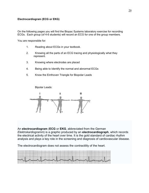
Electrocardiogram (<strong>ECG</strong> or EKG)<br />
On the following pages you will find the Biopac Systems laboratory exercise for recording<br />
<strong>ECG</strong>s. Each group (of 4-6 students) will record an <strong>ECG</strong> for one of the group members.<br />
You are responsible for:<br />
1. Reading about <strong>ECG</strong>s in your textbook.<br />
2. Knowing all the parts of an <strong>ECG</strong> tracing <strong>and</strong> physiologically what they<br />
represent.<br />
3. Knowing where electrodes are placed<br />
4. Being able to Identify the normal <strong>and</strong> abnormal <strong>ECG</strong>s<br />
5. Know the Einthoven Triangle for Biopolar Leads<br />
Bipolar Leads:<br />
An electrocardiogram (<strong>ECG</strong> or EKG, abbreviated from the German<br />
Elektrokardiogramm) is a graphic produced by an electrocardiograph, which records<br />
the electrical activity of the heart over time. It is the gold st<strong>and</strong>ard of cardiac rhythm<br />
analysis <strong>and</strong> plays a key role in the screening <strong>and</strong> diagnosis of cardiovascular disease.<br />
The electrocardiogram does not assess the contractility of the heart.<br />
20