Student Book (Unit 1 Module 2) - Pearson Schools
Student Book (Unit 1 Module 2) - Pearson Schools
Student Book (Unit 1 Module 2) - Pearson Schools
Create successful ePaper yourself
Turn your PDF publications into a flip-book with our unique Google optimized e-Paper software.
Pulmonary<br />
valve<br />
Right<br />
atrium<br />
36<br />
1.2.1 1 The heart<br />
Right<br />
atrioventricular<br />
valve<br />
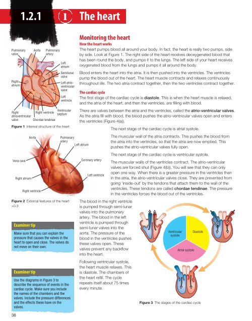
Figure 1 Internal structure of the heart<br />
Vena cava<br />
Right atrium<br />
Aorta<br />
Right ventricle<br />
Examiner tip<br />
Examiner tip<br />
Aorta Pulmonary<br />
artery<br />
Right ventricle<br />
Chordae tendinae<br />
Left<br />
atrium<br />
Semilunar<br />
valve<br />
Left atrioventricular<br />
valve<br />
Left<br />
ventricle<br />
Ventricular<br />
septum<br />
Pulmonary<br />
artery<br />
Figure 2 External features of the heart<br />
×0.3<br />
Make sure that you can explain the<br />
pressure that causes the valves in the<br />
heart to open and close. The valves do<br />
not move on their own.<br />
Use the diagrams in Figure 3 to<br />
describe the sequence of events in the<br />
cardiac cycle. Make sure you include<br />
the names of the chambers and the<br />
valves. Include the pressure differences<br />
and the effects these have on the<br />
valves.<br />
Monitoring the heart<br />
How the heart works<br />
The heart pumps blood all around your body. In fact, the heart is really two pumps, side<br />
by side. Look at Figure 1. The right side of the heart receives deoxygenated blood that<br />
has been round the body, and pumps it to the lungs. The left side of your heart receives<br />
oxygenated blood from the lungs and pumps it all around the body.<br />
Blood enters the heart into the atria. It is then pushed into the ventricles. The ventricles<br />
pump the blood out of the heart. The heart muscle contracts and relaxes continuously<br />
throughout life. The two atria contract together, then the two ventricles contract together.<br />
The cardiac cycle<br />
The fi rst stage of the cardiac cycle is diastole. This is when the heart muscle is relaxed,<br />
and the atria of the heart, and then the ventricles, are fi lling with blood.<br />
There are valves between the atria and the ventricles, called the atrio-ventricular valves.<br />
As the atria fi ll with blood, the blood pushes the atrio-ventricular valves open and enters<br />
the ventricles (Figure 4(a)).<br />
Left atrium<br />
Coronary artery<br />
Left ventricle<br />
The blood in the right ventricle<br />
is pumped through semi-lunar<br />
valves into the pulmonary<br />
artery. The blood in the left<br />
ventricle is pumped through<br />
semi-lunar valves into the<br />
aorta. The pressure of the<br />
blood in the ventricles pushes<br />
these valves open. These<br />
valves prevent any backfl ow<br />
into the heart.<br />
Following ventricular systole,<br />
the heart muscle relaxes. This<br />
is diastole. The chambers of<br />
the heart refi ll. The cycle<br />
repeats itself about 75 times<br />
every minute.<br />
The next stage of the cardiac cycle is atrial systole.<br />
The muscular wall of the atria contracts. This pushes the blood from<br />
the atria into the ventricles, so that the atria are now emptied. This<br />
pushes the atrio-ventricular valves fully open.<br />
The next stage of the cardiac cycle is ventricular systole.<br />
The muscular walls of the ventricles contract. The atrio-ventricular<br />
valves are forced shut (Figure 4(b)). You will see that they can only<br />
open one way. When there is a greater pressure in the ventricles than<br />
in the atria, the atrio-ventricular valves close. They are prevented from<br />
going ‘inside-out’ by the tendons that attach them to the wall of the<br />
ventricles. These tendons are called chordae tendinae. The pressure<br />
in the ventricles forces the blood out of the ventricles.<br />
Ventricular<br />
systole<br />
Atrial systole<br />
Diastole<br />
Figure 3 The stages of the cardiac cycle
Case study<br />
Martin visited his GP because he had a bad, chesty cough. His doctor examined his chest. He told Martin<br />
that he would need some antibiotics to clear up the infection, and then added, ‘By the way, did you know<br />
that you have a very slight heart murmur?’ Martin was not aware of this, and was a little worried at first.<br />
His GP reassured him. ‘It’s only a very slight heart murmur. This means there is a very tiny hole in one of<br />
your heart valves. Clearly it’s caused you no problems up until now. I’ll refer you to a specialist to get it<br />
checked out. However, I’d like to assure you that most minor heart murmurs like this cause no problems<br />
at all, and don’t need any treatment.’<br />
(a) Valve open<br />
Valve flap<br />
Chordae<br />
tendinae<br />
Muscle on<br />
ventricle wall<br />
Higher blood pressure above<br />
valve forces it open<br />
Lower blood pressure<br />
beneath valve<br />
Figure 4 How the heart valves work<br />
(b) Valve closed<br />
Valve flaps<br />
fit together<br />
High pressure<br />
pushes valve closed<br />
Tendinous cords<br />
stop valve inverting<br />
Pressure and volume changes in the heart<br />
Figure 5 shows the pressure and volume changes in the<br />
heart during one cardiac cycle.<br />
You will see that the pressure in the ventricle increases a<br />
lot when the muscular wall of the ventricle contracts. As<br />
the ventricle contracts, the pressure of blood inside it<br />
increases. When the pressure of blood in the ventricle<br />
exceeds the pressure in the artery, blood is forced out and<br />
the volume of the ventricle decreases.<br />
The pressure in each atrium increases a little as it fills with<br />
blood, and then as it contracts, but it never gets very high.<br />
This is because the atrium only needs to pump the blood<br />
into the ventricle.<br />
The pressure in the aorta does go up and down, but you<br />
will see that it never goes very low. This is important,<br />
because the blood in the aorta has only just left the heart. It<br />
needs to have enough pressure to get round the body and<br />
back to the heart again.<br />
Questions<br />
1 Explain why the wall of the left ventricle is thicker than<br />
the wall of the right ventricle.<br />
2 Use Figure 5 to estimate the heart rate, in beats per<br />
minute, for this person.<br />
3 Figure 5 shows the pressure changes in the left side of<br />
the heart. Suggest where the line showing pressure in<br />
the right ventricle would be.<br />
Pressure/kPa<br />
Volume<br />
Electrical activity<br />
20<br />
15<br />
10<br />
Lower blood pressure<br />
cannot open valve<br />
5<br />
0<br />
Atrial<br />
systole<br />
Semilunar<br />
valve opens<br />
P<br />
Q<br />
R<br />
S<br />
Ventricular<br />
systole<br />
Atrioventricular<br />
valve closes<br />
T<br />
Diastole<br />
Semilunar<br />
valve closes<br />
Atrioventricular<br />
valve opens<br />
0 0.1 0.2 0.3 0.4 0.5 0.6<br />
Time/s<br />
<strong>Unit</strong> F221<br />
<strong>Module</strong> 2 Gas Exchange Systems<br />
Aortic<br />
pressure<br />
Ventricle<br />
pressure<br />
Atrial<br />
pressure<br />
Ventricle<br />
volume<br />
1.2.1 a, b<br />
Figure 5 Pressure and volume changes in the heart during one cardiac cycle<br />
ECG<br />
37
38<br />
1.2.1 2 Electrical activity in the heart<br />
Examiner tip<br />
In module 2 you will come across the<br />
three types of muscle – cardiac, smooth<br />
and striated. They are all muscle<br />
tissues, but they differ in a number of<br />
key features. In particular their<br />
appearance and what controls their<br />
contraction. It is important that you<br />
specify which type of muscle you are<br />
talking about if you want to gain<br />
maximum marks.<br />
Sinoatrial node<br />
Excitation wave<br />
spreads over<br />
atria<br />
Atrioventricular<br />
node<br />
Purkyne tissue<br />
carries wave<br />
down septum<br />
Excitation wave spreads<br />
up walls of ventricles<br />
Vena cava<br />
Figure 1 Electrical activity in the heart<br />
Figure 2 Recording an electrocardiogram<br />
Cardiac muscle<br />
The wall of the heart is made up of a very special kind of muscle called cardiac muscle.<br />
It is special because it does not need any stimulation from a nerve to make it contract.<br />
We say that it is myogenic.<br />
There is a group of specialised cardiac cells in the wall of the right atrium called the<br />
sino-atrial node (SAN). These cells generate electrical impulses that pass rapidly across<br />
the walls of the atria from cell to cell. As a result, the atrial walls contract, causing atrial<br />
systole.<br />
The impulses cannot pass straight on from the atria to the ventricle walls, because<br />
there is a ring of fi brous tissue preventing this. The only way that impulses can pass<br />
from the atria to the ventricles is by a group of specialised muscle cells called the<br />
atrio-ventricular node (AVN), which acts as a relay point. There is a slight delay here,<br />
allowing enough time for the atria to empty completely.<br />
From the AV node, impulses pass very quickly down heart muscle fi bres called the<br />
bundle of His that spread down the septum between the two ventricles. This means<br />
that impulses soon reach the bottom of the ventricles. After this, the fi bres divide into<br />
right and left branches at the tip (base) of the ventricles. They then spread throughout the<br />
muscular walls in Purkyne tissue. The impulse causes the muscular ventricle walls to<br />
contract. This is ventricular systole.<br />
After this, there is a short time when no impulses pass through the heart muscle. This<br />
allows the muscle to relax and diastole to occur.<br />
Recording an electrocardiogram<br />
An electrocardiogram (ECG) is used to monitor heart function. A cardiology technician<br />
will ask the patient to remove his clothes from the waist upwards. Then the technician<br />
will place electrodes on the arms, legs and chest. A special ECG cream is used between<br />
the electrodes and the skin. The patient is asked to lie down and remain completely<br />
relaxed, because any movement will interfere with the recording. The machine records<br />
for about 5 minutes. It gives a recording from each electrode.<br />
Look at Figure 3a. This shows a normal ECG for a person with a healthy heart. The line<br />
represents the electrical activity in the heart during the cardiac cycle.<br />
You will see that the P wave occurs shortly before the pressure in the atria increases.<br />
This means that the P wave represents the impulses passing from the SAN to the AVN,<br />
through the walls of the atria, leading to atrial systole.<br />
The QRS wave occurs just before the pressure in the ventricle increases. This shows you<br />
that the QRS wave shows the electrical activity in the ventricles that results in ventricular<br />
systole. In other words, the QRS wave shows the electrical impulses passing down the<br />
bundle of His and along the Purkyne fi bres.<br />
The T wave is a short phase that occurs as the ventricles recover.<br />
Abnormal ECGs<br />
Sometimes people have a problem with the electrical activity in the heart. In these cases,<br />
the ECG produced will change.<br />
Look at Figure 3. The ECG in 3b shows ventricular fi brillation. You will see that this ECG<br />
looks very different. There is no P wave and no QRS wave. This is because the muscle in<br />
the heart wall is not contracting in a coordinated way. It is likely that a person with an<br />
ECG like this has had a myocardial infarction (heart attack) and they will almost<br />
certainly be unconscious. This person needs urgent medical attention or he will die.
The ECG in 3c shows atrial fibrillation. There is a small and unclear P wave. The deep S<br />
wave in 3d indicates ventricular hypertrophy, which is an increase in muscle thickness.<br />
A heightened P wave can indicate an enlarged atrium. A raised S-T segment can indicate<br />
a myocardial infarction (see spread 2.4.1.1).<br />
(a)<br />
(b)<br />
(c)<br />
(d)<br />
Figure 3 Normal and abnormal ECGs<br />
P<br />
R<br />
Q<br />
S<br />
T<br />
A normal ECG<br />
Elevation of<br />
the ST section<br />
indicates heart<br />
attack<br />
Small and<br />
unclear<br />
P wave<br />
indicates<br />
atrial<br />
fibrillation<br />
Deep S wave<br />
indicates<br />
ventricular<br />
hypertrophy<br />
(increase in<br />
muscle<br />
thickness)<br />
(a) Normal ECG<br />
(b) Tachycardia<br />
(c) Bradycardia<br />
(d) Ventricular fibrillation<br />
(e) Heart block<br />
0.2s<br />
Figure 4 ECGs<br />
Questions<br />
1 Explain the advantage of impulses passing very quickly down the bundle of His and<br />
Purkyne tissue.<br />
2 Explain why there is a short interval between the P wave and QRS wave in a normal<br />
person.<br />
3 Use the scale on Figure 4 to calculate the heart rate in beats per minute of the ECGs<br />
in (a), (b) and (c).<br />
Timebase for all ECGs<br />
<strong>Unit</strong> F221<br />
<strong>Module</strong> 2 Gas Exchange Systems<br />
1.2.1 c, d<br />
39
40<br />
1.2.1 3 Changing heart rates<br />
Examiner tip<br />
When calculating cardiac output, stroke<br />
volume or heart rate, always include<br />
units and write them correctly. When<br />
you exercise, your heart rate increases.<br />
This means that the cardiac output also<br />
increases.<br />
Exercising muscles require more<br />
oxygen and glucose to be delivered to<br />
fuel the increase in respiration required.<br />
Remember to link the increase in heart<br />
rate to this increase in respiration.<br />
It is important that you make it clear<br />
when you are measuring heart rate and<br />
stroke volume. Everyone’s heart rate<br />
and stroke volume will increase when<br />
they exercise. What training does is<br />
increase the thickness of the left<br />
ventricle muscle so that stroke volume<br />
at rest will be higher in a trained<br />
athlete. This means it will take fewer<br />
beats to deliver the same cardiac output<br />
so resting heart rate will be lower.<br />
Changes during exercise<br />
The stroke volume is the volume of blood pumped out of the left ventricle in one cardiac<br />
cycle. This is normally 60–80 cm 3 .<br />
The cardiac output is the volume of blood pumped out of the left ventricle in one<br />
minute. This fi gure is normally 4–8 dm 3 min –1 .<br />
Therefore, cardiac output = stroke volume × heart rate.<br />
Another effect of strenuous exercise is that body muscles contract more strongly. The<br />
muscles compress the veins and this increases the rate at which deoxygenated blood<br />
returns to the heart. The vena cava contains more blood than before, so the heart rate<br />
increases. The increased volume of blood in the heart also stretches the heart muscle.<br />
This makes the heart muscle contract more strongly. The result of this is that the stroke<br />
volume increases. If an athlete undergoes training, her stroke volume will be permanently<br />
higher than that of a non-athlete.<br />
Look at Figure 1. You can see that a trained athlete has a greater stroke volume than a<br />
non-athlete when their heart rates are the same.<br />
Stroke volume cm 3<br />
120<br />
100<br />
80<br />
60<br />
40<br />
20<br />
Trained<br />
Untrained<br />
0<br />
70 80 90 100 110 120 130 140 150 160 170 180<br />
Heart rate/bpm<br />
Figure 1 Graph showing the effect of training on stroke volume<br />
Measuring pulse rate<br />
The heart rate can be measured by counting how many ‘pulses’ are felt in an artery per<br />
minute. This is because every time the left ventricle contracts, a ‘pulse’ of blood pushed<br />
out into the aorta.<br />
Your pulse can be measured anywhere where you can press an artery against a bone.<br />
What you can feel is the expansion of the artery wall as the pulse of blood (the pressure<br />
wave) passes through. Pulse rate is usually measured using the ‘radial’ pulse in the wrist.<br />
If you are measuring the pulse rate of another person, wash your hands carefully before<br />
you start.<br />
• Find the position of the radial artery.<br />
• Press fi rmly against the radial artery with the second and third fi ngers (because your<br />
thumb and fi rst fi nger have a pulse).<br />
• Count the number of pulses in 1 minute. Alternatively, count the number of pulses in<br />
30 seconds, then multiply by 2 to give a pulse rate in beats per minute.<br />
There are other places where you can measure a pulse. You can see some of these in<br />
Figure 2.<br />
There are also meters that give you a reading of your pulse rate. These can be useful to<br />
measure your pulse rate before, during and after exercise.
Table 1 shows the normal resting pulse rate for people of different ages.<br />
Person Resting heart rate/beats per minute<br />
Babies 0–12 months 100–160<br />
Children aged 1–10 60–140<br />
Children aged 10+ and adults 60–100<br />
Highly trained athletes 40–60<br />
Table 1 Normal resting pulse<br />
The effect of exercise on heart rate<br />
The effects of exercise on the heart rate of two boys were investigated. Both boys<br />
started to ride on an exercise bike at 2 minutes and stopped exercising at 7 minutes.<br />
They wore heart rate monitors. The graph in Figure 3 shows how their heart rate<br />
changed before, during and after exercise.<br />
Period of exercise<br />
Heart rate<br />
110<br />
105<br />
100<br />
95<br />
90<br />
85<br />
80<br />
75<br />
70<br />
65<br />
60<br />
0<br />
0<br />
1 2 3 4 5 6 7 8 9 10 11 12 13 14 15<br />
Time/minutes<br />
Figure 3 Effects of exercise on the heart rate of two boys<br />
• Name the units that should be present on the y-axis.<br />
• One of the boys is a highly trained athlete. Which boy is it? Explain your answer.<br />
• How could you improve the reliability of this investigation?<br />
Questions<br />
1 A person has a stroke volume of 74 cm 3 and a cardiac output of 5700 cm 3 when he is<br />
at rest. Calculate his heart rate in beats per minute.<br />
2 Following vigorous exercise, the same person has a heart rate of 195 beats per minute<br />
and a cardiac output of 18 900 cm 3 . Calculate his stroke volume.<br />
3 Suggest why a trained athlete has a greater stroke volume than a non-athlete when<br />
their heart rates are the same.<br />
4 Explain why it is an advantage to an athlete to have a greater stroke volume than a<br />
non-athlete when their heart rates are the same.<br />
A<br />
B<br />
(a)<br />
(b)<br />
<strong>Unit</strong> F221<br />
<strong>Module</strong> 2 Gas Exchange Systems<br />
Figure 2 Places where you can<br />
measure a pulse<br />
1.2.1 e, f<br />
41
42<br />
1.2.2 1 The structure of blood vessels<br />
Key defi nition<br />
Humans and many other animals have a<br />
closed blood system in which the blood<br />
is carried only inside blood vessels.<br />
Blood never leaves this system of blood<br />
vessels.<br />
Examiner tip<br />
Pay attention to the tissues and the key<br />
words that go with them. Smooth<br />
muscle can contract and relax to<br />
regulate the pressure by changing the<br />
lumen diameter. Elastic tissue stretches<br />
to accommodate the pulse of blood<br />
from ventricular systole and recoils to<br />
maintain the blood pressure. Get the<br />
right words with the right tissue! A<br />
common error is for candidates to<br />
suggest that smooth muscle ‘smoothes’<br />
the bloodfl ow by preventing friction.<br />
This is the function of the endothelium.<br />
Terminology is key to good marks. For<br />
example, a common mistake is to state<br />
that when smooth muscle in an artery<br />
wall contracts, the artery gets smaller.<br />
It doesn’t, the lumen gets smaller.<br />
Artery<br />
Tunica<br />
media<br />
Tunica<br />
externa<br />
Lumen<br />
Endothelium<br />
or tunica intima<br />
Elastic fibres<br />
Smooth muscle<br />
Collagen fibres<br />
Types of blood vessel<br />
Arteries carry blood away from the heart. Away from the heart, they divide into smaller<br />
vessels called arterioles. In turn, arterioles lead into tiny blood vessels called capillaries,<br />
which exchange materials between the blood and the tissues. Blood leaving the<br />
capillaries drains into vessels called venules. Venules join up to form veins, which carry<br />
blood back to the heart.<br />
Arteries<br />
Look at Figure 1a. This shows the structure of an artery. You can see that it has a thick<br />
wall with smooth muscle and elastic tissue in it. This is because the arteries carry blood<br />
away from the heart. The blood is under high pressure. Every time the heart beats, a<br />
surge of blood passes through the artery. This causes the artery wall to bulge a little. As<br />
this happens, the thick elastic layer allows the artery wall to stretch and spring back. This<br />
process is called elastic recoil. This helps to keep blood pressure high and helps to<br />
smooth out blood fl ow. When you measure your pulse, you can feel these surges in<br />
pressure.<br />
There is also a tough fi brous layer around the outside of the artery. This protects the<br />
artery from damage as we move around and our skeletal muscles contract and relax.<br />
The lumen is the space through which the blood fl ows. You can see that it is relatively<br />
small, keeping the blood under pressure. The lining of the artery, the endothelium, is a<br />
thin, smooth layer. This helps the blood to fl ow along with as little friction as possible.<br />
Veins<br />
Figure 1b shows a vein. You can see that it has a much thinner layer of muscle and elastic<br />
tissue than the artery. This is because the blood in the vein is under much lower pressure.<br />
The lumen of the vein is larger than the lumen of the artery, so the blood is under less<br />
pressure and it fl ows more slowly. The vein also has a thick fi brous layer to protect it from<br />
damage. It has a smooth endothelium to reduce friction as blood fl ows along it.<br />
Figure 1 Structure of a an artery, b a vein and c a capillary. The diagram of the capillary is drawn to a much larger scale<br />
Vein<br />
Lumen<br />
Endothelium<br />
Elastic fibres<br />
Smooth muscle<br />
Collagen fibres<br />
Capillary<br />
Lumen<br />
Endothelium
A special feature of veins is that they contain valves. You can see how these work in<br />
1.2.2 a, b<br />
Figure 2. Valves help the blood to keep flowing in one direction only, back to the heart.<br />
This happens even in veins in the legs and arms, where blood flows against the force of<br />
gravity. As our skeletal muscles contract, they squeeze the veins. This raises<br />
(a) Pressure falls behind the valve. Pockets fill and close the valve<br />
the pressure in the veins, which shuts the valves behind and opens the valves<br />
ahead, so making sure that the blood keeps flowing back towards the heart.<br />
Capillaries<br />
Look at Figure 1c. This shows a capillary. It is not drawn to the same scale<br />
as the artery and the vein. A capillary is a tiny blood vessel, only 7–10 mm<br />
wide. This is actually about the same diameter as a red blood cell. Red<br />
blood cells can pass through capillaries, but they have to be squeezed<br />
through. This means they pass through one at a time, allowing more efficient<br />
exchange between the blood and the tissues.<br />
The capillary wall is made of a single layer of thin, flattened endothelium<br />
cells. In fact, the capillary wall is like the layer of cells that lines the artery and<br />
the vein. Because the capillary wall is very thin, it allows exchange between<br />
the blood and the tissues. There are also tiny gaps between the endothelium<br />
cells. These also allow substances to be exchanged between the capillaries<br />
and the tissues, and allow phagocytic white cells (phagocytes) to migrate<br />
into tissues.<br />
Arterioles and venules<br />
Look at Figure 3. You can see that the arterioles have a thin wall, mainly of<br />
muscle fij46<br />
bres, but with some elastic fibres. When this muscle contracts, it makes the<br />
lumen of the arteriole narrower. When it relaxes, the lumen becomes wider.<br />
This means that arterioles can increase or decrease the flow of blood to<br />
particular tissues. This is also one means of regulating blood pressure.<br />
Venules have a very thin wall of muscle and elastic tissue. They are like<br />
small veins, and carry blood from the capillaries back to the veins.<br />
Artery Capillary Vein<br />
Transports blood away from<br />
the heart<br />
Thick wall with muscle and a<br />
great deal of elastic tissue<br />
Links arteries to veins. Allows<br />
exchange of materials between the<br />
blood and tissues<br />
Transports blood towards the heart<br />
No elastic or muscle fibres Relatively thin wall with only a small<br />
amount of muscle and elastic<br />
fibres<br />
Bloodflow rapid Bloodflow slowing Bloodflow slow<br />
High blood pressure – blood<br />
flows in pulses<br />
Pressure of blood falling – not in<br />
pulses<br />
Low blood pressure – not in<br />
pulses<br />
Table 1 The main differences between arteries, veins and capillaries<br />
Questions<br />
1 The lumen of an artery is narrower than the lumen of a vein, although arteries carry the<br />
same volume of blood as veins do. Explain why their lumens are different sizes.<br />
2 Look at Figure 4.<br />
(a) Explain how it is possible to identify which vessel is the artery and which vessel is<br />
the vein.<br />
(b) A vein is really round in cross-section. Suggest why the section through the vein in<br />
this figure is not round.<br />
(c) Use the magnification shown to calculate the thickness of the wall, in mm, in the<br />
artery and the vein.<br />
Artery<br />
Figure 2 The action of valves<br />
<strong>Unit</strong> F221<br />
<strong>Module</strong> 2 Gas Exchange Systems<br />
(b) Pressure builds from blood being pushed by the heart. Higher<br />
pressure behind the valve pushes it open and blood flows<br />
through<br />
Arteriole<br />
Capillary<br />
Venule<br />
Figure 3 Relationship between different<br />
kinds of blood vessel<br />
Figure 4 Photo of artery and vein as<br />
seen under microscope (×30)<br />
Vein<br />
43
44<br />
1.2.2 2 Mass transport<br />
Examiner tip<br />
You should be prepared to describe the<br />
meaning of ‘double’ and ‘closed’<br />
circulatory systems but you should also<br />
be able to explain the advantages of<br />
both systems. The advantage of a<br />
double circulatory system is that blood<br />
pressure can be maintained at a high<br />
level around the body. If blood had to<br />
travel directly to the body from the<br />
lungs, without fi rst returning to the<br />
heart, then the resistance to fl ow in the<br />
lungs would mean that blood pressure<br />
round the body would be much lower<br />
even if the heart was pumping as hard<br />
as it could. A double circulatory system<br />
also means that oxygenated and<br />
deoxygenated blood don’t mix. The<br />
outcome of the double circulatory<br />
system is, therefore, that oxygen<br />
delivery to respiring cells is optimised.<br />
The advantages of a closed system?<br />
Again, a closed system also means that<br />
high pressure can be maintained, but it<br />
also means that a lower volume of<br />
transport fl uid (blood in this case) is<br />
needed. Furthermore, it allows a more<br />
complete separation of function<br />
between organs.<br />
Examiner tip<br />
There is a clear link here with 1.2.2.3 –<br />
the section on measuring blood<br />
pressure. From Figure 2 you should be<br />
able to estimate the pressure during<br />
systole (when the ventricles are<br />
contracting) and diastole (when the<br />
ventricle is relaxing and the pressure is<br />
due to the elastic recoil of the artery<br />
wall). This is why blood pressure<br />
measurements are always given as two<br />
fi gures – the systolic pressure and the<br />
diastolic pressure. Asking you to read<br />
this from a graph is a common exam<br />
question. The fl uctuations in pressure<br />
decrease in the arterioles and this<br />
illustrates part of their function – to<br />
smooth out the blood fl ow.<br />
The human circulatory system<br />
The human circulatory system transports materials, such as oxygen, round the body<br />
by mass transport. Mass transport is when everything is moving in a stream in one<br />
direction. The cells, plasma and dissolved substances are all moving together in the<br />
blood.<br />
Very small organisms, with very few cells, do not need a circulatory system as each cell<br />
in the organism is very close to the medium in which they live so oxygen and nutrients<br />
can be absorbed over their whole body surface by diffusion. Larger organisms like<br />
humans cannot do this because the diffusion path would be so long that substances<br />
wouldn’t move fast enough. We need a blood system to carry substances such as<br />
oxygen and glucose to respiring cells.<br />
Venae<br />
cavae<br />
Head<br />
Pulmonary<br />
artery<br />
Liver<br />
Hepatic<br />
portal vein<br />
Heart<br />
Rest<br />
of<br />
body<br />
Lungs<br />
Figure 1 The human circulatory system<br />
Look at Figure 1. This shows the human circulatory system. You will see that it is a<br />
closed system. This means that blood stays in the blood vessels all the time. Blood<br />
does not leave the blood vessels at any time, except when the body is injured.<br />
The human circulatory system is also a double circulation. This means that there are<br />
two ‘circuits’. The pulmonary circulation goes from the heart to the lungs and back to<br />
the heart. The systemic circulation goes from the heart to the body organs and then<br />
back again. When blood passes round the body once, it goes through the heart twice.<br />
Deoxygenated blood that has come from the tissues returns to the right atrium of the<br />
heart via the vena cava. It passes into the right ventricle, and gets pumped to the lungs<br />
through the pulmonary artery. Oxygenated blood from the lungs returns to the left atrium<br />
of the heart. It passes into the left ventricle. From here, it is pumped to the body in the<br />
aorta.<br />
Gut<br />
Pulmonary<br />
vein<br />
Aorta
Blood pressure in different blood vessels<br />
Figure 2 shows how the blood pressure changes as the blood passes through the<br />
different blood vessels in the systemic circulation. Pressure is high in the arteries, as the<br />
blood has just left the heart. It is under pressure because the left ventricle contracts very<br />
strongly. The pressure in the arteries shows small ‘up and down’ movements. This is<br />
because the pressure drops slightly during diastole, when the ventricle muscle 20<br />
relaxes, and then increases again at ventricular systole.<br />
The ‘up and down’ movements decrease as blood passes through the arteries<br />
and into the arterioles. This is because the elastic recoil of the elastic tissue in<br />
the artery wall smoothes out the flow. Pressure drops slowly as the blood moves<br />
through the arteries and the arterioles because it is slowed down by resistance<br />
to bloodflow, mainly due to friction.<br />
Exchanging materials<br />
The rate of blood flow drops considerably as it passes through the many<br />
capillaries. This gives plenty of time for exchange of materials and a large surface<br />
area over which the exchange can occur.<br />
The blood at the arteriole end of the capillary network is under much higher<br />
pressure than the blood at the venule end. This higher hydrostatic pressure<br />
forces water and small soluble molecules out of the blood plasma. The loss of<br />
these materials from the capillaries causes much of the loss of pressure in the<br />
capillaries. The water, with dissolved nutrients and oxygen, forms a liquid called tissue<br />
fluid. This bathes the cells, bringing oxygen and nutrients such as glucose and amino<br />
acids, and removing waste materials such as carbon dioxide. These materials are<br />
exchanged between the tissue fluid and the body cells by diffusion across the plasma<br />
membranes.<br />
Red blood cells and larger molecules such as plasma proteins remain in the capillary.<br />
Although these proteins exert an osmotic ‘pull’ on water, tending to keep it in the<br />
capillary, this force is smaller than the hydrostatic ‘push’ of the blood pressure, so there<br />
is an overall or net ‘push’ which tends to force water and dissolved solutes out through<br />
the capillary walls.<br />
At the venule end of the capillary, the blood has a much lower water potential than the<br />
tissue fluid. This is because dissolved proteins are still present, but more water has been<br />
lost. The inward ‘pull’ is now much greater than the outward ‘push’ of the blood<br />
pressure. So at the venule end, water from the tissue fluid returns to the blood capillary<br />
by osmosis down a water potential gradient along with solutes.<br />
Some of the tissue fluid does not return directly to the blood. The rest of the tissue fluid<br />
drains into a separate set of vessels – the lymph vessels. Lymph is not pumped through<br />
the lymph vessels. Instead, they have a series of valves, like the ones in veins, to prevent<br />
backflow. Lymph is squeezed along the vessels as skeletal muscles contract. Eventually<br />
the lymph is returned to the blood at a vein in the neck region.<br />
Questions<br />
1 Explain the advantage to a human of having a double circulatory system, rather than a<br />
single circulatory system.<br />
2 Give one difference between blood plasma and tissue fluid.<br />
3 Give two differences between tissue fluid and lymph.<br />
Blood pressure/kPa<br />
15<br />
10<br />
5<br />
0<br />
Examiner tip<br />
<strong>Unit</strong> F221<br />
<strong>Module</strong> 2 Gas Exchange Systems<br />
1.2.2 c, d, e, f<br />
Arteries Capillaries Veins<br />
Arterioles Venules<br />
Figure 2 Pressure changes in the blood vessels<br />
Remember that the blood pressure units<br />
and the water potential units are the<br />
same (kilopascals) but the water<br />
potential will be negative. The<br />
difference in water potential between<br />
the blood and the tissue fluid means<br />
that water will tend to leave the<br />
capillary at the arterial end and re-enter<br />
it at the venule end.<br />
45
46<br />
1.2.2 3 Measuring blood pressure<br />
Key defi nition<br />
Blood pressure is the force exerted by<br />
the blood against the artery wall. It is<br />
also called hydrostatic pressure. It<br />
depends upon the force generated by<br />
contraction of the ventricle and the<br />
diameter of the lumen of the blood<br />
vessel.<br />
Systolic<br />
mm Hg<br />
230<br />
220<br />
210<br />
200<br />
190<br />
180<br />
170<br />
160<br />
150<br />
140<br />
130<br />
120<br />
110<br />
100<br />
90<br />
80<br />
70<br />
60<br />
50<br />
Stressed, red, bloated, sedentary,<br />
increased risk of cardiovascular disease,<br />
heart attack, kidney disease, stroke, death<br />
Requires treatment<br />
Morning BP, or after salty, fatty food<br />
Hypertension<br />
high blood pressure<br />
‘High normal’<br />
Evening BP, or before salty, fatty food<br />
Normal blood pressure<br />
BP after strenuous exercise<br />
‘Suggested optimal’<br />
‘Low normal’ Athletes, children = normal<br />
Hypotension<br />
Low blood pressure<br />
Weak, tired<br />
Dizzy, fainting<br />
Coma<br />
Death<br />
Figure 1 Interpreting blood pressure measurements<br />
Blood pressure<br />
You have already seen that blood needs to fl ow through the circulation at a certain<br />
pressure. If the pressure is too low, exchange of nutrients and oxygen within the tissues<br />
will not happen effi ciently. Also, key organs such as the kidney will not function. This is<br />
why blood pressure measurement is one of the key ‘obs’ or observations recorded by<br />
GPs and on hospital charts. Blood pressure must increase when we exercise, as our<br />
tissues need more glucose and oxygen at this time and more carbon dioxide will need to<br />
be removed.<br />
Blood pressure is measured by medical professionals using an instrument called a<br />
sphygmomanometer. The SI units for this are kilopascals (kPa), but most<br />
sphygmomanometers use ‘millimetres of mercury’ (mm Hg).<br />
Borderline<br />
Diastolic<br />
mm Hg<br />
140<br />
130<br />
120<br />
110<br />
100<br />
95<br />
85<br />
75<br />
70<br />
65<br />
55<br />
50<br />
45<br />
40<br />
35<br />
90<br />
80<br />
60<br />
30 mm<br />
20 mm<br />
10 mm<br />
What is normal blood pressure?<br />
You will remember from spread 1.2.2.2. that blood pressure<br />
fl uctuates a little in the arteries. You will remember that the<br />
‘up’ part is when ventricular systole is occurring, and the<br />
heart is pumping blood into the aorta. The ‘down’ part is<br />
when diastole is occurring, i.e. the heart muscle is relaxing.<br />
Blood pressure is usually given as two fi gures, e.g. 120/70.<br />
The top fi gure is the systolic pressure (i.e. the pressure in the<br />
artery when the left ventricle is contracting) and the lower<br />
fi gure is the diastolic fi gure (i.e. the pressure generated by the<br />
elastic recoil ‘between beats’), both in mm Hg.<br />
A person with normal blood pressure should have a fi gure of<br />
130/85 or below, but it is better to have a blood pressure of<br />
120/70 or below. If a person has blood pressure above<br />
180/110, they are considered to have severe hypertension.<br />
In other words, their blood pressure is too high and the<br />
doctor may give the patient medication to reduce their blood<br />
pressure.<br />
Normal vs. average blood pressure<br />
A person’s blood pressure will vary throughout the day,<br />
depending on their level of activity, whether they are<br />
sitting or standing, and whether they have eaten salty<br />
food. This means that a person’s ‘normal’ blood pressure<br />
will vary within a range. We also use the word ‘normal’ to<br />
refer to populations, where the term ‘average’ might be<br />
better. Figure 1 shows how different blood pressure<br />
measurements are interpreted.<br />
As blood pressure can vary in an individual from time to<br />
time, it is important that conditions should be<br />
standardised when taking measurements.
Measurement of pressure<br />
No<br />
flow<br />
Slight<br />
Uninterrupted<br />
B´ A´<br />
flow<br />
begins<br />
B´ A´<br />
resistance<br />
A´ B´ A´ B´<br />
flow<br />
No sound<br />
120 mm Hg<br />
80<br />
120<br />
120<br />
120<br />
D Soft D<br />
intermittent<br />
tapping<br />
C<br />
J<br />
C<br />
D<br />
C D<br />
80<br />
80 Low, muffled<br />
80<br />
Figure 2 Korokov sounds heard in a stethoscope when measuring blood pressure<br />
How to measure blood pressure<br />
• Make the person sit down for at least<br />
five minutes beforehand. Ensure he is<br />
relaxed and not moving.<br />
• Ensure that the person is not wearing<br />
any tight clothing on the arm.<br />
• Place the cuff placed around the upper<br />
arm (not over clothing). Then place a<br />
stethoscope over the brachial artery, on<br />
the inside of the elbow, so that you can<br />
hear the brachial pulse.<br />
• Pump air slowly into the cuff so that it<br />
0<br />
Inflation<br />
0<br />
0<br />
bulb To measure arterial blood pressure, wrap a pressure cuff<br />
around an arm and initiate bulb. Arteries collapse and<br />
blood flow stops. Now release pressure in the cuff slowly.<br />
The pressure where sounds first occur corresponds to the<br />
pressure where the artery is just barely able to open for a<br />
moment. It is a systolic pressure. Continue releasing<br />
pressure until the sounds muffle; the pressure is a diastolic<br />
pressure. The sounds arise from the turbulent blood flow<br />
through the narrowed (partially collapsed) artery under the<br />
cuff.<br />
Figure 3 A medical professional<br />
measuring blood pressure<br />
No sound<br />
inflates. This tightens around the upper arm and restricts the flow of blood to the<br />
brachial artery.<br />
• When you cannot hear the pulse any more, very gradually release the air in the cuff.<br />
• As soon as you can hear the pulse again in the stethoscope, record the reading in the<br />
cuff. This means that when ventricular systole is occurring, the pressure is just<br />
enough to squeeze past the cuff. So the pressure in the artery at systole and the<br />
pressure in the cuff are about equal at this point.<br />
• Allow the cuff to deflate slowly until there is no sound in the stethoscope. Again<br />
record the pressure in the cuff. This is the diastolic pressure. This means that even<br />
when diastole is occurring, there is enough pressure for blood to get past the cuff.<br />
• You can also measure blood pressure using a digital sphygmomanometer.<br />
• Make the person sit down for at least five minutes. She should be relaxed and not<br />
moving.<br />
• Ensure that the person is not wearing any tight clothing on the arm.<br />
• Place the cuff around the upper arm (not over clothing). The indicator mark should<br />
be over the brachial artery.<br />
• The cuff will automatically inflate and deflate again. The blood pressure reading will<br />
be shown on a digital display.<br />
• Repeat twice more.<br />
Examiner tip<br />
<strong>Unit</strong> F221<br />
<strong>Module</strong> 2 Gas Exchange Systems<br />
1.2.2 g, h<br />
The sounds you hear in the stethoscope<br />
during a blood pressure measurement<br />
are called the Korotkov sounds. The<br />
diagrams explain what is happening in<br />
the artery as the cuff deflates and<br />
shows what sound this corresponds to<br />
in the ear piece of the stethoscope.<br />
Questions<br />
1 A person’s blood pressure<br />
should be measured when they<br />
are resting and relaxed. Explain<br />
why.<br />
2 People with high blood pressure<br />
sometimes have swelling in their<br />
legs and feet. This is because<br />
tissue fluid has accumulated<br />
there. Explain how this happens.<br />
3 Somebody with very low blood<br />
pressure will feel faint and may<br />
become unconscious. Explain<br />
why.<br />
4 Nicotine in cigarette smoke<br />
causes the muscle fibres in<br />
arteriole walls to constrict.<br />
Explain how this causes blood<br />
pressure to increase.<br />
47
48<br />
1.2.3 1 The lungs<br />
Key defi nition<br />
A tissue is a group of similar cells<br />
specialised to carry out the same<br />
function.<br />
Examiner tip<br />
Cells need not always be the same type<br />
in a tissue. For example, ciliated<br />
epithelium tissue contains ciliated cells<br />
and mucus-secreting cells. Epithelial<br />
tissues are one of four tissue types.<br />
There is also muscle tissue and<br />
connective tissue (e.g. cartilage). The<br />
fourth type of tissue is nervous tissue,<br />
which you will meet in A2. In each case<br />
you should be able to point out how the<br />
properties of the tissue and its<br />
component cells adapt it to its function.<br />
The structure of the lungs<br />
Look at Figure 1. This shows the structure of the lungs and breathing system. When we<br />
breathe in, air passes down the trachea into the two bronchi. The bronchi branch into<br />
smaller bronchioles, which end in clusters of air sacs called alveoli.<br />
Nasal cavity Buccal cavity<br />
Right bronchus<br />
Right lung<br />
Heart<br />
Bronchiole<br />
Cartilage rings<br />
Pleural cavity<br />
between the two<br />
pleural membranes<br />
Diaphragm<br />
Figure 1 The lungs and breathing system<br />
The alveoli<br />
The alveoli are the actual site of gas exchange. They are tiny hollow sacs made of thin,<br />
fl attened cells called squamous epithelium. An ‘epithelium’ is a lining tissue.<br />
Figure 2 shows squamous epithelium. You can see that the cells are thin and fl at. This<br />
means that there is only a short distance between the air in the alveoli and the blood in<br />
the capillary, so gas exchange is as effi cient as possible.<br />
Squamous epithelium is an example of a tissue.<br />
Figure 2 Squamous epithelium<br />
Abdominal<br />
cavity<br />
Pharynx<br />
Trachea<br />
Ribs<br />
Intercostal muscles<br />
Sternum
The trachea<br />
The trachea has rings of cartilage around it to make sure that it stays open when you<br />
breathe in and out. It is lined with a layer of ciliated epithelium cells and goblet cells.<br />
You can see this in Figure 3.<br />
Figure 3 Ciliated epithelium cells and goblet cells from the lining of the<br />
trachea<br />
The goblet cells are so-called because they are shaped like a goblet. These cells<br />
produce large amounts of mucus, which is a glycoprotein. The ciliated epithelium cells<br />
have many tiny hairs called cilia. These beat together in a rhythm and move the mucus<br />
back up the trachea to the throat. Dirt and bacteria in the air that is breathed in gets<br />
trapped in the mucus. When the mucus reaches the throat, it is swallowed. This means<br />
that the dirt and bacteria are destroyed by the acid and the enzymes in the stomach.<br />
The lungs<br />
Look at Figure 4. This shows a photo of lung tissue. You can see that it consists of many<br />
alveoli, which are lined with squamous epithelium cells. It also contains many blood<br />
capillaries. The lung is an example of an organ. An organ is a structure made up of<br />
different kinds of tissue. The lung is an organ because it contains different kinds of tissue,<br />
such as squamous epithelium and elastic tissue (a kind of connective tissue).<br />
Questions<br />
1 Compare the role of the squamous epithelium cells in the capillaries and in the alveoli<br />
of the lungs.<br />
2 Goblet cells make large amounts of mucus, which is a glycoprotein. Name the<br />
organelles that would be involved in producing this mucus.<br />
3 Cigarette smoke damages the cilia on the ciliated epithelium cells lining the trachea.<br />
Explain why people who smoke develop a persistent chesty cough and are prone to<br />
lung infections.<br />
<strong>Unit</strong> F221<br />
<strong>Module</strong> 2 Gas Exchange Systems<br />
1.2.3 a, b, c, d<br />
Figure 4 Photomicrograph of lung tissue<br />
showing squamous epithelium and<br />
capillaries<br />
49
50<br />
1.2.3 2 Surface area and volume ratios<br />
A single-celled organism can take in molecules, such as oxygen, all over its surface. But<br />
the cells in a multicellular organism cannot take in oxygen through their surface unless<br />
they are on the outside. Look at Figure 1. You can see that the cells in the middle of an<br />
organism are too far from the outside. They cannot obtain the oxygen they need from the<br />
environment.<br />
Multicellular<br />
Inner cells – no contact with environment<br />
1 mm takes 83 minutes<br />
Outer cells –<br />
in contact with<br />
environment<br />
Unicellular – whole cell<br />
surface in contact with<br />
environment<br />
12 µm takes 48 milliseconds<br />
Figure 1 Diffusion cannot supply molecules to all the cells of a multicellular organism effi ciently<br />
Surface area and volumes<br />
The amount of diffusion between a multicellular organism and its environment<br />
depends on:<br />
• its surface area – in other words, the number of cells in contact with the environment<br />
• its volume – in other words, the space occupied by all the cells that need to be<br />
supplied with molecules.<br />
As the number of cells increases, the volume increases. The surface area also increases,<br />
but not as much. Look at Table 1. To make it easier, we can assume that an organism is<br />
shaped like a cube. Notice how the surface area:volume ratio goes down sharply as the<br />
size of the organism increases.<br />
Length of side/cm Surface area/cm 2 Volume/cm 3 Surface area: volume ratio<br />
1 6 1 6<br />
2 24 8 3<br />
3 54 27 2<br />
4 96 64 1.5<br />
5 150 125 1.2<br />
6 216 216 1<br />
7 294 343 0.86<br />
Table 1 Surface area:volume ratios<br />
Gas exchange in the alveoli<br />
Look at Figure 2. This shows the structure of an alveolus in the lungs.<br />
The alveolus wall contains some elastic fi bres. These allow the alveoli to expand when<br />
breathing in, and recoil easily when breathing out. The alveoli are also lined with a watery<br />
liquid. This contains a detergent-like substance called surfactant. It lowers the surface<br />
tension of the alveoli, and therefore reduces the amount of effort needed to breathe in<br />
and infl ate the lungs. It also has an antibacterial effect, helping to remove any harmful<br />
bacteria that reach the alveoli.
Oxygenated blood to pulmonary vein<br />
High CO 2 concentration<br />
Low O 2 concentration inside red blood cells<br />
Endothelial cell of capillary<br />
Red blood cell compressed against<br />
capillary wall<br />
Capillaries<br />
Cavity of alveolus<br />
Low CO 2<br />
concentration<br />
High O 2<br />
concentration<br />
Blood plasma<br />
Figure 2 The structure of an alveolus (not drawn to scale)<br />
Deoxygenated blood from pulmonary artery<br />
Alveolus<br />
Exhaled air<br />
Inhaled air<br />
Alveolar duct<br />
Alveolus wall<br />
(contains elastic fibres)<br />
Epithelial cell of alveolus<br />
Moist alveolar surface<br />
Pulmonary capillary<br />
A good exchange surface has a large surface area, a thin surface and a steep diffusion<br />
gradient.<br />
The lungs are adapted for efficient gas exchange because of a number of factors.<br />
A large surface area<br />
• The bronchioles are highly branched, giving a large number of pathways for air to<br />
enter and leave the lungs.<br />
• There are millions of alveoli in each lung.<br />
• The alveoli are highly folded, giving an even greater surface area.<br />
A thin surface<br />
• The squamous epithelium cells in the alveoli are only 0.1–0.5 mm thick, which allows<br />
rapid diffusion across them.<br />
• The capillary wall is also made of a single layer of thin, flattened cells.<br />
A steep diffusion gradient<br />
• The blood circulation carries oxygenated blood away from the alveoli, and brings<br />
deoxygenated blood to the alveoli.<br />
• Ventilation brings air rich in oxygen into the alveoli, and air with increased carbon<br />
dioxide is removed from the alveoli.<br />
• The capillaries surrounding the alveoli are narrow, slowing down the bloodflow and<br />
allowing plenty of time for efficient gas exchange.<br />
Examiner tip<br />
<strong>Unit</strong> F221<br />
<strong>Module</strong> 2 Gas Exchange Systems<br />
1.2.3 e, f, g<br />
When answering a question on how the<br />
lungs are adapted for efficient gas<br />
exchange, read the question carefully.<br />
Does it ask how the alveoli are<br />
adapted? Then there is more you could<br />
say. The dense capillary network is in<br />
close contact with the alveoli. The<br />
movement of blood through the<br />
capillaries maintains the steepness of<br />
the gradient. The narrow width of the<br />
capillaries means that erythrocytes are<br />
pressed close to the capillary wall and<br />
so close to the alveolar wall. This<br />
reduces the distance for gas exchange<br />
and so speeds it up. And don’t forget, if<br />
the question is about the lungs, then<br />
features such as the cartilage in the<br />
trachea and bronchi keeping the<br />
airways open are important.<br />
Questions<br />
1 A premature baby may suffer<br />
from respiratory distress<br />
syndrome. This happens<br />
because the baby’s lungs do not<br />
contain enough surfactant.<br />
Suggest the symptoms of<br />
respiratory distress syndrome.<br />
2 Describe in detail the path taken<br />
by a molecule of oxygen as it<br />
passes from the air in the alveoli<br />
to the haemoglobin in a red<br />
blood cell.<br />
51
Volume/dm 3<br />
52<br />
7<br />
6<br />
5<br />
4<br />
3<br />
2<br />
1<br />
0<br />
1.2.3 3 Measuring lung volumes<br />
Tidal volume<br />
Inspiratory<br />
reserve<br />
volume<br />
Figure 1 Lung capacities<br />
Expiratory<br />
reserve<br />
volume<br />
Residual<br />
volume<br />
Lung capacity<br />
The volume of air breathed in and out of the lungs depends on a number of factors.<br />
These include our level of activity, the size of our lungs and how healthy we are.<br />
Vital<br />
capacity<br />
Soda lime<br />
Total<br />
lung<br />
capacity<br />
Disposable<br />
mouthpiece<br />
Figure 2 Using a spirometer<br />
Look at Figure 1. This shows some of the volume changes that<br />
take place in the lungs during breathing. The actual volumes<br />
vary from one person to another.<br />
• The tidal volume is the volume of air breathed in and out<br />
with a normal breath. This is usually about 0.5 dm 3 .<br />
• If you breathe out as much air as possible, and then breathe<br />
in as much air as possible, you will breathe in about 3.5 dm 3 .<br />
This volume is called the vital capacity.<br />
• Even when you have breathed out as much air as possible,<br />
there is still about 1.5 dm 3 of air left in the lungs. This is called<br />
the residual volume. It is important that there is some air left<br />
in the lungs when you breathe out, or the walls of the alveoli<br />
would stick together and it would be diffi cult to re-infl ate the<br />
lungs.<br />
Using a spirometer<br />
Lung volumes can be measured using a piece of apparatus called a spirometer. You<br />
can see a spirometer in Figure 2.<br />
Flow rates<br />
Medical practitioners may also wish to measure the rate at which air can be expelled<br />
from the lungs when a person forcibly breathes out. This can help them to diagnose and<br />
monitor conditions such as asthma.<br />
• The forced expiratory volume per second (FEV1) is the volume of air that can be<br />
breathed out in the fi rst second of forced breathing out.<br />
• The peak expiratory fl ow rate (PEFR) is the maximum rate at which air can be<br />
forcibly breathed out through the mouth.<br />
These volumes can be measured using a peak fl ow meter.<br />
Floating<br />
chamber lid<br />
Movements recorded<br />
on a rotating drum of<br />
graph paper with a pen<br />
or on a datalogger to<br />
produce a trace<br />
Chamber filled with<br />
medical-grade oxygen<br />
Water
Using a peak flow meter<br />
• Stand up straight and make sure that the indicator is at the bottom of the meter.<br />
• Take a deep breath and fill your lungs completely with air.<br />
• Place the mouthpiece in your mouth and close your lips firmly around it.<br />
• Blow air out of your mouth into the meter as hard as you can in one blow.<br />
• Record the reading on the meter.<br />
• Re-set the meter and take two more readings. Record the highest reading.<br />
Some peak flow meters and digital spirometers will also measure FEV 1.<br />
Normal values for PEFR<br />
Figure 5 shows the normal range of values for PEFR for males and females of<br />
different ages.<br />
Questions<br />
1 Look at Figure 5.<br />
(a) Estimate the person’s tidal<br />
volume at rest using Figure 3.<br />
(b) Estimate this person’s normal<br />
breathing rate in breaths per<br />
second.<br />
2 Asthma is a condition in which the<br />
bronchi and bronchioles constrict.<br />
Some people with asthma find that<br />
their condition is made worse when<br />
they are exposed to house dust<br />
mites. Figure 6 shows a graph of a<br />
person’s PEFR over several days.<br />
(a) Explain the evidence that this<br />
person’s asthma is caused by<br />
exposure to house dust mites.<br />
(b) Explain why PEFR is lower<br />
when a person is having an<br />
asthma attack.<br />
Using a spirometer<br />
A person breathes in air through a tube connected to a container of oxygen that floats<br />
in a tank of water. You can see this in Figure 2.<br />
The floating container rises and falls as the person breathes in and out. The rise and fall<br />
is related to the volume of air the person is breathing in and out.<br />
The container has an arm attached to it, with a pen on the end. The pen draws a trace<br />
on some graph paper on a rotating drum. You can see a trace in Figure 3.<br />
The air breathed out passes through a chamber containing soda lime, which absorbs the<br />
carbon dioxide in the air breathed out before it returns to the oxygen chamber. This<br />
stops the person re-breathing carbon dioxide, which would increase their breathing<br />
rate.<br />
As the oxygen in the chamber is gradually used up, the volume of oxygen in the<br />
chamber reduces gradually. You can see this happening as the trace gradually goes<br />
down in Figure 3.<br />
Note that you can also measure lung volumes using a digital spirometer.<br />
dm 3 min 1<br />
700<br />
680<br />
660<br />
640<br />
620<br />
600<br />
580<br />
560<br />
540<br />
520<br />
500<br />
480<br />
460<br />
440<br />
420<br />
400<br />
380<br />
360<br />
340<br />
320<br />
300<br />
15 25 35 45 55 65 75 85<br />
Age (years)<br />
Figure 5 Graph showing PEFR values<br />
Height:<br />
men<br />
190 cm<br />
183 cm<br />
175 cm<br />
167 cm<br />
160 cm<br />
Height:<br />
women<br />
183 cm<br />
175 cm<br />
167 cm<br />
160 cm<br />
152 cm<br />
Peak flow rate (dcm 3 min 1 )<br />
(a)<br />
(b)<br />
0.3 dm 3 A<br />
0.8 dm 3<br />
<strong>Unit</strong> F221<br />
<strong>Module</strong> 2 Gas Exchange Systems<br />
1.2.3 h, i<br />
45 100<br />
Time/s<br />
A<br />
30 Time/s 60<br />
Figure 3 Spirometer traces of a subject<br />
at a rest and b during exercise<br />
Figure 4 Using a peak flow meter<br />
600<br />
500<br />
400<br />
300<br />
200<br />
Date<br />
29/9<br />
30/9 1/10 2/10 3/10<br />
Figure 6 Graph of PEFR, with exposure<br />
to house dust mites indicated by arrows<br />
B<br />
B<br />
53
54<br />
1.2.3 4 Respiratory arrest<br />
Examiner tip<br />
Whenever breathing has stopped or the<br />
pulse is weak, cyanosis occurs. The<br />
word comes from the Greek word for<br />
dark blue – cyan. Cyanosis describes<br />
the bluish appearance of the skin<br />
especially around the lips. This is due to<br />
the build up of deoxygenated<br />
haemoglobin.<br />
Causes of respiratory arrest<br />
Respiratory arrest is when a person stops breathing. It is possible for a person to stop<br />
breathing even though her heart is still beating. Many things can cause respiratory arrest,<br />
including:<br />
• a respiratory disorder, e.g. severe asthma or pneumonia<br />
• an obstruction in the trachea or bronchi, e.g. caused by choking on food or a child<br />
putting a small object into their mouth<br />
• overdosing on drugs that suppress the respiratory system, e.g. heroin, barbiturates.<br />
Respiratory arrest may also occur following cardiac arrest (when the heart stops beating).<br />
Respired air resuscitation<br />
This is a fi rst aid procedure that you should carry out on a person who is not breathing<br />
but who still has a pulse. It is sometimes called ‘rescue breathing’.<br />
First of all, dial 999 for an ambulance. If there is another person with you, send them to<br />
get help while you start rescue breathing. If you can, wear latex gloves and use a<br />
breathing mask.<br />
• Roll the person on to his back, being very careful not to twist the person’s neck, head<br />
or spine. Then pull his head back and lift the chin as in Figure 1. This will open the<br />
person’s airway.<br />
• Ensure nothing is blocking the person’s airway.<br />
• Gently pinch the person’s nose shut, using the thumb and index fi nger. Then place<br />
your mouth over the person’s mouth, making a seal.<br />
• Breathe slowly into the person’s mouth, and watch their chest to see if it rises. Pause<br />
between each breath to let the air fl ow out.<br />
• If the person’s chest does not rise, tilt the head back again and try again.<br />
• After giving two breaths, check for a pulse. If the person has a pulse, continue rescue<br />
breathing. You should give one breath every fi ve seconds.<br />
• If the person’s pulse stops, you should perform CPR (cardiopulmonary resuscitation,<br />
also called Basic Life Support). You can read about this in spread 2.4.1.1.<br />
Figure 1 Opening the airway
Rescue breathing in children<br />
This is very similar to the procedure for adults, but there are a few differences.<br />
• To open the airway of an infant or child, you do not need to tilt the head so far back.<br />
• Children or infants need one slow breath every three seconds.<br />
• On a baby, you should use your mouth to make a seal over the baby’s mouth and<br />
nose at the same time.<br />
• Check for a pulse after a minute of rescue breathing (about 20 breaths).<br />
Figure 2 Rescue breathing<br />
Questions<br />
1 Suggest why you should use latex gloves and a breathing mask if these are available.<br />
2 Suggest why you should avoid twisting the person’s head, neck or spine.<br />
3 Suggest why it is important to check regularly for a pulse.<br />
4 When you do rescue breathing, you are blowing exhaled air into the person’s lungs.<br />
This contains about 16% oxygen, although fresh air has 21% oxygen. Also, expired air<br />
has 4% carbon dioxide, while fresh air has 0.04% carbon dioxide. Explain why it is still<br />
helpful to blow expired air into the lungs of a person who is not breathing.<br />
Examiner tip<br />
<strong>Unit</strong> F221<br />
<strong>Module</strong> 2 Gas Exchange Systems<br />
1.2.3 j, k<br />
Many of the procedures described in<br />
this textbook such as respired air<br />
resuscitation and CPR are constantly<br />
reviewed and updated. Organisations<br />
such as the UK Resuscitation Council<br />
and St John Ambulance provide up to<br />
the minute advice and you should<br />
consult these. It is not impossible that<br />
these procedures could change over the<br />
lifetime of this book.<br />
One problem frequently encountered by<br />
students is the use of the word<br />
‘respiration’ which really should be<br />
restricted to cellular respiration but, in<br />
practice, is frequently used to mean<br />
‘breathing’. In answer to the question<br />
‘What is meant by respiratory arrest’, a<br />
careless student could write ‘When<br />
respiration stops’ and would get no<br />
marks! It is when breathing stops or is<br />
no longer detectable!<br />
And then, of course, there is the<br />
careless student who reads ‘respiratory<br />
arrest’ but who answers in terms of<br />
‘cardiac arrest’!!! Remember, a well<br />
prepared student will lose more marks<br />
from not reading questions than from<br />
not knowing answers!<br />
55
56<br />
1.2 Circulatory and gas exchange<br />
systems summary<br />
Self-check questions<br />
Fill the blanks<br />
1 The human heart has four chambers. …………………<br />
blood entering the heart from the vena cava enters the right<br />
………………… . From here, the blood passes through<br />
………………… valves into the right ………………… . The<br />
right ventricle contracts to pump the blood to<br />
the ………………… via the pulmonary ………………… .<br />
………………… blood from the lungs enters the left<br />
………………… through the ………………… veins. This<br />
blood passes through the ………………… valves into the<br />
left ………………… . When the left ventricle contracts,<br />
blood passes out of the left ventricle along the<br />
………………… .<br />
2 Atrial ………………… is when the atria …………………,<br />
forcing blood from the atria into the ………………… .<br />
During ventricular systole, the ………………… contract.<br />
The pressure in the ventricles during systole causes the<br />
atrio-ventricular valves to ………………… and the<br />
semi-lunar valves to ………………… . This forces blood<br />
out of the heart along the pulmonary ………………… and<br />
the ………………… . During ………………… the atria and<br />
the ventricles relax.<br />
3 Heart muscle is ………………… which means that it<br />
contracts without being stimulated by a nerve. The<br />
………………… node in the wall of the …………………<br />
atrium initiates the heartbeat. From here, impulses pass<br />
across the walls of the ………………… to the<br />
………………… node between the atria and the ventricles.<br />
These cause the atria to contract. From the atrio-ventricular<br />
node, impulses pass along the ………………… tissue to<br />
the ventricles. This causes the ventricles to contract.<br />
4 ………………… volume is the volume of blood pumped<br />
out by the heart during one cardiac cycle. Cardiac output =<br />
………………… × ………………… rate. During exercise,<br />
both stroke volume and heart rate …………………<br />
5 ………………… have a thick muscular wall, containing a<br />
great deal of ………………… tissue. The diameter of the<br />
………………… is comparatively small. They transport<br />
blood at ………………… pressure away from the<br />
………………… have thinner walls, containing some<br />
muscle and a little ………………… tissue. The diameter of<br />
the ………………… is comparatively large. They carry<br />
blood at ………………… pressure back to the heart. They<br />
contain ………………… to prevent backfl ow.<br />
………………… have no ………………… or elastic tissue<br />
in their walls. They have permeable walls and supply<br />
materials from the blood to cells.<br />
6 Squamous ………………… cells in the ………………… of<br />
the lung are an example of a …………………, because<br />
they consist of a group of ………………… cells carrying<br />
out the same function. A lung is an example of an<br />
………………… because it consists of several different<br />
kinds of tissue, all working together to carry out the same<br />
function. The respiratory tract is lined with …………………<br />
epithelium cells. These move mucus that has been<br />
secreted by ………………… cells back towards the throat.<br />
In this way, dirt and ………………… are kept out of the<br />
lungs.<br />
7 The alveoli are effi cient at gas exchange because they have<br />
a ………………… surface area. The squamous epithelium<br />
cells are very ………………… and fl at, giving a short<br />
………………… pathway. The alveoli are well supplied with<br />
blood ………………… and ………………… keeps bringing<br />
fresh air into the lungs. This maintains a steep<br />
………………… gradient.
Summary questions<br />
1 The diagram shows some cells from the lining of the<br />
respiratory tract.<br />
C<br />
B<br />
A<br />
80 m<br />
(a) (i) Name the type of tissue shown in the diagram. [1]<br />
(ii) Names structures A, B and C. [3]<br />
(b) Calculate the magnification of the drawing. Show your<br />
working. [2]<br />
(c) Explain how the tissue shown in the diagram may help<br />
to protect against lung infections. [3]<br />
2 The drawing shows some structures involved in gas<br />
exchange in a human.<br />
(a) Name structures A and B. [2]<br />
(b) Surfactant is found lining the walls of structure A.<br />
Explain the role of surfactant. [2]<br />
(c) Describe how a spirometer may be used to measure<br />
tidal volume. [4]<br />
A<br />
B<br />
3 A<br />
B<br />
<strong>Unit</strong> F221<br />
<strong>Module</strong> 2 Gas Exchange Systems<br />
(a) Name structures A, B, C and D. [4]<br />
(b) Add arrows to the diagram to show the pathway of<br />
deoxygenated blood through the heart. [2]<br />
(c) Explain why the wall of structure D is thicker than the<br />
wall of structure C. [2]<br />
(d) Some children are born with a ‘hole in the heart’. This<br />
is a hole in the septum between the left and the right<br />
atria. Suggest the effects that this would have on a<br />
person with a ‘hole in the heart’. [3]<br />
C<br />
Summary questions<br />
D<br />
57
Question 1<br />
Lymphocytes can be identifi ed in a blood smear by using a<br />
differential stain.<br />
(a) Describe the structure of a lymphocyte as seen in a blood<br />
smear examined using a light microscope. [3]<br />
Lymphocytes can also be examined using an electron<br />
microscope. A diagram of mature B lymphocyte is shown below.<br />
58<br />
F221 Examination questions<br />
Examiner Tip<br />
The paper is 1h and worth 60 marks. This corresponds to 30% of the<br />
total AS mark (15%) of the full A level). The questions will require<br />
short structured responses. Some may be ‘gap fi lls’ or single word<br />
responses. The paper will be designed to test A01 (Knowledge and<br />
Understanding) and A02 (Application of Knowledge and Understanding)<br />
equally. There will be very little on AO3 (How Science Works) on this<br />
paper. Details of the Assessment Objectives (AO1,AO2 and A03) can<br />
be found on page 49 in the Specifi cation)<br />
D<br />
C<br />
Figure 1.1<br />
E F G<br />
(b) Identify the structures by completing the table using EITHER<br />
the appropriate letter OR the name of the organelle. [6]<br />
Letter Name<br />
A<br />
D<br />
E<br />
F<br />
smooth endoplasmic reticulum<br />
nuclear envelope<br />
The function of mature B lymphocytes is to produce<br />
antibodies. Antibodies are glycoproteins which are<br />
secreted into the blood plasma.<br />
B<br />
H<br />
A<br />
(c) (i) What features of the cell in Figure 1.1 suggests that its<br />
main function is antibody production? [3]<br />
(ii) Which structure in the cell will convert the antibody<br />
proteins into glycoproteins? [1]<br />
(d) Describe how antibody molecules will be secreted from the<br />
mature lymphocyte. [4]<br />
Question 2<br />
Fill in the missing words in the following passage about<br />
erythrocytes and haemoglobin.<br />
(a) Erythrocytes contain a solution of the …………………<br />
protein haemoglobin. Haemoglobin consists of<br />
……………………… polypeptide chains. Each chain is<br />
attached to a ………………………. group – a<br />
non-protein group which in this case is …………………. .<br />
The chains consist of a sequence of amino acids linked by<br />
……………………. bonds which form their primary<br />
structure. The sequence of amino …………………………<br />
is determined by DNA in the nuclei of stem cells in the<br />
…………………………………. . The nucleus is lost as the<br />
cells mature and are released into the plasma. [6]<br />
(b) In the formation of a blood clot, erythrocytes become<br />
trapped in a network of insoluble protein fi bres.<br />
Outline how soluble plasma proteins are converted into<br />
insoluble protein fi bres when blood clotting occurs. [4]<br />
(c) Antithrombin is a small protein molecule that inactivates<br />
enzymes in the clotting cascade. Antithrombin and enzyme<br />
molecules bind and the active site of the enzyme is<br />
blocked. The substrate cannot enter the active site and a<br />
product cannot be formed.<br />
Using examples from the clotting cascade, identify an<br />
enzyme, its substrate and its normal product. [3]
(a)<br />
Question Cuff3<br />
(a)<br />
(b)<br />
140<br />
(b)<br />
120<br />
140<br />
100<br />
120<br />
80<br />
100<br />
60<br />
80<br />
40<br />
60<br />
20<br />
40<br />
mmHg<br />
mmHg<br />
Cuff<br />
Cuff<br />
Cuff<br />
B<br />
B<br />
A<br />
A<br />
Cuff pressure > 120<br />
Cuff pressure > 120<br />
Cuff pressure < 80<br />
Cuff pressure < 80<br />
20 1 2 3 4 5 6<br />
Time/sec<br />
Figure 3.1 1 2 3 4 5 6<br />
Time/sec<br />
(a) Figure 3.1 (a) represents what happens in the arm when a<br />
blood pressure measurement is taken.<br />
(i) Name the instrument which is being used to measure<br />
the blood pressure. [1]<br />
(ii) Name the TYPE of blood vessel which is labelled A. [1]<br />
(iii) Indicate the direction of blood flow in this vessel by<br />
placing an arrow on the diagram. [1]<br />
(b) Line B to C on the graph indicates the pressure exerted by<br />
the cuff around the arm. This is inflated and then deflated<br />
slowly. A stethoscope is placed over the blood vessel. The<br />
noises which are detected are called the Korotkov sounds.<br />
(i) Explain why no sounds are heard at one second. [2]<br />
(ii) Between two and four seconds, a regular tapping<br />
noise is heard. Using the information on the graph,<br />
describe the blood flow responsible for this sound. [2]<br />
(iii) Between four and six seconds, the sounds become<br />
softer. Using the information on the graph, explain<br />
why. [2]<br />
(iii) Using the information on the graph – give the blood<br />
pressure reading that would have been recorded. [2]<br />
C<br />
C<br />
<strong>Unit</strong> F221<br />
Molecules, Blood and Gas Exchange<br />
Examination questions<br />
Question 4<br />
A spirometer was used to measure lung volumes. The<br />
measurements were carried out at two different breathing<br />
rates. The data collected was used to calculate the pulmonary<br />
ventilation rate.<br />
(a) State two precautions that should be taken when carrying<br />
out investigations with a spirometer. [2]<br />
(b) Pulmonary ventilation is the volume of air breathed<br />
per minute. If the pulmonary ventilation rate was 6000 cm3 min –1 and the tidal volume was 200 cm3 , what was the<br />
breathing rate during the experiment? [2]<br />
(c) The volume of air arriving at the alveolar surface was only<br />
1500 cm3 . With reference to the structure of the gas<br />
exchange system, comment on the implications of so little<br />
of the air inhaled reaching the alveoli. You will gain credit<br />
for the use of figures in your answer.<br />
Credit will also be given for good organisation and the use<br />
of technical terms. [5]<br />
59


