CHAPTER 7: CARDIOVASCULAR SYSTEM
CHAPTER 7: CARDIOVASCULAR SYSTEM
CHAPTER 7: CARDIOVASCULAR SYSTEM
Create successful ePaper yourself
Turn your PDF publications into a flip-book with our unique Google optimized e-Paper software.
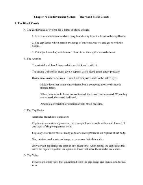
I. The Blood Vessels<br />
Chapter 5: Cardiovascular System –– Heart and Blood Vessels<br />
A. The cardiovascular system has 3 types of blood vessels:<br />
1. Arteries (and arterioles) which carry blood away from the heart to the capillaries.<br />
2. The capillaries which permit exchange of nutrients, wastes, and gases with the<br />
tissues.<br />
3. Veins (and venules) which return blood from the capillaries to the heart.<br />
B. The Arteries<br />
The arterial wall has 3 layers which are thick and resilient.<br />
The strong walls of an artery give it support when blood enters under pressure.<br />
Divide into smaller arterioles –– small arteries just visible to the naked eye.<br />
C. The Capillaries<br />
D. The Veins<br />
Middle layer has some elastic tissue, but is composed mostly of smooth<br />
muscle fibers.<br />
When these muscle fibers are contracted, the vessel is constricted. When they<br />
are relaxed, the vessel is dilated.<br />
Arteriole constriction or dilation affects blood pressure.<br />
Arterioles branch into capillaries.<br />
Capillaries are extremely narrow, microscopic blood vessels with a wall formed of<br />
one layer of simple squamous cells.<br />
Capillary beds (networks of many capillaries) are present in all regions of the body.<br />
Gas, nutrient, and waste exchange occur across their thin walls.<br />
Only certain capillaries are open at any given time. After eating, the capillaries that<br />
serve the digestive system are open and those that serve the muscles are closed.<br />
Venules are small veins that drain blood from the capillaries and then join to form a<br />
vein.
II. The Heart<br />
The walls of veins and venules have the same 3 layers as arteries, but they are much<br />
thinner.<br />
Thinner walls allow veins to expand to a greater extent.<br />
Veins often have valves which allow blood to flow only toward the heart when open<br />
and prevent the backward flow of blood when closed.<br />
A. The heart is a cone-shaped, muscular organ about the size of a fist.<br />
B. It is located between the lungs directly behind the sternum (breast bone) and is tilted so<br />
the apex (pointed end) is directed left.<br />
C. Myocardium is the major portion of the heart consisting of cardiac muscle tissue.<br />
D. Heart lies within a pericardium sac that contains pericardial fluid which provides<br />
cushioning.<br />
E. Endocardium lines inner surface of the heart.<br />
F. Internal wall called the septum separates heart into right and left halves.<br />
G. Heart has 2 upper, thin-walled atria and 2 lower, thick-walled ventricles.<br />
Atria receive blood from the venous portion of the cardiovascular system.<br />
Atria are much smaller and weaker than muscular ventricles, but hold the same<br />
volume of blood.<br />
Ventricles pump blood into arterial portion of cardiovascular system.<br />
H. Heart valves direct flow of blood and prevent backward movement.<br />
Valves are supported by strong, fibrous tendons attached to muscular projections of<br />
ventricular walls. They support the valves and prevent them from inverting when the<br />
heart contracts.<br />
Atrioventricular valves occur between atria and ventricles and prevent back flow of<br />
blood from ventricle to atrium.<br />
Right atrioventricular or tricuspid valve on right side of heart consists of 3<br />
cusps or flaps.<br />
Left atrioventricular or bicuspid (mitral) valve on left side of heart consists of<br />
2 cusps or flaps.
Semilunar valves have flaps that resemble half-moons and are located between the<br />
ventricles and their attached vessels.<br />
III. Passage of Blood through the Heart<br />
The pulmonary semilunar valve lies between the right ventricle and the<br />
pulmonary trunk.<br />
The aortic semilunar valve lies between the left ventricle and the aorta.<br />
A. Oxygen-poor (deoxygenated) blood enters right atrium from both superior and inferior<br />
vena cava.<br />
B. Right atrium sends blood through right atrioventricular valve (tricuspid valve) to right<br />
ventricle.<br />
C. Right ventricle sends blood through pulmonary semilunar valve into pulmonary trunk.<br />
The pulmonary trunk divides into two pulmonary arteries that go to the lungs.<br />
D. Oxygen-rich (oxygenated) blood returns from lungs through pulmonary veins and is<br />
delivered to the left atrium.<br />
E. Left atrium sends blood through left atrioventricular valve (bicuspid or mitral valve) to<br />
left ventricle.<br />
F. Left ventricle sends blood through aortic semilunar valve into the aorta to the body proper<br />
through arteries.<br />
G. Oxygen-poor blood never mixes with oxygen-rich blood.<br />
H. Blood must go through the lungs in order to pass from the right side to the left side of the<br />
heart.<br />
I. The heart is a double pump serving the lungs and the body circulations simultaneously.<br />
J. Since the left ventricle has the harder job of pumping blood throughout the body, its walls<br />
are thicker than those of the right ventricle.<br />
K. The Heartbeat<br />
Each heartbeat is called a cardiac cycle.<br />
The heart contracts or beats about 70 times a minute.<br />
Each heartbeat lasts about 0.85 seconds.<br />
A normal adult heart rate at rest can vary from 60 to 80 beats per minute.
The heartbeat consists of phases: Systole refers to contraction of heart chambers and<br />
Diastole is relaxation of heart chambers.<br />
Atria contract first while ventricles relax (0.15 sec), then ventricles contract while<br />
atria relax (0.30 sec), then all chambers rest (0.40 sec).<br />
When the heart beats, the familiar lub-dup sound is heard as valves of the heart close.<br />
Lub is caused by vibrations occurring when the atrioventricular valves close<br />
due to ventricular contraction.<br />
Dup is heard when the semilunar valves close due to back pressure of blood<br />
in the arteries.<br />
A heart murmur is often due to ineffective valves, which allow blood to pass back<br />
into the atria after the atrioventricular valves have closed<br />
Rhythmical contraction of the atria and ventricles is due to the intrinsic conduction<br />
system of the heart.<br />
SA (Sinoatrial) node is found in the upper dorsal wall of the right atrium. It<br />
initiates the heartbeat by sending out an excitatory impulse every 0.85<br />
seconds that cause atria to contract. It is called the pacemaker because it<br />
serves to keep the heartbeat regular.<br />
AV (Atrioventricular) node is found at the base of the right atrium very near<br />
the septum. When stimulated by impulses from SA node, it sends out<br />
impulses through septum to cause ventricles to contract.<br />
An electrocardiogram (ECG) is a recording of the electrical changes that occur in<br />
myocardium during a cardiac cycle.<br />
An abnormality called ventricular fibrillation can be detected using an ECG.<br />
Ventricular fibrillation is the most common cause of sudden cardiac death in<br />
seemingly healthy individuals over the age of 35.<br />
IV. Features of the Cardiovascular System<br />
A. Pulse<br />
Causes uncoordinated contraction of the ventricles.<br />
Can be caused by injury or drug overdose.<br />
The surge of blood entering the arteries causes their elastic walls to stretch and then<br />
recoil. This alternating stretching and recoiling of an arterial wall can be felt as a<br />
pulse in any artery that runs close to the body’s surface.
Pulse can be felt by placing several fingers on the radial artery which lies near the<br />
outer border of the palm side of a wrist. A carotid artery, on either side of the trachea<br />
in the neck, is another accessible location for feeling the pulse.<br />
Normally the pulse rate indicates the rate of heartbeat because the arterial walls pulse<br />
whenever the left ventricle contracts.<br />
B. Blood Flow<br />
The pulse rate is usually 60–80 beats per minute.<br />
Blood pressure is the pressure of blood against the wall of a blood vessel.<br />
A sphygmomanometer which has a pressure cuff is used to measure blood pressure<br />
in the brachial artery of the arm.<br />
Systolic pressure is the highest arterial pressure reached during ejection of blood<br />
from the heart.<br />
Diastolic pressure is the lowest arterial pressure that occurs while the heart ventricles<br />
are relaxing.<br />
Normal resting blood pressure for a young adult is 120mm mercury (Hg) over 80<br />
mm Hg or 120/80.<br />
The higher number is the systolic pressure and the lower number is the<br />
diastolic pressure.<br />
Blood pressure varies throughout the body. It is highest in the aorta and lowest in the<br />
vena cava.<br />
Both systolic and diastolic pressure decrease with distance from the left ventricle.<br />
Blood pressure is minimal in venules and veins.<br />
Instead of blood pressure, venous return is dependent on skeletal muscle contraction,<br />
the presence of valves in veins, and respiratory movements.<br />
V. The Cardiovascular Pathways<br />
A. The cardiovascular system has 2 major circular pathways: The pulmonary circuit which<br />
circulates blood through the lungs and the systemic circuit which serves the needs of the<br />
body tissues.<br />
B. The Pulmonary Circuit<br />
Deoxygenated blood from the body collects in the right atrium and then passes into<br />
the right ventricle, which pumps it to pulmonary trunk.
Pulmonary trunk divides into right and left pulmonary arteries to carry blood to each<br />
lung.<br />
In the pulmonary capillaries of the lungs, carbon dioxide is unloaded and oxygen is<br />
picked up by blood.<br />
Oxygenated blood from lungs is returned through pulmonary veins to left atrium.<br />
Not all arteries carry oxygenated blood and not all veins carry deoxygenated blood.<br />
Since blood in the pulmonary arteries is oxygen poor and blood in the pulmonary<br />
veins is oxygen rich they serve as the exception.<br />
C. The Systemic Circuit<br />
The aorta (artery) and vena cava (veins) are the main pathways for blood in the<br />
systemic circuit.<br />
The superior vena cava collects blood from the head, chest, and arms while<br />
the inferior vena cava collects blood from the lower body regions.<br />
Transport of oxygenated blood moves from left ventricle through aorta out to all<br />
tissues.<br />
Deoxygenated blood returns from all tissues via vena cava.<br />
The coronary arteries serve the heart muscle itself and are the first branches off the<br />
aorta.<br />
Coronary arteries lie on the external surface of the heart, where they divide into<br />
arterioles.<br />
Coronary veins collect deoxygenated blood from capillaries and empty into the right<br />
atrium.<br />
VI. Cardiovascular Disorders<br />
A. Cardiovascular Disease (CVD) is the leading cause of untimely deaths in the U.S.<br />
B. Risk of CVD can be reduced by following guidelines for a heart-healthy life style.<br />
C. Hypertension (High Blood Pressure)<br />
Occurs when blood moves through the arteries at a higher pressure than normal.<br />
Hypertension is present when the systolic blood pressure is 140 or greater or the<br />
diastolic blood pressure is 90 or greater.
Sometimes called the silent killer because it may not be detected until a stroke or<br />
heart attack occurs.<br />
Genetic predisposition.<br />
D. Atherosclerosis<br />
An accumulation of soft masses of fatty materials, including cholesterol, beneath the<br />
inner linings of arteries –– plaque.<br />
As plaque develops, it tends to protrude into a vessel interfering with blood flow.<br />
Atherosclerosis begins in early adulthood and develops progressively through middle<br />
age, but symptoms may not appear until an individual is age 50 or older. Plaque can<br />
cause a clot to form on the arterial wall.<br />
As long as the clot remains stationary, it is called a thrombus.<br />
If a clot dislodges and moves along with the blood, it is called an embolus.<br />
Aspirin reduces the stickiness of platelets and thereby lowers the probability that a<br />
clot will form.<br />
E. Stroke, Heart Attack, and Aneurysm<br />
A cerebrovascular accident (CVA) also called a stroke, often results when a small<br />
cranial arteriole bursts or is blocked by an embolus.<br />
A lack of oxygen causes a portion of the brain to die, and paralysis or death<br />
can result.<br />
A myocardial infarction (MI), also called a heart attack, occurs when a portion of the<br />
heart muscle dies due to a lack of oxygen.<br />
If a coronary artery becomes partially blocked, the individual may then suffer<br />
from angina pectoris, characterized by radiating pain in the left arm.<br />
When a coronary artery is completely blocked, a heart attack occurs.<br />
An aneurysm is a ballooning of a blood vessel, most often the abdominal artery or<br />
the arteries leading to the brain.<br />
Atherosclerosis and hypertension can weaken the wall of an artery to the<br />
point that an aneurysm develops.<br />
F. Varicose veins develop when the valves of veins become weak and ineffective due to the<br />
backward pressure of blood.
VII. The Lymphatic System Helps the Cardiovascular System<br />
A. The lymphatic system consists of lymphatic vessels and the lymphatic organs.<br />
B. The lymphatic system has three main functions that contribute to homeostasis:<br />
Lymphatic capillaries take up excess tissue fluid and return it to the bloodstream; lacteals<br />
receive fat molecules at the intestinal villi and transport them to the bloodstream; the<br />
lymphatic system helps defend the body against disease.<br />
C. The lymphatic vessels carry lymph which has the same composition as tissue fluid.<br />
The lymphatic vessels merge before entering one of two ducts: the thoracic duct or<br />
the right lymphatic duct. The lymphatic ducts enter the subclavian veins, which are<br />
cardiovascular veins in the thoracic region.


