Multicellular Parasites
Multicellular Parasites
Multicellular Parasites

Create successful ePaper yourself
Turn your PDF publications into a flip-book with our unique Google optimized e-Paper software.
generalized acute illness occurs, with fever, hives, cough, abdominal,<br />
joint and muscle pain, and diarrhea. Some people die during<br />
this stage, but usually the symptoms subside and infected individuals<br />
are free of symptoms for a number of years. Then a chronic,<br />
slowly progressing illness appears, with weakness, accumulation of<br />
fluid in the abdominal cavity, and sometimes vomiting blood.<br />
Causative Agent<br />
Most cases of schistosomiasis are caused by three species of<br />
flukes in the genus Schistosoma. Other genera of flukes that<br />
infect humans are hermaphroditic, meaning that each worm has<br />
both male and female reproductive organs, but Schistosoma<br />
species have male and female worms. Schisto-soma means “splitbody,”<br />
referring to a deep groove running along the male’s<br />
body in which he clasps his female partner. Schistosoma mansoni,<br />
the only species established in the Americas, and the most common<br />
cause of schistosomiasis worldwide, is 10 to 20 mm long<br />
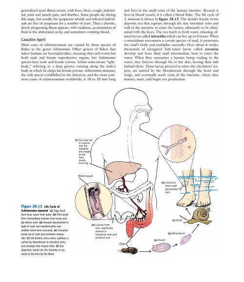
Figure 28.13 Life Cycle of<br />
Schistosoma mansoni (a) Eggs (ova)<br />
from feces reach fresh water. (b) First larval<br />
form (miracidium) hatches from ovum and<br />
(c) infects snail. (d) Asexual reproduction in<br />
body of snail and transformation into<br />
another larval form (cercaria). (e) Cercariae<br />
break out of snail and penetrate human<br />
skin.Tail left behind, larva enters capillary, is<br />
carried by bloodstream to intestinal veins,<br />
and develops into mature fluke. (f) Ova<br />
deposited, break into the intestine or are<br />
swept to the liver by the blood.<br />
(f) Ova that fail<br />
to rupture<br />
into the<br />
intestine<br />
are swept<br />
to the<br />
liver,<br />
cause<br />
scarring<br />
Blood vessel<br />
(e) Larvae from<br />
skin capillaries<br />
mature in<br />
intestinal vein and<br />
produce ova<br />
Eggs<br />
and lives in the small veins of the human intestine. Because it<br />
lives in blood vessels, it is called a blood fluke. The life cycle of<br />
S. mansoni is shown in figure 28.13. The slender female worm<br />
deposits ova that rupture through the tiny intestinal veins and<br />
wall of the intestine to enter the lumen, ultimately to be eliminated<br />
with the feces. The ova hatch in fresh water, releasing ciliated<br />
larvae called miracidia which can live up to 6 hours. When<br />
a miracidium encounters a certain species of snail, it penetrates<br />
the snail’s body and multiplies asexually. Over about 6 weeks,<br />
thousands of elongated fork-tailed larvae called cercariae<br />
develop and leave their snail intermediate host to enter the<br />
water. When they encounter a human being wading in the<br />
water, they burrow through his or her skin, leaving their tails<br />
behind them. These larvae proceed to enter the circulatory system,<br />
are carried by the bloodstream through the heart and<br />
lungs, and eventually reach veins of the intestine, where they<br />
mature, mate, and begin ova production.<br />
(a) Ovum<br />
(d) Cercaria<br />
from snail<br />
penetrates<br />
skin<br />
(c) Snail<br />
(b) Miracidium