Chapter 13 - McGraw-Hill
Chapter 13 - McGraw-Hill
Chapter 13 - McGraw-Hill

You also want an ePaper? Increase the reach of your titles
YUMPU automatically turns print PDFs into web optimized ePapers that Google loves.
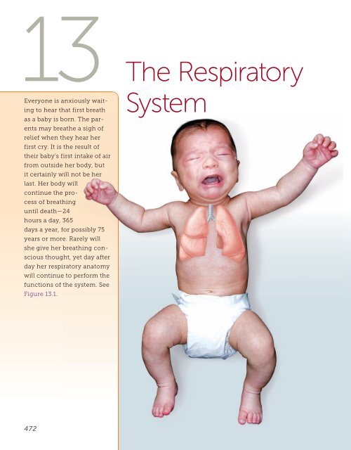
<strong>13</strong> The Respiratory<br />
System<br />
Everyone is anxiously waiting<br />
to hear that first breath<br />
as a baby is born. The parents<br />
may breathe a sigh of<br />
relief when they hear her<br />
first cry. It is the result of<br />
their baby’s first intake of air<br />
from outside her body, but<br />
it certainly will not be her<br />
last. Her body will<br />
continue the process<br />
of breathing<br />
until death—24<br />
hours a day, 365<br />
days a year, for possibly 75<br />
years or more. Rarely will<br />
she give her breathing conscious<br />
thought, yet day after<br />
day her respiratory anatomy<br />
will continue to perform the<br />
functions of the system. See<br />
Figure <strong>13</strong>.1 .<br />
472
outcomes<br />
learning<br />
<strong>13</strong>.1 learning outcome<br />
Use medical terminology<br />
related to the<br />
respiratory system.<br />
After completing this chapter, you should be able to:<br />
<strong>13</strong>.1 Use medical terminology related to the respiratory<br />
system.<br />
<strong>13</strong>.2 Trace the flow of air from the nose to the pulmonary<br />
alveoli and relate the function of each part of the<br />
respiratory tract to its gross and microscopic anatomy.<br />
<strong>13</strong>.3 Explain the role of surfactant.<br />
<strong>13</strong>.4 Describe the respiratory membrane.<br />
<strong>13</strong>.5 Explain the mechanics of breathing in terms of anatomy<br />
and pressure gradients.<br />
<strong>13</strong>.6 Define the measurements of pulmonary function.<br />
<strong>13</strong>.7 Define partial pressure and explain its relationship to a<br />
gas mixture such as air.<br />
<strong>13</strong>.8 Explain gas exchange in terms of the partial pressures<br />
of gases at the capillaries and the alveoli and at the<br />
capillaries and the tissues.<br />
<strong>13</strong>.9 Compare the composition of inspired and expired air.<br />
<strong>13</strong>.10 Explain the factors that influence the efficiency<br />
of alveolar gas exchange.<br />
<strong>13</strong>.11 Describe the mechanisms for transporting O 2 and CO 2<br />
in the blood.<br />
<strong>13</strong>.12 Explain how respiration is regulated to homeostatically<br />
control blood gases and pH.<br />
<strong>13</strong>.<strong>13</strong> Explain the functions of the respiratory system.<br />
<strong>13</strong>.14 Summarize the effects of aging on the respiratory<br />
system.<br />
<strong>13</strong>.15 Describe common diagnostic tests used for respiratory<br />
system disorders.<br />
<strong>13</strong>.16 Describe respiratory system disorders and relate<br />
abnormal function to pathology.<br />
word roots<br />
& combining forms<br />
alveol/o: alveolus, air sac<br />
bronch/o: bronchial tube<br />
bronchi/o: bronchus<br />
bronchiol/o: bronchiole<br />
capn/o: carbon dioxide<br />
cyan/o: blue<br />
laryng/o: larynx<br />
lob/o: lobe<br />
nas/o: nose<br />
pharyng/o: pharynx<br />
phren/o: diaphragm<br />
pneum/o, pneumon/o: air<br />
pulmon/o: lung<br />
rhin/o: nose<br />
sinus/o: sinus<br />
spir/o: breathing<br />
thorac/o: chest<br />
trache/o: trachea<br />
pronunciation key<br />
alveoli: al-VEE-oh-lye<br />
arytenoid: ah-RIT-en-oyd<br />
bronchi: BRONG-kye<br />
bronchus: BRONG-kuss<br />
conchae: KON-kee<br />
corniculate: kor-NIK-you-late<br />
laryngopharynx: lah-RING-oh-<br />
FAIR-inks<br />
larynx: LAIR-inks<br />
nares: NAH-reez<br />
pharynx: FAIR-inks<br />
trachea: TRAY-kee-ah
Overview<br />
The word respiration has<br />
several usages. In Levels of<br />
Organization of the Human<br />
Body, you studied cellular<br />
respiration as a cellular process<br />
performed by mitochondria<br />
to release energy<br />
from the bonds in a glucose<br />
molecule. In the muscular<br />
system chapter, you studied<br />
aerobic and anaerobic respiration<br />
as variations of cellular<br />
respiration. In this chapter,<br />
you will study respiration<br />
first as the movement of air<br />
into (inspiration) and out<br />
of the lungs (expiration),<br />
commonly called breathing.<br />
Then you will explore<br />
respiration as the exchange<br />
of gases in two areas—<br />
between the air in the lungs<br />
and the blood in capillaries<br />
and between the blood in<br />
the capillaries and the tissues<br />
out in the body. Once<br />
you understand how the<br />
Respiratory System<br />
Major Organs and Structures:<br />
nose, pharynx, larynx, trachea,<br />
bronchi, lungs<br />
Accessory Structures:<br />
diaphragm, sinuses, nasal cavity<br />
Functions<br />
gas exchange, acid-base balance,<br />
speech, sense of smell, creation<br />
of pressure gradients necessary<br />
to circulate blood and lymph<br />
FIGURE <strong>13</strong>.1 The respiratory system.<br />
exchange of gases takes place, you will be prepared to investigate how gases are transported<br />
in the blood.<br />
As with all of the other human body systems you have covered so far, it is important<br />
to understand the anatomy of the system before tackling the physiology. So you will begin<br />
below by studying the anatomy of this system.<br />
l<br />
l<br />
i<br />
<strong>13</strong>.2 learning outcome<br />
Trace the flow of air from<br />
the nose to the pulmonary<br />
alveoli and relate the<br />
function of each part of the<br />
respiratory tract to its gross<br />
and microscopic anatomy.<br />
pharynx: FAIR-inks<br />
. c o m<br />
larynx: LAIR-inks<br />
trachea: TRAY-kee-ah<br />
bronchi: BRONG-kye<br />
alveoli: al-VEE-oh-lye<br />
audio<br />
. m c g r a w - h<br />
c o n n e c t<br />
Anatomy of the Respiratory System<br />
As you can see in Figure <strong>13</strong>.2 , the entire respiratory system’s anatomy is housed in the<br />
head, neck, and thorax. In general, the anatomy in the head and neck is the upper<br />
respiratory tract, while the anatomy from the trachea through the lungs is the lower<br />
respiratory tract.<br />
You have already studied some of this anatomy, such as the pleurae (serous membrane),<br />
in The Basics. To refresh your memory, a serous membrane is a double-walled,<br />
fluid-filled membrane. In the case of the pleurae, the visceral pleura is in contact with the<br />
lung’s surface, while the parietal pleura is not. The parietal pleura lines the thoracic cavity<br />
and covers the diaphragm’s superior surface. Fluid exists between the visceral and parietal<br />
pleurae. This anatomy will be important when you study the mechanics of breathing,<br />
later in the chapter.<br />
Before you get started on the rest of the anatomy, consider the way air enters and<br />
moves through the body. Take a deep breath now with your mouth closed, and trace the<br />
air in that breath as it travels on its route (follow along with Figure <strong>13</strong>.2 ). The air enters<br />
the nasal cavity through the nose. From there it goes to the pharynx, to the larynx, to<br />
the trachea, to the bronchi (where it enters the lungs), to the bronchial tree, and finally<br />
to the tiny air sacs called alveoli (not shown in the figure). At the alveoli, the second part<br />
of respiration—the exchange of gases—takes place.<br />
474 CHAPTER <strong>13</strong> The Respiratory System
Frontal sinus<br />
Nasal cavity<br />
Sphenoid<br />
sinus<br />
Soft palate<br />
Hard palate<br />
Pharynx<br />
Nostril<br />
Oral cavity<br />
Larynx<br />
Bronchus<br />
Bronchial tree<br />
Right lung<br />
Epiglottis<br />
Esophagus<br />
Trachea<br />
Visceral<br />
pleural<br />
membrane<br />
Pleural fluid<br />
(located<br />
between<br />
membranes)<br />
Parietal<br />
pleural<br />
membrane<br />
Left lung<br />
Diaphragm<br />
FIGURE <strong>13</strong>.2 The respiratory system anatomy.<br />
disease p int<br />
Pleurisy is characterized by inflammation of the pleurae. This condition has<br />
a variety of causes, including respiratory infections such as tuberculosis or<br />
pneumonia, cancer, trauma or injury to the chest, inflammatory conditions such<br />
as lupus or rheumatoid arthritis, pulmonary embolus, or conditions related to<br />
asbestos exposure. Pleurisy causes pain in the chest, especially when taking a<br />
deep breath or coughing. The pain is caused by the friction of the two inflamed<br />
membranes rubbing against each other. Breathing can become difficult, and this<br />
could lead to additional symptoms such as cyanosis, tachypnea, and shortness of<br />
breath. To diagnose pleurisy, physicians listen to breath sounds and may order a<br />
series of tests including a blood test, CT scan, chest x-ray, and ultrasound.<br />
Now you are ready to zoom in on all of the respiratory system’s specific anatomy (mentioned<br />
above) in the order that the air traveled through it in your deep breath. You will<br />
need to become familiar with the gross and microscopic anatomy along the way because<br />
this is important in understanding precisely how the anatomy functions.<br />
Anatomy of the Respiratory System 475
l<br />
l<br />
i<br />
. c o m<br />
Nasal<br />
bone<br />
Lateral<br />
cartilage<br />
Septal<br />
nasal<br />
cartilage<br />
Major<br />
alar<br />
cartilage<br />
FIGURE <strong>13</strong>.3 The nose.<br />
nares: NAH-reez<br />
conchae: KON-kee<br />
audio<br />
c o n n e c t<br />
. m c g r a w - h<br />
Minor<br />
alar<br />
cartilages<br />
Dense<br />
connective<br />
tissue<br />
Nose<br />
Air enters the nasal cavity through the nose’s two nares<br />
(nostrils). The nasal bones superiorly and the plates of hyaline<br />
cartilage at the end of the nose are responsible for the<br />
nose’s shape. You can feel where the nasal bone ends and<br />
cartilage begins at the bridge of the nose. See Figure <strong>13</strong>.3 .<br />
Nasal Cavity<br />
As you can see in Figure <strong>13</strong>.4c , a septum divides the<br />
nasal cavity into right and left sides. The ethmoid bone<br />
(superiorly), the vomer (inferiorly), and a septal cartilage<br />
anteriorly form the septum. The anterior part of the nasal<br />
cavity (the vestibule ) is lined by stratified squamous epithelial<br />
tissue with stiff guard hairs to block debris from<br />
entering the respiratory tract.<br />
The nasal cavity widens posterior to the vestibule to<br />
make room for three bony, lateral ridges called the nasal<br />
conchae. See Figure <strong>13</strong>.4b . The ethmoid bone forms the<br />
superior and middle nasal conchae, while the inferior nasal<br />
concha is a separate bone. This portion of the nasal cavity<br />
is lined by mucous membranes that trap debris and warm and moisturize the incoming air.<br />
study hint<br />
Take a look up your own nose by using a flashlight and a mirror. You can see the hairs<br />
in the vestibule and notice that the posterior nasal cavity appears very red and moist.<br />
These are the moist, mucous membranes, and their rich blood supply moisturizes<br />
and warms the air. You should also see that there is limited space for air<br />
to pass because of the protruding nasal conchae. This<br />
causes more air to come in contact with the mucous<br />
membranes, so they are better able to function.<br />
FIGURE <strong>13</strong>.4 The anatomy<br />
of the upper respiratory<br />
tract: (a) sagittal view of<br />
cadaver, (b) sagittal section<br />
showing internal anatomy<br />
(nasal septum has been<br />
removed), (c) nasal septum<br />
and regions of the pharynx.<br />
Frontal sinus<br />
Nasal conchae:<br />
Superior<br />
Middle<br />
Inferior<br />
Hard palate<br />
Tongue<br />
Larynx:<br />
Epiglottis<br />
Cribriform plate<br />
Sphenoidal sinus<br />
Nasopharynx<br />
Uvula<br />
Oropharynx<br />
Laryngopharynx<br />
Trachea<br />
(a)<br />
Esophagus<br />
476 CHAPTER <strong>13</strong> The Respiratory System
FIGURE <strong>13</strong>.4 concluded<br />
Frontal sinus<br />
Superior<br />
Middle<br />
Inferior<br />
Nasal<br />
conchae<br />
Vestibule<br />
Nostril<br />
Hard palate<br />
Uvula<br />
Tongue<br />
Sphenoidal sinus<br />
Pharyngeal tonsil<br />
Opening of<br />
auditory tube<br />
Palatine tonsil<br />
Lingual tonsil<br />
Hyoid bone<br />
Epiglottis<br />
Larynx<br />
Trachea<br />
Esophagus<br />
(b)<br />
Nasal septum:<br />
Ethmoid bone<br />
Vomer<br />
Septal<br />
cartilage<br />
Pharynx:<br />
Nasopharynx<br />
Oropharynx<br />
Laryngopharynx<br />
(c)<br />
Anatomy of the Respiratory System 477
The nasal conchae provide extra surface area for the mucous membranes to function. The<br />
mucous membranes are composed of ciliated pseudostratified columnar epithelial tissue. The<br />
cilia move mucus and any trapped debris posteriorly so that it can be swallowed. Olfactory neurons<br />
located in the roof of the posterior nasal cavity detect odors and provide the sense of smell.<br />
spot check<br />
of mucous membranes?<br />
Why do you think the vestibule is stratified epithelial tissue instead<br />
Sinuses You studied the sinuses of the frontal, ethmoid, sphenoid, and maxilla bones in<br />
the skeletal system chapter. The frontal and sphenoidal sinuses are shown in Figure <strong>13</strong>.4 .<br />
These cavities within the bones are also lined with respiratory epithelial tissue to warm<br />
and moisturize the air. The mucus produced in the sinuses is drained to the nasal cavity<br />
through small openings.<br />
disease p<br />
int<br />
Inflammation of the epithelium in the sinuses (sinusitis) causes<br />
increased mucus production, and the accompanying<br />
swelling may block its drainage to the nasal cavity. The<br />
pressure within the sinuses created by the buildup of<br />
mucus causes a sinus headache. Decongestants<br />
(vasoconstrictors) help reduce the swelling,<br />
thereby improving mucus drainage, which<br />
reduces the increased pressure.<br />
At this point in your deep breath, the inspired air leaving the nasal cavity has been<br />
partially warmed and moistened, and some of its debris has been trapped. The structure<br />
the air encounters next—the pharynx—is explained below.<br />
l<br />
l<br />
i<br />
. c o m<br />
laryngopharynx:<br />
lah-RING-oh-FAIR-inks<br />
audio<br />
. m c g r a w - h<br />
c o n n e c t<br />
Pharynx<br />
The pharynx, commonly called the throat, is divided into three regions based on location<br />
and anatomy—the nasopharynx, the oropharynx, and the laryngopharynx. You will<br />
explore these in the paragraphs that follow.<br />
Nasopharynx As you can see in Figure <strong>13</strong>.4c , the nasopharynx is located posterior to<br />
the nasal cavity and the soft palate. This passageway is also lined by ciliated pseudostratified<br />
columnar epithelial tissue whose cilia move mucus and trapped debris to the next<br />
region of the pharynx so that it can be swallowed. The pharyngeal tonsils and the opening<br />
to the auditory tube (eustachian tube) are located in this region.<br />
Oropharynx This region of the pharynx (shown in Figure <strong>13</strong>.4c ) is inferior to the<br />
nasopharynx. The oropharynx is common to the respiratory and digestive systems as a<br />
passageway for air, food, and drink. For the oropharynx to withstand the possible abrasions<br />
caused by the passage of solid food, it must be lined with a more durable tissue—<br />
stratified squamous epithelial tissue. In addition, the palatine tonsils are located in this<br />
region to deal with any incoming pathogens.<br />
Laryngopharynx This region of the pharynx extends from the level of the epiglottis to the<br />
beginning of the esophagus. Like the oropharynx, the laryngopharynx is lined by stratified<br />
squamous epithelial tissue to handle the passage of air, food, and drink. See Figure <strong>13</strong>.4c . Solids<br />
and liquids continue on from the laryngopharynx to the esophagus, but inspired air moves<br />
through an opening (glottis) to the larynx, the next structure in the respiratory pathway.<br />
478 CHAPTER <strong>13</strong> The Respiratory System
Larynx<br />
The larynx is a cartilage box (voice box) of nine separate cartilages, eight of which are<br />
composed of hyaline cartilage connective tissue. The epiglottis (the ninth cartilage of the<br />
larynx) is composed of elastic cartilage connective tissue. As you can see in Figure <strong>13</strong>.4b ,<br />
the epiglottis stands almost vertically over the glottis. Its function is to fold over the glottis<br />
during swallowing to prevent solids and liquids from entering the larynx. You will learn<br />
more about how this works in the digestive system chapter. The epiglottis remains in its<br />
vertical position at all other times to ensure the easy passage of air from the laryngopharynx<br />
through the glottis to the larynx.<br />
Figure <strong>13</strong>.5 gives you a closer look at the larynx. Here you can see the laryngeal prominence<br />
(“Adam’s apple”) of the thyroid cartilage. It enlarges to be more visible in men<br />
arytenoid: ah-RIT-en-oyd<br />
corniculate:<br />
kor-NIK-you-late<br />
l<br />
l<br />
i<br />
. c o m<br />
audio<br />
c o n n e c t<br />
. m c g r a w - h<br />
Epiglottis<br />
Epiglottis<br />
Hyoid<br />
bone<br />
Hyoid<br />
bone<br />
Laryngeal<br />
prominence<br />
Thyroid<br />
cartilage<br />
Corniculate<br />
cartilage<br />
Fat<br />
Thyroid<br />
cartilage<br />
Trachea<br />
Cricoid<br />
cartilage<br />
Tracheal<br />
cartilage<br />
Arytenoid<br />
cartilage<br />
Cricoid<br />
cartilage<br />
Membranous<br />
part of trachea<br />
(a) (b) (c)<br />
Vestibular<br />
fold (false<br />
vocal cord)<br />
Vocal fold<br />
(true vocal<br />
cord)<br />
FIGURE <strong>13</strong>.5 The larynx: (a) anterior view, (b) posterior view, (c) sagittal section.<br />
than women due to the presence of<br />
testosterone. You can also see (in this<br />
figure) the two arytenoid cartilages<br />
and the two corniculate cartilages<br />
that operate the vocal cords.<br />
Vocal Cords The walls of the larynx<br />
are muscular to operate the vocal<br />
cords shown in Figures <strong>13</strong>.5c , <strong>13</strong>.6 ,<br />
and <strong>13</strong>.7 . There are two sets of folds<br />
in the inner wall of the larynx—the<br />
vestibular folds and the vocal cords.<br />
The vestibular folds have no function<br />
in speech. They are important in closing<br />
the larynx during swallowing.<br />
Figure <strong>13</strong>.6 shows how the vocal<br />
cords are abducted (spread apart) and<br />
adducted (brought closer together) by<br />
muscles pulling on the arytenoid and corniculate<br />
cartilages. The opening formed<br />
by abducting the vocal cords is the glottis.<br />
Air passing through adducted vocal cords<br />
causes them to vibrate to make sounds<br />
(a)<br />
(c)<br />
Thyroid cartilage<br />
Cricoid cartilage<br />
Vocal cord<br />
Arytenoid<br />
cartilage<br />
Corniculate<br />
cartilage<br />
(b)<br />
(d)<br />
Vestibular<br />
fold<br />
Vocal cord<br />
Glottis<br />
Corniculate<br />
cartilage<br />
Epiglottis<br />
Vestibular<br />
fold<br />
Glottis<br />
Inner lining<br />
of trachea<br />
FIGURE <strong>13</strong>.6 Action of laryngeal muscles on the vocal cords: (a) adduction<br />
showing just the cartilages and the vocal cords, (b) adduction as seen with all<br />
tissues present, (c) abduction showing just the cartilages and the vocal cords,<br />
(d) abduction as seen with all tissues present.<br />
Anatomy of the Respiratory System 479
Anterior<br />
Posterior<br />
Epiglottis<br />
Glottis<br />
Vestibular fold<br />
Vocal cord<br />
Trachea<br />
Corniculate<br />
cartilage<br />
FIGURE <strong>13</strong>.7 Endoscopic view of the vocal<br />
cords as seen with a laryngoscope.<br />
of varying pitch depending on the tautness of the cords. So speech is a very<br />
active process, which involves muscles pulling on cartilages of the larynx to<br />
operate the vocal cords. The larynx at the vocal cords is lined with stratified<br />
squamous epithelial tissue to withstand the vibrations.<br />
Trachea<br />
From the larynx, inspired air travels to the trachea, a rigid tube with 18<br />
to 20 C-shaped cartilages composed of hyaline cartilage connective tissue.<br />
See Figure <strong>13</strong>.8 . These cartilages hold the trachea open for the easy<br />
flow of air. The C-shaped cartilages are open posteriorly with smooth<br />
muscle bridging the gap. This feature allows the esophagus (directly<br />
posterior to the trachea) room to expand into the tracheal space when<br />
swallowed food passes on its way to the stomach. If the cartilages were<br />
circular instead of C-shaped, a swallowed piece of meat could get hung<br />
up on each cartilage as it passed down the esophagus.<br />
Like the nasal cavity and the nasopharynx, the trachea is lined with<br />
ciliated pseudostratified columnar epithelial tissue with goblet cells that<br />
secrete mucus. See Figures <strong>13</strong>.8 and <strong>13</strong>.9. The air you breathe is full<br />
Larynx<br />
Trachea<br />
C-shaped<br />
cartilage<br />
Bronchi and<br />
bronchial<br />
tree<br />
(a)<br />
Mucus<br />
Mucociliary<br />
escalator<br />
Particles of<br />
debris<br />
Ciliated<br />
epithelial cell<br />
Goblet<br />
epithelial<br />
cell<br />
Anterior<br />
C-shaped<br />
hyaline<br />
cartilage<br />
Ciliated<br />
epithelium<br />
Cartilage<br />
Mucous<br />
gland<br />
Mucous<br />
gland<br />
Lumen of<br />
trachea<br />
Chondrocytes<br />
Mucosa<br />
Smooth<br />
muscle<br />
(b)<br />
(c)<br />
Posterior<br />
Connective<br />
tissue<br />
FIGURE <strong>13</strong>.8 The trachea and bronchi: (a) anterior view, (b) longitudinal view of the trachea showing cilia moving<br />
mucus and debris, (c) transverse section of the trachea showing C-shaped cartilage.<br />
480 CHAPTER <strong>13</strong> The Respiratory System
FIGURE <strong>13</strong>.9 Lining of<br />
trachea. Epithelial tissue of the<br />
trachea, showing ciliated cells<br />
and goblet cells.<br />
Cilia<br />
Goblet cell<br />
4 mm<br />
of particles, such as dust, pollen, and smoke particles. You may have seen the dust in the<br />
air as the sun shines through a window. Even during sleep, the cilia of the trachea move<br />
mucus and any trapped debris up (like an escalator) toward the pharynx to be swallowed.<br />
See Figure <strong>13</strong>.8b . This prevents the accumulation of debris in the lungs.<br />
disease p int<br />
The smoke inhaled with each drag on a cigarette<br />
contains a lot of particles, but the respiratory anatomy<br />
is designed to prevent this debris from accumulating<br />
in the lungs. However, the increased amount of debris<br />
may, over time, cause the lining of a habitual smoker’s<br />
trachea to go through metaplasia, changing from<br />
ciliated epithelial tissue to a more durable, nonciliated<br />
tissue. Without the ciliated escalator, the respiratory<br />
system resorts to coughing up the debris. As a result,<br />
the long-term smoker develops the smoker’s hack<br />
each morning to move the debris inspired each night.<br />
spot check<br />
Compare the direction the cilia move debris in the nasopharynx to<br />
the direction they move debris in the trachea. How do they differ?<br />
Anatomy of the Respiratory System 481
spot check A patient on a ventilator has a tube inserted into the trachea<br />
through a procedure called a tracheostomy. What should be done to the air delivered<br />
through a ventilator considering the respiratory anatomy leading to the trachea has<br />
been bypassed?<br />
The trachea splits to become the right and left main bronchi, each of which enters<br />
its respective lung. You will explore the lungs and bronchial tree together, looking first at<br />
their gross anatomy, as shown in Figure <strong>13</strong>.10 .<br />
l<br />
l<br />
i<br />
. c o m<br />
bronchus: BRONG-kuss<br />
audio<br />
. m c g r a w - h<br />
c o n n e c t<br />
Lungs and the Bronchial Tree<br />
As you can see in Figure <strong>13</strong>.10b , the right and left main bronchi each enters its respective<br />
lung at an area on the medial surface of the lung called the hilum. This is the same location<br />
used by pulmonary arteries and veins to enter and leave the lung. The left bronchus is<br />
slightly more horizontal than the right bronchus due to the location of the heart. The main<br />
bronchi and all of their further branches make up the bronchial tree. See Figure <strong>13</strong>.10c .<br />
Upon entering the lung, each main bronchus branches to become the lobar bronchi,<br />
each going to a separate lobe of the lung. The left lung has fewer lobes (two) than the<br />
right, again because of the position of the heart. The right lung has three lobes, and therefore<br />
three lobar bronchi.<br />
FIGURE <strong>13</strong>.10 Gross<br />
anatomy of the lungs and<br />
bronchial tree: (a) anterior view,<br />
(b) medial views of the right<br />
and left lungs, (c) bronchogram<br />
(radiograph of the bronchial<br />
tree), anterior view.<br />
Apex of lung<br />
Right superior<br />
(upper) lobe<br />
Right main<br />
bronchus<br />
Superior lobar<br />
bronchus<br />
Larynx<br />
Trachea<br />
Left superior<br />
(upper) lobe<br />
Small bronchus<br />
of bronchial tree<br />
Terminal<br />
bronchiole<br />
Cardiac<br />
impression<br />
(a)<br />
Right inferior<br />
(lower) lobe<br />
Right middle lobe<br />
Left inferior<br />
(lower) lobe<br />
Apex<br />
Superior lobe<br />
Hilum<br />
Middle<br />
lobe<br />
Pulmonary arteries<br />
Main bronchi<br />
Pulmonary veins<br />
Inferior lobe<br />
Hilum<br />
Cardiac<br />
impression<br />
(b)<br />
Right lung<br />
Diaphragmatic<br />
surface<br />
Left lung<br />
482 CHAPTER <strong>13</strong> The Respiratory System
FIGURE <strong>13</strong>.10 concluded<br />
Trachea<br />
Main bronchi<br />
Lobar bronchi<br />
Small bronchi of<br />
the bronchial tree<br />
(c)<br />
w rning<br />
The lungs fill with air, but they are not hollow like a balloon. A cross section of a<br />
lung appears solid—more like Styrofoam composed of tiny beads. Each of the tiny<br />
beads is a tiny, hollow air sac that can fill with inspired air. See Figure <strong>13</strong>.11.<br />
Anterior<br />
Pericardial<br />
cavity<br />
Heart<br />
Breast<br />
Sternum<br />
Ribs<br />
Left lung<br />
Visceral<br />
pleura<br />
Pleural<br />
cavity<br />
Parietal<br />
pleura<br />
Right lung<br />
Aorta<br />
Vertebra<br />
Spinal cord<br />
Posterior<br />
FIGURE <strong>13</strong>.11 Cross section of a cadaver through the thoracic cavity.<br />
Anatomy of the Respiratory System 483
Lobar bronchi further divide to smaller and smaller bronchi that branch to form the<br />
bronchial tree. See Figure <strong>13</strong>.10c . All of the bronchi are supported by cartilage plates,<br />
which hold them open for the easy passage of air. The smallest bronchi further branch<br />
to form bronchioles. These small tubes do not have cartilage in their walls. Instead,<br />
their walls have smooth muscle that allows them to dilate or constrict to adjust airflow.<br />
You will learn more about this later in the chapter. Each bronchiole supplies air to a<br />
lobule (subsection of a lobe) of the lung composed of tiny air sacs called alveoli. See<br />
Figure <strong>13</strong>.12a .<br />
Pulmonary<br />
artery<br />
Smooth<br />
muscle<br />
Blood flow<br />
Bronchioles<br />
Pulmonary<br />
arteriole<br />
Blood flow<br />
Pulmonary<br />
venule<br />
Pulmonary<br />
vein<br />
Blood flow<br />
Capillary network on<br />
surface of alveolus<br />
Alveolar<br />
sac<br />
Alveoli<br />
(a)<br />
Macrophage<br />
Air space<br />
within<br />
alveolus<br />
Nucleus of<br />
simple<br />
squamous<br />
alveolar cell<br />
Fluid with<br />
surfactant<br />
Great alveolar<br />
cell<br />
Diffusion<br />
of O 2<br />
Diffusion<br />
of CO 2<br />
Alveolus<br />
Respiratory<br />
membrane:<br />
Alveolar<br />
fluid (with<br />
surfactant)<br />
Single<br />
squamous<br />
cell alveolar<br />
wall<br />
Single cell<br />
capillary<br />
wall<br />
Pulmonary<br />
capillary<br />
endothelium<br />
(wall)<br />
Red blood<br />
cell<br />
Capillary<br />
(b)<br />
(c)<br />
FIGURE <strong>13</strong>.12 Bronchiole, alveoli, and the respiratory membrane: (a) clusters of alveoli at the end of a bronchiole and<br />
the network of capillaries covering them, (b) cells of the alveoli, (c) respiratory membrane.<br />
spot check Penny is an inquisitive 18-month-old girl who likes to see what fits<br />
into what. One morning, she put a small, metal washer that she found on the floor<br />
into her nose just as her mother entered the room. Her mother gasped when she saw<br />
what Penny had done. This scared Penny, so she gasped, too, and the metal washer<br />
was gone. She had inhaled it. What route do you think the metal washer will take (trace<br />
the pathway)?<br />
484 CHAPTER <strong>13</strong> The Respiratory System
spot check<br />
Explain.<br />
In which lung will the doctor at the clinic find the metal washer?<br />
Alveoli The alveoli are clustered like grapes at the end of the bronchiole. As you can see<br />
in Figure <strong>13</strong>.12 , a network of capillaries covers the alveoli. This is vital for gas exchange,<br />
as you will read shortly. Figure <strong>13</strong>.<strong>13</strong> shows the histology of the alveoli with respect to<br />
the bronchioles and blood supply to the capillaries. There are approximately 150 million<br />
alveoli in each human lung. Each alveolus is a tiny air sac with two types of cells in its<br />
walls—simple squamous cells and great alveolar cells. Most of the alveolar wall is composed<br />
of one layer of thin squamous cells that allow for rapid gas exchange across their<br />
surface. The great alveolar cells (shown in Figure <strong>13</strong>.12b ) are important because they<br />
secrete a fluid called surfactant. Below, you will find out why this fluid is so important.<br />
Bronchiole:<br />
Epithelium<br />
Smooth muscle<br />
FIGURE <strong>13</strong>.<strong>13</strong> Histology<br />
of the lung: micrograph of<br />
alveoli, a bronchiole, and a<br />
branch of a pulmonary artery.<br />
Alveoli<br />
Branch of<br />
pulmonary artery<br />
Alveolar duct<br />
1 mm<br />
spot check Why must the vessel represented in this figure be an artery and<br />
not a capillary, and why must the tube be a bronchiole and not a bronchus of the<br />
bronchial tree? ( Hint: Look at the histology.)<br />
Surfactant To understand the importance of surfactant, you must first understand a<br />
property of water: high surface tension. This basically means that water will always try to<br />
have the smallest surface area to volume ratio possible. In other words, water forms beads<br />
or drops because a sphere has a smaller surface area to volume ratio than a flat sheet. This<br />
is why water forms beads or drops on smooth surfaces like glassware in your dishwasher.<br />
Surfactant reduces the surface tension of water much like the rinse agent you may<br />
add to your dishwasher to avoid water spots. The rinse agent reduces the surface tension<br />
<strong>13</strong>.3 learning outcome<br />
Explain the role of surfactant.<br />
Anatomy of the Respiratory System 485
of water (sheeting action), so water sheets off your glassware instead of forming beads<br />
that leave water spots as the glasses dry. Surfactant also causes water to form a thin sheet<br />
instead of a bead. Why is this important? By the time air has entered the alveoli, it has<br />
been thoroughly moisturized by all the mucous membranes it has passed along the respiratory<br />
route. If a bead of water were to form inside the tiny alveoli, the plump bead might<br />
touch the wall on the opposite side of the air sac and cause the thin, delicate walls of the<br />
alveoli to stick together, and this would cause the alveoli to collapse. A thin sheet of water<br />
in the alveoli (instead of a plump bead) reduces the chance of the alveoli walls collapsing<br />
on each other. Collapsed alveoli do not easily fill with air.<br />
disease p<br />
int<br />
A fetal respiratory system does not mature until late<br />
in pregnancy. The alveoli in infants born before the<br />
lungs are mature often collapse because of the lack of<br />
sufficient surfactant. This condition, called respiratory<br />
distress syndrome (hyaline membrane disease), is a<br />
common cause of neonatal death. Oxygen under positive<br />
pressure can be administered along with surfactant to<br />
keep the lungs (alveoli) inflated between breaths.<br />
<strong>13</strong>.4 learning outcome<br />
Describe the respiratory<br />
membrane.<br />
Respiratory Membrane So far, you have seen in Figure <strong>13</strong>.12a and b the relationship<br />
of the alveoli to the bronchioles and the cells that make up the alveoli. In Figure <strong>13</strong>.12c ,<br />
you can see the structure formed by the capillary network adjacent to the alveoli—the<br />
respiratory membrane. This is a very important structure because it is the location of<br />
gas exchange in the lung. Take a closer look at this figure. The respiratory membrane is<br />
composed of the thin layer of water with surfactant in the alveoli, the single squamous cell<br />
alveolar wall, and the single cell capillary wall. If all of the respiratory membrane in one<br />
lung were laid out in a single layer, it would cover approximately 70 square meters (m 2 ),<br />
equivalent to the floor of a room 25 feet by 30 feet.<br />
You have now covered all of the anatomy that the air of your deep breath encountered<br />
along its way to the respiratory membrane. It is time to explore the way this respiratory<br />
anatomy functions, starting with how you took the deep breath in the first place.<br />
<strong>13</strong>.5 learning outcome<br />
Explain the mechanics of<br />
breathing in terms of anatomy<br />
and pressure gradients.<br />
Physiology of the Respiratory System<br />
Mechanics of Taking a Breath<br />
Air moves (but is not pushed) along the respiratory passageways on its way to the lungs<br />
because of pressure differences within the chest. This is much like the syringe example<br />
you became familiar with while studying blood flow through the heart (see Figure 11.12<br />
in The Cardiovascular System—Heart and Vessels). The syringe example explained the<br />
relationship between volume, pressure, and flow. If the volume of space in the syringe<br />
is increased, the pressure inside the syringe is decreased, so air flows into the syringe to<br />
equalize the pressures inside and outside the syringe. Likewise, if the volume of space in<br />
the syringe is decreased, the pressure inside the syringe is increased, so air flows out of the<br />
syringe to equalize the pressures. As a result, pushing or pulling on the plunger changes<br />
the volume of the syringe.<br />
How does the body change the volume of the chest? See Figure <strong>13</strong>.14 . Concentrate<br />
on the major muscles for breathing shown in bold in this figure. As you can see in Figure<br />
<strong>13</strong>.14a and b, during inspiration the external intercostal, pectoralis minor, and sternocleidomastoid<br />
muscles contract to expand the rib cage, and the diaphragm contracts to<br />
486 CHAPTER <strong>13</strong> The Respiratory System
flatten its dome shape. The combined effect of these contractions is an increase in the size<br />
(volume) of the chest cavity. All that needs to be done for normal expiration is to have the<br />
same muscles relax. Then the rib cage returns to its normal position and the diaphragm<br />
becomes dome-shaped again due to the recoil of abdominal organs. See Figure <strong>13</strong>.14c . The<br />
volume of the chest is decreased, and air flows from the body. Expiration during normal<br />
breathing is a passive process (no energy required) involving the relaxation of muscles. But<br />
you can also see in Figure <strong>13</strong>.14d that forced expiration involves the contraction of muscles<br />
too. The internal intercostals and abdominal wall muscles do contract in forced expiration;<br />
however, these muscles are not used for expiration during normal breathing. Forced expiration<br />
is intentionally forcing air out of the lungs, which happens when blowing out a candle<br />
or inflating a balloon. In this case, energy for muscle contraction is required.<br />
So far in this explanation of the mechanics of breathing, you have seen how the muscles<br />
of the chest can increase the volume of the chest, but what about the volume of each lung?<br />
How is the volume of the lungs increased? This involves the pleural membranes and the<br />
pleural fluid. The parietal pleura is attached to the thoracic wall and diaphragm, while the<br />
visceral pleura is attached to the lung. The pleural fluid between the parietal and visceral<br />
pleurae cause the two pleurae to stick together and move as one. As the respiratory muscles<br />
expand the thoracic wall and flatten the diaphragm, the parietal pleura moves with the<br />
wall and diaphragm. As the parietal pleura moves with the thoracic wall and diaphragm,<br />
the visceral pleura and the lung move with it—expanding the lung along with the thoracic<br />
cavity. As the lung expands, the pressure within the lung (intrapulmonary pressure)<br />
decreases, so air moves in until the pressure inside the lung is equal to the pressure outside<br />
the body. The intrapulmonary and atmospheric pressures are then equal. When inspiration<br />
ends, the thoracic wall returns to its original position and its volume is diminished.<br />
Sternocleidomastoid<br />
elevates sternum<br />
External<br />
intercostal<br />
muscles pull<br />
ribs up and out<br />
Sternum<br />
moves<br />
up and out<br />
Pectoralis minor<br />
elevates ribs<br />
Diaphragm<br />
contracts<br />
Diaphragm<br />
contracts more<br />
(a)<br />
(b)<br />
FIGURE <strong>13</strong>.14 Respiratory muscles: (a) external intercostal muscles and diaphragm at the beginning of inspiration,<br />
(b) additional muscle action to continue inspiration, (c) recoil of abdominal organs, causing diaphragm to dome when it<br />
relaxes, (d) muscle actions during forced expiration.<br />
Physiology of the Respiratory System 487
FIGURE <strong>13</strong>.14 concluded<br />
Diaphragm<br />
Abdominal organs<br />
recoil and press<br />
diaphragm upward<br />
Posterior internal<br />
intercostal muscles<br />
pull ribs down and<br />
inward<br />
Diaphragm<br />
Abdominal organs<br />
force diaphragm<br />
higher<br />
Abdominal wall<br />
muscles contract<br />
and compress<br />
abdominal organs<br />
(c)<br />
(d)<br />
disease p int<br />
A pneumothorax (collapsed lung) occurs if air is introduced in the pleural cavity<br />
between the pleural membranes. Just as fluid holds the parietal and visceral pleurae<br />
together, air between the pleurae allows them to separate. Normally, there is tension<br />
on the lung, keeping it partially inflated at all times. However, in a pneumothorax, the<br />
pleurae separate, so the lung may recoil and separate from the thoracic wall. The air<br />
in a pneumothorax may be introduced by a penetrating trauma like a knife wound<br />
or broken rib, medical procedures such as inserting a needle to withdraw pleural<br />
fluid, or even a disease like emphysema (covered later in this chapter). Diagnosis of a<br />
pneumothorax involves listening to breath sounds to determine if they are absent or<br />
decreased, doing a chest x-ray, and testing arterial blood gases to determine if there<br />
is an adequate ratio of O 2 and CO 2 in the blood. In mild cases, the pneumothorax<br />
may correct itself without medical intervention. In more severe cases, a chest tube<br />
may need to be introduced into the pleural space to remove the air to inflate the<br />
lung, and surgery may be required to repair the opening into the pleural space. In<br />
a hemothorax, blood is introduced into the pleural cavity. This can be caused by<br />
blood clotting defects, thoracic surgery, severe respiratory infections, or lung cancer.<br />
If the hemothorax is not treated, it can lead to a pneumothorax, among other severe<br />
complications. Diagnosis of a hemothorax includes listening to breath sounds to<br />
determine if they are absent or decreased, doing a chest x-ray and a CT scan of<br />
the lungs, and analyzing plural fluid to determine the presence of blood. Treatment<br />
involves controlling the cause of the bleeding and inserting a chest tube to remove<br />
the blood from the pleural cavity. In severe cases, surgery may be necessary.<br />
488 CHAPTER <strong>13</strong> The Respiratory System
The pressure is now greater in the lung than outside the body, so air flows from the body<br />
until the intrapulmonary and atmospheric pressures are again equal. Figure <strong>13</strong>.15 shows<br />
the muscle action and the pressure changes during inspiration and expiration.<br />
No airflow<br />
Pleural cavity<br />
Atmospheric pressure<br />
Diaphragm<br />
Intrapulmonary pressure<br />
Ribs swing upward<br />
like bucket handles<br />
during inspiration.<br />
2<br />
1<br />
Inspiration<br />
1<br />
At rest<br />
3<br />
Expiration<br />
Ribs swing downward<br />
like bucket handles<br />
during expiration.<br />
Airflow<br />
Airflow<br />
Diaphragm flattens<br />
Diaphragm rises<br />
Rib<br />
Rib<br />
Rib<br />
Rib<br />
Sternum<br />
Sternum<br />
Sternum<br />
Sternum<br />
Ribs elevated, thoracic<br />
cavity expands laterally<br />
Sternum swings up,<br />
thoracic cavity expands<br />
anteriorly<br />
Ribs depressed, thoracic<br />
cavity narrows<br />
Sternum swings down,<br />
thoracic cavity contracts<br />
posteriorly<br />
FIGURE <strong>13</strong>.15 A respiratory cycle of inspiration, expiration, and rest. (1) At rest, atmospheric and intrapulmonary<br />
pressures are equal, and there is no airflow. (2) In inspiration, the thoracic cavity expands laterally, vertically, and anteriorly;<br />
intrapulmonary pressure falls below atmospheric pressure; and air flows into the lungs. (3) In expiration, the thoracic cavity<br />
contracts in all three directions, intrapulmonary pressure rises above atmospheric pressure, and air flows out of the lungs.<br />
There is a rest between breaths. The handles of the pails represent ribs.<br />
Physiology of the Respiratory System 489
<strong>13</strong>.6 learning outcome<br />
Define the measurements<br />
of pulmonary function.<br />
Measurements of Pulmonary Function<br />
How well the respiratory system functions to move air into and out of the lungs can be<br />
measured in pulmonary function (spirometry) tests. A spirometer is a device used to<br />
measure the volume of air moved. Figure <strong>13</strong>.16 shows a photo of Gabe, who is breathing<br />
into the spirometer to determine his various lung volumes and lung capacities (capacities<br />
are determined by adding two volumes). Table <strong>13</strong>.1 defines the various values, and<br />
Figure <strong>13</strong>.17 shows a graph of Gabe’s values.<br />
FIGURE <strong>13</strong>.16<br />
Spirometry. A spirometer is<br />
used to measure lung volumes<br />
and capacities.<br />
FIGURE <strong>13</strong>.17 Graph of<br />
pulmonary volumes and<br />
capacities.<br />
Volume (mL)<br />
6,000<br />
5,000<br />
4,000<br />
3,000<br />
2,000<br />
1,000<br />
0<br />
Maximum<br />
expiration<br />
Time<br />
Maximum<br />
inspiration<br />
Volumes<br />
Inspiratory reserve volume<br />
(3,000 mL)<br />
Tidal<br />
volume<br />
(500<br />
mL)<br />
Expiratory<br />
reserve<br />
volume<br />
(1,100 mL)<br />
Residual<br />
volume<br />
(1,200 mL)<br />
Functional residual<br />
capacity (2300 mL)<br />
Capacities<br />
Inspiratory capacity (3,500 mL)<br />
Vital capacity (4,600 mL)<br />
Total lung capacity (5,800 mL)<br />
490 CHAPTER <strong>13</strong> The Respiratory System
TABLE <strong>13</strong>.1<br />
Lung volumes and capacities<br />
Volume or capacity Definition Typical value<br />
Tidal volume (TV)<br />
Inspiratory reserve<br />
volume (IRV)<br />
Expiratory reserve<br />
volume (ERV)<br />
Residual volume (RV)<br />
Functional residual<br />
capacity (FRC)<br />
Inspiratory capacity (IC)<br />
Vital capacity (VC)<br />
Total lung capacity (TLC)<br />
The tidal volume is the amount of air<br />
moved in a normal breath (inspired or<br />
expired) at rest.<br />
The inspiratory reserve volume is the<br />
amount of air that can be forcefully<br />
inspired beyond the amount inspired in<br />
a normal breath at rest.<br />
The expiratory reserve volume is the<br />
amount of air that can be forcefully<br />
expired beyond the amount expired in<br />
a normal breath at rest.<br />
The residual volume is the amount of<br />
air in the lungs that cannot be moved.<br />
The functional residual capacity is the<br />
amount of air remaining in the lungs<br />
after the expiration of a normal breath<br />
at rest. FRC 5 ERV 1 RV<br />
The inspiratory capacity is the<br />
maximum amount of air that can be<br />
inspired after the expiration of a normal<br />
breath at rest. IC 5 TV 1 IRV<br />
Vital capacity is the maximum amount of<br />
air that can be moved. VC 5 IC 1 FRC<br />
The total lung capacity is the maximum<br />
amount of air the lung can hold.<br />
TLC 5 VC 1 RV<br />
500 mL<br />
3,000 mL<br />
1,100 mL<br />
1,200 mL<br />
2,300 mL<br />
3,500 mL<br />
4,600 mL<br />
5,800 mL<br />
Exercise may temporarily increase the tidal volume for an individual, but this does<br />
not mean that all of the other values will increase. The maximum amount of air the<br />
respiratory system can move (vital capacity) does not change on a temporary (minute-byminute)<br />
basis. So if there is an increase in tidal volume during a workout, there must be a<br />
decrease in the inspiratory and expiratory reserve volumes.<br />
Lung volumes and capacities vary from one individual to another due to gender,<br />
size, age, and physical condition. In general, a woman’s vital capacity is less than a<br />
man’s; a tall, thin person has a greater vital capacity than someone short and obese;<br />
and a trained athlete has a greater vital capacity than someone who has a sedentary<br />
lifestyle.<br />
Compliance is another measurement of pulmonary function. It measures how well<br />
the lung can expand and return to shape (elasticity). It is harder to expand the lungs and<br />
the thorax if there is decreased compliance. This may be due to the buildup of scar tissue<br />
in the lung (pulmonary fibrosis), collapse of the alveoli (respiratory distress syndrome),<br />
skeletal disorders (scoliosis or kyphosis), or chronic obstructive pulmonary disorders<br />
(COPDs), such as asthma, chronic bronchitis, emphysema, and lung cancer (discussed<br />
later in the chapter).<br />
At this point, you have become familiar with the anatomy of the respiratory system<br />
and how it works to deliver air into and out of the lungs. You can now begin to explore<br />
the second part of respiration—the exchange of gases—by looking at the gases present in<br />
the air you breathe.<br />
Physiology of the Respiratory System 491
<strong>13</strong>.7 learning outcome<br />
Define partial pressure and<br />
explain its relationship to a<br />
gas mixture such as air.<br />
Composition of Air<br />
Gases diffuse across membranes from high concentration to low concentration until the<br />
concentrations are equal. So it is important to be able to talk about quantities of gases.<br />
The air you breathe is a mixture of gases—78.6 percent nitrogen, 20.9 percent oxygen,<br />
0.04 percent carbon dioxide, and variable amounts of water vapor depending on humidity<br />
levels. Gases fill whatever space is available to them and can be compressed, so volume<br />
is not a good measure of the amount of a gas. For example, an open scuba tank (of a<br />
given volume) will fill with air, but more air can be pumped under pressure into the same<br />
tank before it is sealed (compressed air). Therefore, the amount of a gas is expressed not<br />
as volume but in terms of the pressure a gas exerts. In the case of a mixture of gases, like<br />
air, the amount of each gas is expressed as a partial pressure —the amount of pressure an<br />
individual gas contributes to the total pressure of the mixture. So, if the total pressure of<br />
the air (atmospheric pressure) is 760 mmHg, then the partial pressure of nitrogen (P N2 ) is<br />
78.6 percent of 760, or 597 mmHg; the partial pressure of oxygen (P O2 ) is 20.9 percent<br />
of 760, or 159 mmHg; the partial pressure of carbon dioxide (P CO2 ) is 0.04 percent of<br />
760, or 0.3 mmHg; and the remainder, 3.7 mmHg, is the partial pressure of water vapor.<br />
All of the partial pressures of the gases added together equal the total pressure of the air<br />
(760 mmHg).<br />
You will need to understand partial pressures as a measurement of the amount of a<br />
gas when you study gas exchange in the lung and out at the tissues in the next section of<br />
this chapter.<br />
spot check The atmospheric pressure in Miami on Wednesday was 760<br />
mmHg. However, the atmospheric pressure in Denver on the same day was 640<br />
mmHg. What was the partial pressure of CO 2 in Denver that day? What was the partial<br />
pressure of O 2 ?<br />
<strong>13</strong>.8 learning outcome<br />
Explain gas exchange in<br />
terms of the partial pressures<br />
of gases at the capillaries<br />
and the alveoli and at the<br />
capillaries and the tissues.<br />
Gas Exchange<br />
Before studying gas exchange, it will be helpful for you to keep these two facts in mind:<br />
(1) Carbon dioxide is a waste product produced in the tissues through cellular respiration,<br />
and (2) blood travels to the lungs to be oxygenated. With that stated, we begin by explaining<br />
gas exchange at the respiratory membrane between an alveolus and a capillary in the<br />
lung. In this discussion, we use general symbols—greater than (>), less than (
Alveolus in lung<br />
4<br />
In the lung,<br />
P CO2 alveolus < P CO 2 capillary ;<br />
therefore, CO 2 diffuses<br />
into the alveolus until<br />
P CO2 alveolus = P CO 2 capillary .<br />
Capillary in lung<br />
CO 2<br />
CO 2<br />
O 2<br />
O 2<br />
1<br />
In the lung,<br />
P O2 alveolus > P O 2 capillary ;<br />
therefore, O 2 diffuses<br />
into the capillary until<br />
P O2 alveolus = P O 2 capillary .<br />
Capillary at tissues<br />
3<br />
2<br />
At the tissues,<br />
P CO2 tissues > P CO 2 capillary ;<br />
therefore, CO 2 diffuses<br />
into the capillary until<br />
P CO2 tissues = P CO 2 capillary .<br />
At the tissues,<br />
P O2 capillary > P O 2 tissues ;<br />
therefore O 2 diffuses into<br />
the tissues until<br />
P O2 capillary = P O 2 tissues .<br />
Tissues of the body<br />
Indicates direction of diffusion<br />
FIGURE <strong>13</strong>.18 Gas exchange.<br />
1. Blood coming from the right side of the heart to the lung is low in oxygen. In comparison,<br />
the air inspired to the alveolus in the lung is high in oxygen. P O2 alveolus ><br />
P O2 capillary , so oxygen diffuses across the respiratory membrane into the blood of the<br />
capillary until the partial pressures on both sides of the respiratory membrane are<br />
equal: P O2 alveolus 5 P O2 capillary . Will there still be some oxygen left in the alveolus after<br />
the gas exchange has taken place? Yes, because not all of it diffused into the blood;<br />
only the amount of oxygen necessary to make the partial pressures equal on both<br />
sides diffused. Some oxygen will be expired from the alveolus.<br />
Physiology of the Respiratory System 493
2. The oxygen-rich blood travels from the lung to the left side of the heart before traveling<br />
to the capillaries at the tissues of the body. Here the tissues have been using<br />
oxygen to perform cellular respiration: C 6 H 12 O 6 1 6O 2 → 6CO 2 1 6H 2 O. As a<br />
result, the tissues are relatively low in oxygen compared to the high amount in the<br />
blood in the capillary: P O2 capillary > P O2 tissues . So oxygen diffuses into the tissues until<br />
the partial pressure of oxygen in the blood equals the partial pressure of oxygen in<br />
the tissues: P O2 capillary 5 P O2 tissues.<br />
3. Meanwhile, mitochondria in the cells of the tissues have been producing carbon<br />
dioxide as a waste product of cellular respiration. As a result, the concentration<br />
of carbon dioxide is much higher in the tissues than in the blood of the capillary:<br />
P CO2 tissues > P CO2 capillary. So carbon dioxide diffuses into the blood of the capillary<br />
until the concentrations are equal: P CO2 tissues 5 P CO2 capillary.<br />
4. The blood leaving the capillaries at the tissues of the body travels to the right side of<br />
the heart before returning to the lungs. It has lost some of its oxygen and has gained<br />
carbon dioxide through diffusion at the tissues of the body. So it makes sense to have<br />
started this explanation of gas exchange by saying the blood coming to the lungs was<br />
oxygen-poor. It makes just as much sense to say the partial pressure of carbon dioxide is<br />
greater in the blood of the capillary at the alveolus than in the air of the alveolus because<br />
there is so little carbon dioxide in inspired air (0.04 percent): P CO2 capillary > P CO2 alveolus .<br />
So carbon dioxide diffuses across the respiratory membrane to the alveolus until the<br />
partial pressure of carbon dioxide in the capillary equals the partial pressure of carbon<br />
dioxide in the alveolus. P CO2 capillary 5 P CO2 alveolus.<br />
<strong>13</strong>.9 learning outcome<br />
Compare the composition<br />
of inspired and expired air.<br />
Comparison of Inspired and Expired Air Given what you have read about the<br />
composition of air and gas exchange, you should be able to compare the composition of<br />
inspired and expired air. For this comparison, you will examine gas exchange using specific<br />
values. See Figure <strong>13</strong>.19 . As you can see in this figure, inspired air has more oxygen<br />
than expired air, and inspired air has less carbon dioxide than expired air.<br />
<strong>13</strong>.10 learning outcome<br />
Explain the factors that<br />
influence the efficiency of<br />
alveolar gas exchange.<br />
Factors That Influence Gas Exchange Several factors influence the effectiveness of<br />
alveolar gas exchange. They are explained in the following list:<br />
• Concentration of the gases. The concentration of the gases matters because<br />
the greater the concentration gradient, the more diffusion takes place. For example,<br />
gas exchange of oxygen will increase if a patient is administered oxygen instead of<br />
breathing room air. In contrast, gas exchange of oxygen will be less at higher altitudes<br />
because the air is thinner and does not contain as much oxygen.<br />
• Membrane area. Membrane area matters because the greater the area of the respiratory<br />
membrane, the greater the opportunity for gas exchange. For example, Figure <strong>13</strong>.20<br />
shows alveolar tissue for a healthy individual, a pneumonia patient, and a person with<br />
emphysema. You will notice the lack of respiratory membrane for the emphysema<br />
patient. This is because emphysema breaks down the alveolar walls. The reduced membrane<br />
area means less gas will be exchanged.<br />
• Membrane thickness. The thickness of the respiratory membrane matters because<br />
the thicker the membrane, the harder it is for gases to diffuse across it. Look again<br />
at Figure <strong>13</strong>.20 . Pneumonia may cause excess fluid in the alveoli and swelling of the<br />
alveolar walls, which make gas exchange much more difficult.<br />
• Solubility of the gas. Gases must be able to dissolve in water if they are to diffuse<br />
across a membrane into the blood. For example, nitrogen is 78.6 percent of the air<br />
494 CHAPTER <strong>13</strong> The Respiratory System
P O2<br />
= 104<br />
Inspired air<br />
P O2<br />
= 160<br />
P CO2 = 0.3<br />
(a)<br />
P O2<br />
= 40 P CO2 = 45 P O2<br />
= 104 P CO2 = 40<br />
Right<br />
P CO2 = 40<br />
Alveolus<br />
P O2<br />
= 104<br />
Pulmonary capillary<br />
Heart<br />
Expired air<br />
P O2<br />
= 120<br />
P CO2 = 27<br />
(b)<br />
P CO2 = 40<br />
Left<br />
P O2<br />
= 95<br />
P CO2 = 40<br />
(c)<br />
Blood in<br />
pulmonary veins<br />
FIGURE <strong>13</strong>.19 Changes<br />
in P O2 and P CO2 along the<br />
respiratory route. Values are<br />
expressed in mmHg.<br />
(a) Oxygen diffuses into the<br />
arterial ends of pulmonary<br />
capillaries, and CO 2 diffuses<br />
into the alveoli because of<br />
differences in partial pressures.<br />
(b) As a result of diffusion<br />
(at the venous ends of the<br />
pulmonary capillaries), the<br />
concentrations of O 2 are<br />
equal on both sides of the<br />
respiratory membrane, as are<br />
the concentrations of CO 2 on<br />
both sides of the respiratory<br />
membrane. (c) The partial<br />
pressure of O 2 is reduced<br />
in the pulmonary veins<br />
due to the mixing of blood<br />
drained from the bronchi and<br />
bronchial tree. (d) Oxygen<br />
diffuses out of the arterial end<br />
of capillaries to the tissues, and<br />
CO 2 diffuses out of the tissues<br />
to the capillaries due to the<br />
differences in partial pressures.<br />
(e) As a result of diffusion (at<br />
the venous ends of tissue<br />
capillaries), the concentrations<br />
of O 2 are equal in the<br />
capillaries and the tissues,<br />
as are the concentrations of<br />
CO 2 in the capillaries and the<br />
tissues.<br />
Tissue capillary<br />
P O2<br />
= 40<br />
P CO2 = 45<br />
P O2<br />
= 95<br />
P CO2 = 40<br />
(e)<br />
Interstitial<br />
fluid<br />
(d)<br />
P O2<br />
= 40<br />
P CO2 = 45<br />
P O2<br />
= 40<br />
P CO2 = 45<br />
P O2<br />
= 20<br />
P CO2 = 46<br />
Tissue cells<br />
you breathe, but it does not diffuse across the respiratory membrane because it is not<br />
soluble at normal atmospheric pressure. Oxygen and carbon dioxide are soluble at<br />
normal atmospheric pressure.<br />
Physiology of the Respiratory System 495
Fluid and blood<br />
cells in alveoli<br />
Alveolar walls<br />
thickened by edema<br />
(a) (b) (c)<br />
FIGURE <strong>13</strong>.20 Influences on gas exchange: (a) normal alveoli, (b) alveoli of pneumonia patient, (c) alveoli of<br />
emphysema patient.<br />
FIGURE <strong>13</strong>.21 Glass of club<br />
soda.<br />
clinical p int<br />
Scuba divers breathe air from their tanks. It is not pure oxygen; it is air that has<br />
been compressed so that the tank can hold more. If divers go to significant depths,<br />
nitrogen becomes soluble because of the increased pressure—every 10 meters of<br />
water is equal to another full atmosphere of pressure. Although nitrogen can then<br />
diffuse across the respiratory membrane, this is alright because nitrogen does not<br />
react with anything in the blood. It becomes very relevant, however, during the<br />
ascent from the dive. If the diver comes up too quickly, nitrogen comes out of<br />
solution as a gas wherever it is in the body. This is similar to club soda being<br />
uncapped and poured. See Figure <strong>13</strong>.21. Removing the cap relieves the pressure<br />
within the can or bottle. The carbon dioxide in the club soda quickly comes out<br />
of solution as bubbles with the reduced pressure. The nitrogen bubbles can cause<br />
severe damage to the diver’s nerves<br />
and other tissues. Divers must ascend<br />
very slowly to allow nitrogen to slowly<br />
come out of solution, diffuse across<br />
the respiratory membrane in the alveoli,<br />
and be exhaled. Decompression<br />
sickness, or the bends, is the disorder<br />
that results if a diver ascends from<br />
depths too quickly. The treatment is to<br />
put the diver immediately in a hyperbaric<br />
(increased-pressure) chamber<br />
and put the body under sufficient<br />
pressure to have the nitrogen dissolve<br />
again and then to slowly decrease the<br />
pressure so that the nitrogen can be<br />
exhaled.<br />
496 CHAPTER <strong>13</strong> The Respiratory System
• Ventilation-perfusion coupling. This basically means that the airflow to the lung<br />
must match the blood flow to the lung. Ideally, the maximum amount of air should<br />
go to where there is the maximum amount of blood in the lung. This is accomplished<br />
through local control in two ways:<br />
1. Lung perfusion (blood flow to alveoli). As blood flows toward alveolar capillaries,<br />
it is directed to lobules in the lung where the partial pressure of oxygen is high.<br />
How? Alveolar capillaries constrict where the partial pressure of oxygen is low, so<br />
blood is diverted to where the partial pressure of oxygen is high.<br />
2. Alveolar ventilation (airflow to alveoli). Smooth muscles in the walls of bronchioles<br />
are sensitive to the partial pressure of carbon dioxide. If the partial pressure<br />
of carbon dioxide increases, the bronchioles dilate. If the partial pressure of carbon<br />
dioxide decreases, the bronchioles constrict. Airflow is therefore directed to lobules<br />
where partial pressure of carbon dioxide is high.<br />
Ventilation-perfusion coupling is very important because it allows the respiratory system<br />
to compensate for damaged lung tissue. If an area of the lung is damaged, less air<br />
and less blood are directed to that area. See Figure <strong>13</strong>.22 .<br />
Reduced P O2<br />
in<br />
blood vessels<br />
Decreased<br />
airflow<br />
Increased<br />
airflow<br />
Elevated P O2<br />
in<br />
blood vessels<br />
Response<br />
to reduced<br />
ventilation<br />
Result:<br />
Blood flow<br />
matches airflow<br />
Response<br />
to increased<br />
ventilation<br />
Vasoconstriction of<br />
pulmonary vessels<br />
Vasodilation of<br />
pulmonary vessels<br />
(a)<br />
Decreased<br />
blood flow<br />
Increased<br />
blood flow<br />
Reduced P CO2<br />
in alveoli<br />
Decreased<br />
blood flow<br />
Increased<br />
blood flow<br />
Elevated P CO2<br />
in alveoli<br />
Response<br />
to reduced<br />
perfusion<br />
Result:<br />
Airflow matches<br />
blood flow<br />
Response<br />
to increased<br />
perfusion<br />
Constriction of<br />
bronchioles<br />
Dilation of<br />
bronchioles<br />
(b)<br />
Decreased<br />
airflow<br />
Increased<br />
airflow<br />
FIGURE <strong>13</strong>.22 Ventilation-perfusion coupling: (a) perfusion adjusted to changes in ventilation,<br />
(b) ventilation adjusted to changes in perfusion.<br />
spot check The atmospheric pressure is 760 mmHg in New York City and<br />
630 mmHg in Breckenridge, Colorado. In which city should more gas exchange take<br />
place? Explain.<br />
Physiology of the Respiratory System 497
<strong>13</strong>.11 learning outcome<br />
Describe the mechanisms<br />
for transporting O 2 and<br />
CO 2 in the blood.<br />
Gas Transport<br />
You have now studied how carbon dioxide and oxygen are exchanged across membranes<br />
into and out of the blood, but how are these gases carried in the blood from one place to<br />
another? To understand gas transport, first look at Figures <strong>13</strong>.23 (systemic gas exchange<br />
and transport) and <strong>13</strong>.24 (alveolar gas exchange and transport). Notice the red and blue<br />
arrows in these figures. The blue arrows represent CO 2 , while the red arrows represent O 2 .<br />
The thickness of the arrows represents the relative amounts of the gases being exchanged.<br />
Although there are three blue arrows and two red arrows in each figure, concentrate on the<br />
largest blue arrow (representing 70 percent of the CO 2 ) and the largest red arrow (representing<br />
98.5 percent of the O 2 ). These two arrows explain the majority of gas transport in<br />
the blood happening at the body’s tissues ( Figure <strong>13</strong>.23 ) and at the alveoli ( Figure <strong>13</strong>.24 ).<br />
Respiring tissue<br />
Capillary blood<br />
CO 2<br />
7%<br />
23%<br />
CO 2<br />
70%<br />
CO 2<br />
98.5%<br />
O 2<br />
1.5%<br />
O 2<br />
Dissolved CO 2 gas<br />
CO 2 + plasma protein Carbamino compounds<br />
CO 2 + Hb HbCO 2<br />
CO 2 + H 2 O H 2 CO 3 HCO –<br />
3 + H +<br />
O 2 + HHb HbO 2 + H +<br />
Dissolved O 2 gas<br />
Plasma<br />
Hb<br />
HbCO 2<br />
HbO 2<br />
HHb<br />
Hemoglobin<br />
Carbaminohemoglobin<br />
Oxyhemoglobin<br />
Deoxyhemoglobin<br />
FIGURE <strong>13</strong>.23 Systemic gas exchange and transport. The blue arrows represent CO 2 transport, while the red arrows<br />
represent O 2 transport. The thickness of the arrows represents the relative amounts of the gases being transported.<br />
Systemic Gas Exchange and Transport You should already be aware that the tissues<br />
of the body produce CO 2 as a waste product of cellular respiration and that, because the<br />
P CO2 tissues > P O2 capillary, CO 2 diffuses from the tissues into the capillaries. Now you need<br />
to understand that the diffused carbon dioxide mixes with water in the blood to form<br />
carbonic acid (H 2 CO 3 ). Carbonic acid, because it is in water, separates into its two<br />
ions: a bicarbonate ion HCO 2 3 and a hydrogen ion (H 1 )—remember from Levels of<br />
498 CHAPTER <strong>13</strong> The Respiratory System
Alveolus air<br />
Capillary blood<br />
CO 2<br />
7%<br />
23%<br />
CO 2<br />
70%<br />
CO 2<br />
98.5%<br />
O 2<br />
1.5%<br />
O 2<br />
Dissolved CO 2 gas<br />
CO 2 + plasma protein Carbamino compounds<br />
CO 2 + Hb HbCO 2<br />
CO 2 + H 2 O H 2 CO 3 HCO –<br />
3 + H +<br />
O 2 + HHb HbO 2 + H +<br />
Dissolved O 2 gas<br />
Respiratory<br />
membrane<br />
Plasma<br />
Hb<br />
HbCO 2<br />
HbO 2<br />
HHb<br />
Hemoglobin<br />
Carbaminohemoglobin<br />
Oxyhemoglobin<br />
Deoxyhemoglobin<br />
FIGURE <strong>13</strong>.24 Alveolar gas exchange and transport. The blue arrows represent CO 2 transport, while the red arrows<br />
represent O 2 transport. The thickness of the arrows represents the relative amounts of the gases being transported.<br />
Organization of the Human Body, that water allows for ions in solution. This reaction is<br />
shown in Figure <strong>13</strong>.23 where the largest blue arrow enters the blood:<br />
CO 2 1 H 2 O → HCO 2 3 1 H 1<br />
Free hydrogen ions in the blood would lower the pH of the blood, but notice in<br />
Figure <strong>13</strong>.23 that the free hydrogen ion (H 1 ) reacts with oxyhemoglobin (HbO 2 ) to<br />
become deoxyhemoglobin (HHb) and oxygen (O 2 ):<br />
H 1 1 HbO 2 → HHb 1 O 2<br />
Hemoglobin releases oxygen in the presence of a hydrogen ion and then binds to it (H 1 ).<br />
By binding to the free hydrogen ions, hemoglobin acts as a buffer, resisting a change of pH<br />
in the blood. P O2 capillary > P O2 tissues so oxygen diffuses to the tissues until P O2 tisues 5 P O2 capillary<br />
The blood containing deoxyhemoglobin and bicarbonate ions continues to the right<br />
side of the heart and on to the alveoli of the lung. Below, you will learn what happens in<br />
alveolar gas exchange and transport, as shown in Figure <strong>13</strong>.24 .<br />
nutrition<br />
acid-base<br />
fluid & electrolytes<br />
Alveolar Gas Exchange and Transport Again, you should focus on the largest<br />
red and blue arrows representing oxygen and carbon dioxide in Figure <strong>13</strong>.24 .<br />
In the alveolus, the P O2 alveolus > P O2 capillary so oxygen diffuses into the capillaries. When<br />
Physiology of the Respiratory System 499
it does, deoxyhemoglobin reacts with oxygen to release hydrogen ions and form<br />
oxyhemoglobin:<br />
HHb 1 O 2 → HbO 2 1 H 1<br />
The now free hydrogen ions (H 1 ) in the capillary at the alveolus bind to the bicarbonate<br />
ions (HCO 2 3 ) to form carbonic acid (H 2 CO 3 ) in the blood. This results in carbon<br />
dioxide and water. Notice that this is the reverse of the reaction happening for carbon<br />
dioxide at the tissues. P CO2 capillary > P CO2 alveolus so carbon dioxide diffuses across the respiratory<br />
membrane to the alveolus until P CO2 capillary 5 P CO2 alveolus<br />
H 1 1 HCO 3 − → H 2 CO 3 → CO 2 1 H 2 O<br />
Basically, most of the oxygen is transported in the blood by hemoglobin as oxyhemoglobin,<br />
and most of the carbon dioxide is transported in the blood as bicarbonate ions.<br />
Hemoglobin functions to carry oxygen from the lungs to the tissues and hydrogen ions<br />
from the tissues to the lungs.<br />
clinical p int<br />
You have already read about hemoglobin carrying carbon monoxide (CO) in<br />
the cardiovascular system chapter on blood. Hemoglobin binds to carbon monoxide<br />
210 times more tightly than to oxygen, and it does not carry oxygen as long<br />
as it is bound to carbon monoxide. CO is produced during combustion, so it can<br />
be emitted from improperly vented furnaces, cars (exhaust), and even cigarettes as<br />
they are smoked. Typically, less than 1.5 percent of hemoglobin is bound to carbon<br />
monoxide in nonsmokers, while 10 percent<br />
of a heavy smoker’s hemoglobin may be<br />
bound to carbon monoxide. Mechanics need<br />
to ventilate their garages while they work<br />
because even just a 0.1 percent concentration<br />
of CO in the air can bind to 50 percent of<br />
the worker’s hemoglobin, and a 0.2 percent<br />
atmospheric concentration can be lethal.<br />
<strong>13</strong>.12 learning outcome<br />
Explain how respiration is<br />
regulated to homeostatically<br />
control blood gases and pH.<br />
Regulation of Respiration<br />
Now that you have become familiar with how oxygen and carbon dioxide are transported<br />
in the blood, you are ready to examine how the respiratory system is regulated to homeostatically<br />
control blood gases and pH.<br />
The main control centers for respiration are located in the medulla oblongata. See<br />
Figure <strong>13</strong>.25 . From there, messages to stimulate inspiration travel through inspiratory (I)<br />
neurons that go to the spinal cord and then out to the diaphragm and intercostal muscles<br />
(by way of the phrenic and intercostal nerves). Expiratory (E) neurons in the medulla<br />
oblongata send signals only for forced expiration.<br />
As you have previously learned, increasing the frequency of nerve impulses causes<br />
longer muscle contractions and, therefore, deeper inspirations. If the length of time (duration)<br />
is increased for each stimulus, the inspiration is prolonged and the breathing is<br />
slower. When nerve impulses from the inspiratory neurons end, muscles relax and expiration<br />
takes place.<br />
Information concerning the need for regulation comes to the respiratory centers in the<br />
medulla oblongata from several sources. These sources are explained in the following list:<br />
• Stretch receptors in the thoracic wall send signals to the medulla oblongata as to<br />
the degree of the chest’s expansion. When maximum expansion has been reached,<br />
500 CHAPTER <strong>13</strong> The Respiratory System
the medulla oblongata stops sending inspiratory messages.<br />
This prevents overinflation of the lungs, and it is most important<br />
in infants. This action is called the Hering-Breuer reflex.<br />
• Proprioceptors in the muscles and joints send signals to<br />
respiratory centers during exercise so that ventilation is<br />
increased. The respiratory centers in the medulla oblongata<br />
can increase the depth and rate of respiration.<br />
• The pontine respiratory group in the pons receives input<br />
from the hypothalamus, limbic system, and cerebral cortex.<br />
It then sends signals to the medulla oblongata to adjust<br />
the transitions from inspiration to expiration. In that way,<br />
the breaths become shorter and shallower or longer and<br />
deeper. This center helps adjust respirations to special circumstances,<br />
such as sleep, exercise, or emotional responses<br />
like crying.<br />
Pons<br />
Pontine respiratory<br />
group<br />
Dorsal<br />
respiratory<br />
group<br />
Ventral<br />
respiratory<br />
group<br />
Medial view<br />
of brainstem<br />
Medulla oblongata<br />
Spinal cord<br />
Medullary<br />
respiratory<br />
center<br />
• The cerebral cortex can exert voluntary control over the<br />
respiratory system, but this is limited control. For example,<br />
a stubborn child may threaten to hold his breath to get his<br />
way. However, if he is allowed to do so, he will eventually<br />
pass out and will start breathing again.<br />
Phrenic<br />
nerve<br />
• Peripheral chemoreceptors in the aortic arch and carotid<br />
arteries (discussed in the cardiovascular system chapter the<br />
on heart and vessels) and central chemoreceptors in the<br />
medulla oblongata send information to the respiratory centers<br />
in the medulla oblongata concerning pH, CO 2 , and O 2 .<br />
The peripheral chemoreceptors (shown in Figure <strong>13</strong>.26 )<br />
monitor the blood, while the central chemoreceptors monitor<br />
the cerebrospinal fluid (CSF). Why monitor both fluids?<br />
Hydrogen ions in the blood cannot pass the blood-brain<br />
barrier, but carbon dioxide does cross the barrier. When<br />
it does, it reacts with the water in the CSF to form H 1 ,<br />
just as it does at the tissues when mixing with the water in<br />
the blood. An increased concentration of hydrogen ions<br />
in either fluid means reduced pH. The most important<br />
driver of respiration is pH, the next is CO 2 , and the driver<br />
of minor importance is O 2 . Respiration is adjusted by the<br />
Anterior<br />
medulla oblongata to maintain pH in the homeostasis range<br />
for blood of 7.35 to 7.45. Acidosis occurs if the pH of the<br />
blood is less than 7.35. The medulla oblongata then stimulates<br />
hyperventilation (increased respiratory rate) to blow<br />
off CO 2 through expiration to raise the pH. Alkalosis results if the pH of the blood is<br />
greater than 7.45. The medulla oblongata then stimulates hypoventilation (decreased<br />
respiratory rate) to keep CO2 in the blood to lower the pH. Hypercapnia, increased<br />
carbon dioxide in the blood, causes the pH to fall in both fluids. Oxygen is of minor<br />
importance as a driver of respiration because the blood’s hemoglobin is usually 97 percent<br />
saturated with oxygen during normal breathing. Oxygen drives respiration only<br />
during extreme conditions, such as mountain climbing at high altitudes, and when it<br />
does, this is called hypoxic drive.<br />
You have now explored all of the respiratory anatomy and the physiology of this system.<br />
It is time to put that information together to see how the functions of this system are<br />
carried out for Carol, who is on a break from her job in the business office at the hospital.<br />
See Figure <strong>13</strong>.27 .<br />
nutrition<br />
Intercostal<br />
nerves<br />
acid-base<br />
fluid & electrolytes<br />
Internal intercostal<br />
muscles (involved<br />
in forced expiration)<br />
External intercostal<br />
muscles (involved<br />
in inspiration)<br />
Diaphragm<br />
(involved in<br />
inspiration)<br />
FIGURE <strong>13</strong>.25 Control centers for respiration.<br />
Physiology of the Respiratory System 501
FIGURE <strong>13</strong>.26 Peripheral<br />
chemoreceptors of<br />
respiration. These<br />
chemoreceptors monitor<br />
blood gases (CO 2 and O 2 ) and<br />
blood pH. They send signals to<br />
the respiratory centers in the<br />
medulla oblongata along the<br />
vagus and glossopharyngeal<br />
nerves.<br />
Chemoreceptors<br />
External carotid<br />
artery<br />
Right common<br />
carotid artery<br />
Right subclavian<br />
artery<br />
External carotid<br />
artery<br />
Left common<br />
carotid artery<br />
Left subclavian<br />
artery<br />
Aortic arch<br />
Chemoreceptors<br />
<strong>13</strong>.<strong>13</strong> learning outcome<br />
Explain the functions<br />
of the respiratory system.<br />
Functions of the Respiratory System<br />
Carol appears to be a relatively young, healthy woman who is a smoker. Although her<br />
respiratory system functions normally at present, her lifestyle choice to continue to smoke<br />
may have harmful, long-term effects on her respiratory system. In the list below, we<br />
explain the effects on each of the functions of this system:<br />
• Gas exchange. Carol’s respiratory system functions to exchange carbon dioxide<br />
and oxygen across the respiratory membranes of her lungs and out to the tissues of<br />
her body. However, each time she inspires through a lit cigarette, she also exchanges<br />
carbon monoxide across her respiratory membrane, which then binds to the hemoglobin<br />
in her blood. Hemoglobin bound to CO no longer functions to carry oxygen, so<br />
her levels of O 2 in the blood will fall (hypoxia). Her kidneys notice the decreased O 2<br />
levels and secrete erythropoietin (EPO, discussed in the cardiovascular system chapter<br />
on blood) to stimulate erythropoiesis to increase the RBC count and the available<br />
hemoglobin to carry O 2 .<br />
502 CHAPTER <strong>13</strong> The Respiratory System
• Acid-base balance. Carol’s respiratory system regulates her acid-base balance by<br />
increasing respirations whenever the pH of her blood begins to fall below homeostasis.<br />
Hyperventilation gets rid of more CO 2 , so the pH of the blood increases. If Carol’s<br />
blood pH rises above homeostasis, her respiratory rate will slow (hypoventilation), so<br />
any CO 2 in her system remains in the blood longer, thus lowering her pH<br />
to normal levels.<br />
nutrition<br />
acid-base<br />
fluid & electrolytes<br />
• Speech. The muscles of Carol’s larynx contract to move the arytenoid<br />
and corniculate cartilages that operate her vocal cords and cause them to<br />
vibrate to produce sound. Even the sinuses connected to her nasal cavity<br />
will aid in her speech by giving resonance to her voice. However, her<br />
smoking tends to irritate and dry the lining of her larynx. This may lead<br />
to laryngitis and a scratchy voice.<br />
• Sense of smell. Olfactory neurons in the epithelium of the roof of<br />
Carol’s nasal cavity detect odors for her sense of smell (covered in detail<br />
in the chapter on the senses). Carol’s smoking may cause these receptors<br />
to become less sensitive, limiting her ability to sense odors and appreciate<br />
flavors.<br />
• Creation of pressure gradients necessary to circulate blood<br />
and lymph. The muscles used during respiration increase the volume<br />
of the thoracic cavity, causing the pressure inside the cavity to fall. As you<br />
have learned in this chapter, this pressure decrease will cause air to flow<br />
into Carol’s lungs. However, you have also studied this mechanism in the<br />
circulatory and lymphatic systems as the thoracic pump. This pressure<br />
decrease in the thorax also helps Carol’s blood return (through the inferior<br />
vena cava) to her heart and helps her lymph return (through the thoracic<br />
duct) to her left subclavian vein. The thoracic pump created by this system<br />
will be less effective if Carol’s blood becomes thicker due to smokingrelated<br />
polycythemia (discussed in the chapter on blood).<br />
Carol’s respiratory system is functioning now, but what can she expect to<br />
be the effects of growing older even if she decides to stop smoking?<br />
FIGURE <strong>13</strong>.27 Carol.<br />
Effects of Aging on the Respiratory System<br />
Aging has many effects on the respiratory system, which basically lead to a decline in<br />
maximum function. These effects are as follows:<br />
<strong>13</strong>.14 learning outcome<br />
Summarize the effects of aging<br />
on the respiratory system.<br />
• With age, more mucus accumulates in the respiratory tract because the ciliated escalator<br />
becomes less efficient. The inability to clear debris efficiently leaves older people<br />
open to more respiratory infections. So vaccines to prevent infections, such as flu and<br />
pneumonia, are highly recommended for elderly individuals.<br />
• Thoracic wall compliance decreases due to the diminished ability to expand the chest<br />
that comes with age. This can be due to weakened respiratory muscles, stiffening of<br />
the cartilages in the rib cage, decreased height of the vertebrae from age-related osteoporosis,<br />
and kyphosis. The net effect of the reduced compliance is reduced vital capacity<br />
because the ability to fill the lungs (inspiratory reserve volume) and the ability to<br />
empty the lungs (expiratory reserve volume) are both decreased.<br />
• Some of the alveoli walls may break down, and this reduces the area of the respiratory<br />
membrane. The remaining membrane thickens with age, reducing alveolar gas<br />
Effects of Aging on the Respiratory System 503
exchange. Tidal volume gradually increases with age to compensate for the reduced<br />
area and thickening of the respiratory membrane.<br />
• Obstructive sleep apnea (breathing repeatedly stops and starts during sleep) may<br />
develop in elderly people as the pharyngeal muscles intermittently relax and block the<br />
airway during sleep. This form of apnea may or may not be accompanied by snoring,<br />
and it is more prevalent in people who are overweight.<br />
The effects of aging may not be readily apparent in a healthy individual, but they may<br />
diminish even the healthy individual’s ability to perform vigorous exercise. Below, after<br />
exploring the relevent diagnostic tests, you will complete your study of this system by<br />
examining what can go wrong—respiratory disorders.<br />
<strong>13</strong>.15 learning outcome<br />
Describe common diagnostic<br />
tests used for respiratory<br />
system disorders.<br />
Diagnostic Tests for Respiratory System Disorders<br />
Table <strong>13</strong>.2 presents common diagnostic tests used for respiratory system disorders. A lot<br />
of the tests may be familiar to you as they have been mentioned in previous chapters, but<br />
their specific relation to the respiratory system is explained.<br />
TABLE <strong>13</strong>.2<br />
Diagnostic tests for respiratory system disorders<br />
Diagnostic test or screening<br />
Arterial blood gas<br />
Biopsy<br />
Chest x-ray<br />
Complete blood count (CBC)<br />
Computed tomography (CT)<br />
Cultures and sputum analysis<br />
Mantoux test for TB<br />
Monospot test<br />
Oxygen saturation test<br />
Peak flow meter<br />
Pulmonary angiogram<br />
Pulse oximetry<br />
Rapid influenza test<br />
Rapid strep test<br />
Spirometry<br />
Thoracentesis<br />
Ultrasound<br />
Description<br />
A test of the arterial blood that determines the level of O 2 and CO 2 in the blood.<br />
A procedure in which tissue is collected and examined for the presence of abnormal<br />
cells.<br />
The use of electromagnetic radiation that sends photons through the body to create a<br />
visual image of dense structures such as the lungs.<br />
A series of blood tests including hematocrit, hemoglobin, red blood cell count, white<br />
blood cell differential, white blood cell count, and platelet count.<br />
An imaging technique used to visualize internal structures. The scan produces images in<br />
“slices” of areas throughout the body. In regard to disorders of the respiratory system, CT<br />
can be used to determine changes in the organs of the respiratory system located in the<br />
head, neck, and chest.<br />
A procedure that involves collecting a culture or sputum from a patient and performing<br />
various tests to identify the microorganism causing an infection.<br />
A test that determines whether a person has developed an immune response to the<br />
bacterium that causes tuberculosis.<br />
A test used to determine the presence of antibodies to infectious mononucleosis.<br />
A test that measures the amount of oxygen being carried by red blood cells.<br />
A test that measures the rate at which a person can exhale air.<br />
An x-ray of the blood vessels in the lungs.<br />
The use of infrared light to determine the amount of oxygenated hemoglobin in the blood.<br />
A test used to determine the presence of influenza antigens.<br />
A test used to determine whether strep bacteria are present in the patient’s throat.<br />
A test that measures the respiratory system functions of moving air into the lungs and<br />
moving air out of the lungs.<br />
A procedure in which fluid is removed from the chest through a needle or tube.<br />
An imaging technique in which sound waves create visual images of internal structures.<br />
504 CHAPTER <strong>13</strong> The Respiratory System
spot check Consider all of the diagnostic tests listed in Table <strong>13</strong>.2 . Which tests<br />
can be used to diagnose a respiratory infection?<br />
Respiratory System Disorders<br />
The respiratory disorders discussed below fall into three categories: infections, chronic<br />
obstructive pulmonary diseases (COPDs), and lung cancer.<br />
Respiratory Infections<br />
Cold The most common respiratory infection is the common cold, which is commonly<br />
caused by a rhinovirus. Its symptoms include congestion, sneezing, and increased mucus<br />
production. This infection can easily spread to the sinuses, throat, and middle ear. Typically,<br />
a cold runs its course in about a week.<br />
<strong>13</strong>.16 learning outcome<br />
Describe respiratory system<br />
disorders and relate abnormal<br />
function to pathology.<br />
Cystic fibrosis is the most common fatal genetic disease in the United States. As you<br />
read in Levels of Organization of the Human Body, cystic fibrosis is caused by a single<br />
gene in the human DNA that codes for a faulty chloride channel on cell membranes.<br />
People with a faulty cystic fibrosis transmembrane regulator (CFTR) gene produce<br />
a sticky mucus that cannot be easily moved by the respiratory epithelium’s ciliated<br />
escalator. As a result, the sticky mucus accumulates in the lungs and airways, and this<br />
then leads to infection. Gene therapy for cystic fibrosis began in 1990 when scientists<br />
were successful in introducing correct copies of the gene to cells in laboratory<br />
cultures. In 1993, common rhinoviruses were tried as a delivery mechanism (vector)<br />
to deliver the correct gene. These viruses were tried as vectors because rhinoviruses<br />
specifically invade respiratory cells and deliver a piece of nucleic acid to the invaded<br />
cell. If the rhinovirus could be modified to carry and insert the correct copy of the<br />
CFTR gene, it would deliver it to the appropriate type of cell in a cystic fibrosis patient.<br />
Since then, other vectors have been tried in an effort to find the most efficient way<br />
of introducing the correct gene to the affected cells. Life span of the respiratory cells<br />
also needs to be considered to determine the correct vector and treatment schedule.<br />
The research into gene therapy for this disease continues. 1<br />
applied genetics<br />
Influenza Flu is a respiratory—not digestive—illness caused by a virus. In addition<br />
to its cold symptoms, flu is characterized by fever, chills, and muscle aches. Flu can be<br />
diagnosed by physical examination. A rapid flu test can also be performed to test for the<br />
presence of influenza viral antigens. The mortality rate for influenza is approximately<br />
1 percent, with most of the deaths occurring in very young children and elderly people.<br />
Influenza viruses mutate and change often, so vaccines are created each year to protect<br />
against the expected viral flu strains for that year.<br />
Pharyngitis This condition is commonly known as a sore throat. Pharyngitis is caused<br />
by inflammation of the pharynx, which usually results from respiratory infections such<br />
as the cold or flu. Other symptoms associated with pharyngitis are fever, headache, rash,<br />
swollen lymph nodes in the neck, and muscle aches. Physical examination of the throat<br />
and laboratory tests such as a rapid test or throat culture may be done to determine which<br />
type of organism (bacterium or virus) is responsible for the infection.<br />
Respiratory System Disorders 505
disease p int<br />
Strep throat is a common bacterial infection<br />
that causes pharyngitis. Group A streptococcus<br />
comprises the bacteria responsible for this<br />
infection. Symptoms of strep throat can include<br />
fever, sore throat, malaise, painful swallowing,<br />
headache, rash, and loss of appetite. Health<br />
care providers can perform a rapid strep test<br />
(a)<br />
or a throat culture to determine whether the<br />
Group A streptococcus are present. If the test<br />
shows the presence of Group A streptococcus<br />
bacteria, then antibiotics can be prescribed for<br />
treatment.<br />
Mononucleosis (mono) is a viral infection<br />
that causes pharyngitis. It is known as the<br />
kissing disease because it is often transmitted<br />
through saliva and close contact with an<br />
(b)<br />
infected person. Symptoms of mono include<br />
fatigue, fever, a sore throat that gets progressively worse, swollen tonsils that<br />
become covered with white or yellow patches, loss of appetite, muscle aches,<br />
rash, swollen lymph nodes, and a swollen spleen. Similar to the test for strep<br />
throat, a monospot test is a rapid test that can be performed to determine whether<br />
a patient is positive for mononucleosis. Because mono is caused by a virus,<br />
antibiotics will not treat the disease; therefore, treatment is usually geared toward<br />
relieving the symptoms.<br />
Laryngitis This condition is characterized by the inflammation of the larynx accompanied<br />
by the loss of the voice (hoarseness). Usually, a respiratory viral infection such as a<br />
cold or flu causes laryngitis. Other causes include pneumonia, croup, bronchitis, gastroesophageal<br />
reflux (which is covered in the digestive system chapter), allergies, or exposure<br />
to irritants and chemicals. Because viral infections commonly cause laryngitis, antibiotics<br />
may not be helpful in treating this condition. Resting the voice and using certain medications<br />
can help with the discomfort associated with laryngitis.<br />
Croup Croup is characterized by a loud, seal-like, barking cough and difficulty breathing.<br />
It is common in infants and children and is usually caused by a viral or bacterial respiratory<br />
infection that results in inflammation of the larynx. In severe cases, the airway can<br />
become so inflamed that it swells shut, cutting off the air supply and possibly resulting<br />
in death if not treated. Croup is diagnosed by listening to the characteristic cough associated<br />
with the disease. Physicians will also listen to breath sounds to determine whether<br />
they are decreased or accompanied by wheezing or whether expiration and inspiration are<br />
prolonged. Visual examination of the chest during respiration may also indicate difficulty<br />
breathing. Treatment depends on the cause of the disease, but medications are used to<br />
manage the symptoms and inflammation associated with croup.<br />
Tuberculosis This infection is caused by the Mycobacterium tuberculosis bacterium that<br />
enters the lungs by way of air, blood, or lymph. The lungs react to the infection by walling<br />
off bacterial lesions with scar tissue that diminishes lung compliance. Health care workers<br />
are tested for exposure to the bacteria with a Mantoux test. See Figure <strong>13</strong>.28 .<br />
506 CHAPTER <strong>13</strong> The Respiratory System
Pertussis This highly contagious bacterial infection causes<br />
the paralysis of cilia in the respiratory epithelium. The accumulation<br />
of mucus and debris results in a whooping cough,<br />
which gives this disorder its common name. Pertussis can be<br />
diagnosed by the symptoms present (the whooping cough), a<br />
CBC (which would show elevated lymphocytes), and sputum<br />
cultures (which would determine the presence of the bacterium<br />
that causes pertussis). Pertussis vaccine is one part of the DPT<br />
shot routinely given in the United States to children. (D stands<br />
for diphtheria, P stands for pertussis, and T stands for tetanus ).<br />
Acute Bronchitis Sometimes, inflammation of the bronchial<br />
tubes follows a respiratory infection, such as the ones<br />
listed above. This is known as acute bronchitis. The bacterium<br />
FIGURE <strong>13</strong>.28 Lung x-ray showing tuberculosis.<br />
or virus responsible for the respiratory infection may travel to<br />
the bronchi and the lungs, causing a productive cough (cough with mucus), wheezing,<br />
shortness of breath, and pain in the chest. Other symptoms can include fatigue and fever.<br />
To determine whether a patient has acute bronchitis, a health care provider listens to the<br />
lungs and may perform the following diagnostic tests: chest x-ray, pulse oximetry, and/or<br />
examination of sputum samples for evidence of bacteria. Treatment for chronic bronchitis<br />
is based on the cause of the disease. Antibiotics may be given if the cause is a bacterial<br />
infection. Other treatments may be geared toward relieving the symptoms associated with<br />
acute bronchitis.<br />
Pneumonia This infection can be caused by bacteria, a virus, a fungus, or even a protozoan.<br />
Symptoms include fever, difficulty breathing, and chest pain. In this type of infection,<br />
fluid accumulates in the alveoli (pulmonary edema), and inflammation causes the respiratory<br />
membrane to thicken, thereby reducing gas exchange. Diagnosis of pneumonia occurs<br />
through a variety of tests such as arterial blood gas, CT scan, CBC, and sputum cultures.<br />
COPDs<br />
Chronic obstructive pulmonary disorders cause the long-term decrease in ventilation of<br />
the lungs. Many COPDs are the result of cigarette smoking.<br />
Chronic Bronchitis You have previously covered acute<br />
bronchitis and its relation to respiratory infections. Chronic<br />
bronchitis often results from long-term irritation of the epithelium<br />
of the bronchial tree. With the subsequent inflammation,<br />
cilia are lost and mucus is overproduced. Without the<br />
cilia escalator, mucus and debris accumulate, leading to further<br />
chronic inflammation and infections. The long-term effect is<br />
a decrease in the diameter of the bronchioles, which reduces<br />
ventilation of the alveoli. Chronic bronchitis often leads to<br />
emphysema.<br />
I M A T 3 D<br />
a n<br />
y A N<br />
b o d<br />
i m a<br />
t<br />
i o n<br />
Emphysema In this disorder, constant inflammation from<br />
irritants narrows bronchioles, reducing the airflow to the<br />
lungs. The respiratory system tries to clear built-up mucus and<br />
debris—often from chronic bronchitis—by coughing. The<br />
coughing causes increased pressure in the alveoli that results<br />
in rupturing of the alveolar walls. See Figure <strong>13</strong>.29 . This loss<br />
of respiratory membrane reduces gas exchange and reduces<br />
the recoil of the lung (compliance). Symptoms of emphysema<br />
include shortness of breath and an enlargement of the thoracic<br />
cavity (barrel chest).<br />
FIGURE <strong>13</strong>.29 Ruptured alveoli from a lung of a<br />
patient with emphysema.<br />
Respiratory System Disorders 507
I M A T 3 D<br />
a n<br />
y A N<br />
b o d<br />
i m a<br />
t<br />
i o n<br />
Asthma This disorder involves increased constriction of the lower respiratory tract<br />
due to a variety of stimuli. The symptoms include wheezing, coughing, and shortness<br />
of breath. Although no definitive cause has been found, asthma and allergies often go<br />
together. Whatever the cause, asthma is characterized by chronic airway inflammation,<br />
airway obstruction, and airway hyperreactivity, in which the smooth muscle overreacts to<br />
a stimulus by constricting the bronchioles. Treatment involves avoiding the stimulus—if<br />
it can be determined—and drug therapy.<br />
Respiratory Distress Syndrome<br />
Respiratory distress syndrome is a condition of the lungs that results in a lack of oxygen<br />
in the blood. Acute respiratory distress syndrome (ARDS) typically occurs in patients<br />
who are already experiencing chronic illness or have had major trauma. Infection or an<br />
injury causes inflammation of the lung tissue, which results in collapsed alveoli and excess<br />
fluid accumulation in the alveoli. This in turn affects gas exchange in the following ways:<br />
• By interfering with membrane area (Collapsed alveoli reduce the amount of membrane<br />
area available for gas exchange.)<br />
• By interfering with membrane thickness (Inflammation of lung tissue increases respiratory<br />
membrane thickness, which makes it harder for gas to diffuse across the respiratory<br />
membrane.)<br />
• By interfering with ventilation-perfusion coupling (Damaged parts of the lung receive<br />
less air and less blood flow.)<br />
Common infections that can lead to ARDS are sepsis (presence of bacteria in the<br />
bloodstream) and pneumonia. Symptoms of ARDS include difficulty breathing, tachypnea,<br />
and the feeling of not getting enough air. Patients may also experience confusion<br />
and low blood pressure, which is related to organs not getting enough oxygen due to<br />
the low amounts of oxygen in the bloodstream. Diagnosis of ARDS involves an arterial<br />
blood gas test to check the oxygen levels in the blood. Chest x-rays are done to determine<br />
whether there is excess fluid in the lungs. Additional tests, such as CBC, blood cultures,<br />
and sputum cultures, are performed in an effort to find the source of possible infection.<br />
ARDS is treated by administering oxygen therapy to increase the levels of oxygen in the<br />
blood. Intravenous fluids may also be given to improve circulation and provide nutrients.<br />
Medications may be prescribed to treat an infection.<br />
Cancers of the Respiratory System<br />
Two types of respiratory system cancers are laryngeal cancer and lung cancer.<br />
Laryngeal Cancer Laryngeal cancer (cancer of the larynx) is commonly associated<br />
with the use of tobacco products or excessive alcohol consumption. See Figure <strong>13</strong>.30 .<br />
The cancer starts in the larynx and can metastasize to other areas of the head and neck.<br />
Symptoms of laryngeal cancer include a hoarse voice, a lump in the neck, and difficulty<br />
swallowing or breathing. Laryngeal cancer is diagnosed by biopsy to determine whether<br />
cancerous cells are present. If cancer is present, the physician may also perform CT scans<br />
and chest x-rays to determine whether the cancer has spread to other areas. Treatment of<br />
laryngeal cancer involves removal of the cancerous tumor by surgery, radiation, or chemotherapy.<br />
Depending on the extent of the tumor, a laryngectomy (removal of the larynx<br />
and vocal cords) may be performed. It is likely that treatment for laryngeal cancer will<br />
affect a person’s ability to speak and swallow, and rehabilitation may be necessary.<br />
Lung Cancer There are three different types of lung cancers:<br />
• Squamous cell carcinoma originates in the bronchial epithelium. In this form of<br />
lung cancer, the ciliated epithelium undergoes metaplasia first, changing to stratified<br />
508 CHAPTER <strong>13</strong> The Respiratory System
(a)<br />
(b)<br />
FIGURE <strong>13</strong>.30 Laryngeal cancer: (a) normal larynx compared to a cancerous larynx (b) cancerous larynx .<br />
epithelial tissue. Cancerous cells further divide, invading tissues of the bronchial walls<br />
and forming tumors that can block airways.<br />
• Adenocarcinoma originates in the mucous glands of the<br />
bronchial tree in the lung. Like squamous cell carcinoma, it<br />
also invades other tissues of the bronchial tree and lung.<br />
• Oat cell carcinoma is the least common form of lung<br />
cancer, but it is the most deadly because it easily metastasizes<br />
to other tissues. It usually begins in a main bronchus and<br />
then invades the mediastinum and travels to other organs.<br />
The symptoms of lung cancer include chest pain, blood<br />
in the sputum, cough, weight loss, and difficulty breathing.<br />
Lung cancer is diagnosed by a variety of tests including biopsy,<br />
CT, MRI, chest x-ray, thoracentesis, sputum test, bone scan,<br />
and CBC. As with laryngeal cancer, treatment of lung cancer<br />
involves removing the cancerous cells and preventing the<br />
FIGURE <strong>13</strong>.31 Lung cancer.<br />
growth of new cancerous cells. Surgery may be performed to<br />
remove part of the lung (lobectomy) or the entire lung (pneumonectomy). Radiation<br />
therapy and chemotherapy may also be used to kill cancerous cells. See Figure <strong>13</strong>.31 .<br />
epidemiology p int<br />
More people die every year from lung cancer than from<br />
any other form of cancer. The most common cause of<br />
lung cancer is cigarette smoking. According to the Centers<br />
for Disease Control and Prevention (CDC), at least 50<br />
carcinogens can be found in cigarette smoke. 2<br />
Respiratory System Disorders 509
Table <strong>13</strong>.3 summarizes all of the diseases and disorders described throughout the<br />
chapter.<br />
TABLE <strong>13</strong>.3 Summary of diseases and disorders<br />
of the respiratory system<br />
Type of disease/<br />
disorder<br />
Disease/<br />
disorder<br />
Description<br />
Infections Acute bronchitis Inflammation of the bronchial tubes<br />
following a respiratory infection.<br />
Cold<br />
The most common respiratory infection,<br />
which is caused by a rhinovirus.<br />
Croup<br />
A viral or bacterial respiratory infection<br />
that results in inflammation of the larynx;<br />
characterized by a loud, seal-like, barking<br />
cough and difficulty breathing.<br />
Flu<br />
A respiratory infection caused by an<br />
influenza virus.<br />
Laryngitis<br />
Inflammation of the larynx accompanied by<br />
loss of the voice or hoarseness of the voice.<br />
Mononucleosis A viral infection that causes pharyngitis.<br />
Pertussis<br />
A bacterial respiratory infection that causes<br />
the paralysis of cilia in the respiratory<br />
epithelium. The resulting accumulation<br />
of mucus and debris causes a distinctive<br />
whooping cough.<br />
Pharyngitis Inflammation of the pharynx, which usually<br />
results from respiratory infections such as a<br />
cold or flu.<br />
Pneumonia An infection resulting in pulmonary edema<br />
and inflammation, which causes the<br />
respiratory membrane to thicken, thereby<br />
reducing gas exchange.<br />
Strep throat A common bacterial infection caused by<br />
Group A streptococcus.<br />
Tuberculosis A respiratory infection caused by the<br />
Mycobacterium tuberculosis bacterium.<br />
Chronic obstructive<br />
pulmonary<br />
disorders (COPDs)<br />
Respiratory distress<br />
Asthma<br />
Chronic<br />
bronchitis<br />
Emphysema<br />
Acute<br />
respiratory<br />
distress<br />
syndrome<br />
(ARDS)<br />
Increased constriction of the lower<br />
respiratory tract due to a variety of stimuli.<br />
Long-term irritation of the bronchial tree’s<br />
epithelium, resulting in inflammation, loss<br />
of cilia, and overproduction of mucus.<br />
Loss of respiratory membrane caused<br />
by chronic coughing, which results in<br />
increased pressure in the alveoli that leads<br />
to rupturing of the alveolar walls.<br />
Respiratory distress in patients who are<br />
already experiencing illness or have had<br />
major trauma.<br />
510 CHAPTER <strong>13</strong> The Respiratory System
TABLE <strong>13</strong>.3<br />
concluded<br />
Type of disease/<br />
disorder<br />
Cancers<br />
Other respiratory<br />
disorders<br />
Disease/<br />
disorder<br />
Hyaline<br />
membrane<br />
disease<br />
Laryngeal<br />
cancer<br />
Lung cancer<br />
Acidosis<br />
Alkalosis<br />
Cystic fibrosis<br />
Hemothorax<br />
Hypercapnia<br />
Pneumothorax<br />
Pleurisy<br />
Sinusitis<br />
Description<br />
Respiratory distress in premature infants<br />
due to the collapse of alveoli from the lack<br />
of surfactant.<br />
Cancer of the larynx, which is commonly<br />
associated with the use of tobacco<br />
products or excessive alcohol consumption.<br />
Cancer of the lungs. There are three types:<br />
squamous cell carcinoma, adenocarcinoma,<br />
and oat cell carcinoma.<br />
Condition in which the pH of the blood is<br />
less than 7.35.<br />
Condition in which the pH of the blood is<br />
greater than 7.45.<br />
A genetic disease that causes the<br />
production of a sticky mucus that cannot<br />
be easily moved by the respiratory<br />
epithelium’s ciliated escalator. The mucus<br />
accumulation in the lungs and airways<br />
leads to infection.<br />
Blood in the pleural cavity.<br />
Increased carbon dioxide in the blood.<br />
A collapsed lung, which occurs if air is<br />
introduced in the pleural cavity between<br />
the pleural membranes.<br />
Condition characterized characterized by<br />
inflammation of the pleurae.<br />
Inflammation of the epithelium in the<br />
sinuses, which results in increased mucus<br />
production.<br />
spot check<br />
What is the difference between acute respiratory distress<br />
syndrome and hyaline membrane disease?<br />
Respiratory System Disorders 511
putting the pieces together<br />
The Respiratory System<br />
Integumentary system<br />
Has guard hairs in the nose to trap<br />
debris.<br />
Allows for the exchange of O 2 and<br />
CO 2 in system tissues.<br />
Skeletal system<br />
Ribs protect lungs, bone gives<br />
structure to the nasal passages to<br />
warm and moisturize inspired air, and<br />
sinuses give resonance to the voice.<br />
Allows for the exchange of O2<br />
and CO 2 in system tissues.<br />
Cardiovascular system<br />
Blood transports O 2 and CO 2 .<br />
Allows for the exchange of O 2 and<br />
CO 2 .<br />
Lymphatic system<br />
Sends white blood cells to fight<br />
pathogens in the respiratory<br />
system.<br />
Allows for the exchange of O 2 and<br />
CO 2 in system tissues; thoracic<br />
pump helps return lymph to the<br />
cardiovascular system.<br />
Muscular system<br />
Skeletal muscles are responsible for<br />
inspiration and forced expiration.<br />
Digestive system<br />
Provides nutrients for respiratory<br />
system tissues.<br />
Allows for the exchange of O 2<br />
and CO 2 in system tissues.<br />
Nervous system<br />
Medulla oblongata regulates the<br />
respiratory rate.<br />
Allows for the exchange of O 2 and<br />
CO 2 in system tissues.<br />
Endocrine system<br />
Hormones such as epinephrine<br />
increase airflow and respiratory<br />
rate.<br />
Allows for the exchange of O 2 and<br />
CO 2 in system tissues.<br />
Allows for the exchange of O 2<br />
and CO 2 in system tissues.<br />
Excretory/urinary system<br />
Kidneys secrete EPO when the<br />
blood oxygen level is low.<br />
Allows for the exchange of O 2 and<br />
CO 2 in system tissues.<br />
Reproductive system<br />
Allows for the exchange of O 2<br />
and CO 2 in a fetus.<br />
Allows for the exchange of O 2<br />
and CO 2 in system tissues.<br />
FIGURE <strong>13</strong>.32 Putting the Pieces Together—The Respiratory System: connections between the respiratory system<br />
and the body’s other systems.<br />
512 CHAPTER <strong>13</strong> The Respiratory System
chapter <strong>13</strong> summary & review<br />
This section of the chapter lists each learning outcome with a short summary and an exam prep<br />
question.<br />
1 3 . 1 Use medical terminology related to the respiratory system. Common respiratory word roots<br />
are introduced. (pp. 473–474)<br />
What is the name of a procedure that involves removing a lobe of the lung?<br />
a. Thoracotomy<br />
b. Lobectomy<br />
c. Pulmonary lobectomy<br />
d. Pulmonectomy<br />
e. Pulmonary lobotomy<br />
1 3 . 2 Trace the flow of air from the nose to the pulmonary alveoli and relate the function of each<br />
part of the respiratory tract to its gross and microscopic anatomy. The path for air includes<br />
the nose, nasal cavity, pharynx, larynx, trachea, lungs, bronchial tree, and alveoli, with<br />
varying histology. (pp. 474–485)<br />
What pathway does air travel through the respiratory system?<br />
a. Inspired air moves from the bronchial tree to bronchioles to alveolar ducts.<br />
b. Inspired air goes through the trachea to the larynx.<br />
c. Inspired air travels from the nasopharynx to the laryngopharynx to the oropharynx.<br />
d. Air moves from areas of lower pressure to areas of higher pressure.<br />
e. Inspired air moves through the glottis into the pharynx.<br />
1 3 . 3 Explain the role of surfactant. Surfactant reduces the surface tension of water. (pp. 485–486)<br />
Which of the following answer choices correctly describes the role of surfactant in the lungs?<br />
a. It causes water to form beads within the lungs.<br />
b. It increases the surface tension of water.<br />
c. It reduces the chance that the alveoli walls will stick together and collapse.<br />
d. It moistens air as it enters the lungs.<br />
1 3 . 4 Describe the respiratory membrane. The respiratory membrane is composed of a layer of<br />
water with surfactant, the alveolar wall, and the capillary wall. (p. 486)<br />
What is the function of the respiratory membrane?<br />
a. It prevents the alveoli from sticking together.<br />
b. It is the location of gas exchange in the lungs.<br />
c. It protects the alveoli as they fill with air.<br />
d. It decreases the surface tension of water in the lungs.<br />
1 3 . 5 Explain the mechanics of breathing in terms of anatomy and pressure gradients. Air enters<br />
the lungs because the intrapulmonary pressure is less than the atmospheric pressure, due to<br />
muscle action and the pleural membranes. (pp. 486–489)<br />
What happens when you breathe?<br />
a. The visceral pleura lining the chest pulls on the parietal pleura attached to the lung, due to surfactant.<br />
b. The diaphragm and intercostal muscles expand the chest during inspiration.<br />
c. The internal intercostals, abdominal wall muscles, and diaphragm contract during normal expiration.<br />
d. As the lung expands, the pressure within the lung increases, so air moves in until the pressure<br />
inside the lung is equal to the pressure outside the body.<br />
e. Expiration during normal breathing involves the contraction of muscles.<br />
1 3 .6 Define the measurements of pulmonary function. The volumes and capacities of a lung<br />
with each breath can be measured with a<br />
spirometer. (pp. 490–491)<br />
Bob participated in an A&P lab during the respiratory<br />
At rest<br />
Immediately<br />
after the run<br />
unit. He recorded his respiratory rate and his tidal volume<br />
at rest and then again after running around the Tidal volume: 400 mL 650 mL<br />
Respiratory rate: 12 breaths/min 20 breaths/min<br />
parking lot at his school. His results were as follows:<br />
5<strong>13</strong>
How much more air per minute was Bob’s respiratory system able to move after the run than before?<br />
a. An additional 9 L/min more air after the run than before.<br />
b. An additional 19 L/min more air after the run than before.<br />
c. An additional 12 L/min more air after the run than before.<br />
d. An additional 3 L/min more air after the run than before.<br />
1 3 . 7 Define partial pressure and explain its relationship to a gas mixture such as air. The volume<br />
of a gas can be measured by its partial pressure—the amount of pressure a single gas<br />
contributes to the total pressure of the mixture of gases. (p. 492)<br />
Which of the following statements correctly lists the partial pressures of nitrogen, oxygen, carbon<br />
dioxide, and water vapor?<br />
a. P N2 is 78.6 percent of 760, or 597 mmHg; P O2 is 0.04 percent of 760, or 0.3 mmHg; P CO2 is 20.9<br />
percent of 760, or 159 mmHg; and the remainder 3.7 mmHg is the partial pressure of water vapor.<br />
b. P N2 is 78.6 percent of 760, or 597 mmHg; P O2 is 20.9 percent of 760, or 159 mmHg; P CO2 is 0.04<br />
percent of 760, or 0.3 mmHg; and the remainder 3.7 mmHg is the partial pressure of water vapor.<br />
c. P N2 is 0.04 percent of 760, or 0.3 mmHg; P O2 is 78.6 percent of 760, or 597 mmHg; P CO2 is 20.9<br />
percent of 760, or 159 mmHg; and the remainder 3.7 mmHg is the partial pressure of water vapor.<br />
d. P N2 is 20.9 percent of 760, or 159 mmHg; P O2 is 78.6 percent of 760, or 597 mmHg; P CO2 is 0.04<br />
percent of 760, or 0.3 mmHg; and the remainder 3.7 mmHg is the partial pressure of water vapor.<br />
1 3 . 8 Explain gas exchange in terms of the partial pressures of gases at the capillaries and the<br />
alveoli and at the capillaries and the tissues. Gases diffuse across membranes, between the<br />
alveoli and the capillaries in the lung and between the capillaries and body tissues, because<br />
of a concentration gradient. (pp. 492–494)<br />
Which of the following does not happen during gas exchange?<br />
a. When P O2 alveolus . P O2 capillary , oxygen diffuses across the respiratory membrane into the capillary<br />
until P O2 alveolus 5 P O2 capillary .<br />
b. When P O2 capillary . P O2 tissues , oxygen diffuses into the capillary until P O2 capillary 5 P O2 tissues.<br />
c. When P O2 tissues . P O2 capillary , carbon dioxide diffuses into the capillary until P O2 tissues 5 P O2 capillary.<br />
d. When P O2 alveolus , P O2 capillary , carbon dioxide diffuses across the respiratory membrane to the<br />
alveolus until P O2 alveolus 5 P O2 capillary.<br />
1 3 . 9 Compare the composition of inspired and expired air. Inspired air has more oxygen and less<br />
carbon dioxide than expired air. (p. 494)<br />
How does inspired air compare to expired air?<br />
a. Inspired air is higher in oxygen and lower in carbon dioxide than expired air.<br />
b. The partial pressure of oxygen in inspired air is 0.03 percent of the total pressure of the air.<br />
c. Nitrogen is the gas in lowest concentration in inspired air.<br />
d. There is no oxygen in expired air because it all diffuses across the respiratory membrane into the blood.<br />
1 3 . 10 Explain the factors that influence the efficiency of alveolar gas exchange. Factors that<br />
influence gas exchange are concentration of gases, membrane area, membrane thickness,<br />
solubility of gas, and ventilation-perfusion coupling. (pp. 494–497)<br />
Which statement explains how factors affect alveolar gas exchange?<br />
a. Alveolar gas exchange of nitrogen is decreased when a diver dives deep because nitrogen is less<br />
soluble under pressure.<br />
b. Alveolar gas exchange is decreased in pneumonia because the respiratory membrane has become thinner.<br />
c. Alveolar gas exchange is decreased in emphysema because the respiratory membrane has become<br />
more extensive.<br />
d. Alveolar gas exchange is reduced in asthma because ventilation does not keep up with blood perfusion.<br />
1 3 . 1 1 Describe the mechanisms for transporting O 2 and CO 2 in the blood. Most of the oxygen is<br />
transported in the blood by hemoglobin as oxyhemoglobin, and most of the carbon dioxide<br />
is transported in the blood as bicarbonate ions. (pp. 498–500)<br />
Which of the following statements is true about hemoglobin?<br />
a. Hemoglobin functions to carry oxygen from the lungs to the tissues and hydrogen ions from the<br />
tissues to the lungs.<br />
b. Hemoglobin binds to bicarbonate ions.<br />
514
c. Hemoglobin functions to carry oxygen from the tissues to lungs and hydrogen ions from the lungs<br />
to tissues.<br />
d. Hemoglobin combines with hydrogen ions to form bicarbonate ions in the capillaries near the tissues.<br />
1 3 . 1 2 Explain how respiration is regulated to homeostatically control blood gases and pH.<br />
Respiratory centers in the medulla oblongata receive information concerning the need to<br />
control respiration from stretch receptors, the pons, the cerebral cortex, and chemoreceptors<br />
when there are changes in pH, CO 2 , and O 2 . (pp. 500–502)<br />
What would happen if a runner does a sprint at the end of a race and undergoes anaerobic respiration,<br />
resulting in a buildup of lactic acid, which enters the blood?<br />
a. Her respiratory rate would increase.<br />
b. Her respiratory rate would decrease.<br />
c. Anaerobic respiration is at the cellular level and would have no effect on the rate of breathing.<br />
d. Chemoreceptors in the medulla oblongata would detect increased hydrogen ions, causing the<br />
medulla oblongata to decrease respirations.<br />
1 3 . 1 3 Explain the functions of the respiratory system. The functions of the respiratory system<br />
include gas exchange, acid-base balance, speech, sense of smell, and the creation of pressure<br />
gradients necessary to circulate blood and lymph. (pp. 502–503)<br />
The ability of the respiratory system to adjust the respiratory rate to maintain a blood pH between<br />
7.35 and 7.45 describes which function of the respiratory system?<br />
a. Gas exchange.<br />
b. Acid-base balance.<br />
c. Creation of pressure gradients necessary to circulate blood and lymph.<br />
d. Speech and sense of smell.<br />
1 3 . 1 4 Summarize the effects of aging on the respiratory system. Aging can cause increased<br />
respiratory infections, reduced vital capacity, reduced gas exchange, and obstructive sleep<br />
apnea. (pp. 503–504)<br />
What condition of aging reduces vital capacity?<br />
a. Breathing that repeatedly stops and starts during sleep.<br />
b. A decrease in thoracic wall compliance due to the diminished ability to expand the chest.<br />
c. Breakdown of alveoli walls.<br />
d. None of the above are correct.<br />
1 3 . 1 5 Describe common diagnostic tests used for respiratory system disorders. Common<br />
diagnostic tests for respiratory system disorders include oxygen saturation test, biopsy,<br />
thoracentesis, pulmonary angiogram, peak flow meter, pulse oximetry, arterial blood<br />
gas, complete blood count, spirometry, Mantoux test, rapid strep test, monospot test,<br />
rapid influenza test, sputum analysis and culture, chest x-ray, ultrasound, and computer<br />
tomography. (pp. 504–505)<br />
Which diagnostic test uses infrared light to determine the amount of oxygenated hemoglobin in the<br />
blood?<br />
a. Oxygen saturation test<br />
b. Pulse oximetry<br />
c. Peak flow meter<br />
d. Spirometry<br />
1 3 . 16 Describe respiratory system disorders and relate abnormal function to pathology. Disorders<br />
that affect the respiratory system include infections, chronic pulmonary obstructive<br />
disorders, respiratory distress syndrome, and cancers. (pp. 505–511)<br />
Which of the following respiratory infections causes the paralysis of cilia in the respiratory epithelium,<br />
which results in the accumulation of mucus and debris causing a whooping cough?<br />
a. Croup<br />
b. Pertussis<br />
c. Mononucleosis<br />
d. Tuberculosis<br />
515