Chapter 13 - McGraw-Hill
Chapter 13 - McGraw-Hill
Chapter 13 - McGraw-Hill

Create successful ePaper yourself
Turn your PDF publications into a flip-book with our unique Google optimized e-Paper software.
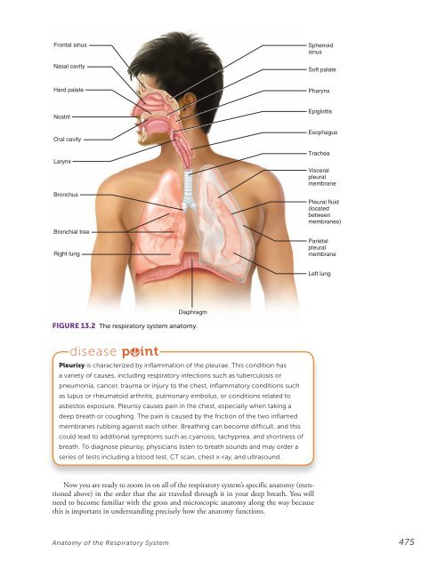
Frontal sinus<br />
Nasal cavity<br />
Sphenoid<br />
sinus<br />
Soft palate<br />
Hard palate<br />
Pharynx<br />
Nostril<br />
Oral cavity<br />
Larynx<br />
Bronchus<br />
Bronchial tree<br />
Right lung<br />
Epiglottis<br />
Esophagus<br />
Trachea<br />
Visceral<br />
pleural<br />
membrane<br />
Pleural fluid<br />
(located<br />
between<br />
membranes)<br />
Parietal<br />
pleural<br />
membrane<br />
Left lung<br />
Diaphragm<br />
FIGURE <strong>13</strong>.2 The respiratory system anatomy.<br />
disease p int<br />
Pleurisy is characterized by inflammation of the pleurae. This condition has<br />
a variety of causes, including respiratory infections such as tuberculosis or<br />
pneumonia, cancer, trauma or injury to the chest, inflammatory conditions such<br />
as lupus or rheumatoid arthritis, pulmonary embolus, or conditions related to<br />
asbestos exposure. Pleurisy causes pain in the chest, especially when taking a<br />
deep breath or coughing. The pain is caused by the friction of the two inflamed<br />
membranes rubbing against each other. Breathing can become difficult, and this<br />
could lead to additional symptoms such as cyanosis, tachypnea, and shortness of<br />
breath. To diagnose pleurisy, physicians listen to breath sounds and may order a<br />
series of tests including a blood test, CT scan, chest x-ray, and ultrasound.<br />
Now you are ready to zoom in on all of the respiratory system’s specific anatomy (mentioned<br />
above) in the order that the air traveled through it in your deep breath. You will<br />
need to become familiar with the gross and microscopic anatomy along the way because<br />
this is important in understanding precisely how the anatomy functions.<br />
Anatomy of the Respiratory System 475