Chapter 13 - McGraw-Hill
Chapter 13 - McGraw-Hill
Chapter 13 - McGraw-Hill
You also want an ePaper? Increase the reach of your titles
YUMPU automatically turns print PDFs into web optimized ePapers that Google loves.
The nasal conchae provide extra surface area for the mucous membranes to function. The<br />
mucous membranes are composed of ciliated pseudostratified columnar epithelial tissue. The<br />
cilia move mucus and any trapped debris posteriorly so that it can be swallowed. Olfactory neurons<br />
located in the roof of the posterior nasal cavity detect odors and provide the sense of smell.<br />
spot check<br />
of mucous membranes?<br />
Why do you think the vestibule is stratified epithelial tissue instead<br />
Sinuses You studied the sinuses of the frontal, ethmoid, sphenoid, and maxilla bones in<br />
the skeletal system chapter. The frontal and sphenoidal sinuses are shown in Figure <strong>13</strong>.4 .<br />
These cavities within the bones are also lined with respiratory epithelial tissue to warm<br />
and moisturize the air. The mucus produced in the sinuses is drained to the nasal cavity<br />
through small openings.<br />
disease p<br />
int<br />
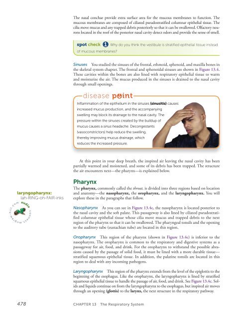
Inflammation of the epithelium in the sinuses (sinusitis) causes<br />
increased mucus production, and the accompanying<br />
swelling may block its drainage to the nasal cavity. The<br />
pressure within the sinuses created by the buildup of<br />
mucus causes a sinus headache. Decongestants<br />
(vasoconstrictors) help reduce the swelling,<br />
thereby improving mucus drainage, which<br />
reduces the increased pressure.<br />
At this point in your deep breath, the inspired air leaving the nasal cavity has been<br />
partially warmed and moistened, and some of its debris has been trapped. The structure<br />
the air encounters next—the pharynx—is explained below.<br />
l<br />
l<br />
i<br />
. c o m<br />
laryngopharynx:<br />
lah-RING-oh-FAIR-inks<br />
audio<br />
. m c g r a w - h<br />
c o n n e c t<br />
Pharynx<br />
The pharynx, commonly called the throat, is divided into three regions based on location<br />
and anatomy—the nasopharynx, the oropharynx, and the laryngopharynx. You will<br />
explore these in the paragraphs that follow.<br />
Nasopharynx As you can see in Figure <strong>13</strong>.4c , the nasopharynx is located posterior to<br />
the nasal cavity and the soft palate. This passageway is also lined by ciliated pseudostratified<br />
columnar epithelial tissue whose cilia move mucus and trapped debris to the next<br />
region of the pharynx so that it can be swallowed. The pharyngeal tonsils and the opening<br />
to the auditory tube (eustachian tube) are located in this region.<br />
Oropharynx This region of the pharynx (shown in Figure <strong>13</strong>.4c ) is inferior to the<br />
nasopharynx. The oropharynx is common to the respiratory and digestive systems as a<br />
passageway for air, food, and drink. For the oropharynx to withstand the possible abrasions<br />
caused by the passage of solid food, it must be lined with a more durable tissue—<br />
stratified squamous epithelial tissue. In addition, the palatine tonsils are located in this<br />
region to deal with any incoming pathogens.<br />
Laryngopharynx This region of the pharynx extends from the level of the epiglottis to the<br />
beginning of the esophagus. Like the oropharynx, the laryngopharynx is lined by stratified<br />
squamous epithelial tissue to handle the passage of air, food, and drink. See Figure <strong>13</strong>.4c . Solids<br />
and liquids continue on from the laryngopharynx to the esophagus, but inspired air moves<br />
through an opening (glottis) to the larynx, the next structure in the respiratory pathway.<br />
478 CHAPTER <strong>13</strong> The Respiratory System


