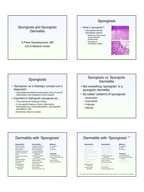
Spongiosis Spongiosis Dermatitis with 'Spongiosis' Dermatitis with ...
Spongiosis Spongiosis Dermatitis with 'Spongiosis' Dermatitis with ...
Spongiosis Spongiosis Dermatitis with 'Spongiosis' Dermatitis with ...
You also want an ePaper? Increase the reach of your titles
YUMPU automatically turns print PDFs into web optimized ePapers that Google loves.
<strong>Spongiosis</strong> and Spongiotic<br />
<strong>Dermatitis</strong><br />
G.Peter Sarantopoulos, MD<br />
UCLA Medical Center<br />
<strong>Spongiosis</strong><br />
• ‘<strong>Spongiosis</strong>’ as a histologic concept (not a<br />
diagnosis!)<br />
– Intra-epidermal edema accompanies many (if not all)<br />
inflammatory skin diseases to some degree<br />
• Important to distinguish spongiosis as…<br />
– The predominant histologic finding<br />
– A non-specific feature of other inflammatory<br />
dermatoses (e.g. lichenoid/interface, vasculopathic,<br />
psoriasiform, etc)<br />
– Sometimes, there is overlap<br />
<strong>Dermatitis</strong> <strong>with</strong> ‘<strong>Spongiosis</strong>’<br />
Neutrophilic:<br />
Pustular psoriasis<br />
Reiter’s syndrome<br />
IgA Pemphigus<br />
Pemphigus herpetiformis<br />
Infantile acropustulosis<br />
AGEP<br />
Palmoplantar pustulosis<br />
SSSS<br />
Neisserial infections<br />
Dermatophytoses<br />
Candidosis<br />
Beetle dermatitis<br />
(Paederus)<br />
Pustular contact<br />
dermatitis<br />
Eosinophilic:<br />
Pemphigus (precursor)<br />
Pemphigus vegetans<br />
Bullous pemphigoid<br />
Cicatricial pemphigoid<br />
Pemphigoid (herpes)<br />
gestationis<br />
Idiopathic eosinophilic<br />
spongiosis<br />
Allergic contact dermatitis<br />
Atopic dermatitis<br />
Arthropod dermatitits<br />
Ofuji’s Disease<br />
Incontinentia pigmenti<br />
Drug reactions<br />
Autoeczematization (‘Id’)<br />
Miliarial:<br />
M. Crystallina<br />
M. Rubra<br />
M. profunda<br />
Follicular:<br />
Infundibulofolliculitis<br />
Atopic dermatitis (follicular<br />
lesions)<br />
Apocrine miliaria<br />
Eosinophilic folliculitis<br />
• What is ‘spongiosis’?<br />
– Intra-epidermal and<br />
intercellular edema<br />
• Widening of intercellular<br />
spaces between<br />
keratinocytes<br />
• Elongation of<br />
intercellular bridges<br />
<strong>Spongiosis</strong><br />
<strong>Spongiosis</strong> vs. Spongiotic<br />
<strong>Dermatitis</strong><br />
• Not everything ‘spongiotic’ is a<br />
spongiotic dermatitis<br />
• So-called ‘patterns of spongiosis’<br />
– Neutrophilic<br />
– Eosinophilic<br />
– Follicular<br />
– Miliarial<br />
<strong>Dermatitis</strong> <strong>with</strong> ‘<strong>Spongiosis</strong>’ *<br />
Neutrophilic:<br />
Pustular psoriasis<br />
Reiter’s syndrome<br />
IgA Pemphigus<br />
Pemphigus herpetiformis<br />
Infantile acropustulosis<br />
AGEP<br />
Palmoplantar pustulosis<br />
SSSS<br />
Neisserial infections<br />
Dermatophytoses<br />
Candidosis<br />
Beetle dermatitis<br />
(Paederus)<br />
Pustular contact<br />
dermatitis<br />
Eosinophilic:<br />
Pemphigus (precursor)<br />
Pemphigus vegetans<br />
Bullous pemphigoid<br />
Cicatricial pemphigoid<br />
Pemphigoid (herpes)<br />
gestationis<br />
Idiopathic eosinophilic<br />
spongiosis<br />
Allergic contact dermatitis<br />
Atopic dermatitis<br />
Arthropod dermatitits<br />
Ofuji’s Disease<br />
Incontinentia pigmenti<br />
Drug reactions<br />
Autoeczematization (‘Id’)<br />
Miliarial:<br />
M. Crystallina<br />
M. Rubra<br />
M. profunda<br />
Follicular:<br />
Infundibulofolliculitis<br />
Atopic dermatitis (follicular<br />
lesions)<br />
Apocrine miliaria<br />
Eosinophilic folliculitis<br />
* Some overlap exists between what Dermatopathologists consider ‘spongiotic dermatitis’ and so-called patterns of spongiosis
Spongiotic <strong>Dermatitis</strong><br />
• Select entities commonly encountered in<br />
daily practice<br />
– Nummular eczema<br />
– Contact dermatitis<br />
– Seborrheic dermatitis<br />
– Pityriasis rosea<br />
Spongiotic <strong>Dermatitis</strong><br />
• Nummular eczema<br />
– Early lesions – spongiosis<br />
leading to vesiculation,<br />
vesicles often contain<br />
inflammatory cells (may<br />
mimic Pautrier’s<br />
microabscesses!)<br />
– Later lesions – progressive<br />
psoriasiform hyperplasia<br />
(less regular than allergic<br />
CD!)<br />
Spongiotic <strong>Dermatitis</strong><br />
• Contact <strong>Dermatitis</strong><br />
–Clinical: Irritant CD<br />
• Reactions vary – simple<br />
erythema to purpura to<br />
eczematous to<br />
vesiculobullous<br />
reactions<br />
• Identified at sites of<br />
exposure<br />
Spongiotic <strong>Dermatitis</strong><br />
• Nummular eczema<br />
–Clinical:<br />
• Tiny papules / papulovesicles,<br />
may coalesce<br />
to form coin-shaped<br />
patches, single or<br />
multiple<br />
• Dorsum of hands,<br />
extensor forearms,<br />
lower legs / outer thigh,<br />
posterior trunk<br />
Spongiotic <strong>Dermatitis</strong><br />
• Nummular eczema<br />
– Early lesions – spongiosis<br />
leading to vesiculation,<br />
vesicles often contain<br />
inflammatory cells (may<br />
mimic Pautrier’s<br />
microabscesses!)<br />
– Later lesions – progressive<br />
psoriasiform hyperplasia<br />
Spongiotic <strong>Dermatitis</strong><br />
• Contact <strong>Dermatitis</strong><br />
–Clinical: Allergic CD<br />
• Erythematous papules,<br />
small vesicles or<br />
weeping plaques<br />
• Lesions arise 12-48 hrs<br />
following exposure to<br />
allergen, lesions often<br />
extend beyond site of<br />
exposure
Spongiotic <strong>Dermatitis</strong><br />
• Contact <strong>Dermatitis</strong><br />
– <strong>Spongiosis</strong> leads to<br />
intraepidermal vesicles<br />
– ‘Irritant’ often more marked<br />
changes, ballooning /<br />
necrosis, possible neuts;<br />
varies <strong>with</strong> irritant<br />
concentration<br />
– ‘Allergic’, spongiosis often<br />
<strong>with</strong> eos, persistent lesions<br />
often show scale crust <strong>with</strong><br />
regular psoriasiform<br />
hyperplasia<br />
Spongiotic <strong>Dermatitis</strong><br />
• Contact <strong>Dermatitis</strong><br />
– <strong>Spongiosis</strong> leads to<br />
intraepidermal vesicles<br />
– ‘Irritant’ often more marked<br />
changes,<br />
ballooning/necrosis,<br />
possible neuts; varies <strong>with</strong><br />
irritant concentration<br />
– ‘Allergic’, spongiosis often<br />
<strong>with</strong> eos, persistent lesions<br />
often show scale crust <strong>with</strong><br />
regular psoriasiform<br />
hyperplasia<br />
Spongiotic <strong>Dermatitis</strong><br />
• Seborrheic dermatitis<br />
– Acute / subacute -<br />
spongiosis <strong>with</strong> scale crust<br />
– Later – psoriasiform<br />
epidermal hyperplasia<br />
– Lymphocytes,<br />
macrophages, occasional<br />
neuts upon a mildly<br />
edematous superficial<br />
papillary dermis<br />
– Note: folliculocentric scale<br />
crust favors SD over<br />
psoriasis<br />
Spongiotic <strong>Dermatitis</strong><br />
• Contact <strong>Dermatitis</strong><br />
– <strong>Spongiosis</strong> leads to<br />
intraepidermal vesicles<br />
– ‘Irritant’ often more marked<br />
changes,<br />
ballooning/necrosis,<br />
possible neuts; varies <strong>with</strong><br />
irritant concentration<br />
– ‘Allergic’, spongiosis often<br />
<strong>with</strong> eos, persistent lesions<br />
often show scale crust <strong>with</strong><br />
regular psoriasiform<br />
hyperplasia<br />
© Weedon, Skin Pathology, 2002<br />
Spongiotic <strong>Dermatitis</strong><br />
• Seborrheic dermatitis<br />
–Clinical:<br />
• Erythematous, scaling<br />
papules and plaques,<br />
sometimes <strong>with</strong> a<br />
greasy appearance<br />
• Found upon<br />
‘seborrheic’ areas –<br />
scalp, ears, eyebrows,<br />
eyelid margins,<br />
nasolabial areas<br />
• Males, after puberty;<br />
common manifestation<br />
in AIDS<br />
Spongiotic <strong>Dermatitis</strong><br />
• Seborrheic dermatitis<br />
– Acute/subacute -<br />
spongiosis <strong>with</strong> scale crust<br />
– Later – psoriasiform<br />
epidermal hyperplasia<br />
– Lymphocytes,<br />
macrophages, occasional<br />
neuts upon a mildly<br />
edematous superficial<br />
papillary dermis<br />
– Note: folliculocentric scale<br />
crust favors SD over<br />
psoriasis
Spongiotic <strong>Dermatitis</strong><br />
• Seborrheic dermatitis<br />
– Acute/subacute -<br />
spongiosis <strong>with</strong> scale crust<br />
– Later – psoriasiform<br />
epidermal hyperplasia<br />
– Lymphocytes,<br />
macrophages, occasional<br />
neuts upon a mildly<br />
edematous superficial<br />
papillary dermis<br />
– Note: folliculocentric scale<br />
crust favors SD over<br />
psoriasis<br />
Spongiotic <strong>Dermatitis</strong><br />
• Pityriasis Rosea<br />
–Clinical:<br />
• Oval, salmon-pink<br />
lesions; initial scaly<br />
plaque ‘herald patch’<br />
often<br />
• Trunk, neck, proximal<br />
extremities; follow lines<br />
of cleavage<br />
• All ages; often 10 – 35<br />
yo<br />
Spongiotic <strong>Dermatitis</strong><br />
• Pityriasis Rosea<br />
– Vaguely undulating<br />
epidermis, ‘mounded’<br />
parakeratosis, usually<br />
lessened granular layer<br />
– Focal spongiosis leads to<br />
small vesicles, dyskeratotic<br />
cells seen at all levels of<br />
epidermis (> in ‘herald<br />
patch’)<br />
– Pigment incontinence,<br />
superficial pap-derm<br />
edema, rbc extrav, mildmod<br />
lymph inflammation<br />
<strong>with</strong> macrophages<br />
Spongiotic <strong>Dermatitis</strong><br />
• Seborrheic dermatitis<br />
– Acute/subacute -<br />
spongiosis <strong>with</strong> scale crust<br />
– Later – psoriasiform<br />
epidermal hyperplasia<br />
– Lymphocytes,<br />
macrophages, occasional<br />
neuts upon a mildly<br />
edematous superficial<br />
papillary dermis<br />
– Note: folliculocentric scale<br />
crust favors SD over<br />
psoriasis<br />
Spongiotic <strong>Dermatitis</strong><br />
• Pityriasis Rosea<br />
– Vaguely undulating<br />
epidermis, ‘mounded’<br />
parakeratosis, usually<br />
lessened granular layer<br />
– Focal spongiosis leads to<br />
small vesicles, dyskeratotic<br />
cells seen at all levels of<br />
epidermis (> in ‘herald<br />
patch’)<br />
– Pigment incontinence,<br />
superficial pap-derm<br />
edema, rbc extrav, mildmod<br />
lymph inflammation<br />
<strong>with</strong> macrophages<br />
Spongiotic <strong>Dermatitis</strong><br />
• Pityriasis Rosea<br />
– Vaguely undulating<br />
epidermis, ‘mounded’<br />
parakeratosis, usually<br />
lessened granular layer<br />
– Focal spongiosis leads to<br />
small vesicles, dyskeratotic<br />
cells seen at all levels of<br />
epidermis (> in ‘herald<br />
patch’)<br />
– Pigment incontinence,<br />
superficial pap-derm<br />
edema, rbc extrav, mildmod<br />
lymph inflammation<br />
<strong>with</strong> macrophages
Spongiotic <strong>Dermatitis</strong><br />
• Pityriasis rosea<br />
– Neuts <strong>with</strong>in parakeratotic mounds favors psoriasis<br />
– Mounded parakeratosis favors PR (over<br />
acute/subacute eczema)<br />
– Always consider drugs - wide range of drugs may<br />
show a PR-like eruption<br />
• Infectious –<br />
– Fungal infections<br />
(dermatophytoses) often<br />
mimic the histologic<br />
features of psoriasis<br />
– Some infections may show<br />
marked spongiosis as well<br />
– even forming marked<br />
vesiculation<br />
– A PAS <strong>with</strong> diastase stain<br />
can quickly lead you to the<br />
diagnosis and save the<br />
patient additional time and<br />
morbidity<br />
Don’t Be Fooled!<br />
Don’t Be Fooled!<br />
Spongiotic <strong>Dermatitis</strong><br />
• Don’t be fooled - spongiosis in and of itself<br />
is a non-specific finding – pitfall!<br />
• <strong>Spongiosis</strong> may be identified as a part of<br />
any number of inflammatory skin disorders<br />
• Look for the predominant reaction pattern<br />
• Examples of overlap…<br />
• Infectious –<br />
– Fungal infections<br />
(dermatophytoses) often<br />
mimic the histologic<br />
features of psoriasis<br />
– Some infections may show<br />
marked spongiosis as well<br />
– even forming marked<br />
vesiculation<br />
– A PAS <strong>with</strong> diastase stain<br />
can quickly lead you to the<br />
diagnosis and save the<br />
patient additional time and<br />
morbidity<br />
Don’t Be Fooled!<br />
Don’t Be Fooled!<br />
• Psoriasiform<br />
– Psoriasis, early psoriasis<br />
may show spongiosis<br />
associated <strong>with</strong> lymphocyte<br />
exocytosis<br />
– Established psoriasis seen<br />
on the palms and soles<br />
may show spongiosis –<br />
making a distinction from<br />
allergic contact dermatitis<br />
difficult<br />
– Erythrodermic psoriasis<br />
may also show spongiosis
Don’t Be Fooled!<br />
• Psoriasiform<br />
– Psoriasis, early psoriasis<br />
may show spongiosis<br />
associated <strong>with</strong> lymphocyte<br />
exocytosis<br />
– Established psoriasis seen<br />
on the palms and soles<br />
may show spongiosis –<br />
making a distinction from<br />
allergic contact dermatitis<br />
difficult<br />
– Erythrodermic psoriasis<br />
may also show spongiosis<br />
Don’t Be Fooled!<br />
• Vasculopathic<br />
– Erythema annulare<br />
centrifugum (EAC),<br />
characteristic annular,<br />
erythematous lesion may<br />
show a fine scale inside the<br />
advancing edge<br />
– Histology shows<br />
spongiosis, parakeratosis<br />
and an underlying<br />
superficial perivascular<br />
lymphocytic inflammation,<br />
often <strong>with</strong> a ‘coat-sleeve’<br />
appearance<br />
Spongiotic <strong>Dermatitis</strong><br />
• Vasculopathic<br />
– Pruritic urticarial papules<br />
and plaques of pregnancy<br />
(PUPPP), aka polymorphic<br />
eruption of pregnancy<br />
– Epidermal changes, to<br />
include spongiosis and<br />
parakeratosis <strong>with</strong><br />
exocytosis of inflammatory<br />
cells may be seen in up to<br />
1/3 of cases<br />
– Lymphocytic vasculitis <strong>with</strong><br />
varying admixture of<br />
eosinophils and variable<br />
edema of the superficial<br />
papillary dermis<br />
Don’t Be Fooled!<br />
• Psoriasiform<br />
– Psoriasis, early psoriasis<br />
may show spongiosis<br />
associated <strong>with</strong> lymphocyte<br />
exocytosis<br />
– Established psoriasis seen<br />
on the palms and soles<br />
may show spongiosis –<br />
making a distinction from<br />
allergic contact dermatitis<br />
difficult<br />
– Erythrodermic psoriasis<br />
may also show spongiosis<br />
Don’t Be Fooled!<br />
• Vasculopathic<br />
– Erythema annulare<br />
centrifugum (EAC),<br />
characteristic annular,<br />
erythematous lesion may<br />
show a fine scale inside the<br />
advancing edge<br />
– Histology shows<br />
spongiosis, parakeratosis<br />
and an underlying<br />
superficial perivascular<br />
lymphocytic inflammation,<br />
often <strong>with</strong> a ‘coat-sleeve’<br />
appearance<br />
Spongiotic <strong>Dermatitis</strong><br />
• Vasculopathic<br />
– Pruritic urticarial papules<br />
and plaques of pregnancy<br />
(PUPPP), aka polymorphic<br />
eruption of pregnancy<br />
– Epidermal changes, to<br />
include spongiosis and<br />
parakeratosis <strong>with</strong><br />
exocytosis of inflammatory<br />
cells may be seen in up to<br />
1/3 of cases<br />
– Lymphocytic vasculitis <strong>with</strong><br />
varying admixture of<br />
eosinophils and variable<br />
edema of the superficial<br />
papillary dermis
Spongiotic <strong>Dermatitis</strong><br />
• Vasculopathic<br />
– Pruritic urticarial papules<br />
and plaques of pregnancy<br />
(PUPPP), aka polymorphic<br />
eruption of pregnancy<br />
– Epidermal changes, to<br />
include spongiosis and<br />
parakeratosis <strong>with</strong><br />
exocytosis of inflammatory<br />
cells may be seen in up to<br />
1/3 of cases<br />
– Lymphocytic vasculitis <strong>with</strong><br />
varying admixture of<br />
eosinophils and variable<br />
edema of the superficial<br />
papillary dermis<br />
Clinical History<br />
• 54 yo woman is seen by her Dermatologist<br />
complaining of an itching, burning rash<br />
• Physical exam showed a morbilliform rash<br />
on the lower extremities and trunk<br />
• Further questioning revealed that the<br />
patient had recently changed one of her<br />
blood pressure medications<br />
Drug Eruption<br />
• Drug eruptions are one of the more<br />
commonly biopsied inflammatory skin<br />
lesions<br />
– Drug eruptions may show a wide range of<br />
histologic patterns<br />
– <strong>Spongiosis</strong> <strong>with</strong> eosinophils is a common<br />
pattern<br />
• Any number of medications may incite a<br />
rash<br />
Spongiotic <strong>Dermatitis</strong><br />
• Interface/lichenoid<br />
– Erythema multiforme, early<br />
lesions may show intraand<br />
intercellular<br />
intraepidermal edema<br />
(spongiosis)<br />
Sources<br />
Weedon, Skin Pathology. Churchill Livingstone, 2002<br />
McKee, Pathology of the Skin, Elsevier-Mosby, 2005<br />
Additional micrographs from personal collection<br />
Questions/comments: gsarantopoulos@mednet.ucla.edu


