Dermoid, Epidermoid, Cysts, Lipoma - Viktor's Notes for the ...
Dermoid, Epidermoid, Cysts, Lipoma - Viktor's Notes for the ...
Dermoid, Epidermoid, Cysts, Lipoma - Viktor's Notes for the ...

You also want an ePaper? Increase the reach of your titles
YUMPU automatically turns print PDFs into web optimized ePapers that Google loves.
DERMOID, EPIDERMOID, CYSTS, LIPOMA Onc30 (1)<br />
<strong>Dermoid</strong>, <strong>Epidermoid</strong>, <strong>Cysts</strong>, <strong>Lipoma</strong><br />
Updated: April 24, 2010<br />
DERMOID TUMOR, EPIDERMOID TUMOR (S. CHOLESTEATOMA)............................................................ 1<br />
COLLOID CYSTS....................................................................................................................................... 3<br />
ARACHNOID CYSTS.................................................................................................................................. 4<br />
LIPOMA .................................................................................................................................................... 5<br />
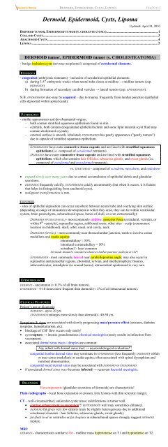
DERMOID tumor, EPIDERMOID tumor (s. CHOLESTEATOMA)<br />
- benign inclusion cysts (not true neoplasms!) composed of ectodermal elements.<br />
ETIOLOGY<br />
- congenital (embryonic remnants) - inclusion of ectodermal epi<strong>the</strong>lial elements:<br />
a) during 3-5 th embryonic weeks when neural tube closes at midline → midline tumors (esp.<br />
DERMOIDS)<br />
b) during <strong>for</strong>mation of secondary cerebral vesicles → lateral tumors (esp. EPIDERMOIDS).<br />
N.B. EPIDERMOIDS also may be acquired – due to trauma, frequently from lumbar puncture (epi<strong>the</strong>lial<br />
cells deposited within spinal canal).<br />
PATHOLOGY<br />
- similar appearances and developmental origins;<br />
– both contain stratified squamous epi<strong>the</strong>lium found in skin.<br />
– centrally, both contain desquamated epi<strong>the</strong>lial keratin and some lipid material (cyst fluid may<br />
contain cholesterol crystals).<br />
– external surface is smooth, lobulated; EPIDERMOIDS has pearly appearance ("pearly tumors")<br />
due to capsule of stratified squamous epi<strong>the</strong>lium.<br />
EPIDERMOIDS have outer connective tissue capsule and are lined with stratified squamous<br />
epi<strong>the</strong>lium (i.e. composed of ectodermal remnants).<br />
DERMOIDS have outer connective tissue capsule and are lined with stratified squamous<br />
epi<strong>the</strong>lium, which also contains hair follicles, sebaceous glands, and sweat glands (i.e.<br />
composed of ectodermal and mesodermal remnants).<br />
vs. TERATOMAS - composed of ectoderm, mesoderm, and endoderm<br />
expand slowly over many years due to central accumulation of epi<strong>the</strong>lial debris and glandular<br />
secretions.<br />
DERMOIDS frequently calcify; EPIDERMOIDS calcify uncommonly (but when it occurs, it is feature<br />
that helps in distinguishing from arachnoid cysts).<br />
malignant trans<strong>for</strong>mation is rare.<br />
LOCATION:<br />
- sites of epi<strong>the</strong>lial deposition can occur anywhere between neural tube and overlying skin surface<br />
(depending on stage of intrauterine development at which <strong>the</strong>y arise, <strong>the</strong>y can lie within ventricular<br />
system, brain parenchyma, subarachnoid space, bones of skull, or even extracranially):<br />
DERMOIDS (INTRACRANIAL) - most commonly midline: posterior fossa (extradural, vermian, or<br />
within 4 th ventricle), suprasellar region, subfrontal areas; o<strong>the</strong>r sites – scalp (commonest<br />
location in childhood), skull, orbit, nasal, oral cavity, neck.<br />
DERMOIDS (SPINAL) - most commonly near thoracolumbar junction, tends to involve conus<br />
medullaris and cauda equina:<br />
intramedullary ≈ 50%<br />
intradural extramedullary ≈ 50%<br />
extradural ≈ least common<br />
<strong>Dermoid</strong>s should be considered whenever lumbar puncture yields fat in CSF!<br />
EPIDERMOIDS - most commonly lateral near cerebellopontine angle; may also occur in<br />
suprasellar and parasellar regions, choroidal, sylvian, and interhemispheric fissures,<br />
intraventricular, intradiploic (in cranial bones); intracerebral epidermoid is very rare.<br />
EPIDEMIOLOGY<br />
DERMOID - uncommon (≈ 0.3% of all brain tumors).<br />
EPIDERMOID - 4-10 times more frequent than dermoid (≈ 2% of all intracranial tumors).<br />
CLINICAL FEATURES<br />
Patient’s age at diagnosis:<br />
DERMOID - up to 20 yrs<br />
EPIDERMOID (enlarges more slowly than dermoid) - 40-50 yrs.<br />
Symptoms & signs are associated with slowly progressing mass/pressure effect (seizures, diabetes<br />
insipidus, hypopituitarism, etc).<br />
blockage of CSF flow occurs only rarely!<br />
cyst rupture → intense granulomatous chemical meningitis (rarely results in infarction from<br />
vasospasm).<br />
associated dermal sinus tracts / dimples are common:<br />
Any infant with dermal sinus tract → neuroradiological evaluation!<br />
congenital lumbar dermal sinus may terminate in EPIDERMOID (less frequently DERMOID) within<br />
or near conus medullaris or cauda equina; often associated with spinal dysraphism and<br />
vertebral abnormalities.<br />
congenital nasal dermal sinus may be associated with DERMOID or EPIDERMOID.<br />
if associated dermal sinus tract becomes infected → recurrent bacterial meningitis.<br />
DIAGNOSIS<br />
Fat components (glandular secretions of dermoids) are characteristic!<br />
Plain radiographs – local bone expansion or erosion, lytic lesions with thin sclerotic margin.<br />
CT – well-circumscribed, unilocular cystic mass; calcifications in tumor wall.<br />
contrast enhancement is uncommon!!! (EPIDERMOID wall may sometimes enhance).<br />
DERMOID fat gives very low density (may be slightly heterogeneous due to additional<br />
ectodermal elements - hair follicles, sebaceous glands, sweat glands).<br />
fat-fluid level in ventricles or fat droplets in subarachnoid spaces strongly suggest DERMOID<br />
rupture.<br />
MRI:<br />
DERMOID - characteristics similar to fat – midline mass hyperintense on T1 and hypointense on T2.
DERMOID, EPIDERMOID, CYSTS, LIPOMA Onc30 (2)<br />
chemical-shift artifact is often present on T2 images as markedly hypointense band posterior at<br />
fat-fluid interface.<br />
EPIDERMOID - characteristics similar to CSF – variably hypointense on T1 and variably hyperintense on<br />
T2.<br />
Angiography - avascular mass.<br />
Prenatal diagnosis with ultrasound (and resection shortly after birth) are now possible.<br />
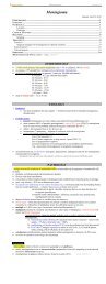
Suprasellar dermoid:<br />
A) noncontrast T1 - high-signal-intensity suprasellar mass extending along planum sphenoidale.<br />
B) contrast T1 with fat saturation - small amount of enhancement along peripheral aspects of lesion (arrow); majority of<br />
mass suppresses with fat saturation:<br />
<strong>Dermoid</strong> (nonenhanced CT) - well-circumscribed,<br />
cystic, low-attenuating lesion at midline in suprasellar<br />
region, posterior to 3 rd ventricle; small focus of<br />
calcification is noted at posterior margin of tumor:<br />
Same dermoid (T1 with contrast) - nodular focus of<br />
enhancement in right side of suprasellar lesion:<br />
<strong>Dermoid</strong> (nonenhanced CT) - large, well-circumscribed<br />
low-attenuating cystic lesion in right temporal lobe<br />
lateral to cranial midline; peripheral marginal<br />
calcification; no erosion in adjacent bone of sella:<br />
Same dermoid (T1) – hyperintense signal in lesion;<br />
multiple small hyperintense foci along sulci of right<br />
temporal lobe (represent fat droplets in subarachnoid<br />
space from focal dermoid rupture):<br />
<strong>Epidermoid</strong> (T1 with contrast) - suprasellar,<br />
prepontine, and interpeduncular location of<br />
nonenhancing tumor (signal intensity similar to<br />
CSF):<br />
Same dermoid (T1) - hypointense lesion; crescentic<br />
posterior rim of hyperintensity represents fat chemicalshift<br />
artifact:<br />
Same dermoid (T2) - hyperintense cystic component in<br />
lesion:<br />
Same dermoid (contrast CT) - partial marginal<br />
enhancement; attenuation degree in center of lesion<br />
consistent with fat:<br />
Same dermoid (T1 with contrast) - hyperintense lesion<br />
(hyperintensity is due to short T1 of fat); multiple<br />
hyperintense foci (fat droplets) in subarachnoid spaces;<br />
mild midline septal shift to left; chemical-shift artifact<br />
at superior marginal surface of lesion:<br />
<strong>Epidermoid</strong> (A- T2-weighted; B - T1-weighted MRI): left<br />
Sylvian fissure is filled by mass which extends into chiasmatic<br />
cistern and encases left internal carotid artery termination<br />
(arrowhead); signal is similar to CSF on T2, but slightly<br />
higher than CSF on T1 (white arrow):
DERMOID, EPIDERMOID, CYSTS, LIPOMA Onc30 (3)<br />
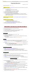
<strong>Epidermoid</strong>:<br />
A. T2-MRI – large homogeneous mass, which is slightly higher in signal than CSF, fills right cerebellopontine angle<br />
B. T1-MRI - lesion is again noted to be hyperintense to cerebrospinal fluid.<br />
TREATMENT<br />
- complete surgical excision is curative.<br />
epidermoids can interdigitate around vital neuronal structures, complicating surgical removal.<br />
avoid spilling of contents (→ chemical meningitis).<br />
associated dermal sinus should be removed completely.<br />
Chemo<strong>the</strong>rapy and radio<strong>the</strong>rapy are not useful.<br />
COLLOID CYSTS<br />
- congenital benign tumors that can cause sudden death because of <strong>the</strong>ir location (almost always found<br />
in 3 rd ventricle → obstructive hydrocephalus).<br />
0.5-1% of all primary brain tumors (15-20% of all intraventricular masses).<br />
ETIOLOGY<br />
- possible sources:<br />
a) in 1910, Sjovall hypo<strong>the</strong>sized that colloid cysts are remnants of PARAPHYSIS (embryonic<br />
midline structure within diencephalic roof immediately rostral to telencephalic border, in<br />
posterior lip of <strong>for</strong>amen of Monro) - cells of paraphysis are similar to those found in colloid<br />
cysts (i.e. low columnar epi<strong>the</strong>lial cells without cilia or blepharoplasts) - colloid cysts were<br />
called paraphysial cysts <strong>for</strong> 50 years.<br />
b) diencephalic ependyma<br />
c) invagination of neuroepi<strong>the</strong>lium of ventricle<br />
d) respiratory epi<strong>the</strong>lium of endodermal origin.<br />
PATHOLOGY<br />
arise in anterior superior portion of 3 rd ventricle between <strong>for</strong>nices, immediately dorsal to Monro<br />
<strong>for</strong>amen.<br />
also have been reported to arise in septum pellucidum, 4 th ventricle, sella turcica.<br />
attached to roof of 3 rd ventricle (and frequently to choroid plexus).<br />
gross appearance of small white ball.<br />
lined with simple or pseudostratified cuboidal or low columnar ciliated epi<strong>the</strong>lial cells (PASpositive;<br />
stain positively <strong>for</strong> S100 and negatively <strong>for</strong> glial fibrillary acidic protein, vimentin, and<br />
neurofilament).<br />
epi<strong>the</strong>lial lining secretes mucinous fluid (greenish, of variable viscosity) → cyst enlargement.<br />
CLINICAL FEATURES<br />
- classic intermittent obstructive hydrocephalus with paroxysmal headache associated with changing<br />
head position (large cyst obstructing Monro <strong>for</strong>amen).<br />
Positional headache!<br />
usually present in age 20-50 yrs (youngest reported case - 2-month-old infant).<br />
o<strong>the</strong>r reported symptoms (sometimes related to changes in posture):<br />
– sudden weakness in lower limbs associated with falls without loss of consciousness.<br />
– symptoms similar to normal pressure hydrocephalus (dementia, gait disturbance,<br />
urinary incontinence).<br />
Mental changes are common (may persist after surgery if <strong>for</strong>nix is damaged)!<br />
sudden death (incidence appears to be low) has been reported; may not correlate to tumor size,<br />
degree of ventricular dilatation, or duration of symptoms.<br />
DIAGNOSIS<br />
CT - well delineated, round or ovoid, homogenous, 66% hyperdense (to surrounding parenchyma) and<br />
33% isodense.<br />
most are 5-25 mm.<br />
typically nonenhancing and uncalcified (occasional thin rim of enhancement).<br />
viscosity* of cyst contents correlates more closely to radiodensity on CT than to density visible on<br />
MRI.<br />
*viscosity determines most appropriate surgical approach; hyperdense cyst is more<br />
likely to have solid contents - more difficult to drain, but reduced capacity to enlarge<br />
over time.<br />
MRI - hyperintense on T1 and hypointense on T2.<br />
N.B. CSF flow artifact at Monro <strong>for</strong>amen can mimic colloid cyst!<br />
amount of rim enhancement is variable.<br />
MRI differentiates colloid cyst from basilar tip aneurysm (may have similar appearance on CT).
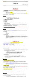
T1-MRI without contrast: well-circumscribed highsignal-intensity<br />
lesion adjacent to <strong>for</strong>amen of Monro:<br />
DERMOID, EPIDERMOID, CYSTS, LIPOMA Onc30 (4)<br />
CT - dense, rounded mass in region of <strong>for</strong>amina of Monro<br />
causing enlargement of lateral ventricles, and indenting<br />
anterior aspect of 3 rd ventricle:<br />
TREATMENT<br />
Immediate attention to hydrocephalus!<br />
Strategy:<br />
a) large cyst & hydrocephalus → surgery.<br />
– if patient is too ill → bilateral CSF diversion (suboptimal because sudden death has<br />
been reported in absence of acute obstructive hydrocephalus)<br />
b) small cyst, large ventricles, few or no symptoms → observation with serial MRIs.<br />
c) small cysts and normal-sized ventricles → observation.<br />
N.B. prevention of sudden death is not indication <strong>for</strong> surgery in asymptomatic patients with small cysts<br />
and no hydrocephalus!<br />
Surgical approaches:<br />
Transcortical approach<br />
The transcortical approach involves making corticectomy over middle frontal gyrus and proceeding to frontal<br />
horn of lateral ventricle. Intraoperative ultrasonography may aid in approach to ventricle. The Monro <strong>for</strong>amen is<br />
visualized at convergence of septal veins, thalamostriate vein, and choroid plexus. The <strong>for</strong>nix arches over<br />
superior and anterior margins of <strong>for</strong>amen. Avoiding <strong>for</strong>nix is important because unilateral <strong>for</strong>nix damage has<br />
been associated with amnesia. The cyst should be readily visualized through <strong>for</strong>amen. The cyst is punctured and<br />
contents are aspirated, internally decompressing walls of cyst.<br />
Avoid excessive retraction of walls of lateral ventricle because genu of internal capsule is in subependyma.<br />
O<strong>the</strong>r concerns include damaging thalamostriate veins, which can result in basal ganglia damage. After cyst has<br />
been decompressed, completely remove it in order to prevent recurrence. Leaving small portion of cyst behind<br />
may be necessary if it is attached to ei<strong>the</strong>r thalamostriate or internal cerebral veins.<br />
The transcortical approach carries increased incidence of epilepsy.<br />
Intraoperative photograph through operating microscope shows colloid cyst in Monro <strong>for</strong>amen. Choroid<br />
plexus is observed overlying cyst, and thalamostriate vein is along inferior border:<br />
Intraoperative photograph showing removal of cyst, leaving dilated Monro <strong>for</strong>amen The third ventricle<br />
can be seen through opening:<br />
Transcallosal approach<br />
The transcallosal approach avoids incising cortex. A right frontal craniotomy is made two thirds anterior and<br />
one third posterior to coronal suture, crossing midline to expose superior sagittal sinus. The right frontal lobe is<br />
<strong>the</strong>n retracted laterally, and corpus callosum is exposed. Draining cortical veins must be avoided if possible. The<br />
preoperative MRI can help identify potential veins. A 1-centimeter incision is made in corpus callosum,<br />
allowing entry to lateral ventricle. The Monro <strong>for</strong>amen can <strong>the</strong>n be visualized, and septum pellucidum may be<br />
divided to see contralateral <strong>for</strong>amen. Ei<strong>the</strong>r ventricle may be entered through standard right frontal transcallosal<br />
approach. Close inspection of orientation of choroid plexus, caudate nucleus, and Monro <strong>for</strong>amen helps<br />
determine which of ventricles has been entered.<br />
The transcallosal approach decreases risk of postoperative epilepsy but risks venous infarction and contralateral<br />
leg weakness from prolonged retraction. An extensive callosal resection may also cause temporary mutism.<br />
Excessive manipulation of <strong>for</strong>nix may affect memory.<br />
Endoscopic approach<br />
The endoscopic approach is same as transcortical approach, with exception that <strong>for</strong>mer is accomplished through<br />
burr hole. The cyst is punctured and aspirated through working channels of endoscope.<br />
The endoscopic approach is least invasive, but it can be used only on cysts that can be aspirated.<br />
Hydrocephalus can persist after surgery, even after resection of cyst. This complication may be secondary to<br />
spillage of cyst contents or to bleeding during surgery. A ventricular ca<strong>the</strong>ter may be placed intraoperatively to<br />
safeguard against ventricular dilatation.<br />
POSTOPERATIVE FOLLOW-UP<br />
Hydrocephalus may develop despite cyst removal; H: periodic CT.<br />
ARACHNOID CYSTS<br />
arise anywhere on brain surface.<br />
some grow to remarkable size.<br />
smooth surface (vs. EPIDERMOIDS - cauliflower-like deep clefts).<br />
never calcify!<br />
majority are incidental findings - best left alone.
DERMOID, EPIDERMOID, CYSTS, LIPOMA Onc30 (5)<br />
T1-MRI - large, fluid-filled structure expands left cerebellopontine angle cistern (arrowheads); note elongation and<br />
thinning of cranial nerves VII and VIII (white arrow):<br />
LIPOMA<br />
derived from mesoderm.<br />
occur chiefly in midline (esp. over corpus callosum*, vermis, quadrigeminal cistern, spinal dural<br />
sac).<br />
*often associated with callosal dysgenesis<br />
majority are incidental findings.<br />
characteristic appearance on both CT and MRI - fat density; calcification is frequent in periphery.<br />
BIBLIOGRAPHY <strong>for</strong> ch. “Neuro-Oncology” → follow this LINK >><br />
Viktor’s <strong>Notes</strong>℠ <strong>for</strong> <strong>the</strong> Neurology Resident<br />
Please visit website at www.NeurologyResident.net